KOLAJO OLAJIDE K PSY 101
15
UNIVERSITY OF IBADAN, OYO STATE, NIGERIA NAME: KOLAJO, OLAJIDE KAZEEM MATRIC/NO: E029368 DEPARTMENT: ECONOMICS LEVEL: 100 COURSE TITLE: INTRO TO PSYCHO-BIOLOGICAL BASIS OF BEHAVIOUR COURSE CODE: PSY 101 QUESTION: 1 WITH ADEQUATE ILLUSTRATION, DESCRIBE FIVE ORGANS OF SENSATION AND PERCEPTION (1) Eye (2) Ear (3) Nose (4) Skin and (5) Tongue 1. EYE The eye is the organ of the sense of sight situated in the orbital cavity. It is supplied by the optic nerve. It is almost spherical in shape and is about 2.5cm in diameter. The eye is the organ of vision. It has a complex structure consisting of a transparent lens that focuses light on the retina. The retina is covered with two basic types of light-sensitive cells-rods and cones. The cone cells are sensitive to color and are located in the part of the retina called the fovea, where the light is focused by the lens. The rod cells are not sensitive to color, but have greater sensitivity to light than the cone cells. These cells are located around the fovea and are responsible for peripheral vision and night vision. The eye is connected to the brain through the optic nerve. The point of this connection is called the "blind spot" because it is insensitive to light. Experiments have shown that the back of the brain maps the visual input from the eyes. The brain combines the input of our two eyes into a single three-dimensional image. In addition, even though the image on the retina is upside-down because of the focusing action of the lens, the brain compensates and provides the right-side-up perception. Experiments have been done with subjects fitted with prisms that invert the images. The subjects go through an initial period of great confusion, but subsequently they perceive the images as right side up. The range of perception of the eye is phenomenal. In the dark, a substance produced by the rod cells increases the sensitivity of the eye so that it is possible to detect very dim light. In strong light, the iris contracts reducing the size of the aperture that admits light into the eye and a protective obscure substance reduces the exposure of the light-sensitive cells. The spectrum of light to which the eye is sensitive varies from the red to the violet. Lower electromagnetic frequencies in the infrared are sensed as heat, but cannot be seen. Higher frequencies in the ultraviolet and beyond cannot be seen either, but can be sensed as tingling of the skin or eyes depending on the frequency. The human eye is not sensitive to the polarization of light, i.e., light that oscillates on a specific plane. Bees, on the other hand, are sensitive to polarized light, and have a visual range that extends into the ultraviolet. Some kinds of snakes have special infrared sensors that enable them to hunt in absolute darkness using only the heat
Transcript of KOLAJO OLAJIDE K PSY 101
UNIVERSITY OF IBADAN, OYO STATE, NIGERIA
NAME: KOLAJO, OLAJIDE KAZEEM MATRIC/NO: E029368 DEPARTMENT: ECONOMICS LEVEL: 100 COURSE TITLE: INTRO TO PSYCHO-BIOLOGICAL BASIS OF BEHAVIOUR COURSE CODE: PSY 101 QUESTION: 1 WITH ADEQUATE ILLUSTRATION, DESCRIBE FIVE ORGANS OF SENSATION AND PERCEPTION
(1) Eye (2) Ear (3) Nose (4) Skin and (5) Tongue
1. EYE
The eye is the organ of the sense of sight situated in the orbital cavity. It is supplied by the optic nerve. It is almost spherical in shape and is about 2.5cm in diameter. The eye is the organ of vision. It has a complex structure consisting of a transparent lens that focuses light on the retina. The retina is covered with two basic types of light-sensitive cells-rods and cones. The cone cells are sensitive to color and are located in the part of the retina called the fovea, where the light is focused by the lens. The rod cells are not sensitive to color, but have greater sensitivity to light than the cone cells. These cells are located around the fovea and are responsible for peripheral vision and night vision. The eye is connected to the brain through the optic nerve. The point of this connection is called the "blind spot" because it is insensitive to light. Experiments have shown that the back of the brain maps the visual input from the eyes.
The brain combines the input of our two eyes into a single three-dimensional image. In addition, even though the image on the retina is upside-down because of the focusing action of the lens, the brain compensates and provides the right-side-up perception. Experiments have been done with subjects fitted with prisms that invert the images. The subjects go through an initial period of great confusion, but subsequently they perceive the images as right side up.
The range of perception of the eye is phenomenal. In the dark, a substance produced by the rod cells increases the sensitivity of the eye so that it is possible to detect very dim light. In strong light, the iris contracts reducing the size of the aperture that admits light into the eye and a protective obscure substance reduces the exposure of the light-sensitive cells. The spectrum of light to which the eye is sensitive varies from the red to the violet. Lower electromagnetic frequencies in the infrared are sensed as heat, but cannot be seen. Higher frequencies in the ultraviolet and beyond cannot be seen either, but can be sensed as tingling of the skin or eyes depending on the frequency. The human eye is not sensitive to the polarization of light, i.e., light that oscillates on a specific plane. Bees, on the other hand, are sensitive to polarized light, and have a visual range that extends into the ultraviolet. Some kinds of snakes have special infrared sensors that enable them to hunt in absolute darkness using only the heat

UNIVERSITY OF IBADAN, OYO STATE, NIGERIA emitted by their prey. Birds have a higher density of light-sensing cells than humans do in their retinas, and therefore, higher visual acuity.
Color blindness or "Daltonism" is a common abnormality in human vision that makes it impossible to differentiate colors accurately. One type of color blindness results in the inability to distinguish red from green. This can be a real handicap for certain types of occupations. To a colorblind person, a person with normal color vision would appear to have extrasensory perception. However, the term "extrasensory perception" is the perception that is beyond the range of the normal.
2. EAR
The ear is the organ of hearing. It is sensitive to sound waves in the air which vibrates the tympanic membrane to cause sensory stimulation of the middle ear. The stimulus arising from this is interpreted in the brain upon which appropriate responses are made by the nervous system. The outer ear protrudes away from the head and is shaped like a cup to direct sounds toward the tympanic membrane, which transmits vibrations to the inner ear through a series of small bones in the middle ear called the malleus, incus and stapes. The inner ear, or cochlea, is a spiral-shaped chamber covered internally by nerve fibers that react to the vibrations and transmit impulses to the brain via the auditory nerve. The brain combines the input of our two ear to determine the direction and distance of sounds.
The inner ear has a vestibular system formed by three semicircular canals that are approximately at right angles to each other and which are responsible for the sense of balance and spatial orientation. The inner ear has chambers filled with a viscous fluid and small particles (Otoliths) containing calcium carbonate. The movement of these particles over small hair cells in the inner ear sends signals to the brain that are interpreted as motion and acceleration.
The human ear can perceive frequencies from 16 cycles per second, which is a very deep bass, to 28,000 cycles per second, which is a very high pitch. Bats and dolphins can detect frequencies higher than 100,000 cycles per second. The human ear can detect pitch changes as small as 3 hundredths of one percent of the original frequency in some frequency ranges. Some people have "perfect pitch", which is the ability to map a tone precisely on the musical scale without reference to an external standard. It is estimated that less than one in ten thousand people have perfect pitch, but speakers of tonal languages like Vietnamese and Mandarin show remarkably precise absolute pitch in reading out lists of words because pitch is an essential feature in conveying the meaning of words in tone languages. The Eguchi Method teaches perfect pitch to children starting before they are 4 years old. After age 7, the ability to recognize notes does not improve much.
The loudness of a sound is determined by the height or amplitude of the sound wave and is measured in units called decibels. A whisper is about 30 decibels whereas the usual live band blasts about 110 decibels. The higher the decibel level, the more intense the sound and the greater the danger of damaging the organs of hearing. This is why people that work near noisy equipment are always advised to wear ear protecting equipment.
3. NOSE
UNIVERSITY OF IBADAN, OYO STATE, NIGERIA The nose is the organ responsible for the sense of smell. The cavity of the nose is lined with mucous membranes that have smell receptors connected to the olfactory nerve. The smells themselves consist of vapors of various substances. The smell receptors interact with the molecules of these vapors and transmit the sensations to the brain. The nose also has a structure called the vomeronasal organ whose function has not been determined, but which is suspected of being sensitive to pheromones that influence the reproductive cycle. The smell receptors are sensitive to seven types of sensations that can be characterized as camphor, musk, flower, mint, ether, acrid, or putrid. The sense of smell is sometimes temporarily lost when a person has a cold. Dogs have a sense of smell that is many times more sensitive than man's.
The nose contains specialized sensory nerve cells, or neurons, with hairlike fibers called cilia on one end. Each neuron sends a nerve fibre called an axon to the olfactory bulb which is a brain structure just above the nose.
Most animals can distinguish thousands of odors. Early studies showed that different olfactory neurons respond to different odors, and that odors stimulate specific patterns of activity in the olfactory bulb.
Olfactory information travels not only to the limbic system -- primitive brain structures that govern emotions, behavior, and memory storage -- but also to the brain’s cortex, or outer layer, where conscious thought occurs. In addition, it combines with taste information in the brain to create the sensation of flavor.
4. SKIN
The skin is the organ of touch. The sense of touch is not really one sense but a combination of at least four senses namely touch, pain, warm, and cold. There are four respective surfaces on the skin through which these senses could be perceived. These physiological processes are embedded in what is known as sematosensation. The sense of touch is distributed throughout the body. Nerve endings in the skin and other parts of the body transmit sensations to the brain. Some parts of the body have a larger number of nerve endings and, therefore, are more sensitive.
Sematosensation refers to a combination of processes related to perception of sensation as well as movement of the body. Mammalian skin has series of sensory receptors located peripherally in various parts of the body with the ability to respond to a variety of sensory stimulus. The sensory receptors which are unevenly distributed could be explored with devices such as fine hairs, needle points, warmed or cooled pointed hammers. Similarly, the separate sensitive spots differ in frequency and location or anatomical distribution.
A touch receptor may consist of a simple bared ending, an elaborated neuron ending, or a bare ending surrounded by non- neural cells that modify its function (Kalat, 1988). Areas with greater sensitivity of the skin such as the fingertips have as many as 700 touch cells in 2 square millimeters surface. Different receptors are associated with different types of sensations. On receiving a stimulus, each of the information received takes specific pathways to communicate with specific areas in the brain. With this unique distinction, any localized tissue damage of a skin surface for instance could destroy the local tissue without destroying the perception of stimulus related to the area damaged, or

UNIVERSITY OF IBADAN, OYO STATE, NIGERIA vice versa. On the other hand, because pain and temperature sensations are perceived along the same path, any injury that affects one path will automatically hinder the other path (Kalat, 1988).
Hairs on the skin magnify the sensitivity and act as an early warning system for the body. The fingertips and the sexual organs have the greatest concentration of nerve endings. The sexual organs have "erogenous zones" that when stimulated start a series of endocrine reactions and motor responses resulting in orgasm.
5. TONGUE
The tongue is the organ of taste. Taste drives appetite and protects us from poisons. So, we like the taste of sugar because we have an absolute requirement for carbohydrates (sugars etc.). We get carvings for salt because we must have sodium chloride (common salt) in our diet. Bitter and sour cause aversive, avoidance reactions because most poisons are bitter (most bitter substances are bad for us- certainly in excess) and off food goes sour (acidic).
The receptors for taste, called taste buds, are situated chiefly in the tongue, but they are also located in the roof of the mouth and near the pharynx. They are able to detect four basic tastes: salty, sweet, bitter, and sour. The tongue also can detect a sensation called "umami" from taste receptors sensitive to amino acids. Generally, the taste buds close to the tip of the tongue are sensitive to sweet tastes, whereas those in the back of the tongue are sensitive to bitter tastes. The taste buds on top and on the side of the tongue are sensitive to salty and sour tastes. At the base of each taste bud there is a nerve that sends the sensations to the brain. The sense of taste functions in coordination with the sense of smell. The number of taste buds varies substantially from individual to individual, but greater numbers increase sensitivity. Women, in general, have a greater number of taste buds than men. As in the case of color blindness, some people are insensitive to some tastes.
QUESTION: 2 DRAWING ILLUSTRATIONS REQUIRED FOR EACH ORGAN WITH ADEQUATE LABELING
HUMAN EYE

UNIVERSITY OF IBADAN, OYO STATE, NIGERIA DIAGRAM OF THE HUMAN EYES SHOWING THE FUNCTIONAL AREA
HUMAN EAR
DIAGRAM OF THE HUMAN EAR
HUMAN NOSE
THE STRUCTURE OF HUMAN NOSE
UNIVERSITY OF IBADAN, OYO STATE, NIGERIA
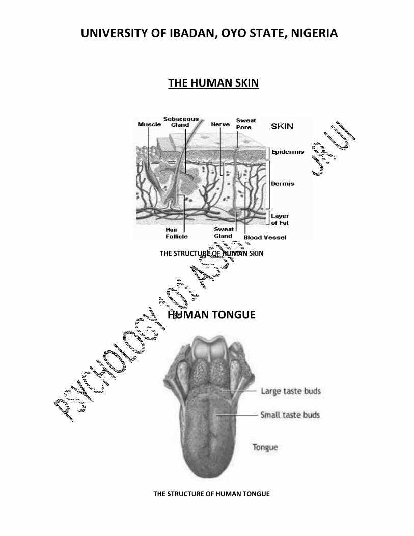
THE HUMAN SKIN
THE STRUCTURE OF HUMAN SKIN
HUMAN TONGUE
THE STRUCTURE OF HUMAN TONGUE

UNIVERSITY OF IBADAN, OYO STATE, NIGERIA
QUESTION: 3 THE ROLE OF EACH STRUCTURE LABELED IN HUMAN SENSATION AND PERCEPTION
HUMAN EYE
THE SCLERA: It is also called white of the eye that forms the outermost layer of tissue of the posterior and lateral aspects of the eyeball and is continuous anteriorly with the transparent cornea. The tough fibrous tissue preserves the shape of the eyeball and also protects the delicate inner layers. The transparent cornea in front allows the passage of light rays to focus on the retina. The extrinsic muscles attached to the sclera permits and limits movements of the eyeball within the orbit.
THE CILIARY BODY: It is the anterior continuation of the choroids. It consists of ciliary muscles and secretory epithelial cells. It gives attachment to the suspensory ligament, which at its other end is attached to the capsule enclosing the lens. Contraction and relaxation of the ciliary muscle changes the thickness of the lens which bends or refracts light rays entering the eye to focus them on the retina. The epithelia cells, secret aqueous fluid into the anterior chamber of the eye.
THE IRIS: This is the visible coloured part of the eye which extends anteriorly from the ciliary body, lying behind the cornea in front of the lens. It divides the anterior segment into the anterior and posterior chambers which contains aqueous fluid secreted by the ciliary body. It is a circular body composed of Parasympathetic stimulation dilates it. The amount of pigment cells in the iris is genetically determined. This determines the colour of the iris. Albinos have no pigment cells, and people with blue eye fewer pigment cells than people with brown eyes.
THE LENS: This is a highly elastic, circular bi convex body lying immediately behind the pupil. It consists of fibers enclosed within a capsule. It is suspended from the ciliary muscle by the suspensory ligament. Its thickness is controlled by the ciliary muscle through the suspensory ligament. The lens bends (refracts) light rays reflected by the objects in front of the eyes. The nearer the object to be viewed is, the thicker the lens becomes to allow focusing.
RETINA: The retina is the innermost layer of the wall of the eye. It is an extremely delicate structure which adapts by stimulation with light rays. The area that is highly sensitive to light is the layer of sensory receptors cells, the rods and cones. The retina lines about ¾ of the eyeball and is thickest at the back and thins out anteriorly to end just behind the ciliary body. Near the center of the posterior part is the macula lutea, or yellow spot. In the center of the area there is a little depression called the fovea centralis, consisting of only cone shaped cells. The rods and cone contain photosensitive pigments that convert light rays into nerve impulses. The optic disc is the place where the optic nerve leaves the retina.
FOVEA: Is a layer of sensory neurons, the key structures being photoreceptors (rod and cone cells) which respond to light. Contains relay neurons and sensory neurons that pass impulses along the optic nerve to the part of the brain that controls vision.
UNIVERSITY OF IBADAN, OYO STATE, NIGERIA PUPIL: This is a thin protective covering of epithelial cells. It protects the cornea against damage by friction (tears from the tear glands help this process by lubricating the surface of the conjunctiva).
CONJUNCTIVA: It has a network of blood vessels to supply nutrients to the cells and remove waste products. It is pigmented that makes the retina appear black, thus preventing reflection of light within the eyeball.
CONEA: Helps to maintain the shape of the anterior chamber of the eyeball. This dome-shaped layer protects your eye from elements that could cause damage to the inner parts of the eye. There are several layers of the cornea, creating a tough layer that provides additional protection. These layers regenerate very quickly, helping the eye to eliminate damage more easily. The cornea also allows the eye to properly focus on light more effectively. Those who are having trouble focusing their eyes properly can have their corneas surgically reshaped to eliminate this problem.
OPTIC NERVE: Transmit sensory impulses from retina to brain.
HUMAN EAR
THE OUTER EAR: It consists of the auricle (pinna) and the external acoustic meatus. The auricle is the expanded portion projecting from the side of the head. The external acoustic meatus (auditory canal) is a slightly s-shaped tube of about 2.5 cm long extending from the auricle to the ear drum. There are numerous sebaceous and ceruninous glands in the skin of the lateral third. The ceruninous glands produce a sticky material (cerumen) which traps dust, insects etc. The tympanic membrane (ear drum) completely separates the external acoustic meatus from the middle ear.
THE MIDDLE EAR: When sound waves strike the ear drum, it causes it to vibrate. This sets in motion three tiny bones, the malleus (hammer), incus (anvil) and the stapes (stirrups) so named because of their shapes. Each of these act as a lever increasing the pressure on the next, until the pressure exerted by the stirrup is many times greater than what the hammer exerts. The stirrups press against a membrane called the oval window, which lies at the end of the middle ear. Because the oval window is much smaller than the ear drum, and because of the lever effects that the three tiny bones create, the oval window receives much more pressure per square millimeter than the ear drum does. This increased pressure is needed to move the fluid in the next chamber of the ear.
THE INNER EAR: The inner ear (labyrinth) contains the organ of hearing and balance. It is generally describe in two parts, the bony labyrinth and the membranous labyrinth. The bony labyrinth is larger than, and encloses the membranous labyrinth of the same shape which fits into it, like a tube within a tube. Between the bony and membranous labyrinths there is a layer of watery fluid called the perilymph and within the membranous labyrinth, there is the endolymph.
The bony labyrinth consists of: 1 vestibule, 1 cochlear and 3 semi circular canals. The vestibule contains the oval and round windows in its lateral walls. The cochlear resembles a snail’s shell. The semi circular canals are three tubes arranged so that one is situated in each of the three planes of space. They are continuous with the vestibule.
The membranous labyrinth lies in the bony labyrinth. It contains endolymph and comprises of the vestibule, cochlear and the three semi circular canals. A cross section of the cochlear contains three compartments: the scala vestibule, the scala media and the scala tympany. On the base of this triangle called the basilar membrane, there are supporting cells and specialized cochlear hair cells
UNIVERSITY OF IBADAN, OYO STATE, NIGERIA containing auditory receptors. These cells form the spiral organ of corti, which is the sensory organ that responds to vibration by initiating nerve impulses that are then perceived as hearing by the brain. The auditory receptors are dendrites of efferent nerves that combine forming the cochlear (auditory) part of the vestibulocochlear nerve, to the hearing area in the cerebrum where the sound is perceived and to various nuclei in the pons and in the midbrain.
HUMAN NOSE
OLFACTORY BULB: Structure located in the forebrain of vertebrates that receives neural input about odors detected by cells in the nasal cavity. The axons of olfactory receptor (smell receptor) cells extend directly into the highly organized olfactory bulb, where information about odors is processed
Within the olfactory bulb are discrete spheres of nerve tissue called glomeruli. They are formed from the branching ends of axons of receptor cells and from the outer (dendrite) branches of interneurons, known in vertebrates as mitral cells that pass information to other parts of the brain. Tufted cells, which are similar to but smaller than mitral cells, and periglomerular cells, another type of interneuron cell, also contribute to the formation of glomeruli. The axons of all the receptor cells that exhibit a response to a specific chemical or range of chemicals with similar structures converge on a single glomerulus, where they connect via synapses with the interneurons. In this way, information from large numbers of receptor cells with similar properties is brought together. Thus, even if only a few receptors are stimulated because of very low concentrations of the stimulating chemical, the effects of signals from these cells are maximized. In mice there are about 1,800 glomeruli on each side of the brain, in rabbits there are about 2,000, and in dogs there are as many as 5,000. Since there are millions of olfactory receptor cells, the degree of convergence of axons, and therefore of information about a particular odour, is enormous. For example, in a rabbit, axons from about 25,000 receptor cells converge on each glomerulus.
Terrestrial vertebrates appear to have fewer glomeruli than fish. Zebra fish, commonly used in laboratory studies, have about 80 glomeruli in each olfactory bulb, and the mitral cells, which synapse (connect) with the axons of receptor cells in the glomeruli, have axons extending to several glomeruli, whereas in mammals the main connection of each mitral cell is with one glomerulus.
THE NASAL CAVITY: The nasal chambers are situated one on either side of the median plane. They open in front through the nares, and communicate behind through the choana with the nasal part of the pharynx. The nares are somewhat pear-shaped apertures, each measuring about 2.5 cm. antero-posteriorly and 1.25 cm. transversely at its widest part. The choana are two oval openings each measuring 2.5 cm. in the vertical, and 1.25 cm. in the transverse direction in a well-developed adult skull.
Inside the aperture of the nostril is a slight dilatation, the vestibule, bounded laterally by the ala and lateral crus of the greater alar cartilage, and medially by the medial crus of the same cartilage. It is lined by skin containing hairs and sebaceous glands, and extends as a small recess toward the apex of the nose. Each nasal cavity, above and behind the vestibule, is divided into two parts: an olfactory region, consisting of the superior nasal concha and the opposed part of the
UNIVERSITY OF IBADAN, OYO STATE, NIGERIA septum, and a respiratory region, which comprises the rest of the cavity.
NASAL CARTILAGE: This is situated below the inferior margin of the nasal bone, and is flattened, and triangular in shape. Its anterior margin is thicker than the posterior, and is continuous above with the cartilage of the septum, but separated from it below by a narrow fissure; its superior margin is attached to the nasal bone and the frontal process of the maxilla; its inferior margin is connected by fibrous tissue with the greater alar cartilage. Where the lateral cartilage meets the greater alar cartilage, the lateral cartilage often curls up, to join with an inward curl of the greater alar cartilage. That curl of the inferior portion of the lateral cartilage is called its "scroll."
THE NASAL BONES: These are two small oblong bones, varying in size and form in different individuals; they are placed side by side at the middle and upper part of the face, and form, by their junction, "the bridge" of the nose. Each has two surfaces and four borders.
THE FRONTAL SINUS: one of a pair of small cavities in the frontal bone of the skull that communicate with the nasal cavity and lie above the orbits. The frontal sinuses are situated behind the superciliary arches and are lined with a mucous membrane that is continuous with that of the nasal cavity. Each sinus opens into the anterior part of the middle meatus through the frontanasal duct. The frontal sinuses are absent at birth, become well developed between the seventh and eighth years, and reach their full size after puberty. The frontal sinuses can have variable drainage depending on the anatomy of the frontal sinus drainage pathway (FSDP). The frontal sinuses have a superior and inferior compartment of the FSDP. The frontal sinus ostium drains into the superior compartment which then communicates directly with the inferior compartment. The inferior compartment is a narrow space that either is formed by the ethmoid infundibulum or middle meatus depending on the anterior attachment of the uncinate process. If the anterior uncinate process attaches superiorly to the skull base, then the inferior compartment of the FSDP is the ethmoid infundibulum which then communicates with the middle meatus via the hiatus semilunaris. If the anterior uncinate process attaches to the lamina papyracea, then the inferior compartment of the FSDP is the middle meatus.
SPHENOIDAL SINUS: One of the paired paranasal sinuses in the body of the sphenoid bone, opening into the superior nasal meatus on the same side.
A TONSIL: Is a collection of lymphoid tissue. The term most commonly refers specifically to the palatine tonsils, which are masses of lymphatic material situated at either side at the back of the human throat. The palatine tonsils and the nasopharyngeal tonsil are lymphoepithelial tissues located near the oropharynx and nasopharynx. These immunocompetent tissues are the immune system's first line of defense against ingested or inhaled foreign pathogens. However, the fundamental immunological roles of tonsils have yet to be understood. Tonsillitis is a disorder in which the tonsils are inflamed (sore and swollen). The most common way to treat it is with anti-inflammatory drugs such as ibuprofen, or if bacterial in origin, antibiotics. Many sufferers treat it by having their tonsils surgically removed by a tonsillectomy. The set of lymphatic tissue known as Waldeyer's tonsillar ring includes the adenoid tonsil, two tubal tonsils, two palatine tonsils, and the lingual tonsil.
Lip Muscle: The muscles acting on the lips are considered part of the muscles of facial expression. All muscles of facial expression are derived from the mesoderm of the second pharyngeal arch, and are therefore supplied (motor supply) by the nerve of the second pharyngeal arch, the facial nerve (7th cranial nerve). The muscles of facial expression are all specialized members of the panniculus carnosus, which attach to the dermis and so wrinkle, or dimple the overlying skin. Functionally, the muscles of facial expression are arranged in groups around the orbits, nose and mouth.

UNIVERSITY OF IBADAN, OYO STATE, NIGERIA HUMAN SKIN
THE EPIDERMIS: Is composed of the outermost layers of cells in the skin. The epidermis is a stratified squamous epithelium, composed of proliferating basal and differentiated suprabasal keratinocytes which acts as the body's major barrier against an inhospitable environment, by preventing pathogens from entering, making the skin a natural barrier to infection. It also regulates the amount of water released from the body into the atmosphere through transepidermal water loss (TEWL). In humans, it is thinnest on the eyelids at 0.05 mm (0.0020 in) and thickest on the palms and soles at 1.5 mm (0.059 in). It is ectodermal in origin.
The main function of the epidermis is to form a tough barrier against the outside world, while the dermis is a soft, thick cushion of connective tissue that lies directly below the epidermis and largely determines the way our skin looks. Both layers keep repairing and renewing themselves throughout or life, but the dermis does it more slowly than the epidermis. Under the dermis is a layer of fat cells, which is known as adipose tissue (or subcutaneous fat layer). It provides insulation and protective padding for the body. It also provides an emergency energy supply.
THE DERMIS: Is the layer responsible for the skin's structural integrity, elasticity and resilience. Wrinkles develop in the dermis. Typical collagen and elastin creams, for example, never reach the dermis because collagen and elastin molecules are too large to penetrate the epidermis. It is the thickest of the skin layers and comprises a tight, sturdy mesh of collagen and elastin fibers. Both collagen and elastin are critically important skin proteins: collagen is responsible for the structural support and elastin for the resilience of the skin. The key type of cells in the dermis is fibroblasts, which synthesize collagen, elastin and other structural molecules. The proper function of fibroblasts is highly important for overall skin health.
The dermis also contains capillaries (tiny blood vessels) and lymph nodes which produce immune cells. Blood capillaries are responsible for bringing oxygen and nutrients to the skin and removing carbon dioxide and products of cell metabolism (what we call waste matter). Lymph nodes are engaged in protecting the skin from invading microorganisms.
The dermis contains sebaceous glands, sweat glands, hair follicles and a small number of nerve and muscle cells. Sebaceous glands, based around hair follicles, produce sebum, an oily protective substance that lubricates the skin and hair and provides protection by forming an acid mantle when mixed with sweat. When sebaceous gland produce too little sebum, as is common in older people, the skin becomes excessively dry and more prone to wrinkling. Too much of sebum as is common in teenagers, often leads to acne.
SUBCUTANEOUS TISSUE: Is the deepest layer of the skin located under the dermis and consisting mainly of fat cells. It acts as a shock absorber and heat insulator, protecting underlying tissues from cold and trauma. The loss of subcutaneous tissue in later years, leads to facial sag and makes wrinkles more visible.
THE SEBACEOUS GLANDS: They are microscopic glands in the skin that secrete an oily/waxy matter, called sebum, to lubricate and waterproof the skin and hair of mammals.[1] In humans, they are found in greatest abundance on the face and scalp, though they are distributed throughout all skin sites except

UNIVERSITY OF IBADAN, OYO STATE, NIGERIA the palms and soles.[2] In the eyelids, meibomian sebaceous glands secrete a special type of sebum into tears. There are several related medical conditions, including acne, sebaceous cysts, hyperplasia, sebaceous adenoma and sebaceous gland carcinoma.
All of the sebaceous glands in humans have been demonstrated to show similarity in structure and secrete sebum by a holocrine process. Sebum excreted by the sebaceous gland is primarily composed of tryglycerides, wax esters, and squalene. Wax esters, like squalene, are unique to sebum and not produced anywhere else in the body. Sebum also contains 45% water-insoluble fatty acids known to have broad antimicrobial activity. Additionally, sebaceous gland secretion provides Vitamin E to the upper layers of facial skin. Sebaceous lipids contribute to maintaining the integrity of the skin barrier, and express pro-inflammatory and anti-inflammatory properties. Recent research suggests that sebum may represent a delivery system for antioxidants, antimicrobial lipids, pheromones, and hydration of stratum corneum. During the last gestation trimester, it is known that sebaceous glands produce vernix caseosa which protects the embryonic skin from amniotic water. Sebaceous secretions in conjunction with apocrine glands also play an important thermoregulatory role. In hot conditions, the secretions emulsify and foment formation of and prevent the loss of sweat drops from the skin. In colder conditions, sebum repels rain from skin and hair. Increased facial surface sebum secretion is also associated with the development of acne.
A HAIR FOLLICLE: Is a mammalian skin organ that produces hair. Hair production occurs in phases, including growth (anagen), cessation (catagen), and rest (telogen) phases. Stem cells are responsible for hair production. Hair grows in cycles of various phases: anagen is the growth phase; catagen is the involuting or regressing phase; and telogen, the resting or quiescent phase. Each phase has several morphologically and histologically distinguishable sub-phases. Prior to the start of cycling is a phase of follicular morphogenesis (formation of the follicle). There is also a shedding phase, or exogen, that is independent of anagen and telogen in which one of several hairs that might arise from a single follicle exits. Normally up to 90% of the hair follicles are in anagen phase while, 10–14% are in telogen and 1–2% in catagen. The cycle's length varies on different parts of the body. For eyebrows, the cycle is completed in around 4 months, while it takes the scalp 3–4 years to finish; this is the reason eyebrow hair have a much shorter length limit compared to hair on the head. Growth cycles are controlled by a chemical signal like epidermal growth factor. DLX3 is a crucial regulator of hair follicle differentiation and cycling. Specifically, colocalization of phosphorylated Smad1/5/8 complex and DLX3 regulate role for BMP signaling to Dlx3 during hair morphogenesis in animal models.
NERVES: Nerves have an important function in the body. A nerve gives all signals in the body from all parts of the body to the brains and back. Nerves exist of two types of nerve-fibers: fast and slow fibers. Through a nerve-fibre signals are passed on to the cell membrane.
Nerves are part of the nervous system and exist of neurons. These cells work by passing on electrical signals from one part of the cell to the other part of the cell, and also by relasing chemical materials, which are also called neurotransmitters. The neurons exist of 4 parts: the cell body, the axon, the dendrites and the axon-ends.
In the cell body is a neuron, which has important information like DNA. The dendrites have to receive signals from other cells. The axon itself is the transmitter of these signals to the ends of the axon. The ends of the axon also have dendrites to other neurons or to organs. Neurons can pass on signals very
UNIVERSITY OF IBADAN, OYO STATE, NIGERIA fast but also very slowly. The speed can vary from signals that are passed on at 6 km/h to signals that are passed on at 320 km/h. Not every neuron connects another neuron, often there is a small space in between which we call the synapse.
When the signal is sent from one neuron to the end of the axon, it will release the neurotransmitters (chemical materials) at the same time, which has to “tell” this to the other neuron which has to pass it on, etc.
Well-known neurotransmitters are Acetylcholine, Dopamine and Serotine. The nervous system exists of the central nervous system and the peripherical nervous system. In the nervous system we find the brains (big and small), the spinal cord and the brain stem. In the perpherical nervous system we find all nerves which are between the central nervous system and the organs and the muscles. This nervous system can also be divided into two parts: the somatic and the autonomic nervous system, at which the somatic activates the muscles and the autonomic the heart muscles, blood vessels and the glands.
SWEAT GLANDS: These are small tubular structures of the skin that produce sweat. There are two main types of sweat glands:
• Eccrine sweat glands are distributed almost all over the body, though their density varies from region to region. Humans utilize eccrine sweat glands as a primary form of cooling
• Apocrine sweat glands are larger, have a different secretion mechanism, and are mostly limited to the axilla (armpits) and perianal areas in humans. Although apocrine glands contribute little to cooling in humans, they are the only effective sweat glands in hoofed animals such as the camel, donkey, horse, and cattle.
Domestic animals have apocrine glands at the base of each hair follicle but eccrine glands only in foot pads and snout. Their apocrine glands, like those in humans, produce an odorless oily milky secretion evolved not to evaporate and cool but rather coat and stick to hair so odor-causing bacteria can grow on it. Eccrine glands on their foot pads, like those on palms and soles of humans, did not evolve to cool either but rather increase friction and enhance grip.
Both apocrine and eccrine sweat glands contain myoepithelial cells, specialized epithelial cells located between the gland cells and the underlying basal lamina. Myoepithelial cell contractions squeeze the gland and discharge the accumulated secretions. The secretory activities of the gland cells and the contractions of myoepithelial cells are controlled by both the autonomic nervous system and by the circulating hormones. Ceruminous glands, which produce ear wax, and mammary glands, which produce milk, and ciliary glands in the eyelids are considered to be modified sweat glands.
BLOOD VESSELS: In the skin play a critical role in regulating body temperature. Blood from the heart enters the aorta. From there blood is delivered to smaller arteries and then arterioles in the skin. These arterioles contain smooth muscle that allows them to constrict or dilate in response to nerve signals from the body. Blood exiting the arterioles enters the smallest type of blood vessel called a "capillary." Capillaries are so small that you need a microscope to see them. In fact, the smallest capillaries only allow red blood cells to travel one-by-one in single file! Capillaries drain into the smallest veins called venules and this blood is eventually delivered back to the heart.
HUMAN TONGUE
UNIVERSITY OF IBADAN, OYO STATE, NIGERIA
TASTE BUDS: contain the receptors for taste. They are located around the small structures on the upper surface of the tongue, soft palate, upper esophagus and epiglottis, which are called papillae.[1] These structures are involved in detecting the five (known) elements of taste perception: salty, sour, bitter, sweet, and umami. Via small openings in the tongue epithelium, called taste pores, parts of the food dissolved in saliva come into contact with taste receptors. These are located on top of the taste receptor cells that constitute the taste buds. The taste receptor cells send information detected by clusters of various receptors and ion channels to the gustatory areas of the brain via the seventh, ninth and tenth cranial nerves. On average, the human tongue has 2,000–8,000 taste buds.
The majority of taste buds on the tongue sit on raised protrusions of the tongue surface called papillae. There are four types of papillae present in the human tongue:
Fungiform papillae: as the name suggests, these are slightly mushroom-shaped if looked at in longitudinal section. These are present mostly at the dorsal surface of the tongue, as well as at the sides. Innervated by facial nerve.
Filiform papillae: these are thin, long papillae "V"-shaped cones that don't contain taste buds but are the most numerous. These papillae are mechanical and not involved in gustation. They are characterized by increased keratinization.
Foliate papillae: these are ridges and grooves towards the posterior part of the tongue found at the lateral borders. Innervated by facial nerve (anterior papillae), and glossopharyngeal nerve (posterior papillae).
Circumvallate papillae - there are only about 10 to 14 of these papillae on most people, and they are present at the back of the oral part of the tongue. They are arranged in a circular-shaped row just in front of the sulcus terminalis of the tongue. They are associated with ducts of Von Ebner's glands, and are innervated by the glossopharyngeal nerve.
Salts, sweet, sour and umami tastes causes depolarization of the taste cells, although different mechanisms are applied.
Bitter causes an internal release of Ca2+, no external Ca2+ is required.
The bud is formed by two kinds of cells: supporting cells and gustatory cells.
The supporting (sustentacular) cells are mostly arranged like the staves of a cask, and form an outer envelope for the bud. Some, however, are found in the interior of the bud between the gustatory cells.
The gustatory (taste) cells, a chemoreceptor, occupy the central portion of the bud; they are spindle-shaped, and each possesses a large spherical nucleus near the middle of the cell.
The peripheral end of the cell terminates at the gustatory pore in a fine hair filament, the gustatory hair.
The central process passes toward the deep extremity of the bud, and there ends in single or bifurcated varicosities.

UNIVERSITY OF IBADAN, OYO STATE, NIGERIA The nerve fibrils after losing their medullary sheaths enter the taste bud, and end in fine extremities between the gustatory cells; other nerve fibrils ramify between the supporting cells and terminate in fine extremities; these, however, are believed to be nerves of ordinary sensation and not gustatory. The average life of a taste bud is 10 days.


![[3937]-101 P553](https://static.fdokumen.com/doc/165x107/63338430a6138719eb0aa5bc/3937-101-p553.jpg)