Kocatepe Veterinary Journal 2020 June 13 / 2 - DergiPark
136
-
Upload
khangminh22 -
Category
Documents
-
view
0 -
download
0
Transcript of Kocatepe Veterinary Journal 2020 June 13 / 2 - DergiPark


II
Publisher Prof. Dr. Turan CİVELEK Dean On behalf of Afyon Kocatepe University Faculty of Veterinary Medicine Afyonkarahisar - TURKEY
Editor in Chief Assoc. Prof. Dr. Zeki GÜRLER
Editors Assoc. Prof. Dr. Recep KARA Assoc. Prof. Dr. Deniz YENİ
Section Editors Prof. Dr. Alpaslan YILDIRIM Prof.Dr. Kerem URAL Prof.Dr. Sadullah BAHAR Prof.Dr. Akın YAKAN Prof.Dr. Kemal Kaan TEKİNŞEN
Foreing Language Editor Prof.Dr. İbrahim DEMİRKAN Assist. Prof. Dr. Ulaş ACARÖZ
Statistics Editors Assoc. Prof. Dr. İbrahim KILIÇ Assist. Prof. Dr. İlkay DOĞAN
Organising Committee Prof. Dr. Fatih FİDAN Assoc. Prof. Dr. Metin ERDOĞAN Assoc. Prof. Dr. Mustafa KABU Assoc. Prof. Dr. Fatih AVDATEK Dr. Eyüp Eren GÜLTEPE Dr. Barış DENK
ADVISORY BOARDS
Prof. Dr. Arif Altıntaş Ankara University -Turkey
Prof. Dr. Atilla Şimşek Selçuk University-Turkey
Prof. Dr. Cevdet Uğuz Afyon Kocatepe University-Turkey
Prof. Dr. Yavuz O. Birdane Afyon Kocatepe University-Turkey
Prof. Dr. İbrahim Demirkan Afyon Kocatepe University-Turkey
Prof. Dr. İlhami Çelik Selçuk University-Turkey
Prof. Dr. İsmail Bayram Afyon Kocatepe University-Turkey
Prof. Dr. Abdullah Kaya Selcuk University-Turkey
Prof. Dr. Mustafa Alişarlı Ondokuz Mayıs University-Turkey
Prof. Dr. Nalan Bayşu Sözbilir Afyon Kocatepe University-Turkey
Prof. Dr. Recep Aslan Afyon Kocatepe University-Turkey
Prof. Dr. Seyfullah Haliloğlu Selçuk University-Turkey
Prof. Dr. Zafer Karaer Ankara University-Turkey
Prof. Dr. Zehra Bozkurt Afyon Kocatepe University-Turkey
Prof. Dr. İbrahim Taşal Mehmet Akif Ersoy University-Turkey
Prof. Dr. Şule Kaya Mehmet Akif Ersoy University-Turkey
Prof. Dr. Korhan Altunbaş Afyon Kocatepe University-Turkey
Prof. Dr. Aysun Demirkan Afyon Kocatepe University-Turkey
Prof. Dr. Hasan Çiçek Afyon Kocatepe University-Turkey
Prof. Dr. Fatih M. Birdane Afyon Kocatepe University-Turkey
Assoc. Prof. Dr. Süleyman Aypak Adnan Menderes University-Turkey
Assoc. Prof. Dr. Oktay Yılmaz Afyon Kocatepe University-Turkey
Assoc. Prof. Dr. İbrahim Kılıç Afyon Kocatepe University-Turkey
Assist. Prof. Dr. M. Fatih Bozkurt Afyon Kocatepe University-Turkey
Kocatepe Veterinary Journal 2020 June 13 / 2
Official Publication of The Afyon Kocatepe University
ISSN: 1308-1594 e-ISSN: 2147-6853
Kocatepe Veterinary Journal is International an Peer-Reviewed Journal and published four times a year.
Kocatepe Veterinary Journal;
indexed in TUBİTAK-ULAKBİM TR-Dizin, Turkey Citation Index, CAB Abstract, ResearchBib, SIS (Scientific Indexing Services),
CiteFactor, CrossRef, Index Copernicus, Google Scholar, SJIFactor
Addressed:
Kocatepe Veterinary Journal, Afyon Kocatepe University, Faculty of Veterinary Medicine, 03200, Afyonkarahisar, TURKEY.
Tel: +90 272 214 9309 Fax: +90 272 214 9309 E-mail: [email protected]
www.kvj.aku.edu.tr http://dergipark.gov.tr/kvj
*Only accepts online submission*
KOCATEPE VETERINARY JOURNAL 2020 13 (2) JUNE
RESEARCH ARTICLES Investigation The Effect of Different Levels of Dry Sugar Beet Pulp mixed Concentrate Feeds on Cadmium Levels in Rabbit Slaughter Products (Kadmiyum İlaveli Konsantre Yemlere Farklı Düzeylerde Kuru Şeker Pancarı Posası Katılmasının Tavşanların Kesim Ürünlerinde Kadmiyum Seviyelerine Etkisinin Araştırılması ) Olena TYTARİOVA, Aamir IQBAL, Leonid DYACHENKO, Prof. Dr. Vitalii BOMKO, Oksana KUZMENKO, Oleksandr CHERNIAVSKYI, Serhii BABENKO, Mykhailo SLOMCHYNSKYI, Oksana TSEKHMISTRENKO, İbrahim Sadi ÇETİNGÜL, Eyup Eren GULTEPE, İsmail BAYRAM
98-103
Staphylococcal Enterotoxins and Enterotoxigenic Staphylococcus aureus in Raw Milk: A Screening Study (Çiğ Sütte Stafilokokal Enterotoksinler ve Enterotoksijenik Staphylococcus aureus Varlığının Belirlenmesine Yönelik Bir Tarama Çalışması ) Erhan KEYVAN, Ozen YURDAKUL, Erdi ŞEN
104-109
The Effects of Chitosan Oligosaccharide (COS) Treatment on Oxidative Stress and Its Relation with Intestinal Microflora in Rats Exposed To Cadmium (Kitosan Oligosakkarit (COS) Tedavisinin Oksidatif Stres Üzerine Etkileri ve Kadmiyuma Maruz Kalan Sıçanlarda Bağırsak Mikroflorası ile İlişkisi ) İhsan KISADERE, Hakan TAVŞANLI, Mukadderat GÖKMEN
110-117
First Report on Tuberculosis Based on Slaughterhouse Data in Bejaia Province, Algeria: A Retrospective 10-Year Survey (Cezayir, Bejaia'daki Mezbaha Verilerine Dayanan Tüberküloz Hakkında İlk Rapor: Geriye Dönük 10 Yıllık Bir Araştırma ) Abdelhanine AYAD, Abdelkader BENSID, Amira Chahrazad BENABDELHAK, Fatima AIT-YAHIA, Nadir Boudjlal DERGAL
118-124
Determination of The Hygienic Quality of Tap Waters of Afyonkarahisar Province (Afyonkarahisar İli Çeşme Sularının Hijyenik Kalitesinin Belirlenmesi ) Gökhan AKARCA, Oktay TOMAR, Ömer İSTEK
125-129
Use of Pumpkin and Its Effect on Quality in Ice Cream Production (Dondurma Üretiminde Bal Kabağı Kullanımı ve Kalite Üzerine Etkisi ) Ayşe YILDIZ, Pelin DEMİR, Ali ARSLAN
130-139
Efficiency of Two Different Synchronization Protocols in Conception in Simmental Heifers (Simental Düvelerde Farklı İki Senkronizasyon Protokolünün Gebelik Üzerine Olan Etkinliği ) Gaye BULUT, Umut TAŞDEMİR
140-144
The Effect of Dietary Probiotic Supplementation on Egg Weight in Laying Hens: A Meta–Analysis Study (Yumurtacı Tavuklarda Diyet Probiyotik Takviyesinin Yumurta Ağırlığına Etkisi: Bir Meta Analizi Çalışması ) İbrahim KILIÇ, İhsan BERK, Zehra BOZKURT, Yağmur Nil DOĞAN
145-151
Molecular Characterization of Echinococcus granulosus Isolates Found in Cattle, Buffaloes, Sheep and Goats in Afyonkarahisar, Turkey (Afyonkarahisar’da Sığır, Manda, Koyun ve Keçilerde Bulunan Echinococcus granulosus İzolatlarının Moleküler Karakterizasyonu ) Kürşat KARTAL, Mustafa KÖSE, Metin ERDOĞAN
152-160
Effects of Chrysin Against Isoniazid-Induced Lung Injury in Rats (Ratlarda İzoniazid Kaynaklı Akciğer Hasarına Karşı Krisinin Etkileri ) Sefa KÜÇÜKLER, Selçuk ÖZDEMİR, Selim ÇOMAKLI, Fatih KANDEMİR
161-171
Pathological and Microbiological Investigations of Naturally Infected Rainbow Trout (Oncorhynchus mykiss) with Flavobacterium psychrophilum (Flavobacterium psychrophilum ile Doğal Enfekte Gökkuşağı Alabalıklarında (Oncorhynchus mykiss) Patolojik ve Mikrobiyolojik İncelemeler) Erkmen EKİPMEN, Hamdi AVCI, Tevfik Tansel TANRIKUL, S.serap BİRİNCİOĞLU, Ahmet AYDOĞAN
172-178
Effect of Vitamin A, D3, E Treatment on Fertility in the Pırlak Sheep (Pırlak Koyunlarında Vitamin A, D3, E Uygulamasının Fertilite Üzerine Etkisi ) Muhammed Kürşad BİRDANE, Fatih AVDATEK
179-184
Investigation of Serum and Wool Levels of Cobalt, Manganese, Selenium and Zinc in Liver-Trematode-Infected Sheep (Karaciğer Trematodlu Koyunların Serum ve Yapağılarında Kobalt, Mangan, Selenyum ve Çinko Düzeylerinin Araştırılması ) Selçuk Seçkin TUNCER, Vural DENİZHAN, Süleyman KOZAT
185-191
Effects of Coenzyme Q10 on Some Blood Antioxidant System Parameters and Histological Changes in the Pancreas and Aorta of Streptozotocin-induced Diabetic Rats (Streptozotosin ile Diyabet Oluşturulan Ratlarda Koenzim Q10’un Bazı Kan Antioksidan Sistem Parametreleri ile Pankreas ve Aorttaki Histolojik Değişiklikler Üzerine Etkileri ) Ercan KESKİN, Deniz ULUIŞIK, Yasemin ÖZNURLU, Tuğba ÖZAYDIN
192-202
Effects of Fructose-Induced Metabolic Syndrome on Kidney Histology in Rats (Ratlarda Fruktoz ile Oluşturulmuş Metabolik Sendromun Böbrek Histolojisi Üzerine Etkileri) Özay GÜLEŞ, Musa TATAR
203-209
Research on Occurrence of Mites in Cheese Consumed in Develi District of Kayseri Province (Kayseri’nin Develi İlçesinde Tüketime Sunulan Peynirlerde Akar Varlığının Araştırılması ) Tugba SOLMAZ, Mustafa KARATEPE
210-213
Protective Effects of Taurine on Imidacloprid-Induced DNA Damage and Reproductive Performance in The Drosophila melanogaster Model (Drosophila melanogaster Modelinde Imidakloprid ile İndüklenen DNA Hasarı ve Üreme Performansı Üzerine Taurinin Koruyucu Etkileri ) Damla ARSLAN ACARÖZ, Sinan İNCE, Fahriye ZEMHERİ NAVRUZ, Nalan BAYŞU SÖZBİLİR
214-218
The Serum Amyloid-A, Haptoglobin, Ceruloplasminand Albumin Levels in Dogs Which are Infected with Babesia canisa (Babesiacanis ile Enfekte Köpeklerde Serum Amiloid-A, Haptoglobin, Seruloplazmin ve Albumin Seviyeleri) Ali KIRMIZIGÜL, Ekin Emre ERKILIÇ, Oğuz MERHAN, Metin ÖĞÜN, Neslihan ÖLMEZ, Gencay Taşkın TAŞÇI, Zati VATASEVER
219-223
CASE REPORT A Case of A 13-Year-Old Dog with Old Dog Encephalitis: A Rare Form of Canine Distemper (13 Yaşlı Bir Köpekte “Old Dog Ensefalit” Olgusu: Köpek Distemper’ının Ender Formu ) Erdem GÜLERSOY, Mahmut OK, Mutlu SEVİNÇ, Murat Kaan DURGUT, Amir NASERİ
224-227
98
Kocatepe Veterinary Journal
Kocatepe Vet J (2020) 13(2):98-103
DOI: 10.30607/kvj.653142 RESEARCH ARTICLE
Investigation The Effect of Different Levels of Dry Sugar Beet Pulp mixed Concentrate Feeds on Cadmium Levels in Rabbit Slaughter
Products
Olena TYTARIOVA1, Aamir IQBAL2*, Leonid DYACHENKO1, Vitalii BOMKO1, Oksana KUZMENKO1, Oleksandr CHERNIAVSKYI1, Serhii BABENKO1,
Mykhailo SLOMCHYNSKYI1, Oksana TSEKHMISTRENKO1, İbrahim Sadi ÇETİNGÜL2, Eyüp Eren GÜLTEPE2, İsmail BAYRAM2
1Department of Technology of Feed, Feed Additives and Animal Feeding, Faculty of Biological Technology, Bila Tserkva National Agrarian University, Bila Tserkva, Ukraine
2Department of Animal Nutrition and Nutritional Diseases, Faculty of Veterinary Medicine, Afyon Kocatepe University, Afyonkarahisar, Turkey
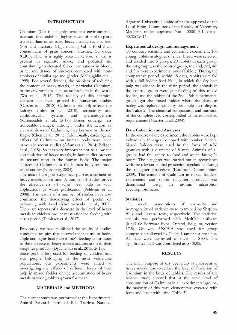
ABSTRACT
Commercial concentrated feeds are traditionally a major component of rabbit diets which contain Cadmium element in quantities above generally accepted level. This toxic trace element can accumulate in meat, kidneys, liver, bones and spleen of rabbits. Consumption of such meat and by-products will contribute to the accumulation of Cadmium in the human body, which may lead to a number of pathological changes. In this study, 100 young rabbits-analogues of silver breed were selected, and divided into 5 groups, 20 rabbits in each group: the 1st group was the control group, the 2nd, 3rd, 4th and 5th were experimental with total 60 days duration period. Results showed the smallest content of Cadmium in slaughter products was observed in the rabbits at 5th experimental group. In conclusion, the course of the experiment, it has been established that supplementation into the rabbit’s mixed fodder of dry sugar beet pulp contributes to reducing the level of Cadmium accumulation in slaughter products. It has also been noted that most of Cadmium is excreted with feces, that is, it is not absorbed into the bloodstream and does not have a negative effect on the body of the rabbit.
Keywords: Cadmium, dry sugar beet pulp, slaughter products, rabbits, mixed fodder
***
Kadmiyum İlaveli Konsantre Yemlere Farklı Düzeylerde Kuru Şeker Pancarı Posası Katılmasının Tavşanların Kesim Ürünlerinde Kadmiyum Seviyelerine Etkisinin Araştırılması
ÖZ
Ticari konsantre yemler genellikle kabul edilen seviyenin üzerinde kadmiyum elementini içeren tavşan diyetlerinin ana bileşenidir. Bu toksik iz element, tavşanların etlerine, böbreklerde, karaciğerde, kemiklerde ve dalaklarında birikebilir. Bu tür et ve yan ürünlerinin tüketimi, insan vücudunda kadmiyum birikimine katkıda bulunabilir ve bu durum bir takım patolojik değişikliklere neden olabilir. Bu çalışmada, 100 adet gümüş ırkın analoğu genç tavşan kullanılmıştır. Tavşanlar, her birinde 20 adet olmak üzere 5 gruba ayrılmıştır. Gruplar 1 kontrol ve 4 deneme grubu olacak şekilde düzenlenmiştir. Araştırma 60 gün boyunca sürdürülmüştür. Sonuçlar, 5. deneme grubundaki tavşanlarda, kesim sonrası elde edilen karkasta ve bazı iç organlarda en az düzeyde kadmiyum içeriğinin tespit edildiğini göstermiştir. Sonuç olarak, deneme boyunca, 0.04 mg/kg düzeyindeki kadmiyum katkılı rasyonlara artan düzeylerde kuru şeker pancarı ilavesinin, karkas ve bazı iç organlarda kadmiyum seviyesinin azaltılmasına katkıda bulunduğu tespit edilmiştir. Ayrıca kadmiyumun çoğunun dışkı ile atıldığı, yani kan dolaşımına emilmediği ve tavşanın vücudu üzerinde olumsuz bir etkiye sahip olmadığı da kaydedilmiştir.
Anahtar Kelimeler: Kadmiyum, kuru şeker pancarı posası, kesim ürünleri, tavşan, karma yem
To cite this article: Tytariova O. Iqbal A. Dyachenko L. Bomko V. Kuzmenko O. Cherniavskyi O. Babenko S. Slomchynsky M. Çetingül İ.S. Gültepe E.E. Bayram İ. Investigation the Effect of Different Levels of Dry Sugar Beet Pulp Mixed Concentrate Feeds on Cadmium Levels in Rabbit Slaughter Products. Kocatepe Vet J. (2020) 13(2):98-103
Submission: 30.11.2019 Accepted: 20.03.2020 Published Online: 24.04.2020 ORCID ID; OT: 0000-0003-4820-809X, AI: 0000-0003-4473-2329, LD: 0000-0003-4615-0277, VB: 0000-0001-5558-6924, OK: 0000-0003-4553-9950, OC: 0000-0003-0713-6587, SB: 0000-0001-5131-4999, MS: 0000-0001-5197-2684, OT: 0000-0003-0509-4627, İSÇ: 0000-0002-7608-6176, EEG: 0000-0002-2404-1232, İB: 0000-0002-2404-1232 *Corresponding author e-mail: [email protected]
99
INTRODUCTION Cadmium (Cd) is a highly persistent environmental toxicant that exhibits higher rates of soil-to-plant transfer than other toxic heavy metals, such as lead (Pb) and mercury (Hg), making Cd a food-chain contaminant of great concern. Further, Cd oxide (CdO), which is a highly bioavailable form of Cd, is present in cigarette smoke and polluted air, contributing to elevated Cd concentrations in blood, urine, and tissues of smokers, compared with non-smokers of similar age and gender (McLaughlin et al., 1999). For several decades, the problem of reducing the content of heavy metals, in particular Cadmium, in the environment is an acute problem in the world (Wu et al., 2016). The toxicity of this chemical element has been proved by numerous studies (Carson et al., 2018). Cadmium primarily affects the kidneys (Johri et al., 2010), respiratory and cardiovascular systems, and spermatogenesis (Rahimzadeh et al., 2017). Bones undergo less noticeable changes, although under the action of elevated doses of Cadmium, they become brittle and fragile (Chen et al., 2011). Additionally, carcinogenic effects of Cadmium on human body have been proven in recent studies (Adams et al., 2014; Eriksen et al., 2015). So it is very important not to allow the accumulation of heavy metals, in human diet prevent its accumulation in the human body. The major sources of Cadmium in the human body are food, water and air (Nordberg, 2004). The idea of using of sugar beet pulp as a sorbent of heavy metals is not new. A number of studies prove the effectiveness of sugar beet pulp in such applications as water purification (Pehlivan et al, 2008). The results of a number of studies have also confirmed the detoxifying effect of pectin on poisoning with Lead (Khotimchenko et al., 2007). There are reports of a decrease in the level of heavy metals in chicken broiler meat after the feeding with citrus pectin (Temiraev et al., 2017). Previously, we have published the results of studies conducted on pigs that showed that the use of beets, apple and sugar beet pulp in pig’s feeding contributes to the decrease of heavy metals accumulation in their slaughter products (Dyachenko et al., 2015, 2017). Since pork is less used for feeding of children and sick people belonging to the most vulnerable populations, our experiments were aimed at investigating the effects of different levels of beet pulp in mixed fodder on the accumulation of heavy metals in young rabbits grown for meat.
MATERIALS and METHODS
The current study was performed at the Experimental Animal Research farm of Bila Tserkva National
Agrarian University Ukraine after the approval of the Local Ethics Committee of the Faculty of Veterinary Medicine under approval No: 00001/01; dated: 30/05/2016. Experimental design and management To conduct scientific and economic experiment, 100 young rabbits-analogues of silver breed were selected, and divided into 5 groups, 20 rabbits in each group: the 1st group was the control group, the 2nd, 3rd, 4th and 5th were experimental ones (Table1). During the comparative period, within 15 days, rabbits were fed with a full-fodder feed № 1, in which the dry beet pulp was absent. In the main period, the animals in the control group were got feeding of this mixed fodder, and the rabbits of the 2nd - 5th experimental groups got the mixed fodder where the share of barley was replaced with dry beet pulp according to the Table 2. The chemical composition and nutrition of the complete feed corresponded to the established requirements (Maertes at all. 2004). Data Collection and Analyses In the course of the experiment, the rabbits were kept individually in cages equipped with bunker feeders. Mixed fodders were used in the form of solid granules with a diameter of 5 mm. Animals of all groups had free access to food and water during 24 hours. The slaughter was carried out in accordance with the relevant animal protection regulations during the slaughter procedure (European Communities, 2009). The content of Cadmium in mixed fodders, excrements and rabbit slaughter products was determined using an atomic adsorption spectrophotometer. Statistics The model assumptions of normality and homogeneity of variance were examined by Shapiro-Wilk and Levene tests, respectively. The statistical analysis was performed with MedCalc software (MedCalc Software bvba, Ostend, Belgium, version 17.5). One-way ANOVA was used for group comparison followed by Tukey-Kramer for post-hoc. All data were expressed as mean ± SEM. The significance level was considered as p <0.05.
RESULTS
The main purpose of dry beet pulp as a sorbent of heavy metals was to reduce the level of fasciation of Cadmium in the body of rabbits. The results of the balance study showed that at the same level of consumption of Cadmium in all experimental groups, the majority of this trace element was excreted with feces and lesser with urine (Table 3).

100
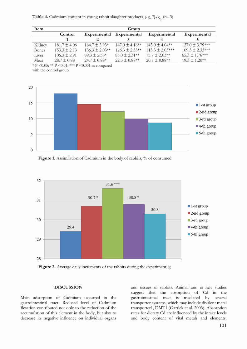
The inclusion of dry beet pulp into the mixed fodder not only improved the growth of rabbits, but also reduced the flow of Cadmium into the products of their slaughter. At the mass fraction of dry pulp in the mixed fodder of 3% the Cadmium fixation in the rabbit’s bodies of the 2nd experimental group decreased by 3.43% compared to the control one. With the 12% mass fraction of dry pulp in the feed (the 5th experimental group) the quantity of Cadmium in the body of rabbits decreased to 8.68%, which is 9.31% less than control group (Table 4).
During the main experimental period the animals in test groups, according to the average daily increments, dominated their peers from the 1st control group (Fig. 1 and Fig. 2). According to Fig.1 in the first group, highest assimilation of Cadmium in the body of rabbits, % of consumed however, in other groups it decrease drastically. According to Fig. 2, in the 3rd experimental group, highest average daily increments (g) in the rabbits were observed as compared to other experimental and control group.
Table 1. In vivo experiment schedule.
Group Feeding terms and conditions
Comparative (preparatory) period (15 days)
Main period (60 days)
1 – control group Mixed fodder (MF) 1 MF 1 (Cadmium content 0.04 mg/kg)1 2 – experimental group MF 1 MF 2 (Cadmium content 0.04 mg/kg)1 3 – experimental group MF 1 MF 3 (Cadmium content 0.04 mg/kg)1 4 – experimental group MF 1 MF 4 (Cadmium content 0.04 mg/kg)1 5 – experimental group MF 1 MF 5 (Cadmium content 0.04 mg/kg)1
1Natural content in feed
Table 2. Composition of the Concentrated Feeds, %
Item MF No 1 MF No 2 MF No 3 MF No 4 MF No 5
Barley grain 19 16 13 10 7 Corn, grain 10 10 10 10 10 Wheat, grain 18 18 18 18 18 Soybean meal 10 10 10 10 10 Alfalfa hay flour 30 30 30 30 30 Dry sugar beet pulp - 3 6 9 12 Meat and bone meal 5 5 5 5 5 Salt (NaCl) 5 5 5 5 5 Chalk (СaCO3) 1 1 1 1 1 Premix Axelarat 2 2 2 2 2 Total 100 100 100 100 100
Table 3. Balance of Cadmium in the body of young rabbits, μg,
ÕSÕ (n=3)
Item Group
Control Experimental Experimental Experimental Experimental
1 2 3 4 5
Consumed with feed 6.00±0.304 5.94±0.173 6.15±0.131 5.92±0.130 5.88±0.148 Consumed with water 0.15±0.004 0.14±0.005 0.16±0.001 0.15±0.002 0.14±0.005 Secreted with excrements 2.67±0.145 2.90±0.153 3.47±0.088* 3.47±0.067* 3.43±0.088* Secreted with urine 2.37±0.033 2.30±0.058 2.07±0.088 2.00±0.116 2.07±0.088 Assimilated 1.11±0.132 0.88±0.054 0.77±0.053 0.60±0.064* 0.52±0.045*
* P <0.05
101
Table 4. Cadmium content in young rabbit slaughter products, μg, Õ
SÕ (n=3)
Item Group
Control Experimental Experimental Experimental Experimental
1 2 3 4 5
Kidney 181.7 ± 4.06 164.7 ± 3.93* 147.0 ± 4.16** 143.0 ± 4.04** 127.0 ± 3.79*** Bones 153.3 ± 2.73 136.3 ± 2.03** 126.3 ± 2.33** 113.3 ± 2.03*** 109.3 ± 2.33*** Liver 106.3 ± 2.91 89.3 ± 2.33* 85.0 ± 2.31** 75.7 ± 2.03** 65.3 ± 1.76*** Meat 28.7 ± 0.88 24.7 ± 0.88* 22.3 ± 0.88** 20.7 ± 0.88** 19.3 ± 1.20**
* P <0.05; ** P <0.01; *** P <0.001 as compared with the control group.
Figure 1. Assimilation of Cadmium in the body of rabbits, % of consumed
Figure 2. Average daily increments of the rabbits during the experiment, g
DISCUSSION
Main adsorption of Cadmium occurred in the gastrointestinal tract. Redused level of Cadmium ficsation contributed not only to the reduction of the accumulation of this element in the body, but also to decrease its negative influence on individual organs
and tissues of rabbits. Animal and in vitro studies suggest that the absorption of Cd in the gastrointestinal tract is mediated by several transporter systems, which may include divalent metal transporter1, DMT1 (Garrick et al. 2003). Absorption rates for dietary Cd are influenced by the intake levels and body content of vital metals and elements.
102
Higher dietary zinc intake levels were associated with lower Cd body burden, as assessed by urinary Cd excretion levels (Vance, et al., 2015). The data of the Cadmium accumulation level in the body of experimental rabbits. As it can be seen, with the increase of the proportion of dry beet pulp in the mixed fodder, the level of Cadmium consumption in the body of rabbits decreased. Moreover, the amount of Cadmium fixation in the rabbit organism was inversely proportional to the content of dry pulp in the feed. The majority of reported dietary Cd intake estimates are within the FAO/WHO tolerable level of 58µg/day for a 70-kg person, with an exception for certain locations in Japan, where intake exceeded the FAO/WHO safe intake guideline (Ikeda, et al., 2015). In particular, the increase of the dry beet pulp mass fraction in the mixed fodder up to 3% resulted in the increase of the average daily increment of the rabbit’s body weight, while the rabbits of the 2nd experimental group, compared with control one, increased body weight by 4.4%. With 6% of mass fraction of dry pulp in mixed fodder, the average daily increment of the rabbits’ body weight of the 3rd experimental group was the highest - 31.6 g, which is higher than in the control group by 7.5%. The average daily increments, the mass of the rabbit’s body of the 4th and 5th experimental groups, with the mass fraction of dry pulp in the mixed fodder 9 and 12%, exceeded the control group, by 4.8 and 3.1% respectively. The best rabbits’ productivity was noted in the rabbits of the 3rd experimental group with the 6% mass fraction of dry pulp in mixed fodder. The body content of Cd assessed by urinary and/or blood Cd levels showed an inverse association with body mass index (BMI), central obesity, and risks of weight gain, and obesity in both children and adults. These have consistently been observed across populations, including the U.S., Belgium, Canada, Korea, and China. In a Chinese study, urinary Cd levels that were equivalent to or greater than 2.95 µg/g creatinine were associated with a reduced risk of being overweight (Nie et al., 2016). The introduction of dry beet pulp into the fodder of rabbits of the 2nd experimental group in an amount of 3% by weight contributed to a decrease of the Cadmium content in the kidneys, liver, bones and meat, respectively, by 9.4%; 16.0; 11.1 and 13.9% compared to the rabbits of the control group. The increase in the mass fraction of dry beet pulp in the mixed fodder of rabbits of the 3rd experimental group up to 6% resulted in a decrease in the Cadmium content in meat by 22.3%, liver by 20%, in kidneys by 19.1%, in bones by 17.6% relatively to benchmarks. A significant decrease in the level of Cadmium in slaughter products was noted in the animals of the 4th experimental group. Thus, they outperformed the control analogues with Cadmium in the kidneys, liver, bones and meat, respectively, by
21.3%; 28.8; 26.1 and 27.9%. The introduction of the 12% of dry pulp into the mixed fodder of rabbits in the 5th experimental group reduced the Cadmium content in meat by 33% compared to the control animals. However, Cadmium content in the kidneys decreased by 30%, in the liver - by 39%, in bones - by 29%. In the Swedish study, a half of total kidney Cd content (10 µg/g kidney cortex) was estimated to come from food consumption, and the other half was attributed to cigarette smoking. The majority of subjects with high kidney Cd levels (>50 µg/g) were women (Elinder, et al., 1976). In conclusion, the addition of dry beet pulp into the mixed fodder for rabbits, which are grown for meat, in an amount from 3 to 12% by weight, reduced the absorption of Cadmium in their bodies and reduced its content in slaughter products (kidney, liver, bone, meat), that increased their quality and environmental safety. In conclusion, the course of the experiment, it has been established that supplementation into the rabbit’s mixed fodder of dry beet pulp contributes to reducing the level of Cadmium accumulation in slaughter products.
ACKOWLEDGEMENT
The current study was performed at the Experimental Animal Research farm of Bila Tserkva National Agrarian University Ukraine after the approval of the Local Ethics Committee of the Faculty of Veterinary Medicine under approval No: 00001/01;dated: 30/05/2016. Conflict of Interest: The authors declare that they have no conflict of interest.
REFERENCES
Bonnie L. Carson, Harry V. Ellis III, Joy L. McCann. Toxicology Biological Monitoring of Metals in Humans, 2018; CRC Press
Chen X., Zhu G., Jin T., Qin B., Zhou W., Gu S. Cadmium is More Toxic on Volume Bone Mineral Density than Tissue Bone Mineral Density. Biol. Trace Elem. Res., 144: 380–387.
Council Regulation (EC) № 1099/2009 of 24 September 2009 on the protection of animals at the time of killing Official Journal of the European Union, 2011; 52: 1–30.
Dyachenko L, Syvyk T, Kosyanenko О. Influence of different levels of Cadmium in ration with natural detoxicant on performance, digestibility of substances and metabolism of nitrogen in young fattening pigs. J. Tech of produc and processing of animal products: Collection of scientific works of Bila Tserkva National Agrarian University, 2015; 1 : 163–168.

103
Dyachenko L.S., Syvyc T.L., Tytariova O.M., Kuzmenko O.A., Bilkevich V.V. Natural detoxicants in pig rations and their impact on productivity and quality of slaughter products. Ukrainian J of Ecology, 2017; 7, (2) :239–246.
Elinder, C.G.; Lind, B.; Kjellstorm, T.; Linnman, L.; Friberg, L. Cadmium in kidney cortex, liver and pancreas from Swedish autopsies: Estimation of biological half time in kidney cortex, considering calorie intake and smoking habits. Arch. Environ. Health 1976, 31, 292–301.
Garrick, M.D.; Dolan, K.G.; Horbinski, C.; Ghio, A.J: A mammalian transporter for multiple metals. Biometals 2003, 16, 41–54
Ikeda, M.; Nakatsuka, H.; Watanabe, T.; Shimbo, S. Estimation of daily cadmium intake from cadmium in blood or cadmium in urine. Environ. Health Prev. Med. 2015, 20, 455–459
Johri N, Jacquillet G, Unwin R. Heavy metal poisoning: the effects of cadmium on the kidney. Biometals, 2010; 23(5): 783-92
Khotimchenko M.Yu., Kolenchenko E.A. Efficiency of low-esterified pectin in toxic damage to the liver inflicted by lead treatment. Bulletin of Experimental Biology and Medicine, 2007; 144 (1) : 60–62
Kirsten T Eriksen, Jytte Halkjær, Jaymie R Meliker. Dietary cadmium intake and risk of prostate cancer: a Danish prospective cohort study. BMC Cancer, 2015; 15 : 177.
Maertes L. Nutritive value of raw materials for rabbits: World rabbits sci., 2004; 10: 157–166.
Mehrdad Rafati Rahimzadeh, Mehravar Rafati Rahimzadeh. Cadmium toxicity and treatment: An update. Caspian J Intern Med, 2017; 8(3): 135–145.
McLaughlin, M.J.; Singh, B.R. Cadmium in soils and plants. In Developments in Plant and Soil Sciences; McLaughlin, M.J., Singh, B.R., Eds.; Kluwer Academic Publishers: Dorddrecht, The Netherlands; Boston, London, 1999; Volume 85, pp. 1–7.
Nordberg GF. Cadmium and health in the 21st century-historical remarks and trends for the future.Biometals, 2004; 17: 485–489
Nie, X.; Wang, N.; Chen, Y.; Chen, C.; Han, B. Blood cadmium in Chinese adults and its relationships with diabetes and obesity. Environ. Sci. Pollut. Res. Int. 2016, 23, 18714–18723.
Vance, T.M.; Chun, O.K. Zinc intake is associated with lower cadmium burden in U.S. adults. J. Nutr. 2015, 145, 2741–2748.
Pehlivan E., Yanık B.H., Ahmetli G., Pehlivanc M. Equilibrium isotherm studies for the uptake of cadmium and lead ions onto sugar beet pulp. Bioresource Technology, 2008; 99 (9): 3520-3527.
Scott V. Adams, Sabah M. Quraishi, Martin M. Shafer. Dietary Cadmium Exposure and Risk of Breast, Endometrial, and Ovarian Cancer in the Women’s Health Initiative. Environ Health Perspect, 2014;122 (6): 594–600.
Temiraev R.B., Kozhokov M.K., Cherchesova S.K., Kokaeva F.F., Tletseruk I.R. Method for diminishing the adverse effect of anthropogenic heavy metal pollution on poultry meat products Journal of Environmental Management and Tourism, 2017; 3(19): 567–573.
Wu X, Samuel J. Cobbina, Guanghua Mao. A review of toxicity and mechanisms of individual and mixtures of heavy metals in the environment. Environmental Science and Pollution Research, 2016; 23 (9): 8244–8259.

104
Kocatepe Veterinary Journal
Kocatepe Vet J (2020) 13(2):104-109
DOI: 10.30607/kvj.676080 RESEARCH ARTICLE
Staphylococcal Enterotoxins and Enterotoxigenic Staphylococcus
aureus in Raw Milk: A Screening Study
Erhan KEYVAN*, Ozen YURDAKUL, Erdi SEN
1Burdur Mehmet Akif Ersoy University, Faculty of Veterinary Medicine, Department of Food Hygiene and Technology, Burdur, Turkey
ABSTRACT
Staphylococcus aureus is one of the most important cause of foodborne intoxications in human beings. Staphylococcal enterotoxins (SEs) may lead to outbreaks because of taking food such as milk and dairy products. The aims of this study were to analyze the presence of staphylococcal enterotoxins and enterotoxigenic properties of the S. aureus isolates in 120 raw milk samples. One hundred and twenty raw milk samples were analyzed to detect SEs using the enzim-linked immunosorbent assay (ELISA) method. Staphylococcal entertoxin genes (sea, seb, sec, sed, see) were analysed by polymerase chain reaction (PCR). In the current study, SEs were found 2 of 120 bulk tank milk samples. Totally 18 (38.3%) of 69 isolates were confirmed by PCR targeting nuc and coa genes in S. aureus. SEs genes were detected as 3 (16.6 %) of 18 S. aureus isolates. Staphylococcal enterotoxins in foods like milk and dairy products are the potential public health hazards. Surveillance programs and effective monitoring systems are required for controlling staphylococcal enterotoxins in raw milk. Keywords: Raw milk, Staphylococcal enterotoxins, Staphylococcus aureus
***
Çiğ Sütte Stafilokokal Enterotoksinler ve Enterotoksijenik Staphylococcus aureus Varlığının
Belirlenmesine Yönelik Bir Tarama Çalışması
ÖZ Staphylococcus aureus, insanlarda gıda kaynaklı zehirlenmelerin başlıca nedenidir. Stafilokokal enterotoksinler (SE'ler) ile kontamine süt ve süt ürünleri tüketimi salgınlara neden olabilmektedir. Bu çalışmanın amacı, 120 çiğ süt örneğinde S. aureus izolatlarının stafilokokal enterotoksinlerin ve enterotoksijenik özelliklerinin analiz edilmesidir. SE'leri saptamak için enzim bağlantılı immünosorban testi (ELISA) yöntemi kullanılarak yüz yirmi çiğ süt örneği analiz edildi. Polimeraz zincir reaksiyonu (PCR) ile stafilokokkal entertoksin genleri (sea, seb, sec, sed, see) araştırıldı. Bu çalışmada, toplama tanklarından alınan 120 çiğ süt örneğinden 2’sinde SE tespit edilmiştir. Toplam 69 S. aureus izolatının 18'i (% 38.3) nuc ve coa genleri PCR yöntemi ile doğrulanmıştır. SE genleri, 18 S. aureus izolatının 3’ünde (%16,6) bulunmuştur. Süt ve süt ürünlerinde bulunan stafilokokal enterotoksinler halk sağlığı açısından potansiyel tehlikedir. Çiğ sütteki stafilokokal enterotoksinlerin kontrolü için sürveyans programları ve etkili izleme sistemleri gereklidir. Anahtar Kelimeler: Çiğ süt, Stafilokokal enterotoksinler, Staphylococcus aureus To cite this article: Keyvan E. Yurdakul O. Sen E. Staphylococcal Enterotoxins and Enterotoxigenic Staphylococcus aureus in Raw Milk: A Screening Study. Kocatepe Vet J. (2020) 13(2):104-109 Submission: 20.01.2020 Accepted: 20.03.2020 Published Online: 20.03.2020
ORCID ID; EK: 0000-0002-2981-437X, OY: 0000-0002-2981-437X, ES: 0000-0002-5140-3833 *Corresponding author e-mail: [email protected]
105
INTRODUCTION Milk and dairy products are a great protein source especially for children in the age of growth (Kandpal et al. 2012). Milk is also suitable medium for foodborne pathogens which can cause a major public health risk (Ding et al. 2016). Although milk is sterile during secretion, it can be contaminated by microorganisms during milk handling, storage and processing. (De Silva et al. 2016). Foodborne outbreaks caused by milk and dairy products have led to hospitalizations and deaths for human beings (Painter et al. 2013). Staphylococcus aureus is recognized worldwide as a major foodborne pathogen and it has to produce a varitey of toxins which cause staphylococcal food poisoning (SFP) in human (Le Loir et al. 2003, Ote et al. 2011). S. aureus is normally found in healthy nose and skin mucosa in human (Kluytmans et al. 1997). Also, the presence of biofilm producing ability of S. aureus in milk and milking environment is a public health concern for the consumers (Lee et al. 2014, Lee et al. 2016). S. aureus produces a variety of toxins called staphylococcal enterotoxins (SE) (Kuzma et al. 2003, Ozdemir and Keyvan 2016). Staphylococcal enterotoxins are divided as classical and new SE like toxins (SEls). Current studies have described 23 SEs and SEls (Benkerroum 2018). Not only SE, but also SE/SEls can lead to staphylococcal foodborne outbreak (Umeda et al. 2017). The presence of a small amount of staphylococcal enterotoxins can cause an intoxication that results from the consumption of contaminated food (Berdgoll 1989). SFP is generally self limiting and symptoms of are abdominal cramp, nausea, vomiting and with or without diarrhea (Argudín et al. 2010). Consumption of contaminated milk and dairy products are the main source of enterotoxins for human (Normanno et al. 2007, Lee et al. 2012). SEs led to outbreak because of the consumption of contaminated milk and dairy products (Schmid et al. 2009, Umeda et al. 2017) S. aureus is also causative agent of mastitis in dairy cows (Peles et al. 2007). Dairy products may create a human illness due to contamination of milk with S. aureus (Jørgensen et al. 2005a, Duquenne et al. 2010). Subclinical mastitis, improper milking conditions during milking in dairy cows are the possible contamination ways of raw milk with S. aureus (Jørgensen et al. 2005b). Pasteurization process can inactivate S. aureus but thermostable SEs may retain biological activity (Schmid et al. 2009). Thus, detection of the staphylococcal enteretoxins and enterotoxic strains in foods is required (Morandi et al. 2007). The objective of this study were to detect stapylococcal enterotoxins and related genes in S. aureus isolated from bulk tank milk samples.
MATERIALS and METHODS Milk samples In this study, a total of 120 raw milk samples were obtained in Burdur province, located in the southern side of Turkey. Fifty ml of each milk sample was taken in sterile plastic collection tubes and transported to the laboratory under refrigeration (4°C–8°C), and the samples were directly processed for further analyses. Isolation and identification of S. aureus Serial dilutions were prepared homogenously in aseptic conditions from milk samples and inoculated on Baird Parker / RPF (BP + RPF Oxoid, CM0961) agar (Bennett and Lancette 2001). Milk samples were incubated at 35ºC for 24-48 hours. Then, typical and atypical colonies were selected, and coagulase test was performed with EDTA coagulase plasma (Oxoid, R21052). Coagulase test positive colonies were analysed for Gram staining, catalase test, DNase activity, hemolytic properties (ß-hemolysis) and mannitol fermentation test. Phenotypically positive colonies from these tests were accepted as suspected isolates of S. aureus (ISO 2003, Parisi et al. 2016). DNA isolation Overnight cultures in Brain Heart Infusion broth (BHI, Oxoid, CM1135) were used for the DNA isolation. For this purpose, 2 ml of broth cultures were centrifuged at 5.000 g. 10 minutes and the supernatant were discarded. Bacterial pellets were washed twice with 1 ml of saline solution and centrifuged again. Bacterial pellets were resuspended in 180 µl Tris EDTA buffer (Sigma-Aldrich, 93283) containing 18 µl of lysostaphin (0.5 U/µl, Sigma, L7386) and incubated at 37°C for 1 hour (Akinedan et al. 2008). Genomic DNA was extracted according to GeneJET Genomic DNA Purification Kit (Thermo Fisher Scientific, Waltham, MA) manufacturer’s protocol. A nano-drop (NanoDrop2000-Thermoscientific™) technique was used to define the quantification of DNA. PCR analyses In the current study, S. aureus ATCC 25923, S. aureus NCTC 10652 FDA 196E (SEA), S. aureus NCTC 10654 FDA 243 (SEB), S. aureus NCTC 10655 137 (SEC), S. aureus NCTC 10656 494 (SED) strains used as positive control. S. aureus reference strain for SEE was kindly provided by Dr. Ömer Akineden (Dairy Sciences, Institute of Veterinary Food Sciences, JustusLiebig- University Giessen, Germany). S. aureus isolates were confirmed related coa and nuc gene primers showing in Table 1. Staphyloccoccal enterotoxin genes were detected by PCR method. For this purpose, S. aureus isolates were analysed for the presence of sea, seb, sec, sed, see genes related primers showing in Table 1. In the current study, annealing temperatures of all genes were detected by gradient
106
PCR. Except for nuc and coa genes, all genes were analysed by uniplex PCR method. The PCR reaction mixture was prepared from 3 µl of DNA, 0,5 µl of each primer, 4 µl of 5x FIREPol® Master Mix (Solis Biodyne, Tartu, Estonia), 12 µl of water, for a total reaction volume of 20 μl. The ampification conditions were 95 °C for 4 min, followed by 30 cycles at 95 °C for 30 s, 55°C (nuc, coa, seb) to 56.5°C (sea, sed, sec, see) for 1 min s, and 72 °C for 40 s and a final extension step of 72°C for 10 min. The amplified PCR products were observed in 1.5% agarose gel electrophoresis (Keyvan and Ozdemir, 2016). Detection of Staphylococcal Enterotoxins Staphylocccal enterotoxins (SET A, B, C, D, E) in raw milk samples were analyzed according to Ridascreen® SET A,B,C,D,E (r-biopharm, Germany, Art.no:R1101) test kit procedure by Enzim-linked immunosorbent assay (ELISA) method. For this purpose, 10 ml of milk sample was centrifuged at 3500 g/10min/10°C and cream layer discarded. The supernatant was used for the detection of enterotoxins. The absorbance value of milk samples was obtained from the ELISA plate reader at 450 nm (ELX-800; Bio-Tek Instruments, Winooski, VT, USA).
RESULTS
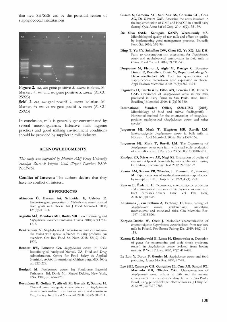
In the current study, 18 (38.3%) of 69 the S. aureus isolates were confirmed by PCR targeting nuc and coa genes in S. aureus (Fig. 1). In this study, classical enterotoxins were detected by Ridascreen and S. aureus isolates from bulk tank milk contained classical enterotoxins genes. seb and sec gene were found as 3 (16.6%) of 18 S. aureus isolates. The Ridascreen® SET A, B, C, D, E test procedure indicates two assessment option for the detection of staphylococcal enterotoxins. First way is the visual determination of the color change after the addition of the stop solution and second way is the calculation of the cut off value. The cut off value is found by adding 0.15 to the negative control absorbance value. Results of the absorbance values are equal or above to the cut off value which are considered positive while results are below the cut off value are that samples considered as negative for staphylococcal enterotoxins. Based on our results, according to the visual determination staphylococcal enterotoxins were detected as positive 4 of 120 bulk tank milk samples while 2 of 120 bulk tank milk samples were found as positive in assessment of the cut off value (Table 2 and Table 3).
Table 1. Primers used in this study Tablo 1. Çalışma kapsamında kullanılan primer dizileri.
Target gene
Primer sequence (5’ 3’) Product size
(bp) References
nuc F: ATA GGG ATG GCT ATC AGT AAT GT
624 bp Lem et al. (2001) R: GAC CTG AAT CAG CGT TGT CTT C
coa F: GTA GAT TGG GCA ATT ACA TTT TGG AGG
117 bp Kearns et al. (1999) R: CGC ATC AGC TTT GTT ATC CCA TGT A
sea F: GGT TAT CAA TGT GCG GGT GG
102 bp Mehrotra et al.
(2000) R: CGG CAC TTT TTT CTC TTC GG
seb F: GTA TGG TGG TGT AAC TGA GC
164 bp Mehrotra et al.
(2000) R: CCA AAT AGT GAC GAG TTA GG
sec F: AGA TGA AGT AGT TGA TGT GTA TGG
451 bp Mehrotra et al.
(2000) R: CAC ACT TTT AGA ATC AAC CG
sed F: CCA ATA ATA GGA GAA AAT AAA
278 bp Mehrotra et al.
(2000) R: ATT GGT ATT TTT TTT CGT TC
see F: AGG TTT TTT CAC AGG TCA TCC
209 bp Mehrotra et al.
(2000) R: CTT TTT TTT CTT CGG TCA ATC
DISCUSSION
Milk is a suitable medium for S. aureus growth and enterotoxin production. Pasteurization process can inactivate S. aureus from raw milk but SEs will remain stable even after heat treatment (Le Loir et al. 2003, Lee at al. 2012 ). Rall et al. (2008) was observed that the presence of enterotoxigenic S. aureus even after
pasteurization. The reason for this, it could be the possible inefficacy of the thermal process. SEs are the most prevalent agent of milk-borne intoxications causing risk on the public health worldwide (Benkerroum 2018). In the current study, staphylococcal enteretoxins were detected in 2 of 120 (1.66%) bulk tank milk samples. In a study from

107
Norway, enterotoxin production was identified 22.1% of S. aureus isolates in bovine milk tank and SE genes were found 52.5% of the isolates (Jørgensen et al. 2005a). Previous studies from different countries were reported levels of enterotoxigenic S. aureus as 9.4 %, 20 %, 37.1 %, 13.1%, 26.1%, 27.1% in Jordan, Portugal, Czech Republic, Poland, Egypt, Hungary, respectively (Peles et al. 2007, Zouharova and Rysanek 2008, Pereira et al. 2009, Mansour et al. 2017, Korpysa-Dzirba and Osek 2018, Obaidat et al. 2018). Enterotoxigenic S. aureus isolates in raw milk may pose potential public health hazard and due to thermostable enterotoxins, dairy products may cause intoxications in humans. Schmid et al. (2009) were reported an outbreak because of consumed school milk products in Austria.
SEA, SEB, SEC, SED and SEE types of staphylococcal enterotoxins are defined as the classical enterotoxins. Classical enteroxins have emetic activity which are associated with most of food poisoning caused by staphylococcal enterotoxins (Riva et al. 2015, Keyvan and Ozdemir 2016) These toxins have emetic activity and are usually associated with outbreaks of food poisoning (Le Loir et al. 2003). In this study, classical enterotoxins were detected by Ridascreen and S. aureus isolates from bulk tank milk contained classical enterotoxins genes. seb and sec gene were found as 16.6% (3) of S. aureus isolates (18).
Table 2. Absorbance value of bulk tank milk samples Tablo 2. Süt toplama tank örnekleri absorbans değerleri.
1 2 3 4 5 6 7 8 9 10 11 12
A 0.792 0.043 0.043 0.044 0.200 0.045 0.044 0.044 0.045 0.045 0.047 0.049 B 0.712 0.045 0.043 0.044 0.044 0.045 0.044 0.044 0.044 0.044 0.044 0.047 C 0.042 0.047 0.043 0.043 0.044 0.044 0.043 0.044 0.044 0.045 0.044 0.047 D 0.042 0.043 0.045 0.044 0.044 0.046 0.044 0.047 0.052 0.044 0.044 0.046 E 0.043 0.043 0.044 0.098 0.059 0.044 0.044 0.044 0.044 0.046 0.045 0.047 F 0.043 0.045 0.046 0.049 0.046 0.045 0.044 0.045 0.045 0.045 0.045 0.048 G 0.045 0.043 0.044 0.044 0.044 0.045 0.046 0.044 0.045 0.045 0.047 0.047 H 0.045 0.045 0.047 0.205 0.047 0.045 0.044 0.050 0.045 0.045 0.046 0.047 1A/1B: Positive Control, 1C/1D: Negative Control, Cut off value: 0.192, 4/H-5/A: Samples are above to cut off value
Table 3. Staphylococcal enterotoxins (A, B, C, D, E) in raw milk samples Tablo 3. Çiğ süt örneklerinde stafilokokal enterotoksinler (A, B, C, D, E).
Number of samples
Color change Positive samples
Cut off value Positive samples
120 4 (3.3%) 2 (1.6%)
Table 4. Enterotoxigenic properties of S. aureus isolates Tablo 4. S. aureus izolatlarının enterotoksijenik özellikleri.
Target gene Number of positive S. aureus
isolates (n=18)
sea -
seb 2 (11.1%)
sec -
sed -
see 1 (5.5%)
Total 3 (16.6)
Mastitis is one of the most economically devastating problems in cattle and S. aureus is a common causative agent of clinical and subclinical mastitis (Türkyılmaz et al. 2010, Ote et al. 2011, Rall et al.
2014). In Brasil, S. aureus was isolated in 6.7% of raw milk samples from dairy cows with subclinical mastitis and 10.8% of bulk tank milk samples. Also, four of S. aureus isolates were reported enterotoxigenic. (Fagundes et al. 2010). Boynukara et al. (2008) was found to be enterotoxigenic 25.5% of S. aureus strains isolated from cows with subclinical mastitis. Rall et al. (2014) were observed that 53.3% of S. aureus isolates contained sea gene in milk from cows with subclinical mastitis. Milk collected from dairy cows with subclinical mastitis may pose a significant source of enterotoxigenic S. aureus which can produce SEs. Transfer of the contaminated milk to bulk tank milk may cause intoxitacations. Ding et al. (2016) were recommended that to control milk-borne staphylococcal intoxication, effcient storage conditions of milk and dairy products are the key step for to minimize the risk of staphylococcal food poisoning. For controlling S. aureus milk and milking environment adopting assurance quality systems are required in dairy industry (Cusato et al. 2014). Although classical enterotoxins are the mainly isolated from staphyloccocal food poisoning, SEls can also cause outbreaks and intoxications. Umeda et al. (2017) were reported an outbreak from Japan caused by new SE/SEls and these findings indicated
108
that new SE/SEls can be the potential reason of staphylococcal intoxitacions.
Figure 2. coa, nuc gene positive S. aureus isolates. M: Marker, +: nuc and coa gene positive S. aureus (ATCC 25923) Şekil 2. coa, nuc geni pozitif S. aureus izolatları. M: Marker, +: nuc ve coa geni pozitif S. aureus (ATCC 25923) In conclusion, milk is generally get contaminated by several microorganisms. Effective milk hygiene practices and good milking environment conditions should be provided by supplier in milk industry.
ACKNOWLEDGMENTS
This study was supported by Mehmet Akif Ersoy University Scientific Research Projects Unit. (Project Number: 0374-NAP-16). Conflict of Interest: The authors declare that they have no conflict of interest.
REFERENCES
Akineden Ö, Hassan AA, Schneider E, Usleber E. Enterotoxigenic properties of Staphylococcus aureus isolated from goats milk cheese. Int J Food Microbiol. 2008; 124(2):211-216.
Argudín MÁ, Mendoza MC, Rodio MR. Food poisoning and Staphylococcus aureus enterotoxins. Toxins. 2010; 2(7):1751–1773.
Benkerroum N. Staphylococcal enterotoxins and enterotoxin-like toxins with special reference to dairy products: An overview. Crit Rev Food Sci Nutr. 2018; 58(12):1943-1970.
Bennett RW, Lancette GA. Staphylococcus aureus, In: BAM Bacteriological Analytical Manual. U.S. Food and Drug Administration, Centre for Food Safety & Applied Nutrition, AOAC International, Gaithersburg, MD. 2001; pp. 222–228.
Berdgoll M. Staphylococcus aureus, In: Foodborne Bacterial Pathogens, Ed; Doyle M, Marcel Dekker, New York, USA. 1989; pp. 464–523.
Boynukara B, Gulhan T, Alisarli M, Gurturk K, Solmaz H. Classical enterotoxigenic characteristics of Staphylococcus aureus strains isolated from bovine subclinical mastitis in Van, Turkey. Int J Food Microbiol. 2008; 125(2):209-211.
Cusato S, Gameiro AH, Sant'Ana AS, Corassin CH, Cruz AG, De Oliveira CAF. Assessing the costs involved in the implementation of GMP and HACCP in a small dairy factory. Qual Assur Saf of Crop. 2014; 6(2):135-139.
De Silva SASD, Kanugala KANP, Weerakkody NS. Microbiological quality of raw milk and effect on quality by implementing good management practices. Procedia Food Sci. 2016; 6:92-96.
Ding T, Yu YY, Schaffner DW, Chen SG, Ye XQ, Liu DH. Farm to consumption risk assessment for Staphylococcus aureus and staphylococcal enterotoxins in fluid milk in China. Food Control. 2016; 59:636-643.
Duquenne M, Fleurot I, Aigle M, Darrigo C, Borezée-Durant E, Derzelle S, Bouix M, Deperrois-Lafarge V, Delacroix-Buchet AS. Tool for quantification of staphylococcal enterotoxin gene expression in cheese. Appl Environ Microbiol. 2010; 76(5):1367-1374.
Fagundes H, Barchesi L, Filho AN, Ferreira LM, Oliveira CAF. Occurrence of Staphylococcus aureus in raw milk produced in dairy farms in São Paulo state, Brazil. Brazilian J Microbiol. 2010; 41(2):376-380.
International Standart Office, 6888-1-ISO (2003). Microbiology of food and animal feedingstuffs – Horizontal method for the enumeration of coagulase-positive staphylococci (Staphylococcus aureus and other species).
Jørgensen HJ, Mørk T, Høgåsen HR, Rørvik LM. Enterotoxigenic Staphylococcus aureus in bulk milk in Norway. J Appl Microbiol. 2005a; 99(1):1589-166.
Jørgensen HJ, Mørk T, Rørvik LM. The Occurrence of Staphylococcus aureus on a farm with small-scale production of raw milk cheese. J Dairy Sci. 2005b; 88(11):3810-3817.
Kandpal SD, Srivastava AK, Negi KS. Estimation of quality of raw milk (Open & branded) by milk adulteration testing kit. Indian J Community Heal. 2012; 24:188-192.
Kearns AM, Seiders PR, Wheeler, J., Freeman, R., Steward, M. Rapid detection of methicillin-resistant staphylococci by multiplex PCR. J Hosp Infect 1999; 43(1):33-37.
Keyvan E, Özdemir H. Occurrence, enterotoxigenic properties and antimicrobial resistance of Staphylococcus aureus on beef carcasses. Ankara Univ Vet Fak Derg. 2016; 63(1):17-23.
Kluytmans J, van Belkum A, Verbrugh H. Nasal carriage of Staphylococcus aureus: epidemiology, underlying mechanisms, and associated risks. Clin Microbiol Rev. 1997; 10:505-520.
Korpysa-Dzirba W, Osek J. Molecular characterization of enterotoxigenic Staphylococcus aureus isolated from raw cow milk in Poland. Foodborne Pathog Dis. 2019; 16(2):114-118.
Kuzma K, Malinowski E, Lassa H, Klossowska A. Detection of genes for enterotoxins and toxic shock syndrome toxin-1 in Staphylococcus aureus isolated from bovine mastitis. B Vet I Pulawy. 2003; 47(2):419-426.
Le Loir Y, Baron F, Gautier M. Staphylococcus aureus and food poisoning. Genet Mol Res. 2003; 2:7-28.
Lee SHI, Camargo CH, Gonçalves JL, Cruz AG, Sartori BT, Machado MB, Oliveira CAF. Characterization of Staphylococcus aureus isolates in milk and the milking environment from small-scale dairy farms of São Paulo, Brazil, using pulsed-field gel electrophoresis. J Dairy Sci. 2012; 95(12):7377-7383.

109
Lee SHI, Cappato LP, Corassin CH, Cruz AGD, Oliveira CAFD. Effect of peracetic acid on biofilms formed by Staphylococcus aureus and Listeria monocytogenes isolated from dairy plants. J Dairy Sci. 2016; 99(3):2384-2390.
Lee SHI, Mangolin BLC, Gonçalves JL, Neeff DV, Silva MP, Cruz AG, Oliveira CAF. Biofilm-producing ability of Staphylococcus aureus isolates from Brazilian dairy farms. J Dairy Sci. 2014; 97(3):1812-1816.
Lem P, Spiegelman J, Toye B, Ramotar K. Direct detection of mecA, nuc and 16S rRNA genes in BacT/Alert blood culture bottles. Diagn Microbiol Infect Dis. 2001; 41(3):165-168.
Mansour AS, Wagih GES, Morgan SD, Elhariri M, El-Shabrawy MA, Abuelnaga ASM, Elgabry EA. Detection of Staphylococcus aureus enterotoxigenic strains in bovine raw milk by reversed passive latex agglutination and multiplex polymerase chain reaction. Vet World. 2017; 10(8):843–847
Mehrotra M, Wang G, Johnson WM. Multiplex PCR for detection of genes for Staphylococcus aureus enterotoxins, exfoliative toxins, toxic shock syndrome toxin 1 and methicillin resistance. J Clin Microbiol. 2000; 38(3):1032-1035.
Morandi S, Brasca M, Lodi R, Cremonesi P, Castiglioni B. Detection of classical enterotoxins and identification of enterotoxin genes in Staphylococcus aureus from milk and dairy products. Vet Microbiol. 2007; 124(1-2):66-72.
Normanno G, La Salandra G, Dambrosio A, Quaglia NC, Corrente M, Parisi A, Santagada G, Firinu A, Crisetti E, Celano GV. Occurrence, characterization and antimicrobial resistance of enterotoxigenic Staphylococcus aureus isolated from meat and dairy products. Int J Food Microbiol. 2007; 115(3):290-296.
Obaidat MM, Salman AEB, Roess AA. High prevalence and antimicrobial resistance of mecA Staphylococcus aureus in dairy cattle, sheep, and goat bulk tank milk in Jordan. Trop Anim Health Prod. 2018; 50(2):405-412.
Ote I, Taminiau B, Duprez JN, Dizier I, Mainil JG. Genotypic characterization by polymerase chain reaction of Staphylococcus aureus isolates associated with bovine mastitis. Vet Microbiol. 2011; 153:285-292.
Özdemir H, Keyvan E. Isolation and characterisation of Staphylococcus aureus strains isolated from beef, sheep and chicken meat. Ankara Univ Vet. Fak. Derg. 2016; 63:333-338.
Painter JA, Hoekstra RM, Ayers T, Tauxe RV, Braden CR, Angulo FJ, Griffin PM. Attribution of foodborne illnesses, hospitalizations, and deaths to food commodities by using outbreak data, United States, 1998–2008. Emerg Infect Dis. 2013; 19:407-415.
Parisi A, Caruso M, Normanno G, L. Latorre R, Sottili A, Miccolupo R, Fraccalvieri G. Prevalence, antimicrobial susceptibility and molecular typing of methicillin-resistant Staphylococcus aureus (MRSA) in bulk tank milk from southern Italy. Food Microbiol. 2016; 58: 36-42.
Peles F, Wagner M, Varga L, Hein I, Rieck P, Gutser K, Keresztúri P, Kardos G, Turcsányi I, Béri B, Szabó A. Characterization of Staphylococcus aureus strains isolated from bovine milk in Hungary. Int J Food Microbiol. 2007; 118(2):186-193.
Pereira V, Lopes C, Castro A, Silva J, Gibbs P, Teixeira P. Characterization for enterotoxin production, virulence factors, and antibiotic susceptibility of Staphylococcus aureus isolates from various foods in Portugal. Food Microbiol. 2009; 26(3):278-282.
Rall VLM, Miranda ES, Castilho IG, Camargo CH, Langoni H, Guimarães FF, Araújo Júnior JP, Fernandes Júnior A. Diversity of Staphylococcus species and prevalence of enterotoxin genes isolated from milk of healthy cows and cows with subclinical mastitis. J Dairy Sci. 2014; 97(2):829-837.
Rall VLM, Vieira FP, Rall R, Vieitis RL, Fernandes A, Candeias JMG, Cardoso KFG, Araújo JP. PCR detection of staphylococcal enterotoxin genes in Staphylococcus aureus strains isolated from raw and pasteurized milk. Vet Microbiol. 2008; 132(3-4):408-413.
Riva A, Borghi E, Cirasols D, Colmegna S, Borgo F, Amato E, Pontello MM, Morace G. Methicillin-resistant Staphylococcus aureus in raw milk: Prevalence, SCC mec typing, enterotoxin characterization and antimicrobial resistance patterns. J Food Prot. 2015; 78(6):1142-1146.
Schmid D, Fretz R, Winter P, Mann M, Höger G, Stöger A, Ruppitsch W, Ladstätter J, Mayer N, De Martin A, Allerberger F. Outbreak of staphylococcal food intoxication after consumption of pasteurized milk products, June 2007, Austria. Wien Klin Wochenschr. 2009; 121(3-4):125-131.
Türkyılmaz S, Tekbıyık S, Oryasin E, Bozdogan B. Molecular epidemiology and antimicrobial resistance mechanisms of methicillin-resistant Staphylococcus aureus isolated from bovine milk. Zoonoses Public Health. 2010; 57(3):197-203.
Umeda K, Nakamura H, Yamamoto K, Nishina N, Yasufuku K, Hirai Y, Hirayama T, Goto K, Hase A, Ogasawara J. Molecular and epidemiological characterization of staphylococcal foodborne outbreak of Staphylococcus aureus harboring seg, sei, sem, sen, seo, and selu genes without production of classical enterotoxins. Int J Food Microbiol. 2017; 256:30-35.
Zouharova M, Rysanek D. Multiplex PCR and RPLA identification of Staphylococcus aureus enterotoxigenic strains from bulk tank milk. Zoonoses Public Health 2008; 55(6):313-319.
110
Kocatepe Veterinary Journal
Kocatepe Vet J (2020) 13(2):110-117
DOI: 10.30607/kvj.677666 RESEARCH ARTICLE
The Effects of Chitosan Oligosaccharide (COS) Treatment on
Oxidative Stress and Its Relation with Intestinal Microflora in Rats Exposed To Cadmium
İhsan KISADERE1*, Hakan TAVŞANLI2, Mukadderat GÖKMEN3
1Balikesir University, Faculty of Veterinary Medicine, Department of Physiology, 10100, Balıkesir, Turkey
2Balıkesir University, Faculty of Veterinary Medicine, Department of Public Health, 10100, Balıkesir, Turkey 3Balikesir University, Faculty of Veterinary Medicine, Department of Food Hygiene and Technology, 10100, Balıkesir, Turkey
ABSTRACT
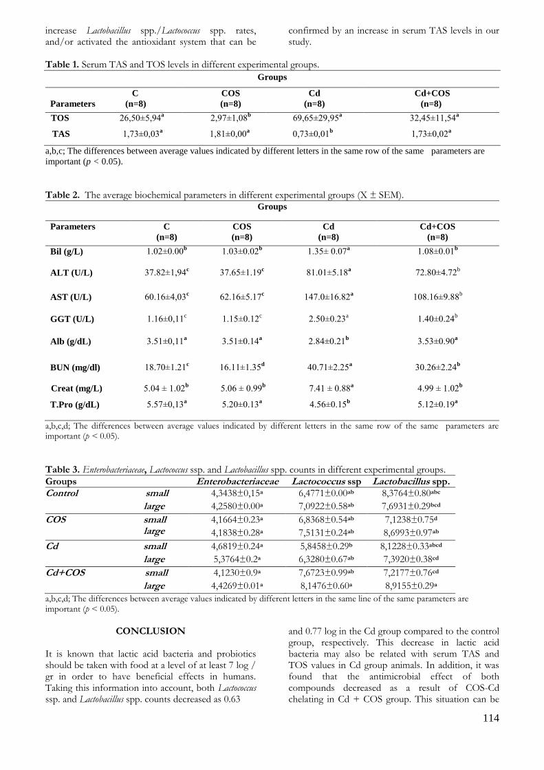
The aim of the study was to investigate the effects of chitosan oligosaccharide (COS) treatment on oxidative stress and its relation with intestinal microflora in rats exposed to chronic cadmium toxicity. Animals were randomly divided into four groups as control (C; n=8), cadmium (Cd; n=8), chitosan oligosaccharide (COS; n=8), cadmium+chitosan oligosaccharide (Cd+COS; n = 8). After, cadmium chloride (CdCl2) (2mg /kg/ day) was orally administered to Cd and Cd+COS groups three times a week for 4 weeks. Chitosan oligosaccharide (200 mg/kg/day) was also orally administered to COS and Cd+COS groups five times a week for 4 weeks. After completion of the experiment, serum TAS, TOS levels, plasma ALT, AST, GGT, T.pro, Alb, Bil, Creat and BUN values were measured. Enterobacteriaceae, Lactococcus spp. and Lactobacillus spp. counts were also detected. Serum TOS values were detected extremely higher in Cd group animals when compared COS group (p <0,05). In the small intestine of the Cd group animals, Cd administration caused a 0.66 log decrease in the Lactococcus spp. count. In conclusion, it was found that the antimicrobial effect of both compounds decreased as a result of COS-Cd chelating in Cd + COS group. Keywords: Cadmium, chitosan oligosaccarides, microflora, oxidative stress, rat
***
Kitosan Oligosakkarit (COS) Tedavisinin Oksidatif Stres Üzerine Etkileri ve Kadmiyuma Maruz Kalan
Sıçanlarda Bağırsak Mikroflorası ile İlişkisi
ÖZ Bu çalışmanın amacı, kronik olarak Cd’a maruz kalan ratlarda kitosan oligosakkarit’in (COS) oksidatif stress ve bağırsak mikroflorası üzerine etkilerinin araştırılmasıdır. Hayvanlar rastgele olacak şekilde; kontrol (C; n=8), kadmiyum (Cd; n=8), kitosan oligosakkarit (COS; n=8) ve kadmiyum+kitosan oligosakkarit (Cd+COS; n=8) gruplarına ayrıldı. Daha sonra, kadmiyum klorid (CdCl2) (2mg/kg/day) Cd ve Cd+COS gruplarındaki hayvanlara haftada 3 kez 4 hafta boyunca oral yoldan verildi. Kitosan oligosakkarit de (200 mg/kg/day) COS ve Cd+COS grubundaki hayvanlara haftada 5 kez 4 hafta boyunca oral olarak uygulandı. Deneme sonunda, serum TAS, TOS seviyeleri, plasma ALT, AST, GGT, T.pro, Alb, Bil, Creat ve BUN değerleri ölçüldü. Enterobacteriaceae, Lactococcus spp. ve Lactobacillus spp. sayılarıda belirlendi. Serum TOS seviyeleri Cd grubundaki hayvanlarda COS grubundakilere oranla önemli derecede yüksek bulundu (p <0,05). Cd grubundaki hayvanların ince bağırsaklarında, kronik Cd uygulaması Lactococcus spp. sayısında 0.66 log’lık bir düşüşe sebep oldu. Sonuç olarak, her iki bileşiğin antimikrobiyel etkinliği şelat oluşumuna bağlı olarak (COS-Cd) Cd+COS grubundaki hayvanlarda azalma gösterdi. Anahtar Kelimeler: Kadmiyum, Kitosan oligosakkarit, mikroflora, oksidatif stres, rat
To cite this article: Kısadere İ. Tavşanlı H. Gökmen M. The Effects of Chitosan Oligosaccharide (COS) Treatment on Oxidative Stress and Its Relation with Intestinal Microflora in Rats Exposed To Cadmium. Kocatepe Vet J. (2020) 13(2):110-117 Submission: 21.01.2020 Accepted: 05.04.2020 Published Online: 24.04.2020
ORCID ID; İK: 0000-0003-0732-0464, HT: 0000-0002-5124-3702, MG: 0000-0002-9371-8956 *Corresponding author e-mail: [email protected]
111
INTRODUCTION Cadmium (Cd), is a non-essential transition metal and considered to be an environmental pollutant, is naturally occurring element that has a high density and atomic weight when compared to water (Tchounwou et al. 2012, Gao et al. 2014). It is released into the environment by various human activities including mining, smelting, and manufacturing of batteries, pigments, stabilizers, and alloys (WHO 2010, Bernhoft 2013, WHO 2019). Cadmium is accumulating in catchments and soils under certain environmental conditions, thus increasing the risk of future exposure through food. The main routes of exposure to Cd are via ingestion of contaminated foods such as vegetables, potatoes, rice, wheat, green leafy grains and seeds, liver and kidney, and crustaceans and mollusks as well as contaminated water (IARC 1993, Paschal et al. 2000, Satarug et al. 2003, WHO 2007, ATSR 2008) It has been reported that acute or chronic exposed to Cd induces lipid peroxidation (LPO) (by stimulation of occuring superoxide anions) and oxidative stress (by increasing free radical production) in the cells (El-Demerdash et al. 2004, López et al. 2006). Morever, it initiates various adverse effects in human and animals such as kidney dysfunction, liver injury and osteoporosis (Tchounwou et al. 2012, Satarug et al. 2011, Amamou et al. 2015). Cd accumulation is mainly occured in the kidney and liver but also in brain, lung, bones, pancreas, placenta and testis in the body (Satarug et al. 2011, Amamou et al. 2015, Fowler 2009). In addition, Cd is a severe gastrointestinal irritant, which can leads to abdominal pain, burning sensation, nausea, vomiting, salivation when acute high dose ingested (Baselt and Cravey 1995, Hammett-Stabler 2000). The gastrointestinal tract, is the interface between ingested nutrients and the body, plays an important role in maintaining of the health, food intake and regulating energy homeostasis (Zhang et al. 2014, Monteiro et al. 2017). In GIS, there are many bacterial populations whose have mutual relationship with intestinal epithelial cells that are known as symbiosis. Although Enterobacteriaceae are normal flora of the human intestinal system, they are common opportunistic pathogens can translocate across the mucosal barrier and lead to systemic infections if intestinal counts are extremely increased (Hsueh et al. 2010, Toh et al.2012, Lai et al. 2016, Jean et al. 2016). On the other hand, lactic acid bacteria such as Lactococcus spp. and Lactobacillus spp also inhabit in the GIS that can produce lactic acid, acetic acid, formic acid and other acids to reduce intestinal pH. Besides, these microorganisms can secrete some antimicrobial molecules, such as ethanol, fatty acid, hydrogen peroxide and bacteriocins to defense against pathogenic bacteria in GIS (Ralitsa et al. 2015, Inglin et al. 2015). Although above mentioned bacteria
populations are mainly affected by the host’s diet intake, the prevalence of bacteria in different parts of the GI tract appears to be depending on certain factors, such as pH, peristalsis, redox potential, bacterial adhesion, bacterial cooperation, mucin secretion, nutrient availability and bacterial antagonism (Tannock 1983, Roberfroid et al. 2010, Amato et al. 2013). Imbalance among the intestinal epithelial cells, pathogen and/or commensal bacteries increases the rate of intestinal microbial disorders and sensitivity to external harmful compounds (Costello et al.2012, Salim et al. 2014, Woodmansey 2007). Heavy metals also reach GI tract through ingestion of contaminated food and water. Although the toxicological effect of heavy metals on different body structures were detected, especially Cd, on GI microflora, is still remains unclear (Upreti et al. 2004, Inaba et al. 2005, Monachese et al. 2012). Recently, it has been reported that harmfull effects of Cd can be ameliorated by using some chelating agents, antioxidants, probiotics and vitamins (Pourmorad et al.2006, fang 2007, El-boshy et al. 2014, Djurasevic et al. 2017). One of them is chitosan oligosaccharide (COS) that is produced by chitosan/chitin via chemical hydrolysis or enzymatic degradation, known for its ability to bind to divalent cations such as Cd. As it known, it has an antioxidant, free radical consumer, antimicrobial, antifungal, anti-inflammatory, anti-diabetic and anti-obesity properties (Guan et al. 2016, Kim et al. 2016, Naveed et al. 2019). Therefore, our study has been designed to evaluate the influences of oral COS administration on oxidative stress, and its relation with intestinal microflora of the rats exposed to chronic Cd toxicity.
MATERIALS and METHODS Animals, Study Design and Experimental Procedure Male albino Wistar rats (n=32; body weight ~ 200 ± 30 g) were housed in standard plastic rat cages at 23 ± 2 ºC room temperature, 55 ± 10% relative humidity and 12 hours night/day light period during the experiment. The animals had free access to drinking water and standart rat feed. All experimental procedures were approved by the Ethical Committee on Animal Experimentation of the University of Balikesir (2019/4-6). Before the experiment, animals were randomly divided into four groups as control (C; n=8), cadmium (Cd; n=8), chitosan oligosaccharide (COS; n=8), cadmium+chitosan oligosaccharide (Cd+COS; n= 8). Then, animals in C group received standard rat feed and fresh drinking water ad libitum. Cadmium chloride (CdCl2) (2mg / day) were orally administered to Cd and Cd+COS groups three times a week for 4 weeks. On the other hand, chitosan oligosaccharide (200 mg/kg/day) was also orally administered to COS and Cd+COS groups five times a week for 4 weeks. After completion of the

112
experiment (4 weeks later), rats were anesthetized by intraperitoneal injection of ketamine/xylazine (0.1 ml/100gm/body weight) and killed by cervical dislocation technique. Blood samples were collected via cardiac puncture and transferred into tubes. Plasma and serum were obtained from the blood samples by using a centrifuge (3000 rpm, 25 min, Heichrich, Germany). Obtained samples were stored at minus 80 °C in a refrigeratior until analysis time. Besides, intestinal fluid content were aseptically collected from the small and large intestines of the each rats. Determination of total antioxidant and oxidants levels Serum total antioxidant status (TAS) and oxidant status (TOS) values were defined by ELISA (Thermoscientific Elisa Reader, USA) using commercial kits (Rel Assay Diagnostics, Gaziantep, Turkey), according to Erel’s method that is automated and colorimetric (Erel 2004, Erel 2005). Determination of some plasma enzyme levels Plasma alanine amino transferase (ALT), aspartate amino transferase (AST), gamma glutamyl transferase (GGT), total protein (T.pro), albumine (Alb), bilirubin (Bil), creatinine (Creat) and blood urea nitrogen (BUN) values were measured by using automatic biochemical analyser (Architect C-8000, Abbott, USA) with commercial kits according to manufacturer instructions. Microbiological analysis During the necropsy, 1 g intestinal fluid content were aseptically collected from the small and large intestines of the each rats (separately with 3 replicates). Then, they were homogenized in the stomacher for 2 minutes with sterile 9 ml Maximum Recovery Diluent (MRD), serial dilutions were prepared from 10-1 to 10-6. For determine to the Enterobacteriaceae count, 1 ml of the dilution was taken and cultured in Violet Red Glucose Bile (VRGB, Oxoid CM1082) Agar according to the double-plate technique. The plates were evaluated as Enterobacteriaceae because of the observing purple-pink colonies after aerobic incubation at 37 °C for 24 h (ISO 21528-2: 2017). On the other hand, 0.1 ml of the dilution was taken and cultured in the M17 (Oxoid CM0785) agar according to spread plate technique for despite to Lactococcus spp count. Then, plates were evaluated as Lactococcus spp depends on occuring yellow-cream colonies after anaerobic incubation at 30 ° C for 24 h (Lee et al.2010). For detection of Lactobacillus spp count, 0.1 ml of dilution was cultured on MRS (CM0361) agar. Plates were also evaluated as Lactobacillus spp. due to occuring of yellow-cream colonies after anaerobic incubation at 37 °C for 72 h (Bauer et al. 2002).
Statistical Analysis Obtained datas were analyzed with using SPSS for Windows version 25.0, and levels were presented as means ± SE. Differences among the groups were performed by analysis of variance (one-way-ANOVA) that is followed by Duncan’s test.
RESULTS and DISCUSSION Serum TOS values were detected extremely higher in Cd group animals when compared COS group (p <0,05). On the other hand, it was not found significant difference among C, Cd and Cd+COS groups according to TOS values, shown in Table 1. In addition, serum TAS values decreased due to Cd administration in Cd group animals compared to other groups (p <0,05). Plasma Bil and Creat levels were found the highest in Cd group compared to other groups (p <0,05). Besides, COS administration did not lead to any changes in Cd+COS group according to Bil and Creat levels (p > 0,05). Conversely, plasma T.pro and Alb values were detected lower in Cd group compared to C, COS and Cd+COS (p <0,05). In addition, plasma BUN levels were ameliorated due to COS administration in Cd+COS group (p <0,05). Although plasma ALT, AST and GGT levels were detected higher in Cd group, the levels of the mentioned parameters decreased in COS group animals, statistically (p <0,05), shown in Table 2. The average Enterobacteriaceae, Lactococcus spp. and Lactobacillus spp. counts were detected as 4.34, 4.25 log cfu / g, 6.47; 7.09 log cfu / g; 8.37, 7.39 log cfu / g in both (small and large) intestines of the control group animals, respectively. On the other hand, Enterobacteriaceae counts were found similar in the control group with another experimental groups in both small and large intestines (P > 0.05). In the small intestine of the Cd group animals, Cd administration caused a 0.66 log decrease in the Lactococcus spp. count. In contrary, Cd+COS chelate lead to increase in the counts of Lactococcus spp. in
small intestines of the rats (p˂0.01). There was a significant difference between the C group and the other experimental groups according to Lactobacillus
spp. count in small intestines (p˂0.01). Besides, Lactobacillus spp. counts significantly decreased in Cd, COS and Cd+COS when compared to the C group. In terms of Lactobacillus spp, the highest decrease was observed in the small intestines of the COS group animals. In the large intestines of the rats, Lactobacillus spp. count significantly increased in COS and Cd+COS, however decreased in Cd group when
compared to C (p˂0.01). The highest increase in the Lactobacillus spp. counts were observed as 0.54 log in the Cd+COS group, shown in Table 3.

113
Although Cd is a well-known environmental pollutant which induces severe organ and tissue damage in human and animals, effect of Cd on GI microflora and its relation with oxidative stress is still remains unclear (Satarug et al. 2011, Amamou et al. 2015, Fowler 2009). In present study, exposed to chronic Cd toxicity increased (not statistically) the serum TOS levels, however significantly supressed the serum TAS in Cd group animals. These results were consistent with previous studies (Karabulut-Bulan et al. 2008, Koçak and Akçil 2006, Kumaş et al. 2016). Either increased TOS nor decreased TAS levels were ameliorated with COS treatment in Cd+COS group when compared to Cd in our study. Similarly, the dose of chitosan more than 20 mg/kg/day was found effective on Cd-induced oxidative damage (SOD activity and MDA content) in the rat kidney by Zhou et al. (2013). Protective effects of COS and chitin on various metal and chemical compound induced oxidative stress were also determined by other researchers which was consistent with our results (Kim et al. 2005, Yan et al. 2006, Li et al. 2011, Toz and Değer 2018). It can be considered that the administration of COS reinforced the antioxidant defence system and also ameliorated the Cd induced oxidative stress in present study. In our study, oral Cd treatment (low dose, 2mg/kg) led to increase of plasma ALT, AST, GGT enzyme levels (an important indicators of liver functions) in Cd group animals. Besides, an important markers of kidney functions are BUN and Creat levels also negative effected by Cd treatment in present study. These findings were corresponding with previous studies (Koçak and Akçil 2006, Lakshmi et al. 2012, Renugadevi and Milton 2010). Although plasma ALT, AST, GGT and BUN levels were improven by using COS in experimental groups, it couldn’t affect to the plasma Creat levels in our study, interestingly. It was also reported that high dose chitosan diet ameliorated the Cd induced increased AST levels but did not lead to significance alterations in plasma ALT, BUN and Creat levels (Kim et al. 2016). In addition, T.pro and Alb values also negative effected by Cd toxicity in Cd group animals. It was consistent with Hussein et al. (2009) and Oyinloye et al. (2016). Increased liver and kidney enzyme levels, and reduction of T.pro and Alb values confirm the tissue damege due to chronic Cd toxicity in present study. There was limited information about the effects of COS on T.pro and Alb levels of Cd induced toxication in rats. Bil levels also increased in Cd group animals but did not effected from COS administration in present study. Hamden et al. (2009), Ibiam et al. (2013) and Markiewicz-Górka et al. (2011) also defined similar results in Cd treated rats according to Bil values. It may be explained that COS can be partially ameliorated the Cd induced tissue damages in the liver and kidney.
In the small intestinal microflora of the rats, neither Cd nor COS didn’t cause any significant changes in the counts of Enterobacteriaceae in present study. Conversely, Escherichia coli and Klebsiella spp., which are the members of Enterobacteriaceae group, counts decreased due to Cd (high doses) treatments in the small intestine of the mouse in a previous study (Fazeli et al. 2011). As it known, COS has positive effects on host gut health and intestinal microbial community (Zhang et al. 2014), however Cd+COS treatment not affected the Enterobacteriaceae count in our study. It can be explained by either antimicrobial effect of both compounds decreased as a result of COS+Cd chelating, both compounds were rapidly absorbed without showing their antimicrobial effects or the doses were insufficient to demonstrate known effects. In addition, Cd treatment did not lead to changes in Enterobacteriaceae count in large intestine in Cd group compared to C group. It has been suggested that E. coli and Klebsiella spp. counts reduced in the large intestine of the mice due to Cd in a previous study which was not corresponding with present study (Fazeli et al. 2011). Although it has been enounced that COS influences GI flora, and thus improving intestinal health, it was not found a significant change in Enterobacteriaceae counts in the large intestine of the Cd+COS group animals. It can be occured due to different dose, time of exposure to Cd and/or animal species. A significant decrease was found in the count of Lactococcus spp. in small intestine of Cd group animals in present study. It was also suggested that gram-positive basilcus and enterecocus microorganisms were more sensitive to Cd toxicity than gram-negative E. coli and Klebsiella spp. (Fazeli et al. 2011). On the other hand, Cd+COS treatment increased the Lactococcus spp count in large intestine of the rats. These results can be explained by the fact that total bacterial rates of microflora varied with decreased count of Enterobacteriaceae due to Cd+Mel administration or the high pH in the small intestine. Lactobacillus spp. count was found lower in Cd group than C, Mel and Cd+Mel groups in small intestine microflora of the rats in our study. It has been reported by Fazeli et al. (Fazeli et al. 2011). that Lactobacillus spp. count decreased due to different high doses of Cd treatment in small intestines of the mice which was corresponding with our results. Although decreased Lactobacillus spp. counts were detected by Fazeli et al. (2011) in large intestines depend on the different doses of Cd, it increased due to Mel and Cd+Mel treatments in large intestine of the rats except C group in our study. It was also reported that Mel treatment increased the Lactobacillus spp. counts in large intestines of colitic mice which was consistent with present study (Wang et al. 2019). It can be also expressed that Cd+Mel treatment may be reduced Enterobacteriaceae count and lead to
114
increase Lactobacillus spp./Lactococcus spp. rates, and/or activated the antioxidant system that can be
confirmed by an increase in serum TAS levels in our study.
Table 1. Serum TAS and TOS levels in different experimental groups.
Groups
C
Parameters (n=8)
COS
(n=8)
Cd
(n=8)
Cd+COS
(n=8)
TOS 26,50±5,94a
2,97±1,08b
69,65±29,95a 32,45±11,54
a
TAS 1,73±0,03a 1,81±0,00
a 0,73±0,01
b 1,73±0,02
a
a,b,c; The differences between average values indicated by different letters in the same row of the same parameters are
important (p < 0.05).
Table 2. The average biochemical parameters in different experimental groups (X ± SEM).
Groups
Parameters C
(n=8)
COS
(n=8)
Cd
(n=8)
Cd+COS
(n=8)
Bil (g/L) 1.02±0.00b 1.03±0.02
b 1.35± 0.07
a 1.08±0.01
b
ALT (U/L) 37.82±1,94c 37.65±1.19
c 81.01±5.18
a 72.80±4.72
b
AST (U/L) 60.16±4,03c 62.16±5.17
c 147.0±16.82
a 108.16±9.88
b
GGT (U/L) 1.16±0,11c
1.15±0.12c
2.50±0.23a
1.40±0.24b
Alb (g/dL) 3.51±0,11a
3.51±0.14a
2.84±0.21b
3.53±0.90a
BUN (mg/dl) 18.70±1.21c 16.11±1.35
d 40.71±2.25
a 30.26±2.24
b
Creat (mg/L) 5.04 ± 1.02b 5.06 ± 0.99
b 7.41 ± 0.88
a 4.99 ± 1.02
b
T.Pro (g/dL) 5.57±0,13a 5.20±0.13
a 4.56±0.15
b 5.12±0.19
a
a,b,c,d; The differences between average values indicated by different letters in the same row of the same parameters are important (p < 0.05).
Table 3. Enterobacteriaceae, Lactococcus ssp. and Lactobacillus spp. counts in different experimental groups.
Groups Enterobacteriaceae Lactococcus ssp Lactobacillus spp.
Control small
large
4,3438±0,15a 6,4771±0.00ab 8,3764±0.80abc
4,2580±0.00a 7,0922±0.58ab 7,6931±0.29bcd
COS small large
4,1664±0.23a 6,8368±0.54ab 7,1238±0.75d
4,1838±0.28a 7,5131±0.24ab 8,6993±0.97ab
Cd small
large
4,6819±0.24a 5,8458±0.29b 8,1228±0.33abcd
5,3764±0.2a 6,3280±0.67ab 7,3920±0.38cd
Cd+COS small 4,1230±0.9a 7,6723±0.99ab 7,2177±0.76cd
large 4,4269±0.01a 8,1476±0.60a 8,9155±0.29a
a,b,c,d; The differences between average values indicated by different letters in the same line of the same parameters are important (p < 0.05).
CONCLUSION
It is known that lactic acid bacteria and probiotics should be taken with food at a level of at least 7 log / gr in order to have beneficial effects in humans. Taking this information into account, both Lactococcus ssp. and Lactobacillus spp. counts decreased as 0.63
and 0.77 log in the Cd group compared to the control group, respectively. This decrease in lactic acid bacteria may also be related with serum TAS and TOS values in Cd group animals. In addition, it was found that the antimicrobial effect of both compounds decreased as a result of COS-Cd chelating in Cd + COS group. This situation can be
115
observed in terms of serum TAS and TOS levels in our study.
ACKNOWLEDGEMENT
All experimental procedures were approved by the Ethical Committee on Animal Experimentation of the University of Balikesir (2019/4-6).
Conflict of Interest: The authors declare that they have no conflict of interest.
REFERENCES
Agency for Toxic Substances and Disease Registry (ATSDR). Toxicological Profile for Cadmium ATSDR, Atlanta, Georgia (2008), p. 454.
Amamou F, Nemmiche S, Meziane RK, Didi A, Yazit SM, Chabane-Sari D. Protective effect of olive oil and colocynth oil against cadmiuminduced oxidative stress in the liver of Wistar rats. Food Chem Toxicol.2015;78:177-84.DOI: 10.1016/j.fct.2015.01.001.
Amato SM, Orman MA, Brynildsen MP. Metabolic control of persister formation in Escherichia coli. Mol Cell. 2013; 50(4): 475-487. DOİ: 10.1016/j.molcel.2013.04.002.
Baselt, RC, Cravey RH. Disposition of Toxic Drugs and Chemicals in Man. 4th Edn. Chicago,IL: Year Book Medical Publishers; 1995. pp. 105-107.
Bauer TM, Fernández J, Navasa M, Vila J, Rodés J. Failure of Lactobacillus spp. to prevent bacterial translocation in a rat model of experimental cirrhosis. J Hepatol, 2002; 36(4), 501-506. DOI:10.1016/S0168-8278(02)00003-X.
Bernhoft RA. Cadmium toxicity and treatment. Sci.World J.2013:394652. DOİ:10.1155/2013/394652.
Costello EK, Stagaman K, Dethlefsen L, Bohannan BJ, Relman DA. The application of ecological theory toward an understanding of the human microbiome. Science. 2012; 336(6086):1255-1262. DOİ: 10.1126/science.1224203.
Djurasevic S, Jama A, Jasnic N, Vujovic P, Jovanovic M, Mitic-Culafic D, Knezevic-Vukcevic J, Cakic-Milosevic M, Ilijevic K, Djordjevic J. The Protective Effects of Probiotic Bacteria on Cadmium Toxicity in Rats. J Med Food. 2017; 20(2):189-196. DOI: 10.1089/jmf.2016.0090.
El-Boshy ME, Risha EF, Abdelhamid FM, Mubarak MS, Hadda TB. Protective effects of selenium against cadmium induced hematological disturbances, immunosuppressive, oxidative stress and hepatorenal damage in rats. J Trace Elem Med Biol. 2014; 29: 104-10. DOİ:10.1016/j.jtemb.2014.05.009.
El-Demerdash FM, Yousef MI, Kedwany FS, Baghdadi HH. Cadmium-induced changes in lipid peroxidation, blood hematology, biochemical parameters and semen quality of male rats: protective role of vitamin E and beta-carotene. Food Chem Toxicol. 2004; 42(10):1563-1571. DOI: 10.1016/j.fct.2004.05.001
Erel O. A new automated colorimetric method for measuring total oxidant status. Clin biochem. 2005; 38(12): 1103-1111. DOI: 10.1016/j.clinbiochem.2005.08.008
Erel O. A novel automated direct measurement method for total antioxidant capacity using a new generation, more stable
ABTS radical cation. Clin biochem. 2004; 37: 277-285. DOI: 10.1016/j.clinbiochem.2003.11.015.
Fang C. Characterization of Polyphenol Oxidase and Antioxidants From Pawpaw (Asimina Tribola) Fruit. 2007; University of Kentucky Master's Theses 477. https://uknowledge.uky.edu/gradschool_theses/477.
Fazeli M, Hassanzadeh P, Alaei S. Cadmium chloride exhibits a profound toxic effect on bacterial microflora of the mice gastrointestinal tract. Hum Exp Toxicol. 2011; 30(2): 152-159. DOI: 10.1177/096032711036982.
Fowler BA. Monitoring of human populations for early markers of cadmium toxicity: A review. Toxicol Applied Pharma. 2009; 238(3): 294–300. DOI: 10.1016/j.taap.2009.05.004.
Gao D, Xu Z, Kuang X, Qiao P, Liu S, Zhang L, He P, Jadwiga WS, Wang Y, Min W. Molecular characterization and expression analysis of the autophagic gene beclin 1 from the purse red common carp (Cyprinus carpio) exposed to cadmium. Comp Biochem Physiol C Toxicol Pharmacol. 2014; 160:15-22.
Guan G., Wang H., Chen S., Liu G., Xiong X., Tan B., Duraipandiyan V, Al-Dhabi NA, Fang J. Dietary chitosan supplementation increases microbial diversity and attenuates the severity of Citrobacter rodentium infection in mice. Mediat Inflamm. 2016:9236196. DOI:10.1155/2016/9236196.
Hamden K, Allouche N, Damak M, Elfeki A. Hypoglycemic and antioxidant effects of phenolic extracts and purified hydroxytyrosol from olive mill waste in vitro and in rats. Chem Biol Interact. 2009;180(3):421-32. DOİ: 10.1016/j.cbi.2009.04.002.
Hammett-Stabler CA. Disposition of Toxic Drugs and Chemicals in Man, 5th ed. R.C. Baselt. Foster City, CA: Chemical Toxicology Institute, 1999, 900 pp., $119.00. ISBN 0-9626523-3-4. Published June 2000.
Hsueh PR, Badal RE, Hawser SP, Hoban DJ, Bouchillon SK, Ni Y, Paterson DL. Epidemiology and antimicrobial susceptibility profiles of aerobic and facultative gram-negative bacilli isolated from patients with intra-abdominal infections in the Asia-Pacific region: 2008 results from SMART (Study for Monitoring Antimicrobial Resistance Trends). Int J Antimicrob Agents. 2010:36(5):408–414. DOI: 10.1016/j.ijantimicag.2010.07.002.
Hussein SA, Abd-el-maksoud HA, Agag BI, El-nile MB. Effect of cadmium toxicity on some biochemical blood parameters and their modulation with certain natural antioxidants in rabbits. Benha Ras Surd. 2009; 338–62.
Ibiam AU, Ugwuja EI, Ejeogo C, Ugwu O. Cadmium-Induced Toxicity and the Hepatoprotective Potentials of Aqueous Extract of Jessiaea Nervosa Leaf. Adv Pharm Bull. 2013; 3(2), 309-313. DOİ: 10.5681/apb.2013.050
Inaba T1, Kobayashi E, Suwazono Y, Uetani M, Oishi M, Nakagawa H, Nogawa K. Estimation of cumulative cadmium intake causing Itai-itai disease. Toxicol Lett. 2005; 159: 192-201. DOI: 10.1016/j.toxlet.2005.05.011
Inglin RC, Stevens MJ, Meile L, Lacroix C, Meile L. High-throughput screening assays for antibacterial and antifungal activities of Lactobacillus species. J Microbiol Methods. 2015; 114:26-29. DOI: 10.1016/j.mimet.2015.04.011.
International Agency for Research on Cancer (IARC). Cadmium and cadmium compounds. IARC Monogr Eval Carcinog Risks Hum. 1993; 58: 119-237.

116
ISO 21528-2:2017. Microbiology of the food chain — Horizontal method for the detection and enumeration of Enterobacteriaceae — Part 2: Colony-count technique
Jean SS, Coombs G, Ling T, Balaji V, Rodrigues C, Mikamo H, Kim MJ, Rajasekaram DG, Mendoza M, Tan TY1, Kiratisin P1, Ni Y1, Weinman B, Xu Y, Hsueh PR. Epidemiology and antimicrobial susceptibility profiles of pathogens causing urinary tract infections in the Asia-Pacific region: results from the study for monitoring antimicrobial resistance trends (SMART), 2010-2013. Int J Antimicrob. Agents. 2016; 47(4):328-34. DOI: 10.1016/j.ijantimicag.2016.01.008.
Karabulut-Bulan O, Bolkent S, Yanardag R, Bilgin-Sokmen B. The role of vitamin C, vitamin E, and selenium on cadmium-induced renal toxicity of rats. Drug Chem Toxicol. 2008; 31(4): 413–26. DOI: 10.1080/01480540802383200
Kim KN, Joo ES, Kim KI, Kim, SK, Yang HP, Jeon YJ. Effect of chitosan oligosaccharides on cholesterol level and antioxidant enzyme activities in hypercholesterolemic rat. J Korean Soc Food Sci Nutr. 2005; 34(1):36-41. DOİ:10.3746/jkfn.2005.34.1.036.
Kim MY, Shon WJ, Park MN, Lee YS, Shin DM. Protective effect of dietary chitosan on cadmium accumulation in rats. Nutr Res Pract. 2016; 10(1):19-25. DOİ:10.4162/nrp.2016.10.1.19
Koçak M, Akçil E. The effects of chronic cadmium toxicity on the hemostatic system. Pathophysiol Haemost Thromb. 2006; 35(6): 411-416. DOI: 10.1159/000102047
Kumas M, Esrefoglu M, Bayindir N, Iraz M, Ayhan S, Meydan S. Protective Effects of Curcumin on Cadmium-Induced Renal Injury in Young and Aged Rats. Bezmialem Science. 2016; 3: 92-8. DOI: 10.14235/bs.2016.788
Lai CC, Chen CC, Huang HL, Chuang YC, Tang HJ. The role of doxycycline in the therapy of multidrug-resistant E. coli - an in vitro study. Sci. Rep. 2016; 6:31964. DOI: 10.1038/srep31964.
Lakshmi GD, Kumar PR, Bharavi K, Annapurna P, Rajendar B, Patel PT, Kumar CS, Rao GS. Protective effect of Tribulus terrestris linn on liver and kidney in cadmium intoxicated rats. Indian J Exp Biol. 2012; 50(2): 141-146.
Lee, K. C., Liu, C. F., Lin, T. H., and Pan, T. M. Safety and risk assessment of the genetically modified Lactococci on rats intestinal bacterial flora. Int J Food Microbiol. 2010; 142, 164–169. DOI: 10.1002/iub.1027
Li R, Zhou Y, Ji J, Wang L. Oxidative damages by cadmium and the protective effects of low-molecular-weight chitosan in the freshwater crab (Sinopotamon yangtsekiense Bott 1967). Aquac Res. 2011; 42(4):506-515. DOİ: 10.1111/j.1365-2109.2010.02646.x.
López E, Arce C, Oset-Gasque MJ, Cañadas S, González MP. Cadmium induces reactive oxygen species generation and lipid peroxidation in cortical neurons in culture. Free Radical Biol Med.2006;40(6):940-951. DOI: 10.1016/j.freeradbiomed.2005.10.062
Markiewicz-Górka I1, Zawadzki M, Januszewska L, Hombek-Urban K, Pawlas K. Influence of selenium and/or magnesium on alleviation alcohol induced oxidative stress in rats, normalization function of liver and changes in serum lipid parameters. Hum Exp Toxicol. 2011; 30(11): 1811-1827. DOI: 10.1177/0960327111401049.
Monachese M, Burton JP, Reid G. Bioremediation and tolerance of humans to heavy metals through microbial
processes: a potential role for probiotics? Appl Environ Microbiol. 2012; 78 (18): 6397–6404. DOI: 10.1128/AEM.01665-12.
Monteiro MP, Batterham RL. The Importance of the Gastrointestinal Tract in Controlling Food Intake and Regulating Energy Balance. Gastroenterology. 2017; 52(7):1707-1717. DOİ: 10.1053/j.gastro.2017.01.053.
Naveed M, Phil L, Sohail M, Hasnat M, Baig MMFA, Ihsan AU, Shumzaid M, Kakar MU, Khan TM, Akabar MD, Hussainc MI, Zhou QG. Chitosan oligosaccharide (COS): An overview. Int J Biol Macromol. 2019; 129:827-843. DOI: 10.1016/j.ijbiomac.2019.01.192.
Oyinloye BE, Ajiboye BO, Ojo OA, Musa HM, Onikanni SA, Ojo AA. Ameliorative potential of Aframomum melegueta extract in cadmium- induced hepatic damage and oxidative stress in male Wistar rats. J Appl Pharm Sci. 2016; 6(7):94-99. DOİ: 10.7324/JAPS.2016.60714.
Pourmorad F, Hosseinimehr SJ, Shahabimajd N. Antioxidant activity, phenol and flavonoid contents of some selected Iranian medicinal plants. Afr J Biotechnol. 2006; 5 (11): 1142-45. doi:10.1055/s-2007-987042.
Ralitsa G, Lyubomira Y, Lilia T, Galina Z, Nina S, Akseniya A, Antonia D, Gergana I, Nikolay K, Nevenka R, Elena K. Antimicrobial activity and antibiotic susceptibility of Lactobacillus and Bifidobacterium spp. intended for use as starter and probiotic cultures. Biotechnol Biotechnol Equip. 2015; 29(1): 84-91. DOİ:10.1080/13102818.2014.987450.
Renugadevi J, Milton prabu S. Quercetin protects against oxidative stress-related renal dysfunction by cadmium in rats. Exp Toxicol Pathol. 2010; 62(5): 471–81. DOİ:10.1016/j. etp.2009.06.006.
Roberfroid M, Gibson GR, Hoyles L. et al., “Prebiotic effects: metabolic and health benefits,” Br J Nutr. 2010; 104 (2): pp. S1–S63.
Salim SY, Jovel J, Wine E, Kaplan GG, Vincent R, Thiesen A, Barkema HW, Madsen KL. Exposure to ingested airborne pollutant particulate matter increases mucosal exposure to bacteria and induces early onset of inflammation in neonatal IL-10-deficient mice. Inflamm Bowel Dis. 2014;20(7):1129-38. DOİ: 10.1097/MIB.0000000000000066.
Satarug S, Baker JR, Urbenjapol S, Haswell-Elkins M, Reilly PE, Williams DJ, Moore MR. A global perspective on cadmium pollution and toxicity in non-occupationally exposed population. Toxicol Lett. 2003;137 (1-2):65-83. DOİ:10.1016/S0378-4274(02)00381-8.
Satarug S, Garrett SH, Sens MA, Sens DA. Cadmium, environmental exposure, and health outcomes. Cien Saude Colet. 2011;16(5): 2587-2602. DOI: 10.1289/ehp.0901234.
Tannock IF. Methotrexate and mitomycin for patients with metastatic transitional cell carcinoma of the urinary tract. Cancer Treat Rep. 1983; 67 (5): 503-504.
Tchounwou PB, Yedjou CG, Patlolla AK, Sutton DJ. Heavy Metals Toxicity and the Environment. EXS. 2012; 101:133-164. DOİ:10.1007/978-3-7643-8340-4_6.
Toh HS1, Chuang YC, Huang CC, Lee YL, Liu YM, Ho CM, Lu PL, Liu CE, Chen YH, Wang JH, Ko WC, Yu KW, Liu YC, Chen YS, Tang HJ, Hsueh PR. Antimicrobial susceptibility profiles of gram-negative bacilli isolated from patients with hepatobiliary infections in Taiwan: results from the study for monitoring antimicrobial resistance trends (SMART), 2006-2010. Int

117
J Antimicrob Agents. 2012; 40S1: S18–S23. DOI: 10.1016/S0924-8579(12)70005-5.
Toz H, Değer Y. The Effect of Chitosan on the Erythrocyte Antioxidant Potential of Lead Toxicity-Induced Rats. Biol Trace Elem Res. 2018; 184(1):114-118. DOI: 10.1007/s12011-017-1164-2
Upreti RK, Shrivastava R, Chaturvedi UC. Gut microflora & toxic metals: chromium as a model. Indian J Med Res. 2004; 119: 49–59.
Wang J, Zhang C, Guo C, Li X. Chitosan Ameliorates DSS-Induced Ulcerative Colitis Mice by Enhancing Intestinal Barrier Function and Improving Microflora. Int. J. Mol. Sci. 2019; 20(22): E5751. DOI: 10.3390/ijms20225751.
Woodmansey EJ. Intestinal bacteria and ageing. J Appl Microbiol. 2007; 102: 1178-1186. DOİ:10.1111/j.1365-2672.2007.03400.x
World Health Organization (WHO). Health risks of heavy metals from long-range transboundary air pollution. Copenhagen, World Health Organization Regional Office for Europe, 2007; (http://www.euro.who.int/document/E91044.pdf)
World Health Organization (WHO). Exposure to Cadmium: A Major Public Health Concern. 2019; 1-6: https://www.who.int/ipcs/assessment/public_health/cadmium/en/
Yan Y, Wanshun L, Baoqin H, Bing L, Chenwei F. Protective effects of chitosan oligosaccharide and its derivatives against carbon tetrachloride-induced liver damage in mice. Hepatol Res. 2006; 35:178-184. DOI: 10.1016/j.hepres.2006.04.001.
Zhang P, Liu W, Peng Y, Han B, Yang Y. Toll like receptor 4 (TLR4) mediates the stimulating activities of chitosan oligosaccharide on macrophages. Int Immunopharmacol. 2014; 23(1): 254-261. DOI: 10.1016/j.intimp.2014.09.007.
Zhou Y, Gao B, Zimmerman AR, Fang J, Sun Y, Cao X. Sorption of heavy metals on chitosan-modified biochars and its biological effects. Chem Eng J. 2013; 231:512–518. DOI: 10.1016/j.cej.2013.07.036.
Zohouri A, Tekeli SK. Ratlarda Kadmiyumun Metabolizma Üzerindeki Etkilerinin Araştırılması. Istanbul Üniv Vet Fak Derg. 1999; 25(2): 261–71.

118
Kocatepe Veterinary Journal
Kocatepe Vet J (2020) 13(2):118-124
DOI: 10.30607/kvj.683269 RESEARCH ARTICLE
First Report on Tuberculosis Based on Slaughterhouse Data in Bejaia
Province, Algeria: A Retrospective 10-Year Survey
Abdelhanine AYAD1*, Abdelkader BENSID2, Amira Chahrazad BENABDELHAK1, Fatima AIT-YAHIA3, Nadir Boudjlal DERGAL4
1Department of Biological Sciences of the Environment, Faculty of Nature and Life Sciences, University of Bejaia, 06000, Algeria.
2Department of Agronomy, Faculty of Natural and Life Sciences, Djelfa, 17000, Algeria. 3Veterinary Services, Directory of Agricultural Services, 06000, Bejaia, Algeria.
4Department of Biotechnology, Faculty of Nature and Life Sciences, University of Oran 1, Oran, 31000, Algeria.
ABSTRACT Tuberculosis (TB) is a widespread and endemic disease of ruminants in Algeria posing a significant threat to public health. A retrospective abattoir study was conducted in Bejaia province (Algeria) from 2009 to 2018 to estimate the prevalence of tuberculosis in cattle, sheep, and goats using detailed meat inspection procedure. The overall prevalence of tuberculosis was 2.06% (4092/199 077) in cattle, 0.007% (11/168 796) in sheep, and 0.008% (11/164 986) in goats. The annual prevalence rate of the disease showed intermittent increase in levels over the ten years period of the study. However, monthly fluctuations of tuberculous lesions in slaughtered cattle were recorded throughout the study period with detection rates ranging from 1.77% and 2.36%. In addition, the variation in seasonal prevalence in cattle and goats is not significant (P > 0.05). Our analysis has revealed the magnitude of TB in the study area and warrants further systematic investigation on the transmission of the disease in Algeria. Keywords: Tuberculosis, Ruminants, Slaughterhouse, Algeria
***
Cezayir, Bejaia'daki Mezbaha Verilerine Dayanan Tüberküloz Hakkında İlk Rapor: Geriye Dönük 10
Yıllık Bir Araştırma
ÖZ Tüberküloz (TB), Cezayir'de halk sağlığı için önemli bir tehdit oluşturan ruminantların yaygın ve endemik bir hastalığıdır. Bejaia'da (Cezayir) sığır eti, koyun ve keçilerde tüberküloz prevalansının ayrıntılı bir et muayene prosedürü kullanılarak tahmin edilmesi için geriye dönük bir mezbaha çalışması yapılmıştır. Tüberküloz prevalansı sığırlarda % 2.06 (2.092/199 077), koyunlarda % 0.007 (11/168 796) ve keçilerde % 0.008 (11/164 986) idi. Hastalığın yıllık yaygınlık oranı, çalışmanın on yıllık dönemi boyunca aralıklarla artmıştır. Ancak, kesilen sığırlarda aylık tüberküloz lezyonlarındaki dalgalanmalar, çalışma süresi boyunca % 1.77 ile % 2.36 arasında değişen tespit oranlarında kaydedilmiştir. Ayrıca sığır ve keçilerde mevsimsel prevalanstaki değişiklik anlamlı değildir (P> 0.05). Analizimiz, çalışma alanındaki TB'nin büyüklüğünü ortaya çıkarmıştır ve Cezayir'de hastalığın bulaşmasına ilişkin daha fazla sistematik araştırmayı garanti etmektedir. Anahtar Kelimeler: Tüberküloz, Ruminant, Kesimhane, Cezayir
To cite this article: Ayad A. Bensid A. Benabdelhak A.C. Ait Yahia F. Dergal N.B. First Report on Tuberculosis Based on Slaughterhouse Data in Bejaia Province, Algeria: A Retrospective 10-Year Survey. Kocatepe Vet J. (2020) 13(2):118-124 Submission: 02.02.2020 Accepted: 20.03.2020 Published Online: 25.04.2020
ORCID ID; AA: 0000-0002-9325-7889, AB: 0000-0002-6972-2250, ACB: 0000-0002-3475-413X, FAY: 0000-0001-5194-3891, NBD: 0000-0002-0945-2930 *Corresponding author e-mail: [email protected]
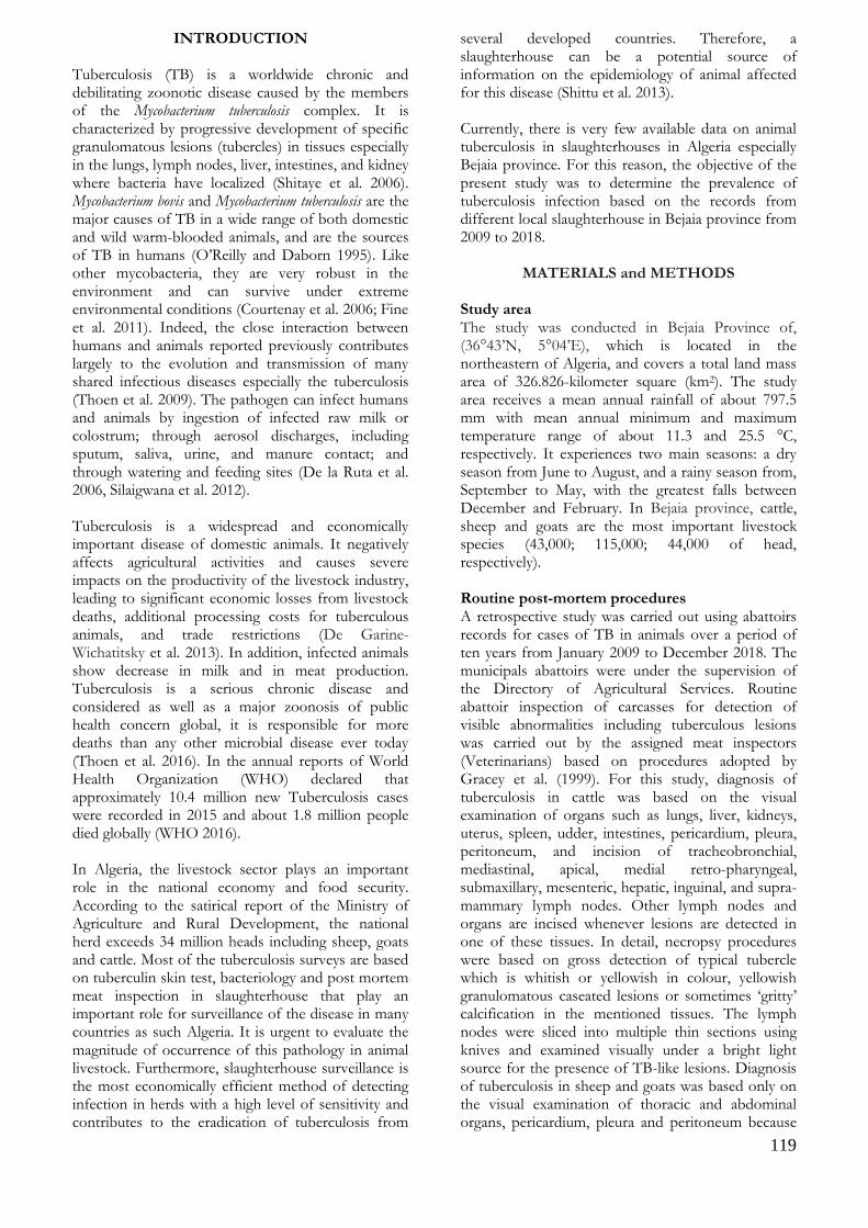
119
INTRODUCTION Tuberculosis (TB) is a worldwide chronic and debilitating zoonotic disease caused by the members of the Mycobacterium tuberculosis complex. It is characterized by progressive development of specific granulomatous lesions (tubercles) in tissues especially in the lungs, lymph nodes, liver, intestines, and kidney where bacteria have localized (Shitaye et al. 2006). Mycobacterium bovis and Mycobacterium tuberculosis are the major causes of TB in a wide range of both domestic and wild warm-blooded animals, and are the sources of TB in humans (O’Reilly and Daborn 1995). Like other mycobacteria, they are very robust in the environment and can survive under extreme environmental conditions (Courtenay et al. 2006; Fine et al. 2011). Indeed, the close interaction between humans and animals reported previously contributes largely to the evolution and transmission of many shared infectious diseases especially the tuberculosis (Thoen et al. 2009). The pathogen can infect humans and animals by ingestion of infected raw milk or colostrum; through aerosol discharges, including sputum, saliva, urine, and manure contact; and through watering and feeding sites (De la Ruta et al. 2006, Silaigwana et al. 2012). Tuberculosis is a widespread and economically important disease of domestic animals. It negatively affects agricultural activities and causes severe impacts on the productivity of the livestock industry, leading to significant economic losses from livestock deaths, additional processing costs for tuberculous animals, and trade restrictions (De Garine-Wichatitsky et al. 2013). In addition, infected animals show decrease in milk and in meat production. Tuberculosis is a serious chronic disease and considered as well as a major zoonosis of public health concern global, it is responsible for more deaths than any other microbial disease ever today (Thoen et al. 2016). In the annual reports of World Health Organization (WHO) declared that approximately 10.4 million new Tuberculosis cases were recorded in 2015 and about 1.8 million people died globally (WHO 2016). In Algeria, the livestock sector plays an important role in the national economy and food security. According to the satirical report of the Ministry of Agriculture and Rural Development, the national herd exceeds 34 million heads including sheep, goats and cattle. Most of the tuberculosis surveys are based on tuberculin skin test, bacteriology and post mortem meat inspection in slaughterhouse that play an important role for surveillance of the disease in many countries as such Algeria. It is urgent to evaluate the magnitude of occurrence of this pathology in animal livestock. Furthermore, slaughterhouse surveillance is the most economically efficient method of detecting infection in herds with a high level of sensitivity and contributes to the eradication of tuberculosis from
several developed countries. Therefore, a slaughterhouse can be a potential source of information on the epidemiology of animal affected for this disease (Shittu et al. 2013). Currently, there is very few available data on animal tuberculosis in slaughterhouses in Algeria especially Bejaia province. For this reason, the objective of the present study was to determine the prevalence of tuberculosis infection based on the records from different local slaughterhouse in Bejaia province from 2009 to 2018.
MATERIALS and METHODS Study area The study was conducted in Bejaia Province of, (36°43’N, 5°04’E), which is located in the northeastern of Algeria, and covers a total land mass area of 326.826-kilometer square (km2). The study area receives a mean annual rainfall of about 797.5 mm with mean annual minimum and maximum temperature range of about 11.3 and 25.5 °C, respectively. It experiences two main seasons: a dry season from June to August, and a rainy season from, September to May, with the greatest falls between December and February. In Bejaia province, cattle, sheep and goats are the most important livestock species (43,000; 115,000; 44,000 of head, respectively). Routine post-mortem procedures A retrospective study was carried out using abattoirs records for cases of TB in animals over a period of ten years from January 2009 to December 2018. The municipals abattoirs were under the supervision of the Directory of Agricultural Services. Routine abattoir inspection of carcasses for detection of visible abnormalities including tuberculous lesions was carried out by the assigned meat inspectors (Veterinarians) based on procedures adopted by Gracey et al. (1999). For this study, diagnosis of tuberculosis in cattle was based on the visual examination of organs such as lungs, liver, kidneys, uterus, spleen, udder, intestines, pericardium, pleura, peritoneum, and incision of tracheobronchial, mediastinal, apical, medial retro-pharyngeal, submaxillary, mesenteric, hepatic, inguinal, and supra-mammary lymph nodes. Other lymph nodes and organs are incised whenever lesions are detected in one of these tissues. In detail, necropsy procedures were based on gross detection of typical tubercle which is whitish or yellowish in colour, yellowish granulomatous caseated lesions or sometimes ‘gritty’ calcification in the mentioned tissues. The lymph nodes were sliced into multiple thin sections using knives and examined visually under a bright light source for the presence of TB-like lesions. Diagnosis of tuberculosis in sheep and goats was based only on the visual examination of thoracic and abdominal organs, pericardium, pleura and peritoneum because
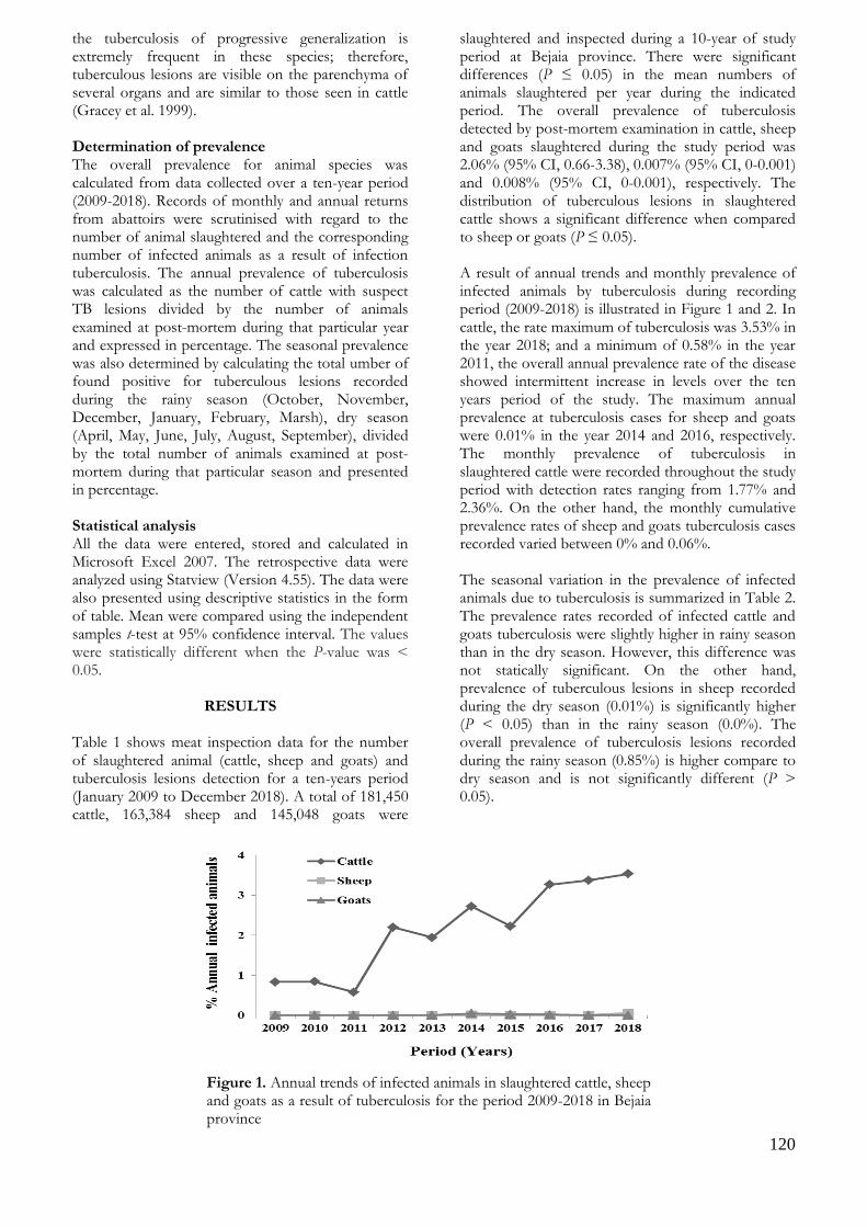
120
the tuberculosis of progressive generalization is extremely frequent in these species; therefore, tuberculous lesions are visible on the parenchyma of several organs and are similar to those seen in cattle (Gracey et al. 1999). Determination of prevalence The overall prevalence for animal species was calculated from data collected over a ten-year period (2009-2018). Records of monthly and annual returns from abattoirs were scrutinised with regard to the number of animal slaughtered and the corresponding number of infected animals as a result of infection tuberculosis. The annual prevalence of tuberculosis was calculated as the number of cattle with suspect TB lesions divided by the number of animals examined at post-mortem during that particular year and expressed in percentage. The seasonal prevalence was also determined by calculating the total umber of found positive for tuberculous lesions recorded during the rainy season (October, November, December, January, February, Marsh), dry season (April, May, June, July, August, September), divided by the total number of animals examined at post-mortem during that particular season and presented in percentage. Statistical analysis All the data were entered, stored and calculated in Microsoft Excel 2007. The retrospective data were analyzed using Statview (Version 4.55). The data were also presented using descriptive statistics in the form of table. Mean were compared using the independent samples t-test at 95% confidence interval. The values were statistically different when the P-value was < 0.05.
RESULTS Table 1 shows meat inspection data for the number of slaughtered animal (cattle, sheep and goats) and tuberculosis lesions detection for a ten-years period (January 2009 to December 2018). A total of 181,450 cattle, 163,384 sheep and 145,048 goats were
slaughtered and inspected during a 10-year of study period at Bejaia province. There were significant differences (P ≤ 0.05) in the mean numbers of animals slaughtered per year during the indicated period. The overall prevalence of tuberculosis detected by post-mortem examination in cattle, sheep and goats slaughtered during the study period was 2.06% (95% CI, 0.66-3.38), 0.007% (95% CI, 0-0.001) and 0.008% (95% CI, 0-0.001), respectively. The distribution of tuberculous lesions in slaughtered cattle shows a significant difference when compared to sheep or goats (P ≤ 0.05). A result of annual trends and monthly prevalence of infected animals by tuberculosis during recording period (2009-2018) is illustrated in Figure 1 and 2. In cattle, the rate maximum of tuberculosis was 3.53% in the year 2018; and a minimum of 0.58% in the year 2011, the overall annual prevalence rate of the disease showed intermittent increase in levels over the ten years period of the study. The maximum annual prevalence at tuberculosis cases for sheep and goats were 0.01% in the year 2014 and 2016, respectively. The monthly prevalence of tuberculosis in slaughtered cattle were recorded throughout the study period with detection rates ranging from 1.77% and 2.36%. On the other hand, the monthly cumulative prevalence rates of sheep and goats tuberculosis cases recorded varied between 0% and 0.06%. The seasonal variation in the prevalence of infected animals due to tuberculosis is summarized in Table 2. The prevalence rates recorded of infected cattle and goats tuberculosis were slightly higher in rainy season than in the dry season. However, this difference was not statically significant. On the other hand, prevalence of tuberculous lesions in sheep recorded during the dry season (0.01%) is significantly higher (P < 0.05) than in the rainy season (0.0%). The overall prevalence of tuberculosis lesions recorded during the rainy season (0.85%) is higher compare to dry season and is not significantly different (P > 0.05).
Figure 1. Annual trends of infected animals in slaughtered cattle, sheep and goats as a result of tuberculosis for the period 2009-2018 in Bejaia province

121
Figure 2. Monthly variations in proportions of infected animals for tuberculosis in slaughterhouse during the year 2009-2018 in Bejaia province
Table 1. Slaughter statistics Tuberculosis rates of infected animal between 2009 and 2018 in Bejaia province
Species slaughtered Cattle Sheep Goats Animal total
Number slaughtered 199 077 168 796 164 986
532 859
Mean ± SD 19907.7±2229 16879.6±4984.6
16498.6±5358.6 907
53285.9±9677.2
Min-Max
17196-23760
12695-28862
9771-24389
40662-67657
Number with tuberculosis lesions
4092
11 11 4114
Prevalence (%) 2.06a 0.007b 0.008b 0.77
95% confidence interval
0.66-3.38 0-0.001 0-0.001 0.13-0.65
a,b Values with different superscripts in the same raw are significantly different (P ≤ 0.05).
Table 2. Seasonal prevalence rates (%) of tuberculosis in infected animal slaughtered between 2009 and 2018 in Bejaia province
Species Season Number slaughtered
Number of infected animals by Tuberculum (%)
Cattle Rainy 87178 1979 (2.27%)
Dry 114033 2454 (2.15%)
Sheep Rainy 72713 0 (0%) *
Dry 106882 11 (0.01%) *
Goats Rainy 75099 10 (0.013%)
Dry 90143 1 (0.001%)
All Rainy 234990 1989 (0.85%)
Dry 311058 2466 (0.79%) * Values with different superscripts in the same species between rain and dry season are significantly different (P ≤ 0.05)

122
DISCUSSION In Africa, epidemiological studies of TB are difficult to perform due to cost and the lack of laboratory techniques, therefore, many epidemiologic and public health aspects of the disease remain mainly unknown. However, the regular post-inspection monitoring of carcasses remains the best option for monitoring animal tuberculosis and can be a valuable source of information on the incidence of animal diseases and conditions, some of which may be zoonotic. The meat inspection data are a potential source of information and have an important role to play in epidemiology and preventive veterinary medicine; however, it is not being fully exploited in Algeria, especially in Bejaia province. In this context, our study aimed at determining the magnitude and distribution of tuberculosis in Bejaia slaughterhouse using a meat inspection procedure from January 2009 to December 2018. To our knowledge, this is one the rare study conducting the prevalence of tuberculosis diagnosed from slaughter in Algeria, especially Bejaia area. However, routine abattoir meat inspection and periodic intra-dermal tuberculin skin testing of cattle from intensive dairy farms have already previously revealed the presence of bTB in Algeria (Sahraoui et al. 2009). Based on detailed post-mortem inspection, the overall prevalence of tuberculosis in cattle, sheep and goats recorded in the current study (2.06%, 0.007% and 0.008%, respectively) is widely different compared to several report. The tuberculosis prevalence of bovine slaughtered in the present study (2.06%) is lower compared to the previous works (Sahraoui et al. 2011) who reported prevalence of 3.58% in four abattoirs in the north of Algeria. Likewise, when compared with the data recorded elsewhere in developing countries, the prevalence recorded in this study was lower than 6.1% prevalence reported in Nigeria by Ahmad et al. (2017), 5.5% in Ethiopia (Dejene et al. 2016) and 9.7% in Uganda (Nalapa et al. 2017). However, this rate was higher than the earlier reports based on abattoir post-mortem detection of tuberculosis-like lesions from different regions; 0.78% (Sa’idu et al. 2017) in Nigeria, 0.48% (Carvelho et al. 2015) in Brazil, and 0.18% (Asil et al., 2013) in Sudan. These differences could be explained by many factors including lower cattle density and housing of animals in open areas, which are unlikely to favour the spread of the disease, breed of animals that are slaughtered in the abattoirs, differences in the disease status in the animal populations, and various environmental influences (Regassa et al. 2010). The transmission and development of infection have been found to differ significantly from place to place, and this difference is most probably linked to the climatic conditions affecting the stability of the agent in the environment, type of the production system (intensive, extensive), malnutrition, insufficient aeration system, over
stocking and herding of different herd groups (Shitaye et al. 2006). The tuberculosis prevalence of sheep slaughtered in the present study (0.007%) is lower compared to the previous works who reported prevalence over of 0.22% at two abattoirs in the central region of Algeria (Sahraoui et al. 2012), and 2.85% in South Darfur State during the period October 2015 to February 2017 (Aljameel et al. 2017). However, this prevalence is higher than the 0.0% reported by Cadmus et al. (2009) at Bodija municipal abattoir in Oyo state of Nigeria. The rate of caprine tuberculosis is revealed lower (0.008%) than the 3.72%, 3.5%, and 1.68% reported by Aljameel et al. (2017) in Sudan, Benti et al. (2013) in Ethiopia and Luboya et al. (2017) in Congo. In one survey conducted in four slaughterhouses in the north of Algeria during the year of 2007, the authors report a prevalence of lesions of caprine tuberculosis of 6.03% (Sahraoui et al. 2008). The low rate of TB in slaughtered sheep and goats in Bejaia province might be attributed to the housing of sheep and goats separately from cattle at night, and to many factors such as animal source, age, breeding management and hereditary resistance. In addition, separate herding may have contributed to a low contact rate between sheep and goats and the other species of animals (Ghebremariam 2018). The low percentage of TB in animals in Bejaia province (Algeria) might be indicative of the success of the TB eradication program in domestic animals. Moreover, the majority of Algerian cattle are registered and cattle movement control systems are well established. Also, this may be due to technique conducted by researchers such as microscopic identification of the post mortem lesions of the disease in the slaughtered and tuberculin test technique which are more sensitive (Danbirni et al. 2009). The difference of tuberculosis prevalence might be underestimated in tuberculosis animal slaughtered because of undetected lesions in early infection or because small lesions might be missed as a result of difficulties in carrying out inspection without pressure. From the results of the epidemiological survey, the tuberculosis cases detected by post-mortem examination in cattle (2.06%), was significantly higher than sheep and goats (0.007%, and 0.008%) at slaughterhouses of Bejaia province. This could be attributed to many factors such as animal source, age, breeding management and hereditary resistance. These variations could be due to different control practices, particularly the diagnose of the TB to improve livestock production. The prevalence of TB is different in various species due to environmental and management factors (malnutrition, pregnancy and concurrent infection) that may suppress the immune responsiveness of animals. This situation

123
could have potential impact on human health directly and threat to human livelihood by compromising food supply, income and social status. The overall annual prevalence rate of the tuberculosis in cattle showed intermittent increase in levels over the ten (10) years period of the study. The reason for this increasing trend in the prevalence of TB in cattle was not clear. Thus, the increase in the bovine TB detection rate in this study may not have been a real increase of the disease state but is probably due to the intensification of the slaughter/meat inspection procedure. Meat inspection offers an effective means of monitoring the level of TB and depends on the work load, time, and diligence of the veterinary staff conducting the examination (Shitaye et al. 2006). The introduction of modern diagnostic techniques with the intensification of meat inspection and tracing of infected/suspicious cases to the herds of origin would be necessary for effective surveillance of TB. Tuberculous lesions in slaughtered cattle showed monthly fluctuations throughout the study period with detection rates ranging from 1.77% and 2.36%. Therefore, TB lesions were not influenced by month but it was higher during stressful periods when slaughtering was elevated during religious feasts and sociocultural ceremonies. In general, the variation in seasonal prevalence in cattle and goats is not significant (P>0.05), as such there was no association between the occurrence of TB and season in the state. These findings were in concordance with other previous report of Awah-Ndukum et al. (2010) in Cameron who reported that the detection of TB lesions was not influenced by season but was high during stressful periods such as inter season and peak-season periods. Nwata et al. (2011) showed that variation in seasonal prevalence was not significant; they reported a prevalence of 13.2% during the rainy season and 12.5% during the dry season. However, Pollock and Neill (2002) and Ahmed et al. (2013) reported strong association between season and tuberculous lesions but the reason for the difference in the seasonal variations and tuberculous detection observed in their studies was not stated. During the dry season, prevalence of tuberculous lesions in sheep was 0.01% while the prevalence was lower during the rainy season (0.0%); this may be due to the small number of infected sheep, which does not lead to a significant result.
CONCLUSION In conclusion, the findings of the present abattoir study have provided a baseline data for monitoring of the ruminant tuberculosis in Bejaia province (Algeria). The results demonstrated that the prevalence of bovine, ovine and caprine tuberculosis recorded was relatively lower than that of some of the previous reports from abattoirs in the same country. However,
we suggest that this disease should be investigated further in farms to determine the risks factors on the prevalence in animals such as ages and breeds to develop effective disease control strategies. Also, standardization and intensification of slaughterhouse detailed meat examination and proficiency testing of meat inspections are suggested as essential and cost-effective interventions to improve meat inspection service in Algeria, with subsequent protection of consumers' health. Measures to prevent infection transmission among animals and to humans should be the primary objective to be achieved with qualified public health personnel, proper hygiene practices and public education.
ACKNOWLEDGEMENT
The data collection was made by many veterinary inspectors of communal abattoirs in Bejaia province which are gratefully acknowledged. Abdelhanine AYAD designed, carried out the experimental work, analyzed the data and wrote the manuscript. Fatima AIT-YAHIA collected, entered and stored the data in Microsoft Excel. Abdelkader BENSID, Amira Chahrazad. BENABDELHAK and Nadir Boudjlal DERGAL reviewed the manuscript. All authors read and approved the final manuscript.
Conflict of Interest: The authors declare that they have no conflict of interest.
REFERENCES
Ahmad I, Kudi CA, Abdulkadir AI, Saidu SNA. Occurrence
and distribution of bovine TB pathology by age, sex, and breed of cattle slaughtered in Gusau Abattoir, Zamfara State Nigeria. Trop Anim Health Prod. 2017; 49(3): 583-589.
Ahmed AM, Ismail SAS,Dessouki AA. Pathological lesions survey and economic loss for male cattle slaughtered at Ismailia abattoir. Int Food Rese J. 2013; 20(2): 857-863.
Aljameel MA, Mohammed GE, Bakhiet AO. Tuberculosis in Sheep and Goats: pathological characteristics based on abattoir Study in South Darfur State, Sudan. Sudan J Sci Technol.2017; 18(2): 107-126.
Asil E, Sanousi S, Gameel A, Beir H, Fathelrahman M, Terab N, Muaz M,Hamid M. Bovine tuberculosis in South Darfur State, Sudan: an abattoir study based on microscopy and molecular detection methods. Trop Anim Health Prod. 2013; 45(2).
Awah-Ndukum J, Kudi AC, Bradley G, Ane-Anyangwe IN, Fon-Tebug S, Tchoumboue J. Prevalence of bovine tuberculosis in abattoirs of the Littoral and Western highland regions of Cameroon: A cause for public health concern. Vet Med Int. 2010.
Benti D, Conraths FJ, Ameni G. Abattoir based study on the epidemiology of caprine tuberculosis in Ethiopia using conventional and molecular tools. Acta Vet Scad. 2013; 55: 15.
124
Cadmus SI, Adesokan, HK, Jenkins AO, Soolingen D. Mycobacterium bovis and Mycobacterium tuberculosis in goats, Nigeria. Emer Infect Dis. 2009; 15(12): 2066-2067.
Carvalho RCT, Furlanetto LV, Maruyama FH, de Araújo CP, Barros SLB, do Nascimento Ramos CA, Dutra V, de Araújo FR, Paschoalin VMF, Nakazato L de Souza Figueiredo EE. Evaluation of the efficiency of nested q-PCR in the detection of Mycobacterium tuberculosis complex directly from tuberculosis-suspected lesions in post-mortem macroscopic inspections of bovine carcasses slaughtered in the state of Mato Grosso, Brazil. Meat sci. 2015; 106: 11-15.
Courtenay O, Reilly LA, Sweeney FP, Hibberd V, Bryan S, Ul-Hassan A, Newman C, Macdonald DW, Delahay RJ., Wilson GJ, Wellington EMH. Is Mycobac-terium bovis in the environment important for the persistence of bovine tuberculosis? Biol Lett. 2006; 2: 460–462.
Danbirni S, Sackey AKB, Kudi AC, Okaiyeto SO, Pewan SB. A comparison of One- Step Anigen® Rapid Bovine TuberculosisAntibodies Test Sensitivity to Postmortem Gross Lesions in Diagnosing Bovine Tuberculosis in a Dairy Herd in Kaduna State. Res J Dairy Sci. 2009; 3(2-4): 32 34.
De Garine-Wichatitsky M, Caron A, Kock R, Tschopp R, Munyeme M, Hofmeyr M, Michel A. A review of bovine tuberculosis at the wildlife–livestock–human interface in sub-Saharan Africa. Epidemiol Infect 2013; 141(7): 1342-1356.
De la Rua-Domenech R. Human Mycobacterium bovis infection in the United Kingdom: Incidence, risks, control measures and review of the zoonotic aspects of bovine tuberculosis. Tuberculosis 2006; 86(2): 77–109.
Dejene SW, Heitkönig IMA, Prins HHT, Lemma FA, Mekonnen DA, Alemu ZE, Kelkay TZ, de Boer WF. Risk Factors for Bovine Tuberculosis (bTB) in Cattle in Ethiopia. PLoS One. 2016; 11(7)
Fine AE, Bolin CA, Gardiner JC, Kaneene JB. A study of the persistence of Mycobacterium bovis in the environment under natural weather conditions in Michigan, USA. Vet Med Int. 2011.
Ghebremariam MK, Michel AL, Vernooij JCM, Nielen M, Rutten VPMG. Prevalence of bovine tuberculosis in cattle, goats, and camels of traditional livestock raising communities in Eritrea. BMC Vet Res. 2018; 14(1): 73.
Gracey JF, Collins DS, Huey RJ. Meat hygiene (1999). 10th ed. London: Elsevier Healthy Sciences. (543-553). Chapter 17: Infectious diseases.
Luboya LW, Malangu M, Kaleka M, Ngulu N, Nkokele B, Maryabo K, Pourrut X, Vincent T, Gonzalez JP. An assessment of caprine tuberculosis prevalence in Lubumbashi slaughterhouse, Democratic Republic of Congo. Trop Anim Health Prod. 2017; 49(4): 875-878
Nalapa DP, Muwonge A, Kankya C, Olea-Popelka F. Prevalence of tuberculous lesion in cattle slaughtered in Mubende district, Uganda. BMC Vet Res. 2017; 13(1): 73.
Nwata JA, Umeononigwe CN, Abonyi GE and Onunkwo JI Retrospective study of bovine and human tuberculosis in abattoirs and hospitals in Enugu State, Southeast Nigeria. J Public Health Epidemiol. 2011; 3(7): 329-336.
O’Reilly LM, Daborn CJ. The epidemiology ofMycobac- terium bovis infections in animals and man: a review. Tuber Lung Dis. 1995; 76(1): 1–46.
Pollock JM, Neill SD. Mycobacterium bovis infection and tuberculosis in cattle. Vet J. 2002; 163: 115–27.
Regassa A, Tassew A, Amenu K, Megersa B, Abunna F, Mekibib B, Macrotty T, Ameni G. A cross-sectional study on bovine tuberculosis in Hawassa town and its surroundings, Southern Ethiopia. Trop Anim Health Prod. 2010; 42(5): 915-920.
Sa’idu AS, Mohammed S, Ashafa M, Gashua MM, Mahre MB, Maigado AI. Retrospective study of bovine tuberculosis in Gombe Township Abattoir, Northeastern Nigeria. Int J Vet Sci Med. 2017; 5(1): 65–69.
Sahraoui N, Hasniou A, Chettab H, Ben Khada G, Tazerart F, Chadi H, Zinsstag J, Guetarni D. Diagnostic de la tuberculose ovine par examen anatomopathologique en Algérie. Journal Recherche Scientifique de l’Université de Lomé. 2012; 14(2): 143-147.
Sahraoui N, Müller B, Guetarni D,, Boulahbal F, Yala D, Ouzrout R, Berg S, Smith NH, Zinsstag J. Molecular characterization of Mycobacterium bovis strains isolated from cattle slaughtered at two abattoirs in Algeria. BMC Vet Res. 2009; 5(1): 4.
Sahraoui N, Muller B, Mamache B, Yala D, Boulahbal F, Zinsstag J, Guetarni D. Tuberculosis in cattle and goats in north of Algeria. Vet Res. 2011; 4(4): 100-103.
Sahraoui N, Muller B, Yala D, Ouzrout R, Zinsstag J, Boulahbal F, Guetarni D. Investigation about the bovine tuberculosis in two Algerian slaughterhouses. Afric J Agricult Res 2008; 3(11): 775-778.
Shitaye JE, Getahun B, Alemayehu T, Skoric M, Treml F, Fictum P, Vrbas V, Pavlik I. A prevalence study of bovine tuberculosis by using abattoir meat inspection and tuberculin skin testing data, histopathological and IS6110 PCR examination of tissues with tuberculous lesions in cattle in Ethiopia. Vet Med. 2006; 51: 512–522.
Shittu A, Clifton-Hadley RS, Ely ER, Upton PU, Downs SH. Factors associated with bovine tuberculosis confirmation rates in suspect lesions found in cattle at routine slaughter in Great Britain, 2003–2008. Prev Vet Med.2013; 110(3-4): 395-404.
Silaigwana B, Green E, Ndip R. Molecular Detection and Drug Resistance of Mycobacterium tuberculosis Complex from Cattle at a Dairy Farm in the Nkonkobe Region of South Africa: A pilot study. Int J Environ Res Public Health. 2012; 9: 2045–2056.
Thoen CO, LoBue P, Enarson DA, Kaneene JB, de Kantor IN. Tuberculosis: a re-emerging disease of animals and humans. Vet Ital, 2009; 45: 135–181.
Thoen, C., LoBue, P. and de Kantor, I., 2006. The importance of Mycobacterium bovis as zoonoses, Veterinary Microbiology, 112, 339-345.
World Health Organization. Global Tuberculosis Report; 2016 (Available at: http://apps.who.int/iris/bitstream/10665/250441/1/9789241565394-eng.pdf?ua=1 (accessed October 23, 2019).

125
Kocatepe Veterinary Journal
Kocatepe Vet J (2020) 13(2):125-129
DOI: 10.30607/kvj.689204 RESEARCH ARTICLE
Determination of The Hygienic Quality of Tap Waters of
Afyonkarahisar Province
Gökhan AKARCA1*, Oktay TOMAR2, Ömer İSTEK1
1Afyon Kocatepe University, Faculty of Engineering, Department of Food Engineering, 03200, Afyonkarahisar, Turkey
2Kocaeli University, Faculty of Agriculture and Natural Science, Arslanbey Campus, 41285, Kocaeli, Turkey
ABSTRACT In this study, the microbiological and chemical properties of drinking water taken from fountains in the city center of Afyonkarahisar province were investigated. As a result, Escherichia coli was detected in 9 (22.5%) of forty samples examined. In 19 (47.5%) of them, it was determined that coliform group bacteria were above the values specified in the statute. It was determined that the total aerobic mesophilic bacteria count was 0-3.45 cfu kob/250ml, the coliform group bacteria count was 0-2.59 cfu kob/250ml, the psychrophilic group bacteria count was 0.48-2.81 log cfu/250ml, and the Escherichia coli count was 0-1.97 log cfu/250ml. In chemical analyses, pH, conductivity, hardness, nitrite, ammonium, bicarbonate, clarity, fluorine, chlorine values were determined to be 7.46, 344.4, 16.03, 0.061, 0.0312, 151.39, 9.8, 0.144, 0.171 mg/mL, respectively, on average. Although the chemical results indicated in samples were found appropriate according to the regulation on water for human consumption, in microbiological results, all samples did not comply with the quality criteria specified in the regulation on water for human consumption in terms of Escherichia coli, coliform, TAMB, total psychrophilic aerobic bacteria. In conclusion, it was concluded that the hygienic quality of drinking and tap water in the city center of Afyonkarahisar did not comply with the regulation on water for human consumption and had significant risks for public health. Keywords: Afyonkarahisar, tap water, hygienic quality, microbiological.
***
Afyonkarahisar İli Çeşme Sularının Hijyenik Kalitesinin Belirlenmesi
ÖZ
Bu çalışmada Afyonkarahisar ili şehir merkezinde bulunan çeşmelerden alınan içme sularının mikrobiyolojik ve kimyasal özellikleri araştırılmıştır. Sonuç olarak; incelenmesi yapılan kırk örneğin 9 adedinde (%22.5) Escherichia coli tespit edilmiştir. 19 adedinde (%47.5) ise; Koliform grubu bakterileri tüzükte belirtilen değerlerin üzerine olduğu saptanmıştır. Toplam aerobik mezofilik bakteri sayısı 0-3.45 log kob/250ml, koliform grubu bakteri sayısı 0-2.59 log kob/250ml, psikrofil bakteri grubu sayısı 0.48-2.81 log kob/250ml, Escherichia coli sayısı 0-1.97 log kob/250ml aralığında olduğu tespit edilmiştir. Kimyasal analizlerde pH, iletkenlik, sertlik, nitrit, amonyum, bikarbonat, berraklık, flor, klor değerleri sırasıyla ortalama 7.46, 344.4, 16.03, 0.061, 0.0312, 151.39, 9.8, 0.144, 0.171 mg/mL olarak saptanmıştır. Örneklerde belirtilen kimyasal sonuçlar İnsani tüketim amaçlı sular yönetmeliğine göre uygun bulunmasına karşın mikrobiyolojik sonuçlarda tüm numuneler Escherichia coli, koliform, TAMB, toplam psikrofil aerobik bakteri yönünden insani tüketim amaçlı sular yönetmeliğinde belirtilen kalite kriterlerine uygunluk göstermemektedir. Sonuç olarak, Afyonkarahisar şehir merkezindeki içme ve kullanma sularının hijyenik kalitesinin insani tüketim amaçlı sular yönetmeliğe uymadığı ve halk sağlığı açısından önemli riskler taşıdığı kanaatine varılmıştır. Anahtar Kelimeler: Afyonkarahisar, çeşme suyu, hijyenik kalite, mikrobiyoloji. To cite this article: Akarca G. Tomar O. İstek Ö. Determination of The Hygienic Quality of Tap Waters of Afyonkarahisar Province. Kocatepe Vet J. (2020) 13(2):125-129 Submission: 14.02.2020 Accepted: 11.04.2020 Published Online: 25.04.2020
ORCID ID; GA: 0000-0002-5055-2722, OT: 0000-0001-5761-7157, Öİ: 0000-0003-2195-1166 *Corresponding author e-mail: [email protected]

126
INTRODUCTION Water is consumed as an indispensable natural resource for the continuation of future generations and the ecosystem. Although people can continue their vitality activities for days and weeks without food intake, they can only live a few days in case of any drought (Uzundumlu et al. 2016). Human beings, who perceived the importance of this situation, always preferred to be close to water resources while establishing their settlements (Demirel 2009, Hare 2014). Furthermore, people need to consume the same amount of water as the daily energy taken to be able to maintain their lives in a healthy way (Sciamanna et al. 2011, Drewnowski et al. 2013, Muckelbauer 2013). Although three-quarters of the world's surface are covered with water, only 2.6% of the water reserve consists of freshwater (Miller 2004). Water is not only home to living creatures but also constitutes the main source of vitality. Water, which may be present in different types of cells and tissues at different ratios, constitutes 65-70% of the human body (Erk 2016). Multiple factors such as population growth, industrialization, and consequently energy need of countries may be effective on water consumption. In addition to the amount of consumption, the fact that water reaches consumers hygienically becomes even more important with each passing day. More technology has been used to meet the needs of the rapidly growing population in recent years. Therefore, access to clean and natural water resources becomes difficult over time since people use natural resources wastefully (Varer 2010). The fact that the bacteriological, chemical, physical, and ecological properties of water resources are negatively affected, and the discharging of substances or wastes that will directly or indirectly cause preventive deteriorations in human health and water quality lead to water pollution (Tosunoglu et al. 1999). In the world, 250 million people contract waterborne epidemics every year, and it is reported that approximately 10 million of them die of microbial infections. In its report entitled “water quality and health strategy" covering the period between 2013 and 2020, the World Health Organization (WHO) reported that 589.854 waterborne infection cases were reported from 58 world countries and there was an 85% increase in waterborne infections compared to 2010 (Rifaat et al. 2014). Contaminated water directly or indirectly constitutes a risk to public health. It was indicated that the consumption of vegetables and fruits washed with contaminated water, the consumption of fish caught in contaminated water or the consumption of this
water may lead to many infections or toxications caused by pathogenic bacteria (Erkan and Vural 2006). Coliform groups are the most problematic group of bacteria in drinking water in terms of health. Coliform group bacteria are divided into two groups according to their characteristic features. Fecal coliform bacteria and Escherichia coli are considered to be the indicators of fecal contamination in water. Pathogenic types of Escherichia coli may cause diarrhea, meningitis, septicemia, arteriosclerosis, Hemolytic Uremic Syndrome (HUS), and various immunological diseases leading to death in living creatures (Rifaat et al. 2014). In this study, it was aimed to investigate tap waters in Afyonkarahisar and its vicinity from physical, chemical, microbiological aspects and to determine a risk they pose to human health.
MATERIAL and METHODS Materials 40 samples were taken from the fountains in the city center of Afyonkarahisar province into previously sterilized jars under suitable conditions in accordance with the hygiene rules. The samples were brought to the Food Engineering Laboratory of Afyon Kocatepe University Faculty of Engineering under suitable conditions and analyzed. Microbiological Analyses Escherichia coli, total psychrophilic aerobic bacteria group, total aerobic mesophilic bacteria (TAMB), and total coliform analyses were performed in the samples taken. Preparation of samples for microbiological analyses Before the analysis, the materials to be used were passed through alcohol, and this alcohol was removed with burner flame and sterilized. The working environment was also ensured to be sterile. This process was repeated for each sample to be analyzed. Escherichia coli analysis 250 mL of the water sample taken was passed through a sterilized membrane filtration system. Then, the membrane filters were placed on the Petri plate with sterile forceps so that no air remained. Attention was paid to ensure that the bacterial surface was at the top while placing the filter on the VRB agar medium. The lid of the Petri plate was allowed to incubate at 37 °C for 24-48 hours on the top. After incubation, microorganism count was performed directly. The colonies that gave bright pink color were considered Escherichia coli and calculated logarithmically (Ekici et al. 2010).

127
Total aerobic mesophilic bacteria analysis 250 mL of the water sample taken was passed through a sterilized membrane filtration system. Then, the membrane filters were placed on the Petri plate with sterile forceps so that no air remained. Attention was paid to ensure that the bacterial surface was at the top while placing the filter on the Lactose TTC agar medium. The lid of the Petri plate was allowed to incubate at 37 °C for 24-48 hours on the top. After incubation, microorganism count was performed directly and calculated logarithmically (Ekici et al. 2010). Total coliform bacteria analysis 250 mL of the water sample taken was passed through a sterilized membrane filtration system. Then, the membrane filters were placed on the Petri plate with sterile forceps so that no air remained. Attention was paid to ensure that the bacterial surface was at the top while placing the filter on the VRB agar medium. The lid of the Petri plate was allowed to incubate at 37 °C for 24-48 hours on the top. After incubation, microorganism count was performed directly. The colonies that gave metallic luster were considered coliform and calculated logarithmically (Ekici et al. 2010). Total psychrophilic aerobic bacteria analysis 250 mL of the water sample taken was passed through a sterilized membrane filtration system. Then, the membrane filters were placed on the Petri plate with sterile forceps so that no air remained. Attention was paid to ensure that the bacterial surface was at the top while placing the filter on the Lactose TTC agar medium. The lid of the Petri plate was allowed to incubate at 4 °C for 3-4 days on the top. After incubation, microorganism count was performed directly and calculated logarithmically (Ekici et al. 2010). Chemical analyses pH analysis A pH meter was standardized by buffer solutions with constant pH value. Thus, the error level was minimized. After the pH meter was standardized, the pH value of each sample was measured. pH and conductivity were measured using the pH meter (Hanna HI 2215) (Akarca et al. 2015). Hardness analysis After 25 ml of the test sample was taken and completed to 50 ml, 1-2 drops of buffer solution were added. Very little powder indicator solution was added with a spatula tip. It was titrated with 0.01 M EDTA solution until the color changed from wine red to blue. Then, the volume spent was recorded. This volume gives us information about the hardness of the sample (Yelekci 2014).
Analysis of free ion amount While the amount of chloride (CI- mg/L) ion was analyzed using the Merck Nova 60-14897 kit by the spectrophotometric method, Nitrite nitrogen (NO2
-N mg/L) was analyzed using the Merck Nova 14776 kit by the spectrophotometric method, Ammonium nitrogen (NH4
-N mg/L) was analyzed using the Merck Nova 60-14752 kit by the spectrophotometric method, and Fluoride (F- mg/L) was analyzed using the Merck Nova 60-14598 kit by the spectrophotometric method (Kalyoncu and Zeybek 2009). Clarity and bicarbonate amount analysis Clarity and bicarbonate were analyzed by spectrophotometric methods (Kalyoncu and Zeybek 2009).
RESULTS and DISCUSSION Results of microbiological analysis The microbiological values of drinking water of Afyonkarahisar province were determined and presented in Table 1. Results of chemical analysis With respect to the chemical analysis of drinking water of Afyonkarahisar province, pH, conductivity, hardness, nitrite, ammonium, bicarbonate, clarity, fluorine, and chlorine analyses were performed. The results of these chemical analyses are presented in Table 2. In this study, Escherichia coli was detected in 9 of the forty samples investigated. Coliform group bacteria in 19 (47.5%) of them were found to be above the values specified in the statutes. It was determined that the count of Escherichia coli varied between 0-1.97 log cfu/250ml, the coliform group bacteria count varied between 0-2.59 log cfu/250ml, TAMB count varied between 0-3.45 log cfu/250ml, and the psychrophilic bacteria group count varied between 0.48-2.81 log cfu/250ml. Analysis results are presented in Table 1. As a result of chemical analyses, it was found that the average value of pH (40 samples) was 7.46, the average value of conductivity (40 samples) was 344.4 (µS/cm), the average value of hardness (40 samples) was 16.03 (mg/L), the average value of nitrite (40 samples) was 0.061 (mg/L), the average value of ammonium (40 samples) was 0.031 (mg/L), the average value of bicarbonate (40 samples) was 151.39 (mg/L), the average value of clarity (40 samples) was 9.8 (mg/L), the average value of fluorine (40 samples) was 0.144 (mg/L), and the average value of chlorine (40 samples) was 0.171 (mg/L). Analysis results are presented in Table 2.
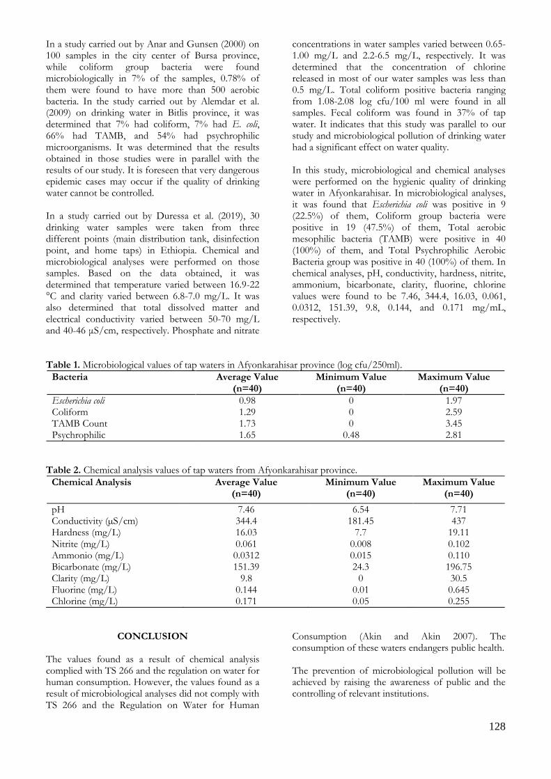
128
In a study carried out by Anar and Gunsen (2000) on 100 samples in the city center of Bursa province, while coliform group bacteria were found microbiologically in 7% of the samples, 0.78% of them were found to have more than 500 aerobic bacteria. In the study carried out by Alemdar et al. (2009) on drinking water in Bitlis province, it was determined that 7% had coliform, 7% had E. coli, 66% had TAMB, and 54% had psychrophilic microorganisms. It was determined that the results obtained in those studies were in parallel with the results of our study. It is foreseen that very dangerous epidemic cases may occur if the quality of drinking water cannot be controlled. In a study carried out by Duressa et al. (2019), 30 drinking water samples were taken from three different points (main distribution tank, disinfection point, and home taps) in Ethiopia. Chemical and microbiological analyses were performed on those samples. Based on the data obtained, it was determined that temperature varied between 16.9-22 °C and clarity varied between 6.8-7.0 mg/L. It was also determined that total dissolved matter and electrical conductivity varied between 50-70 mg/L and 40-46 μS/cm, respectively. Phosphate and nitrate
concentrations in water samples varied between 0.65-1.00 mg/L and 2.2-6.5 mg/L, respectively. It was determined that the concentration of chlorine released in most of our water samples was less than 0.5 mg/L. Total coliform positive bacteria ranging from 1.08-2.08 log cfu/100 ml were found in all samples. Fecal coliform was found in 37% of tap water. It indicates that this study was parallel to our study and microbiological pollution of drinking water had a significant effect on water quality. In this study, microbiological and chemical analyses were performed on the hygienic quality of drinking water in Afyonkarahisar. In microbiological analyses, it was found that Escherichia coli was positive in 9 (22.5%) of them, Coliform group bacteria were positive in 19 (47.5%) of them, Total aerobic mesophilic bacteria (TAMB) were positive in 40 (100%) of them, and Total Psychrophilic Aerobic Bacteria group was positive in 40 (100%) of them. In chemical analyses, pH, conductivity, hardness, nitrite, ammonium, bicarbonate, clarity, fluorine, chlorine values were found to be 7.46, 344.4, 16.03, 0.061, 0.0312, 151.39, 9.8, 0.144, and 0.171 mg/mL, respectively.
Table 1. Microbiological values of tap waters in Afyonkarahisar province (log cfu/250ml).
Bacteria Average Value (n=40)
Minimum Value (n=40)
Maximum Value (n=40)
Escherichia coli 0.98 0 1.97 Coliform 1.29 0 2.59 TAMB Count 1.73 0 3.45 Psychrophilic 1.65 0.48 2.81
Table 2. Chemical analysis values of tap waters from Afyonkarahisar province.
Chemical Analysis Average Value (n=40)
Minimum Value (n=40)
Maximum Value (n=40)
pH 7.46 6.54 7.71 Conductivity (µS/cm) 344.4 181.45 437 Hardness (mg/L) 16.03 7.7 19.11 Nitrite (mg/L) 0.061 0.008 0.102 Ammonio (mg/L) 0.0312 0.015 0.110 Bicarbonate (mg/L) 151.39 24.3 196.75 Clarity (mg/L) 9.8 0 30.5 Fluorine (mg/L) 0.144 0.01 0.645 Chlorine (mg/L) 0.171 0.05 0.255
CONCLUSION The values found as a result of chemical analysis complied with TS 266 and the regulation on water for human consumption. However, the values found as a result of microbiological analyses did not comply with TS 266 and the Regulation on Water for Human
Consumption (Akin and Akin 2007). The consumption of these waters endangers public health. The prevention of microbiological pollution will be achieved by raising the awareness of public and the controlling of relevant institutions.

129
When it is considered that our body needs to take 2.5 liters of water on average every day, the quality of drinking water is very important for our health. Nowadays, drinking water is exposed to many contaminations, especially originating from humans or animals. It is certain that drinking these waters polluted as a result of the contamination by intestinal bacteria such as Escherichia coli will bring along many outbreaks, as well. When the relevant studies that have been carried out so far are considered, hygiene and sanitation processes of drinking-tap water should be conducted without delay. The improvement of the hygiene and sanitation conditions of water will prevent diarrheal diseases that are likely to occur depending on water consumption and the deaths related to these diseases and will decrease the incidence of other waterborne diseases. Conflict of Interest: The authors declare that they have no conflict of interest.
REFERENCES
Akarca G, Kahraman A, Tomar O. Change of the shelf life of
pasteurized milk which added in various amonds of cinnamon. Afyon Kocatepe University Journal of Sciences and Engineering, 2015; 15: 1-9.
Akin M, Akin G. Importance of water, water potential in Turkey, water basins and water pollution. Ankara University the Journal of the Faculty of Languages and History-Geography, 2007; 47(2): 105-118.
Alemdar S, Kahraman T, Agaoglu S, Alisarli M. Some microbiological and physicochemical properties of drinking water in Bitlis district. Ekoloji Journal, 2009; 19(73): 29-38.
Anar S, Gunsen U. The hygienic quality of drinking and usage water in Bursa. Medical Journal of Süleyman Demirel University, 2000; 7: 31-34.
Demirel M. Policies and problems in water resources management: an assesment in Denizli sample. MSc Thesis, Pamukkale University, Denizli, Turkey, 2009.
Drewnowski A, Colin DR, Florence C. Water and beverage consumption among adults in the united states: cross-sectional study using data from NHANES 2005–2010. BMC Public Health, 2013; 13: 1068.
Duressa G, Assefa F, Jida M. Assessment of bacteriological and physicochemical quality of drinking water from source to household tap connection in Nekemte, Oromia, Ethiopia. Hindawi Journal of Environmental and Public Health, 2019; 7 page.
Ekici K, Korkoca H, Sancak YC, Atalan E. The determination coliforms and E. coli in drinking water of Van and province. Uludag University Journal of the Faculty of Veterinary Medicine, 2010; 29(2): 21-25.
Erk I. Experimental and theoretical investigation of tissue equivalency for phantom materials. MSc Thesis, Istanbul Technical University, Istanbul, Turkey, 2016.
Erkan ME, Vural A. A study about of hygienic quality of Dicle (Tigris) river. Dicle Medical Journal, 2006; 33(4): 205-209.
Hare KE. Water supply assessment in Kilifi county. PhD Thesis, University of Nairobi, Nairobi, Kenya, 2014.
Kalyoncu H, Zeybek M. Benthic fauna of Ağlasun and Isparta streams and determination of water quality. Research Journal of Biological Science, 2009; 2(1): 41-48.
Miller GT. Sustaining the Earth, 6th edition. Thompson Learning, Inc. Pacific Grove, 211-216. California, USA, 2004.
Muckelbauer R, Sarganas G, Gruneis A, Muller-Nordhorn J. Association between water consumption and body weight outcomes: A systematic review. The American Journal of Clinical Nutrition, 2013; 98(2): 282-299.
Rifaat EA, Tekiner IH, Ozpinar H. Classical and MASS spectrometer examination of the existence of coliform and fecal coliform bacteria in drinking and use waters for public health. Electronic Journal of Food Technologies, 2014; 9(2): 20-32.
Sciamanna CN, Kiernan M, Rolls BJ, Boan J, Stuckey H, Kephart D, Miller CK, Jensen G, Hartmann TJ, Loken E. Practices associated with weight loss versus weight-loss maintenance results of a national survey. American Journal of Preventive Medicine, 2011; 41: 159-166.
Tosunoglu V, Boncukoglu R, Anapali O, Sahin U. Contamination of Karasu river by city and industrial wastes. Atatürk University Journal of the Faculty of Agricultural, 1999; 30(2): 169-176.
Uzundumlu AS, Fakioglu O, Kokturk M, Temel T. Determining of the best drinking water preference in Erzurum province. Alinteri Journal of Agriculture Science, 2016; 30(1): 1-7.
Varer KV. Microbiological Quality Analysis’s Parameter of Bottled Water Being Sold in Trabzon. MSc Thesis, Karadeniz Technical University, Trabzon, Turkey, 2010.
Yelekci S, Acemioglu B, Avci H. Investigation of the usability of the drinking water of Kilis province. Research Journal of Biological Science, 2014; 5(2): 77-81.

130
Kocatepe Veterinary Journal
Kocatepe Vet J (2020) 13(2):130-139
DOI: 10.30607/kvj.677652 RESEARCH ARTICLE
Use of Pumpkin and Its Effect on Quality in Ice Cream Production
Ayşe YILDIZ1, Pelin DEMİR1*, Ali ARSLAN1
1Fırat University, Faculty of Veterinary Medicine, Department of Food Hygiene and Technology, Elazığ, Turkey
ABSTRACT This study was conducted to investigate the effect of pumpkin, ginger, cinnamon, and coconut on the quality of ice cream (sensory. chemical and microbial). For this purpose. 4 groups ice cream were made and were stored at -18 °C. Samples were subjected to microbiological (total aerobic mesophile bacteria. yeast. and mold). chemical (dry matter. non-fat dry matter. oil and pH) and sensory (color and appearance. structure and consistency. taste and smell) analysis on days 0th. 10th. 20th and 30th of the storage. Statistically significant differences were found between the groups in terms of dry matter. non-fat dry matter and fat values (p <0.05). As a result. it is shown that the production of ice cream with quite low sugar which makes the product more dietetic and highly nutritious. and it was detected that the ice-creams could maintain their quality for at least 30 days at -18 °C. Keywords: Ice Cream, Production, Pumpkin, Quality.
***
Dondurma Üretiminde Bal Kabağı Kullanımı ve Kalite Üzerine Etkisi
ÖZ
Bu çalışma bal kabağı, zencefil, tarçın ve hindistan cevizinin dondurmanın kalitesi (duyusal, kimyasal ve mikrobiyel) üzerine olan etkisini incelemek amacıyla yapıldı. Bu amaçla 4 grup dondurma yapıldı ve -18°C’de muhafaza edildi. Örneklerin 0., 10., 20. ve 30. günlerinde mikrobiyolojik, kimyasal ve duyusal analizleri yapıldı. İstatistiksel olarak gruplar arasında kuru madde, yağsız kuru madde ve yağ değerleri bakımından önemli farklar tespit edildi (p < 0.05). Sonuç olarak, bu araştırmada şeker miktarı oldukça düşük, daha diyetik, besleyici değeri yüksek, bal kabaklı kaliteli dondurma üretiminin mümkün olabileceği ve dondurmaların -18 °C’de muhafaza edilmesiyle kalitelerini en az 30 gün koruyabildiği tespit edildi. Anahtar Kelimeler: Balkabağı, Dondurma, Kalite, Üretim.
To cite this article: Yıldız A. Demir P. Arslan A. Use of Pumpkin and Its Effect on Quality in Ice Cream Production. Kocatepe Vet J. (2020) 13(2):130-139 Submission: 20.01.2020 Accepted: 11.04.2020 Published Online: 27.04.2020
ORCID ID; AY: 0000-0002-1657-0361, PD: 0000-0002-0824-1672, AA: 0000-0002-3011-5592 *Corresponding author e-mail: [email protected]

131
GİRİŞ Dondurma, hoşa giden tadı, aroması ve ferahlatıcı etkisiyle birlikte herkes tarafından sevilerek tüketilen ve kolay sindirilen bir besin kaynağıdır (Tekinşen ve Tekinşen 2008). Özellikle enerji, protein, kalsiyum, fosfor, vitamin A, vitamin D ve vitamin B2 riboflavin için önemli bir kaynaktır (Anonim 2004). Süt ve süt ürünlerinden elde edilen dondurma; genellikle tatlandırıcı, stabilizatör, emülgatör, aroma ve renk veren maddelerden oluşan karışımdır (Tekinşen ve Tekinşen 2008). Dondurma karışımı ise; tat ve çeşidine göre süt veya süt ürünlerini içme suyu, şeker ve müsaade edilen katkı maddelerini içeren, arzu edildiğinde salep, yumurta veya yumurta ürünleri, aroma maddeleri ve çeşni maddelerini bulunduran, dondurulmamış karışım olarak tanımlanır (Anonim 2004). Dondurma, dondurma tebliğine (Anonim 2004) göre sade ve çeşnili (meyve ve sebzeler ile bunların suyu, konsantresi, püresi, ezmesi, fındık, fıstık, badem, ceviz gibi sert kabuklu meyveler, tahıllar gibi bitkiler ve bitki preperatları, şekerli mamuller ile bal, kahve, kakao, çikolata, baharat gibi) olarak sınıflandırılır. Dondurma üretiminde bal kabağı, kullanılan yardımcı maddeler arasındadır. Bal kabağı (Cucurbita moschata) 100 gramı 3.77 g sakaroz, 1.57 g glikoz, 1.06 g früktoz, 88.6 su, 0.20 g yağ ve 0.46 protein içermektedir (Web 2016a). Antiinflamatuar etkisi olan balkabağı, lif içeriği yüksek olmasının yanı sıra zayıflama diyetlerinde uzun süre tokluk hissi sağlayıp zayıflamaya yardımcı olur. Sindirim sistemi problemi yaşayanlarda kabızlığı önler. Balkabağı ve çekirdeği özellikle göz sağlığında, deri ve hücre yapısının yenilenmesinde etkili olup A ve E vitaminleri içerir. Bir su bardağı pişmiş balkabağında 11.7 mg alfa karoten bulunmaktadır. Kalp hastalıklarında ve hiperlipidemi vakalarında LDL (low density lipoprotein)’nin düşmesinde beta karoten etkilidir. Antioksidan özelliği ve bağışıklığı güçlendirdiği için kansere karşı koruyucu etki sağlar. Balkabağı demir, potasyum, kalsiyum, fosfor gibi önemli mineralleri de içerir. Sağlıklı kemik gelişmesi ve kansızlığa karşı önemli rol oynar. Yapısında bulunan çinko sayesinde saçların güçlenmesini ve dökülmesini önler (Web 2016b). Aksoy ve ark. (2013), Kars’ta satılan dondurmalarda toplam aerobik mezofilik bakteri sayısını ortalama olarak 4.0x10⁷ kob/g, toplam psikrofil aerob bakteri sayısını 2.0x10⁵ kob/g, koliform bakteri sayısını 1.4x10³ kob/g ve Staphylococcus-Micrococcus sayısını ise 9.0x10⁶ kob/g olarak saptamışlardır. Dondurma örneklerinde Salmonella spp. ve Staphylococcus aureus tespit etmediklerini, sadece bir örnekte Escherichia coli tespit edildiği ve dondurmaların mikrobiyolojik
kalitesinin standartlarda belirtilen değerlerin üzerinde olduğunu vurgulamışlardır. Patır ve ark. (2004), Elazığ’da açıkta satılan kaymaklı (sade) ve meyve aromalı dondurmalarda koliform bakterilerinin türleri ile dağılımlarını incelemişlerdir. Bu amaçla, 50’şer adet kaymaklı ve meyve aromalı (limonlu, kakaolu, fıstıklı, vişneli ve çilekli dondurmaların her birinden 10 adet) dondurma incelenmişlerdir. Koliform bakterileri en az <1.00 log kob/g, en çok 5.74 log kob/g düzeyinde saptamışlardır. Antepüzümü (2000), çiğ keçi sütüne şeker, krema ve yağsız süttozu, ilave edilerek hazırladığı dondurma miksine % 20, 30, 40, 50 oranlarında bal ve glikoz şurubu katılarak yaptığı dondurmaların fiziksel ve kimyasal özelliklerini araştırmıştır. Dondurmaların pH değerlerinin 6.00-6.57 arasında değiştiğini belirlemiştir. Hazırlanan örneklerde kullanılan bal oranı arttıkça pH değerlerinin düştüğünü, glikoz şurubu kullanılan örneklerde de yaklaşık sonuçlar saptadığını vurgulamıştır. Bu çalışmada dondurma üretiminde balkabağı kullanımının dondurmanın mikrobiyolojik, kimyasal ve duyusal özellikleri gibi çeşitli kalite parametrelerinin belirlenmesi amaçlanmıştır.
MATERYAL ve METOT Materyal Bu çalışmadaki dondurma örnekleri, Elazığ’da dondurma üreten özel bir pastanede, imalatın bütün aşamalarında azami hijyenik koşullarına dikkat edilerek yapıldı. Üretimde % 35 süt yağı içeren özel bir firmaya ait krema, pH’sı 6.3 ve % 11.6 kuru madde içeren bal kabağı püresi kullanıldı. Bal kabağı önce yıkandı ve sonra dilimlendi, dilimlerin kabuğu soyuldu ve su içinde haşlandı. Haşlanan bal kabağı süzgece konulup, suları süzüldükten sonra dondurma üretiminde kullanıldı. Bu amaçla 4 farklı dondurma karışımı hazırlandı (Tablo 1). I. grup dondurma piyasada satılan sade dondurmanın temel bileşenleri esas alınarak hazırlandı. Diğer 3 grupta kullanılan bal kabağı haşlanıp, püre haline getirildikten sonra belirtilen oranlarda karışıma ilave edildi. Hazırlanan karışımlar dondurma makinesine konuldu ve dondurma üretim şemasında (Şekil 1) belirtildiği şekilde dondurma yapıldı. Hazırlanan bu karışım 150 g’lık ağzı kapaklı saklama kaplarına (tupperware) (polikarbonat) (Sera marka) konulup, –18 oC’ de muhafaza edildi. Dondurma örnekleri muhafazanın 0., 10., 20. ve 30. günlerinde mikrobiyolojik, kimyasal ve duyusal bakımdan incelendi. Her dönemde her gruptan 2 örnek olmak üzere toplam 96 örnek incelendi. Çalışma 1 Ocak- 30 Nisan 2017 tarihleri arasında 30 gün ara ile 3 kez tekrarlandı ve her deney çift paralel yapıldı.
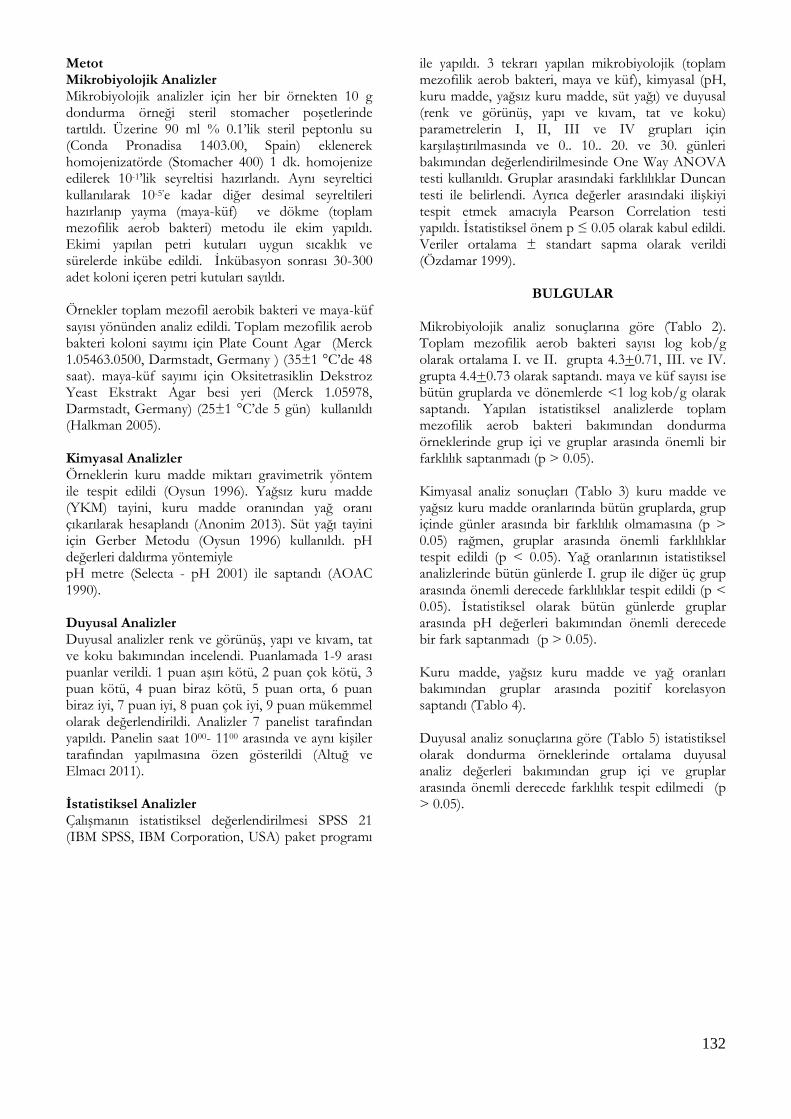
132
Metot Mikrobiyolojik Analizler Mikrobiyolojik analizler için her bir örnekten 10 g dondurma örneği steril stomacher poşetlerinde tartıldı. Üzerine 90 ml % 0.1’lik steril peptonlu su (Conda Pronadisa 1403.00, Spain) eklenerek homojenizatörde (Stomacher 400) 1 dk. homojenize edilerek 10-1’lik seyreltisi hazırlandı. Aynı seyreltici kullanılarak 10-5’e kadar diğer desimal seyreltileri hazırlanıp yayma (maya-küf) ve dökme (toplam mezofilik aerob bakteri) metodu ile ekim yapıldı. Ekimi yapılan petri kutuları uygun sıcaklık ve sürelerde inkübe edildi. İnkübasyon sonrası 30-300 adet koloni içeren petri kutuları sayıldı. Örnekler toplam mezofil aerobik bakteri ve maya-küf sayısı yönünden analiz edildi. Toplam mezofilik aerob bakteri koloni sayımı için Plate Count Agar (Merck 1.05463.0500, Darmstadt, Germany ) (35±1 °C’de 48 saat). maya-küf sayımı için Oksitetrasiklin Dekstroz Yeast Ekstrakt Agar besi yeri (Merck 1.05978, Darmstadt, Germany) (25±1 °C’de 5 gün) kullanıldı (Halkman 2005). Kimyasal Analizler Örneklerin kuru madde miktarı gravimetrik yöntem ile tespit edildi (Oysun 1996). Yağsız kuru madde (YKM) tayini, kuru madde oranından yağ oranı çıkarılarak hesaplandı (Anonim 2013). Süt yağı tayini için Gerber Metodu (Oysun 1996) kullanıldı. pH değerleri daldırma yöntemiyle pH metre (Selecta - pH 2001) ile saptandı (AOAC 1990). Duyusal Analizler Duyusal analizler renk ve görünüş, yapı ve kıvam, tat ve koku bakımından incelendi. Puanlamada 1-9 arası puanlar verildi. 1 puan aşırı kötü, 2 puan çok kötü, 3 puan kötü, 4 puan biraz kötü, 5 puan orta, 6 puan biraz iyi, 7 puan iyi, 8 puan çok iyi, 9 puan mükemmel olarak değerlendirildi. Analizler 7 panelist tarafından yapıldı. Panelin saat 1000- 1100 arasında ve aynı kişiler tarafından yapılmasına özen gösterildi (Altuğ ve Elmacı 2011). İstatistiksel Analizler Çalışmanın istatistiksel değerlendirilmesi SPSS 21 (IBM SPSS, IBM Corporation, USA) paket programı
ile yapıldı. 3 tekrarı yapılan mikrobiyolojik (toplam mezofilik aerob bakteri, maya ve küf), kimyasal (pH, kuru madde, yağsız kuru madde, süt yağı) ve duyusal (renk ve görünüş, yapı ve kıvam, tat ve koku) parametrelerin I, II, III ve IV grupları için karşılaştırılmasında ve 0.. 10.. 20. ve 30. günleri bakımından değerlendirilmesinde One Way ANOVA testi kullanıldı. Gruplar arasındaki farklılıklar Duncan testi ile belirlendi. Ayrıca değerler arasındaki ilişkiyi tespit etmek amacıyla Pearson Correlation testi yapıldı. İstatistiksel önem p ≤ 0.05 olarak kabul edildi. Veriler ortalama ± standart sapma olarak verildi (Özdamar 1999).
BULGULAR Mikrobiyolojik analiz sonuçlarına göre (Tablo 2). Toplam mezofilik aerob bakteri sayısı log kob/g olarak ortalama I. ve II. grupta 4.3+0.71, III. ve IV. grupta 4.4+0.73 olarak saptandı. maya ve küf sayısı ise bütün gruplarda ve dönemlerde <1 log kob/g olarak saptandı. Yapılan istatistiksel analizlerde toplam mezofilik aerob bakteri bakımından dondurma örneklerinde grup içi ve gruplar arasında önemli bir farklılık saptanmadı (p > 0.05). Kimyasal analiz sonuçları (Tablo 3) kuru madde ve yağsız kuru madde oranlarında bütün gruplarda, grup içinde günler arasında bir farklılık olmamasına (p > 0.05) rağmen, gruplar arasında önemli farklılıklar tespit edildi (p < 0.05). Yağ oranlarının istatistiksel analizlerinde bütün günlerde I. grup ile diğer üç grup arasında önemli derecede farklılıklar tespit edildi (p < 0.05). İstatistiksel olarak bütün günlerde gruplar arasında pH değerleri bakımından önemli derecede bir fark saptanmadı (p > 0.05). Kuru madde, yağsız kuru madde ve yağ oranları bakımından gruplar arasında pozitif korelasyon saptandı (Tablo 4). Duyusal analiz sonuçlarına göre (Tablo 5) istatistiksel olarak dondurma örneklerinde ortalama duyusal analiz değerleri bakımından grup içi ve gruplar arasında önemli derecede farklılık tespit edilmedi (p > 0.05).

133
Hammadde
Hammadde tartımı ve hazırlanması
Karıştırma
Pastörizasyon ( Pastörizatörde. 72 oC’de 15 dk )
Soğutma (4 oC’ de 1 saat)
Olgunlaştırma (Dinlendirme) (0 oC’de 3 saat)
Bal kabağı. zencefil. tarçın ve hindistan cevizi ilavesi
Karışımın dondurulması (115 rpm’de 15 dk. -30 oC’de )
Dolum ve ambalajlama (Ağzı kapaklı saklama kapları (tupperware) kaplar)
Sertleştirme (-30 oC’de 6 saat)
Muhafaza (-18 oC’de 30 gün )
Şekil 1. Dondurma Üretim Şeması Figure 1. Ice Cream Production Scheme
Tablo 1. Dondurma gruplarının bileşimleri (%) Table 1. Composition of ice cream groups (%)
Maddeler I. Grup (Kontrol)
II. Grup
III. Grup
IV. Grup Krema 48.5 31.5 31.5 31.5
Süt 10 - - -
Şeker 40 18 5 2.5
Salep 1.5 - - -
Bal kabağı püresi - 42 55 57.5
Çiğ yumurta sarısı - 7 7 7
Zencefil - 0.43 0.21 0.21
Tarçın - 0.21 0.43 0.21
Hindistan cevizi - 0.21 0.21 0.43
Vanilya - 0.6 0.6 0.6

134
Tablo 2. Dondurma örneklerinin mikrobiyolojik analiz sonuçları (log kob/g) (n: 96) Table 2. Microbiological analysis results of ice cream samples (log cfu/g) (n: 96)
Mikroorganizma Gruplar Gün Ortalama+Standard
Sapma 0 10 20 30
Toplam mezofilik aerob bakteri I 4.3 4.3 4.1 4.2 4.3+0.71
II 4.3 4.2 4.3 4.4 4.3+0.71
III 4.3 4.4 4.6 4.5 4.4+0.73
IV 4.3 4.4 4.6 4.5 4.4+0.73
Maya-Küf I <1 <1 <1 <1 <1
II <1 <1 <1 <1 <1
III <1 <1 <1 <1 <1
IV <1 <1 <1 <1 <1
I: Kontrol grubu, II: % 42 bal kabağı, III: % 55 bal kabağı, IV: % 57.5 bal kabağı içermektedir.
Tablo 3. Dondurma örneklerinin kimyasal analiz değerleri (n: 96) Table 3. Chemical analysis values of ice cream samples (n: 96)
Değer Gruplar Gün Ortalama+Standard Sapma
0 10 20 30
Kuru Madde (%)
I 57.64a 57.66a 57.69a 57.62a 57.65+0.78
II 37.90b 37.60b 37.58b 37.73b 37.70+ 0.78
III 26.56c 26.70c 26.65c 26.63c 26.63+ 0.63
IV 23.60d 23.56d 23.90d 23.58d 23.66+ 0.79
Yağsız Kuru Madde (YKM) (%)
I 42.14a 42.16a 42.59a 42.62a 42.37+0.51
II 27.10b 27.23b 27.25b 27.13b 27.18+ 0.44
III 16.56c 16.70c 16.65c 16.63c 16.64+ 0.52
IV 13.40d 13.26d 13.33d 13.58d 13.40 + 0.52
Yağ (%) I 15.50a 15.50a 15.10a 15.00a 15.28+0.11
II 10.80b 10.37b 10.33b 10.06b 10.39+0.23
III 10.21b 10.12b 10.15b 10.13b 10.15+0.23
IV 10.02b 10.03b 10.37b 10.00b 10.11+0.23
pH I 6.74 6.76 6.79 6.72 6.75+0.45
II 6.63 6.65 6.68 6.67 6.65+0.49
III 6.61 6.63 6.68 6.67 6.64+0.46
IV 6.62 6.66 6.63 6.62 6.63+0.80
I: Kontrol grubu, II: % 42 bal kabağı, III: % 55 bal kabağı, IV: % 57.5 bal kabağı içermektedir; a, b, c, d: Aynı sütunda farklı harfler ile gösterilen değerler arasındaki fark önemlidir (p < 0.05).
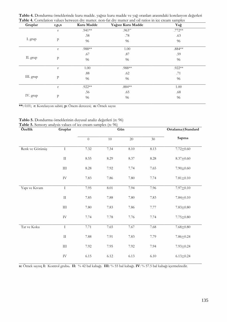
135
Tablo 4. Dondurma örneklerinde kuru madde. yağsız kuru madde ve yağ oranları arasındaki korelasyon değerleri Table 4. Correlation values between dry matter. non-fat dry matter and oil ratios in ice cream samples
Gruplar r,p,n
Değerleri
Kuru Madde Yağsız Kuru Madde Yağ
I. grup
r
p
n
.941** .963** .772**
.58 .78 .63
96 96 96
II. grup
r
p
n
.988** 1.00 .884**
.67 .87 .59
96 96 96
III. grup
r
p
n
1.00 .988** .922**
.88 .62 .71
96 96 96
IV. grup
r
p
n
.922** .884** 1.00
.56 .65 .68
96 96 96
**: 0.01; r: Korelasyon sabiti; p: Önem derecesi; n: Örnek sayısı
Tablo 5. Dondurma örneklerinin duyusal analiz değerleri (n: 96) Table 5. Sensory analysis values of ice cream samples (n: 96)
Özellik Gruplar Gün Ortalama+Standard
Sapma 0 10 20 30
Renk ve Görünüş I 7.32 7.34 8.10 8.13 7.72+0.60
II 8.55 8.29 8.37 8.28 8.37+0.60
III 8.28 7.92 7.74 7.65 7.90+0.60
IV 7.83 7.86 7.80 7.74 7.81+0.10
Yapı ve Kıvam I 7.95 8.01 7.94 7.96 7.97+0.10
II 7.85 7.88 7.80 7.83 7.84+0.10
III 7.80 7.83 7.86 7.77 7.83+0.80
IV 7.74 7.78 7.76 7.74 7.75+0.80
Tat ve Koku I 7.71 7.65 7.67 7.68 7.68+0.80
II 7.88 7.91 7.83 7.79 7.86+0.24
III 7.92 7.95 7.92 7.94 7.93+0.24
IV 6.15 6.12 6.13 6.10 6.13+0.24
n: Örnek sayısı; I: Kontrol grubu. II: % 42 bal kabağı. III: % 55 bal kabağı. IV: % 57.5 bal kabağı içermektedir.

136
TARTIŞMA Yapılan literatür taramalarında, doğrudan bal kabağı kullanılarak üretilen dondurmalar ile ilgili herhangi bir araştırmaya rastlanılmadı. Bu nedenle tartışma, meyve aromalı dondurmalar üzerinde yapılan araştırmalar ile kıyaslandı. İncelenen örneklerde toplam mezofilik aerob bakteri sayısı I. ve II. grupta 4.3 log kob/g, III. ve IV. grupta 4.4 log kob/g olarak tespit edildi. Yapılan istatistiksel analizler sonucunda toplam mezofilik aerob bakteri sayısı bakımından dondurma örneklerinde grup içi ve gruplar arasında önemli bir farklılık saptanmadı (p > 0.05) (Tablo 2). Toplam mezofilik aerob bakteri sayısını Patır ve ark. (2004) 3.44-4.49 log kob/g, Güner ve ark. (2004) 9.8x10⁵-2.5xl07 kob/g, Çubukçı ve Atasever (2018) 4.40 log kob/g ve Aksoy ve ark. (2013) 4.0x10⁷ kob/g olarak bildirmişlerdir. Bu çalışma ile diğer araştırmacıların sonuçları arasındaki farklılıklar, incelenen örneklerin bileşim, satış sırasında muhafaza koşulları ve üretim hijyenindeki farklılıklarından kaynaklanabilir. Çalışmamızda bütün gruplarda ve bütün dönemlerde maya ve küf sayısı <1 log kob/g olarak saptandı. Bu durum kaliteli ham madde kullanımına, dondurma üretiminin hijyenik koşullarda yapılmasına ve muhafaza şartlarına bağlanabilir. İstatistiksel analizlerde maya-küf sayısı bakımından dondurma örneklerinde grup içi ve gruplar arasında önemli farklılıklar saptanmadı (p > 0.05) (Tablo 2). Maya ve küf sayısını, Patır ve ark. (2004) 1.58-2.99 log kob/g, Erol ve ark. (1998) 4.0-5.9x10² kob/g, Güner ve ark. (2004) 3.4x10²-1.9xl04 kob/g, İşleyici ve ark. (2016) 2.18 log kob/g olarak tespit etmişlerdir. Bu çalışma ile diğer araştırmacıların çalışmalarından elde edilen analiz sonuçları arasındaki farklılıklar, örneklerin bileşimine, satış sırasında muhafaza koşullarına ve üretim hijyenine bağlanabilir. Örneklerin, ortalama kuru madde miktarı % 23.66-57.65 arasında saptandı. I. ve II. gruplarda saptadığımız kuru madde oranlarının TS-4265 Dondurma Standardı’na (Anonim 2013) ve Türk Gıda Kodeksi Dondurma Tebliği’ne (Anonim 2004) uygun olduğu tespit edildi. III. ve IV. grupların kuru madde oranlarının ise Dondurma Tebliği’ne ve standarda göre düşük olduğu gözlendi. Bu gruplarda tespit edilen kuru madde oranındaki düşüklük, bal kabağı miktarının yüksek olmasından (su oranı yüksek) kaynaklanmaktadır. Bütün gruplarda dönemler arasında bir farklılık olmamasına (p > 0.05) rağmen, gruplar arasında önemli farklılıklar tespit edildi (p < 0.05) (Tablo 3). Gruplar arasındaki farklılıklar, üretimde kullanılan bal kabağının miktarından kaynaklanmaktadır. Kır (2007) hazırladığı dondurma örneklerinde kuru madde miktarını % 32.32-38.39, Özcan ve Kurdal (1997) limonlu
dondurmalarda % 33.18, vişneli dondurmalarda % 31.67 ve çilekli dondurmalarda da % 31.80 olarak saptamışlardır. Açu (2014) üretiminde %10 oranında frambuaz meyvesi kullandığı ve % 3 oranında probiyotik kültür ilave ederek ürettiği dondurmalarda kuru madde miktarını % 26.27 düzeyinde saptamıştır. Karaman (2011) bazı bitki çayları ve çay ile harmanlanmış dondurma karışımı muhtevasında kuru madde oranını % 37.90 olarak belirlemiştir. Korel (2005) Manisa ilinde satışa sunulan kakaolu, sade ve meyveli (vişneli, çilekli ve limonlu) dondurmalarda kuru madde oranlarını % 62.48-71.00 arasında tespit etmiştir. Aloğlu ve ark. (2018) %0 (kontrol), %15 ve %25 kocayemiş içeren 3 grup dondurma üretmişler ve dondurmaların kuru madde miktarlarını sırasıyla %40.82, %39.04 ve % 37.91 olarak saptamışlardır. Kotan (2018) %0 (kontrol), %5, %10 ve %15 yaban mersini içeren 4 farklı dondurma üretmiş ve bu dondurmalarda kuru madde oranlarını sırasıyla %28.73, %29.07, %27.91 ve %26.83 olarak tespit etmiştir. Bu çalışma ile diğer çalışmalarda analizi yapılan dondurma örneklerinden elde edilen kuru madde oranlarındaki farklılığın nedeni, üretimde kullanılan maddelerin bileşenlerine bağlanabilir. İncelenen dondurma örneklerinde ortalama yağsız kuru madde oranları % 13.40-42.37 arasında tespit edildi. I. ve II. gruplarda saptadığımız kuru madde oranları TS-4265 Dondurma Standardı’na (Anonim 2013) ve Türk Gıda Kodeksi Dondurma Tebliği’ne (Anonim 2004) uygun olarak, III. ve IV. grupların kuru madde oranları ise düşük olarak tespit edildi. III. ve IV. gruplarda kuru madde oranının düşük olmasının nedeni üretimde kullanılan bal kabağı miktarının bu gruplardan yüksek olmasından kaynaklanmaktadır. Bütün gruplarda dönemler arasında bir farklılık olmamasına (p > 0.05) rağmen, gruplar arasındaki farklılıklar istatistiksel olarak önemli bulundu (p < 0.05) (Tablo 3). Özcan ve Kurdal (1997) limonlu dondurmalarda ortalama yağsız kuru madde oranını % 31.47, vişneli dondurmalarda % 31.67 ve çilekli dondurmalarda ise % 30.82 olarak saptamışlardır. Öztürk (1969) meyve aromalı (vişne, limon, çilek) dondurmalarda yağsız kuru madde oranını ortalama olarak % 28.31 olarak saptamıştır. Çalışmalar arasındaki bu farklılıklar, kullanılan bal kabağı miktarına, dondurma yapımında kullanılan maddelere ve oranlarına bağlanabilir. Çalışmamızda ortalama yağ değerleri I. grupta % 15.28; II. III. ve IV grupta sırasıyla, % 10.39, % 10.15, % 10.11 olarak saptandı. TS 42658 Dondurma Standardı’na (Anonim 2013) ve Türk Gıda Kodeksi Dondurma Tebliği’ne (Anonim 2004) göre yağ oranı bakımından I. grup tam yağlı dondurma kategorisine, diğer üç grup ise yağlı dondurma kategorisine girmektedir. İstatistiksel analizlerde dondurma örneklerinin yağ oranları bakımından bütün dönemlerde I. grup ile diğer üç grup arasında önemli
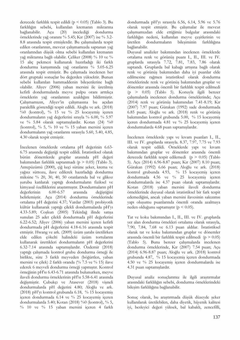
137
derecede farklılık tespit edildi (p < 0.05) (Tablo 3). Bu farklılığın sebebi, kullanılan kremanın miktarına bağlanabilir. Açu (20) incelediği dondurma örneklerinde yağ oranını % 5.45; Kır (2007) ise % 3.2-8.8 arasında tespit etmişlerdir. Bu çalışmalarda tespit edilen oranlarının, mevcut çalışmamızda saptanan yağ oranlarından düşük olma sebebi kullanılan kremanın yağ miktarına bağlı olabilir. Çeliker (2008) % 10 ve % 15 alıç pekmezi kullanarak hazırladığı iki farklı dondurma karışımında yağ oranlarını % 5.05-6.25 arasında tespit etmiştir. Bu çalışmada incelenen her dört gruptaki sonuçlar bu değerden yüksektir. Bunun sebebi kullanılan hammaddenin bileşenlerine bağlı olabilir. Aliyev (2006) yaban mersini ile üretilmiş kefirli dondurmalarda meyve pulpu oranı arttıkça örneklerin yağ oranlarının azaldığını bildirmiştir. Çalışmamızın, Aliyev’in çalışmasına bu açıdan paralellik gösterdiği tespit edildi. Aloğlu ve ark. (2018) %0 (kontrol), % 15 ve % 25 kocayemiş içeren dondurmaların yağ değerlerini sırayla % 6.00, % 5.97 ve % 5.84 olarak saptamışlardır. Kotan (24) %0 (kontrol), % 5, % 10 ve % 15 yaban mersini içeren dondurmaların yağ oranlarını sırasıyla 5.60, 5.40, 4.85, 4.30 olarak tespit etmiştir. İncelenen örneklerde ortalama pH değerinin 6.63-6.75 arasında değiştiği tespit edildi. İstatistiksel olarak bütün dönemlerde gruplar arasında pH değeri bakımından farklılık saptanmadı (p > 0.05) (Tablo 3). Antepüzümü (2000), çiğ keçi sütüne şeker, krema ve yağsız süttozu, ilave edilerek hazırladığı dondurma miksine % 20, 30, 40, 50 oranlarında bal ve glikoz şurubu katılarak yaptığı dondurmaların fiziksel ve kimyasal özelliklerini araştırmıştır. Dondurmaların pH değerlerinin 6.00-6.57 arasında değiştiğini belirlemiştir. Açu (2014) dondurma örneklerinde ortalama pH değerini 4.37; Vardar (2003) probiyotik kültür kullanarak yaptığı çilekli dondurmalarda pH’yı 4.33-5.89; Coşkun (2005) Tekirdağ ilinde satışa sunulan 25 adet çilekli dondurmada pH değerlerini 6.22-6.52; Aliyev (2006) yaban mersini içeren kefirli dondurmada pH değerlerini 4.18-6.16 arasında tespit etmiştir. Hwang ve ark. (2009) üzüm şarabı üretilirken elde edilen çökelti halindeki üzüm tortularını kullanarak ürettikleri dondurmaların pH değerlerini 6.32-7.14 arasında saptamışlardır. Özdemir (2018) yaptığı çalışmada kontrol grubu dondurma örneği ile birlikte, süte 3 farklı meyveden (böğürtlen, yaban mersini ve çilek) 2 farklı oranda (% 7.5 ve % 15) ilave ederek 6 meyveli dondurma örneği yapmıştır. Kontrol örneğinin pH'sı 6.43-6.71 arasında bulunurken, meyve ilaveli dondurma örneklerinin pH'sı 5.58-6.41 arasında değişmiştir. Çubukçı ve Atasever (2018) vişneli dondurmalarda pH değerini 4.80; Aloğlu ve ark. (2018) pH’yı kontrol grubunda 6.18, % 15 kocayemiş içeren dondurmada 6.14 ve % 25 kocayemiş içeren dondurmalarda 5.40; Kotan (2018) %0 (kontrol), % 5, % 10 ve % 15 yaban mersini içeren 4 farklı
dondurmada pH’yı sırasıyla 6.56, 6.14, 5.96 ve 5.76 olarak tespit etmiştir. Bu çalışmalar ile mevcut çalışmamızdan elde ettiğimiz bulgular arasındaki farklılığın nedeni, kullanılan meyve çeşitlerinin ve üretilen dondurmaların bileşiminin farklılığına bağlanabilir. Duyusal analizler bakımından incelenen örneklerde ortalama renk ve görünüş puanı I., II., III. ve IV. gruplarda sırasıyla 7.72, 7.81, 7.83, 7.86 olarak saptandı. Gruplarda bal kabağı artışına bağlı olarak renk ve görünüş bakımından daha iyi puanlar elde edilmesine rağmen istatistiksel olarak dondurma örneklerinde renk ve görünüş bakımından gruplar ve dönemler arasında önemli bir farklılık tespit edilmedi (p > 0.05) (Tablo 5). Konuyla ilgili benzer çalışmalarda incelenen dondurma örneklerinde; Açu (2014) renk ve görünüş bakımından 7.41-8.19; Kır (2007) 7.97 puan; Gürakan (1992) sade dondurmada 6.85 puan; Aloğlu ve ark. (2018) renk ve görünüş bakımından kontrol grubunda 5.00, % 15 kocayemiş içeren dondurmada 4.81 ve % 25 kocayemiş içeren dondurmalarda 4.68 puan saptamışlardır. İncelenen örneklerde yapı ve kıvam puanları I., II., III. ve IV. gruplarda sırasıyla. 8.37, 7.97, 7.75 ve 7.93 olarak tespit edildi. Örneklerde yapı ve kıvam bakımından gruplar ve dönemler arasında önemli derecede farklılık tespit edilemedi (p > 0.05) (Tablo 5). Açu (2014) 6.96-8.87 puan; Kır (2007) 8.10 puan; Gürakan (1992) 6.66 puan; Aloğlu ve ark. (2018) kontrol grubunda 4.93, % 15 kocayemiş içeren dondurmada 4.56 ve % 25 kocayemiş içeren dondurmalarda ise 4.37 puan olarak saptamışlardır. Kotan (2018) yaban mersini ilaveli dondurma örneklerinde duyusal olarak istatistiksel bir fark tespit edemediğini, ancak yaban mersini ilavesinin sakızımsı yapı oluşumu puanlarında önemli oranda azalmaya neden olduğunu saptamıştır (p < 0.05). Tat ve koku bakımından I., II., III. ve IV. gruplarda yer alan dondurma örnekleri ortalama olarak sırasıyla, 7.90, 7.84, 7.68 ve 6.13 puan aldılar. İstatistiksel olarak tat ve koku bakımından gruplar ve dönemler arasında önemli bir farklılık tespit edilmedi (p > 0.05) (Tablo 5). Buna benzer çalışmalarda incelenen dondurma örneklerinde, Kır (2007) 7.54 puan; Açu (2014) 6.96-8.87 puan; Aloğlu ve ark. (2018) kontrol grubunda 4.87, % 15 kocayemiş içeren dondurmada 4.50 ve % 25 kocayemiş içeren dondurmalarda ise 4.31 puan saptamışlardır. Duyusal analiz sonuçlarımız ile ilgili araştırmalar arasındaki farklılığın sebebi, dondurma örneklerindeki bileşim farklılığına bağlanabilir. Sonuç olarak, bu araştırmada düşük düzeyde şeker kullanılarak üretilebilen, daha diyetik, hijyenik kalitesi iyi, besleyici değeri yüksek, bal kabaklı, zencefilli,

138
tarçınlı ve hindistan cevizli kaliteli dondurma üretiminin mümkün olabileceği ve dondurmaların -18 oC’de muhafaza edilmesiyle kalitelerini en az 30 gün koruyabildikleri, dolayısıyla dondurma üretiminde balkabağı kullanılmasının kalite ve muhafaza süresi yönünden bir risk oluşturmadığı ve bu yöntem ile dondurma endüstrisine yeni bir ürün çeşidi kazandırılabileceği ve dondurma teknolojisine katkı sağlanabileceği sonucuna varılmıştır.
TEŞEKKÜR
Bu çalışma Fırat Üniversitesi Bilimsel Araştırma Projeleri Birimi (FÜBAP) tarafından V.F.16.30 No’ lu proje numarası ile desteklenmiştir. Bu makale Ayşe YILDIZ’ın aynı isimli yüksek lisans tezinden (FÜBAP, VF.16.30) özetlenmiştir. III. Uluslararası Battalgazi Bilimsel Çalışmalar Kongresi (21-23 Eylül 2019 / Malatya) sözlü bildiri olarak kabul edilmiş ve yayınlanmıştır. Çıkar Çatışması: Yazarlar, çıkar çatışması olmadığını beyan eder.
KAYNAKLAR
Açu M, Kınık Ö. Fonksiyonel özellikleri geliştirilmiş dondurma
üretimi. Ege Üniv. Zir. Fak. Derg. 2014; 50(1): 1-12.
Aksoy A, Sezer Ç, Vatansever L. Kars piyasasında tüketime sunulan sade dondurmaların mikrobiyolojik kalitesinin belirlenmesi. Kafkas Üniv. Vet. Fak. Derg. 2013; 2(1): 1-5.
Aliyev C. Kefir ve yaban mersinini dondurmanın fizikokimyasal, duyusal ve mikrobiyolojik özelliklerine etkisi. Yüksek Lisans Tezi. Ondokuz Mayıs Üniversitesi Fen Bilimleri Enstitüsü Gıda Mühendisliği Anabilim Dalı. Samsun. 2006.
Aloğlu HŞ, Gökgöz Y, Bayraktar M. Kocayemiş (dağ çileği-arbutus unedo l.) meyveli dondurma üretimi, fiziksel, kimyasal ve duyusal parametreler açısından irdelenmesi. J of Food 2018; 43(6): 1030-1039.
Altuğ T, Elmacı Y. Gıdalarda Duyusal Değerlendirme. 1. Baskı. Sidas Medya: İzmir. 2011.
Anonim. 2013. TS 4265. Dondurma Standardı. Türk Standartları Enstitüsü. TSE. 13s. Ankara.
Anonim. Türk Gıda Kodeksi Dondurma Tebliği. Tarım ve Köy İşleri Bakanlığı Tebliğ No: 2004/45. Ankara. 2004.
Antepüzümü F. Bal ve glikoz şurubu kullanımının Kahramanmaraş tipi dondurmaların kalitesi üzerine etkileri. Yüksek Lisans Tezi, Çukurova Üniversitesi Fen Bilimleri Enstitüsü Gıda Mühendisliği Anabilim Dalı, Adana, 2000.
Association Official Analytical Chemists. Official methods of analysis of the association of official analytical chemists, 15th Ed., Washington DC., 1990.
Coşkun. F. Tekirdağ ilinde satılan sade ve çilekli dondurmalarda fekal kontaminasyonun belirlenmesi. Tekirdağ Zir. Fak. Derg. 2005; 2(2): 135-142.
Çeliker MB. Alıç meyvesinin pekmeze işlenerek dondurma üretimine ilavesiyle dondurmanın kalite kriterleri üzerine etkileri. Yüksek Lisans Tezi, Afyon Kocatepe Üniversitesi Fen Bilimleri Enstitüsü Gıda Mühendisliği Anabilim Dalı, Afyonkarahisar, 2008.
Çubukçı S, Atasever MA. Erzurum piyasasında tüketime sunulan dondurmaların mikrobiyolojik kalitesi. Atatürk Üniv. Vet. Bil. Derg. 2018; 13(1): 54-62.
Erol İ, Küplülü Ö, Sırıken B, Çelik TH. Ankara’daki çeşitli pastanelere ait dondurmaların mikrobiyolojik kalitesinin belirlenmesi. Türkiye Vet. ve Hayv. Derg. 1998; 22: 345-352.
Güner A, Ardıç M, Keleş A. Konya'da pastanelerde tüketime sunulan dondurmaların mikrobiyolojik kalitesi. Vet. Bil. Derg. 2004; 20(2): 59-64.
Gürakan İ. Samsun il merkezinde tüketime sunulan sade dondurmaların duyusal, fiziksel. kimyasal ve mikrobiyolojik nitelikleri üzerinde bir araştırma. Yüksek Lisans Tezi, Ondokuz Mayıs Üniversitesi Fen Bilimleri Enstitüsü, Samsun, 1992.
Halkman AK. Gıda Mikrobiyolojisi Uygulamaları. Başak Matbaacılık, Ankara, 2005.
http://www.turkomp.gov.tr/food/200 20/09/2016 Erişim tarihi: 03.03.2016 (Web 2016a) http://www.milliyet.com.tr/balkabaginin-faydalari-saymakla-gundem-1982089/Erişim tarihi: 03.03.2016 (Web 2016b)
Hwang JY, Shyu YS, Hsu CK. Grape wine lees improves the rheological and adds antioxidant properties to ice cream. J. Food Sci. Technol. 2009; 42: 312-318.
İşleyici Ö, Sancak H, Tuncay RM. Van ilinde satışa sunulan ambalajlı ve dondurmaların mikrobiyolojik kalitesi. Van Vet. Derg. 2016; 27 (2): 7-67.
Karaman N. Salep ve bazı stabilizatörlerin maraş dondurmasının çeşitli nitelikleri üzerine etkilerinin belirlenmesi. Yüksek Lisans Tezi, Sütçü İmam Üniversitesi, Fen Bilimleri Enstitüsü Gıda Mühendisliği Ana Bilim Dalı, Kahramanmaraş, 2011.
Kır R. Farklı Tip Yağ Kullanımının dondurmanın fiziksel, kimyasal ve duyusal kalite özellikleri üzerine etkisi. Yüksek Lisans Tezi, Selçuk Üniversitesi, Fen Bilimleri Enstitüsü Gıda Mühendisliği Anabilim Dalı, Konya, 2007.
Korel F, Ömeroğlu S, Tan G. Manisa piyasasında satılan ambalajlı ve ambalajsız dondurmaların kalitelerinin değerlendirilmesi. Harran Üniv. Zir. Fak. Derg. 2005; 9(2):11-18.
Kotan TE. Mineral composition and some quality characteristics of ıce creams manufactured with the addition of blueberry. Gıda 2018; 43(4): 635-643.
Oysun G. Süt ve Ürünlerinde Analiz Yöntemleri. Ege Üniversitesi Ziraat Fakültesi Yayınları No: 504. İzmir. 1996. sayfa 306.
Özcan T, Kurdal E. Bursa ili merkezinde satılan meyveli dondurmaların kimyasal ve mikrobiyolojik nitelikleri üzerine araştırma. Gıda 1997; 22(3): 217-225.
Özdamar K. SPSS ile Biyoistatistik. 3. Baskı. Kaan Kitapevi. Eskişehir. 1999.
Özdemir DRS. Süte farklı oranlarda böğürtlen, yaban mersini ve çilek pulpu katılarak yapılan meyveli dondurmaların kalitesi. Doktora Tezi, Atatürk Üniversitesi, Fen Bilimleri Enstitüsü, Erzurum, 2018.

139
Öztürk, A. Ankara’da işlenen dondurmaların yapılışları ve genel özellikleri üzerinde araştırmalar. Ankara Üniv. Zir. Fak. Yayınları No:341. Ankara. 1969.
Patır B, Öksüztepe G, İlhak Oİ. Elazığ’da tüketime sunulan kaymaklı (sade) dondurmaların mikrobiyolojik ve kimyasal kalitesi. Selçuk Üniv. Vet. Bil. Derg. 2004; 20(1): 23-29.
Tekinşen C, Tekinşen K. Dondurma Temel Bilgiler Teknoloji Kalite Kontrolü. Selçuk Üniversitesi Basımevi. Konya. 2008. sayfa 189.
Vardar NB. Probiyotik bakteriler kullanılarak üretilen çilekli dondurmaların bazı fiziksel, kimyasal ve mikrobiyolojik özelliklerinin belirlenmesi. Yüksek Lisans Tezi, Trakya Üniversitesi, Fen Bilimleri Enstitüsü, Trakya, 2003.

140
Kocatepe Veterinary Journal
Kocatepe Vet J (2020) 13(2):140-144
DOI: 10.30607/kvj.674641 RESEARCH ARTICLE
Efficiency of Two Different Synchronization Protocols in Conception in
Simmental Heifers
Gaye BULUT1, Umut TAŞDEMİR2*
1Aksaray University, Faculty of Veterinary Medicine, Department of Obstetric and Gyneacology, 68100, Aksaray, Turkey 2Aksaray University, Technical Sciences Vocational School, 68100, Aksaray, Turkey
ABSTRACT
This study aimed to reveal the effects of timed artificial insemination applied after the synchronization protocol known as day5 (D5) and the prostaglandin F2α (PGF2α) protocol applied in 11-day intervals (D11) on pregnancy rates in Simmental heifers. A total of 88 heifers were divided into two treatment groups. In the D5 group (n: 56), after gonadotrophin releasing hormone (GnRH) application, PGF2α injection was made on the 5th day and the second PGF2α injection was given one day later. The second GnRH was applied at the time of TAI and at the 44th hour of the second PGF2α application. In the D11 group (n: 32), double-dose PGF2α was given in 11-day intervals. GnRH was applied at the time of TAI and at the 81st hour following the second PGF2α injection. Pregnancy-associated glycoprotein visual test was carried out for determining to pregnancy on the 28th day. Additionally, for the purpose of determining embryonic deaths, the pregnancies were checked again by ultrasonography on the 63rd day following TAI. The 28th-day pregnancy rates in the synchronized heifers were 51.8% (29/56) in the D5 group and 43.8% (14/32) in the D11 group P>0.05. The 63rd-day pregnancy rates were 48.21% (27/56) in the D5 group and 40.62% (13/32) in the D11 group P>0.05. Consequently, although there was no statistically significant difference in terms of pregnancy rates between the D5 and D11 protocols, there was a numerical increase in the D5 protocol. Considering its short application time, D5 may be used for TAI in Simmental heifers. Keywords: GnRH, Heifer, PGF2α, Synchronization, Timed Artificial Insemination
***
Simental Düvelerde Farklı İki Senkronizasyon Protokolünün Gebelik Üzerine Olan Etkinliği
ÖZ
Bu çalışma ile day5 (D5) olarak isimlendirilen senkronizasyon protokolü ile 11 gün aralıklarla uygulanan PGF2α (D11) protokolü sonrasında yapılan zaman ayarlı suni tohumlamanın (TAI) kombine verimli bir ırk olan Simmental düvelerde gebe kalma oranı üzerine etkilerinin ortaya konması amaçlandı. 88 adet düve rastgele iki tedavi grubuna ayrıldı. D5 grubuna (n: 56), GnRH uygulamasının ardından 5. günde PGF2α enjeksiyonu yapıldı ve bir gün sonra ikinci PGF2α verildi. İkinci GnRH, TAI zamanında, ikinci PGF2α uygulamasından 44 saat uygulandı. D11 grubuna (n: 32) ise, çift doz prostaglandin PGF2α 11 gün aralıklarla verildi, GnRH TAI zamanında, ikinci PGF2α enjeksiyonunu takiben 81. saatte uygulandı. Gebelikleri belirlemek amacıyla 28. günde gebelik ile ilişkili proteinlerin belirlenmesine yönelik pregnant association glycoprotein (PAG) testi uygulandı. Ayrıca, TAI'yi takiben 63. günde embriyonik ölümlerin belirlenmesi amacıyla ultrasonografi ile gebelikler tekrar kontrol edildi. Sekronize adilen düvelerde 28. gün gebelik oranları D5 grubunda %51.8 (29/56) ve D11 grubunda %43.8 (14/32) oranındaydı P>0.05. 63. gün gebe kalma oranlarıı D5 grubunda %48.21 (27/56) ve D11 grubunda %40.62 (13/32) olarak tespit edildi P>0.05. Sonuç olarak, D5 protokolü D11 protokolü ile karşılaştırıldığında gebelik üzerine etkisi istatistiksel olarak farklı bulunmamasına rağmen, rakamsal olarak yüksek bulundu. Uygulama süresinin kısa olması da göz önüne alındığında Simmental düvelerde TAI amacıyla kullanılabilir. Anahtar Kelimeler: GnRH, Düve, PGF2α, Senkronizasyon, Zaman Ayarlı Suni Tohumlama To cite this article: Bulut G. Taşdemir U. Efficiency of Two Different Synchronization Protocols in Conception in Simmental Heifers. Kocatepe Vet J. (2020) 13(2):140-144 Submission: 14.01.2020 Accepted: 30.04.2020 Published Online: 02.05.2020 ORCID ID; GB: 0000-0003-4500-1958, UT: 0000-0003-2827-1286 *Corresponding author e-mail: [email protected]; [email protected]

141
INTRODUCTION
Fertility is one of the most important components of efficient and profitable dairy cattle breeding (Kuhn et al. 2006). However, there is a negative correlation between milk yield and fertility, heifers are selected based on their milk yield in establishments that breed dairy cattle. While heritability has a low effect on fertility, considering the fertility outcomes obtained today, this approach has led to a decrease in fertility in comparison to the past, and with this decrease, studies on fertility have increased in numbers (VanRaden et al. 2004). Artificial insemination (AI) is a technique that is prevalently used worldwide for animal breeding and obtaining generations with superior genetic characteristics (Bó and Baruselli 2014). Moreover, AI comes to the fore as additional feeding costs are eliminated by not including bulls that need to be included in cases of natural insemination, it is more inexpensive, it prevents diseases that are transmitted by mating and it provides results that are similar to those obtained by natural mating (Ribeiro et al. 2012). Estrus synchronization, on the other hand, is not only a practical tool for reproduction management of dairy (Pursley et al. 1997) and beef (Patterson et al. 2003) cattle, but it also provides advantages in terms of reducing the usage of inputs of time and labor, and thus, several studies have focused on development of estrus synchronization techniques for productive synchronization of estrus (Schmitt et al. 1996). Estrus synchronization protocols are a set of methods that are applied to control follicular growth and ovulation by applying hormones (Bó et al. 2016). As these protocols reduce the first AI interval and increase AI usage in stock dairy heifers, they allow timed artificial insemination (TAI) procedures without needing estrus determination (Peeler et al. 2004, Ribeiro et al. 2012). However, as the pregnancy success rates obtained from heifers with TAI that is applied after synchronization protocols are lower than that in cows, usage of synchronization protocols in heifers has remained limited (Pursley et al. 1997). On the other hand, recent studies have reported that 50-60% successful pregnancies could be obtained from a 5-day synchronization program applied on heifers (Rabaglino et al. 2010, Silva et al. 2015) and the pregnancy rates obtained with this protocol were similar to the outcomes obtained by AI applied after estrus monitoring (Kuhn et al. 2006, Silva et al. 2015). Lima et al. (2013) asserted that ovulation time could be adjusted in heifers with a 5-day synchronization protocol, and this way, it is an attractive alternative for breeders in terms of achievement of pregnancy in a shorter time in heifers. However, synchronization studies have usually been carried out on Holstein heifers so far, and there are a few to no studies on combined breeds. For this reason, this study aimed to reveal the effects of TAI applied after the
synchronization protocol known as day5 (D5) and the prostaglandin F2α (PGF2α) protocol applied in 11-day intervals (D11) on pregnancy rates in Simmental heifers, which are a combined breed.
MATERIALS and METHODS Heifers The study was conducted between March and September 2018 and included Simmental heifers at the ages of 14 to 20 months (mean: 17.1 months). The heifers were obtained from a herd that was being bred at THS Livestock (Gölbaşı, Ankara, Turkey), did not have any genital or contagious diseases and had a purebred herd certificate. Before starting the study, to determine whether or not the heifers were cyclic, an ultrasound device was used with a 5-MHz probe (ImaGo S, IMV, France). The heifers that had at least one functional corpus luteum were accepted as cyclic. The heifers were put in a semi-open cowshed. They were fed with a balanced mixed ration that corresponded to their daily feed requirements based on the directives of the National Research Council (2001) and water intake was provided as ad libitum. The sperm that was used in AI was supplied from a company (Masttering Genetic, Hohenzell, Austria) that produced sperm. Animal experiments were carried out in accordance with the directive 2010/63/EU of the European Parliament and of the Council of Europe on the protection of animals used for scientific purposes. Synchronization protocols and determination of pregnancy rates A total of 88 heifers were used in the study, and the heifers were divided into two treatment groups. In the D5 group (n: 56), after gonadotrophin releasing hormone (GnRH; 100 µg, im, gonadorelin diasetat, Ovarelin, Ceva, Turkey) application, PGF2α (25 mg, im, dinoprost, Dinolytic, Zoetis, Turkey) injection was made on the 5th day and the second PGF2α injection was given one day later. The second GnRH was applied at the time of TAI and at the 44th hour of the second PGF2α application (GnRH–5d–PGF2α–1d–PGF2α-44h TAI and GnRH). In the D11 group (n: 32), double-dose PGF2α was given in 11-day intervals. GnRH was applied at the time of TAI and at the 81st hour following the second PGF2α injection (PGF2α–11d– PGF2α–81h TAI and GnRH). TAI was carried out by an same experienced expert. To determine pregnancies, on the 28th day, pregnancy-associated glycoprotein (PAG) test was carried out for determining to pregnancy as described by Bulut et al. (2018). Bluish color changes as a result of the PAG test were accepted as positive pregnancy. Additionally, for the purpose of determining embryonic deaths, the pregnancies were checked again by ultrasonography on the 63rd day following TAI. The presence of a fetus on the 63rd day
142
following TAI was determined as an indicator of pregnancy. Statistical Analysis Analysis of the data were performed by using a computer software SPSS for windows (version 22.0). The effect of protocols (D5; D11) on pregnancy rates were computed using chi-square analysis. The level of significance was held at P<0.05 to show statistically significant differences among variables.
RESULTS The pregnancy rates of the heifers determined on the 28th day (d28) were similar between the groups, the 28th-day pregnancy rates in the synchronized heifers were 51.8% (29/56) in the D5 group and 43.8% (14/32) in the D11 group P>0.05. When the two groups were analyzed based on their 63rd-day pregnancy rates, there was no statistically significant difference. The 63rd-day pregnancy rates were 48.21% (27/56) in the D5 group and 40.62% (13/32) in the D11 group P>0.05.
DISCUSSION The first synchronization protocol to synchronize ovulation was used by Pursley et al. (1995) in 1995. The effects of TAI after ovulation synchronization on pregnancy were evaluated for the first time with this protocol. With this protocol named Ovsynch, while successful results were obtained in cows, the outcomes obtained in heifers were not considered much satisfactory. Studies have shown that only 43-60% of dairy and beef heifers ovulate as a response to GnRH (Pursley et al. 1997), while 64-75% of dairy and beef cows ovulate in response to a similar treatment (El-Zarkouny et al. 2004). It is argued that this difference is caused by the ovary follicular dynamic. Considering the results of previous studies, it was reported that there is a positive relationship between follicular development and pregnancy rates, follicular diameters of higher than 10.8 will turn into a corpus luteum with a desired size in the next diestrus period, and this corpus luteum that is formed will have positive effects on pregnancy rates by causing sufficient amounts of progesterone secretion (Martins et al. 2014). For this reason, within time, different synchronization protocols have been studied in cows (Tenhagen et al. 2004, Wiltbank et al. 2011) and heifers (Stevenson et al. 2008) with the purpose of synchronizing ovulation. Recent studies reported higher rates of success in studies with GnRH and PGF2α in comparison to those with only PGF2α (Archbald et al. 1992, Dahlen et al. 2003). Application of PGF2α with GnRH to synchronize the estrous cycle has been demonstrated to induce ovulation, shorten the cycle and increase the embryo quality
(Vasconcelos et al. 1999). Additionally, as the strongest response to GnRH is between the 5th and 8th days of the estrous cycle (Atkins et al. 2008), it was reported that pre-synchronization practices applied on heifers before the first GnRH application have a positive contribution on pregnancy outcomes (Leitman et al. 2008). In this study, although there was no statistically significant difference between the D5 and D11 protocols in terms of pregnancy rates, it was determined as a significant advantage of the D5 protocol that its application time is shorter. In the D5 protocol, it is though that ovulation in the ovaries in the follicular period is triggered by the first GnRH application, regression of the corpus luteum is achieved in the ovaries by applying PGF2α in 24 intervals on heifers all of which are in their diestrus period at the end of the 5th day, and the newly developed follicle afterwards has a more positive effect on the pregnancy rates. In comparison to similar studies, the rates of pregnancy in this study were found to be higher. Such that, the pregnancy rate was obtained as 12% in a study which applied PGF2α with a 12-day interval in beef heifers and facilitated TAI 6 hours after the last PGF2α injection. In the same study, GnRH was applied on the 6th day following the first PGF2α application, PGF2α was given on the 12th day after the first PGF2α application, and GnRH+TAI was implemented 2 days later, but the pregnancy outcomes were still low (22.1%) (Dahlen et al. 2003). Tenhagen et al. (2005) applied PGF2α on Holstein-Friesian heifers 7 days after GnRH application, they facilitated TAI on the 48th and 72nd hours following the last PGF2α application, and they obtained similar pregnancy rates to those in this study (52.8% on the 48th hour, 49.0% on the 72th hour). In difference to this study, in a study where the CIDIR-PGF2α protocol was used for 14 days, it was proposed that simultaneous usage of GnRH with TAI did not have a positive effect on pregnancy rates in cases of estrus monitoring (Bishop et al. 2017). On the other hand, this study produced a lower rate of pregnancy in comparison to those in many others. In a study on Angus heifers, 55.0% pregnancy rate after the controlled internal drug release (CIDR) protocol and 66.0% pregnancy rate after CIDR select and PGF2α application were obtained (Leitman et al. 2009). In a study on heifers that were synchronized by intravaginal progesterone administration, estradiol cypionate (ECP) and PGF2α which were monitored for estrus later, the pregnancy rate was determined as 60.1% (Peeler et al. 2004). A previous study of our group divided Holstein-Friesian heifers that were synchronized by two PGF2α applications with a 14-day interval into two groups, and in the first group, GnRH was applied at the 56th hour following the second PGF2α, and TAI was performed at the 16th-

143
18th hours following GnRH injection. In the second group, GnRH was applied 62 hours after the second PGF2α simultaneously with TAI. We achieved the pregnancy rates of 59.8% in the first group and 55.3% in the second group (Taşdemir et al. 2011). In their synchronization program, Ahmadzadeh et al. (2015) reported that they applied 5 and 7 days of CIDIR, they carried out artificial insemination by PGF2α application, the 5-day protocol provided better results on pregnancy (64.5%), and application of GnRH at the beginning of the synchronization program did not have an advantage. Considering the results that were obtained, it is believed that the differences in the studies were caused by that the heifers were in the different periods of their estrous cycle during hormone applications, and different responses developed against the hormones that were applied. Consequently, although there was no statistically significant difference in terms of pregnancy rates between the D5 and D11 protocols, there was a numerical increase in the D5 protocol. Considering its short application time, D5 may be used for TAI in Simmental heifers.
ACKNOWLEDGEMENTS
“This study was presented III. International Congress on Advances in Veterinary Sciences and Technics, Belgrade-Serbia, September 05-09, 2018”. “The authors thank THS Livestock (Gölbaşı, Ankara, TURKEY) for the use of their facilities and herd”. Conflict of Interest: The authors declare that they have no conflict of interest.
REFERENCES
Ahmadzadeh A, Gunn D, Hall JB, Glaze JB. Evaluation of treatment with a 5-day versus 7-day controlled internal drug-release insert on reproductive outcomes of beef heifers using a modified timed-artificial insemination protocol. Prof. Anim. Sci. 2015; 31(3): 270–277.
Archbald LF, Tran T, Massey R, Klapstein E. Conception rates in dairy cows after timed-insemination and simultaneous treatment with gonadotrophin releasing hormone and/or prostaglandin F2 alpha. Theriogenology. 1992; 37(3): 723–731.
Atkins JA, Busch DC, Bader JF, Keisler DH, Patterson DJ, Lucy MC, Smith MF. Gonadotropin-releasing hormone-induced ovulation and luteinizing hormone release in beef heifers: Effect of day of the cycle. J. Anim. Sci. 2008; 86(1): 83–93.
Bishop BE, Thomas JM, Abel JM, Poock SE, Ellersieck MR, Smith MF, Patterson DJ. Split-time artificial insemination in beef cattle: III. Comparing fixed-time artificial insemination to split-time artificial insemination with delayed administration of GnRH in postpartum cows. Theriogenology. 2017; 99: 48–52.
Bó GA, Baruselli PS. Synchronization of ovulation and fixed-time artificial insemination in beef cattle. Animal. 2014; 8(1): 144–150.
Bó GA, de la Mata JJ, Baruselli PS, Menchaca A. Alternative programs for synchronizing and resynchronizing ovulation in beef cattle. Theriogenology. 2016; 86(1): 388–396.
Bulut G, Taşdemir U, Yıldırım AÇ. The efficiency pregnancy associated glycoproteins in pregnancy diagnosis. III International Congress on Advances in Veterinary Sciences and Technics. 2018. pp. 89.
Dahlen CR, Lamb GC, Zehnder CM, Miller LR, DiCostanzo A. Fixed-time insemination in peripuberal, lightweight replacement beef heifers after estrus synchronization with PGF2alpha and GnRH. Theriogenology. 2003; 59(8): 1827–1837.
El-Zarkouny SZ, Cartmill JA, Hensley BA, Stevenson JS. Pregnancy in dairy cows after synchronized ovulation regimens with or without presynchronization and progesterone. J. Dairy Sci. 2004; 87: 1024–1037.
Kuhn MT, Hutchison JL, Wiggans GR. Characterization of Holstein heifer fertility in the United States. J. Dairy Sci. 2006; 89(12): 4907–4920.
Leitman NR, Busch DC, Bader JF, Mallory DA, Wilson DJ, Lucy MC, Patterson DJ. Comparison of protocols to synchronize estrus and ovulation in estrous-cycling and prepubertal beef heifers. J. Anim. Sci. 2008; 86(8): 1808–1818.
Leitman NR, Busch DC, Mallory DA, Wilson DJ, Ellersieck MR, Smith MF, Patterson DJ. Comparison of long-term CIDR-based protocols to synchronize estrus in beef heifers. Anim. Reprod. Sci. 2009; 114(4): 345–355.
Lima FS, Ribeiro ES, Bisinotto RS, Greco LF, Martinez N, Amstalden M, Santos JEP. Hormonal manipulations in the 5-day timed artificial insemination protocol to optimize estrous cycle synchrony and fertility in dairy heifers. J. Dairy Sci. 2013; 96(11): 7054–7065.
Martins T, Peres RFG, Rodrigues ADP, Pohler KG, Pereira MHC, Day ML, Vasconcelos JLM. Effect of progesterone concentrations, follicle diameter, timing of artificial insemination, and ovulatory stimulus on pregnancy rate to synchronized artificial insemination in postpubertal Nellore heifers. Theriogenology. 2014; 81(3): 446–453.
National Research Council. Nutrient Requirements of Dairy Cattle, Seventh Revised Edition, Washington DC, The National Academies Press, 2001. https://doi.org/10.17226/9825
Patterson DJ, Kojima FN, Smith MF. Methods to Synchronize Estrous Cycles of Postpartum Beef Cows with Melengestrol Acetate. Prof. Anim. Sci. 2003; 19(2): 109–115.
Peeler ID, Nebel RL, Pearson RE, Swecker WS, Garcia A. Pregnancy rates after timed AI of heifers following removal of intravaginal progesterone inserts. J. Dairy Sci. 2004; 87(9): 2868-2873.
Pursley JR, Mee MO, Wiltbank MC. Synchronization of ovulation in dairy cows using PGF2α and GnRH. Theriogenology. 1995; 44: 915–923.
Pursley JR, Wiltbank MC, Stevenson JS, Ottobre JS, Garverick HA, Anderson LL. Pregnancy Rates Per Artificial Insemination for Cows and Heifers Inseminated at a Synchronized Ovulation or Synchronized Estrus. J.

144
Dairy Sci. 1997; 80(2): 295–300.
Rabaglino MB, Risco CA, Thatcher MJ, Kim IH, Santos JEP, Thatcher WW. Application of one injection of prostaglandin F2α in the five-day Co-Synch+CIDR protocol for estrous synchronization and resynchronization of dairy heifers. J. Dairy Sci. 2010; 93(3): 1050–1058.
Ribeiro ES, Galvão KN, Thatcher WW, Santos JEP. Economic aspects of applying reproductive technologies to dairy herds. Anim. Reprod. 2012; 9(3): 370-387.
Schmitt EJ, Diaz T, Drost M, Thatcher WW. Use of a gonadotropin-releasing hormone agonist or human chorionic gonadotropin for timed insemination in cattle. J. Anim. Sci. 1996; 74(5): 1084.
Silva TV, Lima FS, Thatcher WW, Santos JEP. Synchronized ovulation for first insemination improves reproductive performance and reduces cost per pregnancy in dairy heifers. J. Dairy Sci. 2015; 98(11): 7810–7822.
Stevenson JL, Dalton JC, Santos JEP, Sartori R, Ahmadzadeh A, Chebel RC. Effect of synchronization protocols on follicular development and estradiol and progesterone concentrations of dairy heifers. J. Dairy Sci. 2008; 91(8): 3045–3056.
Taşdemir U, Yilmazbaş-Mecitoğlu G, Keskin A, Karakaya E, Çelik Y, Güzeloğlu A, Gümen A. Conception rate following timed artificial insemination protocols in dairy heifers synchronised by PGF 2α and GnRH. Ank. Üniv. Vet. Fak. Derg. 2011; 58: 135-139.
Tenhagen BA, Drillich M, Surholt R, Heuwieser W. Comparison of timed AI after synchronized ovulation to AI at estrus: Reproductive and economic considerations. J. Dairy Sci. 2004; 87(1); 85–94.
Tenhagen BA, Kuchenbuch S, Heuwieser W. Timing of ovulation and fertility of heifers after synchronization of oestrus with GnRH and prostaglandin F2α. Reprod. Dom. Anim. 2005; 40(1): 62–67.
VanRaden PM, Sanders AH, Tooker ME, Miller RH, Norman HO, Kuhn MT, Wiggans GR. Development of a national genetic evaluation for cow fertility. J. Dairy Sci. 2004; 87(7): 2285-2292.
Vasconcelos JLM, Silcox RW, Rosa GJM, Pursley JR, Wiltbank MC. Synchronization rate, size of the ovulatory follicle, and pregnancy rate after synchronization of ovulation beginning on different days of the estrous cycle in lactating dairy cows. Theriogenology. 1999; 52(6): 1067–1078.
Wiltbank MC, Sartori R, Herlihy MM, Vasconcelos JLM, Nascimento AB, Souza AH, Gumen A. Managing the dominant follicle in lactating dairy cows. Theriogenology. 2011; 76; 1568–1582.

145
Kocatepe Veterinary Journal
Kocatepe Vet J (2020) 13(2):145-151
DOI: 10.30607/kvj.695761 RESEARCH ARTICLE
The Effect of Dietary Probiotic Supplementation on Egg Weight in
Laying Hens: A Meta–Analysis Study
İbrahim KILIÇ1*, İhsan BERK2, Zehra BOZKURT3, Yağmur Nil DOĞAN4
1Afyon Kocatepe University, Faculty of Veterinary Medicine, Department of Biostatistics, 03200, Afyonkarahisar, Turkey
2Afyon Kocatepe University, Faculty of Sciences, Department of Statistics, 03200, Afyonkarahisar, Turkey 3Afyon Kocatepe University, Faculty of Veterinary Medicine, Department of Animal Husbandry, 03200, Afyonkarahisar, Turkey
4Gaziantep University, İslahiye Vocational School, Department of Veterinary, 27000, Gaziantep, Turkey
ABSTRACT
The aim of this study was to determine the effect of probiotic supplementation on egg weight in laying hens by using meta–analysis. The limitations of the eight studies –indexed in SCI– taken into the meta–analysis were 18 – 42 week old brown and white laying hens; 107 – 1010 CFU/g Bacillus subtilis was used as a probiotic and the dose ranged between 400 – 1000 g/ton. The meta–analysis was carried out using (experimental–control) means for continuous data. As a result of the research, it was determined that all studies did not share a single common effect (heterogeneous). In addition, the overall effect size using the random effect model was calculated as 0.223. Based on z and p values, the hypothesis of the study was accepted (z = 2.90; p < 0.05). In other words, "the probiotic has a significant effect on egg weight in laying hens". Keywords: Probiotic, Bacillus subtilis, egg weight, laying hens, meta-analysis
***
Yumurtacı Tavuklarda Diyet Probiyotik Takviyesinin Yumurta Ağırlığına Etkisi: Bir Meta Analizi
Çalışması
ÖZ
Bu çalışmanın amacı, yumurtacı tavuklarda diyet probiyotik takviyesinin yumurta ağırlığı üzerindeki etkisini meta-analiz kullanarak belirlemektir. Meta analize alınan sekiz çalışmanın sınırlılıkları; SCI-exp endeksinde olması, 18-42 haftalık kahverengi ve beyaz yumurtlayan tavukların kullanılması; probiyotik olarak 107-1010 CFU / g Bacillus subtilis kullanılması ve doz olarak 400-1000 g / ton arasında değişmesiydi. Meta-analizinde, sürekli veriler için (deneysel-kontrol) ortalamalar kullanılarak etki büyüklüğü hesaplanmıştır. Araştırma sonucunda, tüm çalışmaların tek bir ortak etkiyi paylaşmadığı belirlendi. Ayrıca, rastgele etki modeli kullanılarak toplam etki büyüklüğü 0.223 olarak hesaplandı ve z ve p değerlerine göre çalışmanın hipotezi kabul edildi (z = 2.90; p <0.05). Başka bir ifadeyle; "yumurtacı tavuklarda diyet probiyotik takviyesinin yumurta ağırlığı üzerinde önemli bir etkisi vardır". Anahtar Kelimeler: Probiyotik, Bacillus subtilis, yumurta ağırlığı, yumurtacı tavuklar, meta-analizi
To cite this article: Kılıç İ. Berk İ. Bozkurt Z. Doğan Y.N. The Effect of Dietary Probiotic Supplementation on Egg Weight in Laying Hens: A Meta–Analysis Study. Kocatepe Vet J. (2020) 13(2):145-151 Submission: 27.02.2020 Accepted: 25.04.2020 Published Online: 04.05.2020
ORCID ID; İK: 0000-0003-0595-8771, İB: 0000-0002-4008-2480, ZB: 0000-0001-8272-7817, YND: 0000-0002-1309-0936 *Corresponding author e-mail: [email protected]

146
INTRODUCTION Due to the long–term genetic selection to improve economically important production characteristics such as meat and eggs, the birds' tolerance for negative environmental conditions decreased while yields increased, animal health and well–being were adversely affected. Probiotics have been used instead of therapeutic agents such as antibiotics to struggle with stress (Fathi et al. 2018) and diseases (Forte et al. 2016a) caused by intensive production methods (Kopp-Hollihan 2001). More commonly, the probiotics, containing beneficial and viable microorganisms that balance the gut microbiota are used as commercial feed additives in poultry feeds to improve growth rate and laying performance by increasing the utilization of feed (Fuller 1989). Bacillus subtilis is the most commonly used probiotic species in animal feeding (Simon et al. 2001). It was reported that the addition of B. subtilis to the laying hen's feed improve the balance of gut microbiota and intestinal absorption capacity (Abdelqader et al. 2013), enhance immune response (Zhang et al. 2012), decrease the cholesterol content in egg yolk (Sobczak and Kozłowski 2015) and promote bird growth (Lee et al. 2015). Although there are reports that the supplementation of probiotics in poultry feed has positive effects on egg weight, internal and external egg quality traits, these reports conflict with each other (Fathi et al. 2018) and do not demonstrate clearly the beneficial effect of probiotics. In such cases where different results can be found in different studies, meta–analysis provides a more general and excellent solution. Meta–analysis is a statistical method that helps to combine qualitative and quantitative research results carried out on the same subject in different places, times and centers to reach a general conclusion (Boissel et al. 1989). As it looks at special aspects rather than relies solely on judgment, meta–analysis uses quantitative methods and this differentiates it from the classic reviews in literature (Mosteller and Colditz 1996). According to Yach (1990), meta–analysis is part of the re-examination process. Additionally, it deals with data analysis that draws results from the main study and uses quantitative methods to explain the heterogeneity of the results, calculating the combined overall impact. In brief, meta–analysis is a method of evaluating previous studies (Dawson et al. 1994). The aim of meta–analysis is to achieve the most accurate quantitative results by combining the studies carried out via small samples and increasing the total sample range. Thus, a more reliable estimation of parameters is ensured and the inconsistencies that emerge in scientific literature can be evaluated.
Furthermore, this method, in which the size of the common (overall) effect is determined, turns small–scale omitted reports into effective and useful materials (Fitz-Gibbon 1985, Cohen and Manion 2001). In the light of this information, the aim of this study was to determine whether the probiotic supplementation (B. subtilis) was effective on egg weight in laying hens by meta–analysis.
MATERIALS and METHODS The following criteria were taken into consideration for eligibility and the studies selection in meta-analysis process, respectively. 1. Genotype 2. Hen age 3. Probiotic type 4. Probiotic dose 5. Database 6. Publication year range. Within the framework of the study protocol, the limitations of the studies included into meta–analysis conducted in this study are as follows: Brown and white laying hens were in the age range of 18 – 42 – weeks; The probiotic type was B. subtilis; the dosage of B. subtilis was in the range of 400 – 1000 g/ton (400 – 1000 g per 1 ton feed) as 107 – 1010 CFU/g. In meta-analysis studies, a database should be determined for the selection of the researches (Boissel et al. 1989, Yach 1990, Mosteller and Colditz 1996, Dawson et al. 1994). In this study, the Web of Science was determined as database. Studies have been published in the last 15 years in journals indexed at least SCI–exp. Reason for selecting articles indexed at SCI–exp was to set a certain restriction for search, and it was decided that there would be journals above a certain level of impact factor. The keywords searched according to the study protocol were laying hens, probiotic, B. Subtilis and egg weight. In this study, 37 studies were examined and 8 studies (Mahdavi et al. 2005, Xu et al. 2006, Zhang et al. 2012, Amani et al. 2013, Sobczak and Kozłowski 2015, Forte et al. 2016b, Mazanko et al. 2017, Hosseindoust et al. 2018) that met the above conditions were selected and subjected to meta-analysis. In these studies, the difference between experimental and control groups was examined and none of them were statistically significant. In this framework, the hypothesis tested by meta–analysis of the study is presented below: Hypothesis: The dietary probiotic supplementation (B. subtilis) has a significant effect on egg weight in laying hens. In the study, there were no a confounding variables whose presence affects the variables being studied (or the confounding effects which may related to the basic hypothesis). This study is a meta-analysis study and it was carried out by calculating the effect size
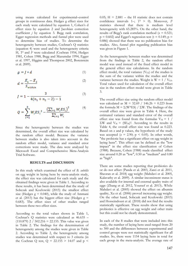
147
using means calculated for experimental–control groups in continuous data. Hedges g effect sizes for each study were calculated by equation 1, Cohen’s d effect sizes by equation 2 and Jacobian correction coefficient j by equation 3. Begg rank correlation, Egger regression methods and funnel plot were used to determine bias of studies. To determine the heterogeneity between studies, Cochran's Q statistics (equation 4) were used and the heterogeneity criteria H, Τ2 and I2 were calculated (Cochran 1954, Hedges 1981, Cohen 1988, Begg and Mazumdar 1994, Egger et al. 1997, Higgins and Thompson 2002, Borenstein et al. 2009).
(1)
(2)
(3)
(4)
Since the heterogeneity between the studies was determined, the overall effect size was calculated by the random effect model. Because the variance between studies is also taken into account in the random effect model, variance and standard error corrections were made. The data were analyzed by Microsoft Excel and Comprehensive Meta-Analysis Trial Software.
RESULTS and DISCUSSION In this study which examined the effect of B. subtilis on egg weight in laying hens by meta-analysis study, the effect size was calculated for each study and the obtained findings were given in Table 1. According to these results, it has been determined that the study of Sobczak and Kozłowski (2015) the smallest effect size (Hedges g = 0.040), while the study of Ammani et al. (2013) has the biggest effect size (Hedges g = 0.683). The effect sizes of other studies ranged between these two effect sizes. According to the total values shown in Table 1, Cochran's Q statistics were calculated as 46.635 – (110.879) 2 / 502.216 = 22.155. This value was given in Table 2. The findings for the determination of heterogeneity among the studies were given in Table 2. According to Table 2, the heterogeneity among studies was determined with respect to 3 criteria (in the Cochran Q test, Q = 22.155 > 14.07 and p <
0.05; H = 2.881 – the H statistic does not contain confidence intervals 1–; T2 > 0). Moreover, I2 statistics showed that there is medium level heterogeneity with 65.285%. On the other hand, the results of Begg's rank correlation method (z = 0.521; p = 0.602) and Egger's regression test (t = 0.149; p = 0.886) showed that there was no publication bias for studies. Also, funnel plot regarding publication bias was given in Figure 1. As the heterogeneity between studies was determined from the findings in Table 2, the random effect model was used instead of the fixed effect model in the general effect size calculations. In the random effect model, the total variance (VES) of the studies is the sum of the variance within the studies and the variance between the studies. Weight is W = 1 / VES. Total values used for calculation of the overall effect size in the random effect model were given in Table 3. The overall effect size using the random effect model was calculated as M = 32.69 / 146.26 = 0.223 from the formula M = Σ(W*ES) / ΣW. The findings of the overall effect size were given in Table 4. Here, the estimated variance and standard error of the overall effect size was found from the formulas VM = 1 / ΣW and VM = (SEM)2. According to the results in Table 4, the overall effect size was found as 0.223. Based on z and p values, the hypothesis of the study was accepted (z = 2.90; p < 0.05). In other words, "the probiotic has a significant effect on egg weight in laying hens". This effect can be defined as the "low impact" in the effect size classification of Cohen (1988). Because, Cohen (1988) made classified effect size around 0.20 as "low", 0.50 as "medium" and 0.80 as "high". There are some studies reporting that probiotics do or do not affect (Panda et al. 2003, Zarei et al. 2011, Sheoran et al. 2018) egg weight (Mahdavi et al. 2005, Kalavathy et al. 2009). A similar inconsistent status is also available for internal and external quality traits of eggs (Zhang et al. 2012, Youssef et al. 2013). While Mahdavi et al. (2005) showed the effect on albumin quality, Xu et al. (2006) proved increasing egg weight. On the other hand, Sobczak and Kozlowski (2015) and Hosseindoust et al. (2018) did not find the results statistically significant. These results show that using probiotics is effective on egg weight and other traits but this could not be clearly demonstrated. In each of the 8 studies that were included into this study, the number of laying hens used ranged from 20 to 300 and the differences between experimental and control groups were not statistically significant for all studies. So, there were 1154 laying hens in total for each group in the meta-analysis. The average rate of
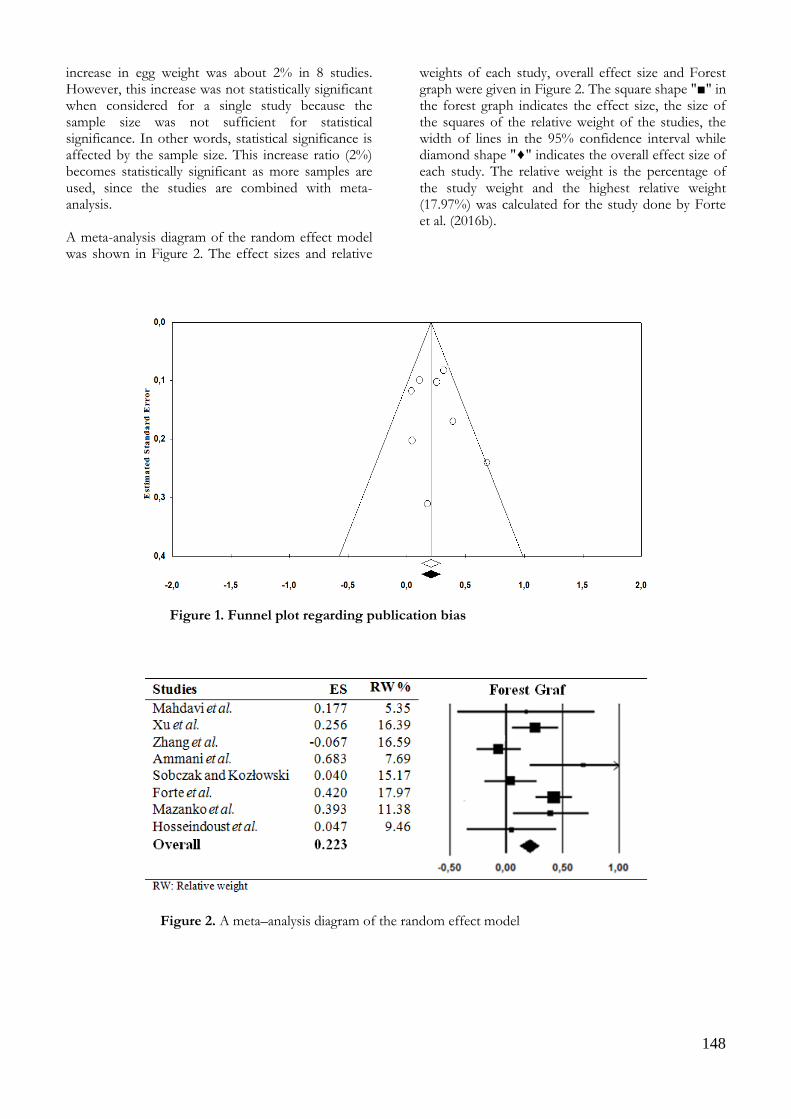
148
increase in egg weight was about 2% in 8 studies. However, this increase was not statistically significant when considered for a single study because the sample size was not sufficient for statistical significance. In other words, statistical significance is affected by the sample size. This increase ratio (2%) becomes statistically significant as more samples are used, since the studies are combined with meta-analysis. A meta-analysis diagram of the random effect model was shown in Figure 2. The effect sizes and relative
weights of each study, overall effect size and Forest graph were given in Figure 2. The square shape "■" in the forest graph indicates the effect size, the size of the squares of the relative weight of the studies, the width of lines in the 95% confidence interval while diamond shape "♦" indicates the overall effect size of each study. The relative weight is the percentage of the study weight and the highest relative weight (17.97%) was calculated for the study done by Forte et al. (2016b).
Figure 1. Funnel plot regarding publication bias
Figure 2. A meta–analysis diagram of the random effect model

149
Table 1. The effect size for each study
Studies Cohen’s
d
J Hedges g ES W W*ES W*ES2
Mahdavi et al. (2005) 0.180 0.980 0.177 10.367 1.834 0.324
Xu et al. (2006) 0.256 0.998 0.256 95.593 24.452 6.254
Zhang et al. (2012) -0.067 0.998 -0.067 100.321 -6.747 0.454
Ammani et al. (2013) 0.690 0.989 0.683 17.359 11.855 8.096
Sobczak and Kozłowski (2015) 0.040 0.997 0.040 72.365 2.880 0.115
Forte et al. (2016a/2016b) 0.421 0.999 0.420 147.123 61.812 25.970
Mazanko et al. (2017) 0.395 0.995 0.393 34.706 13.650 5.368
Hosseindoust et al. (2018) 0.047 0.992 0.047 24.382 1.143 0.054
Total 502.216 110.879 46.635
ES: Effect Size; W: Weight
Table 2. The findings for the determination of heterogeneity or homogeneity among studies
Methods Parameters df Chi square
table value P
Cochran's Q test 22.155 7 14.07 < 0.05
95% Confidence Interval
Min. Max.
H 2.881 2.456 3.379
T2 0.031 0.018 0.049
I2 65.285 52.189 74.795
Begg's rank correlation z = 0.521; P = 0.602 and Egger's regression test t = 0.149; P = 0.886 for publication bias
Table 3. Total values used for the overall effect size in the random effect model
Studies Hedges g (ES) SEES VES W W*ES
Mahdavi et al. (2005) 0.177 0.357 0.128 7.830 1.385
Xu et al. (2006) 0.256 0.204 0.042 23.971 6.132
Zhang et al. (2012) -0.067 0.203 0.041 24.258 -1.632
Ammani et al. (2013) 0.683 0.298 0.089 11.253 7.685
Sobczak and Kozłowski (2015)
0.040 0.212
0.045 22.185 0.883
Forte et al. (2016a/2016b) 0.420 0.195 0.038 26.279 11.041
Mazanko et al. (2017) 0.393 0.245 0.060 16.647 6.547
Hosseindoust et al. (2018) 0.047 0.269 0.072 13.837 0.649
Total 146.260 32.690
Table 4. The findings of overall effect size
Model Overall effect size VM SEM
95% Confidence Interval
z value P Min. Max.
Random Effects Model
0.223 0.006 0.077 0.073 0.374 2.90 < 0.05
150
CONCLUSION Eight studies were investigated by meta-analysis and the sample size was a total of 1154 laying hens for each group (experimental and control groups) in the study. It was detected that using probiotics had an effect on egg weight and thus, a more general result was obtained. As a matter of fact, it was possible to encounter different findings in the literature. These results emphasized once again the importance of meta-analysis that can provide certain results by combining studies that have been worked with a small sample size. More reliable scientific results can be revealed by extending the time span and combining more studies with meta–analysis because working with larger samples gives a more realistic result.
ACKNOWLEDGEMENT This article is a summary of the second author's Master thesis.
Conflict of Interest: The authors declare that they have no conflict of interest.
REFERENCES
Abdelqader A, Irshaid R, Al-Fataftah AR. Effects of dietary
probiotic inclusion on performance, eggshell quality, cecal microflora composition, and tibia traits of laying hens in the late phase of production. Trop Anim Health Pro. 2013; 45: 1017 – 1024. doi.org/10.1007/s11250-012-0326-7
Amani W, Youssef HMA, Hassan HM, Mohamed MA. Effect of probiotics, prebiotics and organic acids on layer performance and egg quality. Asian J Poult Sci. 2013; 7: 65 – 74. doi.org/10.3923/ajpsaj.2013.65.74
Begg CB, Mazumdar M. Operating characteristics of a rank correlation test for publication bias. Biometrics. 1994; 50:1088-1101.
Boissel JP, Blanchard J, Panak E. Considerations for the Meta – Analysis of randomized clinical trials. Control Clin Trials. 1989; 10: 254 – 281.
Borenstein M, Hedges LV, Higgins JPT, Rothstein HR. Introduction to Meta-Analysis. UK: John Wiley and Sons, Ltd., Publication; 2009.
Cochran WG. The combination of estimates from different experiments. Biometrics. 1954; 10: 101 – 129. doi.org/10.2307/3001666
Cohen J. Statistical Power Analysis for the Behavioral Sciences. New York, USA: Academic Press; 1988.
Cohen L, Manion L. Research Mehhods in Education. New York, USA: Rotledge Falmer; 2001.
Dawson B, Saunders B, Trapp G. Basic and Clinical Biostatistics. ABD: Appleton and Lange, Norwalk; 1994.
Egger M, Smith GD, Phillips AN. Principles and Procedures. BMJ. 1997; 315: 1533 – 1537. doi.org/10.1136/bmj.315.7121.1533
Fathi M, Al-Homida I, Al-Dokhail A, Ebeid T, Abou-Emera O, Alsagan A. Effects of dietary probiotic (Bacillus subtilis) supplementation on productive performance, immune response and egg quality characteristics in laying hens under high ambient temperature. Ital J Anim Sci. 2018; 17: 804–814. doi.org/10.1080/1828051X.2018.1425104
Fitz-Gibbon CT. The implications of meta analysis for educational research. Brit Educ Res J. 1985; 11: 45 - 49. doi.org/10.1080/0141192850110105
Forte C, Acuti G, Manuali E, Casagrande Proietti P, Pavone S, Trabalza-Marinucci M, Moscati L, Onofri A, Lorenzetti C, Franciosini MP. Effects of two different probiotics on microflora, morphology, and morphometry of gut in organic laying hens. Poultry Sci. 2016a; 95: 2528–2535. doi.org/10.3382/ps/pew164
Forte C, Moscati L, Acuti G, Mugnai C, Franciosini MP, Costarelli S, Cobellis G, Trabalza-Marinucci M. Effects of dietary Lactobacillus acidophilus and Bacillus subtilis on laying performance, egg quality, blood biochemistry and immune response of organic laying hens. J Anim Physiol An N. 2016b; 100: 977 – 987. doi.org/10.1111/jpn.12408
Fuller R. Probiotics in man and animals. J Appl Bacteriol. 1989; 66: 365 – 378.
Hedges L. Distribution theory for Glass’s estimator of effect size and related estimators. J Educ Stat. 1981; 6: 107 – 128. doi.org/10.3102/10769986006002107
Higgins JPT, Thompson SG. Quantifying heterogeneity in a meta analysis. Stat Med. 2002; 21: 1539 – 1558. doi.org/10.1002/sim.1186
Hosseindoust A, Mohammadi M, Yao ZP, Jung M, Kim IH. Dietary Bacillus subtilis B2A strain in laying hens challenged with Salmonella gallinarum: effects on egg production, egg quality, blood haptoglobin and targeted intestinal Salmonella shedding. J Appl Anim Res. 2018; 46: 512 – 517. doi.org/10.1080/09712119.2017.1351369
Kalavathy R, Abdullah N, Jalaludin S, Wong M, Ho YW. Effect of Lactobacillus cultures on performance of laying hens and total cholesterol, lipids and fatty acid composition of egg yolk. J Sci Food Agr. 2009; 89: 482 – 486. doi.org/10.1002/jsfa.3477
Kopp-Hollihan L. Prophylactic and therapeutic uses of probiotics: A review. J Am Diet Assoc. 2001; 10: 229–238. doi.org/10.1016/S0002-8223(01)00060-8
Lee KW, Kim DK, Lillehoj HS, Jang SI, Lee SH. Immune modulation by Bacillus subtilis-based direct-fed microbials in commercial broiler chickens. Anim Feed Sci Tech. 2015; 200: 76 –85. doi.org/10.1016/j.anifeedsci.2014.12.-006
Mahdavi AH, Rahmani HR, Pourr J. Effect of probiotic supplements on egg quality and laying hen's performance. International Journal of Poultry Science. 2005; 4: 488 – 492.
Mazanko MS, Gorlov IF, Prazdnova EV, Makarenko MS, Usatov AV, Bren AB, Chistyakov VA, Tutelyan AV, Komarova ZB, Mosolova NI, Pilipenko DN, Krotova OE, Struk AN, Lin A, Chikindas ML. Bacillus probiotic supplementations ımprove laying performance, egg quality, hatching of laying hens, and sperm quality of
151
roosters. Probiot Antimicrobiol Proteins. 2017; 10: 367 – 373. doi.org/10.1007/s12602-017-9369-4.
Mosteller F, Colditz GA. Understanding research synthesis (Meta Analysis). Annu Rev Publ Health. 1996; 17:1-23. doi.org/10.1146/annurev.pu.17.050196.000245
Panda AK, Reddy MR, Rama RA, Praharah NK. Production performance, serum/yolk cholesterol and immune competence of white leghorn layers as influenced by dietary supplementation with probiotic. Trop Anim Health Pro. 2003; 35: 85 – 94.
Sheoran N, Maan S, Kumar A, Batra K, Chaudhary D, Sihag S, Kumar V, Maan NS. Probiotic and prebiotic supplementation improving the production performance and immune characteristics of laying hens. Indian J Anim Res. 2018; 52:1433 – 1439. doi.org/10.18805/ijar.B-3394
Simon O, Jadamus A, Vahjen W. Probiotic feed additives—Effectiveness and expected modes of action. J Anim Feed Sci. 2001; 10: 51 – 67. doi.org/10.22358/jafs/70012/2001
Sobczak A, Kozłowski K. The effect of a probiotic preparation containing Bacillus subtilis ATCC PTA-6737 on egg production and physiological parameters of laying hens. Ann Anim Sci. 2015; 15: 711 – 723. doi.org/10.1515/aoas-2015-0040
Xu CL, Ji C, Ma Q, Hao K, Jin ZY, Li K. Effects of a dried Bacillus subtilis culture on egg quality. Poultry Sci. 2006; 85: 364 – 368. doi.org/10.1093/ps/85. 2.364.
Yach D. Meta-analysis in epidemiology. S Afr Med J. 1990; 78: 94 – 97.
Youssef AW, Hassan IM, Ali IM, Mohamed MA. Effect of probiotics, prebiotic and organic acids on layer performance and egg quality. Asian J Poult Sci. 2013; 7: 65 – 74. doi.org/10.3923/ajpsaj.2013.65.74
Zarei M, Ehsani M, Torki M. Dietary inclusion of probiotics, probiotics and symbiotic and evaluating performance of laying hens. Am J Agric Biol Sci. 2011; 6: 249 – 255.
Zhang JL, Xie QM, Ji J, Yang WH, Wu YB, Li C, Ma JY, Bi YZ. Different combinations of probiotics improve the production performance, egg quality, and immune response of layer hens. Poultry Sci. 2012; 91: 2755 – 2760. doi.org/10.3382/ps.2012-02339.

152
Kocatepe Veterinary Journal
Kocatepe Vet J (2020) 13(2):152-160
DOI: 10.30607/kvj.711873 RESEARCH ARTICLE
Molecular Characterization of Echinococcus granulosus Isolates Found
in Cattle, Buffaloes, Sheep and Goats in Afyonkarahisar, Turkey
Kürşat KARTAL1*, Mustafa KÖSE2, Metin ERDOĞAN3
1Gazi Mustafa Kemal Anatolian High School, Biology Teacher, TR-26470, Eskisehir, Turkey
2 Afyon Kocatepe Üniversity, Faculty of Veterinary Medicine, Department of Parasitology, Afyonkarahisar, Turkey 3Afyon Kocatepe Üniversity, Faculty of Veterinary Medicine, Department of Medical Biology and Genetics, Afyonkarahisar, Turkey
ABSTRACT
This study has been carried out to determine the genotypes of Echinococcus granulosus cysts in cattle, buffaloes, sheep and goats raised in Afyonkarahisar region. Cysts were collected from the internal organs of 258 animals, including 65 goats, 71 sheep, 119 cattle and 3 buffaloes infected with hydatid cysts. DNA was isolated from a total of 78 cysts from germinal membranes and protoscoleces extracted from cysts to identify the genotypes of E. granulosus in infected animals. PCR-RFLP was carried out using the Hin6I and StuI restriction enzymes in the ND1 gene and no polymorphism could be determined in all isolates. In the COX1 gene analysis, G1 strain known as domestic sheep strains and 18 different haplotypes were found in all isolates from cattle, buffaloes, sheep and goats. As a result, it was concluded that all isolates of the cattle, buffaloes, sheep and goats grown in Afyonkarahisar region determined in the analyses carried on the COX1 gene were G1 strain. Keywords: Ruminants, Echinococcus granulosus, ND1, COX1, PCR-RFLP, DNA Sequencing.
***
Afyonkarahisar’da Sığır, Manda, Koyun ve Keçilerde Bulunan Echinococcus granulosus İzolatlarının
Moleküler Karakterizasyonu
ÖZ Bu çalışma, Afyonkarahisar yöresinde yetiştirilen sığır, manda, koyun ve keçilerde bulunan Echinococcus granulosus kistlerinin genotiplerinin belirlenmesi amacıyla yapılmıştır. Araştırmada, hidatik kistle enfekte 65 keçi, 71 koyun, 119 sığır ve 3 manda olmak üzere toplam 258 hayvanın iç organlarından kistler toplanmıştır. Enfekte hayvanlardaki E. granulosus genotiplerini belirlemek amacıyla kistlerden çıkarılan germinal membran ve protoskolekslerden toplam 78 kistten DNA izole edilmiştir. ND1 gen bölgesi Hin6I ve StuI restriksiyon enzimleri kullanılarak PCR-RFLP yapılmış ve tüm izolatlarda polimorfizm belirlenememiştir. COX1 gen bölgesi analizinde sığır, manda, koyun ve keçilerden elde edilen izolatların tümünde evcil koyun suşu olarak bilinen G1 suşu ve 18 farklı haplotip bulunmuştur. Sonuç olarak, Afyonkarahisar yöresinde yetiştirilen sığır, manda, koyun ve keçilerde bulunan izolatların COX1 geninde yapılan genetik analizler sonucunda tüm izolatların G1 suşu olduğu kanısına varılmıştır. Anahtar Kelimeler: Ruminantlar, Echinococcus granulosus, ND1, COX1, PCR-RFLP, DNA Dizileme
To cite this article: Kartal K. Köse M. Erdoğan M. Molecular Characterization of Echinococcus granulosus Isolates Found in Cattle, Buffaloes, Sheep and Goats in Afyonkarahisar, Turkey. Kocatepe Vet J. (2020) 13(2):152-160 Submission: 31.03.2020 Accepted: 08.05.2020 Published Online: 14.05.2020
ORCID ID; KK: 0000-0002-0803-2635, MK: 0000-0003-3206-2508, ME: 0000-0003-0975-1989 *Corresponding author e-mail: [email protected]
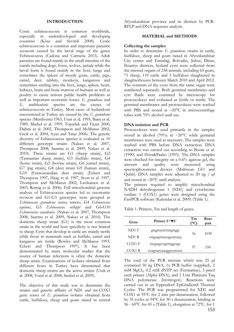
153
INTRODUCTION Cystic echinococcosis is common worldwide, especially in underdeveloped and developing countries (Köse and Sevimli 2008). Cystic echinococcosis is a common and important parasitic zoonosis caused by the larval stage of the genus Echinococcus (Cadona and Carmena 2013). Adult parasites are found mainly in the small intestine of the canids including dogs, foxes, wolves, jackals while the larval form is found mainly in the liver, lungs and sometimes the spleen of mostly goats, cattle, pigs, camel, deer, rabbits, monkeys, kangaroos and sometimes settling into the liver, lungs, spleen, heart, kidneys, brain and bone marrow of humans as well as poultry to cause serious public health problems as well as important economic losses. E. granulosus and E. multilocularis species are the causes of echinococcosis in Turkey. Most cases of hydatidosis encountered in Turkey are caused by the E. granulosus species (Merdivenci 1963, Unat et al. 1995, Barış et al. 1989, Markel et al. 1999, Toparlak and Tüzer 2000, Dalimi et al. 2002, Thompson and McManus 2002, Gıcık et al. 2004, Ayaz and Tınar 2006). The genetic diversity of Echinococcus species is evaluated as 10 different genotype strains (Nakao et al. 2007, Thompson 2008, Saarma et al. 2009, Nakao et al. 2010). These strains are G1 (sheep strain), G2 (Tasmanian sheep strain), G3 (buffalo strain), G4 (horse strain), G5 (bovine strain), G6 (camel strain), G7 (pig strain), G8 (deer strain G9 (human strain), G10 (Fennoscandian deer strain) (Eckert and Thompson 1997, Haag et al. 1997, Scott et al. 1997, Thompson and McManus 2002, Lavikainen et al. 2003, Romig et al. 2006). Full mitochondrial genome analysis of Echinococcus species led to taxonomic revision and G1-G3 genotypes were grouped as Echinococcus granulosus sensu stricto, G4 Echinococcus equinus, G5 Echinococcus ortleppi and G6-G10 Echinococcus canadensis. (Nakao et al. 2007, Thompson 2008, Saarma et al. 2009, Nakao et al. 2010). The domestic sheep strain (G1) is the most common strain in the world and host specificity is not limited to sheep. Cysts that develop in cattle are mainly sterile while those in mammals such as buffalo, camel and kangaroo are fertile (Bowles and McManus 1993, Eckert and Thompson 1997). It has been demonstrated by many molecular studies that the source of human infections is often the domestic sheep strain. Examinations of isolates obtained from different hosts in Turkey have determined that domestic sheep strains are the active strains (Utuk et al. 2008, Vural et al. 2008, Snabel et al. 2009). The objective of this study was to determine the strains and genetic affinity of ND1 and mt-COX1 gene zones of E. granulosus isolates obtained from cattle, buffaloes, sheep and goats raised in central
Afyonkarahisar province and its districts by PCR-RFLP and DNA sequence analysis.
MATERIAL and METHODS
Collecting the samples In order to determine E. granulosus strains in cattle, buffaloes, sheep and goats raised in Afyonkarahisar City center and Emirdağ, Bolvadin, Şuhut, Dinar, İhsaniye districts, hydatid cysts were collected from the internal organs of 258 animals, including 65 goats, 71 sheep, 119 cattle and 3 buffaloes slaughtered in slaughterhouses between March 2010 and April 2012. The contents of the cysts from the same organ were numbered separately. Both germinal membranes and cyst fluids were examined by microscopy for protoscoleces and evaluated as fertile or sterile. The germinal membranes and protoscoleces were washed
with PBS and stored at 20°C in microcentrifuge tubes with 70% alcohol until use. DNA isolation and PCR Protoscoleces were used primarily in the samples
stored in alcohol (70%) at 20°C while germinal membranes were used as necessary. The samples were washed with PBS before DNA extraction. DNA extraction was carried out according to Boom et al. (1990) and Höss&Paabo (1993). The DNA samples were checked for integrity on a 0.6% agarose gel, the amount and quality were measured using spectrophotometer devices (Multiscan GO and Qubit). DNA samples were adjusted to 20 ng / µl
and stored at 20°C until analysis. The primers required to amplify mitochondrial NADH dehydrogenase 1 (ND1) and cytochrome oxidase 1 (COX1) genes were designed using the FastPCR software (Kalendar et al. 2009) (Table 1). Table 1. Primers, Tm and length of genes.
Gene Primer 5´ 3´ Tm (°C)
Base pair
ND1 F gtagttactcttatgttggt 56 1038
ND1 R cttgaagttaacagcatcacg
COX1 F tacgttgcctgttttggctgc 57 550
COX1 R ccagtaatcaaaggccatcacc
The total of the PCR mixture which was 25 µl contained 50 ng DNA, 1x PCR buffer (supplied), 2 mM MgCl2, 0.2 mM dNTP set (Fermantas), 3 pmol each primer (Alpha DNA), and 1 Unit Platinum Taq DNA polymerase (Invitrogen). Reactions were carried out in an Eppendorf EpGradientS Thermal Cycler. The PCR was programmed for ND1 and COX1 at 95°C for 2 min pre-denaturation, followed by 35 cycles at 94°C for 30 s denaturation, binding at 56 - 60°C for 45 s (Table 1), elongation at 72°C. for 1

154
min, final elongation step at 72°C for 10 min. The PCR products were checked under UV by using 1% agarose gel and GelRed (Biotium, 41003). PCR-RFLP ND1 gene PCR products were cut separately with Hin6I (Thermo) and StuI (Thermo) restriction enzymes. For this, 8 μl of PCR product, 1 μl of restriction enzyme, 2 μl of restriction buffer and 9μl of distilled water were used. Subsequently a 14-hour incubation at 37ºC was carried out. After incubation the Hin6I and the StuI enzymes were inactivated at 65ºC and 80ºC respectively for 20 min. The products subjected to cutting were examined under UV with 2.5% agarose gel supplemented with GelRed and the band patterns of the samples were displayed (Figure 1 and 2).
Figure 1. Agarose gel electrophoresis image of PCR-RFLP products of the ND1 gene cut with Hin6I. M: Marker D: DNA P: PCR product H: PCR products treated with Hin6I restriction enzyme.
Figure 2. Agarose gel electrophoresis image of PCR-RFLP products of the ND1 gene cut with StuI M: Marker D: DNA P: PCR product S: PCR products treated with StuI restriction enzyme.
Sequence analysis DNA double-sided sequence analysis of the PCR products of 78 isolates for the COX1 gene was carried out. PCR products were purified using 0.5μl Exo I and 1μl FastAp mixture prior to DNA sequence analysis. The mixture was kept at 37ºC for
15 minutes and at 85ºC for 15 minutes. All of samples sequenced using the BigDye® Terminator v3.1 Cycle Sequencing Kit (Life Technologies). Sequence PCR products were purged with ethanol / EDTA / sodium acetate and the reactions were run on an ABI 3500Genetic Analyzer. The DNA sequences were edited with the Sequencher 5.4.1 computer program (Gene Code Corporation, Ann Arbor, Michigan, USA) and were aligned with BioEdit 7.0.9 Sequence Alignment (Hall 1999) programs. Phylogenetic and Statistical Analysis The nucleotide differences between haplotypes (π),
the haplotype mutation rate (Ѳ) and the Tajima D value were calculated and UPGMA dendrogram was created with the Mega 4 computer package program (Tamura et al. 2007).
RESULTS As a result of DNA isolation from the internal organs of 258 animals, including 65 goats, 71 sheep, 119 cattle and 3 buffaloes infected with hydatid cysts, DNA was isolated from 78 cysts (30 goats, 26 sheep, 19 cattle and 3 buffaloes). ND1 gene DNA obtained from 78 isolates was used for PCR-RFLP and after the PCR analysis, a DNA sequence with the length of 1038 bp was obtained. As a result of cutting the PCR products with Hin6I and StuI restriction enzymes, two bands (Figure 1) with a 1038 bp band and two bands measuring 391 and 647 bp were observed (Figure 2), respectively. All samples showed an exemplary band structure in terms of the ND1 gene. COX1 Gene After PCR analysis, a length of 550 bp DNA sequence was obtained in the 1608 bp mt-COX1 gene zone. As a result of the comparison of sequence analysis, 18 different haplotypes (GenBank ID: MT318680-MT318697) were found for domestic sheep strain G1 and variants of isolates. When the distribution of these 18 haplotypes in goats, sheep, cattle and buffaloes and percentage ratios are observed, it is evident that TR_AF001 (MT318680) haplotype is more common (Table 2). The total number of polymorphic zones(S) for the nucleotide sequences of E. granulosus, polymorphic siteratio (ps), nucleotide differences (π), population mutation rate (Θ) and Tajima D value are given in Table 3.
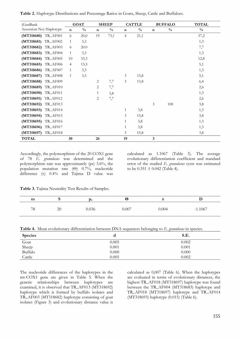
155
Table 2. Haplotype Distributions and Percentage Ratios in Goats, Sheep, Cattle and Buffaloes.
(GenBank Accession No) Haplotype
GOAT SHEEP CATTLE BUFFALO TOTAL
n % n % n % n % %
(MT318680) TR_AF001 6 20,0 19 73,1 4 21,1 37,2
(MT318681) TR_AF002 1 3,3 1,3
(MT318682) TR_AF003 6 20.0 7,7
(MT318683) TR_AF004 1 3,3 1,3
(MT318684) TR_AF005 10 33,3 12,8
(MT318685) TR_AF006 4 13,3 5,1
(MT318686) TR_AF007 1 3,3 1,3
(MT318687) TR_AF008 1 3,3 3 15,8 5,1
(MT318688) TR_AF009 2 7,7 3 15,8 6,4
(MT318689) TR_AF010 2 7,7 2,6
(MT318690) TR_AF011 1 3,8 1,3
(MT318691) TR_AF012 2 7,7 2,6
(MT318692) TR_AF013 3 100 3,8
(MT318693) TR_AF014 1 3,8 1,3
(MT318694) TR_AF015 3 15,8 3,8
(MT318695) TR_AF016 1 3,8 1,3
(MT318696) TR_AF017 1 3,8 1,3
(MT318697) TR_AF018 3 15,8 3,8
TOTAL 30 26 19 3
Accordingly, the polymorphism of the 20 COX1 gene of 78 E. granulosus was determined and the polymorphism rate was approximately (ps) 3.6%, the population mutation rate (Θ) 0.7%, nucleotide difference (π) 0.4% and Tajima D value was
calculated as 1.1067 (Table 3). The average evolutionary differentiation coefficient and standard error of the studied E. granulosus cysts was estimated to be 0.351 ± 0.042 (Table 4).
Table 3. Tajima Neutrality Test Results of Samples.
m S ps Θ π D
78 20 0.036 0.007 0.004 -1.1067
Table 4. Mean evolutionary differentiation between DNA sequences belonging to E. granulosus in species.
Species d S.E.
Goat 0.005 0.002 Sheep 0.001 0.001 Buffalo 0.000 0.000 Cattle 0.005 0.002
The nucleotide differences of the haplotypes in the mt-COX1 gene are given in Table 5. When the genetic relationships between haplotypes are examined, it is observed that TR_AF013 (MT318692) haplotype which is formed by buffalo isolates and TR_AF003 (MT318682) haplotype consisting of goat isolates (Figure 3) and evolutionary distance value is
calculated as 0,007 (Table 6). When the haplotypes are evaluated in terms of evolutionary distances, the highest TR_AF018 (MT318697) haplotype was found between the TR_AF004 (MT318683) haplotype and TR_AF018 (MT318697) haplotype and TR_AF014 (MT318693) haplotype (0.015) (Table 6).
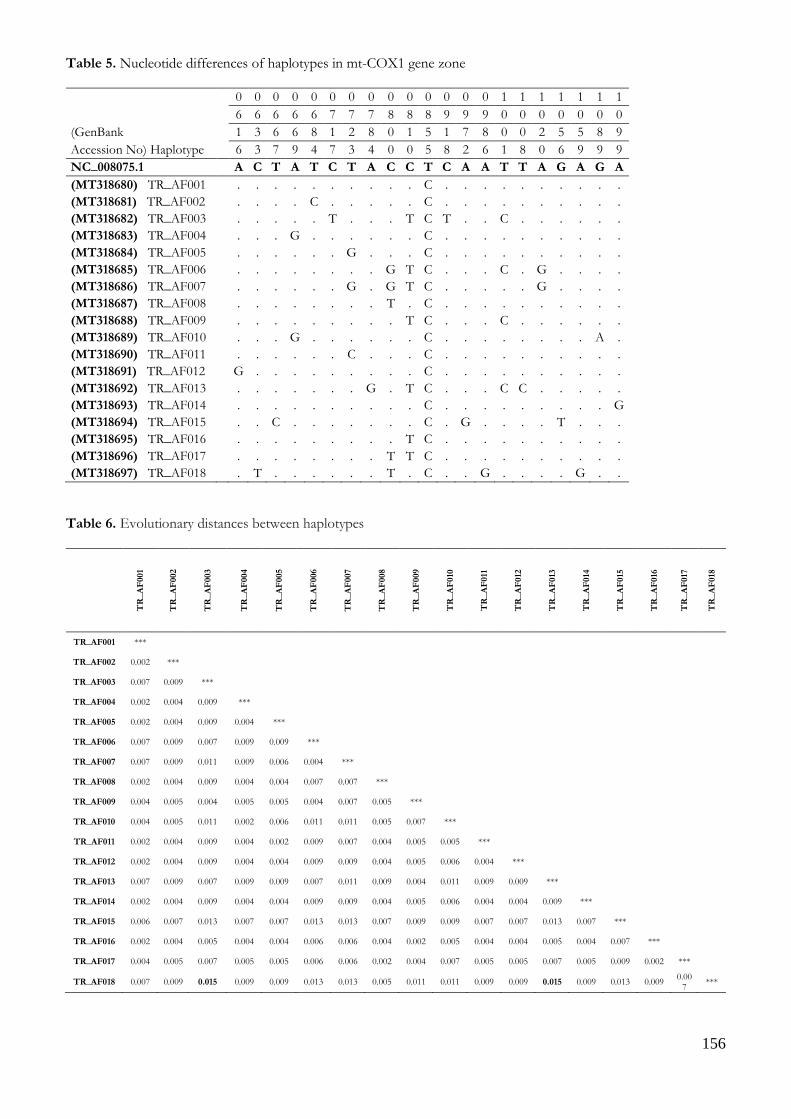
156
Table 5. Nucleotide differences of haplotypes in mt-COX1 gene zone
0 0 0 0 0 0 0 0 0 0 0 0 0 0 1 1 1 1 1 1 1
6 6 6 6 6 7 7 7 8 8 8 9 9 9 0 0 0 0 0 0 0
(GenBank
1 3 6 6 8 1 2 8 0 1 5 1 7 8 0 0 2 5 5 8 9
Accession No) Haplotype
6 3 7 9 4 7 3 4 0 0 5 8 2 6 1 8 0 6 9 9 9
NC_008075.1 A C T A T C T A C C T C A A T T A G A G A
(MT318680) TR_AF001
. . . . . . . . . . C . . . . . . . . . .
(MT318681) TR_AF002
. . . . C . . . . . C . . . . . . . . . .
(MT318682) TR_AF003
. . . . . T . . . T C T . . C . . . . . .
(MT318683) TR_AF004
. . . G . . . . . . C . . . . . . . . . .
(MT318684) TR_AF005
. . . . . . G . . . C . . . . . . . . . .
(MT318685) TR_AF006
. . . . . . . . G T C . . . C . G . . . .
(MT318686) TR_AF007
. . . . . . G . G T C . . . . . G . . . .
(MT318687) TR_AF008
. . . . . . . . T . C . . . . . . . . . .
(MT318688) TR_AF009
. . . . . . . . . T C . . . C . . . . . .
(MT318689) TR_AF010
. . . G . . . . . . C . . . . . . . . A .
(MT318690) TR_AF011
. . . . . . C . . . C . . . . . . . . . .
(MT318691) TR_AF012
G . . . . . . . . . C . . . . . . . . . .
(MT318692) TR_AF013
. . . . . . . G . T C . . . C C . . . . .
(MT318693) TR_AF014
. . . . . . . . . . C . . . . . . . . . G
(MT318694) TR_AF015
. . C . . . . . . . C . G . . . . T . . .
(MT318695) TR_AF016
. . . . . . . . . T C . . . . . . . . . .
(MT318696) TR_AF017
. . . . . . . . T T C . . . . . . . . . .
(MT318697) TR_AF018
. T . . . . . . T . C . . G . . . . G . .
Table 6. Evolutionary distances between haplotypes
TR
_A
F001
TR
_A
F002
TR
_A
F003
TR
_A
F004
TR
_A
F005
TR
_A
F006
TR
_A
F007
TR
_A
F008
TR
_A
F009
TR
_A
F010
TR
_A
F011
TR
_A
F012
TR
_A
F013
TR
_A
F014
TR
_A
F015
TR
_A
F016
TR
_A
F017
TR
_A
F018
TR_AF001 ***
TR_AF002 0.002 ***
TR_AF003 0.007 0.009 ***
TR_AF004 0.002 0.004 0.009 ***
TR_AF005 0.002 0.004 0.009 0.004 ***
TR_AF006 0.007 0.009 0.007 0.009 0.009 ***
TR_AF007 0.007 0.009 0.011 0.009 0.006 0.004 ***
TR_AF008 0.002 0.004 0.009 0.004 0.004 0.007 0.007 ***
TR_AF009 0.004 0.005 0.004 0.005 0.005 0.004 0.007 0.005 ***
TR_AF010 0.004 0.005 0.011 0.002 0.006 0.011 0.011 0.005 0.007 ***
TR_AF011 0.002 0.004 0.009 0.004 0.002 0.009 0.007 0.004 0.005 0.005 ***
TR_AF012 0.002 0.004 0.009 0.004 0.004 0.009 0.009 0.004 0.005 0.006 0.004 ***
TR_AF013 0.007 0.009 0.007 0.009 0.009 0.007 0.011 0.009 0.004 0.011 0.009 0.009 ***
TR_AF014 0.002 0.004 0.009 0.004 0.004 0.009 0.009 0.004 0.005 0.006 0.004 0.004 0.009 ***
TR_AF015 0.006 0.007 0.013 0.007 0.007 0.013 0.013 0.007 0.009 0.009 0.007 0.007 0.013 0.007 ***
TR_AF016 0.002 0.004 0.005 0.004 0.004 0.006 0.006 0.004 0.002 0.005 0.004 0.004 0.005 0.004 0.007 ***
TR_AF017 0.004 0.005 0.007 0.005 0.005 0.006 0.006 0.002 0.004 0.007 0.005 0.005 0.007 0.005 0.009 0.002 ***
TR_AF018 0.007 0.009 0.015 0.009 0.009 0.013 0.013 0.005 0.011 0.011 0.009 0.009 0.015 0.009 0.013 0.009 0.00
7 ***

157
Figure 3. UPGMA dendrogram showing relationships of genetic distances between haplotypes

158
DISCUSSION Cystic echinococcosis is an important and widespread parasitic zoonosis all over the world and mainly in less developed and developing countries observed in humans and animals caused by the larval stage of the genus Echinococcus (Köse and Sevimli 2008, Cadona and Carmena 2013). Although the last host is usually carnivores like dogs, foxes, jackals, wolves, different
strains of the agent can be found in different geographical regions in numerous intermediate host mammals such as cattle, sheep, goats, deer, camels, buffaloes, rabbits, kangaroos, pigs, horses, donkeys able to infect humans (McManus et al. 2003). Studies on E. granulosus species have been carried out in different regions of the world by using molecular techniques. These studies are shown in Table 7.
Table 7. DNA sequence analysis studies on E. granulosus.
Country Source Gene Strain
Spain Gonzalez et al., 2002 mt-COX1, ND1 G1,G7 Bulgaria Breyer et al., 2004 ND1 G1 Italy Capuano et al., 2006 mt-COX1 G1,G3
China Bart et al., 2006a mt-COX1 G6 Romania Bart et al., 2006b mt-COX1, ND1 G1,G2,G7 Greece Varcasia et al., 2007 mt-COX1, ND1 G1,G3,G7 Turkey Vural et al., 2008 mt-COX1 G1,G3 Turkey Snabel et al., 2009 mt-COX1, ND1 G1,G3,G7 Kenya Casulli et al., 2010 mt-COX1, ND1 G1,G6 Pakistan Latif et al., 2010 mt-COX1 G1,G3 Argentina Soriano et al., 2010 mt-COX1 G1,G3,G6,G7 Turkey Simsek et al., 2010 mt-COX1 G1,G3 Turkey Beyhan and Umur, 2011 mt-COX1 G1,G2,G3 Iran Pour et al., 2011 mt-COX1 G1,G3 Japan Guo et al., 2011 mt-COX1, ND1 G1,G2,G3 Mongolia Jabbar et al., 2011 mt-COX1, ND1 G1,G3,G6,G10 India Singh et al., 2012 mt-COX1 G1,G3 Peru Sanchez et al., 2012 mt-COX1, ND1 G1,G7 Egypt Aboelhadid et al., 2013 mt-COX1, ND1 G1,G7 Palestine Adwan et al., 2013 mt-COX1 G1,G2,G3
In this study, the ND1 gene zone of the isolates was examined by PCR-RFLP technique and shows that the isolates may have a similar genetic structure and the same strain. The DNA sequence analysis of the mt-COX1 gene of the isolates suggests that all isolates are G1 genotype. As a result of the mutations in the COX1 gene region, 18 different haplotypes have been manifested. Table 1 shows that most haplotypes are from goat and cattle species with eight haplotypes. Five haplotypes have been found in sheep. The presence of fewer haplotypes in sheep is probably explained by sampling in nearby regions. In the analyzes, the TR_AF005 (MT318684) haplotype (33.3%) found in goats and the frequency of the TR_AF001 (MT318680) haplotype in sheep and cattle was 21.1% and 73.1%, respectively. The fact that haplotype frequencies vary according to species suggests that the examined animals may have come from the same location. Results in Table 6 confirm this. When Table 6 is examined, it is noted that the frequency of TR_AF001 (MT318680) haplotype in sheep and cattle is high because of the animals in
Şuhut district and the high frequency of the TR_AF005 (MT318684) haplotype in goats is caused by animals from Dinar district. The presence of only one haplotype in buffaloes (TR_AF013 (MT318692)) suggests that the number of buffaloes is low and that the samples may have been collected from the same area or from the same herd. The prevalence of the TR_AF001 (MT318680) haplotype over the entire Afyonkarahisar province (37.2%) can be explained by the fact that this haplotype is more likely to produce different types of infection than other haplotypes, or that it may be more widely distributed by animal movements. The results of the study show that E. granulosus is a dominant genotype of domestic sheep strain in Afyonkarahisar and the limited number of studies (Vural et al. 2008, Utuk et al. 2008, Beyhan and Umur 2011, Eryıldız and Şakru 2012) in this subject support the study results.

159
CONCLUSION A sequencing analysis of the mt-COX1 gene zone of E. granulosus was carried out in this study and as a result of the evaluation of the obtained sequence analysis information, the intermediate hosts were found to be infected with the domestic sheep strain (G1) which is accepted to be the most common and most pathogenic strain in Afyonkarahisar region and the World.
ACKNOWLEDGMENTS This study was supported by the Scientific Research Projects Coordination Unit of Afyon Kocatepe University (Project number: 12. SAG. BIL. 01). This study has been approved by the local ethics committee of Afyon Kocatepe University. This article has been summarized from a doctoral dissertation (2014/006) supported by Afyon Kocatepe University Scientific Research Projects Coordination Unit (12. SAĞ. BİL. 01) and accepted by Afyon Kocatepe University Institute of Health Sciences and was presented as a poster in the 19th National Parasitology Congress (Erzurum-2015). Conflict of interest: The authors declare that they have no conflict of interest.
REFERENCES Aboelhadid SM, El-Dakhly KM, Yanai T, Fukushi H,
Hasanin KM. Molecular Characterization of Echinococcus granulosus in Egyptian. Vet Parasitol. 2013; 193: 292-296.
Adwan G, Adwan K, Bdir S, Abuseir S. Molecular Characterization of Echinococcus granulosus Isolated from Sheep in Palestine. Exp Parasitol. 2013; 134: 195-199.
Ayaz E, Tınar R. Cestoda. In: Tınar R (ed) Helmintoloji, 1st edn. Nobel yayınevi, Ankara. 2006; pp 103-213.
Barış İ, Şahin A, Bilir N, Kalyoncu F, Emri S, Barış B, Çapur S, Selçuk T. Hidatik Kist Hastalığı ve Türkiye’deki Konumu. Türkiye Akciğer Hastalıkları Vakfı Yayını No: 1 Ankara. 1989
Bart JM, Abdukader M, Zhang YL, Lin RY, Wang YH, Nakao M, Ito A, Craig PS, Piarroux R, Vuitton DA, Wen H. Genotyping of Human Cystic Echinococcosis in Xinjiang, PR China. Parasitology. 2006a; 133(5): 571-579.
Bart JM, Morariu S, Knapp J, Ilie MS, Pitulescu M, Anghel A, Cosoroaba I, Piarroux R. Genetic Typing of Echinococcus granulosus in Romania. Parasitol Res. 2006b; 98: 130-137.
Beyhan YE, Umur Ş. Molecular Characterization and Prevalence of Cystic Echinococcosis in Slaughtered Water Buffaloes in Turkey. Vet Parasitol. 2011; 181: 174-179.
Boom R, Sol CJA, Salimans MMM, Jansen CL, Wertheim-Van Dillen PME, Van Der Noorda J. Rapid and Simple Method for Purification of Nucleic Acids. J Clin Microbiol. 1990; 28(3): 495-503.
Bowles J, McManus DP. Rapid discrimination of Echinoccus specises and strains using a polymerase chain reaction-based RFLP method. Mol Biochem Parasitol. 1993; 57: 231–240.
Breyer I, Georgieva D, Kurdova R, Gottstein B. Echinococcusgranulosus Strain Typing in Bulgaria: The G1 Genotype is Predominant in Intermediate and Definitive Wild Hosts. Parasitol Res. 2004; 93: 127-130
Cadona GA, Carmena D. A Review of The Global Prevalence, Molecular Epidemiology and Economics of Cystic Echinococcus In Production Animals. Vet Parasitol. 2013; 192: 10-32.
Capuano F, Rinaldi L, Maurelli MP, Perugini AG, Veneziano V, Garippa G, Genchi C, Musella V, Cringoli G. Cystic echinococcocosis in water buffaloes: epidemiological survey and molecular evidence of ovine (G1) and buffalo (G3) strains. Vet Parasitol. 2006; 137: 262-268.
Casulli A, Zeyhle E, Brunetti E, Pozio E, Meroni V, Genco F, Filice C. Molecular Evidence of the Camel Strain (G6 Genotype) of Echinococcusgranulosus in humans from Turkana, Kenya. Trans Roy Soc Trop Med Hyg. 2010; 104: 29-32.
Dalimi A, Motamedi GH, Hosseni M, Mohammadian B, Malaki H, Ghamari Z, Far GF. Echinococcosis/ hydatidosis in western Iran. Vet Parasitol. 2002; 105: 161–171.
Eckert J, Thompson RCA. IntraspesificVarition of Echinococcus granulosus and Related Species with Emphises on their Infectivity to Humans. Acta Trop. 1997; 64: 19-34.
Eryıldız, C., Şakru, N. Molecular Characterization of Human and Animal Isolates of Echinococcus granulosus in the Thrace Region, Turkey. Balkan Med J. 2012; 29: 261-267.
Gıcık Y, Arslan MÖ, Kara M, Köse M. Kars ilinde Kesilen Sığır ve Koyunlarda Kistik Ekinokokkozisin Yaygınlığı. Türkiye Parazitol Derg. 2004; 28(3): 136-139.
Gonzalez LM, Daniel-Mwambete K, Montero E, Rosenzvit MC, McManus DP, Garate T, Cuesta-Bandera C. Further Molecular Discrimination of Spanish Strains of Echinococcus granulosus. Exp Parasitol. 2002; 102: 46-56
Guo ZH, Kubo M, Kudo M, Nibe K, Horii Y, Nonaka N. Growth and Genotypes of Echinococcus granulosus Found in Cattle Imported from Australia and Fattened in Japan. Parasitol Int. 2011; 60: 498-502.
Haag KL, Zaha A, Araujo AM, Gottstein B. Reduced Genetic Variability within Coding and Non-coding Regions of the Echinococcus multilocularis Genome. Parasitology. 1997; 115: 521-529.
Hall TA. BioEdit: A User-Friendly Biological Sequence Alignment Editor and Analysis Program for Windows 95/98/NT. Nucleic Acids Symposium Series. 1999; 41: 95-98.
Höss M, Paabo S. DNA extraction from Pleistocene bones by a silica-based purification method. Nucleic Acids Res. 1993; 21(16): 3913-3914. (Bu kaynak metin içinde kullanılmamıştır.)
Jabbar A, Narankhajid M, Nolan MJ, Jex AR, Campbell BE, Gasser RB. A First İnsight into the Genotypes of
160
Echinococcus granulosus from Humans in Mongolia. Mol Cell Probe. 2011; 25: 49-54.
Kalendar R, Lee D, Schulman AH. Fast PCR Software for PCR Primer and Probe Design and Repeat Search. G3. 2009; 3(1): 1-14.
Köse M, Sevimli FK. Prevalence of Cystic Echinococcosis in Slaughtered Cattle in Afyonkarahisar. Türkiye Parazitol Derg. 2008; 32(1): 27-30.
Latif AA, Tanveer A, Maqbool A, Siddiqi N, Kyaw-Tanner M, Traub RJ. Morphological and Molecular Characterisation of Echinococcus granulosus in Livestock and Humans in Punjab, Pakistan. Vet Parasitol. 2010; 170: 44-49.
Lavikainen A, Lehtinen MJ, Meri T, Hirvela-Koski V, Meri S. Molecular genetic characterization of the Fennoscandiancervidstrain, a new genotypic group (G10) of Echinococcus granulosus. Parasitology. 2003; 127: 207-215.
Markel EK, Voge M, John DT. Medical Parasitology. 8th Edn, WB Saunders Company, USA. 1999; pp 234-268.
McManus DP, Wenbao Z, Li J, Bartley PB. Echinococcosis. Lancet. 2003; 326: 1295-1304.
Merdivenci A. İstanbul Sokak Köpeklerinde Echinococcus granulosus (Batsch, 1786) Rudolphi, 1805. Mikrobiol Derg. 1963; 16(1): 23-28.
Nakao M, McManus DP, Schantz PM, Craig PS, Ito A. A Molecular Phylogeny of the Genus Echinococcus Inferred from Complete Mitochondrial Genomes. Parasitology. 2007; 134: 713-722.
Nakao M, Yangida T, Okomoto M, Knapp J, Nkouawa A, Sako Y, Ito A. State-of-the-Art Echinococcus and Taenia: Phylogenetic Taxanomy of Human Pathogenic Tapeworms an Its Application to Molecular Diagnosis. Infect Genet Evol. 2010; 10: 444-452.
Pour AA, Hosseini SS, Shayan P. Comparative Genotyping of Echinococcus granulosus Infecting Buffalo in Iran Using Cox1 Gene. Parasitol Res. 2011; 108: 1229-1234.
Romig T, Dinkel A, Mackenstedt U. The Present Situation of Echinococcus in Europe. Parasitol Int. 2006; 55: 187-191.
Saarma U, Jogisalu I, Moks E, Varcasia A, Lavikainen A, Oksanen A. A Novel pylogeny for the Genus Echinococcus, Based on Nuclear Data, Challenges Relationships Based on Mitochondrial Evidence. Parasitology. 2009; 136: 317-328.
Sanchez E, Caceres O, Naquira C, Miranda E, Samudio F, Fernandes O. Echinococcus granulosus Genotypes Circulating in Alpacas (Lama pacos) and Pigs (Susscrofa) from an Endemic Region in Peru. Mem Inst Oswaldo Cruz, Rio de Janeiro. 2012; 107: 275-278.
Scott JC, Stefaniak J, Pawlowski ZS, McManus DP. Molecular Genetic Analysis of Human Cystic Hydatid Cases from Poland: Identification of a New Genotipic Group (G9) of Echinococcus granulosus. Parasitology. 1997; 114: 37-43.
Şimşek S, Balkaya I, Köroğlu E. Epidemiological Survey and Molecular Characterization of Echinococcusgranulosus in Cattle in an Endemic Area Eastern Turkey. Vet Parasitol. 2010; 172: 347-349.
Singh BB, Sharma JK, Ghatak S, Sharma R., Bal MS, Tuli A, Gill JPS. Molecular Epidemiology of Echinococcosis from Food Producing Animals in North India. Vet Parasitol. 2012; 186: 503-506.
Snabel V, Altıntaş N, D’amelio S, Nakao M, Romig T, Yolasığmaz A, Güneş K, Turk M, Busi M, Huttner M, Sevcova D, Ito A, Altıntaş N, Dubinsky P. Cystic Echinococcosis in Turkey: Genetic Variability and First Record of the Pig Strain (G7) in the Country. Parasitol Res. 2009; 105(1): 145-154.
Soriano SV, Pierangelii NB, Pianciola L, Mazzeo M, Lazzarini LE, Saiz MS, Kossman AV, Bergagna HFJ, Chartier K, Basualdo JA. Molecular characterization of Echinococcus canadensis G6 Genotype in Neuquen, Patagonia Argentina. Parasitol Int. 2010; 59: 626-628.
Tamura K, Dudley J, Nei M, Kumar S. MEGA4: Molecular Evolutionary Genetics Analysis (MEGA) Software Version 4.0. Molecular Biology and Evolution 10.1093/molbev/msm092. 2007.
Tajima F. Statistical Methods to Test for Nucleotide Mutation Hypothesis by DNA Polymorphism. Genetics. 1989; 123: 585-595.
Thompson RCA. The Taxanomy, Phylogeny and Transmission of Echinococcus. Exp parasitol. 2008; 119: 439-446.
Thompson RCA, McManus DP. Towards a Taxonomic Revision of the Genus Echinococcus. Trends Parasitol. 2002; 18: 452-457.
Toparlak M, Tüzer E. Veteriner Helmintoloji. İ.Ü. Veteriner Fakültesi Parazitoloji Anabilim Dalı. 2000; pp 50-56.
Unat EK, Yücel A, Atlaş K, Samastı M. Unat’ ın Tıp Parazitolojisi (5. Baskı). İ.Ü Cerrahpaşa Tıp Fak. Vakfı Yayın no: 15, İstanbul. 1995.
Utuk AE, Şimşek S, Köroğlu E, McManus DP. Molecular Genetic Characterization of Different İsolates of Echinococcus granulosus in east and southeast regions of Turkey. Acta Trop. 2008; 107: 192-194.
Varcasia A, Canu S, Kogkos A, Pipia AP, Scala A, Garippa G, Seimenis A. Molecular characterization of Echinococcus granulosus in sheep and goats of Peloponnesus, Greece. Parasitol Res. 2007; 101: 1135-1139.
Vural G, Unsal BA, Gauci CG, Bagci O, Gıcık Y, Lightowlers MW. Variability in the Echinococcus granulosus Cytochrome c oxidase 1 Mitochondrial Gene Sequencefrom Livestock in Turkey and a Re-appraisal of the G1-3 Genotype Cluster. Vet Parasitol. 2008; 154(3-4): 347-350.

161
Kocatepe Veterinary Journal
Kocatepe Vet J (2020) 13(2):161-171
DOI: 10.30607/kvj.709842 RESEARCH ARTICLE
Effects of Chrysin Against Isoniazid-Induced Lung Injury in Rats
Sefa KÜÇÜKLER1*, Selçuk ÖZDEMİR2, Selim ÇOMAKLI3, Fatih Mehmet KANDEMİR1
1Department of Biochemistry, Faculty of Veterinary Medicine, Ataturk University, Erzurum, Turkey
2Department of Genetics, Faculty of Veterinary Medicine, Atatürk University, Erzurum, Turkey 3Department of Pathology, Faculty of Veterinary Medicine, Atatürk University, Erzurum, Turkey
ABSTRACT
The aim of the study was to investigate the effects of chrysin (CH), one of the natural flavonoids, against isoniazid lung damage caused by isoniazid (INH), which was widely used in the treatment of tuberculosis. Male Sprague-Dawley rats were randomly divided into five groups: a control group, INH-treated group, CH alone treated group 50 mg / kg, INH + CH 25 mg / kg treated group, and INH+ CH 50 mg / kg treated group. It was determined that INH caused oxidative damage by decreasing antioxidant enzyme activities such as glutathione peroxidase (GPx), superoxide dismutase (SOD), and catalase (CAT) and increasing lipid peroxidation (LPO). In addition, it was found that the administration of CH to INH-treated rats increased GSH level and antioxidant enzyme activities, and decreased lipid peroxidation. It was observed that the nuclear factor erythroid 2 related factor 2 (Nrf-2) and oxygenase-1 (HO-1) expression levels were up-regulated in the INH-treated group, and the expression of NF-κB increased in the INH-treated group in the immunohistochemical examination, and the CH administration, on the other hand, decreased the levels of these markers. Taken together, these results suggested that CH had beneficial effects in INH-induced lung toxicity by maintaining the oxidant-antioxidant balance and decreasing NF-κB, Nrf-2, and HO-1 expressions. Keywords: Chrysin, Isoniazid, Lung, Oxidative Stress.
***
Ratlarda İzoniazid Kaynaklı Akciğer Hasarına Karşı Krisinin Etkileri
ÖZ
Bu çalışmanın amacı; tüberküloz tedavisinde yaygın olarak kullanılan izoniazid (İZN) kaynaklı akciğer hasarına karşı doğal flavonoidlerden olan krisin (KRS)’in etkilerinin araştırılmasıdır. Çalışmada Spraque Dawley cinsi 35 adet erkek rat rastgele 5 gruba ayrıldı: Kontrol grubu, İZN uygulanan grup, KRS 50 mg/kg uygulanan grup, İZN+ KRS 25 mg/kg uygulanan grup ve İZN+ KRS 50 mg/kg uygulanan grup. İZN’nin glutatyon peroksidaz (GPx), süperoksit dismutaz (SOD) ve katalaz (KAT) gibi antioksidan enzim aktivitelerini ve glutatyon (GSH) düzeylerini azaltıp, lipid peroksidasyonunu (LPO) artırarak oksidatif hasara neden olduğu belirlendi. Ayrıca İZN ile kombine uygulanan KRS uygulamasının GSH seviyesini ve antioksidan enzim aktivitelerini artırdığı, lipid peroksidasyonunu ise azalttığı ettiği tespit edildi. Çalışmada incelenen nükleer faktör eritroid 2 ile ilişkili faktör 2 (Nrf-2) ve hem oksijenaz-1 (HO-1) seviyelerinin İZN grubunda gen ekspresyonu düzeyinde, nükleer faktör kappa B (NF-κB) ekspresyonunu ise immunhistokimyasal incelemede arttığı tespit edilmiş, buna karşın KRS uygulamasının bu belirteçlerin düzeylerinde azalmaya neden olduğu gözlenmiştir. Birlikte ele alındığında, bu sonuçlar KRS'in oksidan-antioksidan dengesini koruyarak ve NF-κB, Nrf-2 ve HO-1 ekspresyonlarını azaltarak İZN’nin neden olduğu akciğer toksisitesinde faydalı etkilere sahip olduğunu düşündürmektedir. Anahtar Kelimeler: Akciğer, İzoniazid, Krisin, Oksidatif Stres. To cite this article: Küçükler S. Özdemir S. Çomaklı S. Kandemir F.M. Effects of Chrysin Against Isoniazid-Induced Lung Injury in Rats. Kocatepe Vet J. (2020) 13(2):161-171 Submission: 26.03.2020 Accepted: 08.05.2020 Published Online: 14.05.2020
ORCID ID; SK: 0000-0002-8222-5515, SÖ: 0000-0001-7539-0523, SÇ: 0000-0002-8744-7686, FMK: 0000-0002-8490-2479 *Corresponding author e-mail: [email protected]

162
GİRİŞ Mycobacterium tuberculosis'in neden olduğu bir hastalık olan tüberküloz (TB), insanlık tarafından bilinen en eski ve en ölümcül hastalıklardan biridir (Organization 2013). Anti-tüberküloz kemoterapisinin ortaya çıkmasıyla, hastalığın tedavisinde ciddi bir iyileşme olmuştur. Anti-tüberküloz ilaçlar arasında bulunan izoniazid (İZN), aktif tüberkülozun tedavisi ve profilaksisi için kullanılan ilaçtır (Basheer ve ark. 2017, Chowdhury ve ark. 2006, Eftekhari ve ark. 2018). Aktif tüberkülozun tedavisinde yaygın olarak kullanılan İZN’nin, gastrointestinal bozukluk, hepatotoksisite, periferik nörotoksisite, merkezi sinir sisteminde başta olmak üzere çeşitli yan etkileri olduğu için kullanımı sınırlanmaktadır (Ahadpour ve ark. 2016). Yapılan araştırmalar, oksidatif stresin İZN kaynaklı sitototoksisiteden sorumlu mekanizmalardan biri olduğunu ileri sürmektedir (Cicek ve ark. 2005). Başarılı bir anti-tüberküloz ilacı ile tedaviden sonra klinik olarak iyileşme gözlenmesine rağmen, anti-tüberküloz ilaçların yüksek düzeyde oksidatif stresin devamına neden oldukları belirtilmiştir (Plit ve ark. 1998). Yeterli düzeyde reaktif oksijen türü (ROS) normal hücresel fonksiyonu korumak için sinyal molekülleri olarak işlev görürken, pro-oksidan ve antioksidan sistemler arasındaki denge bozuklukları nedeniyle aşırı ROS üretimi çoğu dokuda oksidatif hasara neden olur (Choi 2019). Oksidatif hasar sonucu artan ROS, DNA oksidasyonu, lipit peroksidasyonu ve endotelyal hücre hasarına neden olur ve bu işleyiş çoğu dokuda görülen hasarın ana mekanizması olarak bilinmektedir (Zhang ve ark. 2018). Nükleer faktör kappa B (NF-κB), inflamasyon ve çeşitli otoimmün hastalıklarda yer alan genlerin indüklenebilir ekspresyonunun önemli bir düzenleyicisi olarak hizmet eden bir redoks transkripsiyon faktörüdür (Caglayan ve ark. 2019). Bilinen bir transkripsiyon faktörü olarak nükleer faktör eritroid-2 ile ilişkili faktör 2 (Nrf-2), antioksidan ve detoksifikasyon enzimlerinin ekspresyonunu uyarabilen ve oksidatif stresin hücresel yanıtını sağlayan önemli bir transkripsiyon faktörüdür (Çelik ve ark. 2020). Nrf-2, oksidatif stresin düzenlenmesinde önemli bir rol oynayan ana redoks algılama transkripsiyon faktörü olduğu iyi bilinmektedir. Normal olarak, oksidatif stres uyarımı altında, Nrf-2 aktive edilir, Keap1- Nrf-2 kompleksinden salınır ve sitoplazmadan çekirdeğe aktarılır. Ayrıca Heme oksijenaz-1 (HO-1) dahil olmak üzere çeşitli anti-oksidatif genlerin transkripsiyonunu başlatır. Bununla birlikte, birçok çalışma, HO-1 indüksiyonunun, Nrf-2 sinyallemesinin in vitro ve in vivo aktivasyonu ile LPS kaynaklı enflamasyon tepkilerini bloke ettiğini
göstermiştir (Chepelev ve ark. 2013, Park ve ark. 2013, Zhang ve ark. 2017). Flavonoidler, anti-inflamatuar, antibakteriyel, anti-alerjik, antiapoptotik, antitrombotik ve vazodilatör dahil biyolojik ve farmakolojik özellikler sergileyen bitki kaynaklı doğal fenolik bileşiklerdir (Kandemir ve
ark. 2020). Krisin (5,7 ‐ dihidroksiflavon) doğal bir flavonoiddir. Birçok bitki özünde, balda, propoliste ve çeşitli çiçeklerin uçucu yağlarında bol miktarda bulunan KRS’nin varolan birçok yararlı etkileri bilinmektedir. KRS yararlı etkilerinden dolayı son yıllarda yaygın olarak kullanılmaktadır (Eldutar ve ark. 2017, Kandemir ve ark. 2017, Mehri ve ark. 2014, Temel ve ark. 2020). Sunulan çalışmada, İZN’nin ratların akciğerleri üzerindeki etkisini ve bu etkilerin KRS ile birlikte tedavi edilerek düzeltilip düzeltilemeyeceğinin değerlendirilmesi amaçlanmıştır. Bu amaca ulaşmak için, ratlara oral olarak gavaj yoluyla 7 gün boyunca İZN ve / veya KRS verildi, ardından akciğer dokularında antioksidan aktivite, patolojik değişiklikler ile farklı parametreler biyokimyasal, histopatolojik ve gen ekspresyonu yöntemleri ile incelenmiştir.
MATERYAL ve METOT Kullanılan Deney Hayvanları Çalışmada ağırlıkları 220-250 gr, yaşları 10-12 haftalık olan erkek Sprague Dawley cinsi rat kullanıldı. Kafeslerde bulunan hayvanlar, 24±1°C sabit sıcaklıkta ve on ikişer (12 h) saatlik karanlık/aydınlık siklüsü sağlanarak kontrollü bir odada tutuldu. Çalışmaya başlamadan önce ratların 7 gün süreyle ortama adaptasyon sağlamaları için herhangi bir uygulama yapılmadan beklendi. Sprague Dawley ratlara yem (pelet) ve su ad libitum olarak verildi. Bu çalışma Atatürk Üniversitesi Rektörlüğü Hayvan Deneyleri Yerel Etik Kurulu Başkanlığı tarafından onaylandı (Karar No: 118/2019). Çalışmada Kullanılan İlaçlar İzoniazid (İZN): Çalışmada kullanılan İZN’nin (I.N.H. 300 mg Tablet, Koçak Farma) doz seçiminde literatür verisinden yararlanıldı (Ruan ve ark. 2018). Krisin (KRS): Çalışmada kullanılan antioksidan KRS Sigma-Aldrich firmasından temin edildi. Çalışmamızda kullanmış olduğumuz doz literatürde belirtildiği şekilde uygulandı (Eldutar ve ark. 2017). Deneysel Uygulamalar Deneyde kullanılacak ratlar, her bir grupta yedi hayvan olacak şekilde beş gruba ayrıldı. Deney tasarımı aşağıdaki gibi bir kontrol ve dört deney grubunu içermiştir;
163
1- Kontrol Grubu: 7 gün oral serum fizyolojik verildi. 2- İZN Grubu: 7 gün 400 mg/kg/gün İZN oral olarak verildi. 3- KRS 50: 7 gün 50 mg/kg vücut ağırlığı/gün KRS oral olarak verildi. 4- İZN + KRS 25 Grubu: 25 mg/kg/gün KRS (oral) uygulamasından 30 dk. sonra İZN 400 mg/kg dozunda oral verildi ve uygulama 7 gün yapıldı. 5- İZN + KRS 50 Grubu: 50 mg/kg/gün KRS (oral) uygulamasından 30 dk sonra İZN 400 mg/kg dozunda oral verildi ve uygulama 7 gün yapıldı. Numunelerin alınması Yapılan son uygulamadan 24 saat sonra (8.gün) ratlar hafif sevofloran anestezisi altında dekapite edildi. Ratlardan akciğer dokuları alınarak bir kısmı gen ekspresyonu ve biyokimyasal analizler yapılıncaya kadar -80 °C’de muhafaza edilirken, diğer bir kısım ise patolojik incelemeler amacıyla 10 %’luk tamponlu formaldehit solüsyonda tespit edildi. Biyokimyasal olarak ve gen ekspresyonu için analizlere başlamadan önce akciğer dokularından gerekli olan miktarlarda tartıldı. Yapılacak analizlerin metodların da belirtilen tamponlarla belirtilen oranda sulandırılarak TissueLyser II (Qiagen) ile homojenizasyon işlemi yapıldı. Biyokimyasal Analizler Akciğer dokuları homojenatlarında LPO (Lipid peroksidasyon) ürünü olan MDA ölçümü Placer ve ark.’nın (Placer ve ark. 1966), GSH düzeyleri Sedlak ve Lindsay’in (Sedlak and Lindsay 1968), GPx aktivitesinin ölçümü Matkovics’in (Matkovics 1988), SOD aktivitesinin ölçümü Sun ve ark.’nın (Sun ve ark. 1988), KAT aktivitesi Aebi’nin metoduna göre spektrofotometrik olarak ölçüldü (Aebi 1974). Homojenattaki protein konsantrasyonu, Lowry ve ark.’nın metoduna göre belirlendi (Lowry ve ark. 1951). Histopatolojik İnceleme Akciğerlere ait histopatolojik incelemeler için önceki çalışmalarımızdaki prosedürler izlendi (Cengiz ve ark. 2017, Comakli ve ark. 2019, Çomakli ve ark. 2020). Tespit edilen akciğer dokuları formaldehitin uzaklaştırılması amacıyla akan su ile muamele edildi. Dereceli alkol ve ksilol solüsyonlarında dehidrasyon ve şeffaflandırma işlemi gerçekleştirilen akciğer doku örnekleri parafin bloklara gömüldü, daha sonra 5 µ kalınlıkta kesitler normal ve polilizinli lamlara alındı. Normal lam üzerine alınan dokular hematoksilen-eozin (HE) ile boyandı. Akciğerlerde gözlenen histopatolojik değişiklikler ışık mikroskobu altında fotoğraflandı (Zeiss, AX10 Scope A1, Almanya). İmmunhistokimyasal boyama Polilizinli lamlara alınan kesitler ksilol içinde iki kez (5 dk.) parafinden arındırıldı, ardından dereceli alkol içinde dehidre edildi ve son olarak distile suda 10 dk.
bekletilerek rehidre edildi. Kesitler spesifik olmayan boyamayı en aza indirmek amacıyla 10 dk. 3%’lük hidrojen peroksit solüsyonunda bekletildi ve fosfat tamponlu salin (PBS) ile 2 kez yıkandı. Antijen retrieval için kesitler, 10 mM sitrat tamponu (pH 6.0) içerisinde 10 dakika kaynatıldı ve PBS ile yıkandı. 10 dakika boyunca protein blok solüsyonu uygulandıktan sonra kesitler, anti- NF-κB (p50) (1: 200, Santa Cruz Biotechnology, Katalog No: sc-8414) ile 30 dk. inkübe edildi. Kesitler üç ayrı PBS yıkaması ile durulandı ve daha sonra biyotinlenmiş sekonder antikor (Ultra Vision Large Volume Detection System; TP-125-HL; Lab Vision, Thermo) kullanılarak 10 dakika süreyle inkübe edildi. Son olarak, kesitler belli bir süre DAB ile muamele edildi ve Mayer hematoksilin ile zıt boyandı. Dehidrasyondan sonra, kesitler ksilen içine alındı ve entellan ile kaplandı. Işık mikroskobu altında immunpozitiflikler yoğunluğa göre yok: 0, zayıf: 1, orta: 2, yoğun boyanma: 3 şeklinde incelendi (Zeiss, AX10 Scope A1, Almanya). Total RNA İzolasyonu Deneysel gruplardaki ratlardan elde edilen akciğer dokularından Trizol kullanılarak total RNA izolasyonu yapıldı. Bu işlem kitin prosedürüne uygun olarak yapıldı. Total RNA izolasyonunda sonra RNA konsantrasyonu NanoDrop ile ölçüldü. Total RNA kalitesini kontrol etmek amacıyla RNA’lar %1.5’lik agaroz jel de 1XTBE solusyonu içerisinde 80 voltta bir saat yürütüldü ve jel görüntüleme sistemi ile görüntülenerek RNA kalitesi belirlendi. Primer Dizayn Apoptozis ile ilişkili olan Nrf-2 ve HO-1 genlerine ait ekspresyon seviyelerinin Real Time PCR ile ölçülebilmesi için gerekli primer dizileri http://bioinfo.ut.ee/primer3-0.4.0/ ilgili linkindeki Primer Design-3 programı kullanılarak tasarlandı. DNaz I uygulaması ve cDNA çevrimi İzole edilen RNA örneklerinde DNA kontaminasyonuna karşı DNaz I (Thermo Scientific) kullanıldı. Dnaz I uygulaması kitte verilen protokole uygun olarak yapıldı. Daha sonra bu RNA’lardan 2-5 µg alındı ve miScript Reverse Transcription Kiti (Qiagen) verilen protokole uygun şekilde kullanılarak cDNA sentezlendi. Elde edilen cDNA’nın saflığı ve miktarı spektrofotometrede yapılan 260-280 nm absorbans ölçümleri ile belirlendi ve cDNA’lar aynı oranlarda sulandırıldı. Daha sonra Real Time PCR çalışmalarında kullanılmak üzere -20 °C de muhafaza edildi. Real time PCR Nrf-2 ve HO-1 genlerinin mRNA transkript seviyelerini ölçmek amacıyla Qiagen Rotor Gene HRM marka cihaz kullanılarak qRT-PCR yapıldı. İnternal kontrol olarak GAPDH geni kullanıldı. Real
164
time PCR deneylerinde oluşturulan master mix içeriği; Syber Green 2X Rox Dye Master mix (Qiagen), genler için tasarlanmış forward ve reverse primerler, template olarak cDNA’lar ve nükleaz free su içermekteydi. Master mixler hazırlandıktan sonra örnekler Real Time cihazında analiz edildi ve elde edilen Ct değerleri 2−ΔΔCT metoduna uygun olarak hesaplanarak ilgili genlerin ekspresyon seviyeleri belirlendi (Livak and Schmittgen 2001). Genlerin reaksiyon koşulları ve primer dizileri Çizelge 1’de gösterilmiştir. İstatistiksel Analiz Çalışmadan elde edilen tüm verilerin istatistiksel analizi SPSS 20.0 yazılımı kullanılarak yapıldı. Biyokimyasal veriler tek yönlü varyans analizi (ANOVA) testi ile belirlenmiş olup çoklu karşılaştırmalar için Tukey’s HSD testi uygulandı. p<0.05 seviyesindeki sonuçlar anlamlı kabul edildi. İmmunhistokimyasal verilerin analizi için parametrik olmayan Kruskal Wallis testi ve Mann-Whitney U testi kullanıldı. Ayrıca genlere ait ekspresyon seviyeleri analiz etmede GrapPad 7.2 (California, USA) programı kullanıldı.
BULGULAR Biyokimyasal Değerlendirme Ratlara uygulanan İZN, kontrol grubu ile karşılaştırıldığında akciğer dokusundaki MDA seviyesinde artışa neden olduğu belirlendi (Şekil 1A). Kontrol grubu ile KRS grupları arasında istatistiksel olarak anlamlı fark olmadığı (p>0.05) tespit edildi. İZN ile uygulanan KRS’nin kombine dozlarının uygulaması ile artan bu MDA düzeylerinde önemli derecede azalmayı sağladığı belirlendi (p<0.05). Benzer şekilde İZN uygulaması, kontrol ve sadece KRS uygulanan gruba kıyasla anlamlı şekilde akciğer dokusundaki GSH düzeylerini azalttığı (p<0.05) gözlenirken İZN + KRS 50 (50 mg/kg doz) uygulamasının, GSH düzeyini İZN uygulanan gruba kıyasla anlamlı şekilde artırdığı (p<0.05) tespit edildi (Şekil 1B). Akciğer dokusunda SOD aktiviteleri incelendiğinde; (Şekil 2A) Kontrol ve KRS gruplarına göre İZN grubunda aktivitenin azaldığı(p<0.05), İZN ile birlikte uygulanan KRS ile azalan SOD aktivitesinin arttığı (p<0.05) tespit edildi. Akciğer dokuları KAT aktiviteleri incelendiğinde (Şekil 2B) kontrol ve KRS gruplarında KAT aktiviteleri arasında istatistiksel olarak anlamlı fark olmadığı (p>0.05), İZN uygulanan grupta ise kontrol ve KRS gruplarına göre aktivitede önemli derecede azalma (p<0.05) olduğu gözlendi. İZN grubu ile İZN + KRS 25 grubu arasında istatistiksel fark bulunamazken (p>0.05), KRS’nin 50 mg/kg’lık dozunun İZN grubundaki aktiviteyi yükselttiği tespit edildi (p<0.05).
Şekil 2C’de verilen GPx enzim aktiviteleri incelendiğinde; Kontrol ile KRS gruplarına göre antioksidan enzim aktivitesinde önemli derecede azalma (p<0.05) olduğu, İZN ile kombine olarak verilen KRS’in 25 ve 50 mg/kg’lık dozlarının GPx aktivitesini artırdığı (p<0.05) İZN ile birlikte verilen KRS’in 25 mg/kg ve 50 mg/kg’lık doz uygulanan grupları arasında istatistiksel olarak anlamlı fark olmadığı (p>0.05) gözlendi. Histopatolojik değerlendirme Kontrol grubundaki ratların akciğerleri, histopatolojik olarak hafif alveoler hiperemi dışında normal histolojik yapıya sahipti (Şekil 3A). KRS uygulanan ratların akciğerlerinde de kontrol grubuna benzer bulgular gözlendi (Şekil 3B). İZN uygulanan gruptaki ratların akciğerlerinde alveoller arası dokunun hücresel infiltrasyona bağlı artış, peribronşiyal lenfoid dokunun hiperplazi, alveollerde hafif düzeyde hiperemi ve amfizem bulguları gözlendi (Şekil 3C). İZN ile birlikte KRS uygulamalarının akciğerdeki histopatolojik bulguları hafiflettiği özellikle KRS 50 mg/kg uygulanan dozunun etkili olduğu saptanmıştır. İZN ile KRS 25 mg/kg doz uygulanan ratların akciğerlerinde alveoller arası hücresel infiltrasyona bağlı kalınlaşmanın ve peribronşiyal lenfoid dokudaki hiperplazinin İZN uygulanan gruba göre nispeten hafiflediği gözlendi (Şekil 3D). İZN ile KRS 50 mg/kg doz uygulanan ratların akciğerlerinde ise alveollerin normal histolojik yapısını çoğunlukla koruduğu bazı bölgelerde alveoller arası hücresel infiltrasyonun arttığı belirlendi (Şekil 3E). İmmunhistokimyasal inceleme NF-κB için pozitif hücreler immunhistokimyasal boyamada kahverengi şeklinde gözlenmektedir. Kontrol ve sadece krisin uygulanan gruptaki ratların akciğerlerinde NF-κB immunpozitifliğine rastlanmadı (Şekil 4A, 4B). İZN uygulanan gruptaki ratların akciğer kesitlerinde yoğun NF-κB immunpozitifliği gözlendi (p<0.05, Çizelge 2, Şekil 4C). İZN ile birlikte uygulanan KRS 25 mg/kg (Şekil 4D) ve 50 mg/kg (Şekil 4E) dozlarının, artan NF-κB seviyelerini önemli düzeyde azalttığı belirlenmiştir (p<0.05, Çizelge 2). Nrf-2 ve HO-1 genlerine ait mRNA transkript seviyeleri Çalışma sonucunda elde edilen qRT-PCR verileri incelendiğinde, kontrol ve KRS gruplarında Nrf-2 ve HO-1’in ekspresyon profilinde herhangi bir değişimin olmadığı (p>0.05) ancak sadece İZN uygulanan ratların akciğer dokularında Nrf-2 ve HO-1’in mRNA transkript seviyesinin arttığı gözlemlendi (p<0.01). Bunun yanında özellikle İZN + KRS 50 grubunda bu genlere ait ekspresyon seviyesinin sadece İZN uygulanan gruba göre önemli derecede azaldığı belirlendi (p<0.05) (Şekil 5A ve Şekil 5B).

165
Şekil 1. Akciğer dokusu MDA (A) ve GSH (B) düzeyleri. Farklı harfler (a, b, c, d), gruplar arası farklılığı ifade eder (p<0.05). Figure 1. Lung tissue MDA (A) and GSH (B) levels. Different letters (a, b, c, d) express the difference between the groups (p <0.05).
Şekil 2. Akciğer dokusu SOD (A), KAT (B) ve GPx (C) aktiviteleri. Farklı harfler (a, b, c, d), gruplar arası farklılığı ifade eder (p<0.05). Figure 2. Lung tissue SOD (A), CAT (B) and GPx (C) activities. Different letters (a, b, c, d) express the difference between the groups (p<0.05).

166
Şekil 3: İZN ve KRS uygulamalarından sonra rat akciğerlerinde histopatolojik değişiklikler. Hematoksilen ve eozin ile boyanmış akciğer kesitlerinin temsili görüntüleri. Farklı deney gruplarındaki yetişkin sıçanların akciğerlerinde histopatoloji tablosunu gösteren Hematoksilen ve eozin boyama (H&E, 20X). (a-b) Normal alveol yapıları ve hafif hiperemi ile gözlenen kontrol ve KRS grupları; (c) İZN uygulanan grup, Peribronşiyoler lenfoid dokuda hiperplazi (yıldız), alveol duvarlarında hafif hiperemi (okbaşı), alveoller arası septumda yangısal hücre infiltrasyonuna bağlı kalınlaşma (ince ok) ve amfizem (kalın ok); (d) İZN+KRS (25 mg/kg) uygulanan grup, alveoller arası septumda yangısal hücre infiltrasyonuna bağlı kalınlaşma (ince ok), alveol duvarlarında hafif hiperemi (okbaşı) ve amfizem (kalın ok); (e) İZN+KRS (50 mg/kg) uygulanan grup, amfizemli (yıldız) ve normal alveol yapıları, alveoller arası septumda yangısal hücre infiltrasyonuna bağlı kalınlaşma (ince ok). Figure 3: Histopathological changes in rat lungs after isoniazid and chyrsin administrations. Representative images of lung sections stained with hematoxylin and eosin. Hematoxylin and eosin staining (H&E, 20X) showing the histopathology table in the lungs of adult rats in different experimental groups. a-b) Control and CH groups observed with normal alveolar structures and mild hyperemia; (c) INH-treated group, hyperplasia in the peribronchiolar lymphoid tissue (star), mild hyperemia in the alveolar walls (arrowheadl), thickening due to inflammatory cell infiltration in the septum between the alveoli (thin arrow) and emphysema (thick arrow); (d) INH+CH (25 mg/kg) treated group, thickening (thin arrow) due to inflammatory cell infiltration in the septum between the alveoli, mild hyperemia (arrowhead) and emphysema (thick arrow); (e) INH+CH (50 mg/kg) group, emphysema (star) and normal alveolar structures, thickening due to inflammatory cell infiltration in the septum between the alveoli (thin arrow).

167
Şekil 4: Akciğer dokusunda NF-κB'nin ekspresyonu gösteren immunhistokimyasal boyama (IHC, 20 X). (a) Kontrol grubunu gösteren fotomikrograf; (b) KRS ile muamele edilmiş grup; (c) İZN uygulanan grup, şiddetli yangı şekillenen akciğer dokusunda histiyositlerin sitoplazmasında yoğun NF-κB immunopozitifliği (oklar); (d) İZN+KRS (25 mg/kg) uygulanan grup, alveoller arasındaki septumda histiyositlerin sitoplazmasında hafif ve orta düzeyde NF-κB immunopozitifliği (ok); (e) İZN+KRS (50 mg/kg) uygulanan grup, bronşiyolün çevresindeki yangı hücrelerinin sitoplazmasında hafif NF-κB immunpozitifliği (oklar). Figure 4: Immunohistochemical staining showing the expression of NF-κB in lung tissue (IHC, 20 X). (a) Photomicrograph showing the control group; (b) CH-treated group; (c) INH-treated group, intense NF-κB immunopositivity in the cytoplasm of histiocytes in the lung tissue with severe inflammation (arrows); (d) INH+CH (25 mg/kg) group, mild and moderate NF-κB immunopositivity in the cytoplasm of histiocytes in the septum between the alveoli (arrow); (e) INH+CH (50 mg/kg) treated group, mild NF-κB immunopositivity (arrows) in the cytoplasm of inflammatory cells around the bronchiole.
Şekil 5. Deneysel gruplardaki ratların akciğer dokularında Nrf-2 ve HO-1 genlerine ait mRNA transkript seviyeleri. Değerler 3 bağımsız örneklemin ortalama ± SD' sini temsil eder; Hata çubukları standart sapmayı gösterir. İstatistiksel anlamlılık (* p <0.05, ** p<0.01 ve *** p<0.001), tek yönlü ANOVA ile analiz edildi. A) Nrf-2 geninin göreceli mRNA ekspresyon seviyelerini temsil eder. B) HO-1 geninin göreceli mRNA ekspresyon seviyelerini temsil eder. Figure 5. The mRNA transcript levels of Nrf-2 and HO-1 genes in the lung tissues of the rats in the experimental groups. Values represent the mean ± SD of 3 independent samples; Error bars indicate standard deviation. Statistical significance (* p<0.05, ** p<0.01 and *** p<0.001) were analyzed by one-way ANOVA. A) Represents relative
mRNA expression levels of the Nrf-2 gene. B) Represents the relative mRNA expression levels of the HO-1 gene.
168
Tablo 1. GAPDH, Nrf-2 ve HO-1 genlerine ait qRT-PCR Primer Dizileri Table 1. qRT-PCR Primer Sequences of GAPDH, Nrf-2 and HO-1 genes
Primer Adı (5’-3’) Bağlanma (°C)
Reaksiyon koşulları
GAPDH F: AGTGCCAGCCTCGTCTCATA R: GATGGTGATGGGTTTCCCGT
56 94°C 15 s / 56°C 30 s / 72°C 30 s (40 cycles)
Nrf-2 F: TTTGTAGATGACCATGAGTCGC R: TCCTGCCAAACTTGCTCCAT
56 94°C 15 s / 56°C 30 s / 72°C 30 s (40 cycles)
HO-1 F: CGACAGCATGTCCCAGGATT R: TCACCAGCTTAAAGCCTTCCC
56 94°C 15 s / 56°C 30 s / 72°C 30 s (40
cycles)
Tablo 2. İZN uygulamasına bağlı değişen NF-κB immunpozitifliği üzerine KRS antioksidanının etkisi Table 2. Effect of CH antioxidant on NF-κB immunopositivity varying due to INH treatment
Gruplar Ortalama±Std. Hata
Kontrol 0,28±0,18a
KRS 0,14±0,14a
İZN 2,85±0,14b
İZN+KRS 25 mg/kg 1,85±0,26c
İZN+KRS 50 mg/kg 1,57±0,20c
Değerler ortalama ± Standart hata, p<0.05, farklı harfler (abc) gruplar arası farklılık olarak ifade edilir.
TARTIŞMA Bu çalışmada, KRS’nin Sprague Dawley cinsi ratlarda İZN kaynaklı akciğer hasarına karşı koruyucu etkilerini inceledik. Anti-tüberküloz tedavisi, hepatotoksisite, nefrotoksisite ve deri döküntüsü dahil olmak üzere çeşitli ilaca bağlı yan etkilere neden olabilir (Ohta ve Nagase 2012, Suzuki ve ark. 1992). Bu ilaçlardan İZN’nin neden olduğu pnömonitis gibi interstisyel akciğer hastalığı hakkında bazı vaka raporları bildirilmiştir (Kunichika ve ark. 2002, Nishizawa ve ark. 2004). Yapılan birçok çalışmada yine İZN’nin ilaç kaynaklı akciğerlerde pnömonitis oluşmasının en yaygın nedeni olduğu öne sürülmüştür (Suzuki ve ark. 1992, Nishizawa ve ark. 2004, Endo ve ark. 1998). İZN ile indüklenen interstisyel pnömoni tanısı esas olarak ilaç kaynaklı interstisyel pnömoni ile uyumlu ilaç öyküsü ve akciğer patolojisine dayanır (Migita ve ark. 2012). Sunulan çalışmada İZN uygulanan gruptaki ratların akciğer patolojisinde akciğerlerin alveol lümenlerinin boş olması, alveoller arası dokuda hücresel infiltrasyona bağlı artış ve peribronşiyal lenfoid dokudaki hiperplazi gibi bulguların gözlenmesi İZN’nin intersitisyel pnömoni oluşturduğu ve literatür veriyle uyumlu olduğunu göstermiştir. Yine çalışmamızda antioksidan olarak kullandığımız KRS’nin farklı maddelerle oluşturulan akciğer hasarlarına karşı koruyucu özellik gösterdiği bildirilmiştir (Kilic ve ark. 2014, Yang ve ark. 2018). Sunulan çalışmada İZN kaynaklı akciğer hasarına karşı KRS uygulamasının oksidatif hasarı ve enflamatuar etkiyi azaltarak etkili olduğu gözlenmiş ve literatür veriyle uyumluluğu belirlenmiştir.
Sunulan çalışmada, İZN'nin ratlarda ROS üretimi, oksidasyon ve antioksidatif sistem üzerindeki etkileri araştırıldı. ROS, oksidatif stres için önemli bir başlangıç faktörüdür ve vücuttaki artan ROS üretimi, oksidatif stresi tetikler (Combrink ve du Preez 2020, Jia ve ark. 2019, Schieber ve Chandel 2014). Enzimatik (SOD, KAT ve GPx) ve enzimatik olmayan (GSH) antioksidanlar, oksidatif hasara maruz kalan hücrelerin korunmasında önemli bir rol oynarlar (Eldutar ve ark. 2017). Çoklu doymamış yağ asitlerinin son ürünü olan MDA, bir lipit peroksidasyon ürünüdür Serbest radikallerin aşırı üretimi lipit peroksidasyonuna yol açar, çünkü lipitler hücre zarının en önemli bileşenidir (Aksu ve ark. 2017, Jia ve ark. 2019). GSH düzeylerinin azalması, SOD, GPx ve KAT aktivitesinin azalması ile vücuttaki artan MDA, serbest radikaller ve reaktif oksijen türleri (ROS) gibi lipit peroksidasyon toksik yan ürünlerini artırarak oksidatif stres, lipit peroksidasyon hasarına neden olur (Cho ve ark. 2015, Jia ve ark. 2019, Xiang ve ark. 2017, You ve ark. 2014). Çalışmada enzimatik ve non-enzimatik enzim aktivitelerinde meydana gelen azalma ve artan MDA düzeyleri, İZN uygulamasından sonra ciddi bir oksidatif stresin oluştuğunu göstermektedir. Bununla birlikte, İZN ile birlikte KRS uygulaması akciğer dokularındaki SOD, KAT ve GPx enzim aktivitelerinde önemli bir artış sağladığı gözlenmiştir. İZN'nin bu antioksidan enzim aktiviteleri üzerine azaltıcı etkisi önceki çalışmalarda gösterilmiş ve bunun muhtemelen İZN’nin oksidan-antioksidan dengeyi bozarak bakteriyi etkisiz hale getirme çabasından kaynaklandığı şeklinde yorumlanmıştır (Ahadpour ve ark. 2016, Karakurt ve ark. 2019, Raghu ve Karthikeyan 2016). Ayrıca farklı
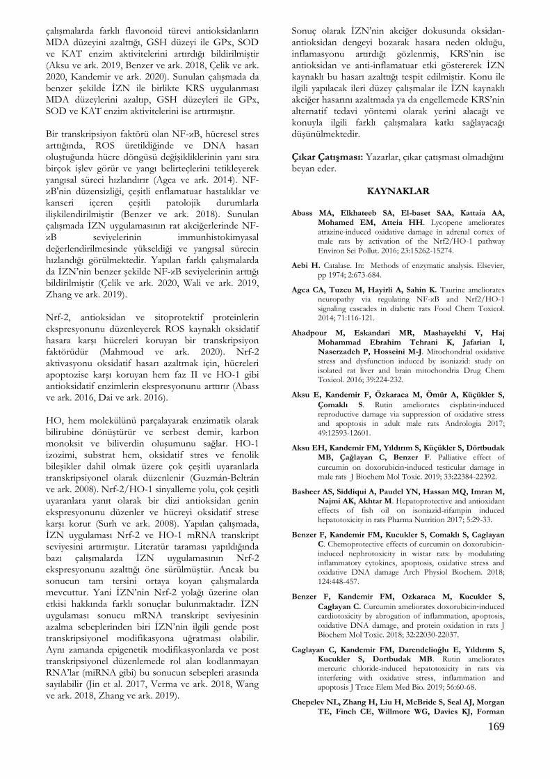
169
çalışmalarda farklı flavonoid türevi antioksidanların MDA düzeyini azalttığı, GSH düzeyi ile GPx, SOD ve KAT enzim aktivitelerini artırdığı bildirilmiştir (Aksu ve ark. 2019, Benzer ve ark. 2018, Çelik ve ark. 2020, Kandemir ve ark. 2020). Sunulan çalışmada da benzer şekilde İZN ile birlikte KRS uygulanması MDA düzeylerini azaltıp, GSH düzeyleri ile GPx, SOD ve KAT enzim aktivitelerini ise artırmıştır. Bir transkripsiyon faktörü olan NF-κB, hücresel stres arttığında, ROS üretildiğinde ve DNA hasarı oluştuğunda hücre döngüsü değişikliklerinin yanı sıra birçok işlev görür ve yangı belirteçlerini tetikleyerek yangısal süreci hızlandırır (Agca ve ark. 2014). NF-κB'nin düzensizliği, çeşitli enflamatuar hastalıklar ve kanseri içeren çeşitli patolojik durumlarla ilişkilendirilmiştir (Benzer ve ark. 2018). Sunulan çalışmada İZN uygulamasının rat akciğerlerinde NF-κB seviyelerinin immunhistokimyasal değerlendirilmesinde yükseldiği ve yangısal sürecin hızlandığı görülmektedir. Yapılan farklı çalışmalarda da İZN’nin benzer şekilde NF-κB seviyelerinin arttığı bildirilmiştir (Çelik ve ark. 2020, Wali ve ark. 2019, Zhang ve ark. 2019). Nrf-2, antioksidan ve sitoprotektif proteinlerin ekspresyonunu düzenleyerek ROS kaynaklı oksidatif hasara karşı hücreleri koruyan bir transkripsiyon faktörüdür (Mahmoud ve ark. 2020). Nrf-2 aktivasyonu oksidatif hasarı azaltmak için, hücreleri apoptozise karşı koruyan hem faz II ve HO-1 gibi antioksidatif enzimlerin ekspresyonunu arttırır (Abass ve ark. 2016, Dai ve ark. 2016). HO, hem molekülünü parçalayarak enzimatik olarak bilirubine dönüştürür ve serbest demir, karbon monoksit ve biliverdin oluşumunu sağlar. HO-1 izozimi, substrat hem, oksidatif stres ve fenolik bileşikler dahil olmak üzere çok çeşitli uyaranlarla transkripsiyonel olarak düzenlenir (Guzmán-Beltrán ve ark. 2008). Nrf-2/HO-1 sinyalleme yolu, çok çeşitli uyaranlara yanıt olarak bir dizi antioksidan genin ekspresyonunu düzenler ve hücreyi oksidatif strese karşı korur (Surh ve ark. 2008). Yapılan çalışmada, İZN uygulaması Nrf-2 ve HO-1 mRNA transkript seviyesini artırmıştır. Literatür taraması yapıldığında bazı çalışmalarda İZN uygulamasının Nrf-2 ekspresyonunu azalttığı öne sürülmüştür. Ancak bu sonucun tam tersini ortaya koyan çalışmalarda mevcuttur. Yani İZN’nin Nrf-2 yolağı üzerine olan etkisi hakkında farklı sonuçlar bulunmaktadır. İZN uygulaması sonucu mRNA transkript seviyesinin azalma sebeplerinden biri İZN’nin ilgili gende post transkripsiyonel modifikasyona uğratması olabilir. Aynı zamanda epigenetik modifikasyonlarda ve post transkripsiyonel düzenlemede rol alan kodlanmayan RNA’lar (miRNA gibi) bu sonucun sebepleri arasında sayılabilir (Jin et al. 2017, Verma ve ark. 2018, Wang ve ark. 2018, Zhang ve ark. 2019).
Sonuç olarak İZN’nin akciğer dokusunda oksidan-antioksidan dengeyi bozarak hasara neden olduğu, inflamasyonu artırdığı gözlenmiş, KRS’nin ise antioksidan ve anti-inflamatuar etki göstererek İZN kaynaklı bu hasarı azalttığı tespit edilmiştir. Konu ile ilgili yapılacak ileri düzey çalışmalar ile İZN kaynaklı akciğer hasarını azaltmada ya da engellemede KRS’nin alternatif tedavi yöntemi olarak yerini alacağı ve konuyla ilgili farklı çalışmalara katkı sağlayacağı düşünülmektedir. Çıkar Çatışması: Yazarlar, çıkar çatışması olmadığını beyan eder.
KAYNAKLAR
Abass MA, Elkhateeb SA, El-baset SAA, Kattaia AA,
Mohamed EM, Atteia HH. Lycopene ameliorates atrazine-induced oxidative damage in adrenal cortex of male rats by activation of the Nrf2/HO-1 pathway Environ Sci Pollut. 2016; 23:15262-15274.
Aebi H. Catalase. In: Methods of enzymatic analysis. Elsevier, pp 1974; 2:673-684.
Agca CA, Tuzcu M, Hayirli A, Sahin K. Taurine ameliorates neuropathy via regulating NF-κB and Nrf2/HO-1 signaling cascades in diabetic rats Food Chem Toxicol. 2014; 71:116-121.
Ahadpour M, Eskandari MR, Mashayekhi V, Haj Mohammad Ebrahim Tehrani K, Jafarian I, Naserzadeh P, Hosseini M-J. Mitochondrial oxidative stress and dysfunction induced by isoniazid: study on isolated rat liver and brain mitochondria Drug Chem Toxicol. 2016; 39:224-232.
Aksu E, Kandemir F, Özkaraca M, Ömür A, Küçükler S,
Çomaklı S. Rutin ameliorates cisplatin‐induced reproductive damage via suppression of oxidative stress and apoptosis in adult male rats Andrologia 2017; 49:12593-12601.
Aksu EH, Kandemir FM, Yıldırım S, Küçükler S, Dörtbudak MB, Çağlayan C, Benzer F. Palliative effect of
curcumin on doxorubicin‐induced testicular damage in male rats J Biochem Mol Toxic. 2019; 33:22384-22392.
Basheer AS, Siddiqui A, Paudel YN, Hassan MQ, Imran M, Najmi AK, Akhtar M. Hepatoprotective and antioxidant effects of fish oil on isoniazid-rifampin induced hepatotoxicity in rats Pharma Nutrition 2017; 5:29-33.
Benzer F, Kandemir FM, Kucukler S, Comaklı S, Caglayan C. Chemoprotective effects of curcumin on doxorubicin-induced nephrotoxicity in wistar rats: by modulating inflammatory cytokines, apoptosis, oxidative stress and oxidative DNA damage Arch Physiol Biochem. 2018; 124:448-457.
Benzer F, Kandemir FM, Ozkaraca M, Kucukler S,
Caglayan C. Curcumin ameliorates doxorubicin‐induced cardiotoxicity by abrogation of inflammation, apoptosis, oxidative DNA damage, and protein oxidation in rats J Biochem Mol Toxic. 2018; 32:22030-22037.
Caglayan C, Kandemir FM, Darendelioğlu E, Yıldırım S, Kucukler S, Dortbudak MB. Rutin ameliorates mercuric chloride-induced hepatotoxicity in rats via interfering with oxidative stress, inflammation and apoptosis J Trace Elem Med Bio. 2019; 56:60-68.
Chepelev NL, Zhang H, Liu H, McBride S, Seal AJ, Morgan TE, Finch CE, Willmore WG, Davies KJ, Forman

170
HJ. Competition of nuclear factor-erythroid 2 factors related transcription factor isoforms, Nrf1 and Nrf2, in antioxidant enzyme induction Redox Biol. 2013; 1:183-189.
Cho YJ, Park SB, Han M. Di-(2-ethylhexyl)-phthalate induces oxidative stress in human endometrial stromal cells in vitro Mol Cell Endocrinol. 2015; 407:9-17.
Choi YH. Activation of the Nrf2/HO-1 signaling pathway contributes to the protective effects of coptisine against oxidative stress-induced DNA damage and apoptosis in HaCaT keratinocytes Gen Physiol Biophys. 2019; 38:281.
Chowdhury A, Santra A, Bhattacharjee K, Ghatak S, Saha DR, Dhali GK. Mitochondrial oxidative stress and permeability transition in isoniazid and rifampicin induced liver injury in mice J Hepatol. 2006; 45:117-126.
Cicek E, Sutcu R, Gokalp O, Yilmaz HR, Ozer MK, Uz E, Ozcelik N, Delibas N. The effects of isoniazid on hippocampal NMDA receptors: protective role of erdosteine Mol Cell Biochem. 2005; 277:131-135.
Comakli S, Sağlam YS, Timurkan MÖ. Comparative detection of bovine herpesvirus-1 using antigen ELISA, immunohistochemistry and immunofluorescence methods in cattle with pneumonia Turk J Vet Anim Sci 2019; 43: 306-313.
Combrink M, du Preez I. Metabolomics describes previously unknown toxicity mechanisms of isoniazid and rifampicin Toxicol Lett. 2020; 332:104-110.
Cunha L, Rodrigues S, Rosa da Costa AM, Faleiro L, Buttini F, Grenha A. Inhalable chitosan microparticles for simultaneous delivery of isoniazid and rifabutin in lung tuberculosis treatment Drug Dev Ind Pharm.i 2019; 45:1313-1320.
Çelik H, Kandemir FM, Caglayan C, Özdemir S, Çomaklı S, Kucukler S, Yardım A. Neuroprotective effect of rutin against colistin-induced oxidative stress, inflammation and apoptosis in rat brain associated with the CREB/BDNF expressions Mol Biol Rep. 2020; 47:2023-2034.
Çelik H, Kucukler S, Çomaklı S, Özdemir S, Caglayan C, Yardım A, Kandemir FM. Morin attenuates ifosfamide-induced neurotoxicity in rats via suppression of oxidative stress, neuroinflammation and neuronal apoptosis NeuroToxicology 2020; 76:126-137.
Çomakli S, Özdemir S, Değirmençay Ş. Canine distemper virus induces downregulation of GABA A, GABA B, and GAT1 expression in brain tissue of dogs. Arch Virol 2020; 1-11.
Dai C, Li B, Zhou Y, Li D, Zhang S, Li H, Xiao X, Tang S. Curcumin attenuates quinocetone induced apoptosis and inflammation via the opposite modulation of Nrf2/HO-1 and NF-kB pathway in human hepatocyte L02 cells Food Chem Toxicol. 2016; 95:52-63.
Eftekhari A, Heidari R, Ahmadian E, Eghbal MA. Cytoprotective Properties of Carnosine against Isoniazid-Induced Toxicity in Primary Cultured Rat Hepatocytes Pharm Sci. 2018; 24:257.
Eldutar E, Kandemir FM, Kucukler S, Caglayan C. Restorative effects of Chrysin pretreatment on oxidant–antioxidant status, inflammatory cytokine production, and apoptotic and autophagic markers in acute
paracetamol‐induced hepatotoxicity in rats: An experimental and biochemical study J Biochem Mol Toxic. 2017; 31:21960-21966.
Endo T, Saito T, Nakayama M, Ohse H, Watanabe S, Tamai S, Hasegawa S. A case of isoniazid-induced
pneumonitis. Nihon Kokyuki Gakkai zasshi= the journal of the Japanese Respiratory Society. 1998; 36:100-105.
Guzmán-Beltrán S, Espada S, Orozco-Ibarra M, Pedraza-Chaverri J, Cuadrado A. Nordihydroguaiaretic acid activates the antioxidant pathway Nrf2/HO-1 and protects cerebellar granule neurons against oxidative stress Neurosci Lett. 2008; 447:167-171.
Han Mc, Durmuş As, Sağliyan A, Günay C, Özkaraca M, Kandemir FM, Çomaklı S, Öztopalan DF. Effects of Nigella sativa and Hypericum perforatum on wound healing Turk J Vet Anim Sci. 2017; 41: 99-105.
Jia Z-l, Cen J, Wang J-b, Zhang F, Xia Q, Wang X, Chen X-q, Wang R-c, Liu K-c, Zhang Y. Mechanism of isoniazid-induced hepatotoxicity in zebrafish larvae: Activation of ROS-mediated ERS, apoptosis and the Nrf2 pathway Chemo. 2019; 227:541-550.
Jin SW, Hwang YP, Choi CY, Kim HG, Kim SJ, Kim Y, Chung YC, Lee KJ, Jeong TC, Jeong HG. Protective effect of rutaecarpine against t-BHP-induced hepatotoxicity by upregulating antioxidant enzymes via the CaMKII-Akt and Nrf2/ARE pathways Food Chem Toxicol. 2017; 100:138-148.
Kandemir FM, Yıldırım S, Kucukler S, Caglayan C, Darendelioğlu E, Dortbudak MB. (b) Protective effects of morin against acrylamide-induced hepatotoxicity and nephrotoxicity: A multi-biomarker approach Food Chem Toxicol. 2020; 138:111190-111201.
Kandemir FM, Kucukler S, Eldutar E, Caglayan C, Gülçin İ. Chrysin protects rat kidney from paracetamol-induced oxidative stress, inflammation, apoptosis, and autophagy: a multi-biomarker approach. Sci Pharm. 2017; 85(1): 4.
Karakurt Y, Süleyman H, Keskin Cimen F, Tasli G, Ucak T, Icel E, Kurt N. The effects of lutein on optic nerve injury induced by ethambutol and isoniazid: an experimental study Cutan Ocul Toxicol. 2019; 38:136-140.
Kilic T, Ciftci O, Cetin A, Kahraman H. Preventive effect of chrysin on bleomycin-induced lung fibrosis in rats Inflammation. 2014; 37:2116-2124.
Kunichika N, Miyahara N, Kotani K, Takeyama H, Harada M, Tanimoto M. Pneumonitis induced by rifampicin Thorax. 2002; 57: 1000-1001.
Liang Z, Ni R, Zhou J, Mao S. Recent advances in controlled pulmonary drug delivery Drug Discov. Today. 2015; 20:380-389.
Livak KJ, Schmittgen TD. Analysis of relative gene expression data using real-time quantitative PCR and the 2− ΔΔCT method Methods 2001; 25:402-408.
Lowry OH, Rosebrough NJ, Farr AL, Randall RJ. Protein measurement with the Folin phenol reagent J Biol Chem. 1951; 193:265-275.
Mahmoud AM, Hussein OE, Hozayen WG, Bin-Jumah M, Abd El-Twab SM. Ferulic acid prevents oxidative stress, inflammation, and liver injury via upregulation of Nrf2/HO-1 signaling in methotrexate-induced rats Environ Sci Pollut Res Int. 2020; 27:7910-7921.
Matkovics B. Determination of enzyme activity in lipid peroxidation and glutathione pathways Lab Diag. 1988; 15:248-250.
Mehri S, Karami HV, Hassani FV, Hosseinzadeh H. Chrysin reduced acrylamide-induced neurotoxicity in both in vitro and in vivo assessments Iran Biomed J. 2014; 18:101.
Migita K, Umeno T, Miyagawa K, Izumi Y, Sasaki E, Kakugawa T, Ito M, Kinoshita A, Miyashita T.

171
Development of interstitial pneumonia in a patient with rheumatoid arthritis induced by isoniazid for tuberculosis chemoprophylaxis Rheumatol Int. 2012; 32:1375-1377.
Nishizawa Y, Yasui M, Yamamori C, Tagami A, Fujimura M, Nakao S. A case of successful desensitization therapy for isoniazid-induced pneumonitis Nihon Kokyuki Gakkai zasshi= the journal of the Japanese Respiratory Society. 2004; 42:649-654.
Ohta K, Nagase H. Drug-induced interstitial lung disease Nihon Rinsho. 2012; 70:517–520.
Organization WH. Global tuberculosis report 2013. WHO,
Park EJ, Kim YM, Park SW, Kim HJ, Lee JH, Lee D-U, Chang KC. Induction of HO-1 through p38 MAPK/Nrf2 signaling pathway by ethanol extract of Inula helenium L. reduces inflammation in LPS-activated RAW 264.7 cells and CLP-induced septic mice Food Chem Toxicol. 2013; 55:386-395.
Placer ZA, Cushman LL, Johnson BC. Estimation of product of lipid peroxidation (malonyl dialdehyde) in biochemical systems Anal Biochem. 1966; 16:359-364.
Plit ML, Theron AJ, Fickl H, Van Rensburg CEJ, Pendel S, Anderson R. Influence of antimicrobial chemotherapy and smoking status on the plasma concentrations of vitamin C, vitamin E, β-carotene, acute phase reactants, iron and lipid peroxides in patients with pulmonary tuberculosis Int J Tuberc Lung Dis. 1998; 2: 590-596.
Raghu R, Karthikeyan S. Zidovudine and isoniazid induced liver toxicity and oxidative stress: Evaluation of mitigating properties of silibinin Environ Toxicol Pharmacol. 2016; 46:217-226.
Ruan L-Y, Fan J-T, Hong W, Zhao H, Li M-H, Jiang L, Fu Y-H, Xing Y-X, Chen C, Wang J-S. Isoniazid-induced hepatotoxicity and neurotoxicity in rats investigated by 1H NMR based metabolomics approach Toxicol Lett. 2018; 295:256-269.
Schieber M, Chandel NS. ROS function in redox signaling and oxidative stress Curr Biol. 2014; 24:453-462.
Sedlak J, Lindsay RH. Estimation of total, protein-bound, and nonprotein sulfhydryl groups in tissue with Ellman's reagent Anal Biochem. 1968; 25:192-205.
Sun Y, Oberley LW, Li Y. A simple method for clinical assay of superoxide dismutase Clin Chem. 1988; 34:497-500.
Surh Y-J, Kundu JK, Na H-K. Nrf2 as a master redox switch in turning on the cellular signaling involved in the induction of cytoprotective genes by some chemopreventive phytochemicals Planta Med. 2008; 74:1526-1539.
Suzuki N, Ohno S, Takeuchi Y, Yamanaka K, Sugiyama Y, Kitamura S. A case of isoniazid (INH)-induced pneumonitis The Japanese journal of thoracic diseases 1992; 30:1563–1568.
Temel Y, Kucukler S, Yıldırım S, Caglayan C, Kandemir FM. Protective effect of chrysin on cyclophosphamide-induced hepatotoxicity and nephrotoxicity via the inhibition of oxidative stress, inflammation, and apoptosis. Naunyn-Schmiedeberg's Arch. Pharmacol. 2020; 393(3):325-337.
Verma AK, Yadav A, Singh SV, Mishra P, Rath SK. Isoniazid induces apoptosis: Role of oxidative stress and inhibition of nuclear translocation of nuclear factor (erythroid-derived 2)-like 2 (Nrf2) Life Sci. 2018; 199:23-33.
Wali AF, Mushtaq A, Rehman MU, Akbar S, Masoodi MH. Amelioration of Rifampicin and Isoniazid Induced Liver Oxidative Damage and Inflammation Response by
Propolis Extracts in Rodent Model Jou of Biolo Acti Prod from Nat 2019; 9:57-66.
Wang J, Luo W, Li B, Lv J, Ke X, Ge D, Dong R, Wang C, Han Y, Zhang C. 2018. Sagittaria sagittifolia polysaccharide protects against isoniazid-and rifampicin-induced hepatic injury via activation of nuclear factor E2-related factor 2 signaling in mice J Ethnopharmacol. 2018; 227:237-245.
Xiang N, Zhao C, Diao X, Han Q, Zhou H. Dynamic responses of antioxidant enzymes in pearl oyster Pinctada martensii exposed to di (2-ethylhexyl) phthalate (DEHP) Environ Toxicol Pharmacol. 2017; 54:184-190.
Yang Z, Guan Y, Li J, Li L, Li Z. Chrysin attenuates carrageenan-induced pleurisy and lung injury via activation of SIRT1/NRF2 pathway in rats Eur J Pharmacol. 2018; 836:83-88.
You H, Chen S, Mao L, Li B, Yuan Y, Li R, Yang X. The adjuvant effect induced by di-(2-ethylhexyl) phthalate (DEHP) is mediated through oxidative stress in a mouse model of asthma Food Chem Toxicol. 2014; 71:272-281.
Zhang D, Xiao Y, Lv P, Teng Z, Dong Y, Qi Q, Liu Z. Edaravone attenuates oxidative stress induced by chronic cerebral hypoperfusion injury: role of ERK/Nrf2/HO-1 signaling pathway Neurol Res. 2018; 40:1-10.
Zhang Q, Zhao Y, Talukder M, Han Y, Zhang C, Li X-N, Li J-L. Di (2-ethylhexyl) phthalate induced hepatotoxicity in quail (Coturnix japonica) via modulating the mitochondrial unfolded protein response and NRF2 mediated antioxidant defense Sci. Total Environ. 2019; 651:885-894.
Zhang Y, Li Y, Li J, Li B, Chong Y, Zheng G, Sun S, Feng F. SIRT1 alleviates isoniazid- induced hepatocyte injury by reducing histone acetylation in the IL-6 promoter region Int Immunopharmacol. 2019; 67:348-355.
Zhang Z, Guo Z, Zhan Y, Li H, Wu S. Role of histone acetylation in activation of nuclear factor erythroid 2-related factor 2/heme oxygenase 1 pathway by manganese chloride Toxicol Appl Pharmacol. 2017; 336:94-100.
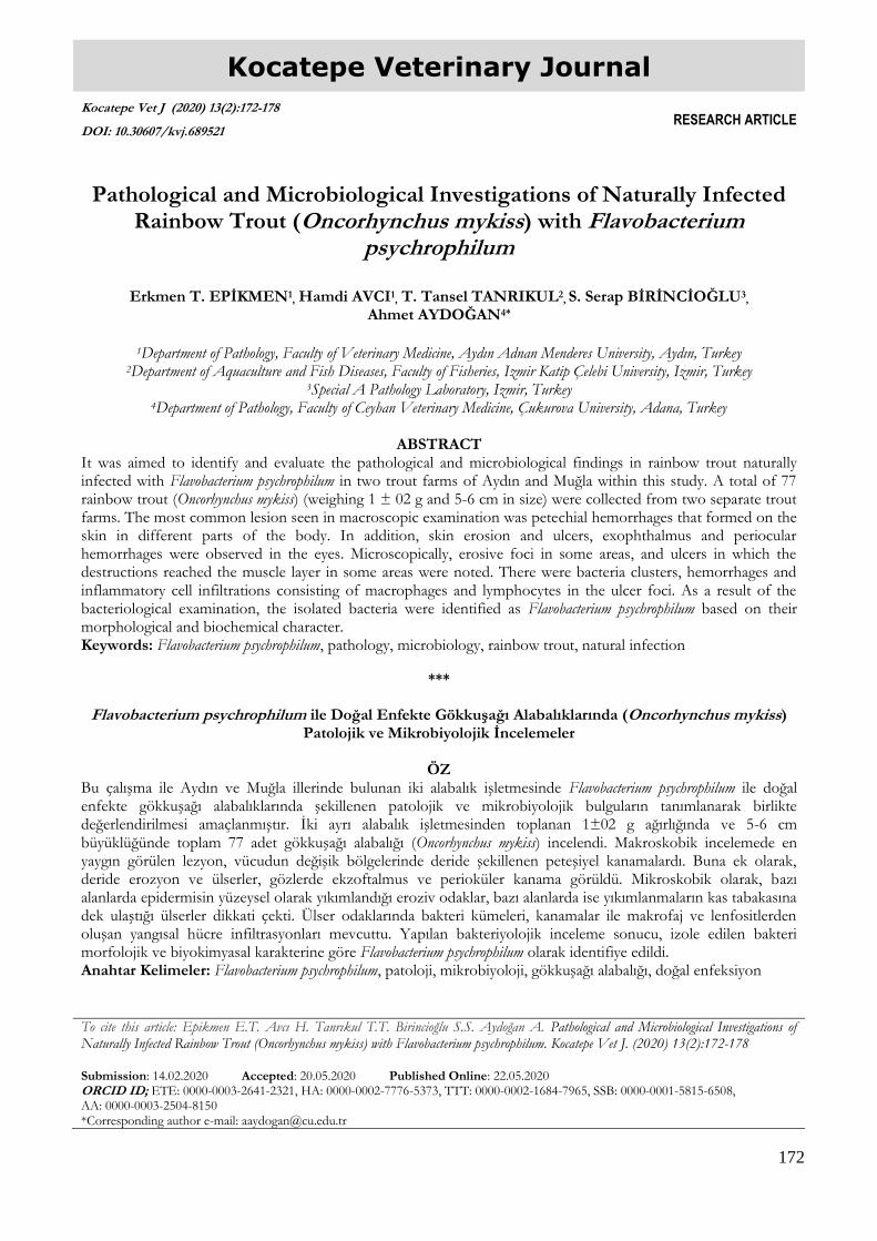
172
Kocatepe Veterinary Journal
Kocatepe Vet J (2020) 13(2):172-178
DOI: 10.30607/kvj.689521 RESEARCH ARTICLE
Pathological and Microbiological Investigations of Naturally Infected
Rainbow Trout (Oncorhynchus mykiss) with Flavobacterium psychrophilum
Erkmen T. EPİKMEN1
, Hamdi AVCI1, T. Tansel TANRIKUL2
, S. Serap BİRİNCİOĞLU3
,
Ahmet AYDOĞAN4*
1Department of Pathology, Faculty of Veterinary Medicine, Aydın Adnan Menderes University, Aydın, Turkey
2Department of Aquaculture and Fish Diseases, Faculty of Fisheries, Izmir Katip Çelebi University, Izmir, Turkey 3Special A Pathology Laboratory, Izmir, Turkey
4Department of Pathology, Faculty of Ceyhan Veterinary Medicine, Çukurova University, Adana, Turkey
ABSTRACT It was aimed to identify and evaluate the pathological and microbiological findings in rainbow trout naturally infected with Flavobacterium psychrophilum in two trout farms of Aydın and Muğla within this study. A total of 77 rainbow trout (Oncorhynchus mykiss) (weighing 1 ± 02 g and 5-6 cm in size) were collected from two separate trout farms. The most common lesion seen in macroscopic examination was petechial hemorrhages that formed on the skin in different parts of the body. In addition, skin erosion and ulcers, exophthalmus and periocular hemorrhages were observed in the eyes. Microscopically, erosive foci in some areas, and ulcers in which the destructions reached the muscle layer in some areas were noted. There were bacteria clusters, hemorrhages and inflammatory cell infiltrations consisting of macrophages and lymphocytes in the ulcer foci. As a result of the bacteriological examination, the isolated bacteria were identified as Flavobacterium psychrophilum based on their morphological and biochemical character. Keywords: Flavobacterium psychrophilum, pathology, microbiology, rainbow trout, natural infection
***
Flavobacterium psychrophilum ile Doğal Enfekte Gökkuşağı Alabalıklarında (Oncorhynchus mykiss)
Patolojik ve Mikrobiyolojik İncelemeler
ÖZ Bu çalışma ile Aydın ve Muğla illerinde bulunan iki alabalık işletmesinde Flavobacterium psychrophilum ile doğal enfekte gökkuşağı alabalıklarında şekillenen patolojik ve mikrobiyolojik bulguların tanımlanarak birlikte değerlendirilmesi amaçlanmıştır. İki ayrı alabalık işletmesinden toplanan 1±02 g ağırlığında ve 5-6 cm büyüklüğünde toplam 77 adet gökkuşağı alabalığı (Oncorhynchus mykiss) incelendi. Makroskobik incelemede en yaygın görülen lezyon, vücudun değişik bölgelerinde deride şekillenen peteşiyel kanamalardı. Buna ek olarak, deride erozyon ve ülserler, gözlerde ekzoftalmus ve perioküler kanama görüldü. Mikroskobik olarak, bazı alanlarda epidermisin yüzeysel olarak yıkımlandığı eroziv odaklar, bazı alanlarda ise yıkımlanmaların kas tabakasına dek ulaştığı ülserler dikkati çekti. Ülser odaklarında bakteri kümeleri, kanamalar ile makrofaj ve lenfositlerden oluşan yangısal hücre infiltrasyonları mevcuttu. Yapılan bakteriyolojik inceleme sonucu, izole edilen bakteri morfolojik ve biyokimyasal karakterine göre Flavobacterium psychrophilum olarak identifiye edildi. Anahtar Kelimeler: Flavobacterium psychrophilum, patoloji, mikrobiyoloji, gökkuşağı alabalığı, doğal enfeksiyon
To cite this article: Epikmen E.T. Avcı H. Tanrıkul T.T. Birincioğlu S.S. Aydoğan A. Pathological and Microbiological Investigations of Naturally Infected Rainbow Trout (Oncorhynchus mykiss) with Flavobacterium psychrophilum. Kocatepe Vet J. (2020) 13(2):172-178 Submission: 14.02.2020 Accepted: 20.05.2020 Published Online: 22.05.2020 ORCID ID; ETE: 0000-0003-2641-2321, HA: 0000-0002-7776-5373, TTT: 0000-0002-1684-7965, SSB: 0000-0001-5815-6508, AA: 0000-0003-2504-8150 *Corresponding author e-mail: [email protected]
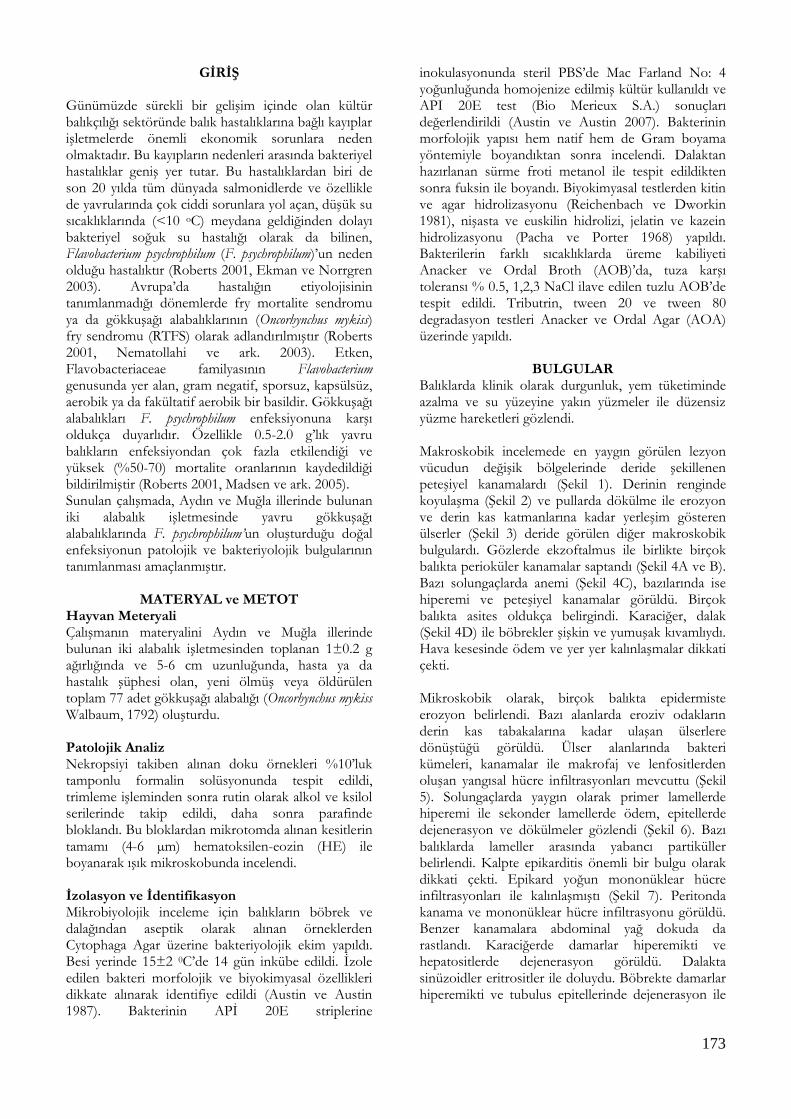
173
GİRİŞ Günümüzde sürekli bir gelişim içinde olan kültür balıkçılığı sektöründe balık hastalıklarına bağlı kayıplar işletmelerde önemli ekonomik sorunlara neden olmaktadır. Bu kayıpların nedenleri arasında bakteriyel hastalıklar geniş yer tutar. Bu hastalıklardan biri de son 20 yılda tüm dünyada salmonidlerde ve özellikle de yavrularında çok ciddi sorunlara yol açan, düşük su sıcaklıklarında (<10 oC) meydana geldiğinden dolayı bakteriyel soğuk su hastalığı olarak da bilinen, Flavobacterium psychrophilum (F. psychrophilum)’un neden olduğu hastalıktır (Roberts 2001, Ekman ve Norrgren 2003). Avrupa’da hastalığın etiyolojisinin tanımlanmadığı dönemlerde fry mortalite sendromu ya da gökkuşağı alabalıklarının (Oncorhynchus mykiss) fry sendromu (RTFS) olarak adlandırılmıştır (Roberts 2001, Nematollahi ve ark. 2003). Etken, Flavobacteriaceae familyasının Flavobacterium genusunda yer alan, gram negatif, sporsuz, kapsülsüz, aerobik ya da fakültatif aerobik bir basildir. Gökkuşağı alabalıkları F. psychrophilum enfeksiyonuna karşı oldukça duyarlıdır. Özellikle 0.5-2.0 g’lık yavru balıkların enfeksiyondan çok fazla etkilendiği ve yüksek (%50-70) mortalite oranlarının kaydedildiği bildirilmiştir (Roberts 2001, Madsen ve ark. 2005). Sunulan çalışmada, Aydın ve Muğla illerinde bulunan iki alabalık işletmesinde yavru gökkuşağı alabalıklarında F. psychrophilum’un oluşturduğu doğal enfeksiyonun patolojik ve bakteriyolojik bulgularının tanımlanması amaçlanmıştır.
MATERYAL ve METOT Hayvan Meteryali Çalışmanın materyalini Aydın ve Muğla illerinde bulunan iki alabalık işletmesinden toplanan 1±0.2 g ağırlığında ve 5-6 cm uzunluğunda, hasta ya da hastalık şüphesi olan, yeni ölmüş veya öldürülen toplam 77 adet gökkuşağı alabalığı (Oncorhynchus mykiss Walbaum, 1792) oluşturdu. Patolojik Analiz Nekropsiyi takiben alınan doku örnekleri %10’luk tamponlu formalin solüsyonunda tespit edildi, trimleme işleminden sonra rutin olarak alkol ve ksilol serilerinde takip edildi, daha sonra parafinde bloklandı. Bu bloklardan mikrotomda alınan kesitlerin tamamı (4-6 µm) hematoksilen-eozin (HE) ile boyanarak ışık mikroskobunda incelendi. İzolasyon ve İdentifikasyon Mikrobiyolojik inceleme için balıkların böbrek ve dalağından aseptik olarak alınan örneklerden Cytophaga Agar üzerine bakteriyolojik ekim yapıldı. Besi yerinde 15±2 0C’de 14 gün inkübe edildi. İzole edilen bakteri morfolojik ve biyokimyasal özellikleri dikkate alınarak identifiye edildi (Austin ve Austin 1987). Bakterinin APİ 20E striplerine
inokulasyonunda steril PBS’de Mac Farland No: 4 yoğunluğunda homojenize edilmiş kültür kullanıldı ve API 20E test (Bio Merieux S.A.) sonuçları değerlendirildi (Austin ve Austin 2007). Bakterinin morfolojik yapısı hem natif hem de Gram boyama yöntemiyle boyandıktan sonra incelendi. Dalaktan hazırlanan sürme froti metanol ile tespit edildikten sonra fuksin ile boyandı. Biyokimyasal testlerden kitin ve agar hidrolizasyonu (Reichenbach ve Dworkin 1981), nişasta ve euskilin hidrolizi, jelatin ve kazein hidrolizasyonu (Pacha ve Porter 1968) yapıldı. Bakterilerin farklı sıcaklıklarda üreme kabiliyeti Anacker ve Ordal Broth (AOB)’da, tuza karşı toleransı % 0.5, 1,2,3 NaCl ilave edilen tuzlu AOB’de tespit edildi. Tributrin, tween 20 ve tween 80 degradasyon testleri Anacker ve Ordal Agar (AOA) üzerinde yapıldı.
BULGULAR Balıklarda klinik olarak durgunluk, yem tüketiminde azalma ve su yüzeyine yakın yüzmeler ile düzensiz yüzme hareketleri gözlendi. Makroskobik incelemede en yaygın görülen lezyon vücudun değişik bölgelerinde deride şekillenen peteşiyel kanamalardı (Şekil 1). Derinin renginde koyulaşma (Şekil 2) ve pullarda dökülme ile erozyon ve derin kas katmanlarına kadar yerleşim gösteren ülserler (Şekil 3) deride görülen diğer makroskobik bulgulardı. Gözlerde ekzoftalmus ile birlikte birçok balıkta perioküler kanamalar saptandı (Şekil 4A ve B). Bazı solungaçlarda anemi (Şekil 4C), bazılarında ise hiperemi ve peteşiyel kanamalar görüldü. Birçok balıkta asites oldukça belirgindi. Karaciğer, dalak (Şekil 4D) ile böbrekler şişkin ve yumuşak kıvamlıydı. Hava kesesinde ödem ve yer yer kalınlaşmalar dikkati çekti. Mikroskobik olarak, birçok balıkta epidermiste erozyon belirlendi. Bazı alanlarda eroziv odakların derin kas tabakalarına kadar ulaşan ülserlere dönüştüğü görüldü. Ülser alanlarında bakteri kümeleri, kanamalar ile makrofaj ve lenfositlerden oluşan yangısal hücre infiltrasyonları mevcuttu (Şekil 5). Solungaçlarda yaygın olarak primer lamellerde hiperemi ile sekonder lamellerde ödem, epitellerde dejenerasyon ve dökülmeler gözlendi (Şekil 6). Bazı balıklarda lameller arasında yabancı partiküller belirlendi. Kalpte epikarditis önemli bir bulgu olarak dikkati çekti. Epikard yoğun mononüklear hücre infiltrasyonları ile kalınlaşmıştı (Şekil 7). Peritonda kanama ve mononüklear hücre infiltrasyonu görüldü. Benzer kanamalara abdominal yağ dokuda da rastlandı. Karaciğerde damarlar hiperemikti ve hepatositlerde dejenerasyon görüldü. Dalakta sinüzoidler eritrositler ile doluydu. Böbrekte damarlar hiperemikti ve tubulus epitellerinde dejenerasyon ile

174
birlikte sitoplazmalarında eozinofilik hyalin damlacıkları dikkati çekti (Şekil 8). Mikrobiyolojik olarak Cytophaga Agar üzerinde 10 günde saf sarı renkli koloniler üredi. Dalaktan hazırlanan sürme frotide çok miktarda ince uzun basiller görüldü. Sarı renkli bu koloniler bakterinin
identifiye edilmesi için biyokimyasal test besi yerlerine inokule edilerek fenotipik özellikleri saptandı. Bakterinin morfolojik ve biyokimyasal karakterlerine göre (Tablo 1 ve 2) hastalığın etkeni F. psychrophilum olarak identifiye edilmiştir.
Şekil 1: Flavobacterium psychrophilum enfeksiyonu, deride peteşiyel kanama (oklar) ve karın boşluğunda asites (okbaşı), gökkuşağı alabalığı Figure 1: Flavobacterium psychrophilum infection, petechial hemorrhage on the skin (arrows) and ascites in the abdominal cavity (arrowhead), rainbow trout
Şekil 2: Flavobacterium psychrophilum enfeksiyonu, deride renkte koyulaşma (A), kontrol (B), gökkuşağı alabalığı Figure 2: Flavobacterium psychrophilum infection, darkening in skin (A), control (B), rainbow trout
Şekil 3: Flavobacterium psychrophilum enfeksiyonu, deride derin ülser (oklar), gökkuşağı alabalığı Figure 3: Flavobacterium psychrophilum infection, deep ulcer on the skin (arrows), rainbow trout

175
Şekil 4: Flavobacterium psychrophilum enfeksiyonu, gözde bilateral ekzoftalmus (A), perioküler kanama (B), solungaçlarda anemi (C), dalakta şişkinlik, konjesyon ve çevresinde kanama (D) Figure 4: Flavobacterium psychrophilum infection, bilateral exophthalmus in the eye (A), periocular hemorrhage (B), anemia in the gills (C), swelling of the spleen, congestion and hemorrhage around it (D)
Şekil 5. Ülser ve yangısal hücre infiltrasyonu (ok), deri, H.E., X10 Figure 5. Ulcer and inflammatory cell infiltration (arrow), skin, H.E., X10
Şekil 6: Sekonder lamellerde ödem (ok başı) ve telangiektazi (ok), solungaç, H.E., X20 Figure 6. Edema (arrowhead) and telangiectasia (arrow) in secondary lamellae, gill, H.E., X20
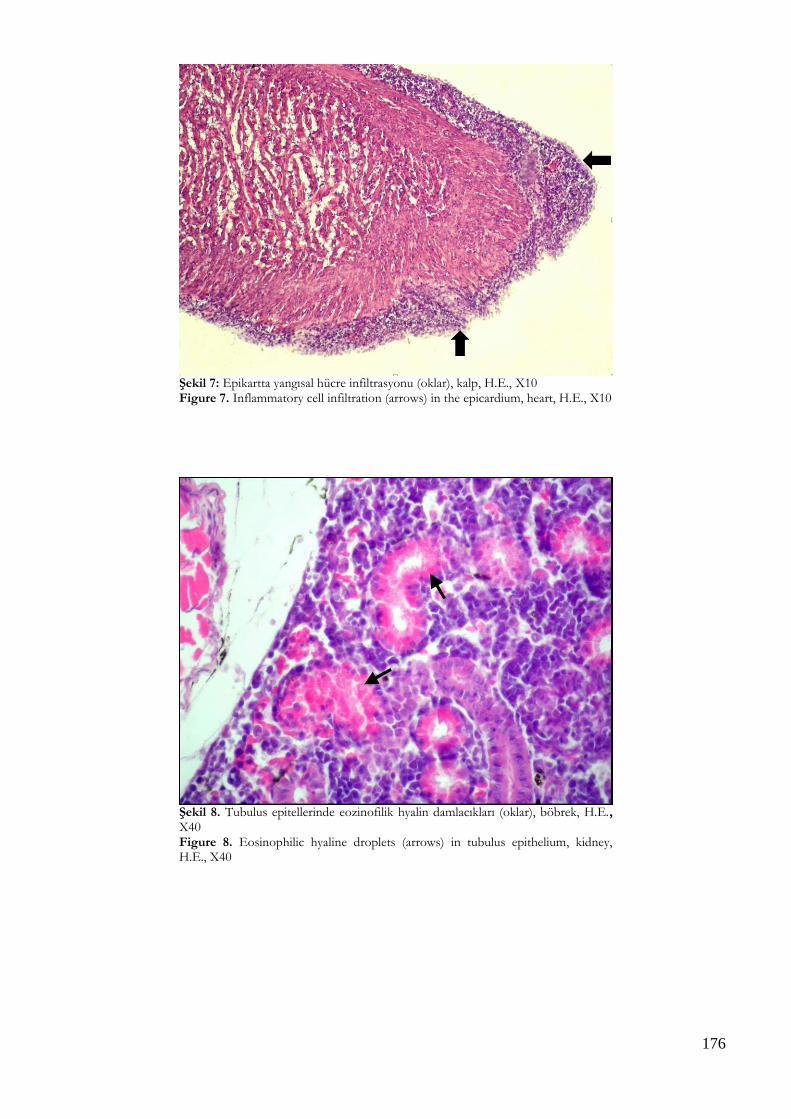
176
Şekil 7: Epikartta yangısal hücre infiltrasyonu (oklar), kalp, H.E., X10 Figure 7. Inflammatory cell infiltration (arrows) in the epicardium, heart, H.E., X10
Şekil 8. Tubulus epitellerinde eozinofilik hyalin damlacıkları (oklar), böbrek, H.E., X40 Figure 8. Eosinophilic hyaline droplets (arrows) in tubulus epithelium, kidney, H.E., X40

177
Tablo 1. Alabalıklardan izole edilen Flavobacterium psycrophilum’un morfolojik ve biyokimyasal özellikleri Table 1. Morphological and biochemical properties of Flavobacterium psycrophilum isolated from rainbow trouts
Sonuç Hidrolizasyon Sonuç Üreme Sonuç
Sarı Pigmentli Koloni + Eskulin - 4 0C +
Gliding Hareket + Kitin - 250C -
Gram Boyama - Agar - 370C -
O/F Metabolizma O Jelatin + %0,5 NaCl +
Katalaz + Nişasta - %1 NaCl -
Oksidaz - Kazein + %2 NaCl -
İndol - Tributirin + %3 NaCl -
Nitrat - Tween 20 + %1 Tripton +
H2S - Tween 80 +
Tablo 2. Alabalıklardan izole edilen Flavobacterium psycrophilum’un API 20E identifikasyon sistemi ile belirlenen biyokimyasal özellikleri Table 2. Biochemical properties of Flavobacterium psycrophilum isolated from rainbow trouts determined by API 20E identification system
Sonuç Şekerden asit üretimi Sonuç
ONPG - Glukoz -
ADH - Mannitol -
LDH - İnositol -
ODC - Sorbitol -
Sitrat - Ramnoz -
H2S - Sukroz -
Üre - Melibioz -
TDA - Amigdalin -
İndol - Arabinoz -
VP - Oksidaz -
Jelatin +
TARTIŞMA F. psychrophilum’un neden olduğu bakteriyel soğuk su hastalığı, dünyanın birçok yerinde bulunan alabalık çiftliklerinde hastalık salgınlarına ve önemli ekonomik sorunlara yol açmaktadır (Roberts 2001, Nematollahi ve ark. 2003). Gökkuşağı alabalıklarında, F. psychrophilum enfeksiyonunda klinik olarak anoreksi, letarji, deri renginde koyulaşma, asites, bilateral ekzoftalmus ve perioküler kanama gibi bulguların görüldüğü bildirilmiştir (Roberts 2001, Nematollahi ve ark. 2003, Yıldırım ve Özer 2010). Çalışmada durgunluk, yem tüketiminde azalma ve su yüzeyine yakın yüzmeler ile düzensiz yüzme hareketleri şeklinde saptanan klinik bulgular önceki bildirimlerle uyum sağlamış, sıklıkla bildirilen iskelet lezyonlarına bu araştırmada rastlanmamıştır (Madsen ve ark. 2005, Kubilay ve ark. 2009). Pek çok literatürde
belirtildiğinin aksine ise karaciğerde solgunluk yerine konjesyon ve şişkinlik belirlenmiştir (Roberts 2001, Ekman ve Norrgren 2003, Yıldırım ve Özer 2010). Sunulan çalışmada dikkati çeken hava keselerinde ödem ve kalınlaşmalara ise önceki çalışma ve raporlarda rastlanmamıştır. Değişik çalışmalarda deride şekillendiği bildirilen tek taraflı ve derin ülser odakları sunulan çalışmada da yaygın olarak saptanmıştır (Madetoja ve Wiklund 2002, Nematollahi ve ark. 2003). Bu ülserlerdeki yoğun bakteri kümelerinin varlığı, derinin portantre olarak rol oynadığı ve bakterilerin suya katılımına kaynak oluşturduğu şeklinde yorumlanmıştır. Bazı çalışmalarda böbrekte tubulus epitellerinde tanımlanan intrasitoplazmik eozinofilik hyalin damlacıkları sunulan çalışmada da yaygın olarak

178
görülmüş, bu bulgular böbreklerdeki proksimal tubulus epitellerinin dejenerasyonlarının bir sonucu olarak değerlendirilmiştir (Ostland ve ark. 2000, Madetoja ve Wiklund 2002, Nematollahi ve ark. 2003). Dalakta konjesyon, kırmızı ve beyaz pulpada ödematöz değişiklikler, yaygın hemoraji, splenomegali ve nekroz gibi patolojik bulguların en belirgin olarak gökkuşağı alabalıklarında görüldüğü bildirilmiş (Roberts 2001, Ekman ve Norrgren 2003), bu çalışmada ise dalakta makroskobik olarak şişkinlik ve yumuşama; mikroskobik olarak ise sinüzoidlerin eritrositler ile dolu olduğu görülmüştür. Bazı kronik enfeksiyon vakalarında kalpte perikarditis görüldüğü belirtilmiş (Roberts 2001) ve sunulan çalışmada epikarditis en önemli histopatolojik bulgulardan biri olarak kaydedilmiştir. Balıklarda yaygın olarak görülen hiperemi ve kanamalar da kalp yetmezliği ile ilişkilendirilmiş ve epikarditis görülen balıklarda bunun birincil ölüm nedeni olduğu sonucuna varılmıştır. F. psychrophilum enfeksiyonunda alabalıklarda solungaçlarda anemiye bağlı solgunluk, hiperemi ve peteşiyel kanama gibi bulguların görüldüğü bildirilmiş (Roberts 2001, Nematollahi ve ark. 2003), sunulan çalışmada da solungaçlarda yaygın olarak primer lamellerde hiperemi ile sekonder lamellerde ödem, epitellerde dejenerasyon ve dökülmeler belirlenmiştir. Bilinen solungaç lezyonlarının yanı sıra solungaçlarda bol miktarda yabancı partikülün bulunması, su kirliliği ile hastalığın şiddetini ve mortalite oranını artıran bir faktör olarak yorumlanmıştır. Bu durum, su kirliliğinin, enfeksiyonun yayılması ile patogenezinde oynadığı rol (Roberts 2001) ve işletmeye verdiği zararın oldukça önemli olduğunu ortaya koymuştur. Hastalığın teşhisinde patolojik incelemelerin yanında mikrobiyolojik izolasyon ve identifikasyon da önemli yer tutmaktadır. Özellikle etkenin izolasyonunda klasik agarlara göre Cytophaga Agar daha sık kullanılmakta, bu agarda kenarları ince ve parlak sarı kolonilerin ürediği dikkati çekmektedir (Nematollahi ve ark. 2003). Bu çalışmada da Cytophaga Agar üzerinde 10 günde saf olarak sarı renkli koloniler üremiş ve etkenin morfolojik ve biyokimyasal karakterleri incelendiğinde, etken F. psychrophilum olarak identifiye edilmiştir.
SONUÇ Sonuç olarak, bu çalışmada, iki ayrı alabalık işletmesinde ortaya çıkan F. psychrophilum enfeksiyonu patolojik bulguları ve bakteriyolojik analizleri ile tanımlanmıştır. Yavru alabalıklarda doğal olarak şekillenen ve bu enfeksiyonun ortalama 1±02 g ağırlığındaki yavru balıklarda klinik olarak ekzoftalmus, makroskobik ve mikroskobik olarak
erozyon, ülser ve yine mikroskobik olarak epikarditis ile seyrettiği düşünüldüğünde; benzer bulguların işletmelerde fark edilmesi durumunda, mortalite oranı oldukça yüksek olabilen bu hastalığın göz önüne alınarak buna ilişkin koruma ve kontrol önlemlerinin alınması gerektiği kanısına varılmıştır. Çıkar Çatışması: Yazarlar, çıkar çatışması olmadığını beyan eder.
KAYNAKLAR
Austin D, Austin DA. Bacterial fish pathogens: Disease of
farmed and wild fish, Ellis Horwood Ltd. 1987.
Austin D, Austin DA. Bacterial fish pathogens: Diseases of farmed and wild fish, 4th Ed., Springer and Praxis Publishing, Chichester, UK. 2007.
Boyacıoğlu M, Akar F. Isolation of Flavobacterium psychrophilum causing rainbow trout fry syndrome and determination of an effective antibacterial treatment in rainbow trout (Oncorhynchus mykiss) fry. Kafkas Univ Vet Fak Derg. 2012; 18 (2): 197-203.
Ekman E, Norrgren L. Pathology and immunohistochemistry in three species of salmonids after experimental infection with Flavobacterium psychrophilum. J Fish Dis. 2003; 26: 529-538.
Kubilay A, Altun S, Didinen BI, Ekici S, Diler Ö. Gökkuşağı alabalığı (Oncorhynchus mykiss) işletmelerinde Flavobacterium psychrophilum izolasyonu. Kafkas Univ Vet Fak Derg. 2009, 15: 709-715.
Madetoja J, Wiklund T. Detection of the fish pathogen Flavobacterium psychrophilum in water from fish farms. Syst Appl Microbiol. 2002; 25: 259-266.
Madsen L, Moller JD, Dalsgaard I. Flavobacterium psychrophilum in rainbow trout (Oncorhynchus mykiss Walbaum) hatcheries: Studies on broodstock, eggs, fry and environment. J Fish Dis. 2005; 28: 39-47.
Nematollahi A, Decostere A, Pasmans F, Haesebrouck F. Flavobacterium psychrophilum infections in salmonid fish. J Fish Dis. 2003; 26: 563-574.
Ostland VE, Byrne PJ, Hoover G, Ferguson HW. Necrotic myositis of rainbow trout Oncorhynchus mykiss (Walbaum): Proteolitic characteristics of a crude extracellular prepation from Flavobacterium psychrophilum. J Fish Dis. 2000; 23: 329-336.
Pacha RE, Porter S. Characteristics of Myxobacteria Isolated from the Surface of Freshwater Fish. Appl Microbiol. 1968; 16: 1901-1906.
Reichenbach H, Dworkin M. The order myxobacterales, In: The Prokaryotes, Ed; Starr MP, Stolp H, Trüper HG, Balows A, Schlegel HG, Springer-Verlag, Berlin, Germany. 1981; pp. 356-379.
Roberts RJ. Fish Pathology, W.B. Saunders. 2001; pp. 299-304.
Yıldırım S, Özer S. Mersin ili çağlarca köyündeki gökkuşağı alabalığı (Oncorhynchus mykiss, walbaum, 1792) kuluçkahanelerinde Flavobacterium spp. varlığı. J Fish Sci. 2010; 4 (1): 112-122.
179
Kocatepe Veterinary Journal
Kocatepe Vet J (2020) 13(2):179-184
DOI: 10.30607/kvj.698697 RESEARCH ARTICLE
Effect of Vitamin A, D3, E Treatment on Fertility in the Pırlak Sheep
Muhammed Kürşad BİRDANE1*, Fatih AVDATEK2
1Afyon Kocatepe University, Faculty of Veterinary Medicine, Obstetrics and Gynaecology Department, 03200,
Afyonkarahisar,Türkiye 2Afyon Kocatepe University, Faculty of Veterinary Medicine, Reproduction and Artificial Insemination Department, 03200,
Afyonkarahisar,Türkiye
ABSTRACT In this study, the effects of vitamins A, D3 and E on fertility were investigated in the Pırlak sheep, whose estrus was synchronized in July, the hottest month of the region. Multiparous 80 Pırlak sheep in the same herd were used as animal materials. The estrus synchronization of the sheep were performed with 20 mg chronolone impregnated intravaginal sponge (Chronogest CR®) for 11 days and 480 IU intramuscular eCG (Chrono-Gest / PMSG® 6000 IU / 25 ml) at the time of sponge removal. Vitamin A (300.000 IU), D3 (100.000 IU) and E (50 mg) combination (Vigantol-E®1ml) was injected intramuscularly at the time of mating in Group 1 (n=40), while Group 2 (n=40) was not applied. The estrus, conception and litter size were determined as 92,5%, 87,5%, 1,54 in Group 1 and 90%,75% and 1,37 in Group 2 respectively and there was no statistical difference between in the groups (P> 0.05). As a result, it was determined in July that the desired levels of reproductive efficiency can be obtained with the 11 day progesterone+eCG synchronization method in Pırlak breed sheep. It was concluded that different studies should be conducted to reveal the effects of vitamin A, D3, E and application on fertility parameters more clearly. Keywords: Chronolone sponge, Multiparous sheep, Hot stress, July, Vitamin A-D3-E, Litter size.
***
Pırlak Koyunlarında Vitamin A, D3, E Uygulamasının Fertilite Üzerine Etkisi
ÖZ
Bu çalışmada bölgenin en sıcak ayı olan Temmuz ayında östrüsleri senkronize edilen Pırlak ırkı koyunlarda vitamin A, D3 ve E’nin fertilite üzerine etkileri incelendi. Hayvan mataryeli olarak aynı sürü içerisinde bulunan daha önce doğum yapmış 80 Pırlak ırkı koyun kullanıldı. Koyunların östrüs senkronizasyonu 11 gün süreyle 20 mg kronolon içeren vagina içi sünger (Chronogest CR®) ve sünger çıkarılma anında 480 IU kas içi eCG (Chrono-Gest/PMSG® 6000 IU/25 ml) uygulamaları ile yapıldı. Çiftleşme anında Grup 1’e (n=40) vitamin A (300.000 IU), D3 (100.000 IU) ve E (50 mg) kombinasyonu (Vigantol-E®, 1ml) kas içi enjekte edilirken Grup 2’ye (n=40) uygulama yapılmadı. Östrüs, gebe kalma ve gebelik başına düşen yavru sayıları sırasıyla Grup 1’de %92,5, % 87,5, 1,54 ve Grup 2’de ise % 90, %75 ve 1,37 olarak belirlendi ve gruplar arasında istatistiki fark bulunmadı (P>0,05). Sonuç olarak Temmuz ayında Pırlak ırkı koyunlarda 11 gün süreli progesteron+eCG senkronizasyon yöntemi ile istenilen düzeylerde reprodüktif verimin elde edilebileceği belirlendi. Vitamin A, D3, E uygulamasının fertilite parametreleri üzerindeki etkilerini daha net ortaya koyabilmek için farklı çalışmaların yapılması gerektiği sonucuna varılmıştır. Anahtar Kelimeler: Kronolon sünger, Multipar koyun, Sıcaklık stresi, Temmuz ayı, Vitamin A-D3-E, Yavru verimi. To cite this article: Birdane M.K. Avdatek F. Effect of Vitamin A, D3, E Treatment on Fertility in the Pırlak Sheep. Kocatepe Vet J. (2020) 13(2):178-184 Submission: 04.03.2020 Accepted: 17.05.2020 Published Online: 22.05.2020
ORCID ID; MKB: 0000-0002-9683-4979, FA: 0000-0003-2345-8826 *Corresponding author e-mail: [email protected]

180
GİRİŞ Koyunların mera ve çayırları en iyi şekilde değerlendirebilmesi, yılın her döneminde kullanabilme yeteneklerinin olması ve yetiştiricilik maliyetinin düşük olması nedeni ile ülkemiz hayvancılığında yetiştiriciliği tercih edilmektedir (Aksoy ve Yavuz 2012). Türkiye’de Türkiye İstatistik Kurumuna (TÜİK) ait yıllık hayvansal üretim verileri incelendiğinde 1990’lı yıllarda yaklaşık 40 milyon baş olan koyun sayısının 2010 yılına kadar neredeyse yarı yarıya düşmüş olduğu, son 10 yılda ise tekrar önem kazanarak 38 milyon başa kadar yükseldiği görülmektedir (Anonim 2019). Koyunlarda gün ışığına maruz kalma süresinin uzadığı aylarda ovulasyonu tetikleyen melatonin ve gonadotropinlerin salınım sıklığının azalması sonucu sürüde gebe kalma oranı düşmektedir (Goodman ve Inskeep 2006). Bu nedenle koyunlardan yaşamları boyunca elde edilen yavru veriminin yükseltilmesi için inaktif ovaryumların bulunduğu aylarda östrüs sikluslarında meydana gelen hormonal değişiklikler esas alınarak, progesteron, GnRH, eCG ve melatonin uygulamaları ile reprodüksiyonun kontrolü gerçekleştirilmektedir (Abecia ve ark. 2011). Ovaryum ve uterus fonksiyonları üzerine özellikle A ve E vitaminlerinin etkilerinin olduğu, ayrıca bu vitaminlerin çiftleşme dönemi ve ortam sıcaklığının yüksek olduğu çeşitli stres faktörlerine karşı mücadele etmek için vücudun antioksidan mekanizmalarında görev yaptığı bildirilmektedir (Chauhan ve ark. 2014). Pırlak ırkı Türkiye’nin İç Anadolu ve Batı Akdeniz bölgelerinde kötü çevre şartları ve hastalıklara karşı dayanıklı olmaları nedeni ile yetiştiriciler tarafından tercih edilen koyun ırklarının arasında yer almaktadır (Anonim 2020). Pırlak ırkına ait özellikle senkronizasyon sonu elde edilen reprodüktif parametrelere ilişkin veri oldukça sınırlı sayıdadır. Pek çok araştırıcı küçük ve büyük ruminantlarda reprodüktif performansı arttırmak için peripartum dönemde meydana gelen metabolik değişiklikler nedeniyle vitaminlerin yavru canlılığı ve doğum sonrası üreme aktivitesi üzerine olan etkilerini ortaya koymaya çalışmışlardır. Bu çalışmanın amacı Temmuz ayında östrüsleri senkronize edilmiş olan Pırlak ırkı koyunlara ovulasyon ve gebeliğin ilk dönemlerinde üreme ve sıcaklık stresi üzerine pozitif etkilerinin olabileceği düşünülen A, D3 ve E vitamin karışımının reprodüktif performans üzerine olan etkilerini araştırmaktır.
MATERYAL ve METOD Bu çalışmada Afyonkarahisar ilinde halk elinde bakım ve beslemesi yapılan günde iki sefer meraya çıkarılan ve diğer zaman dilimlerinde kapalı ağıl içerisinde
tutulan Pırlak ırkı koyunlar kullanıldı. Araştırmaya Afyon Kocatepe Üniversitesi Hayvan Deneyleri Yerel Etik Kurulu tarafından onay (49533702/321) verildi. Sürü içerisinden en az bir kere doğum yapmış 20-42 ay yaş ve yaklaşık 40-50 kg canlı ağırlığına sahip laktasyonda olmayan 80 baş Pırlak ırkı koyun çalışma için seçildi. En yüksek sıcaklık ortalamasına sahip Temmuz ayı içerisinde birer hafta ara ile her hafta gruplarda eşit sayıda koyun olacak şekilde 80 baş koyunun senkronizasyonu tamamlandı. Koyunların östrüs senkronizasyonu 11 gün süreyle 20 mg kronolon içeren vagina içi sünger (Chronogest CR®) ve sünger çıkarılma anında 480 IU kas içi eCG (Chrono-Gest/PMSG® 6000 IU/25 ml) uygulamaları ile yapıldı. Enjeksiyonu takip eden yaklaşık 48. saatten itibaren koç katımı gerçekleştirilerek 8 koyun için bir baş Pırlak koçu kullanılarak elde aşım yöntemi uygulandı. Çiftleşme anında Grup 1’e (n=40) vitamin A (300.000 IU), vitamin D3 (100.000 IU) ve vitamin E (50 mg) kombinasyonu (Vigantol-E®,1ml/koyun) kas içi enjekte edilirken Grup 2’ye (n=40) uygulama yapılmadı. Koçlar elde aşım yapıldıktan sonra bir gün boyunca sürüde tutularak koyunlarla çiftleşmesine izin verildi. Gebelik oranı çiftleşmeyi takip eden 30. günde sırt üstü pozisyonda transrektal ultrasonografi (Tringa Linear 5/7.5 MHz) yöntemi kullanılarak fötusların görülmesi ile belirlendi. İlk ve ikinci hafta sünger uygulama ve çıkarılma günleri, östrüs ve ilerleyen günlerdeki sıcaklık değerleri bu iki uygulamanın belirtilen günlerde tespit edilen sıcaklık değerlerinin ortalamalarının alınması ile belirlendi. Günlük maksimum, minimum ve ortalama sıcaklık verileri (°C) ile nem değerleri Orman ve Su İşleri Bakanlığı Meteoroloji Genel Müdürlüğü’nden elde edildi. Gruplar arasındaki reprodüktif parametrelerin istatistiksel farklılıkları SPSS (16.0) programında Chi-square testi kullanılarak belirlendi (P<0,05). Reprodüktif parametreler aşağıdaki yöntemler kullanılarak belirlendi. Östrüs oranı: çiftleşen koyun sayısı/toplam koyun sayısı x 100 Gebelik oranı: gebe kalan koyun sayısı / toplam koyun sayısı x 100 Tekiz doğum oranı: bir adet kuzulayan koyun sayısı / doğum yapan koyun sayısı x 100 Çoklu doğum oranı: ≥ iki adet kuzulayan koyun sayısı / doğum yapan koyun sayısı x 100 Gebe kalan koyun başına düşen yavru sayısı: doğan kuzu sayısı / doğum yapan koyun sayısı
BULGULAR Gruplarda eCG uygulamasını takip eden 48.saatten itibaren 8 saatlik bir zaman diliminde Grup 1’de 3 ve Grup 2’de ise 4 koyun çiftleşmeyi kabul etmeyerek östrüste olmadıkları anlaşıldı. Çiftleşmeyi kabul etmeyen bu koyunların gebelik muayenesinde fötal
181
yapıya rastlanılmadı. Grup 1’de tüm koyunların 35’i, Grup 2’de ise koyunların 30’unun gebe kaldıkları belirlendi. Her iki grupta gebe kalan koyunların tamamı doğurarak kuzulama oranı %100 olarak tespit edildi. Grup 1’de ve Grup 2’de 20’şer koyun tek kuzuladı. Grup 1’de 12 koyun ikiz, 2 koyun üçüz ve bir koyunda dördüz kuzuladı. Grup 2’de ise 9 koyun ikiz, 1 koyun ise üçüz kuzuladı. Gebe kalan koyun başına düşen yavru sayısı ise Grup 1 ve Grup 2’de sırasıyla 1.54 (54/35) ve 1,37 (41/30) olarak belirlendi (Tablo 1). Tüm bulgularda iki grup arasında herhangi bir istatistiki önem bulunamadı (P>0,05). Çalışmanın
yapıldığı yılda gün içinde en yüksek sıcaklık ortalaması 32,4 ± 3,6°C (24,4-38,7°C) ve gün içindeki sıcaklık ortalaması 25,8 ± 2,6°C (20-31°C) ile Temmuz ayı olarak tespit edildi. İlk ve ikinci hafta yapılan senkronizasyon günlerinde östrüs ve takip eden ilk 7 günün en düşük sıcaklık ortalaması 17,8 ± 1,9°C (14,7-21,7°C), en yüksek sıcaklık ortalaması 32,8 ± 3,7°C (28,8-38,7°C) ve ortalama sıcaklık ise 25,4 ± 3°C (20,6-31°C) olarak tespit edildi (Tablo 2). Östrüs ve takip eden ilk 7 günün günlük ortalama nem değerleri ise % 46,5 olarak kaydedildi.
Tablo 1. Gruplara göre elde edilen fertilite parametreleri (P>0,05) Table 1. Fertility parameters obtained by groups (p>0,05 )
Fertilite Parametreleri Grup1 (vitamin) Fertilite parametrelerini hesaplama
yöntemi
Grup2 Fertilite parametrelerini hesaplama
yöntemi
Koyun sayısı (n) 40 40 Östrüs Oranı(%) 92,5 (37/40 x100) 90 (36/40 x100) Gebelik oranı(%) 87,5 (35/40 x100) 75 (30/40 x100) Gebe kalan koyun başına düşen yavru sayısı
1,54 (54/35) 1,37 (41/30)
Tekiz doğum oranı (%) 57,1 (20/35 x100) 66,7 (20/30 x100) Çoklu doğum oranı 42,9 (15/35 x100) 33,3 (10/30 x100) İkiz (%) 34,2 (12/35 x100) 30 (9/30 x100) Üçüz (%) 5,71 (2/35 x100) 3,33 (1/30 x100) Dörtüz (%) 2,86 (1/35 x100) 0 (0/30 x100)
Tablo 2. Östrüs ve takip eden ilk 7 günün sıcaklık ortalamaları Table 2. Estrus and temperature means of the following 7 days
Gün içinde en düşük °C 17,8 ± 1,9 (14,7-21,7)
Gün içinde en yüksek °C 32,8 ± 3,7 (28,8-38,7)
Gün Ortalama °C 25,4 ± 3 (20,6-31)
TARTIŞMA
Pırlak ırkı koyunlarda yapılan senkronizasyon çalışmalarında, Ocak-Mart aylarında iki farklı senkronizasyon çalışması yapılmış, 11-12 gün süreli progesteron+eCG metodu ile elde edilen östrüs oranları % 92-97, gebelik oranları %40-41, gebe kalan koyun başına düşen yavru sayıları 1,20-1,36 ve çoklu doğum oranları ise % 20-36,5 olarak tespit edilmiştir (Algan ve ark. 2017, Kuru ve ark. 2017). Araştırıcılar süngerlerin uzaklaştırılmasını takip eden ortalama 36-45. saatlerde östrüslerin başladığı, 24 saat kadar devam ettiğini belirtmişlerdir. Bu bulgular araştırmamızda elde ettiğimiz östrüs bulguları ile benzerlik göstermektedir. Bu çalışmalarda elde edilen düşük gebelik oranı ve yavru veriminin ana nedeni çalışmalarda kullanılan koyunların laktasyonda olmaları ve mevsim farklılığına bağlı olabilir. Çünkü mevsim, laktasyon durumu veya laktasyona bağlı yetersiz beslenme, ovulasyon ve ovulasyon sonrası
embriyonun yaşamasını etkileyen hipofiz, hipotalamus ve ovaryum fonksiyonlarını etkileyebilmektedir (Goodman ve Inskeep 2006, Goff ve ark. 2014). Koyunlarla ilgili senkronizazyon çalışmalarından elde edilen reprodüktif bulgular incelendiğinde birbirleri ile benzerlik veya farklılık göstermektedir. Reprodüktif parameteler ırk (Fair ve ark. 2005), senkronizasyon yöntemi (Martemucci ve D'Alessandro 2011), mevsim (Hashem ve ark. 2011), progestagen analoğu (Hashami ve ark. 2006) ve uygulama süresi (Karaca ve ark. 2009), eCG kullanımı (Martinez-Ros ve ark. 2019) vücut kondisyon skoru (Fukui ve ark. 2010), sıcaklık stresi (Kumar ve ark. 2017) gibi faktörlerden etkilenmesi senkronizasyon araştırmalarında elde edilen farklı bulguların nedeni olabilmektedir. Yavru verimi yüksek ırklara yüksek konsantre yem verilmesinin ikizlik oranı ile pozitif korelasyon taşıdığı ancak prolifik özelliği bulunmayan koyunlarda ise senkronizasyon sonrasında reprodüktif performansı
182
etkilemediği belirtilmiştir (Lassoued ve ark. 2014). Bu nedenle yapılan çalışmalarda her koyun ırkının özellikle yavru verimi açısından beslenmeye bağlı olarak verdiği yanıt farklı bulgulara yol açabilir. Özellikle GDF9 ve BMP15 gibi genlerde meydana gelen mutasyonların ovulasyon oranı, yavru sayısı ve fertiliteyi etkilediğinin belirlenmesi hem ırklar arasında hem de ırklar içinde tespit edilen farklı yavru verimi bulgularının bir diğer nedeni olabilir (Hanrahan ve ark. 2004). Ülkemizde hayvan varlığı artarken, hayvanların otlayabileceği mera alanlarının azaldığı ve bazı meraların ileri derecede tahrip olduğu ve bitki kalitesinin önemli ölçüde düştüğü belirtilmektedir (Babalık ve Fakir 2017). Mera kalitesinin düşük olduğu veya orta kaliteli meralarda otlatılan koyunlarda koç katımı sonrası stres faktörlerinin arttığı tespit edilmiştir (Mohebbi-Fani ve ark. 2012a). Kötü kaliteli meralarda beslenen koyunlarda koç katımını takip eden özellikle ilk 21. günde A ve E vitaminlerinin kandaki düzeylerinin düştüğü ve protein yetersizliğine bağlı olarak antioksidan enzim düzeyinde düşüş ve oksidatif stres düzeyinde yükseliş rapor edilmiştir (Mohebbi-Fani ve ark. 2012b). Bununla berababer yüksek enerjili diyetle beslenen canlılarda çiftleşme öncesi uygulanan A vitamini takviyesinin, bu diyete bağlı olarak fertiliteyi olumsuz etkileyen etkenlere karşı follikül, oosit ve embriyo kalitesini koruduğu ve progesteron seviyesini ovulasyon sonu yükselttiği belirtilmiştir (Whaley ve ark. 2000). Koyun ve keçilerde D Vitaminin reprodüksiyon üzerine etkilerine yönelik çalışmalar yok denecek kadar azdır. İnsanlarda olduğu gibi keçi ovaryumunda da Vitamin D reseptörlerinin varlığı geçtiğimiz yıllarda tespit edilmiş, Vitamin D3’ün follikül gelişimi, oksidatif stress ve steroid hormon üretiminde rol aldığı in vitro ortamda belirlenmiştir. (Yao ve ark. 2017). Ancak in vivo ortamda reprodüksiyon üzerine etkileri henüz ortaya konulmamıştır. Vitamin D düzeyi düşük olan koyun ve keçilerde ultraviyole ışığın koyun ve keçilerde Vitamin D üretimini uyardığı belirtilirken (Nemeth ve ark. 2017) aynı merada otlatılan farklı genotipe sahip koyun ırklarında Vitamin D2’nin benzer olduğu ancak Vitamin D3’ün ise genotip ile yakından bağlantılı olduğu ve ırklar arasında değişkenlik gösterdiği tespit edilmiştir (Zhou ve ark. 2019). Çalışmada uyguladığımız vitamin A, D3 ve E preparatı özelllikle antiparaziter ilaç uygulamalarının yan etkilerine karşı koyun ve laktasyondaki keçilere 1-3 ml (Rojo ve ark. 2015, Vallejo ve ark. 2018) dozlarda uygulanabilmekte ve etkinliğini 15 gün ve üzeri korumaktadır (Velasco 2017). Ancak A vitamini başta olmak üzere bazı vitaminlerin depo organlarında tükenerek eksilmediği müddetçe vücuttaki vitamin
düzeyi durumlarının serumdan değerlendirilemeyeceği hipotezi bulunmaktadır (Koutsoumpas ve ark. 2013). Koyun ve keçilerde üreme sezonu veya anöstrüs dönemlerde yapılan çalışmalarda A ve E vitamin uygulamalarının yavru sayısını arttırdığı belirtilse de (Koyuncu ve Yerlikaya 2007, Sönmez ve ark. 2009, Koyuncu ve ark. 2019) bazı çalışmalarda her hangi bir etkisinin olmadığı tespit edilmiştir (Segerson ve ark. 1986, Köse ve ark. 2013). Senkronizasyon başlangıcı ve sonunda uygulanan A, D3 ve E vitamin kombinasyonun gebelik oranını arttırdığı (Koyuncu ve ark. 2019), sünger uygulama ve çıkarılma günleri ile çiftleşmeyi takip eden 19.günde vitamin E+selenyum uygulamasının embriyonik ölümleri azaltarak gebe kalma oranını yükselttiği ancak çoğul gebeliği etkilemediği (Awawdeh ve ark. 2019), senkronizasyon bitiminde beta karoten veya E vitamini+selenyumun uygulamasının reprodüktif parametreleri etkilemediği (Köse ve ark. 2013) belirlenmiştir. Yaptığımız çalışmada da vitamin enjeksiyonu reprodüktif parametreleri etkilememiştir. Her ne kadar çalışmamızda stres faktörleri ve vitamin düzeyleri ölçülmese de ovulasyon ve ovulasyon sonrası erken dönemlerde kontrol grubundaki koyunların çalışmanın yapıldığı zaman diliminde fertiliteyi etkilemeyen benzer stres faktörlerine maruz kaldıkları veya yeterli düzeyde vitamini aldıkları düşünülmektedir. Yapılan çalışmalarda elde edilen farklı bulguların nedeni uygulama şekli ve zamanı, vitamin dozu ve etki süresi, çayır ve mera kalitesi gibi faktörlerden kaynaklanmış olabilir. Sıcaklık stresine karşı kullanılan antioksidan vitaminlerin üreme üzerinde koruyucu etkilerinin olduğu rapor edilmiştir (Chauhan ve ark. 2014). Koyun ve koçlarda yapılan çalışmalarda 30-32°C’lik bir sıcaklık stresinin kalp atım hızı, solunum sayısı ve rektal ısıda artışa neden olduğu, kortizol düzeylerini yükselttiği ve rumen motilitesinde düşüşe yol açtığı belirlenmiştir (Sunagawa ve ark. 2002, Cwynar ve ark. 2014). Koyunlarda embriyo kayıplarının büyük bir bölümünün çiftleşmeyi takip eden ilk 10 gün içerisinde olduğu belirtilirken çiftleşme sonrası 1-4.günler arasında 40°C’lik bir sıcaklık stresinde embriyo kayıplarının % 55, 1-7.günlerde ise bu sıcaklığa maruz kalmanın %83 oranında embriyo kayıplarına yol açtığı, 11-21°C’de tutulan kontrol grubunda ise 1-4. günde % 9, 1-7 günler arasında ise % 18’lik bir embriyo kaybının yaşandığı belirlenmiştir (Thwaites 1971). Bölgeye adepte olmuş yerli ırk koyunlarda yapılan bir başka çalışmada ise preovulatör dönemide kapsayan 4 hafta boyunca her gün 6 saat’lik 40°C’lik ortam sıcaklığına maruz bırakılan grubun, 19-34°C de tutulan gruba göre transfer edilebilir embriyo sayısı ve kalitesinde düşüşün olduğu ancak oosit gelişiminin bozulmadığı, ovulasyon oranı ve fertilizasyonun ise

183
etkilenmediği belirtilmiştir (Naqvi ve ark. 2004). Araştırıcılar bu sonucun nedeni olarak bölgeye yıllardır adepte olan yerli koyun ırklarının adepte olmayan diğer ırklara kıyasla yüksek ortam sıcaklıklarının fizyolojik fonksiyonlar ve reprodüktif verim üzerine negatif etkilerinden daha az etkilenmesi veya preovülatör dönemde follikülogenesizi bozacak yeteri kadar sıcaklık artışının yaşanmamasından kaynaklandığını düşünmektedirler. Ancak başka bir araştırmada ise gün içinde maksimum sıcaklığın ≥32.0°C olduğu haftalarda (ortalama maksimum sıcaklık 18.9–31.9°C) çiftleşen koyunların fertilite oranlarının düştüğü ve çiftleşen koyunların tekrar kızgınlığa geldiği, ≥32.0 °C olan ortam sıcaklığının embriyonun yaşamını olumsuz etkiliyebileceği ve bu nedenle gebelik oranlarının düştüğü belirtilmiştir (Kleemann ve Walker 2005). Çalışmanın yapıldığı bölgenin üreme dönemi olan Ekim ayında ortalama maksimum sıcaklık 15°C ve günlük ortalama sıcaklık ise 10°C dolaylarında olmaktadır. Bu dönemde 14 gün süreli progestagen+eCG metodu ile Akkaraman ve Dağlıç koyunlarından elde edilen gebelik oranları %75 ve %66, kuzu verimi ise 1,27 ve 1,41 olarak tespit edilmiştir (Uçar ve ark. 2002). Yaptığımız çalışmada ise östrüs ve takip eden ilk 7 günün en yüksek sıcaklık ortalaması 32,8°C (28,8-38,7°C), ortalama sıcaklık değeri ise 25,3°C (20,6-31°C) ve en yüksek sıcaklık değeri ise 38,7°C’ye kadar yükseldiği görülmektedir. Çalışmada kötü çevre şartlarında dayanıklı olan Pırlak ırkı koyunların, günde iki sefer otlatıldığı ve diğer zaman dilimlerinde kapalı ağıl içerisinde bulundukları dönemlerde maruz kaldıkları stresi tolere edebildikleri düşünülmektedir. Çalışma her ne kadar bölgenin en yüksek sıcaklık ortalamasına sahip Temmuz ayında yürütülse de, koyunların 32°C ve üzeri sıcaklığa maruz kalma sürelerinin diğer çalışmalara göre daha kısa süreli ve güneşin etkili olduğu zaman dilimlerinde ağıl içerisinde olmaları veya kapalı alanda oksijen ve sıcaklık yönünden uygulanan vitaminlerin koruyucu etkilerini ortaya koyabilecek stres parametelerinin açığa çıkmadığını düşünmekteyiz. Sonuç olarak Temmuz ayında 11 gün süreli progesteron+eCG senkronizasyon yöntemi ile Pırlak ırkı koyunlarda istenilen düzeylerde gebelik oranı ve yavru verimi elde edilebileceği belirlendi. Fertilite, sıcaklık stresi ile vitamin uygulamaları arasındaki ilişkinin daha net ortaya konulabilmesi için koyunların sıcaklık stresine maruz kalma süreleri, mera veya yemden aldıkları vitamin düzeylerinin daha net ortaya konulması gerektiği düşünülmektedir.
TEŞEKKÜR
Bu araştırmanın verileri Türk Veteriner Jinekoloji Derneği tarafından düzenlenilen V.Veteriner Doğum ve Jinekoloji Kongresinde poster özet bildirisi olarak sunulmuştur.
Çıkar Çatışması: Yazarlar, çıkar çatışması olmadığını beyan eder.
KAYNAKLAR
Abecia JA, Forcada. F, González-Bulnes, A. Pharmaceutical
control of reproduction in sheep and goats. Vet Clin North Am Food Anim Pract. 2011; 27(1): 67-79.
Aksoy A, Yavuz F. Çiftçilerin küçükbaş hayvan yetiştiriciliğini bırakma nedenlerinin analizi: doğu anadolu bölgesi örneği. Anadolu Tarım Bilim. Derg. 2012; 27(2):76-79.
Algan M, Uçar M, Yilmaz O . Effect of fluorogestone acetate and eCG on reproductive parameters in lactating Pırlak ewes. Turkish Journal of Vet Anim Sci. 2017; 41(3):392-387.
Anonim. http://www.tuik.gov.tr/PreTablo.do?alt_id=1002. Erişim tarihi 10.10. 2019
Anonim.https://www.tarimorman.gov.tr/HAYGEM/Belgeler/Hayvancılık/Küçükbaş%20Hayvancılık/Koyun%20Yetiştiriciliği/Yerli%20Koyun%20Irkları.pdf. Erişim tarihi. 1.1.2020
Awawdeh MS, Eljarah AH, Ababneh MM. Multiple injections of vitamin E and selenium improved the reproductive performance of estrus-synchronized Awassi ewes. Trop Anim Health Prod. 2019; 51(6):1421-1426.
Babalık AA, Fakir H. Korunan ve otlatılan mera alanlarında vejetasyon özelliklerinin karşılaştırılması: Kocapınar Merası örneği. Turkish Journal of Forestry. 2017; 18(3):207-211.
Chauhan SS, Celi P, Leury BJ, Clarke IJ, Dunshea FR. Dietary antioxidants at supranutritional doses improve oxidative status and reduce the negative effects of heat stress in sheep. J Anim Sci. 2014; 92(8):3364-3374.
Cwynar P, Kolacz R, Czerski A. Effect of heat stress on physiological parameters and blood composition in Polish Merino rams. Berl Munch Tierarztl Wochenschr. 2014; 127(5-6):177-182.
Fair S, Hanrahan JP, O'meara CM, Duffy P, Rizos D. et al. Differences between Belclare and Suffolk ewes in fertilization rate, embryo quality and accessory sperm number after cervical or laparoscopic artificial insemination. Theriogenology. 2015; 63(7):1995-2005.
Fukui Y, Kohno H, Okabe K, Katsuki S, Yoshizawa M. et al. Factors affecting the fertility of ewes after intrauterine insemination with frozen-thawed semen during the non-breeding season. J Reprod Dev. 2010; 56(4):460-466.
Goff KJ, Notter DR, Vanimisetti HB, Knight JW. Strategies for rapid rebreeding of lactating ewes in the spring. Animal. 2014; 8(6):968-974.
Goodman R, Inskeep EK. Neuroendocrine Control of the Ovarian Cycle of the Sheep. In: Knobil and Neill's Physiology of Reproduction. Chapter:44.,3rdedition, Elsevier.USA. 2006; pp. 2390-2425;.
Hanrahan JP, Gregan MS, Mulsant P, Mullen M, Davis GH, Powel R, Galloway SM. Mutation in the genes for oocyte derived growth factors GDF9 and BMP15 are associated with both increased ovulation rate and sterility in Cambridge and Belclare sheep (Ovis aries). Biol of Reprod. 2004; 70:900-909.
Hashem NM, El-Zarkouny SZ, Taha TA, Abo-Elezz ZR. Effect of season, month of parturition and lactation on estrus behavior and ovarian activity in Barkix Rahmani crossbred ewes under subtropical conditions. Theriogenology. 2011; 75(7):1327-1335.
184
Hashemi M, Safdarian M, Kafi M. Estrous response to synchronization of estrus using different progesterone treatments outside the natural breeding season in ewes Small Rumin Res. 2006; 65(3):279-283.
Karaca F, Ataman MB, Çoyan K. Synchronization of estrus with short- and long-term progestagen treatments and the use of GnRH prior to short-term progestagen treatment in ewes Small Rumin Res. 2009; 81:185-188.
Kleemann DO, Walker SK. Fertility in South Australian commercial Merinoflocks: relationships between reproductive traits and environmental cue. Theriogenology. 2005; 63(9):2416-2433.
Köse M, Kırbaş M, Dursun Ş, Bayrıl T. Anöstrüs döneminde koyunlara β-karoten veya e vitamini + selenyum enjeksiyonlarının döl verimi üzerine etkisi. YYU Vet Fak Derg. 2013; 24(2):83-86.
Koutsoumpas AT, Giadinis ND, Lafi SD, Petridou EJ et al. Serum vitamin A and vitamin E concentrations after parenteral vitamin A administration in sheep. Small Rumin Res. 2013; 109,28-30.
Koyuncu M, Öziş Altınçekiç Ş, Nageye F. Anöstrus dönemindeki koyunlarda cıdr, vitamin A, E + selenium kullanımının döl verimine etkileri. KSU J. Agric Nat. 2019; 22(Eksayı 1):179-187.
Koyuncu M. Yerlikaya H. Effect of selenium vitamin E injections of ewes on reproduction and growth of their lambs. S Afr J Anim Sci. 2007; 37:233-236.
Kumar D, De K, Sejian V, Naqvi SMK. Impact of Climate Change on Sheep Reproduction. In: Sejian V., Bhatta R., Gaughan J., Malik P., Naqvi S., Lal R. (eds) Sheep Production Adapting to Climate Change. Springer, Singapore. 2017; pp.71-93.
Kuru M., Sogukpınar O, Makav M, Çetin N.. Effect of barium selenate injections on fertility of Pirlak ewes subjected to estrus synchronization during non-breeding season. Med. Weter. 2017; 73(8),479-482.
Lassoued N, Rekik M, Mahouachi M, Ben H. The effect of nutrition prior to and during mating on ovulation rate, reproductive wastage, and lambing rate in three sheep breeds. Small Rumin Res. 2014; 52:117–125.
Martemucci G, D'alessandro AG. Synchronization of oestrus and ovulation by short time combined FGA, PGF(2α), GnRH, eCG treatments for natural service or AI fixed-time. Anim Reprod Sci. 2011; 123(1-2):32-39.
Martinez-Ros P; Rios-Abellan A, Gonzalez-Bulnes A. Influence of progesterone-treatment length and eCG administration on appearance of estrous behavior, ovulatory success and fertility in sheep. Animals. 2019; 9:1-9.
Mohebbi-Fani M, Mirzaei A, Nazifi S, Tabandeh MR. Oxidative status and antioxidant enzyme activities in erythrocytes from breeding and pregnant ewes grazing natural pastures in dry season. Rev. Med. Vet. 2012a; 163:454–460.
Mohebbi-Fani, M., Mirzaei, A., Nazifi, S, Shabbooie, Z. Changes of vitamins A, E, and C and lipid peroxidation status of breeding and pregnant sheep during dry seasons on medium-to-low quality forages. Trop. Anim. Health Prod. 2012b; 44:259-265.
Naqvi SMK, Maurya VP, Gulyani R, Joshi A, Mittal JP. The effect of thermal stress on superovulatory response and embryo production in Bharat Merino ewes. Small Rumin Res. 2004; 55(1–3)57-63.
Nemeth MV, Wilkens MR, Liesegang A. Vitamin D status in growing dairy goats and sheep: Influence of ultraviolet B radiation on bone metabolism and calcium homeostasis. J Dairy Sci. 2017; 100(10):8072-8086.
Rojo R, Kholif A, Salem A, Elghandour M, Odongo N et al. Influence of cellulase addition to dairy goat diets on digestion and fermentation, milk production and fatty acid content. The J of Agr Sci. 2015; 153(8)1514-1523.
Segerson EC, Gunsett FC, Getz WR. Selenium-vitamin E supplementation and production efficiency in ewes marginally deficient in selenium. Livestock Prod Sci. 1986; 14:149–159.
Sönmez M, Bozkurt T,Türk G, et al. The effect of vitamin E treatment during preovulatory period on reproductive performance of goats following estrous synchronization using intravaginal sponges Anim Reprod Sci. 2009; 114:183–192.
Sunagawa K, Arikawa Y, Higashi M, Matsuda H, Takahashi H et al. Direct effect of a hot environment on ruminal motility in sheep. Asian Australas J Anim Sci. 2002; 15:859–865.
Thwaites CJ. Short term heat stress and embryo mortality in the ewe. Aust J Exp Agr. 1971; 11(50): 265-267.
Uçar M, Gündoğan M, Özdemir ve ark. Değişik ırk koyunlarda progesteron+ecg ile östrusların senkronize edilmesi ve hayvanlarda kolesterol ile progesteron seviyelerinin araştırılması. Vet.Bil. Derg. 2002; 18(3):79-85.
Vallejo LH, Buendía G, Elghandour MM,
Menezes‐Blackburn D et al. The effect of exogenous phytase supplementation on nutrient digestibility, ruminal fermentation and phosphorous bioavailability in Rambouillet sheep. J. Sci. Food Agric. 2018; 98:5089-5094.
Velasco J. Efficacy study of combining vitamin complex AD3E with ivermectin, doramectin and moxidectin respectively, evaluated on biochemical parameters and A, D3 and E vitamins serum levels in cattle. Redvet. 2017; 17(12):1-30.
Whaley S, Hedgpeth VS, Farin CE, Martus NS, Jayes FC, Britt JH. Influence of vitamin A injection before mating on oocyte development, follicular hormones, and ovulation in gilts fed high-energy diets. J Anim Sci. 2000; 78:1598-607.
Yao X, Zhang G, Guo Y,Ei-Samahy M, Wang S, Wan Y. Vitamin D receptor expression and potential role of vitamin D on cell proliferation and steroidogenesis in goat ovarian granulosa cells Theriogenology. 2017; 102:162-173.
Zhou P, McEvoy TG, Gill AC, Lambe NR et al. Investigation of relationship between vitamin D status and reproductive fitness in Scottish hill sheep. Sci Rep. 2019; 9(1):1162.
185
Kocatepe Veterinary Journal
Kocatepe Vet J (2020) 13(2):185-191
DOI: 10.30607/kvj.696241 RESEARCH ARTICLE
Investigation of Serum and Wool Levels of Cobalt, Manganese, Selenium and Zinc in Liver-Trematode-Infected Sheep
Selçuk Seçkin TUNCER1*, Vural DENİZHAN2, Süleyman KOZAT3
1Van Yuzuncu Yıl University, Agricultural Faculty, Department of Animal Sciences, Van, Turkey 2Van Yuzuncu Yıl University, Özalp Vocational School, Department of Medical Laboratory Technician, Van, Turkey
3Van Yuzuncu Yıl University, Faculty of Veterinary Medicine, Department of Internal Medicine, Van, Turkey
ABSTRACT In this study, the hematological and biochemical blood values and the differences in serum and wool trace elements (cobalt [Co], manganese [Mn], zinc [Zn], and selenium [Se]) were compared in healthy and liver-trematode-infected sheep. A total of 100 ovines (80 trematode-infected and 20 healthy sheep) were included. The trematode-infected sheep had significantly greater (P<0.01) leucocyte (WBC), neutrophil (Neu), and eosinophil (Eo) values and significantly lower (P<0.01) erythrocyte (RBC), hematocrit (Hct), and hemoglobin (Hb) values when compared with the healthy control group. The trematode-infected sheep had significantly higher serum alanine aminotransferase (ALT), aspartate aminotransferase (AST), and gamma glutamyl transferase (GGT) enzyme activity values when compared with the healthy control group (P<0.01, P<0.05, and P<0.01, respectively). Serum and wool Co, Mn, Zn, and Se levels were significantly lower (P<0.01) in the trematode-infected sheep than in the healthy control group. The effect of liver infections on trace element concentrations was determined to be similar in measurements in wool and serum. Decreases in trace element concentrations were mostly attributed to changes in the biotransformation of trace elements induced by pathologic disorders in the liver. Keywords: Liver-trematode, Serum, Sheep, Trace element, Wool.
***
Karaciğer Trematodlu Koyunların Serum ve Yapağılarında Kobalt, Mangan, Selenyum ve Çinko
Düzeylerinin Araştırılması
ÖZ Bu çalışmada sağlıklı ve karaciğer trematodu ile enfekte olmuş koyunlarda hematolojik ve biyokimyasal kan değerleri ile serum ve yün eser elementlerindeki (kobalt [Co], mangan [Mn], çinko [Zn] ve selenyum [Se]) farklılıklar karşılaştırılmıştır. Bu amaçla 80 baş karaciğer trematodlu ve 20 baş da sağlıklı olmak üzere toplam 100 baş koyun kullanıldı. Karaciğer trematodlu koyunların lökosit (WBC), nötrofil (Neu) ve eozinofil (Eo) düzeyleri kontrol grubunun aynı parametrelerinden istatistiki olarak önemli düzeyde yüksek (P<0,01) tespit edilirken, eritrosit (RBC), hematokrit (Hct) ve hemoglobin (Hb) değerleri daha düşük (P<0,01) bulunmuştur. Karaciğer trematodlu koyunların alanin aminotransferaz (ALT), aspartat aminotransferaz (AST) ve gamma glutamil transpeptidaz (GGT) değerleri sağlıklı koyunların aynı parametrelerine göre sırasıyla P<0,001, P<0,005 ve P<0,001 düzeylerinde yüksek tespit edildi. Karaciğer trematodlu koyunların serum ve yün Co, Mn, Zn ve Se seviyeleri sağlıklı koyunlara göre istatistiksel olarak önemli düzeyde düşük bulunmuştur (P<0,01). Karaciğer enfeksiyonlarının iz element konsantrasyonları üzerindeki etkisinin yün ve serumdaki ölçümlerinin benzerlik gösterdiği saptanmıştır. Karaciğer trematodlu koyunların serum ve yün iz elementlerin konsantrasyonundaki düşüşlerin karaciğerdeki patolojik bozukluklarına bağlı olarak oluşan iz elementlerin biyotransformasyonundaki değişimlerinde kaynaklanabileceği düşünülmektedir. Anahtar Kelimeler: Karaciğer trematodu, Serum, Koyun, İz element, Yün. To cite this article: Tuncer S.S. Denizhan V. Kozat S. Investigation of Serum and Wool Levels of Cobalt, Manganese, Selenium and Zinc in Liver-Trematode-Infected Sheep. Kocatepe Vet J. (2020) 13(2):185-191 Submission: 02.03.2020 Accepted: 20.05.2020 Published Online: 24.05.2020
ORCID ID; SST: 0000-0001-8252-8009, VD: 0000-0002-0531-9550, SK: 0000-0001-5089-2623 *Corresponding author e-mail: [email protected]
186
INTRODUCTION Liver trematode infections decrease animal growth, reduce yields, suppress the immune system, and ultimately cause death in cases of severe infection. Consequently, these infections generate significant economic losses every year (Levieux et al. 1992, Matanovic et al. 2007). Trematodes migrating to the liver may deplete potential glycogen reservoirs, as the liver is the primary glycogen depot of animals, so these parasites can cause severe destruction of hepatocytes (Phiri et al. 2007, Kozat and Denizhan 2010). The most precise indicators of the level of liver cell damage in liver diseases, including trematode infections, are the activities of glutamate dehydrogenase (GLDH) and gamma glutamyl transferase (GGT) (Kozat et al. 2006, Mert et al. 2006, Kozat and Denizhan 2010). Plasma GLDH and GGT activities are therefore considered better and more sensitive indicators of liver cell damage, such as that caused by sub-clinical and chronic fascioliasis, when compared to aspartate aminotransferase (AST) activity (Sykes et al. 1980). In general, GGT might be more appropriate for the diagnosis of liver cell damage due to its greater stability. A healthy liver is essential for the preservation of animal health and growth performance (Chalabis-Mazurek and Walkuska 2014). In particular, the liver organ plays a central role in the use and distribution of macro and micro elements absorbed from the intestines, and animal health depends on sufficient and well-balanced trace element concentrations. The serum cobalt (Co) and zinc (Zn) levels in ruminant animals are directly related to the Co and Zn concentrations of the animals diets (Jacob 1987, Oldfield 1987). These two trace elements play significant roles in hair follicle maturation, ovulation, and the estrus cycle (Unal 1987), so their deficiencies can generate significant animal yield losses and consequent economic losses for growers (Şendil et al. 1975, Jacob 1987, Aytuğ et al. 1990). Zn, in particular, serves as a cofactor for more than 300 enzymes and plays a substantial role in growth, DNA synthesis, immune system performance, neuro-sensory functions, and several other cellular process in both humans and animals (Kozat 2007). Zn deficiency reduces cell division and appetite, decreases growth and development, and generates parakeratosis lesions over the skin (Kaneko et al. 1997). Zn is present in all animal tissues and particularly in muscles, bones, blood, glands, genital organs, skin, hair, wool, and nails (Gabor 1991). Cobalt is an essential trace element in ruminants, as it is required for vitamin B12 synthesis in the rumen. Unlike the case in other domesticated animals, this vitamin is synthesized by the microorganisms of the ruminant proventriculus, so sufficient quantities of cobalt are required in the diet (Stangl et al. 1999). Co
absorption is quite low in ruminants, and only 3% of the Co taken up is synthesized into vitamin B12 and only 3% of the synthesized vitamin B12 is absorbed (NCR 1987). Vitamin B12 synthesis by microorganisms is totally inhibited when the Co content of the rumen fluid is less than 0.5 ng/ml (Underwood 1977). The trace element concentrations are significantly lower in sheep with liver cystic echinococcosis than in healthy sheep (P <0.01) (Taşçı et al. 1995). Decreases in serum trace element levels also occur in animals afflicted with parasitic diseases, mostly due to malnutrition, transfer of parasites to other sections of the body, or substantial uptake of these elements by the parasites (Seyrek et al. 2009). Manganese is also an essential trace element for animals (Kozat 2007). It is required for the activity of glucoside enzymes that form the mucopolysaccharide chondroitin that is involved in cartilage, cartilage activity, and bone formation (Miranda et al. 2006). Absorption of Mn in the gastrointestinal tract depends on its chemical form. Absorbed Mn is initially transported to the liver and then secreted into the intestines with the gall. The Mn secreted into intestines is reabsorbed through the enterohepatic cycle. The absorbed Mn is initially sequestered in the mitochondria of the liver, kidneys, and pancreas. About 40% of the body Mn is preserved in the bone marrow (Kozat 2007). Selenium is another important biochemical component, as it forms part of the structure of glutathione peroxidase enzyme. Together with vitamin E, this enzyme inhibits oxidative processes that destroy cells and tissues (Değer et al. 2008). The trace element contents of animal tissues are determined from serum, wool, hair, and liver and kidney tissues (Bayşu et al. 1984). Determination of mineral levels can aid in determining preventive health measures, thereby maintaining high animal yields (Spears 2003), so several studies have investigated the relationships of trace element deficiencies with these yields (White et al. 1994). In sheep, trace element deficiencies result in yield losses, fleece abnormalities, and various malnutritional problems (Kozat 2007). Minerals taken up by the animal, together with vitamins, play critical roles in the healthy development of fetuses and young, in the improvement of yield and resistance, in the sustainability of production, and in the performance of various metabolic functions. Trace elements, and especially those with co-factor roles, are of significant importance for the functions of metalloenzymes (Şendil et al. 1975, Jacob 1987). The deficiency or abundance of trace elements can therefore result in a number of functional disorders (Aytuğ et al. 1990). In the present study, changes in serum and wool trace element concentrations were
187
investigated in sheep with disorders in the liver induced by liver trematode infection. According to the results of the research, it is thought that the determination of trace element in wool instead of serum in animals with liver infection will be beneficial for animal welfare.
MATERIAL and METHODS Animals and Sampling The final report of this research study was approved (2017/12) by Van Yuzuncu Yil University Animal Researches Local Ethic Committee. This study was conducted on 8 sheep farms with an average size of 200–280 ewes. Animal feces samples were analyzed to select 80 sheep with trematode infection and 20 healthy sheep. This selection was conducted by collecting 30–50 g feces samples from the rectums into sample cups and analyzing them in the laboratory using the Benedek Sedimentation method (Toparlak and Tüzer 1994). After taking feces and blood samples, 10 mg / kg triclabendazole + 7.5 mg / kg levamisole were administered orally to sheep infected with tremadotes. From blood-sampled sheep, wool samples (about 5 g) were also taken from the occipital area with steel scissors. While taking the wool samples, the wool was cut only from the bottom to eliminate potential differences in Co, Mn, Se, and Zn concentrations in the top, middle, and bottom sections of the wool. Samples were placed into polyethylene bags and kept sealed until analysis. Laboratory Analyses Blood samples were taken from trematode-infected and healthy sheep in accordance with the relevant techniques for hematologic and biochemical analyses. Samples were collected into anticoagulant tubes and analyzed for hematocrit value (Hct), hemoglobin concentration (Hb), and leucocyte (WBC), erythrocyte (RBC), eosinophil (Eo), and neutrophil (Neu) ratios with a veterinary hemogram device (Veterinary MS4-s-Melet Schloesing Laboratories in France) in the Animal Hospital laboratory. Biochemical Analyses Blood samples collected into non-anticoagulant tubes (biochemistry tubes) were centrifuged at 3000 rate per minute (rpm) for 10 minutes to obtain blood sera, which were then stored in serum preservation tubes in a fridge at -20 ºC until biochemical analysis (Özdemir et al. 2014). Serum AST, alanine aminotransferase (ALT), and GGT levels were measured with autoanalyzer device (BS-120 Vet-Mindray). Serum Co, Mn, Zn, and Se concentrations were measured with an Inductively Coupled Plasma – Mass Spectrometer (ICP-MS) instrument in the Scientific Research Center of University.
Analysis of Wool Samples Wool samples were initially washed with a 1% Triton-X 100 solution four times and then rinsed with double distilled deionized water. The washed samples were then dried in a sterilizer at 100 °C for two hours. About 100 mg of the dried wool samples were placed into tubes, 1 ml of a 1:5 nitric acid + perchloric acid mixture was added, and the samples were allowed to dissolve for four hours. The dissolved mixtures were made up to 10 ml with distilled water. From this mixture, 1 ml was removed and diluted with 2 ml distilled water. The prepared and diluted (1:30) wool samples were then subjected to Co, Mn, Zn, and Se analyses with an ICP-MS instrument in the Scientific Research Center of University (Kozat 2007). Statistical Analysis Statistical analyses were performed using the GLM sub-procedure of the SAS 9.4 statistical software (SAS 2018). Data from serum and wool samples were subjected to variance analysis, and significant means were compared with Duncan’s multiple range test.
RESULTS Clinical Results Inspections of the feces revealed Fasciola hepatica and Dicrocoelium dendriticum eggs. The trematode-infected sheep showed inappetence, weight loss, poor performance, reductions in milk yield and fleece quality, and digestive disorders; some sheep had diarrhea and anemia. Hematological Results Data for hematological parameters of the trematode-infected and healthy sheep are provided in Table 1. The WBC, Neu, and Eo levels were significantly higher in the trematode-infected sheep (13.91, 9.28, and 0.73, respectively) than in the control group (8.60, 5.80, and 0.18, respectively) (P<0.01), but the RBC (×106 µL), Hct (%), and Hb (g/dl) values were lower in the infected sheep (9.80, 25.90, and 9.75, respectively) than in control group (11.80, 33.00, and 13.41, respectively) (P<0.01).
Biochemical Results Statistical analysis results for blood serum biochemical parameters of infected and healthy sheep are provided in Table 2. The ALT (30.60 U/L), AST (148.25 U/L), and GGT (80.44 U/L) values were significantly greater in the trematode-infected sheep than in the healthy sheep (17.70 U/L, 94.20 U/L, and 54.20 U/L, respectively; P<0.01, P<0.05, and P<0.01, respectively). Data for the serum mineral levels of trematode-infected and healthy sheep are provided in Table 3. The serum Co, Mn, Se and Zn levels were significantly lower (P<0.01) in the infected sheep (2.50 µg/dl, 31.00 µg/dl, 2.73 µg/dl, 31.12 µg/dl,

188
respectively) than in the healthy controls (4.90 µg/dl, 58.00 µg/dl, 4.69 µg/dl, 83.76 µg/dl, respectively). Data for the wool mineral levels of trematode-infected and healthy sheep are provided in Table 4. The mineral (Co, Mn, Se and Zn) levels were significantly lower (P<0.01) in the infected sheep (0.03 µg/g, 4.22 µg/g, 1.35 µg/g and 26.71 µg/g, respectively) than in the healthy controls (0.06 µg/g, 10.61 µg/g, 4.66 µg/g and 45.96 µg/g, respectively). The correlations between serum trace element levels of the healthy sheep are provided in Table 5. Significant positive correlations were observed between Se and Co (0.948; P<0.01) and significant negative correlations were observed between Se and Zn (- 0.647; P<0.05). The correlations between serum trace element levels of the trematode-infected sheep are provided in Table 6. Significant positive correlations were observed between Se and Zn (0.340; P<0.01), and significant negative correlations were observed between Zn and Mn (- 0.624; P<0.01) and between Se and Mn (- 0.227; P<0.05). No significant correlations were noted between the wool trace element levels in healthy sheep (Table 7). The correlations between wool trace element levels of liver trematode-infected sheep are provided in Table 8. Significant positive correlations were noted between Zn and Mn (0.449; P<0.01) and between Zn and Se (0.521; P<0.01). Table 1. Hematologic blood parameters of healthy control sheep and sheep infected with liver trematodes
Parameter Control (n=20)
Trematode infected (n= 80)
WBC (x103/µl) 8.60±0.60a** 13.91±2.27b Neu (%) 5.80±0.34a** 9.28±1.58b Eo (%) 0.18±0.03a** 0.73±0.06b RBC (x106 µl) 11.80±0.50a** 9.80±0.48b Hct (%) 33.00±2.27a** 25.90±2.33b Hb (g/dl) 13.41±1.33a** 9.75±1.76b
a, b: Means with different lower case letters in the same row are significantly different ** :P <0.01 WBC: leucocyte, Neu: neutrophil, Eo: eosinophil, RBC: erythrocyte, Hct: hematocrit, Hb: hemoglobin.
Table 2. Biochemical parameters of healthy control sheep and sheep infected with liver trematodes
Parameter Control (n= 20)
Trematode infected (n= 80)
ALT (U/L) 17.70±2.32a** 30.60±6.15b AST (U/L) 94.20±18.79a* 148.25±60.14b
GGT (U/L) 54.20±5.55a** 80.44±16.12b a, b: Means with different lower case letters in the same row are significantly different * : P<0.05; **P<0.01 ALT: alanine aminotransferase, AST: aspartate aminotransferase, GGT: gamma glutamyl transferase.
Table 3. Serum trace element levels in healthy control sheep and sheep infected with liver trematodes
Parameter Control (n= 20)
Trematode infected (n= 80)
Co (µg/dl) 4.90±0.80a** 2.50±0.50b Mn (µg/dl) 58.00±5.10a** 31.00±0.10b Se (µg/dl) 4.69±1.20a* 2.73±1.29b
Zn (µg/dl) 83.76±43.64a* 31.12±10.32b a, b: Means with different lower case letters in the same row are significantly different * : P<0.05; **P<0.01 Co: cobalt, Mn: manganese, Zn: zinc, Se: selenium.
Table 4. Wool trace element levels in healthy control sheep and sheep infected with liver trematodes
Parameter Control (n= 20)
Trematode infected (n= 80)
Co (µg/g) 0.06±0.01a** 0.03±0.01b Mn (µg/g) 10.61±3.89a** 4.22±1.78b Se (µg/g) 4.66±2.17a** 1.35±0.50b Zn (µg/g) 45.96±16.62a** 26.71±12.80b
a, b: Means indicated with different small letters in the same row are significantly different **: P< 0.01.
Table 5. Correlations between serum trace element levels of healthy sheep Co Mn Se Zn
Co 1 Mn -0.123 1 Se 0.948** -0.297 1 Zn -0.534 0.362 -0.647* 1 *: P<0.05; **: P<0.01.
Table 6. Correlations between serum trace element levels of trematode-infected sheep Co Mn Se Zn
Co 1 Mn 0.164 1 Se 0.019 -0.227* 1 Zn -0.113 -0.624** 0.340** 1 *: P<0.05; **: P<0.01.
Table 7. Correlations between wool trace element levels in healthy sheep Co Mn Se Zn
Co 1 Mn -0.128 1 Se -0.236 0.493 1 Zn -0.090 0.169 0.496 1
Table 8. Correlations between wool trace element levels in liver trematode-infected sheep Co Mn Se Zn
Co 1 Mn -0.120 1 Se 0.094 0.069 1 Zn -0.210 0.449** 0.521** 1 **: P<0.01.
189
DISCUSSION The RBC, Hct, and Hb values were lower in the trematode-infected sheep than in the healthy sheep (Table 1), in agreement with the literature (Vengust et al. 2003; Samadieh et al. 2017). Of the hematological parameters of healthy and trematode-infected sheep, the WBC, RBC, Hct, and Hb levels were in compliance with the reference values specified for sheep, whereas the Neu and Eo levels were lower than the reference values (Babeker and Elmansoury 2013). The higher ALT, AST, and GGT levels in the trematode-infected sheep than in the healthy sheep (Table 2) can be attributed to enzyme leakage from destroyed organs (Gerber 1969). Increasing AST and ALT activities have been reported previously in lambs infected with 1000 and 3000 metacercariae of D. dendriticum (Manga-González et al. 2004). The ALT, AST, and GGT values of the trematode-infected sheep in the present study were similar to those reported in the literature for liver parasite infections (Mert et al. 2006, Kozat and Denizhan 2010). The ALT, AST, and GGT values of the healthy sheep were also in compliance with the values reported in similar studies (Kozat and Denizhan 2010, Samadieh et al. 2017). The mean GGT value for the trematode-infected sheep (80.44 U/L) was greater than the specified reference values (20–52 U/L), but the GGT value for the healthy sheep (54.20 U/L) was similar to the reference values. The ALT (17.70 U/L and 30.60 U/L) and AST (94,20 U/L and 148,25 U/L) values of the healthy and infected animals were in compliance with the normal reference values (26-34 U/L and 60-280 U/L, respectively) (Kahn and Line 2011). Trace elements are micronutrients necessary for the growth and preservation of healthy tissues. Blood is a medium where trace elements are collected and transported. Therefore, serum is usually a suitable sample for determining the trace element status of animals (Özdemir et al. 2014). However, mineral measurements from tissue, hair, or wool may provide more accurate results than serum analyses (Bayşu et al. 1984). Serum and wool trace element (Co, Mn, Se and Zn) levels were significantly lower (P<0.01) in the trematode-infected sheep than in the healthy sheep (Table 3 and Table 4). No studies in the literature have reported the trace element levels of parasite-infected sheep. However, decreasing serum trace element levels have been reported in parasitic diseases (Taşçı et al. 1995, Seyrek et al. 2009). Decreases in serum trace element concentrations in animals subjected to parasite infections are mostly attributed to malnutrition, transfer of trace elements to other parts of the body, and/or high uptake of these elements by the parasites (Seyrek et al. 2009).
The serum Zn levels in the healthy sheep (83.76 µg/dl) (Table 3) were in compliance with the reference values (80-150 µg/dl) (Kurt et al. 2001, Erdoğan et al. 2002), but the serum Zn levels in the trematode-infected sheep (31.12 µg/dl) (Table 3) were lower than the reference values. Decreasing plasma Zn levels were observed in echinococcosis diseases (Heidarpour et al. 2012), in response to the accelerated metabolism of animals during the progression of disease (Beisel 1991), and in insufficient nutrition, stress, and hyperthermia cases (Heidarpour et al. 2012). Another study reported that serum Zn levels decreased in parasitic diseases (Taşçı et al. 1995). The low Zn levels observed in the trematode-infected sheep in the present study were also attributed to these reasons. The serum Co concentrations in the healthy control group (4.90 µg/dl) (Table 3) were within the reference value range. The observed decreases in Co concentrations in the trematode-infected sheep (2.50 µg/dl) (Table 3) agreed with the available literature (Taşçı et al. 1995, Seyrek et al. 2009).
The serum Se levels were significantly greater in the healthy sheep (4.69 µg/dl) than in the trematode-infected sheep (2.73 µg/dl) (P<0.05) (Table 3). The serum Se levels of healthy sheep were in compliance with the values specified for sheep, at 5.37µg/dl (Yokus et al. 2004), and for lambs, between 6.0µg/dl (Kozat 2007) and 5.65µg/dl (Değer et al. 2008). Decreasing plasma Zn levels were reported in Echinococcus-infected camels (Heidarpour et al. 2012), during the disease periods of animals (Beisel 1991), in animals with insufficient Zn nutrition, and in stress and hyperthermia cases (Heidarpour et al. 2012). In the present study, low Zn concentrations were determined in sheep infected with liver trematodes (31.12 µg/dl) (Table 3), in agreement with previous reports (Beisel 1991, Taşçı et al. 1995, Humann–Ziehank et al. 2008, Heidarpour et al. 2012).
CONCLUSIONS In conclusion, highly significant decreases were observed in blood serum Co and Mn levels (P<0.01) and significant decreases were observed in Se and Zn levels in sheep infected with liver trematodes (P<0.05), with highly significant decreases observed all trace element levels in wool (P<0.01). The effect of liver infections on trace element concentrations was determined to be similar in measurements in wool and serum. This result shows that trace elements can be determined in wool instead of serum for animal welfare. Decreases in trace element concentrations were mostly attributed to changes in the biotransformation of trace elements induced by pathologic disorders in the liver.

190
ACKNOWLEDGEMENT
This study was supported by Research Fund of the Van Yuzuncu Yil University. Project number FBA-2018-6823. The final report of this research project was approved by Van Yuzuncu Yil University Animal Research Local Ethic Committee, decision number 2017/12.
Conflict of Interest: The authors declare that they have no conflict of interest.
REFERENCES
Aytuğ CN, Alaçam E, Özkoç U, Yalçın BC, Gökçen H,
Türker H. Sheep - Goat diseases and breeding. Tüm Vet Hay Hizm Yay, İstanbul, Turkey. 1990; 2: 289–301.
Babeker EA, Elmansoury YHA. Observations concerning haematological profile and certain biochemical in Sudanese Desert goat. Online J Anim Feed Res. 2010; 3(1): 80-86.
Bayşu N, Dündar Y, Bayrak S. The relationship between copper levels in wool and blood of sheep and lambs and its diagnostic importance. Doğa Bil Derg. 1984; 8(1): 117–122.
Beisel WR. Nutrition and infection, In; Nutritional Biochemistry and Metabolism with Clinical Application, Ed; Linder MC, 2nd Ed., Elsevier, New York, USA. 1991; pp. 507–542.
Chalabis-Mazurek A, Walkuska G. Effect of different forms of selenium on trace elements in the blood serum and liver tissue of lambs. J Elem. 2014; 19(1): 41–53.
Değer Y, Mert H, Mert N, Yur F, Kozat S, Yörük İH, Sel T. Serum selenium, vitamin E, and sialic acids concentrations in lambs with white muscle disease. Biol Trace Elem Res. 2008; 121(1): 39-43.
Erdoğan S, Ergün Y, Erdoğan Z, Kontaş T. Some mineral substance levels in serum of sheep and goat grazing in Hatay Region. Turk J Vet Anim Sci. 2002; 26: 177–182.
Gabor S. Trace elements and lipid peroxidation. Trace elements in health and disease. ÇÜ Pub Trace. 89, Adana, Turkey. pp. 273–277.
Gerber H. Serum enzyme determination in equine medicine. Equine Vet J. 1969; 1(3): 129-139.
Heidarpour M, Mohri M, Borji H, Moghdass E. Oxidative stress and trace elements in camel (Camelus dromedarius) with liver cystic echinococcosis. Vet Parasitol. 2012; 187(3-4): 459–463.
Humann–Ziehank E, Ganter M, Hennig–Pauka I, Binder A. Trace mineral status and liver and blood parameters in sheep without mineral supply compared to local roe deer (Capreolus capreolus) populations. Small Rumin Res. 2008; 75(2-3): 185–191.
Jacob RA. Trace elements, In; Textbook of Clinical Chemistry, Ed;Tietz NN and Saunders WB, Philadelphia, USA. 1987; pp. 965–978.
Kahn CM, Line S. The Merck veterinary manual. 10th Ed., 2 nd print, Merck and Co., Whitehouse Station, Great Britain. 2011; p. 2945.
Kaneko JJ, Harvey JW, Bruss ML. Clinical Biochemistry of Domestic Animals. 5th Ed., Academic Press. California, USA. 1997; p. 932.
Kozat S. Importance, necessity and the effects of deficiencies of trace elements in ruminants. YYU Health Sci J. 2007; 10: 58–67.
Kozat S, Denizhan V. Glucose, lipid, and lipoprotein levels in sheep naturally infected with Fasciola hepatica. J Parasitol. 2010; 96(3): 657–659.
Kozat S, Ekin S, Kaya A, Denizhan V. Serum adenosine deaminase activity and selected biochemical parameters in chronic fascioliasis in sheep. Indian Vet J. 2006; 83, 249–350.
Kurt D, Denli O, Kanay Z, Güzel C, Ceylan K. An investigation of the copper (Cu), zinc (Zn) and selenium (Se) levels of blood serum and the Cu and Zn levels of wool of Akkaraman ewes in the Diyarbakır region. Turk J Vet Anim Sci. 2001; 25(4): 431–436.
Levieux D, Levieux A, Mage C, Venien A. Early immunodiagnosis of bovine fascioliasis using the specific antigen f2 in a passive hemagglutination test. Vet Parasitol. 1992; 44: 77–86.
Manga-González MY, Ferreras MC, Campo R, González-Lanza C, PérezV, García-Marín JF. Hepatic marker enzymes, biochemical parameters and pathological effects in lambs experimentally infected with dicrocoelium dendriticum. Parasitol Res. 2004; 93(5): 344–55.
Matanovic K, Severin K, Martinkovic´ F, Simpraga M, Janicki Z, Barisic J. Hematological and biochemical changes inorganically farmed sheep naturally infected with Fasciola hepatica. Parasitol Res. 2007; 101: 1657–1661.
Mert H, Kozat S, Ekin S, Yoruk IH. Serum sialic acid, lipid-bound sialic acid levels in sheep naturally chronic infected with Fasciola hepatica. YYU Health Sci J. 2006; 2: 40–46.
Miranda M, Alonso ML, Benedito JL. Copper, zinc, iron, and manganese accumulation in cattle from Asturias (northern Spain). Biol Trace Elem Res. 2006; 109(2): 135–143.
NCR. Vitamin Tolerance of Animals. The National Academy Press, Washington DC, USA. 1987; p. 108.
Oldfield EJ. The two faces of selenium. J Nutr. 1987; 117: 2002–2008.
Özdemir HS, Karadaş F, Yörük İ, Oto G, Erdemoğlu E, Yeşilova A. Vitamin and Mineral Levels of Newborns in Van Basin and Their Relation to Maternal Vitamin and Mineral Status. Open J Pediatr. 2014; 4: 107-114.
Phiri K, Phiri AM, Harrison LJ. The serum glucose and beta-hydroxybutyrate levels in sheep with experimental Fasciola hepatica and Fasciola gigantica infection. Vet Parasitol. 2007; 143: 287–293.
Samadieh H, Mohammadi GR, Maleki M, Borji H, Azizzadeh M, Heidarpour M. Relationships between oxidative stress, liver, and erythrocyte injury, trace elements and parasite burden in sheep naturally infected with dicrocoelium dendriticum. Iran J Parasitol. 2017; 12(1): 46–55.
SAS, Statistical Analysis System. SAS/STAT Software: Hangen and enhanced. SAS Inst Inc, USA. 2018.
Seyrek K, Karagenc T, Pasa S, Kıral F, Atasoy A. Serum zinc, iron and copper concentrations in dogs infected with Hepatozoon canis. Acta Vet Brno. 2009; 78: 471–475.
Spears JW. Trace mineral bioavailability in ruminants. J Nut. 203; 133: 1506–1509.
Stangl GI, Schwarz FJ, Kirchgessner M. Moderate long-term cobalt-deficiency affects liver, brain and erythrocyte lipids and lipoproteins of cattle. Nutr Res. 1999; 19(3): 415–

191
427.
Sykes AR, Coop RL, Robinson MG. Chronic subclinical ovine fascioliasis: Plasma glutamate dehydrogenase, gamma-glutamyl liver lipids in fatty liver of dairy cows. Anim Sci J. 1980; 77: 347–351.
Şendil Ç, Bayşu N, Ünsüren H, Çelikkan M. Studies on the presence and the incidence of enzootic ataxie in our country. FÜ Vet Fak Derg. 1975; 2(1): 38–52.
Taşçı S, Sengil AZ, Altindis M, Arisoy K. The effect of zinc supplementation in experimentally induced Toxoplasma gandii infection. J Egypt Soc Parasitol. 1995; 25: 745–751.
Toparlak M, Tüzer E. Laboratory Techniques in the Diagnosis of Parasitic Diseases. Istanbul University Faculty of Veterinary Medicine Publication, Istanbul, Turkey. 1994; p. 19.
Unal EF. Infertility in Small Ruminants, In:Birth and Infertility in Domestic Animals, Ed; Alaçam E, Medisan Yayınevi, Ankara, Turkey. 1987; pp. 195–212.
Underwood EJ. Trace Elements in Human and Animal Nutrition. 4th Ed., Academic Press Inc., New York, USA. 1977; pp. 245-315.
Vengust G, Klinkon M, Bidovec A, Vengust A. Fasciola hepatica: effects on blood constituents and liver minerals in fallow deer (Dama dama). Vet Parasitol. 2003; 112(1-2): 51–61.
White CL, Martin GB, Hynd PI, Chopeman RE. The effect of zinc deficiency on wool growth and skin and wool follicle histology of male merino lambs. Br J Nutr. 1994; 71(3): 425–35.
Yokus B, Cakir DU, Kurt D. Effects of seasonal and physiological variations on the serum major and trace element levels in sheep. Biol Trace Elem Res. 2004; 101(3): 241–255.

192
Kocatepe Veterinary Journal
Kocatepe Vet J (2020) 13(2):192-202
DOI: 10.30607/kvj.702306 RESEARCH ARTICLE
Effects of Coenzyme Q10 on Some Blood Antioxidant System Parameters and Histological Changes in the Pancreas and Aorta of
Streptozotocin-induced Diabetic Rats
Ercan KESKIN1, Deniz ULUIŞIK1*, Yasemin ÖZNURLU2, Tuğba ÖZAYDIN2
1Selcuk University, Faculty of Veterinary Medicine, Department of Physiology, Konya, Turkey
2Selcuk University, Faculty of Veterinary Medicine, Department of Histology and Embryology, Konya, Turkey
ABSTRACT The purpose of the present study was to determine the effects of Coenzyme Q10 on antioxidant enzymes in diabetic rats. Group I was not exposed any application. Group II was intraperitoneally administrated 0.3 ml corn oil in daily for four weeks. Group III received 10 mg/kg CoQ10 in 0.3 ml corn oil intraperitoneally daily for four weeks. Diabetes was induced by subcutaneous injections of streptozotocin in group IV. Group V was made diabetic in the same way and then these animals were intraperitoneally injected with 10 mg/kg CoQ10 in 0.3 ml cornoil daily for four weeks. In blood samples, GSH, TBARS, SOD, NO levels and GPx, CAT activities were determined. Pancreas and aorta tissue samples were examined using histological and immunohistochemical methods. Plasma SOD, GPx, CAT and GSH levels in diabetic group were significantly lower than control group. These parameters significantly increased with CoQ10 application to diabetic rats when compared to diabetic group. The increased plasma TBARS level with diabetes reduced with CoQ10 treatment. The histological findings of the study support the changes in enzyme levels as a result of CoQ10 application. In conclusion, CoQ10 application to diabetic rats may have beneficial effects on some negative changes caused by diabetes. Keywords: Antioxidants, Coenzyme Q10, diabetes, pancreas, rats
***
Streptozotosin ile Diyabet Oluşturulan Ratlarda Koenzim Q10’un Bazı Kan Antioksidan Sistem
Parametreleri ile Pankreas ve Aorttaki Histolojik Değişiklikler Üzerine Etkileri
ÖZ Bu çalışmanın amacı diyabet oluşturulan ratlarda koenzim Q10’un antioksidan parametreleri üzerine etkilerini değerlendirmektir. Çalışmada 38 yetişkin, erkek Wistar Abino rat (250-300 gr) kullanıldı. Grup I’deki hayvanlara herhangi bir uygulama yapılmadı. Grup II’deki hayvanlara günde 0.3 ml mısır yağı dört hafta boyunca intraperitoneal olarak uygulandı. Grup III’deki hayvanlara günde 0.3 ml mısır yağında çözdürülen 10 mg/kg CoQ10 dört hafta boyunca intraperitoneal olarak uygulandı. Grup IV’deki hayvanlarda, 40 mg/kg streptozotosinin günde tek doz olmak üzere iki subkutan enjeksiyonu ile diyabet oluşturuldu. Grup V’deki hayvanlarda aynı protokolle diyabet oluşturuldu ve daha sonra bu hayvanlara dört hafta boyunca 0.3 ml mısır yağında çözdürülen 10 mg/kg CoQ10 intraperitoneal olarak uygulandı. Kan örneklerinde GSH, TBARS, SOD, NO seviyeleri ile GPx, CAT aktiviteleri belirlendi. Pankreas ve aorta doku örnekleri histolojik ve immünohistokimyasal yöntemler kullanılarak incelendi. Diyabetik grupta plazma SOD, GPx, CAT ve GSH seviyeleri control grubuna gore önemli oranda düşüktü. Diyabetik ratlara CoQ10 uygulaması ile bu parametreler diyabetik gruba göre önemli oranda arttı. Diyabetle birlikte artan plazma TBARS seviyesi, CoQ10 uygulamasıyla azaldı. Çalışmanın histolojik bulguları CoQ10 uygulaması sonucunda enzim düzeylerindeki değişiklikleri desteklemektedir. Sonuç olarak, diyabetik ratlara CoQ10 uygulaması diyabetin neden olduğu bazı olumsuz değişiklikler üzerine faydalı etkilere sahip olabilir. Anahtar Kelimeler: Antioksidan, Koenzim Q10, diyabet, pankreas, rat To cite this article: Keskin E. Uluışık D. Öznurlu Y. Özaydın T. Effects of Coenzyme Q10 on Some Blood Antioxidant System Parameters and Histological Changes in the Pancreas and Aorta of Streptozotocin-induced Diabetic Rats. Kocatepe Vet J. (2020) 13(2):192-202 Submission: 02.03.2020 Accepted: 20.05.2020 Published Online: 24.05.2020
ORCID ID; EK: 0000-0003-3839-0414, DU: 0000-0003-1462-0836, YÖ: 0000-0002-6296-3107, TÖ: 0000-0002-4552-5658 *Corresponding author e-mail: [email protected]

193
INTRODUCTION Diabetes, which is a chronic metabolic disease, continues to be a major health problem in worldwide (Rahimi et al. 2005). Diabetes causes many complications arising from imbalances in the metabolism of carbohydrates, lipids and proteins. The high glucose level causes oxidative stress by leading glucose autoxidation, nonenzymatic glycation of proteins, and increased production of mitochondrial reactive oxygen species (ROS) (Brownlee 2001). The elevation in the amount of free fatty acids in diabetes increases β-oxidation and it results in high ROS production and hence the oxidative stress. There are a number of findings suggesting that the mechanisms of antioxidant defense against oxidative stress are low in diabetes. Oxidative stress resulting from the increased production and the inability of adequately removing of ROS plays a crucial role in the pathogenesis of diabetic complications (Brownlee 2000, Brownlee 2001). Free radicals similar to protein
kinase C (PKC), nuclear factor ҡB (NFҡB), NH2-terminal Jun kinase/stress-activated protein kinases (JNK/SAPK) and p38 mitogen-activated protein (MAP) kinase can function as signals patways causing cellular stress (Giuglian et al. 1995, Mohamed et al. 1999, Rosen et al. 2001, Stehouwer and Schaper 1996, West 2000, Yaqoob et al. 1993). Activation of these pathways is also associated with insulin resistance and β-cell dysfunction, including complications observed in the late stages of the disease (Brownlee 2000, Brownlee 2001, Modi et al. 2006, Rosen et al. 2001). It was reported that the decreased in plasma/serum total antioxidant capacity or free radical scavenging activity increase the tendency to oxidative stress in type 2 diabetes. However, it was also reported that the levels of specific antioxidants such as ascorbic acid and vitamin E have decreased (Aguirre et al. 1998, Ashour et al. 1999, Ceriello et al. 1997a, b, Ceriello et al. 1998, Haffner et al. 1995, Maxwell et al. 1997, Paolisso et al. 1994). The decreases in antioxidant enzyme activities (superoxide dismutase, glutathione peroxidase and catalase) were stated in diabetes (Ashour et al. 1999, Mohan and Das 1997, Tüzün et al. 1999). Also, there was also observed a decrease in endothelial NO synthesis together with decrease in vascular antioxidant defense in type 2 diabetes (Laight et al. 2000, Makimattila et al. 1999). A study conducted in patients with type 1 diabetes showed that plasma total antioxidant capacity was 16% lower than healthy humans (Vessby et al. 2002). The reduction in antioxidant enzyme activities (SOD ve CAT) in kidney tissue was determined in diabetic rats (Kedziora-Kornatowska et al. 2000). On the other hand, it was suggested that people with high levels of other serum antioxidant, especially serum tocopherol levels, had lower risk of Type 2 diabetes (Reunanen et al. 1998). In contrast, there are also studies in which there is no difference with regard to antioxidant capacity between healthy and diabetic
individuals (Feillet et al. 1998, Rahimi et al. 2005, Willems et al. 1998). There are many studies using exogenous and endogenous antioxidants to reduce or prevent oxidative stress (Kucharská et al. 2000, Meghana et al. 2007, Modi et al. 2006). Endogenous antioxidants play a crucial role in protecting the balance between oxidants and antioxidants (Kucharská et al. 2000). Although Coenzyme Q10 plays an important role in mitochondrial energy systems, it also has antioxidant properties. It functions as a dehydrogenase cofactor in the transport of electrons and protons like ATP production (Crane and Navas 1997). On the other hand, this enzyme has been regarded as an important antioxidant since it was determined the decline of biosynthesis and tissues level due to degenerative changes with age (Beyer et al. 1985, Kalén et al. 1989, Kucharská et al. 2000). CoQ10 described as a powerful systemic radical scavenger is reported to have the ability to function synergistically with other antioxidants as well as prevent oxidative damage of lipids, DNA, proteins and other important molecules (Lass et al. 1999, Prosek et al. 2008). While tissue lipid peroxidation and SOD levels were found to be significantly higher in diabetic animals, CoQ10 supplementation to these animals significantly decreased lipid peroxidation and increased SOD enzyme level. Reduced levels of tissue catalase and glutathione in diabetic animals are significantly increased with CoQ10 application (Modi et al. 2006). SOD and GSH levels were determined significantly lower in diabetic rats and its levels significantly increased with CoQ10 application paralelly with dose. It was stated that the significant increase of MDA levels in diabetic rats is reduced by the addition of CoQ10 (Visnagri et al. 2012). According to mentioned information above, this study aimed to evaluate the histological changes in the pancreas and aorta with some antioxidant enzymes of CoQ10 application in diabetic rats.
MATERIAL and METHOD In this study, 38 adult, male, healthy Wistar Abino rats (250-300 gr) were used. The animals were divided into five groups and fed ad libitum with standard rat pellet for four weeks. While animals in group I (n=6) was not exposed any application, 0.3 ml corn oil was intraperitoneally administrated at animals in group II (n=6) daily for four weeks. Animals in group III (n=6) received 10 mg/kg CoQ10 (Sigma-Aldrich, St. Louis, MO, USA) intraperitoneally daily for four weeks. At the beginning of the study, diabetes was induced by subcutaneous injections of streptozotocin (Sigma-Aldrich, St. Louis, MO, USA) at dose of 40 mg/kg in 0.1 M citrate buffer (pH 4.5) for two days as a single daily dose in group IV (n=7) animals. Animals in group V (n=9) was made diabetic by subcutaneous injections of streptozotocin in the same
194
way and then was injected intraperitoneally with 10 mg/kg CoQ10 daily for four weeks. To prevent the streptozotocin-induced hypoglycemia, rats received 5% dextrose solution after 6 h of streptozotocin administration for next 3 days. After one week from streptozotocin injections, diabetes was verified by measuring blood glucose level strips using glucometer (PlusMED Accuro, Taiwan) via the tail vein. Animals having a blood glucose level higher than 250 mg/dl were accepted as diabetic and were included in the experiment. During the experiment, three animals from group IV and one animal from group V were died due to streptozotocin-induced hypoglycemia. Blood samples were taken from all animals at the end of the study. In plasma samples, SOD (Cayman), GSH (Cayman), TBARS (Oxis), NO (Cayman) levels and GPx (Cayman), CAT (Cayman) activities were determined with ELISA (Biotek ELx800, Biotek Instrumentations, Inc, Winooski, VT, USA) using sandwich enzyme-linked immunosorbent method via commercial kits. All animals were sacrificed via cervical dislocation. Tissue samples were taken from pancreas and aorta of animals. These samples fixed in 10% neutral-buffered formalin and embedded in paraffin blocks. The sections (6 µm thickness) were taken from blocks and were stained with Crossman’s triple stain for histological examination (Crossman 1937). Insulin and eNOS was stained immunohistochemically using a sensitive peroxidase-labelled streptavidin-biotin detection system (Ultra Tek HRP Anti-Polyvalent Lab Pack, ScyTek Laboratories, Inc., Logan, UT). Insulin antibody (ISL-8J) (Genetex GTX11163) and eNOS antibody (Genetex GTX50892) was used with 1:100 dilution. Negative control slides were stained by incubating tissue sections with PBS instead of primer antibody. All specimens were examined under light microscope (Leica DM2500, Leica Microsystems GmbH, Wetzlar, Germany) and photographed by digital camera (Leica DFC 320). In pancreas sections, four different Langerhans islets’ areas which were randomly chosen were measured and the percentages of insulin immunreactive cells (the number of immunreactive cells/the number of total islet cells X 100) were determined using a IM-50 image analysis program (AG CH-9435, Leica Microsystems, Heerbrugg Switzerland). eNOS immunreactivity in aorta sections was assessed semi-quantitatively. In addition, aortic wall thickness was measured from five different regions of each aorta using a IM-50 image analysis program. The data obtained from the study were analyzed by one-way ANOVA (SPSS 17). Differences among the groups were determined by Duncan’s multiple range test. Differences were considered significant at p<0.05.
RESULTS
In this study, the effect of CoQ10 on plasma oxidant status markers in experimentally induced diabetic rats were summarized Tablo 1. Plasma GSH and CAT levels in diabetic group were found to be significantly lower than control group (Table 1, p<0.05). Plasma GSH and CAT levels with CoQ10 application to diabetic rats statistically increased to diabetic group (Table 1, p<0.05). Experimentally induced diabetes resulted in significantly increments in plasma TBARS level, while this parameter statistically reduced with CoQ10 treatment to diabetic rats when compared to diabetic group (Table 1, p<0.05). Plasma SOD and GPx levels were importantly diminished depend on diabetes (Table 1, p<0.05) and the changes in both parameters of CoQ10 application to diabetic rats compared to diabetic group were not important (Table 1). In diabetic group, plasma NO level statistically decreased compared to control group (Table 1, p<0.05). This parameter slightly increased with CoQ10 application to diabetic rats but the increment was not important when compared to diabetic group. The CoQ10 application to the rats did not affect the plasma GSH, TBARS, SOD, GPx, CAT and NO levels compared to the control group. It was observed that the normal histological structure was preserved in pancreatic tissue of control, corn oil and CoQ10 groups. The contours of Langerhans islets in these groups were clearly seen (Figures 1A, 1B) and the insulin immunreactive cells were intensively stained at the center of the islets (Figures 2A, 2B). In the diabetic group, irregularity in the contours of Langerhans islets, vacuolization and atrophy in the islet cells (Figure 1C) and significant decrease in the percentage of the insulin immunreactive cells (Figure 2C, Table 2) were observed (p<0.05). It was seen that the CoQ10 application to diabetic rats led to partially improve in pancreatic tissues (Figure 1D). Also, a statistically significant increase in the percentage of the insulin immunreactive cells was noted in this group when compared to the diabetes group (p<0.05) (Figure 2D, Table 2). No immunohistochemical staining was observed in the negative control preparation (Figure 3). There was no statistically significant difference between the groups in terms of the area of Langerhans islets in the pancreas (Table 2). There was no statistically significant difference in terms of aortic wall thickness among the groups (p>0.05, Figures 4A, 4B, Table 2). In all groups, eNOS immunreactivity was seen in the endothelium. There was no difference in eNOS immunostaining intensity among the groups (Figures 5A, 5B). No immunohistochemical staining was observed in the negative control preparation (Figure 5C).

195
Figure 1. A: Pancreatic tissue of control group rat. B: Pancreatic tissue of CoQ10 group rat. C. Pancreatic tissue of diabetes group rat. D. Pancreatic tissue of CoQ10 and diabetes group rat. Crosmans's Triple stain.
Figure 2. A: Pancreatic tissue of control group rat. B: Pancreatic tissue of CoQ10 group rat. C. Pancreatic tissue of diabetes group rat. D. Pancreatic tissue of CoQ10 and diabetes group rat. Insulin immunohistochemical staining. Arrows: Insulin immunreactive cells.
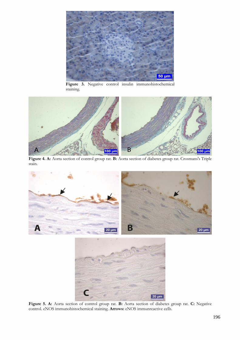
196
Figure 3. Negative control insulin immunohistochemical staining.
Figure 4. A: Aorta section of control group rat. B: Aorta section of diabetes group rat. Crosmans's Triple stain.
Figure 5. A: Aorta section of control group rat. B: Aorta section of diabetes group rat. C: Negative control. eNOS immunohistochemical staining. Arrows: eNOS immunreactive cells.
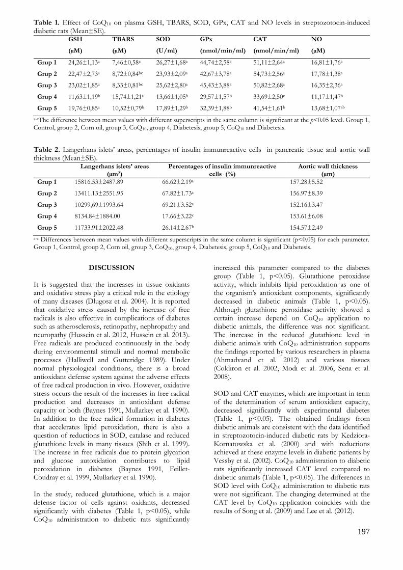
197
Table 1. Effect of CoQ10 on plasma GSH, TBARS, SOD, GPx, CAT and NO levels in streptozotocin-induced diabetic rats (Mean±SE).
GSH
(µM)
TBARS
(µM)
SOD
(U/ml)
GPx
(nmol/min/ml)
CAT
(nmol/min/ml)
NO
(µM)
Grup 1 24,26±1,13a 7,46±0,58c 26,27±1,68a 44,74±2,58a 51,11±2,64a 16,81±1,76a
Grup 2 22,47±2,73a 8,72±0,84bc 23,93±2,09a 42,67±3,78a 54,73±2,56a 17,78±1,38a
Grup 3 23,02±1,85a 8,33±0,81bc 25,62±2,80a 45,43±3,88a 50,82±2,68a 16,35±2,36a
Grup 4 11,63±1,19b 15,74±1,21a 13,66±1,05b 29,57±1,57b 33,69±2,50c 11,17±1,47b
Grup 5 19,76±0,85a 10,52±0,79b 17,89±1,29b 32,39±1,88b 41,54±1,61b 13,68±1,07ab
a-cThe difference between mean values with different superscripts in the same column is significant at the p<0.05 level. Group 1, Control, group 2, Corn oil, group 3, CoQ10, group 4, Diabetesis, group 5, CoQ10 and Diabetesis.
Table 2. Langerhans islets’ areas, percentages of insulin immunreactive cells in pancreatic tissue and aortic wall thickness (Mean±SE).
Langerhans islets’ areas (µm2)
Percentages of insulin immunreactive cells (%)
Aortic wall thickness (µm)
Grup 1 15816.53±2487.89 66.62±2.19a 157.28±5.52
Grup 2 13411.13±2551.95 67.82±1.73a 156.97±8.39
Grup 3 10299,69±1993.64 69.21±3.52a 152.16±3.47
Grup 4 8134.84±1884.00 17.66±3.22c 153.61±6.08
Grup 5 11733.91±2022.48 26.14±2.67b 154.57±2.49
a-c Differences between mean values with different superscripts in the same column is significant (p<0.05) for each parameter. Group 1, Control, group 2, Corn oil, group 3, CoQ10, group 4, Diabetesis, group 5, CoQ10 and Diabetesis.
DISCUSSION It is suggested that the increases in tissue oxidants and oxidative stress play a critical role in the etiology of many diseases (Dlugosz et al. 2004). It is reported that oxidative stress caused by the increase of free radicals is also effective in complications of diabetes such as atherosclerosis, retinopathy, nephropathy and neuropathy (Hussein et al. 2012, Hussein et al. 2013). Free radicals are produced continuously in the body during environmental stimuli and normal metabolic processes (Halliwell and Gutteridge 1989). Under normal physiological conditions, there is a broad antioxidant defense system against the adverse effects of free radical production in vivo. However, oxidative stress occurs the result of the increases in free radical production and decreases in antioxidant defense capacity or both (Baynes 1991, Mullarkey et al. 1990). In addition to the free radical formation in diabetes that accelerates lipid peroxidation, there is also a question of reductions in SOD, catalase and reduced glutathione levels in many tissues (Shih et al. 1999). The increase in free radicals due to protein glycation and glucose autoxidation contributes to lipid peroxidation in diabetes (Baynes 1991, Feillet-Coudray et al. 1999, Mullarkey et al. 1990). In the study, reduced glutathione, which is a major defense factor of cells against oxidants, decreased significantly with diabetes (Table 1, p<0.05), while CoQ10 administration to diabetic rats significantly
increased this parameter compared to the diabetes group (Table 1, p<0.05). Glutathione peroxidase activity, which inhibits lipid peroxidation as one of the organism's antioxidant components, significantly decreased in diabetic animals (Table 1, p<0.05). Although glutathione peroxidase activity showed a certain increase depend on CoQ10 application to diabetic animals, the difference was not significant. The increase in the reduced glutathione level in diabetic animals with CoQ10 administration supports the findings reported by various researchers in plasma (Ahmadvand et al. 2012) and various tissues (Coldiron et al. 2002, Modi et al. 2006, Sena et al. 2008). SOD and CAT enzymes, which are important in term of the determination of serum antioxidant capacity, decreased significantly with experimental diabetes (Table 1, p<0.05). The obtained findings from diabetic animals are consistent with the data identified in streptozotocin-induced diabetic rats by Kedziora-Kornatowska et al. (2000) and with reductions achieved at these enzyme levels in diabetic patients by Vessby et al. (2002). CoQ10 administration to diabetic rats significantly increased CAT level compared to diabetic animals (Table 1, p<0.05). The differences in SOD level with CoQ10 administration to diabetic rats were not significant. The changing determined at the CAT level by CoQ10 application coincides with the results of Song et al. (2009) and Lee et al. (2012).

198
NO formation and oxidative stress play an important role in the development of diabetic complications. Particularly, it was reported that nitric oxide reduction is effective in disorders in the vascular system in diabetes, while it has been suggested that other vasodilatators and hyperlipidemia also contribute to vascular disorders (Cohen 2005). It has been reported that insulin stimulates NO production by specific signaling pathways (phophotidyl-inositol 3-(PI3)-kinase and protein kinase B), high glucose levels reduce NO in diabetes and NO release and production also deteriorate due to loss of positive effect of insülin (Balletshofer et al. 2000, Tuck 2003, Zeng et al. 2000). In this study, significant reductions (Table 1, p<0.05) in NO levels with experimental diabetes compared to control group seems consistent with the above notifications. It was observed that the increase in NO level with CoQ10 application to diabetic animals removed the difference with the control group level. It was observed significantly increase in thiobarbituric acid reactive substances (TBARS) as lipid peroxidation marker of lipoproteins and membranes in diabetic group compared to the control group (Table 1, p<0.05). This increase seems to be in line with the notifications that TBARS levels increase in diabetes (Griesmacher et al. 1995, Kakkar et al. 1998, Sundaram et al. 1996). On the other hand, there was a significant decline in TBARS level with CoQ10 application to diabetic animals compared to the diabetes group (Table 1, p<0.05). Unimportant changes determined in these parameters with only CoQ10 application compared to control group may be due to the absence of oxidative damage in healthy animals. It has been proposed various mechanisms in related to the positive effects of CoQ10 on the antioxidant system in diabetes (Prakash et al. 2010, Song et al. 2009). It has been reported that CoQ10 can directly eliminate free radicals such as lipid peroxyl, peroxyl or alkoxyl radicals (Roginsky et al. 2009, Sohal and Forster 2007). Forsmark-Andree and Ernster (1994) have been reported that CoQ10 may indirectly act as an antioxidant via providing α-tocopherol regeneration from α-tocoperoxyl radicals formed by a reaction between α-tocopherol and lipid peroxyl radicals. Tiano et al. (2007) have been suggested that nitric oxide is an active antioxidant against free radical-mediated lipid peroxidation and that CoQ10 can reduce free radical and superoxide formation via improving nitric oxide bioactivity. Abdin and Hamouda (2008) have been also stated that CoQ10 showed in vivo antiapoptotic effects by increasing the expression and activation of mitochondrial proteins and consequently, it may function as an antioxidant by reduce the formation of free radicals. Streptozotocin is a substance that causes decrease of insulin-producing capacity by creating damaged via oxidative stress in insulin-producing β-cells. Studies have shown that the most common organs affected
oxidative stress related organ damage following diabetes is liver, kidney, pancreas, and eye. Severe metabolic changes and oxidative disturbances in the pancreas also play an important role in the pathogenesis of diabetes (Baynes and Thorpe 1999, Ihara et al. 1999). In experimental diabetes, it has been stated that there is an increase in oxidative stress markers in pancreatic islet cells and it has been determined damage and dysfunctions in β cells of rats (Ihara et al. 1999, Meghana et al. 2007). In this study, irregularity in the contours of Langerhans islets, vacuolization and atrophy in the islet cells were observed in streptozotocin induced diabetic groups. These histological findings were found to be consistent with datas of the previous conducted studies (Kamalakkannan and Prince 2005, Hassan et al. 2015, Yaman et al. 2017). In recent studies, it has been reported that antioxidants and plant extracts improved functional state of pancreatic β cells and partially reversed the damage caused by streptozotocin to β-cells of the pancreatic islets (Kanter et al. 2004, Kamalakkannan and Prince 2005, Omar and Atia 2012, Abunasef et al. 2014, Niture et al. 2014, Hassan et al. 2015, Yaman et al. 2017). Luo et al. (2019) suggested that CoQ10 has beneficial effects for reducing mitochondrial injury via its antioxidative properties and is effective in ameliorating pancreatic β cell dysfunction by tacrolimus. Similarly our study, it was seen that the CoQ10 application to diabetic rats led to partially improve in pancreatic tissues. Also, a statistically significant increase in the percentage of the insulin immun reactive cells was noted in this group when compared to the diabetes group. Nitric oxide occurs during conversion of L-arginine to L-citrulline. This enzymatic pathway is managed by nitric oxide synthase (NOS). Nitric oxide synthase has at least 3 isoenzymes: Inducible NOS (iNOS) is found in macrophages and it is involved in pathological events. Structural neuronal NOS (nNOS) are found in the brain. Structural endothelial NOS (eNOS) are synthesized from endothelial cells, endocardial cells, ventricular myocytes and other myocardial cells and its activation is dependent on Ca and calmodulin (Felaco et al. 2001). It is stated that endothelial damage in diabetes may be a consequence of glucometabolic and peroxidative stress. It was reported that even if high amounts of NO produced by iNOS have been seen to be toxic and damaging, structurally optimum of NO produced by eNOS is required for the protection of endothelial function. Therefore, eNOS is important determining of the microvascular complications of diabetes and the susceptibility to cardiovascular disease (İnan 2015, Kröncke et al. 1995, Özgün et al. 2014). In this study conducted by Felaco et al. (2001) related with eNOS localization and expression in myocardial tissue in healthy and diabetic rats, it was reported that there was no difference between the diabetic group and the

199
control group in terms of eNOS immunreactivity in myocytes and there was less immunreactivity in the diabetic group compared to the control group in the vascular endothelium. In this study, there was no difference among the groups in terms of eNOS immunreactivity in aortic endothelium. Long-term complications of diabetes are retinopathy, nephropathy and cardiovascular disease. In fact, the duration of this study is thought to be insufficient for the development of cardiovascular complications. As a result, if it regards to the significant changes particularly in the levels of GSH, CAT, TBARS and positively changes in NO level with CoQ10 administration to experimental diabetic rats, it was concluded that CoQ10 may alleviate the negative effects of diabetes on these parameters and that it may be useful due to protective properties in diabetes. The histological findings of the study support partially the changes in some plasma enzyme levels as a result of CoQ10 application. It has been thought that the determined data may contribute to the studies be conducted the same subject in the future and further studies are required in different dose and duration.
ACKNOWLEDGEMENTS This study was supported by The Scientific Research Projects Coordination Unit of Selcuk University (Project no. 15401074). This study was presented in 1st International & Xth National Veterinary Anatomy Congress of Turkey. The study protocol was approved by The Ethical Committee of Selcuk University Experimental Medicine Research and Application Center (Report no. 2015-22). Conflict of Interest: The authors declare that they have no conflict of interest.
REFERENCES Abdin AA, Hamouda HE. Mechanism of the neuroprotective
role of coenzyme Q10 with or without L-dopa in rotenone-induced parkinsonism. Neuropharmacology. 2008; 55(8):1340-1346. doi: 10.1016/j.neuropharm.2008.08.033.
Abunasef SK, Amin HA, Abdel-Hamid GA. A histological and immunohistochemical study of beta cells in streptozotocin diabetic rats treated with caffeine. Folia Histochem Cytobiol. 2014;52(1):42-50. doi: 10.5603/FHC.2014.0005.
Aguirre F, Martin I, Grinspon D, Ruiz M, Hager A, De Paoli T et al. Oxidative damage, plasma antioxidant capacity and glucemic control in elderly NIDDM patients. Free Radic Biol Med. 1998;24(4):580-585. doi: 10.1016/S0891-5849(97)00293-1.
Ahmadvand H, Tavafi M, Khosrowbeygi A. Amelioration of altered antioxidant enzymes activity and glomerulosclerosis by coenzyme Q10 in alloxan-induced diabetic rats. J Diabetes Complications. 2012;26(6):476-
482. doi: 10.1016/j.jdiacomp.2012.06.004.
Ashour M, Salem S, Hassaneen H, El-Gadban H, Elwan N, Awad A et al. Antioxidant status and insulin-dependent diabetes mellitus (IDDM). J Clin Biochem Nutr. 1999; 26(2):99-107. doi: https://doi.org/10.3164/jcbn.26.99.
Balletshofer BM, Rittig K, Enderle MD, Volk A, Maerker E, Jacob S, Matthaei S, Rett K, Häring HU. Endothelial dysfunction is detectable in young normotensive first-degree relatives of subjects with type 2 diabetes in association with insulin resistance. Circulation. 2000;101(15):1780-1784. doi: https://www.ahajournals.org/doi/full/10.1161/01.CIR.101.15.1780.
Baynes JW. Role of oxidative stress in the development of complications in diabetes. Diabetes. 1991;40(4):405-412. doi: https://doi.org/10.2337/diab.40.4.405.
Baynes JW, Thorpe SR. Role of oxidative stress in diabetic complications: a new perspective on an old paradigm. Diabetes. 1999;48(1):1-9. doi: https://doi.org/10.2337/diabetes.48.1.1.
Beyer RE, Burnett BA, Cartwright KJ, Edington DW, Falzon MJ, Kreitman KR, Kuhn TW, Ramp BJ, Rhee SYS, Rosenwasser MJ, Stein M, An LC. Tissue coenzyme Q (ubiquinone) and protein concentrations over the life span of the laboratory rat. Mech Ageing Dev. 1985;32(2-3):267-281. doi: https://doi.org/10.1016/0047-6374(85)90085-5.
Brownlee M. Biochemistry and molecular cell biology of diabetic complications. Nature. 2001;414(6865):813-820. doi: 10.1038/414813a.
Brownlee M. Negative consequence of glycation. Metabolism. 2000;49(2 Suppl 1):9-13. doi: https://doi.org/10.1016/S0026-0495(00)80078-5.
Ceriello A, Bortolotti N, Pirisi M, Crescentini A, Tonutti L, Motz E, Russo A, Giacomello R, Stel G, Taboga C. Total plasma antioxidant capacity predicts thrombosis-prone status in NIDDM patients. Diabetes Care. 1997b;20(10):1589-1593. doi: https://doi.org/10.2337/diacare.20.10.1589.
Ceriello A, Bortolotti N, Crescentini A, Motz E, Lizzio S, Russo A, Ezsol Z, Tonutti L, Taboga C. Antioxidant defences are reduced during the oral glucose tolerance test in normal and non-insulin-dependent diabetic subjects. Eur J Clin Invest. 1998;28(4):329-333. doi: https://doi.org/10.1046/j.1365-2362.1998.00295.x.
Ceriello A, Bortolotti N, Falleti E, Taboga C, Tonutti L, Crescentini A, Motz E, Lizzio S, Russo A, Bartoli E. Total radical-trapping antioxidant parameter in NIDDM patients. Diabetes Care. 1997a;20(2):194-197. doi: https://doi.org/10.2337/diacare.20.2.194.
Cohen RA. Role of nitric oxide in diabetic complications. Am J Ther. 2005;12(6):499-502. doi: 10.1097/01.mjt.0000178776.77267.19.
Coldiron AD Jr, Sanders RA, Watkins JB 3rd. Effects of combined quercetin and coenzyme Q(10) treatment on oxidative stress in normal and diabetic rats. J Biochem Mol Toxicol. 2002;16(4):197-202. doi: 10.1002/jbt.10035.
Crane FL, Navas P. The diversity of coenzyme Q function. Mol Aspects Med. 1997;18:1-6. doi: https://doi.org/10.1016/S0098-2997(97)00016-2.
Crossmon G. A modification of Mallory’s connective tissue stain with a discussion of the principles involved. Anat Rec. 1937;69:33-38. doi: https://doi.org/10.1002/ar.1090690105.
Dlugosz A, Kuzniar J, Sawicka E, Marchewka Z, Lembas-
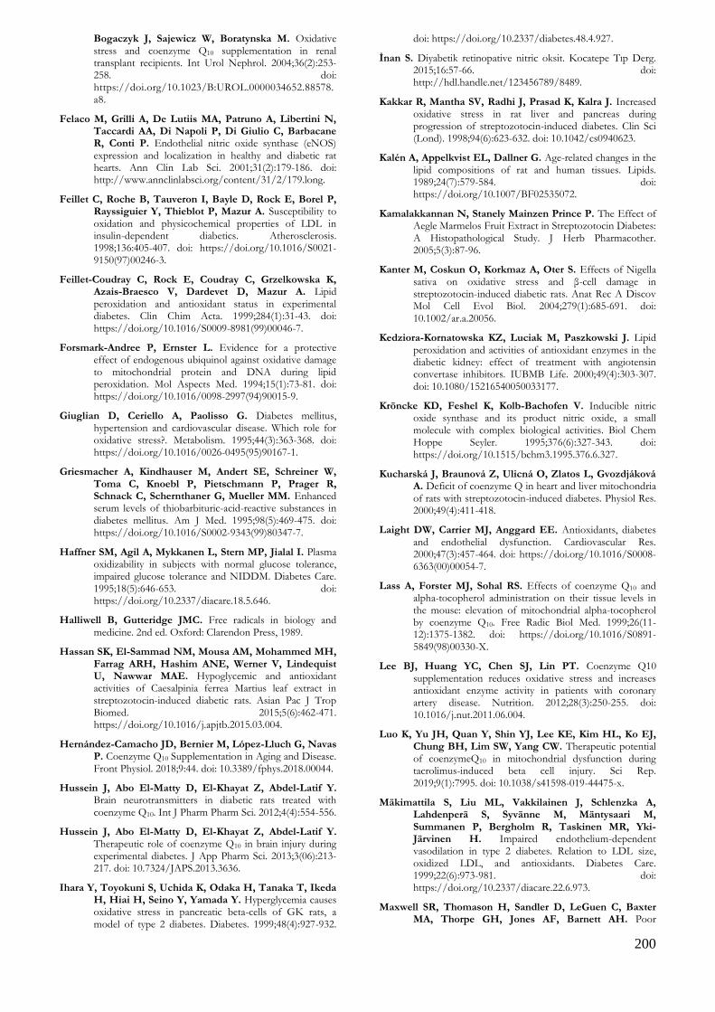
200
Bogaczyk J, Sajewicz W, Boratynska M. Oxidative stress and coenzyme Q10 supplementation in renal transplant recipients. Int Urol Nephrol. 2004;36(2):253-258. doi: https://doi.org/10.1023/B:UROL.0000034652.88578.a8.
Felaco M, Grilli A, De Lutiis MA, Patruno A, Libertini N, Taccardi AA, Di Napoli P, Di Giulio C, Barbacane R, Conti P. Endothelial nitric oxide synthase (eNOS) expression and localization in healthy and diabetic rat hearts. Ann Clin Lab Sci. 2001;31(2):179-186. doi: http://www.annclinlabsci.org/content/31/2/179.long.
Feillet C, Roche B, Tauveron I, Bayle D, Rock E, Borel P, Rayssiguier Y, Thieblot P, Mazur A. Susceptibility to oxidation and physicochemical properties of LDL in insulin-dependent diabetics. Atherosclerosis. 1998;136:405-407. doi: https://doi.org/10.1016/S0021-9150(97)00246-3.
Feillet-Coudray C, Rock E, Coudray C, Grzelkowska K, Azais-Braesco V, Dardevet D, Mazur A. Lipid peroxidation and antioxidant status in experimental diabetes. Clin Chim Acta. 1999;284(1):31-43. doi: https://doi.org/10.1016/S0009-8981(99)00046-7.
Forsmark-Andree P, Ernster L. Evidence for a protective effect of endogenous ubiquinol against oxidative damage to mitochondrial protein and DNA during lipid peroxidation. Mol Aspects Med. 1994;15(1):73-81. doi: https://doi.org/10.1016/0098-2997(94)90015-9.
Giuglian D, Ceriello A, Paolisso G. Diabetes mellitus, hypertension and cardiovascular disease. Which role for oxidative stress?. Metabolism. 1995;44(3):363-368. doi: https://doi.org/10.1016/0026-0495(95)90167-1.
Griesmacher A, Kindhauser M, Andert SE, Schreiner W, Toma C, Knoebl P, Pietschmann P, Prager R, Schnack C, Schernthaner G, Mueller MM. Enhanced serum levels of thiobarbituric-acid-reactive substances in diabetes mellitus. Am J Med. 1995;98(5):469-475. doi: https://doi.org/10.1016/S0002-9343(99)80347-7.
Haffner SM, Agil A, Mykkanen L, Stern MP, Jialal I. Plasma oxidizability in subjects with normal glucose tolerance, impaired glucose tolerance and NIDDM. Diabetes Care. 1995;18(5):646-653. doi: https://doi.org/10.2337/diacare.18.5.646.
Halliwell B, Gutteridge JMC. Free radicals in biology and medicine. 2nd ed. Oxford: Clarendon Press, 1989.
Hassan SK, El-Sammad NM, Mousa AM, Mohammed MH, Farrag ARH, Hashim ANE, Werner V, Lindequist U, Nawwar MAE. Hypoglycemic and antioxidant activities of Caesalpinia ferrea Martius leaf extract in streptozotocin-induced diabetic rats. Asian Pac J Trop Biomed. 2015;5(6):462-471. https://doi.org/10.1016/j.apjtb.2015.03.004.
Hernández-Camacho JD, Bernier M, López-Lluch G, Navas P. Coenzyme Q10 Supplementation in Aging and Disease. Front Physiol. 2018;9:44. doi: 10.3389/fphys.2018.00044.
Hussein J, Abo El-Matty D, El-Khayat Z, Abdel-Latif Y. Brain neurotransmitters in diabetic rats treated with coenzyme Q10. Int J Pharm Pharm Sci. 2012;4(4):554-556.
Hussein J, Abo El-Matty D, El-Khayat Z, Abdel-Latif Y. Therapeutic role of coenzyme Q10 in brain injury during experimental diabetes. J App Pharm Sci. 2013;3(06):213-217. doi: 10.7324/JAPS.2013.3636.
Ihara Y, Toyokuni S, Uchida K, Odaka H, Tanaka T, Ikeda H, Hiai H, Seino Y, Yamada Y. Hyperglycemia causes oxidative stress in pancreatic beta-cells of GK rats, a model of type 2 diabetes. Diabetes. 1999;48(4):927-932.
doi: https://doi.org/10.2337/diabetes.48.4.927.
İnan S. Diyabetik retinopative nitric oksit. Kocatepe Tıp Derg. 2015;16:57-66. doi: http://hdl.handle.net/123456789/8489.
Kakkar R, Mantha SV, Radhi J, Prasad K, Kalra J. Increased oxidative stress in rat liver and pancreas during progression of streptozotocin-induced diabetes. Clin Sci (Lond). 1998;94(6):623-632. doi: 10.1042/cs0940623.
Kalén A, Appelkvist EL, Dallner G. Age-related changes in the lipid compositions of rat and human tissues. Lipids. 1989;24(7):579-584. doi: https://doi.org/10.1007/BF02535072.
Kamalakkannan N, Stanely Mainzen Prince P. The Effect of Aegle Marmelos Fruit Extract in Streptozotocin Diabetes: A Histopathological Study. J Herb Pharmacother. 2005;5(3):87-96.
Kanter M, Coskun O, Korkmaz A, Oter S. Effects of Nigella sativa on oxidative stress and β-cell damage in streptozotocin-induced diabetic rats. Anat Rec A Discov Mol Cell Evol Biol. 2004;279(1):685-691. doi: 10.1002/ar.a.20056.
Kedziora-Kornatowska KZ, Luciak M, Paszkowski J. Lipid peroxidation and activities of antioxidant enzymes in the diabetic kidney: effect of treatment with angiotensin convertase inhibitors. IUBMB Life. 2000;49(4):303-307. doi: 10.1080/15216540050033177.
Kröncke KD, Feshel K, Kolb-Bachofen V. Inducible nitric oxide synthase and its product nitric oxide, a small molecule with complex biological activities. Biol Chem Hoppe Seyler. 1995;376(6):327-343. doi: https://doi.org/10.1515/bchm3.1995.376.6.327.
Kucharská J, Braunová Z, Ulicná O, Zlatos L, Gvozdjáková A. Deficit of coenzyme Q in heart and liver mitochondria of rats with streptozotocin-induced diabetes. Physiol Res. 2000;49(4):411-418.
Laight DW, Carrier MJ, Anggard EE. Antioxidants, diabetes and endothelial dysfunction. Cardiovascular Res. 2000;47(3):457-464. doi: https://doi.org/10.1016/S0008-6363(00)00054-7.
Lass A, Forster MJ, Sohal RS. Effects of coenzyme Q10 and alpha-tocopherol administration on their tissue levels in the mouse: elevation of mitochondrial alpha-tocopherol by coenzyme Q10. Free Radic Biol Med. 1999;26(11-12):1375-1382. doi: https://doi.org/10.1016/S0891-5849(98)00330-X.
Lee BJ, Huang YC, Chen SJ, Lin PT. Coenzyme Q10 supplementation reduces oxidative stress and increases antioxidant enzyme activity in patients with coronary artery disease. Nutrition. 2012;28(3):250-255. doi: 10.1016/j.nut.2011.06.004.
Luo K, Yu JH, Quan Y, Shin YJ, Lee KE, Kim HL, Ko EJ, Chung BH, Lim SW, Yang CW. Therapeutic potential of coenzymeQ10 in mitochondrial dysfunction during tacrolimus-induced beta cell injury. Sci Rep. 2019;9(1):7995. doi: 10.1038/s41598-019-44475-x.
Mäkimattila S, Liu ML, Vakkilainen J, Schlenzka A, Lahdenperä S, Syvänne M, Mäntysaari M, Summanen P, Bergholm R, Taskinen MR, Yki-Järvinen H. Impaired endothelium-dependent vasodilation in type 2 diabetes. Relation to LDL size, oxidized LDL, and antioxidants. Diabetes Care. 1999;22(6):973-981. doi: https://doi.org/10.2337/diacare.22.6.973.
Maxwell SR, Thomason H, Sandler D, LeGuen C, Baxter MA, Thorpe GH, Jones AF, Barnett AH. Poor
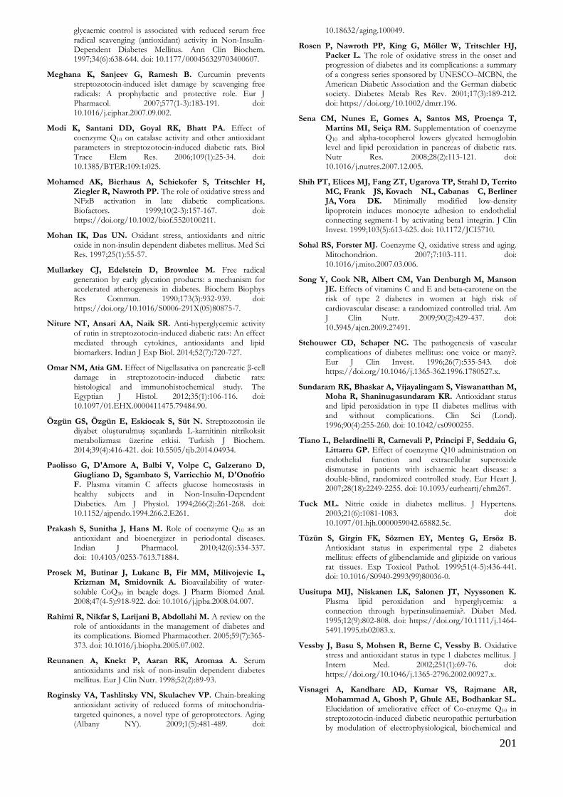
201
glycaemic control is associated with reduced serum free radical scavenging (antioxidant) activity in Non-Insulin-Dependent Diabetes Mellitus. Ann Clin Biochem. 1997;34(6):638-644. doi: 10.1177/000456329703400607.
Meghana K, Sanjeev G, Ramesh B. Curcumin prevents streptozotocin-induced islet damage by scavenging free radicals: A prophylactic and protective role. Eur J Pharmacol. 2007;577(1-3):183-191. doi: 10.1016/j.ejphar.2007.09.002.
Modi K, Santani DD, Goyal RK, Bhatt PA. Effect of coenzyme Q10 on catalase activity and other antioxidant parameters in streptozotocin-induced diabetic rats. Biol Trace Elem Res. 2006;109(1):25-34. doi: 10.1385/BTER:109:1:025.
Mohamed AK, Bierhaus A, Schiekofer S, Tritschler H, Ziegler R, Nawroth PP. The role of oxidative stress and NFκB activation in late diabetic complications. Biofactors. 1999;10(2-3):157-167. doi: https://doi.org/10.1002/biof.5520100211.
Mohan IK, Das UN. Oxidant stress, antioxidants and nitric oxide in non-insulin dependent diabetes mellitus. Med Sci Res. 1997;25(1):55-57.
Mullarkey CJ, Edelstein D, Brownlee M. Free radical generation by early glycation products: a mechanism for accelerated atherogenesis in diabetes. Biochem Biophys Res Commun. 1990;173(3):932-939. doi: https://doi.org/10.1016/S0006-291X(05)80875-7.
Niture NT, Ansari AA, Naik SR. Anti-hyperglycemic activity of rutin in streptozotocin-induced diabetic rats: An effect mediated through cytokines, antioxidants and lipid biomarkers. Indian J Exp Biol. 2014;52(7):720-727.
Omar NM, Atia GM. Effect of Nigellasativa on pancreatic β-cell damage in streptozotocin-induced diabetic rats: histological and immunohistochemical study. The Egyptian J Histol. 2012;35(1):106-116. doi: 10.1097/01.EHX.0000411475.79484.90.
Özgün GS, Özgün E, Eskiocak S, Süt N. Streptozotosin ile diyabet oluşturulmuş sıçanlarda L-karnitinin nitrikoksit metabolizması üzerine etkisi. Turkish J Biochem. 2014;39(4):416-421. doi: 10.5505/tjb.2014.04934.
Paolisso G, D'Amore A, Balbi V, Volpe C, Galzerano D, Giugliano D, Sgambato S, Varricchio M, D'Onofrio F. Plasma vitamin C affects glucose homeostasis in healthy subjects and in Non-Insulin-Dependent Diabetics. Am J Physiol. 1994;266(2):261-268. doi: 10.1152/ajpendo.1994.266.2.E261.
Prakash S, Sunitha J, Hans M. Role of coenzyme Q10 as an antioxidant and bioenergizer in periodontal diseases. Indian J Pharmacol. 2010;42(6):334-337. doi: 10.4103/0253-7613.71884.
Prosek M, Butinar J, Lukanc B, Fir MM, Milivojevic L, Krizman M, Smidovnik A. Bioavailability of water-soluble CoQ10 in beagle dogs. J Pharm Biomed Anal. 2008;47(4-5):918-922. doi: 10.1016/j.jpba.2008.04.007.
Rahimi R, Nikfar S, Larijani B, Abdollahi M. A review on the role of antioxidants in the management of diabetes and its complications. Biomed Pharmacother. 2005;59(7):365-373. doi: 10.1016/j.biopha.2005.07.002.
Reunanen A, Knekt P, Aaran RK, Aromaa A. Serum antioxidants and risk of non-insulin dependent diabetes mellitus. Eur J Clin Nutr. 1998;52(2):89-93.
Roginsky VA, Tashlitsky VN, Skulachev VP. Chain-breaking antioxidant activity of reduced forms of mitochondria-targeted quinones, a novel type of geroprotectors. Aging (Albany NY). 2009;1(5):481-489. doi:
10.18632/aging.100049.
Rosen P, Nawroth PP, King G, Möller W, Tritschler HJ, Packer L. The role of oxidative stress in the onset and progression of diabetes and its complications: a summary of a congress series sponsored by UNESCO–MCBN, the American Diabetic Association and the German diabetic society. Diabetes Metab Res Rev. 2001;17(3):189-212. doi: https://doi.org/10.1002/dmrr.196.
Sena CM, Nunes E, Gomes A, Santos MS, Proença T, Martins MI, Seiça RM. Supplementation of coenzyme Q10 and alpha-tocopherol lowers glycated hemoglobin level and lipid peroxidation in pancreas of diabetic rats. Nutr Res. 2008;28(2):113-121. doi: 10.1016/j.nutres.2007.12.005.
Shih PT, Elices MJ, Fang ZT, Ugarova TP, Strahl D, Territo MC, Frank JS, Kovach NL, Cabanas C, Berliner JA, Vora DK. Minimally modified low-density lipoprotein induces monocyte adhesion to endothelial connecting segment-1 by activating beta1 integrin. J Clin Invest. 1999;103(5):613-625. doi: 10.1172/JCI5710.
Sohal RS, Forster MJ. Coenzyme Q, oxidative stress and aging. Mitochondrion. 2007;7:103-111. doi: 10.1016/j.mito.2007.03.006.
Song Y, Cook NR, Albert CM, Van Denburgh M, Manson JE. Effects of vitamins C and E and beta-carotene on the risk of type 2 diabetes in women at high risk of cardiovascular disease: a randomized controlled trial. Am J Clin Nutr. 2009;90(2):429-437. doi: 10.3945/ajcn.2009.27491.
Stehouwer CD, Schaper NC. The pathogenesis of vascular complications of diabetes mellitus: one voice or many?. Eur J Clin Invest. 1996;26(7):535-543. doi: https://doi.org/10.1046/j.1365-362.1996.1780527.x.
Sundaram RK, Bhaskar A, Vijayalingam S, Viswanatthan M, Moha R, Shaninugasundaram KR. Antioxidant status and lipid peroxidation in type II diabetes mellitus with and without complications. Clin Sci (Lond). 1996;90(4):255-260. doi: 10.1042/cs0900255.
Tiano L, Belardinelli R, Carnevali P, Principi F, Seddaiu G, Littarru GP. Effect of coenzyme Q10 administration on endothelial function and extracellular superoxide dismutase in patients with ischaemic heart disease: a double-blind, randomized controlled study. Eur Heart J. 2007;28(18):2249-2255. doi: 10.1093/eurheartj/ehm267.
Tuck ML. Nitric oxide in diabetes mellitus. J Hypertens. 2003;21(6):1081-1083. doi: 10.1097/01.hjh.0000059042.65882.5c.
Tüzün S, Girgin FK, Sözmen EY, Menteş G, Ersöz B. Antioxidant status in experimental type 2 diabetes mellitus: effects of glibenclamide and glipizide on various rat tissues. Exp Toxicol Pathol. 1999;51(4-5):436-441. doi: 10.1016/S0940-2993(99)80036-0.
Uusitupa MIJ, Niskanen LK, Salonen JT, Nyyssonen K. Plasma lipid peroxidation and hyperglycemia: a connection through hyperinsulinaemia?. Diabet Med. 1995;12(9):802-808. doi: https://doi.org/10.1111/j.1464-5491.1995.tb02083.x.
Vessby J, Basu S, Mohsen R, Berne C, Vessby B. Oxidative stress and antioxidant status in type 1 diabetes mellitus. J Intern Med. 2002;251(1):69-76. doi: https://doi.org/10.1046/j.1365-2796.2002.00927.x.
Visnagri A, Kandhare AD, Kumar VS, Rajmane AR, Mohammad A, Ghosh P, Ghule AE, Bodhankar SL. Elucidation of ameliorative effect of Co-enzyme Q10 in streptozotocin-induced diabetic neuropathic perturbation by modulation of electrophysiological, biochemical and
202
behavioral markers. Biomed Aging Pathol. 2012;2(4):157-172. doi: https://doi.org/10.1016/j.biomag.2012.10.006.
Yaman T, Uyar A, Celik İ, Alkan EE, Keles ÖF, Yener Z. Histopathological and immunohistochemical study of antidiabetic effects of heracleum persicum extract in experimentally diabetic rats. Indian J Pharm Educ Res. 2017;51(3):450-457. doi:10.5530/ijper.51.3s.66.
Yaqoob M, Patrick AW, McClelland P, Stevenson A, Mason H, White MC et al. Relationship between markers of endothelial dysfunction, oxidant injury and tubular damage in patients with insulin-dependent diabetes mellitus. Clin Sci (Lond). 1993;85(5):557-562.
West IC. Radicals and oxidative stress in diabetes. Diabet Med. 2000;17(3):171-180. doi: https://doi.org/10.1046/j.1464-5491.2000.00259.x.
Willems D, Dorchy H, Dufrasne D. Serum antioxidant status and oxidized LDL in well-controlled young type 1 diabetic patients with and without subclinical complications. Atherosclerosis. 1998;137:61-64. doi: https://doi.org/10.1016/S0021-9150(97)00320-1.
Zeng G, Nystrom FH, Ravichandran LV, Cong LN, Kirby M, Mostowski H, Quon MJ. Roles for insulin receptor, PI3-kinase, and Akt in insulin signaling pathways related to production of nitric oxide in human vascular endothelial cells. Circulation. 2000;101(13):1539-1545. doi: https://www.ahajournals.org/doi/abs/10.1161/01.cir.101.13.1539.

203
Kocatepe Veterinary Journal
Kocatepe Vet J (2020) 13(2):203-209
DOI: 10.30607/kvj.715053 RESEARCH ARTICLE
Effects of Fructose-Induced Metabolic Syndrome on Kidney Histology
in Rats
Özay GÜLEŞ1*, Musa TATAR2
1Afyon Kocatepe University, Faculty of Veterinary Medicine, Department of Histology and Embryology, 03200, Afyon, Turkey
2Burdur Mehmet Akif Ersoy University, Faculty of Veterinary Medicine, Department of Histology and Embryology, 15030, Burdur, Turkey
ABSTRACT
Metabolic syndrome is a fatal endocrinopathy, which is progressed towards pandemic, and characterized by insulin resistance, abdominal obesity, dyslipidemia, diabetes mellitus, hypertension and coronary artery disease (CAD). In the present study, it is aimed to investigate the histomorphological and histochemical changes caused by the metabolic syndrome in rat kidneys. For this purpose, the material was divided into two groups: 10 rats in the control group and 15 rats in the experimental group. During 16 weeks, while tap water was given to control group rats, water including 20% fructose solution was given to experimental group rats as drinking water. At the end of the study, Crossman's triple staining method was performed to determine the histological appearance and histomorphological changes in the sections taken from the kidneys. Periodic Acid Schiff Reagent (PAS) staining method was performed for histochemical analysis. The results of the study showed that the tubulus proximalis diameter, tubulus proximalis glycogen density, and glomerular mesangial matrix density increased, corpusculum renis diameter, width of cavum glomeruli and ascending limb of Henle’s loop diameter decreased. In conclusion, it is demonstrated that metabolic syndrome may adversely affect kidney histology and cause renal damage. Keywords: Metabolic syndrome, fructose, histology, kidney, rat
***
Ratlarda Fruktoz ile Oluşturulmuş Metabolik Sendromun Böbrek Histolojisi Üzerine Etkileri
ÖZ
Metabolik sendrom pandemiye doğru ilerleyen, insülin direnci, abdominal obezite, dislipidemi, diabetes mellitus, hipertansiyon ve koroner arter hastalığı (KAH) ile karakterize ölümcül bir endokrinopatidir. Sunulan çalışamada metabolik sendromun rat böbreklerinde oluşturduğu histomorfolojik ve histokimyasal değişimlerin araştırılması amaçlanmıştır. Bu amaçla materyal kontrol grubunda 10, metabolik sendrom grubunda 15 rat içerecek şekilde iki gruba ayrıldı. Kontrol grubundaki ratlara 16 hafta süresince çeşme suyu verilirken, metabolik sendrom grubundaki ratlara içme suyu olarak % 20 fruktoz çözeltisi verildi. Deneme sürecinin sonunda böbreklerden alınan kesitlerde histolojik görünümü ve histomorfolojik değişimleri belirlemek amacıyla Crossman’s üçlü boyama metodu, histokimyasal analiz amacıyla Periyodik Asit Schiff Reagent (PAS) boyama metodu uygulandı. Elde edilen veriler tubulus proksimalis çapı ve glikojen yoğunluğu ile glomerular mezangial matriks yoğunluğunun arttığını, korpuskulum renis çapı, Bowman aralığı genişliği ve çıkan henle çapının azaldığını göstermiştir. Sonuç olarak, metabolik sendromun böbrek histolojisini olumsuz yönde etkileyerek renal hasara sebep olabileceği ortaya konmuştur. Anahtar Kelimeler: Metabolik sendrom, fruktoz, histoloji, böbrek, rat
To cite this article: Güleş Ö. Tatar M. Effects of Fructose-Induced Metabolic Syndrome on Kidney Histology in Rats. Kocatepe Vet J. (2020) 13(2):203-209 Submission: 06.04.2020 Accepted: 20.05.2020 Published Online: 24.05.2020
ORCID ID; ÖG: 0000-0001-6170-1706, MT: 0000-0002-5707-8832 *Corresponding author e-mail: [email protected]
204
INTRODUCTION Metabolic syndrome is a fatal endocrinopathy characterized by a combination of cardiometabolic risk factors such as insulin resistance, abdominal obesity, atherogenic dyslipidemia, glucose intolerance/diabetes mellitus, high blood pressure and coronary artery disease (CAD) (Zimmet et al. 2005). Metabolic syndrome is also defined by different terms such as insulin resistance syndrome, syndrome X, polymetabolic syndrome, fatal quart, and civilization syndrome. World Health Organization (WHO) emphasized and suggested that it is more appropriate to term all of the risk factors as “metabolic syndrome” in 1998 (Alberti and Zimmet 1998). Metabolic syndrome is an important cause of morbidity that affects more and more people in both developed and developing countries. In addition to environmental factors such as adopting a sedentary lifestyle and changes in nutritional habits, some of the inheritance features play a role in this growth that progresses to pandemic (Işıldak et al. 2004). Insulin resistance is caused by increased blood sugar, excessive secretion of insulin, increased low-density lipoprotein (VLDL cholesterol, bad cholesterol) and increased free fatty acids in the blood (Aganović and Dušek 2007). In particular, the consumption of large amounts of fructose plays a major role in the formation of insulin resistance (Ng et al. 2018). Fructose is a six-carbon monasaccharide found in many foods. It is very easily soluble in water and has a white solid appearance. Many foods such as fruits, honey, roots of some vegetables contain significant amounts of fructose in nature. It is estimated that approximately 240 000 tons of fructose is produced naturally every year in the world through autotrophic organisms (Wach 2004). Pure fructose, taken with foods, is not digested. However, when taken as sucrose, the sucrase enzyme in the small intestine catalyzes this disaccharide and decomposes it into its basic units: fructose and glucose. Fructose joins the small intestine without undergoing any change and joins the bloodstream (Ribby et al. 1993). Glucose transporter 5 (GLUT-5) mediates the uptake of fructose into the cell in the small intestine. However, fructose can not be taken into the cell since GLUT-5 is not present in the ß cells of the brain and pancreas. Therefore, while feeding on a diet rich in fructose, a feeling of satiety does not occur. Since there is no saturation feeling, metabolic syndrome develops due to more food intake (Bray et al. 2004). To form metabolic syndrome models in laboratory animals, it is given diets including fructose 60-70% of the total energy or added 10-20% of fructose to water (Sanchez-Lozada et al. 2007, Gelmez et al. 2013 ). The metabolic syndrome formed by this method in rats increases serum urea, creatinine, aspartate aminotransferase (AST), alanine aminotransferase
(ALT) and glucose levels while decreasing high-density lipoprotein (HDL, good cholesterol) (Yıldırım 2017). Histologically, in the kidneys, it increases the tubulus proximalis area (Oudot et al. 2013, Yang et al. 2014) and width of cavum glomeruli (Yanti et al. 2014), while the corpusculum renis and glomerulus area (Saleh et al. 2017) and the glomerulus diameter are reduced (Yanti et al. 2014). It also causes tubular basement membrane thickness (Öztürk et al. 2005), dilatation of tubules in the medulla area (Kizhner and Werman 2002), glomerular mesangial matrix increase (Kizhner and Werman 2002, Öztürk et al. 2005), mesangial collagen accumulation and Bowman capsule thickness (Kizhner and Werman 2002). Also, it causes tubular degeneration (Yang et al. 2014, Yıldırım. 2017), tubular vacuolations (Yıldırım, 2017), cortical tubular necrosis (Kizhner and Werman 2002), interstitial inflammation (Kizhner and Werman 2002, De Castro et al. 2013, Yıldırım 2017), fat cells accumulation, hemosiderin pigment formation in tubular cell cytoplasmes and glomerulosclerosis formation (De Castro et al. 2013). In the present study, it is aimed to investigate the histomorphological and histochemical changes caused by the metabolic syndrome in rat kidneys.
MATERIAL and METHODS The study was performed with prior permission (no. 64583101/2016/75) from the Ethics Committee of Aydin Adnan Menderes University (Aydin, Turkey). A total of 25 healthy adult male Wistar albino rats (Rattus rattus norvegicus) (approximately 90 days old) were used in the present study. The rats were obtained from the Department of Laboratory Animals of Aydin Adnan Menderes University (Aydin, Turkey). The animals were housed in polycarbonate rat cages under standard laboratory conditions (temperature 24 + 1C, a 12-h light/dark cycle). Food (Bil-Yem, Ankara, Turkey), and water was supplied ad libitum. The duration of treatment was 16 weeks. The rats were randomly separated into two groups consisting of 10 rats in the control group and 15 rats in the experimental group. The number of animals in the experimental group was kept higher than the control group in order to ensure compliance of the data to normal distribution. During 16 weeks, while tap water was given to the rats in the control group, water including 20% D-fructose solution was given to the rats in the experimental group as drinking water (Merck D (-)-Fructose for Biochemistry 1.04007.0250) (De Moura et al. 2008). The fructose solution was prepared daily. At the end of the sixteen-week experiment, animals in all groups were sacrified by cervical dislocation under ether anesthesia. After the kidneys were removed, both kidneys were weighed separately and together.

205
Kidneys of animals were fixed in 10% neutral buffered formalin for 24 hours for histological examinations. Fixed tissues were embedded in paraffin, following routine procedures. The paraffin tissue blocks were cut serially at intervals of 300 μm and a thickness of 6 μm. Histological and Histomorphological Analyzes Crossman’s triple staining method was used for histomorphological analysis and the evaluation of histological changes in serial sections (Crossman 1937). After Crossman’s triple staining, sections were examined using a light microscope. (Leica DMLB, Meyer Instruments, Inc., Houston, TX). Corpusculum renis count was determined on three slides for each animal using triple staining method. For this purpose, corpusculum renises were counted in 15 fields in 9x106 µm2 each area. Besides, corpusculum renis diameter, width of cavum glomeruli, tubulus proximalis diameter and ascending limb of Henle's loops diameter were measured. For each animal, a total of 30 corpusculum renis, tubulus proximalis and ascending limb of Henle's loop were examined and measured interactively using of image analysis program (Leica Q-Win Standard, Q-Win Plus 3.5 software, Leica Cambridge Ltd., Cambridge, UK). Histochemical Analyzes Periodic Acid Schiff Reagent (PAS) staining method (Culling et al. 1985) was performed to the sections for histochemical analysis and the cortex of these preparations were examined under a light microscope. Glomerular mesangial matrix density, glycogen density in tubulus proximalis epithelial cells and tubulus proximalis basement membrane thickness were determined semi-quantitatively in two sections, which were performed PAS staining method. For this purpose, a total of 20 pieces of the glomerulus and tubulus proximalis for each animal were examined with an x20 objective. Subjective scoring (0: No cell staining, 1: Low-intensity staining, 2: Moderate intensity staining, 3: Intensive staining) was performed to evaluate the staining intensity at the end of the experiment. Statistical Analysis A computerized statistical package SPSS (for Windows; version 22.0) was used to perform the statistical analysis. The results were presented as mean + standard deviation. A student's t-test was performed to determine the statistical difference between the obtained from control and experimental
groups in terms of both individual and total weights of the kidneys, corpusculum renis diameter, width of cavum glomeruli, tubulus proximalis diameter, and ascending limb of Henle's loops diameter. Corpusculum renis count, glomerular mesangial matrix density, glycogen density in tubulus proximalis epithelial cells and tubulus proximalis basement membrane thickness were determined by Mann-Whitney U test. The values of p<0.05, p<0.01, and p<0.001 were considered to be statistically significant.
RESULTS Kidney weights Kidney weights in the control and experimental groups are given in Table 1. When the groups were compared in terms of kidney weights, it was determined that there was no statistical difference (Table 1). Histological and histomorphological analyzes Kidneys of the rats in the control group have a normal histological appearance. The corpusculum renis count, corpusculum renis diameter, width of cavum glomeruli, tubulus proximalis diameter and ascending limb of Henle's loops diameter in the control and experimental groups are given in Table 2. Corpusculum renis count determined in cross-sections using Crossman's triple staining method did not make a statistical difference between the control and experimental groups. In the experimental group, while the corpusculum renis diameter, width of cavum glomeruli and the ascending limb of Henle's loops diameter decreased (p<0.01), the tubulus proximalis diameter increased (p<0.01) (Table 2) (Figure 1, 2 and 3). Histochemical analyzes The glomerular mesangial matrix density, glycogen density in tubulus proximalis epithelial cells and tubulus proximalis basement membrane thickness scores in the control and experimental groups are given in Table 3. In the examination performed in terms of glomerular mesangial matrix density and glycogen density in tubulus proximalis epithelial cells in PAS stained sections, it was found that the density of the staining significantly increased in the experimental group compared to the control group (p<0.01) (Figure 4). Also, tubulus proximalis basement membrane thickness was found to be higher in the experimental group, but no statistical significance was determined (Table 3).

206
Figure 1. Corpusculum renis in the control (A) and experimental (B) groups. Corpusculum renis diameter (black lines) and width of cavum glomeruli (stars) significantly reduced in the experimental group compared to the control group. Crossman triple staining. Scale bar: 20 µm.
Figure 2. Tubulus proximalis (stars) in the control (A) and experimental (B) groups. Tubulus proximalis diameters were significantly higher in the experimental group than the control group. Crossman triple staining. Scale bar: 20 µm.
Figure 3. Ascending limb of Henle's loops in the control (A) and experimental (B) groups. Ascending limb of Henle's loops diameter (stars) significantly reduced in the experimental group compared to the control group. Crossman triple staining. Scale bar: 20 µm.

207
Figure 4. Corpusculum renis (triangle) and tubulus proximalis (stars) in the control (A) and experimental (B) groups. PAS-positive mesangial matrix and tubulus proximalis were significantly higher in the experimental group than the control group. PAS: Periodic acid-Shiff. PAS staining. Scale bar: 20 µm.
Table 1. Kidney weights in the control and experimental groups.
Group n Right kidney weight
(g) (x±Sx)
Left kidney weight
(g) (x±Sx)
Total kidney weight
(g) (x±Sx)
Control 10 1.54 ± 0.04 1.44 ± 0.03 2.98 ± 0.06
Experimental 15 1.61 ± 0.06 1.43 ± 0.04 3.04 ± 0.09
p NS NS NS
NS: not significant, n: no. of rats, x: mean, Sx: standard error of mean (SEM).
Table 2. Corpusculum renis count, corpusculum renis diameter, width of cavum glomeruli, tubulus proximalis diameter and ascending limb of Henle's loops diameter in the control and experimental groups.
Group n Corpusculum
renis
count
(x±Sx)
Corpusculum
renis
diameter
(µm)
(x±Sx)
Width of cavum
glomeruli
(µm)
(x±Sx)
Tubulus
proximalis
diameter
(µm)
(x±Sx)
Ascending limb of
Henle's loops
diameter (µm)
(x±Sx)
Control 10 6.31 ± 0.38 102.17 ± 1.05 15.83 ± 0.25 40.07 ± 0.42 30.39 ± 0.32
Experimental 15 5.56 ± 0.28 95.13 ± 0.68 14.02 ± 0.19 42.24 ± 0.36 27.58 ± 0.22
p NS *** *** *** ***
***: p < 0.01, NS: not significant, n: no. of rats, x: mean, Sx: standard error of mean (SEM).
Table 3. Glomerular mesangial matrix density, glycogen density in proximal tubular epithelial cells, and tubulus proximalis basement membrane thickness in the control and experimental groups.
Group n Glomerular
mesangial
matrix density
(x±Sx)
Glycogen density in
tubulus proximalis
epithelial cells
(x±Sx)
Tubulus proximalis
basement membrane
thickness
(x±Sx)
Control 10 1.42 ± 0.04 1.38 ± 0.03 1.32 ± 0.03
Experimental 15 2.13 ± 0.04 1.62 ± 0.04 1.40 ± 0.03
p *** *** NS
***: p < 0.01, NS: not significant, n: no. of rats, x: mean, Sx: standard error of mean (SEM).
208
DISCUSSION Kidney weights Oudot et al. (2013) and Bratoeva et al. (2017) found that feeding with high doses of fructose increases statistically kidney weight. In study by Oudor et al. (2013), this increased kidney weight due to feeding with high fructose diet was associated with kidney hypertrophy characterized by an increase in tubulus proximalis and glomerulus area. In this study, a non-significant difference was observed between the control and experimental group; however, a numerical increase was observed in the kidney weight of the rats in the experimental group. The reason for the absence of a significant difference is thought to be due to using a lower dose of fructose in our study than the other related studies. Histological and histomorphological analyzes The histological appearance of the kidney tissues in the control group was compatible with the literature (Saleh et al. 2017, El-Kafoury et al. 2019). As a result of a study performed by adding 10% and 20% fructose to drinking water for eight weeks in rats, they found that the corpusculum renis and glomerulus area and the glomerulus diameter decreased significantly in comparison to the control group (Yanti et al. 2014, Saleh et al. 2017). In addition, Oudot et al. (2013) found that feeding with high doses of fructose increases statistically glomerulus area. In the present study, it was determined that the corpusculum renis diameter decreases in the experimental group that fructose was administered. The results were compatible with some related studies (Yanti et al. 2014, Saleh et al. 2017). The reduction in corpusculum renis diameter may be associated with interstitial fibrosis, characterized by the accumulation of matrix proteins in the kidneys due to fructose administration (Saleh et al. 2017). Another study found that fructose administration was increased the width of cavum glomeruli, whereas width of cavum glomeruli decreased in the present study (Yanti et al. 2014). It can be thought that the reason for this decrease occurred due to the increase in glomerular mesangial matrix density. Other studies showed that fructose administration to rats increased tubulus proximalis area in kidneys (Oudot et al. 2013, Yang et al. 2014). Consistent with literature, present study determined that the tubulus proximalis diameter increased in the experimental group. In the study of Choi et al (2011), they reported that the reason for this increase may be related to tubular cell proliferation caused by high fructose feeding of rats. Our result showed that the diameter of the ascending limb of Henle's loops was decreased. But, in the
literature review, there is no study about how fructose administration affects the diameter of the ascending limb of Henle's loops. The decrease in the diameter of ascending limb of Henle's loops may be thought to occur due to an increase in fat cell deposition and fibrous tissue formation in kidneys due to high fructose exposure (De Castro et al. 2013, Yang et al. 2014, Abdel-Kawi et al. 2016, Bratoeva et al. 2017). Histochemical analyzes The present study showed that increase in glomerular mesangial matrix density in rats in the experimental group was found to be compatible with the studies conducted by Kizhner and Werman (2002) and Öztürk et al (2005). Increased density in the mesangial matrix can cause mesangial sclerosis and thus kidney dysfunction (Tomooka et al. 1992). Therefore, it can be said that long-term fructose administration may negatively affect kidney function. In the literature review, it was not found a histological study on how fructose administration affects PAS positivity in tubulus proximalis in the kidneys. In the present study, it was found that fructose administration on rats for 16 weeks increased the positivity of PAS in tubulus proxmalis. Glomerulosclerosis is characterized by mesangial cell proliferation and increased PAS positivity in the mesangial matrix. Tubular ischemia and interstitial fibrosis develop due to the progression of glomerulosclerosis. When glucosuria can not be controlled, glucose that absorbed back can be stored as glycogen in tubulus epithelium (Crawford and Cotran 1999). Therefore, it can be said that the increase in glycogen density in tubulus proximalis epithelial cells is associated with glomerulosclerosis developing due to fructose intake. Öztürk et al. (2005) reported that the addition of 10% fructose to the drinking water of rats for eight weeks rarely increases the tubular basement membrane thickness. We found no statistically significant difference in the tubulus proximalis basement membrane thickness among the groups; however, a numerical increase was observed in the tubulus proximalis basement membrane thickness in the experimental group.
CONCLUSIONS
In the present study revealed histological changes occurring in the kidneys of the rats with metabolic syndrome, which were formed by adding 20% D-fructose to drinking water for 16 weeks. The present study showed that while the corpusculum renis diameter, width of cavum glomeruli and the ascending limb of Henle's loop diameters decreased in the experimental group. Tubulus proximalis diameters, glomerular mesangial matrix density and glycogen density in tubulus proximalis epithelial cells
209
increased. In the literature review, there are no studies on how the metabolic syndrome affects the ascending limb of Henle's loop diameter and the density of glycogen staining in tubulus proximalis histologically. This is the first study that reveals this subject. These results about the effects of metabolic syndrome on kidney histochemistry and histomorphology will serve as a reference for future studies.
ACKNOWLEDGEMENT
Ethics committee report of the study was obtained from Aydın Adnan Menderes University Animal Experiments Local Ethics Committee with the number of 64583101/2016/75 dated April 18, 2016. Conflict of Interest: The authors declare that they have no conflict of interest.
REFERENCES Abdel-Kawi SH, Hassanin KMA, Hashem KS. The effect of
high dietary fructose on the kidney of adult albino rats and the role of curcumin supplementation: A biochemical and histological study. Beni-Suef University Journal of Basic and Applied Sciences. 2016; 52-60.
Aganovic I, Dušek T. Pathophysiology of metabolic syndrome. EJIFCC. 2007; 18(1): 3-6.
Alberti KG, Zimmet PZ. Definition, diagnosis and classification of diabetes mellitus and its complications. Part 1: diagnosis and classification of diabetes mellitus provisional report of a WHO consultation. Diabet Med. 1998; 15: 539-553.
Bratoeva K, Stoyanov GS, Merdzhanova A, Radanova M. Manifestations of renal impairment in fructose-induced metabolic syndrome. Cureus. 2017; 9(11): e1826.
Bray GA, Nielsen SJ, Popkin BM. Consumption of high-fructose corn syrup in beverages may play a role in the epidemic of obesity. Am J Clin Nutr. 2004; 79: 537-543.
Choi HN, Park YH, Kim JH, Kang MJ, Jeong SM, Kim HH, Kim JI. Renoprotective and antioxidant effects of Saururus chinensis Baill in rats fed a high-fructose diet. Nutr Res Pract. 2011; 5(4): 365–9.
Crawford JM, Cotran RS. The Pancreas, In: Pathologic Basis of Disease, Ed: Cotran RS, Kumar V, Collins T. WB Saunders Com, Philadelphia, USA. 1999; pp. 913-23.
Crossman GA. A modification of Mallory’s connective tissue stain with a discussion of the principles involved. Anat Rec. 1937; 69(1): 33–38.
Culling CFA, Allison RT, Barr WT. Cellular pathology technique. Butterworths and Co Ltd London, 1985.
De Castro UG, dos Santos RA, Silva ME, de Lima WG, Campagnole-Santos MJ, Alzamora AC. Age-dependent effect of high-fructose and high-fat diets on lipid metabolism and lipid accumulation in liver and kidney of rats. Lipids Health Dis. 2013; 12: 136.
De Moura RF, Ribeiro C, Oliveira JA, Stevanato E, Mello MAR. Metabolic syndrome signs in Wistar rats submitted to different high-fructose ingestion protocols. Br J Nutr. 2008; 101(8): 1178-1184.
El-Kafoury BMA, Ahmed MA, Hammouda GA, El Kady AH, Lasheen NN. Possible role of l-carnitine in
improvement of metabolic and hepatic changes in hyperuricemic and hyperuricemic-Fructose-supplemented rats. Physiol Rep. 2019; 7(22): e14282.
Gelmez MY, Kasapoğlu P, Adaş ÇU, Tahralı İ, Bilgiç GS, Çevik A, Deniz G. Metabolik sendromda deneysel hayvan modelleri. Deneysel Tıp Araştırma Enstitüsü Dergisi, 2012; 2(4): 15-21.
Işıldak M, Güven GS, Gürlek A. Metabolik sendrom ve insülin direnci. Hacettepe Tıp Dergisi 2004; 35: 96-99.
Kizhner T, Werman MJ. Long-term fructose intake: biochemical consequences and altered renal histology in the male rat. Metabolism 2002; 51: 1538-1547.
Ng HY, Lee YT, Kuo WH, Huang PC, Lee WC, Lee CT. Alterations of renal epithelial glucose and uric acid transporters in fructose induced metabolic syndrome. Kidney Blood Press Res. 2018; 43(6): 1822-1831.
Oudot C, Lajoix AD, Jover B, Rugale C. Dietary sodium restriction prevents kidney damage in high fructose-fed rats. Kidney Int. 2013; 83(4): 674-683.
Öztürk F, Iraz M, Eşrefoğlu M, Kuruş M, Gül M, Otlu A. Deneysel diyabetin sıçan böbreklerinde meydana getirdiği histolojik değişiklikler. İnönü Üniversitesi Tıp Fakültesi Dergisi 2005; 12(1): 1-4.
Riby JE, Fusijawa T, Kretchmer N. Fructose absorption. Am J Clin Nutr. 1993; 58(5): 748-753.
Saleh R, Merghani BH, Awadin W. Effect of high fructose administration on histopathology of kidney, heart and aorta of rats. JAVAR. 2017; 4(1): 71-79.
Sanchez-Lozada LG, Tapia E, Jimenez A, Bautista P, Cristobal M, Nepomuceno T, Soto V, Avila-Casado C, Nakagawa T, Johnson R, Herrera-Acosta J, Franco M. Fructose-induced metabolic syndrome is associated with glomerular hypertension and renal microvascular damage in rats. Am J Physiol Renal Physiol. 2007; 292: 423- 429.
Tomooka S, Border WA, Marshall BC, Noble NA. Glomerular matrix accumulation is linked to inhibition of the plasmin protease system. Kidney Int. 1992; 42:1462-1469.
Watch W. Fructose. In: H.Pelc et al.(eds). Ullmann’s Encylopedia of Industrial Chemistry, Wiley-VCH, Weinheim, Germany, 2004; pp. 474-499.
Yang M, Liu C, Jiang J, Zuo G, Lin X, Yamahara J, Wang J, Li Y. Ginger extract diminishes chronic fructose consumption-induced kidney injury through suppression of renal overexpression of proinflammatory cytokines in rats. BMC Complement Altern Med. 2014; 14: 174.
Yanti AR, Radji M, Mun’im A, Suyatna FD. Methanol Extract of Phaleria macrocarpa (Scheff.) Boerl improved renal and liver histological changes in fructose 10 % induced rat. JPBR. 2014; 2(1): 79-84.
Yıldırım K. Model rats fructose-induced metabolc syndrome allopurunol the renal function on protective effect. Master thesis, Fırat University, Faculty of Medicine, Elazığ, 2017.
Zimmet P, Magliano D, Matsuzawa Y, Alberti G, Shaw J. The metabolic syndrome: a global public health problem and a new definition. J Atheroscler Thromb. 2005; 12: 295-300.

210
Kocatepe Veterinary Journal
Kocatepe Vet J (2020) 13(2):210-213
DOI: 10.30607/kvj.710180 RESEARCH ARTICLE
Research on Occurrence of Mites in Cheese Consumed in Develi
District of Kayseri Province
Tuğba SOLMAZ1, Mustafa KARATEPE2*
1Niğde Ömer Halisdemir University, Graduate School of Natural and Applied Sciences, Biology Department, 51240, Niğde,
Turkey 2Niğde Ömer Halisdemir University, Arts and Sciences Faculty, Biotechnology Department, 51240, Niğde, Turkey
ABSTRACT
The aim of present study was to determine the prevalence of mites in the cheese of Develi district of Kayseri region and to examine the state of the infestation in the province. The present study detects the presence of mites in 122 cheese samples (39 Turkish kashar cheese samples and 83 tulum cheese samples) collected between September 2011 and February 2012. When the samples were taken from the cheese, it was preferred that the cheese was colored, rotten and moldy. The collected samples were scraped into different petri dishes for each sample and lactophenol was added to these petri dishes and they were closed and left to become transparent for 24 hours. The petri dishes were examined under a stereo-microscope and the presence of mites was investigated. These results show that no mites were found in 122 cheese samples analyzed. This is the first study to identify of mites in the cheese of Develi district of Kayseri region. Also, investigating the presence of mites would be helpful in the maintaining health of humans and environment against mite infestations. Keywords: Mite, Cheese, Kashar cheese, Tulum cheese, Develi
***
Kayseri’nin Develi İlçesinde Tüketime Sunulan Peynirlerde Akar Varlığının Araştırılması
ÖZ
Bu çalışma, Kayseri ilinin Develi ilçesinde tüketime sunulan peynirlerde akar varlığının ortaya çıkarılması ve bölgenin akar enfestasyonu durumunun saptanması amacı ile yapılmıştır. Araştırma için 2011 Eylül-2012 Şubat tarihleri arasında Develi ilçesinde toplam 122 tane peynir (39 tane kaşar ve 83 tane tulum peyniri) toplanmış ve akar varlığı açısından incelenmiştir. Peynir numuneleri peynirlerin renkli, kokuşmuş ve küflü kısımlarından alınmıştır. Elde edilen peynir örnekleri etiketlenerek farklı petri kutularında ezilmiştir. Daha sonra bunların üzerine laktofenol çözeltisi eklenerek üstü kapatılmış ve 24 saat süre ile şeffaflaştırılmıştır. Elde edilen preparatlar stereomikroskop ile incelenmiş ve akarların varlığı araştırılmıştır. Çalışma sonucunda 122 adet peynir numunesinde akar varlığına rastlanılmamıştır. Sonuç olarak Kayseri’nin Develi ilçesinde peynir akarları ilk defa araştırılmış olup muayene edilen peynirlerde akar tespit edilmemiştir. Peynirlerde akar varlığının araştırılması, akar enfestasyonlarından insan sağlığı ve çevrenin korunmasında faydalı olacaktır. Anahtar Kelimeler: Akar, Peynir, Kaşar, Tulum, Develi
To cite this article: Solmaz T. Karatepe M. Research on Occurrence of Mites in Cheese Consumed in Develi District of Kayseri Province. Kocatepe Vet J. (2020) 13(2):210-213 Submission: 27.03.2020 Accepted: 25.05.2020 Published Online: 27.05.2020
ORCID ID; TS: 0000-0003-4662-9202, MK: 0000-0002-2944-6837 *Corresponding author e-mail: [email protected]

211
GİRİŞ Arthropoda’nın Arachnida sınıfının Acarina sınıf altında bulunan akarlar insan sağlığı ve ekonomik açıdan oldukça önemlidir (Göçmen 2000). İç ortam alerjenlerinin en başta gelen kaynaklarından biri olan akarlar insanların tüm yaşam alanlarında bulunabilirler (Cevizci ve ark. 2010). Akarlar küçük (100-700µm), kolaylıkla dağılabilen, nemli çevrelerde ve besinlerle birlikte birkaç ay canlı kalmayı başarabilen artropodlardır (Peace 1983).Yapılan çalışmalarda akarların; alerjik rinit, astım, atopik dermatit, alerjik nezle, mevsimsel keratokonjonktivit, ürtiker ve benzeri alerjik hastalıklara neden olduğu ortaya konulmuştur (Çobanoğlu 1996, Thind ve Clarke 2001). Türkiye’de peynir akarları ile ilgili ilk çalışma Mimioğlu tarafından 1959 yılında gerçekleştirilmiş olup, bu çalışmayı sırasıyla Oytun (1969), Tiğin ve Özer (1971), Çobanoğlu ve Toros (1988), Umur (1995), Yaman ve ark. (2000), Aygun ve ark. (2007) ile Aygün ve ark. (2007), Karatepe ve ark. (2017), Karadere ve Karatepe (2017) tarafından yapılan çalışmalar takip etmiştir. Türkiye’de yapılan bu çalışmalarda; Acarus siro, Caloglyphus rhizoglyphoides, A. immobilis, Tyrophagus longior, Glycophagus domesticus ve T. putrescentiae akar türleri tespit edilmiştir. Bu araştırma, Kayseri’nin Develi ilçesinde semt pazarlarında satışı yapılan peynirlerde akar varlığının belirlenmesi amacı ile yapılmıştır.
MATERYAL ve METOT Çalışma Merkezi Kayseri’ye bağlı Develi ilçesi yer şekilleri itibarıyla sade bir görünüme sahiptir ve bu yönüyle İç Anadolu’nun kendisine mahsus olan yeryüzü şekillerinin genel özelliklerini yansıtır. Develi, Erciyes Dağı’nın 6 km güneyinde kurulmuş olup, 38°-27’
kuzey enlemi ve 33°-17’ doğu boylamında bulunmaktadır (Süme 2008). Develi havzasında karasal iklim gözlenmektedir. Yaz ayları kurak ve sıcak, kış ayları soğuk, gece ve gündüz, yaz ve kış ısı farkları yüksektir. Temmuz-Ağustos ayları en sıcak aylar olarak belirlenmiştir (Kaya 2008). Çalışmanın yapıldığı 6 ay boyunca Kayseri’nin Develi ilçesine ait meteorolojik bilgiler elde edilmiştir (Tablo 1). Peynir numunelerinin toplandığı Develi ilçesi Kayseri şehir merkezine 46,5 km uzaklıkta bulunmaktadır. Materyalin Toplanması ve Saklanması Develi ilçesinde çeşitli peynirler üzerinde akar faunasını belirlemek amacıyla yapılan bu çalışmadaki numuneler, Eylül 2011-Şubat 2012 ayları arasında toplanmıştır (Tablo 2). Alınan peynir örneklerinin küflü, kokuşmuş ve renk değişikliği gösteren kısımlar olmasına dikkat edilmiştir. Her ay alınan peynirler küçük naylon poşetlere konularak protokol numarası verilmiş ve laboratuvara getirilerek buzdolabı şartları altında saklanmıştır. Şeffaflandırma İşlemi Peynir örnekleri etiketlenerek farklı petri kutularına konulmuş ve plastik bıçak kullanılarak ezilmiştir. Daha sonra petri kutularına laktofenol (44 ml laktik asit, 44 gr kristal fenol, 88 ml gliserin, 88 ml distile su) eklenerek bir gün süre ile saydamlaştırılmaya bırakılmıştır. Akar Preparatlarının Yapılması Petri kutuları stereomikroskop altında incelenerek akarların belirlenmesi amacıyla lam lamel arasında preparatlar hazırlanarak Kanada balzamı ile yapıştırılması planlanmıştır. Preparatların stereomikroskop altında incelenmesi ve akar türlerinin teşhislerinin yapılarak fotoğraflarının çekilmesi planlanmıştır.
Tablo 1. Kayseri iline ait 2011 Eylül-2012 Şubat ayları arası meteorolojik bilgileri Table 1. Meteorological information of Kayseri province between September 2011 and February 2012
AYLAR
Aylık Yağış Toplamı (mm)
Ortalama Sıcaklık (oC) Aylık Ortalama Nispi Nem (%)
Eylül 2011 3,0 17,5 45,6 Ekim 2011 28,2 10,0 60,1 Kasım 2011 23,5 1,0 69,5 Aralık 2011 29,9 0,7 69,3 Ocak 2012 36,5 -1,5 76,5 Şubat 2012 47,4 -3.7 76,6 TOPLAM 168,5 24 397,6
ORTALAMA 28,08 4,0 66,26
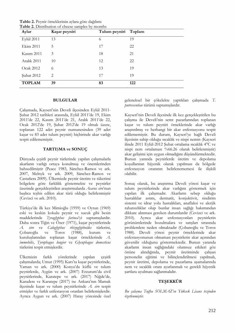
212
Tablo 2. Peynir örneklerinin aylara göre dağılımı Table 2. Distribution of cheese samples by months
Aylar Kaşar peyniri Tulum peyniri Toplam
Eylül 2011 13 6 19
Ekim 2011 5 17 22
Kasım 2011 3 18 21
Aralık 2011 10 12 22
Ocak 2012 6 13 19
Şubat 2012 2 17 19
TOPLAM 39 83 122
BULGULAR Çalışmada, Kayseri’nin Develi ilçesinden Eylül 2011-Şubat 2012 tarihleri arasında, Eylül 2011’de 19, Ekim 2011’de 22, Kasım 2011’de 21, Aralık 2011’de 22, Ocak 2012’de 19, Şubat 2012’de 19 olmak üzere, toplanan 122 adet peynir numunesinden (39 adet kaşar ve 83 adet tulum peyniri) hiçbirinde akar varlığı tespit edilememiştir.
TARTIŞMA ve SONUÇ Dünyada çeşitli peynir türlerinde yapılan çalışmalarla akarların varlığı ortaya konulmuş ve önemlerinden bahsedilmiştir (Peace 1983, Sánchez-Ramos ve ark. 2007, Melnyk ve ark. 2009, Sánchez-Ramos ve Castañera 2009). Ülkemizde peynir üretim ve tüketimi bölgelere göre farklılık göstermekte ve peynirler üzerinde gerçekleştirilen araştırmalarda Acarus siro’nun başlıca teşhis edilen akar türü olduğu belirlenmiştir (Cevizci ve ark. 2010). Türkiye’de ilk kez Mimioğlu (1959) ve Oytun (1969) eski ve keskin kokulu peynir ve sucuk gibi besin maddelerinde Tyraglyphus farinea’yi saptamışlardır. Daha sonra Tiğin ve Özer (1971), kaşar peynirlerinde A. siro ve Caloglyphus rhizoglyphoides türlerini, Çobanoğlu ve Toros (1988), kurum ve kuruluşlarından toplanan kaşar örneklerinde A. immobilis, Tyrophagus longior ve Glycophagus domesticus türlerini tespit etmişlerdir. Ülkemizin farklı yörelerinde yapılan çeşitli çalışmalarda; Umur (1995) Kars’ta kaşar peynirlerinde, Yaman ve ark. (2000) Konya’da küflü ve tulum peynirlerde, Aygün ve ark. (2007) Erzurum’da civil peynirlerinde, Karatepe ve ark. (2017) Niğde’de, Karadere ve Karatepe (2017) ise Ankara’nın Mamak ilçesinde kaşar ve tulum peynirlerinde A. siro tespit etmişler ve farklı enfestasyon oranları belirlemişlerdir. Ayrıca Aygun ve ark. (2007) Hatay yöresinde özel
geleneksel bir çökelekte yaptıkları çalışmada T. putrescentiae türünü saptamışlardır. Kayseri’nin Develi ilçesinde ilk kez gerçekleştirilen bu çalışma ile Develi’nin semt pazarlarından toplanan kaşar ve tulum peyniri örneklerinde akar varlığı araştırılmış ve herhangi bir akar enfestasyonu tespit edilememiştir. Bu durum, Kayseri’ye bağlı Develi ilçesinin sahip olduğu sıcaklık ve nispi nemin (Kayseri ilinde 2011 Eylül-2012 Şubat ortalama sıcaklık 4°C ve nispi nem ortalaması %66.26 olarak belirlenmiştir) akar gelişimi için uygun olmadığını düşündürmektedir. Bunun yanında peynirlerde üretim ve depolama koşullarının hijyenik olarak yapılması da bölgede enfestasyon oranının belirlenememesi ile ilişkili olabilir. Sonuç olarak, bu araştırma Develi yöresi kaşar ve tulum peynirlerinde akar varlığını göstermek için yapılan ilk çalışmadır. Akarların sebep olduğu hastalıklar astım, dermatit, konjuktivit, sindirim sistemi ve idrar yolu hastalıkları, anaflaksi ve alerjik rahatsızlıklar olup bunlar insan sağlığı bakımından dikkate alınması gereken durumlardır (Cevizci ve ark. 2010). Ayrıca akar enfestasyonları peynirlerin görünümlerinde bozulmalara ve satışları sırasında problemlere neden olmaktadır (Çobanoğlu ve Toros 1988). Develi yöresi peynir örneklerinde akar enfestasyonunun olmaması peynirlerin akar açısından güvenilir olduğunu göstermektedir. Bunun yanında akarların insan sağlığındaki olumsuz etkileri göz önüne alındığında, peynir üretiminde çalışan personelin eğitimi ve bilinçlendirilmesi yapılmalı, peynir üretimi, depolama ve pazarlama aşamalarında nem ve sıcaklık oranı ayarlanmalı ve gerekli hijyenik şartlara uyulması sağlanmalıdır.
TEŞEKKÜR Bu çalışma Tuğba SOLMAZ’ın Yüksek Lisans tezinden özetlenmiştir.

213
Çıkar Çatışması: Yazarlar, çıkar çatışması olmadığını beyan eder.
KAYNAKLAR
Aygun O, Yaman M, Durmaz H. A survey on occurrence of
Tyrophagus putrescentiae (Acari: Acaridae) in Surk, a traditional turkish dairy product. J Food Eng. 2007; 78:878-881.
Aygün O, Yaman M, Durmaz H. Erzurum’da tüketime sunulan geleneksel civil peynirinde akar varlığının araştırılması. Fırat Üniv Sağ Bil Derg. 2007; 21(1):41-43.
Cevizci S, Gökçe S, Bostan K, Kaypmaz A. Depo gıdalarını ve peynirleri enfeste eden akarlara halk sağlığı açısından bakış. Türkiye Parazitol Derg. 2010; 34(3):191-199.
Çobanoğlu S, Toros S. Kaşar peynirlerinde zararlı akarlar. Gıda, 1988; 13(6): 409-415.
Çobanoğlu S. Edirne ilinde depolanmış ürünlerde saptanan zararlı ve yararlı acarina türleri ve konukçuları. Türk Entomoloji Derg. 1996; 20(3):199-210.
Göçmen B. Genel Parazitoloji Ders Kitabı, Ege Üniversitesi Basımevi, Bornova-İzmir, 2000.
Karadere A, Karatepe B. Research on occurrence of mites in cheese consumed in mamak district of Ankara province. Kocatepe Vet J. 2019; 12(3):300-304.
Karatepe M, Bağcı C, Karatepe B, Şenel T, Karadere A. Niğde’de tüketime sunulan peynirlerde akar varlığının araştırılması. Gıda. 2017; 42 (4): 431-436.
Kaya M. Develi ovası (Kayseri) ve çevresinin rotifera faunası üzerine taksonomik bir çalışma. Ankara Üniversitesi Fen Bilimleri Enstitüsü, Ankara, 2008.
Melnyk JP, Smith A, Scott-Dupree C, Maarcone MF, Hill A. Identification of cheese mite species inoculated on mimolette and milbenkase cheese througg cryogenic scanning electron microscopy, American Dairy Science Association. J Dairy Sci. 2009; 93:3461-3468.
Mimioğlu MM. Genel ve Özel Tıbbi Arthropodoloji (Tıbbi Entomoloji) AÜ Vet Fak Yay. 111, Ders Kitabı, 51. Desen Matbaası, 818, 1959.
Oytun HŞ. Tıbbi Entomoloji, AÜ Tıp Fak Yay. 218, 552, Ankara, 1969.
Peace DM. Reproductive success of the mite Acarus siro L. on stored cheddar cheese of different ages. J Stored Prod Res. 1983; 19: 97-104.
Sanchez-Ramos I, Alvarez-Alfageme F, Castanera P. Development and survival of the cheese mites, Acarus farris and Tyrophagus neiswanderi (Acari : Acaridae), at constant temperatures and 90% relative humidity, J Stored Prod Res. 2007; 43: 64-72.
Sánchez-Ramos I, Castañera P. Chemical and physical methods for the control of the mite Acarus farris on cabrales cheese. J Stored Prod Res. 2009; 45:61-66.
Süme M. Develi Kazası (1839-1910), Doktora Tezi, Gazi Üniversitesi Sosyal Bilimler Enstitüsü, Ankara, 2008.
Thind BB, Clarke PG. The occurrrence of mites in cereal-based foods destined for human consumption and possible consequences of infestation. Exp Appl Acarol. 2001; 25:203-215.
Tiğin Y, Özer İ. Kaşar peynirinde bulduğumuz akarlar. Ankara
Üniv Vet Fak Derg. 1971; 18: 418-431.
Umur Ş. Kars ili peynirlerinde Acarus siro’nun yaygınlığı. Türkiye Parazitol Derg. 1995; 19(4) : 576-582.
Yaman M, Sevinç F, Altınöz F, Uslu U. Küflü peynirlerde ve tulum peynirlerınde Acarus siro varlığının araştırılması. Türkiye Parazitol Derg. 2000; 24(3): 313-316.

214
Kocatepe Veterinary Journal
Kocatepe Vet J (2020) 13(2):214-218
DOI: 10.30607/kvj.710779 RESEARCH ARTICLE
Protective Effects of Taurine on Imidacloprid-Induced DNA Damage
and Reproductive Performance in The Drosophila melanogaster Model
Damla ARSLAN ACARÖZ1*, Sinan İNCE2, Fahriye ZEMHERİ NAVRUZ3, Nalan BAYŞU SÖZBİLİR4
1Afyon Kocatepe University, Bayat Vocational School, Afyonkarahisar, Turkey
2Afyon Kocatepe University, Faculty of Veterinary Medicine, Department of Pharmacology and Toxicology, Afyonkarahisar, Turkey
3Bartın University, Faculty of Science, Department of Molecular Biology and Genetics, Bartın, Turkey 4Afyon Kocatepe University, Faculty of Veterinary Medicine, Department of Biochemistry, Afyonkarahisar, Turkey
ABSTRACT
Imidacloprid is a neonicotinoid group insecticide and is widely used in veterinary medicine for the control of pests such as lice and fleas in domestic animals. Insecticides are known that they induce toxic effects on living organism, causing oxidative stress and DNA damage. Taurine plays a role in many physiological and biochemical functions and provides an antioxidant effect by stabilization of biological membranes. This study investigated the effect of imidacloprid on DNA damage and reproductive performance and the possible protective effect of taurine in Drosophila melanogasters. Imidacloprid (0.6 µM) alone or in combination with taurine (1, 2 ve 3 mM) were given to broths for 20 days. The results of the study showed that imidacloprid application decreased reproductive performance and increased DNA damage in groups, whereas these effects decreased with taurine administration. In conclusion, it was determined that the adverse effects of imidacloprid regarding DNA and reproductive performance in Drosophila melanogasters were prevented by taurine application. Keywords: DNA damage, Drosophila melanogaster, imidacloprid, taurine
***
Drosophila melanogaster Modelinde Imidakloprid ile İndüklenen DNA Hasarı ve Üreme Performansı
Üzerine Taurinin Koruyucu Etkileri
ÖZ İmidakloprid, neonikotinoid grubu bir insektisid olup evcil hayvanlarda bit ve pire gibi zararlı böceklerin kontrolü için veteriner hekimlik alanında yaygın olarak kullanılmaktadır. İnsektisitlerin canlılarda toksik etki gösterdiği, oksidatif strese ve DNA hasarına neden olduğu bilinmektedir. Taurin birçok fizyolojik ve biyokimyasal olayda rol almakta ve biyolojik membranlarda stabilizasyonunu sağlayarak antioksidan etki göstermektedir. Bu çalışmada imidaklopridin DNA hasarı ve üreme performansına etkisi ve buna karşın taurinin olası koruyucu etkisi Drosophila melanogaster’lerde araştırıldı. İmidakloprid (0,6 µM) tek başına ve taurin (1, 2 ve 3 mM) ile birlikte besi yerlerine 20 gün boyunca ilave edildi. Çalışmanın sonuçları, imidakloprid uygulamasının gruplarda üreme performansını azalttığını ve DNA hasarında artış meydana getirdiğini, buna karşın taurin uygulaması ile bu etkilerin azaldığını gösterdi. Sonuç olarak Drosophila melanogaster’lerde imidaklopridin DNA ve üreme performansında yol açtığı olumsuz etkilerin taurin uygulaması ile engellendiği belirlendi. Anahtar Kelimeler: DNA hasarı, Drosophila melanogaster, imidakloprid, taurin
To cite this article: Arslan Acaröz D. İnce S. Zemheri Navruz F. Bayşu Sözbilir N. Protective Effects of Taurine on Imidacloprid-Induced DNA Damage and Reproductive Performance in The Drosophila melanogaster Model. Kocatepe Vet J. (2020) 13(2):214-218 Submission: 29.03.2020 Accepted: 25.05.2020 Published Online: 26.05.2020
ORCID ID; DAA: 0000-0001-9230-6725, Sİ: 0000-0002-1915-9797, FZN: 0000-0003-1744-1091, NBS: 0000-0002-2270-8312 *Corresponding author e-mail: [email protected]

215
INTRODUCTION Pesticides can be defined as preparations or substances that are employed to control, repel, destroy pests including weeds, insects, rodents, fungi and molds. They possess many different groups and are classified based on their target pests (Costa 2013; Richardson 2019). Pesticides are essential both in agriculture and public health. However, their molecular targets are mainly found to be in pests and also in non-target species such as humans. This situation especially involves neurotoxic pesticides (Bonner and Alavanja 2017, Richardson et al. 2019). Neonicotinoid pesticides have been firstly introduced at the beginning of the 1990s, and widely employed as insecticides throughout the world in many areas including veterinary, agriculture, and residential environment due to their ease of application and high efficacy against insect controls (Zhang et al. 2018). Imidacloprid appeared in the global market as the first representative of neonicotinoids and became best-selling insecticide worldwide (Amjad et al. 2018). Imidacloprid exhibits its effect by ingestion or contact and shows its mode of action by interfering with many nicotinic acetylcholine receptors of the nervous system. This insecticide binds irreversibly to receptors, discharges nerve impulses, and causes failure of a neuron. Imidacloprid has a lower binding affinity towards the nicotinic receptors of mammals than that nicotinic receptors of insects (Kumar et al. 2013). Taurine is a sulphur containing amino acid that is abundantly found intracellular in humans. This amino acid plays important role in many physiological and biological functions such as cholestasis prevention, bile acid conjugation, osmoregulation, membrane stability, neuromodulation of the central nervous system, metabolic properties and antioxidant effects (Lourenço and Camilo 2002, Ince et al. 2017). Taurine deficiency may be resulted in some pathological conditions such as renal dysfunctions, cardiomyopathy, loss in retinal photoreceptors and dysfunctions in pancreatic β cells (Ince et al. 2018). Also, some studies showed protective effect of taurine against genotoxic damage (Türkez and Geyikoğlu 2010, Alam et al. 2011). Drosophila melanogaster, a eukaryote, has a very short and rapid reproductive rate and its many biological, physiological and neurological features are similar to that of mammals. (Pandey and Nichols 2011, Miguel-Aliaga et al. 2018). It is employed as a model organism in oxidative stress (Soares et al. 2017), genotoxicity (Mukhopadhyay et al. 2004) studies. The present study aimed to investigate the effect of taurine against imidacloprid-induced DNA damage and reproductive performance in a Drosophila melanogaster model.
MATERIALS and METHODS
Imidacloprid was provided by Biyoteknik A.Ş. (Istanbul, Turkey) while taurine was purchased from Sigma-Aldrich (St. Louis, MO, USA). In the preparation of culture medium 6 g agar, 94 g sugar, 104 g cornflour, 9 g beer yeast, 6 ml acid mixture (7.83 ml orthophosphoric acid + 8.36 ml propionic acid + 1081 ml distilled water) and 1020 ml distilled water were used. Drosophila melanogaster cultures were allocated into five groups: Group I served as a control, Group II was given only imidacloprid at the dose of 0.6 µM, Group III, IV and V were given both same doses of imidacloprid with Group II and different doses of taurine (1, 2 and 3 mM, respectively). The dose of imidacloprid was determined based on the previous study (Charpentier et al. 2014). Each experimental group possessed 10 male and 10 female unpaired and mature Drosophila melanogaster inoculated in glass culture flasks. These flasks contained 50 ml of broth and the incubation period was 20 days. Drosophila melanogaster cultures were incubated at 60-70% humidity and 24±1°C under laboratory conditions. The present study evaluated pupa numbers and reproductive performance of Drosophila melanogaster groups. The data obtained for pupa numbers and reproductive performances in Drosophila melanogasters were expressed as numbers and percentages. Also, DNA damage (Olive and Banáth 2006) was determined by Comet analysis and expressed as an arbitrary unit (AU). Duncan post-hoc test was performed with one-way analysis of variance (SPSS 20.0) in DNA damage assessment. Statistically, p <0.05 value was considered significant.
RESULTS and DISCUSSION The effects of imidacloprid alone and in combination with taurine on pupa development and reproductive performance in Drosophila melanogasters were given Table 1 while their effects on DNA damage findings were shown in Figure 1. According to the results of the present study, imidacloprid application caused a decrease in pupa development and reproductive performance. However, the concomitant application of taurine increased development and reproductive performance compared to the alone application of imidacloprid. Also, DNA damage in Drosophila melanogasters was found to be higher in the imidacloprid treated group compared to the control group (p<0.05) whereas the taurine application reduced imidacloprid-induced DNA damage (p <0.05). Toxic and genotoxic effects of imidacloprid were evaluated in different models. Feng et al. (2005) assessed the genotoxic effects of imidacloprid in human peripheral blood lymphocytes by sister
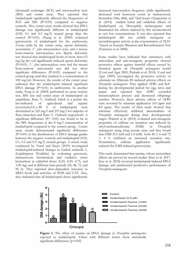
216
chromatid exchanges (SCE) and micronucleus tests (MN) and comet assay. They reported that imidacloprid significantly affected the frequencies of SCE and MN (P<0.05) compared to negative controls. Also, comet assay results showed that DNA damage was significantly different in imidacloprid (0.05, 0.1 and 0.5 mg/l) treated group than the control (P<0.01). Zhang et al. (2000) evaluated genotoxicity of imidacloprid for the earthworm, Eisenia fetida by the comet assay, sperm deformity assessment, V. faba micronucleus tests, and a mouse bone-marrow micronucleus test. Sperm deformity test showed that imidacloprid levels of more than 0.5 mg/kg dry soil significantly induced sperm deformity (P<0.01). V. faba micronucleus tests and the mouse bone-marrow micronuclei test did not show significant differences (P>0.05) compared to the control group until they reached to a concentration of 100 mg/ml. However, the results of the comet assay indicated that the imidacloprid induced significant DNA damage (P<0.01) in earthworms. In another study, Feng et al. (2004) performed an acute toxicity test, MN test and comet assay of imidacloprid on amphibian, Rana N. Hallowell, which is a potent the bio-indicator of agricultural and aquatic ecosystems.LC50-48 h of imidacloprid were determined as 165 mg/l and 219 mg/l for tadpoles of Rana limnocharis and Rana N. Hallowell, respectively. A significant difference (P< 0.05) was found to be in the MN frequencies at the 8 mg/l concentration of imidacloprid compared to the control group. Comet assay results demonstrated significant differences (P<0.01) in the distributions of DNA damage grades between the negative controls and imidacloprid (0.05, 0.1, 0.2 and 0.5 mg/l) treated groups. A recent study conducted by Yucel and Kayis (2019) investigated imidacloprid-induced changes in Galleria mellonella L. (Lepidoptera: Pyralidae) by evaluating genotoxic, immunotoxic biochemical, and oxidative stress biomarkers at sublethal doses (0.25, 0.50, 0.75, and 1.00 mg) and at different time periods (24, 48, 72, and 96 h). They reported dose-dependent increases in MDA levels and activities of SOD and CAT. Also, they indicated that all imidacloprid doses significantly
increased micronucleus frequency while significantly decreased total hemocyte count as immunotoxic biomarker 24th, 48th, and 72nd hours. Charpentier et al. (2014) studied lethal and sublethal effects of Imidacloprid on Drosophila melanogaster and illuminated the effects induced by this neonicotinoid at very low concentrations. It was also reported that imidacloprid did not exhibit mutagenic or recombinogenic activity at the concentration of 5X10-
5 based on Somatic Mutation and Recombination Test (Frantzios et al. 2008). Some studies have indicated that substances with antioxidant and anti-mutagenic properties showed protective effects against harmful effects caused by chemical agents on Drosophila melanogaster model (Uysal and Agar 2005, Prakash et al. 2014). Uysal and Agar (2005) investigated the protective activity of selenium on Aflatoxin B1-induced adverse effects on Drosophila melanogaster. They applied AFB1 and Se4+ during the developmental period for egg, larva and pupae and reported that AFB1 extended metamorphosis process and decreased offsprings number. However, these adverse effects of AFB1 were reversed by selenium application (4.0 ppm and 8.0 ppm). The results of their study showed that selenium effectively inhibited abnormalities of Drosophila melanogaster during their developmental stages. Prakash et al. (2014) evaluated anti-mutagenic properties of caffeine on mutation rate induced by ethyl-methanesulfonate (EMS) in Drosophila melanogaster using wing mosaic assay and they found that EMS (0.5 mM and 1.0 mM) both 48 ± 4 and 72 ± 4 h exhibited an increased mutation rate. Nonetheless, caffeine application significantly reduced the EMS-induced genotoxicity. This study determined that taurine, whose antioxidant effects are proven by several studies (Ince et al. 2017, İnce et al. 2018) reversed imidacloprid-induced DNA damage and ameliorated productive performance of Drosophila melanogaster.
Figure 1. The effect of taurine on DNA damage in Drosophila melanogasters exposed to imidacloprid. Values with different letters show statistically significant differences (p<0.05)

217
Table 1. The effect of taurine on reproductive performance in Drosophila melanogasters exposed to imidacloprid.
Groups Mature Pupa
Female % Male % Total Development %
I Control 23 100 38 100 61 186 100.00
II Imidacloprid (0.6 µM) 5 21.74 6 15.79 11 30 16.13
III Imidacloprid+Taurine (1 mM) 7 30.43 9 23.68 16 49 26.34
IV Imidacloprid+Taurine (2 mM) 14 60.87 16 42.11 30 88 47.31
V Imidacloprid+Taurine (3 mM) 15 65.22 22 57.89 37 115 61.83
CONCLUSION
Consequently, it was determined that imidacloprid negatively affected the reproductive performance and pupa development of Drosophila melanogasters and caused DNA damage. In contrast, taurine application with imidacloprid has been found to increase reproductive performance and pupa development. Also, taurine application prevented DNA damage in Drosophila melanogasters. Conflict of Interest: The authors declare that they have no conflict of interest.
REFERENCES
Alam SS, Hafiz NA, Abd El-Rahim AH. Protective role of
taurine against genotoxic damage in mice treated with methotrexate and tamoxfine. Environ. Toxicol. Pharmacol. 2011;31(1): 143–52.
Amjad A, Azam I, Sarwar MK, Malik MF, Sattar A. A review of imidacloprid toxicity in coccinellids. Arthropods. 2018;7(1): 1–10.
Bonner MR, Alavanja MCR. Pesticides, human health, and food security. Food Energy Secur. 2017;6(3): 89–93.
Charpentier G, Louat F, Bonmatin JM, Marchand PA, Vanier F, Locker D, Decoville M. Lethal and sublethal effects of imidacloprid, after chronic exposure, on the insect model drosophila melanogaster. Environ. Sci. Technol. 2014;48(7): 4096–102.
Costa LG Toxic Effects of Pesticides. In: Klaassen CD (ed) Casarett & Doull’s toxicology: the basic science of poisons. 2013. McGraw-Hill Education, New York.
Feng S, Kong Z, Wang X, Peng P, Zeng EY. Assessing the genotoxicity of imidacloprid and RH-5849 in human peripheral blood lymphocytes in vitro with comet assay and cytogenetic tests. Ecotoxicol. Environ. Saf. 2005;61(2): 239–46.
Feng S, Kong Z, Wang X, Zhao L, Peng P. Acute toxicity and genotoxicity of two novel pesticides on amphibian, Rana N. Hallowell. Chemosphere. 2004;56(5): 457–63.
Frantzios G, Paptsiki K, Sidiropoulou B, Lazaridis I, Theophilidis G, Mavragani-Tsipidou P. Evaluation of insecticidal and genotoxic effects of imidacloprid and acetochlor in Drosophila melanogaster. J. Appl. Entomol. 2008;132(7): 583–90.
İnce S, Acaröz U, Arslan-Acaröz D, Varol N, Gürler Z, Küçükkurt İ, Demirel HH, Eryavuz A. Sıçanlarda Akrilamid ile İndüklenen Oksidatif Strese Karşı Taurinin Koruyucu Etkisi. Kocatepe Vet. J.2018;11: 1–12.
Ince S, Arslan-Acaroz D, Demirel HH, Varol N, Ozyurek HA, Zemheri F, Kucukkurt I. Taurine alleviates malathion induced lipid peroxidation, oxidative stress, and proinflammatory cytokine gene expressions in rats. Biomed. Pharmacother. 2017;96 (May): 263–8.
Kumar A, Verma A, Kumar A. Accidental human poisoning with a neonicotinoid insecticide, imidacloprid: A rare case report from rural India with a brief review of literature. Egypt. J. Forensic Sci. Forensic Medicine Authority; 2013;3(4): 123–6.
Lourenço R, Camilo ME. Taurine: A conditionally essential amino acid in humans? An overview in health and disease. Nutr. Hosp. 2002;17(6): 262–70.
Miguel-Aliaga I, Jasper H, Lemaitre B. Anatomy and Physiology of the Digestive Tract of Drosophila melanogaster. Genetics. 2018;210(2): 357–96.
Mukhopadhyay I, Chowdhuri DK, Bajpayee M, Dhawan A. Evaluation of in vivo genotoxicity of cypermethrin in Drosophila melanogaster using the alkaline Comet assay. Mutagenesis. 2004;19(2): 85-90.
Olive PL, Banáth JP. The comet assay: A method to measure DNA damage in individual cells. Nat. Protoc. 2006;1(1): 23-29.
Pandey UB, Nichols CD. Human Disease Models in Drosophila melanogaster and the Role of the Fly in Therapeutic Drug Discovery. Barker EL, editor. Pharmacol. Rev. 2011;63(2): 411–36.
Prakash G, Hosetti B, Dhananjaya B. Protective effect of caffeine on Ethyl methanesulfonate-induced wing primordial cells of Drosophila melanogaster. Toxicol. Int. 2014;21(1): 96–100.
Richardson JR, Fitsanakis V, Westerink RHS, Kanthasamy AG. Neurotoxicity of pesticides. Acta Neuropathol. 2019;138(3): 343–62.
Soares JJ, Rodrigues DT, Gonçalves MB, Lemos MC, Gallarreta MS, Bianchini MC, Gayer MC, Puntel RL, Roehrs R, Denardin ELG. Paraquat exposure-induced Parkinson’s disease-like symptoms and oxidative stress in Drosophila melanogaster: Neuroprotective effect of Bougainvillea glabra Choisy. Biomed. Pharmacother. 2017;95(July): 245–51.
Türkez H, Geyikoğlu F. The Anti-Genotoxic Effect of Taurine on Aluminum Sulphate-Induced DNA Damage in Human Peripheral Lymphocytes. Eur. J. Biol. 2010;69(1): 25–32.
Uysal H, Agar G. Selenium protective activity against Aflatoxin B1 adverse affects on Drosophila melanogaster. Brazilian

218
Arch. Biol. Technol. 2005;48(2): 227–33.
WHO. 2005. The WHO Recommended Classification of Pesticides by Hazard and Guidelines to Classification. IPCS, Geneva 2004, Switzerland.
Yucel MS, Kayis T. Imidacloprid induced alterations in oxidative stress, biochemical, genotoxic, and immunotoxic biomarkers in non-mammalian model organism Galleria mellonella L. (Lepidoptera: Pyralidae). J. Environ. Sci. Heal. - Part B Pestic. Food Contam. Agric. Wastes 2019;54(1): 27–34.
Zang Y, Zhong Y, Luo Y, Kong ZM. Genotoxicity of two novel pesticides for the earthworm, Eisenia fetida. Environ. Pollut. 2000;108(2): 271–8.
Zhang Q, Li Z, Chang CH, Lou JL, Zhao MR, Lu C. Potential human exposures to neonicotinoid insecticides: A review. Environ. Pollut. 2018;236: 71–81.

219
Kocatepe Veterinary Journal
Kocatepe Vet J (2020) 13(2):219-223
DOI: 10.30607/kvj.670744 RESEARCH ARTICLE
The Serum Amyloid-A, Haptoglobin, Ceruloplasmin and Albumin
Levels in Dogs Which are Infected with Babesia canis
Ali Haydar KIRMIZIGÜL1*, Ekin Emre ERKILIÇ1, Oğuz MERHAN2, Metin ÖĞÜN3, Neslihan ÖLMEZ4, Gencay Taşkın TAŞÇI5, Zati VATANSEVER5
1Kafkas University, Faculty of Veterinary Medicine, Department of Internal Medicine, TR-36100 Kars - Turkey
2Kafkas University, Faculty of Veterinary Medicine, Department of Biochemistry, TR-36100 Kars - Turkey 3Kafkas University, Faculty of Medicine, Department of Biochemistry, TR-36100 Kars - Turkey
4Kafkas University, Kars Vocational School, TR-36100 Kars - Turkey 5Kafkas University, Faculty of Veterinary Medicine, Department of Parasitology, TR-36100 Kars - Turkey
ABSTRACT
In this study, we aimed to determine the serum haptoglobin (Hp), serum amyloid-A (SAA), ceruloplasmin (Cp) and albümin (Alb) levels in the dogs which are naturally infected with Babesia canis. 20 patient dogs which were diagnosed as B. canis by staining of their blood smears with giemsa method, which were brought to Kafkas University Veterinary Faculty Internal Medicine Department clinics formed the material of the study. 10 Healthy dogs were used for control. The serum Hp, SAA, Cp and Alb levels of the sick animals were determined as 0.44±0.12 mg/mL, 43.18±13.77 µg/mL, 10.75±2.45 mg/dL, 2.74±0.15 g/dL, respectively. Whereas, the serum Hp, SAA, Cp and Alb values of the healthy animals were measured as 1.84±0.29 mg/mL, 1.18±0.40 µg/mL, 4.80±0.53 mg/dL, 3.01±0.34 g/dL, respectively. While the SAA and Cp values of the sick animals were determined higher than that of the healthy animals, the (P <0.001), the Hp (P <0.001) and the Alb (P <0.01) levels were found to be lower. As a result, it was determined that the serum SAA and Cp levels increased, and the Hp and Alb levels decreased in the dogs which are infected with B. canis. Keywords: Albumin, Babesia canis, Ceruloplasmin, Dog, Haptoglobin, Serum Amyloid-A
***
Babesia canis ile Enfekte Köpeklerde Serum Amiloid-A, Haptoglobin, Seruloplazmin ve Albumin
Seviyeleri
ÖZ Bu çalışmada Babesia canis ile doğal enfekte köpeklerde serum haptoglobin (Hp), serum amiloid-A (SAA), seruloplazmin (Cp)ve albümin (Alb) seviyelerinin belirlenmesi amaçlanmıştır. Çalışmanın materyalini, Kafkas Üniversitesi Veteriner Fakültesi İç Hastalıkları Anabilim Dalı kliniklerine getirilen, kan frotilerinin giemsa yöntemi ile boyanması sonucunda B. canis tanısı konulan 20 hasta köpek oluşturdu. Kontrol amacı ile 10 sağlıklı köpek kullanıldı. Hasta hayvanların serum Hp, SAA, Cp ve Alb seviyeleri sırasıyla 0.44±0.12 mg/mL, 43.18±13.77 µg/mL, 10.75±2.45 mg/dL, 2.74±0.15 g/dL olarak belirlendi. Sağlıklı hayvanların serum Hp, SAA, Cp ve Alb değerleri ise sırasıyla 1.84±0.29 mg/mL, 1.18±0.40 µg/mL, 4.80±0.53 mg/dL, 3.01±0.34 g/dL olarak ölçüldü. Hasta hayvanların SAA ve Cp değerleri sağlıklı hayvanlara göre daha yüksek bulunurken (P<0.001), Hp (P<0.001) ve Alb (P<0.01) seviyelerinin daha düşük olduğu belirlendi. Sonuç olarak köpeklerde B. canis enfeksiyonunda serum SAA ve Cp değerleri yükselirken, Hp ve Alb seviyelerinin düştüğü belirlendi. Anahtar Kelimeler: Albumin, Babesia canis, Seruloplazmin, Köpek, Haptoglobin, Serum Amiloid-A
To cite this article: Kırmızıgül A.H. Erkılıç E.E. Merhan O. Öğün M. Ölmez N. Taşçı G.T. Vatansever Z. The Serum Amyloid-A, Haptoglobin, Ceruloplasmin and Albumin Levels in Dogs Which Are Infected with Babesia canis. Kocatepe Vet J. (2020) 13(2):219-223 Submission: 06.01.2020 Accepted: 14.05.2020 Published Online: 29.05.2020
ORCID ID; AHK: 0000-0002-6660-2149, EEE: 0000-0003-2461-5598, OM: 0000-0002-3399-0667, MÖ: 0000-0002-2599-8589, NÖ: 0000-0002-2191-8924, GTT: 0000-0002-8590-1101, ZV: 0000-0003-3460-3849 *Corresponding author e-mail: [email protected]

220
GİRİŞ
Kanin babeziozis, köpeklere keneler tarafından taşınan, Babesia türü protozoonlar tarafından oluşturulan ve ölümcül sonuçlara neden olabilen protozoer bir hastalıktır. (Dantas-Torres ve Figueredo 2006, Beck ve ark. 2009, Solano-Gallego ve Baneth 2011, Erkılıç 2019). Hastalık etkenleri eritrositler içerisine yerleşerek eritrositlerin parçalanmasına neden olurlar. Etkenler eritrositlerin içerisinde armut şeklinde görülmektedir. Enfeksiyon evcil köpekler ve yabani karnivorlarda görülmekte olup dünyada yaygındır (Gökçe ve ark. 2013, Sudhakara Reddy ve ark. 2016, Erkılıç 2019). Hastalık klinik olarak perakut, akut, kronik ve subklinik seyretmektedir (Gökçe ve ark. 2013). Klinik olgularda genellikle ateş, anoreksi, depresyon, hemoglobinüri, kusma, ikterus ve anemi görülmektedir (Shah ve ark. 2011).
Doku hasarı sonucu nöro-immüno-humoral sistemin uyarılarak doku hasarına hızlı adaptasyon, zararlı ajanların ortadan kaldırılması ve hasarlı dokunun onarılmasını sağlanmasına akut faz yanıt (AFY) denir (Milanović ve ark. 2018). Akut lokal ve sistemik yangıların dışında kronik yangılarda da AFY meydana gelmektedir (Tuna ve Ulutaş 2015). AFY sonucunda sentezlenen proteinlere akut faz proteinler (AFP) denilmekte olup genellikle karaciğerde hepatositler ve bazı ekstrahepetik alanda üretilmektedir (Gökce ve Bozukluhan 2009, Tuna ve Ulutaş 2015). Bu AFP’lerden biri olan serum amiloid-A (SAA), bir yangı modülatörü olup kolesterolün metabolizmasında ve taşınmasında önemli rol oynar (Sevgisunar ve Şahinduran 2014, Tuna ve Ulutaş 2015). Hastalıklarda serum SAA değerinin ölçülmesi, yangının şiddetinin belirlenmesi, yangısal ve yangısal olmayan hastalıkların ayrımının yapılmasında önem arz etmektedir. Ayrıca hastalığın prognozunun belirlenmesi ve uygulanan tedavinin etkinliğinin değerlendirmesinde de önemlidir (Batırel ve ark. 2003).
Haptoglobin (Hp) karaciğer tarafından sentezlenmekte olup görevi kandaki serbest hemoglobinle stabil kompleksler oluşturarak demir kaybını önlemektir (Sevgisunar ve Şahinduran 2014, Tuna ve Ulutaş 2015). Bunun sonucunda Hp bakteriyel büyüme için gerekli olan demirin kullanılabilirliğini sınırlamakta ve bakteriostatik etki göstermektedir. Ayrıca Hp hemoglobini ve lökositlerin hücre duvarında ana reseptörler olan integrinleri bağlayarak antienflamatuar özellik gösterir (Sevgisunar ve Şahinduran 2014). Köpeklerde serum Hp seviyesi yangı, travma ve enfeksiyon durumlarında artmaktadır (Mcgrotty ve ark. 2003).
Seruloplazmin (Cp), plazmada bakırın taşınmasında görevli bir protein olup özellikle karaciğerde sentezlenmektedir. Bunun dışında ekstrahepatik
alanlarda ve akciğer hava yollu epitellerinden de üretilmektedir. Dokuları demir içeren serbest radikallerin oluşturacağı hasarlardan korur, antioksidan ve hücre koruyucu aktivite gösterir (Sevgisunar ve Şahinduran 2014, Erkılıç 2019).
Albumin (Alb) karaciğer tarafından sentezlenmekte olup en önemli görevi plazma onkotik basıncının dengede tutulmasını sağlamaktadır (Gökce ve Bozukluhan 2009). Akut faz reaksiyonlarda plazma Alb seviyesinde düşüş meydana gelmektedir (Tuna ve Ulutaş 2015). Bu nedenle negatif akut faz proteinleri içerisinde yer almaktadır (Coşkun ve Şen 2011).
Bu çalışmada kanin babesiozis ile doğal enfekte köpeklerde serum Hp, SAA, Cp ve Alb seviyelerinin belirlenmesi amaçlanmıştır.
MATERYAL ve METOT
Çalışmanın materyalini, Kafkas Üniversitesi Veteriner Fakültesi İç Hastalıkları Anabilim Dalı kliniklerine getirilen B. canis enfeksiyonu tanısı konulan 20 hasta ve 10 sağlıklı köpek oluşturdu. B. canis enfeksiyonu tanısı klinik muayeneler ve kan frotilerinin giemsa boyama yöntemi ile boyanarak mikroskobik muayenede (100X) eritrositler içerisinde B. canis etkenlerinin görülmesi ile konuldu. Hasta ve sağlıklı hayvanların V. cephalica accecorius’larından serum tüplerine 5’er mL kan alındı. Bu kanlar 3000 devirde 10 dakika santrifüj edildikten sonra serumları ayrıştırıldı. Serumlar SAA, Hp, Cp ve Alb seviyeleri ölçülene kadar -20ºC’de muhafaza edildi.
Serum örneklerinde albümin konsantrasyonu ticari kit (Biolabo, Fransa) kullanılarak, haptoglobin, serum amiloid A (Tridelta development limited, İrlanda) konsantrasyonu ELISA kitleri ile seruloplazmin ise Colombo ve Ricterich (1964) bildirdikleri yönteme göre kolorimetrik (Epoch, Biotek, USA) olarak belirlendi.
İstatiksel Analiz
Elde edilen sonuçlara SPSS 18 paket programında normalite testi yapıldı ve verilerin normal dağılım gösterdiği tespit edildikten sonra t-testi yapıldı. Tüm sonuçlar ortalama±standart sapma olarak verildi. P değeri 0.05’ten küçük olan sonuçlar istatistiksel olarak anlamlı kabul edildi.
BULGULAR
Hasta hayvanların klinik muayenesinde depresyon, anoreksi, ikterus, anemi ve hemoglobinüri olduğu belirlendi. Kan frotilerinin giemsa boyama yöntemi ile boyanması sonucunda 100X büyüme ile mikroskobik muayenede eritrositler içerisinde B. canis etkenleri görüldü (Resim 1).
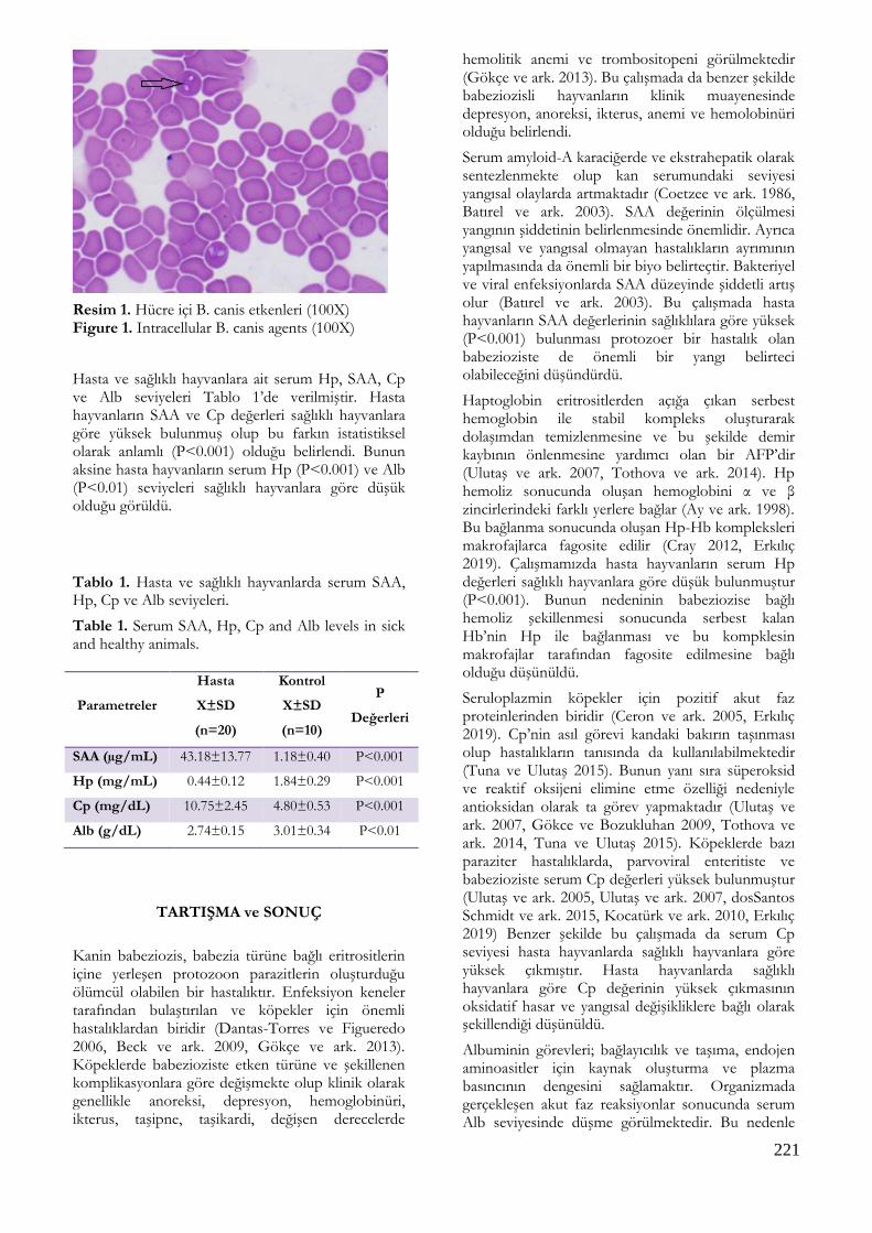
221
Resim 1. Hücre içi B. canis etkenleri (100X) Figure 1. Intracellular B. canis agents (100X)
Hasta ve sağlıklı hayvanlara ait serum Hp, SAA, Cp ve Alb seviyeleri Tablo 1’de verilmiştir. Hasta hayvanların SAA ve Cp değerleri sağlıklı hayvanlara göre yüksek bulunmuş olup bu farkın istatistiksel olarak anlamlı (P<0.001) olduğu belirlendi. Bunun aksine hasta hayvanların serum Hp (P<0.001) ve Alb (P<0.01) seviyeleri sağlıklı hayvanlara göre düşük olduğu görüldü.
Tablo 1. Hasta ve sağlıklı hayvanlarda serum SAA, Hp, Cp ve Alb seviyeleri.
Table 1. Serum SAA, Hp, Cp and Alb levels in sick and healthy animals.
Parametreler
Hasta
X±SD
(n=20)
Kontrol
X±SD
(n=10)
P
Değerleri
SAA (µg/mL) 43.18±13.77 1.18±0.40 P<0.001
Hp (mg/mL) 0.44±0.12 1.84±0.29 P<0.001
Cp (mg/dL) 10.75±2.45 4.80±0.53 P<0.001
Alb (g/dL) 2.74±0.15 3.01±0.34 P<0.01
TARTIŞMA ve SONUÇ
Kanin babeziozis, babezia türüne bağlı eritrositlerin içine yerleşen protozoon parazitlerin oluşturduğu ölümcül olabilen bir hastalıktır. Enfeksiyon keneler tarafından bulaştırılan ve köpekler için önemli hastalıklardan biridir (Dantas-Torres ve Figueredo 2006, Beck ve ark. 2009, Gökçe ve ark. 2013). Köpeklerde babezioziste etken türüne ve şekillenen komplikasyonlara göre değişmekte olup klinik olarak genellikle anoreksi, depresyon, hemoglobinüri, ikterus, taşipne, taşikardi, değişen derecelerde
hemolitik anemi ve trombositopeni görülmektedir (Gökçe ve ark. 2013). Bu çalışmada da benzer şekilde babeziozisli hayvanların klinik muayenesinde depresyon, anoreksi, ikterus, anemi ve hemolobinüri olduğu belirlendi.
Serum amyloid-A karaciğerde ve ekstrahepatik olarak sentezlenmekte olup kan serumundaki seviyesi yangısal olaylarda artmaktadır (Coetzee ve ark. 1986, Batırel ve ark. 2003). SAA değerinin ölçülmesi yangının şiddetinin belirlenmesinde önemlidir. Ayrıca yangısal ve yangısal olmayan hastalıkların ayrımının yapılmasında da önemli bir biyo belirteçtir. Bakteriyel ve viral enfeksiyonlarda SAA düzeyinde şiddetli artış olur (Batırel ve ark. 2003). Bu çalışmada hasta hayvanların SAA değerlerinin sağlıklılara göre yüksek (P<0.001) bulunması protozoer bir hastalık olan babezioziste de önemli bir yangı belirteci olabileceğini düşündürdü.
Haptoglobin eritrositlerden açığa çıkan serbest hemoglobin ile stabil kompleks oluşturarak dolaşımdan temizlenmesine ve bu şekilde demir kaybının önlenmesine yardımcı olan bir AFP’dir (Ulutaş ve ark. 2007, Tothova ve ark. 2014). Hp hemoliz sonucunda oluşan hemoglobini α ve β zincirlerindeki farklı yerlere bağlar (Ay ve ark. 1998). Bu bağlanma sonucunda oluşan Hp-Hb kompleksleri makrofajlarca fagosite edilir (Cray 2012, Erkılıç 2019). Çalışmamızda hasta hayvanların serum Hp değerleri sağlıklı hayvanlara göre düşük bulunmuştur (P<0.001). Bunun nedeninin babeziozise bağlı hemoliz şekillenmesi sonucunda serbest kalan Hb’nin Hp ile bağlanması ve bu kompklesin makrofajlar tarafından fagosite edilmesine bağlı olduğu düşünüldü.
Seruloplazmin köpekler için pozitif akut faz proteinlerinden biridir (Ceron ve ark. 2005, Erkılıç 2019). Cp’nin asıl görevi kandaki bakırın taşınması olup hastalıkların tanısında da kullanılabilmektedir (Tuna ve Ulutaş 2015). Bunun yanı sıra süperoksid ve reaktif oksijeni elimine etme özelliği nedeniyle antioksidan olarak ta görev yapmaktadır (Ulutaş ve ark. 2007, Gökce ve Bozukluhan 2009, Tothova ve ark. 2014, Tuna ve Ulutaş 2015). Köpeklerde bazı paraziter hastalıklarda, parvoviral enteritiste ve babezioziste serum Cp değerleri yüksek bulunmuştur (Ulutaş ve ark. 2005, Ulutaş ve ark. 2007, dosSantos Schmidt ve ark. 2015, Kocatürk ve ark. 2010, Erkılıç 2019) Benzer şekilde bu çalışmada da serum Cp seviyesi hasta hayvanlarda sağlıklı hayvanlara göre yüksek çıkmıştır. Hasta hayvanlarda sağlıklı hayvanlara göre Cp değerinin yüksek çıkmasının oksidatif hasar ve yangısal değişikliklere bağlı olarak şekillendiği düşünüldü.
Albuminin görevleri; bağlayıcılık ve taşıma, endojen aminoasitler için kaynak oluşturma ve plazma basıncının dengesini sağlamaktır. Organizmada gerçekleşen akut faz reaksiyonlar sonucunda serum Alb seviyesinde düşme görülmektedir. Bu nedenle

222
negatif AFP olarak değerlendirilmektedir (Gökce ve Bozukluhan 2009). Çalışmamızda da buna uyumlu olarak hasta hayvanların serum Alb seviyesi sağlıklı hayvanlara göre düşük (P<0.01) bulunmuştur.
Sonuç olarak babeziozisli köpeklerde serum SAA ve Cp değerleri sağlıklılara göre yükselirken Hp ve Alb seviyelerinin düştüğü belirlendi. Serum SAA ve Cp değerlerinin yangıya bağlı olarak yükseldiği tespit edildi. Serum Hp değerindeki düşüşün hemolize bağlı olduğu, Alb değerinde ki düşmenin ise negatif AFP olma özelliğinden ileri geldiği düşünüldü.
TEŞEKKÜR
Bu çalışma Kafkas Üniversitesi Bilimsel Araştırma Projeleri Komisyonu tarafından 2017-TS-40 sayısı ile desteklenmiştir.
Çıkar Çatışması: Yazarlar, çıkar çatışması olmadığını beyan eder.
KAYNAKLAR
Ay M, Gürbilek M, Vatansev H. Akut faz proteinleri. Genel Tıp Derg. 1998; 8(3): 125-132.
Aysul N, Ural K, Ulutaş B, Eren H, Karagenç T. First detection and molecular identification of Babesia gibsoni in two dogs from the Aydın province of Turkey. Turk J Vet Anim Sci. 2013; 37: 226-229.
Batırel A, Gençer S, Özer S. Enfeksiyon göstergesi olarak akut faz reaktanları: C-reaktif protein (CRP) ve serum amiloid A (SAA). Kartal Eğitim ve Araştırma Hastanesi Tıp Dergisi. 2003; 3: 220-224.
Beck R, Vojta L, Mrljak V, Marinculić A, Beck A, Zivicnjak T, Cacciò SM. Diversity of Babesia and Theileria species in symptomatic and asymptomatic dogs in Croatia. Int J Parasitol. 2009; 39: 843-8.
Ceron JJ, Eckersall PD, Martýnez-Subiela S. Acute phase proteins in dogs and cats: current knowledge and future perspectives. Vet Clin Pathol. 2005; 34, 85-99.
Coetzee GA, Strachan AF, van der Westhuyzen DR, Hoppe HC, Jeenah MS, Beer FC. Serum amyloid A-containing human high density lipoprotein 3. Ther Journal of Biological Chemistry. 1986; 261: 9644-9651.
Colombo JP, Richterich R. Zur bestimmung des caeruloplasmin im plasma (on the determination of ceruloplasmin in plasma). Schweiz Med Wochenschr. 1964; 94: 715-720.
Coşkun A, Şen İ. Sığırlarda akut faz proteinleri ve klinik kullanım alanları. Sağlık Bilimleri Dergisi. 2011; 20(3): 240-246.
Cray C. Acute phase proteins in animals. Prog Mol Biol Transl Sci. 2012; 105: 113-150.
Dantas-Torres F, Figueredo LA. Canine babesiosis: a Brazilian perspective. Vet Parasitol. 2006; 141: 197-203.
Dobryszycka W. Biological functions of Haptoglobin. Eur J Clin Biochem. 1997; 35(9): 647-654.
dos Santos Schmidt EM, Rubio CP, dos Santos GJ, Barbosa L, da Motta Santos TF, de Cezaro MC. Serum protein profile of hookworm infection in dogs. Comp Clin Pathol. 2015; 24: 1463-1466.
Erkılıç EE. Babesiosis’li köpeklerde tedavi öncesi ve sonrası haptoglobin, seruloplazmin ve bazı biyokimyasal parametrelerin seviyelerinin belirlenmesi. Harran Üniv Vet Fak Derg. 2019; 8(1): 77-80.
Gökce Hİ, Bozukluhan K. Çiftlik hayvanlarında önemli akut faz proteinleri ve bunların veteriner hekimlik alanındaki kullanımı. Dicle Üniv Vet Fak Derg. 2009; 1(1): 1-14.
Gökçe E, Kırmızıgül AH, Taşçı GT, Uzlu E, Gündüz N, Vatansever Z. Türkiye’de Köpeklerde Babesia canis canis’in Klinik ve Parazitolojik Olarak İlk Tespiti. Kafkas Univ Vet Fak Derg. 2013; 19: 717-720.
Kocaturk M, Martinez S, Eralp O, Tvarijonaviciute A, Ceron J, Yilmaz Z. Prognostic value of serum acute phase proteins in dogs with parvoviral enteritis. J Small Anim Pract. 2010; 51: 478-483.
McGrotty YL, Knottenbelt CM, Ramsey IK, Reid SWJ, Eckersall PD. Haptoglobin concentrations in a canine hospital population. Vet Rec. 2003; 152: 562-564.
Milanović Z, Vekić J, Radonjić V, Božović AI, Zeljković A, Janac J, Spasojević-Kalimanovska V, Buch J, Chandrashekar R, Bojić-Trbojević Ž, Hajduković L, Christopher MM, Kovaćević Filipović M. Association of acute Babesia canis infection and serum lipid, lipoprotein, and apoprotein concentrations in dogs. J Vet Intern Med. 2018; 33: 1686-1694.
Sevgisunar NS, Şahinduran Ş. Hayvanlarda akut faz proteinleri, kullanım amaçları ve klinik önemi. MAKÜ Sag. Bil. Enst. Derg. 2014, 2(1): 50-72.
Shah SA, Sood NK, Tumati SR. Haematobiochemical changes in natural cases of canine babesiosis. Asian J Anim Sci. 2011; 5: 387-392.
Solano-Gallego L, Baneth G: Babesiosis in dogs and cats-expanding parasitological and clinical spectra. Vet Parasitol. 2011; 181: 48-60.
Sudhakara Reddy B, Sivajothi S, Varaprasad Reddy LS, Solmon Raju KG. Clinical and laboratory findings of Babesia infection in dogs. J Parasit Dis. 2016; 40: 268-272.
Tothova C, Nagy O, Kovac G. Acute phase proteins and their use in the diagnosis of diseases in ruminants: a review. Veterinarni Medicina. 2014; 59: 163-180.
223
Tuna GE, Ulutaş B. Hastalıkların biyobelirteçleri olarak akut faz proteinleri. Turkiye Klinikleri J Vet Sci Intern Med-Special Topics. 2015; 1: 8-19.
Ulutaş B, Bayramlı G, Alkim Ulutaş B, Karagenç T. Serum concentration of some acute phase proteins in naturally occuring canine abesiosis: A preliminary study. Vet Clin Pathol. 2005; 34:144-147.
Ulutaş PA, Ulutaş B, Sarıerler M, Bayramlı G. Serum haptoglobin and ceruloplasmin concentrations in dogs with various diseases. İstanbul Üniv Vet Fak Derg. 2007; 33: 35-42.

224
Kocatepe Veterinary Journal
Kocatepe Vet J (2020) 13(2):224-227
DOI: 10.30607/kvj.610338 CASE REPORT
A Case of A 13-Year-Old Dog with Old Dog Encephalitis: A Rare Form
of Canine Distemper
Erdem GÜLERSOY1*, Mahmut OK1, Mutlu SEVİNÇ1, Murat Kaan DURGUT1, Amir NASERİ1
1Selcuk University, Faculty of Veterinary Medicine, Department of Internal Medicine, 42003, Konya, TURKEY
ABSTRACT
13 year-old male dog with complaints of fever, loss of appetite, stagnation as well as respiratory signs such as purulent nasal and eye discharge, cough and neurologic signs including apathy, ataxia, quadriplegia, muscular atrophy and myoclonus which admitted to the Animal Hospital of Faculty of Veterinary Medicine of Selcuk University. “Old dog encephalitis” was determined by clinical, laboratory examinations and rapid distemper test. Clinical findings revealed hyperthermia, tachypnea and tachycardia. Intravenous 0.9% NaCl and 5% dextrose solution, vitamin and amino acids, ceftriaxone, n-acetylcysteine for improving clinical appearance and for controlling myoclonus pregabalin were used. In conclusion canine distemper virus may affect mature dogs over six years old as “old dog encephalitis” and this condition may provide a valuable model for further study of demyelinating diseases including measles in humans. Keywords: Canine Distemper, Demyelination, Encephalitis, Old Dog
***
13 Yaşlı Bir Köpekte “Old Dog Ensefalit” Olgusu: Köpek Distemper’ının Ender Formu
ÖZ 13 yaşlı erkek bir köpek ateş, iştahsızlık, durgunluk gibi genel; purulent burun ve gözyaşı akıntısı, öksürük gibi respiratorik; apati, ataksi, kuadripleji, kas atrofisi ve miyoklonus gibi nörolojik bulgular ile Selçuk Üniversitesi Veteriner Fakültesi Hayvan Hastanesine getirilmiştir. Klinik muayene, laboratuvar analizleri ve yapılan distemper hızlı kiti ile “old dog ensefalit” tespit edilmiştir. Klinik muayenede vücut ısısı, solunum sayısı ve kalp ritminde artış belirlenmiştir. Tedavi olarak intravenöz 0.9% NaCl ve 5% dekstroz solüsyonları, vitamin ve aminoasitler, seftriakson, N-asetil sistein, miyoklonusu kontrol altına almak için pregabalin uygulanmıştır. Sonuç olarak köpeklerin distemper virüsünün 6 yaşından büyük erişkin köpekleri “old dog ensefalit” olarak etkileyebileceği ve bu durumun insanlarda kızamık dahil demiyelinizasyon hastalıklarının daha ileri çalışmaları için değerli bir model olabileceği kanısına varılmıştır. Anahtar Kelimeler: Distemper, Demiyelinizasyon, Ensefalitis, Yaşlı Köpek
To cite this article: Gülersoy E. Ok M. Sevinç M. Durgut M.K. Naseri A. A Case of A 13-Year-Old Dog with Old Dog Encephalitis: A Rare Form of Canine Distemper. Kocatepe Vet J. (2020) 13(2):224-227 Submission: 24.08.2019 Accepted: 26.02.2020 Published Online: 02.05.2020 ORCID ID; EG: 0000-0001-8511-0150, MO: 0000-0002-8210-6735, MS: 0000-0002-9805-5194, MKD: 0000-0002-4582-8536, AN: 0000-0001-9140-5879 *Corresponding author e-mail: [email protected]

225
INTRODUCTION
Canine distemper is an infectious disease, which is closely related to measles virus, a single-stranded, non-segmented, enveloped, RNA virus in the Paramyxoviridae family and genus Morbillivirus, infects variety of species. Virus can be found in dogs, wild dogs, jackals, foxes, hyenas and also in wild cats but not in domestic cats. The domestic dog is the most effected species. Morbilliviruses are transmitted by aerosols to the upper respiratory tract and cause clinical symptoms such as cough, fever and serous nasal discharge, as well as gastrointestinal signs such as vomiting and diarrhea complicated by secondary bacterial infections. When the nervous system is affected by canine distemper virus (CDV) dullness, progressive weakness, ataxia, paraplegia or quadriplegia, myoclonus, tremor, incontinence, seizures, depression, circling, head pressing, and visual deficits can be observed (Martella et al. 2008). Cordy (1942) described an encephalitis in mature dogs that was characterized pathologically by lymphomonocytic perivascular inflammatory cell infiltrates in the central nervous system (CNS) and intranuclear inclusion bodies in neurons. Since the original description by Cordy (1942), any other report for ODE has not published. Clinical appearance was described as ataxia, convulsions, and circling. In mature dogs, this type of progressive panencephalitis is defined as old dog encephalitis (ODE). This type of encephalitis has not been seen in young dogs (Lincoln et al. 1971). The rarity and unusual morphologic characteristics of ODE are interesting. Different hypotheses have been proposed to elucidate the pathogenesis and occurrence of ODE. In mature dogs over six years old, viral perseverance in the CNS after acute infection which is characterized by progressive inflammation of the grey matter in brain hemispheres may trigger old dog encephalitis (Carvalho et al. 2012). Neurologic signs may also occur without gastrointestinal or respiratory findings. The brain lesions mostly seen in cerebellopontine angle. These brain lesions consist of multifocal demyelination areas and necrosis of the white matter. The relationship of this multifocal encephalitis to old dog encephalitis is still not explained (Vandevelde et al. 1980). CDV-induced encephalitis can be described in four forms. These forms are; acute and severe encephalitis in young dogs, with multisystemic appearance,
including neurological signs; chronic encephalitis in adult dogs, with possibility of common neurological findings; old dog encephalitis; and chronic, relapsing encephalitis, the latter with minor occurrence (Carvalho et al. 2012). Case History 13 year-old male Anatolian shepherd dog presented with neurologic signs of depression and unsteady gait for a few months. Clinical examination revealed diarrhea, mucopurulent nasal and ocular discharge (Figure 1a), dispnea, fever (39.8 ºC) abnormal respiratory sounds, head tilt, tremors, muscle atrophy and myoclonus all over the patient’s body (Figure 1b). During physical examination findings such as nystagmus, ear scratching, rubbing and rotating did not observed. Also because dog was reacting to the environmental sounds, deafness was not considered. To confirm CDV, rapid distemper test (ASAN Easy Test, Canine Distemper Virus Antigen Test, Cat. No. AM9125-K, Seoul, Korea, relative sensitivity: 97.96%, relative specificity: 97.50%) was applied according to the manufacturer’s instructions (Figure 1c). Complete blood count, blood gases analysis and cerebrospinal fluid (CSF) analysis were performed and presented in Table 1 and 2, respectively. Before the collection of CSF, the dog was sedated with xylazine at 1 mg / kg dosage intramuscular injection. CSF was collected between the occipital bone and the atlas using 22 gauge, 1.5-inch spinal needles with a stylet as Tipold (2003) reported. Excessive flexion of the head to collect CSF was avoided to prevent obstruction of the airway. For improving clinical appearance, intravenous 0.9% NaCl and 5% dextrose solution, vitamin and amino acids, ceftriaxone at 25 mg / kg dosage and n-acetylcysteine at 100 mg / kg dosage were used. For controlling myoclonus, head tilt and tremors pregabalin at 4 mg / kg dosage were used orally. Because the owner refused euthanasia we were unable to perform necropsy and add the necropsy findings to the report. After a few weeks it was learned from the owner that the euthanasia was made in another veterinary clinic due to worsening the clinical appearance.

226
Figure 1.General appearance of the dog (a), purulent ocular discharge (b) and positive rapid distemper test (c).
Table 1. Blood gases analysis and CBC
Blood Gases Range Haemogram Range
pH 7.38 7.35 – 7.45 WBC m/mm3 26.53 ↑ 5.0 – 19.0 pCO2 mmHg 30.3 ↓ 40 – 45 Lym % 20 5.0 – 30.0 pO2 mmHg 26.2 ↓ 30 – 42 Mon % 3.40 2.0 – 6.0 K mmol/L 3.9 3.4 – 5.6 Gra % 76.60 40.0 – 80.0 Na mmol/L 149 ↓ 150 – 165 Lym # m/mm3 5.30 0.2 – 5.7 Ca mmol/L 0.96 ↓ 2.0 – 2.7 Mon # m/mm3 0.90 0.1 – 1.1 Cl mmol/L 114 104 – 128 Gra # m/mm3 20.33 ↑ 2.0 – 15.2 Glukoz mg/dL 43 ↓ 64 – 170 RBC M/mm3 9.30 ↑ 4.0 – 9.0 Lactate mmol/L 10 ↑ 0 – 2 MCV fl 59.1 ↑ 35.5 – 55.0 Hct % 51.2 ↑ 29 – 48 Hct % 54.9 ↑ 24.0 – 45.0 Base(ecf) mmol/L -6.8 ↑ -4 – 4 MCH pg 18.4 16.0 – 24.0 Base (B) mmol/L -5.4 ↑ -4 – 4 MCHC g/dl 31.3 28.0 – 40.0 HCO3 (P,st) mmol/L 18.8 ↓ 19 – 24 RDW 10.7 8.0 – 12.0 HCO3 (P) mmol/L 28.2 ↓ 19 – 24 THR # m/mm3 166 120 - 500
Table 2. Cerebrospinal fluid analysis
Analysis Results Range
Leukocyte (cell/mm3) - <5 Glucose (mmol/L)* 2.8 35 Protein (mg/dl) - <30 Specific gravity 1.015 1.005
* %60-80 concentration of blood glucose
b a
c
227
DISCUSSION Canine distemper is an important viral disease which affects variety of species but mostly dogs in a multi systemic manner. At about 10 days post infection, CDV starts to spread from primer replication site which is the lymphoid tissue (Vandevelde and Zurbriggen 2005). In th body, CDV infects various cell types including epithelial, mesenchymal, hematopoietic, and neuroendocrine cells. Clinical findings include immunosuppression, respiratory and gastrointestinal signs, and demyelinating leukoencephalitis. CDV mediates the early destruction of the lymphocytes results in immunosuppression. Ezeibe and Udegbunam (2008) reported in dogs with distemper virus, leukopenia initially associated with lymphopenia and later lymphocytosis which leads to leucocytosis. In this case report, significant leukocytosis observed probably due to secondary bacterial infection. Also our haemogram findings were compatible as Ezeibe and Udegbunam (2008) reported. In CNS, CDV causes severe demyelinating lesions. Cytokines such as interleukin 8, interleukin 10 and interferon gamma can be measured in CSF. But an elevation of cytokines levels in CSF are not specific for particular diseases. Nevertheless in the CSF of animals with acute myelin lesions high interleukin 8 titers can be found (Vandevelde and Zurbriggen 2005). In non-inflammatory distemper cases, protein and cell content of CSF may be in normal ranges. In this case report the results of CSF analysis were compatible as Tipold (2003) reported. Regardless of its cause, demyelination occurs thru two main processes: direct damage to the myelin itself or to myelinogenic cells which is described as primary demyelination or axonal injury. This type of injury promotes a secondary effect in the myelinic degeneration and referred to secondary demyelination (Carvalho et al. 2012). In addition, virus-induced microglial cell activation probably plays a role in demyelinating process (Vandevelde and Zurbriggen 2005). This neurologic disease is characterized by mental impairment and occurs mostly in middle aged dogs (Vandevelde et al. 1980). As the naturally occurring incidence of ODE is low and generally the cause is related to CDV, old dog encephalitis is a rare condition and a valuable model for severe demyelinating diseases of dogs and humans. Adams et al. (1975) indicate that to elucidate the mechanisms of demyelination diseases both in animals and humans, a cooperation of veterinarians and physicians is required. It is not possible to differentiate old dog encephalitis and multifocal distemper encephalitis by breed, sex or initial neurologic signs. In conclusion, old dog encephalitis is a rare CDV-induced subacute neurologic disease affecting mature
dogs over six years old and it would be a beneficial animal model for the further study of demyelinating diseases in humans including chronic progressive paramyxovirus infections as CDV closely related to measles virus. Conflict of Interest: The authors declare that they have no conflict of interest.
REFERANCES
Adams JM, Brown WJ, Snow HD, Lincoln SD, Sears AW,
Barenfus M, Holliday TA, Cremer NE, Lennette EH. Old Dog Encephalitis and Demyelinating Diseases in Man. Veterinary Pathology. 1975; 12: 220-226.
Carvalho OV, Botelho CV, Ferreira CGT, Scherer PO, Soares-Matins JAP, Almeida MR, Junior AS. Immunopathogenic and Neurologic Mechanisms of Canine Distemper Virus. Adv Virol. 2012; 2: 163860.
Ezeibe MCO, Udegbunam RI. Haematology of dogs infected with canine distemper virus. Sokoto J Vet Sci. 2008; 7: 30-32.
Lincoln SD, Gorham JR, Hegreberh GA. Etiologic Studies of Old Dog Encephalitis. Veterinary Pathology. 1971; 8: 1-8.
Martella V, Elia G, Buonavoglia C. Canine distemper virus. Vet Clin Small Anim. 2008; 38:787–797.
Tipold A. Cerebrospinal Fluid. Clinical Neurology in Small Animals - Localization, Diagnosis and Treatment, K.G. Braund (Ed.) Publisher: International Veterinary Information Service. Ithaca, New York, USA. 2003.
Vandevelde M, Kristensen B, Braund KG, Greene CE, Swango LJ, Hoerlein BF. Chronic Canine Distemper Virus Encephalitis in Mature Dogs. Veterinary Pathology. 1980; 17: 17-29.
Vandevelde M, Zurbriggen A. Demyelination in canine distemper virus: a review. Acta Neuropathol. 2005; 109: 56-68.
Instruction for Authors
Kocatepe Veterinary Journal (KVJ)has the policy with One Medicine OneHealth. Research article, reviews, brief communication and case reports, lettersto editor and book reviews are also welcome for consideration to publisharticles of high scientific and ethical standards. The journal is published four times a year. The publication of the text andfigures is free of change. Acceptance of papers for the KVJ is undertaken by Editors. Editorial Board members adjudicate in the case of conflicting or adverse reports. Manuscripts are accepted for consideration on the understanding thatthey are for publication solely in KVJ and that they neither have beenpublished nor are under consideration for publication elsewhere. Submissionalso implies that all authors have approved the paper for release and are inagreement with its content. Upon acceptance of the article by the journal, theauthor(s) will be asked to transfer the copyright of the article to thePublisher. Each author accepts all ethical responsibility of the article and all authors agree with the content of the study. After article is checked by Professional Plagiarism Prevention program, article will be sent to authors. Articles are checked by iThenticate® program, when plagiarism or self-plagiarism are detected, they will not be evaluated for publication. If animals are used in the studies, study should be approved by an Ethical Committee, Name of Ethical Committee and Approved Number should be mentioned in the Material and Method section. Editor may be reject directly the article, if animal is exposed to stressful or painful conditions. Authors accept ethical rules when article is sent for publication. Author(s) should send CopyrightTransfer Agreement, after acceptance of article. Each author accepts all ethical responsibility of the article and allauthors agree with the content of the study. Article should be written using Garamond, font of 11 point, with 1.5 line spacing, margins of the A4 paper should be 2.5 cm from all edges (Word97-2010.doc). Abbreviations should be written in SI. Research article submittedto Kocatepe Veterinary Journal should be divided into the following sections: Title page (Abstract, Key words without authors name and adress), Materials and Methods, Results, Discussion, Conclusions, References,Tables, Graphics, Figures. Title page: Papers should be headed with the full title, the initial letters ofname and surnames of the authors, the name and address of the institution wherethe work is carried out. The telephone number, fax number and e-mail address ofthe corresponding author should also be provided. The title should be short,specific and informative.
Abstract Should be no more than 200words, outlining in a single paragraph. Keywords, 5 keywords that describe the crucial points of the paper should beprovided. Keywords should be chosen fromTurkey Science Term (www.bilimterimleri.com)
Introduction, an updated literature related to paper and aim(s) of the study shouldbe clearly given in this section. Materials and methods, a clear account of materialsused and methods employed should be given and it should beapplicaple/repeatable by other researhers. Results, as concise as possible. Text, tables and figures illustrating the samedata should be limited and succinctly outline the pertinent outcomes of thestudy. Discussion: Results of the study should be discussed with directly relevantreferences. This section may also be divided into subsections. Conclusions: This section should state clearly the main conclusions of theresearch. Results should not be repeated. Acknowledgements, it is adviced to acknowledge persons or institutions directly orindirectly involved in the study. References References in the text should be made as follows: Kara (2012) described. / . was reported (Zemheri 2015, Eryavuz and Yeni, Eryavuz et al. 2015). List of references should be given alphabetically in the reference list. Different publications having the same author(s) of same year should be writtenas 2011a, 2011b. Web address should be referenced as anonim for example Anonim 2015. Only official web pages should be used. Author name(s) and date sould be written bold. The reference list at the end of the paper should be written asbelow.

Journal: Ince S, Kucukkurt I, Cigerci IH, Fidan AF, Eryavuz A. The effects of dietary boric acid and borax supplementation on lipid peroxidation, antioxidant activity, and DNA damage in rats. J Trace Elem Med Biol. 2010; 24(3):161-164. Book section: Juneja R, Koide SS.Molecular Biology of Reproduction, In: Reproduction in Farm Animals, Ed; HafezB, Hafez ESE, 7th Ed., LippincottWilliams and Wilkins, Philadelphia, USA. 2000; pp. 354-361. Web page: Anonymous. http://www.tuik.gov.tr/VeriBilgi.do?tb_id=46&ust_id=13;Accessien date: 02.01.2012. Thesis: Yeni D. Some andrological parameters and biochemical properties in relation toseason in rams. PhD thesis, Afyon Kocatepe University Health Science Institute, Afyonkarahisar,2010. Tables: Tables should be presented in a separate page atthe end of manuscript. Graphics: Figures should be presented in a separate page atthe end of manuscript. Figures : Figures should be presented in a separate page atthe end of manuscript. Figures should be 80 or 160 mm, minimum 300 dpi. Titles of tables, graphics and figures should be both Turkish and English. Brief Communications: Briefcommunications should be consise but complete description of a limitedivestigation, which will not be included in a later publication. They shouldnot exceed 1600 words. They should bear no more than two tables or figures. AnABSTRACT should be given but no other sections. Typescripts should be clearlymarked Brief Communication. Review Articles: Review articles related to allmedical topics are welcome for publication. They should give an update onrecent advances in a particular field and be targeted at research veterinariansor clinicians who are not necessarily working in the same field. The lengthshould not exceed 4500 words. It should have a precise abstract. Author ofreview should have at least two citations. For each issue maximum 2 reviews arepublished. Case Reports: Reports of SINGLE or small numbers of cases willbe considered for publication in KVJ if the case(s) are particularlyunusual/rare or the report contributes materially to the literature. A casereport should not exceed 1500 words and must comprise a Summary (maximum 150words), Introduction, Case History and Discussion. The report should accomplishone of the followings:
To be a substantially novel presentation
To be a technique or treatment that wouldsubstantially alter management and prognosis of the described condition
The first clinical report or first case(s) ofdiseases in a particular location where epidemiology is an important factor
– To exemplify bestpractice in medical science. Letters to The Editor: Letters describingcase reports or original material may be published in the KVJ and will bepeer-reviewed prior to publication. Letters making critisisms on recentlypublished papers in the KVJ will also be considered and the correspondingauthors of the original paper will be invited to respond accordingly. All articles sent to KVJ (Kocatepe Veterinary Journal) ONLINE submition only. During submition documentswhish are listed below, have to instal to the system; 1. Title Page: Author and institution names 2. Main text: Author and institution names should NOT be.Tables(s), graphic(s) and figure(s) etc. Should be an the last page of article,olso title of them both in Turkish and English. 3. Article addition: Table(s), graphic(s) andfigure(s) should have been installed to the system separately. 4. Author Approval Form (Cover Letter): All authorsneed to sing it and install to the system. Signatures should be wet signuturesand send to the Editorial Board of Kocatepe Veterinary Journal. 5. Copyright: All authors need to sing it and install to thesystem. Signatures should be wet signutures and send to the Editorial Board ofKocatepe Veterinary Journal.