Kinematic analysis of the daily activity of drinking from a glass in a population with cervical...
12
RESEARCH Open Access Kinematic analysis of the daily activity of drinking from a glass in a population with cervical spinal cord injury Ana de los Reyes-Guzmán * , Angel Gil-Agudo, Benito Peñasco-Martín, Marta Solís-Mozos, Antonio del Ama-Espinosa, Enrique Pérez-Rizo Abstract Background: Three-dimensional kinematic analysis equipment is a valuable instrument for studying the execution of movement during functional activities of the upper limbs. The aim of this study was to analyze the kinematic differences in the execution of a daily activity such as drinking from a glass between two groups of patients with tetraplegia and a control group. Methods: A total of 24 people were separated into three groups for analysis: 8 subjects with metameric level C6 tetraplegia, 8 subjects with metameric level C7 tetraplegia and 8 control subjects (CG). A set of active markers that emit infrared light were positioned on the upper limb. Two scanning units were used to record the sessions. The activity of drinking from a glass was broken down into a series of clearly identifiable phases to facilitate analysis. Movement times, velocities, and the joint angles of the shoulder, elbow and wrist in the three spatial planes were the variables analyzed. Results: The most relevant differences between the three groups were in the wrist. Wrist palmar flexion during the back transport phase was greater in the patients with C6 and C7 tetraplegia than in the CG, whereas the highest wrist dorsal flexion values were in forward transport in the subjects with C6 or C7 tetraplegia, who required complete activation of the tenodesis effect to complete grasping. Conclusions: A detailed description was made of the three-dimensional kinematic analysis of the task of drinking from a glass in healthy subjects and in two groups of patients with tetraplegia. This was a useful application of kinematic analysis of upper limb movement in a clinical setting. Better knowledge of the execution of this movement in each of these groups allows therapeutic recommendations to be specifically adapted to the functional deficit present. This information can be useful in designing wearable robots to compensate the performance of AVD, such as drinking, in people with cervical SCI. Background Upper limb functionality is fundamental for the execu- tion of basic activities of daily living (ADL) like drinking, eating and personal hygiene. Impaired upper limb func- tion is one of the most common sequelae in central ner- vous system injury [1,2]. In the case of spinal cord injury (SCI), the upper limb is affected in more than 50% of cases [3]. Upper limb strength is impaired to some extent in people who have suffered cervical SCI, making it difficult for them to perform many ADLs essential for their autonomy. They may require technical assistance. Therefore, these patients experience sharp limitations in their level of activity and participation in the social setting, as people who have suffered another central nervous system injury, such as stroke [4]. Until now, upper limb function has been evaluated using a series of scales, such as the Fugl-Meyer Assess- ment, Frenchay Arm Test, Motor Assessment Scale, Box and Block Test, and the Nine-Hole Peg Test [5,6]. These tools are sensitive to gross functional changes, but less sensitive in measuring small and more specific * Correspondence: [email protected] Biomechanics and Technical Aids Unit, National Hospital for Spinal Cord Injury. SESCAM, Finca la Peraleda s/n, Toledo, Spain de los Reyes-Guzmán et al. Journal of NeuroEngineering and Rehabilitation 2010, 7:41 http://www.jneuroengrehab.com/content/7/1/41 JNER JOURNAL OF NEUROENGINEERING AND REHABILITATION © 2010 de los Reyes-Guzmán et al; licensee BioMed Central Ltd. This is an Open Access article distributed under the terms of the Creative Commons Attribution License (http://creativecommons.org/licenses/by/2.0), which permits unrestricted use, distribution, and reproduction in any medium, provided the original work is properly cited.
-
Upload
independent -
Category
Documents
-
view
3 -
download
0
Transcript of Kinematic analysis of the daily activity of drinking from a glass in a population with cervical...
RESEARCH Open Access
Kinematic analysis of the daily activity of drinkingfrom a glass in a population with cervical spinalcord injuryAna de los Reyes-Guzmán*, Angel Gil-Agudo, Benito Peñasco-Martín, Marta Solís-Mozos,Antonio del Ama-Espinosa, Enrique Pérez-Rizo
Abstract
Background: Three-dimensional kinematic analysis equipment is a valuable instrument for studying the executionof movement during functional activities of the upper limbs. The aim of this study was to analyze the kinematicdifferences in the execution of a daily activity such as drinking from a glass between two groups of patients withtetraplegia and a control group.
Methods: A total of 24 people were separated into three groups for analysis: 8 subjects with metameric level C6tetraplegia, 8 subjects with metameric level C7 tetraplegia and 8 control subjects (CG). A set of active markers thatemit infrared light were positioned on the upper limb. Two scanning units were used to record the sessions. Theactivity of drinking from a glass was broken down into a series of clearly identifiable phases to facilitate analysis.Movement times, velocities, and the joint angles of the shoulder, elbow and wrist in the three spatial planes werethe variables analyzed.
Results: The most relevant differences between the three groups were in the wrist. Wrist palmar flexion during theback transport phase was greater in the patients with C6 and C7 tetraplegia than in the CG, whereas the highestwrist dorsal flexion values were in forward transport in the subjects with C6 or C7 tetraplegia, who requiredcomplete activation of the tenodesis effect to complete grasping.
Conclusions: A detailed description was made of the three-dimensional kinematic analysis of the task of drinkingfrom a glass in healthy subjects and in two groups of patients with tetraplegia. This was a useful application ofkinematic analysis of upper limb movement in a clinical setting. Better knowledge of the execution of thismovement in each of these groups allows therapeutic recommendations to be specifically adapted to thefunctional deficit present. This information can be useful in designing wearable robots to compensate theperformance of AVD, such as drinking, in people with cervical SCI.
BackgroundUpper limb functionality is fundamental for the execu-tion of basic activities of daily living (ADL) like drinking,eating and personal hygiene. Impaired upper limb func-tion is one of the most common sequelae in central ner-vous system injury [1,2]. In the case of spinal cord injury(SCI), the upper limb is affected in more than 50% ofcases [3]. Upper limb strength is impaired to someextent in people who have suffered cervical SCI, making
it difficult for them to perform many ADLs essential fortheir autonomy. They may require technical assistance.Therefore, these patients experience sharp limitations intheir level of activity and participation in the socialsetting, as people who have suffered another centralnervous system injury, such as stroke [4].Until now, upper limb function has been evaluated
using a series of scales, such as the Fugl-Meyer Assess-ment, Frenchay Arm Test, Motor Assessment Scale, Boxand Block Test, and the Nine-Hole Peg Test [5,6].These tools are sensitive to gross functional changes,but less sensitive in measuring small and more specific
* Correspondence: [email protected] and Technical Aids Unit, National Hospital for Spinal CordInjury. SESCAM, Finca la Peraleda s/n, Toledo, Spain
de los Reyes-Guzmán et al. Journal of NeuroEngineering and Rehabilitation 2010, 7:41http://www.jneuroengrehab.com/content/7/1/41 J N E R JOURNAL OF NEUROENGINEERING
AND REHABILITATION
© 2010 de los Reyes-Guzmán et al; licensee BioMed Central Ltd. This is an Open Access article distributed under the terms of theCreative Commons Attribution License (http://creativecommons.org/licenses/by/2.0), which permits unrestricted use, distribution, andreproduction in any medium, provided the original work is properly cited.
changes [7]. Moreover, the use of these scales is notexempt from a degree of subjectivity.In contrast with the lower limb, the upper limb has
extensive functionality due to the mobility of numerousjoints that can execute fine movements thanks to com-plex neuromuscular control [7]. For that reason, objec-tive measurement elements and exact systems ofmovement analysis are necessary to be able to describeupper limb activities more precisely and specifically. Bio-mechanical analysis and, specifically, kinematic analysistechniques are interesting tools for obtaining objectivedata. At present, complex systems of kinematic analysisallow the automated analysis of movement in threedimensions. The biomechanical model of the lower limbhas been implemented for most equipment because gaitis one of the movements most analyzed by biomechanicslaboratories. Consequently, in order to analyze theupper limb it is necessary to previously define anddevelop the biomechanical model based on the activityto be analyzed.Kinematic studies have been made of the upper limb
in which reaching/grasping movements on a horizontalplane as a free movement without arm support [8] andwith arm support [9-11] have been analyzed. However,the analysis of purpose-oriented movements must beproposed because the musculoskeletal system has poten-tially a larger number of ways to achieve the motor task,permitting the organism to adapt to different environ-mental conditions. So, the musculoskeletal system takesadvantage of this feature of the motor apparatus byselecting a desired trajectory and an interjoint coordina-tion among many possible strategies to make goal-oriented movements [12-14]. Studies have beenpublished on kinetic analysis of the shoulder and elbowin healthy subjects performing a set of ADLs [15,16]and on complete kinematic analysis of the upper limbduring the movement of drinking from a glass [7].It has been confirmed that the characteristics of
movement can vary depending on the objective to becompleted. For example, the kinematics of the upperlimb is not the same in pointing to an object as when agrasping function is added [11,17,18].Several studies have been published recently on the
three-dimensional analysis of ADLs in healthy subjects[7,8,19,20]. Similar studies have been made in patientswith different neurological conditions [21-23].Although there have been few reports in patients withSCI, the results of the kinematics of grasping and themovements of pointing toward an object in patientswith C6 tetraplegia have been described [24]. However,we found no studies of the kinematic analysis of theupper limb when performing a functional activity likedrinking from a glass in patients with different levelsof cervical SCI.
One working hypothesis has been that differences arelikely to exist between people with cervical SCI and peo-ple without such an injury. On the other hand, the dif-ferent levels of injury affect the upper limb musculaturedifferently and such differences should be manifested bytheir respective movement patterns when executing thedrinking task. Identification of the different mobility pat-terns could be useful in clinical practice to set therapeu-tic goals appropriate to the severity of the injury.Consequently, the objectives of the present study
were:1. To compare the data obtained from kinematic ana-
lysis of the upper limb during the drinking task in peo-ple with cervical SCI and a control group.2. To compare the data obtained by kinematic analysis
of the upper limb during the drinking task between peo-ple with two different levels of cervical SCI.
MethodsPopulationTwenty-four subjects divided into three groups wereincluded in this study: a control group (CG), subjectswith metameric level C6 tetraplegia (C6 group) and sub-jects with metameric level C7 tetraplegia (C7 group).Each group contained 8 subjects. The demographic andanthropometric characteristics of the CG were similar tothose of the two groups of patients with SCI (Table 1).All subjects were right-handed. In the case of subjectswith C6 and C7 tetraplegia, the etiology of injury wastrauma in every case. The patients screened had to fulfillthe following criteria to be included in the study: age 16to 65 years, injury of at least 6 months’ duration andlevel of injury C6 or C7 classified according to theAmerican Spinal Injury Association (ASIA) [25] scaleinto grades A or B. Patients who presented any vertebraldeformity, joint restriction, surgery on any of the upperlimbs, balance disorders, dysmetria due to associatedneurologic disorders, visual acuity defects, cognitive defi-cit, or head injury associated with the SCI wereexcluded. The subjects were classified into C6 or C7 tet-raplegia by a physical examination. The upper limbMotor Index was obtained [25], with the assessment ofthe strength of five muscle groups of the right upperlimb by a physiotherapist. Each muscle group can beevaluated between 0 (no function)-5 (normal function)with a total of 25 points. All patients signed an informedconsent form before the study. The guidelines of thedeclaration of Helsinki were followed in every case andthe study design was approved by the local ethicscommittee.
Movement recording system and markersThree-dimensional movement capture was recordedwith CodaMotion equipment (Charnwood Dynamics,
de los Reyes-Guzmán et al. Journal of NeuroEngineering and Rehabilitation 2010, 7:41http://www.jneuroengrehab.com/content/7/1/41
Page 2 of 12
Ltd, UK). This equipment has active markers that emitinfrared light, which was recorded by two scanningunits in this study. The marker images are displayed ona computer screen and projected as X, Y, and Z coordi-nate values.One of the cameras was placed in front of the table,
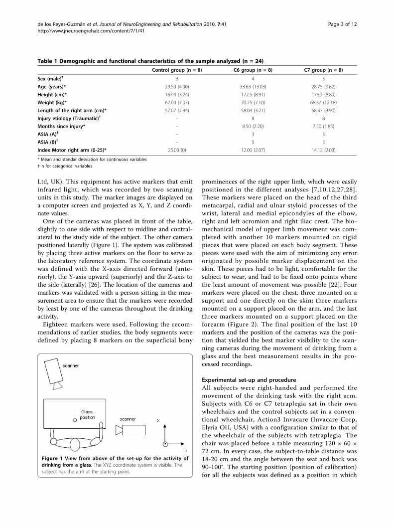
slightly to one side with respect to midline and contral-ateral to the study side of the subject. The other camerapositioned laterally (Figure 1). The system was calibratedby placing three active markers on the floor to serve asthe laboratory reference system. The coordinate systemwas defined with the X-axis directed forward (ante-riorly), the Y-axis upward (superiorly) and the Z-axis tothe side (laterally) [26]. The location of the cameras andmarkers was validated with a person sitting in the mea-surement area to ensure that the markers were recordedby least by one of the cameras throughout the drinkingactivity.Eighteen markers were used. Following the recom-
mendations of earlier studies, the body segments weredefined by placing 8 markers on the superficial bony
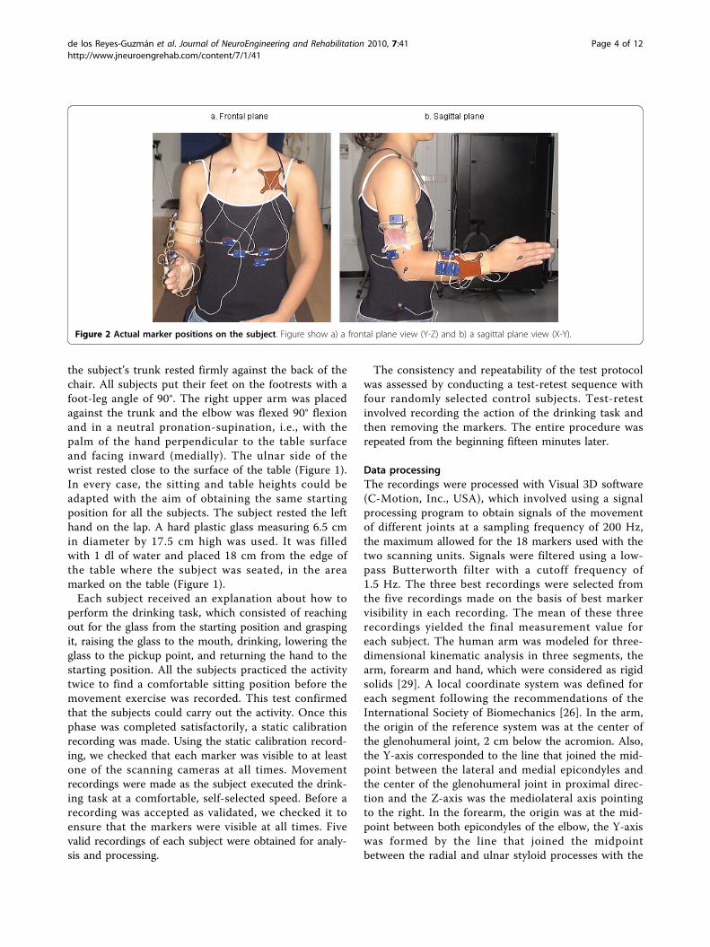
prominences of the right upper limb, which were easilypositioned in the different analyses [7,10,12,27,28].These markers were placed on the head of the thirdmetacarpal, radial and ulnar styloid processes of thewrist, lateral and medial epicondyles of the elbow,right and left acromion and right iliac crest. The bio-mechanical model of upper limb movement was com-pleted with another 10 markers mounted on rigidpieces that were placed on each body segment. Thesepieces were used with the aim of minimizing any errororiginated by possible marker displacement on theskin. These pieces had to be light, comfortable for thesubject to wear, and had to be fixed onto points wherethe least amount of movement was possible [22]. Fourmarkers were placed on the chest, three mounted on asupport and one directly on the skin; three markersmounted on a support placed on the arm, and the lastthree markers mounted on a support placed on theforearm (Figure 2). The final position of the last 10markers and the position of the cameras was the posi-tion that yielded the best marker visibility to the scan-ning cameras during the movement of drinking from aglass and the best measurement results in the pro-cessed recordings.
Experimental set-up and procedureAll subjects were right-handed and performed themovement of the drinking task with the right arm.Subjects with C6 or C7 tetraplegia sat in their ownwheelchairs and the control subjects sat in a conven-tional wheelchair, Action3 Invacare (Invacare Corp,Elyria OH, USA) with a configuration similar to that ofthe wheelchair of the subjects with tetraplegia. Thechair was placed before a table measuring 120 × 60 ×72 cm. In every case, the subject-to-table distance was18-20 cm and the angle between the seat and back was90-100°. The starting position (position of calibration)for all the subjects was defined as a position in which
Table 1 Demographic and functional characteristics of the sample analyzed (n = 24)
Control group (n = 8) C6 group (n = 8) C7 group (n = 8)
Sex (male)† 3 4 5
Age (years)* 29.50 (4.00) 33.63 (13.03) 28.75 (9.82)
Height (cm)* 167.4 (3.24) 172.5 (8.91) 176.2 (8.89)
Weight (kg)* 62.00 (7.07) 70.25 (7.10) 68.37 (12.18)
Length of the right arm (cm)* 57.07 (2.34) 58.03 (3.21) 58.37 (3.90)
Injury etiology (Traumatic)† - 8 8
Months since injury* - 8.50 (2.20) 7.50 (1.85)
ASIA (A)† - 3 3
ASIA (B)† - 5 5
Index Motor right arm (0-25)* 25.00 (0) 12.00 (2.07) 14.12 (2.03)
* Mean and standar desviation for continuous variables
† n for categorical variables
Figure 1 View from above of the set-up for the activity ofdrinking from a glass. The XYZ coordinate system is visible. Thesubject has the arm at the starting point.
de los Reyes-Guzmán et al. Journal of NeuroEngineering and Rehabilitation 2010, 7:41http://www.jneuroengrehab.com/content/7/1/41
Page 3 of 12
the subject’s trunk rested firmly against the back of thechair. All subjects put their feet on the footrests with afoot-leg angle of 90°. The right upper arm was placedagainst the trunk and the elbow was flexed 90° flexionand in a neutral pronation-supination, i.e., with thepalm of the hand perpendicular to the table surfaceand facing inward (medially). The ulnar side of thewrist rested close to the surface of the table (Figure 1).In every case, the sitting and table heights could beadapted with the aim of obtaining the same startingposition for all the subjects. The subject rested the lefthand on the lap. A hard plastic glass measuring 6.5 cmin diameter by 17.5 cm high was used. It was filledwith 1 dl of water and placed 18 cm from the edge ofthe table where the subject was seated, in the areamarked on the table (Figure 1).Each subject received an explanation about how to
perform the drinking task, which consisted of reachingout for the glass from the starting position and graspingit, raising the glass to the mouth, drinking, lowering theglass to the pickup point, and returning the hand to thestarting position. All the subjects practiced the activitytwice to find a comfortable sitting position before themovement exercise was recorded. This test confirmedthat the subjects could carry out the activity. Once thisphase was completed satisfactorily, a static calibrationrecording was made. Using the static calibration record-ing, we checked that each marker was visible to at leastone of the scanning cameras at all times. Movementrecordings were made as the subject executed the drink-ing task at a comfortable, self-selected speed. Before arecording was accepted as validated, we checked it toensure that the markers were visible at all times. Fivevalid recordings of each subject were obtained for analy-sis and processing.
The consistency and repeatability of the test protocolwas assessed by conducting a test-retest sequence withfour randomly selected control subjects. Test-retestinvolved recording the action of the drinking task andthen removing the markers. The entire procedure wasrepeated from the beginning fifteen minutes later.
Data processingThe recordings were processed with Visual 3D software(C-Motion, Inc., USA), which involved using a signalprocessing program to obtain signals of the movementof different joints at a sampling frequency of 200 Hz,the maximum allowed for the 18 markers used with thetwo scanning units. Signals were filtered using a low-pass Butterworth filter with a cutoff frequency of1.5 Hz. The three best recordings were selected fromthe five recordings made on the basis of best markervisibility in each recording. The mean of these threerecordings yielded the final measurement value foreach subject. The human arm was modeled for three-dimensional kinematic analysis in three segments, thearm, forearm and hand, which were considered as rigidsolids [29]. A local coordinate system was defined foreach segment following the recommendations of theInternational Society of Biomechanics [26]. In the arm,the origin of the reference system was at the center ofthe glenohumeral joint, 2 cm below the acromion. Also,the Y-axis corresponded to the line that joined the mid-point between the lateral and medial epicondyles andthe center of the glenohumeral joint in proximal direc-tion and the Z-axis was the mediolateral axis pointingto the right. In the forearm, the origin was at the mid-point between both epicondyles of the elbow, the Y-axiswas formed by the line that joined the midpointbetween the radial and ulnar styloid processes with the
Figure 2 Actual marker positions on the subject. Figure show a) a frontal plane view (Y-Z) and b) a sagittal plane view (X-Y).
de los Reyes-Guzmán et al. Journal of NeuroEngineering and Rehabilitation 2010, 7:41http://www.jneuroengrehab.com/content/7/1/41
Page 4 of 12
midpoint between the lateral and medial epicondylesproximally and the Z-axis was the line that joined bothepicondyles in the lateral direction. In the hand, the ori-gin was at the midpoint between radial and ulnar styloidof the wrist, the Y-axis was the line joining the head ofthe third metacarpal with the midpoint between theradial and ulnar styloid processes proximally and the Z-axis joined both styloid processes laterally. We obtainedtrunk movement with respect to the laboratory coordi-nate system, arm movement with respect to the trunk,forearm movement with respect to the arm, and handmovement with respect to the forearm using Euler anglenotation and a sequence of ZXY rotations of the trunk,arm and hand, and ZYX rotations of the forearm.In each recording, a complete cycle of the drinking
task was identified. The beginning of the cycle was theonset of displacement of the marker on the head of thethird metacarpal and the end of the cycle was the returnof the marker to the starting point after completing thedrinking task. As it happens with other cyclical move-ments, such as walking, several phases were establishedin the drinking task to facilitate task analysis. We usedphases and events delimiting the phases that have beendescribed previously: reaching, forward transport, drink-ing, back transport and return [7].Once the recordings were made and analyzed, the
results were described in terms of analysis of the follow-ing variables:
• Movement times: the duration of each phase andthe complete cycle.• Peak velocities: the velocities were obtained by cal-culating the linear velocity with which the handmoves in the phases of the cycle of reaching, forwardtransport, back transport and return to startposition.• Joint angles: flexion-extension and lateral inclina-tion of the trunk; flexion-extension, abduction-adduction and external-internal rotation of theshoulder joint; flexion-extension and pronation-supi-nation of the elbow joint; and dorsal-palmar flexionof the wrist. For each joint angle, we calculated themaximum, minimum, range of motion (ROM) andmoment in the complete drinking cycle in whichthese values were reached.• Coordination between the shoulder and elbowjoints, particularly between the shoulder flexionangle and the elbow flexion angle, in the reachingphase.
In order to compare the three groups analyzed, theduration of the cycles was adjusted for time andexpressed as percentages. Consequently, data wereexpressed in relation to the percentage of the drinking
task cycle that had lapsed (0-100% of the drinking taskcycle) when the movement was recorded.
Statistical analysisA descriptive analysis was made of the clinical and func-tional variables by calculating the median and interquar-tile range of the quantitative variables and thefrequencies and percentages of the qualitative variables.Given the limited number of participants, non-para-
metric methods were used. The Kruskal-Wallis test wasused to find possible differences in each variablebetween the three groups analyzed; the Kruskal-Wallistest began by testing the equivalence hypothesis betweengroups. If the significance of the Kruskal-Wallis test is p< 0.05, the equivalence of behavior between groups canbe rejected and a pairwise comparison can be madeusing the Mann-Whitney test. The Bonferroni correc-tion was applied, which takes into account randomnessdue to multiple comparisons.The interrelation between the shoulder flexion angle
and the elbow flexion angle was analyzed in the reach-ing phase using the Pearson correlation coefficient.The repeatability of the protocol was evaluated with
the Student t test using a level of significance of 0.05.The mean difference between the test and retest valueswas calculated for each of the four control subjects inwhich test-retest consistency was analyzed with a 95%confidence interval.Data were analyzed statistically using the SPSS for
Windows (version 12.0) statistical package (SPSS Inc,Chicago, IL, USA).
ResultsMovement timesThe duration of the complete cycle of the drinkingactivity was more prolonged in the group of subjectswith C6 tetraplegia than in CG (p < 0.05). In the phaseanalysis the duration of the reaching phase was shorterin CG than in subjects with C6 (p < 0.01) and C7 tetra-plegia (p < 0.05) (Table 2). In both groups of subjectswith tetraplegia, not only was the reaching phase oflonger duration than in CG, it was also the longestphase in the cycle. In the CG the longest phase in thecycle was the back transport phase. The contribution ofeach phase to the complete drinking task cycle in eachanalyzed group is detailed in Tables 2.
Peak velocitiesThe peak velocities of the forward and back transportphases were slower in subjects with C6 tetraplegia thanin subjects with C7 tetraplegia (p < 0.05 and p < 0.01,respectively) and in CG (p < 0.05) (Table 2). In addition,the peak velocity of the reaching phase was reachedlater in controls than in the subjects with C6 tetraplegia
de los Reyes-Guzmán et al. Journal of NeuroEngineering and Rehabilitation 2010, 7:41http://www.jneuroengrehab.com/content/7/1/41
Page 5 of 12
(p < 0.01) and C7 tetraplegia (p < 0.05). In contrast, thepeak velocity of the forward transport phase was delayedin the subjects with C6 tetraplegia (p < 0.01) and C7 tet-raplegia (p < 0.01) compared to CG (Table 2).
Joint anglesIn the shoulder joint, only the minimum abductionangle was greater in CG than in subjects with C7 tetra-plegia (p < 0.01) (Table 3). In the elbow joint, the peakminimum of flexion angle was smaller in the subjectswith C6 tetraplegia (p < 0.05) and C7 tetraplegia (p <0.05) than in CG, but none of the differences in theelbow flexion-extension ROM of the three groups wasstatistically significant (Table 3 and Figure 3). None ofthe joint angles analyzed in the trunk showed significantdifferences (Table 3).The wrist was the joint in which the most relevant dif-
ferences were found. The wrist palmar flexion angle wasgreater in the two tetraplegia groups than in CG (p <0.01), which made the ROM of wrist dorsal-palmar flex-ion smaller in CG than in the subjects with C6 (p <0.01) and C7 tetraplegia (p < 0.05). In CG, wrist palmarflexion values were not even found in the cycle (Table3). The maximum wrist palmar flexion in the threegroups was found in the back transport phase (at themoment corresponding to 72.64% of the cycle of
subjects with C6 tetraplegia, 73.45% of the cycle of sub-jects with C7 tetraplegia and 71.38% of the cycle of CG)(Figure 4). On the other hand, minimum wrist palmarflexion in subjects with C6 or C7 tetraplegia was in theforward transport phase (at 36.54% and 22.14% of thecycle, respectively), and in CG it was in the back trans-port phase (at 59.41% of the cycle) (Table 3).
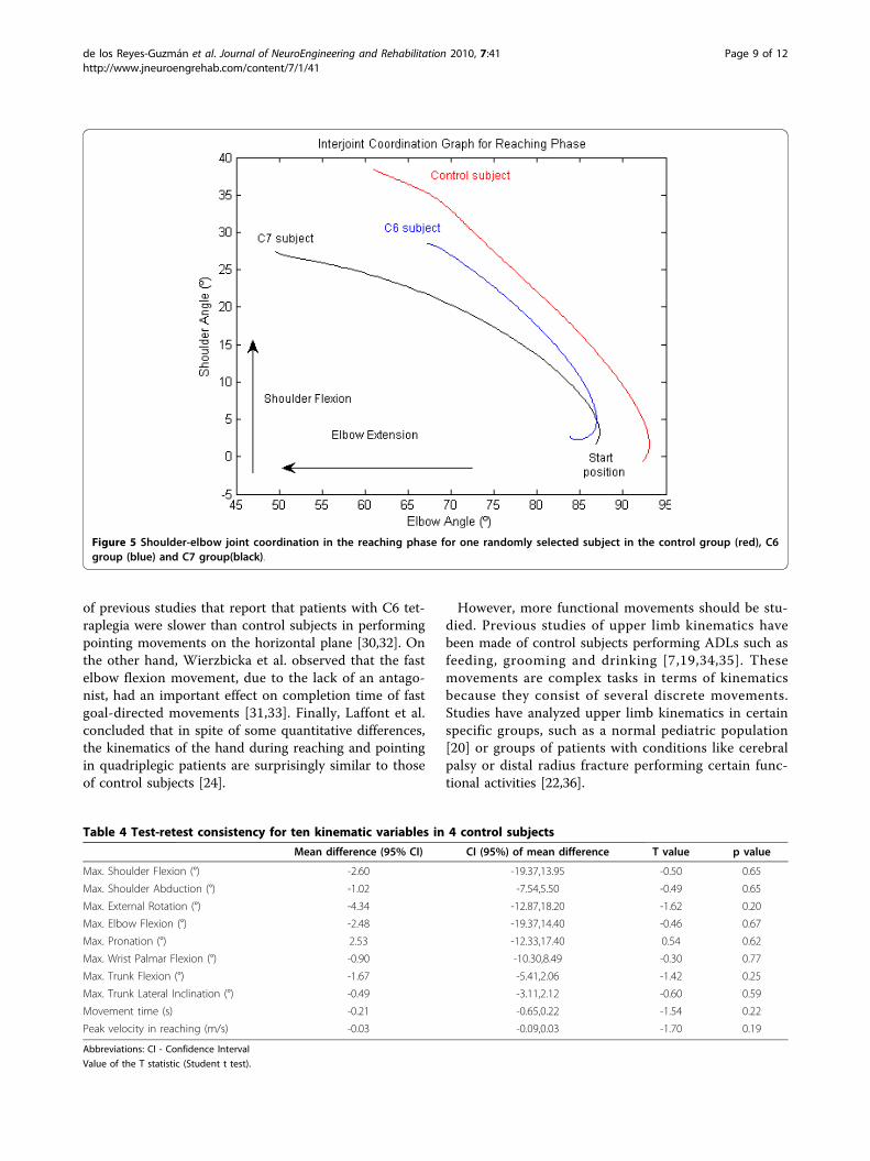
Interjoint coordination between the shoulder and elbowIn the three study groups, there was a strong coordina-tion between the shoulder and elbow joint angles. ThePearson correlation index ranged from -0.95 (IR 0.08) to-0.91 (IR 0.11) (Table 2). The negative value of the cor-relation index meant that as shoulder flexion increased,elbow extension also increased. The trajectory of thesecorrelations was continuous, forming an almost linearrelation between the shoulder and elbow joint angles(Figure 5).
Test-retest consistencyThe statistical results of ten variables are shown inTable 4. Mean retest values were within for the 95%confidence interval of the first test. Based on this data,we concluded that there were no differences betweenthe test and retest with a probability of 95%. However,particularly for measures as maximum shoulder flexion,
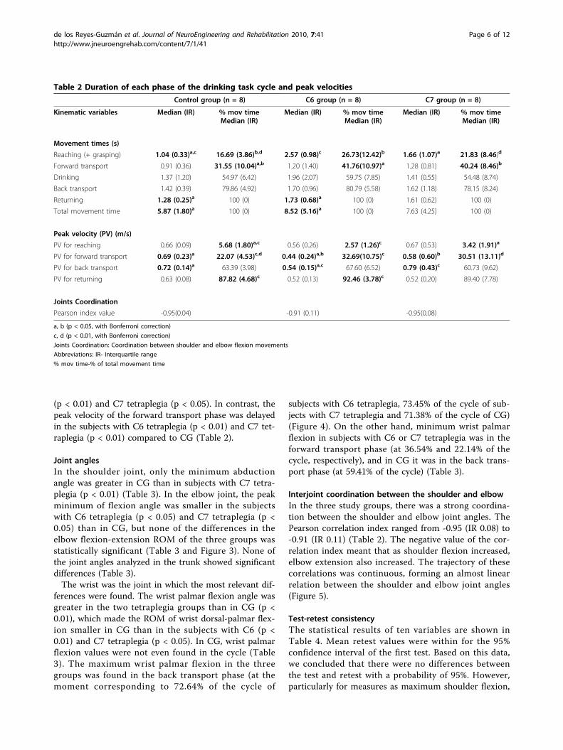
Table 2 Duration of each phase of the drinking task cycle and peak velocities
Control group (n = 8) C6 group (n = 8) C7 group (n = 8)
Kinematic variables Median (IR) % mov timeMedian (IR)
Median (IR) % mov timeMedian (IR)
Median (IR) % mov timeMedian (IR)
Movement times (s)
Reaching (+ grasping) 1.04 (0.33)a,c 16.69 (3.86)b,d 2.57 (0.98)c 26.73(12.42)b 1.66 (1.07)a 21.83 (8.46)d
Forward transport 0.91 (0.36) 31.55 (10.04)a,b 1.20 (1.40) 41.76(10.97)a 1.28 (0.81) 40.24 (8.46)b
Drinking 1.37 (1.20) 54.97 (6.42) 1.96 (2.07) 59.75 (7.85) 1.41 (0.55) 54.48 (8.74)
Back transport 1.42 (0.39) 79.86 (4.92) 1.70 (0.96) 80.79 (5.58) 1.62 (1.18) 78.15 (8.24)
Returning 1.28 (0.25)a 100 (0) 1.73 (0.68)a 100 (0) 1.61 (0.62) 100 (0)
Total movement time 5.87 (1.80)a 100 (0) 8.52 (5.16)a 100 (0) 7.63 (4.25) 100 (0)
Peak velocity (PV) (m/s)
PV for reaching 0.66 (0.09) 5.68 (1.80)a,c 0.56 (0.26) 2.57 (1.26)c 0.67 (0.53) 3.42 (1.91)a
PV for forward transport 0.69 (0.23)a 22.07 (4.53)c,d 0.44 (0.24)a,b 32.69(10.75)c 0.58 (0.60)b 30.51 (13.11)d
PV for back transport 0.72 (0.14)a 63.39 (3.98) 0.54 (0.15)a,c 67.60 (6.52) 0.79 (0.43)c 60.73 (9.62)
PV for returning 0.63 (0.08) 87.82 (4.68)c 0.52 (0.13) 92.46 (3.78)c 0.52 (0.20) 89.40 (7.78)
Joints Coordination
Pearson index value -0.95(0.04) -0.91 (0.11) -0.95(0.08)
a, b (p < 0.05, with Bonferroni correction)
c, d (p < 0.01, with Bonferroni correction)
Joints Coordination: Coordination between shoulder and elbow flexion movements
Abbreviations: IR- Interquartile range
% mov time-% of total movement time
de los Reyes-Guzmán et al. Journal of NeuroEngineering and Rehabilitation 2010, 7:41http://www.jneuroengrehab.com/content/7/1/41
Page 6 of 12
maximum external rotation, maximum elbow flexion,maximum pronation, even maximum wrist palmar flex-ion, wide confidence intervals were obtained.
DiscussionThe goal of this study was to analyze the three-dimen-sional kinematic differences between two groups of peo-ple with tetraplegia and a control group during the ADLof drinking from a glass. The most relevant findings of
this study suggest that subjects with C6 tetraplegia per-form the drinking task at a slower velocity and withmore prolonged phases. The greatest differencesbetween the two tetraplegia groups and controls were inthe wrist. A few studies have been made of the kine-matic properties of the arms of patients with tetraplegia,but none of them has analyzed an ADL [24,30,31]. Theslower velocity of subjects with C6 tetraplegia whenexecuting the drinking task coincides with the findings
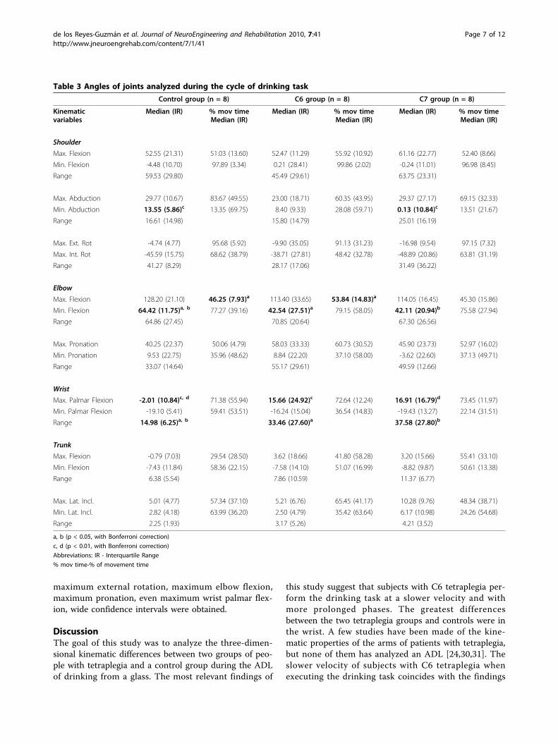
Table 3 Angles of joints analyzed during the cycle of drinking task
Control group (n = 8) C6 group (n = 8) C7 group (n = 8)
Kinematicvariables
Median (IR) % mov timeMedian (IR)
Median (IR) % mov timeMedian (IR)
Median (IR) % mov timeMedian (IR)
Shoulder
Max. Flexion 52.55 (21.31) 51.03 (13.60) 52.47 (11.29) 55.92 (10.92) 61.16 (22.77) 52.40 (8.66)
Min. Flexion -4.48 (10.70) 97.89 (3.34) 0.21 (28.41) 99.86 (2.02) -0.24 (11.01) 96.98 (8.45)
Range 59.53 (29.80) 45.49 (29.61) 63.75 (23.31)
Max. Abduction 29.77 (10.67) 83.67 (49.55) 23.00 (18.71) 60.35 (43.95) 29.37 (27.17) 69.15 (32.33)
Min. Abduction 13.55 (5.86)c 13.35 (69.75) 8.40 (9.33) 28.08 (59.71) 0.13 (10.84)c 13.51 (21.67)
Range 16.61 (14.98) 15.80 (14.79) 25.01 (16.19)
Max. Ext. Rot -4.74 (4.77) 95.68 (5.92) -9.90 (35.05) 91.13 (31.23) -16.98 (9.54) 97.15 (7.32)
Max. Int. Rot -45.59 (15.75) 68.62 (38.79) -38.71 (27.81) 48.42 (32.78) -48.89 (20.86) 63.81 (31.19)
Range 41.27 (8.29) 28.17 (17.06) 31.49 (36.22)
Elbow
Max. Flexion 128.20 (21.10) 46.25 (7.93)a 113.40 (33.65) 53.84 (14.83)a 114.05 (16.45) 45.30 (15.86)
Min. Flexion 64.42 (11.75)a, b 77.27 (39.16) 42.54 (27.51)a 79.15 (58.05) 42.11 (20.94)b 75.58 (27.94)
Range 64.86 (27.45) 70.85 (20.64) 67.30 (26.56)
Max. Pronation 40.25 (22.37) 50.06 (4.79) 58.03 (33.33) 60.73 (30.52) 45.90 (23.73) 52.97 (16.02)
Min. Pronation 9.53 (22.75) 35.96 (48.62) 8.84 (22.20) 37.10 (58.00) -3.62 (22.60) 37.13 (49.71)
Range 33.07 (14.64) 55.17 (29.61) 49.59 (12.66)
Wrist
Max. Palmar Flexion -2.01 (10.84)c, d 71.38 (55.94) 15.66 (24.92)c 72.64 (12.24) 16.91 (16.79)d 73.45 (11.97)
Min. Palmar Flexion -19.10 (5.41) 59.41 (53.51) -16.24 (15.04) 36.54 (14.83) -19.43 (13.27) 22.14 (31.51)
Range 14.98 (6.25)a, b 33.46 (27.60)a 37.58 (27.80)b
Trunk
Max. Flexion -0.79 (7.03) 29.54 (28.50) 3.62 (18.66) 41.80 (58.28) 3.20 (15.66) 55.41 (33.10)
Min. Flexion -7.43 (11.84) 58.36 (22.15) -7.58 (14.10) 51.07 (16.99) -8.82 (9.87) 50.61 (13.38)
Range 6.38 (5.54) 7.86 (10.59) 11.37 (6.77)
Max. Lat. Incl. 5.01 (4.77) 57.34 (37.10) 5.21 (6.76) 65.45 (41.17) 10.28 (9.76) 48.34 (38.71)
Min. Lat. Incl. 2.82 (4.18) 63.99 (36.20) 2.50 (4.79) 35.42 (63.64) 6.17 (10.98) 24.26 (54.68)
Range 2.25 (1.93) 3.17 (5.26) 4.21 (3.52)
a, b (p < 0.05, with Bonferroni correction)
c, d (p < 0.01, with Bonferroni correction)
Abbreviations: IR - Interquartile Range
% mov time-% of movement time
de los Reyes-Guzmán et al. Journal of NeuroEngineering and Rehabilitation 2010, 7:41http://www.jneuroengrehab.com/content/7/1/41
Page 7 of 12
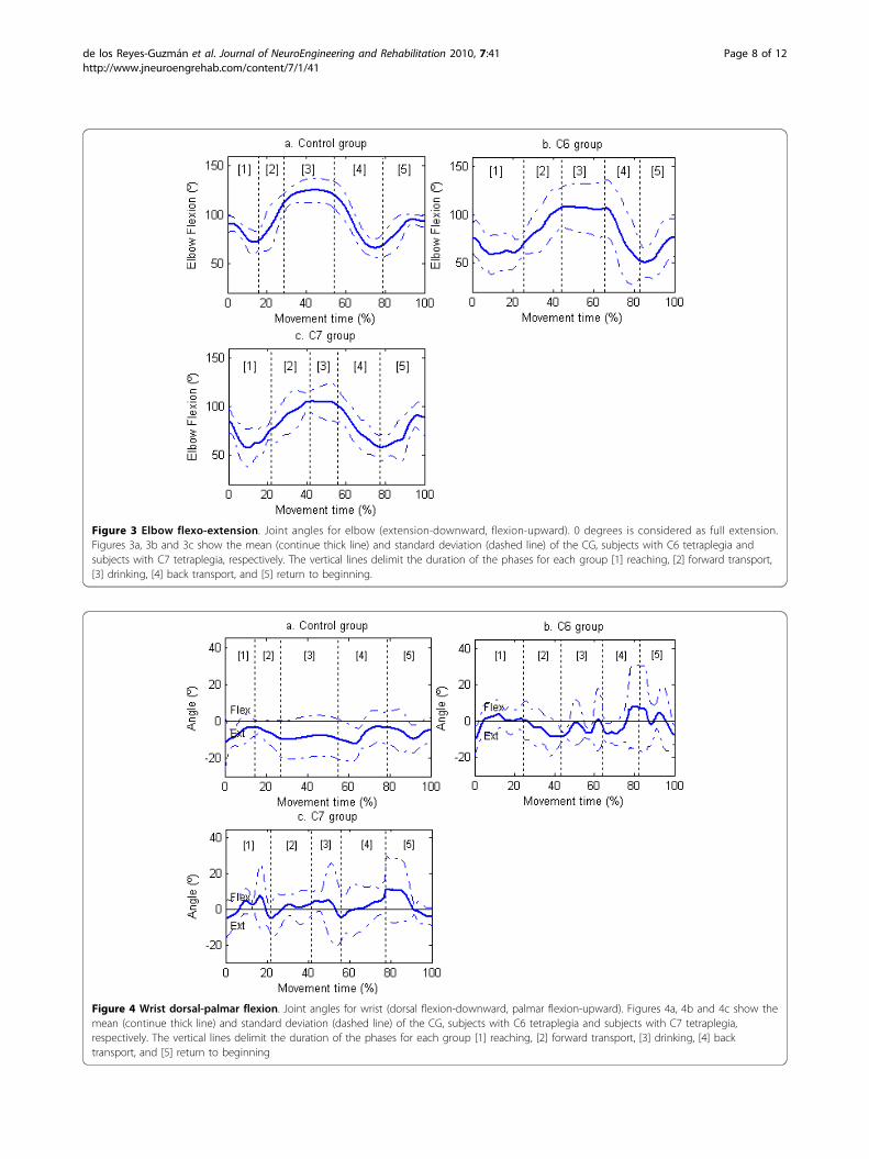
Figure 3 Elbow flexo-extension. Joint angles for elbow (extension-downward, flexion-upward). 0 degrees is considered as full extension.Figures 3a, 3b and 3c show the mean (continue thick line) and standard deviation (dashed line) of the CG, subjects with C6 tetraplegia andsubjects with C7 tetraplegia, respectively. The vertical lines delimit the duration of the phases for each group [1] reaching, [2] forward transport,[3] drinking, [4] back transport, and [5] return to beginning.
Figure 4 Wrist dorsal-palmar flexion. Joint angles for wrist (dorsal flexion-downward, palmar flexion-upward). Figures 4a, 4b and 4c show themean (continue thick line) and standard deviation (dashed line) of the CG, subjects with C6 tetraplegia and subjects with C7 tetraplegia,respectively. The vertical lines delimit the duration of the phases for each group [1] reaching, [2] forward transport, [3] drinking, [4] backtransport, and [5] return to beginning
de los Reyes-Guzmán et al. Journal of NeuroEngineering and Rehabilitation 2010, 7:41http://www.jneuroengrehab.com/content/7/1/41
Page 8 of 12
of previous studies that report that patients with C6 tet-raplegia were slower than control subjects in performingpointing movements on the horizontal plane [30,32]. Onthe other hand, Wierzbicka et al. observed that the fastelbow flexion movement, due to the lack of an antago-nist, had an important effect on completion time of fastgoal-directed movements [31,33]. Finally, Laffont et al.concluded that in spite of some quantitative differences,the kinematics of the hand during reaching and pointingin quadriplegic patients are surprisingly similar to thoseof control subjects [24].
However, more functional movements should be stu-died. Previous studies of upper limb kinematics havebeen made of control subjects performing ADLs such asfeeding, grooming and drinking [7,19,34,35]. Thesemovements are complex tasks in terms of kinematicsbecause they consist of several discrete movements.Studies have analyzed upper limb kinematics in certainspecific groups, such as a normal pediatric population[20] or groups of patients with conditions like cerebralpalsy or distal radius fracture performing certain func-tional activities [22,36].
Figure 5 Shoulder-elbow joint coordination in the reaching phase for one randomly selected subject in the control group (red), C6group (blue) and C7 group(black).
Table 4 Test-retest consistency for ten kinematic variables in 4 control subjects
Mean difference (95% CI) CI (95%) of mean difference T value p value
Max. Shoulder Flexion (°) -2.60 -19.37,13.95 -0.50 0.65
Max. Shoulder Abduction (°) -1.02 -7.54,5.50 -0.49 0.65
Max. External Rotation (°) -4.34 -12.87,18.20 -1.62 0.20
Max. Elbow Flexion (°) -2.48 -19.37,14.40 -0.46 0.67
Max. Pronation (°) 2.53 -12.33,17.40 0.54 0.62
Max. Wrist Palmar Flexion (°) -0.90 -10.30,8.49 -0.30 0.77
Max. Trunk Flexion (°) -1.67 -5.41,2.06 -1.42 0.25
Max. Trunk Lateral Inclination (°) -0.49 -3.11,2.12 -0.60 0.59
Movement time (s) -0.21 -0.65,0.22 -1.54 0.22
Peak velocity in reaching (m/s) -0.03 -0.09,0.03 -1.70 0.19
Abbreviations: CI - Confidence Interval
Value of the T statistic (Student t test).
de los Reyes-Guzmán et al. Journal of NeuroEngineering and Rehabilitation 2010, 7:41http://www.jneuroengrehab.com/content/7/1/41
Page 9 of 12
Much of the methodology developed in the presentstudy followed the recommendations of a previous oneof healthy subjects in which five sequential phases ofdrinking task were identified: reaching, forward trans-port, drinking, back transport and returning [7]. How-ever, the current experience has resolved previouslimitations and provides a full and detailed three-dimen-sional kinematic analysis of the drinking task in controlsubjects and two groups of patients with tetraplegia,analyzing the shoulder, elbow and wrist at all possiblejoint angles except for lateral wrist inclination.Using the upper limb model developed, we were able to
estimate the location of the center of the joints involved,which made it possible to measure all the joint anglesdescribed. Likewise, the use of markers mounted on rigidpieces to position some of the markers helped to reducetissue artifacts. These artifacts appear with limb displace-ment when markers are placed on the skin surface.It has been reported that trunk movement can act as
both a stabilizer and an integral component in position-ing the hand close to the target [37]. It has been shownthat hemiparetic subjects reaching within arm’s lengthuse a compensatory strategy that involves trunk displace-ment [38,39]. In the present study, the glass was placedwithin arm’s length and the subject could reach it with-out separating the trunk from the back of the wheelchair.Our findings confirmed those of earlier experience car-ried out in control subjects, in which trunk displacementwas not relevant in the groups analyzed [36].
1. Movement timesThe total duration of the drinking task was somewhatshorter in our CG than in an earlier report, probablybecause in the present study the palm of the hand wascloser to the drinking glass whereas in the earlier reportthe wrist line was closer to the edge of the table [7].However, both two studies had the same conclusion:back transport is the most prolonged phase in controls[7]. The duration of the drinking activity was longer insubjects with C6 tetraplegia compared to controls andthe duration of the reaching phase was longer in sub-jects with C6 and C7 tetraplegia. As mentioned, thereaching phase includes grasping. In order to grasp,both groups of patients with tetraplegia developed acompensatory strategy called “tenodesis,” in which thesepatients extend the wrist to close the fingers passively.This pattern suggests that in subjects with tetraplegiareaching and grasping are executed sequentially com-pared to controls, who prepare for grasping during thereaching phase [40].
2. Peak velocityAs the duration of the drinking task was shorter, thevelocity of each phase of the cycle in the controls was
somewhat faster than in a previous report [7]. Theabsence of triceps brachialis muscle activity in subjectswith C6 tetraplegia slows the velocity of the forwardtransport and back transport phases, in which this mus-cle controls the eccentric or concentric displacement ofthe elbow in flexion-extension. As in an earlier study,the peak velocity of the reaching phase was similar inpatients with tetraplegia and controls [24]. Another fac-tor that could condition the velocity of movements isperforming the movement with a load. The weakness ofthe upper limbs becomes more evident when raising anobject with a certain weight. In the absence of any addi-tional load, peak velocity in the reaching phase isreached earlier in groups of patients with tetraplegia.However, in the forward transport phase in which theglass of water is raised to the mouth, peak velocity isnotably faster in controls. It is difficult to compare thevelocities attained in other pathologies because theyhave not been studied using the phases defined in ourstudy [23].
3. Joint anglesAs in healthy subjects, but in contrast with subjects whohave experienced stroke and have a hemiparetic arm,there was a strong coordination between shoulder andelbow joint excursion in the reaching phase, indicatinggood interjoint coordination in C6 and C7 tetraplegia[10,12]. The wrist was the joint with the most relevantdifferences between the three groups. Wrist palmar flex-ion angles were greater in both groups of subjects withtetraplegia and the maximum wrist palmar flexion inboth cases was observed in the back transport phase,probably because no eccentric resistance is offered bywrist extensor muscles as the glass is lowered from themouth to the table; passive wrist palmar flexionoccurred in both tetraplegia groups. The minimum wristpalmar flexion angle was found in subjects with C6 orC7 tetraplegia in the forward transport phase. This isprobably because at this time the subject required maxi-mum wrist dorsal flexion to grasp a glass that has someweight, which optimized the tenodesis effect and theability to pick up an object. The elbow extension wasgreater in both tetraplegia groups and occurred in theback transport phase, perhaps also because elbow exten-sion favored the tenodesis effect in the wrist.
4. Test-retest consistencyMean retest values were within for the 95% confidenceinterval of the first test. Based on this data, we con-cluded that there were not differences between the testand retest with a probability of 95%. However, for mea-surements as maximum shoulder flexion, maximumexternal rotation, maximum elbow flexion, maximumpronation, even maximum wrist palmar flexion, wide
de los Reyes-Guzmán et al. Journal of NeuroEngineering and Rehabilitation 2010, 7:41http://www.jneuroengrehab.com/content/7/1/41
Page 10 of 12
confidence intervals were obtained. It could be probablydue to the natural large variation between the subjectsin those measurements. It is necessary to take intoaccount that people can perform a goal-oriented taskwith many different combinations of individual jointmovements.However, although the results obtained were sound,
for further research in this field, it would be good toinclude another scanning unit to offer a view fromabove with the aim of providing the best visibility of themarkers in the three space planes.Robotic tools provide opportunities to study functional
adaptation after central nervous system injuries and canprovide objective measurements of the time-course ofchanges in motor control of the affected limbs. Robot-assisted therapy permits semi-autonomous practice oftherapeutic tasks [41]. In the last years, wearable tech-nology has made an impact in the clinical setting andrecent studies have been focused on integrating thistechnology with orthotic and prosthetic devices [42]. So,wearable robots development can be one of the mostinnovative therapeutic options for people with cervicalSCI, to improve upper limb functionality and so, facili-tate the independence and quality of life during the per-formance of ADL. In order to design these devices, it isnecessary to identify the movement patterns performedby these patients during functional activities, such as thedrinking task. Then, these movement patterns can beimplemented into robotic devices to imitate or improvethese movements. In this research field, the presentstudy is of particular clinical relevance.
ConclusionsKinematic analysis has shown great potential for use asan outcome in clinical research to understand how func-tional activities, such as drinking, are performed bypatients with upper limb impairment. The most relevantdifferences were in the wrist, where the palmar flexionvalues were greater in patients with C6 and C7 tetraple-gia than in controls during the back transport phase,whereas the highest wrist dorsal flexion value was in theforward transport phase in subjects with C6 or C7 tetra-plegia, in which complete activation of the tenodesiseffect is needed for grasping. This information can beuseful in designing wearable robots to compensate theperformance of AVD, such as drinking, in people withcervical SCI.
AcknowledgementsThis work was part of a project financed by FISCAM (Fundación para laInvestigación Sanitaria de Castilla-La Mancha, Spain) which does not haveany commercial interest in the results of this investigation. Ref no.: PI-2007-09.We thank Dr. Antonio Sánchez-Ramos (Head of Department of PhysicalMedicine and Rehabilitation) for facilitating our work. We would like to
thank José Luis Rodríguez-Martín for his critical review of the manuscriptand methodology recommendations and Barbara Thomas and Elaine VanStaalduinen for the revision of this manuscript in English.
Authors’ contributionsARG contributed to the concept and design, planning of study, softwaredevelopment, analysis and interpretation of the data, drafting andcompletion of the manuscript. AGA contributed to design, analysis of thedata and completion of the manuscript. BPM contributed to the concept,software development, design and acquisition of the data. MSM contributedto the analysis and acquisition of the data. AAE contributed to the analysisand acquisition of the data. EPR contributed to the software development.All authors read and approved the manuscript to be published.
Competing interestsNone of the authors of this paper has any conflict of interest in relation toany sources of any kind pertinent to this study.
Received: 3 March 2010 Accepted: 20 August 2010Published: 20 August 2010
References1. Parker VM, Wade DT, Langton Hewer R: Loss of an arm function after
stroke: measurement, frequency and recovery. Int Rehabil Med 1986,8(2):69-73.
2. Nakayama H, Jorgensen HS, Raaschou HO, Olsen TS: Compensation inrecovery of upper extremity function after stroke: the CopenhagenStroke Study. Arch Phys Med Rehabil 1994, 75(8):852-857.
3. Wyndaele M, Wyndaele JJ: Incidence, prevalence and epidemiology ofspinal cord injury: what learns a worldwide literature survey? Spinal Cord2006, 44:523-529.
4. Broeks JG, Lankhorst GJ, Rumping K, Prevo AJ: The long-term outcome ofarm function after stroke: results of a follow-up study. Disabil Rehabil1999, 21(8):357-364.
5. Wade DT: Measurement in neurological rehabilitation. Oxford medicalpublications Oxford, Oxford Univ. Press 1992, 388.
6. Finch E: Physical rehabilitation outcome measures: a guide to enhancedclinical decision making Hamilton, Ontario, Decker, 2 2002, ix:292.
7. Murphy MA, Sunnerhagen KS, Johnels B, Willen C: Three-dimensionalkinematic motion analysis of a daily activity drinking from a glass: apilot study. J Neuroeng Rehabil 2006, 3:18 [http://www.jneuroengrehab.com/content/3/1/18].
8. Van Anden CJ, Wolterbeek N, Doorenbosch AM, Veeger HEJ, Harlaar J:Complete 3D Kinematics of upper extremity functional tasks. Gait Posture2008, 27:120-127.
9. McCrea PH, Eng JJ, Hodgson AJ: Biomechanics of reaching: clinicalimplications for individuals with acquired brain injury. Disabil Rehabil2002, 24(10):534-541.
10. Levin MF: Interjoint coordination during pointing movements isdisrupted in spastic hemiparesis. Brain 1996, 119(1):281-293.
11. Dwan LN, McIntosh AS: Kinematics of the upper limb: A reaching andplacing task with resistance in children. Gait & Posture 2006, 24(S):S235-S238.
12. Cirstea MC, Levin MF: Compensatory strategies for reaching in stroke.Brain 2000, 123(Pt 5):940-953.
13. Roby-Brami A, Feydy A, Combeaud M, Biryukova EV, Bussel B, Levin MF:Motor compensation and recovery for reaching in stroke patients. ActaNeurol Scand 2003, 107(5):369-381.
14. Roby-Brami A, Bennis N, Levin MF: Hand orientation for grasping and armjoint rotation patterns in healthy subjects and hemiparetic strokepatients. Brain Res 2003, 969(1-2):217-229.
15. Murray IA, Johnson GR: A study of the external forces and moments atthe shoulder and elbow while performing every day tasks. Clin Biomech(Bristol Avon) 2004, 19(6):586-594.
16. Murgia A, Kyberd PJ, Chapell PH, Light CM: Marker placement to describethe wrist movement during activities of daily living in cyclic tasks. ClinBiomech (Bristol Avon) 2004, 19(4):248-254.
17. Safaee-Rad R, Shwedyk E, Quanbury AO, Cooper JE: Normal functionalrange of motion of upper limb joints during performance of threefeeding activities. Arch Phys Med Rehabil 1990, 71(7):505-509.
de los Reyes-Guzmán et al. Journal of NeuroEngineering and Rehabilitation 2010, 7:41http://www.jneuroengrehab.com/content/7/1/41
Page 11 of 12
18. Trombly CA, Wu CY: Effect of rehabilitation tasks on organization ofmovement after stroke. Am J Occup Ther 1999, 53(4):333-344.
19. Magermans DJ, Chadwick EKJ, Veeger HEJ, van der Helm FCT:Requirements for upper extremity motions during activities of dailyliving. Clin Biomech (Bristol, Avon) 2005, 20:591-599.
20. Petuskey K, Bagley A, Abdala E, James MA, Rab G: Upper extremitykinematics during functional activities: Three-dimensional studies in anormal pediatric population. Gait Posture 2007, 25:573-579.
21. Mosqueda T, James MA, Petuskey K, Bagley A, Abdala E, Rab G: Kinematicassessment of the upper extremity in brachial plexus birth palsy. JPediatr Orthop 2004, 24(6):695-699.
22. Fitoussi F, Diop A, Maurel N, Laassel EM, Pennecot GF: Kinematic analysisof the upper limb: a useful tool in children with cerebral palsy. J PediatrOrthop B 2006, 15(4):247-256.
23. Rönnqvist L, Rösblad B: Kinematic analysis of unimanual reaching andgrasping movements in children with hemiplegic cerebral palsy. ClinBiomech (Bristol Avon) 2007, 22:165-17.
24. Laffont I, Briand E, Dizien O, Combeaud M, Bussel B, Revol M, Roby-Brami A:Kinematics of prehension and pointing movements in C6 quadriplegicpatients. Spinal Cord 2000, 38(6):354-362.
25. Maynard FM, Bracken MB, Creasey G, Ditunno JF Jr, Donovan WH,Ducker TB, Garber SL, Marino RJ, Stover SL, Tator CH, Waters RL,Wilberger JE, Young W: International Standards for Neurological andFunctional Classification of Spinal Cord Injury. American Spinal InjuryAssociation. Spinal Cord 1997, 35:266-274.
26. Wu G, van der Helm F, Veeger HEJ, Makhsous M, Van Roy P, Anglin C,Nagels J, Karduna A, McQuade K, Wang X, Werner F, Bucholz B: ISBrecommendation on definitions of joint coordinate systems of variusjoints for the reporting of human joint motion- Part II: shoulder, elbow,wrist and hand. J Biomechanics 2005, 38:981-992.
27. Michaelsen SM, Luta A, Roby-Brami A, Levin MF: Effect of trunk restrainton the recovery of reaching movements in hemiparetic patients. Stroke2001, 32(8):1875-1883.
28. Turner-Stokes L, Reid K: Three-dimensional motion analysis of upper limbmovement in the bowing arm of string-playing musicians. Clin Biomech(Bristol Avon) 1999, 14(6):426-433.
29. Biryukova EV, Roby-Brami A, Frolov AA, Mokhtari M: Kinematics of humanarm reconstructed from spatial tracking system recordings. J Biomech2000, 33:985-995.
30. Popovic M, Popovic D: A new approach to reaching control forquadriplegic patients. J Electromyogr Kinesiol 1994, 4:242-253.
31. Wierzbicka MM, Wiegner AW: Accuracy of motor responses in subjectswith and without control of antagonist muscle. J Neurophys 1996,75(N86):2533-2541.
32. Popovic M, Tomovic R, Popovic D: Joint angle synergy in control armmovements. Ser Aut Control 1993, 1:5-17.
33. Wierzbicka MM, Wiegner AW: Effects of weak antagonist on fast elbowflexion movements in man. Exp Brain Res 1992, 91:509-519.
34. Packer TL, Peat M, Wyss U, Sorbie C: Examinating the elbow duringfunctional activities. Occup Ther J Res 1990, 10(6):323-333.
35. Cooper JE, Shweddyk E, Quandbury AO, Miller J, Hildebrand D: Elbow jointrestriction: effect on functional upper limb motion during performanceof three feeding activities. Arch Phys Med Rehabil 1993, 74:805-809.
36. Murgia A, Kyberd P, Barnhill T: The use of kinematic and parametricinformation to highlight lack of movement and compensation in theupper extremities during activities of daily living. Gait & Posture 2010,31:300-306.
37. Kaminski TR, Bock C, Gentile AM: The coordination between trunk andarm motion during pointing movements. Exp Brain Res 1995,106(3):457-466.
38. Levin MF, Michaelsen SM, Cirstea CM, Roby-Brami A: Use of the trunk forreaching targets placed within and beyond the reach in adulthemiparesis. Exp Brain Res 2002, 143(2):171-180.
39. Michaelsen SM, Levin MF: Short-term effects of practice with trunkrestraint on reaching movements in patients with chronic stroke: acontrolled trial. Stroke 2004, 35(8):1914-1919.
40. Jaennerod M: The timing of natural prehension movements. J Mot Behav1984, 16:235-254.
41. Johnson JMichelle: Recent trends in robot-assited therapy environmentsto improve real-life functional performance after stroke. J NeuroengRehabil 2006, 3:29 [http://www.jneuroengrehab.com/content/3/1/29].
42. Bonato P: Advances in wearable technology and applications in physicalmedicine and rehabilitation. J Neuroeng Rehabil 2005, 2:2 [http://www.jneuroengrehab.com/content/2/1/2].
doi:10.1186/1743-0003-7-41Cite this article as: de los Reyes-Guzmán et al.: Kinematic analysis of thedaily activity of drinking from a glass in a population with cervicalspinal cord injury. Journal of NeuroEngineering and Rehabilitation 20107:41.
Submit your next manuscript to BioMed Centraland take full advantage of:
• Convenient online submission
• Thorough peer review
• No space constraints or color figure charges
• Immediate publication on acceptance
• Inclusion in PubMed, CAS, Scopus and Google Scholar
• Research which is freely available for redistribution
Submit your manuscript at www.biomedcentral.com/submit
de los Reyes-Guzmán et al. Journal of NeuroEngineering and Rehabilitation 2010, 7:41http://www.jneuroengrehab.com/content/7/1/41
Page 12 of 12