Kapur-FNAIT blood 2014
11
doi:10.1182/blood-2013-09-527978 Prepublished online November 15, 2013; 2014 123: 471-480 der Schoot, Manfred Wuhrer and Gestur Vidarsson Björn Skogen, Mette Kjaer Killie, Terje E. Michaelsen, Masja de Haas, Theo Rispens, C. Ellen van Einarsdottir, Leendert Porcelijn, Dave Jackson, Belinda Kumpel, André M. Deelder, Dennis Blank, Rick Kapur, Iwan Kustiawan, Anne Vestrheim, Carolien A. M. Koeleman, Remco Visser, Helga K. pregnancy A prominent lack of IgG1-Fc fucosylation of platelet alloantibodies in http://bloodjournal.hematologylibrary.org/content/123/4/471.full.html Updated information and services can be found at: (134 articles) Thrombocytopenia (399 articles) Plenary Papers (423 articles) Platelets and Thrombopoiesis (292 articles) Pediatric Hematology (5140 articles) Immunobiology (2158 articles) Free Research Articles Articles on similar topics can be found in the following Blood collections http://bloodjournal.hematologylibrary.org/site/misc/rights.xhtml#repub_requests Information about reproducing this article in parts or in its entirety may be found online at: http://bloodjournal.hematologylibrary.org/site/misc/rights.xhtml#reprints Information about ordering reprints may be found online at: http://bloodjournal.hematologylibrary.org/site/subscriptions/index.xhtml Information about subscriptions and ASH membership may be found online at: Copyright 2011 by The American Society of Hematology; all rights reserved. Washington DC 20036. by the American Society of Hematology, 2021 L St, NW, Suite 900, Blood (print ISSN 0006-4971, online ISSN 1528-0020), is published weekly For personal use only. at Sanquin on January 28, 2014. bloodjournal.hematologylibrary.org From For personal use only. at Sanquin on January 28, 2014. bloodjournal.hematologylibrary.org From
-
Upload
independent -
Category
Documents
-
view
0 -
download
0
Transcript of Kapur-FNAIT blood 2014

doi:10.1182/blood-2013-09-527978Prepublished online November 15, 2013;2014 123: 471-480
der Schoot, Manfred Wuhrer and Gestur VidarssonBjörn Skogen, Mette Kjaer Killie, Terje E. Michaelsen, Masja de Haas, Theo Rispens, C. Ellen vanEinarsdottir, Leendert Porcelijn, Dave Jackson, Belinda Kumpel, André M. Deelder, Dennis Blank, Rick Kapur, Iwan Kustiawan, Anne Vestrheim, Carolien A. M. Koeleman, Remco Visser, Helga K. pregnancyA prominent lack of IgG1-Fc fucosylation of platelet alloantibodies in
http://bloodjournal.hematologylibrary.org/content/123/4/471.full.htmlUpdated information and services can be found at:
(134 articles)Thrombocytopenia � (399 articles)Plenary Papers �
(423 articles)Platelets and Thrombopoiesis � (292 articles)Pediatric Hematology �
(5140 articles)Immunobiology � (2158 articles)Free Research Articles �
Articles on similar topics can be found in the following Blood collections
http://bloodjournal.hematologylibrary.org/site/misc/rights.xhtml#repub_requestsInformation about reproducing this article in parts or in its entirety may be found online at:
http://bloodjournal.hematologylibrary.org/site/misc/rights.xhtml#reprintsInformation about ordering reprints may be found online at:
http://bloodjournal.hematologylibrary.org/site/subscriptions/index.xhtmlInformation about subscriptions and ASH membership may be found online at:
Copyright 2011 by The American Society of Hematology; all rights reserved.Washington DC 20036.by the American Society of Hematology, 2021 L St, NW, Suite 900, Blood (print ISSN 0006-4971, online ISSN 1528-0020), is published weekly
For personal use only. at Sanquin on January 28, 2014. bloodjournal.hematologylibrary.orgFrom For personal use only. at Sanquin on January 28, 2014. bloodjournal.hematologylibrary.orgFrom
Plenary Paper
PLATELETS AND THROMBOPOIESIS
A prominent lack of IgG1-Fc fucosylation of platelet alloantibodiesin pregnancyRick Kapur,1 Iwan Kustiawan,2 Anne Vestrheim,3,4 Carolien A. M. Koeleman,5 Remco Visser,1 Helga K. Einarsdottir,1
Leendert Porcelijn,6 Dave Jackson,7 Belinda Kumpel,7 Andre M. Deelder,5 Dennis Blank,5 Bjorn Skogen,8,9
Mette Kjaer Killie,8,9 Terje E. Michaelsen,3,4 Masja de Haas,1 Theo Rispens,2 C. Ellen van der Schoot,1 Manfred Wuhrer,5
and Gestur Vidarsson1
1Department of Experimental Immunohematology and 2Department of Immunopathology, Sanquin Research and Landsteiner Laboratory, Academic
Medical Centre, University of Amsterdam, Amsterdam, The Netherlands; 3Department of Bacteriology and Immunology, Norwegian Institute of Public
Health, Oslo, Norway; 4School of Pharmacy, University of Oslo, Oslo, Norway; 5Biomolecular Mass Spectrometry Unit, Leiden University Medical Center,
Leiden, The Netherlands; 6Thrombocyte and Leukocyte Serology, Sanquin, Amsterdam, The Netherlands; 7Bristol Institute for Transfusion Sciences,
National Health Service Blood and Transplant, Bristol, United Kingdom; 8Institute of Medical Biology, University of Tromso, Tromso, Norway; and 9University
Hospital of North Norway, Tromso, Norway
Key Points
• Antibodies causing FNAIThave decreased Fcfucosylation, unlike inrefractory thrombocytopenia.
• Decreased Fc fucoseincreases affinity toFcgRIIIa/b, enhancesplatelet phagocytosis, andcorrelates with increaseddisease severity.
Immunoglobulin G (IgG) formed during pregnancy against human platelet antigens
(HPAs) of the fetus mediates fetal or neonatal alloimmune thrombocytopenia (FNAIT).
Because antibody titer or isotype does not strictly correlate with disease severity, we
investigated bymass spectrometry variations in the glycosylation at Asn297 in the IgGFc
because the composition of this glycan can be highly variable, affecting binding to
phagocyte IgG-Fc receptors (FcgR). We found markedly decreased levels of core
fucosylation of anti-HPA-1a–specific IgG1 from FNAIT patients (n 5 48), but not in total
serum IgG1. Antibodies with a low amount of fucose displayed higher binding affinity to
FcgRIIIa andFcgRIIIb, butnot toFcgRIIa, comparedwithantibodieswith ahighamountofFc
fucose. Consequently, these antibodies with a low amount of Fc fucose showed enhanced
phagocytosis of platelets usingFcgRIIIb1polymorphonuclear cells or FcgRIIIa1monocytes
as effector cells, but not with FcgRIIIa– monocytes. In addition, the degree of anti-HPA-1a
fucosylation correlated positively with the neonatal platelet counts in FNAIT, and negatively
to the clinical disease severity. In contrast to the FNAIT patients, no changes in core
fucosylationwere observed for anti-HLA antibodies in refractory thrombocytopenia (post platelet transfusion), indicating that the level of
fucosylation may be antigen dependent and/or related to the immune milieu defined by pregnancy. (Blood. 2014;123(4):471-480)
Introduction
Fetal or neonatal alloimmune thrombocytopenia (FNAIT) is apotentially life-threatening disease where the fetal platelets aretargeted by maternal anti-platelet immunoglobulin G (IgG) alloanti-bodies crossing the placenta. This leads to IgG-Fc receptor (FcgR)–mediated uptake by phagocytes in the fetal spleen and liver, finallyresulting in thrombocytopenia.1 Clinical outcome may differ fromasymptomatic to petechiae or intracerebral hemorrhage. The strengthof the interaction between IgG and FcgR depends on several factors,including the IgG subclass formed during the immune response, itsrelative affinity to FcgRs, the expression levels of FcgR allotypes,FcgR copy number variation, cytokines (influencing the expression ofFcgR), and also the IgG-Fc glycosylation pattern.2
IgG antibodies are glycoproteins containing a branched sugarmoiety attached to the Asn297 residue in the Fc part. This glycan isessential for themaintenance of a functional structure and for bindingof IgG with FcgR.3-5 In addition, the Asn297-linked glycans are
substituted with variable amounts of galactose and sialic acid andmay additionally carry a bisecting N-acetylglucosamine (GlcNAc)and core fucose. Variation in this composition influences antibodyaffinity to FcgR and thus antibody effector activity. IgG galacto-sylation has been found to be decreased in several inflammatorydiseases (primary osteoarthritis, rheumatoid arthritis, and tuber-culosis),6,7 as well as in ovarian cancer.8 Interestingly, increase ingalactosylation correlates with remission of arthritis in pregnantrheumatoid arthritis patients.7,9,10 The degree of IgG galactosylationhas been shown to depend on age and gender; IgG galactosylation ishigher in females at a young age and decreases for both males andfemales with increasing age.11,12 In addition, an increase for total IgGgalactosylation has been observed in pregnancy.7,9,10,13 Recently, ananti-inflammatory activity of immune complexes containing highlyFc-galactosylated mouse IgG1 was reported to enhance the associationof FcgRIIb with dectin-1.14
Submitted September 18, 2013; accepted November 9, 2013. Prepublished
online as Blood First Edition paper, November 15, 2013; DOI 10.1182/blood-
2013-09-527978.
The online version of this article contains a data supplement.
There is an Inside Blood commentary on this article in this issue.
The publication costs of this article were defrayed in part by page charge
payment. Therefore, and solely to indicate this fact, this article is hereby
marked “advertisement” in accordance with 18 USC section 1734.
© 2014 by The American Society of Hematology
BLOOD, 23 JANUARY 2014 x VOLUME 123, NUMBER 4 471
For personal use only. at Sanquin on January 28, 2014. bloodjournal.hematologylibrary.orgFrom
Changes related to age, gender, and pregnancy have also beendescribed for IgG sialylation. Sialylation slightly reduces theaffinity of IgG to FcgRs but enhances the binding to dendritic cell-specific intercellular adhesion molecule-3–grabbing nonintegrin (DC-SIGN), thereby leading to an anti-inflammatory response attributableto increased expression of the inhibitory FcgRIIb.15-17
Importantly, a lack of core fucose has been demonstrated to resultin a stronger binding affinity to FcgRIIIa and FcgRIIIb because ofglycan-glycan interactions between Asn162 found only in FcgRIIIand Asn297 in IgG1.5,18,19 Curiously, although nonfucosylated anti-bodies clearly enhance the binding to FcgRIIIa and FcgRIIIb, thisonly results in enhanced antibody-dependent cellular cytotoxicity onmononuclear cells through FcgRIIIa,3,20-24 but not through the gly-cosylphosphatidylinositol (GPI)–linkedFcgRIIIb onpolymorphonuclearcells (PMNs).18,25 The therapeutic anticancer potential of antibodies withdecreased fucosylation, however, has been well recognized, and theirpower is currently being harnessed in clinical trials.18,20-24,26-30
In whites, the main antibodies causing FNAIT are the anti–humanplatelet antigen (HPA) 1a antibodies, found in;85%of the cases and in;1:1500 pregnancies.31-33 The HPA-1a epitope resides in the GPIIIaprotein of the GPIIb/IIIa complex and is missing in individuals witha single nucleotide polymorphism resulting in an L to P change atposition 33 in the mature protein.31 In a previous study, we reporteda reduced fucosylation, an increased galactosylation, and sialylation ofvarious anti-platelet IgG1 for a small series of FNAIT sera (including2 samples with anti-HPA-1a antibodies).32 We now expand this toa cohort of 48 FNAIT anti-HPA-1a samples. Furthermore, we comparethe glycosylation of IgG formed during immune responses againstplatelets in pregnancy with that of IgG formed against HLA afterplatelet transfusion or pregnancies. Our main findings show that Fcfucosylation is decreased in FNAIT, but not in refractory thrombo-cytopenia (RT), based on Fc analysis of anti-HPA-1a antibodies andanti-HLA class I antibodies, respectively, indicating that Fcfucosylation is regulated. Decreased Fc fucosylation increasedthe IgG affinity to FcgRIIIa and FcgRIIIb and also increasedplatelet phagocytosis by both monocytes and neutrophils, a keyprocess in the pathogenesis of FNAIT.
Methods
Patient samples
Anti-HPA-1a platelet alloantibodies were diagnosed at Sanquin (Amsterdam,The Netherlands; n 5 39), at University Hospital North Norway (Tromso,Norway; n5 5), and at National Health Service Blood and Transplant (Bristol,United Kingdom; n 5 4), from maternal FNAIT sera using a monoclonalantibody immobilization of platelet antigens assay, performed as described byKiefel et al.33 TheHLA class I antibodies were all diagnosed at Sanquin (n5 13)and detected in serum from RT patients (refractory for platelet transfusions),using the Lifescreen Luminex HLA class I antibody screening method.34 Patientcharacteristics for all samples included in the study are listed in supplementalTable 1 (see the BloodWeb site). Samples were obtained with informed consentfrom the patients in accordance with the Declaration of Helsinki.
Purification of anti-platelet antibodies from sera
HPA-1a–specific alloantibodies were purified in a similar way as describedpreviously,32 but now instead of eluting from platelets, we increased thespecificity by eluting the antibodies with formic acid from antigen-coatedplates (PAK12; Immucor GTI Diagnostics, Waukesha, WI). The same plateswere used for purification of anti-HLA class I antibodies. Further details aredescribed in the supplemental Methods.
Mass spectrometric IgG-Fc glycosylation analysis
Nano liquid chromatography–tandem mass spectrometry was performed asdescribed in the supplemental Methods.
Production of recombinant anti-TNP IgG1 antibodies with low
and high amounts of Fc fucose
The variable regions of the heavy and light chains (VH, VL) of the mouseIgG1 anti-2,4,6-trinitrophenol (TNP) hapten antibodies were cloned ontohuman IgG1 or k backbone, respectively, as described previously,35 andproduced in the HEK-293F FreeStyle cell line expression system (LifeTechnologies, Paisley, United Kingdom), but now in the presence or absenceof 2-deoxy-2-fluoro-L-fucose (2F; Carbosynth, Compton, Berkshire, UnitedKingdom) to control the level of fucosylation.36 Antibodies were purified ona proteinA (WT IgG1)HiTrapHP column (GEHealthcare Life Sciences, LittleChalfont, United Kingdom) using the Acta Prime Plus system (GE HealthcareLife Sciences) and dialyzed against phosphate-buffered saline (PBS) overnight.IgG-Fc glycosylation was determined by mass spectrometry.
Surface plasmon resonance (SPR)
SPR measurements were performed with the Biacore 3000 system (BiacoreAB, Breda, The Netherlands) at 25°C. Anti-histidine antibody (GEHealthcare, The Netherlands), 25 mg/mL in sodium acetate buffer pH 4.5(GE Healthcare), was coupled covalently to a CM5 chip (GE Healthcare)following activation with 1-ethyl-3-(3-dimethylaminopropyl)-carbodiimide(Sigma-Aldrich, Zwijndrecht, The Netherlands) 0.4 M in water andN-hydroxysuccinimide (Sigma-Aldrich) 0.1 M. To measure binding ofmonoclonal IgG to FcgRs, 20 ng of human recombinant polyHisTag FcgRIIaor 10 ng of human recombinant polyHisTag FcgRIIIa (Novoprotein, Summit,NJ) or 5 ng human recombinant polyHisTag FcgRIIIb (Sino Biologicals Inc.,Beijing, China), diluted in 0.01MHEPES pH 7.4, 0.15MNaCl, 3 mMEDTA,0.005% v/v Surfactant P20 buffer (GE Healthcare), was injected, followed bythe injection of anti-TNP IgG at different concentrations. Further details aredescribed in the supplemental Methods.
Cell isolations, TNP haptenization, labeling, and
antibody opsonization
Human platelets, PMNs, and monocytes were isolated freshly, and plateletswere haptenized with TNP, as described in the supplemental Methods.
Hereafter, platelets were labeled with pHrodo by resuspending a plateletpellet in 0.23 mM pHrodo succinimidyl ester (100 mL/108 platelets) (Mo-lecular Probes; Invitrogen, Eugene, OR), in 100 ng/mL Prostaglandin E1, for45 minutes in the dark at room temperature. Then, the platelets were washedtwice and resuspended in PBS/EDTA/Prostaglandin E1 at 108 platelets permL.
The haptenized and pHrodo-labeled platelets were opsonized by resus-pending a pellet of platelets with 100 mL of 10 mg/mL anti-TNP antibody low/high fucose or isotype antibody for every 108 platelets and subsequent in-cubation for 30 minutes at room temperature. Hereafter, platelets were washedtwice with PBS/bovine serum albumin and resuspended at 108/mL in NormalHuman Serum (NHS).
Similarly, unhaptenized and pHrodo-labeled platelets were opsonized byresuspending a pellet of platelets with 100mLFNAIT/anti-HPA-1a serumdilutedto 35 arbitrary units (AU) of anti-HPA-1a antibody (using a human anti-HPA-1astandard, 100 AU, National Institute for Biological Standards and Control code:03/152; National Institute for Biological Standards and Control, Hertfordshire,United Kingdom), for every 108 platelets. NHS was used as isotype control.Hereafter, the platelets were incubated for 30 minutes at room temperature,washed twice with PBS, and resuspended at 108/mL in NHS.
Platelet phagocytosis using anti-TNP antibodies or FNAIT
anti-HPA-1a sera
Phagocytosis was carried out using freshly isolated human PMNs orCD161/CD16– (FcgRIIIa1/ FcgRIIIa–) sorted peripheral monocytes (effectorcells). Equal volumes of 2.0 3 106/mL effector cells and 1.0 3 108/mLopsonized platelets weremixed in a total volume of 100mL in 1.4-mLU-bottomtubes (Micronic, Lelystad, The Netherlands) for 20 minutes in a shaking
472 KAPUR et al BLOOD, 23 JANUARY 2014 x VOLUME 123, NUMBER 4
For personal use only. at Sanquin on January 28, 2014. bloodjournal.hematologylibrary.orgFrom
incubator at 37°C. After the reaction, the cells were kept on ice and washed incoldPBS. Samplesweremeasuredwith aflowcytometer (LSRII; BDBioscience,San Jose, CA) and analyzed for colored events using FacsDIVA software (BDBioscience). Phagocytosis data were eventually compared with the degree of Fcfucosylation as determined by mass spectrometry, as described previously.
Results
Fc glycosylation of anti-HPA-1a antibodies formed in FNAIT
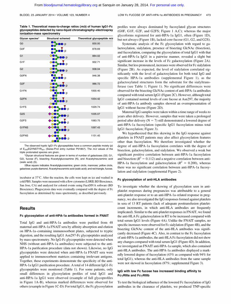
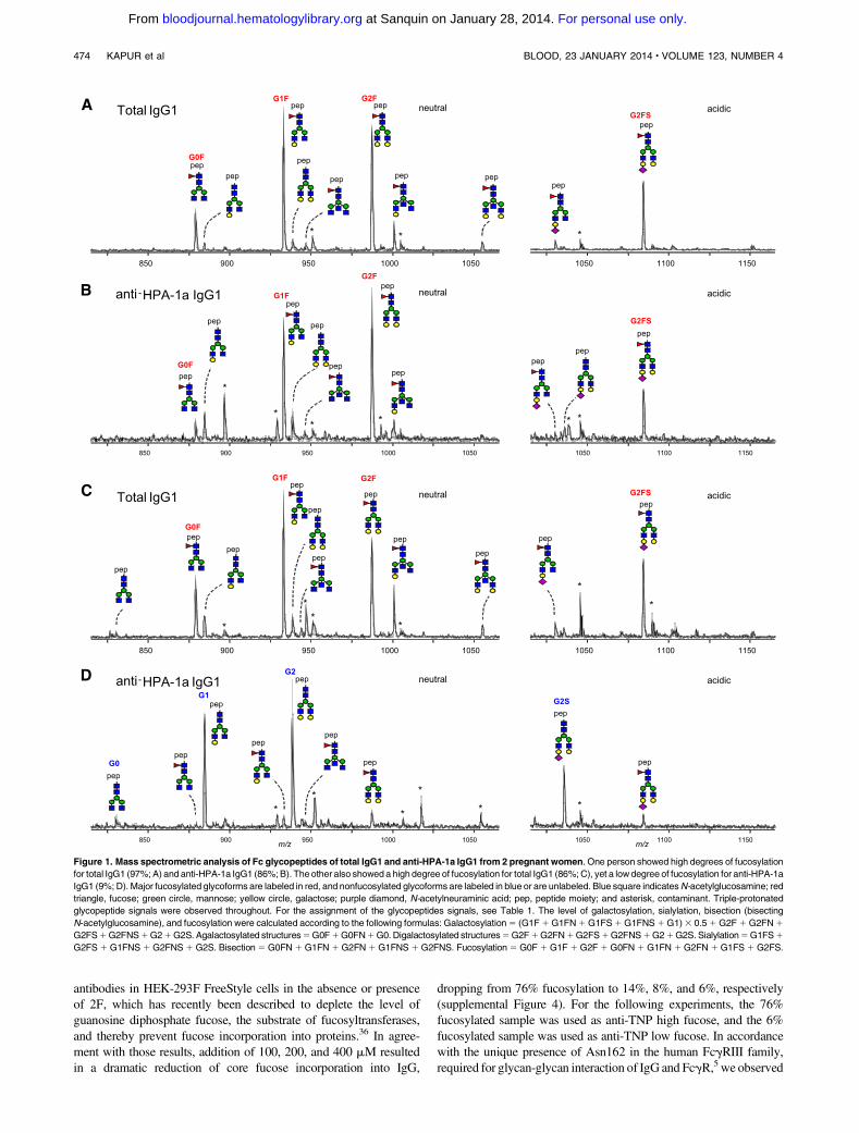
Total IgG and anti-HPA-1a antibodies were purified from 48maternal anti-HPA-1a FNAIT sera by affinity absorption and elutionon HPA-1a–containing immunosorbent plates, subjected to trypticdigestion, and the resulting IgG1 Asn297-Fc glycopeptides analyzedby mass spectrometry. No IgG-Fc glycopeptides were detected whenNHS (without anti-HPA-1a antibodies) were subjected to the anti-HPA-1a purification procedure (data not shown). Likewise, no IgGglycopeptides were detected when anti-HPA-1a FNAIT sera wereapplied to immunosorbent matrices containing irrelevant antigens.Together, these experiments demonstrate the specificity of the anti-HPA-1a IgG1 purification procedure. A total of 14 different IgG1-Fcglycopeptides were monitored (Table 1). For some patients, onlysmall differences in glycosylation profiles of total IgG1 andanti-HPA-1a IgG1 were observed (example of full glycoprofilesin Figure 1A-B), whereas marked differences were observed forothers (example in Figure 1C-D): For total IgG1, the Fc glycosylation
profiles were always dominated by fucosylated glycan structures(G0F, G1F, G2F, and G2FS; Figure 1 A,C); whereas the majorglycoforms registered for anti-HPA-1a IgG1, often (Figure 1D),but not always (Figure 1B), lacked core fucose (G1, G2, and G2S).
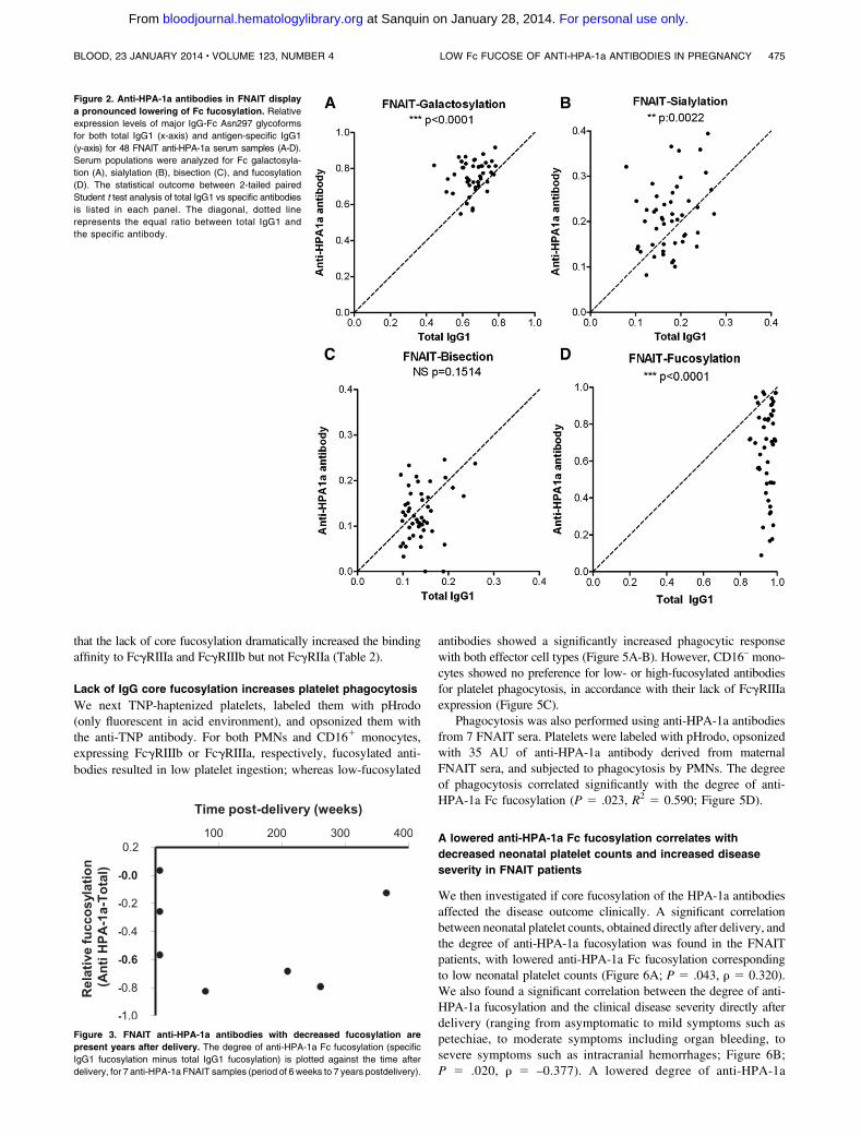
Systematic analysis of the Fc glycosylation with regard to ga-lactosylation, sialylation, presence of bisecting GlcNAc (bisection),and fucosylation, comparing the glycosylation of total IgG1 with thatof anti-HPA-1a IgG1 in a pairwise manner, revealed a slight butsignificant increase in the levels of Fc galactosylation (Figure 2A).Similar, but less pronounced, increaseswere observed for Fc sialylation(Figure 2B). As expected, the level of sialylation correlated sig-nificantly with the level of galactosylation for both total IgG andspecific HPA-1a antibodies (supplemental Figure 1), as thegalactosylated structures form the substrate for the sialyltrans-ferase (see Table 1; Figure 1). No significant differences wereobserved for the bisecting GlcNAc content of anti-HPA-1a antibodiescomparedwith total serum IgG1 (Figure 2C). However, although totalIgG1 contained normal levels of core fucose at Asn297, the majorityof anti-HPA-1a antibody samples showed an overrepresentation ofIgG1 without fucose (Figure 2D).
Maternal IgG samples were taken within a time range of weeks toyears after delivery. However, samples that were taken a prolongedperiod after delivery (N5 7) still demonstrated a lowered degree ofanti-HPA-1a fucosylation (specific IgG1 fucosylation minus totalIgG1 fucosylation; Figure 3).
We hypothesized that this skewing in the IgG response againstplatelets in FNAIT patients may also affect glycosylation featuresother than fucosylation. We therefore investigated whether thedegree of anti-HPA-1a fucosylation correlates with the degree ofbisection, galactosylation, and sialylation. We observed a weak butsignificant positive correlation between anti-HPA-1a fucosylationand bisection (R2 5 0.112) and a negative correlation between anti-HPA-1a fucosylation and galactosylation (R2 5 0.200), whereasthere was no significant correlation between anti-HPA-1a fucosy-lation and sialylation (supplemental Figure 2).
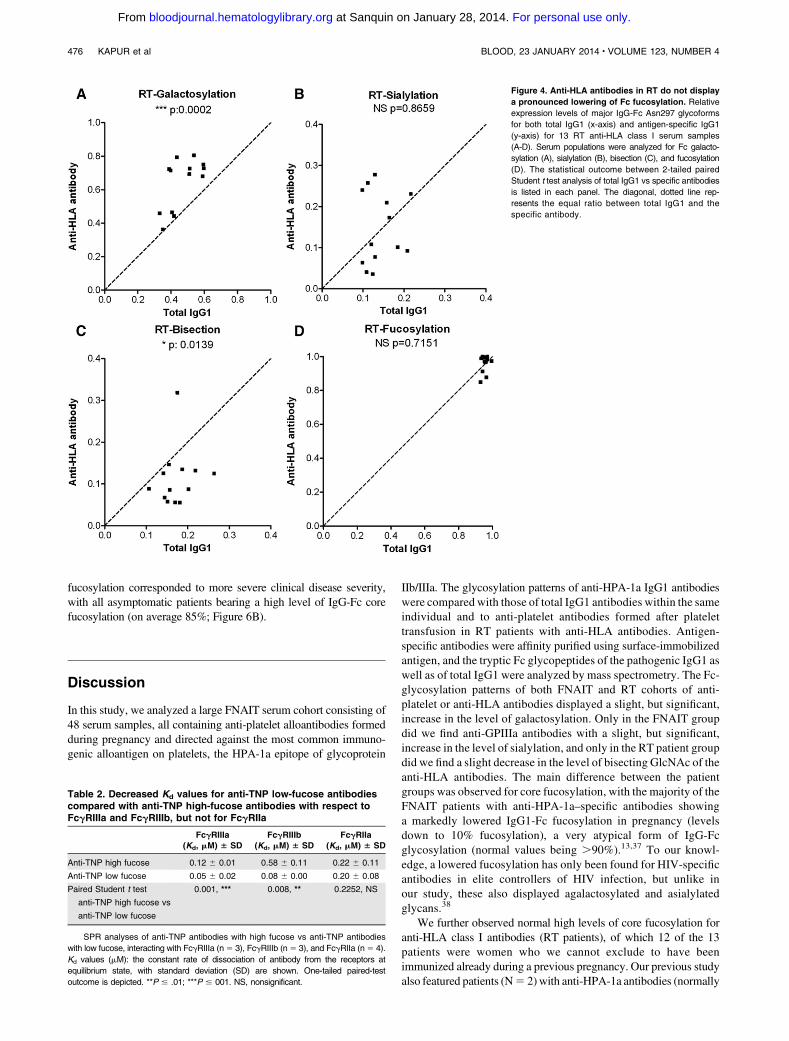
Fc glycosylation of anti-HLA antibodies
To investigate whether the skewing of glycosylation seen in anti-platelet responses during pregnancies was attributable to a generalanti-platelet response or to an anti-HPA-1a–related response in preg-nancy, we also investigated the IgG responses formed against plateletsin sera of 13 RT patients (lack of adequate posttransfusion platelet-count increments, in which anti-HLA antibodies are frequentlyimplicated). Similar to the anti-platelet responses in FNAIT, we foundthe anti-HLA Fc galactosylation in RT to be increased compared withtotal serum IgG1 levels (Figure 4A). Unlike the FNAIT samples, nospecific increaseswere observed for Fc sialylation (Figure 4B), and thebisecting GlcNAc content of the anti-HLA antibodies was signifi-cantly decreased (Figure 4C). Also, in contrast to the Fc fucosylationof anti-HPA-1a antibodies, the anti-HLAFc fucosylation did not showany changes compared with total serum IgG1 (Figure 4D). In addition,we investigated an FNAIT anti-HPA-1a sample, which also containedanti-HLA antibodies. The anti-HPA-1a antibodies displayed a mark-edly lowered degree of fucosylation (43% as compared with 94% fortotal IgG1), whereas the anti-HLA antibodies from the same samplewere not skewed in fucosylation (87%; supplemental Figure 3).
IgG with low Fc fucose has increased binding affinity to
FcgRIIIa and FcgRIIIb
To test the biological influence of the lowered Fc fucosylation of IgGantibodies in the clearance of platelets, we produced TNP-specific
Table 1. Theoretical mass-to-charge ratios (m/z) of human IgG1-Fcglycopeptides detected by nano-liquid chromatography–electrosprayionization-mass spectrometry
Glycan species* Structural scheme† Theoretical glycopeptide m/z
G0 830.00
G0F 878.69
G1 884.02
G1F 932.71
G2 938.04
G0FN 946.38
G2F 986.72
G1FN 1000.40
G2FN 1054.42
G1FS 1029.74
G2S 1035.07
G2FS 1083.75
G1FNS 1097.43
G2FNS 1151.45
The observed tryptic IgG1-Fc glycopeptides have a common peptide moiety (p)
of E293EQYNSTYR301 (Swiss-Prot entry number P01857). The m/z values of the
triple protonated species are given.
*Glycan structural features are given in terms of number of galactoses (G0, G1,
G2), fucose (F), bisecting N-acetylglucosamine (N), and N-acetylneuraminic acid
(sialic acid) (S).
†Blue square indicates N-acetylglucosamine; green circle, mannose; yellow circle,
galactose; purplediamond,N-acetylneuraminic acid (sialic acid); and red triangle, fucose.
BLOOD, 23 JANUARY 2014 x VOLUME 123, NUMBER 4 LOW Fc FUCOSE OF ANTI-HPA-1a ANTIBODIES IN PREGNANCY 473
For personal use only. at Sanquin on January 28, 2014. bloodjournal.hematologylibrary.orgFrom
antibodies in HEK-293F FreeStyle cells in the absence or presenceof 2F, which has recently been described to deplete the level ofguanosine diphosphate fucose, the substrate of fucosyltransferases,and thereby prevent fucose incorporation into proteins.36 In agree-ment with those results, addition of 100, 200, and 400 mM resultedin a dramatic reduction of core fucose incorporation into IgG,
dropping from 76% fucosylation to 14%, 8%, and 6%, respectively(supplemental Figure 4). For the following experiments, the 76%fucosylated sample was used as anti-TNP high fucose, and the 6%fucosylated sample was used as anti-TNP low fucose. In accordancewith the unique presence of Asn162 in the human FcgRIII family,required for glycan-glycan interaction of IgG and FcgR,5we observed
Figure 1. Mass spectrometric analysis of Fc glycopeptides of total IgG1 and anti-HPA-1a IgG1 from 2 pregnant women. One person showed high degrees of fucosylation
for total IgG1 (97%;A) and anti-HPA-1a IgG1 (86%;B). The other also showed a high degree of fucosylation for total IgG1 (86%; C), yet a lowdegree of fucosylation for anti-HPA-1a
IgG1 (9%; D). Major fucosylated glycoforms are labeled in red, and nonfucosylated glycoforms are labeled in blue or are unlabeled. Blue square indicatesN-acetylglucosamine; red
triangle, fucose; green circle, mannose; yellow circle, galactose; purple diamond, N-acetylneuraminic acid; pep, peptide moiety; and asterisk, contaminant. Triple-protonated
glycopeptide signals were observed throughout. For the assignment of the glycopeptides signals, see Table 1. The level of galactosylation, sialylation, bisection (bisecting
N-acetylglucosamine), and fucosylation were calculated according to the following formulas: Galactosylation5 (G1F1G1FN1G1FS1G1FNS1G1)3 0.51G2F1G2FN1
G2FS1G2FNS1G21G2S. Agalactosylated structures5G0F1G0FN1G0.Digalactosylated structures5G2F1G2FN1G2FS1G2FNS1G21G2S.Sialylation5G1FS1
G2FS1G1FNS1G2FNS1 G2S. Bisection5 G0FN1G1FN1G2FN1G1FNS1G2FNS. Fucosylation5 G0F1 G1F1G2F1G0FN1G1FN1G2FN1G1FS1G2FS.
474 KAPUR et al BLOOD, 23 JANUARY 2014 x VOLUME 123, NUMBER 4
For personal use only. at Sanquin on January 28, 2014. bloodjournal.hematologylibrary.orgFrom
that the lack of core fucosylation dramatically increased the bindingaffinity to FcgRIIIa and FcgRIIIb but not FcgRIIa (Table 2).
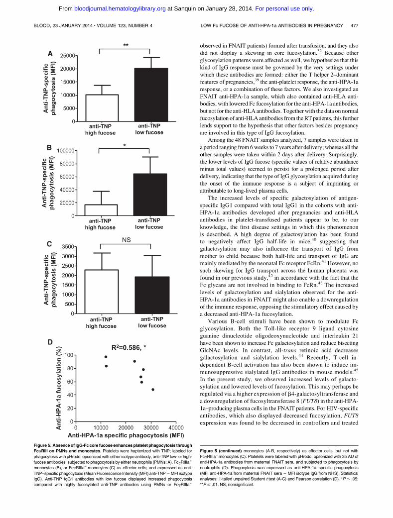
Lack of IgG core fucosylation increases platelet phagocytosis
We next TNP-haptenized platelets, labeled them with pHrodo(only fluorescent in acid environment), and opsonized them withthe anti-TNP antibody. For both PMNs and CD161 monocytes,expressing FcgRIIIb or FcgRIIIa, respectively, fucosylated anti-bodies resulted in low platelet ingestion; whereas low-fucosylated
antibodies showed a significantly increased phagocytic responsewith both effector cell types (Figure 5A-B). However, CD16– mono-cytes showed no preference for low- or high-fucosylated antibodiesfor platelet phagocytosis, in accordance with their lack of FcgRIIIaexpression (Figure 5C).
Phagocytosis was also performed using anti-HPA-1a antibodiesfrom 7 FNAIT sera. Platelets were labeled with pHrodo, opsonizedwith 35 AU of anti-HPA-1a antibody derived from maternalFNAIT sera, and subjected to phagocytosis by PMNs. The degreeof phagocytosis correlated significantly with the degree of anti-HPA-1a Fc fucosylation (P 5 .023, R2 5 0.590; Figure 5D).
A lowered anti-HPA-1a Fc fucosylation correlates with
decreased neonatal platelet counts and increased disease
severity in FNAIT patients
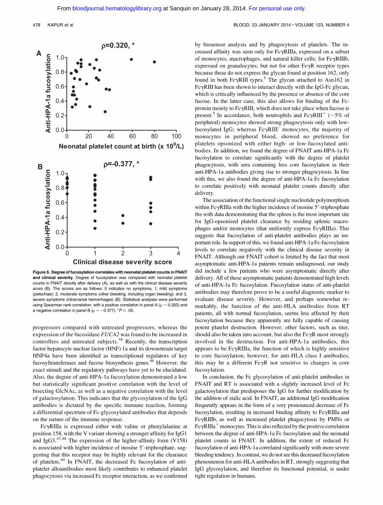
We then investigated if core fucosylation of the HPA-1a antibodiesaffected the disease outcome clinically. A significant correlationbetween neonatal platelet counts, obtained directly after delivery, andthe degree of anti-HPA-1a fucosylation was found in the FNAITpatients, with lowered anti-HPA-1a Fc fucosylation correspondingto low neonatal platelet counts (Figure 6A; P 5 .043, r 5 0.320).We also found a significant correlation between the degree of anti-HPA-1a fucosylation and the clinical disease severity directly afterdelivery (ranging from asymptomatic to mild symptoms such aspetechiae, to moderate symptoms including organ bleeding, tosevere symptoms such as intracranial hemorrhages; Figure 6B;P 5 .020, r 5 –0.377). A lowered degree of anti-HPA-1a
Figure 2. Anti-HPA-1a antibodies in FNAIT display
a pronounced lowering of Fc fucosylation. Relative
expression levels of major IgG-Fc Asn297 glycoforms
for both total IgG1 (x-axis) and antigen-specific IgG1
(y-axis) for 48 FNAIT anti-HPA-1a serum samples (A-D).
Serum populations were analyzed for Fc galactosyla-
tion (A), sialylation (B), bisection (C), and fucosylation
(D). The statistical outcome between 2-tailed paired
Student t test analysis of total IgG1 vs specific antibodies
is listed in each panel. The diagonal, dotted line
represents the equal ratio between total IgG1 and
the specific antibody.
Figure 3. FNAIT anti-HPA-1a antibodies with decreased fucosylation are
present years after delivery. The degree of anti-HPA-1a Fc fucosylation (specific
IgG1 fucosylation minus total IgG1 fucosylation) is plotted against the time after
delivery, for 7 anti-HPA-1a FNAIT samples (period of 6 weeks to 7 years postdelivery).
BLOOD, 23 JANUARY 2014 x VOLUME 123, NUMBER 4 LOW Fc FUCOSE OF ANTI-HPA-1a ANTIBODIES IN PREGNANCY 475
For personal use only. at Sanquin on January 28, 2014. bloodjournal.hematologylibrary.orgFrom
fucosylation corresponded to more severe clinical disease severity,with all asymptomatic patients bearing a high level of IgG-Fc corefucosylation (on average 85%; Figure 6B).
Discussion
In this study, we analyzed a large FNAIT serum cohort consisting of48 serum samples, all containing anti-platelet alloantibodies formedduring pregnancy and directed against the most common immuno-genic alloantigen on platelets, the HPA-1a epitope of glycoprotein
IIb/IIIa. The glycosylation patterns of anti-HPA-1a IgG1 antibodieswere compared with those of total IgG1 antibodies within the sameindividual and to anti-platelet antibodies formed after platelettransfusion in RT patients with anti-HLA antibodies. Antigen-specific antibodies were affinity purified using surface-immobilizedantigen, and the tryptic Fc glycopeptides of the pathogenic IgG1 aswell as of total IgG1 were analyzed by mass spectrometry. The Fc-glycosylation patterns of both FNAIT and RT cohorts of anti-platelet or anti-HLA antibodies displayed a slight, but significant,increase in the level of galactosylation. Only in the FNAIT groupdid we find anti-GPIIIa antibodies with a slight, but significant,increase in the level of sialylation, and only in the RT patient groupdid we find a slight decrease in the level of bisecting GlcNAc of theanti-HLA antibodies. The main difference between the patientgroups was observed for core fucosylation, with the majority of theFNAIT patients with anti-HPA-1a–specific antibodies showinga markedly lowered IgG1-Fc fucosylation in pregnancy (levelsdown to 10% fucosylation), a very atypical form of IgG-Fcglycosylation (normal values being .90%).13,37 To our knowl-edge, a lowered fucosylation has only been found for HIV-specificantibodies in elite controllers of HIV infection, but unlike inour study, these also displayed agalactosylated and asialylatedglycans.38
We further observed normal high levels of core fucosylation foranti-HLA class I antibodies (RT patients), of which 12 of the 13patients were women who we cannot exclude to have beenimmunized already during a previous pregnancy. Our previous studyalso featured patients (N5 2) with anti-HPA-1a antibodies (normally
Figure 4. Anti-HLA antibodies in RT do not display
a pronounced lowering of Fc fucosylation. Relative
expression levels of major IgG-Fc Asn297 glycoforms
for both total IgG1 (x-axis) and antigen-specific IgG1
(y-axis) for 13 RT anti-HLA class I serum samples
(A-D). Serum populations were analyzed for Fc galacto-
sylation (A), sialylation (B), bisection (C), and fucosylation
(D). The statistical outcome between 2-tailed paired
Student t test analysis of total IgG1 vs specific antibodies
is listed in each panel. The diagonal, dotted line rep-
resents the equal ratio between total IgG1 and the
specific antibody.
Table 2. Decreased Kd values for anti-TNP low-fucose antibodiescompared with anti-TNP high-fucose antibodies with respect toFcgRIIIa and FcgRIIIb, but not for FcgRIIa
FcgRIIIa(Kd, mM) 6 SD
FcgRIIIb(Kd, mM) 6 SD
FcgRIIa(Kd, mM) 6 SD
Anti-TNP high fucose 0.12 6 0.01 0.58 6 0.11 0.22 6 0.11
Anti-TNP low fucose 0.05 6 0.02 0.08 6 0.00 0.20 6 0.08
Paired Student t test
anti-TNP high fucose vs
anti-TNP low fucose
0.001, *** 0.008, ** 0.2252, NS
SPR analyses of anti-TNP antibodies with high fucose vs anti-TNP antibodies
with low fucose, interacting with FcgRIIIa (n5 3), FcgRIIIb (n5 3), and FcgRIIa (n5 4).
Kd values (mM): the constant rate of dissociation of antibody from the receptors at
equilibrium state, with standard deviation (SD) are shown. One-tailed paired-test
outcome is depicted. **P # .01; ***P # 001. NS, nonsignificant.
476 KAPUR et al BLOOD, 23 JANUARY 2014 x VOLUME 123, NUMBER 4
For personal use only. at Sanquin on January 28, 2014. bloodjournal.hematologylibrary.orgFrom
observed in FNAIT patients) formed after transfusion, and they alsodid not display a skewing in core fucosylation.32 Because otherglycosylation patterns were affected as well, we hypothesize that thiskind of IgG response must be governed by the very settings underwhich these antibodies are formed: either the T helper 2–dominantfeatures of pregnancies,39 the anti-platelet response, the anti-HPA-1aresponse, or a combination of these factors. We also investigated anFNAIT anti-HPA-1a sample, which also contained anti-HLA anti-bodies, with lowered Fc fucosylation for the anti-HPA-1a antibodies,but not for the anti-HLAantibodies. Togetherwith the data on normalfucosylation of anti-HLA antibodies from theRTpatients, this furtherlends support to the hypothesis that other factors besides pregnancyare involved in this type of IgG fucosylation.
Among the 48 FNAIT samples analyzed, 7 samples were taken ina period ranging from 6weeks to 7 years after delivery;whereas all theother samples were taken within 2 days after delivery. Surprisingly,the lower levels of IgG fucose (specific values of relative abundanceminus total values) seemed to persist for a prolonged period afterdelivery, indicating that the type of IgG glycosylation acquired duringthe onset of the immune response is a subject of imprinting orattributable to long-lived plasma cells.
The increased levels of specific galactosylation of antigen-specific IgG1 compared with total IgG1 in the cohorts with anti-HPA-1a antibodies developed after pregnancies and anti-HLAantibodies in platelet-transfused patients appear to be, to ourknowledge, the first disease settings in which this phenomenonis described. A high degree of galactosylation has been foundto negatively affect IgG half-life in mice,40 suggesting thatgalactosylation may also influence the transport of IgG frommother to child because both half-life and transport of IgG aremainly mediated by the neonatal Fc receptor FcRn.41 However, nosuch skewing for IgG transport across the human placenta wasfound in our previous study,42 in accordance with the fact that theFc glycans are not involved in binding to FcRn.43 The increasedlevels of galactosylation and sialylation observed for the anti-HPA-1a antibodies in FNAIT might also enable a downregulationof the immune response, opposing the stimulatory effect caused bya decreased anti-HPA-1a fucosylation.
Various B-cell stimuli have been shown to modulate Fcglycosylation. Both the Toll-like receptor 9 ligand cytosineguanine dinucleotide oligodeoxynucleotide and interleukin 21have been shown to increase Fc galactosylation and reduce bisectingGlcNAc levels. In contrast, all-trans retinoic acid decreasesgalactosylation and sialylation levels.44 Recently, T-cell in-dependent B-cell activation has also been shown to induce im-munosuppressive sialylated IgG antibodies in mouse models.45
In the present study, we observed increased levels of galacto-sylation and lowered levels of fucosylation. This may perhaps beregulated via a higher expression of b4-galactosyltransferase anda downregulation of fucosyltransferase 8 (FUT8) in the anti-HPA-1a–producing plasma cells in the FNAIT patients. For HIV-specificantibodies, which also displayed decreased fucosylation, FUT8expression was found to be decreased in controllers and treated
Figure 5. Absenceof IgG-Fccore fucose enhancesplatelet phagocytosis through
FcgRIII on PMNs and monocytes. Platelets were haptenized with TNP; labeled for
phagocytosis with pHrodo; opsonizedwith either isotype antibody, anti-TNP low- or high-
fucose antibodies; subjected to phagocytosis by either neutrophils (PMNs; A), FcgRIIIa1
monocytes (B), or FcgRIIIa– monocytes (C) as effector cells; and expressed as anti-
TNP–specific phagocytosis (Mean Fluorescence Intensity (MFI) anti-TNP2MFI isotype
IgG). Anti-TNP IgG1 antibodies with low fucose displayed increased phagocytosis
compared with highly fucosylated anti-TNP antibodies using PMNs or FcgRIIIa1
Figure 5 (continued) monocytes (A-B, respectively) as effector cells, but not with
FcgRIIIa– monocytes (C). Platelets were labeled with pHrodo, opsonized with 35 AU of
anti-HPA-1a antibodies from maternal FNAIT sera, and subjected to phagocytosis by
neutrophils (D). Phagocytosis was expressed as anti-HPA-1a–specific phagocytosis
(MFI anti-HPA-1a from maternal FNAIT sera 2 MFI isotype IgG from NHS). Statistical
analyses: 1-tailed unpaired Student t test (A-C) and Pearson correlation (D). *P # .05;
**P # .01. NS, nonsignificant.
BLOOD, 23 JANUARY 2014 x VOLUME 123, NUMBER 4 LOW Fc FUCOSE OF ANTI-HPA-1a ANTIBODIES IN PREGNANCY 477
For personal use only. at Sanquin on January 28, 2014. bloodjournal.hematologylibrary.orgFrom
progressors compared with untreated progressors, whereas theexpression of the fucosidase FUCA2was found to be increased incontrollers and untreated subjects.38 Recently, the transcriptionfactor hepatocyte nuclear factor (HNF) 1a and its downstream targetHNF4a have been identified as transcriptional regulators of keyfucosyltransferases and fucose biosynthesis genes.46 However, theexact stimuli and the regulatory pathways have yet to be elucidated.Also, the degree of anti-HPA-1a fucosylation demonstrated a lowbut statistically significant positive correlation with the level ofbisecting GlcNAc, as well as a negative correlation with the levelof galactosylation. This indicates that the glycosylation of the IgGantibodies is dictated by the specific immune reaction, forminga differential spectrum of Fc-glycosylated antibodies that dependson the nature of the immune response.
FcgRIIIa is expressed either with valine or phenylalanine atposition 158, with the V variant showing a stronger affinity for IgG1and IgG3.47,48 The expression of the higher-affinity form (V158)is associated with higher incidence of inosine 59-triphosphate, sug-gesting that this receptor may be highly relevant for the clearanceof platelets.49 In FNAIT, the decreased Fc fucosylation of anti-platelet alloantibodies most likely contributes to enhanced plateletphagocytosis via increased Fc receptor interaction, as we confirmed
by biosensor analysis and by phagocytosis of platelets. The in-creased affinity was seen only for FcgRIIIa, expressed on a subsetof monocytes, macrophages, and natural killer cells; for FcgRIIIb,expressed on granulocytes; but not for other FcgR receptor typesbecause these do not express the glycan found at position 162, onlyfound in both FcgRIII types.5 The glycan attached to Asn162 inFcgRIII has been shown to interact directly with the IgG-Fc glycan,which is critically influenced by the presence or absence of the corefucose. In the latter case, this also allows for binding of the Fc-protein moiety to FcgRIII, which does not take place when fucose ispresent.5 In accordance, both neutrophils and FcgRIII1 (;5% ofperipheral) monocytes showed strong phagocytosis only with low-fucosylated IgG; whereas FcgRIII– monocytes, the majority ofmonocytes in peripheral blood, showed no preference forplatelets opsonized with either high- or low-fucosylated anti-bodies. In addition, we found the degree of FNAIT anti-HPA-1a Fcfucosylation to correlate significantly with the degree of plateletphagocytosis, with sera containing less core fucosylation in theiranti-HPA-1a antibodies giving rise to stronger phagocytosis. In linewith this, we also found the degree of anti-HPA-1a Fc fucosylationto correlate positively with neonatal platelet counts directly afterdelivery.
The association of the functional single nucleotide polymorphismwithin FcgRIIIa with the higher incidence of inosine 59-triphosphatefits with data demonstrating that the spleen is the most important sitefor IgG-opsonized platelet clearance by residing splenic macro-phages and/or monocytes (that uniformly express FcgRIIIa). Thissuggests that fucosylation of anti-platelet antibodies plays an im-portant role. In support of this, we found anti-HPA-1a Fc-fucosylationlevels to correlate negatively with the clinical disease severity inFNAIT. Although our FNAIT cohort is limited by the fact that mostasymptomatic anti-HPA-1a patients remain undiagnosed, our studydid include a few patients who were asymptomatic directly afterdelivery. All of these asymptomatic patients demonstrated high levelsof anti-HPA-1a Fc fucosylation. Fucosylation status of anti-plateletantibodies may therefore prove to be a useful diagnostic marker toevaluate disease severity. However, and perhaps somewhat re-markably, the function of the anti-HLA antibodies from RTpatients, all with normal fucosylation, seems less affected by theirfucosylation because they apparently are fully capable of causingpotent platelet destruction. However, other factors, such as titer,should also be taken into account, but also the FcgR most stronglyinvolved in the destruction. For anti-HPA-1a antibodies, thisappears to be FcgRIIIa, the function of which is highly sensitiveto core fucosylation; however, for anti-HLA class I antibodies,this may be a different FcgR not sensitive to changes in corefucosylation.
In conclusion, the Fc glycosylation of anti-platelet antibodies inFNAIT and RT is associated with a slightly increased level of Fcgalactosylation that predisposes the IgG for further modification bythe addition of sialic acid. In FNAIT, an additional IgG modificationfrequently appears in the form of a very pronounced decrease of Fcfucosylation, resulting in increased binding affinity to FcgRIIIa andFcgRIIIb, as well as increased platelet phagocytosis by PMNs orFcgRIIIa1monocytes. This is also reflected by the positive correlationbetween the degree of anti-HPA-1a Fc fucosylation and the neonatalplatelet counts in FNAIT. In addition, the extent of reduced Fcfucosylation of anti-HPA-1a correlated significantly with more severebleeding tendency. In contrast,wedonot see this decreased fucosylationphenomenon for anti-HLA antibodies in RT, strongly suggesting thatIgG glycosylation, and therefore its functional potential, is undertight regulation in humans.
Figure 6. Degree of fucosylation correlateswith neonatal platelet counts in FNAIT
and clinical severity. Degree of fucosylation was compared with neonatal platelet
counts in FNAIT directly after delivery (A), as well as with the clinical disease severity
score (B). The scores are as follows: 0 indicates no symptoms; 1, mild symptoms
(petechiae); 2, moderate symptoms (other bleeding, including organ bleeding); and 3,
severe symptoms (intracranial hemorrhages) (B). Statistical analyses were performed
using Spearman rank correlation, with a positive correlation in panel A (r 5 0.320) and
a negative correlation in panel B (r 5 20.377). *P # .05.
478 KAPUR et al BLOOD, 23 JANUARY 2014 x VOLUME 123, NUMBER 4
For personal use only. at Sanquin on January 28, 2014. bloodjournal.hematologylibrary.orgFrom
Acknowledgments
This work was supported by grants from Sanquin (PPOC-09- 025)(R.K.), the Landsteiner Foundation for Blood Transfusion (grant0721) (H.K.E. and R.V.), and the European Union’s SeventhFramework Programme (FP7-Health-F5-2011) under Grant Agree-ment No. 278535 (HighGlycan) (M.W.).
Authorship
Contribution: R.K. coordinated all sample gathering, analyzed clinicaldata, and performed phagocytosis experiments; L.P., M.d.H., A.V.,D.J., B.K., D.B., B.S., M.K.K., and T.E.M. assisted with gathering
patient material for this study; H.K.E. and R.V. produced recombinantantibodies; I.K. performed SPR experiments and coanalyzed themtogetherwithT.R.; C.A.M.K. prepared samples and conducted themassspectrometry; M.W. and A.M.D. processed the raw mass spectrometrydata;R.K.,M.W., andG.V.made thefigures and tables; R.K. performedstatistical analyses; G.V. conceived and supervised the study;M.W. andC.E.v.d.S. cosupervised the study; R.K., G.V., C.E.v.d.S., and M.W.wrote the paper, which was critically revised and approved by allauthors; and all authors contributed to analysis and interpretation of data.
Conflict-of-interest disclosure: B.S. is CEO, M.K.K. is COO, andboth are stock owners of Prophylix Pharma AS. The remainingauthors declare no competing financial interests.
Correspondence: Dr Gestur Vidarsson, Department of Exper-imental Immunohematology, Sanquin Research and LandsteinerLaboratory, Plesmanlaan 125, 1066 CX, Amsterdam, The Netherlands;e-mail: [email protected].
References
1. Semple JW, Italiano JE Jr, Freedman J. Plateletsand the immune continuum. Nat Rev Immunol.2011;11(4):264-274.
2. Nimmerjahn F, Ravetch JV. Anti-inflammatoryactions of intravenous immunoglobulin. Annu RevImmunol. 2008;26:513-533.
3. Shields RL, Lai J, Keck R, et al. Lack of fucose onhuman IgG1 N-linked oligosaccharide improvesbinding to human Fcgamma RIII and antibody-dependent cellular toxicity. J Biol Chem. 2002;277(30):26733-26740.
4. Sondermann P, Huber R, Oosthuizen V, Jacob U.The 3.2-A crystal structure of the human IgG1 Fcfragment-Fc gammaRIII complex. Nature. 2000;406(6793):267-273.
5. Ferrara C, Grau S, Jager C, et al. Uniquecarbohydrate-carbohydrate interactions arerequired for high affinity binding betweenFcgammaRIII and antibodies lacking core fucose.Proc Natl Acad Sci USA. 2011;108(31):12669-12674.
6. Parekh RB, Dwek RA, Sutton BJ, et al.Association of rheumatoid arthritis and primaryosteoarthritis with changes in the glycosylationpattern of total serum IgG. Nature. 1985;316(6027):452-457.
7. Arnold JN, Wormald MR, Sim RB, Rudd PM,Dwek RA. The impact of glycosylation on thebiological function and structure of humanimmunoglobulins. Annu Rev Immunol. 2007;25:21-50.
8. Saldova R, Royle L, Radcliffe CM, et al. Ovariancancer is associated with changes in glycosylationin both acute-phase proteins and IgG.Glycobiology. 2007;17(12):1344-1356.
9. Rook GA, Steele J, Brealey R, et al. Changes inIgG glycoform levels are associated withremission of arthritis during pregnancy.J Autoimmun. 1991;4(5):779-794.
10. van de Geijn FE, Wuhrer M, Selman MH, et al.Immunoglobulin G galactosylation and sialylationare associated with pregnancy-inducedimprovement of rheumatoid arthritis and thepostpartum flare: results from a large prospectivecohort study. Arthritis Res Ther. 2009;11(6):R193.
11. Ruhaak LR, Uh HW, Beekman M, et al.Decreased levels of bisecting GlcNAc glycoformsof IgG are associated with human longevity. PLoSONE. 2010;5(9):e12566.
12. Pucic M, Knezevic A, Vidic J, et al. Highthroughput isolation and glycosylation analysis ofIgG-variability and heritability of the IgG glycomein three isolated human populations. Mol CellProteomics. 2011;10(10):M111.010090.
13. Selman MH, Derks RJ, Bondt A, et al. Fc specificIgG glycosylation profiling by robust nano-reversephase HPLC-MS using a sheath-flow ESI sprayerinterface. J Proteomics. 2012;75(4):1318-1329.
14. Karsten CM, Pandey MK, Figge J, et al. Anti-inflammatory activity of IgG1 mediated by Fcgalactosylation and association of FcgRIIB anddectin-1. Nat Med. 2012;18(9):1401-1406.
15. Kaneko Y, Nimmerjahn F, Ravetch JV.Anti-inflammatory activity of immunoglobulin Gresulting from Fc sialylation. Science. 2006;313(5787):670-673.
16. Anthony RM, Kobayashi T, Wermeling F, RavetchJV. Intravenous gammaglobulin suppressesinflammation through a novel T(H)2 pathway.Nature. 2011;475(7354):110-113.
17. Sondermann P, Pincetic A, Maamary J,Lammens K, Ravetch JV. General mechanismfor modulating immunoglobulin effector function.Proc Natl Acad Sci USA. 2013;110(24):9868-9872.
18. Shibata-Koyama M, Iida S, Misaka H, et al.Nonfucosylated rituximab potentiates humanneutrophil phagocytosis through its high bindingfor FcgammaRIIIb and MHC class II expressionon the phagocytotic neutrophils. Exp Hematol.2009;37(3):309-321.
19. Mizushima T, Yagi H, Takemoto E, et al.Structural basis for improved efficacy oftherapeutic antibodies on defucosylation of theirFc glycans. Genes Cells. 2011;16(11):1071-1080.
20. Junttila TT, Parsons K, Olsson C, et al. Superiorin vivo efficacy of afucosylated trastuzumab in thetreatment of HER2-amplified breast cancer.Cancer Res. 2010;70(11):4481-4489.
21. Masuda K, Kubota T, Kaneko E, et al. Enhancedbinding affinity for FcgammaRIIIa of fucose-negative antibody is sufficient to induce maximalantibody-dependent cellular cytotoxicity. MolImmunol. 2007;44(12):3122-3131.
22. Suzuki E, Niwa R, Saji S, et al. A nonfucosylatedanti-HER2 antibody augments antibody-dependent cellular cytotoxicity in breast cancerpatients. Clin Cancer Res. 2007;13(6):1875-1882.
23. Niwa R, Shoji-Hosaka E, Sakurada M, et al.Defucosylated chimeric anti-CC chemokinereceptor 4 IgG1 with enhanced antibody-dependent cellular cytotoxicity shows potenttherapeutic activity to T-cell leukemia andlymphoma. Cancer Res. 2004;64(6):2127-2133.
24. Niwa R, Natsume A, Uehara A, et al. IgGsubclass-independent improvement of antibody-dependent cellular cytotoxicity by fucose removalfrom Asn297-linked oligosaccharides. J ImmunolMethods. 2005;306(1-2):151-160.
25. Peipp M, Lammerts van Bueren JJ, Schneider-Merck T, et al. Antibody fucosylation differentiallyimpacts cytotoxicity mediated by NK and PMNeffector cells. Blood. 2008;112(6):2390-2399.
26. Yamane-Ohnuki N, Satoh M. Production oftherapeutic antibodies with controlledfucosylation. MAbs. 2009;1(3):230-236.
27. Jefferis R. Glycosylation as a strategy to improveantibody-based therapeutics. Nat Rev DrugDiscov. 2009;8(3):226-234.
28. von Horsten HH, Ogorek C, Blanchard V, et al.Production of non-fucosylated antibodies by co-expression of heterologous GDP-6-deoxy-D-lyxo-4-hexulose reductase. Glycobiology. 2010;20(12):1607-1618.
29. van Berkel PHC, Gerritsen J, van Voskuilen E,et al. Rapid production of recombinant human IgGwith improved ADCC effector function in atransient expression system. Biotechnol Bioeng.2010;105(2):350-357.
30. Reichert JM, Dhimolea E. The future of antibodiesas cancer drugs. Drug Discov Today. 2012;17(17-18):954-963.
31. Metcalfe P, Watkins NA, Ouwehand WH, et al.Nomenclature of human platelet antigens. VoxSang. 2003;85(3):240-245.
32. Wuhrer M, Porcelijn L, Kapur R, et al. Regulatedglycosylation patterns of IgG during alloimmuneresponses against human platelet antigens.J Proteome Res. 2009;8(2):450-456.
33. Kiefel V, Santoso S, Weisheit M, Mueller-Eckhardt C. Monoclonal antibody—specificimmobilization of platelet antigens (MAIPA):a new tool for the identification of platelet-reactiveantibodies. Blood. 1987;70(6):1722-1726.
34. Pei R, Lee J, Chen T, Rojo S, Terasaki PI. Flowcytometric detection of HLA antibodies usinga spectrum of microbeads. Hum Immunol. 1999;60(12):1293-1302.
35. Kruijsen D, Einarsdottir HK, Schijf MA, et al.Intranasal administration of antibody-boundrespiratory syncytial virus particles efficientlyprimes virus-specific immune responses in mice.J Virol. 2013;87(13):7550-7557.
36. Okeley NM, Alley SC, Anderson ME, et al.Development of orally active inhibitors of proteinand cellular fucosylation. Proc Natl Acad Sci USA.2013;110(14):5404-5409.
37. Bakovic MP, Selman MHJ, Hoffmann M, et al.High-throughput IgG Fc N-glycosylation profilingby mass spectrometry of glycopeptides.J Proteome Res. 2013;12(2):821-831.
38. Ackerman ME, Crispin M, Yu X, et al. Naturalvariation in Fc glycosylation of HIV-specific
BLOOD, 23 JANUARY 2014 x VOLUME 123, NUMBER 4 LOW Fc FUCOSE OF ANTI-HPA-1a ANTIBODIES IN PREGNANCY 479
For personal use only. at Sanquin on January 28, 2014. bloodjournal.hematologylibrary.orgFrom
antibodies impacts antiviral activity. J Clin Invest.2013;123(5):2183-2192.
39. Kumpel BM, Manoussaka MS. Placentalimmunology and maternal alloimmune responses.Vox Sang. 2012;102(1):2-12.
40. Newkirk MM, Novick J, Stevenson MM, FournierMJ, Apostolakos P. Differential clearance ofglycoforms of IgG in normal and autoimmune-prone mice. Clin Exp Immunol. 1996;106(2):259-264.
41. Roopenian DC, Akilesh S. FcRn: the neonatal Fcreceptor comes of age. Nat Rev Immunol. 2007;7(9):715-725.
42. Einarsdottir HK, Selman MH, Kapur R, et al.Comparison of the Fc glycosylation of fetal andmaternal immunoglobulin G. Glycoconj J. 2013;30(2):147-157.
43. West AP Jr, Bjorkman PJ. Crystal structure andimmunoglobulin G binding properties of the humanmajor histocompatibility complex-related Fcreceptor. Biochemistry. 2000;39(32):9698-9708.
44. Wang J, Balog CIA, Stavenhagen K, et al.Fc-glycosylation of IgG1 is modulated by B-cellstimuli. Mol Cell Proteomics. 2011;10(5):M110.004655.
45. Hess C, Winkler A, Lorenz AK, et al.T cell-independent B cell activation inducesimmunosuppressive sialylated IgG antibodies.J Clin Invest. 2013;123(9):3788-3796.
46. Lauc G, Essafi A, Huffman JE, et al. Genomicsmeets glycomics—the first GWAS study of humanN-Glycome identifies HNF1a as a masterregulator of plasma protein fucosylation. PLoSGenet. 2010;6(12):e1001256.
47. de Haas M, Koene HR, Kleijer M, et al. A triallelicFc gamma receptor type IIIA polymorphisminfluences the binding of human IgG by NK cell Fcgamma RIIIa. J Immunol. 1996;156(8):2948-2955.
48. Koene HR, Kleijer M, Algra J, Roos D,von dem Borne AE, de Haas M. FcgammaRIIIa-158V/F polymorphism influencesthe binding of IgG by natural killer cell FcgammaRIIIa, independently of the FcgammaRIIIa-48L/R/H phenotype. Blood. 1997;90(3):1109-1114.
49. Breunis WB, van Mirre E, Bruin M, et al. Copynumber variation of the activating FCGR2Cgene predisposes to idiopathicthrombocytopenic purpura. Blood. 2008;111(3):1029-1038.
480 KAPUR et al BLOOD, 23 JANUARY 2014 x VOLUME 123, NUMBER 4
For personal use only. at Sanquin on January 28, 2014. bloodjournal.hematologylibrary.orgFrom