KANSAS CITY MISSOURI FIRE DEPARTMENT EMS ...
183
KANSAS CITY MISSOURI FIRE DEPARTMENT EMS PROTOCOLS OFFICE OF THE EMS MEDICAL DIRECTOR KANSAS CITY, MISSOURI Effective May 1 2021
-
Upload
khangminh22 -
Category
Documents
-
view
1 -
download
0
Transcript of KANSAS CITY MISSOURI FIRE DEPARTMENT EMS ...
KANSAS CITY MISSOURI FIRE DEPARTMENT
EMS PROTOCOLS
OFFICE OF THE EMS MEDICAL DIRECTORKANSAS CITY, MISSOURI
Effective May 1 2021
Preface
The policies, protocols, procedures and medications described in this document are applicable to the Kansas City, Missouri Fire Department and the Kansas City, Missouri Emergency Medical Services (EMS) System. The document represents the hard work of multiple individuals and agencies including; Kansas City, Missouri Fire Department, the Emergency Physician’s Advisory Board (EPAB), the Medical Equipment and Protocol Labor Management Committee, the Office of the EMS Medical Director and the Emergency Medical Services Coordinating Committee (EMSCC) of Kansas City, Missouri.
This is a “living” document and is frequently updated. We would like to thank the above agencies For their tireless work on this document and most especially we would like to thank the individual Paramedics, Emergency Medical Technicians (EMT), Fire Fighters, Communication Specialists, and other individuals who continuously provide excellent Emergency Medical Care under frequently difficult and sometimes dangerous circumstances to the citizens of Kansas City, Missouri, and it’s visitors.
Erica Carney, M.D. KCMO EMS Medical Director
Donna Lake, Chief Kansas City Fire Department
Theodore Barnett, M.D., Chair Emergency Physicians Advisory Board
Table of Contents
2 Table of Contents
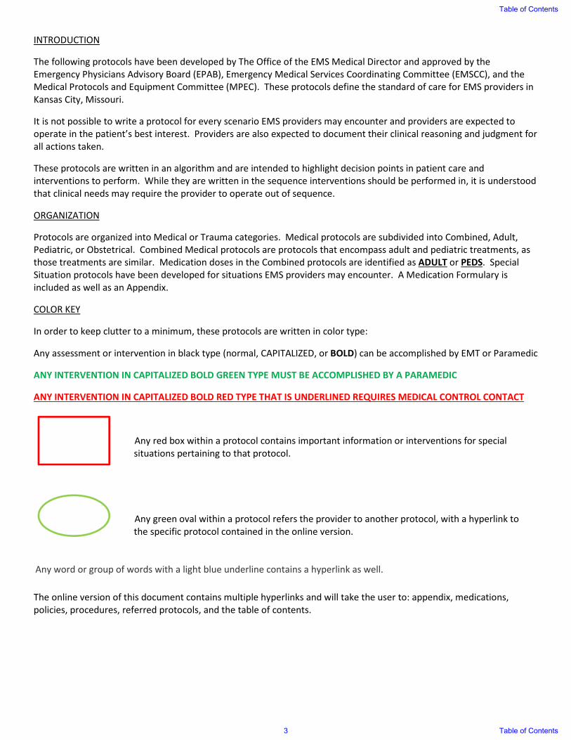
INTRODUCTION
The following protocols have been developed by The Office of the EMS Medical Director and approved by the
Emergency Physicians Advisory Board (EPAB), Emergency Medical Services Coordinating Committee (EMSCC), and the
Medical Protocols and Equipment Committee (MPEC). These protocols define the standard of care for EMS providers in
Kansas City, Missouri.
It is not possible to write a protocol for every scenario EMS providers may encounter and providers are expected to
operate in the patient’s best interest. Providers are also expected to document their clinical reasoning and judgment for
all actions taken.
These protocols are written in an algorithm and are intended to highlight decision points in patient care and
interventions to perform. While they are written in the sequence interventions should be performed in, it is understood
that clinical needs may require the provider to operate out of sequence.
ORGANIZATION
Protocols are organized into Medical or Trauma categories. Medical protocols are subdivided into Combined, Adult,
Pediatric, or Obstetrical. Combined Medical protocols are protocols that encompass adult and pediatric treatments, as
those treatments are similar. Medication doses in the Combined protocols are identified as ADULT or PEDS. Special
Situation protocols have been developed for situations EMS providers may encounter. A Medication Formulary is
included as well as an Appendix.
COLOR KEY
In order to keep clutter to a minimum, these protocols are written in color type:
Any assessment or intervention in black type (normal, CAPITALIZED, or BOLD) can be accomplished by EMT or Paramedic
ANY INTERVENTION IN CAPITALIZED BOLD GREEN TYPE MUST BE ACCOMPLISHED BY A PARAMEDIC
ANY INTERVENTION IN CAPITALIZED BOLD RED TYPE THAT IS UNDERLINED REQUIRES MEDICAL CONTROL CONTACT
Any red box within a protocol contains important information or interventions for special
situations pertaining to that protocol.
Any green oval within a protocol refers the provider to another protocol, with a hyperlink to
the specific protocol contained in the online version.
The online version of this document contains multiple hyperlinks and will take the user to: appendix, medications,
policies, procedures, referred protocols, and the table of contents.
Any word or group of words with a light blue underline contains a hyperlink as well.
Table of Contents
3 Table of Contents
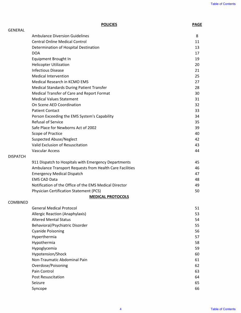
POLICIES PAGEGENERAL
Ambulance Diversion Guidelines 8Central Online Medical Control 11Determination of Hospital Destination 13DOA 17Equipment Brought In 19Helicopter Utilization 20Infectious Disease 21Medical Intervention 25Medical Research in KCMO EMS 27Medical Standards During Patient Transfer 28Medical Transfer of Care and Report Format 30Medical Values Statement 31On Scene AED Coordination 32Patient Contact 33Person Exceeding the EMS System's Capability 34Refusal of Service 35Safe Place for Newborns Act of 2002 39Scope of Practice 40Suspected Abuse/Neglect 42Valid Exclusion of Resuscitation 43Vascular Access 44
DISPATCH911 Dispatch to Hospitals with Emergency Departments 45Ambulance Transport Requests from Health Care Facilities 46Emergency Medical Dispatch 47EMS CAD Data 48Notification of the Office of the EMS Medical Director 49Physician Certification Statement (PCS) 50
MEDICAL PROTOCOLSCOMBINED
General Medical Protocol 51Allergic Reaction (Anaphylaxis) 53Altered Mental Status 54Behavioral/Psychiatric Disorder 55Cyanide Poisoning 56Hyperthermia 57Hypothermia 58Hypoglycemia 59Hypotension/Shock 60Non-Traumatic Abdominal Pain 61Overdose/Poisoning 62Pain Control 63Post Resuscitation 64Seizure 65Syncope 66
Table of Contents
4 Table of Contents
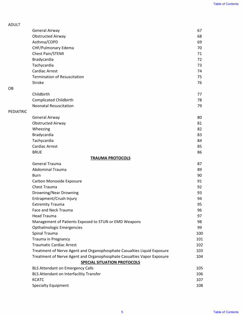
ADULTGeneral Airway 67Obstructed Airway 68Asthma/COPD 69CHF/Pulmonary Edema 70Chest Pain/STEMI 71Bradycardia 72Tachycardia 73Cardiac Arrest 74Termination of Resuscitation 75Stroke 76
OBChildbirth 77Complicated Childbirth 78Neonatal Resuscitation 79
PEDIATRICGeneral Airway 80Obstructed Airway 81Wheezing 82Bradycardia 83Tachycardia 84Cardiac Arrest 85BRUE 86
TRAUMA PROTOCOLSGeneral Trauma 87Abdominal Trauma 89Burn 90Carbon Monoxide Exposure 91Chest Trauma 92Drowning/Near Drowning 93Entrapment/Crush Injury 94Extremity Trauma 95Face and Neck Trauma 96Head Trauma 97Management of Patients Exposed to STUN or EMD Weapons 98Opthalmologic Emergencies 99Spinal Trauma 100Trauma in Pregnancy 101Traumatic Cardiac Arrest 102Treatment of Nerve Agent and Organophosphate Casualties Liquid Exposure 103Treatment of Nerve Agent and Organophosphate Casualties Vapor Exposure 104
SPECIAL SITUATION PROTOCOLSBLS Attendant on Emergency Calls 105BLS Attendant on Interfacility Transfer 106KCATC 107Specialty Equipment 108
Table of Contents
5 Table of Contents
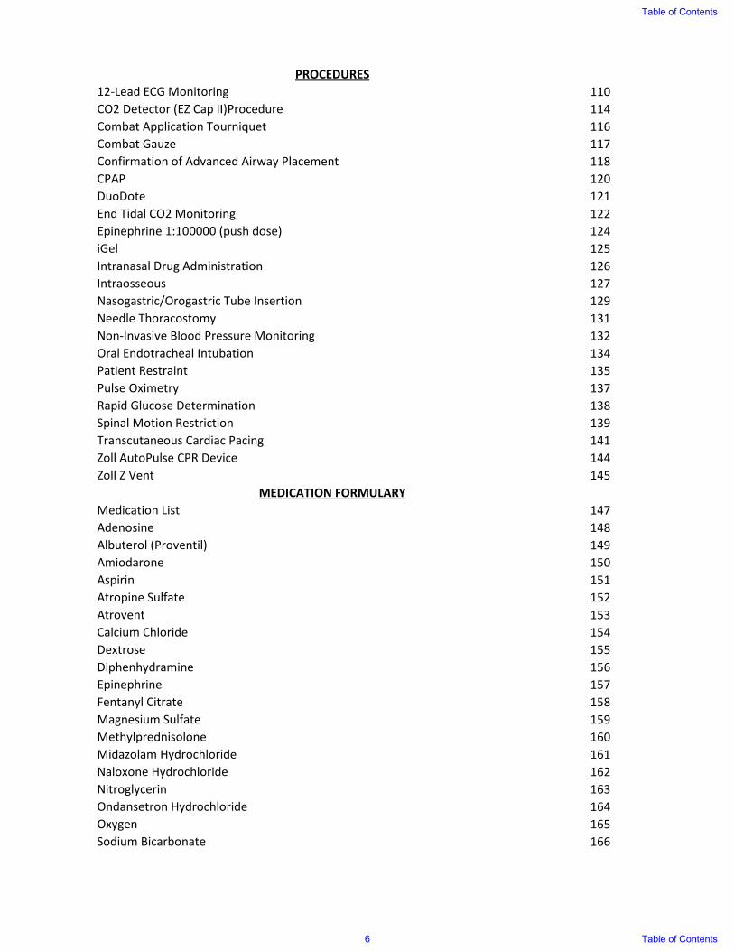
PROCEDURES12-Lead ECG Monitoring 110CO2 Detector (EZ Cap II)Procedure 114Combat Application Tourniquet 116Combat Gauze 117Confirmation of Advanced Airway Placement 118CPAP 120DuoDote 121End Tidal CO2 Monitoring 122Epinephrine 1:100000 (push dose) 124iGel 125Intranasal Drug Administration 126Intraosseous 127Nasogastric/Orogastric Tube Insertion 129Needle Thoracostomy 131Non-Invasive Blood Pressure Monitoring 132Oral Endotracheal Intubation 134Patient Restraint 135Pulse Oximetry 137Rapid Glucose Determination 138Spinal Motion Restriction 139Transcutaneous Cardiac Pacing 141Zoll AutoPulse CPR Device 144Zoll Z Vent 145
MEDICATION FORMULARYMedication List 147Adenosine 148Albuterol (Proventil) 149Amiodarone 150Aspirin 151Atropine Sulfate 152Atrovent 153Calcium Chloride 154Dextrose 155Diphenhydramine 156Epinephrine 157Fentanyl Citrate 158Magnesium Sulfate 159Methylprednisolone 160Midazolam Hydrochloride 161Naloxone Hydrochloride 162Nitroglycerin 163Ondansetron Hydrochloride 164Oxygen 165Sodium Bicarbonate 166
Table of Contents
6 Table of Contents
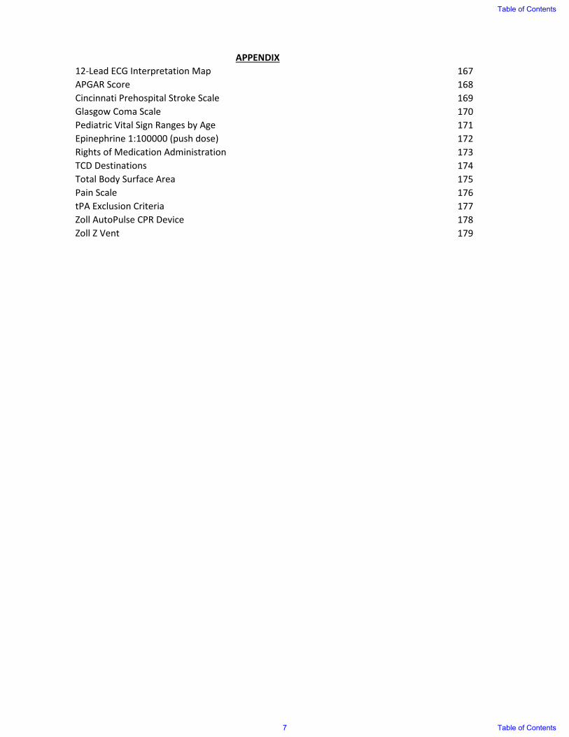
APPENDIX12-Lead ECG Interpretation Map 167APGAR Score 168Cincinnati Prehospital Stroke Scale 169Glasgow Coma Scale 170Pediatric Vital Sign Ranges by Age 171Epinephrine 1:100000 (push dose) 172Rights of Medication Administration 173TCD Destinations 174Total Body Surface Area 175Pain Scale 176tPA Exclusion Criteria 177Zoll AutoPulse CPR Device 178Zoll Z Vent 179
Table of Contents
7 Table of Contents
m
BACKGROUND:
The Diversion Work Group of the Health Alliance of MidAmerica and MARCER have revised, and the EMS Medical Director has endorsed, updated regional diversion guidelines entitled “Organization and Management for Hospitals and EMS Agencies: A Community Plan for Diversion”. The “Policy”, “Definitions”, “Procedures”, and “Regional Catchment Areas” portions are reproduced here. The policy of the Kansas City, Missouri EMS System is as follows, in so far as the guidelines do not conflict with any other City regulation or policy.
POLICY:
I. Patient care and safety should be the central consideration in all status change decisions. EMS should consider alternative destinations for patient routing when hospitals experience high volume.
II. The decision to communicate a change in status should be based on the immediate capabilities and capacities of the emergency department and institution to care for patients. (An exception is trauma diversion, in which availability of an operating room or appropriate surgeon may limit the ability to function as a trauma center.)
III. Patients who are in cardiac arrest will be taken to the closest appropriate hospital, unless the hospital is “out of service.” Patients who are “unstable” may still be taken to the closest appropriate hospital, unless it is “out of service” or on “trauma diversion” (for “unstable” trauma patients only).
IV. Patients should be taken to the nearest, open and appropriate hospital. If a patient requests transport to a facility that is experiencing high volume and is informed of this status, then the medic may take the patient to the hospital of their choice.
V. Designated trauma centers may close to ambulances carrying patients who meet EMS trauma routing criteria. VI. Designated trauma centers may remain open for EMS trauma routing, while the ED is experiencing high volume.
VII. Designated STEMI and/or stroke centers may close to ambulances that have patients that meet TCD routing criteria for STEMI and/or stroke.
VIII. Designated STEMI and/or stroke centers may remain open for patients meeting TCD routing criteria, while the ED is experiencing high volume.
IX. No facility can close to patients on the basis of ability to pay. X. Hospitals changing their status must do so prior to being notified of an ambulance’s impending arrival (i.e. there
should be no “diversions in route”). During multicasualty incidents (MCI) the EMS agency may distribute patients to multiple facilities in order to optimize utilization of resources.
XI. Status notifications should be made to all EMS providers, hospitals and EMCCs (Emergency Coordination Centers) through EMResource®. (If there is a local problem with EMResource®, the appropriate EMCC can be contacted by phone or FAX and enter the notification into EMResource®.)
XII. If all hospitals within a predefined catchment area are experiencing high volume, then all hospitals in the catchment area will have their status changed to “catchment area high volume” and the patient will be taken to the closest appropriate hospital within the catchment area (with the exception of hospitals that are out of service).
A. If all hospitals in a catchment area are “experiencing high volume” and therefore all hospitals in the catchment area have their status changed to “catchment area high volume” then ambulances transporting patients to these hospitals will be distributed in a fashion so to equalize as much as possible the number of patients going to these hospitals.
B. If a trauma, STEMI or stroke center is in a catchment area in which all hospitals are now experiencing high volume, and as such all hospitals in the catchment area have their status changed to “catchment area high volume,” it does not automatically mean that the trauma, STEMI or stroke center is open for trauma, STEMI or stroke patients. (There are specific criteria that must be met in order to be designated a trauma, STEMI or stroke center.) That decision is made by the involved trauma, STEMI or stroke center.
KANSAS CITY MISSOURI FIRE DEPARTMENT EMS PROTOCOLS
AMBULANCE DIVERSION GUIDELINES POLICY
Table of Contents
8 Table of Contents
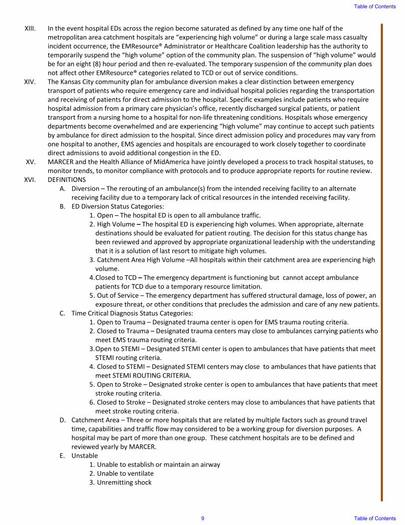
XIII. In the event hospital EDs across the region become saturated as defined by any time one half of the metropolitan area catchment hospitals are “experiencing high volume” or during a large scale mass casualty incident occurrence, the EMResource® Administrator or Healthcare Coalition leadership has the authority to temporarily suspend the “high volume” option of the community plan. The suspension of “high volume” would be for an eight (8) hour period and then re-evaluated. The temporary suspension of the community plan does not affect other EMResource® categories related to TCD or out of service conditions.
XIV. The Kansas City community plan for ambulance diversion makes a clear distinction between emergency transport of patients who require emergency care and individual hospital policies regarding the transportation and receiving of patients for direct admission to the hospital. Specific examples include patients who require hospital admission from a primary care physician’s office, recently discharged surgical patients, or patient transport from a nursing home to a hospital for non-life threatening conditions. Hospitals whose emergency departments become overwhelmed and are experiencing “high volume” may continue to accept such patients by ambulance for direct admission to the hospital. Since direct admission policy and procedures may vary from one hospital to another, EMS agencies and hospitals are encouraged to work closely together to coordinate direct admissions to avoid additional congestion in the ED.
XV. MARCER and the Health Alliance of MidAmerica have jointly developed a process to track hospital statuses, to monitor trends, to monitor compliance with protocols and to produce appropriate reports for routine review.
XVI. DEFINITIONS A. Diversion – The rerouting of an ambulance(s) from the intended receiving facility to an alternate
receiving facility due to a temporary lack of critical resources in the intended receiving facility. B. ED Diversion Status Categories:
1. Open – The hospital ED is open to all ambulance traffic. 2. High Volume – The hospital ED is experiencing high volumes. When appropriate, alternate
destinations should be evaluated for patient routing. The decision for this status change has been reviewed and approved by appropriate organizational leadership with the understanding that it is a solution of last resort to mitigate high volumes.
3. Catchment Area High Volume –All hospitals within their catchment area are experiencing high volume.
4. Closed to TCD – The emergency department is functioning but cannot accept ambulance patients for TCD due to a temporary resource limitation.
5. Out of Service – The emergency department has suffered structural damage, loss of power, an exposure threat, or other conditions that precludes the admission and care of any new patients.
C. Time Critical Diagnosis Status Categories: 1. Open to Trauma – Designated trauma center is open for EMS trauma routing criteria. 2. Closed to Trauma – Designated trauma centers may close to ambulances carrying patients who
meet EMS trauma routing criteria. 3. Open to STEMI – Designated STEMI center is open to ambulances that have patients that meet
STEMI routing criteria. 4. Closed to STEMI – Designated STEMI centers may close to ambulances that have patients that
meet STEMI ROUTING CRITERIA. 5. Open to Stroke – Designated stroke center is open to ambulances that have patients that meet
stroke routing criteria. 6. Closed to Stroke – Designated stroke centers may close to ambulances that have patients that
meet stroke routing criteria. D. Catchment Area – Three or more hospitals that are related by multiple factors such as ground travel
time, capabilities and traffic flow may considered to be a working group for diversion purposes. A hospital may be part of more than one group. These catchment hospitals are to be defined and reviewed yearly by MARCER.
E. Unstable 1. Unable to establish or maintain an airway 2. Unable to ventilate 3. Unremitting shock
Table of Contents
9 Table of Contents
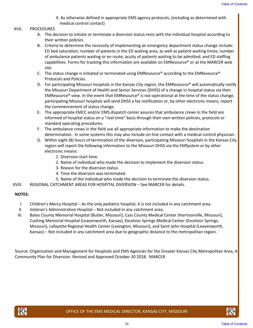
4. As otherwise defined in appropriate EMS agency protocols, (including as determined with medical control contact)
XVII. PROCEDURES A. The decision to initiate or terminate a diversion status rests with the individual hospital according to
their written policies. B. Criteria to determine the necessity of implementing an emergency department status change include:
ED bed saturation; number of patients in the ED waiting area, as well as patient waiting times; number of ambulance patients waiting or en route; acuity of patients waiting to be admitted; and ED staffing capabilities. Forms for tracking this information are available on EMResource® or at the MARCER web site.
C. The status change is initiated or terminated using EMResource® according to the EMResource® Protocols and Policies.
D. For participating Missouri hospitals in the Kansas City region, the EMResource® will automatically notify the Missouri Department of Health and Senior Services (DHSS) of a change in hospital status via their EMResource® view. In the event that EMResource® is not operational at the time of the status change, participating Missouri hospitals will send DHSS a fax notification or, by other electronic means, report the commencement of status change.
E. The appropriate EMCC and/or EMS dispatch center assures that ambulance crews in the field are informed of hospital status on a “real-time” basis through their own written policies, protocols or standard operating procedures.
F. The ambulance crews in the field use all appropriate information to make the destination determination. In some systems this may also include on-line contact with a medical control physician.
G. Within eight (8) hours of termination of the diversion, participating Missouri hospitals in the Kansas City region will report the following information to the Missouri DHSS via the EMSystem or by other electronic means:
1. Diversion start time. 2. Name of individual who made the decision to implement the diversion status. 3. Reason for the diversion status. 4. Time the diversion was terminated. 5. Name of the individual who made the decision to terminate the diversion status.
XVIII. REGIONAL CATCHMENT AREAS FOR HOSPITAL DIVERSION – See MARCER for details.
NOTES:
I. Children’s Mercy Hospital – As the only pediatric hospital, it is not included in any catchment area. II. Veteran’s Administration Hospital – Not included in any catchment area.
III. Bates County Memorial Hospital (Butler, Missouri), Cass County Medical Center (Harrisonville, Missouri), Cushing Memorial Hospital (Leavenworth, Kansas), Excelsior Springs Medical Center (Excelsior Springs, Missouri), Lafayette Regional Health Center (Lexington, Missouri), and Saint John Hospital (Leavenworth, Kansas) – Not included in any catchment area due to geographic distance to the metropolitan region.
Source: Organization and Management for Hospitals and EMS Agencies for the Greater Kansas City Metropolitan Area, A Community Plan for Diversion. Revised and Approved October 30 2018. MARCER
OFFICE OF THE EMS MEDICAL DIRECTOR, KANSAS CITY, MISSOURI
Table of Contents
10 Table of Contents
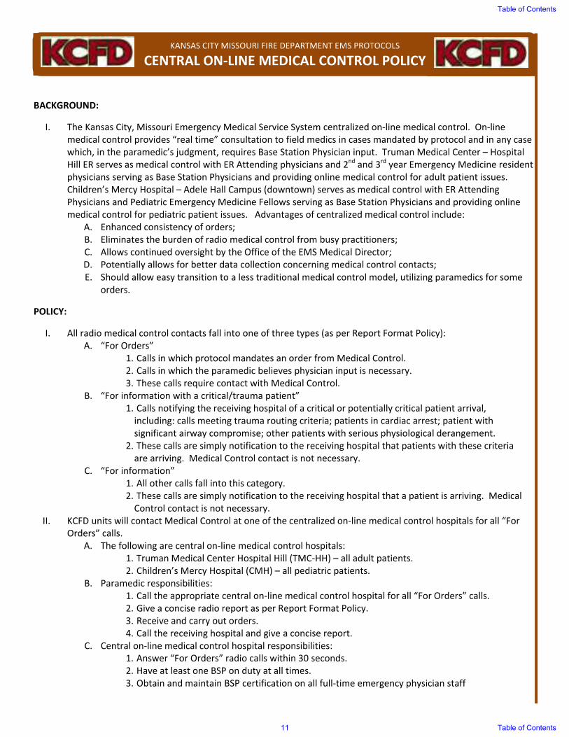
BACKGROUND:
I. The Kansas City, Missouri Emergency Medical Service System centralized on-line medical control. On-line medical control provides “real time” consultation to field medics in cases mandated by protocol and in any case which, in the paramedic’s judgment, requires Base Station Physician input. Truman Medical Center – Hospital Hill ER serves as medical control with ER Attending physicians and 2
nd and 3
rd year Emergency Medicine resident
physicians serving as Base Station Physicians and providing online medical control for adult patient issues. Children’s Mercy Hospital – Adele Hall Campus (downtown) serves as medical control with ER Attending Physicians and Pediatric Emergency Medicine Fellows serving as Base Station Physicians and providing online medical control for pediatric patient issues. Advantages of centralized medical control include:
A. Enhanced consistency of orders;
B. Eliminates the burden of radio medical control from busy practitioners;
C. Allows continued oversight by the Office of the EMS Medical Director;
D. Potentially allows for better data collection concerning medical control contacts;
E. Should allow easy transition to a less traditional medical control model, utilizing paramedics for some orders.
POLICY:
I. All radio medical control contacts fall into one of three types (as per Report Format Policy):
A. “For Orders”
1. Calls in which protocol mandates an order from Medical Control.
2. Calls in which the paramedic believes physician input is necessary.
3. These calls require contact with Medical Control.
B. “For information with a critical/trauma patient”
1. Calls notifying the receiving hospital of a critical or potentially critical patient arrival,
including: calls meeting trauma routing criteria; patients in cardiac arrest; patient with
significant airway compromise; other patients with serious physiological derangement.
2. These calls are simply notification to the receiving hospital that patients with these criteria
are arriving. Medical Control contact is not necessary.
C. “For information”
1. All other calls fall into this category.
2. These calls are simply notification to the receiving hospital that a patient is arriving. Medical Control contact is not necessary.
II. KCFD units will contact Medical Control at one of the centralized on-line medical control hospitals for all “For Orders” calls.
A. The following are central on-line medical control hospitals:
1. Truman Medical Center Hospital Hill (TMC-HH) – all adult patients.
2. Children’s Mercy Hospital (CMH) – all pediatric patients.
B. Paramedic responsibilities:
1. Call the appropriate central on-line medical control hospital for all “For Orders” calls.
2. Give a concise radio report as per Report Format Policy.
3. Receive and carry out orders.
4. Call the receiving hospital and give a concise report.
C. Central on-line medical control hospital responsibilities:1. Answer “For Orders” radio calls within 30 seconds.
2. Have at least one BSP on duty at all times.
3. Obtain and maintain BSP certification on all full-time emergency physician staff
KANSAS CITY MISSOURI FIRE DEPARTMENT EMS PROTOCOLS
CENTRAL ON-LINE MEDICAL CONTROL POLICY
Table of Contents
11 Table of Contents
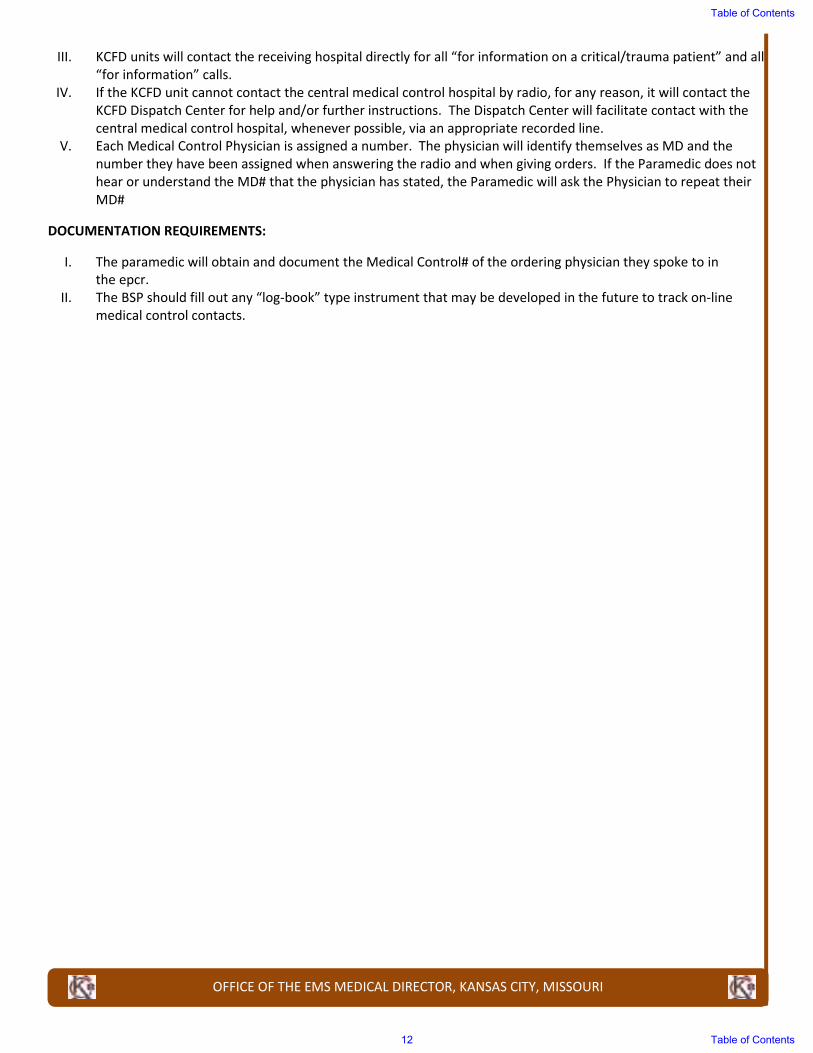
III. KCFD units will contact the receiving hospital directly for all “for information on a critical/trauma patient” and all
“for information” calls.
IV. If the KCFD unit cannot contact the central medical control hospital by radio, for any reason, it will contact the
KCFD Dispatch Center for help and/or further instructions. The Dispatch Center will facilitate contact with the
central medical control hospital, whenever possible, via an appropriate recorded line.
V. Each Medical Control Physician is assigned a number. The physician will identify themselves as MD and the
number they have been assigned when answering the radio and when giving orders. If the Paramedic does not
hear or understand the MD# that the physician has stated, the Paramedic will ask the Physician to repeat their
MD#
DOCUMENTATION REQUIREMENTS:
I. The paramedic will obtain and document the Medical Control# of the ordering physician they spoke to in
the epcr.
II. The BSP should fill out any “log-book” type instrument that may be developed in the future to track on-line
medical control contacts.
OFFICE OF THE EMS MEDICAL DIRECTOR, KANSAS CITY, MISSOURI
Table of Contents
12 Table of Contents
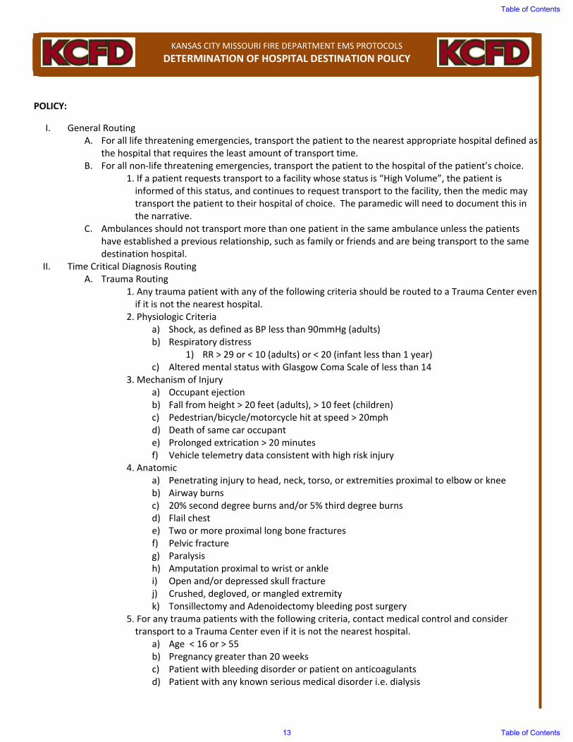
POLICY:
I. General Routing A. For all life threatening emergencies, transport the patient to the nearest appropriate hospital defined as
the hospital that requires the least amount of transport time. B. For all non-life threatening emergencies, transport the patient to the hospital of the patient’s choice.
1. If a patient requests transport to a facility whose status is “High Volume”, the patient is informed of this status, and continues to request transport to the facility, then the medic may transport the patient to their hospital of choice. The paramedic will need to document this in the narrative.
C. Ambulances should not transport more than one patient in the same ambulance unless the patients have established a previous relationship, such as family or friends and are being transport to the same destination hospital.
II. Time Critical Diagnosis Routing A. Trauma Routing
1. Any trauma patient with any of the following criteria should be routed to a Trauma Center even if it is not the nearest hospital.
2. Physiologic Criteria a) Shock, as defined as BP less than 90mmHg (adults) b) Respiratory distress
1) RR > 29 or < 10 (adults) or < 20 (infant less than 1 year) c) Altered mental status with Glasgow Coma Scale of less than 14
3. Mechanism of Injury a) Occupant ejection b) Fall from height > 20 feet (adults), > 10 feet (children) c) Pedestrian/bicycle/motorcycle hit at speed > 20mph d) Death of same car occupant e) Prolonged extrication > 20 minutes f) Vehicle telemetry data consistent with high risk injury
4. Anatomic a) Penetrating injury to head, neck, torso, or extremities proximal to elbow or knee b) Airway burns c) 20% second degree burns and/or 5% third degree burns d) Flail chest e) Two or more proximal long bone fractures f) Pelvic fracture g) Paralysis h) Amputation proximal to wrist or ankle i) Open and/or depressed skull fracture j) Crushed, degloved, or mangled extremity k) Tonsillectomy and Adenoidectomy bleeding post surgery
5. For any trauma patients with the following criteria, contact medical control and consider transport to a Trauma Center even if it is not the nearest hospital.
a) Age < 16 or > 55 b) Pregnancy greater than 20 weeks c) Patient with bleeding disorder or patient on anticoagulants d) Patient with any known serious medical disorder i.e. dialysis
KANSAS CITY MISSOURI FIRE DEPARTMENT EMS PROTOCOLS DETERMINATION OF HOSPITAL DESTINATION POLICY
Table of Contents
13 Table of Contents

6. Patients who are less than 16 years old and who meet the physiologic (pediatric), mechanism of injury, or anatomic criteria should be routed to a Missouri state designated pediatric trauma center.
7. Traumatic cardiopulmonary arrest patients should be taken to the nearest Trauma Center, unless it is “out of service”.
8. When there is more than one adult trauma patient, attempt to evenly distribute patients. If this is not feasible, contact central medical control at Truman Medical Center – Hospital Hill for routing assistance.
9. When more than one patient less than 16 years old meets physiologic or anatomic criteria per the trauma routing protocol, contact central medical control at Children’s Mercy Hospital for routing assistance.
10. For the following burn patients, CONSIDER routing directly to the closest Burn Center a) > 20% partial thickness (second degree) burns b) > 5% full thickness (third degree) burns c) Facial burns/inhalation injury d) Circumferential burns e) High voltage electrical burns f) Genital perineum burns g) Burns over joints or exposed bone h) Burns to hands or feet
B. STEMI Routing 1. Patients with identified ST elevation of 1mm or greater in two or more contiguous leads or left
bundle branch block should be transported to the closest appropriate STEMI center. C. Stroke Routing
1. Patients identified with acute stroke symptoms should be transported to the closest appropriate stroke center per the following guidelines:
a) Patients with onset of symptoms/last known well less than 3.5hrs AND do not meet any tPA Exclusion Criteria, transport to closest Primary or Comprehensive Stroke Center
b) Patients with onset of symptoms/last known well between 3.5hrs and 12hrs OR less than 3.5hrs AND meet tPA Exclusion Criteria, OR woke up with symptoms, transport to closest Comprehensive Stroke Center
c) Patients with onset of symptoms/last known well greater than 12hrs may be transported to closest appropriate hospital.
III. Hospital Diversion Routing A. Definitions
1. Diversion – The rerouting of an ambulance(s) from the intended receiving facility to an alternate receiving facility due to a temporary lack of critical resources in the intended receiving facility.
2. ED Diversion Status Categories: a) Open – The hospital ED is open to all ambulance traffic. b) High Volume – The hospital ED is experiencing high volumes. When appropriate,
alternate destinations should be evaluated for patient routing. The decision for this status change has been reviewed and approved by appropriate organizational leadership with the understanding that it is a solution of last resort to mitigate high volumes.
c) Catchment Area High Volume –All hospitals within their catchment area are experiencing high volume.
d) Closed to TCD – The emergency department is functioning but cannot accept ambulance patients for TCD due to a temporary resource limitation.
e) Out of Service – The emergency department has suffered structural damage, loss of power, an exposure threat, or other conditions that precludes the admission and care of any new patients.
Table of Contents
14 Table of Contents
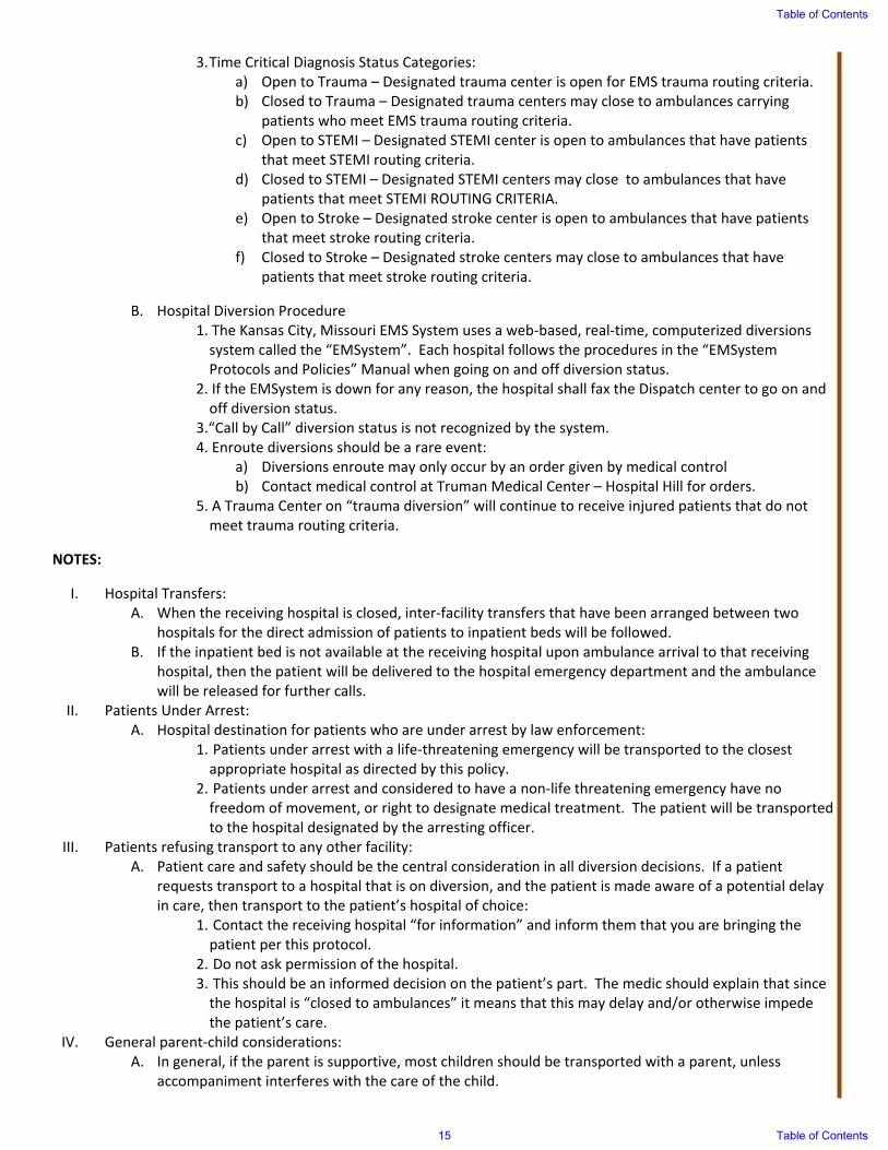
3. Time Critical Diagnosis Status Categories: a) Open to Trauma – Designated trauma center is open for EMS trauma routing criteria. b) Closed to Trauma – Designated trauma centers may close to ambulances carrying
patients who meet EMS trauma routing criteria. c) Open to STEMI – Designated STEMI center is open to ambulances that have patients
that meet STEMI routing criteria. d) Closed to STEMI – Designated STEMI centers may close to ambulances that have
patients that meet STEMI ROUTING CRITERIA. e) Open to Stroke – Designated stroke center is open to ambulances that have patients
that meet stroke routing criteria. f) Closed to Stroke – Designated stroke centers may close to ambulances that have
patients that meet stroke routing criteria.
B. Hospital Diversion Procedure 1. The Kansas City, Missouri EMS System uses a web-based, real-time, computerized diversions
system called the “EMSystem”. Each hospital follows the procedures in the “EMSystem Protocols and Policies” Manual when going on and off diversion status.
2. If the EMSystem is down for any reason, the hospital shall fax the Dispatch center to go on and off diversion status.
3.“Call by Call” diversion status is not recognized by the system. 4. Enroute diversions should be a rare event:
a) Diversions enroute may only occur by an order given by medical control b) Contact medical control at Truman Medical Center – Hospital Hill for orders.
5. A Trauma Center on “trauma diversion” will continue to receive injured patients that do not meet trauma routing criteria.
NOTES:
I. Hospital Transfers: A. When the receiving hospital is closed, inter-facility transfers that have been arranged between two
hospitals for the direct admission of patients to inpatient beds will be followed. B. If the inpatient bed is not available at the receiving hospital upon ambulance arrival to that receiving
hospital, then the patient will be delivered to the hospital emergency department and the ambulance will be released for further calls.
II. Patients Under Arrest: A. Hospital destination for patients who are under arrest by law enforcement:
1. Patients under arrest with a life-threatening emergency will be transported to the closest appropriate hospital as directed by this policy.
2. Patients under arrest and considered to have a non-life threatening emergency have no freedom of movement, or right to designate medical treatment. The patient will be transported to the hospital designated by the arresting officer.
III. Patients refusing transport to any other facility: A. Patient care and safety should be the central consideration in all diversion decisions. If a patient
requests transport to a hospital that is on diversion, and the patient is made aware of a potential delay in care, then transport to the patient’s hospital of choice:
1. Contact the receiving hospital “for information” and inform them that you are bringing the patient per this protocol.
2. Do not ask permission of the hospital. 3. This should be an informed decision on the patient’s part. The medic should explain that since
the hospital is “closed to ambulances” it means that this may delay and/or otherwise impede the patient’s care.
IV. General parent-child considerations: A. In general, if the parent is supportive, most children should be transported with a parent, unless
accompaniment interferes with the care of the child.
Table of Contents
15 Table of Contents
B. Children usually respond best to open and honest dialogue. C. In situations of major trauma or cardiopulmonary arrest, it is usually better to have the parent not ride
in the ambulance.
OFFICE OF THE EMS MEDICAL DIRECTOR, KANSAS CITY, MISSOURI
Table of Contents
16 Table of Contents
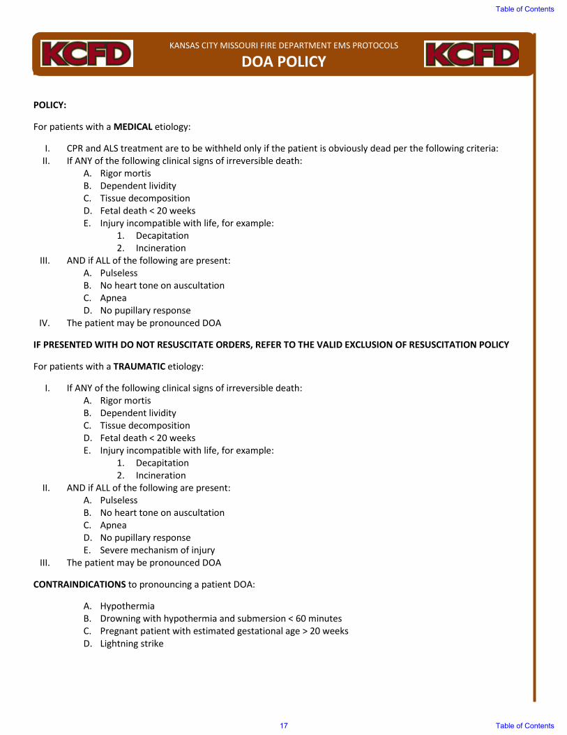
POLICY:
For patients with a MEDICAL etiology:
I. CPR and ALS treatment are to be withheld only if the patient is obviously dead per the following criteria: II. If ANY of the following clinical signs of irreversible death:
A. Rigor mortis B. Dependent lividity C. Tissue decomposition D. Fetal death < 20 weeks E. Injury incompatible with life, for example:
1. Decapitation 2. Incineration
III. AND if ALL of the following are present: A. Pulseless B. No heart tone on auscultation C. Apnea D. No pupillary response
IV. The patient may be pronounced DOA
IF PRESENTED WITH DO NOT RESUSCITATE ORDERS, REFER TO THE VALID EXCLUSION OF RESUSCITATION POLICY
For patients with a TRAUMATIC etiology:
I. If ANY of the following clinical signs of irreversible death: A. Rigor mortis B. Dependent lividity C. Tissue decomposition D. Fetal death < 20 weeks E. Injury incompatible with life, for example:
1. Decapitation 2. Incineration
II. AND if ALL of the following are present: A. Pulseless B. No heart tone on auscultation C. Apnea D. No pupillary response E. Severe mechanism of injury
III. The patient may be pronounced DOA
CONTRAINDICATIONS to pronouncing a patient DOA:
A. Hypothermia B. Drowning with hypothermia and submersion < 60 minutes C. Pregnant patient with estimated gestational age > 20 weeks D. Lightning strike
KANSAS CITY MISSOURI FIRE DEPARTMENT EMS PROTOCOLS
DOA POLICY
Table of Contents
17 Table of Contents
NOTES:
I. It is intended for this policy to be utilized by an EMT or a Paramedic. II. An ePCR detailing the situation and the specific DOA criteria met is to be completed by the pronouncing
provider. III. If resuscitation efforts are in progress and the patient meets the above criteria, CONSIDER discontinuing the
resuscitation efforts. IV. Police are in charge at a crime scene. They have the right to deny EMS providers access to the patient. V. It is not the policy of KCFD to move or transport deceased patients. These patients should not be moved to an
ambulance unless expressly directed to do so by KCFD Incident Command or KCPD as the result of the potential threat of a riot.
VI. The pronouncing crew must remain on scene with the body until released by law enforcement or coroner.
OFFICE OF THE EMS MEDICAL DIRECTOR, KANSAS CITY, MISSOURI
Table of Contents
18 Table of Contents
BACKGROUND:
The purpose of the policy is to assist EMS personnel for what equipment needs to be brought in for the patient by the
EMS crew.
POLICY:
I. Any call of an emergency nature should be considered to have potential for serious medical conditions until proven otherwise.
II. The optimal amount and type of equipment brought into the scene is dependent on multiple factors including but not limited to the type of call, the location of the call, ingress and egress capabilities, and the operating environment.
III. It is incumbent upon the EMS crew to be aware of all problems that could reasonably be foreseen at the scene.
IV. Ultimately the EMS crew must decide which equipment to take to the patient on each call and must take responsibility for the consequences if the crew fails to recognize problems that could have reasonably been foreseen. EMS crews will be held accountable for bad outcomes that arise due to a poor decision process.
V. EMS crews should not be complacent with bringing in less equipment on lower priority calls; even the lowest priority call can have potential for emergency medical attention.
VI. At a minimum, an EMS crew should bring in the appropriate amount of equipment to provide medical treatment for any condition which could reasonably be foreseen.
KANSAS CITY MISSOURI FIRE DEPARTMENT EMS PROTOCOLS
EQUIPMENT BROUGHT IN POLICY
OFFICE OF THE EMS MEDICAL DIRECTOR, KANSAS CITY, MISSOURI
Table of Contents
19 Table of Contents
BACKGROUND: Dispatching of air ambulances within the City of Kansas City, Missouri, will be done through the KCFD
Communications Center.
POLICY:
I. Indications
A. Use of air ambulance transport is the judgment of the on-scene paramedic.
B. Factors to consider include:
1. Optimum speed of response and transport
a) Be aware of helicopter spin up time, response time, load time, and transport time.
2. Impaired ground accessibility
a) Location
b) Traffic volume
c) Condition of roads or streets
3. Shortage of conventional ground transport units and need for rapid transport.
4. Rapid transport of medical personnel and supplies.
5. Distribution among area hospitals to avoid overloading the nearest emergency facility
a) In a multiple casualty incident, the air unit should transport to the furthest appropriate
facility from the scene.
6. Need for multiple response of ground vehicles, usually in outlying areas.
II. Procedures
A. It is within the authority of the KCFD Communication Specialist to dispatch air ambulances as additional
units.
B. When KCFD Communications receives a request for air ambulance service from other entities prior to
the arrival of a KCFD unit, air ambulances may be ordered depending on location of KCFD units and
number of patients.
C. KCFD Communications will advise responding KCFD unit(s) and other ground units that an air ambulance
has been ordered.
D. It is the responsibility of the on-scene paramedic to:
1. Triage patients and consider the mode of transportation and destination of all patients.
2. Immediately notify the ranking fire and law enforcement on-scene personnel that an air
ambulance is enroute.
3. Confirm a suitable landing zone (LZ) for air ambulance.
a) This is primarily a fire and police responsibility depending on jurisdiction.
b) In Kansas City, Missouri, the Kansas City Fire Department will coordinate the LZ.
E. The air ambulance pilot has final authority if an LZ is acceptable.
1. If another LZ is requested/suggested by the pilot, it must be provided.
KANSAS CITY MISSOURI FIRE DEPARTMENT EMS PROTOCOLS
HELICOPTER UTILIZATION POLICY
OFFICE OF THE EMS MEDICAL DIRECTOR, KANSAS CITY, MISSOURI
Table of Contents
20 Table of Contents

The following information is taken from: Assistant Secretary for Preparedness and Response: EMS INFECTIOUS
DISEASE PLAYBOOK
POLICY:
I. Standard Precautions:
A. EXAMPLE DISEASES:
1. AIDS/HIV, anthrax (cutaneous or pulmonary)
2. Botulism
3. Cellulitis/abscess
4. Dengue fever
5. Nonspecific upper respiratory infections
B. PPE:
1. Gloves during patient contact
2. Goggles/face shield and surgical mask for any airway procedures (intubation, suctioning) or patient with active cough from apparent infectious source
3. Impermeable gown for any situation likely to generate splash/liquid exposures
4. PPE should be removed in an appropriate doffing area
5. Avoid self-contamination
6. PPE waste should be placed in a labeled leak-proof container
7. Potential exposures should be reported according to existing service protocols
C. PATIENT CARE CONSIDERATIONS:
1. Provide a surgical mask for all patients with acute infectious respiratory symptoms who can tolerate it
2. Provide tissues to patients for secretion control
3. Encourage patient to use hand hygiene and cough etiquette practices
D. TRANSPORT CONSIDERATIONS:
1. Standard transportation to appropriate hospital facility
2. Turn on patient compartment exhaust fan
E. Complete appropriate AMBULANCE DECONTAMINATION
II. Contact Precautions:
A. EXAMPLE DISEASES:
1. Excessive wound drainage
2. MRSA
3. Vancomycin-resistant enterococci (VRE)
4. C. difficile
5. Norovirus
6. Infectious diarrhea
7. Head lice/body lice/scabies
B. PPE:
1. Ensure strict adherence to standard precautions based on situation (e.g., mask, goggles/face shield for splatter risk or airway interventions)
2. Disposable fluid-resistant gown that protects the provider’s legs as needed
3. Disposable gloves
C. PATIENT CARE CONSIDERATIONS:
1. Provide a surgical mask for all patients with acute infectious respiratory symptoms who can tolerate it
2. Provide tissues to patients for secretion control
KANSAS CITY MISSOURI FIRE DEPARTMENT EMS PROTOCOLS
INFECTIOUS DISEASE POLICY
Table of Contents
21 Table of Contents
3. Encourage patient to use hand hygiene and cough etiquette practices
D. TRANSPORT CONSIDERATIONS:
1. Standard transportation to appropriate hospital facility
2. Turn on patient compartment exhaust fan
E. Ambulance should be taken out of service for appropriate AMBULANCE DECONTAMINATION
III. Droplet Precautions:
A. EXAMPLE DISEASES:
1. Neisseria meningitides
2. Mumps
3. Mycoplasma
4. Streptococcal and many other causes of pneumonia
5. Parvovirus
6. Pertussis
7. Pneumonic plague
8. Rhinovirus
9. Rubella
10. Seasonal influenza
11. Streptococcal pharyngitis
B. PPE:
1. Ensure strict adherence to standard precautions based on situation (e.g., mask, goggles/face
shield for splatter risk or airway interventions)
2. Disposable surgical mask (N95 respirator not required)
3. Disposable gloves
4. Eye protection – cleanable goggles or disposable face shield
C. PATIENT CARE CONSIDERATIONS:
1. Provide a surgical mask for all patients with acute infectious respiratory symptoms who can
tolerate it
2. Provide tissues to patients for secretion control
3. Encourage patient to use hand hygiene and cough etiquette practices
4. Personnel not in appropriate PPE: maintain a distance of 6 feet from the patient. Wear gloves
5. Minimize use of nebulizers (consider metered dose inhalers)
6. Minimize airway interventions that may cause coughing (e.g., suctioning)
D. TRANSPORT CONSIDERATIONS:
1. Standard transportation
2. Consider putting patient compartment exhaust vent on high and isolating the driver
compartment if performing aerosol producing procedures (airway suctioning, intubation,
aerosolized medication administration)
3. Increase ventilation - have air or heat on non-recirculating cycle and/or open windows
4. Advise receiving hospital of respiratory symptoms
E. Ambulance should be taken out of service for appropriate AMBULANCE DECONTAMINATION
IV. Airborne Precautions:
A. EXAMPLE DISEASES:
1. Measles, TB (suspected or confirmed),
2. Varicella (chickenpox)
B. PPE:
1. Ensure strict adherence to standard precautions based on situation (e.g., mask, goggles/face
shield for splatter risk or airway interventions)
2. Disposable NIOSH-approved, fit-tested N95 respirator or powered air purifying respirators
(PAPRs) with full hood and high efficiency particulate air (HEPA) filter
3. Disposable exam gloves
C. PATIENT CARE CONSIDERATIONS:
1. Ensure strict adherence with standard precautions (e.g., add gown or coverall for significant
bodily fluid exposures and follow doffing for contact precautions)
Table of Contents
22 Table of Contents
2. Ask the patient to wear a surgical mask (N95 respirator not required) if they are able to tolerate
it
3. Provide tissues to patients for secretion control
4. Encourage patient to use hand hygiene and cough etiquette practices
5. Procedures that can generate small particle aerosols (endotracheal intubation and suctioning)
a) Protection of the eyes, nose, and mouth – in addition to gown and gloves – is
recommended
b) Use of an N95 respirator is recommended
D. TRANSPORT CONSIDERATIONS :
1. Notify the receiving hospital of the need for an airborne infection isolation room
2. Consider having the patient compartment exhaust vent on high and isolating the driver
compartment
3. Consider having the driver compartment ventilation fan set to high without recirculation
4. If driver/pilot compartment cannot be isolated from the patient compartment, vehicle operator
to wear NIOSH-approved, fit-tested N95 respirator
5. Intubated patients should be ventilated with a bag-valve device or ventilator equipped with a
HEPA filter on exhalation port
E. Ambulance should be taken out of service for appropriate AMBULANCE DECONTAMINATION
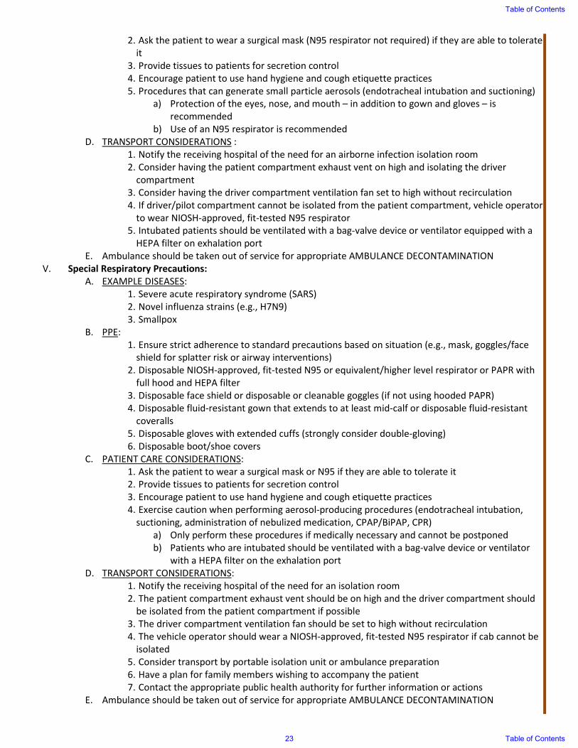
V. Special Respiratory Precautions:
A. EXAMPLE DISEASES:
1. Severe acute respiratory syndrome (SARS)
2. Novel influenza strains (e.g., H7N9)
3. Smallpox
B. PPE:
1. Ensure strict adherence to standard precautions based on situation (e.g., mask, goggles/face
shield for splatter risk or airway interventions)
2. Disposable NIOSH-approved, fit-tested N95 or equivalent/higher level respirator or PAPR with
full hood and HEPA filter
3. Disposable face shield or disposable or cleanable goggles (if not using hooded PAPR)
4. Disposable fluid-resistant gown that extends to at least mid-calf or disposable fluid-resistant
coveralls
5. Disposable gloves with extended cuffs (strongly consider double-gloving)
6. Disposable boot/shoe covers
C. PATIENT CARE CONSIDERATIONS:
1. Ask the patient to wear a surgical mask or N95 if they are able to tolerate it
2. Provide tissues to patients for secretion control
3. Encourage patient to use hand hygiene and cough etiquette practices
4. Exercise caution when performing aerosol-producing procedures (endotracheal intubation,
suctioning, administration of nebulized medication, CPAP/BiPAP, CPR)
a) Only perform these procedures if medically necessary and cannot be postponed
b) Patients who are intubated should be ventilated with a bag-valve device or ventilator
with a HEPA filter on the exhalation port
D. TRANSPORT CONSIDERATIONS:
1. Notify the receiving hospital of the need for an isolation room
2. The patient compartment exhaust vent should be on high and the driver compartment should
be isolated from the patient compartment if possible
3. The driver compartment ventilation fan should be set to high without recirculation
4. The vehicle operator should wear a NIOSH-approved, fit-tested N95 respirator if cab cannot be
isolated
5. Consider transport by portable isolation unit or ambulance preparation
6. Have a plan for family members wishing to accompany the patient
7. Contact the appropriate public health authority for further information or actions
E. Ambulance should be taken out of service for appropriate AMBULANCE DECONTAMINATION
Table of Contents
23 Table of Contents
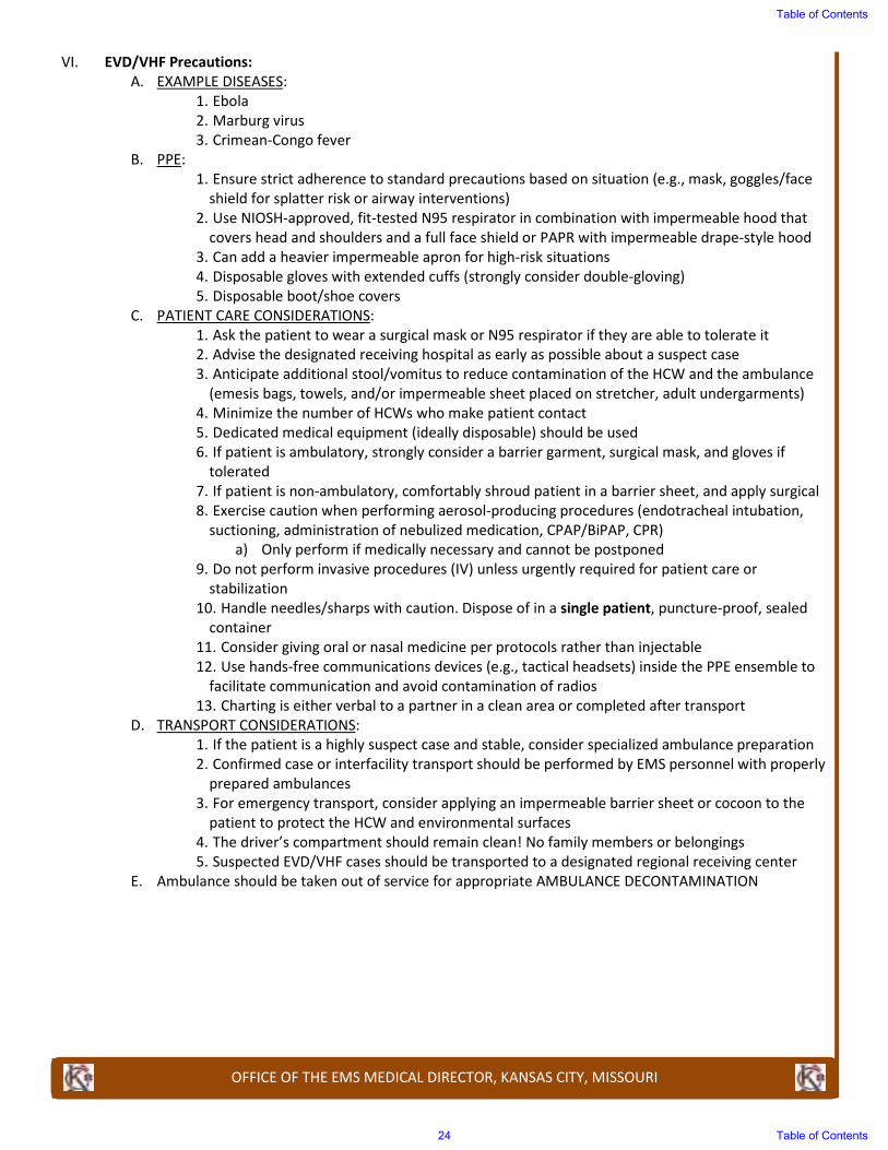
VI. EVD/VHF Precautions:
A. EXAMPLE DISEASES:
1. Ebola
2. Marburg virus
3. Crimean-Congo fever
B. PPE:
1. Ensure strict adherence to standard precautions based on situation (e.g., mask, goggles/face
shield for splatter risk or airway interventions)
2. Use NIOSH-approved, fit-tested N95 respirator in combination with impermeable hood that
covers head and shoulders and a full face shield or PAPR with impermeable drape-style hood
3. Can add a heavier impermeable apron for high-risk situations
4. Disposable gloves with extended cuffs (strongly consider double-gloving)
5. Disposable boot/shoe covers
C. PATIENT CARE CONSIDERATIONS:
1. Ask the patient to wear a surgical mask or N95 respirator if they are able to tolerate it
2. Advise the designated receiving hospital as early as possible about a suspect case
3. Anticipate additional stool/vomitus to reduce contamination of the HCW and the ambulance
(emesis bags, towels, and/or impermeable sheet placed on stretcher, adult undergarments)
4. Minimize the number of HCWs who make patient contact
5. Dedicated medical equipment (ideally disposable) should be used
6. If patient is ambulatory, strongly consider a barrier garment, surgical mask, and gloves if
tolerated
7. If patient is non-ambulatory, comfortably shroud patient in a barrier sheet, and apply surgical
8. Exercise caution when performing aerosol-producing procedures (endotracheal intubation,
suctioning, administration of nebulized medication, CPAP/BiPAP, CPR)
a) Only perform if medically necessary and cannot be postponed
9. Do not perform invasive procedures (IV) unless urgently required for patient care or
stabilization
10. Handle needles/sharps with caution. Dispose of in a single patient, puncture-proof, sealed
container
11. Consider giving oral or nasal medicine per protocols rather than injectable
12. Use hands-free communications devices (e.g., tactical headsets) inside the PPE ensemble to
facilitate communication and avoid contamination of radios
13. Charting is either verbal to a partner in a clean area or completed after transport
D. TRANSPORT CONSIDERATIONS:
1. If the patient is a highly suspect case and stable, consider specialized ambulance preparation
2. Confirmed case or interfacility transport should be performed by EMS personnel with properly
prepared ambulances
3. For emergency transport, consider applying an impermeable barrier sheet or cocoon to the
patient to protect the HCW and environmental surfaces
4. The driver’s compartment should remain clean! No family members or belongings
5. Suspected EVD/VHF cases should be transported to a designated regional receiving center
E. Ambulance should be taken out of service for appropriate AMBULANCE DECONTAMINATION
OFFICE OF THE EMS MEDICAL DIRECTOR, KANSAS CITY, MISSOURI
Table of Contents
24 Table of Contents
BACKGROUND:
Kansas City, MO EMS Ordinance, Section 34-364 states: “The medical director shall serve as the primary source of day-
to-day medical direction and clinical oversight of all elements of the pre-hospital emergency medical services system.”
EMS providers are operating under the authority of the EMS Medical Director or their designee.
If a licensed physician appears on a scene and desires to assume direction and control of patient care, the physician shall
execute a form which declares the physician has assumed responsibility for patient care.
Two scenarios are common:
I. Personal Physician
A. Individual is the personal physician of the patient and has previously established a doctor-patient
relationship with the patient.
B. Most commonly occurs when the patient is still in the doctor’s office.
C. May also include a “team physician”.
II. Intervener Physician
A. Physician does not have a prior formal doctor-patient relationship with the patient.
B. Most commonly occurs when the doctor happens on scene prior to, concurrent with, or after EMS
arrival.
The highest priority is the provision of effective and efficient care and transport of the patient. This policy describes the
relationship between the patient, the EMS system, and the physician in these scenarios.
POLICY:
I. Personal Physician:
A. If the patient’s personal physician is present and desires to continue care, the prehospital provider
should defer to the orders of the personal physician as within the paramedics scope of practice.
B. Regardless of a physician executing a physician intervention form; EMS personnel retain the authority to
establish medical direction with on-line medical control and assume medical authority of the patient if
they believe the care rendered by the personal physician is not the standard of care. EMS personnel will
not comply with orders that exceed their scope of practice or are inconsistent with the standard of care.
C. The medical direction of EMS personnel shall revert to protocols and/or on-line medical control any time
the personal physician is no longer in attendance.
II. Intervener Physician On Scene:
A. If an individual identifies themselves as a physician on scene and expresses intent to assume medical
care for a patient, EMS personnel will briefly discuss the situation with the physician.
B. EMS personnel will ask for proof of licensure to practice medicine in the State of Missouri. The only
acceptable evidence of licensure are:
1. State of Missouri medical license card
2. KCMO Base Station Physician certification card
C. If the physician cannot provide this proof of licensure, the paramedic will not transfer medical authority.
D. If after establishing proof of licensure listed in II.B.2, EMS personnel will provide the physician with a
“medical intervention form” for review.
E. If after reading the document and any further discussion, the physician wishes to take over medical care
of the patient the paramedic will perform the following:
1. Contact with on-line medical control is mandatory to determine whether to relinquish
responsibility to the intervener physician.
2. If on-line medical control relinquishes responsibility to the intervener physician, then:
KANSAS CITY MISSOURI FIRE DEPARTMENT EMS PROTOCOLS
MEDICAL INTERVENTION POLICY
Table of Contents
25 Table of Contents

a) The intervener physician must sign the “medical intervention form”.
b) The intervener physician must accompany the patient to the hospital, sign the KCFD
Patient Care Report, and give a report to the receiving hospital physician.
3. If a patient explicitly requests that the intervener physician not provide care then the
paramedic maintains medical authority of the patient.
4. If patient care has progressed to time or transport, the paramedic will decline the offer of
assistance and transport.
5. Contact with the on-line medical control is not necessary if the intervener physician is a BSP and
can demonstrate proof of certification as a BSP in the KCMO EMS System.
F. At no time shall patient care be compromised for on-line medical control contact to relinquish
responsibility to the intervener physician.
G. The on-scene paramedic will continue to have ultimate authority for the patient. If the paramedic feels
at any time that the intervening physician is either not providing the standard of care or is interfering
with the furtherance of patient care, the paramedic will terminate the medical intervention relationship
and assume care of the patient per KCMO EMS protocols.
OFFICE OF THE EMS MEDICAL DIRECTOR, KANSAS CITY, MISSOURI
Table of Contents
26 Table of Contents
BACKGROUND:
I. Emergency Medical Services is a unique form of medicine.
II. There are numerous clinical and quality of care issues that can only be studied using a research format.
III. The EMS system should hold itself to the same patient safeguards and ethical conduct set forth under various
governing bodies.
POLICY:
I. Any research that is proposed to be conducted in the Kansas City Missouri Emergency Medical Services System,
including, but not limited to the Kansas City Missouri Fire Department shall meet the following guidelines.
II. Approval of the concept, plan, and protocol by the Office of the EMS Medical Director and the EMSCC.
III. Research plan and protocol must be approved by an Institutional Review Board (IRB) of a certified research
institution as determined by the EMS Medical Director.
IV. Any research approved for the Kansas City Missouri Emergency Medical Services System will include oversight
and participation from the Office of the EMS Medical Director.
V. Any costs for conducting research will be borne by the investigator.
VI. Investigators proposing research for the Kansas City Missouri Emergency Medical Services System will disclose to
the Office of the EMS Medical Director any financial or other relationships with companies or organizations that
may influence their proposed research.
KANSAS CITY MISSOURI FIRE DEPARTMENT EMS PROTOCOLS
MEDICAL RESEARCH IN KCMO EMS POLICY
OFFICE OF THE EMS MEDICAL DIRECTOR, KANSAS CITY, MISSOURI
Table of Contents
27 Table of Contents
POLICY:
I. Medication Administration
A. During the transfer of patients between health care facilities, paramedics may be required to monitor
the intravenous infusion of medications and/or blood products, or administer medications and/or blood
products not approved by the EMS Medical Director for use in the prehospital setting (non-standard
medications)
B. Policy:
1. The paramedic may monitor and administer non-standard medications prescribed by the
patient’s transferring physician with on-line medial control guidance during the patient transfer
as needed. Medications requiring continuous infusion will be administered via a mechanical
infusion pump:
a) Understanding of a mechanical infusion pump is the paramedic’s responsibility (i.e. if
the paramedic does not understand, then the paramedic should not leave the facility
until it has been explained.)
b) If the paramedic is asked to manipulate more drugs or devices than the paramedic
thinks is appropriate, then the paramedic should notify the appropriate personnel at the
transferring hospital. If the problem cannot be solved, then the paramedic should
contact a supervisor
2. The administration of any non-standard medications shall be recorded on the patient care
report (PCR) noting the transferring physician’s name, medical control contacted, and dosage
and route of administration of medication.
II. Ventilator Support
A. Paramedics may be asked to transfer patients who are receiving positive pressure ventilator support.
Overall responsibility for the patient rests with the paramedic. A second medically trained person is
preferred to help to attend to the airway and provide ventilatory support.
B. Policy
1. Paramedic should not transport the positive pressure ventilated patient without a second
medically trained person to assist if in the paramedic’s judgment additional help is needed to
help attend to the airway and provide ventilatory support.
2. If the paramedic is asked to do so, the paramedic should inform the appropriate personnel at
the transferring hospital of the policy. If the problem cannot be solved, then the paramedic
should contact a supervisor.
3. If a patient is on oxygen of any source or has any respiratory symptoms, then an appropriately
sized bag-valve-mask must always accompany the patient.
4. If a patient is found to be on Positive End Expiratory Pressure (PEEP) at the transferring facility,
then a bag-valve-mask with an attached PEEP valve and a person knowledgeable in it use will
accompany the patient in transfer.
5. The level of transfer (emergency or non-emergency) from the sending hospital to the receiving
hospital can be specified by the transferring physician. If no level is specified, then paramedic
judgement will determine the level of ambulance transfer.
6. If a patient is determined to be critical by the paramedic, at least an information call should be
made to the receiving hospital to request a standby elevator. If a critical patient is not going to
a critical area, the paramedic may request an evaluation by the ED prior to proceeding to the
non-critical area.
7. The use of ventilator support shall be recorded on the patient care report (PCR) noting the
transferring physician’s name, and any instructions or orders pertaining to the ventilator
support provided by the transferring physician.
KANSAS CITY MISSOURI FIRE DEPARTMENT EMS PROTOCOLS
MEDICAL STANDARDS DURING PATIENT TRANSFER POLICY
Table of Contents
28 Table of Contents

III. Mechanical or other Nonstandard EMS Devices
A. Paramedics may be asked to transfer patients who are receiving mechanical circulatory support and/or
other non-standard EMS equipment not approved by the EMS medical director. Overall responsibility
for the patient rests with the paramedic. A second medically trained person from the transferring
hospital is preferred to help attend to any non-standard mechanical devices. Examples of non-standard
mechanical devices include, but is not limited to: intra-aortic balloon pumps, external chest compression
devices, internal or external cooling/warming devices and left ventricular assist devices
1. If the paramedic is asked to supervise or manipulate more devices than the paramedic thinks is
appropriate, then the paramedic should notify the appropriate personnel at the transferring
hospital. If the problem cannot be solved, then the paramedic should contact a supervisor.
2. The level of transfer (emergency or non-emergency) from the sending hospital to the receiving
hospital can be specified by the transferring physician. If no level is specified, then paramedic
judgement will determine the level of ambulance transfer.
3. If a patient is determined to be critical by the paramedic, at least an information call should be
made to the receiving hospital to request a standby elevator. If a critical patient is not going to
a critical area, the paramedic may request an evaluation by the ED prior to proceeding to the
non-critical area.
B. The use of any non-standard mechanical devices shall be recorded on the patient care report (PCR)
noting the transferring physician’s name, and any instructions or orders pertaining to the device
provided by the transferring physician.
IV. Transports of Patients from Hospitals to Psychiatric Facilities
A. The patient cannot legally refuse treatment and transport once a physician has determined that the
patient is, or may be, suicidal. This MUST be documented on the PCR.
B. The patient must be transported to the psychiatric facility regardless of the patient’s demands to refuse
transport.
C. Verbal, physical, or chemical restraint may be appropriate. Contact law enforcement personnel for
assistance as needed.
OFFICE OF THE EMS MEDICAL DIRECTOR, KANSAS CITY, MISSOURI
Table of Contents
29 Table of Contents
BACKGROUND: The reason for contact should be stated clearly at the beginning of the transmission.
POLICY:
I. The three accepted reasons for contact are:
A. “FOR ORDERS”
1. Requests for orders
2. Refusals
3. Consultations with the base station physician in unusual circumstances
B. “FOR INFORMATION WITH CRITICAL/TRAUMA PATIENT”
1. Patients meeting trauma routing criteria
2. Patients in cardiac arrest
3. Patient with airway compromise
4. Patients with other serious physiologic derangements per paramedic discretion
C. “FOR INFORMATION”
1. This is a courtesy to the hospital and does not include patients in the above groups
II. Ambulance identification
A. Vehicle identification
B. Paramedic name
C. Estimated time of arrival (ETA)
III. Patient report
A. Patient’s age and sex
B. Basic problem or chief complaint
C. Brief relevant history; include past medical history, medications, and allergies only if relevant
D. Vital signs (pulse, blood pressure, respirations, cardiac monitor pattern if appropriate)
E. General appearance include level of consciousness
F. Pertinent physical findings
G. Care in progress (i.e. airway, splints, backboard, collar, oxygen, etc)
H. Request specific orders following the appropriate protocols
IV. Special notes
A. Be as concise and accurate as possible. Radio reports should not take more than one minute per
patient.
B. Significant objective findings (i.e. critical vital signs) may take precedence over history and need to be
reported first.
C. Patient’s name may be broadcast if it will further patient care; patient name may be broadcast at
hospital request.
D. Radio report is a paramedic function except under extreme conditions. Unstable patients may
necessitate an EMT giving a radio report.
E. Any request for drug orders will include dose and route.
KANSAS CITY MISSOURI FIRE DEPARTMENT EMS PROTOCOLS
MEDICAL TRANSFER OF CARE AND RADIO REPORT FORMAT POLICY
OFFICE OF THE EMS MEDICAL DIRECTOR, KANSAS CITY, MISSOURI OFFICE OF THE EMS MEDICAL DIRECTOR, KANSAS CITY, MISSOURI
Table of Contents
30 Table of Contents
POLICY:
I. The following is a list of the recognized values for the use in meeting the customer’s medical needs within the KCMO EMS System. This list of values will be used to evaluate clinical care that falls outside of the established protocols.
A. Safety: In order to protect the crew, the patient, or the public from a danger on the scene, established treatment modalities may need to be modified.
B. Follow the ABC’s: Generally, the care of the patient should be in accordance with the following priorities:
1. Airway Maintenance: Beginning with the simple, non-invasive techniques, and working to the more invasive.
2. Assurance of adequate ventilations and oxygenation: Any patient in significant distress
should receive as high a concentration of oxygen as is practical to deliver. If any doubt exists as
to the adequacy of ventilation, then the patient should receive positive pressure ventilation
with the maximum available concentration of oxygen.
3. Assurance of adequate circulation: Through CCC-CCR and /or the appropriate treatment of bleeding and shock.
C. Use Medical Control: When in doubt, call medical control.
D. Primum Non Nocere (First Do No Harm):
1. A medication or invasive treatment should only be used if both the treatment is indicated, and there exists no contraindication to the treatment. On-line medical control should be used prior to an invasive treatment unless that treatment is authorized as a standing order.
2. Potentially unstable patients may be stressed by exertion, as a general rule, exertion should be minimized. (Potentially unstable includes, but is not limited to: patients with chest pain, dyspnea, or altered mental status.)
E. Default to Transport: The preference is to transport the patient to an emergency department or a hospital with inpatient capabilities. If any doubt exists as to the legal or medical competence of the patient to refuse care, on-line medical control should be contacted.
F. Customer Service: The human needs of the customer must be met including physical (medical and non-
medical), and psychological (including needs of reassurance and comfort). In all cases, be PROFESSIONAL, POLITE, and ATTENTIVE to those needs.
G. Clear Documentation: All patient encounters must be documented clearly and accurately.
H. Render Timely Care: The clinical needs of the patient must be met in a timely fashion. This generally includes initiating treatment prior to moving the patient to the ambulance. Exceptions include major trauma, crew/patient safety, and other circumstances as determined by the senior paramedic.
KANSAS CITY MISSOURI FIRE DEPARTMENT EMS PROTOCOLS
MEDICAL VALUES STATEMENT POLICY
OFFICE OF THE EMS MEDICAL DIRECTOR, KANSAS CITY, MISSOURI
Table of Contents
31 Table of Contents
POLICY:
I. EMT-First Responder (EMT-FR) has medical authority over public access defibrillator (PAD) personnel at scene until arrival of the transporting EMT-Paramedic (EMT-P).
II. Transporting EMT-Paramedic has medical authority at scenes.
III. On arrival, the EMT-P or EMT-FR should receive a verbal report from the EMT-FR or PAD site personnel to include:
A. Age
B. Witnessed or unwitnessed arrest
C. Approximate time from collapse
D. Initiation of CPR prior to EMT-FR arrival
E. Initial rhythm (shockable vs non-shockable)
F. Number of shocks delivered
G. Response to treatment
IV. If the EMT-FR arrives first on scene with a PAD program AED being used, the EMT-FR has the following options:
A. If the PAD personnel are proceeding with a shock, the EMT-FR should wait until the shock is complete.
B. Have the PAD personnel remove the AED and the EMT-FR apply the EMT-FR AED and follows the appropriate protocol.
V. When the EMT-P arrives on the scene and has assumed care of the patient, he will proceed as directed by current system advanced life support protocols.
VI. The EMT-P will follow these procedures:
A. If the EMT-FR or PAD AED is proceeding with a shock, the EMT-P will wait until this is complete.
B. After completion of the shock from the AED, the EMT-P or his designee will immediately switch to the Zoll monitor, keeping interruptions to CPR at a minimum.
VII. The EMT-P should consider the shocks delivered by any AED as part of the dysrhythmia algorithm. For example, if the patient remains in V-fib after shock by the AED, the EMT-P should enter the treatment sequence at the point where the shock has been delivered and establish vascular access, give epinephrine and manage the airway.
KANSAS CITY MISSOURI FIRE DEPARTMENT EMS PROTOCOLS
ON-SCENE AED COORDINATION POLICY
OFFICE OF THE EMS MEDICAL DIRECTOR, KANSAS CITY, MISSOURI
Table of Contents
32 Table of Contents
BACKGROUND:
I. To measure a complete sequence of events while delivering prehospital care, times to patient contact are
essential.
II. Times to patient contact allows the system to analyze delays from time to first responder/ALS on scene to
patient’s side and possibly provide solutions to improving access to patients once on scene.
POLICY:
I. KCFD emergency responders and ALS providers will verbally confirm “patient contact” with their respective
dispatch centers when they arrive at the patient’s side to begin patient care.
II. KCFD Communication Center will accurately record in the Computer Aided Dispatch program the “patient
contact” time.
III. Quality Assurance Director will compile compliance with this policy in aggregate and by medic/EMT or Fire
Company and report as request to the EMS Medical Director.
KANSAS CITY MISSOURI FIRE DEPARTMENT EMS PROTOCOLS
PATIENT CONTACT POLICY
OFFICE OF THE EMS MEDICAL DIRECTOR, KANSAS CITY, MISSOURI
Table of Contents
33 Table of Contents
POLICY:
When it is perceived by the responding Kansas City, Missouri EMS agencies that a person has exceeded the extrication
limits of the agencies (even acting in concert) as a result of but not limited to the patient’s size and weight, then the
individual case will be referred to an interagency task force for review and recommendations to the Office of the EMS
Medical Director. The EMS Medical Director will make a decision as to whether the patient has exceeded the system’s
capabilities and can no longer be transported. This decision will be presented to the EMSCC for review and approval.
This policy applies only to the transport of the person. When a request for service is made to the KCFD Communications
Center, an ambulance will be dispatched to the person.
KANSAS CITY MISSOURI FIRE DEPARTMENT EMS PROTOCOLS
PERSONS EXCEEDING THE EMS SYSTEM’S CAPABILITY POLICY
OFFICE OF THE EMS MEDICAL DIRECTOR, KANSAS CITY, MISSOURI
Table of Contents
34 Table of Contents
Purpose: The purpose of this policy is to assist EMS personnel with clear rules for managing situations in which the
patient or the patient’s representative refuses care or transportation by EMS.
Right to Refuse: An adult patient who is conscious, mentally competent, and understands the consequences of his or
her decision has the right to refuse are or transportation by EMS. A patient that refuses treatment or transport must be
offered treatment or transport and advised of the benefits of treatment or transport and the specific risks of refusing
treatment or transport. The offer of treatment or transport and description of the risks of refusal should be
documented in the epcr and on the Refusal Form.
No Right to Refuse: Conversely, where a patient is a minor or otherwise not competent, or unable to understand the
consequences of his or her decision, the patient may not refuse treatment or transport for himself, and EMS may have
implied consent to treat or transport. In this situation, EMS must contact Medical Control for directions regarding
treatment and transport (Mandatory Medical Control Contact).
Mandatory Medical Control Contact: Medical Control contact is mandatory if the crew believes that the patient may be
at significant harm to him or herself by refusing treatment or transport. The crew may contact Medical Control at any
time there is a question in regards to the patient condition. Utilization of the EMS radio is mandatory if communication
allows. The purpose of mandatory Medical Control Contact is to:
a. Assist the paramedic in persuading a reluctant patient to be transported. Occasionally, a
recommendation from a physician may be all that is needed to facilitate transport.
b. Serve to support the paramedic’s judgment that the patient is acting against medical advice.
POLICY:
I. Patient: Any person who is ill or injured or in need of treatment by medical personnel. This includes any person
that has activated the EMS system or for whom the EMS system has been activated, including emergency and
non-emergency calls for service, or any person that presents himself to EMS personnel with a medically related
complaint such that it could be reasonable inferred that the person is seeking or in need of medical attention.
II. Not a Patient: A person who is not ill or injured or in need of treatment by medical personnel. This includes
individuals who may have been involved in a situation that either did result or could have resulted in the
creation of a patient requiring medical treatment as defined above.
III. Capacity: A person 18 years of age or older of sound mind who is able to receive and evaluate information and
to communicate a decision. If a patient is able to answer the following questions, the patient may refuse
medical treatment:
A. Recite three objects to the patient (apple, table, and penny). Ask patient to wait until you say all three
words then have them repeat the three words. Tell the patient you will ask them to repeat the words
later.
B. What year is this?
C. What month is this?
D. What is the day of the week?
E. What were the three objects I asked you to remember?
1. Apple
2. Table
3. Penny
IV. Minor: A person who is less than 18 years of age.
KANSAS CITY MISSOURI FIRE DEPARTMENT EMS PROTOCOLS
REFUSAL OF SERVICE POLICY
Table of Contents
35 Table of Contents
V. Emancipated Minor: a minor who is:
A. Married;
B. A parent or legal custodian of a child;
C. Enlisted or commissioned in the U.S. Armed Forces;
D. Self-supporting and the custodial parent has relinquished the child from parental control by express or implied consent; or
E. Declared an adult by a court of competent jurisdiction
EMS shall use reasonable care and judgment in ascertaining the age of the patient and determining if a
minor is emancipated. EMS may treat or transport a minor patient who claims to be emancipated and is
refusing treatment or transport if emancipation status cannot be reasonable determined and an emergency
exists.
VI. Suicidal Patient: There is reason to believe that the patient is at risk of self-inflicted physical harm as evidenced
by, but not limited to, threats or attempts to commit suicide or to inflict physical harm on himself or herself.
VII. Durable Power of Attorney for Health Care: a legal instrument that, if properly drafted and executed, has the
effect of delegating legal authority to another in the case of incapacity. If a person at the scene presents a
Durable Power of Attorney for Health Care, Medical Control shall be contacted for further advice and direction
regarding the validity and effect of the instrument.
VIII. Implied Consent: consent to medical treatment is implied where an emergency exists if there has been no
protest or refusal of consent by a person authorized and empowered to consent or, if so, there has been a
substantial change in the condition of the person affected that is material and morbid and there is no one
immediately available wo is authorized, empowered, willing, and capacitated to consent.
IX. Emergency: For purposes of implied consent, “emergency” is defined as a situation where, in competent
medical judgment, the proposed medical treatment is immediately or imminently necessary and any delay
occasioned by an attempt to obtain consent would reasonably jeopardize the life, health, or limb of the person
affected or would reasonable result in disfigurement or impairment of faculties. For purpose of the emergency
services statutes, “emergency” is the sudden and, at the time, unexpected onset of a health condition that
manifests itself by symptoms of sufficient severity that would lead a prudent layperson, possessing an average
knowledge of health and medicine, to believe the absence of immediate medical care could result in: (a) placing
a person’s health, or with respect to a pregnant woman, the health of the woman or her unborn child, in
significant jeopardy; (b) serious impairment to a bodily function; (c) serious dysfunction of any bodily organ or
part; or (d) inadequately controlled pain.
No Right to Refuse
I. Suicidal Patient:
A. If the patient meets the definition for “suicidal patient”, then the patient cannot refuse medical
treatment and transport.
B. Assistance form the appropriate law enforcement agency should be sought for those patients exceeding
the capabilities of the transporting unit.
C. Patients do not have to verbalize suicidal intent to be at risk of harm to themselves. If a patient’s
actions, behavior, or verbal comments are consistent with and could reasonable be construed to be an
act of suicidal intention, or risk of self-harm, the patient cannot refuse medical treatment and transport.
D. If the patient attempts to refuse medical treatment and transport:
1. Law enforcement should be contacted for assistance (protective custody).
2. If the patient is not placed in protective custody, Medical Control should be contacted to
determine the patient’s disposition.
3. If Medical Control contact is required, the Medical Control physician will be apprised of the
patient’s condition, including the history, vital signs, level of consciousness, and mental status,
and including all reasons that the patient is determined to be at risk of physical harm to
themselves. From this information, the Medical Control physician will determine transport and
treatment.
Table of Contents
36 Table of Contents
4. If the Medical Control physician determines that the patient is at risk of physical harm and
should be transported, but the patient continues to refuse, reasonable restraint (to assure
patient and allied health personnel safety) may be utilized to transport the patient to the
hospital.
5. The appropriate law enforcement agency should be advised that a KCMO-licensed Medical
Control physician has determined that the patient is at risk of physical harm to themselves and
has ordered the patient to be transported for evaluation.
6. All actions taken to restrain the patient as well as the Medical Control physician’s orders to
restrain must be documented on the patient care report.
II. Incompetent Adults (Intellectually Disabled, Mentally Ill, Dementia, Etc.):
A. If the patient’s guardian is on the scene with evidence of guardianship, the patient may not refuse
treatment or transport for himself.
B. If the guardian is not competent, as defined above, Medical Control must be contacted.
C. If the patient’s guardian cannot prove guardianship, Medical Control must be contacted.
D. If the patient’s guardian is not present, Medical Control should be contacted to approve transport. If
the patient’s condition is urgent, the patient will be transported by standing order prior to guardian
contact or Medical Control approval.
E. If a person at the scene presents a Durable Power of Attorney for Health Care, contact Medical Control
for advice and direction regarding the validity and effect of the instrument.
III. Unemancipated Minor (Guardian Not Present):
A. An unemancipated minor must be treated and transported. Or, a competent adult, as defined above,
must be on scene and take responsibility en loco parentis for the patient and sign the Refusal
Information Form.
B. Law enforcement may take these patients into protective custody pending parental notification and
response to the scene. In the instances, the law enforcement officer should sign the Refusal
Information Form.
IV. Intoxicated Patient:
A. Intoxicated patients may have serious underlying medical conditions. These patients should be properly
assessed to include a blood glucose reading.
B. If the patient has capacity as defined above, and is turned over to law enforcement, the patient must
sign a Refusal of Care form along with the highest ranking police officer as a witness.
C. If the patient has capacity as defined above, but is in such condition that a reasonable person would
consider them not capable of being able to adequately care for themselves, the patient should be
transported. If the patient continues to refuse, then contact Medical Control for guidance on medical
treatment and transport.
V. Special Situations:
A. Medication Administration: If, upon administration of a medication, the patient decides to refuse
transport, an assessment of the patient’s condition and capacity should be made.
B. Law Enforcement-Mandated Transport:
1. If law enforcement does transport the patient, the patient refusal must be signed by the
highest ranking law enforcement agent on the scene.
2. If the transporting paramedic believes the patient is at risk of physical harm if transport is
initiated by law enforcement then he/she should notify the highest ranking law enforcement
officer at the scene that a danger exists.
DOCUMENTATION REQUIREMENTS:
I. Refusal Information Form
A. Complete the Refusal Information Form
B. If the patient refuses to sign, document that verbal review of the information has occurred and, if possible, have a witness (preferably someone related to the patient) sign the Refusal Information Form.
C. Leave a copy of the Refusal Information Form with the patient and document the patient’s acceptance or rejection of the form.
II. Document in the epcr all findings during assessment, including two (2) complete sets of vital signs.
Table of Contents
37 Table of Contents
III. Document all recommendations given to the patient on the Refusal Information Form.
IV. Document any Medical Control recommendations on the Refusal Information Form.
OFFICE OF THE EMS MEDICAL DIRECTOR, KANSAS CITY, MISSOURI
Table of Contents
38 Table of Contents
BACKGROUND:
This act allows a parent or a person acting on the parent’s behalf to relinquish the custody of a child less than 30 days
old to hospital, EMS, Fire, or Police personnel. If the delivery of the child is to any place other than a hospital, the
person taking custody must arrange for the immediate transportation of the child to the hospital.
PURPOSE:
To protect newborn babies from injury and/or death caused by abandonment by a parent, to provide a safe and secure
alternative to such abandonment. To outline the responsibilities and procedures to follow in the event an infant is left at
an organization destination, listed under the Safe Place for Newborns Act of 2002. STATUTE – Chapter 210: Child
protection and reformation Section 210.950 (www.moga.mo.gov/mostatutes/stathtml/21000009501.html)
POLICY:
Recognize that a “Safe Haven” event is occurring and make every effort to maintain anonymity of the surrender process:
The person purporting to be the parent voluntarily delivers the infant to the physical custody of any on duty KCFD
Personnel. The law does not require that personal information be obtained.
The on duty KCFD Personnel shall remain immune from civil, criminal and administrative liability for accepting physical
custody of an infant if such persons accept custody in good faith.
I. If the physical custody of the child is relinquished to KCFD personnel:
A. Treat the child according to appropriate protocols.
B. Contact the KCFD Communications Center and request an ambulance for transport of the child to the
closes appropriate hospital.
C. Document the details of the call on the KCFD EMS Report.
II. If the physical custody of the child is relinquished to KCFD ambulance personnel:
A. Treat the child according to the appropriate protocols.
B. Notify the KCFD Communication Center and deliver the child to the closest appropriate hospital.
(Generally, Children’s Mercy Hospital would be considered to be the most appropriate hospital.)
C. Document the details of the call on the KCFD EMS Report.
KANSAS CITY MISSOURI FIRE DEPARTMENT EMS PROTOCOLS
SAFE PLACE FOR NEWBORNS ACT OF 2002 POLICY
OFFICE OF THE EMS MEDICAL DIRECTOR, KANSAS CITY, MISSOURI
Table of Contents
39 Table of Contents
POLICY:
I. “Paramedic” means city clinically privileged Paramedic, on duty with KCFD, a licensed paramedic operating in a mutual aid capacity, or employed by the Office of the EMS Medical Director. “EMT” means city clinically privileged EMT, on-duty with KCFD. “Emergency Responder” means city clinically privileged KCMO Emergency Responder, on duty with KCFD.
II. Paramedics are authorized to perform the following skills within the KCMO EMS System: A. All patient care activities authorized by the EMS Medical Director protocols.
1. If a specific patient care activity is not located within the “Kansas City Missouri Fire Department EMS Protocols” document, it is not an authorized activity and therefore should not be performed.
B. The senior paramedic on the responding ambulance is in charge of all patient care activities relating to that call unless specifically relieved of those duties by another provider on scene due to incident command or physician intervention per the physician intervention policy.
III. EMTs are authorized to perform the following skills within the KCMO EMS System: A. All patient care activities authorized by the EMS Medical Director protocols, EXCEPT the following:
1. Endotracheal intubation. 2. Invasive circulatory procedures such as intravenous cannulation, intraosseous cannulation, and
regulating intravenous infusions. 3. Administration of medications except as noted below:
a) Oxygen b) Oral glucose c) Aspirin d) Patient’s own medications where EMS may assist patient e) DuoDote are permitted to be administered for “self-aid”
4. Needle decompression to the chest. 5. Defibrillation or cardioversion using a manual defibrillator. 6. Interpretation of electrocardiographs. 7. External cardiac pacing.
B. If a specific patient care activity is not located within the “Kansas City Missouri Fire Department EMS Protocols” document, it is not an authorized activity and therefore should not be performed.
C. When performing patient care activities, EMTs are expected to follow the EMS Medical Director protocols until the arrival of the paramedic (paramedic then assumes responsibilities for patient care activities) or designation of the medical operations officer incident command, except in cases where:
1. Following the direction of the paramedic would present a danger to the patient or public OR 2. There are multiple patients on the scene such that the paramedic and EMT are taking care of
separate patients, in which case the EMT on the responding ambulance should take responsibility for individual patient care.
IV. Emergency Responders are authorized to perform the following skills within the KCMO EMS System: A. All patient care activities authorized by the EMS Medical Director protocols, EXCEPT for the following:
1. Endotracheal intubation. 2. Invasive circulatory procedures such as intravenous cannulation, intraosseous cannulation, and
regulating intravenous infusions. 3. Administration of medications except as noted below:
a) Oxygen b) Oral glucose c) Aspirin d) Patient’s own medications where EMS may assist patient
KANSAS CITY MISSOURI FIRE DEPARTMENT EMS PROTOCOLS
SCOPE OF PRACTICE POLICY
Table of Contents
40 Table of Contents
e) DuoDote are permitted to be administered for “self-aid” 4. Needle decompression to the chest. 5. Defibrillation or cardioversion using a manual defibrillator. 6. Interpretation of electrocardiographs. 7. External cardiac pacing.
B. If a specific patient care activity is not located within the “Kansas City Missouri Fire Department EMS Protocols” document, it is not an authorized activity and therefore should not be performed.
C. When performing patient care activities, Emergency Responders are expected to follow the EMS Medical Director protocols until the arrival of the paramedic (paramedic the assumes responsibilities over patient care activities) or designation of a medical operations officer under incident command, except in cases where:
1. Following the direction of the paramedic would present danger to the patient or public OR 2. There are multiple patients on the scene such that the paramedic and First Responder are
taking care of separate patients, in which case the First Responder should take responsibility for individual patient care.
NOTES:
I. Some manual monitor/defibrillators have an “automatic external defibrillator” (AED) mode. EMTs may operate the AED function of a manual monitor/defibrillator.
II. EMTs may obtain a blood sample for, and perform a rapid bedside glucose test as allowed by the State of Missouri and as delineated in the KCMO EMS System Protocols. If the blood sample is obtained by the EMT, the blood sample will be obtained with a lancet or similar device.
OFFICE OF THE EMS MEDICAL DIRECTOR, KANSAS CITY, MISSOURI
Table of Contents
41 Table of Contents
POLICY:
I. Definition – Includes physical, emotional, or sexual abuse as well as physical or emotional neglect. II. Duty to Report
A. State law requires that suspected child abuse or neglect be reported to the Missouri Department of Health and Senior Services. Any Paramedic, EMT, or First Responder having reasonable cause to suspect that a child (any person less than 18 years of age) has been or may be abused or neglected must report or cause a report to be made to the Division of Family Services. Notification should be done by telephoning 1-800-392-3738 or 1-573-751-3448.
1. Notification may also be made at the State of Missouri website at https://health.mo.gov/abuse B. State law requires that suspected elder abuse of neglect be reported to the Missouri Department of
Health and Senior Services. Any Paramedic, EMT, or First Responder having reasonable cause to suspect that a person sixty years of age or older (or handicapped person between the ages of 18 and 59) has been or may be abuse or neglected must report or cause a report to be made to the Department of Health and Senior Services. Notification should be done by telephoning 1-800-392-0210 or 1-800-392-0272.
1. Notification may also be made at the State of Missouri website at https://health.mo.gov/abuse III. Assessment/Treatment/Notification
A. Provide necessary medical care as related by the appropriate medical or trauma protocol. B. Although a preliminary assessment of abuse of neglect is made in the field, the final determination is
made upon review of the incident by appropriate personnel. Accusation of abuse or neglect should not be made in the field.
C. If the provider’s preliminary assessment is that abuse or neglect may have occurred, the provider should relay this assessment to appropriate personnel as patient care is transferred. (i.e. KCFD personnel should notify the KCFD paramedic and the KCFD paramedic should notify the receiving physician.)
D. Report or cause a report to be made as noted under “Duty to Report”.
DOCUMENTATION REQUIREMENTS:
I. Documentation A. The KCFD EMS Report (Narrative Section) or the PCR shall include:
1. Facts upon which the abuse or neglect is suspected. (Emotional or accusatory accusations are improper.)
2. Identity of persons who were notified. 3. That the applicable agency was notified and by whom.
KANSAS CITY MISSOURI FIRE DEPARTMENT EMS PROTOCOLS
SUSPECTED ABUSE/NEGLECT POLICY
OFFICE OF THE EMS MEDICAL DIRECTOR, KANSAS CITY, MISSOURI
Table of Contents
42 Table of Contents
POLICY:
I. Do Not Resuscitate (DNR) requests are orders by a patient's physician to refrain from initiating cardiopulmonary
resuscitative measures in the event of a cardiac or respiratory arrest. DNR Requests are compatible with
maximal therapeutic care and the patient may receive vigorous support (e.g. intravenous lines, drugs) up until
the point of respiratory or cardiac arrest.
II. The Emergency Medical Services (EMS) provider will honor the DNR Request Form when properly executed and
presented. Guidelines for acceptance of DNR orders include:
A. If any doubt exists about the validity of the DNR Request Form, or if alternately worded directives to
limit medical care are presented, or if the intent of the patient or the appropriate surrogate is unclear,
resuscitation must be initiated and medical control should be contacted.
B. The DNR Request Form must be signed and dated by the patient or the appropriate surrogate and the
witness and the patient’s physician.
C. The revocation provision must remain unsigned in order for this DNR Request Form to remain in effect.
D. No cardiac monitoring is necessary. No medical control contact is necessary.
E. If this DNR Request Form is presented after BLS or ALS resuscitative procedures (including the AED) have
begun, all resuscitative efforts shall cease.
F. The EMS provider shall assist appropriate agencies in documenting the existence of the DNR Request
Form.
G. The DNR Form shall remain at the facility/home. Under circumstances in which the patient is being
transported, a copy of the form will accompany the patient to the hospital.
H. If the patient expires during transport, the paramedic must contact medical control for routing
instructions.
III. Physician Orders for Life Sustaining Treatment (POLST) or Medical Orders for Life Sustaining Treatment (MOLST)
– explicitly describe acceptable interventions for the patient in the form of medical orders, must be signed by a
physician or other empowered medical provider to be valid.
A. These are not currently issued within the State of Missouri, but are being issued in the State of Kansas.
B. If presented with a POLST or MOLST form, CONTACT MEDICAL CONTROL for guidance.
IV. A DNR request may be overridden by the request of the patient, the patient’s guardian, or the patient’s on-
scene physician
V. If family members are present and ask that resuscitative efforts be withheld, determine their relationship to the
patient and the patient’s history. Based on the following hierarchy, decision making for end-of life issues may be
executed:
A. Healthcare Power of Attorney
B. Spouse
C. Parents or Adult Children
D. Adult Siblings
E. Adult Grandchildren
F. Grandparents
VI. If the patient has an obvious life-limiting illness (terminal cancer, advanced neurological disease, etc.)
resuscitative efforts may be withheld. If there is no obvious life-limiting illness, begin resuscitation based on
appropriate protocol(s) and CONTACT MEDICAL CONTROL for further guidance.
VII. When confronted with a seriously ill patient who is not in cardiac arrest, all resuscitative efforts should be
initiated.
KANSAS CITY MISSOURI FIRE DEPARTMENT EMS PROTOCOLS
VALID EXCLUSION OF RESUSCITATION POLICY
OFFICE OF THE EMS MEDICAL DIRECTOR, KANSAS CITY, MISSOURI
Table of Contents
43 Table of Contents
POLICY:
I. Intent
A. Standardize vascular access
B. Insure patient safety by decreasing the likelihood of inappropriate administration of fluid or drug
II. Policy
A. Vascular access can be achieved using either:
1. Saline lock (used only on patients who have stable vital signs and do not require volume replacement).
2. IV for administration of fluids or medications, including external jugular IV if patient 8 years old or older.
3. IO (only in critically ill patients)
B. Type/amount of fluid determined by appropriate protocol
C. NS/LR – used if fluid administration is/may be necessary
D. IO – Failed IV attempts x 2 or very low likelihood of IV success given clinical situation
1. Clinical Situation Examples:
a) Cardiac Arrest/Traumatic Cardiac Arrest
b) Life-Threatening Conditions
E. Continuing drug infusion therapy (i.e. drug added to IV fluid to give at constant controlled infusion
rate).
F. Pre-existing dialysis shunt (AV Fistula) or external central venous catheters:
1. May be used if patient is in cardiac arrest.
2. If patient is hemodynamically unstable, CONTACT MEDICAL CONTROL prior to accessing.
a) 10mL of blood must be withdrawn on initial access and before pushing any medication or fluids due to being packed with heparin.
3. Any working venous catheter that has been accessed prior to EMS arrival may be used for EMS IV fluids and medication administration.
KANSAS CITY MISSOURI FIRE DEPARTMENT EMS PROTOCOLS
VASCULAR ACCESS POLICY
OFFICE OF THE EMS MEDICAL DIRECTOR, KANSAS CITY, MISSOURI
Table of Contents
44 Table of Contents
BACKGROUND:
The system has experienced incidences in which 911 calls are made from ambulance response to hospital emergency
department waiting rooms, hospital lobbies, and other areas of the hospital. These calls are made by persons who are
either awaiting medical care by these facilities or have been evaluated and dismissed, yet want transportation to
another hospital. This has the potential to create negative situations for the persons calling, the hospitals, and the EMS
service. As a result, the following policy has been developed for this situation:
POLICY:
I. If a call is made from an area within a hospital with an emergency department by a person who is not an
employee or staff member of that hospital, then the KCFD Communications Center will call the emergency
department physician of that facility to report this request.
II. The emergency department physician or his/her designee will confirm the request and an ambulance will be
immediately dispatched to the hospital, OR
A. The emergency department physician or his/her designee will deny the request and an ambulance will
not be dispatched to the hospital, OR
B. The emergency department physician or his/her designee will neither confirm nor deny the report, but
will recontact the KCFD Communications Center within 10 minutes to confirm or deny the request. An
ambulance will not be immediately dispatched to the hospital.
III. If the emergency department physician or his/her designee has neither confirmed nor denies the request for an
ambulance by recontacting the KCFD Communications Center, an ambulance will be dispatched to the hospital
after 10 minutes from the call to the emergency department by the KCFD Communications Center.
IV. If any question exists as to whether the facility is a hospital with an emergency department or whether the
request is made by an employee of staff member, then an ambulance will be immediately dispatched in the
usual fashion.
KANSAS CITY MISSOURI FIRE DEPARTMENT EMS PROTOCOLS
911 DISPTACH TO HOSPITALS WITH EMERGENCY DEPARTMENTS POLICY
OFFICE OF THE EMS MEDICAL DIRECTOR, KANSAS CITY, MISSOURI
Table of Contents
45 Table of Contents
POLICY:
I. Ambulance Transport Request Received from Hospital Emergency Department
A. If received via an emergency line (911 or regional number) then the Communication Specialist will use
the EMD Protocol and prioritize the call per the Protocol.
B. If received via a non-emergency line then the Communication Specialist will prioritized the call as
follows:
1. ED to home or nursing home – Routine transfer.
2. ED to ED
a) Routine transfer
b) Non-emergency without delay if:
1) Cardiac patient being transferred for emergency catheterization.
2) Any patient being transferred for emergency surgery.
3) Stroke patient being transferred for emergency intervention (cath lab)
4) Physiologically unstable patient being transferred to a higher standard of care.
c) Non-Life Threatening Emergency (Red lights and sirens response) if requested by ED
II. Ambulance Transport Request Received from Nursing Home (or other Non-Hospital Health Care Facility)
A. If received via an emergency line (911 or regional number) then the Communication Specialist will use
the EMD Protocol and prioritize the call per the Protocol.
B. If received via a non-emergency line then the Communication Specialist will prioritized the call as
follows:
1. If the patient has a priority complaint then the Communication Specialist will use the EMD
Protocol and prioritize the call per the Protocol.
2. If the patient has a non-priority complaint then the Communication Specialist will use “Card
33/Transfer/Interfacility/Palliative Care” Protocol. After following the Protocol, if the patient
still has no priority complaint, the Communication Specialist may prioritize the call as a Code 4.
3. Requests for ambulance transport that do not involve priority or non-priority complaints do not
mandate the use of the EMD Protocol. These types of calls include (but are not limited to):
a) Transport to or from scheduled appointments (i.e. doctors appointment, dialysis, etc.).
b) Transport to or from scheduled diagnostic testing (i.e. CT or MRI scans).
c) Transport to or from procedures (i.e. radiation therapy, feeding tube replacement, splint
replacement, wound care, etc.)
d) If a new priority complaint is discovered during questioning, then the Communication
Specialist will default to the EMD Protocol.
KANSAS CITY MISSOURI FIRE DEPARTMENT EMS PROTOCOLS
AMBULANCE TRANSPORT REQUESTS FROM HEALTH CARE FACILITIES POLICY
OFFICE OF THE EMS MEDICAL DIRECTOR, KANSAS CITY, MISSOURI
Table of Contents
46 Table of Contents

BACKGROUND:
I. The Emergency Medical Dispatch Protocol developed by Medical Priority Consultants has historically been the protocol that KCFD Communication Specialist have followed when receiving calls and prioritizing EMS System responses.
II. The Protocol includes a standardized call interrogation process that determines a “nature code” and/or a “response determinant”. Each individual EMS System uses this information to make a System specific decision on what resources to send and how fast (response configuration).
POLICY:
I. KCFD Communication Center Communication Specialist will follow the Emergency Medical Dispatch Protocols when prioritizing all emergency and selected non-emergency calls.
II. The initial step in call prioritization is the determination of a “nature code” or a specific “response determinant”. Each nature code and each response determinant must have an EMS System response configuration assigned to it. Response configurations include:
A. Life Threatening Emergency – red lights and sirens (RLS) response by KCFD ALS with a response time compliance criteria of 9 minutes or less on not less than 90% of responses. KCFD Emergency Responders are also sent with a RLS response.
B. Non-Life Threatening Emergency – RLS response by ALS with response time compliance criteria of 11 minutes or less on not less than 90% of responses. KCFD Emergency Responders are also sent with a RLS response in select Non-Life Threatening Emergency calls.
C. Non-Emergency without Delay – non-RLS response by ALS response time compliance criteria of 20 minutes or less on not less than 90% of responses.
D. Routine Transfer – non-RLS response by ambulance with a response time compliance criteria of 60 minutes or less on not less than 90% of responses.
III. Contact the Office of the EMS Medical Director for a complete listing that references the possible nature codes, response determinants, and their respective response configurations. Call prioritized by Communication Specialists will be dispatched with these response configurations as per protocol.
KANSAS CITY MISSOURI FIRE DEPARTMENT EMS PROTOCOLS
EMERGENCY MEDICAL DISPATCH POLICY
OFFICE OF THE EMS MEDICAL DIRECTOR, KANSAS CITY, MISSOURI
Table of Contents
47 Table of Contents
BACKGROUND:
The oversight and daily supervision of the system is the responsibility of the EMS Medical Director. One of the priority
component measures of the clinical and operational efficiency of the EMS system is response times. Since response
times are critical to the measurement of the performance of the EMS system itself, it is vital that the data integrity of
the CAD system be preserved.
POLICY:
I. All CAD data will be secured, and at no times will edits, revisions, alterations, corrections, or adjustments to the
initially recorded CAD data times be allowed. All exception reports, changes and corrections will be noted using
an agreed upon auditing system, and justifications for any discrepancies in the recorded CAD times will be noted
and reviewed by the EMS Medical Director and/or his designee. The actual CAD times will not be altered or
amended in any way.
II. Unaltered copied of the CAD database will be transmitted periodically to the Office of the EMS Medical Director
and interval periodic reviews of the CAD times will be audited for inaccuracies or edits.
III. The EMS Medical Director and/or his designee will review the monthly response time reports.
KANSAS CITY MISSOURI FIRE DEPARTMENT EMS PROTOCOLS
EMS CAD DATA POLICY
OFFICE OF THE EMS MEDICAL DIRECTOR, KANSAS CITY, MISSOURI
Table of Contents
48 Table of Contents
BACKGROUND:
The Office of the EMS Medical Director serves as the day to day oversight body for all EMS activities in Kansas City, Missouri. Timely notification of the Office of the EMS Medical Director allows for the EMS Medical Director or his designee to determine when and if there should be a scene response or other action in response to an incident.
POLICY:
I. The KCFD Communication Center will immediately notify the Office of the EMS Medical Director for the following types of incidents:
A. Four or more units responding emergency to the same call. B. Mass casualty incidents involving , but not limited to:
1. Nuclear, biological, or chemical exposures. 2. Collapsed buildings. 3. Aircraft accidents. 4. Train derailments.
C. Significant job related injuries to KCFD, KCPD personnel, or City officials. D. Requests by KCFD supervisor/management personnel. E. EMS region is forced open per diversion policy. F. Trauma Centers forced open due to multiple closures per diversion policy.
II. Procedure A. KCFD Communication Center will activate pagers using the automatic paging system. At SSC discretion,
the EMS Medical Director and/or the Assistants to the EMS Medical Director can be contacted directly.
EMS Medical Director: Erica Carney, MD Email: [email protected] Mobile: 816-898-1676 Associate EMS Medical Director Tyler Haas, MD Email: [email protected] Mobile: 405-823-8022 Paramedic Assistant to the Medical Director: Michelle Shearer, RN, BSN, EMT-P Email: [email protected] Mobile: 816-896-6579
KANSAS CITY MISSOURI FIRE DEPARTMENT EMS PROTOCOLS NOTIFICATION OF OFFICE OF THE EMS MEDICAL DIRECTOR POLICY
OFFICE OF THE EMS MEDICAL DIRECTOR, KANSAS CITY, MISSOURI
Table of Contents
49 Table of Contents
BACKGROUND:
This policy is designed to address the proper execution of Physician Certification Statements (PCS) in the non-emergency
setting and to allow KCFD to receive a completed PCS before dispatching a non-emergency transport ambulance. In
addition, this policy is designed to encourage healthcare facilities to utilize the most appropriate transportation method
for their customers. This policy addresses non-emergency transportation requests from hospitals, nursing facilities,
outpatient clinics, and physician offices. A PCS is required, pursuant to 42.C.F.R 410.40(d)(2) and (3), by the Health Care
Financing Administration (HCFA) on all non-emergency ambulance transports.
POLICY:
I. This policy pertains to non-emergency transportation requests.
II. All requests for non-emergency transportation will be handled in the KCFD system by a KCFD ambulance.
III. Upon receipt of a non-emergency transport request from a hospital, nursing facility, outpatient clinic, or
physician office, the calling party will be asked to supply to KCFD Communications Center, by fax, a properly
executed PCS prior to scheduling an ambulance transport. (THE AMBULANCE WILL NOT BE SCHEDULED PRIOR
TO RECEIPT OF THE PCS.) The Communication Specialist shall fax a blank PCS to the calling facility if one is not
readily available at the calling facility.
IV. If the facility does not have fax capabilities, and the calling party states a completed PCS is available verifying
medical necessity (indicating “no” on question 4), the Communication Specialist will schedule ambulance
transportation and advise the transporting crew to pickup the PCS on their arrival.
V. If the sending facility is unwilling to complete and/or fax a PCS, or the faxed PCS indicates “yes” on question 4
(indicating the patient can be safely transported by wheelchair van”, the Communication Specialist shall inform
the caller that an ambulance will not be sent and the Communication Specialist will direct the calling party to the
appropriate agency for transportation.
VI. Contraindications:
A. Any residential request for non-emergency transportation.
B. Any emergency request for transportation.
KANSAS CITY MISSOURI FIRE DEPARTMENT EMS PROTOCOLS
PHYSICIAN CERTIFICATION STATEMENT (PCS) POLICY
OFFICE OF THE EMS MEDICAL DIRECTOR, KANSAS CITY, MISSOURI
Table of Contents
50 Table of Contents
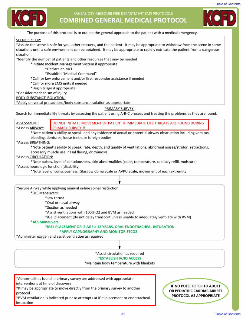
KANSAS CITY MISSOURI FIRE DEPARTMENT EMS PROTOCOLS
COMBINED GENERAL MEDICAL PROTOCOL
The purpose of this protocol is to outline the general approach to the patient with a medical emergency.
SCENE SIZE UP:*Assure the scene is safe for you, other rescuers, and the patient. It may be appropriate to withdraw from the scene in some situations until a safe environment can be obtained. It may be appropriate to rapidly extricate the patient from a dangerous situation.*Identify the number of patients and other resources that may be needed
*Initiate Incident Management System if appropriate*Declare an MCI*Establish “Medical Command”
*Call for law enforcement and/or first responder assistance if needed*Call for more EMS units if needed*Begin triage if appropriate
*Consider mechanism of injuryBODY SUBSTANCE ISOLATION:*Apply universal precautions/body substance isolation as appropriate
PRIMARY SURVEY:Search for immediate life threats by assessing the patient using A‐B‐C process and treating the problems as they are found.
ASSESSMENT:*Assess AIRWAY:
*Note patient’s ability to speak, and any evidence of actual or potential airway obstruction including vomitus, bleeding, dentures, loose teeth, or foreign bodies
*Assess BREATHING:*Note patient’s ability to speak, rate, depth, and quality of ventilations, abnormal noises/stridor, retractions, accessory muscle use, nasal flaring, or cyanosis
*Assess CIRCULATION:*Note pulses, level of consciousness, skin abnormalities (color, temperature, capillary refill, moisture)
*Assess neurologic function (disability)*Note level of consciousness, Glasgow Coma Scale or AVPU Scale, movement of each extremity
*Secure Airway while applying manual in‐line spinal restriction*BLS Maneuvers:
*Jaw thrust*Oral or nasal airway*Suction as needed*Assist ventilations with 100% O2 and BVM as needed*iGel placement (do not delay transport unless unable to adequately ventilate with BVM)
*ALS Maneuvers:*iGEL PLACEMENT OR IF AGE > 12 YEARS, ORAL ENDOTRACHEAL INTUBATION
*APPLY CAPNOGRAPHY AND MONITOR ETCO2 *Administer oxygen and assist ventilation as required
*Assist circulation as required*ESTABLISH IV/IO ACCESS
*Maintain body temperature with blankets
IF NO PULSE REFER TO ADULT OR PEDIATRIC CARDIAC ARREST PROTOCOL AS APPROPRIATE
*Abnormalities found in primary survey are addressed with appropriate interventions at time of discovery*It may be appropriate to move directly from the primary survey to another protocol*BVM ventilation is indicated prior to attempts at iGel placement or endotracheal intubation
DO NOT INITIATE MOVEMENT OF PATIENT IF IMMEDIATE LIFE THREATS ARE FOUND DURING PRIMARY SURVEY!!!
Table of Contents
51 Table of Contents

OFFICE OF THE EMS MEDICAL DIRECTOR, KANSAS CITY MISSOURI
A systematic history and physical exam, focused on the patient’s complaint’s, searching for problems that may not be immediately life or limb threatening, but that may become so if not addressed appropriately.
SECONDARY SURVEY
ASSESSMENT:*Obtain chief complaint*Obtain “SAMPLE” history*Obtain vital signs (MANUAL Blood Pressure)*Perform focused physical exam (this evaluation is dependent on the history as well as findings from the primary survey and may be more or less detailed depending on the situation)*Consider application of cardiac monitor*Consider application of pulse oximeter*Consider obtaining rapid blood glucose determination*Assessment summation: Consider information gathered in primary and secondary survey, determine an impression of the patient’s primary problem and proceed to the appropriate treatment protocol*Obtain repeat set of vital signs prior to transfer of care to receiving facility or whenever there is an observed change in the patient’s status
Secure airway, administer oxygen and assist ventilation as required
CONSIDER ESTABLISHING IV ACCESS
Transport while monitoring vital sings and patient condition*Medical Control contact as determined by appropriate protocol
REFER TO APPROPRIATE PROTOCOL FOR SPECIFIC MEDICAL EMERGENCY
REFER TO PAIN CONTROL PROTOCOL IF INDICATED
Table of Contents
52 Table of Contents
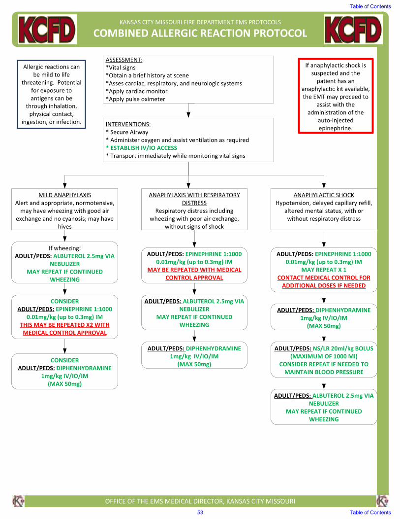
KANSAS CITY MISSOURI FIRE DEPARTMENT EMS PROTOCOLS
COMBINED ALLERGIC REACTION PROTOCOL
OFFICE OF THE EMS MEDICAL DIRECTOR, KANSAS CITY MISSOURI
ASSESSMENT:
*Vital signs
*Obtain a brief history at scene
*Asses cardiac, respiratory, and neurologic systems
*Apply cardiac monitor
*Apply pulse oximeter
INTERVENTIONS:
* Secure Airway
* Administer oxygen and assist ventilation as required
* ESTABLISH IV/IO ACCESS
* Transport immediately while monitoring vital signs
MILD ANAPHYLAXIS
Alert and appropriate, normotensive,
may have wheezing with good air
exchange and no cyanosis; may have
hives
ANAPHYLAXIS WITH RESPIRATORY
DISTRESS
Respiratory distress including
wheezing with poor air exchange,
without signs of shock
ANAPHYLACTIC SHOCK
Hypotension, delayed capillary refill,
altered mental status, with or
without respiratory distress
If wheezing:
ADULT/PEDS: ALBUTEROL 2.5mg VIA
NEBULIZER
MAY REPEAT IF CONTINUED
WHEEZING
CONSIDER
ADULT/PEDS: EPINEPHRINE 1:1000
0.01mg/kg (up to 0.3mg) IM
THIS MAY BE REPEATED X2 WITH
MEDICAL CONTROL APPROVAL
CONSIDER
ADULT/PEDS: DIPHENHYDRAMINE
1mg/kg IV/IO/IM
(MAX 50mg)
ADULT/PEDS: EPINEPHRINE 1:1000
0.01mg/kg (up to 0.3mg) IM
MAY BE REPEATED WITH MEDICAL
CONTROL APPROVAL
ADULT/PEDS: ALBUTEROL 2.5mg VIA
NEBULIZER
MAY REPEAT IF CONTINUED
WHEEZING
ADULT/PEDS: DIPHENHYDRAMINE
1mg/kg IV/IO/IM
(MAX 50mg)
ADULT/PEDS: EPINEPHRINE 1:1000
0.01mg/kg (up to 0.3mg) IM
MAY REPEAT X 1
CONTACT MEDICAL CONTROL FOR
ADDITIONAL DOSES IF NEEDED
ADULT/PEDS: DIPHENHYDRAMINE
1mg/kg IV/IO/IM
(MAX 50mg)
ADULT/PEDS: NS/LR 20ml/kg BOLUS
(MAXIMUM OF 1000 Ml)
CONSIDER REPEAT IF NEEDED TO
MAINTAIN BLOOD PRESSURE
ADULT/PEDS: ALBUTEROL 2.5mg VIA
NEBULIZER
MAY REPEAT IF CONTINUED
WHEEZING
Allergic reactions can
be mild to life
threatening. Potential
for exposure to
antigens can be
through inhalation,
physical contact,
ingestion, or infection.
If anaphylactic shock is
suspected and the
patient has an
anaphylactic kit available,
the EMT may proceed to
assist with the
administration of the
auto-injected
epinephrine.
Table of Contents
53 Table of Contents
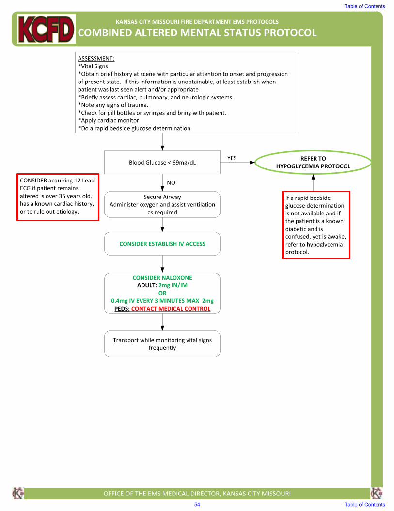
KANSAS CITY MISSOURI FIRE DEPARTMENT EMS PROTOCOLS
COMBINED ALTERED MENTAL STATUS PROTOCOL
OFFICE OF THE EMS MEDICAL DIRECTOR, KANSAS CITY MISSOURI
ASSESSMENT:
*Vital Signs
*Obtain brief history at scene with particular attention to onset and progression
of present state. If this information is unobtainable, at least establish when
patient was last seen alert and/or appropriate
*Briefly assess cardiac, pulmonary, and neurologic systems.
*Note any signs of trauma.
*Check for pill bottles or syringes and bring with patient.
*Apply cardiac monitor
*Do a rapid bedside glucose determination
Blood Glucose < 69mg/dLREFER TO
HYPOGLYCEMIA PROTOCOL
Secure Airway
Administer oxygen and assist ventilation
as required
CONSIDER NALOXONE
ADULT: 2mg IN/IM
OR
0.4mg IV EVERY 3 MINUTES MAX 2mg
PEDS: CONTACT MEDICAL CONTROL
Transport while monitoring vital signs
frequently
If a rapid bedside
glucose determination
is not available and if
the patient is a known
diabetic and is
confused, yet is awake,
refer to hypoglycemia
protocol.
YES
NO
CONSIDER ESTABLISH IV ACCESS
CONSIDER acquiring 12 Lead
ECG if patient remains
altered is over 35 years old,
has a known cardiac history,
or to rule out etiology.
Table of Contents
54 Table of Contents
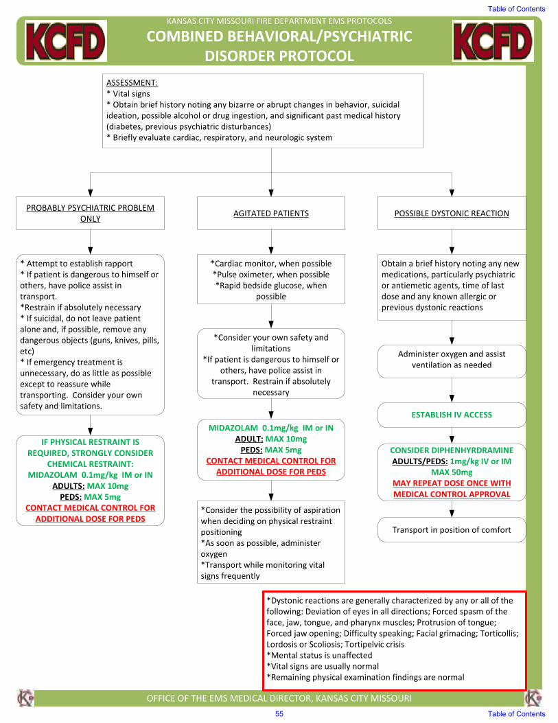
KANSAS CITY MISSOURI FIRE DEPARTMENT EMS PROTOCOLS
COMBINED BEHAVIORAL/PSYCHIATRIC
DISORDER PROTOCOL
OFFICE OF THE EMS MEDICAL DIRECTOR, KANSAS CITY MISSOURI
ASSESSMENT:
* Vital signs
* Obtain brief history noting any bizarre or abrupt changes in behavior, suicidal
ideation, possible alcohol or drug ingestion, and significant past medical history
(diabetes, previous psychiatric disturbances)
* Briefly evaluate cardiac, respiratory, and neurologic system
PROBABLY PSYCHIATRIC PROBLEM
ONLYAGITATED PATIENTS POSSIBLE DYSTONIC REACTION
* Attempt to establish rapport
* If patient is dangerous to himself or
others, have police assist in
transport.
*Restrain if absolutely necessary
* If suicidal, do not leave patient
alone and, if possible, remove any
dangerous objects (guns, knives, pills,
etc)
* If emergency treatment is
unnecessary, do as little as possible
except to reassure while
transporting. Consider your own
safety and limitations.
*Cardiac monitor, when possible
*Pulse oximeter, when possible
*Rapid bedside glucose, when
possible
*Consider your own safety and
limitations
*If patient is dangerous to himself or
others, have police assist in
transport. Restrain if absolutely
necessary
MIDAZOLAM 0.1mg/kg IM or IN
ADULT: MAX 10mg
PEDS: MAX 5mg
CONTACT MEDICAL CONTROL FOR
ADDITIONAL DOSE FOR PEDS
*Consider the possibility of aspiration
when deciding on physical restraint
positioning
*As soon as possible, administer
oxygen
*Transport while monitoring vital
signs frequently
Obtain a brief history noting any new
medications, particularly psychiatric
or antiemetic agents, time of last
dose and any known allergic or
previous dystonic reactions
Administer oxygen and assist
ventilation as needed
ESTABLISH IV ACCESS
CONSIDER DIPHENHYRDRAMINE
ADULTS/PEDS: 1mg/kg IV or IM
MAX 50mg
MAY REPEAT DOSE ONCE WITH
MEDICAL CONTROL APPROVAL
Transport in position of comfort
*Dystonic reactions are generally characterized by any or all of the
following: Deviation of eyes in all directions; Forced spasm of the
face, jaw, tongue, and pharynx muscles; Protrusion of tongue;
Forced jaw opening; Difficulty speaking; Facial grimacing; Torticollis;
Lordosis or Scoliosis; Tortipelvic crisis
*Mental status is unaffected
*Vital signs are usually normal
*Remaining physical examination findings are normal
IF PHYSICAL RESTRAINT IS
REQUIRED, STRONGLY CONSIDER
CHEMICAL RESTRAINT:
MIDAZOLAM 0.1mg/kg IM or IN
ADULTS: MAX 10mg
PEDS: MAX 5mg
CONTACT MEDICAL CONTROL FOR
ADDITIONAL DOSE FOR PEDS
Table of Contents
55 Table of Contents
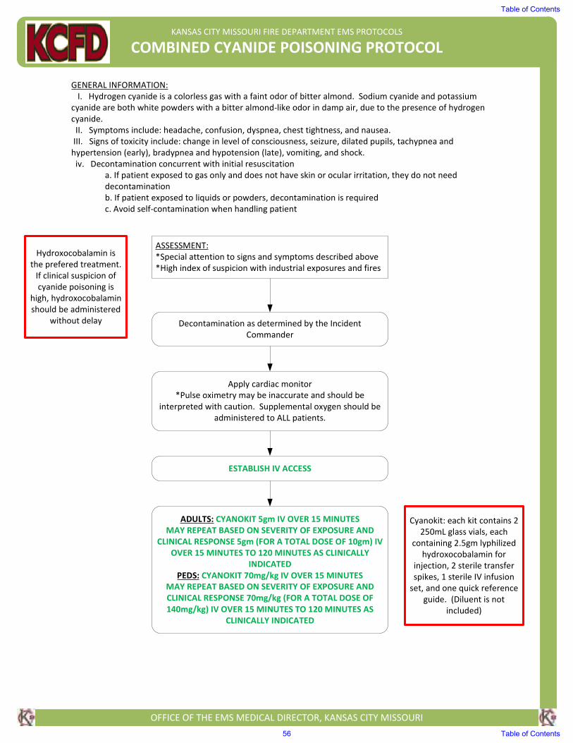
KANSAS CITY MISSOURI FIRE DEPARTMENT EMS PROTOCOLS
COMBINED CYANIDE POISONING PROTOCOL
OFFICE OF THE EMS MEDICAL DIRECTOR, KANSAS CITY MISSOURI
GENERAL INFORMATION:
I. Hydrogen cyanide is a colorless gas with a faint odor of bitter almond. Sodium cyanide and potassium
cyanide are both white powders with a bitter almond-like odor in damp air, due to the presence of hydrogen
cyanide.
II. Symptoms include: headache, confusion, dyspnea, chest tightness, and nausea.
III. Signs of toxicity include: change in level of consciousness, seizure, dilated pupils, tachypnea and
hypertension (early), bradypnea and hypotension (late), vomiting, and shock.
iv. Decontamination concurrent with initial resuscitation
a. If patient exposed to gas only and does not have skin or ocular irritation, they do not need
decontamination
b. If patient exposed to liquids or powders, decontamination is required
c. Avoid self-contamination when handling patient
ASSESSMENT:
*Special attention to signs and symptoms described above
*High index of suspicion with industrial exposures and fires
Decontamination as determined by the Incident
Commander
Apply cardiac monitor
*Pulse oximetry may be inaccurate and should be
interpreted with caution. Supplemental oxygen should be
administered to ALL patients.
ESTABLISH IV ACCESS
ADULTS: CYANOKIT 5gm IV OVER 15 MINUTES
MAY REPEAT BASED ON SEVERITY OF EXPOSURE AND
CLINICAL RESPONSE 5gm (FOR A TOTAL DOSE OF 10gm) IV
OVER 15 MINUTES TO 120 MINUTES AS CLINICALLY
INDICATED
PEDS: CYANOKIT 70mg/kg IV OVER 15 MINUTES
MAY REPEAT BASED ON SEVERITY OF EXPOSURE AND
CLINICAL RESPONSE 70mg/kg (FOR A TOTAL DOSE OF
140mg/kg) IV OVER 15 MINUTES TO 120 MINUTES AS
CLINICALLY INDICATED
Hydroxocobalamin is
the prefered treatment.
If clinical suspicion of
cyanide poisoning is
high, hydroxocobalamin
should be administered
without delay
Cyanokit: each kit contains 2
250mL glass vials, each
containing 2.5gm lyphilized
hydroxocobalamin for
injection, 2 sterile transfer
spikes, 1 sterile IV infusion
set, and one quick reference
guide. (Diluent is not
included)
Table of Contents
56 Table of Contents
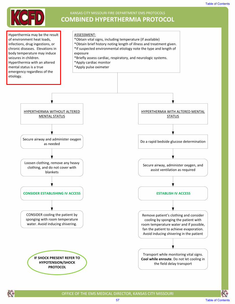
KANSAS CITY MISSOURI FIRE DEPARTMENT EMS PROTOCOLS
COMBINED HYPERTHERMIA PROTOCOL
OFFICE OF THE EMS MEDICAL DIRECTOR, KANSAS CITY MISSOURI
ASSESSMENT:
*Obtain vital signs, including temperature (if available)
*Obtain brief history noting length of illness and treatment given.
*If suspected environmental etiology note the type and length of
exposure
*Briefly assess cardiac, respiratory, and neurologic systems.
*Apply cardiac monitor
*Apply pulse oximeter
Hyperthermia may be the result
of environment heat loads,
infections, drug ingestions, or
chronic diseases. Elevations in
body temperature may induce
seizures in children.
Hyperthermia with an altered
mental status is a true
emergency regardless of the
etiology.
HYPERTHERMIA WITHOUT ALTERED
MENTAL STATUS
HYPERTHERMIA WITH ALTERED MENTAL
STATUS
Secure airway and administer oxygen
as needed
Loosen clothing, remove any heavy
clothing, and do not cover with
blankets
CONSIDER ESTABLISHING IV ACCESS
CONSIDER cooling the patient by
sponging with room temperature
water. Avoid inducing shivering.
Do a rapid bedside glucose determination
Secure airway, administer oxygen, and
assist ventilation as required
ESTABLISH IV ACCESS
Remove patient’s clothing and consider
cooling by sponging the patient with
room temperature water and if possible,
fan the patient to achieve evaporation.
Avoid inducing shivering in the patient
Transport while monitoring vital signs.
Cool while enroute. Do not let cooling in
the field delay transport
IF SHOCK PRESENT REFER TO
HYPOTENSION/SHOCK
PROTOCOL
Table of Contents
57 Table of Contents
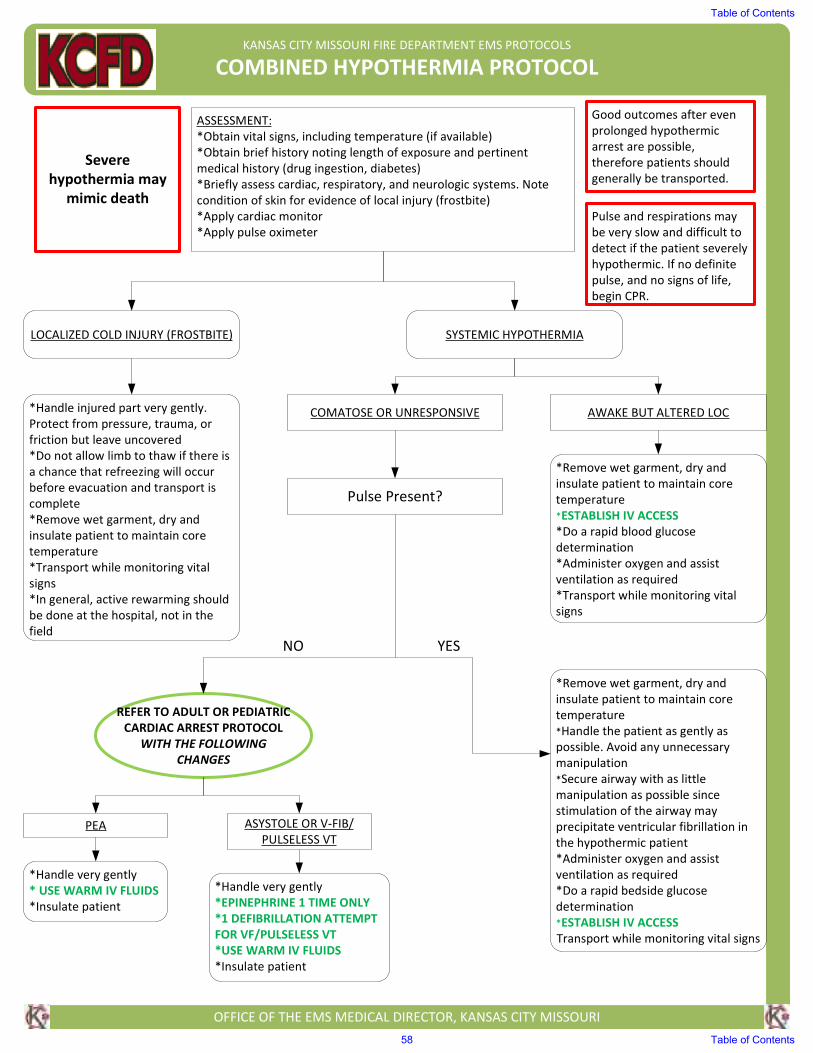
KANSAS CITY MISSOURI FIRE DEPARTMENT EMS PROTOCOLS
COMBINED HYPOTHERMIA PROTOCOL
OFFICE OF THE EMS MEDICAL DIRECTOR, KANSAS CITY MISSOURI
ASSESSMENT:
*Obtain vital signs, including temperature (if available)
*Obtain brief history noting length of exposure and pertinent
medical history (drug ingestion, diabetes)
*Briefly assess cardiac, respiratory, and neurologic systems. Note
condition of skin for evidence of local injury (frostbite)
*Apply cardiac monitor
*Apply pulse oximeter
LOCALIZED COLD INJURY (FROSTBITE) SYSTEMIC HYPOTHERMIA
*Handle injured part very gently.
Protect from pressure, trauma, or
friction but leave uncovered
*Do not allow limb to thaw if there is
a chance that refreezing will occur
before evacuation and transport is
complete
*Remove wet garment, dry and
insulate patient to maintain core
temperature
*Transport while monitoring vital
signs
*In general, active rewarming should
be done at the hospital, not in the
field
Severe
hypothermia may
mimic death
COMATOSE OR UNRESPONSIVE AWAKE BUT ALTERED LOC
*Remove wet garment, dry and
insulate patient to maintain core
temperature
*ESTABLISH IV ACCESS
*Do a rapid blood glucose
determination
*Administer oxygen and assist
ventilation as required
*Transport while monitoring vital
signs
Pulse Present?
NO YES
*Remove wet garment, dry and
insulate patient to maintain core
temperature
*Handle the patient as gently as
possible. Avoid any unnecessary
manipulation
*Secure airway with as little
manipulation as possible since
stimulation of the airway may
precipitate ventricular fibrillation in
the hypothermic patient
*Administer oxygen and assist
ventilation as required
*Do a rapid bedside glucose
determination
*ESTABLISH IV ACCESS
Transport while monitoring vital signs
REFER TO ADULT OR PEDIATRIC
CARDIAC ARREST PROTOCOL
WITH THE FOLLOWING
CHANGES
PEA ASYSTOLE OR V-FIB/
PULSELESS VT
*Handle very gently
* USE WARM IV FLUIDS
*Insulate patient
*Handle very gently
*EPINEPHRINE 1 TIME ONLY
*1 DEFIBRILLATION ATTEMPT
FOR VF/PULSELESS VT
*USE WARM IV FLUIDS
*Insulate patient
Good outcomes after even
prolonged hypothermic
arrest are possible,
therefore patients should
generally be transported.
Pulse and respirations may
be very slow and difficult to
detect if the patient severely
hypothermic. If no definite
pulse, and no signs of life,
begin CPR.
Table of Contents
58 Table of Contents
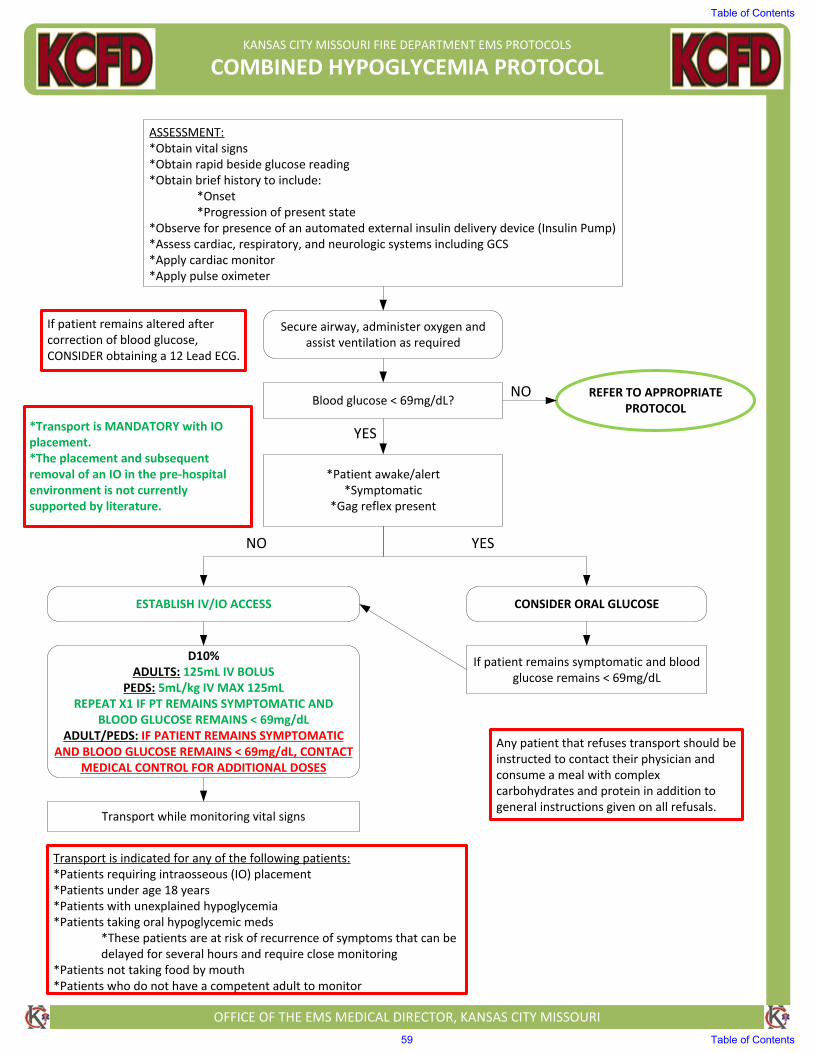
KANSAS CITY MISSOURI FIRE DEPARTMENT EMS PROTOCOLS
COMBINED HYPOGLYCEMIA PROTOCOL
OFFICE OF THE EMS MEDICAL DIRECTOR, KANSAS CITY MISSOURI
ASSESSMENT:*Obtain vital signs*Obtain rapid beside glucose reading*Obtain brief history to include:
*Onset*Progression of present state
*Observe for presence of an automated external insulin delivery device (Insulin Pump)*Assess cardiac, respiratory, and neurologic systems including GCS*Apply cardiac monitor*Apply pulse oximeter
Secure airway, administer oxygen and assist ventilation as required
ESTABLISH IV/IO ACCESS
Blood glucose < 69mg/dL?REFER TO APPROPRIATE
PROTOCOL
*Patient awake/alert*Symptomatic
*Gag reflex present
CONSIDER ORAL GLUCOSE
If patient remains symptomatic and blood glucose remains < 69mg/dL
D10% ADULTS: 125mL IV BOLUS
PEDS: 5mL/kg IV MAX 125mLREPEAT X1 IF PT REMAINS SYMPTOMATIC AND
BLOOD GLUCOSE REMAINS < 69mg/dLADULT/PEDS: IF PATIENT REMAINS SYMPTOMATIC
AND BLOOD GLUCOSE REMAINS < 69mg/dL, CONTACT MEDICAL CONTROL FOR ADDITIONAL DOSES
Transport while monitoring vital signs
*Transport is MANDATORY with IOplacement.*The placement and subsequentremoval of an IO in the pre‐hospitalenvironment is not currentlysupported by literature.
Transport is indicated for any of the following patients:*Patients requiring intraosseous (IO) placement*Patients under age 18 years*Patients with unexplained hypoglycemia*Patients taking oral hypoglycemic meds
*These patients are at risk of recurrence of symptoms that can bedelayed for several hours and require close monitoring
*Patients not taking food by mouth*Patients who do not have a competent adult to monitor
Any patient that refuses transport should be instructed to contact their physician and consume a meal with complex carbohydrates and protein in addition to general instructions given on all refusals.
NO
YES
YESNO
If patient remains altered after correction of blood glucose, CONSIDER obtaining a 12 Lead ECG.
Table of Contents
59 Table of Contents
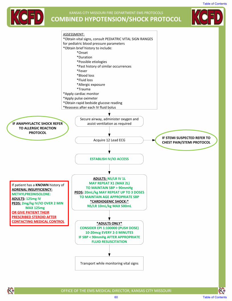
KANSAS CITY MISSOURI FIRE DEPARTMENT EMS PROTOCOLS
COMBINED HYPOTENSION/SHOCK PROTOCOL
OFFICE OF THE EMS MEDICAL DIRECTOR, KANSAS CITY MISSOURI
ASSESSMENT:*Obtain vital signs, consult PEDIATRIC VITAL SIGN RANGES for pediatric blood pressure parameters*Obtain brief history to include:
*Onset*Duration*Possible etiologies*Past history of similar occurrences*Fever*Blood loss*Fluid loss*Allergic exposure*Trauma
*Apply cardiac monitor*Apply pulse oximeter*Obtain rapid bedside glucose reading*Reassess after each IV fluid bolus
Secure airway, administer oxygen and assist ventilation as required
Acquire 12 Lead ECGIF STEMI SUSPECTED REFER TO CHEST PAIN/STEMI PROTOCOL
ESTABLISH IV/IO ACCESS
ADULTS: NS/LR IV 1LMAY REPEAT X1 (MAX 2L)
TO MAINTAIN SBP > 90mmHgPEDS: 20mL/kg MAY REPEAT UP TO 3 DOSES
TO MAINTAIN AGE APPROPRIATE SBP*CARDIOGENIC SHOCK:*
NS/LR 10mL/kg MAX 500mL
*ADULTS ONLY*CONSIDER EPI 1:100000 (PUSH DOSE)
10‐20mcg EVERY 2‐3 MINUTES IF SBP < 90mmHg AFTER APPROPRIATE
FLUID RESUSCITATION
Transport while monitoring vital signs
If patient has a KNOWN history of ADRENAL INSUFFICIENCY:METHYLPREDNISOLONE: ADULTS: 125mg IVPEDS: 2mg/kg IV/IO OVER 2 MIN
MAX 125mg OR GIVE PATIENT THEIR PRESCRIBED STEROID AFTER CONTACTING MEDICAL CONTROL
IF ANAPHYLACTIC SHOCK REFER TO ALLERGIC REACTION
PROTOCOL
Table of Contents
60 Table of Contents
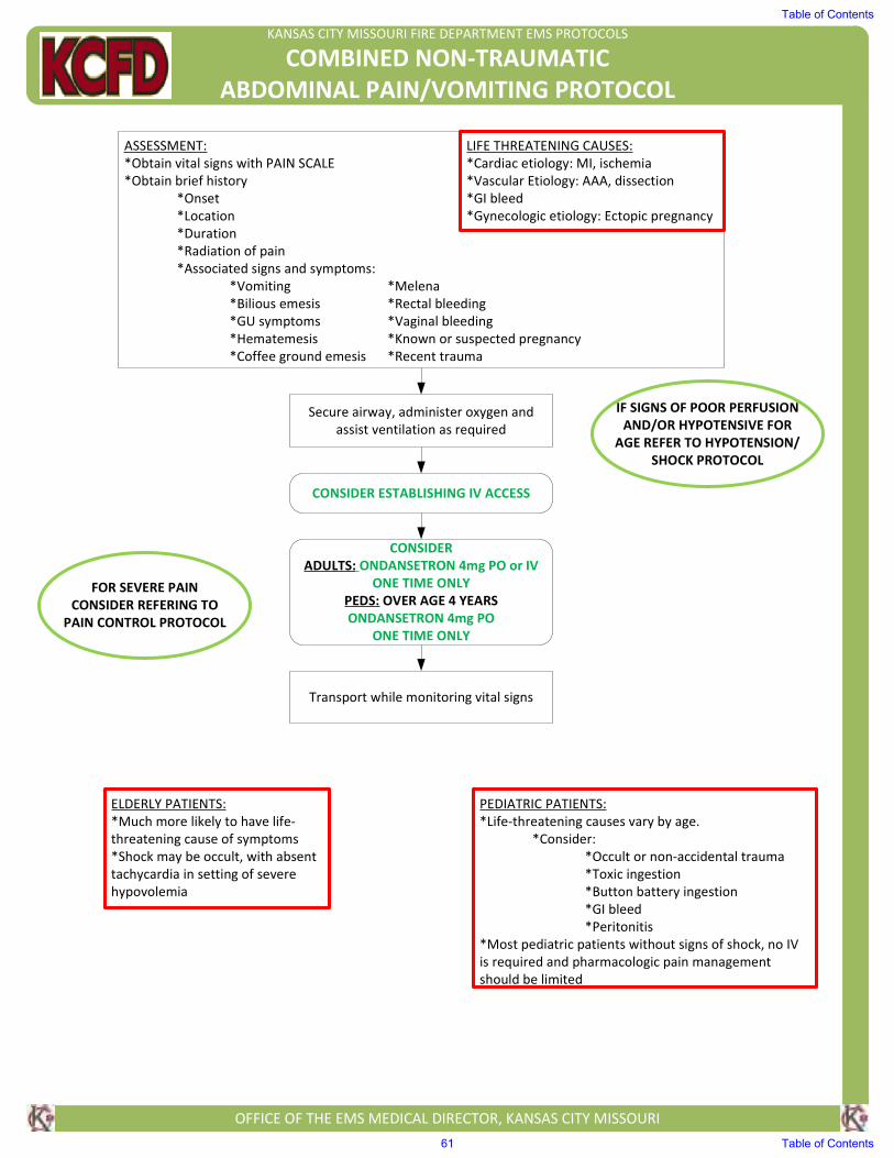
KANSAS CITY MISSOURI FIRE DEPARTMENT EMS PROTOCOLS
COMBINED NON-TRAUMATIC
ABDOMINAL PAIN/VOMITING PROTOCOL
OFFICE OF THE EMS MEDICAL DIRECTOR, KANSAS CITY MISSOURI
ASSESSMENT:
*Obtain vital signs with PAIN SCALE
*Obtain brief history
*Onset
*Location
*Duration
*Radiation of pain
*Associated signs and symptoms:
*Vomiting *Melena
*Bilious emesis *Rectal bleeding
*GU symptoms *Vaginal bleeding
*Hematemesis *Known or suspected pregnancy
*Coffee ground emesis *Recent trauma
Secure airway, administer oxygen and
assist ventilation as required
IF SIGNS OF POOR PERFUSION
AND/OR HYPOTENSIVE FOR
AGE REFER TO HYPOTENSION/
SHOCK PROTOCOL
CONSIDER ESTABLISHING IV ACCESS
CONSIDER
ADULTS: ONDANSETRON 4mg PO or IV
ONE TIME ONLY
PEDS: OVER AGE 4 YEARS
ONDANSETRON 4mg PO
ONE TIME ONLY
Transport while monitoring vital signs
LIFE THREATENING CAUSES:
*Cardiac etiology: MI, ischemia
*Vascular Etiology: AAA, dissection
*GI bleed
*Gynecologic etiology: Ectopic pregnancy
FOR SEVERE PAIN
CONSIDER REFERING TO
PAIN CONTROL PROTOCOL
ELDERLY PATIENTS:
*Much more likely to have life-
threatening cause of symptoms
*Shock may be occult, with absent
tachycardia in setting of severe
hypovolemia
PEDIATRIC PATIENTS:
*Life-threatening causes vary by age.
*Consider:
*Occult or non-accidental trauma
*Toxic ingestion
*Button battery ingestion
*GI bleed
*Peritonitis
*Most pediatric patients without signs of shock, no IV
is required and pharmacologic pain management
should be limited
Table of Contents
61 Table of Contents
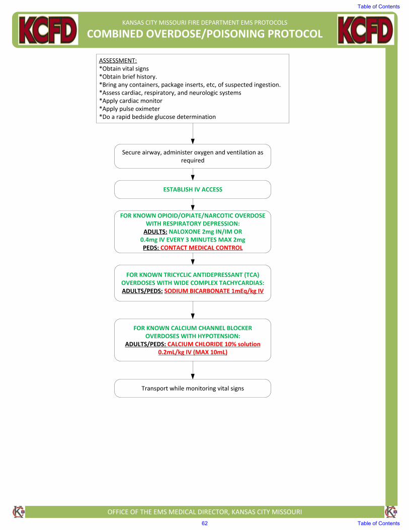
KANSAS CITY MISSOURI FIRE DEPARTMENT EMS PROTOCOLS
COMBINED OVERDOSE/POISONING PROTOCOL
OFFICE OF THE EMS MEDICAL DIRECTOR, KANSAS CITY MISSOURI
ASSESSMENT:*Obtain vital signs*Obtain brief history. *Bring any containers, package inserts, etc, of suspected ingestion. *Assess cardiac, respiratory, and neurologic systems*Apply cardiac monitor*Apply pulse oximeter*Do a rapid bedside glucose determination
Transport while monitoring vital signs
Secure airway, administer oxygen and ventilation as required
ESTABLISH IV ACCESS
FOR KNOWN TRICYCLIC ANTIDEPRESSANT (TCA) OVERDOSES WITH WIDE COMPLEX TACHYCARDIAS:ADULTS/PEDS: SODIUM BICARBONATE 1mEq/kg IV
FOR KNOWN CALCIUM CHANNEL BLOCKER OVERDOSES WITH HYPOTENSION:
ADULTS/PEDS: CALCIUM CHLORIDE 10% solution 0.2mL/kg IV (MAX 10mL)
FOR KNOWN OPIOID/OPIATE/NARCOTIC OVERDOSE WITH RESPIRATORY DEPRESSION:ADULTS: NALOXONE 2mg IN/IM OR0.4mg IV EVERY 3 MINUTES MAX 2mgPEDS: CONTACT MEDICAL CONTROL
Table of Contents
62 Table of Contents
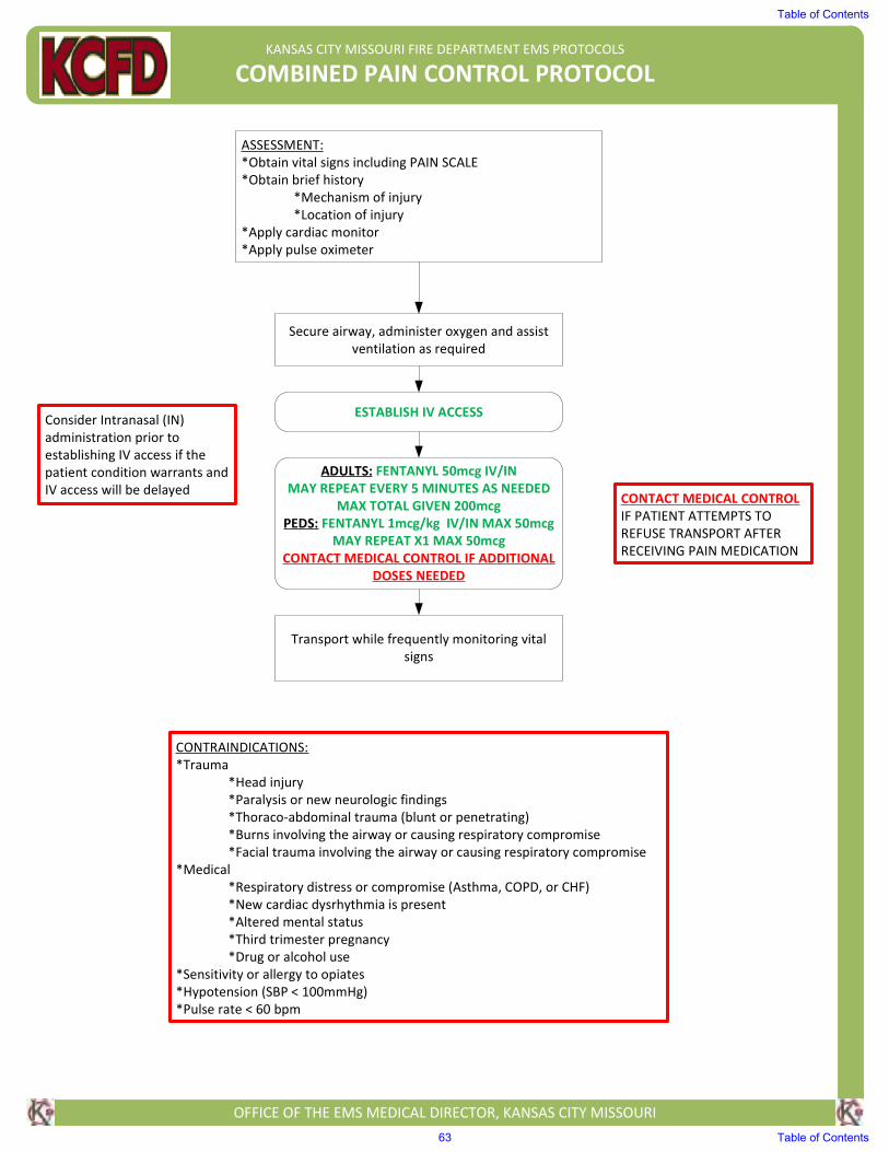
KANSAS CITY MISSOURI FIRE DEPARTMENT EMS PROTOCOLS
COMBINED PAIN CONTROL PROTOCOL
OFFICE OF THE EMS MEDICAL DIRECTOR, KANSAS CITY MISSOURI
ASSESSMENT:
*Obtain vital signs including PAIN SCALE
*Obtain brief history
*Mechanism of injury
*Location of injury
*Apply cardiac monitor
*Apply pulse oximeter
Secure airway, administer oxygen and assist
ventilation as required
ESTABLISH IV ACCESS
ADULTS: FENTANYL 50mcg IV/IN
MAY REPEAT EVERY 5 MINUTES AS NEEDED
MAX TOTAL GIVEN 200mcg
PEDS: FENTANYL 1mcg/kg IV/IN MAX 50mcg
MAY REPEAT X1 MAX 50mcg
CONTACT MEDICAL CONTROL IF ADDITIONAL
DOSES NEEDED
Transport while frequently monitoring vital
signs
CONTRAINDICATIONS:
*Trauma
*Head injury
*Paralysis or new neurologic findings
*Thoraco-abdominal trauma (blunt or penetrating)
*Burns involving the airway or causing respiratory compromise
*Facial trauma involving the airway or causing respiratory compromise
*Medical
*Respiratory distress or compromise (Asthma, COPD, or CHF)
*New cardiac dysrhythmia is present
*Altered mental status
*Third trimester pregnancy
*Drug or alcohol use
*Sensitivity or allergy to opiates
*Hypotension (SBP < 100mmHg)
*Pulse rate < 60 bpm
Consider Intranasal (IN)
administration prior to
establishing IV access if the
patient condition warrants and
IV access will be delayedCONTACT MEDICAL CONTROL
IF PATIENT ATTEMPTS TO
REFUSE TRANSPORT AFTER
RECEIVING PAIN MEDICATION
Table of Contents
63 Table of Contents
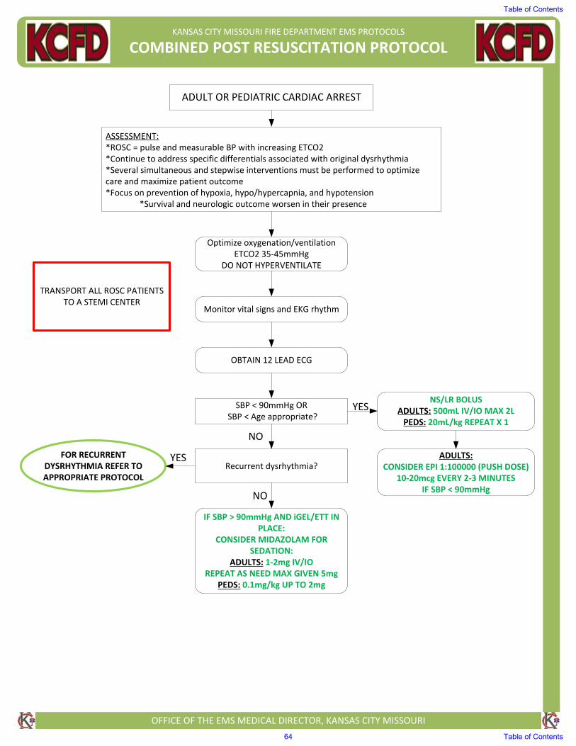
KANSAS CITY MISSOURI FIRE DEPARTMENT EMS PROTOCOLS
COMBINED POST RESUSCITATION PROTOCOL
OFFICE OF THE EMS MEDICAL DIRECTOR, KANSAS CITY MISSOURI
ADULT OR PEDIATRIC CARDIAC ARREST
ASSESSMENT:*ROSC = pulse and measurable BP with increasing ETCO2*Continue to address specific differentials associated with original dysrhythmia*Several simultaneous and stepwise interventions must be performed to optimize care and maximize patient outcome*Focus on prevention of hypoxia, hypo/hypercapnia, and hypotension
*Survival and neurologic outcome worsen in their presence
Optimize oxygenation/ventilationETCO2 35‐45mmHg
DO NOT HYPERVENTILATE
Monitor vital signs and EKG rhythm
OBTAIN 12 LEAD ECG
SBP < 90mmHg OR SBP < Age appropriate?
NS/LR BOLUS ADULTS: 500mL IV/IO MAX 2LPEDS: 20mL/kg REPEAT X 1
ADULTS:CONSIDER EPI 1:100000 (PUSH DOSE)
10‐20mcg EVERY 2‐3 MINUTESIF SBP < 90mmHg
Recurrent dysrhythmia?FOR RECURRENT
DYSRHYTHMIA REFER TO APPROPRIATE PROTOCOL
IF SBP > 90mmHg AND iGEL/ETT IN PLACE:
CONSIDER MIDAZOLAM FOR SEDATION:
ADULTS: 1‐2mg IV/IOREPEAT AS NEED MAX GIVEN 5mg
PEDS: 0.1mg/kg UP TO 2mg
YES
NO
TRANSPORT ALL ROSC PATIENTS TO A STEMI CENTER
YES
NO
Table of Contents
64 Table of Contents
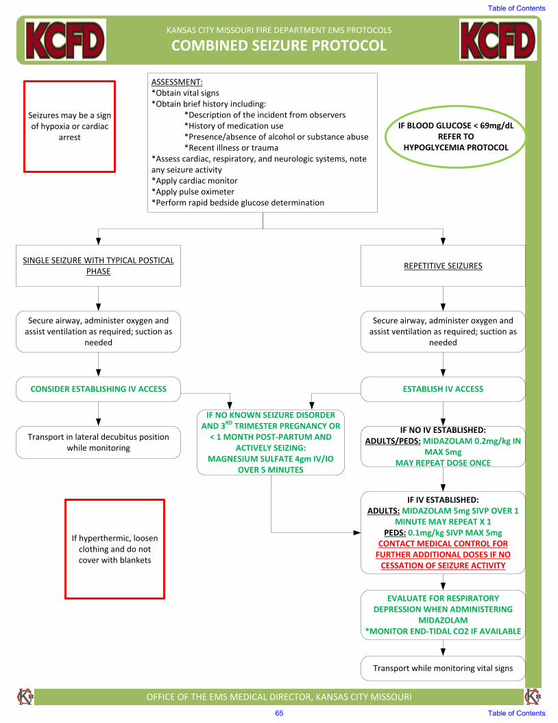
KANSAS CITY MISSOURI FIRE DEPARTMENT EMS PROTOCOLS
COMBINED SEIZURE PROTOCOL
OFFICE OF THE EMS MEDICAL DIRECTOR, KANSAS CITY MISSOURI
ASSESSMENT:*Obtain vital signs*Obtain brief history including:
*Description of the incident from observers*History of medication use*Presence/absence of alcohol or substance abuse*Recent illness or trauma
*Assess cardiac, respiratory, and neurologic systems, noteany seizure activity*Apply cardiac monitor*Apply pulse oximeter*Perform rapid bedside glucose determination
Seizures may be a sign of hypoxia or cardiac
arrest
IF BLOOD GLUCOSE < 69mg/dL REFER TO
HYPOGLYCEMIA PROTOCOL
SINGLE SEIZURE WITH TYPICAL POSTICAL PHASE
REPETITIVE SEIZURES
Secure airway, administer oxygen and assist ventilation as required; suction as
needed
Secure airway, administer oxygen and assist ventilation as required; suction as
needed
Transport in lateral decubitus position while monitoring
CONSIDER ESTABLISHING IV ACCESS ESTABLISH IV ACCESS
IF NO IV ESTABLISHED:ADULTS/PEDS: MIDAZOLAM 0.2mg/kg IN
MAX 5mgMAY REPEAT DOSE ONCE
IF IV ESTABLISHED:ADULTS: MIDAZOLAM 5mg SIVP OVER 1
MINUTE MAY REPEAT X 1PEDS: 0.1mg/kg SIVP MAX 5mg
CONTACT MEDICAL CONTROL FOR FURTHER ADDITIONAL DOSES IF NO
CESSATION OF SEIZURE ACTIVITY
EVALUATE FOR RESPIRATORY DEPRESSION WHEN ADMINISTERING
MIDAZOLAM*MONITOR END-TIDAL CO2 IF AVAILABLE
Transport while monitoring vital signs
If hyperthermic, loosen clothing and do not cover with blankets
IF NO KNOWN SEIZURE DISORDER AND 3RD TRIMESTER PREGNANCY OR
< 1 MONTH POST-PARTUM AND ACTIVELY SEIZING:
MAGNESIUM SULFATE 4gm IV/IO OVER 5 MINUTES
Table of Contents
65 Table of Contents
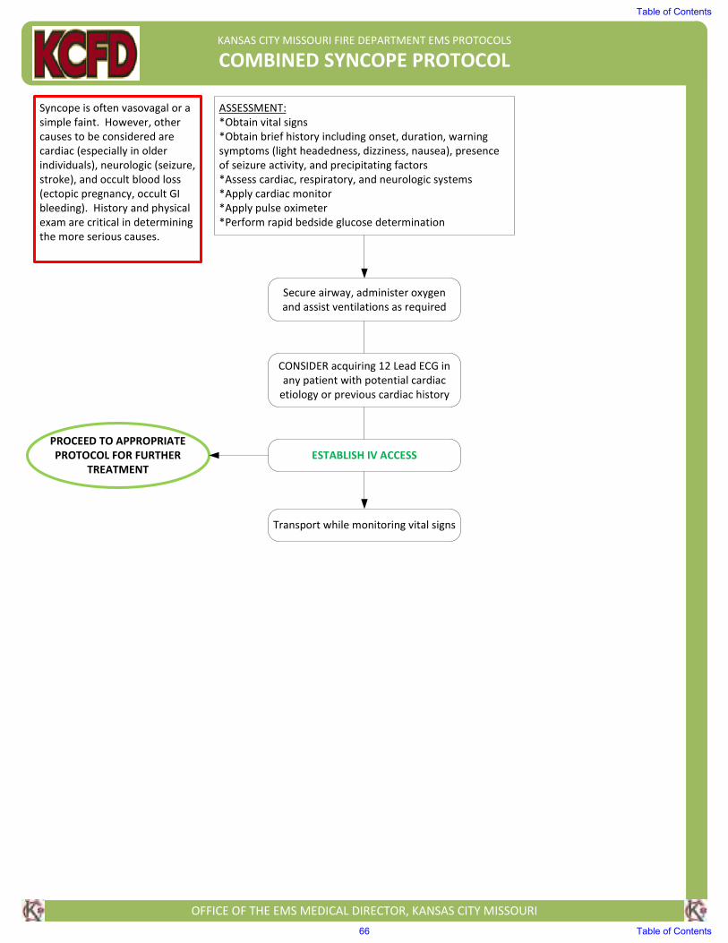
KANSAS CITY MISSOURI FIRE DEPARTMENT EMS PROTOCOLS
COMBINED SYNCOPE PROTOCOL
OFFICE OF THE EMS MEDICAL DIRECTOR, KANSAS CITY MISSOURI
ASSESSMENT:
*Obtain vital signs
*Obtain brief history including onset, duration, warning
symptoms (light headedness, dizziness, nausea), presence
of seizure activity, and precipitating factors
*Assess cardiac, respiratory, and neurologic systems
*Apply cardiac monitor
*Apply pulse oximeter
*Perform rapid bedside glucose determination
Syncope is often vasovagal or a
simple faint. However, other
causes to be considered are
cardiac (especially in older
individuals), neurologic (seizure,
stroke), and occult blood loss
(ectopic pregnancy, occult GI
bleeding). History and physical
exam are critical in determining
the more serious causes.
Secure airway, administer oxygen
and assist ventilations as required
ESTABLISH IV ACCESS
Transport while monitoring vital signs
PROCEED TO APPROPRIATE
PROTOCOL FOR FURTHER
TREATMENT
CONSIDER acquiring 12 Lead ECG in
any patient with potential cardiac
etiology or previous cardiac history
Table of Contents
66 Table of Contents
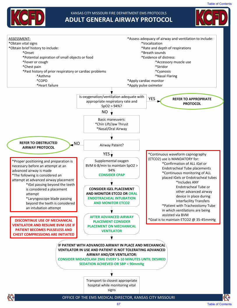
KANSAS CITY MISSOURI FIRE DEPARTMENT EMS PROTOCOLS
ADULT GENERAL AIRWAY PROTOCOL
OFFICE OF THE EMS MEDICAL DIRECTOR, KANSAS CITY MISSOURI
ASSESSMENT:*Obtain vital signs*Obtain brief history to include:
*Onset*Potential aspiration of small objects or food*Fever or cough*Chest pain*Past history of prior respiratory or cardiac problems
*Asthma*COPD*Heart failure
Is oxygenation/ventilation adequate with appropriate respiratory rate and
SpO2 > 94%?
REFER TO OBSTRUCTED AIRWAY PROTOCOL
Basic maneuvers:*Chin Lift/Jaw Thrust*Nasal/Oral Airway
Airway Patent?
Supplemental oxygen BVM 6‐8/min to maintain SpO2 >
94%CONSIDER CPAP
CONSIDER iGEL PLACEMENT AND MONITOR ETCO2 OR ORAL ENDOTRACHEAL INTUBATION
AND MONITOR ETCO2
Transport to closest appropriate hospital while monitoring vital
signs
*Continuous waveform capnography(ETCO2) use is MANDATORY for:
*Confirmation of ALL iGel orEndotracheal Tube placements*Continuous monitoring of ALLplaced iGels or Endotracheal tubes
*Includes ANYEndotracheal Tube orother advanced airwaydevice in place duringInterfacility Transfers
*Patient with Tracheostomy Tubein which ventilations are beingassisted via BVM
*Goal is to maintain ETCO2 @ 35‐45mmHg
*Proper positioning and preparation isnecessary before an attempt at anadvanced airway is made*The following is considered anattempt at advanced airway placement
*iGel passing beyond the teethis considered a placementattempt*Laryngoscope blade passingbeyond the teeth is consideredan intubation attempt
NO
YES
REFER TO APPROPRIATE PROTOCOL
YES
NO
*Assess adequacy of airway and ventilation to include:*Vocalization*Rate and depth of respirations*Breath sounds*Evidence of distress:
*Accessory muscle use*Stridor*Cyanosis*Nasal Flaring
*Apply cardiac monitor*Apply pulse oximeter
AFTER ADVANCED AIRWAY PLACEMENT CONSIDER
PLACEMENT ON MECHANICAL VENTILATOR
IF PATIENT WITH ADVANCED AIRWAY IN PLACE AND MECHANICAL VENTILATOR IN USE AND PATIENT IS NOT TOLERATING ADVANCED
AIRWAY AND/OR VENTILATOR:CONSIDER MIDAZOLAM 2MG EVERY 5‐10 MINUTES UNTIL DESIRED
SEDATION ACHIEVED OR SBP < 90mmHg
DISCONTINUE USE OF MECHANICAL VENTILATOR AND RESUME BVM USE IF PATIENT BECOMES PULSELESS AND
CHEST COMPRESSIONS ARE INITIATED
Table of Contents
67 Table of Contents
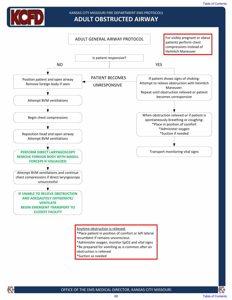
KANSAS CITY MISSOURI FIRE DEPARTMENT EMS PROTOCOLS
ADULT OBSTRUCTED AIRWAY
OFFICE OF THE EMS MEDICAL DIRECTOR, KANSAS CITY MISSOURI
ADULT GENERAL AIRWAY PROTOCOL
Is patient responsive?
If patient shows signs of choking:
Attempt to relieve obstruction with Heimlich
Maneuver.
Repeat until obstruction relieved or patient
becomes unresponsive
Position patient and open airway
Remove foreign body if seen
Reposition head and open airway
Attempt BVM ventilations
Transport monitoring vital signs
IF UNABLE TO RELIEVE OBSTRUCTION
AND ADEQAUTELY OXYGENATE/
VENTILATE
BEGIN EMERGENT TRANSPORT TO
CLOSEST FACILITY
PERFORM DIRECT LARYNGOSCOPY
REMOVE FOREIGN BODY WITH MAGILL
FORCEPS IF VISUALIZED
Attempt BVM ventilations
Attempt BVM ventilations and continue
chest compressions if direct laryngoscopy
unsuccessful
Begin chest compressions
Anytime obstruction is relieved:
*Place patient in position of comfort or left lateral recumbent if remains unconscious
*Administer oxygen, monitor SpO2 and vital signs
*Be prepared for vomiting as is common after an obstruction is relieved
*Suction as needed
When obstruction relieved or if patient is
spontaneously breathing or coughing:
*Place in position of comfort
*Administer oxygen
*Suction if needed
For visibly pregnant or obese
patients perform chest
compressions instead of
Heimlich Maneuver
PATIENT BECOMES
UNRESPONSIVE
NO YES
Table of Contents
68 Table of Contents
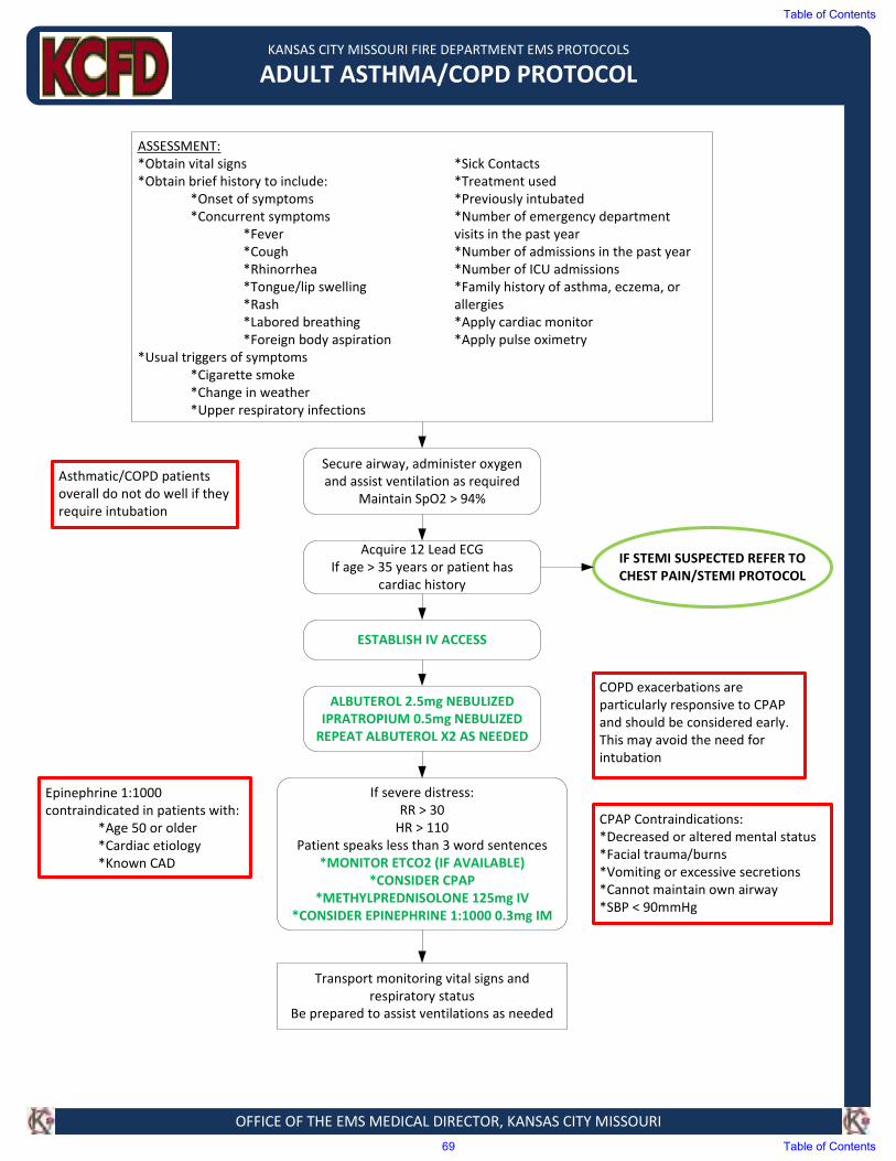
KANSAS CITY MISSOURI FIRE DEPARTMENT EMS PROTOCOLS
ADULT ASTHMA/COPD PROTOCOL
OFFICE OF THE EMS MEDICAL DIRECTOR, KANSAS CITY MISSOURI
ASSESSMENT:
*Obtain vital signs *Sick Contacts
*Obtain brief history to include: *Treatment used
*Onset of symptoms *Previously intubated
*Concurrent symptoms *Number of emergency department
*Fever visits in the past year
*Cough *Number of admissions in the past year
*Rhinorrhea *Number of ICU admissions
*Tongue/lip swelling *Family history of asthma, eczema, or
*Rash allergies
*Labored breathing *Apply cardiac monitor
*Foreign body aspiration *Apply pulse oximetry
*Usual triggers of symptoms
*Cigarette smoke
*Change in weather
*Upper respiratory infections
IF STEMI SUSPECTED REFER TO
CHEST PAIN/STEMI PROTOCOL
Secure airway, administer oxygen
and assist ventilation as required Maintain SpO2 > 94%
Acquire 12 Lead ECG
If age > 35 years or patient has
cardiac history
ESTABLISH IV ACCESS
ALBUTEROL 2.5mg NEBULIZED IPRATROPIUM 0.5mg NEBULIZED
REPEAT ALBUTEROL X2 AS NEEDED
If severe distress:
RR > 30
HR > 110
Patient speaks less than 3 word sentences
*MONITOR ETCO2 (IF AVAILABLE)
*CONSIDER CPAP
*METHYLPREDNISOLONE 125mg IV
*CONSIDER EPINEPHRINE 1:1000 0.3mg IM
Epinephrine 1:1000
contraindicated in patients with:
*Age 50 or older
*Cardiac etiology
*Known CAD
Asthmatic/COPD patients
overall do not do well if they
require intubation
COPD exacerbations are
particularly responsive to CPAP
and should be considered early.
This may avoid the need for
intubation
Transport monitoring vital signs and
respiratory status
Be prepared to assist ventilations as needed
CPAP Contraindications:
*Decreased or altered mental status
*Facial trauma/burns
*Vomiting or excessive secretions
*Cannot maintain own airway
*SBP < 90mmHg
Table of Contents
69 Table of Contents
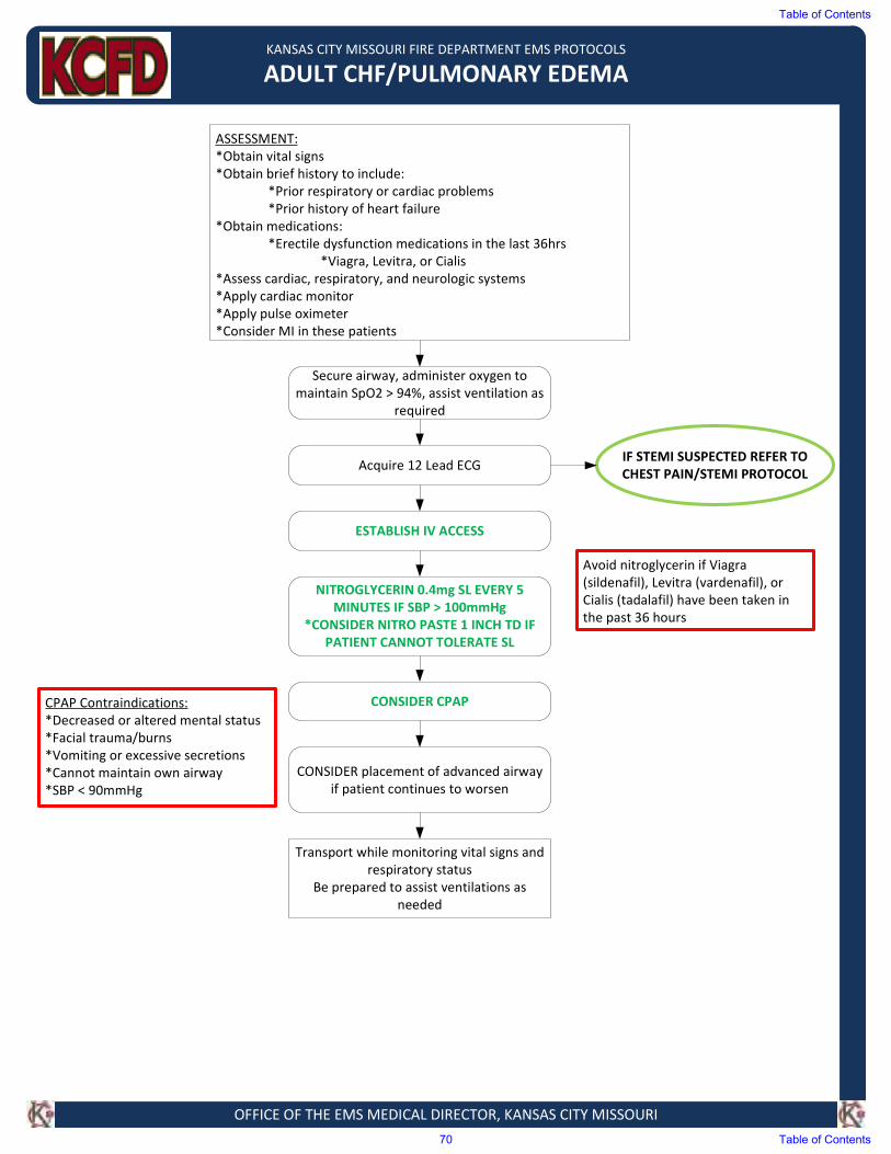
KANSAS CITY MISSOURI FIRE DEPARTMENT EMS PROTOCOLS
ADULT CHF/PULMONARY EDEMA
OFFICE OF THE EMS MEDICAL DIRECTOR, KANSAS CITY MISSOURI
ASSESSMENT:
*Obtain vital signs
*Obtain brief history to include:
*Prior respiratory or cardiac problems
*Prior history of heart failure
*Obtain medications:
*Erectile dysfunction medications in the last 36hrs
*Viagra, Levitra, or Cialis
*Assess cardiac, respiratory, and neurologic systems
*Apply cardiac monitor
*Apply pulse oximeter
*Consider MI in these patients
Secure airway, administer oxygen to
maintain SpO2 > 94%, assist ventilation as
required
Acquire 12 Lead ECGIF STEMI SUSPECTED REFER TO
CHEST PAIN/STEMI PROTOCOL
ESTABLISH IV ACCESS
NITROGLYCERIN 0.4mg SL EVERY 5
MINUTES IF SBP > 100mmHg
*CONSIDER NITRO PASTE 1 INCH TD IF
PATIENT CANNOT TOLERATE SL
CONSIDER CPAP
CONSIDER placement of advanced airway
if patient continues to worsen
Transport while monitoring vital signs and
respiratory status
Be prepared to assist ventilations as
needed
CPAP Contraindications:
*Decreased or altered mental status
*Facial trauma/burns
*Vomiting or excessive secretions
*Cannot maintain own airway
*SBP < 90mmHg
Avoid nitroglycerin if Viagra
(sildenafil), Levitra (vardenafil), or
Cialis (tadalafil) have been taken in
the past 36 hours
Table of Contents
70 Table of Contents
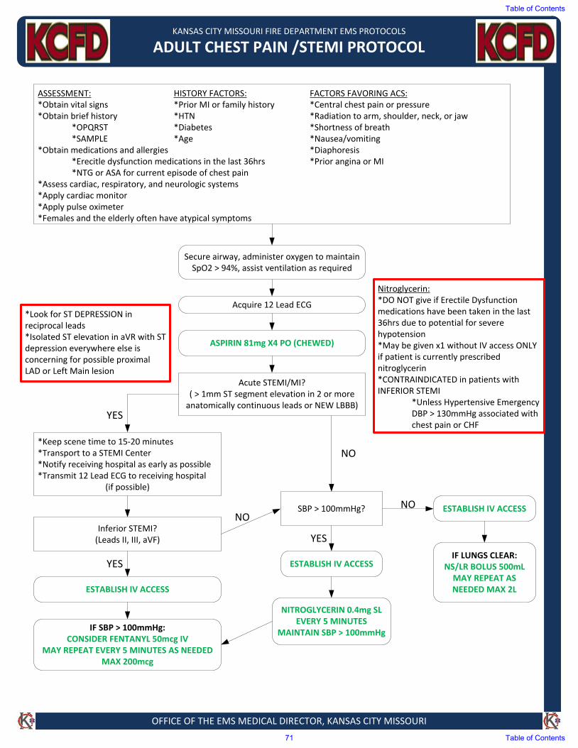
KANSAS CITY MISSOURI FIRE DEPARTMENT EMS PROTOCOLS
ADULT CHEST PAIN /STEMI PROTOCOL
OFFICE OF THE EMS MEDICAL DIRECTOR, KANSAS CITY MISSOURI
ASSESSMENT: HISTORY FACTORS: FACTORS FAVORING ACS:*Obtain vital signs *Prior MI or family history *Central chest pain or pressure*Obtain brief history *HTN *Radiation to arm, shoulder, neck, or jaw
*OPQRST *Diabetes *Shortness of breath*SAMPLE *Age *Nausea/vomiting
*Obtain medications and allergies *Diaphoresis*Erecitle dysfunction medications in the last 36hrs *Prior angina or MI*NTG or ASA for current episode of chest pain
*Assess cardiac, respiratory, and neurologic systems*Apply cardiac monitor*Apply pulse oximeter*Females and the elderly often have atypical symptoms
Secure airway, administer oxygen to maintain SpO2 > 94%, assist ventilation as required
Acquire 12 Lead ECG
ASPIRIN 81mg X4 PO (CHEWED)
Acute STEMI/MI?( > 1mm ST segment elevation in 2 or more
anatomically continuous leads or NEW LBBB)
*Keep scene time to 15-20 minutes*Transport to a STEMI Center*Notify receiving hospital as early as possible*Transmit 12 Lead ECG to receiving hospital
(if possible)
Inferior STEMI?(Leads II, III, aVF)
ESTABLISH IV ACCESS
IF SBP > 100mmHg:CONSIDER FENTANYL 50mcg IV
MAY REPEAT EVERY 5 MINUTES AS NEEDEDMAX 200mcg
SBP > 100mmHg?
NITROGLYCERIN 0.4mg SL EVERY 5 MINUTES
MAINTAIN SBP > 100mmHg
ESTABLISH IV ACCESSIF LUNGS CLEAR:
NS/LR BOLUS 500mLMAY REPEAT AS NEEDED MAX 2L
ESTABLISH IV ACCESS
YES
NO
YES
YES
NONO
Nitroglycerin:*DO NOT give if Erectile Dysfunction medications have been taken in the last 36hrs due to potential for severe hypotension*May be given x1 without IV access ONLY if patient is currently prescribed nitroglycerin*CONTRAINDICATED in patients with INFERIOR STEMI
*Unless Hypertensive Emergency DBP > 130mmHg associated with chest pain or CHF
*Look for ST DEPRESSION in reciprocal leads *Isolated ST elevation in aVR with ST depression everywhere else is concerning for possible proximal LAD or Left Main lesion
Table of Contents
71 Table of Contents

KANSAS CITY MISSOURI FIRE DEPARTMENT EMS PROTOCOLS
ADULT BRADYCARDIA (HR < 60) PROTOCOL
OFFICE OF THE EMS MEDICAL DIRECTOR, KANSAS CITY MISSOURI
ASSESSMENT:*Obtain vital signs*Obtain brief history of current symptoms, especially current chest pain or shortness of breath*Obtain past medical history
*Congenital disease*Previous dysrhythmias*Coronary artery disease*Heart failure*Heart murmur
*Current medications looking specifically for*Beta‐blockers*Calcium channel blockers*Clonidine*Digoxin
*Apply cardiac monitor*Apply Pulse Oximeter
SIGNS OF POOR PERFUSION:*Altered Mental Status*Chest Pain*CHF*Shortness of Air*Syncope*Hypotension/Shock
Acquire 12 Lead ECG
Secure airway, administer oxygen to maintain SpO2 > 94%, assist ventilation as required
ESTABLISH IV/IO ACCESS
ATROPINE 0.5mg IV/IO EVERY 3‐5 MINUTESMAX 3mg
TRANSCUTANEOUS PACINGIF NOT RESPONSIVE TO ATROPINE (CONSIDER
EARLY IN 2ND OR 3RD DEGREE AV BLOCK)
CONSIDER MIDAZOLAM 1‐2 mg IV/IN FOR SEDATION
REPEAT AS NEEDED TO MAX 5mg GIVEN
IF POOR PERFUSION PERSISTS:CONSIDER
NS/LR 10ml/kg MAX 500mLOR
EPI 1:100000 (PUSH DOSE) 10‐20mcg IV/IO EVERY 2‐3 MINUTES
Transport monitoring vital signs
IF SUSPECTED BETA‐BLOCKER OR CALCIUM CHANNEL
BLOCKER OVERDOSE REFER TO OVERDOSE/POISONING
PROTOCOL
YES NO
*Atropine in the presence of MI may worsen heart damage*Atropine is ineffective in cardiac transplant patients*DO NOT DELAY pacing patients with poor perfusion and presence of MI or 2nd or 3rd degree AV Block
Table of Contents
72 Table of Contents
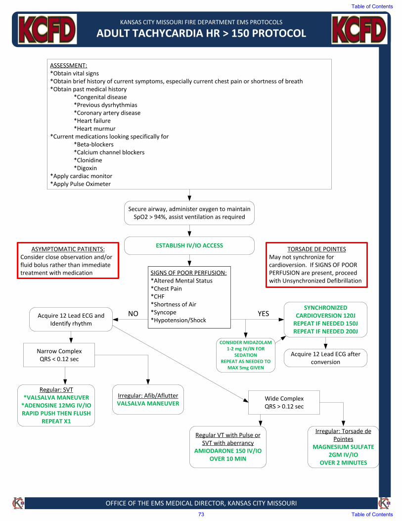
KANSAS CITY MISSOURI FIRE DEPARTMENT EMS PROTOCOLS
ADULT TACHYCARDIA HR > 150 PROTOCOL
OFFICE OF THE EMS MEDICAL DIRECTOR, KANSAS CITY MISSOURI
ASSESSMENT:*Obtain vital signs*Obtain brief history of current symptoms, especially current chest pain or shortness of breath*Obtain past medical history
*Congenital disease*Previous dysrhythmias*Coronary artery disease*Heart failure*Heart murmur
*Current medications looking specifically for*Beta‐blockers*Calcium channel blockers*Clonidine*Digoxin
*Apply cardiac monitor*Apply Pulse Oximeter
SIGNS OF POOR PERFUSION:*Altered Mental Status*Chest Pain*CHF*Shortness of Air*Syncope*Hypotension/Shock
Secure airway, administer oxygen to maintain SpO2 > 94%, assist ventilation as required
ESTABLISH IV/IO ACCESS
Acquire 12 Lead ECG and Identify rhythm
SYNCHRONIZED CARDIOVERSION 120JREPEAT IF NEEDED 150JREPEAT IF NEEDED 200J
Narrow ComplexQRS < 0.12 sec
Wide ComplexQRS > 0.12 sec
Regular: SVT*VALSALVA MANEUVER*ADENOSINE 12MG IV/IORAPID PUSH THEN FLUSH
REPEAT X1
Irregular: Afib/AflutterVALSALVA MANEUVER
Regular VT with Pulse or SVT with aberrancy
AMIODARONE 150 IV/IO OVER 10 MIN
Irregular: Torsade de Pointes
MAGNESIUM SULFATE 2GM IV/IO
OVER 2 MINUTES
ASYMPTOMATIC PATIENTS:Consider close observation and/or fluid bolus rather than immediate treatment with medication
TORSADE DE POINTES May not synchronize for cardioversion. If SIGNS OF POOR PERFUSION are present, proceed with Unsynchronized Defibrillation
NO YES
Acquire 12 Lead ECG after conversion
CONSIDER MIDAZOLAM 1‐2 mg IV/IN FOR
SEDATIONREPEAT AS NEEDED TO
MAX 5mg GIVEN
Table of Contents
73 Table of Contents
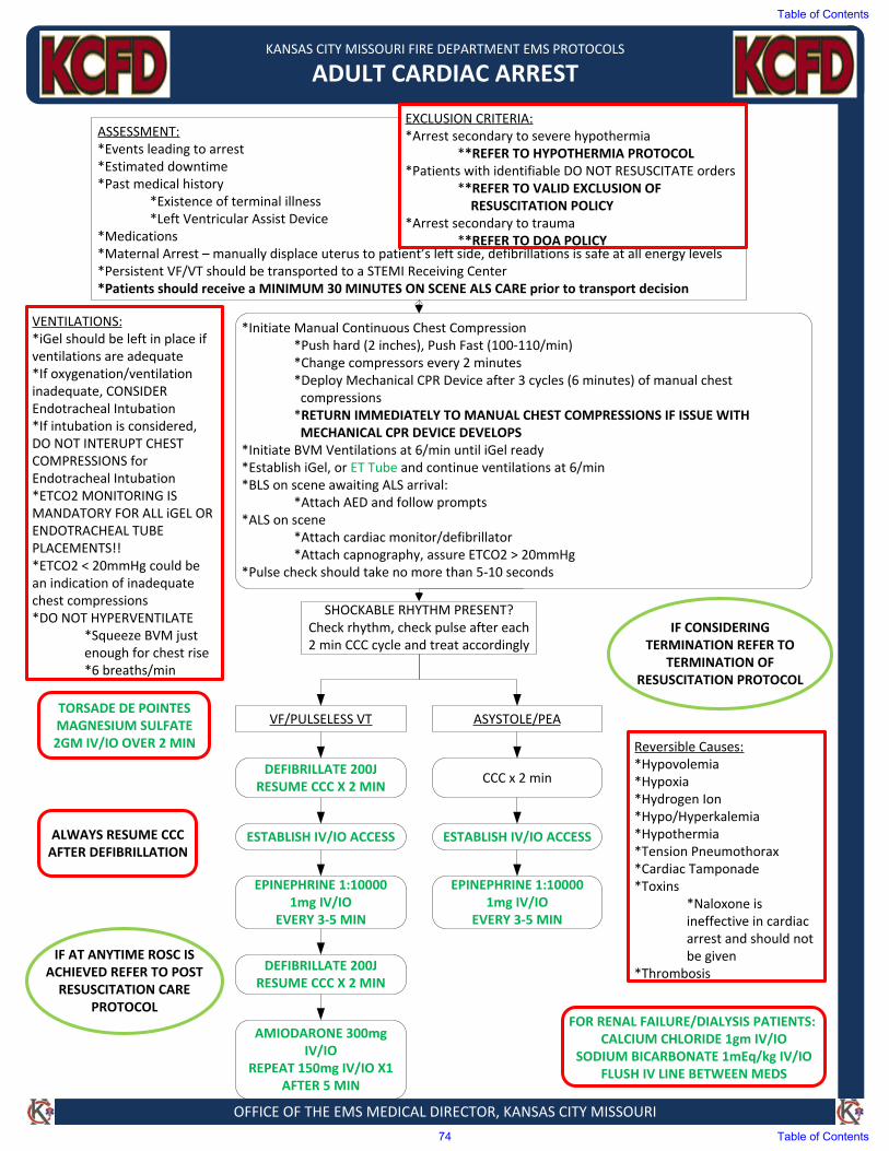
KANSAS CITY MISSOURI FIRE DEPARTMENT EMS PROTOCOLS
ADULT CARDIAC ARREST
OFFICE OF THE EMS MEDICAL DIRECTOR, KANSAS CITY MISSOURI
ASSESSMENT:*Events leading to arrest*Estimated downtime*Past medical history
*Existence of terminal illness*Left Ventricular Assist Device
*Medications*Maternal Arrest – manually displace uterus to patient’s left side, defibrillations is safe at all energy levels*Persistent VF/VT should be transported to a STEMI Receiving Center*Patients should receive a MINIMUM 30 MINUTES ON SCENE ALS CARE prior to transport decision
EXCLUSION CRITERIA:*Arrest secondary to severe hypothermia
**REFER TO HYPOTHERMIA PROTOCOL*Patients with identifiable DO NOT RESUSCITATE orders
**REFER TO VALID EXCLUSION OF RESUSCITATION POLICY
*Arrest secondary to trauma**REFER TO DOA POLICY
*Initiate Manual Continuous Chest Compression*Push hard (2 inches), Push Fast (100‐110/min)*Change compressors every 2 minutes*Deploy Mechanical CPR Device after 3 cycles (6 minutes) of manual chestcompressions*RETURN IMMEDIATELY TO MANUAL CHEST COMPRESSIONS IF ISSUE WITHMECHANICAL CPR DEVICE DEVELOPS
*Initiate BVM Ventilations at 6/min until iGel ready*Establish iGel, or ET Tube and continue ventilations at 6/min*BLS on scene awaiting ALS arrival:
*Attach AED and follow prompts*ALS on scene
*Attach cardiac monitor/defibrillator*Attach capnography, assure ETCO2 > 20mmHg
*Pulse check should take no more than 5‐10 seconds
SHOCKABLE RHYTHM PRESENT?Check rhythm, check pulse after each 2 min CCC cycle and treat accordingly
VF/PULSELESS VT ASYSTOLE/PEA
DEFIBRILLATE 200JRESUME CCC X 2 MIN
CCC x 2 min
ESTABLISH IV/IO ACCESS ESTABLISH IV/IO ACCESS
EPINEPHRINE 1:10000 1mg IV/IO
EVERY 3‐5 MIN
EPINEPHRINE 1:10000 1mg IV/IO
EVERY 3‐5 MIN
DEFIBRILLATE 200JRESUME CCC X 2 MIN
AMIODARONE 300mg IV/IO
REPEAT 150mg IV/IO X1 AFTER 5 MIN
FOR RENAL FAILURE/DIALYSIS PATIENTS:CALCIUM CHLORIDE 1gm IV/IO
SODIUM BICARBONATE 1mEq/kg IV/IOFLUSH IV LINE BETWEEN MEDS
VENTILATIONS:*iGel should be left in place ifventilations are adequate*If oxygenation/ventilationinadequate, CONSIDEREndotracheal Intubation*If intubation is considered,DO NOT INTERUPT CHESTCOMPRESSIONS forEndotracheal Intubation*ETCO2 MONITORING ISMANDATORY FOR ALL iGEL ORENDOTRACHEAL TUBEPLACEMENTS!!*ETCO2 < 20mmHg could bean indication of inadequatechest compressions*DO NOT HYPERVENTILATE
*Squeeze BVM justenough for chest rise*6 breaths/min
Reversible Causes:*Hypovolemia*Hypoxia*Hydrogen Ion*Hypo/Hyperkalemia*Hypothermia*Tension Pneumothorax*Cardiac Tamponade*Toxins
*Naloxone isineffective in cardiacarrest and should notbe given
*Thrombosis
TORSADE DE POINTESMAGNESIUM SULFATE 2GM IV/IO OVER 2 MIN
IF AT ANYTIME ROSC IS ACHIEVED REFER TO POST RESUSCITATION CARE
PROTOCOL
ALWAYS RESUME CCC AFTER DEFIBRILLATION
IF CONSIDERING TERMINATION REFER TO
TERMINATION OF RESUSCITATION PROTOCOL
Table of Contents
74 Table of Contents
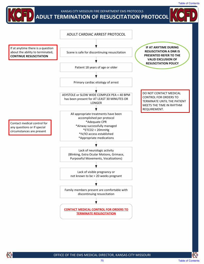
KANSAS CITY MISSOURI FIRE DEPARTMENT EMS PROTOCOLS
ADULT TERMINATION OF RESUSCITATION PROTOCOL
OFFICE OF THE EMS MEDICAL DIRECTOR, KANSAS CITY MISSOURI
ADULT CARDIAC ARREST PROTOCOL
Scene is safe for discontinuing resuscitation
Patient 18 years of age or older
Primary cardiac etiology of arrest
ASYSTOLE or SLOW WIDE COMPLEX PEA < 40 BPM has been present for AT LEAST 30 MINUTES OR
LONGER
All appropriate treatments have been accomplished per protocol
*Adequate CPR*Airway successfully managed
*ETCO2 < 20mmHg*IV/IO access established*Appropriate medications
Lack of neurologic activity(Blinking, Extra Ocular Motions, Grimace, Purposeful Movements, Vocalizations)
Lack of visible pregnancy or not known to be > 20 weeks pregnant
Family members present are comfortable with discontinuing resuscitation
CONTACT MEDICAL CONTROL FOR ORDERS TO TERMINATE RESUSCITATION
If at anytime there is a question about the ability to terminated, CONTINUE RESUSCITATION
IF AT ANYTIME DURING RESUSCITATION A DNR IS PRESENTED REFER TO THE
VALID EXCLUSION OF RESUSCITATION POLICY
Contact medical control for any questions or if special circumstances are present
DO NOT CONTACT MEDICAL CONTROL FOR ORDERS TO TERMINATE UNTIL THE PATIENT MEETS THE TIME IN RHYTHM REQUIREMENT.
Table of Contents
75 Table of Contents
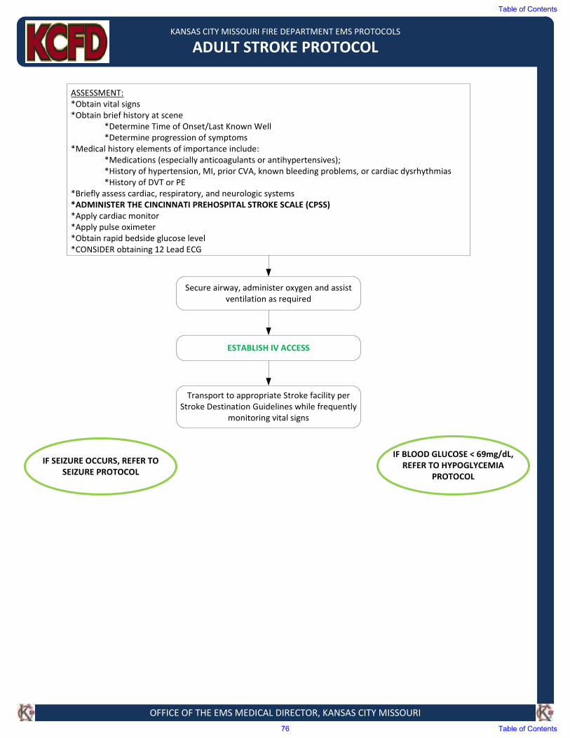
KANSAS CITY MISSOURI FIRE DEPARTMENT EMS PROTOCOLS
ADULT STROKE PROTOCOL
OFFICE OF THE EMS MEDICAL DIRECTOR, KANSAS CITY MISSOURI
ASSESSMENT:
*Obtain vital signs
*Obtain brief history at scene
*Determine Time of Onset/Last Known Well
*Determine progression of symptoms
*Medical history elements of importance include:
*Medications (especially anticoagulants or antihypertensives);
*History of hypertension, MI, prior CVA, known bleeding problems, or cardiac dysrhythmias
*History of DVT or PE
*Briefly assess cardiac, respiratory, and neurologic systems
*ADMINISTER THE CINCINNATI PREHOSPITAL STROKE SCALE (CPSS)
*Apply cardiac monitor
*Apply pulse oximeter
*Obtain rapid bedside glucose level
*CONSIDER obtaining 12 Lead ECG
IF BLOOD GLUCOSE < 69mg/dL,
REFER TO HYPOGLYCEMIA
PROTOCOL
Secure airway, administer oxygen and assist
ventilation as required
ESTABLISH IV ACCESS
Transport to appropriate Stroke facility per
Stroke Destination Guidelines while frequently
monitoring vital signs
IF SEIZURE OCCURS, REFER TO
SEIZURE PROTOCOL
Table of Contents
76 Table of Contents
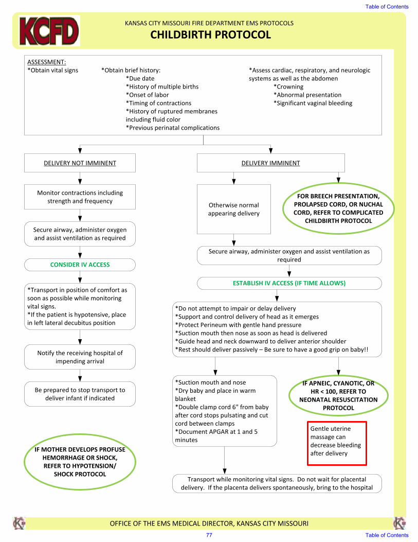
KANSAS CITY MISSOURI FIRE DEPARTMENT EMS PROTOCOLS
CHILDBIRTH PROTOCOL
OFFICE OF THE EMS MEDICAL DIRECTOR, KANSAS CITY MISSOURI
ASSESSMENT:
*Obtain vital signs *Obtain brief history: *Assess cardiac, respiratory, and neurologic
*Due date systems as well as the abdomen
*History of multiple births *Crowning
*Onset of labor *Abnormal presentation
*Timing of contractions *Significant vaginal bleeding
*History of ruptured membranes
including fluid color
*Previous perinatal complications
DELIVERY NOT IMMINENT DELIVERY IMMINENT
Monitor contractions including
strength and frequency
Secure airway, administer oxygen
and assist ventilation as required
*Transport in position of comfort as
soon as possible while monitoring
vital signs.
*If the patient is hypotensive, place
in left lateral decubitus position
Notify the receiving hospital of
impending arrival
Be prepared to stop transport to
deliver infant if indicated
Secure airway, administer oxygen and assist ventilation as
required
ESTABLISH IV ACCESS (IF TIME ALLOWS)
Transport while monitoring vital signs. Do not wait for placental
delivery. If the placenta delivers spontaneously, bring to the hospital
IF MOTHER DEVELOPS PROFUSE
HEMORRHAGE OR SHOCK,
REFER TO HYPOTENSION/
SHOCK PROTOCOL
CONSIDER IV ACCESS
*Do not attempt to impair or delay delivery
*Support and control delivery of head as it emerges
*Protect Perineum with gentle hand pressure
*Suction mouth then nose as soon as head is delivered
*Guide head and neck downward to deliver anterior shoulder
*Rest should deliver passively – Be sure to have a good grip on baby!!
FOR BREECH PRESENTATION,
PROLAPSED CORD, OR NUCHAL
CORD, REFER TO COMPLICATED
CHILDBIRTH PROTOCOL
Otherwise normal
appearing delivery
*Suction mouth and nose
*Dry baby and place in warm
blanket
*Double clamp cord 6" from baby
after cord stops pulsating and cut
cord between clamps
*Document APGAR at 1 and 5
minutes
IF APNEIC, CYANOTIC, OR
HR < 100, REFER TO
NEONATAL RESUSCITATION
PROTOCOL
Gentle uterine
massage can
decrease bleeding
after delivery
Table of Contents
77 Table of Contents
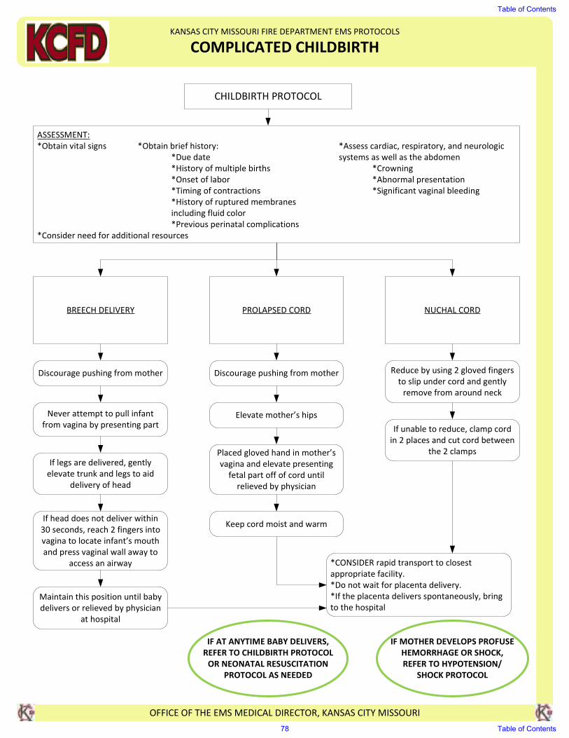
KANSAS CITY MISSOURI FIRE DEPARTMENT EMS PROTOCOLS
COMPLICATED CHILDBIRTH
OFFICE OF THE EMS MEDICAL DIRECTOR, KANSAS CITY MISSOURI
CHILDBIRTH PROTOCOL
ASSESSMENT:
*Obtain vital signs *Obtain brief history: *Assess cardiac, respiratory, and neurologic
*Due date systems as well as the abdomen
*History of multiple births *Crowning
*Onset of labor *Abnormal presentation
*Timing of contractions *Significant vaginal bleeding
*History of ruptured membranes
including fluid color
*Previous perinatal complications
*Consider need for additional resources
BREECH DELIVERY PROLAPSED CORD NUCHAL CORD
Never attempt to pull infant
from vagina by presenting part
If legs are delivered, gently
elevate trunk and legs to aid
delivery of head
If head does not deliver within
30 seconds, reach 2 fingers into
vagina to locate infant’s mouth
and press vaginal wall away to
access an airway
Maintain this position until baby
delivers or relieved by physician
at hospital
Discourage pushing from motherDiscourage pushing from mother
Elevate mother’s hips
Placed gloved hand in mother’s
vagina and elevate presenting
fetal part off of cord until
relieved by physician
Keep cord moist and warm
Reduce by using 2 gloved fingers
to slip under cord and gently
remove from around neck
If unable to reduce, clamp cord
in 2 places and cut cord between
the 2 clamps
*CONSIDER rapid transport to closest
appropriate facility.
*Do not wait for placenta delivery.
*If the placenta delivers spontaneously, bring
to the hospital
IF AT ANYTIME BABY DELIVERS,
REFER TO CHILDBIRTH PROTOCOL
OR NEONATAL RESUSCITATION
PROTOCOL AS NEEDED
IF MOTHER DEVELOPS PROFUSE
HEMORRHAGE OR SHOCK,
REFER TO HYPOTENSION/
SHOCK PROTOCOL
Table of Contents
78 Table of Contents
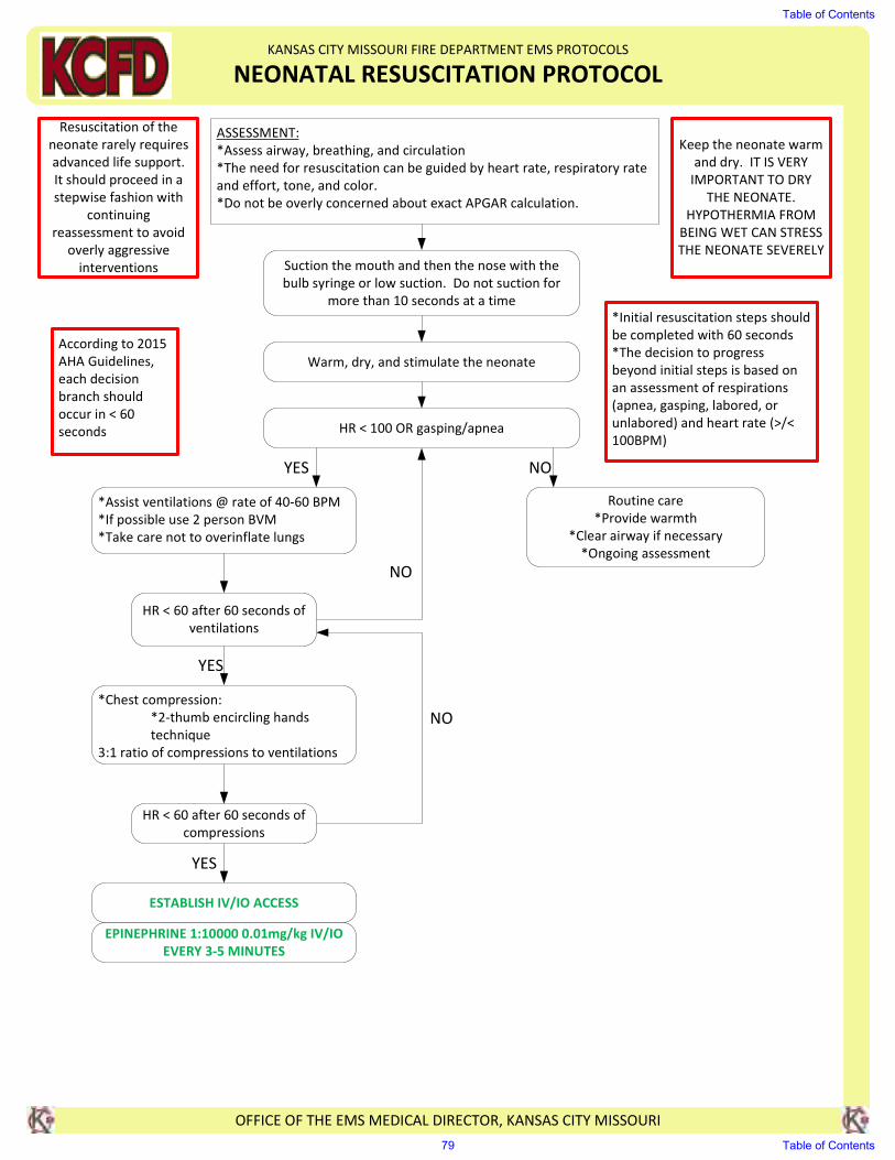
KANSAS CITY MISSOURI FIRE DEPARTMENT EMS PROTOCOLS
NEONATAL RESUSCITATION PROTOCOL
OFFICE OF THE EMS MEDICAL DIRECTOR, KANSAS CITY MISSOURI
ASSESSMENT:
*Assess airway, breathing, and circulation
*The need for resuscitation can be guided by heart rate, respiratory rate
and effort, tone, and color.
*Do not be overly concerned about exact APGAR calculation.
Resuscitation of the
neonate rarely requires
advanced life support.
It should proceed in a
stepwise fashion with
continuing
reassessment to avoid
overly aggressive
interventions
Keep the neonate warm
and dry. IT IS VERY
IMPORTANT TO DRY
THE NEONATE.
HYPOTHERMIA FROM
BEING WET CAN STRESS
THE NEONATE SEVERELYSuction the mouth and then the nose with the
bulb syringe or low suction. Do not suction for
more than 10 seconds at a time
Warm, dry, and stimulate the neonate
ESTABLISH IV/IO ACCESS
HR < 100 OR gasping/apnea
*Assist ventilations @ rate of 40-60 BPM
*If possible use 2 person BVM
*Take care not to overinflate lungs
HR < 60 after 60 seconds of
ventilations
*Chest compression:
*2-thumb encircling hands
technique
3:1 ratio of compressions to ventilations
HR < 60 after 60 seconds of
compressions
EPINEPHRINE 1:10000 0.01mg/kg IV/IO
EVERY 3-5 MINUTES
Routine care
*Provide warmth
*Clear airway if necessary
*Ongoing assessment
*Initial resuscitation steps should
be completed with 60 seconds
*The decision to progress
beyond initial steps is based on
an assessment of respirations
(apnea, gasping, labored, or
unlabored) and heart rate (>/<
100BPM)
According to 2015
AHA Guidelines,
each decision
branch should
occur in < 60
seconds
YES NO
NO
YES
NO
YES
Table of Contents
79 Table of Contents
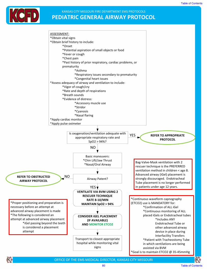
KANSAS CITY MISSOURI FIRE DEPARTMENT EMS PROTOCOLS
PEDIATRIC GENERAL AIRWAY PROTOCOL
OFFICE OF THE EMS MEDICAL DIRECTOR, KANSAS CITY MISSOURI
ASSESSMENT:
*Obtain vital signs
*Obtain brief history to include:
*Onset
*Potential aspiration of small objects or food
*Fever or cough
*Chest pain
*Past history of prior respiratory, cardiac problems, or
prematurity
*Asthma
*Respiratory issues secondary to prematurity
*Congenital heart issues
*Assess adequacy of airway and ventilation to include:
*Vigor of cough/cry
*Rate and depth of respirations
*Breath sounds
*Evidence of distress:
*Accessory muscle use
*Stridor
*Cyanosis
*Nasal flaring
*Apply cardiac monitor
*Apply pulse oximeter
Is oxygenation/ventilation adequate with
appropriate respiratory rate and
SpO2 > 94%?
REFER TO OBSTRUCTED
AIRWAY PROTOCOL
Basic maneuvers:
*Chin Lift/Jaw Thrust
*Nasal/Oral Airway
Airway Patent?
Transport to closest appropriate
hospital while monitoring vital
signs
*Continuous waveform capnography
(ETCO2) use is MANDATORY for:
*Confirmation of ALL iGel
*Continuous monitoring of ALL
placed iGels or Endotracheal tubes
*Includes ANY
Endotracheal Tube or
other advanced airway
device in place during
Interfacility Transfers
*Patient with Tracheostomy Tube
in which ventilations are being
assisted via BVM
*Goal is to maintain ETCO2 @ 35-45mmHg
*Proper positioning and preparation is
necessary before an attempt at
advanced airway placement is made
*The following is considered an
attempt at advanced airway placement
*iGel passing beyond the teeth
is considered a placement
attempt
NO
Bag-Valve-Mask ventilation with 2
rescuer technique is the PREFERRED
ventilation method in children < age 8.
Advanced airway (iGel) placement is
strongly discouraged. Endotracheal
Tube placement is no longer performed
in patients under age 12 years.YESVENTILATE VIA BVM USING 2
RESCUER TECHNIQUE
RATE 8-10/MIN
MAINTAIN SpO2 > 94%
CONSIDER iGEL PLACEMENT
(IF AVAILABLE)
AND MONITOR ETCO2
REFER TO APPROPRIATE
PROTOCOL
YES
NO
Table of Contents
80 Table of Contents
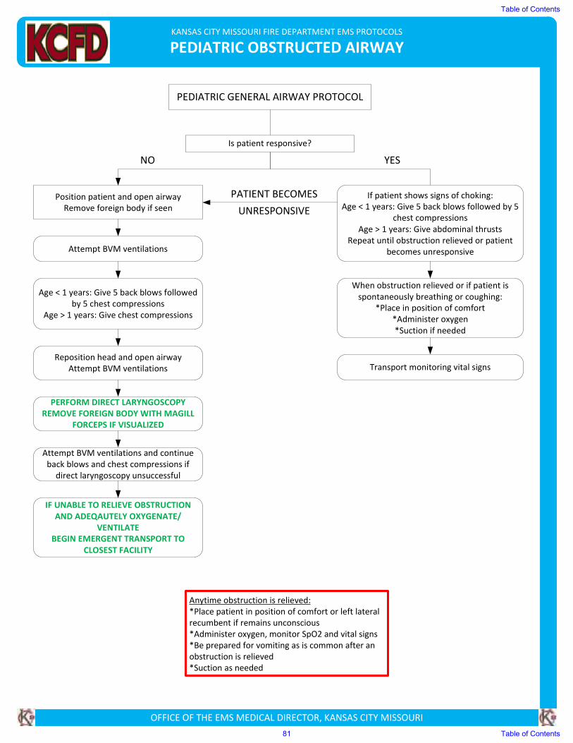
KANSAS CITY MISSOURI FIRE DEPARTMENT EMS PROTOCOLS
PEDIATRIC OBSTRUCTED AIRWAY
OFFICE OF THE EMS MEDICAL DIRECTOR, KANSAS CITY MISSOURI
PEDIATRIC GENERAL AIRWAY PROTOCOL
Is patient responsive?
If patient shows signs of choking:
Age < 1 years: Give 5 back blows followed by 5
chest compressions
Age > 1 years: Give abdominal thrusts
Repeat until obstruction relieved or patient
becomes unresponsive
Position patient and open airway
Remove foreign body if seen
Reposition head and open airway
Attempt BVM ventilations Transport monitoring vital signs
IF UNABLE TO RELIEVE OBSTRUCTION
AND ADEQAUTELY OXYGENATE/
VENTILATE
BEGIN EMERGENT TRANSPORT TO
CLOSEST FACILITY
PERFORM DIRECT LARYNGOSCOPY
REMOVE FOREIGN BODY WITH MAGILL
FORCEPS IF VISUALIZED
Attempt BVM ventilations
Attempt BVM ventilations and continue
back blows and chest compressions if
direct laryngoscopy unsuccessful
Age < 1 years: Give 5 back blows followed
by 5 chest compressions
Age > 1 years: Give chest compressions
Anytime obstruction is relieved:
*Place patient in position of comfort or left lateral recumbent if remains unconscious
*Administer oxygen, monitor SpO2 and vital signs
*Be prepared for vomiting as is common after an obstruction is relieved
*Suction as needed
When obstruction relieved or if patient is
spontaneously breathing or coughing:
*Place in position of comfort
*Administer oxygen
*Suction if needed
PATIENT BECOMES
UNRESPONSIVE
NO YES
Table of Contents
81 Table of Contents
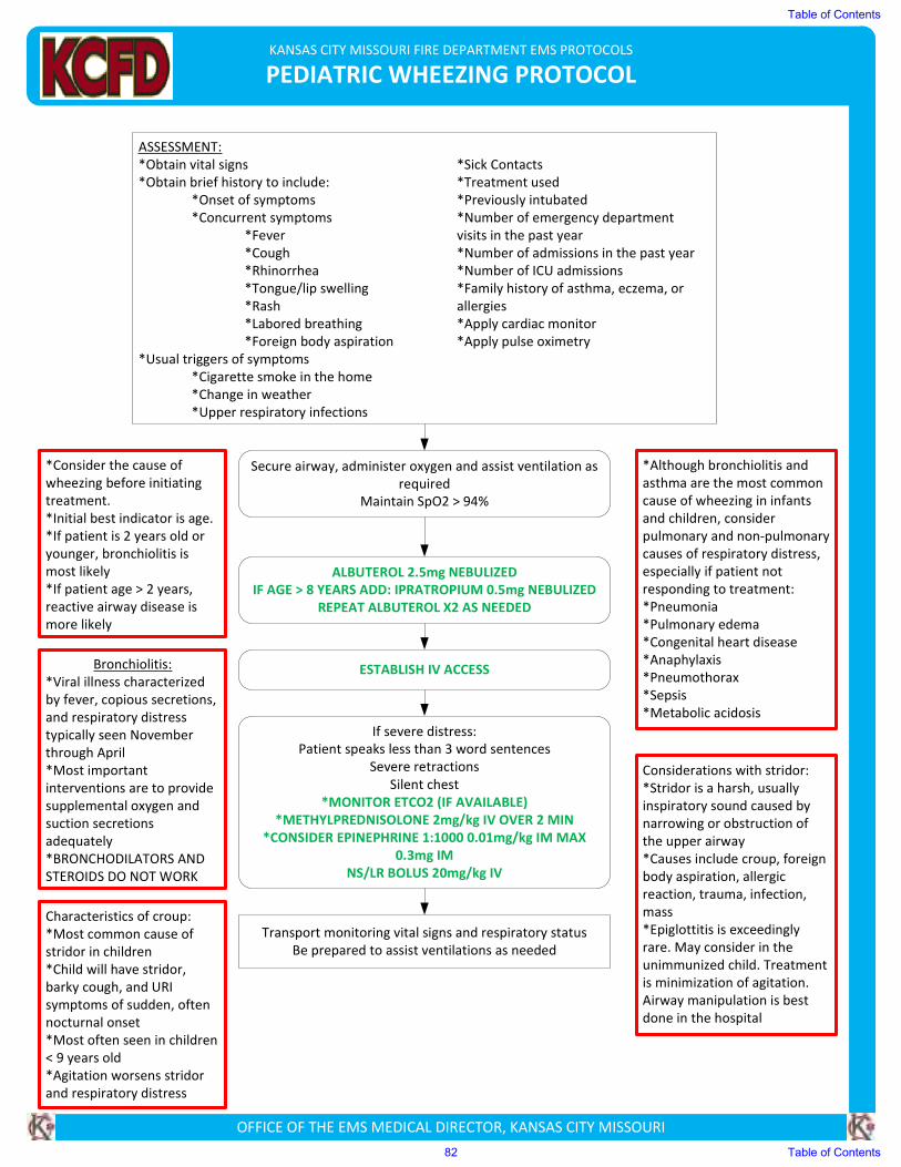
KANSAS CITY MISSOURI FIRE DEPARTMENT EMS PROTOCOLS
PEDIATRIC WHEEZING PROTOCOL
OFFICE OF THE EMS MEDICAL DIRECTOR, KANSAS CITY MISSOURI
ASSESSMENT:
*Obtain vital signs *Sick Contacts
*Obtain brief history to include: *Treatment used
*Onset of symptoms *Previously intubated
*Concurrent symptoms *Number of emergency department
*Fever visits in the past year
*Cough *Number of admissions in the past year
*Rhinorrhea *Number of ICU admissions
*Tongue/lip swelling *Family history of asthma, eczema, or
*Rash allergies
*Labored breathing *Apply cardiac monitor
*Foreign body aspiration *Apply pulse oximetry
*Usual triggers of symptoms
*Cigarette smoke in the home
*Change in weather
*Upper respiratory infections
Secure airway, administer oxygen and assist ventilation as
required
Maintain SpO2 > 94%
ESTABLISH IV ACCESS
ALBUTEROL 2.5mg NEBULIZED
IF AGE > 8 YEARS ADD: IPRATROPIUM 0.5mg NEBULIZED
REPEAT ALBUTEROL X2 AS NEEDED
If severe distress:
Patient speaks less than 3 word sentences
Severe retractions
Silent chest
*MONITOR ETCO2 (IF AVAILABLE)
*METHYLPREDNISOLONE 2mg/kg IV OVER 2 MIN
*CONSIDER EPINEPHRINE 1:1000 0.01mg/kg IM MAX
0.3mg IM
NS/LR BOLUS 20mg/kg IV
Transport monitoring vital signs and respiratory status
Be prepared to assist ventilations as needed
*Consider the cause of
wheezing before initiating
treatment.
*Initial best indicator is age.
*If patient is 2 years old or
younger, bronchiolitis is
most likely
*If patient age > 2 years,
reactive airway disease is
more likely
Bronchiolitis:
*Viral illness characterized
by fever, copious secretions,
and respiratory distress
typically seen November
through April
*Most important
interventions are to provide
supplemental oxygen and
suction secretions
adequately
*BRONCHODILATORS AND
STEROIDS DO NOT WORK
*Although bronchiolitis and
asthma are the most common
cause of wheezing in infants
and children, consider
pulmonary and non-pulmonary
causes of respiratory distress,
especially if patient not
responding to treatment:
*Pneumonia
*Pulmonary edema
*Congenital heart disease
*Anaphylaxis
*Pneumothorax
*Sepsis
*Metabolic acidosis
Considerations with stridor:
*Stridor is a harsh, usually
inspiratory sound caused by
narrowing or obstruction of
the upper airway
*Causes include croup, foreign
body aspiration, allergic
reaction, trauma, infection,
mass
*Epiglottitis is exceedingly
rare. May consider in the
unimmunized child. Treatment
is minimization of agitation.
Airway manipulation is best
done in the hospital
Characteristics of croup:
*Most common cause of
stridor in children
*Child will have stridor,
barky cough, and URI
symptoms of sudden, often
nocturnal onset
*Most often seen in children
< 9 years old
*Agitation worsens stridor
and respiratory distress
Table of Contents
82 Table of Contents
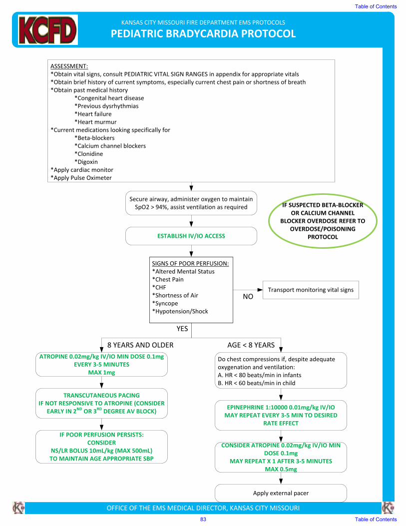
KANSAS CITY MISSOURI FIRE DEPARTMENT EMS PROTOCOLS
PEDIATRIC BRADYCARDIA PROTOCOL
OFFICE OF THE EMS MEDICAL DIRECTOR, KANSAS CITY MISSOURI
ASSESSMENT:
*Obtain vital signs, consult PEDIATRIC VITAL SIGN RANGES in appendix for appropriate vitals
*Obtain brief history of current symptoms, especially current chest pain or shortness of breath
*Obtain past medical history
*Congenital heart disease
*Previous dysrhythmias
*Heart failure
*Heart murmur
*Current medications looking specifically for
*Beta-blockers
*Calcium channel blockers
*Clonidine
*Digoxin
*Apply cardiac monitor
*Apply Pulse Oximeter
SIGNS OF POOR PERFUSION:
*Altered Mental Status
*Chest Pain
*CHF
*Shortness of Air
*Syncope
*Hypotension/Shock
Secure airway, administer oxygen to maintain
SpO2 > 94%, assist ventilation as required
ESTABLISH IV/IO ACCESS
ATROPINE 0.02mg/kg IV/IO MIN DOSE 0.1mg
EVERY 3-5 MINUTES
MAX 1mg
TRANSCUTANEOUS PACING
IF NOT RESPONSIVE TO ATROPINE (CONSIDER
EARLY IN 2ND
OR 3RD
DEGREE AV BLOCK)
IF POOR PERFUSION PERSISTS:
CONSIDER
NS/LR BOLUS 10mL/kg (MAX 500mL)
TO MAINTAIN AGE APPROPRIATE SBP
Transport monitoring vital signs
IF SUSPECTED BETA-BLOCKER
OR CALCIUM CHANNEL
BLOCKER OVERDOSE REFER TO
OVERDOSE/POISONING
PROTOCOL
YES
NO
Do chest compressions if, despite adequate
oxygenation and ventilation:
A. HR < 80 beats/min in infants
B. HR < 60 beats/min in child
EPINEPHRINE 1:10000 0.01mg/kg IV/IO
MAY REPEAT EVERY 3-5 MIN TO DESIRED
RATE EFFECT
CONSIDER ATROPINE 0.02mg/kg IV/IO MIN
DOSE 0.1mg
MAY REPEAT X 1 AFTER 3-5 MINUTES
MAX 0.5mg
Apply external pacer
8 YEARS AND OLDER AGE < 8 YEARS
Table of Contents
83 Table of Contents
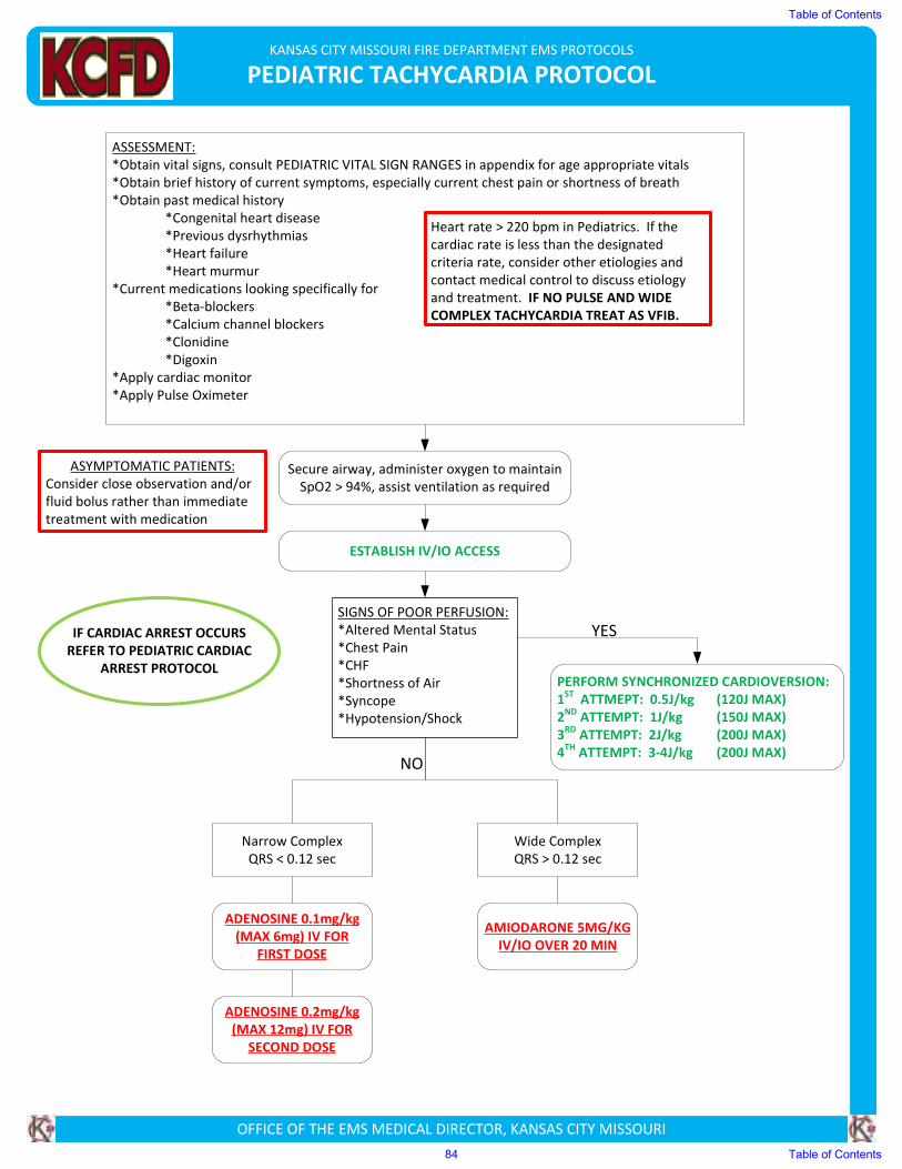
KANSAS CITY MISSOURI FIRE DEPARTMENT EMS PROTOCOLS
PEDIATRIC TACHYCARDIA PROTOCOL
OFFICE OF THE EMS MEDICAL DIRECTOR, KANSAS CITY MISSOURI
ASSESSMENT:
*Obtain vital signs, consult PEDIATRIC VITAL SIGN RANGES in appendix for age appropriate vitals
*Obtain brief history of current symptoms, especially current chest pain or shortness of breath
*Obtain past medical history
*Congenital heart disease
*Previous dysrhythmias
*Heart failure
*Heart murmur
*Current medications looking specifically for
*Beta-blockers
*Calcium channel blockers
*Clonidine
*Digoxin
*Apply cardiac monitor
*Apply Pulse Oximeter
SIGNS OF POOR PERFUSION:
*Altered Mental Status
*Chest Pain
*CHF
*Shortness of Air
*Syncope
*Hypotension/Shock
Secure airway, administer oxygen to maintain
SpO2 > 94%, assist ventilation as required
ESTABLISH IV/IO ACCESS
Narrow Complex
QRS < 0.12 sec
Wide Complex
QRS > 0.12 sec
ADENOSINE 0.1mg/kg
(MAX 6mg) IV FOR
FIRST DOSE
AMIODARONE 5MG/KG
IV/IO OVER 20 MIN
ASYMPTOMATIC PATIENTS:
Consider close observation and/or
fluid bolus rather than immediate
treatment with medication
NO
YES
Heart rate > 220 bpm in Pediatrics. If the
cardiac rate is less than the designated
criteria rate, consider other etiologies and
contact medical control to discuss etiology
and treatment. IF NO PULSE AND WIDE
COMPLEX TACHYCARDIA TREAT AS VFIB.
PERFORM SYNCHRONIZED CARDIOVERSION:
1ST
ATTMEPT: 0.5J/kg (120J MAX)
2ND
ATTEMPT: 1J/kg (150J MAX)
3RD
ATTEMPT: 2J/kg (200J MAX)
4TH
ATTEMPT: 3-4J/kg (200J MAX)
ADENOSINE 0.2mg/kg
(MAX 12mg) IV FOR
SECOND DOSE
IF CARDIAC ARREST OCCURS
REFER TO PEDIATRIC CARDIAC
ARREST PROTOCOL
Table of Contents
84 Table of Contents
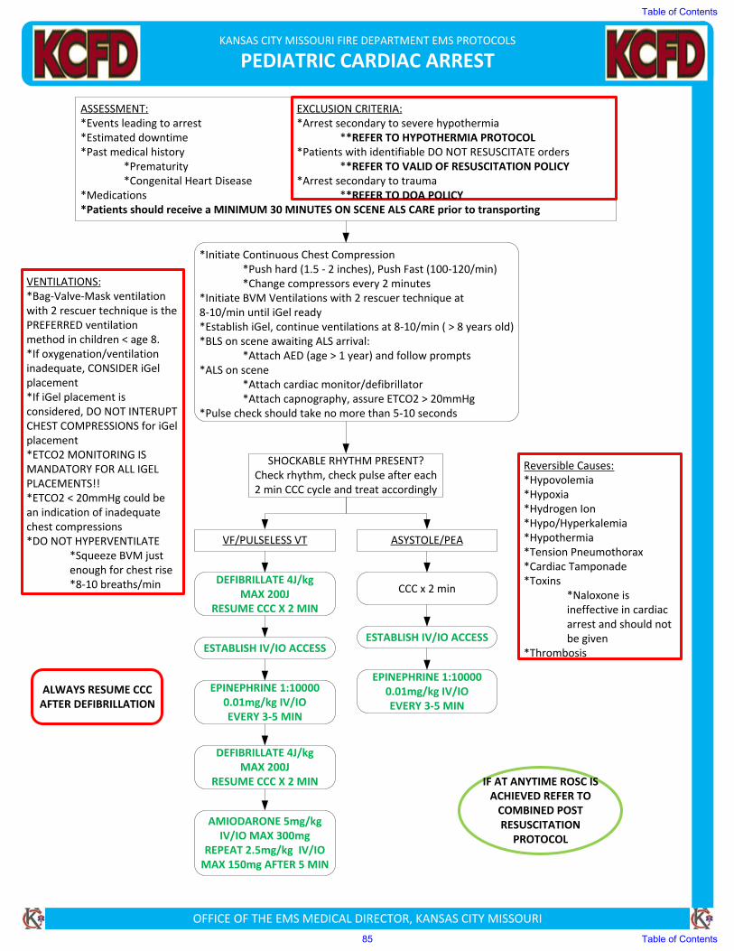
KANSAS CITY MISSOURI FIRE DEPARTMENT EMS PROTOCOLS
PEDIATRIC CARDIAC ARREST
OFFICE OF THE EMS MEDICAL DIRECTOR, KANSAS CITY MISSOURI
ASSESSMENT:*Events leading to arrest*Estimated downtime*Past medical history
*Prematurity*Congenital Heart Disease
*Medications*Patients should receive a MINIMUM 30 MINUTES ON SCENE ALS CARE prior to transporting
EXCLUSION CRITERIA:*Arrest secondary to severe hypothermia
**REFER TO HYPOTHERMIA PROTOCOL*Patients with identifiable DO NOT RESUSCITATE orders
**REFER TO VALID OF RESUSCITATION POLICY*Arrest secondary to trauma
**REFER TO DOA POLICY
*Initiate Continuous Chest Compression*Push hard (1.5 ‐ 2 inches), Push Fast (100‐120/min)*Change compressors every 2 minutes
*Initiate BVM Ventilations with 2 rescuer technique at 8‐10/min until iGel ready*Establish iGel, continue ventilations at 8‐10/min ( > 8 years old) *BLS on scene awaiting ALS arrival:
*Attach AED (age > 1 year) and follow prompts*ALS on scene
*Attach cardiac monitor/defibrillator*Attach capnography, assure ETCO2 > 20mmHg
*Pulse check should take no more than 5‐10 seconds
SHOCKABLE RHYTHM PRESENT?Check rhythm, check pulse after each 2 min CCC cycle and treat accordingly
VF/PULSELESS VT ASYSTOLE/PEA
DEFIBRILLATE 4J/kg MAX 200J
RESUME CCC X 2 MIN
CCC x 2 min
ESTABLISH IV/IO ACCESSESTABLISH IV/IO ACCESS
EPINEPHRINE 1:10000 0.01mg/kg IV/IOEVERY 3‐5 MIN
EPINEPHRINE 1:10000 0.01mg/kg IV/IOEVERY 3‐5 MIN
AMIODARONE 5mg/kg IV/IO MAX 300mg
REPEAT 2.5mg/kg IV/IO MAX 150mg AFTER 5 MIN
VENTILATIONS:*Bag‐Valve‐Mask ventilation with 2 rescuer technique is the PREFERRED ventilation method in children < age 8.*If oxygenation/ventilation inadequate, CONSIDER iGel placement*If iGel placement is considered, DO NOT INTERUPT CHEST COMPRESSIONS for iGel placement*ETCO2 MONITORING IS MANDATORY FOR ALL IGEL PLACEMENTS!!*ETCO2 < 20mmHg could be an indication of inadequate chest compressions*DO NOT HYPERVENTILATE
*Squeeze BVM just enough for chest rise*8‐10 breaths/min
Reversible Causes:*Hypovolemia *Hypoxia*Hydrogen Ion *Hypo/Hyperkalemia *Hypothermia*Tension Pneumothorax*Cardiac Tamponade*Toxins
*Naloxone is ineffective in cardiac arrest and should not be given
*Thrombosis
IF AT ANYTIME ROSC IS ACHIEVED REFER TO COMBINED POST RESUSCITATION
PROTOCOL
ALWAYS RESUME CCC AFTER DEFIBRILLATION
DEFIBRILLATE 4J/kg MAX 200J
RESUME CCC X 2 MIN
Table of Contents
85 Table of Contents
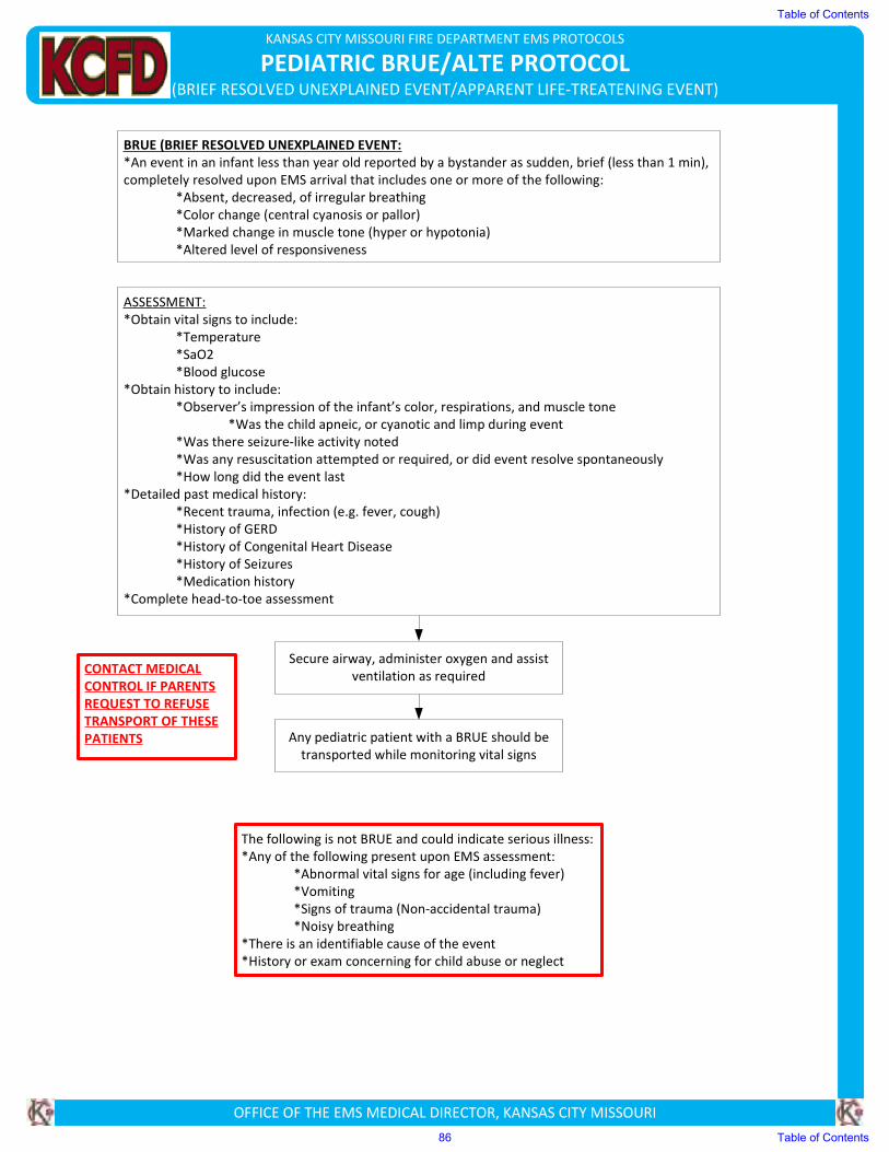
KANSAS CITY MISSOURI FIRE DEPARTMENT EMS PROTOCOLS
PEDIATRIC BRUE/ALTE PROTOCOL(BRIEF RESOLVED UNEXPLAINED EVENT/APPARENT LIFE-TREATENING EVENT)
OFFICE OF THE EMS MEDICAL DIRECTOR, KANSAS CITY MISSOURI
BRUE (BRIEF RESOLVED UNEXPLAINED EVENT:
*An event in an infant less than year old reported by a bystander as sudden, brief (less than 1 min),
completely resolved upon EMS arrival that includes one or more of the following:
*Absent, decreased, of irregular breathing
*Color change (central cyanosis or pallor)
*Marked change in muscle tone (hyper or hypotonia)
*Altered level of responsiveness
ASSESSMENT:
*Obtain vital signs to include:
*Temperature
*SaO2
*Blood glucose
*Obtain history to include:
*Observer’s impression of the infant’s color, respirations, and muscle tone
*Was the child apneic, or cyanotic and limp during event
*Was there seizure-like activity noted
*Was any resuscitation attempted or required, or did event resolve spontaneously
*How long did the event last
*Detailed past medical history:
*Recent trauma, infection (e.g. fever, cough)
*History of GERD
*History of Congenital Heart Disease
*History of Seizures
*Medication history
*Complete head-to-toe assessment
Secure airway, administer oxygen and assist
ventilation as required
Any pediatric patient with a BRUE should be
transported while monitoring vital signs
The following is not BRUE and could indicate serious illness:
*Any of the following present upon EMS assessment:
*Abnormal vital signs for age (including fever)
*Vomiting
*Signs of trauma (Non-accidental trauma)
*Noisy breathing
*There is an identifiable cause of the event
*History or exam concerning for child abuse or neglect
CONTACT MEDICAL
CONTROL IF PARENTS
REQUEST TO REFUSE
TRANSPORT OF THESE
PATIENTS
Table of Contents
86 Table of Contents
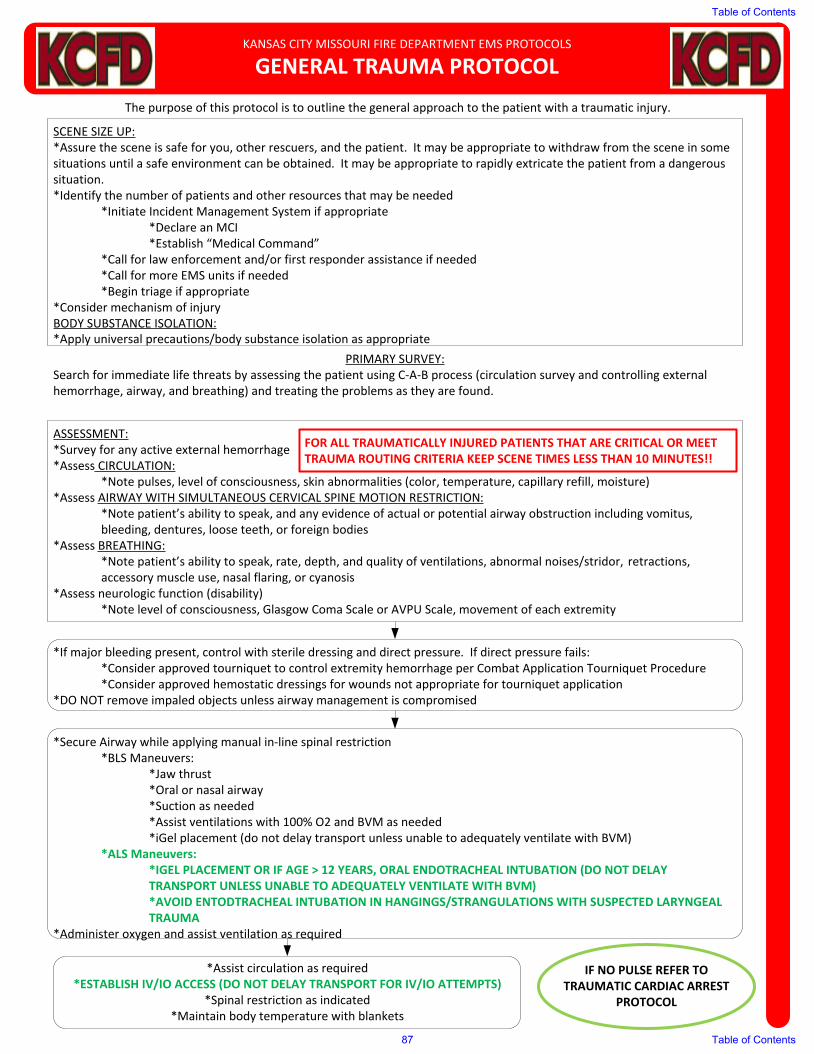
KANSAS CITY MISSOURI FIRE DEPARTMENT EMS PROTOCOLS
GENERAL TRAUMA PROTOCOL
The purpose of this protocol is to outline the general approach to the patient with a traumatic injury.
SCENE SIZE UP:*Assure the scene is safe for you, other rescuers, and the patient. It may be appropriate to withdraw from the scene in some situations until a safe environment can be obtained. It may be appropriate to rapidly extricate the patient from a dangerous situation.*Identify the number of patients and other resources that may be needed
*Initiate Incident Management System if appropriate*Declare an MCI*Establish “Medical Command”
*Call for law enforcement and/or first responder assistance if needed*Call for more EMS units if needed*Begin triage if appropriate
*Consider mechanism of injuryBODY SUBSTANCE ISOLATION:*Apply universal precautions/body substance isolation as appropriate
PRIMARY SURVEY:Search for immediate life threats by assessing the patient using C-A-B process (circulation survey and controlling external hemorrhage, airway, and breathing) and treating the problems as they are found.
ASSESSMENT:*Survey for any active external hemorrhage*Assess CIRCULATION:
*Note pulses, level of consciousness, skin abnormalities (color, temperature, capillary refill, moisture)*Assess AIRWAY WITH SIMULTANEOUS CERVICAL SPINE MOTION RESTRICTION:
*Note patient’s ability to speak, and any evidence of actual or potential airway obstruction including vomitus, bleeding, dentures, loose teeth, or foreign bodies
*Assess BREATHING:*Note patient’s ability to speak, rate, depth, and quality of ventilations, abnormal noises/stridor, retractions, accessory muscle use, nasal flaring, or cyanosis
*Assess neurologic function (disability)*Note level of consciousness, Glasgow Coma Scale or AVPU Scale, movement of each extremity
*If major bleeding present, control with sterile dressing and direct pressure. If direct pressure fails:*Consider approved tourniquet to control extremity hemorrhage per Combat Application Tourniquet Procedure*Consider approved hemostatic dressings for wounds not appropriate for tourniquet application
*DO NOT remove impaled objects unless airway management is compromised
*Secure Airway while applying manual in-line spinal restriction*BLS Maneuvers:
*Jaw thrust*Oral or nasal airway*Suction as needed*Assist ventilations with 100% O2 and BVM as needed*iGel placement (do not delay transport unless unable to adequately ventilate with BVM)
*ALS Maneuvers:*IGEL PLACEMENT OR IF AGE > 12 YEARS, ORAL ENDOTRACHEAL INTUBATION (DO NOT DELAY TRANSPORT UNLESS UNABLE TO ADEQUATELY VENTILATE WITH BVM)*AVOID ENTODTRACHEAL INTUBATION IN HANGINGS/STRANGULATIONS WITH SUSPECTED LARYNGEAL TRAUMA
*Administer oxygen and assist ventilation as required
*Assist circulation as required*ESTABLISH IV/IO ACCESS (DO NOT DELAY TRANSPORT FOR IV/IO ATTEMPTS)
*Spinal restriction as indicated*Maintain body temperature with blankets
IF NO PULSE REFER TO TRAUMATIC CARDIAC ARREST
PROTOCOL
FOR ALL TRAUMATICALLY INJURED PATIENTS THAT ARE CRITICAL OR MEET TRAUMA ROUTING CRITERIA KEEP SCENE TIMES LESS THAN 10 MINUTES!!
Table of Contents
87 Table of Contents
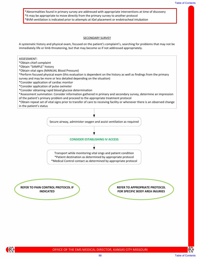
OFFICE OF THE EMS MEDICAL DIRECTOR, KANSAS CITY MISSOURI
A systematic history and physical exam, focused on the patient’s complaint’s, searching for problems that may not be immediately life or limb threatening, but that may become so if not addressed appropriately.
SECONDARY SURVEY
ASSESSMENT:*Obtain chief complaint*Obtain “SAMPLE” history*Obtain vital signs (MANUAL Blood Pressure)*Perform focused physical exam (this evaluation is dependent on the history as well as findings from the primary survey and may be more or less detailed depending on the situation)*Consider application of cardiac monitor*Consider application of pulse oximeter*Consider obtaining rapid blood glucose determination*Assessment summation: Consider information gathered in primary and secondary survey, determine an impression of the patient’s primary problem and proceed to the appropriate treatment protocol*Obtain repeat set of vital signs prior to transfer of care to receiving facility or whenever there is an observed change in the patient’s status
Secure airway, administer oxygen and assist ventilation as required
CONSIDER ESTABLISHING IV ACCESS
*Abnormalities found in primary survey are addressed with appropriate interventions at time of discovery*It may be appropriate to move directly from the primary survey to another protocol*BVM ventilation is indicated prior to attempts at iGel placement or endotracheal intubation
Transport while monitoring vital sings and patient condition*Patient destination as determined by appropriate protocol
*Medical Control contact as determined by appropriate protocol
REFER TO APPROPRIATE PROTOCOL FOR SPECIFIC BODY AREA INJURIES
REFER TO PAIN CONTROL PROTOCOL IF INDICATED
Table of Contents
88 Table of Contents
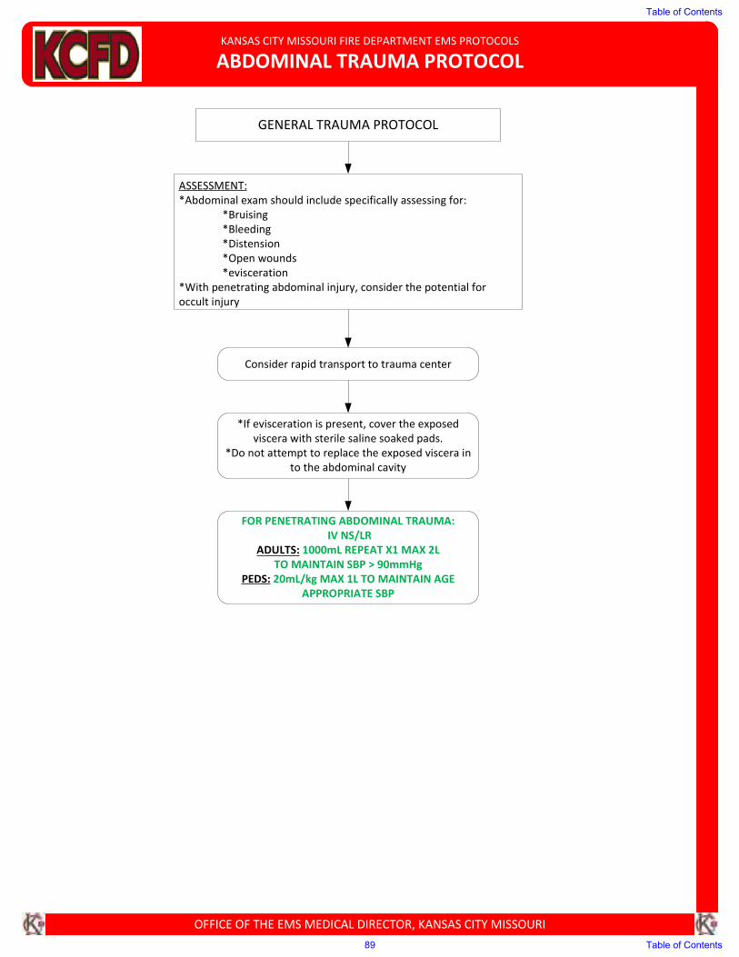
KANSAS CITY MISSOURI FIRE DEPARTMENT EMS PROTOCOLS
ABDOMINAL TRAUMA PROTOCOL
OFFICE OF THE EMS MEDICAL DIRECTOR, KANSAS CITY MISSOURI
ASSESSMENT:
*Abdominal exam should include specifically assessing for:
*Bruising
*Bleeding
*Distension
*Open wounds
*evisceration
*With penetrating abdominal injury, consider the potential for
occult injury
GENERAL TRAUMA PROTOCOL
Consider rapid transport to trauma center
*If evisceration is present, cover the exposed viscera with sterile saline soaked pads.
*Do not attempt to replace the exposed viscera in
to the abdominal cavity
FOR PENETRATING ABDOMINAL TRAUMA:
IV NS/LR
ADULTS: 1000mL REPEAT X1 MAX 2L
TO MAINTAIN SBP > 90mmHg
PEDS: 20mL/kg MAX 1L TO MAINTAIN AGE
APPROPRIATE SBP
Table of Contents
89 Table of Contents
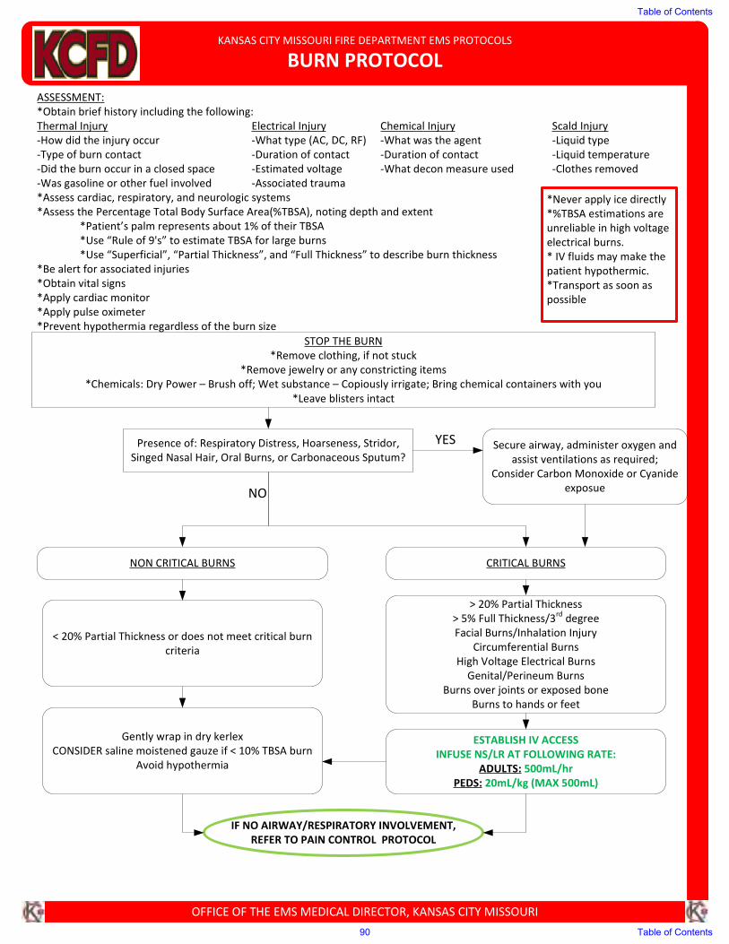
KANSAS CITY MISSOURI FIRE DEPARTMENT EMS PROTOCOLS
BURN PROTOCOL
ASSESSMENT:
*Obtain brief history including the following:
Thermal Injury Electrical Injury Chemical Injury Scald Injury
-How did the injury occur -What type (AC, DC, RF) -What was the agent -Liquid type
-Type of burn contact -Duration of contact -Duration of contact -Liquid temperature
-Did the burn occur in a closed space -Estimated voltage -What decon measure used -Clothes removed
-Was gasoline or other fuel involved -Associated trauma
*Assess cardiac, respiratory, and neurologic systems
*Assess the Percentage Total Body Surface Area(%TBSA), noting depth and extent
*Patient’s palm represents about 1% of their TBSA
*Use “Rule of 9's” to estimate TBSA for large burns
*Use “Superficial”, “Partial Thickness”, and “Full Thickness” to describe burn thickness
*Be alert for associated injuries
*Obtain vital signs
*Apply cardiac monitor
*Apply pulse oximeter
*Prevent hypothermia regardless of the burn size
ESTABLISH IV ACCESS
INFUSE NS/LR AT FOLLOWING RATE:
ADULTS: 500mL/hr
PEDS: 20mL/kg (MAX 500mL)
IF NO AIRWAY/RESPIRATORY INVOLVEMENT,
REFER TO PAIN CONTROL PROTOCOL
*Never apply ice directly
*%TBSA estimations are
unreliable in high voltage
electrical burns.
* IV fluids may make the
patient hypothermic.
*Transport as soon as
possible
STOP THE BURN
*Remove clothing, if not stuck
*Remove jewelry or any constricting items
*Chemicals: Dry Power – Brush off; Wet substance – Copiously irrigate; Bring chemical containers with you
*Leave blisters intact
Presence of: Respiratory Distress, Hoarseness, Stridor,
Singed Nasal Hair, Oral Burns, or Carbonaceous Sputum?Secure airway, administer oxygen and
assist ventilations as required;
Consider Carbon Monoxide or Cyanide
exposue
NON CRITICAL BURNS CRITICAL BURNS
< 20% Partial Thickness or does not meet critical burn
criteria
> 20% Partial Thickness
> 5% Full Thickness/3rd
degree
Facial Burns/Inhalation Injury
Circumferential Burns
High Voltage Electrical Burns
Genital/Perineum Burns
Burns over joints or exposed bone
Burns to hands or feet
Gently wrap in dry kerlex
CONSIDER saline moistened gauze if < 10% TBSA burn
Avoid hypothermia
YES
NO
OFFICE OF THE EMS MEDICAL DIRECTOR, KANSAS CITY MISSOURI
Table of Contents
90 Table of Contents
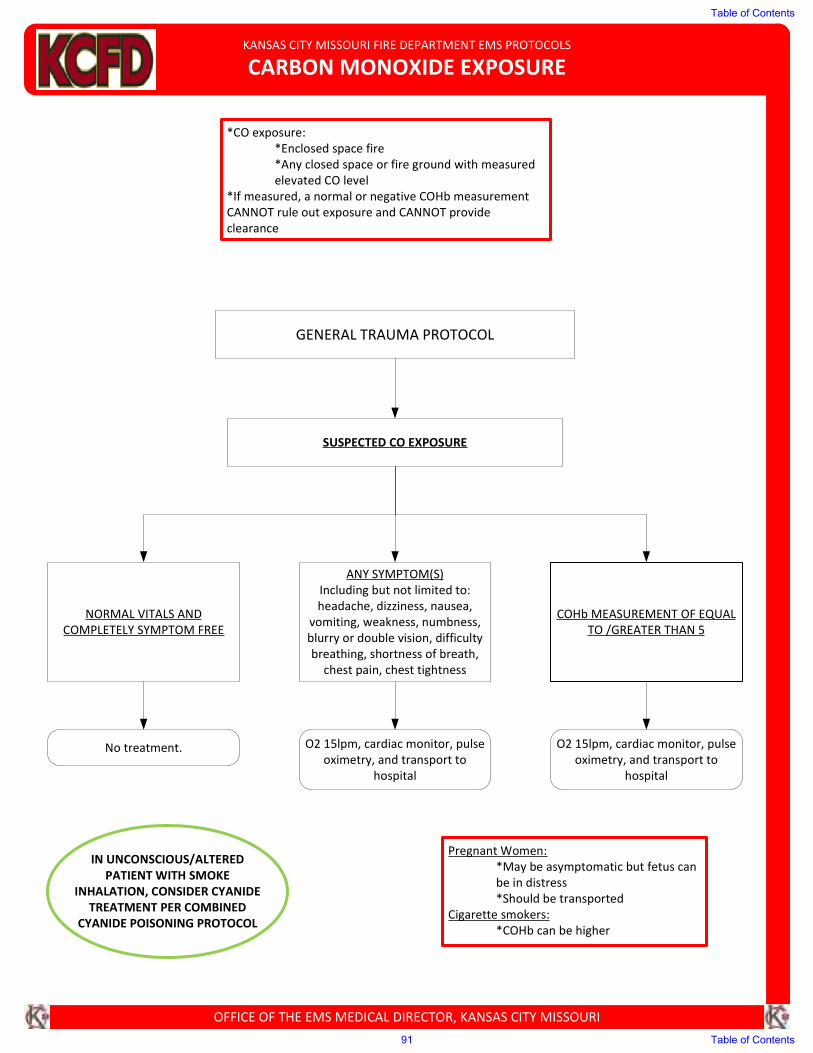
KANSAS CITY MISSOURI FIRE DEPARTMENT EMS PROTOCOLS
CARBON MONOXIDE EXPOSURE
OFFICE OF THE EMS MEDICAL DIRECTOR, KANSAS CITY MISSOURI
*CO exposure:
*Enclosed space fire
*Any closed space or fire ground with measured
elevated CO level
*If measured, a normal or negative COHb measurement
CANNOT rule out exposure and CANNOT provide
clearance
SUSPECTED CO EXPOSURE
NORMAL VITALS AND
COMPLETELY SYMPTOM FREE
ANY SYMPTOM(S)
Including but not limited to:
headache, dizziness, nausea,
vomiting, weakness, numbness,
blurry or double vision, difficulty
breathing, shortness of breath,
chest pain, chest tightness
COHb MEASUREMENT OF EQUAL
TO /GREATER THAN 5
No treatment. O2 15lpm, cardiac monitor, pulse
oximetry, and transport to
hospital
O2 15lpm, cardiac monitor, pulse
oximetry, and transport to
hospital
GENERAL TRAUMA PROTOCOL
IN UNCONSCIOUS/ALTERED
PATIENT WITH SMOKE
INHALATION, CONSIDER CYANIDE
TREATMENT PER COMBINED CYANIDE POISONING PROTOCOL
Pregnant Women:
*May be asymptomatic but fetus can
be in distress
*Should be transported
Cigarette smokers:
*COHb can be higher
Table of Contents
91 Table of Contents
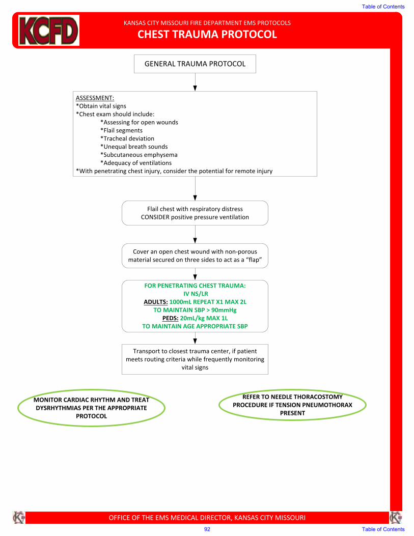
KANSAS CITY MISSOURI FIRE DEPARTMENT EMS PROTOCOLS
CHEST TRAUMA PROTOCOL
OFFICE OF THE EMS MEDICAL DIRECTOR, KANSAS CITY MISSOURI
ASSESSMENT:
*Obtain vital signs
*Chest exam should include:
*Assessing for open wounds
*Flail segments
*Tracheal deviation
*Unequal breath sounds
*Subcutaneous emphysema
*Adequacy of ventilations
*With penetrating chest injury, consider the potential for remote injury
Flail chest with respiratory distress
CONSIDER positive pressure ventilation
Cover an open chest wound with non-porous
material secured on three sides to act as a “flap”
FOR PENETRATING CHEST TRAUMA:
IV NS/LR
ADULTS: 1000mL REPEAT X1 MAX 2L
TO MAINTAIN SBP > 90mmHg
PEDS: 20mL/kg MAX 1L
TO MAINTAIN AGE APPROPRIATE SBP
MONITOR CARDIAC RHYTHM AND TREAT
DYSRHYTHMIAS PER THE APPROPRIATE
PROTOCOL
GENERAL TRAUMA PROTOCOL
REFER TO NEEDLE THORACOSTOMY
PROCEDURE IF TENSION PNEUMOTHORAX
PRESENT
Transport to closest trauma center, if patient
meets routing criteria while frequently monitoring
vital signs
Table of Contents
92 Table of Contents
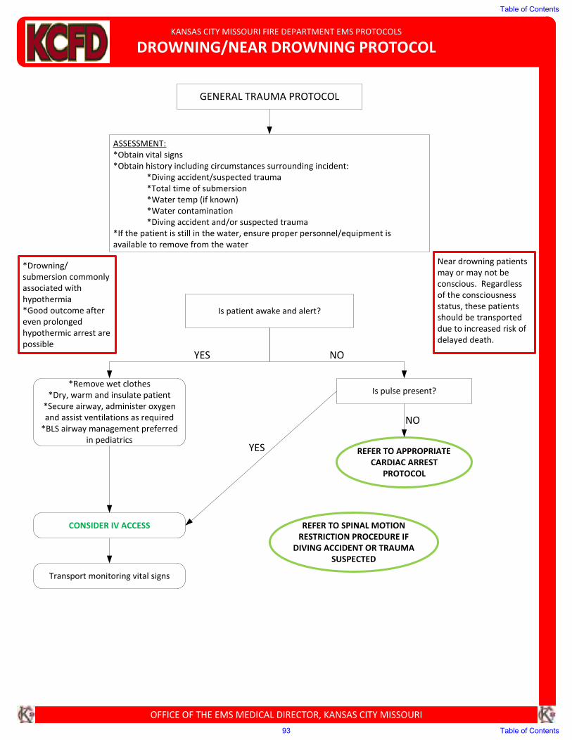
KANSAS CITY MISSOURI FIRE DEPARTMENT EMS PROTOCOLS
DROWNING/NEAR DROWNING PROTOCOL
OFFICE OF THE EMS MEDICAL DIRECTOR, KANSAS CITY MISSOURI
Near drowning patients
may or may not be
conscious. Regardless
of the consciousness
status, these patients
should be transported
due to increased risk of
delayed death.
GENERAL TRAUMA PROTOCOL
ASSESSMENT:
*Obtain vital signs
*Obtain history including circumstances surrounding incident:
*Diving accident/suspected trauma
*Total time of submersion
*Water temp (if known)
*Water contamination
*Diving accident and/or suspected trauma
*If the patient is still in the water, ensure proper personnel/equipment is
available to remove from the water
Is patient awake and alert?
REFER TO SPINAL MOTION
RESTRICTION PROCEDURE IF
DIVING ACCIDENT OR TRAUMA
SUSPECTED
Is pulse present?
Transport monitoring vital signs
*Remove wet clothes
*Dry, warm and insulate patient
*Secure airway, administer oxygen
and assist ventilations as required
*BLS airway management preferred
in pediatrics
CONSIDER IV ACCESS
REFER TO APPROPRIATECARDIAC ARREST
PROTOCOL
*Drowning/
submersion commonly
associated with
hypothermia
*Good outcome after
even prolonged
hypothermic arrest are
possible
YES NO
YES
NO
Table of Contents
93 Table of Contents
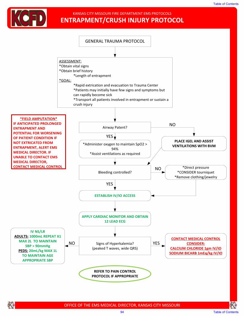
KANSAS CITY MISSOURI FIRE DEPARTMENT EMS PROTOCOLS
ENTRAPMENT/CRUSH INJURY PROTOCOL
OFFICE OF THE EMS MEDICAL DIRECTOR, KANSAS CITY MISSOURI
GENERAL TRAUMA PROTOCOL
ASSESSMENT:
*Obtain vital signs
*Obtain brief history
*Length of entrapment
*GOAL:
*Rapid extrication and evacuation to Trauma Center
*Patients may initially have few signs and symptoms but
can rapidly become sick
*Transport all patients involved in entrapment or sustain a
crush injury
Airway Patent?
PLACE IGEL AND ASSIST
VENTILATIONS WITH BVM
Bleeding controlled?
*Direct pressure
*CONSIDER tourniquet
*Remove clothing/jewelry
ESTABLISH IV/IO ACCESS
*Administer oxygen to maintain SpO2 >
94%
*Assist ventilations as required
APPLY CARDIAC MONITOR AND OBTAIN
12 LEAD ECG
Signs of Hyperkalemia?
(peaked T waves, wide QRS)
CONTACT MEDICAL CONTROL
CONSIDER:
CALCIUM CHLORIDE 1gm IV/IO
SODIUM BICARB 1mEq/kg IV/IO
IV NS/LR
ADULTS: 1000mL REPEAT X1
MAX 2L TO MAINTAIN
SBP > 90mmHg
PEDS: 20mL/kg MAX 1L
TO MAINTAIN AGE
APPROPRIATE SBP
REFER TO PAIN CONTROL
PROTOCOL IF APPROPRIATE
NO
YES
NO
YES
YESNO
*FIELD AMPUTATION*
IF ANTICIPATED PROLONGED
ENTRAPMENT AND
POTENTIAL FOR WORSENING
OF PATIENT CONDITION IF
NOT EXTRICATED FROM
ENTRAPMENT, ALERT EMS
MEDICAL DIRECTOR. IF
UNABLE TO CONTACT EMS
MEDICAL DIRECTOR,
CONTACT MEDICAL CONTROL
Table of Contents
94 Table of Contents
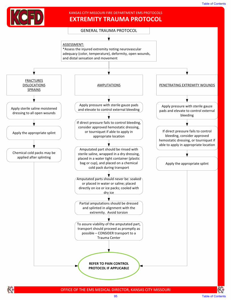
KANSAS CITY MISSOURI FIRE DEPARTMENT EMS PROTOCOLS
EXTREMITY TRAUMA PROTOCOL
OFFICE OF THE EMS MEDICAL DIRECTOR, KANSAS CITY MISSOURI
ASSESSMENT:
*Assess the injured extremity noting neurovascular
adequacy (color, temperature), deformity, open wounds,
and distal sensation and movement
FRACTURES
DISLOCATIONS
SPRAINS
AMPUTATIONS PENETRATING EXTREMITY WOUNDS
Apply sterile saline moistened
dressing to all open wounds
Apply the appropriate splint
Chemical cold packs may be
applied after splinting
Apply pressure with sterile gauze pads
and elevate to control external bleeding
If direct pressure fails to control bleeding,
consider approved hemostatic dressing,
or tourniquet if able to apply in
appropriate location
Amputated part should be rinsed with
sterile saline, wrapped in a dry dressing,
placed in a water tight container (plastic
bag or cup), and placed on a chemical
cold pack during transport
Amputated parts should never be: soaked
or placed in water or saline; placed
directly on ice or ice packs; cooled with
dry ice
Partial amputations should be dressed
and splinted in alignment with the
extremity. Avoid torsion
To assure viability of the amputated part,
transport should proceed as promptly as
possible – CONSIDER transport to a
Trauma Center
Apply pressure with sterile gauze
pads and elevate to control external
bleeding
If direct pressure fails to control
bleeding, consider approved
hemostatic dressing, or tourniquet if
able to apply in appropriate location
Apply the appropriate splint
GENERAL TRAUMA PROTOCOL
REFER TO PAIN CONTROL
PROTOCOL IF APPLICABLE
Table of Contents
95 Table of Contents
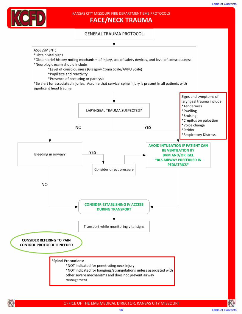
KANSAS CITY MISSOURI FIRE DEPARTMENT EMS PROTOCOLS
FACE/NECK TRAUMA
OFFICE OF THE EMS MEDICAL DIRECTOR, KANSAS CITY MISSOURI
ASSESSMENT:
*Obtain vital signs
*Obtain brief history noting mechanism of injury, use of safety devices, and level of consciousness
*Neurologic exam should include
*Level of consciousness (Glasgow Coma Scale/AVPU Scale)
*Pupil size and reactivity
*Presence of posturing or paralysis
*Be alert for associated injuries. Assume that cervical spine injury is present in all patients with
significant head trauma
GENERAL TRAUMA PROTOCOL
LARYNGEAL TRAUMA SUSPECTED?
Signs and symptoms of
laryngeal trauma include:
*Tenderness
*Swelling
*Bruising
*Crepitus on palpation
*Voice change
*Stridor
*Respiratory Distress
Bleeding in airway?
AVOID INTUBATION IF PATIENT CAN
BE VENTILATION BY
BVM AND/OR IGEL
*BLS AIRWAY PREFERRED IN
PEDIATRICS*
Consider direct pressure
CONSIDER ESTABLISHING IV ACCESS
DURING TRANSPORT
CONSIDER REFERING TO PAIN
CONTROL PROTOCOL IF NEEDED
NO YES
NO
YES
*Spinal Precautions:
*NOT indicated for penetrating neck injury
*NOT indicated for hangings/strangulations unless associated with
other severe mechanisms and does not prevent airway
management
Transport while monitoring vital signs
Table of Contents
96 Table of Contents
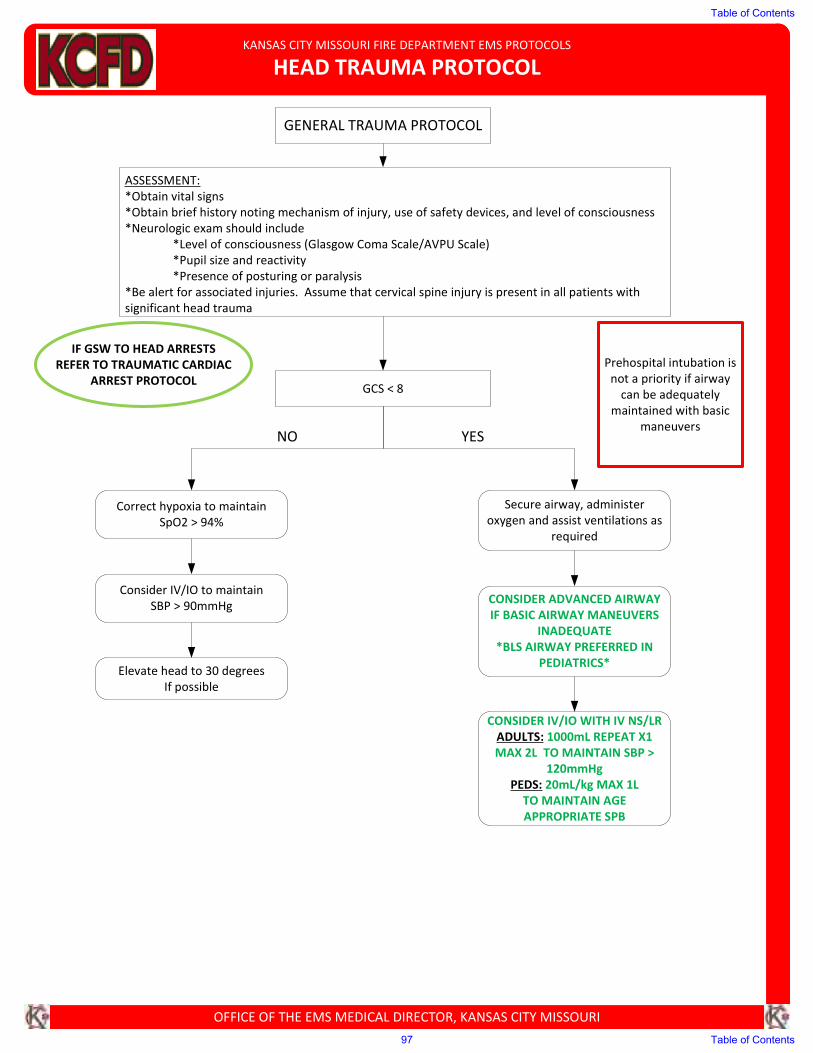
KANSAS CITY MISSOURI FIRE DEPARTMENT EMS PROTOCOLS
HEAD TRAUMA PROTOCOL
OFFICE OF THE EMS MEDICAL DIRECTOR, KANSAS CITY MISSOURI
ASSESSMENT:
*Obtain vital signs
*Obtain brief history noting mechanism of injury, use of safety devices, and level of consciousness
*Neurologic exam should include
*Level of consciousness (Glasgow Coma Scale/AVPU Scale)
*Pupil size and reactivity
*Presence of posturing or paralysis
*Be alert for associated injuries. Assume that cervical spine injury is present in all patients with
significant head trauma
Prehospital intubation is
not a priority if airway
can be adequately
maintained with basic
maneuvers
GENERAL TRAUMA PROTOCOL
GCS < 8
Correct hypoxia to maintain
SpO2 > 94%
Consider IV/IO to maintain
SBP > 90mmHg
Elevate head to 30 degrees
If possible
IF GSW TO HEAD ARRESTS
REFER TO TRAUMATIC CARDIAC
ARREST PROTOCOL
Secure airway, administer
oxygen and assist ventilations as
required
CONSIDER ADVANCED AIRWAY
IF BASIC AIRWAY MANEUVERS
INADEQUATE
*BLS AIRWAY PREFERRED IN
PEDIATRICS*
CONSIDER IV/IO WITH IV NS/LR
ADULTS: 1000mL REPEAT X1
MAX 2L TO MAINTAIN SBP >
120mmHg
PEDS: 20mL/kg MAX 1L
TO MAINTAIN AGE
APPROPRIATE SPB
NO YES
Table of Contents
97 Table of Contents
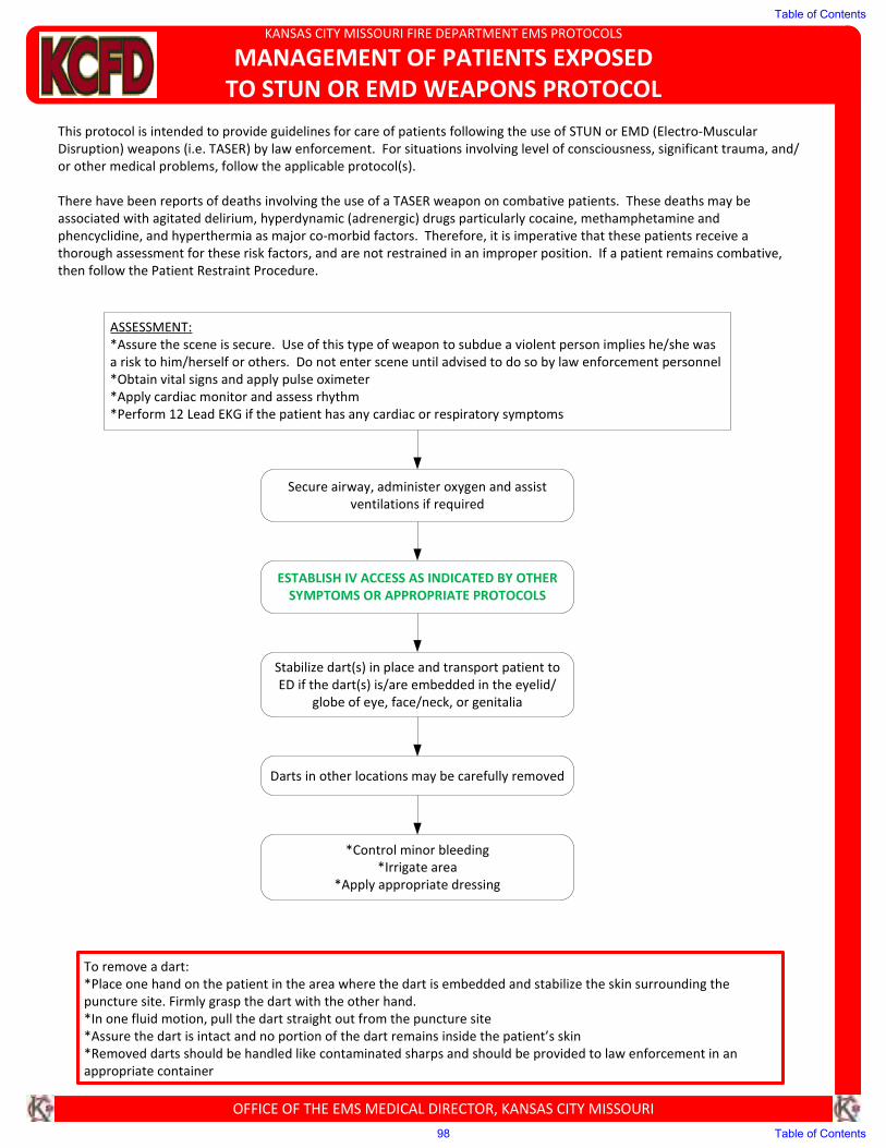
KANSAS CITY MISSOURI FIRE DEPARTMENT EMS PROTOCOLS
MANAGEMENT OF PATIENTS EXPOSED
TO STUN OR EMD WEAPONS PROTOCOL
OFFICE OF THE EMS MEDICAL DIRECTOR, KANSAS CITY MISSOURI
This protocol is intended to provide guidelines for care of patients following the use of STUN or EMD (Electro-Muscular
Disruption) weapons (i.e. TASER) by law enforcement. For situations involving level of consciousness, significant trauma, and/
or other medical problems, follow the applicable protocol(s).
There have been reports of deaths involving the use of a TASER weapon on combative patients. These deaths may be
associated with agitated delirium, hyperdynamic (adrenergic) drugs particularly cocaine, methamphetamine and
phencyclidine, and hyperthermia as major co-morbid factors. Therefore, it is imperative that these patients receive a
thorough assessment for these risk factors, and are not restrained in an improper position. If a patient remains combative,
then follow the Patient Restraint Procedure.
ASSESSMENT:
*Assure the scene is secure. Use of this type of weapon to subdue a violent person implies he/she was
a risk to him/herself or others. Do not enter scene until advised to do so by law enforcement personnel
*Obtain vital signs and apply pulse oximeter
*Apply cardiac monitor and assess rhythm
*Perform 12 Lead EKG if the patient has any cardiac or respiratory symptoms
Secure airway, administer oxygen and assist
ventilations if required
ESTABLISH IV ACCESS AS INDICATED BY OTHER
SYMPTOMS OR APPROPRIATE PROTOCOLS
Stabilize dart(s) in place and transport patient to
ED if the dart(s) is/are embedded in the eyelid/
globe of eye, face/neck, or genitalia
Darts in other locations may be carefully removed
*Control minor bleeding
*Irrigate area
*Apply appropriate dressing
To remove a dart:
*Place one hand on the patient in the area where the dart is embedded and stabilize the skin surrounding the
puncture site. Firmly grasp the dart with the other hand.
*In one fluid motion, pull the dart straight out from the puncture site
*Assure the dart is intact and no portion of the dart remains inside the patient’s skin
*Removed darts should be handled like contaminated sharps and should be provided to law enforcement in an
appropriate container
Table of Contents
98 Table of Contents
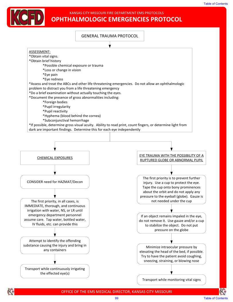
KANSAS CITY MISSOURI FIRE DEPARTMENT EMS PROTOCOLS
OPHTHALMOLOGIC EMERGENCIES PROTOCOL
OFFICE OF THE EMS MEDICAL DIRECTOR, KANSAS CITY MISSOURI
ASSESSMENT:
*Obtain vital signs.
*Obtain brief history
*Possible chemical exposure or trauma
*Loss or change in vision
*Eye pain
*Eye redness
*Assess and treat the ABCs and other life threatening emergencies. Do not allow an ophthalmologic
problem to distract you from a life threatening emergency
*Do a brief examination without actually touching the eyes.
*Document the presence of gross abnormalities including:
*Foreign bodies
*Pupil irregularity
*Pupil reactivity
*Hyphema (blood behind the cornea)
*Subconjunctival hemorrhage
*If possible, determine gross visual acuity. Ability to read print, count fingers, or determine light from
dark are important findings. Determine this for each eye independently
CHEMICAL EXPOSURESEYE TRAUMA WITH THE POSSIBILITY OF A
RUPTURED GLOBE OR ABNORMAL PUPIL
The first priority, in all cases, is
IMMEDIATE, thorough, and continuous
irrigation with water, NS, or LR until
emergency department personnel
assume care. Tap water, bottled water,
IV fluids, etc. can provide this
Attempt to identify the offending
substance causing the injury and bring in
any containers
Transport while continuously irrigating
the effected eye(s)
The first priority is to prevent further
injury. Use a cup to protect the eye.
Tape the cup onto bony prominences
about the orbit and do not apply any
pressure to the eyeball (globe). Gauze is
not needed under the cup
If an object remains impaled in the eye,
do not remove it. Use gauze and/or a cup
to stabilize the object. Do not put
pressure on the globe
Minimize intraocular pressure by
elevating the head of the bed, if possible.
Try to have the patient avoid coughing,
sneezing, straining, or blowing nose
Transport while monitoring vital signs
GENERAL TRAUMA PROTOCOL
CONSIDER need for HAZMAT/Decon
Table of Contents
99 Table of Contents
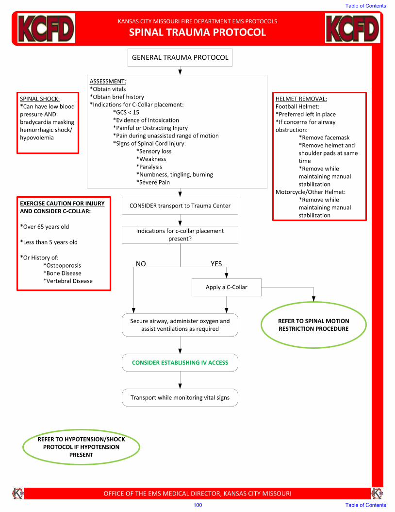
KANSAS CITY MISSOURI FIRE DEPARTMENT EMS PROTOCOLS
SPINAL TRAUMA PROTOCOL
OFFICE OF THE EMS MEDICAL DIRECTOR, KANSAS CITY MISSOURI
GENERAL TRAUMA PROTOCOL
ASSESSMENT:*Obtain vitals*Obtain brief history*Indications for C-Collar placement:
*GCS < 15*Evidence of Intoxication*Painful or Distracting Injury*Pain during unassisted range of motion*Signs of Spinal Cord Injury:
*Sensory loss*Weakness*Paralysis*Numbness, tingling, burning*Severe Pain
HELMET REMOVAL:Football Helmet:*Preferred left in place*If concerns for airwayobstruction:
*Remove facemask*Remove helmet andshoulder pads at sametime*Remove whilemaintaining manualstabilization
Motorcycle/Other Helmet:*Remove whilemaintaining manualstabilization
SPINAL SHOCK:*Can have low bloodpressure ANDbradycardia maskinghemorrhagic shock/hypovolemia
Indications for c-collar placement present?
REFER TO HYPOTENSION/SHOCK PROTOCOL IF HYPOTENSION
PRESENT
REFER TO SPINAL MOTION RESTRICTION PROCEDURE
Secure airway, administer oxygen and assist ventilations as required
CONSIDER ESTABLISHING IV ACCESS
Transport while monitoring vital signs
CONSIDER transport to Trauma Center
NO YES
Apply a C-Collar
EXERCISE CAUTION FOR INJURY AND CONSIDER C-COLLAR:
*Over 65 years old
*Less than 5 years old
*Or History of:*Osteoporosis*Bone Disease*Vertebral Disease
Table of Contents
100 Table of Contents
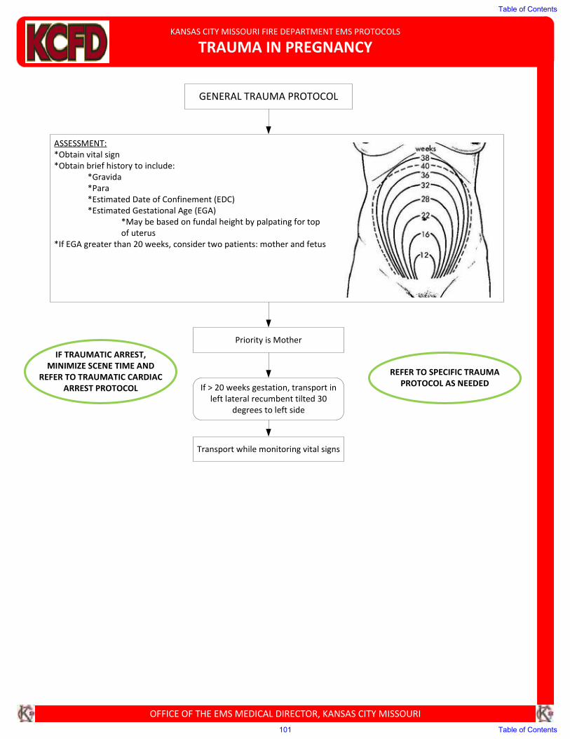
KANSAS CITY MISSOURI FIRE DEPARTMENT EMS PROTOCOLS
TRAUMA IN PREGNANCY
OFFICE OF THE EMS MEDICAL DIRECTOR, KANSAS CITY MISSOURI
GENERAL TRAUMA PROTOCOL
ASSESSMENT:
*Obtain vital sign
*Obtain brief history to include:
*Gravida
*Para
*Estimated Date of Confinement (EDC)
*Estimated Gestational Age (EGA)
*May be based on fundal height by palpating for top
of uterus
*If EGA greater than 20 weeks, consider two patients: mother and fetus
Priority is Mother
REFER TO SPECIFIC TRAUMA
PROTOCOL AS NEEDEDIf > 20 weeks gestation, transport in
left lateral recumbent tilted 30
degrees to left side
IF TRAUMATIC ARREST,
MINIMIZE SCENE TIME AND
REFER TO TRAUMATIC CARDIAC
ARREST PROTOCOL
Transport while monitoring vital signs
Table of Contents
101 Table of Contents
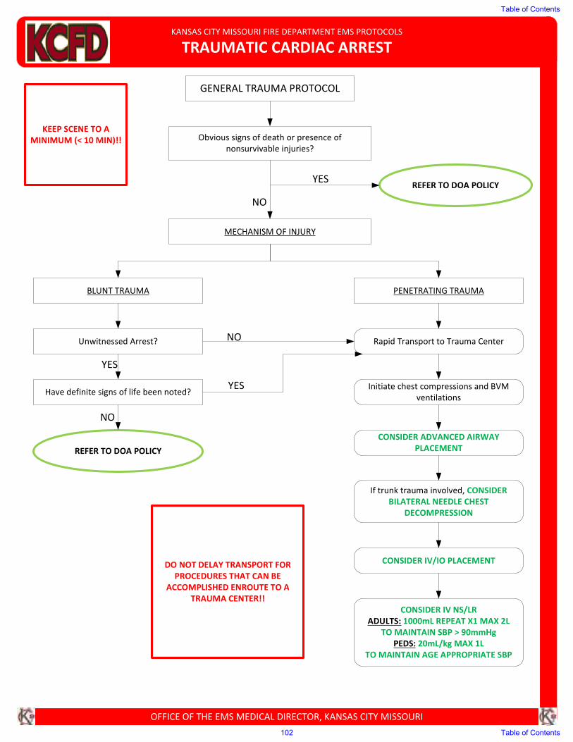
KANSAS CITY MISSOURI FIRE DEPARTMENT EMS PROTOCOLS
TRAUMATIC CARDIAC ARREST
OFFICE OF THE EMS MEDICAL DIRECTOR, KANSAS CITY MISSOURI
GENERAL TRAUMA PROTOCOL
Obvious signs of death or presence of
nonsurvivable injuries?
REFER TO DOA POLICY
MECHANISM OF INJURY
BLUNT TRAUMA PENETRATING TRAUMA
Unwitnessed Arrest?
Have definite signs of life been noted?
REFER TO DOA POLICY
Rapid Transport to Trauma Center
Initiate chest compressions and BVM
ventilations
CONSIDER ADVANCED AIRWAY
PLACEMENT
If trunk trauma involved, CONSIDER
BILATERAL NEEDLE CHEST
DECOMPRESSION
CONSIDER IV/IO PLACEMENT
CONSIDER IV NS/LR
ADULTS: 1000mL REPEAT X1 MAX 2L TO MAINTAIN SBP > 90mmHg
PEDS: 20mL/kg MAX 1L
TO MAINTAIN AGE APPROPRIATE SBP
NO
YES
YES
NO
KEEP SCENE TO A
MINIMUM (< 10 MIN)!!
DO NOT DELAY TRANSPORT FOR
PROCEDURES THAT CAN BE
ACCOMPLISHED ENROUTE TO A
TRAUMA CENTER!!
YES
NO
Table of Contents
102 Table of Contents
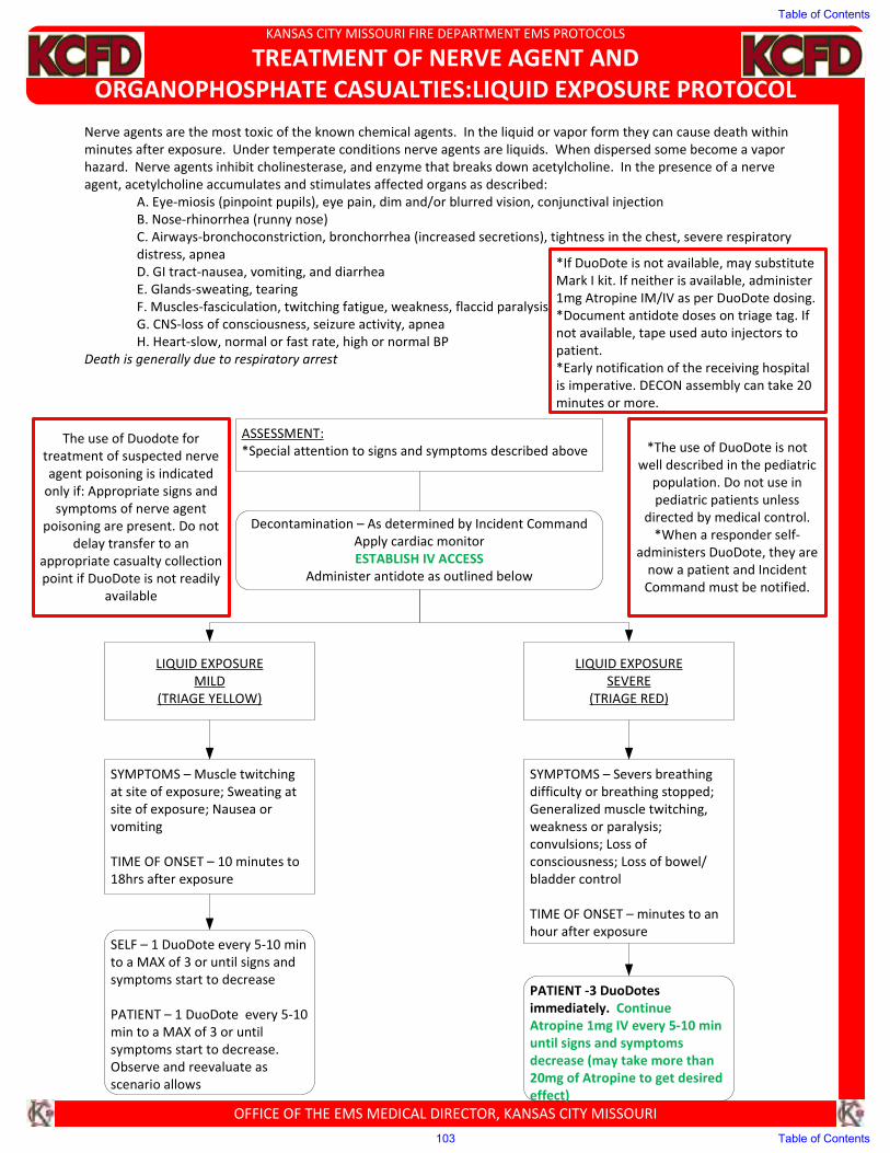
KANSAS CITY MISSOURI FIRE DEPARTMENT EMS PROTOCOLS
TREATMENT OF NERVE AGENT AND
ORGANOPHOSPHATE CASUALTIES:LIQUID EXPOSURE PROTOCOL
OFFICE OF THE EMS MEDICAL DIRECTOR, KANSAS CITY MISSOURI
Nerve agents are the most toxic of the known chemical agents. In the liquid or vapor form they can cause death within
minutes after exposure. Under temperate conditions nerve agents are liquids. When dispersed some become a vapor
hazard. Nerve agents inhibit cholinesterase, and enzyme that breaks down acetylcholine. In the presence of a nerve
agent, acetylcholine accumulates and stimulates affected organs as described:
A. Eye-miosis (pinpoint pupils), eye pain, dim and/or blurred vision, conjunctival injection
B. Nose-rhinorrhea (runny nose)
C. Airways-bronchoconstriction, bronchorrhea (increased secretions), tightness in the chest, severe respiratory
distress, apnea
D. GI tract-nausea, vomiting, and diarrhea
E. Glands-sweating, tearing
F. Muscles-fasciculation, twitching fatigue, weakness, flaccid paralysis
G. CNS-loss of consciousness, seizure activity, apnea
H. Heart-slow, normal or fast rate, high or normal BP
Death is generally due to respiratory arrest
ASSESSMENT:
*Special attention to signs and symptoms described above
Decontamination – As determined by Incident Command
Apply cardiac monitor
ESTABLISH IV ACCESS
Administer antidote as outlined below
LIQUID EXPOSURE
MILD
(TRIAGE YELLOW)
LIQUID EXPOSURE
SEVERE
(TRIAGE RED)
SYMPTOMS – Muscle twitching
at site of exposure; Sweating at
site of exposure; Nausea or
vomiting
TIME OF ONSET – 10 minutes to
18hrs after exposure
SELF – 1 DuoDote every 5-10 min
to a MAX of 3 or until signs and
symptoms start to decrease
PATIENT – 1 DuoDote every 5-10
min to a MAX of 3 or until
symptoms start to decrease.
Observe and reevaluate as
scenario allows
SYMPTOMS – Severs breathing
difficulty or breathing stopped;
Generalized muscle twitching,
weakness or paralysis;
convulsions; Loss of
consciousness; Loss of bowel/
bladder control
TIME OF ONSET – minutes to an
hour after exposure
PATIENT -3 DuoDotes
immediately. Continue
Atropine 1mg IV every 5-10 min
until signs and symptoms
decrease (may take more than
20mg of Atropine to get desired
effect)
The use of Duodote for
treatment of suspected nerve
agent poisoning is indicated
only if: Appropriate signs and
symptoms of nerve agent
poisoning are present. Do not
delay transfer to an
appropriate casualty collection
point if DuoDote is not readily
available
*The use of DuoDote is not
well described in the pediatric
population. Do not use in
pediatric patients unless
directed by medical control.
*When a responder self-
administers DuoDote, they are
now a patient and Incident
Command must be notified.
*If DuoDote is not available, may substitute
Mark I kit. If neither is available, administer
1mg Atropine IM/IV as per DuoDote dosing.
*Document antidote doses on triage tag. If
not available, tape used auto injectors to
patient.
*Early notification of the receiving hospital
is imperative. DECON assembly can take 20
minutes or more.
Table of Contents
103 Table of Contents
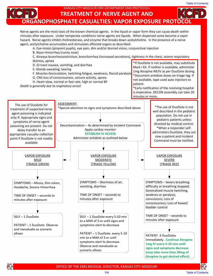
KANSAS CITY MISSOURI FIRE DEPARTMENT EMS PROTOCOLS
TREATMENT OF NERVE AGENT AND
ORGANOPHOSPHATE CASUALTIES: VAPOR EXPOSURE PROTOCOL
OFFICE OF THE EMS MEDICAL DIRECTOR, KANSAS CITY MISSOURI
Nerve agents are the most toxic of the known chemical agents. In the liquid or vapor form they can cause death within
minutes after exposure. Under temperate conditions nerve agents are liquids. When dispersed some become a vapor
hazard. Nerve agents inhibit cholinesterase, and enzyme that breaks down acetylcholine. In the presence of a nerve
agent, acetylcholine accumulates and stimulates affected organs as described:
A. Eye-miosis (pinpoint pupils), eye pain, dim and/or blurred vision, conjunctival injection
B. Nose-rhinorrhea (runny nose)
C. Airways-bronchoconstriction, bronchorrhea (increased secretions), tightness in the chest, severe respiratory
distress, apnea
D. GI tract-nausea, vomiting, and diarrhea
E. Glands-sweating, tearing
F. Muscles-fasciculation, twitching fatigue, weakness, flaccid paralysis
G. CNS-loss of consciousness, seizure activity, apnea
H. Heart-slow, normal or fast rate, high or normal BP
Death is generally due to respiratory arrest
ASSESSMENT:
*Special attention to signs and symptoms described above
Decontamination – As determined by Incident Command
Apply cardiac monitor
ESTABLISH IV ACCESS
Administer antidote as outlined below
VAPOR EXPOSURE
MILD
(TRIAGE GREEN)
VAPOR EXPOSURE
MODERATE
(TRIAGE YELLOW)
VAPOR EXPOSURE
SEVERE
(TRIAGE RED)
SYMPTOMS – Miosis, Dim vision,
Headache, Severe rhinorrhea
TIME OF ONSET – seconds to
minutes after exposure
SELF – 1 DuoDote
PATIENT – 1 DuoDote Observe
and reevaluate as scenario
allows
SYMPTOMS – Shortness of air,
vomiting, diarrhea
TIME OF ONSET – seconds to
minutes after exposure
SELF – 1 DuoDote every 5-10 min
to a MAX of 3 or until signs and
symptoms start to decrease
PATIENT – 1 DuoDote every 5-10
min to a MAX of 3 or until
symptoms start to decrease.
Observe and reevaluate as
scenario allows
SYMPTOMS – Severs breathing
difficulty or breathing stopped;
Generalized muscle twitching,
weaknes or paralysis;
convulsions; Loss of
consciousness; Loss of bowel/
bladder control
TIME OF ONSET – seconds to
minutes after exposure
PATIENT -3 DuoDotes
immediately. Continue Atropine
1mg IV every 5-10 min until
signs and symptoms decrease
(may take more than 20mg of
Atropine to get desired effect)
The use of Duodote for
treatment of suspected nerve
agent poisoning is indicated
only if: Appropriate signs and
symptoms of nerve agent
poisoning are present. Do not
delay transfer to an
appropriate casualty collection
point if DuoDote is not readily
available
*The use of DuoDote is not
well described in the pediatric
population. Do not use in
pediatric patients unless
directed by medical control.
*When a responder self-
administers DuoDote, they are
now a patient and Incident
Command must be notified.
*If DuoDote is not available, may substitute
Mark I kit. If neither is available, administer
1mg Atropine IM/IV as per DuoDote dosing.
*Document antidote doses on triage tag. If
not available, tape used auto injectors to
patient.
*Early notification of the receiving hospital
is imperative. DECON assembly can take 20
minutes or more.
Table of Contents
104 Table of Contents
KANSAS CITY MISSOURI FIRE DEPARTMENT EMS PROTOCOLS
BLS ATTENDANT ON EMERGENCY CALLS PROTOCOL
OFFICE OF THE EMS MEDICAL DIRECTOR, KANSAS CITY MISSOURI
The purpose of this protocol is to identify those patients from an emergency call that may be attended by an EMT during transport to the hospital.
ACCEPTABLE VITAL SIGNS
*Pulse Rate:50‐110 bpm
*Systolic Blood Pressure:100‐190mmHg
*Diastolic Blood Pressure:< 120mmHg
*Respiratory Rate:8‐24 per minute
*Pulse Oximeter:> 93%
*Glasgow Coma Scale:> 13
*Blood Glucose:70‐250mg/dL
Has the patient required airway management (OPA, NPA, iGel, ETT) at any time during the current encounter?
Has the patient required ventilatory support (CPAP or BVM use) at any time during the current encounter?
Has the patient required chest compressions at anytime during the current encounter?
Has the patient required vascular access (either IV or IO) at anytime during the current encounter?
Has the patient had acute mental status changes and GCS < 14 at anytime during the current encounter?
Has the patient required EKG monitoring, a 12 Lead ECG obtained, or ETCO2 monitoring at anytime during the current encounter?
Has the patient required any medication found within the KCFD EMS Protocols Medication Formulary (except O2 or Oral Glucose) at anytime during the current encounter?
Does the patient require TCD Routing (Stroke, STEMI, or Trauma) per the Determination of Hospital Destination Policy found in the KCFD EMS Protocols?
Has the patient’s vital signs been outside acceptable parameters at anytime during the current encounter?
If the answer is “NO” to ALL OF THE ABOVE, the patient may be attended by an EMT during transport to the hospital
Table of Contents
105 Table of Contents
KANSAS CITY MISSOURI FIRE DEPARTMENT EMS PROTOCOLS
BLS ATTENDANT ON INTERFACILITY TRANSFER PROTOCOL
OFFICE OF THE EMS MEDICAL DIRECTOR, KANSAS CITY MISSOURI
Obtain report, appropriate paperwork, and confirm patient destination
Assess patient including full set of vitals and mental status
Is the patient on no more than O2 6 lpm via nasal cannula, all vascular access lines locked off, NG and feeding tubes able to be disconnected from suction or feeding,
and tracheostomy stoma has no equipment present?
Transport
Is this normal for this patient
Contact Transport Coordinator and
advise
NO
NO
NO
YES YES
YES
ACCEPTABLE VITAL SIGNS
*Pulse Rate:50‐110 bpm
*Systolic Blood Pressure:100‐190mmHg
*Diastolic Blood Pressure:< 120mmHg
*Respiratory Rate:8‐24 per minute
*Pulse Oximeter:> 93%
*Glasgow Coma Scale:> 13
*Blood Glucose:70‐250mg/dL
Table of Contents
106 Table of Contents
KANSAS CITY MISSOURI FIRE DEPARTMENT EMS PROTOCOLS
KC ASSESSMENT AND TRIAGE CENTER PROTOCOL
OFFICE OF THE EMS MEDICAL DIRECTOR, KANSAS CITY MISSOURI
The purpose of this protocol is to identify those patients that meet criteria for transport to the Kansas City
Assessment and Triage Center by KCPD or KCFD, and are requesting transport to the Center
Age 18 years or older
Patient has:
*No acute medical conditions
*No wounds that require sutures
*No evidence of trauma to head/face/scalp
Patient is not combative, does not require
restraints, and does not need sedation
Vital signs are within acceptable limits
Patient is ambulatory and is able to self-transfer
without assistance
Patient does not require treatments such as;
*Oxygen
*IV fluids or medications
*CPAP
*Cardiac monitoring
Or other interventions from KCFD EMS Crews
If the patient meets ALL criteria listed above, they
may be transported by KCPD or KCFD to the KC
Assessment and Triage Center
ACCEPTABLE VITAL SIGNS
*Pulse Rate:
50-110 bpm
*Systolic Blood Pressure:
100-190mmHg
*Diastolic Blood Pressure:
< 120mmHg
*Respiratory Rate:
8-24 per minute
*Pulse Oximeter:
> 93%
*Glasgow Coma Scale:
> 13
*Blood Glucose:
60-250mg/dL
If there is any question as to
whether the patient meets ALL
criteria, default is to transport
to the Emergency Department
If patient preference is to be
transported to the hospital,
the patient is to be
transported to their hospital of
choice.
Patients that are released
to KCPD for transport to
KCATC do not sign a
refusal.
Table of Contents
107 Table of Contents
KANSAS CITY MISSOURI FIRE DEPARTMENT EMS PROTOCOLS
SPECIALTY EQUIPMENT PROTOCOL
The purpose of this protocol is to address specialty equipment that the patient may using and the mitigation of an emergency that may be occurring with that equipment
VENTRICULAR ASSIST DEVICE:*A ventricular assist device (VAD) is a mechanical device used to support circulation in a patient with significant cardiacventricular dysfunction*A patient with a ventricular assist device can be identified by an electric driveline cable that come directly out of theirabdomen and connects to an external control pack powered by two external batteries they will be wearing with a bag,harness, or vest*Typically, VAD patients have no discernible pulse. Blood pressure measurement requires manual BP cuff and Doppler(which the patient may have). Utilize other parameters for patient assessment:
*Level of consciousness*Respiratory rate and work of breathing*Signs of perfusion:
*Skin color/temperature*Capillary refill (HR > 100 is hemodynamically unstable)*Cardiac monitor*SaO2*Blood glucose level
*If patient stable: Contact VAD Coordinator (patient should have this information) and transport to VAD Hospital*If patient unstable: VAD RUNNING VAD NOT RUNNING
*250mL NS/LR BOLUS IV *CONSIDER CCC IF NO CLINICAL SIGNS OF*CONSIDER CCC IF NO CLINICAL PERFUSIONSIGNS OF PERFUSION *Address VAD alarms*ACLS *CONSIDER DEFIBRILLATION
*ACLS*Contact VAD Coordinator/Physician ASAP:
*St Lukes Plaza: 816-932-2000*If patient stable, ask for VAD Coordinator*If patient unstable, as for Emergency VAD Physician On Call
*KU Medical Center: 913-293-6616*This is a cell phone carried 24/7 by the On-Duty VAD Coordinator. The patient may have differentnumber that connects to the department with a menu option.
AV FISTULA:If patient is hemorrhaging from AV Fistula:
*Apply firm finger tip pressure to bleeding site*Apply dressing but avoid bulky dressing*Dressing must not compress fistula/shunt as this will cause clotting of the shunt*If direct pressure and dressing not effective and life-threatening hemorrhage, apply tourniquet to affectedextremity several inches above fistula/shunt
TRACHEOSTOMY:*If trach tube has come out, ventilate with BVM over stoma if possible. May ventilate over mouth, however besure to cover stoma with gloved hand to prevent air leak*Keep area protected*Bring patient’s replacement equipment (if available) to hospital
FEEDING TUBE:*May be referred to as G-tube, J-tube, Peg tube, or Mickey button depending on location*If feeding tube has been pulled out:
*Keep area protected*Bring replacement equipment (if available) to hospital
Table of Contents
108 Table of Contents
OFFICE OF THE EMS MEDICAL DIRECTOR, KANSAS CITY MISSOURI
SERVICE ANIMALS:*As defined by the American Disabilities Act, “any guide dog, signal dog, or other animal individually trained to do work or perform tasks for the benefit of an individual with a disability, including, but not limited to guiding individuals with impaired vision, alerting individuals with impaired hearing to intruders or sounds, providing minimal protection or rescue work, pulling a wheelchair, or fetching dropped items.”
*Service animals are not classified as a pet and should by law, always be permitted to accompany the patient with the following exceptions:
*A public entity may as an individual with a disability to remove a service animal from the premises if:*The animal is out of control and the animal’s handler does not take effective action to control it. OR:*The animal is not housebroken
*Service animals are not required to wear a vest or a leash. It is illegal to make a request for special identification or documentation from the service animal’s partner. EMS providers may only ask the patient if the service animal is required because of a disability and the form of assistance the animal has been trained to perform.*EMS providers are not responsible for the care for the service animal. If the patient is incapacitated and cannot personally care for the service animal, a decision can be made whether or not to transport the animal in this situation.*Animals that solely provide emotional support, comfort, or companionship do not qualify as service animals.
Table of Contents
109 Table of Contents
INTRODUCTION: The principal purpose of obtaining a 12-lead ECG in the prehospital setting is to identify patients
having an acute ST-elevation myocardial infarction (STEMI) and to relay this information to the receiving hospitals
allowing them to appropriately prepare for patient arrival. This has been shown to decrease “door to drug” and “door
to invasive intervention” time, both of which improve patient survival.
INDICATIONS:
I. A prehospital 12-lead ECG should specifically be considered during the assessment of patients being treated as
per the following protocols:
A. Suspected myocardial infarction:
1. Chest pain
B. Respiratory Distress (possible congestive heart failure/pulmonary edema)
1. Asthma
2. COPD
3. CHF
4. Pulmonary Edema
C. Dysrhythmia diagnosis:
1. Cardiac Dysrhythmia (may be particularly helpful in pediatric patients)
D. Stroke
E. Return of Spontaneous Circulation (ROSC)
F. Altered Mental Status
G. Hypotension/Shock
H. Syncope
CONTRAINDICATIONS: Do not delay the treatment and/or transport of unstable patients to obtain a 12-lead EKG.
PRECAUTIONS:
I. Do not prolong scene time to obtain 12-lead ECG (should take less than 5 minutes)
II. A normal (or nonspecific) 12-lead ECG does not rule out myocardial infarction or ischemia, therefore do not base
prehospital treatment on the results of the ECG
III. If a patient on whom you have obtained a 12-lead ECG is refusing transport you should strongly consider
contacting medical control for advice
TECHNIQUE:
I. The patient should be in the supine position. If the patient cannot tolerate that position,
place them in the semi-reclining or sitting position.
II. Prep the skin and shave hair as needed.III. Apply electrodes as described:
Limb Leads: Precordial (Chest) Leads:
Right arm (RA) – Right wrist V1 – 4th intercostal space to the right of the sternum
Right leg (RL) – Right ankle V2 – 4th intercostal space to the left of the sternum
Left arm (LA) – Left wrist V3 – Directly between leads V 2 and V 4
Left leg (LL) – Left ankle V4 – 5th intercostal space at midclavicular line V5
– Level with V4 at left anterior axillary line V6 –
Level with V5 at left midaxillary line
KANSAS CITY MISSOURI FIRE DEPARTMENT EMS PROTOCOLS
12-LEAD ECG MONITORING PROCEDURE
Table of Contents
110 Table of Contents
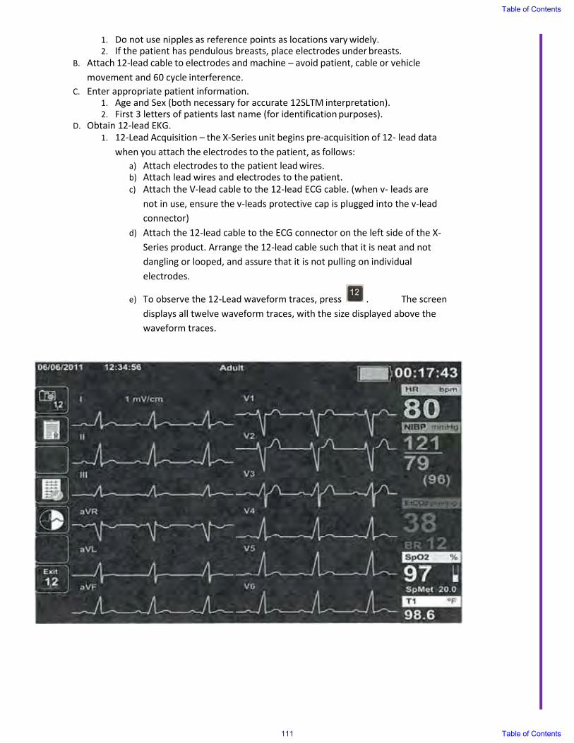
1. Do not use nipples as reference points as locations vary widely. 2. If the patient has pendulous breasts, place electrodes under breasts.
B. Attach 12-lead cable to electrodes and machine – avoid patient, cable or vehicle
movement and 60 cycle interference.
C. Enter appropriate patient information. 1. Age and Sex (both necessary for accurate 12SLTM interpretation). 2. First 3 letters of patients last name (for identification purposes).
D. Obtain 12-lead EKG.
1. 12-Lead Acquisition – the X-Series unit begins pre-acquisition of 12- lead data
when you attach the electrodes to the patient, as follows:
a) Attach electrodes to the patient lead wires. b) Attach lead wires and electrodes to the patient.
c) Attach the V-lead cable to the 12-lead ECG cable. (when v- leads are
not in use, ensure the v-leads protective cap is plugged into the v-lead
connector)
d) Attach the 12-lead cable to the ECG connector on the left side of the X-
Series product. Arrange the 12-lead cable such that it is neat and not
dangling or looped, and assure that it is not pulling on individual
electrodes.
e) To observe the 12-Lead waveform traces, press . The screen
displays all twelve waveform traces, with the size displayed above the
waveform traces.
Table of Contents
111 Table of Contents
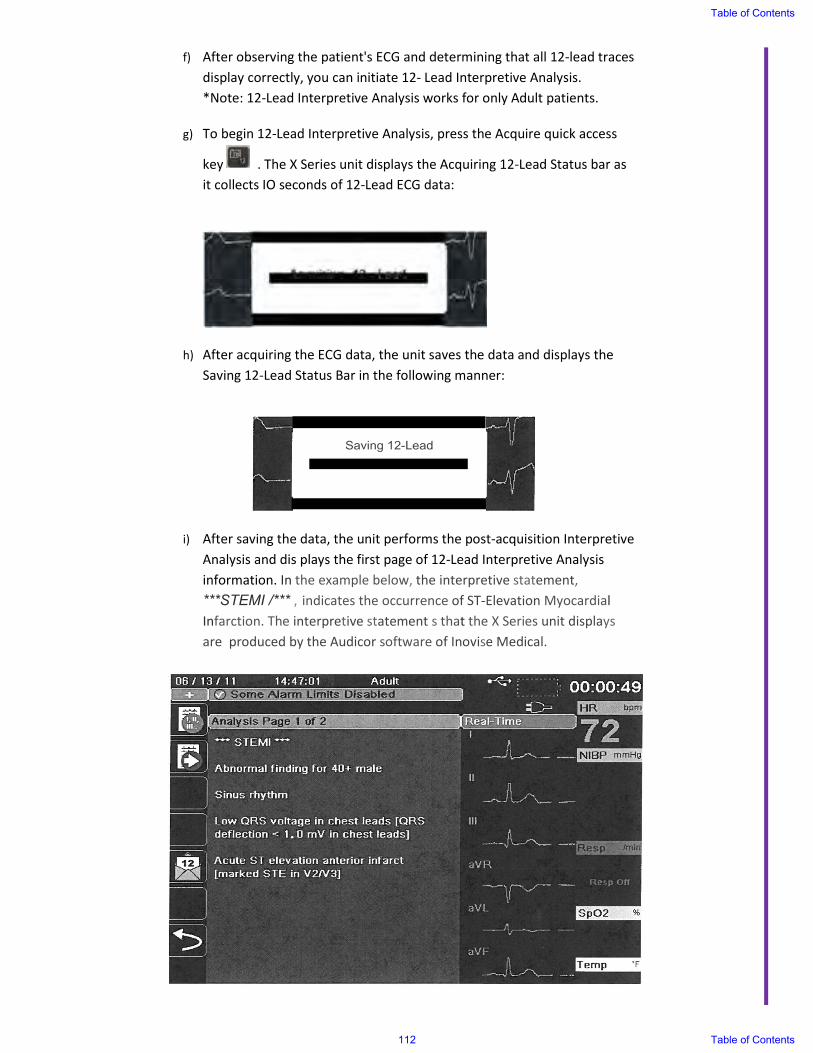
f) After observing the patient's ECG and determining that all 12-lead traces
display correctly, you can initiate 12- Lead Interpretive Analysis.
*Note: 12-Lead Interpretive Analysis works for only Adult patients.
g) To begin 12-Lead Interpretive Analysis, press the Acquire quick access
key . The X Series unit displays the Acquiring 12-Lead Status bar as
it collects IO seconds of 12-Lead ECG data:
h) After acquiring the ECG data, the unit saves the data and displays the
Saving 12-Lead Status Bar in the following manner:
i) After saving the data, the unit performs the post-acquisition Interpretive
Analysis and dis plays the first page of 12-Lead Interpretive Analysis information. In the example below, the interpretive statement,
***STEMI /*** , indicates the occurrence of ST-Elevation Myocardial Infarction. The interpretive statement s that the X Series unit displays are produced by the Audicor software of Inovise Medical.
Table of Contents
112 Table of Contents
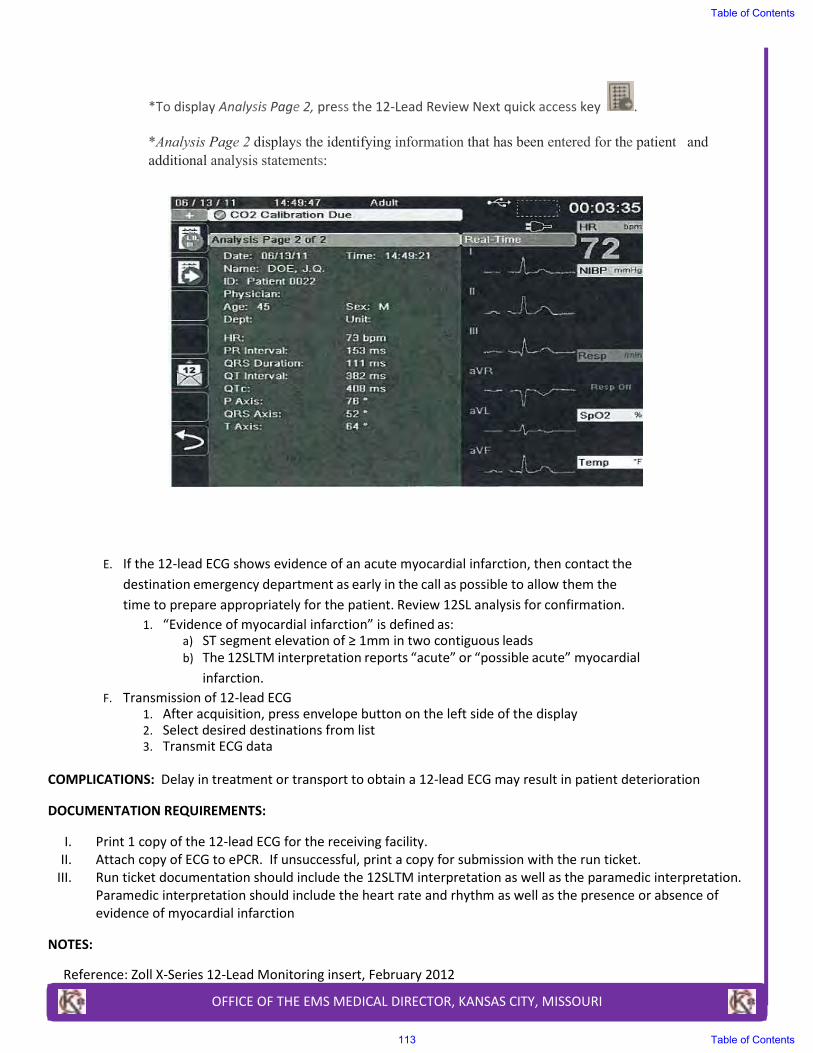
*To display Analysis Page 2, press the 12-Lead Review Next quick access key .
*Analysis Page 2 displays the identifying information that has been entered for the patient and
additional analysis statements:
E. If the 12-lead ECG shows evidence of an acute myocardial infarction, then contact the
destination emergency department as early in the call as possible to allow them the
time to prepare appropriately for the patient. Review 12SL analysis for confirmation.
1. “Evidence of myocardial infarction” is defined as: a) ST segment elevation of ≥ 1mm in two contiguous leads
b) The 12SLTM interpretation reports “acute” or “possible acute” myocardial
infarction.
F. Transmission of 12-lead ECG 1. After acquisition, press envelope button on the left side of the display 2. Select desired destinations from list 3. Transmit ECG data
COMPLICATIONS: Delay in treatment or transport to obtain a 12-lead ECG may result in patient deterioration
DOCUMENTATION REQUIREMENTS:
I. Print 1 copy of the 12-lead ECG for the receiving facility.
II. Attach copy of ECG to ePCR. If unsuccessful, print a copy for submission with the run ticket.
III. Run ticket documentation should include the 12SLTM interpretation as well as the paramedic interpretation.
Paramedic interpretation should include the heart rate and rhythm as well as the presence or absence of
evidence of myocardial infarction
NOTES:
Reference: Zoll X-Series 12-Lead Monitoring insert, February 2012
OFFICE OF THE EMS MEDICAL DIRECTOR, KANSAS CITY, MISSOURI
Table of Contents
113 Table of Contents
INTRODUCTION:
1. CO2 is a product of cellular metabolism.
2. Exhaled air has a high pCO2, inspired air has essentially no CO2.
3. Colorimetric end-tidal CO2 monitors measure pCO2 of inspired and expired air by color indicator.
4. Color varies between expiration and inspiration as CO2 levels rise and fall.
5. In cardiac arrest patients, CO2 may not be carried to the lungs because of poor perfusion. Therefore, the
expired pCO2 may be very low.
INDICATIONS:
1. Waveform capnography on Zoll X-Series is not functioning properly or is unavailable.
2. Colorimetric end-tidal CO2 detection is an adjunct used to help confirm correct iGel or endotracheal tube
placement and to help monitor placement. It does not replace clinical evaluation.
CONTRAINDICATIONS:
1. Mouth to iGel or endotracheal tube ventilation.
2. Patients less than 15kg (30lbs) in weight.
TECHNIQUE:
Initial Verification of iGel or Endotracheal Tube Placement:
1. Remove from package immediately prior to using device.
2. If initial color indicator is not the same or darker than the “area” then do not use.
3. Insert iGel or endotracheal tube, inflate cuff (if applicable), attach device.
4. Ventilate patient with six breaths of moderate tidal volume.
5. Compare color indicator to color chart on the colorimetric device on full end expiration.
a. If color in “A”range (purple)
i. iGel or endotracheal tube placement incorrect.
ii. Remove airway and attempt to reinsert
b. If color in “B” range (tan)
i. Indicates retained CO2 in esophagus or low pulmonary blood flow, hypocarbia
ii. Deliver six more breaths
1. If color shifts to “A” range, airway placement incorrect, remove airway and ensure
adequate ventilations
2. If color remains in “B” range (tan), airway placement correct with low pulmonary blood
flow, check clinical signs and secure airway
3. If color shifts to “C” range (yellow), airway placement correct, secure tube
c. If color in “C” range (yellow)
i. Indicates iGel or endotracheal tube placement is correct
ii. Secure airway in place and monitor placement
Continuing Verification of iGel or Endotracheal Tube Placement:
1. If color changes to an A or B range after an initial C range the either:
a. The iGel or endotracheal tube has become dislodged from the trachea OR
b. The patient has deteriorated and now has poor pulmonary perfusion
2. If color changes to an A range after an initial B range then either:
KANSAS CITY MISSOURI FIRE DEPARTMENT EMS PROTOCOLS
CO2 DETECTOR (EZ CAP II) PROCEDURE
Table of Contents
114 Table of Contents
a. The iGel or endotracheal tube has become dislodged from trachea OR
b. The patient has deteriorated and now has poor pulmonary perfusion
3. If either of the two above scenarios occurs, use clinical means to confirm proper placement and or change in
perfusion status.
NOTES:
• Interpreting results before 6 full breaths can lead to false positive results
o Gastric distension with air prior to iGel or endotracheal tube placement may introduce CO2 levels high
enough into the stomach to give a false positive reading when placement is actually incorrect.
• The colorimetric CO2 detector should not be used to detect right main stem bronchus intubations. Use your
clinical assessment to confirm correct placement
• Reflux of gastric contents, mucus, edema fluid, or tracheal epinephrine into the CO2 detector can yield
persistent yellow or white discoloration that does not vary with respiratory cycle. Discard if this happens.
OFFICE OF THE EMS MEDICAL DIRECTOR, KANSAS CITY, MISSOURI
Table of Contents
115 Table of Contents
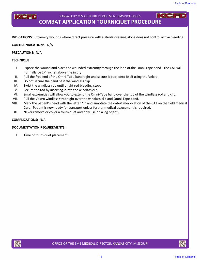
INDICATIONS: Extremity wounds where direct pressure with a sterile dressing alone does not control active bleeding
CONTRAINDICATIONS: N/A
PRECAUTIONS: N/A
TECHNIQUE:
I. Expose the wound and place the wounded extremity through the loop of the Omni-Tape band. The CAT will
normally be 2-4 inches above the injury.
II. Pull the free end of the Omni-Tape band tight and secure it back onto itself using the Velcro.
III. Do not secure the band past the windlass clip.
IV. Twist the windlass rob until bright red bleeding stops
V. Secure the rod by inserting it into the windlass clip.
VI. Small extremities will allow you to extend the Omni-Tape band over the top of the windlass rod and clip.
VII. Pull the Velcro windlass strap tight over the windlass clip and Omni-Tape band.
VIII. Mark the patient’s head with the letter “T” and annotate the date/time/location of the CAT on the field medical
Card. Patient is now ready for transport unless further medical assessment is required.
IX. Never remove or cover a tourniquet and only use on a leg or arm.
COMPLICATIONS: N/A
DOCUMENTATION REQUIREMENTS:
I. Time of tourniquet placement
KANSAS CITY MISSOURI FIRE DEPARTMENT EMS PROTOCOLS
COMBAT APPLICATION TOURNIQUET PROCEDURE
OFFICE OF THE EMS MEDICAL DIRECTOR, KANSAS CITY, MISSOURI
Table of Contents
116 Table of Contents
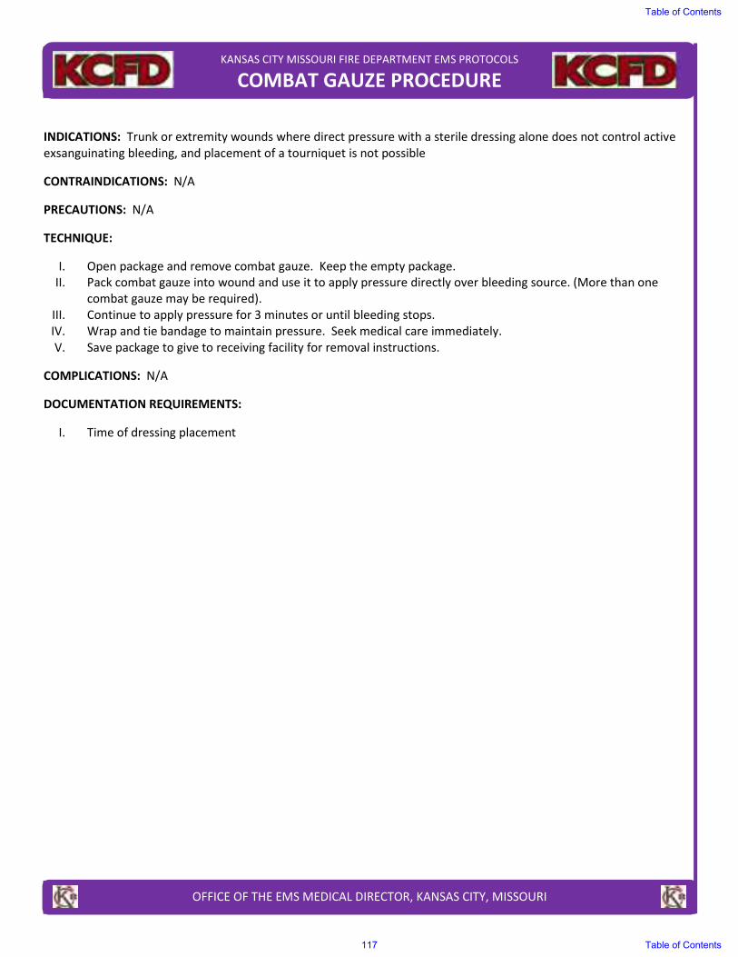
INDICATIONS: Trunk or extremity wounds where direct pressure with a sterile dressing alone does not control active
exsanguinating bleeding, and placement of a tourniquet is not possible
CONTRAINDICATIONS: N/A
PRECAUTIONS: N/A
TECHNIQUE:
I. Open package and remove combat gauze. Keep the empty package.
II. Pack combat gauze into wound and use it to apply pressure directly over bleeding source. (More than one
combat gauze may be required).
III. Continue to apply pressure for 3 minutes or until bleeding stops.
IV. Wrap and tie bandage to maintain pressure. Seek medical care immediately.
V. Save package to give to receiving facility for removal instructions.
COMPLICATIONS: N/A
DOCUMENTATION REQUIREMENTS:
I. Time of dressing placement
KANSAS CITY MISSOURI FIRE DEPARTMENT EMS PROTOCOLS
COMBAT GAUZE PROCEDURE
OFFICE OF THE EMS MEDICAL DIRECTOR, KANSAS CITY, MISSOURI
Table of Contents
117 Table of Contents
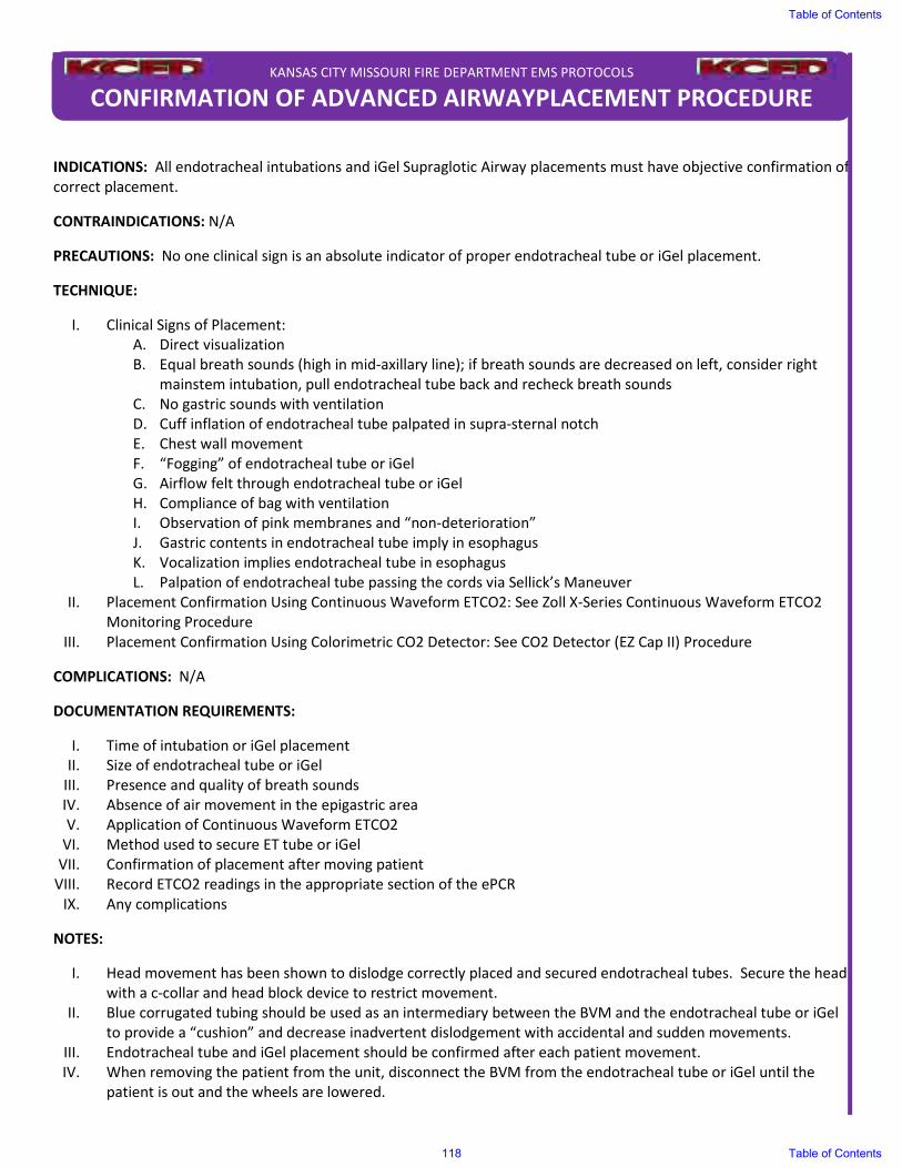
INDICATIONS: All endotracheal intubations and iGel Supraglotic Airway placements must have objective confirmation of
correct placement.
CONTRAINDICATIONS: N/A
PRECAUTIONS: No one clinical sign is an absolute indicator of proper endotracheal tube or iGel placement.
TECHNIQUE:
I. Clinical Signs of Placement:
A. Direct visualization
B. Equal breath sounds (high in mid-axillary line); if breath sounds are decreased on left, consider right
mainstem intubation, pull endotracheal tube back and recheck breath sounds
C. No gastric sounds with ventilation
D. Cuff inflation of endotracheal tube palpated in supra-sternal notch
E. Chest wall movement
F. “Fogging” of endotracheal tube or iGel
G. Airflow felt through endotracheal tube or iGel
H. Compliance of bag with ventilation
I. Observation of pink membranes and “non-deterioration”
J. Gastric contents in endotracheal tube imply in esophagus
K. Vocalization implies endotracheal tube in esophagus
L. Palpation of endotracheal tube passing the cords via Sellick’s Maneuver
II. Placement Confirmation Using Continuous Waveform ETCO2: See Zoll X-Series Continuous Waveform ETCO2
Monitoring Procedure
III. Placement Confirmation Using Colorimetric CO2 Detector: See CO2 Detector (EZ Cap II) Procedure
COMPLICATIONS: N/A
DOCUMENTATION REQUIREMENTS:
I. Time of intubation or iGel placement
II. Size of endotracheal tube or iGel
III. Presence and quality of breath sounds
IV. Absence of air movement in the epigastric area
V. Application of Continuous Waveform ETCO2
VI. Method used to secure ET tube or iGel
VII. Confirmation of placement after moving patient
VIII. Record ETCO2 readings in the appropriate section of the ePCR
IX. Any complications
NOTES:
I. Head movement has been shown to dislodge correctly placed and secured endotracheal tubes. Secure the head
with a c-collar and head block device to restrict movement.
II. Blue corrugated tubing should be used as an intermediary between the BVM and the endotracheal tube or iGel
to provide a “cushion” and decrease inadvertent dislodgement with accidental and sudden movements.
III. Endotracheal tube and iGel placement should be confirmed after each patient movement.
IV. When removing the patient from the unit, disconnect the BVM from the endotracheal tube or iGel until the
patient is out and the wheels are lowered.
KANSAS CITY MISSOURI FIRE DEPARTMENT EMS PROTOCOLS
CONFIRMATION OF ADVANCED AIRWAYPLACEMENT PROCEDURE
Table of Contents
118 Table of Contents
V. None of the clinical signs are absolute; there are documented cases of unrecognized esophageal intubation
using each of these techniques.
VI. “When in doubt, take it out” and assure oxygenation and ventilation.
VII. If an initial attempt at intubation results in esophageal intubation, the endotracheal tube may be left in the
esophagus to provide a landmark for another attempt if this does not impede BVM ventilation.
OFFICE OF THE EMS MEDICAL DIRECTOR, KANSAS CITY, MISSOURI
Table of Contents
119 Table of Contents
INDICATIONS:
CPAP is indicated in all patients in whom inadequate ventilation is suspected. This could be as a result of pulmonary edema, pneumonia, asthma, COPD, etc. The patient must have an adequate mental status and a spontaneous respiratory drive in order to permit the CPAP device to function properly.
CONTRAINDICATIONS:
I. Decreased or Altered Mental Status. II. Facial features or deformities that prevent an adequate mask seal.
III. Vomiting or excessive respiratory secretions. IV. Inability to maintain own airway. V. SBP < 90mmHg
PRECAUTIONS:
I. Emesis may result in aspiration
TECHNIQUE:
I. Ensure adequate oxygen supply to CPAP device. Limit the time spent on portable oxygen. II. Explain the procedure to the patient.
III. Place the delivery mask over the mouth and nose. To improve compliance and tolerance, manually hold the mask for 3-5 minutes while continuously reassuring the patient. Oxygen should be flowing through the device at this point.
IV. Secure the mask with provided straps starting with the lower straps until minimal air leak occurs. V. Encourage the patient to allow forced ventilation to occur. Observe closely for signs of complications.
VI. Evaluate the response of the patient, assessing general appearance, breath sounds, and oxygen saturation. VII. Frequently reassess the patient, being vigilant for worsening respiratory failure or arrest.
VIII. Add nebulizer as needed.
DOCUMENTATION:
I. Document time, vital signs, FIO2 and clinical response on EPCR. II. When adding medications, document medication, time, vital signs, and clinical response epcr.
OFFICE OF THE EMS MEDICAL DIRECTOR, KANSAS CITY, MISSOURI
KANSAS CITY MISSOURI FIRE DEPARTMENT EMS PROTOCOLS
CPAP PROCEDURE
Table of Contents
120 Table of Contents
DOCUMENTATION:
I. Document time, vital signs, FIO2 and clinical response on EPCR.
II. When adding medications, document medication, time, vital signs, and clinical response epcr.
OFFICE OF THE EMS MEDICAL DIRECTOR, KANSAS CITY, MISSOURI
Table of Contents
121 Table of Contents
INDICATIONS: Emergent treatment of nerve agent or organophosphate poisoning.
CONTRAINDICATIONS: None if symptoms of nerve agent or organophosphate poisoning are present
PRECAUTIONS: Ensure that no objects obstruct the administration site at lateral thigh in clothes pockets, etc.
TECHNIQUE: (CAUTION: Never touch the green tip (needle end)!)
I. Before injecting (Do not remove gray safety release until ready to use)
A. Tear open the plastic pouch at any of the notches. Remove the DuoDote Auto-Injector from the pouch.
B. Place the DuoDote Auto-Injector in your dominant hand. Firmly grasp the center of the DuoDote Auto-
Injector with the green tip (needle end) pointing down.
C. With your other hand, pull off the gray safety release. The DuoDote Auto-Injector is now ready to be
administered.
II. Select site and inject
A. The injection site is the mid-outer thigh area. The DuoDote Auto-Injector can inject through clothing.
However, make sure pockets at the injection site are empty.
B. Swing and firmly push the green tip straight down (a 90 degree angle) against the mid-outer thigh.
Continue to firmly push until you feel the DuoDote Auto-Injector trigger.
C. IMPORTANT: After the auto-injector triggers, hold the DuoDote Auto-Injector in place against the
injection site for approximately 10 seconds.
III. After injecting
A. Remove the DuoDote Auto-Injector from the thigh and look at the green tip. If the needle is visible, the
drug has been administered. If the needle is not visible, check to be sure the gray safety release has
been removed and repeat the previous steps beginning with Step 4, but push harder in Step 5.
B. After the drug has been administered, push the needle against a hard surface to bend the needle back
against the DuoDote Auto-Injector.
C. Put the DuoDote Auto-Injector into the plastic pouch, if available. Leave used DuoDote Auto-injector(s)
with the patient to see the number of DuoDote Auto-Injectors that have been administered.
D. Immediately move yourself and the patient away from the contaminated area and seek definitive
medical care for the patient.
COMPLICATIONS:
I. Failure to administer full dose
II. Bleeding from administration site
III. Under dosing in sever poisoning
DOCUMENTATION REQUIREMENTS:
I. Indications
II. Dosing
NOTES: Does not replace standard therapy (i.e., oxygen and monitoring)
KANSAS CITY MISSOURI FIRE DEPARTMENT EMS PROTOCOLS
DUODOTE PROCEDURE
OFFICE OF THE EMS MEDICAL DIRECTOR, KANSAS CITY, MISSOURI
Table of Contents
122 Table of Contents
INTRODUCTION:
I. CO2 is a product of cellular metabolism that is removed from the body by ventilation, thus end-tidal air has a
high pCO2. Inspired air has essentially no CO2. The CO2 monitor is a device which measures the pCO2 of
expired air and displays a numerical value of measured end-tidal CO2. The unit can display a capnogram that
demonstrates the change in pCO2 during inspiration and expiration by a waveform on a monitor and will
calculate and display respiratory rate based on the measured time interval between detection peaks of the CO2
waveform.
II. In the cardiac arrest patient, CO2 may not be carried to the lungs because of poor perfusion. Therefore, the
expired pCO2 may be very low.
INDICATIONS:
I. The Zoll X-Series EtCO2 option is indicated for the continuous noninvasive monitoring of end tidal carbon dioxide (EtCO2) and respiration rate in intubated patients.
II. The X-Series EtCO2 option supports two methods for continuous measurement of end tidal carbon dioxide and respiration rate. The method used will be the Oridion Microstream Filterline CO2 Sensor attached to an airway adapter that connects to an endotracheal tube, mask or disposable mouthpiece.
III. Any patient that has had an iGel (or other) supraglotic airway placed.
IV. Any patient that has had an endotracheal tube placed.
V. Any patient that is receiving BVM ventilations via tracheostomy tube.
CONTRAINDICATION:
I. Mouth to adjunct ventilations
II. Patients less than 15 kg (30 lbs) in weight
PRECAUTIONS:
I. The Zoll X-Series is able to monitor ETCO2 for adult, pediatric, and neonatal patients.
II. The CO2 monitor is an adjunct used to help confirm correct advanced airway placement and continuously
monitor placement. It does not replace clinical evaluation.
III. Always ensure the integrity of the patient breathing circuit after insertion of the airway adapter by verifying
proper ventilation by auscultation and by verifying a proper CO2 waveform (capnogram) on the monitor display.
IV. Failure to recognize deterioration in patient status with changes in ETCO2 levels or loss of capnogram.
TECHNIQUE:
I. Initial Verification of Endotracheal Tube Placement or iGel:a. Turn on the monitor and ensure that the CAPNOSTAT Sensor is plugged into the
monitor.
b. Attach CAPNOSTAT Sensor to the adapter and ensure adaptor is placed
between endotracheal tube and BVM or ventilator.
c. Once setup is complete, press CO2 soft button on the left side of the unit. The numeric CO2 display appears on the screen and displays the message “initializing”. The CO2 display gives the current EtCO2 value and after a delay of approximately 1 minute, the patients respiratory rate (in bpm), identified as BR. Check to see if displayed properly by displaying of wave form.
d. Continually monitor indicator and waveform which changes with ventilation, throughout transport.
KANSAS CITY MISSOURI FIRE DEPARTMENT EMS PROTOCOLS
END TIDAL CO2 MONITORING PROCEDURE
Table of Contents
123 Table of Contents
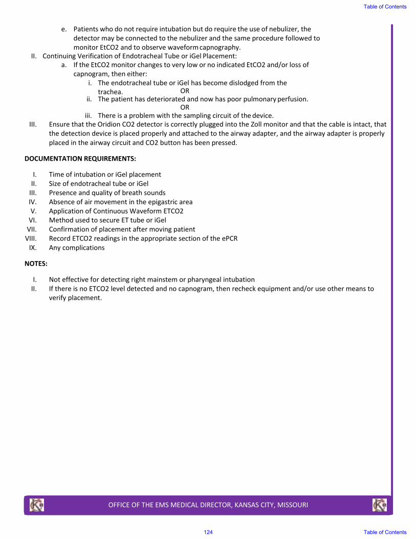
e. Patients who do not require intubation but do require the use of nebulizer, the
detector may be connected to the nebulizer and the same procedure followed to
monitor EtCO2 and to observe waveform capnography.
II. Continuing Verification of Endotracheal Tube or iGel Placement:
a. If the EtCO2 monitor changes to very low or no indicated EtCO2 and/or loss of capnogram, then either:
i. The endotracheal tube or iGel has become dislodged from the
trachea. OR ii. The patient has deteriorated and now has poor pulmonary perfusion.
OR iii. There is a problem with the sampling circuit of the device.
III. Ensure that the Oridion CO2 detector is correctly plugged into the Zoll monitor and that the cable is intact, that
the detection device is placed properly and attached to the airway adapter, and the airway adapter is properly
placed in the airway circuit and CO2 button has been pressed.
DOCUMENTATION REQUIREMENTS:
I. Time of intubation or iGel placement
II. Size of endotracheal tube or iGel
III. Presence and quality of breath sounds
IV. Absence of air movement in the epigastric area
V. Application of Continuous Waveform ETCO2
VI. Method used to secure ET tube or iGel
VII. Confirmation of placement after moving patient
VIII. Record ETCO2 readings in the appropriate section of the ePCR
IX. Any complications
NOTES:
I. Not effective for detecting right mainstem or pharyngeal intubation
II. If there is no ETCO2 level detected and no capnogram, then recheck equipment and/or use other means to
verify placement.
OFFICE OF THE EMS MEDICAL DIRECTOR, KANSAS CITY, MISSOURI
Table of Contents
124 Table of Contents
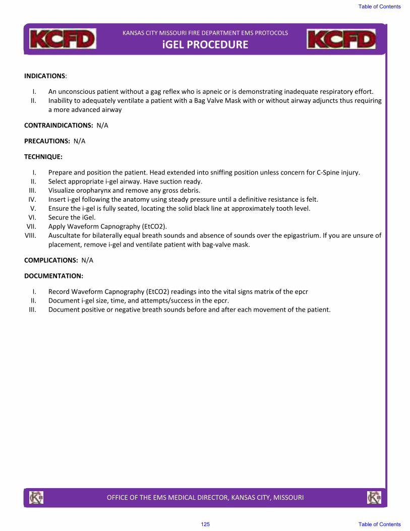
INDICATIONS:
I. An unconscious patient without a gag reflex who is apneic or is demonstrating inadequate respiratory effort.
II. Inability to adequately ventilate a patient with a Bag Valve Mask with or without airway adjuncts thus requiring
a more advanced airway
CONTRAINDICATIONS: N/A
PRECAUTIONS: N/A
TECHNIQUE:
I. Prepare and position the patient. Head extended into sniffing position unless concern for C-Spine injury.
II. Select appropriate i-gel airway. Have suction ready.
III. Visualize oropharynx and remove any gross debris.
IV. Insert i-gel following the anatomy using steady pressure until a definitive resistance is felt.
V. Ensure the i-gel is fully seated, locating the solid black line at approximately tooth level.
VI. Secure the iGel.
VII. Apply Waveform Capnography (EtCO2).
VIII. Auscultate for bilaterally equal breath sounds and absence of sounds over the epigastrium. If you are unsure of
placement, remove i-gel and ventilate patient with bag-valve mask.
COMPLICATIONS: N/A
DOCUMENTATION:
I. Record Waveform Capnography (EtCO2) readings into the vital signs matrix of the epcr
II. Document i-gel size, time, and attempts/success in the epcr.
III. Document positive or negative breath sounds before and after each movement of the patient.
KANSAS CITY MISSOURI FIRE DEPARTMENT EMS PROTOCOLS
iGEL PROCEDURE
OFFICE OF THE EMS MEDICAL DIRECTOR, KANSAS CITY, MISSOURI
Table of Contents
125 Table of Contents
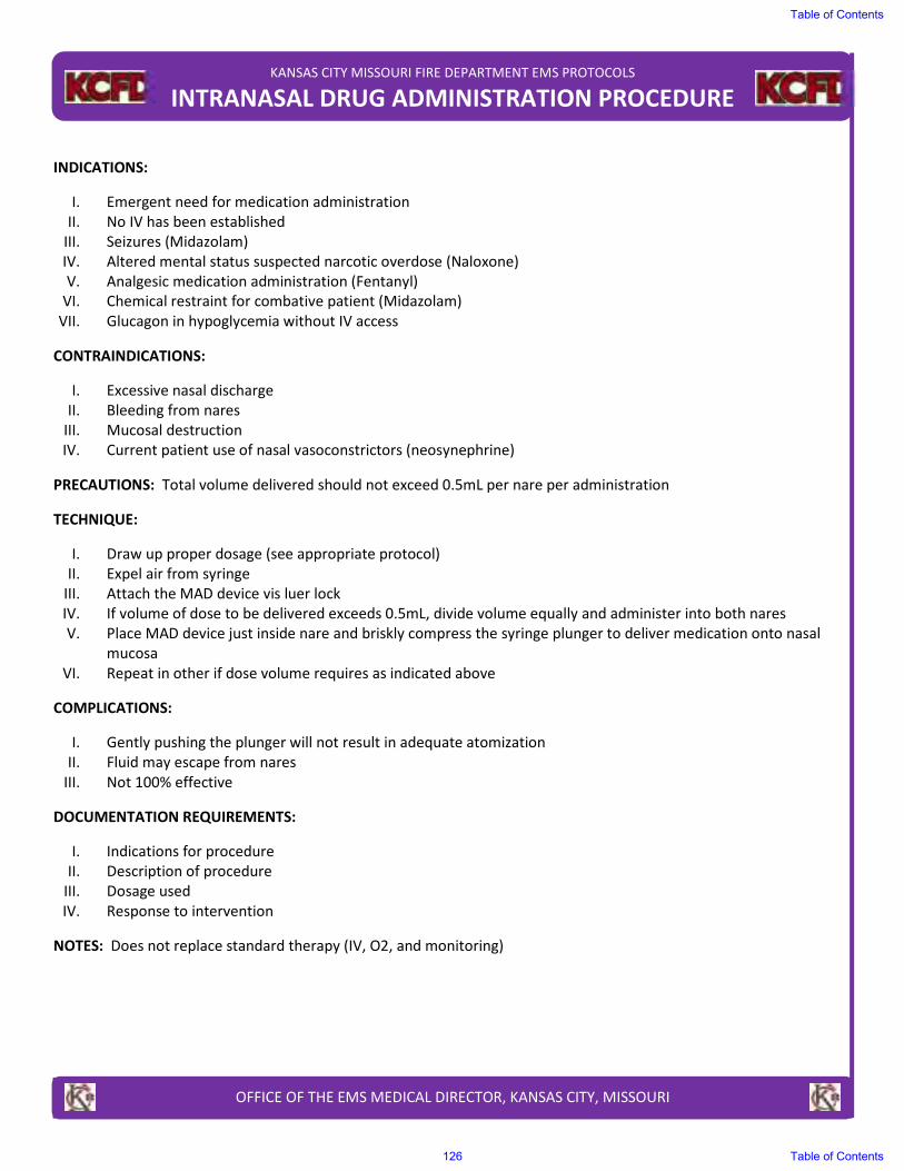
INDICATIONS:
I. Emergent need for medication administration
II. No IV has been established
III. Seizures (Midazolam)
IV. Altered mental status suspected narcotic overdose (Naloxone)
V. Analgesic medication administration (Fentanyl)
VI. Chemical restraint for combative patient (Midazolam)
VII. Glucagon in hypoglycemia without IV access
CONTRAINDICATIONS:
I. Excessive nasal discharge
II. Bleeding from nares
III. Mucosal destruction
IV. Current patient use of nasal vasoconstrictors (neosynephrine)
PRECAUTIONS: Total volume delivered should not exceed 0.5mL per nare per administration
TECHNIQUE:
I. Draw up proper dosage (see appropriate protocol)
II. Expel air from syringe
III. Attach the MAD device vis luer lock
IV. If volume of dose to be delivered exceeds 0.5mL, divide volume equally and administer into both nares
V. Place MAD device just inside nare and briskly compress the syringe plunger to deliver medication onto nasal
mucosa
VI. Repeat in other if dose volume requires as indicated above
COMPLICATIONS:
I. Gently pushing the plunger will not result in adequate atomization
II. Fluid may escape from nares
III. Not 100% effective
DOCUMENTATION REQUIREMENTS:
I. Indications for procedure
II. Description of procedure
III. Dosage used
IV. Response to intervention
NOTES: Does not replace standard therapy (IV, O2, and monitoring)
KANSAS CITY MISSOURI FIRE DEPARTMENT EMS PROTOCOLS
INTRANASAL DRUG ADMINISTRATION PROCEDURE
OFFICE OF THE EMS MEDICAL DIRECTOR, KANSAS CITY, MISSOURI
Table of Contents
126 Table of Contents
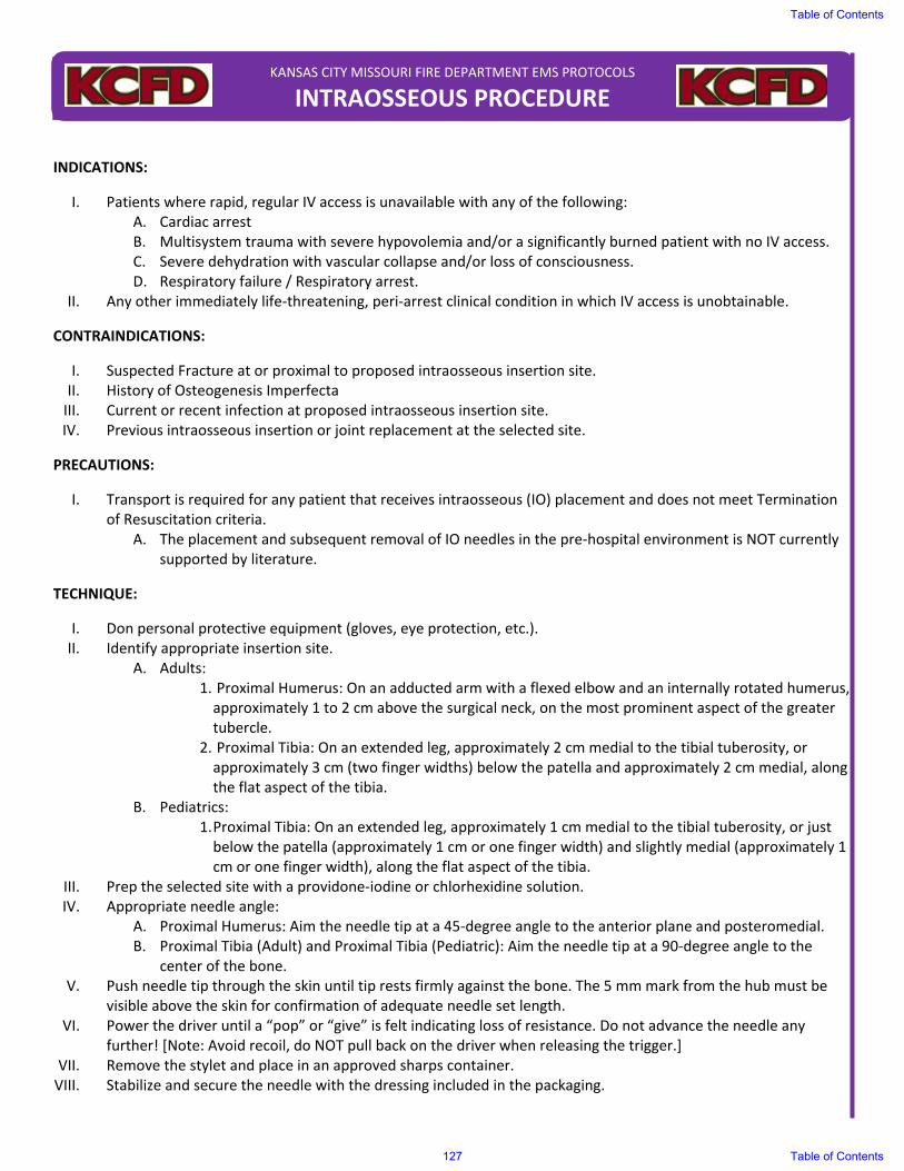
INDICATIONS:
I. Patients where rapid, regular IV access is unavailable with any of the following:A. Cardiac arrestB. Multisystem trauma with severe hypovolemia and/or a significantly burned patient with no IV access.C. Severe dehydration with vascular collapse and/or loss of consciousness.D. Respiratory failure / Respiratory arrest.
II. Any other immediately life-threatening, peri-arrest clinical condition in which IV access is unobtainable.
CONTRAINDICATIONS:
I. Suspected Fracture at or proximal to proposed intraosseous insertion site.II. History of Osteogenesis Imperfecta
III. Current or recent infection at proposed intraosseous insertion site.IV. Previous intraosseous insertion or joint replacement at the selected site.
PRECAUTIONS:
I. Transport is required for any patient that receives intraosseous (IO) placement and does not meet Termination of Resuscitation criteria.
A. The placement and subsequent removal of IO needles in the pre-hospital environment is NOT currently supported by literature.
TECHNIQUE:
I. Don personal protective equipment (gloves, eye protection, etc.).II. Identify appropriate insertion site.
A. Adults:1. Proximal Humerus: On an adducted arm with a flexed elbow and an internally rotated humerus,
approximately 1 to 2 cm above the surgical neck, on the most prominent aspect of the greatertubercle.
2. Proximal Tibia: On an extended leg, approximately 2 cm medial to the tibial tuberosity, orapproximately 3 cm (two finger widths) below the patella and approximately 2 cm medial, alongthe flat aspect of the tibia.
B. Pediatrics:1. Proximal Tibia: On an extended leg, approximately 1 cm medial to the tibial tuberosity, or just
below the patella (approximately 1 cm or one finger width) and slightly medial (approximately 1cm or one finger width), along the flat aspect of the tibia.
III. Prep the selected site with a providone-iodine or chlorhexidine solution.IV. Appropriate needle angle:
A. Proximal Humerus: Aim the needle tip at a 45-degree angle to the anterior plane and posteromedial.B. Proximal Tibia (Adult) and Proximal Tibia (Pediatric): Aim the needle tip at a 90-degree angle to the
center of the bone.V. Push needle tip through the skin until tip rests firmly against the bone. The 5 mm mark from the hub must be
visible above the skin for confirmation of adequate needle set length.VI. Power the driver until a “pop” or “give” is felt indicating loss of resistance. Do not advance the needle any
further! [Note: Avoid recoil, do NOT pull back on the driver when releasing the trigger.]VII. Remove the stylet and place in an approved sharps container.
VIII. Stabilize and secure the needle with the dressing included in the packaging.
KANSAS CITY MISSOURI FIRE DEPARTMENT EMS PROTOCOLS
INTRAOSSEOUS PROCEDURE
Table of Contents
127 Table of Contents
IX. Verify needle placement. A. Needle feels secure. B. Attach a syringe filled with at least 5 ml NS C. Aspirate bone marrow
X. Inject at least 5-10 ml of NS in an adult or 2-5 cc of NS in a pediatric patient to clear the lumen of the needle and open the intramedullary space.
XI. Attach the IV line and adjust flow rate. A pressure bag may assist with achieving desired flows. XII. Re-verify placement prior to each infusion and assess frequently for complications, including extravasation
which can lead to compartment syndrome. Following the administration of any intraosseous medications, flush the line with IV fluid.
OFFICE OF THE EMS MEDICAL DIRECTOR, KANSAS CITY, MISSOURI
Table of Contents
128 Table of Contents
Nasogatric/Orogastric intubation is indicated for decompression of a distended stomach caused by aggressive positive-
pressure ventilation. It should always be preceded by airway or intubation of the trachea with an endotracheal tube, in
order to protect the airway from possible aspiration.
INDICATIONS: Cardiopulmonary arrest with gastric distension compromising ventilation efforts.
CONTRAINDICATIONS:
I. Conscious patient
II. Non-intubated patient
III. The nasogastric tube should not be used in those patients with midface trauma. If the cribriform plate has been
fractured, the tube could be placed in the patient’s brain.
PRECAUTIONS:
I. Despite careful technique, the Levine tube may become lodged in the trachea. It is imperative that ET and
Levine tube placement be monitored continuously.
II. Incorrect NG insertion technique may cause trauma and bleeding from the nasal turbinates. If placed
incorrectly, pull back gently on the Levine tube and advance again.
III. The tube may coil up in the posterior pharynx. Visualize the patient’s oropharynx. If a coil of the Levine tube is
visualized, pull back gently until the tip of the tube is visible in the posterior pharynx, then advance the tube
again.
IV. The Levine tube is not to be used for medication or fluid administration.
TECHNIQUE:
I. Nasogastric Route (Adults Only)
A. Lubricate the tip and the first few inches of a number 16 french Levine tube generously with water-
soluble lubricant.
B. Pass the tube GENTLY along the floor of the nasal passage.
C. As the tube begins to enter the oropharynx, you will feel a lessening of resistance of the tube. Advance
the tip of the tube into the stomach. The appropriate length to insert is approximately equal to the
distance between the nose and the umbilicus.
D. Verify correct placement by connecting an irrigating syringe, pushing 20mL of air into the Levine tube,
and auscultating over the stomach for the sound of gurgling.
E. Connect the Levine tube to portable or on-board suction (low) to provide continuous decompression.
II. Orogastric Route (Children and Adults)
A. Using a laryngoscope and proper size blade, visualize the trachea and insure the ET tube is properly
placed.
B. Visualize the esophagus immediately posterior to the trachea. Quickly advance the lubricated Levine
tube (number 16 french for adults, number 12 french for children) through the mouth into the
esophagus.
C. Advance the tip of the tube into the stomach. The appropriate length to insert is approximately equal to
the distance between the mouth and the umbilicus.
D. Verify correct placement by connecting an irrigating syringe, pushing 20mL of air into the Levine tube,
and auscultating over the stomach for the sound of gurgling.
E. Connect the Levine tube to portable or on-board suction (low) to provide continuous decompression.
KANSAS CITY MISSOURI FIRE DEPARTMENT EMS PROTOCOLS
NASOGASTRIC/OROGASTRIC TUBE INSERTION PROCEDURE
Table of Contents
129 Table of Contents

COMPLICATIONS: N/A
DOCUMENTATION REQUIREMENTS: N/A
NOTES: N/A
OFFICE OF THE EMS MEDICAL DIRECTOR, KANSAS CITY, MISSOURI
Table of Contents
130 Table of Contents
INDICATIONS:
I. Tension pneumothorax is a clinical diagnosis which should be considered when the following signs/symptoms
are present:
A. Progressing severe respiratory distress
B. Progressing shock
C. Decreased or absent breath sounds on the involved side
D. Jugular venous distension
E. Tympany to percussion on the involved side
F. Tracheal deviation away from the involved side
II. 2 of the above signs/symptoms MUST be present or approval from a Medical Control should be obtained prior
to performing the procedure.
III. Tension pneumothorax is most common in the patient with:
A. Chest trauma
B. The intubated patient with high airway pressures causing rupture of the bronchioles or alveoli
CONTRADICTIONS: N/A
PRECAUTIONS: N/A
TECHNIQUE:
I. Locate the second intercostal space in the mid clavicular line on the involved side of the chest.
II. Cleanse with betadine or alcohol as time permits.
III. Insert a large gauge “over the needle” catheter (use 18 gauge if < 2 months age) through the open end of a glove
finger tip, puncture, and advance needle to hub of catheter to establish the flutter valve mechanism.
IV. Insert the needle/catheter with attached flutter valve into the chest just over the top of the third rib.
V. As you enter the pleural space air and/or blood will escape.
VI. Advance the catheter and remove the needle.
VII. Secure in place.
VIII. The catheter has a tendency to kink. If reaccumulation of air in the pleural space is occurring, proceed with
repeat needle thoracostomy.
COMPLICATIONS:
I. Creation of pneumothorax.
II. Damage to lung or viscera.
III. Bleeding (intercostal vessels are below each rib, therefore always go above the rib).
IV. Infection.
DOCUMENTATION REQUIREMENTS:
I. Indications for procedure
II. Technique used
III. Response to intervention
NOTES: N/A
KANSAS CITY MISSOURI FIRE DEPARTMENT EMS PROTOCOLS
NEEDLE THORACOSTOMY PROCEDURE
OFFICE OF THE EMS MEDICAL DIRECTOR, KANSAS CITY, MISSOURI
Table of Contents
131 Table of Contents
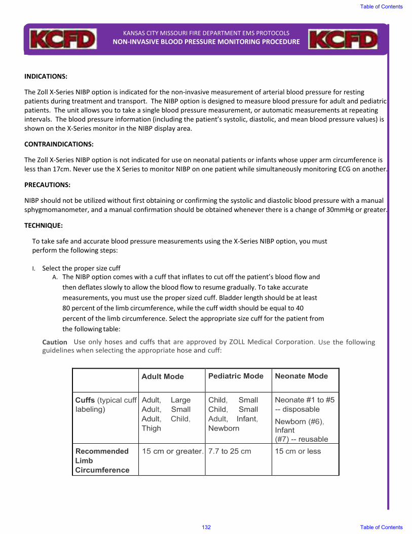
INDICATIONS:
The Zoll X-Series NIBP option is indicated for the non-invasive measurement of arterial blood pressure for resting
patients during treatment and transport. The NIBP option is designed to measure blood pressure for adult and pediatric
patients. The unit allows you to take a single blood pressure measurement, or automatic measurements at repeating
intervals. The blood pressure information (including the patient’s systolic, diastolic, and mean blood pressure values) is
shown on the X-Series monitor in the NIBP display area.
CONTRAINDICATIONS:
The Zoll X-Series NIBP option is not indicated for use on neonatal patients or infants whose upper arm circumference is
less than 17cm. Never use the X Series to monitor NIBP on one patient while simultaneously monitoring ECG on another.
PRECAUTIONS:
NIBP should not be utilized without first obtaining or confirming the systolic and diastolic blood pressure with a manual
sphygmomanometer, and a manual confirmation should be obtained whenever there is a change of 30mmHg or greater.
TECHNIQUE:
To take safe and accurate blood pressure measurements using the X-Series NIBP option, you must
perform the following steps:
I. Select the proper size cuff
A. The NIBP option comes with a cuff that inflates to cut off the patient’s blood flow and
then deflates slowly to allow the blood flow to resume gradually. To take accurate
measurements, you must use the proper sized cuff. Bladder length should be at least
80 percent of the limb circumference, while the cuff width should be equal to 40
percent of the limb circumference. Select the appropriate size cuff for the patient from
the following table:
Caution Use only hoses and cuffs that are approved by ZOLL Medical Corporation. Use the following
guidelines when selecting the appropriate hose and cuff:
Adult Mode Pediatric Mode Neonate Mode
Cuffs (typical cuff labeling)
Adult, Large Adult, Small Adult, Child, Thigh
Child, Small Child, Small Adult, Infant, Newborn
Neonate #1 to #5 -- disposable Newborn (#6), Infant (#7) -- reusable
Recommended
Limb
Circumference
15 cm or greater. 7.7 to 25 cm 15 cm or less
KANSAS CITY MISSOURI FIRE DEPARTMENT EMS PROTOCOLS
NON-INVASIVE BLOOD PRESSURE MONITORING PROCEDURE
Table of Contents
132 Table of Contents
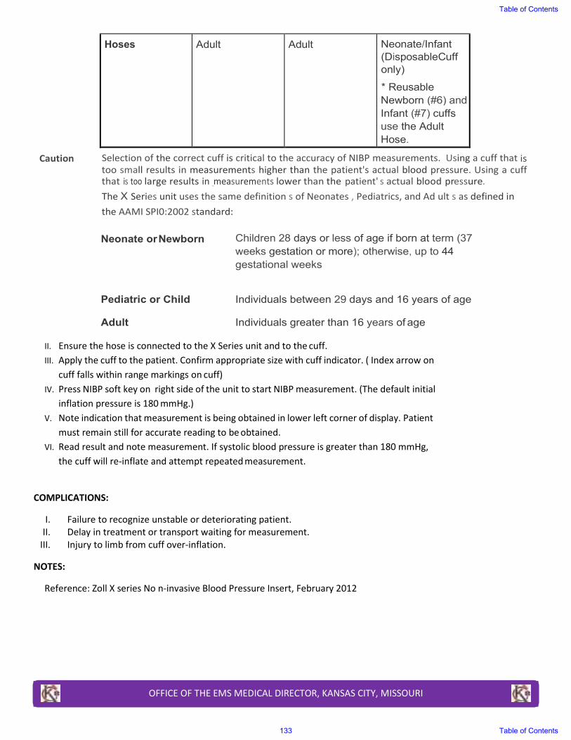
Hoses Adult Adult Neonate/Infant (DisposableCuff only)
* ReusableNewborn (#6) and Infant (#7) cuffsuse the AdultHose.
Caution Selection of the correct cuff is critical to the accuracy of NIBP measurements. Using a cuff that is
too small results in measurements higher than the patient's actual blood pressure. Using a cuff
that is too large results in _measurements lower than the patient' s actual blood pressure.
The X Series unit uses the same definition s of Neonates , Pediatrics, and Ad ult s as defined in
the AAMI SPI0:2002 standard:
Neonate or Newborn
Pediatric or Child
Adult
Children 28 days or less of age if born at term (37 weeks gestation or more); otherwise, up to 44 gestational weeks
Individuals between 29 days and 16 years of age
Individuals greater than 16 years of age
II. Ensure the hose is connected to the X Series unit and to the cuff.
III. Apply the cuff to the patient. Confirm appropriate size with cuff indicator. ( Index arrow on
cuff falls within range markings on cuff)
IV. Press NIBP soft key on right side of the unit to start NIBP measurement. (The default initial
inflation pressure is 180 mmHg.)
V. Note indication that measurement is being obtained in lower left corner of display. Patient
must remain still for accurate reading to be obtained.
VI. Read result and note measurement. If systolic blood pressure is greater than 180 mmHg,
the cuff will re-inflate and attempt repeated measurement.
COMPLICATIONS:
I. Failure to recognize unstable or deteriorating patient.
II. Delay in treatment or transport waiting for measurement.
III. Injury to limb from cuff over-inflation.
NOTES:
Reference: Zoll X series No n-invasive Blood Pressure Insert, February 2012
OFFICE OF THE EMS MEDICAL DIRECTOR, KANSAS CITY, MISSOURI
Table of Contents
133 Table of Contents
INDICTATIONS:
I. An unconscious patient without a gag reflex who is apneic or is demonstrating inadequate respiratory effort.
II. Inability to adequately ventilate a patient with a Bag Valve Mask with or without airway adjuncts thus requiring
a more advanced airway.
CONTRAINDICATIONS: N/A
PRECAUTIONS: N/A
TECHNIQUE:
I. Prepare, position and oxygenate the patient with 100% Oxygen.
II. Select proper laryngoscope, ET tube and stylette, if used. Have suction, Waveform Capnography (EtCO2), and
back-up Endotracheal Tube Introducer (Gum Elastic Bougie) ready.
III. Using laryngoscope, visualize vocal cords. (Use Sellick maneuver/BURP to assist you if needed).
IV. Limit each intubation attempt to no more than 30 seconds with adequate re-oxygenation utilizing a BVM
between attempts.
V. Visualize ET tube passing through vocal cords.
VI. Inflate the cuff with 10mL of air.
VII. Confirm and document tube placement using Continuous Waveform Capnography (EtCO2).
VIII. Auscultate for bilaterally equal breath sounds and absence of sounds over the epigastrium. If you are unsure of
placement, remove tube and ventilate patient with bag-valve mask.
IX. Secure the tube to the patient.
X. After 3 ventilations, ETCO2 should be >20 or comparable to pre-intubation values. If < 20, check for adequate
circulation, equipment, and ventilatory rate. If ETCO2 still < 20 without physiologic explanation, remove the ET
Tube and ventilate by BVM.
DOCUMENTATION:
I. Record Waveform Capnography (EtCO2) readings in the vital signs matrix of the epcr.
II. Document ETT size, time, attempts/success, and placement location by the centimeter marks either at the
patient’s teeth or lips in the epcr.
III. Document positive or negative breath sounds before and after each movement of the patient.
KANSAS CITY MISSOURI FIRE DEPARTMENT EMS PROTOCOLS
ORAL ENDOTRACHEAL INTUBATION PROCEDURE
OFFICE OF THE EMS MEDICAL DIRECTOR, KANSAS CITY, MISSOURI
Table of Contents
134 Table of Contents
The purpose of this policy is to set forth principles and procedures for the prehospital patient restraint (PPR) of persons
at risk for harm to self or others, including emergency medical services personnel.
The safety of EMS personnel is paramount for the consideration, initiation, and application of PPR. Also, the protection
of patients from self-harm or harm to others while maintaining patient dignity to the greatest extent possible while
utilizing the least restrictive method of restraint appropriate to the situation.
This policy applies to all patients whom have demonstrated violence toward self or others, or there is reason to believe
that combative or violent behavior is imminent, through actions or verbalizations by the patient or reasonable layperson
bystanders.
INDICATIONS:
I. All patients who are demonstrating physical harm to self or others including, but not limited to, evidence of
suicidal or homicidal behavior, by act or verbalization.
II. All patients demonstrating decreased ability to care for self as evidence by, but not limited to, cognitive
impairment or suspected intoxication leading to exposure of self or others to potentially limb or life threatening
situations.
III. Patient with altered mental status leading to decreased capacity to understand situation and circumstances of
their current medical condition resulting in aggressive or combative behavior towards emergency personnel.
IV. Patients in custody of law enforcement or under involuntary detention.
CONTRAINCATIONS: N/A
PRECAUTIONS: N/A
TECHNIQUE:
I. When at all possible, the patient should be approached in a calm, non-threatening and supporting manner.
Verbal de-escalation efforts should be attempted unless there is imminent danger to patient or bystanders. If
the EMS personnel believe that their personal safety is at risk, law enforcement should be contacted for
assistance.
II. When imminent danger to the patient or others is identified, a unified, multiple emergency personnel approach
should be made to gain control of the patient. Placement of physical restraints, including approved hood with
spit shield, approved soft wrist and ankle restraints should be considered first. Progressive restraint beginning
with wrists, then ankles, to immobilization on long spine board should be determined to provide the least
amount of restraint necessary to accomplish protection of crew and patient.
III. Hard extremity restraints including handcuffs and shackles should be used only when other methods fail or
unusable due to circumstances and efforts to replace with approved restraints made.
IV. If handcuffs or shackles are used, they should only be placed by appropriate law enforcement personnel and
when law enforcement will travel with the patient or in immediate vicinity of the ambulance. EMS personnel
should always have available, the means to immediately remove restraints.
V. Patients under arrest shall be thoroughly searched by law enforcement prior to being placed into any
ambulance. Law enforcement agencies retain the responsibility for safe transport of patients under arrest or
involuntary detention.
VI. Patients should be restrained in a supine position and immobilized on a long spine board whenever possible. It
is never appropriate to “sandwich” patient between boards, or to “hog-tie” in any fashion a patient for transport
by EMS. Any restraint placed should not compromise normal respiratory efforts or circulation.
KANSAS CITY MISSOURI FIRE DEPARTMENT EMS PROTOCOLS
PATIENT RESTRAINT PROCEDURE
Table of Contents
135 Table of Contents
VII. Vital signs and patient monitoring should be accomplished as soon as possible after placement of appropriate
restraints. Potential causes of the behavior, including hypoxemia and hypoglycemia, should be considered and
efforts to identify and manage as soon as possible.
VIII. ONCE PHYSICAL RESTRAINTS ARE IN PLACE, CHEMICAL RESTRAINT SHOULD BE STRONGLY CONSIDERED AND
ADMINISTERED IF THE PATIENT CONTINUES TO EXHIBIT BEHAVIOR IN RESTRAINTS THAT COULD HARM SELF
OR OTHERS. CHEMICAL RESTRAINT SHOULD ONLY BE UTILITZED WHEN THE PATIENT CAN BE ADEQUATELY
AND CONTINUOUSLY MONITORED BY THE PARAMEDIC. REFER TO COMBINED BEHAVIORAL/PSYCHIATRIC
DISORDER PROTOCOL
IX. Once restrained, the patient should be isolated and placed into the ambulance as soon as possible and
transported to the closest appropriate facility as per the Determination of Hospital Destination Policy.
X. Physical restraints should be reassessed at a minimum of every ten minutes during transport for the first hour,
and every thirty minutes thereafter. The restrained extremities should be assessed for neurovascular function
and documented.
XI. Always considered consultation with on-line medical control for questions or assistance in determining the most
appropriate restraint method for any situation. Patients with medical conditions that appear to compromise
their ability to consent for or decline care may be restrained as necessary and transported without law
enforcement authority in situations in which a life-threatening emergency exists or potentially exists. Medical
control should be contacted in all instances where patients are transported against their consent unless under
arrest or duly exercised involuntary detention.
COMPLICATIONS: N/A
DOCUMENTATION REQUIREMENTS:
I. Detailed description of scene, situation, and patient behaviors
II. Note assistance by other emergency providers
III. Complete vital signs including pulse oximetry initially and at hospital arrival at a minimum
IV. EKG rhythm analysis
V. Detailed description of patient injuries and any bystander injuries
VI. Description of restraint method(s) and re-examination(s)
OFFICE OF THE EMS MEDICAL DIRECTOR, KANSAS CITY, MISSOURI
Table of Contents
136 Table of Contents
The pulse oximeter is a cutaneous monitor used as an adjunct in the assessment of respiratory issues. The device also
assists in evaluating improvement or deterioration during treatment. This device is never used to withhold O2 to a
patient who needs it. Any patient who would currently receive O2 per system protocol, or who appears to clinically
need it, should continue to be given oxygen.
INDICATIONS:
I. The following is a partial list of situations where pulse oximetry may be used:
A. Only for use in perfusing patient
B. Respiratory disorders (e.g. Asthma, COPD, respiratory distress, airway obstruction or injury)
C. Cardiovascular disorders (e.g. CHF, chest pain, dysrhythmia)
D. Altered mental status (e.g. Coma, Overdose, CVA, Seizures)
E. Trauma
II. The pulse oximeter must be used prior to and after intubation or assisted ventilation of the perfusing patient
III. The pulse oximeter must be used prior to and after administering sedative agents
CONTRAINDICATIONS:
I. Non-perfusing rhythm
PRECAUTIONS:
I. Pulse oximetry values may be inaccurate in a variety of situations:
a. Inaccurate readings can be seen with patient movement, the presence of nail polish, vasoconstriction,
decreased peripheral perfusion, hypotension, hypothermia, abnormal hemoglobins, hypovolemia,
carbon monoxide, poisoning, smoke inhalation, and methemoglobinemia
b. Prehospital personnel should correlate the SaO2 reading with the clinical status of the patient
II. Sickle cell anemia (readings are generally falsely low secondary to the abnormal hemoglobin molecule)
TECHNIQUE:
I. Check vital signs
II. Turn on the Zoll X-Series monitor
III. Ensure that the pulse oximetry probe cable is connected to the monitor.
IV. Select appropriate site. Avoid placing the probe on areas distal to orthopedic injuries
or distal to a blood pressure cuff
V. Place probe on the patient
VI. Read the pulse rate, O2 saturation, and document findings at least every 10 minutes and with
any change in therapy or clinical condition
VII. Oxygen will be applied or increased according to the clinical setting. Although normal SaO2
levels are > 95%, SaO2 levels above 90% are generally acceptable in almost any adult
patient. Pediatric patients in respiratory distress should be placed on supplemental
oxygen regardless of the oxygen saturation reading
A. For patient's not on home O2 therapy, oxygen should be applied via nasal cannula
or mask per system protocol
B. Patients currently on chronic home O2 therapy should have an initial SaO2 reading
done. Oxygen may be increased until SaO2 levels of 90%-92% are obtained.
Reference: Zoll X-Series Pulse Oximetry (SpO2) Insert, February 2012
KANSAS CITY MISSOURI FIRE DEPARTMENT EMS PROTOCOLS
PULSE OXIMETRY PROCEDURE
OFFICE OF THE EMS MEDICAL DIRECTOR, KANSAS CITY, MISSOURI
Table of Contents
137 Table of Contents
INDICATIONS: For use in the management of bradycardia, hypotension/shock, and post-resuscitation care
CONTRAINDICATIONS: Known hypersensitivity
PRECAUTIONS: In patients taking digitalis, epinephrine may exacerbate ventricular ectopy
TECHNIQUE:
I. Take a 10mL saline flush and eject 1mL of normal saline II. Into this syringe draw up 1mL of Epinephrine from the 1:10000 preload.
III. This now gives you 100mcg Epinephrine 1:100000 in 10mL syringe, 10mcg/mL
COMPLICATIONS: N/A
DOCUMENTATION REQUIREMENTS:
I. Time medication given II. Paramedic giving medication
III. Dose given IV. Route given
NOTES:
KANSAS CITY MISSOURI FIRE DEPARTMENT EMS PROTOCOLS
EPINEPHRINE 1:100000 (PUSH DOSE) PROCEDURE
OFFICE OF THE EMS MEDICAL DIRECTOR, KANSAS CITY, MISSOURI
Table of Contents
138 Table of Contents
INDICATIONS:
I. Any suspected alteration in glucose level that might impact patient assessment or treatment. This could include
patients with:
A. Altered Mental Status
B. Behavioral or Psychiatric Disorders
C. Hyper or Hypothermia
D. Ingestions or Overdose
E. Seizures
F. Cardiac Arrest
CONTRAINDICATIONS: N/A
PRECAUTIONS: N/A
I. Airway, breathing, and circulation should be assessed and treated in the initial evaluation of every patient and
obtaining a rapid bedside glucose determination should not interfere with this evaluation and treatment.
II. If problems arise with the device, obtaining a rapid bedside glucose determination should not delay the
administration of glucose, if indicated.
TECHNIQUE: Refer to the manufacturer’s instructions for specific directions on the utilization of each model of
glucometer in use.
COMPLICATIONS:
I. Pain, infection or bleeding at the site of lancet (or IV) stick.
II. Delay in administration of glucose.
DOCUMENTATION REQUIREMENTS:
I. Include the following (minimum):
A. The blood glucose level obtained
B. Complications
NOTES: N/A
KANSAS CITY MISSOURI FIRE DEPARTMENT EMS PROTOCOLS
RAPID GLUCOSE DETERMINATION PROCEDURE
OFFICE OF THE EMS MEDICAL DIRECTOR, KANSAS CITY, MISSOURI
Table of Contents
139 Table of Contents
BACKGROUND: Spinal Motion Restriction utilizes a cervical collar, the cot mattress, and adequately and appropriately securing the patient in order to limit the excessive motion of the spinal column during patient transport. The goal is to minimize secondary injury to spine in patients who have, or may have, an unstable spinal injury, and most importantly, to minimize patient morbidity from the use of immobilization devices.
INDICATIONS:
Spinal Motion Restriction is indicated in trauma patients who sustain a mechanism of injury sufficient to cause a neck or back injury.
I. GCS < 15 II. Evidence of intoxication
III. Painful or distracting injury IV. Pain during unassisted range of motion V. Signs of spinal cord injury such as:
A. Sensory loss B. Weakness C. Paralysis D. Numbness, tingling, or burning E. Pain, deformity, or tenderness to the neck or back
VI. Exercise caution for injury and consider c-collar placement: A. Patients over 65 years old B. Patients under 5 years old C. Any patient with a history of osteoporosis, bone disease, or vertebral disease
VII. Patients presenting to KCFD on a LSB from another medical provider may be subject to the SMR Procedure under the Spinal Trauma Protocol
CONTRAINDICATIONS:
I. Some patients, due to size or age, will not be able to be managed through in-line stabilization and standard cervical collars. Never force a patient from a non-neutral position. Manual stabilization or improvised padding may be required during transport
II. Patients with penetrating injury to the neck should not be placed in a cervical collar or other spinal precautions regardless of whether they are exhibiting neurologic symptoms or not. Doing so can lead to delayed identification of injury or airway compromise, and has been associated with increased mortality
PRECAUTIONS:
I. Assess the scene to determine the mechanism of injury II. Patients may be safely immobilized by being placed supine on gurney with cervical collar and straps and will not
generally require a spine board III. Reserve long spine board use for the movement of patients whose injuries limit ambulation. Remove from the
long spine board as soon as is practical IV. If a patient absolutely needs transport on a spinal board, long boards should be padded V. These patients (except in cardiac arrest) should NOT be transported to the hospital while still secured to a long
spine board. Dangers of transporting a patient on a long spine board include but are not limited to; A. Potential airway compromise or aspiration in immobilized patient with nausea/vomiting, or with
facial/oral bleeding B. Excessively tight immobilization straps can limit chest excursion and cause hypoventilation C. Prolonged immobilization on spine board can lead to ischemic pressure injuries to skin
KANSAS CITY MISSOURI FIRE DEPARTMENT EMS PROTOCOLS
SPINAL MOTION RESTRICTION PROCEDURE
Table of Contents
140 Table of Contents
D. Prolonged immobilization on spine board can be very uncomfortable for patient VI. Children are abdominal breathers, so immobilization straps should go across chest and pelvis and not across the
abdomen, when possible VII. Children have disproportionately larger heads. When securing pediatric patients to a spine board, the board
should have a recess for the head, or the body should be elevated approximately 1-2 cm to accommodate the larger head size and avoid neck flexion when immobilized
VIII. In an uncooperative patient, avoid interventions that may promote increased spinal movement
TECHNIQUE:
I. Gather appropriate devices II. Assess neurological exam and distal pulses pre-procedure
III. While maintaining in-line stabilization in a neutral/middle position, place the patient in an appropriately sized cervical collar (stabilization should not involve traction or tension)
IV. If patient is clinically stable, alert, and without neurological deficits: A. If currently ambulating, position patient onto the stretcher and place supine B. If patient is sitting in vehicle or chair and feels able to, they may self-extricate to the stretcher C. If patient is sitting in vehicle or chair and feels unable to self-extricate, use a long spine board to
extricate from vehicle or chair to cot, removing the patient from the long spine board via log-roll after placement on the stretcher.
D. If patient is lying on the ground or other surface, use a long spine board or scoop stretcher to move the patient to the stretcher, removing the patient from the long spine board via log-roll after placement on the stretcher
V. Ensure that the patient is adequately secured to the stretcher, ensuring minimal movement VI. Preferred position for transport is supine
VII. Manual in-line stabilization must be used during any transfer VIII. Assess neurological exam and distal pulses post-procedure
IX. All stretcher to stretcher transfers should be accomplished using manual in-line stabilization.
DOCUMENTATION:
I. History and physical exam findings indicating the use of SMR II. Patient complaint of neck or spine pain
III. Spinal tenderness IV. Mental status/GCS V. Neurologic examination
VI. Evidence of intoxication VII. Documentation of multiple trauma
VIII. Documentation of mechanism of injury IX. Document patient capacity with: Any and all barriers to patient care X. Patient age
XI. Patients under age and not emancipated: Guardian name, contact, and relationship XII. How the patient was moved to the ambulance cot
XIII. Neurologic exam before and after movement and/or intervention XIV. Removal from spine board
NOTES:
From NASEMSO Model Clinical Guidelines
OFFICE OF THE EMS MEDICAL DIRECTOR, KANSAS CITY, MISSOURI
Table of Contents
141 Table of Contents
INDICATIONS:
I. Signs of poor perfusion in the presence of bradycardia
A. Altered mental status
B. Chest Pain
C. CHF
D. Shortness of Air
E. Syncope
F. Hypotension/Shock
II. Bradycardic/asystolic cardiac arrest
A. The outcome of prolonged bradycardic/asystolic cardiac arrest is dismal even with pacing.
Indiscriminate pacing of this rhythm is unwarranted.
B. Pacing of bradycardic/asystole of short duration, especially post countershock bradycardic/asystole is
more likely to be useful.
CONTRAINDICATIONS: Prophylactic pacing – the pacer electrodes may be placed in the stable bradycardic patient by
should not be used unless the patient deteriorates.
PRECAUTIONS: Transcutaneous pacing is not the initial step in treatment of either unstable bradycardia or
bradycardic/systolic cardiac arrest. Appropriate BLS and ALS maneuvers, as mandated by protocol, should be carried
out first.
TECHNIQUE:
I. Connect ECG electrodes to patient in standard positions (pacer will not operate without
ECG monitor intact).
II. Clean & dry chest. Remove excess hair if necessary to obtain electrode to skin contact. III. Connect pacing electrodes to matching pacing cable. IV. Peel off protective covering and attach electrodes to patient’s chest as described:
A. Anterior - Posterior (Preferred due to better capture rates and will not interfere with
defibrillation.)
1. Place negative electrode on left anterior chest halfway between the xiphoid
process and left nipple, with the upper edge of the electrode below the nipple
line.
2. Place positive electrode on left posterior chest beneath the scapula and lateral to
the spine.
B. Anterior – Anterior
1. Place negative electrode on left chest mid-axillary over fourth interspace.
2. Place positive electrode on right chest, sub- clavicular area.
V. Push “pacer” button.
VI. If no intrinsic beats then skip this step. Insure that each QRS complex is being sensed. If not, then adjust the QRS size and change between leads I, II, III to get the best size. VII. Select pacing rate
A. Adults and children ≥ 12 years, begin at 70 bpm.
B. Children ≥ 1 year and < 12 years, begin at 90 bpm.
C. Infants < 1 year, begin at 100 bpm. VIII. Activate pacer by pushing pacer button. The pacer will display a drop down menu to permit selection of rate, mA and to initiate and terminate pacing.
IX. Increase current by 10 mA increments until electrical capture occurs. (Note: The pacer
KANSAS CITY MISSOURI FIRE DEPARTMENT EMS PROTOCOLS
TRANSCUTANEOUS CARDIAC PACING PROCEDURE
Table of Contents
142 Table of Contents
adjusts upwards by 10 mA and downwards by 5 mA.)
X. Determine that mechanical capture has occurred. If so, consider using tape to secure electrodes to the patches to prevent displacement.
XI. To terminate pacing, select STOP from the pacer screen drop down box.
COMPLICATIONS: N/A
DOCUMENTATION REQUIREMENTS:
I. Monitor information uploaded to EPCR.
II. Documentation of indications for and response to pacing (including mechanical capture)
NOTES:
I. Patient discomfort
A. Pacing may be painful to the awake patient
B. Sedation may be useful (simple reassurance may also be useful)
II. Electrical capture
A. Usually evidenced by wide QRS
B. In some patients capture is less obvious and only manifested by minor changes in QRS configuration
III. Mechanical capture
A. Manifested by signs of improving cardiac output (i.e. increased level of consciousness or BP)
B. Do not confuse skeletal muscle contractions for carotid or femoral pulse
IV. CPR may be safely accomplished during this procedure
V. Defibrillation/cardioversion may be done without removing pacing electrodes. Charging the defibrillator
automatically prompts to turn off the pacer. When pacing is restarted, the pacer returns to the last used
setting.
VI. To obtain adequate tissue perfusion, rates may need to be higher than the initial setting.
VII. If pacing is not successful, consider further ALS interventions as indicated.
VIII. While pacing, if you initiate the defibrillation sequence by selecting an energy level, a pop-up box will display a
selection to stop pacing.
IX. After defibrillation, when reinitiating pacing, the machine returns to the last used settings.
OFFICE OF THE EMS MEDICAL DIRECTOR, KANSAS CITY, MISSOURI
Table of Contents
143 Table of Contents
INDICATIONS: Adult patients in medical cardiac arrest
CONTRAINDICATIONS:
I. Do not use in patients under 18 years old.II. Do not use in traumatic cardiac arrest.
III. Do not use on patients that weigh over 300lbs.IV. Do not use on pregnant patients.
PRECAUTIONS:
I. DO NOT DELAY INITIATION OF MANUAL CHEST COMPRESSIONS FOR DEPLOYMENT OF CPR DEVICE.II. IF AN ISSUE DEVELOPS WITH CPR DEVICE, IMMEDIATELY INITIATE MANUAL CHEST COMPRESSIONS.
III. The AutoPulse Platform is not intended to be the sole means of carrying the patient. The AutoPulse Platformshould be secured to the top of a backboard or other equipment used to carry or transport the patient. Duringtransport, regular checks of the patient’s alignment should be performed.
IV. Failure to properly position the LifeBand at the patient’s armpit line may cause injury to the patient.A. Do not place a strap across the LifeBand.B. Do not place your hands or any other objects on or under the LifeBand while the AutoPulse is analyzing
the patient of during active operation.V. Do not place or position the patient on the AutoPulse Platform in either a face down orientation or on the
patient’s side.A. Failure to properly position a patient, both vertically and laterally may cause injury.
TECHNIQUE:
I. Remove ALL clothing from torso (both front and back) to ensure skin-to-platform contact.II. Align armpits onto yellow line on Platform.
III. Do no twist the chest bandsIV. Maintain chest ands at 90 degrees to Platform and free of obstructionsV. Power up AutoPulse
VI. Close chest bandsVII. Press CONTINUE (green button)
VIII. Press START (green button) to begin compressionsIX. To pause or stop operation press STOP (orange button)
DOCUMENTATION REQUIREMENTS:
I. Initiation of manual chest compressions must be documented in the flow chart under the entry “CPR”.II. Time of deployment of CPR device must be documented in the flow chart under the entry “Mechanical CPR”.
III. Zoll EKG Monitor must be uploaded to ESO EHR.
NOTES:
• Information from Zoll AutoPulse Resuscitation System Model 100 User Guide and Quick Reference Guide
KANSAS CITY MISSOURI FIRE DEPARTMENT EMS PROTOCOLS
ZOLL AUTOPULSE CPR DEVICE
OFFICE OF THE EMS MEDICAL DIRECTOR, KANSAS CITY, MISSOURI
Table of Contents
144 Table of Contents
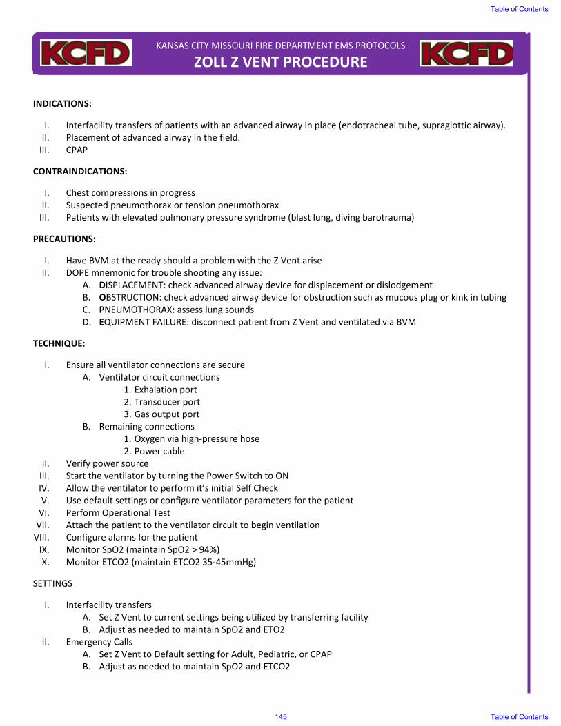
INDICATIONS:
I. Interfacility transfers of patients with an advanced airway in place (endotracheal tube, supraglottic airway).II. Placement of advanced airway in the field.
III. CPAP
CONTRAINDICATIONS:
I. Chest compressions in progressII. Suspected pneumothorax or tension pneumothorax
III. Patients with elevated pulmonary pressure syndrome (blast lung, diving barotrauma)
PRECAUTIONS:
I. Have BVM at the ready should a problem with the Z Vent ariseII. DOPE mnemonic for trouble shooting any issue:
A. DISPLACEMENT: check advanced airway device for displacement or dislodgementB. OBSTRUCTION: check advanced airway device for obstruction such as mucous plug or kink in tubingC. PNEUMOTHORAX: assess lung soundsD. EQUIPMENT FAILURE: disconnect patient from Z Vent and ventilated via BVM
TECHNIQUE:
I. Ensure all ventilator connections are secureA. Ventilator circuit connections
1. Exhalation port2. Transducer port3. Gas output port
B. Remaining connections1. Oxygen via high-pressure hose2. Power cable
II. Verify power sourceIII. Start the ventilator by turning the Power Switch to ONIV. Allow the ventilator to perform it’s initial Self CheckV. Use default settings or configure ventilator parameters for the patient
VI. Perform Operational TestVII. Attach the patient to the ventilator circuit to begin ventilation
VIII. Configure alarms for the patientIX. Monitor SpO2 (maintain SpO2 > 94%)X. Monitor ETCO2 (maintain ETCO2 35-45mmHg)
SETTINGS
I. Interfacility transfersA. Set Z Vent to current settings being utilized by transferring facilityB. Adjust as needed to maintain SpO2 and ETO2
II. Emergency CallsA. Set Z Vent to Default setting for Adult, Pediatric, or CPAPB. Adjust as needed to maintain SpO2 and ETCO2
KANSAS CITY MISSOURI FIRE DEPARTMENT EMS PROTOCOLS
ZOLL Z VENT PROCEDURE
Table of Contents
145 Table of Contents
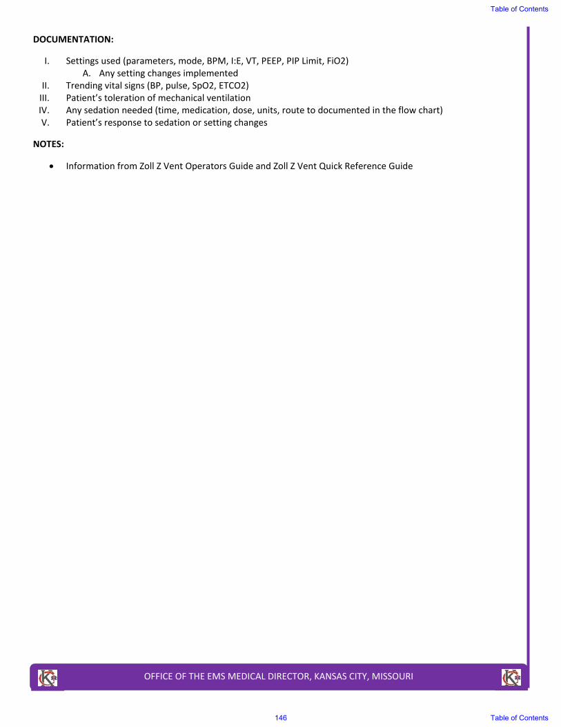
DOCUMENTATION:
I. Settings used (parameters, mode, BPM, I:E, VT, PEEP, PIP Limit, FiO2) A. Any setting changes implemented
II. Trending vital signs (BP, pulse, SpO2, ETCO2) III. Patient’s toleration of mechanical ventilation IV. Any sedation needed (time, medication, dose, units, route to documented in the flow chart) V. Patient’s response to sedation or setting changes
NOTES:
• Information from Zoll Z Vent Operators Guide and Zoll Z Vent Quick Reference Guide
OFFICE OF THE EMS MEDICAL DIRECTOR, KANSAS CITY, MISSOURI
Table of Contents
146 Table of Contents
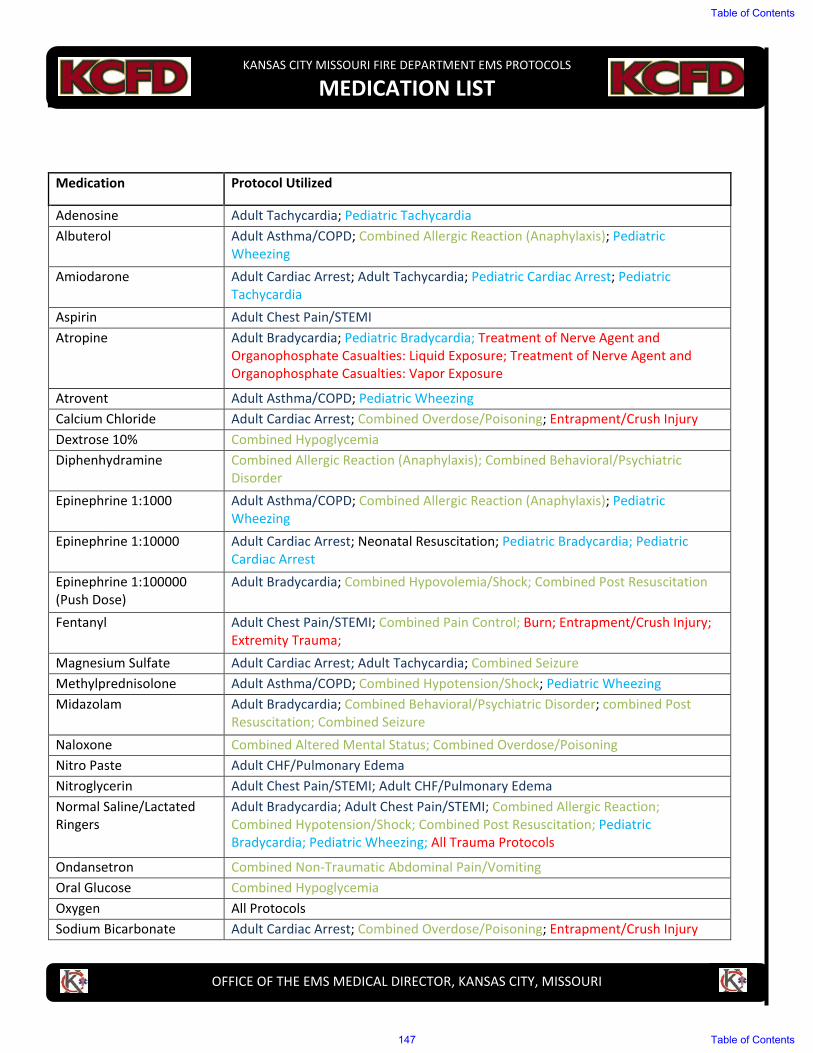
Medication Protocol Utilized
Adenosine Adult Tachycardia; Pediatric Tachycardia Albuterol Adult Asthma/COPD; Combined Allergic Reaction (Anaphylaxis); Pediatric
Wheezing Amiodarone Adult Cardiac Arrest; Adult Tachycardia; Pediatric Cardiac Arrest; Pediatric
Tachycardia Aspirin Adult Chest Pain/STEMI Atropine Adult Bradycardia; Pediatric Bradycardia; Treatment of Nerve Agent and
Organophosphate Casualties: Liquid Exposure; Treatment of Nerve Agent and Organophosphate Casualties: Vapor Exposure
Atrovent Adult Asthma/COPD; Pediatric Wheezing Calcium Chloride Adult Cardiac Arrest; Combined Overdose/Poisoning; Entrapment/Crush Injury Dextrose 10% Combined Hypoglycemia Diphenhydramine Combined Allergic Reaction (Anaphylaxis); Combined Behavioral/Psychiatric
Disorder Epinephrine 1:1000 Adult Asthma/COPD; Combined Allergic Reaction (Anaphylaxis); Pediatric
Wheezing Epinephrine 1:10000 Adult Cardiac Arrest; Neonatal Resuscitation; Pediatric Bradycardia; Pediatric
Cardiac Arrest Epinephrine 1:100000 (Push Dose)
Adult Bradycardia; Combined Hypovolemia/Shock; Combined Post Resuscitation
Fentanyl Adult Chest Pain/STEMI; Combined Pain Control; Burn; Entrapment/Crush Injury; Extremity Trauma;
Magnesium Sulfate Adult Cardiac Arrest; Adult Tachycardia; Combined Seizure Methylprednisolone Adult Asthma/COPD; Combined Hypotension/Shock; Pediatric Wheezing Midazolam Adult Bradycardia; Combined Behavioral/Psychiatric Disorder; combined Post
Resuscitation; Combined Seizure Naloxone Combined Altered Mental Status; Combined Overdose/Poisoning Nitro Paste Adult CHF/Pulmonary Edema Nitroglycerin Adult Chest Pain/STEMI; Adult CHF/Pulmonary Edema Normal Saline/Lactated Ringers
Adult Bradycardia; Adult Chest Pain/STEMI; Combined Allergic Reaction; Combined Hypotension/Shock; Combined Post Resuscitation; Pediatric Bradycardia; Pediatric Wheezing; All Trauma Protocols
Ondansetron Combined Non-Traumatic Abdominal Pain/Vomiting Oral Glucose Combined Hypoglycemia Oxygen All Protocols Sodium Bicarbonate Adult Cardiac Arrest; Combined Overdose/Poisoning; Entrapment/Crush Injury
KANSAS CITY MISSOURI FIRE DEPARTMENT EMS PROTOCOLS
MEDICATION LIST
OFFICE OF THE EMS MEDICAL DIRECTOR, KANSAS CITY, MISSOURI
Table of Contents
147 Table of Contents
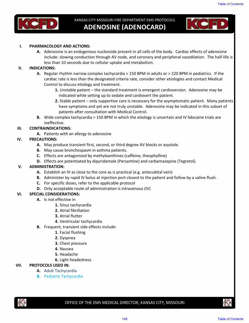
I. PHARMACOLOGY AND ACTIONS:
A. Adenosine is an endogenous nucleoside present in all cells of the body. Cardiac effects of adenosine
include: slowing conduction through AV node, and coronary and peripheral vasodilation. The half-life is
less than 10 seconds due to cellular uptake and metabolism.
II. INDICATIONS:
A. Regular rhythm narrow complex tachycardia > 150 BPM in adults or > 220 BPM in pediatrics. If the
cardiac rate is less than the designated criteria rate, consider other etiologies and contact Medical
Control to discuss etiology and treatment.
1. Unstable patient – the standard treatment is emergent cardioversion. Adenosine may be
indicated while setting up to sedate and cardiovert the patient.
2. Stable patient – only supportive care is necessary for the asymptomatic patient. Many patients
have symptoms and yet are not truly unstable. Adenosine may be indicated in this subset of
patients after consultation with Medical Control.
B. Wide complex tachycardia > 150 BPM in which the etiology is uncertain and IV lidocaine trials are
ineffective.
III. CONTRAINDICATIONS:
A. Patients with an allergy to adenosine
IV. PRECAUTIONS:
A. May produce transient first, second, or third degree AV blocks or asystole.
B. May cause bronchospasm in asthma patients.
C. Effects are antagonized by methylxanthines (caffeine, theophylline)
D. Effects are potentiated by dipyridamole (Persantine) and carbamazepine (Tegretol).
V. ADMINISTRATION:
A. Establish an IV as close to the core as is practical (e.g. antecubital vein)
B. Administer by rapid IV bolus at injection port closest to the patient and follow by a saline flush.
C. For specific doses, refer to the applicable protocol
D. Only acceptable route of administration is intravenous (IV)
VI. SPECIAL CONSIDERATIONS:
A. Is not effective in
1. Sinus tachycardia
2. Atrial fibrillation
3. Atrial flutter
4. Ventricular tachycardia
B. Frequent, transient side effects include:
1. Facial flushing
2. Dyspnea
3. Chest pressure
4. Nausea
5. Headache
6. Light headedness
VII. PROTOCOLS USED IN:
A. Adult Tachycardia
B. Pediatric Tachycardia
KANSAS CITY MISSOURI FIRE DEPARTMENT EMS PROTOCOLS
ADENOSINE (ADENOCARD)
OFFICE OF THE EMS MEDICAL DIRECTOR, KANSAS CITY, MISSOURI
Table of Contents
148 Table of Contents
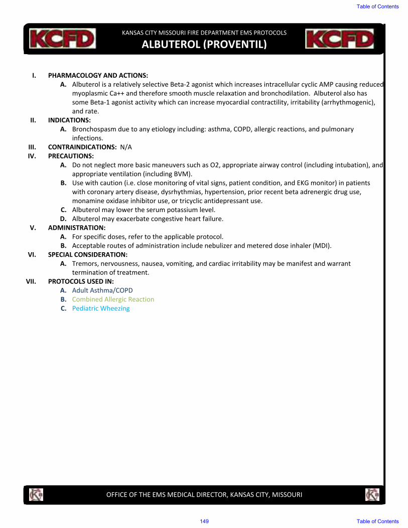
I. PHARMACOLOGY AND ACTIONS:
A. Albuterol is a relatively selective Beta-2 agonist which increases intracellular cyclic AMP causing reduced myoplasmic Ca++ and therefore smooth muscle relaxation and bronchodilation. Albuterol also has some Beta-1 agonist activity which can increase myocardial contractility, irritability (arrhythmogenic), and rate.
II. INDICATIONS:
A. Bronchospasm due to any etiology including: asthma, COPD, allergic reactions, and pulmonary infections.
III. CONTRAINDICATIONS: N/A
IV. PRECAUTIONS:
A. Do not neglect more basic maneuvers such as O2, appropriate airway control (including intubation), and appropriate ventilation (including BVM).
B. Use with caution (i.e. close monitoring of vital signs, patient condition, and EKG monitor) in patients with coronary artery disease, dysrhythmias, hypertension, prior recent beta adrenergic drug use, monamine oxidase inhibitor use, or tricyclic antidepressant use.
C. Albuterol may lower the serum potassium level.
D. Albuterol may exacerbate congestive heart failure.
V. ADMINISTRATION:
A. For specific doses, refer to the applicable protocol.
B. Acceptable routes of administration include nebulizer and metered dose inhaler (MDI).
VI. SPECIAL CONSIDERATION:
A. Tremors, nervousness, nausea, vomiting, and cardiac irritability may be manifest and warrant termination of treatment.
VII. PROTOCOLS USED IN:
A. Adult Asthma/COPD
B. Combined Allergic Reaction
C. Pediatric Wheezing
KANSAS CITY MISSOURI FIRE DEPARTMENT EMS PROTOCOLS
ALBUTEROL (PROVENTIL)
OFFICE OF THE EMS MEDICAL DIRECTOR, KANSAS CITY, MISSOURI
Table of Contents
149 Table of Contents
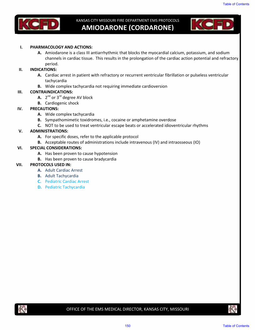
I. PHARMACOLOGY AND ACTIONS:
A. Amiodarone is a class III antiarrhythmic that blocks the myocardial calcium, potassium, and sodium
channels in cardiac tissue. This results in the prolongation of the cardiac action potential and refractory
period.
II. INDICATIONS:
A. Cardiac arrest in patient with refractory or recurrent ventricular fibrillation or pulseless ventricular
tachycardia
B. Wide complex tachycardia not requiring immediate cardioversion
III. CONTRAINDICATIONS:
A. 2nd
or 3rd
degree AV block
B. Cardiogenic shock
IV. PRECAUTIONS:
A. Wide complex tachycardia
B. Sympathomimetic toxidromes, i.e., cocaine or amphetamine overdose
C. NOT to be used to treat ventricular escape beats or accelerated idioventricular rhythms
V. ADMINISTRATIONS:
A. For specific doses, refer to the applicable protocol
B. Acceptable routes of administrations include intravenous (IV) and intraosseous (IO)
VI. SPECIAL CONSIDERATIONS:
A. Has been proven to cause hypotension
B. Has been proven to cause bradycardia
VII. PROTOCOLS USED IN:
A. Adult Cardiac Arrest
B. Adult Tachycardia
C. Pediatric Cardiac Arrest
D. Pediatric Tachycardia
KANSAS CITY MISSOURI FIRE DEPARTMENT EMS PROTOCOLS
AMIODARONE (CORDARONE)
OFFICE OF THE EMS MEDICAL DIRECTOR, KANSAS CITY, MISSOURI
Table of Contents
150 Table of Contents
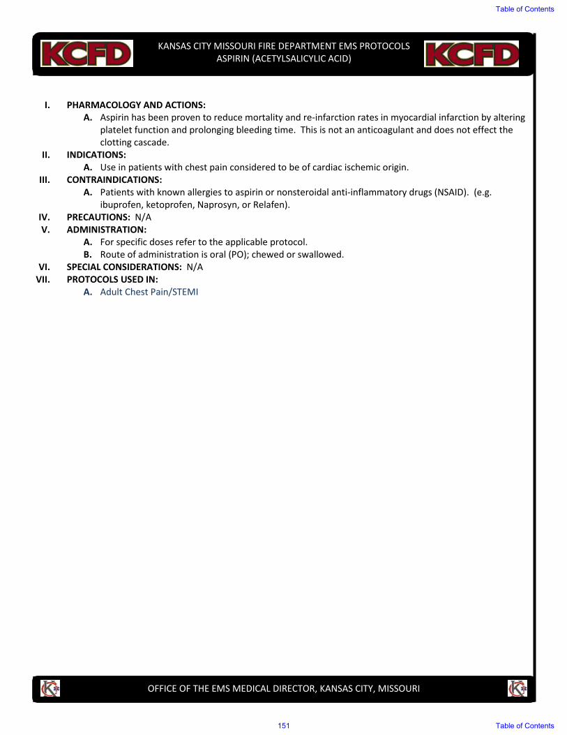
I. PHARMACOLOGY AND ACTIONS:A. Aspirin has been proven to reduce mortality and re-infarction rates in myocardial infarction by altering
platelet function and prolonging bleeding time. This is not an anticoagulant and does not effect theclotting cascade.
II. INDICATIONS:A. Use in patients with chest pain considered to be of cardiac ischemic origin.
III. CONTRAINDICATIONS:A. Patients with known allergies to aspirin or nonsteroidal anti-inflammatory drugs (NSAID). (e.g.
ibuprofen, ketoprofen, Naprosyn, or Relafen).IV. PRECAUTIONS: N/AV. ADMINISTRATION:
A. For specific doses refer to the applicable protocol.B. Route of administration is oral (PO); chewed or swallowed.
VI. SPECIAL CONSIDERATIONS: N/AVII. PROTOCOLS USED IN:
A. Adult Chest Pain/STEMI
KANSAS CITY MISSOURI FIRE DEPARTMENT EMS PROTOCOLS ASPIRIN (ACETYLSALICYLIC ACID)
OFFICE OF THE EMS MEDICAL DIRECTOR, KANSAS CITY, MISSOURI
Table of Contents
151 Table of Contents
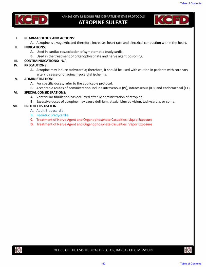
I. PHARMACOLOGY AND ACTIONS:
A. Atropine is a vagolytic and therefore increases heart rate and electrical conduction within the heart.
II. INDICATIONS:
A. Used in cardiac resuscitation of symptomatic bradycardia.
B. Used in the treatment of organophosphate and nerve agent poisoning.
III. CONTRAINDICATIONS: N/A
IV. PRECAUTIONS:
A. Atropine may induce tachycardia; therefore, it should be used with caution in patients with coronary
artery disease or ongoing myocardial ischemia.
V. ADMINISTRATION:
A. For specific doses, refer to the applicable protocol.
B. Acceptable routes of administration include intravenous (IV), intraosseous (IO), and endotracheal (ET).
VI. SPECIAL CONSIDERATIONS:
A. Ventricular fibrillation has occurred after IV administration of atropine.
B. Excessive doses of atropine may cause delirium, ataxia, blurred vision, tachycardia, or coma.
VII. PROTOCOLS USED IN:
A. Adult Bradycardia
B. Pediatric Bradycardia
C. Treatment of Nerve Agent and Organophosphate Casualties: Liquid Exposure
D. Treatment of Nerve Agent and Organophosphate Casualties: Vapor Exposure
KANSAS CITY MISSOURI FIRE DEPARTMENT EMS PROTOCOLS
ATROPINE SULFATE
OFFICE OF THE EMS MEDICAL DIRECTOR, KANSAS CITY, MISSOURI
Table of Contents
152 Table of Contents
I. PHARMACOLOGY AND ACTIONS:
A. Atrovent is chemically related to atropine, but has minimal systemic absorption in the inhaled form.
Atrovent causes bronchodilation without the anticholinergic side effects of atropine.
II. INDICATIONS:
A. Bronchospasm in patients with emphysema or chronic bronchitis.
III. CONTRAINDICATIONS: N/A
IV. PRECAUTIONS:
A. Do not neglect more basic maneuvers such as oxygen, airway control, and proper ventilation.
B. Generally considered safe but is controversial in patients with asthma.
V. ADMINISTRATION:
A. For specific doses, refer to the applicable protocol.
B. Acceptable routes of administration include nebulizer only.
VI. SPECIAL CONSIDERATIONS: N/A
VII. PROTOCOLS USED IN:
A. Adult COPD/Asthma
B. Pediatric Wheezing
KANSAS CITY MISSOURI FIRE DEPARTMENT EMS PROTOCOLS
ATROVENT (IPRATROPRIUM BROMIDE)
OFFICE OF THE EMS MEDICAL DIRECTOR, KANSAS CITY, MISSOURI
Table of Contents
153 Table of Contents
I. PHARMACOLOGY AND ACTIONS:
A. Calcium increases myocardial contractile function.
B. Calcium has a myocardial cellular membrane stabilizing effect with hyperkalemia.
II. INDICTIONS:
A. Known or suspected hyperkalemia
B. Known or suspected hypocalcemia
C. As an antidote for toxicity from calcium channel blocker overdose
III. CONTRAINDICATIONS: N/A
IV. PRECAUTIONS:
A. Use with caution in patients receiving digitalis (digoxin) because of the increased ventricular irritability associated with digitalis toxicity.
B. Calcium chloride will precipitate when mixed with sodium bicarbonate.
C. Extravasation of calcium chloride can result in tissue necrosis.
D. Calcium chloride is not routinely used as a first line cardiac arrest drug.
V. ADMINISTRATION:
A. For specific doses, refer to the applicable protocol.
B. Acceptable routes of administration include intravenous (IV), and intraosseous (IO).
VI. SPECIAL CONSIDERATIONS:
A. Calcium chloride may be useful in the treatment of calcium channel blocker overdoses.
VII. PROTOCOLS USED IN:
A. Adult Cardiac Arrest
B. Combined Overdose/Poisoning
C. Entrapment/Crush
KANSAS CITY MISSOURI FIRE DEPARTMENT EMS PROTOCOLS
CALCIUM CHLORIDE
OFFICE OF THE EMS MEDICAL DIRECTOR, KANSAS CITY, MISSOURI
Table of Contents
154 Table of Contents
I. PHARMACOLOGY AND ACTIONS:
A. Glucose is the major metabolic substrate for energy metabolism. Although all tissues need glucose, the
brain is particularly sensitive to low glucose levels. Glucose specifically reverse hypoglycemia
II. INDICATION:
A. Confirmed hypoglycemia with a rapid bedside glucose test
B. Suspected hypoglycemia as manifested by altered mental status (including apparent drug or alcohol use,
seizure or postictal state) or coma
III. CONTRAINDICATIONS: N/A
IV. PRECAUTIONS:
A. Extravasation of glucose will cause skin necrosis. The IV should be secure and the free return of blood
should be checked once or twice during administration. If extravasation does occur, immediately stop
administration. Notify the ED staff upon arrival of possible glucose extravasation.
B. High glucose levels have been associated with worsened neurologic outcomes of patients with stroke,
cardiac arrest, and low perfusion states. When these exist, it is preferable to only administer glucose
after hypoglycemia has been documented by a rapid bedside glucose test.
V. ADMINISTRATION:
A. For specific doses, refer to the applicable protocol
B. Acceptable routes of administration include intravenous (IV) and intraosseous (IO)
VI. SPECIAL CONSIDERATIONS:
A. Consider oral glucose if mental status permits it use
B. One bolus should raise the blood sugar by 100-200%
VII. PROTOCOLS USED IN:
A. Combined Hypoglycemia
KANSAS CITY MISSOURI FIRE DEPARTMENT EMS PROTOCOLS
DEXTROSE
OFFICE OF THE EMS MEDICAL DIRECTOR, KANSAS CITY, MISSOURI
Table of Contents
155 Table of Contents
I. PHARMACOLOGY AND ACTIONS:
A. Diphenhydramine is an antihistamine which competitively blocks H-1 histamine receptors. Histamine can cause bronchoconstriction, vasodilation and capillary leak. It also has some anticholinergic (atropine like) effects as well as direct CNS depressant effects.
II. INDICATIONS:
A. Anaphylaxis
1. Mild anaphylaxis
2. Anaphylaxis with respiratory distress
3. Anaphylactic shock
B. Dystonic reaction
III. CONTRAINDICATIONS: N/A
IV. PRECAUTIONS:
A. In anaphylaxis, oxygen, epinephrine, fluids, and albuterol should be utilized prior to administration of diphenhydramine.
B. Diphenhydramine causes sedation.
C. Due to anticholinergic effects, use with caution in patients with glaucoma, prostatism, or peptic ulcer disease.
D. Hypotension or hypertension may occur after IV use.
V. ADMINISTRATION:
A. For specific doses, refer to the applicable protocol
B. Acceptable routes of administration include intravenous (IV), intraosseous (IO), and intramuscular (IM).
VI. SPECIAL CONSIDERATION: N/A
VII. PROTOCOLS USED IN:
A. Combined Allergic Reaction
B. Combined Behavioral/Psychiatric Disorder
KANSAS CITY MISSOURI FIRE DEPARTMENT EMS PROTOCOLS
DIPHENHYDRAMINE (BENADRYL)
OFFICE OF THE EMS MEDICAL DIRECTOR, KANSAS CITY, MISSOURI
Table of Contents
156 Table of Contents
I. PHARMACOLOGY AND ACTIONS:A. Epinephrine is a natural catecholamine that acts to stimulate sympathetic (i.e. fight or flight) activity.
Accordingly this exhibits an increase in blood pressure, coronary blood flow, cardiac electrical activity,heart rate, strength of contraction and broncho-dilatation. Epinephrine also make ventricular fibrillationmore susceptible to defibrillation.
II. INDICATIONS:A. Used in cardiac arrest protocol for ventricular fibrillation, pulseless ventricular activity, asystole,
bradycardia, and pulseless electrical activity.B. Used to treat severe bronchospasm or laryngospasm as seen in asthma and anaphylaxisC. Used to treat unremitting hypotension after appropriate fluid resuscitation in post-cardiac arrest,
cardiogenic shock, or other medical etiologyD. Used in bradycardia if poor perfusion persists secondary to transcutaneous pacing and appropriate fluid
resuscitationIII. CONTRAINDICATIONS: N/AIV. PRECAUTIONS:
A. Epinephrine should be used with caution in patients with suspected myocardial ischemia, history ofcoronary artery disease, or age greater than 50.
B. In patients greater than 50 years of age, the role epinephrine in asthma is controversial.C. In patients taking digitalis, epinephrine may exacerbate ventricular ectopy.
V. ADMINISTRATION:A. For specific doses, refer to the applicable protocol.B. Acceptable routes of administration include intravenous (IV), intramuscular (IM) at lateral thigh,
intraosseous (IO), and endotracheal (ET).VI. SPECIAL CONSIDERATIONS: N/A
VII. PROTOCOLS USED IN:A. EPINEPHRINE 1:1000
1. Adult Asthma/COPD2. Combined Allergic Reaction3. Pediatric Wheezing
B. EPINEPHRINE 1:100001. Adult Cardiac Arrest2. Neonatal Resuscitation3. Pediatric Bradycardia4. Pediatric Cardiac Arrest
C. EPINEPHRINE 1:100000 (PUSH DOSE)1. Adult Bradycardia2. Combined Hypotension/Shock3. Combined Post Resuscitation
KANSAS CITY MISSOURI FIRE DEPARTMENT EMS PROTOCOLS
EPINEPHRINE
OFFICE OF THE EMS MEDICAL DIRECTOR, KANSAS CITY, MISSOURI
Table of Contents
157 Table of Contents
I. PHARMACOLOGY AND ACTIONS:A. Acts as an opiate agonistB. Alters the patient’s perception of pain and acts as an analgesic by stimulating opiate receptor sites
II. INDICATIONS:A. The relief of moderate to severe acute painB. As an analgesic in patients with ischemic chest pain
III. CONTRAINDICATIONS:A. Trauma
1. Head injury2. Paralysis or new neurologic finding3. Thoraco-abdominal trauma (blunt or penetrating)4. Burns involving the airway or causing respiratory compromise5. Facial trauma involving the airway or causing respiratory compromise
B. Medical1. Respiratory distress or compromise (Asthma or COPD)2. New cardiac dysrhythmia is present3. Altered mental status4. Third trimester pregnancy
C. Drugs1. Drug/alcohol intoxication2. Sensitivity/allergy to opiates/fentanyl
D. Hypotension or suspected shock (SBP < 100mmHg)E. Pulse rate < 60 bpm
IV. PRECAUTIONS:A. This drug causes decreased respiratory drive, and as such should only be given while monitoring pulse
oximetry and closely monitoring respiratory status.B. Overdose and side effects can be countered by the administration of naloxoneC. This drug may cause hypotension and direct myocardial depression.D. Cardiac monitoring accompanied by frequent blood pressure checks are mandatory.E. Other precautions as listed in other applicable protocols
V. ADMINISTRATION:A. For specific doses, refer to applicable protocolB. May be administered intravenous (IV), intraosseous (IO), or intranasal (IN)
VI. SPECIAL CONSIDERATIONS: N/AVII. PROTOCOLS USED IN:
A. Adult Chest Pain/STEMIB. Combined Pain Control
KANSAS CITY MISSOURI FIRE DEPARTMENT EMS PROTOCOLS
FENTANYL CITRATE
OFFICE OF THE EMS MEDICAL DIRECTOR, KANSAS CITY, MISSOURI
Table of Contents
158 Table of Contents

I. PHARMACOLOGY AND ACTIONS: A. Magnesium sulfate reduces striated muscle contractions and blocks peripheral neuromuscular
transmission by reducing acetylcholine release at the myoneural junction. In cardiac patients, it stabilizes the potassium pump, correcting repolarization. It also shortens the Q-T interval in the presence of ventricular arrhythmias due to drug toxicity or electrolyte imbalance. In respiratory patients, it may act as a bronchodilator in acute bronchospasm due to asthma or other bronchospastic disease.
II. INDICATIONS: A. Torsade de pointes associated with prolonged QT interval B. Actively seizing patient in 3rd trimester pregnancy or 1 month post-partum if no known seizure history
III. CONTRAINDICATIONS: N/A IV. PRECAUTIONS:
A. Known to cause bradycardia B. Known to cause hypotension C. Known to cause respiratory depression
V. ADMINISTRATION: A. For specific dose, refer to the applicable protocol B. Acceptable routes of administration include intravenous (IV), and intraosseous (IO)
VI. SPECIAL CONSIDERATIONS: N/A VII. PROTOCOLS USED IN:
A. Adult Cardiac Arrest B. Adult Tachycardia C. Combined Seizure
KANSAS CITY MISSOURI FIRE DEPARTMENT EMS PROTOCOLS
MAGNESIUM SULFATE
OFFICE OF THE EMS MEDICAL DIRECTOR, KANSAS CITY, MISSOURI
Table of Contents
159 Table of Contents
I. PHARMACOLOGY AND ACTIONS:
A. Methylprednisolone is a synthetic steroid that suppresses acute and chronic inflammation.
B. Potentiates vascular smooth muscle relaxation by beta-adrenergic agonists and may alter airway
hyperactivity
II. INDICATIONS:
A. Severe asthma
B. COPD
C. Suspected Addisonian crisis (cardiovascular collapse in patient at risk for adrenal insufficiency)
III. CONTRAINDICATIONS:
A. Evidence of active GI bleed
IV. PRECAUTIONS:
A. Most adverse reactions are a result of long-term therapy and include gastrointestinal bleeding,
hypertension, and hyperglycemia
V. ADMINISTRATION:
A. For specific doses, refer to the applicable protocol
B. Only acceptable routes of administration include intravenous (IV), and intraosseous (IO)
VI. SPECIAL CONSIDERATIONS:
A. Must be reconstituted and used immediately
B. The effect of methylprednisolone is generally delayed for several hours
C. Methylprednisolone is not considered a first line drug. Be sure to attend to the patient’s primary
treatment priorities (i.e. airway, ventilation, beta-agonist nebulization) first.
D. If primary treatment priorities have been completed and there is time while in route to hospital, then
methylprednisolone can be administered.
E. Don not delay transport to administer this drug
VII. PROTOCOLS USED IN:
A. Adult Asthma/COPD
B. Combined Hypotension/Shock
C. Pediatric Wheezing
KANSAS CITY MISSOURI FIRE DEPARTMENT EMS PROTOCOLS
METHYLPREDNISOLONE (SOLU-MEDROL)
OFFICE OF THE EMS MEDICAL DIRECTOR, KANSAS CITY, MISSOURI
Table of Contents
160 Table of Contents
I. PHARMACOLOGY AND ACTIONS: A. Midazolam is a water soluble, short acting sedative with muscle relaxing and amnesic properties similar
to diazepam (Valium). B. Its relatively short duration of actin makes it the drug of choice for sedation when prolonged effects are
not needed or wanted. C. Midazolam given intranasally (IN) has been shown to be safe and effective for the management of acute
seizures in pediatric patients. II. INDICATIONS:
A. Intranasal administration in seizure patients with no IV established. B. Sedation and amnesia prior to performing painful procedures, e.g. cardioversion C. Chemical restraint
III. CONTRAINDICATIONS: N/A IV. PRECAUTIONS:
A. Respiratory depression and/or hypotension are potential complications. B. The patient’s respiratory status should be monitored closely when using midazolam. C. Pulse oximetry is mandatory to monitor the patient’s oxygen saturation. D. Cardiac monitoring accompanied by frequent blood pressure checks are mandatory.
V. ADMINISTRATION: A. For specific doses, refer to the applicable protocol. B. May be administered intravenous (IV), intramuscular (IM), or intranasal (IN).
VI. SPECIAL CONSIDERATIONS: A. Airway support equipment and IV fluids must be readied for use prior to the administration of
midazolam (with exception of pediatric seizures) in the event that blood pressure and ventilator support are required.
VII. PROTOCOLS USED IN: A. Adult General Airway B. Adult Bradycardia C. Adult Tachycardia D. Combined Behavioral/Psychiatric Disorder E. Combined Post Resuscitation F. Combined Seizure
KANSAS CITY MISSOURI FIRE DEPARTMENT EMS PROTOCOLS
MIDAZOLAM HYDROCHLORIDE (VERSED)
OFFICE OF THE EMS MEDICAL DIRECTOR, KANSAS CITY, MISSOURI
Table of Contents
161 Table of Contents
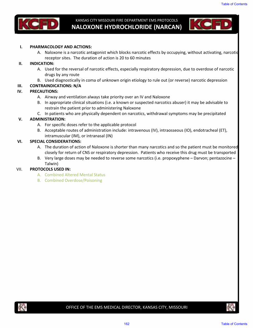
I. PHARMACOLOGY AND ACTIONS:
A. Naloxone is a narcotic antagonist which blocks narcotic effects by occupying, without activating, narcotic
receptor sites. The duration of action is 20 to 60 minutes
II. INDICATION:
A. Used for the reversal of narcotic effects, especially respiratory depression, due to overdose of narcotic
drugs by any route
B. Used diagnostically in coma of unknown origin etiology to rule out (or reverse) narcotic depression
III. CONTRAINDICATIONS: N/A
IV. PRECAUTIONS:
A. Airway and ventilation always take priority over an IV and Naloxone
B. In appropriate clinical situations (i.e. a known or suspected narcotics abuser) it may be advisable to
restrain the patient prior to administering Naloxone
C. In patients who are physically dependent on narcotics, withdrawal symptoms may be precipitated
V. ADMINISTRATION:
A. For specific doses refer to the applicable protocol
B. Acceptable routes of administration include: intravenous (IV), intraosseous (IO), endotracheal (ET),
intramuscular (IM), or intranasal (IN)
VI. SPECIAL CONSIDERATIONS:
A. The duration of action of Naloxone is shorter than many narcotics and so the patient must be monitored
closely for return of CNS or respiratory depression. Patients who receive this drug must be transported
B. Very large doses may be needed to reverse some narcotics (i.e. propoxyphene – Darvon; pentazocine –
Talwin)
VII. PROTOCOLS USED IN:
A. Combined Altered Mental Status
B. Combined Overdose/Poisoning
KANSAS CITY MISSOURI FIRE DEPARTMENT EMS PROTOCOLS
NALOXONE HYDROCHLORIDE (NARCAN)
OFFICE OF THE EMS MEDICAL DIRECTOR, KANSAS CITY, MISSOURI
Table of Contents
162 Table of Contents
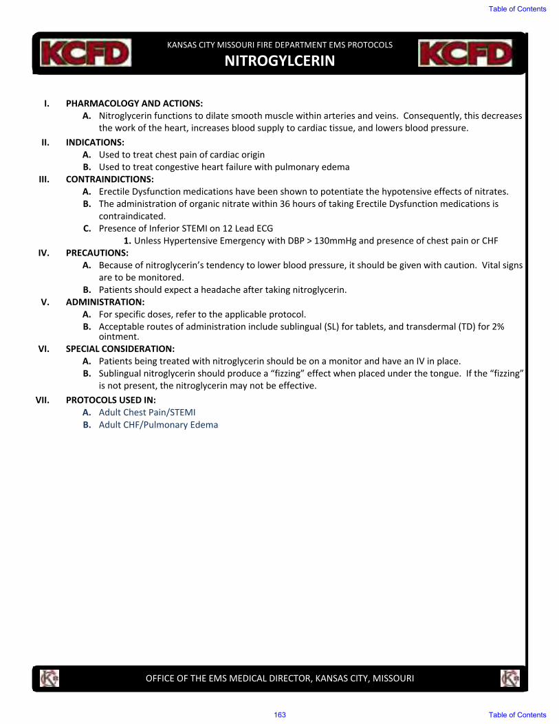
I. PHARMACOLOGY AND ACTIONS:
A. Nitroglycerin functions to dilate smooth muscle within arteries and veins. Consequently, this decreases the work of the heart, increases blood supply to cardiac tissue, and lowers blood pressure.
II. INDICATIONS:
A. Used to treat chest pain of cardiac origin
B. Used to treat congestive heart failure with pulmonary edema
III. CONTRAINDICTIONS:
A. Erectile Dysfunction medications have been shown to potentiate the hypotensive effects of nitrates.
B. The administration of organic nitrate within 36 hours of taking Erectile Dysfunction medications is contraindicated.
C. Presence of Inferior STEMI on 12 Lead ECG
1. Unless Hypertensive Emergency with DBP > 130mmHg and presence of chest pain or CHF
IV. PRECAUTIONS:
A. Because of nitroglycerin’s tendency to lower blood pressure, it should be given with caution. Vital signs are to be monitored.
B. Patients should expect a headache after taking nitroglycerin.
V. ADMINISTRATION:
A. For specific doses, refer to the applicable protocol.
B. Acceptable routes of administration include sublingual (SL) for tablets, and transdermal (TD) for 2% ointment.
VI. SPECIAL CONSIDERATION:
A. Patients being treated with nitroglycerin should be on a monitor and have an IV in place.
B. Sublingual nitroglycerin should produce a “fizzing” effect when placed under the tongue. If the “fizzing” is not present, the nitroglycerin may not be effective.
VII. PROTOCOLS USED IN:
A. Adult Chest Pain/STEMI
B. Adult CHF/Pulmonary Edema
KANSAS CITY MISSOURI FIRE DEPARTMENT EMS PROTOCOLS
NITROGYLCERIN
OFFICE OF THE EMS MEDICAL DIRECTOR, KANSAS CITY, MISSOURI
Table of Contents
163 Table of Contents
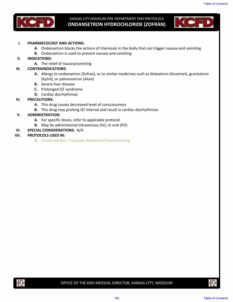
I. PHARMACOLOGY AND ACTIONS:
A. Ondansetron blocks the actions of chemicals in the body that can trigger nausea and vomiting
B. Ondansetron is used to prevent nausea and vomiting
II. INDICATIONS:
A. The relief of nausea/vomiting
III. CONTRAINDICATIONS:
A. Allergy to ondansetron (Zofran), or to similar medicines such as dolasetron (Anzemet), granisetron
(Kytril), or palonosetron (Aloxi)
B. Severe liver disease
C. Prolonged QT syndrome
D. Cardiac dysrhythmias
IV. PRECAUTIONS:
A. This drug causes decreased level of consciousness
B. This drug may prolong QT interval and result in cardiac dysrhythmias
V. ADMINISTRATION:
A. For specific doses, refer to applicable protocol
B. May be administered intravenous (IV), or oral (PO)
VI. SPECIAL CONSIDERATIONS: N/A
VII. PROTOCOLS USED IN:
A. Combined Non-Traumatic Abdominal Pain/Vomiting
KANSAS CITY MISSOURI FIRE DEPARTMENT EMS PROTOCOLS
ONDANSETRON HYDROCHLORIDE (ZOFRAN)
OFFICE OF THE EMS MEDICAL DIRECTOR, KANSAS CITY, MISSOURI
Table of Contents
164 Table of Contents
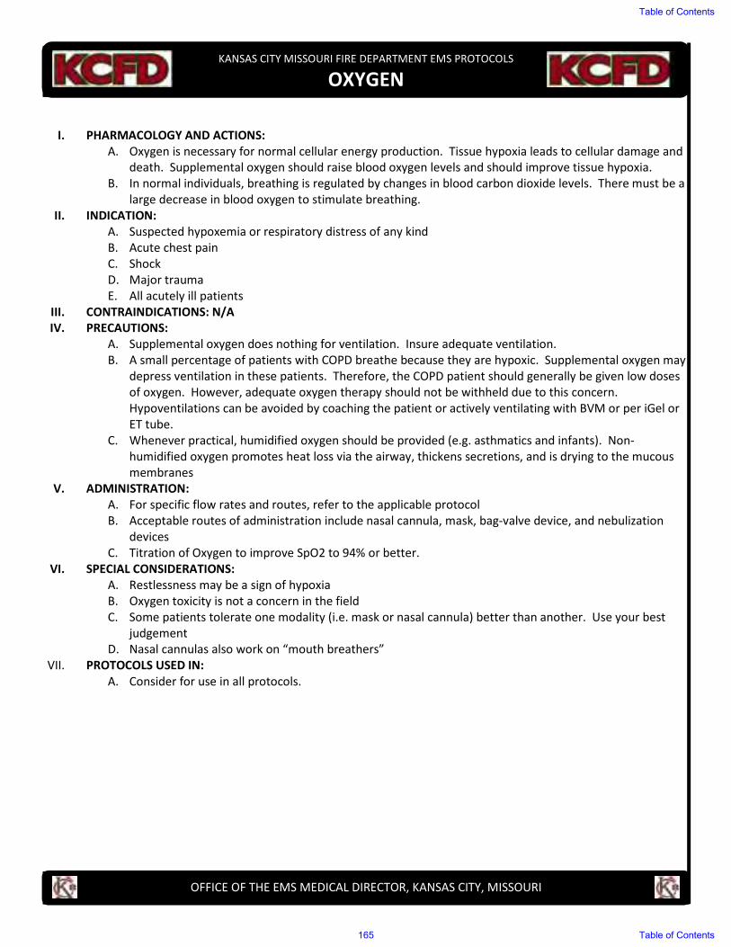
I. PHARMACOLOGY AND ACTIONS:
A. Oxygen is necessary for normal cellular energy production. Tissue hypoxia leads to cellular damage and
death. Supplemental oxygen should raise blood oxygen levels and should improve tissue hypoxia.
B. In normal individuals, breathing is regulated by changes in blood carbon dioxide levels. There must be a
large decrease in blood oxygen to stimulate breathing.
II. INDICATION:
A. Suspected hypoxemia or respiratory distress of any kind
B. Acute chest pain
C. Shock
D. Major trauma
E. All acutely ill patients
III. CONTRAINDICATIONS: N/A
IV. PRECAUTIONS:
A. Supplemental oxygen does nothing for ventilation. Insure adequate ventilation.
B. A small percentage of patients with COPD breathe because they are hypoxic. Supplemental oxygen may
depress ventilation in these patients. Therefore, the COPD patient should generally be given low doses
of oxygen. However, adequate oxygen therapy should not be withheld due to this concern.
Hypoventilations can be avoided by coaching the patient or actively ventilating with BVM or per iGel or
ET tube.
C. Whenever practical, humidified oxygen should be provided (e.g. asthmatics and infants). Non-
humidified oxygen promotes heat loss via the airway, thickens secretions, and is drying to the mucous
membranes
V. ADMINISTRATION:
A. For specific flow rates and routes, refer to the applicable protocol
B. Acceptable routes of administration include nasal cannula, mask, bag-valve device, and nebulization
devices
C. Titration of Oxygen to improve SpO2 to 94% or better.
VI. SPECIAL CONSIDERATIONS:
A. Restlessness may be a sign of hypoxia
B. Oxygen toxicity is not a concern in the field
C. Some patients tolerate one modality (i.e. mask or nasal cannula) better than another. Use your best
judgement
D. Nasal cannulas also work on “mouth breathers”
VII. PROTOCOLS USED IN:
A. Consider for use in all protocols.
KANSAS CITY MISSOURI FIRE DEPARTMENT EMS PROTOCOLS
OXYGEN
OFFICE OF THE EMS MEDICAL DIRECTOR, KANSAS CITY, MISSOURI
Table of Contents
165 Table of Contents
I. PHARMACOLOGY AND ACTIONS:
A. Sodium bicarbonate serves as a buffer for acidosis.
B. H+ + HCO3- = H2CO3 + CO2
II. INDICATIONS:
A. Known or suspected hyperkalemia
B. Known or suspected tricyclic antidepressant overdose
C. Considered during CPR only after adequate ventilation and chest compressions are insured. The patient
should be intubated and there should exist a continued cardiac arrest interval during which
defibrillations, epinephrine, and lidocaine have been utilized.
III. CONTRAINDICATIONS: N/A
IV. PRECAUTIONS:
A. Administration of sodium bicarbonate rapidly generates carbon dioxide which can result in tissue and
cerebrospinal fluid acidosis.
B. Sodium bicarbonate is not recommended for routine use in cardiac arrest patients.
V. ADMINISTRATION:
A. For specific doses, refer to the applicable protocol.
B. Acceptable routes of administration include intravenous (IV), and intraosseous (IO).
VI. SPECIAL CONSIDERATIONS:
A. Sodium bicarbonate may be useful in the treatment of tricyclic antidepressant overdose.
B. Adequate ventilation is a major buffering agent and is essential prior to the administration of sodium
bicarbonate.
VII. PROTOCOLS USED IN:
A. Adult Cardiac Arrest
B. Combined Overdose/Poisoning
C. Entrapment/Crush Injury
KANSAS CITY MISSOURI FIRE DEPARTMENT EMS PROTOCOLS
SODIUM BICARBONATE
OFFICE OF THE EMS MEDICAL DIRECTOR, KANSAS CITY, MISSOURI
Table of Contents
166 Table of Contents
KANSAS CITY MISSOURI FIRE DEPARTMENT EMS PROTOCOLS
12 LEAD ECG INTERPRETATION MAP
OFFICE OF THE EMS MEDICAL DIRECTOR, KANSAS CITY MISSOURI
Table of Contents
167 Table of Contents
KANSAS CITY MISSOURI FIRE DEPARTMENT EMS PROTOCOLS
APGAR SCORE
OFFICE OF THE EMS MEDICAL DIRECTOR, KANSAS CITY MISSOURI
Table of Contents
168 Table of Contents
KANSAS CITY MISSOURI FIRE DEPARTMENT EMS PROTOCOLS
CINCINNATI PREHOSPITAL STROKE SCALE
OFFICE OF THE EMS MEDICAL DIRECTOR, KANSAS CITY MISSOURI
Table of Contents
169 Table of Contents
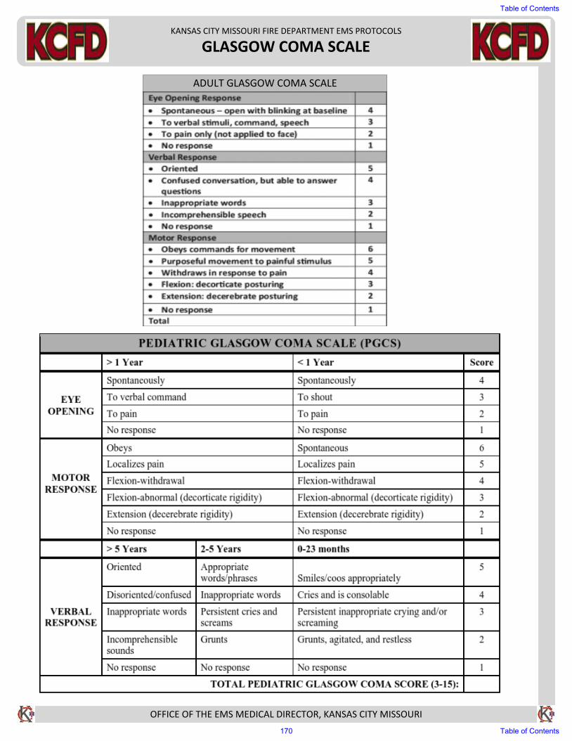
KANSAS CITY MISSOURI FIRE DEPARTMENT EMS PROTOCOLS
GLASGOW COMA SCALE
OFFICE OF THE EMS MEDICAL DIRECTOR, KANSAS CITY MISSOURI
ADULT GLASGOW COMA SCALE
Table of Contents
170 Table of Contents
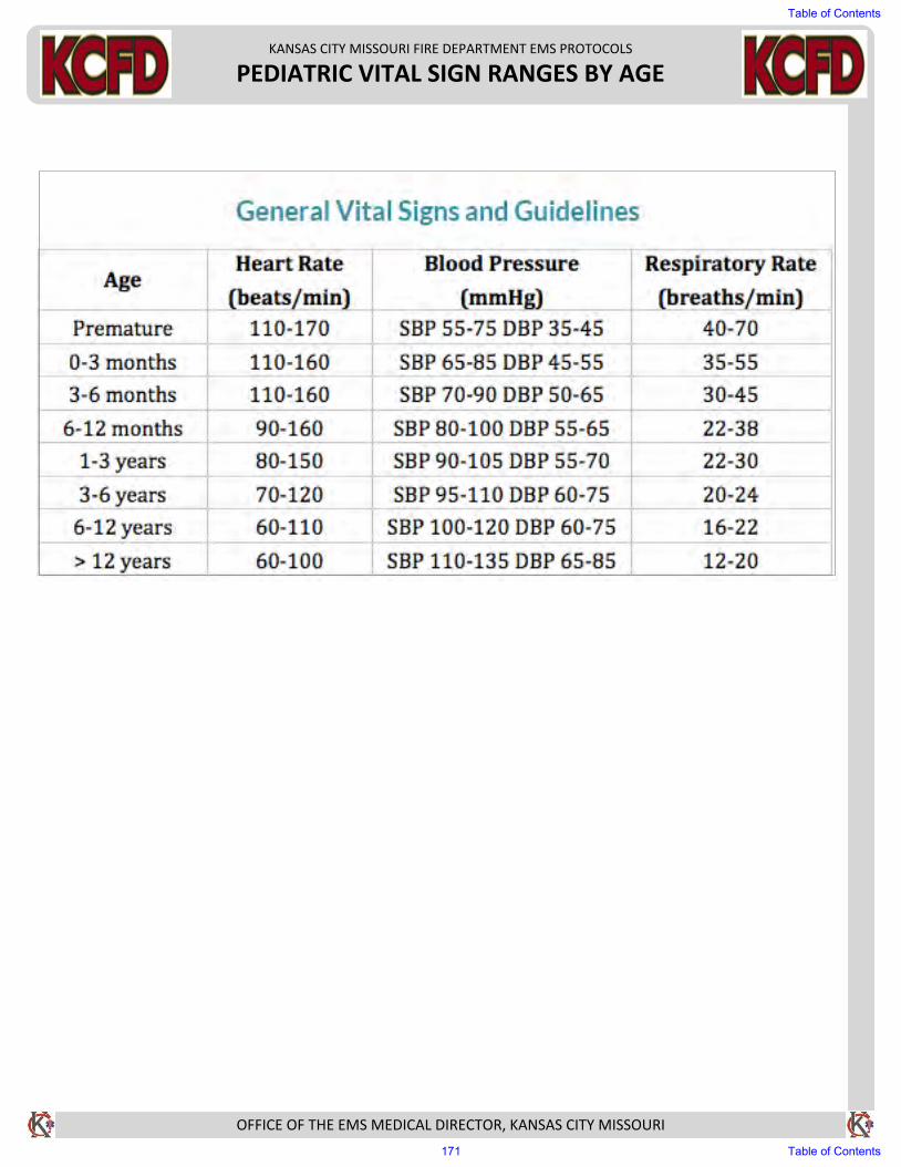
KANSAS CITY MISSOURI FIRE DEPARTMENT EMS PROTOCOLS
PEDIATRIC VITAL SIGN RANGES BY AGE
OFFICE OF THE EMS MEDICAL DIRECTOR, KANSAS CITY MISSOURI
Table of Contents
171 Table of Contents
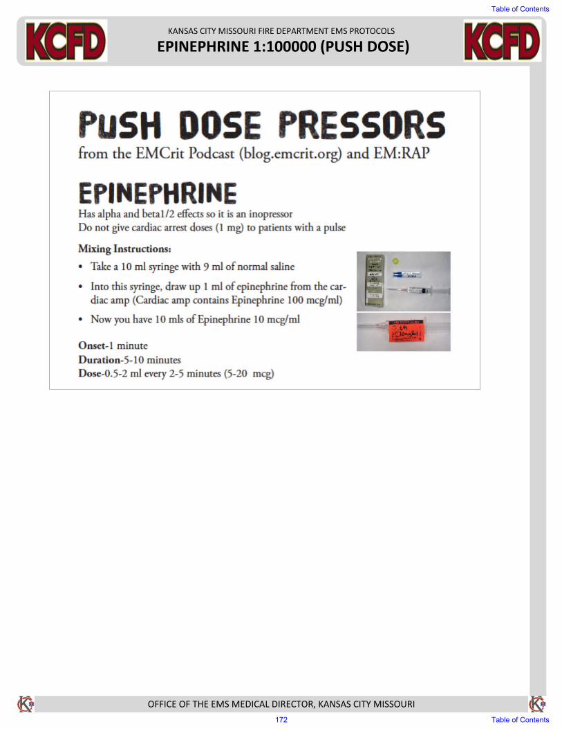
KANSAS CITY MISSOURI FIRE DEPARTMENT EMS PROTOCOLS
EPINEPHRINE 1:100000 (PUSH DOSE)
OFFICE OF THE EMS MEDICAL DIRECTOR, KANSAS CITY MISSOURI
Table of Contents
172 Table of Contents
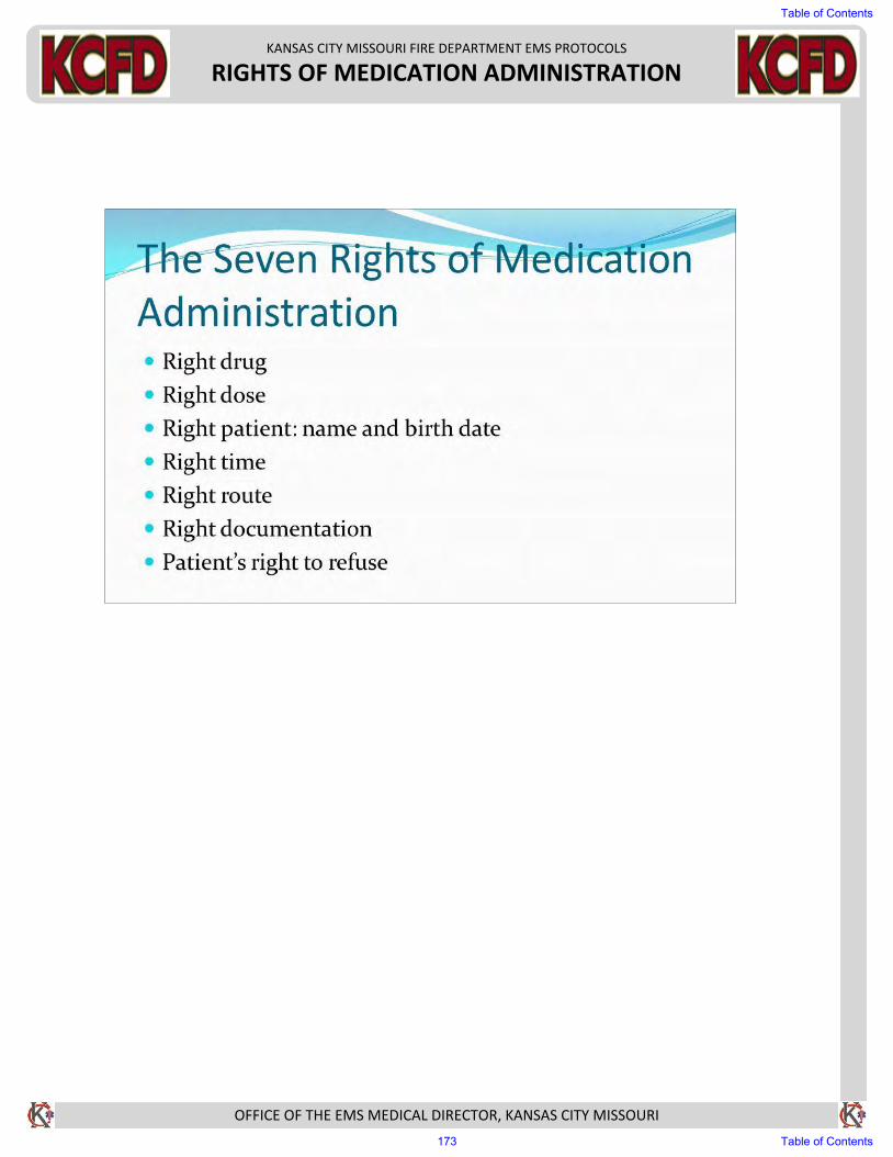
KANSAS CITY MISSOURI FIRE DEPARTMENT EMS PROTOCOLS
RIGHTS OF MEDICATION ADMINISTRATION
OFFICE OF THE EMS MEDICAL DIRECTOR, KANSAS CITY MISSOURI
Table of Contents
173 Table of Contents
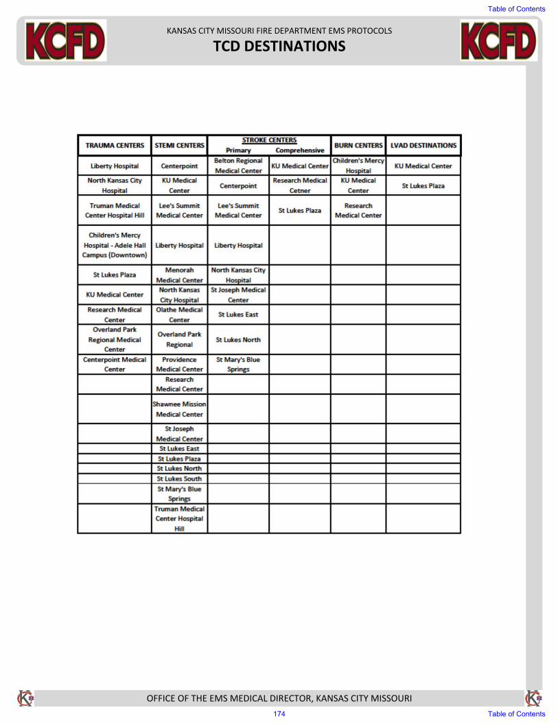
KANSAS CITY MISSOURI FIRE DEPARTMENT EMS PROTOCOLS
TCD DESTINATIONS
OFFICE OF THE EMS MEDICAL DIRECTOR, KANSAS CITY MISSOURI
Table of Contents
174 Table of Contents
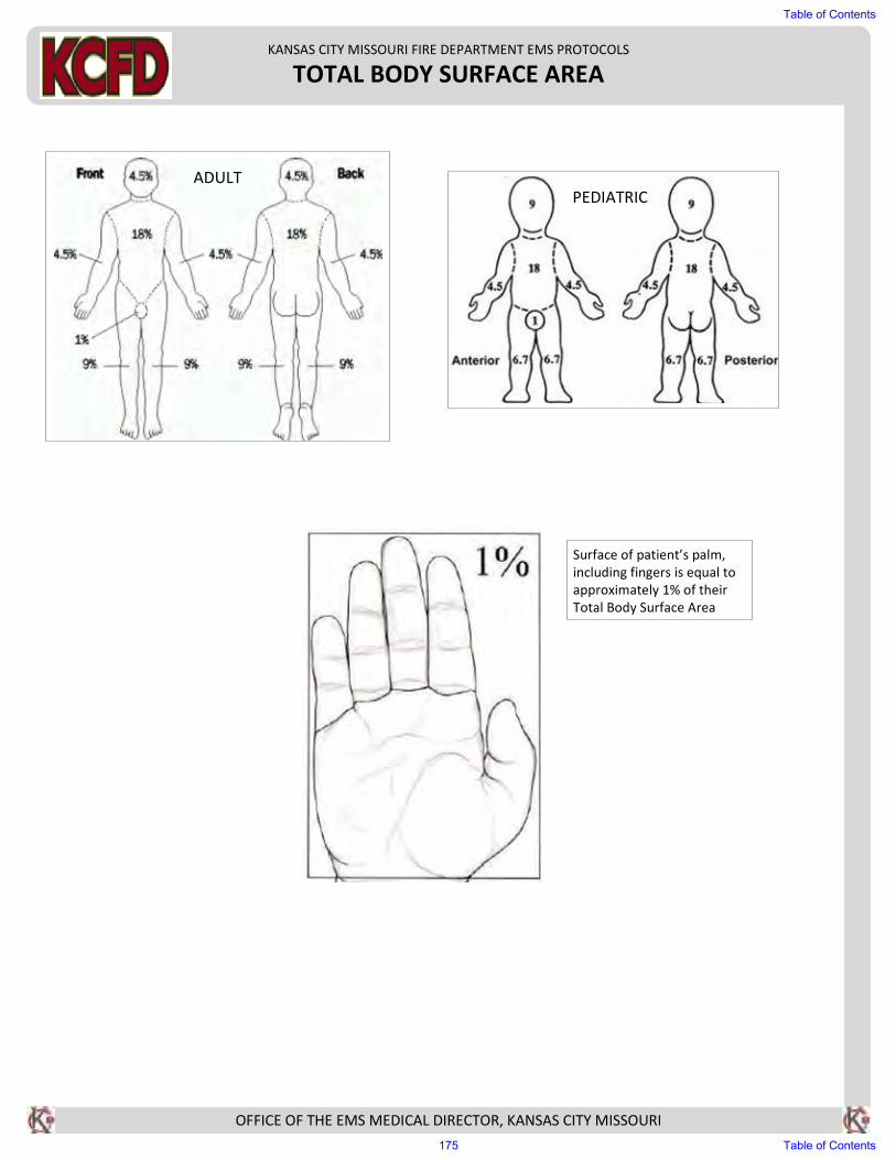
KANSAS CITY MISSOURI FIRE DEPARTMENT EMS PROTOCOLS
TOTAL BODY SURFACE AREA
OFFICE OF THE EMS MEDICAL DIRECTOR, KANSAS CITY MISSOURI
ADULT
PEDIATRIC
Surface of patient’s palm,
including fingers is equal to
approximately 1% of their
Total Body Surface Area
Table of Contents
175 Table of Contents
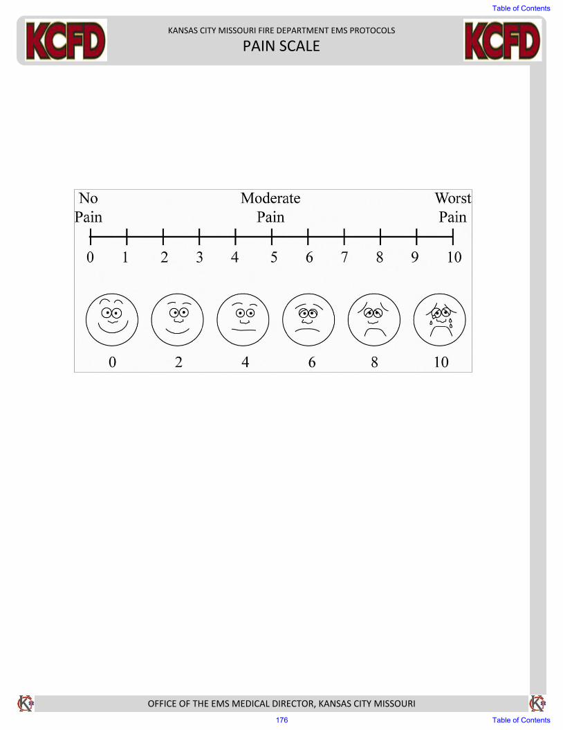
KANSAS CITY MISSOURI FIRE DEPARTMENT EMS PROTOCOLS
PAIN SCALE
OFFICE OF THE EMS MEDICAL DIRECTOR, KANSAS CITY MISSOURI
Table of Contents
176 Table of Contents

KANSAS CITY MISSOURI FIRE DEPARTMENT EMS PROTOCOLS
tPA EXCLUSION CRITERIA
OFFICE OF THE EMS MEDICAL DIRECTOR, KANSAS CITY MISSOURI
Thrombolytic (tPA) Exclusion Criteria:· Head Trauma at onset· History of recent bleeding, current bleeding, surgery or invasive procedure in the past three
months· Bleeding disorder
Table of Contents
177 Table of Contents
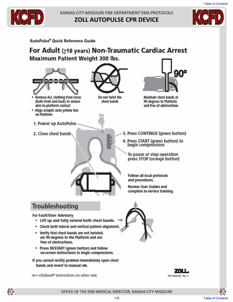
KANSAS CITY MISSOURI FIRE DEPARTMENT EMS PROTOCOLS
ZOLL AUTOPULSE CPR DEVICE
OFFICE OF THE EMS MEDICAL DIRECTOR, KANSAS CITY MISSOURI
Table of Contents
178 Table of Contents
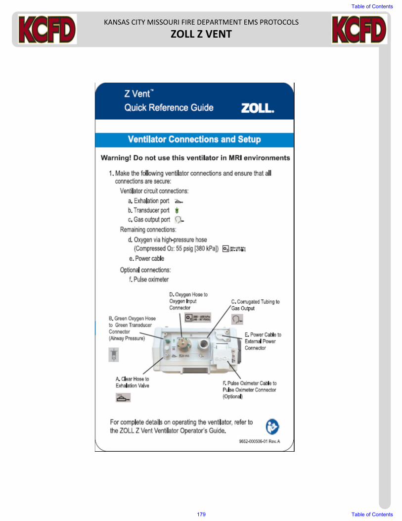
KANSAS CITY MISSOURI FIRE DEPARTMENT EMS PROTOCOLS
ZOLL Z VENT
Table of Contents
179 Table of Contents
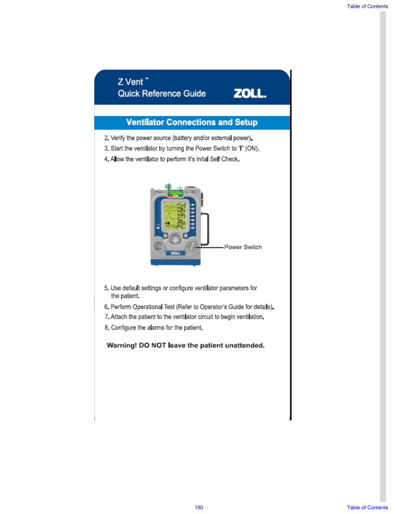
Table of Contents
180 Table of Contents
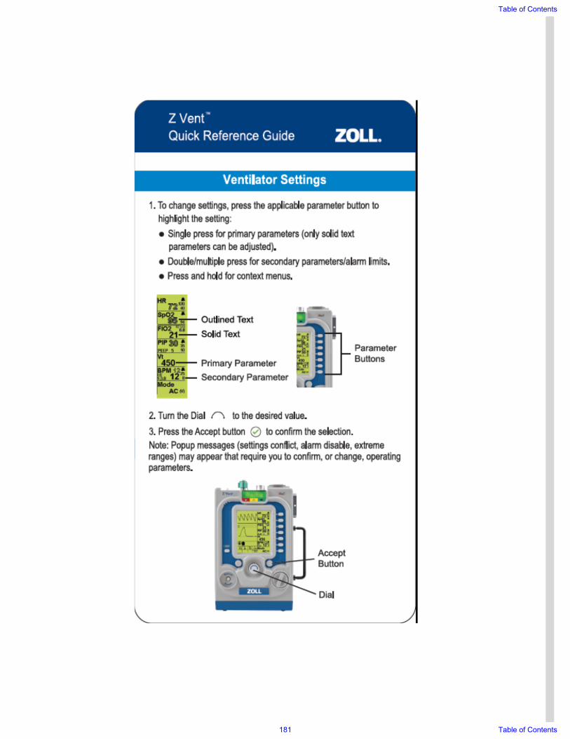
Table of Contents
181 Table of Contents
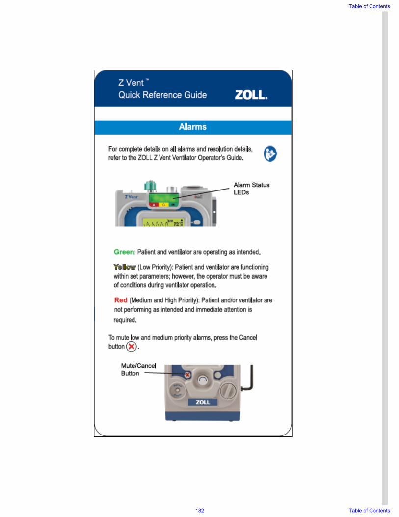
Table of Contents
182 Table of Contents
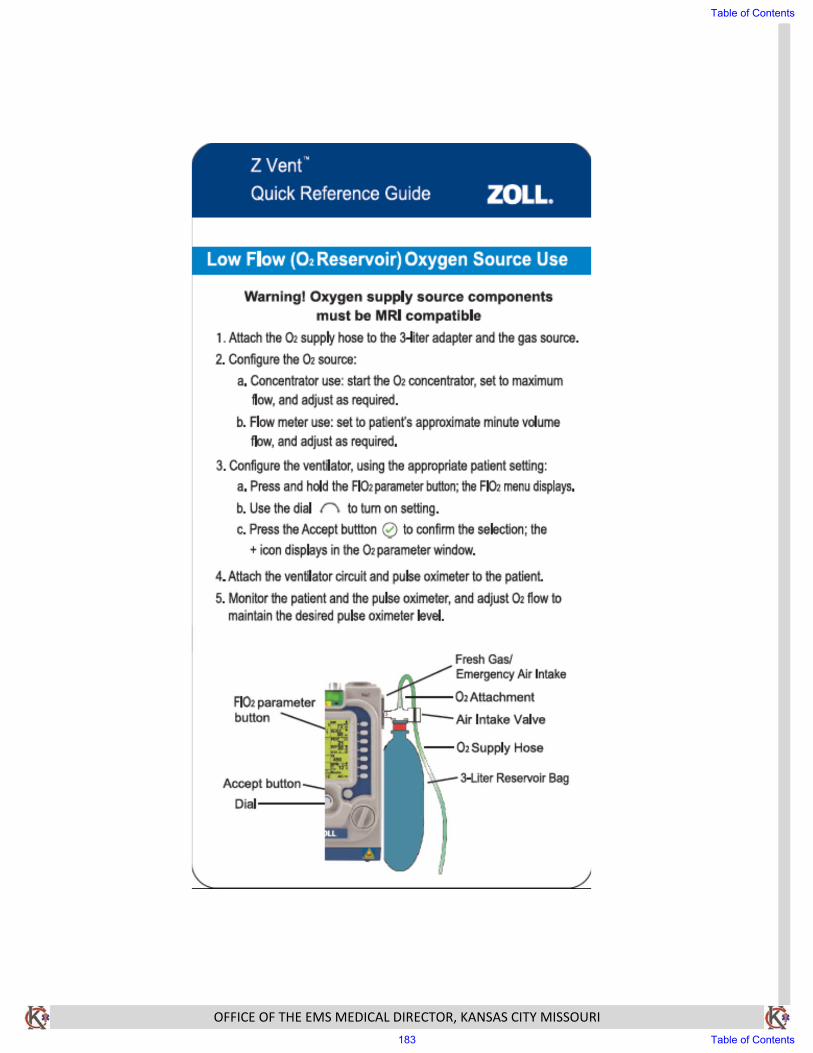
OFFICE OF THE EMS MEDICAL DIRECTOR, KANSAS CITY MISSOURI
Table of Contents
183 Table of Contents