JOURNAL OF - DergiPark
60
ejb.istanbul.edu.tr ISSN 2602-2575 • EISSN 2618-6144 Volume: 77 • Issue: 2 • December 2018 EUROPEAN JOURNAL OF BIOLOGY OFFICIAL JOURNAL OF ISTANBUL UNIVERSITY FACULTY OF SCIENCE
-
Upload
khangminh22 -
Category
Documents
-
view
3 -
download
0
Transcript of JOURNAL OF - DergiPark

ejb.istanbul.edu.tr
ISSN 2602-2575 • EISSN 2618-6144
Volume: 77 • Issue: 2 • December 2018
EUROPEANJOURNAL OF
BIOLOGYOFFICIAL JOURNAL OF ISTANBUL UNIVERSITY FACULTY OF SCIENCE

Publisherİbrahim KARA Publication DirectorAli ŞAHİN Finance and AdministrationZeynep YAKIŞIRER Deputy Publication DirectorGökhan ÇİMEN Editorial DevelopmentGizem KAYAN
Publication CoordinatorsBetül ÇİMENÖzlem ÇAKMAKOkan AYDOĞANİrem DELİÇAYArzu YILDIRIM
Project AssistantsEcenur ASLIMSinem KOZDoğan ORUÇ
Graphics DepartmentÜnal ÖZERDeniz DURANBeyzanur KARABULUT
ContactAddress: Buyukdere Street No: 105/9 34394 Mecidiyekoy, Sisli, Istanbul, TURKEYPhone: +90 212 217 17 00Fax : +90 212 217 22 92E-mail : [email protected]
EUROPEANJOURNAL OF
BIOLOGY
I
Owner
Yesim Oktem Istanbul University, Turkey
Editor in Chief
Sehnaz Bolkent Istanbul University, Turkey
Deputy Editor in Chief
Fusun OztayIstanbul University, Turkey
Editors
Amal AmerThe Ohio State University, USA
Gulriz Baycu Kahyaoglu Istanbul University, Turkey
Aysegul Mulayim Istanbul University, Turkey
Jan Zima Charles University, Czech Republic
Statistical Editor
Ahmet DiricanIstanbul University, Turkey
Language Editors
Dorian Gordon BatesIstanbul University, Turkey
Alan James NewsonIstanbul University, Turkey
Hafiz AhmedUniversity of Maryland, USA
Ahmed AsanTrakya University, Turkey
Ricardo Antunes de AzevedoUniversidade de Sao Paulo, Brazil
Levent BatSinop University, Turkey
Mahmut CaliskanIstanbul University, Turkey
Carmela CaroppoInstitute for Coastal Marine Environment, Italy
Cihan DemirciIstanbul University, Turkey
Mustafa DjamgozImperial College, United Kingdom
Aglika EdrewaBulgarian Academy of Science, Bulgaria
Refet GojakUniversity of Sarajevo, Bosnia
Ufuk GunduzMiddle East Technical University, Turkey
Dietmar KeyserUniversity of Hamburg, Germany
Ayten KimiranIstanbul University, Turkey
Domenico MorabitoUniversité d’Orléans, France
Michael MoustakasAristotle University, GreeceIstanbul University, Turkey
Nurdan OzkucurTufts University, USA
Nesrin OzorenBogazici University, Turkey
Majeti Narasimha Vara PrasadUniversity of Hyderabad, India
Thomas SawidisAristotle University, GreeceIstanbul University, Turkey
Alex SivanThe Hebrew University of Jerusalem, Israel
Nico M. Van StraalenVrije Universiteit, The Netherlands
Ismail TurkanEge University, Turkey
Argyro ZenetosHellenic Centre for Marine Research, Greece
Coert J. ZuurbierAcademisch Medisch Centrum Universiteit, The Netherlands
Editorial Board
Owner on behalf of the Istanbul University Faculty of Science: Yesim Oktem – Publication Type: Biannually – Printed at: Hamdiogullari Ic ve Dis Ticaret A.S., Zubeyde Hanim Mah., Elif Sk. No: 7/197, Altindag, Ankara, Turkey (+903123420800) – Printing Date: December 2018

European Journal of Biology (Eur J Biol) is an international, scientific, open access periodical published in accordance with indepen-dent, unbiased, and double-blinded peer-review principles. The journal is the official publication of Istanbul University Faculty of Science and it is published biannually on June and December. The publication language of the journal is English. European Journal of Biology has been previously published as IUFS Journal of Biology. It has been published in continuous publication since 1940.
European Journal of Biology aims to contribute to the literature by publishing manuscripts at the highest scientific level on all fields of biology. The journal publishes original research and review articles, and short communications that are prepared in accordance with the ethical guidelines in all fields of biology and life sciences.
The scope of the journal includes but not limited to; botany, zoology, hydrobiology, animal and plant systematics, ecology, environ-mental biology, microbiology, radiobiology, molecular biology, biochemistry, genetics, biotechnology, physiology, toxicology, cell biology, cancer biology, neurobiology, developmental biology, stem cell biology, regenerative and reparative biology, nanobiote-chnology, system biology, tissue engineering, biomaterials, and omic sciences.
The target audience of the journal includes specialists and professionals working and interested in all disciplines of biology.
The editorial and publication processes of the journal are shaped in accordance with the guidelines of the International Committee of Medical Journal Editors (ICMJE), World Association of Medical Editors (WAME), Council of Science Editors (CSE), Committee on Publication Ethics (COPE), European Association of Science Editors (EASE), and National Information Standards Organization (NISO). The journal is in conformity with the Principles of Transparency and Best Practice in Scholarly Publishing (doaj.org/bestpractice).
European Journal of Biology is currently indexed by Web of Science Zoological Record and CAB Abstract.
Processing and publication are free of charge with the journal. No fees are requested from the authors at any point throughout the evaluation and publication process. All manuscripts must be submitted via the online submission system, which is available at der-gipark.gov.tr/iufsjb. The journal guidelines, technical information, and the required forms are available on the journal’s web page.
All expenses of the journal are covered by the Istanbul University.
Statements or opinions expressed in the manuscripts published in the journal reflect the views of the author(s) and not the opini-ons of the Istanbul University Faculty of Science, editors, editorial board, and/or publisher; the editors, editorial board, and publisher disclaim any responsibility or liability for such materials.
All published content is available online, free of charge at dergipark.gov.tr/iufsjb. Printed copies of the journal are distributed free of charge.
Istanbul University Faculty of Science holds the international copyright of all the content published in the journal.
Editor in Chief: Prof. Sehnaz BOLKENTAddress: Istanbul University, Faculty of Science, Department of Biology, 34134 Vezneciler, Fatih, Istanbul, TURKEY Phone: +90 212 4555700 (Ext. 15079)Fax: +90 212 5280527E-mail: [email protected]
Publisher: AVESAddress: Buyukdere Street, No: 105/9 34394 Mecidiyekoy, Sisli, Istanbul, TURKEYPhone: +90 212 217 17 00Fax: +90 212 217 22 92E-mail: [email protected] page: avesyayincilik.com
Aims and Scope
EUROPEANJOURNAL OF
BIOLOGY
II
European Journal of Biology (Eur J Biol) is an international, scienti-fic, open access periodical published in accordance with indepen-dent, unbiased, and double-blinded peer-review principles. The journal is the official publication of Istanbul University Faculty of Science and it is published biannually on June and December. The publication language of the journal is English. European Journal of Biology has been previously published as IUFS Journal of Biology. It has been published in continuous publication since 1940.
European Journal of Biology aims to contribute to the literature by publishing manuscripts at the highest scientific level on all fields of biology. The journal publishes original research and review articles, and short communications that are prepared in accordance with the ethical guidelines in all fields of biology and life sciences.
The scope of the journal includes but not limited to; botany, zoology, hydrobiology, animal and plant systematics, ecology, environmental biology, microbiology, radiobiology, molecular biology, biochemistry, genetics, biotechnology, physiology, toxicology, cell biology, cancer biology, neurobiology, develop-mental biology, stem cell biology, regenerative and reparative biology, nanobiotechnology, system biology, tissue enginee-ring, biomaterials, and omic sciences.
The editorial and publication processes of the journal are shaped in accordance with the guidelines of the International Council of Me-dical Journal Editors (ICMJE), the World Association of Medical Edi-tors (WAME), the Council of Science Editors (CSE), the Committee on Publication Ethics (COPE), the European Association of Science Editors (EASE), and National Information Standards Organization (NISO). The journal conforms to the Principles of Transparency and Best Practice in Scholarly Publishing (doaj.org/bestpractice).
Originality, high scientific quality, and citation potential are the most important criteria for a manuscript to be accepted for pub-lication. Manuscripts submitted for evaluation should not have been previously presented or already published in an electronic or printed medium. Manuscripts that have been presented in a mee-ting should be submitted with detailed information on the organi-zation, including the name, date, and location of the organization.
Manuscripts submitted to European Journal of Biology will go through a double-blind peer-review process. Each submission will be reviewed by at least three external, independent peer reviewers who are experts in their fields in order to ensure an unbiased evaluation process. The editorial board will invite an external and independent editor to manage the evaluation pro-cesses of manuscripts submitted by editors or by the editorial board members of the journal. The Editor in Chief is the final authority in the decision-making process for all submissions.
An approval of research protocols by the Ethics Committee in accordance with international agreements (World Medical As-sociation Declaration of Helsinki “Ethical Principles for Medi-
cal Research Involving Human Subjects,” amended in October 2013, www.wma.net) is required for experimental, clinical, and drug studies. If required, ethics committee reports or an equiva-lent official document will be requested from the authors.
For manuscripts concerning experimental research on humans, a statement should be included that shows the written informed con-sent of patients and volunteers was obtained following a detailed explanation of the procedures that they may undergo. Information on patient consent, the name of the ethics committee, and the ethi-cs committee approval number should also be stated in the Materi-als and Methods section of the manuscript. It is the authors’ respon-sibility to carefully protect the patients’ anonymity. For photographs that may reveal the identity of the patients, signed releases of the patient or of their legal representative should be enclosed.
European Journal of Biology requires experimental research stu-dies on vertebrates or any regulated invertebrates to comply with relevant institutional, national and/or international guidelines. The journal supports the principles of Basel Declaration (basel-declara-tion.org) and the guidelines published by International Council for Laboratory Animal Science (ICLAS) (iclas.org). Authors are advised to clearly state their compliance with relevant guidelines.
European Journal of Biology advises authors to comply with IUCN Policy Statement on Research Involving Species at Risk of Extinction and the Convention on the Trade in Endangered Species of Wild IUCN Policy Statement on Research Involving Species at Risk of Extinction and the Convention on the Trade in Endangered Species of Wild Fauna and Flora.
All submissions are screened by a similarity detection software (iThenticate by CrossCheck).
In the event of alleged or suspected research misconduct, e.g., plagiarism, citation manipulation, and data falsification/fabrica-tion, the Editorial Board will follow and act in accordance with COPE guidelines.
Each individual listed as an author should fulfil the authorship criteria recommended by the International Committee of Medi-cal Journal Editors
(ICMJE - www.icmje.org). The ICMJE recommends that authors-hip be based on the following 4 criteria:
1 Substantial contributions to the conception or design of the work; or the acquisition, analysis, or interpretation of data for the work; AND
2 Drafting the work or revising it critically for important intel-lectual content; AND
3 Final approval of the version to be published; AND4 Agreement to be accountable for all aspects of the work in en-
suring that questions related to the accuracy or integrity of any part of the work are appropriately investigated and resolved.
Instructions to Authors
EUROPEANJOURNAL OF
BIOLOGY
III

In addition to being accountable for the parts of the work he/she has done, an author should be able to identify which co-a-uthors are responsible for specific other parts of the work. In addition, authors should have confidence in the integrity of the contributions of their co-authors.
All those designated as authors should meet all four criteria for authorship, and all who meet the four criteria should be identi-fied as authors. Those who do not meet all four criteria should be acknowledged in the title page of the manuscript.
European Journal of Biology requires corresponding authors to submit a signed and scanned version of the authorship contri-bution form (available for download through the journal’s web page) during the initial submission process in order to act ap-propriately on authorship rights and to prevent ghost or hono-rary authorship. If the editorial board suspects a case of “gift aut-horship,” the submission will be rejected without further review. As part of the submission of the manuscript, the corresponding author should also send a short statement declaring that he/she accepts to undertake all the responsibility for authorship during the submission and review stages of the manuscript.
European Journal of Biology requires and encourages the aut-hors and the individuals involved in the evaluation process of submitted manuscripts to disclose any existing or potential conflicts of interests, including financial, consultant, and institu-tional, that might lead to potential bias or a conflict of interest. Any financial grants or other supports received for a submitted study from individuals or institutions should be disclosed to the Editorial Board. To disclose a potential conflict of interest, the ICMJE Potential Conflict of Interest Disclosure Form should be filled and submitted by all contributing authors. Cases of a po-tential conflict of interest of the editors, authors, or reviewers are resolved by the journal’s Editorial Board within the scope of COPE and ICMJE guidelines.
The Editorial Board of the journal handles all appeal and comp-laint cases within the scope of COPE guidelines. In such cases, authors should get in direct contact with the editorial office regarding their appeals and complaints. When needed, an om-budsperson may be assigned to resolve cases that cannot be resolved internally. The Editor in Chief is the final authority in the decision-making process for all appeals and complaints.
When submitting a manuscript to European Journal of Biology, authors accept to assign the copyright of their manuscript to Is-tanbul University Faculty of Science. If rejected for publication, the copyright of the manuscript will be assigned back to the authors. European Journal of Biology requires each submission to be accompanied by a Copyright Transfer Form (available for download at the journal’s web page). When using previously published content, including figures, tables, or any other ma-terial in both print and electronic formats, authors must obtain permission from the copyright holder. Legal, financial and cri-minal liabilities in this regard belong to the author(s).
Statements or opinions expressed in the manuscripts published in European Journal of Biology reflect the views of the author(s) and not the opinions of the editors, the editorial board, or the publis-her; the editors, the editorial board, and the publisher disclaim any responsibility or liability for such materials. The final responsibility in regard to the published content rests with the authors.
MANUSCRIPT PREPARATION
European Journal of Biology endorses ICMJE-Recommendations for the Conduct, Reporting, Editing, and Publication of Scholarly Work in Medical Journals (updated in December 2015 - http://www.icmje.org/icmje-recommendations.pdf). Authors are requ-ired to prepare manuscripts in accordance with the CONSORT guidelines for randomized research studies, STROBE guidelines for observational original research studies, STARD guidelines for studies on diagnostic accuracy, PRISMA guidelines for systematic reviews and meta-analysis, ARRIVE guidelines for experimental animal studies, TREND guidelines for non-randomized public be-haviour, and COREQ guidelines for qualitative research.
Manuscripts can only be submitted through the journal’s online manuscript submission and evaluation system, available at the journal’s web page. Manuscripts submitted via any other medi-um will not be evaluated.
Manuscripts submitted to the journal will first go through a te-chnical evaluation process where the editorial office staff will ensure that the manuscript has been prepared and submitted in accordance with the journal’s guidelines. Submissions that do not conform to the journal’s guidelines will be returned to the submitting author with technical correction requests.
During the initial submission, authors are required to submit the following:
• Copyright Transfer Form,• Author Contributions Form, and
ICMJE Potential Conflict of Interest Disclosure Form (should be filled in by all contributing authors). These forms are available for download at the journal’s web page.
Preparation of the Manuscript
Title page: A separate title page should be submitted with all submissions and this page should include:
• The full title of the manuscript as well as a short title (run-ning head) of no more than 50 characters,
• Name(s), affiliations, and highest academic degree(s) of the author(s),
• Grant information and detailed information on the other sources of support,
• Name, address, telephone (including the mobile phone number) and fax numbers, and email address of the cor-responding author,
• Acknowledgment of the individuals who contributed to the preparation of the manuscript but who do not fulfil the authorship criteria.
EUROPEANJOURNAL OF
BIOLOGY
IV

Abstract: An English abstract should be submitted with all su-bmissions except for Letters to the Editor. Please check Table 1 below for word count specifications (250 words).
Keywords: Each submission must be accompanied by a mi-nimum of three to a maximum of six keywords for subject in-dexing at the end of the abstract. The keywords should be listed in full without abbreviations.
Manuscript Types
Original Articles: This is the most important type of article since it provides new information based on original research. A structured abstract is required with original articles and it should include the following subheadings: Objective, Materials and Methods, Results and Conclusion. The main text of original articles should be structured with Introduction, Materials and Methods, Results, Discussion, and Conclusion subheadings. Ple-ase check Table 1 for the limitations of Original Articles.
Statistical analysis to support conclusions is usually necessary. Statistical analyses must be conducted in accordance with in-ternational statistical reporting standards. Information on sta-tistical analyses should be provided with a separate subheading under the Materials and Methods section and the statistical software that was used during the process must be specified.
Units should be prepared in accordance with the International System of Units (SI).
Short Communications: Short communication is for a concise, but independent report representing a significant contribution to Biology. Short communication is not intended to publish prelimi-nary results. But if these results are of exceptional interest and are particularly topical and relevant will be considered for publication.
Short Communications should include an abstract and should be structed with the following subheadings: “Introduction”, “Materials and Methods”, “Results and Discussion”.
Editorial Comments: Editorial comments aim to provide a brief critical commentary by reviewers with expertise or with high reputation in the topic of the research article published in the journal. Authors are selected and invited by the journal to pro-vide such comments. Abstract, Keywords, and Tables, Figures, Images, and other media are not included.
Review Articles: Reviews prepared by authors who have ex-tensive knowledge on a particular field and whose scientific ba-ckground has been translated into a high volume of publicati-ons with a high citation potential are welcomed. These authors may even be invited by the journal. Reviews should describe, discuss, and evaluate the current level of knowledge of a topic in clinical practice and should guide future studies. The main text should contain Introduction, Experimental and Clinical Re-search Consequences, and Conclusion sections. Please check Table 1 for the limitations for Review Articles.
Letters to the Editor: This type of manuscript discusses important parts, overlooked aspects, or lacking parts of a previously publis-hed article. Articles on subjects within the scope of the journal that might attract the readers’ attention, particularly educative cases, may also be submitted in the form of a “Letter to the Editor.” Readers can also present their comments on the published manuscripts in the form of a “Letter to the Editor.” Abstract, Keywords, and Tables, Figures, Images, and other media should not be included. The text should be unstructured. The manuscript that is being commented on must be properly cited within this manuscript.
Tables
Tables should be included in the main document, presented after the reference list, and they should be numbered conse-cutively in the order they are referred to within the main text. A descriptive title must be placed above the tables. Abbrevia-tions used in the tables should be defined below the tables by footnotes (even if they are defined within the main text). Tables should be created using the “insert table” command of the word processing software and they should be arranged clearly to provide easy reading. Data presented in the tables should not be a repetition of the data presented within the main text but should be supporting the main text.
Figures and Figure Legends
Figures, graphics, and photographs should be submitted as se-parate files (in TIFF or JPEG format) through the submission sys-tem. The files should not be embedded in a Word document or the main document. When there are figure subunits, the subu-nits should not be merged to form a single image. Each subunit should be submitted separately through the submission system.
EUROPEANJOURNAL OF
BIOLOGY
V
Table 1. Limitations for each manuscript type
Type of manuscript Word limit Abstract word limit Reference limit Table limit Figure limit
Original Article 4500 250 (Structured) No limit 6 7 or total of15 images
Short Communication 2500 200 30 3 4
Review Article 5500 250 No limit 5 6
Letter to the Editor 500 No abstract 5 No tables No media

Images should not be labeled (a, b, c, etc.) to indicate figure subu-nits. Thick and thin arrows, arrowheads, stars, asterisks, and simi-lar marks can be used on the images to support figure legends. Like the rest of the submission, the figures too should be blind. Any information within the images that may indicate an indivi-dual or institution should be blinded. The minimum resolution of each submitted figure should be 300 DPI. To prevent delays in the evaluation process, all submitted figures should be clear in re-solution and large in size (minimum dimensions: 100 × 100 mm). Figure legends should be listed at the end of the main document.
All acronyms and abbreviations used in the manuscript should be defined at first use, both in the abstract and in the main text. The abb-reviation should be provided in parentheses following the definition.
When a drug, chemical, product, hardware, or software program is mentioned within the main text, product information, including the name of the product, the producer of the product, and city and the country of the company (including the state if in USA), should be provided in parentheses in the following format: “Discovery St PET/CT scanner (General Electric, Milwaukee, WI, USA)”
All references, tables, and figures should be referred to within the main text, and they should be numbered consecutively in the order they are referred to within the main text.
Limitations, drawbacks, and the shortcomings of original artic-les should be mentioned in the Discussion section before the conclusion paragraph.
References
While citing publications, preference should be given to the la-test, most up-to-date publications. If an ahead-of-print publica-tion is cited, the DOI number should be provided. Authors are responsible for the accuracy of references. Journal titles should be abbreviated in accordance with the journal abbreviations in Index Medicus/ MEDLINE/PubMed. When there are six or fewer authors, all authors should be listed. If there are seven or more authors, the first six authors should be listed followed by “et al.” In the main text of the manuscript, references should be cited using Arabic numbers in parentheses. The reference styles for different types of publications are presented in the following examples.
Journal Article: Rankovic A, Rancic N, Jovanovic M, Ivanović M, Gajo-vić O, Lazić Z, et al. Impact of imaging diagnostics on the budget – Are we spending too much? Vojnosanit Pregl 2013; 70: 709-11.
Book Section: Suh KN, Keystone JS. Malaria and babesiosis. Gor-bach SL, Barlett JG, Blacklow NR, editors. Infectious Diseases. Philadelphia: Lippincott Williams; 2004.p.2290-308.
Books with a Single Author: Sweetman SC. Martindale the Comp-lete Drug Reference. 34th ed. London: Pharmaceutical Press; 2005.
Editor(s) as Author: Huizing EH, de Groot JAM, editors. Functional reconstructive nasal surgery. Stuttgart-New York: Thieme; 2003.
Conference Proceedings: Bengisson S. Sothemin BG. Enforce-ment of data protection, privacy and security in medical infor-
matics. In: Lun KC, Degoulet P, Piemme TE, Rienhoff O, editors. MEDINFO 92. Proceedings of the 7th World Congress on Medi-cal Informatics; 1992 Sept 6-10; Geneva, Switzerland. Amster-dam: North-Holland; 1992. pp.1561-5.
Scientific or Technical Report: Cusick M, Chew EY, Hoogwerf B, Agrón E, Wu L, Lindley A, et al. Early Treatment Diabetic Retino-pathy Study Research Group. Risk factors for renal replacement therapy in the Early Treatment Diabetic Retinopathy Study (ETDRS), Kidney Int: 2004. Report No: 26.
Thesis: Yılmaz B. Ankara Üniversitesindeki Öğrencilerin Beslen-me Durumları, Fiziksel Aktiviteleri ve Beden Kitle İndeksleri Kan Lipidleri Arasındaki Ilişkiler. H.Ü. Sağlık Bilimleri Enstitüsü, Dok-tora Tezi. 2007.
Epub Ahead of Print Articles: Cai L, Yeh BM, Westphalen AC, Roberts JP, Wang ZJ. Adult living donor liver imaging. Diagn Interv Radiol. 2016 Feb 24. doi: 10.5152/dir.2016.15323. [Epub ahead of print].
Manuscripts Published in Electronic Format: Morse SS. Factors in the emergence of infectious diseases. Emerg Infect Dis (serial online) 1995 Jan-Mar (cited 1996 June 5): 1(1): (24 screens). Ava-ilable from: URL: http:/ www.cdc.gov/ncidodlElD/cid.htm.
REVISIONS
When submitting a revised version of a paper, the author must submit a detailed “Response to the reviewers” that states point by point how each issue raised by the reviewers has been cove-red and where it can be found (each reviewer’s comment, fol-lowed by the author’s reply and line numbers where the chan-ges have been made) as well as an annotated copy of the main document. Revised manuscripts must be submitted within 30 days from the date of the decision letter. If the revised version of the manuscript is not submitted within the allocated time, the revision option may be cancelled. If the submitting author(s) believe that additional time is required, they should request this extension before the initial 30-day period is over.
Accepted manuscripts are copy-edited for grammar, punctua-tion, and format. Once the publication process of a manuscript is completed, it is published online on the journal’s webpage as an ahead-of-print publication before it is included in its sc-heduled issue. A PDF proof of the accepted manuscript is sent to the corresponding author and their publication approval is requested within 2 days of their receipt of the proof.
Editor in Chief: Prof. Sehnaz BOLKENTAddress: Istanbul University, Faculty of Science, Department of Biology, 34134 Vezneciler, Fatih, Istanbul, TURKEY Phone: +90 212 4555700 (Ext. 15079)Fax: +90 212 5280527E-mail: [email protected]
Publisher: AVESAddress: Buyukdere Street, No: 105/9 34394 Mecidiyekoy, Sisli, Istanbul, TURKEYPhone: +90 212 217 17 00Fax: +90 212 217 22 92E-mail: [email protected]
EUROPEANJOURNAL OF
BIOLOGY
VI

Research ArticlesMulti-Mode Assessment Approach on Anti-Cancer Potency of Vanadium on Breast Cancer Cells Canan Vejselova Sezer
Serum Concentration of Ghrelin, Oxidative Stress and Lipid Parameters in Obese SubjectsSevilay Zora, Turkan Yigitbasi , Gozde Ulfer, Nesrin Emekli
Serum Protein Profile of Juvenile Pacu (Piaractus mesopotamicus) and Dourado (Salminus brasiliensis) Fed Bovine Colostrum as Partial Source of Protein in the DietDebora Moretti , Wiolene Nordi , Raul Machado-Neto
Plant Preferences of Halictus Latreille (Halictidae: Hymenoptera) in the Mediterranean Region of Southern TurkeyFatih Dikmen, Demet Tore, Ahmet Murat Aytekin
Antioxidant Activity, Total Phenol and Total Flavonoid Contents of Trachystemon orientalis (L.) G. DonOzlem Sacan
Pathology of Flavobacterium sp. KG3 in Experimentally Challenged Ornamental Goldfish Carassius auratus (L.)Sudeshna Sarker, Thangapalam Jawahar Abraham, Sayani Banerjee, Harresh Adikesavalu
The Antiproliferative Activity of Colchicum umbrosum Plant Extract and Paclitaxel on C-4 I and Vero CellsOzlem Dagdeviren Ozsoylemez, Gul Ozcan
“Small Protected Areas” for Conservation Priorities in South Anatolia (Başkonuş Mountain-Kahramanmaraş)Mine Kocyigit , Serpil Demirci
Contents
EUROPEANJOURNAL OF
BIOLOGY
47
55
59
70
65
76
81
89

47
INTRODUCTION
Cancer occurs with an uncontrolled proliferation and differentiation of the normal functions of cells. The disease causes irreversible functional abnormalities in tissues and organs of the body. Statistical reports have shown that cancer remains one of the most seri-ous public health problems with its high global mor-tality rates. In addition, it is estimated that cancer will be ranked highest among those diseases that result in death in the next five years. Breast cancer has been reported as ranked second highest among those can-cer cases with high mortality and morbidity (1). Breast cancer occurs in the mammary tissues of woman and men and this problem has become more intense in the last few years (2). Moreover, it is well document-
ed that breast cancer is the primary cause of death in women-sufferers of cancer with a high mortality rate woldwide (3). The main issue underlying breast can-cer-caused death is the metastasis of the cancer cells to the other organs or the whole body because of de-layed diagnosis or inappropriate treatment approach-es (4). Therapy approaches for breast cancer mainly include surgery, radiation and chemotherapy (3). Metal-based compounds such as cisplatin, ruthenium and gold are widely used in cancer treatments due to their growth inhibition efficacies. However, they have several limitations such as severe side effetcs, narrow bioavailability and difficult administration. Further-more, development of resistance to current chemo-therapeutic agents is frequently reported (5). Based on the above mentioned limitations, investigations
Eur J Biol 2018; 77(2): 47-54
RESEARCH ARTICLE
Multi-Mode Assessment Approach on Anti-Cancer Potency of Vanadium on Breast Cancer Cells
Canan Vejselova SezerAnadolu University, Faculty of Science, Department of Biology, Eskisehir, Turkey
Address for Correspondence: Canan Vejselova Sezer E-mail: [email protected] Received: 06.06.2018 Accepted: 13.09.2018© Copyright 2018 by The Istanbul University Faculty of Science • Available online at http://ejb.istanbul.edu.tr • DOI: 10.26650/EurJBiol.2018.18007
Please cite this article as: Vejselova Sezer C. Multi-Mode Assessment Approach on Anti-Cancer Potency of Vanadium on Breast Cancer Cells. Eur J Biol 2018; 77(2): 47-54.
ABSTRACT
Objective: The anti-cancer activities of vanadium and its compounds have been widely investigated in cancer research recently. This is mainly attributed to vanadium and its compounds’ near ideal properties for being an anti-cancer agent. Most of the current classical chemotherapeutics used in cancer therapy are known to bring numerous strong side effects. Thus, there is a need to discover new drugs which have mild or no side effects and which are effective in low doses. For the treatment of breast cancer (a disease with a difficult and costly treatment process, high mortality, and which is especially prevalent in woman), novel drugs and approaches are required. With this in mind, this study investigates the potential therapeutic efficacies of vanadyl sulphate, a member of the vanadium compounds with ideal anti-cancer properties such as cytotoxicity, antiproliferative and proapoptotic activities on human breast adenocarcinoma cells (MCF-7) including morphological and ultrastructural changes.
Materials and Methods: A MTT colorimetric assay was used for cell viability assessment. Morphological and ultrastructural changes were evaluated using confocal and transmission electron microscopy methods, respectively. The apoptosis stimulating property of vanadyl sulphate was tested under a flow cytometry. And also, cell cycle and proliferation inhibitory effects were examined using the immunohistochemistry technique.
Results: Consequently, vanadyl sulphate was detected to be cytotoxic on MCF-7 cells and also damaged the morphology and ultrastructure of cells, stimulated the expression of cyclins and E-cadherin, which in turn triggered apoptotic cell death.
Conclusion: According to our findings, vanadyl sulphate was determined to be a strong, potent candidate for anti-cancer drug development and is advisable for further investigations in this area.
Keywords: Breast cancer, vanadium, cytotoxic activity

48
Eur J Biol 2018; 77(2): 47-54Vejselova Sezer C. Vanadyl Sulphate on Breast Cancer Treatment
into finding novel agents for cancer therapy have increased over the last decades. Today, metal-based agents are one of the most investigated compounds because of their anti-can-cer potencies. Vanadium and its various compounds are the focus of cancer research into using metal-based agents. Ova-ry cancer, testicular cancer, basophilic leukemia, lymphomas, nasopharyngeal tumors, bone tumors, and neuroblastomas, were investigated as cancer types against which the anti-can-cer efficacies of vanadium and its compounds were demon-strated (5,6). Vanadium as a micronutrient as well as vanadium compounds were determined to be effective in killing cells of a variety of human cancers (7). Vanadium enters the human body mostly via the daily consumption of food such as black pepper, parsley, mushroom etc. and is found in two oxidation states (+4 and +5). The oxidation state of +4 is called vanadyl cation and diffuses into the cell through the cell membrane or uses anion channels. Vanadium also exists in extracellular fluids in the form of metavanadate (+5 oxidation state). Also, human blood contains vanadium in the range of 0.42 and 0.08 µg/L (8). Furthermore, vanadium and its compounds were found to accumulate in cancerous cells/tissues more than normal cells (9). Accordingly, it was shown that the accumu-lation of vanadium and vanadium compounds in the heter-ochromatin sides of the nuclei temporarily suppresses mitosis and leads to a reversible inhibiton of cell cycle at late S and G2 phases (10).
The first anti-cancer research using vanadium salts dates from 1965. Thereafter, a variety of vanadium salts were in-vestigated in several malign cell lines such as B and T cell lymphoma, hepatoma, osteosarcoma and testis, uterus, lung, kidney, nasopharynx and esophagus carcinoma cells (10,11). In addition, the salt vanadyl sulphate (+4), was found to have high anti-cancer activity under lymphoma, neuroblastoma, T cell, basophilic and eritroleukemia, liver, over, testis, esophagus and bone tumor cell lines (6,12,13). Also, the cytotoxic effect of vanadyl sulphate was report-ed to be lower in normal cells than in the cancer cells (14). Moreover, a greater accumulation of vanadium in cancerous breast tissues than in normal tissues has been well docu-mented (9). The most important property of vanadium com-pounds to be investigated in terms of cancer research is that they have the potential to be an ideal/near ideal agent for cancer treatment in terms of inhibiting cell growth, causing cytotoxicity, stimulating cell death (apoptosis/necrosis), de-creasing/inhibiting metastasis as well as reducing resistance development in cancer cells (15). In addition, their antipro-liferative and proapoptotic activities vary among cell types exposed to them (12). Despite all of the above given proper-ties, the anti-cancer activities of vanadyl sulphate on human breast cancer cells MCF-7 are still poorly investigated. Based on this knowledge, herein it is aimed to explore the cytotox-ic, antiproliferative and proapoptotic potencies of vanadyl sulphate on human breast cancer cells MCF-7 along with vanadyl sulphate-derived morphological and ultrastructural changes in the cells.
MATERIALS AND METHODS
MaterialsMCF-7 (ATCC® HTB-22™) cells were purchased from the Amer-ican Type Culture Collection (Manassas, USA). Vanadium (IV)-oxid sulphate pentahydrate pure (VOSO4) obtained from (Riedel-de Haen cat: 14229 Lot: s29267-275, CA) was used as a test agent. Dimethyl sulfoxide (DMSO), MTT (3-(4,5-dimethylth-iazol-2-yl)-2,5 diphenyl-2H-tetrazolium bromide) (M2003), Dul-becco’s Phosphate Buffered Saline (PBS) were purchased from Sigma-Aldrich (St. Louis, USA), Roswell Park Memorial Institute medium (RPMI-1640) was obtained from GIBCO (Grand Island, USA), fetal bovine serum (FBS) and penicillin-streptomycin were purchased from Merck Schuchardt (Darmstadt, Germany). Osmium tetraoxide, glutaraldehyde, araldite, propilen oxide, uranyl acetate, lead citrate were from Electron Microscopy Sci-ence (USA). Cell Cycle DNA test plus reagent Kit and Annexin V apoptosis detection kit were from BD, Pharmingen (USA) and phalloidine, Anti-E cadherin were obtained from Thermo Scien-tific (USA). Fluo-3, ATP, Anti-cyclin B1 and Anti-cyclin D1 were purchased from Santa Cruz (CA, USA).
Methods
Cell CultureBreast cancer cells MCF-7 (ATCC® HTB-22™) were cultured in RPMI 1640 medium (Gibco, USA) supplemented with penicil-lin-streptomycin (1%), fetal bovine serum (10%) at 37ºC in a humidified atmosphere with CO2 (5%). Passage 8 cells with the confluency of 85% were used as test cells in all experimenta-tions.
MTT Colorimetric AssayFor the cell viability assesment, a MTT assay was used. In short, a stock solution of vanadyl sulphate was prepared in distiled water. MCF-7 cells were plated at a concentration of 1 ×10³ cells per well into 96-well plates. Concentrations ranging from 20-170µM were exposed to the cells and incubated for 24 hours under incubator conditions of 37 °C in a humidified atmosphere of 5% CO2 in air. After incubation, the media were removed and MTT (20 µL in 200 µL fresh medium/per well) was added to the cells and re-incubated for 2 hours under the same incubation conditions. Following, incubation media from each well were replaced with 200 µL DMSO and plates were read on an ELISA reader at a wavelength of 570 nm (n=3). Viability percentages and the IC50 value were determined from the absorbances from the ELISA reader by using the TRAP Version 1-22 programme of the United States Environmental Protection Agency (EPA).
Analyses of the Morphological Changes For detecting any morphological changes in the MCF-7 cells caused by vanadyl sulphate, the confocal microscopy method was used. In this method, MCF-7 cells were plated on coverslips in 6-well plates at (3x10⁵/well) and exposed to IC50 dose of va-nadyl sulphate for 24 hours at 37 °C for 24 h. A plate of cells was kept untreated as control cells. Following the incubation peri-od, cells were double stained with phalloidin and acridine or-ange at room temperature. Stained cells were imaged under a

49
Eur J Biol 2018; 77(2): 47-54Vejselova Sezer C. Vanadyl Sulphate on Breast Cancer Treatment
confocal microscope Leica TCS-SP5 II supplemented with Leica Confocal Software Version 2.00 (16).
Semiquantitative Measurement of Intracellular Calcium Level The confocal microscopy technique was used for detecting any changes in intracellular calcium level. In this respect, MCF-7 cells were seeded onto circular coverslips (3x10⁵) and incubated for 24 hours at 37°C, 5% CO2 incubator conditions with the IC50
value of vanadyl sulphate. Untreated MCF-7 cells were grown under the same conditions as control cells. After incubation, all the cell groups were washed in PBS and incubated with fluo-3 dye solution containing pluronic acid for one hour under the same incubation conditions. Following this, coverslips were re-washed, placed on a sample holder and analysed after adding ATP solution under a confocal microscope Leica TCS-SP5 II sup-plemented with Leica Confocal Software Version 2.00 (17).
Analysing the Ultrastructural Changes For ultrastructural analyses vanadyl sulphate (IC50 value) treat-ed MCF-7 cells were fixed in glutaraldehyde and post fixed in osmium tetraoxide (2%). Following fixation, the cells were dehydrated in graded ethanol then embedded in Epon 812 epoxy. After polymerisation for 48 hours at 60°C, samples of thin sections were prepared at a maximum thickness of 100 nm. The sections were placed under copper grids and were stained with lead citrate and uranyl acetate, respectively. Stained samples were imaged under a TEM (FEI Tecnai BioT-WIN, The Nederlands) (18).
Analysing the Cell Death Mode The flow cytometry technique was used for detecting the cell death mode caused by vanadyl sulphate on MCF-7 cells. For this process, the MCF-7 cells were plated in 6-well plates at a density of 5x10⁵ cells/well. After this, cells were exposed to IC50
value vanadyl sulphate for 24 hours at 37°C, in a 5% CO2 incu-bator. At the end of incubation period, the cells were harvested by trypsinization, washed (2xPBS) and the cell count in 1 mL of
medium was adjusted to 1x10⁶ cells. For the staining process, 5 µL of annexin and 5 µL of PI were added to a facs tube, then 100 µL of cell suspension was added to the tubes containing the fluorescent dyes. Samples were incubated in the dark at room temperature for 15 minutes. After the incubation period the samples were read on a flow cytometer (BD FACSCaliburTM, USA) according to the user manual processes laid out in the Annexin V-FITC apoptosis detection kit (BD, Pharmingen, USA). All steps were done in triplicate.
Labelling the Cyclins and E-cadherin Proteins by Immuno-histochemical Analyses The vanadyl sulphate treated and untreated MCF-7 cells were incubated in the above mentioned incubator conditions. After the incubation period, the cells were fixed in glutaraldehyde (4%), placed on slides using a CytoSpin 3 (Thermo Scientific Shandon) device and were allowed to air dry. The dried sam-ples were washed in distilled water and kept in PBS containing tween 20 for 3 minutes at room temperature. Then, all the sam-ples were exposed to hydrogen peroxide (3%) for 10 minutes in a humidified staining chamber at room temperature. After this step, the samples were washed again for 3 minutes at room temperature in PBS (with tween 20) and kept in the staining chamber with ultra V block solution for 5 minutes under the same conditions. After that, the samples were incubated with the primary antibodies (Anti-cyclin B1, Anti-cyclin D1 and An-ti-E-cadherin) for 1 hour in dilutions as indicated in the user manuals of Santa Cruz and Thermo Scientific, respectively. Then, an amplifier was added to the samples and treated with secondary antibodies for 30 minutes at room temperature. At the end of incubation time, AEC chromogene and haematox-ylin retrieval were performed and samples were washed with distilled water and mounted with coverslips for imaging under a light microscope (Leica DM6000 B).
Statistical AnalysisFor detecting the IC50 value and confidence intervals of 95%, TRAP Version 1-22 software of the United States Environmental Protection Agency (EPA) was used. The results were evaluated by using one way anova for multiple comparisons and Tukey post-test of Graphpad Prism 6.0 for Windows. The data showed as Mean±SDs and p<0.05 was taken as significant.
RESULTS
MTT Cytotoxicity Assay FindingsThe viability percentages of the MCF-7 cells exposed to differ-ent concentrations of vanadyl sulphate for 24 hours decreased according to dose (Figure 1). The half maximal inhibition con-centration (IC50) of vanadyl sulphate for 24 hours on the MCF-7 cells was detected to be 85µM (82,29-91,01 µM with confidence interval of 95%). A statistically significant decrease (p<0.05) was detected at the IC50 concentration applied to the cells.
Confocal Microscopic FindingsIntensive alterations were detected on the confocal microscop-ic evaluation of the MCF-7 cells treated with IC50 concentra-tion of vanadyl sulphate for 24 hours when compared to the
Figure 1. Curve of viability decrease in vanadyl sulphate applied MCF-7 cells. p<0.05 was detected for IC50 inhibition concentra-tion.
50
Eur J Biol 2018; 77(2): 47-54Vejselova Sezer C. Vanadyl Sulphate on Breast Cancer Treatment
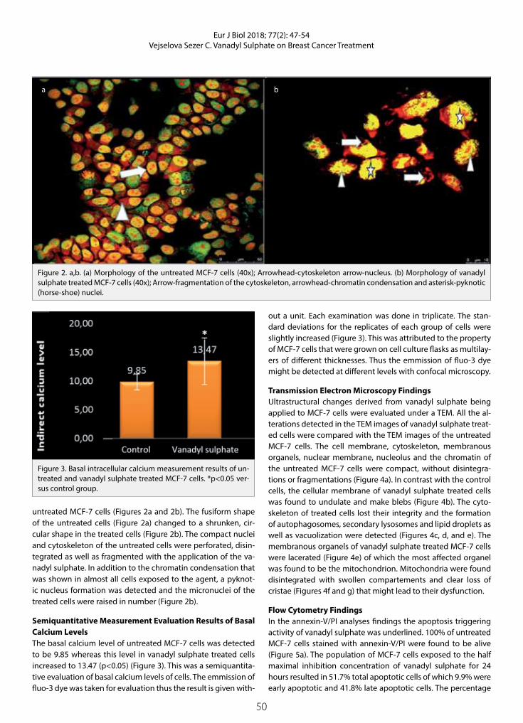
untreated MCF-7 cells (Figures 2a and 2b). The fusiform shape of the untreated cells (Figure 2a) changed to a shrunken, cir-cular shape in the treated cells (Figure 2b). The compact nuclei and cytoskeleton of the untreated cells were perforated, disin-tegrated as well as fragmented with the application of the va-nadyl sulphate. In addition to the chromatin condensation that was shown in almost all cells exposed to the agent, a pyknot-ic nucleus formation was detected and the micronuclei of the treated cells were raised in number (Figure 2b).
Semiquantitative Measurement Evaluation Results of Basal Calcium Levels The basal calcium level of untreated MCF-7 cells was detected to be 9.85 whereas this level in vanadyl sulphate treated cells increased to 13.47 (p<0.05) (Figure 3). This was a semiquantita-tive evaluation of basal calcium levels of cells. The emmission of fluo-3 dye was taken for evaluation thus the result is given with-
out a unit. Each examination was done in triplicate. The stan-dard deviations for the replicates of each group of cells were slightly increased (Figure 3). This was attributed to the property of MCF-7 cells that were grown on cell culture flasks as multilay-ers of different thicknesses. Thus the emmission of fluo-3 dye might be detected at different levels with confocal microscopy.
Transmission Electron Microscopy FindingsUltrastructural changes derived from vanadyl sulphate being applied to MCF-7 cells were evaluated under a TEM. All the al-terations detected in the TEM images of vanadyl sulphate treat-ed cells were compared with the TEM images of the untreated MCF-7 cells. The cell membrane, cytoskeleton, membranous organels, nuclear membrane, nucleolus and the chromatin of the untreated MCF-7 cells were compact, without disintegra-tions or fragmentations (Figure 4a). In contrast with the control cells, the cellular membrane of vanadyl sulphate treated cells was found to undulate and make blebs (Figure 4b). The cyto-skeleton of treated cells lost their integrity and the formation of autophagosomes, secondary lysosomes and lipid droplets as well as vacuolization were detected (Figures 4c, d, and e). The membranous organels of vanadyl sulphate treated MCF-7 cells were lacerated (Figure 4e) of which the most affected organel was found to be the mitochondrion. Mitochondria were found disintegrated with swollen compartements and clear loss of cristae (Figures 4f and g) that might lead to their dysfunction.
Flow Cytometry FindingsIn the annexin-V/PI analyses findings the apoptosis triggering activity of vanadyl sulphate was underlined. 100% of untreated MCF-7 cells stained with annexin-V/PI were found to be alive (Figure 5a). The population of MCF-7 cells exposed to the half maximal inhibition concentration of vanadyl sulphate for 24 hours resulted in 51.7% total apoptotic cells of which 9.9% were early apoptotic and 41.8% late apoptotic cells. The percentage
Figure 3. Basal intracellular calcium measurement results of un-treated and vanadyl sulphate treated MCF-7 cells. *p<0.05 ver-sus control group.
Figure 2. a,b. (a) Morphology of the untreated MCF-7 cells (40x); Arrowhead-cytoskeleton arrow-nucleus. (b) Morphology of vanadyl sulphate treated MCF-7 cells (40x); Arrow-fragmentation of the cytoskeleton, arrowhead-chromatin condensation and asterisk-pyknotic (horse-shoe) nuclei.
a b

51
Eur J Biol 2018; 77(2): 47-54Vejselova Sezer C. Vanadyl Sulphate on Breast Cancer Treatment
of necrotic cells in this group was 4.3% and 44% of the treated cells were live cells (Figure 5b).
Immunohistochemical ResultsThe semiquantitative evaluation results of cyclin B1 and D1 and E-cadherin are shown in Table 1. The average staining score of the untreated MCF-7 cells was 0.66. The scoring method for vanadyl
sulphate treated MCF-7 cells was made in comparison to untreat-ed cells. The cyclin B1 staining average score decreased to 0.33 in the treated cells. In contrast, the cyclin D1 average increased threefold to 1.66 in the same group of cells. The E-cadherin stain-ing score of vanadyl sulphate treated MCF-7 cells was raised to 2, i.e. almost three times bigger than that of control cells (Table 1).
Figure 4. a-g. TEM micrographs of untreated (a) and vanadyl sulphate treated (b, c, d, e, f and g) MCF-7 cells. A; Arrowhead-nucle-ar membrane, arrow-nucleolus, asterisk-cytoskeleton and organels. (b) Arrow-undulation of cell membrane. (c) Arrow-loss of cristae, double headed arrow-autophagosome, asterisk-disintegrated mitochondrion. (d) Arrowhead-lipid droplets, rhomboid-vacuolization. (e) Arrow-dysintegrated membranous organels, asterisk-secondary lysosome. (f ) Arrow-enlarged mitochondrial compartments and (g) Arrow-Loss of cristae.
a
e
b
f
c d
g
Figure 5. a,b. Cell death mode of untreated (a) and vanadyl sulphate treated (b) MCF-7 cells. (a) Q1-Necrotic/dead cells (0.00%), Q2-Late apoptotic cells (0.00%), Q3-Live cells (100%) and Q4-Early apoptotic cells (0.00%). (b) Q1-Necrotic/dead cells (4.3%), Q2-Late apoptotic cells (41.8%), Q3-Live cells (44.0%) and Q4-Early apoptotic cells (9.9%).
a b

52
Eur J Biol 2018; 77(2): 47-54Vejselova Sezer C. Vanadyl Sulphate on Breast Cancer Treatment
The stainings of cyclins (B1, D1) and E-cadherin of MCF-7 con-trol cells are shown in Figures 6a, b and c, respectively. In Figures 6d, 6e and 6f the stainings of cyclin B1 cyclin D1 and E-cadherin for vanadyl sulphate treated MCF-7 cells are given. These images were used in the scoring method whose results are given in Table 1. Images were given to show the staining type of the cells.
DISCUSSION
Cancer treatment with metal based chemotherapeutics has become an intensively applied method in current therapy. Current therapeutics such as cisplatin, gold and ruthenium are quite effective in killing cancer cells due to their biochemical properties on inhibiting the growth of cancer cells (5). How-
Table 1. Semiquantitative evaluation of labelings of cyclins (B1, D1) and E-cadherin in untreated and vanadyl sulphate treated MCF-7 cells. In this table, unstained cells were indicated by a 0, slightly stained by 1, moderately stained by 2 and intensively stained cells were indicated by the number 3
Cyclin B1 1. Replication 2. Replication 3. Replication Average
Untreated cells 1 1 0 0.66
Vanadyl sulphate treated cells 0 0 1 0.33
Cyclin D1 1. Replication 2. Replication 3. Replication Average
Untreated cells 1 0 1 0.66
Vanadyl sulphate treated cells 1 2 2 1.66
E-cadherin 1. Replication 2. Replication 3. Replication Average
Untreated cells 1 0 1 0.66
Vanadyl sulphate treated cells 2 2 2 2
Figure 6. a-f. Light microscopy images of cyclin B1 (a), cyclin D1 (b) and E-cadherin (c) proteins labelled in untreated MCF-7 cells (40x). In images (d), (e) and (f ) are shown cyclin B1, cyclin D1 and E-cadherin stainings of vanadyl sulphate treated MCF-7 cells (40x), respectively. Intensively stained cells are indicated by arrowheads.
a
d
b
e
c
f

53
Eur J Biol 2018; 77(2): 47-54Vejselova Sezer C. Vanadyl Sulphate on Breast Cancer Treatment
ever, their strong side effects and development of resistance make their application and effectiveness on cancer therapy limited (19). Thus, novel agents for cancer treatment are really needed. Vanadium and its compounds have recently been re-ported as good candidates for anti-cancer potency (5). Based on this, in this study, we evaluated the anti-cancer poten-cy of a vanadium salt, vanadyl sulphate, with a multi-mode assesment approach on its antiproliferative, cytotoxic and proapoptotic properties on human breast cancer MCF-7 cells. According to the MTT findings, the viability of MCF-7 cells de-creased dependent on dose when applied for 24 hours with different vanadyl sulphate concentrations. As shown in Figure 1, the half maximal inhibition concentration (IC50) of vanadyl sulphate on MCF-7 cells was determined to be 85 µM for this length of exposure. Similarly with our results, in L929 mouse fibrosarcoma cells and human hepatocarcinoma HepG2 cells vanadyl sulphate was shown to act as an antiproliferative agent according to dose and time (14). In addition, in another study, A549 human lung adenocarcinoma cells were inhibited with an IC50 value of 15 µM. In the same study, human prostate cancer cells DU145 were totally inhibited by the application of IC50 concentration of 15 µM (20,21). This can be attributed to the dependence of the antiproliferative activity of vanadyl sulphate on the cells type and application dose and time. This property of vanadium and vanadium compounds has been well described in a previous study (14).
Apoptosis triggering activity on malign cells is the most com-mon property of vanadium compounds (14). For vanadium compounds, it is described that they are proapoptotic via DNA fragmentation (9,14). Based on the investigations into finding an apoptosis stimulating agent for cancer therapy (19), we eval-uated the mode of cell death triggered by vanadyl sulphate application to MCF-7 cells. On the morphological analyses on confocal microscopy, on MCF-7 cells exposed to IC50 value of va-nadyl sulphate for 24 hours clear apoptotic sparks such as chro-matin condensation, fragmentation of cell and the nuclei, hole formation in the cytoskeleton and horse-shoe nucleus forma-tion were detected (Figure 2). In line with our results, on A549 and DU145 cells exposed to vanadyl sulphate researchers have shown apoptotic cells with fragmented nuclei (21).
In one piece of research it was reported that increased intra-cellular basal calcium level lead to apoptosis (22). The death of T-cell hybridoma cells is found to be related to the increased intracellular calcium level (23). In accordance with these find-ings in our study, the basal intracellular calcium level of MCF-7 cells exposed to vanadyl sulphate for 24 hours was found to be augmented (Figure 3). The apoptosis triggering activity of vanadyl sulphate on MCF-7 is underlined in our study based on the morphological and ultrastructural changes (Figure 4) of the applied cells as well as annexin V-FITC and PI evaluations of cell death mode (Figure 5) in the same group of cells. In annexin-V analyses results, apoptosis was shown in 51.7 % of vanadyl sul-phate applied cells. In addition the detected increase in basal calcium levels of the treated cells suggest apoptosis in accor-dance with the findings of other researchers on vanadate ap-
plied cells that reporting inhibition of Ca-ATPase activity, and in turn causing intracellular calcium accumulation and apoptosis (24). Results of a study on H35-19 rat hepatoma cells exposed to VOSO4, Na3VO4 ve NaVO3 salts, showed undulations in the nuclear membrane, trabecular nucleus, fragmentations on the cisternae of Golgi aparatus as ultrastructural changes (25). Sim-ilarly, a loss of cristae and swelling of mitochondrion, shrunken cell structure, secondary lysosome formation, chromatin con-densation and pyknotic nuclei were detected as ultrastructural changes after exposure to vanadyl sulphate for 24 hours.
Sodium orthovanadate was reported to stop cell cycles by causing increased cyclin B1 expression on HepG2, Sk-Hep-1 and Hep3B liver carcinoma cells relative to dose (14). Moreover, ox-ovanadium compounds were declared to inhibit cell cycles at a concentration of 100 µM over the course of a 24 hours applica-tion (26). In our study, in vanadyl sulphate treated MCF-7 cells, cyclin B1 expression was relatively decreased (Table 1) com-pared to the control cells. On the contrary, cyclin D1 expression on these cells was significantly increased (Table 1).
The expression of E-cadherin in a variety of aggressive human cancers has been reported to be reduced (27). In breast cancer cells the expression of this protein was declared to be partly or totally absent. The absence of E-cadherin expression makes breast cancer cells more invasive and metastatic (28). Stimulat-ing expression of E-cadherin in breast cancer cells was deter-mined to significantly inhibit the proliferation of cells in vitro and in vivo (20). Herein, it was detected increased E-cadherin expression in MCF-7 cells caused by vanadyl sulphate applica-tion. Our result is supported by a study that declared vanadium (IV) compounds to inhibit the invasion and adhesion of osteo-sarcoma cells relative to dose (29).
In conclusion, vanadyl sulphate was found to be cytotoxic and antiproliferative on MCF-7 cells when applied for a short time. It triggered apoptosis by changing the morphology and ultra-structure of the exposed cells. Thus, vanadyl sulphate might be suggested for further investigations for its usage in cancer ther-apy due to its anti-cancer properties considered above.
Peer-review: Externally peer-reviewed.
Acknowledgements: Many thanks to the Scientific and Technological Research Council of Turkey (TUBITAK) for the support given for the au-thor and the Anadolu University Scientific Research Project Unit for the support provided for this project with number 1403F089.
Conflict of Interest: The author has no conflict of interest to declare.
Financial Disclosure: This study was supported by Anadolu University Scientific Research Project Unit (No: 1403F089).
REFERENCES
1. Siegel RL, Miller KD, Jemal A. Cancer statistics. CA Cancer J Clin 2015; 65(1): 5-29.
2. Mobasheri A, Barrett-Jolley R. Aquaporin water channels in the mammary gland: From physiology to pathophysiology and neo-plasia. J Mammary Gland Biol Neoplasia 2014;19(1): 91-102.

54
Eur J Biol 2018; 77(2): 47-54Vejselova Sezer C. Vanadyl Sulphate on Breast Cancer Treatment
3. Harris HR, Bergkvist L, Wolk A. Adherence to the world cancer re-search fund/American Institute for Cancer Research recommenda-tions and breast cancer risk. Int J Cancer 2016; 138(11): 2657-64.
4. Rillema JA. Development of the mammary gland and lactation. Trends Endocrinol Metab 1994; 5(4): 149-54.
5. Petanidis S, Kioseoglou E, Hadzopoulou-Cladaras M, Salifoglou A. Novel ternary vanadium-betaine-peroxido species suppresses H-ras and matrix metalloproteinase-2 expression by increasing re-active oxygen species-mediated apoptosis in cancer cells. Cancer Lett 2013; 335(2): 387-96.
6. Gruzewska K, Michno A, Pawelczyk T, Bielarczyk H. Essentiality and toxicity of vanadium supplements in health and pathology. J Physiol Pharmacol 2014; 65(5): 603-11.
7. Bishayee A, Waghray A, Patel MA, Chatterjee M. Vanadium in the detection, prevention and treatment of cancer: The in vivo evi-dence. Cancer Lett 2010; 294(1): 1-12.
8. Gonzalez-Villalva A, Pinon-Zarate G, De la Pena Diaz A, Flores-García M, Bizarro-Nevares P, Rendón-Huerta EP, et al. The effect of vanadium on platelet function. Environ Toxicol Pharmacol 2011; 32(3): 447-56.
9. Evangelou AM. Vanadium in cancer treatment. Critical Rev Oncol Hematol 2002; 42(3): 249-65.
10. Ekert PG, Vaux DL. Apoptosis and the immune system. Br Med Bul-letin 1997; 53(3): 591-603.
11. Kostova I. Titanium and vanadium complexes as anticancer agents. Anticancer Agents Med Chem 2009; 9(8): 827-42.
12. Korbecki J, Baranowska-Bosiacka I, Gutowska I, Chlubek D. Bio-chemical and medical importance of vanadium compounds. Acta Biochim Pol 2012; 59(2): 195-200.
13. Liem DA, Gho CC, Gho BC, Kazim S, Manintveld OC, Verdouw PD, et al. The tyrosine phosphatase inhibitor bis(maltolato)-oxovanadi-um attenuates myocardial reperfusion injury by opening ATP-sen-sitive potassium channels. J Pharmacol Exp Ther 2004; 309(3): 1256-62.
14. Abakumova OY, Podobed OV, Belayeva NF, Tochilkin AI. Antican-cer activity of oxovanadium compounds. Biomed Khim 2012; 6(2): 164-70.
15. Capella LS, Alcantara JS, Maura-Neto V, Lopes AG, Capella MA. Va-nadate is toxic to adherent-growing multidrug-resistant cells. Tu-mor Biol 2000; 21(1): 54-62.
16. Suzuki T, Fujikura K, Higashiyama T, Takata K. DNA staining for flu-orescence and laser confocal microscopy. J Histochem Cytochem 1997; 45(1): 49-53.
17. Canel M, Serrels A, Frame MC, Brunton VG. E-Cadherin-integrin crosstalk in cancer invasion and metastasis. J Cell Sci 2013; 126(Pt 2): 393-401.
18. Glauert AM, Lewis PR. Biological specimen preparation for trans-mission electron microscopy. Princeton University Press, New Jer-sey, 1998; ISBN: 0-691-00749-2 (Cloth), ISBN: 0-691-00900-7 (pbk).
19. Yamaguchi T, Watanabe S, Matsumura Y, Tokuoka Y, Yokoyama A. Oxovanadium complexes with quinoline and pyridinone ligands: Syntheses of the complexes and effect of alkyl chains on their apoptosis-inducing activity in leukemia cells. Bioorganic Med Chem 2012; 20(9): 3058-64.
20. Berx G, Van Roy F. Commentary: The E-cadherin/catenin complex: an important gatekeeper in breast cancer tumorigenesis and ma-lignant progression. Breast Cancer Res 2001; 3(5): 289-93.
21. Holko P, Ligeza J, Kisielewska J, Kordowiak AM, Klein A. The effect of vanadyl sulphate (VOSO4) on autocrine growth on human epi-thelial cancer cell lines. Pol J Pathol 2008; 59(1): 3-8.
22. Tadakuma T, Kizaki H, Odaka C, Kubota R, Ishimura Y, Yagita H, et al. CD4+CD8+ thymocytes are susceptible to DNA fragmentation induced by phorbol ester, calcium ionophore and anti-CD3 anti-body. Eur J Immunol 1990; 20(4): 779-84.
23. McConkey DJ, Hartzell P, Amador-Perez FJ, Orenius S, Jondal M. Calcium-dependent killing of immature thymocytes by stimu-lation via CD37T cell receptor complex. J Immunol 1989; 143(6): 1801-6.
24. Varecka L, Peterajova E, Sevcik J. Ca(2+)-activated K+ channel and the activation of Ca2+ influx in vanadate-treated red blood cells. Gen Physiol Biophys 1997; 16(4): 339-57.
25. Kordowiak AM, Klein A, Goc A, Dabroś W. Comparison of the effect of VOSO4, Na3VO4 and NaVO3 on proliferation, viability and mor-phology of H35-19 rat hepatoma cell line. Pol J Pathol 2007; 58(1): 51-7.
26. León IE, Cadavid Vargas JF, Tiscornia I, Porro V, Castelli S, Katkar P, et al. Oxidovanadium(IV) complexes with chrysin and silibinin: anticancer activity and mechanisms of action in a human colon adenocarcinoma model. J Biol Inorg Chem 2015; 20(7): 1175-91.
27. Umbas R, Schalken JA, Aalders TW, Carter BS, Karthaus HF, Schaafs-ma HE, et al. Expression of the cellular adhesion molecule E-cad-herin is reduced or absent in high-grade prostate cancer. Cancer Res 1994; 52(18): 5104-09.
28. Oka H, Shiozaki H, Kobayashi K, Inoue M, Tahara H, Kobayashi T, et al. Expression of E-cadherin cell adhesion molecules in human breast cancer tissues and its relationship to metastasis. Cancer Re-search 1993; 53(7): 1696-01.
29. Molinuevo MS, Cortizo AM, Etcheverry SB. Vanadium (IV) complex-es inhibit adhesion, migration and colony formation of UMR106 osteosarcoma cells. Cancer Chemother Pharmacol 2008; 61(5): 767-73.

55
INTRODUCTION
Obesity is an increase in the amount of fat in the body, which occurs when energy intake is more than energy spent.
Today fatty tissue is no more regarded as a mere fat stor-age since its carries an important duty. That is, it affects other organs and carries communication between them, therefore fatty tissue is regarded as an endocrine organ which synthesizes and releases many chemical messen-gers, the cytokine of fatty tissue (adipokine) (1-3).
In obesity, an increased fatty tissue brings many physi-cal and biochemical pathologies (1,2).
Ghrelin is an acylated peptide which contains 28 ami-no acids and it is primarily produced in the stomach and the proximal small intestine (4). Ghrelin activates the hypothalamus and other related systems in the brain, therefore increasing gastrointestinal motility and decreasing insulin secretion (5). The growth hormone secretagogue receptor (GHS-R) mediates the differ-ent actions of the synthetic growth hormone secre-tagogues (GHS) and the endogenous ligand of this
Eur J Biol 2018; 77(2): 55-58
RESEARCH ARTICLE
Serum Concentration of Ghrelin, Oxidative Stress and Lipid Parameters in Obese Subjects
Sevilay Zora1 , Turkan Yigitbasi2* , Gozde Ulfer2 , Nesrin Emekli2 1Istanbul Aydın University, Vocational Faculty of Health Services, Istanbul, Turkey2Istanbul Medipol University, Faculty of Medicine, Department of Biochemistry, Istanbul, Turkey
This study was presented at the 6th World Congress of Oxidative Stress, Calcium Signaling and TRP Channels, 24-27 May 2016, Isparta, Turkey.
Address for Correspondence: Turkan Yigitbasi E-mail: [email protected] Received: 12.07.2018 Accepted: 18.09.2018© Copyright 2018 by The Istanbul University Faculty of Science • Available online at http://ejb.istanbul.edu.tr • DOI: 10.26650/EurJBiol.2018.18008
Please cite this article as: Zora S, Yigitbasi T, Ulfer G, Emekli N. Serum Concentration of Ghrelin, Oxidative Stress and Lipid Parameters in Obese Subjects. Eur J Biol 2018; 77(2): 55-58.
ORCID IDs of the authors: S.Z. 0000-0002-6534-0538; T.Y. 0000-0002-0675-1839; G.U. 0000-0003-2350-6381; N.E. 0000-0002-0109-5086.
ABSTRACT
Objective: Ghrelin is a hormone with peptide structure. It has fatty tissue and increases appetite. Obesity is a multifactorial chronic disease characterized by an increase in fat tissue. Fat tissue, like the endocrine organ, triggers oxidative stress and can lead to the development of obesity-related pathologies. The purpose of this study is to examine the relationship between the ghrelin in blood, and oxidative stress and lipid parameters.
Materials and Methods: The study was conducted with 61 obese and 24 healthy individuals. Ghreline levels were measured using the ELISA method, while total antioxidant status (TAS) and oxidant status (TOS), triglyceride (TG), total cholesterol (TC), HDL-cholesterol (HDL-C) and LDL cholesterol (LDL-C) levels were measured using the photometric method.
Results: A negative correlation was found between body mass index (BMI) and ghrelin levels in the obese group (p<0.05). But there was no significant difference of ghrelin levels in obese and control groups (p>0.05). TAS was observed to be lower in obese compared to control group, while The Oxidative Stress Index (OSI) was found to be significantly higher than the obese group (p<0.05). TG levels were found to be increased in obese; whereas ghrelin, TC, LDL-C and HDL-C levels did not show any difference (p>0.05).
Conclusion: Increasing obesity level (BMI) and decreasing ghrelin level were found to be correlated. New studies are needed in order to discover the changes in ghrelin level connected to oxidative stress.
Keywords: Ghrelin, obesity, oxidative stress

56
Eur J Biol 2018; 77(2): 55-58Zora et al. Ghrelin and Oxidative Stress in Obese Subjects
receptor, ghrelin (6). This endogenous ligand for this GHS re-ceptor (GHS-R) was generally identified by Kojima et al. in 1999 and named ‘ghrelin’ (7). Currently it’s the only known oroxigenic hormone (8). Cancer patients with loss of appetite were report-ed to gain back appetite when administered ghrelin (9).
In healthy cells, oxidation of molecular oxygen is a well-con-trolled process. However, in cases of cell damage and disease, superoxide radical (O2
.−) and hydrogen peroxide (H2O2) amounts increase. In the case of increased reactive oxygen radicals (ROS), insufficient antioxidants lead to oxidative stress. In the case of oxidative stress, proteins, lipids and DNA are damaged. Several studies report that increased oxidative stress in obesity contrib-utes to development of atherosclerosis (10-14).
The aim of this study was to investigate the relationship be-tween blood levels of ghrelin hormone, body mass index (BMI), oxidative stress and lipid parameters, which are important in carbohydrate and fat metabolism.
MATERIALS AND METHODS
Study Design and Data CollectionThis study includes 24 controls (13 male and 11 female) and 61 obese (37 male and 24 female) who have consulted Mega Medi-pol Hospital Laboratory of Medipol University between Sep-tember and October 2015. After taking the approval of ethical committee of Medipol University, all the patients were informed and confirmed consent documents were taken from all them. Groups were classified according to their BMI into two groups: BMI>18.9 and BMI<24.9 kg/m2 are considered as normal weight and BMI>30 kg/m2 are considered as obese. BMI values were ob-tained by dividing the weight (in kg) by the square of height (m2).
The mean BMI (kg/m2) in the control group was 23.52±0.89, while in the obese group it was 33.76±6.15.
Exclusion Criteria in the StudyExclusion criteria in the study was as follows: younger than 18, over 75 years old, smoking habits, hypertension, heart diseas-es, osteoarthritis, cancer, polycystic disease, inflammation and infectious diseases not included in the study. The study started after the approval of Medipol University Ethics Board. All the subjects were informed about the study and their approved consent forms were received.
Blood Collection and StorageVenous blood was collected in the early morning before breakfast and after overnight sleep. Blood samples were collected in yellow covered flat tubes and purple covered (EDTA containing) tubes. Yellow covered tubes were centrifuged at 2400 rpm for 10 minutes in the clinical biochemistry laboratory of Medipol University, and blood cells were separated from serum. Separated serums were taken into Eppendorf tubes and kept at -80°C until the analysis.
Methods UsedFollowing analyses were carried out: Ghrelin, triglyceride (TG), total cholesterol (TC), low density lipoprotein cholesterol
(LDL-C), high density lipoprotein cholesterol (HDL-C), total an-tioxidant status (TAS), total oxidant status (TOS) and oxidative stress index (OSI) levels.
Serum ghrelin levels were determined by using Ray Bio EIA-GHR-1 Elisa kit; TAS and TOS were determined by colorimetric method. OSI was calculated using the formula given below: OSI= [(TOS, μmol H2O2 equivalent/l)/(TAS, μmol Trolox equiva-lent/l)] x100 (15). Serum TC, HDL-C, LDL-C and TG levels were measured with Roche/Hitachi C501 instrument photometrically with the kits recommended by the instrument company.
Statistical AnalysisThe Statistical Package for the Social Sciences (SPSS) Windows version 22.0 (IBM Corp.; Armonk, NY, USA) program was used to evaluate the statistical analysis of the study. Variables were de-fined in ±SD limits. T-test was used for the comparison of mea-sured variable average values that obey normal distribution, in two groups. In order to compare dependent variables paired t-test was applied. Also, Mann-Whitney U-test was preferred to compare the average values obtained that do not obey normal distribution. For all the test p<0.05 was considered significant.
RESULTS
As it was shown in Table 1, there was no significant change in the plasma levels of ghrelin (p>0.05) in obese and control groups.
Serum TG levels were increased in obese group (p<0.05) where-as other lipid parameters such as TC, LDL-C and HDL-C were nor-mal levels (p>0.05).
There was a significant decrease in serum TAS in obese group (p<0.001), serum TOS were not significantly changed (p>0.05) and OSI were significantly high in obese compare to normal subjects.
When correlation analyses were examined, a positive relation was found between TOS and OSI (r: 0.77, p<0.05) TOS and TG (r: 0.52, p<0.05), a negative correlation was observed between TOS values and HDL (r:-0.34, p<0.05). In addition, positive rela-tion was found between OSI and TG (r:0.33, p<0.05).
The plasma levels of ghrelin were significantly negatively cor-related with BMI (r: -2,65, p<0.05).
DISCUSSION
Known as the “orexigenic hormone”, ghrelin maintains the en-ergy balance of the organism together with neuroendocrine regulation, intestinal and pancreatic peptides (4,5,16). Despite these systems for protecting the organism, the prevalence of obesity is increasing in the world. Obesity results in insulin resis-tance, inflammation, oxidative stress in parallel with increasing fat tissue (1).
In this study, we investigated the relationship between ghrelin serum concentration and oxidative stress and lipid parameters

57
Eur J Biol 2018; 77(2): 55-58Zora et al. Ghrelin and Oxidative Stress in Obese Subjects
in the obese and healthy control group, which increased appe-tite and food intake.
Mucioli et al. (17) reported that ghrelin, one of the peptides in the appetite center, increases appetite and causes obesity. As reported by Kara et al. (18), Ghrelin injections to mice caused an increase in fat tissue by reducing fat use. Ghrelin’s fat tissue and appetite-enhancing effects are independent of GH effects and are thought to be regulated by specific neurons in the CNS where leptin is also a mediator.
Wren et al. reported that when ghrelin is administered intrave-nously to normal weight healthy people, the desire to eat is in-creased. Blood levels of ghrelin decrease after fasting and after a sugary and fatty meal (5).
Tschöp et al. (19) reported that ghrelin levels were lower in obese subjects than in weaker subjects. In the Suematsu et al. study, ghrelin was measured in 17 obese and 17 healthy sub-jects and the ghrelin level was found to be lower in the obese group than in the control group (12). Participants had an in-crease in serum ghrelin levels as a result of their weight loss after the diet (14,17-19).
In the study of Cinaz et al. (20), hunger and satiety ghrelin levels were measured in 38 obese and 19 healthy children. In both obese and control groups fasting ghrelin levels were higher than satiety ghrelin levels (p<0.05). The researchers also showed that the hunger and satiety ghrelin levels of obese chil-dren were lower than the control group (p<0.05). The study also found a negative correlation between BMI and hunger strike levels in the obese group.
In our study, the ghrelin concentration was not statistically different in the control and obese group (p>0.05). Howev-er, increased obesity level (BMI) was found to be correlated with decreased ghrelin level (r: -2.65 p<0.05). This situation is caused by positive energy balance which suppresses ghrelin secretion in obese people. This finding is consistent with stud-
ies suggesting that ghrelin levels are reduced in obese indi-viduals.
In our study, TAS was observed to be lower in obese compared to control group, while OSI was found to be significantly high-er than the obese group (p<0.05). Obese group with oxidative stress do not differ in terms of ghrelin levels (p>0.05). The only study in this area in the literature was reported by Suematsu et al. In their study, free 8-epi-prostaglandin F2α was measured as a systemic marker of oxidative stress and, independently from obesity, it was discovered that increases in oxidative stress de-creases ghrelin (12).
In the literature, in general, ghrelin levels were found to be de-creased in obese subjects, but the mechanism of this decrease has not been explained (19).
The LDL-C, HDL-C and TC levels did not significantly change when compared to control group and remained within the nor-mal reference limits when the lipid profile in both groups were examined and TG levels were found to be statistically higher in the obese group (p<0.05). Positive correlation between TG and TOS and OSI shows the role of TG increase in the formation of oxidative stress.
In conclusion, ghrelin levels did not significantly change in obese group when compared to control group. This situation is caused by positive energy balance suppresses ghrelin secretion in obese people. Understanding obesity and its associated dis-eases with the appetite hormone ghrelin will help to develop new strategies for the prevention of obesity. New studies are needed for guidance in this area.
Ethics Committee Approval: Ethics Committee Approval was received for this study from the ethics committee of Medipol University.
Informed Consent: Written informed consent was obtained from pa-tients who participated in this study.
Peer-review: Externally peer-reviewed.
Table 1. Laboratory findings of control and obese group.
Control group (Mean±SD) Obese group (Mean±SD) p*
Ghrelin (pg/mL) 110.78±25.46 110.43±25.43 0.955
TAS (μmol Trolox Equiv./L) 0.92±0.11 0.81±0.13 <0.05
TOS (μmol H2O2 Equiv./L) 26.96±5.49 28.26±6.31 0.386
OSI (AU) 2.92±0.62 3.56±0.86 <0.05
HDL (mg/dL) 53.22±17.82 47.65±13.52 0.149
LDL (mg/dL) 119.35±34.45 121.98±37.88 0.768
TG (mg/dL) 126.14±82.92 158.13±85.36 0.033
TC (mg/dL) 195.46±41.36 198.35±40.99 0.771
TAS: total antioxidant response; TOS: total oxidant status; OSI: oxidative stress index value; AU: Arbitrary unit; TC: total cholesterol*p<0.05 for t test.
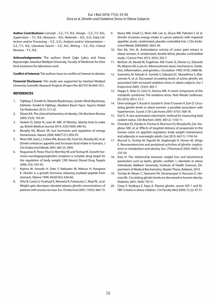
58
Eur J Biol 2018; 77(2): 55-58Zora et al. Ghrelin and Oxidative Stress in Obese Subjects
Author Contributions: Concept – S.Z., T.Y., N.E.; Design – S.Z., T.Y., N.E.; Supervision – T.Y., N.E.; Resource – N.E.; Materials – N.E., G.Ü.; Data Col-lection and/or Processing – S.Z., G.Ü.; Analysis and/or Interpretation – S.Z., T.Y., N.E.; Literature Search – S.Z., N.E.; Writing – S.Z., N.E.; Critical Reviews – T.Y., N.E.
Acknowledgements: The authors thank Çağrı Çakıcı and Feyza Bayramoğlu (Istanbul Medipol University, Faculty of Medicine) for their kind assistance for laboratory work.
Conflict of Interest: The authors have no conflict of interest to declare.
Financial Disclosure: This study was supported by Istanbul Medipol University Scientific Research Projects (Project No: 86770134-604-101).
REFERENCES
1. Yiğitbaşı T, Emekli N. Obezite Biyokimyası. İçinde: Klinik Biyokimya, Editörler: Emekli & Yiğitbaşı, Akademi Basım Yayın, Yayımcı Nobel Tıp Kitabevleri 2015; 311-22.
2. Sikaris KA. The clinical bichemistry of obesity. Clin Biochem Review 2004; 25(3): 165-81.
3. Haslam D, Sattar N, Lean M. ABC of Obesity: obesity time to wake up. British Medical Journal 2014; 333(7569): 640-42.
4. Murphy KG, Bloom SR. Gut hormones and regulation of energy hemeostasis. Nature 2006; 444(7121): 854-59.
5. Wren AM, Seal LJ, Cohen MA, Brynes AE, Frost GS, Murphy KG, et al. Ghrelin enhances appetite and increases food intake in humans. J Clin Endocrinol Metab 2001; 86(12): 5992.
6. Noguerias R, Perez-Tilve D, Worrtley KE and Tschop M. Growth hor-mone secretagogue(ghrelin) receptors a complex drug target for the regulation of body weight. CNS Neurol Disord Drug Targets 2006; 5(3): 335-43.
7. Kojima M, Hosoda H, Date Y, Nakazato M, Matsuo H, Kangawa K. Ghrelin is a growth hormone releasing acylated peptide from stomach. Nature 1999; 402(6762): 656-60.
8. Otto B, Cuntz U, Fruehauf E, Wawarta R, Fulwaczny C, Riepl RL, et al. Weight gain decreases elevated plasma ghrelin concentrations of patients with anoxia nervosa. Eur J Endocrinol 2001; 145(5): 669-73.
9. Neary NM, Small CJ, Wren AM, Lee JL, Druce MR, Palmieri C et al. Ghrelin increases energy intake in cancer patients with impaired appetite: acute, randomized, placebo controlled trial. J Clin Endo-crinol Metab 2004;89(6): 2832-36.
10. Kim KA, Yim JE. Antioxidative activity of onion peel extract in obese women: A randomized, double-blind, placebo controlelled study. J Cancer Prev 2015; 20(3): 202-7.
11. Berliner JA, Navab M, Fogelman AM, Frank JS, Demer LL, Edwards PA, Watson AD, Lusis AJ. Atherosclerosis: basic mechanisms. Oxida-tion, inflammation, and genetics. Circulation 1995; 91(9): 2488-96.
12. Suematsu M, Katsuki A, Sumida Y, Gabazza EC, Murashima S, Mat-sumoto K, et al. Decreased circulating levels of active ghrelin are associated with increased oxidative stress in obese subjects. Eur J Endocrinol 2005; 153(3): 403-7.
13. Hopps E, Noto D, Caim G, Averna MR. A novel component of the metabolic syndrome: The oxidative stress. Nutr Metab Cardiovasc Dis 2010; 20(1): 72-7.
14. Oner-Iyidogan Y, Kocak H, Gurdol H, Oner P, Issever H, Esin D. Circu-lating ghrelin levels in obese women: a possible association with hypertension. Scand J Clin Lab Invest 2007; 67(5): 568-76.
15. Erel O. A new automated colorimetric method for measuring total oxidant status. Clin Biochem 2005; 38(12): 1103-11.
16. Chamber ES, Viardot A, Psichas A, Morrison DJ, Murphy KG, Zac-Var-ghese SEK, et al. Effects of targeted delivery of propionate to the human colon on appetite regulation, body weight maintenance and adiposity in overweight adults. Gut 2015; 64(11): 1744-54.
17. Mucioli G, Tschöp M, Papotti M, Deghenghi R, Himen M, Ghigo E. Neuroendoncrine and peripheral activities of ghrelin: implica-tions in metabolism and obesity. Eur J Pharmacol 2003; 440(2-3): 235-54.
18. Kara H. The relationship between weight loss and biochemical parameters such as leptin, ghrelin, nesfatin 1, obestatin in obese individuals. Balikesir University, Institute of Health Sciences, De-partment of Medical Biochemistry. Master Thesis, Balıkesir, 2014.
19. Tschöp M, Weyer C, Tataranni PA, Devanarayan V, Ravussin E, Hei-man ML. Circulating ghrelin levels are decreased in human obesity. Diabetes 2001; 50(4): 707-9.
20. Cinaz P, Yesilkaya E, Kaya A. Plasma ghrelin, serum IGF-1 and IG-FBP-3 levels in obese children. J Ist Faculty Med 2009; 72 (2): 47-51.

59
INTRODUCTION
Fish farming has increased mainly due to a greater demand for healthier meats for human consumption (1). However, the intensive production of these spe-cies presents a great challenge for the producer as a concentration of large populations increases the pre-disposition to bacterial and parasitic diseases (2). This problem is especially critical in the early stages of life of teleosts (3). Therefore, the formulation of diets that contribute to the health and well-being of juveniles are of great relevance.
Bovine colostrum is the first mammal lacteal secre-tion that is crucial for nutritional, metabolic and endocrine status of neonates (4,5). The effects of co-lostrum consumption include the protection of the enteric and respiratory tract and reduction in mortal-ity and morbidity of the newborn (6-8). The high nu-tritional value and concentration of biological mole-cules, such as antioxidant and antimicrobial factors, designate colostrum as a nutraceutical food (9). This lacteal secretion from bovine has been widely used as an immunological and nutritional source for sev-
Eur J Biol 2018; 77(2): 59-64
RESEARCH ARTICLE
Serum Protein Profile of Juvenile Pacu (Piaractus mesopotamicus) and Dourado (Salminus brasiliensis) Fed Bovine Colostrum as Partial Source of Protein in the Diet
Debora Moretti1* , Wiolene Nordi2 , Raul Machado-Neto1
1University of São Paulo, Luiz de Queiroz College of Agriculture, Department of Animal Science, Piracicaba, Brazil2Agriculture and Agri-Food Canada, Lethbridge Research and Development Centre, Government of Canada, Alberta, Canada
This study was presented at the 39th CONBRAVET-Congresso Brasileiro de Medicina Veterinária, December 4th-6th, 2012, São Paulo, Brazil.
Address for Correspondence: Debora Moretti E-mail: [email protected] Received: 01.03.2018 Accepted: 19.06.2018© Copyright 2018 by The Istanbul University Faculty of Science • Available online at http://ejb.istanbul.edu.tr • DOI: 10.26650/EurJBiol.2018.400108
Please cite this article as: Moretti D, Nordi W, Machado-Neto R. Serum Protein Profile of Juvenile Pacu (Piaractus mesopotamicus) and Dourado (Salminus brasiliensis) Fed Bovine Colostrum as Partial Source of Protein in the Diet. Eur J Biol 2018; 77(2): 59-64.
ORCID IDs of the authors: D.M. 0000-0003-0912-9989; W.N. 0000-0003-3666-6245; R.M.N. 0000-0002-5982-2776.
ABSTRACT
Objective: The electrophoretic profile of serum proteins was investigated in juvenile Piaractus mesopotamicus and Salminus brasiliensis fed with diets containing bovine colostrum, a nutraceutical food, in lyophilized form (LBC) for either 30 or 60 days.
Materials and Methods: Blood samples were collected from juveniles of P. mesopotamicus and S. brasiliensis fed for either 30 or 60 days with diets containing 0%, 10% or 20% of LBC. Serum protein fractions were then determined by electrophoresis in agarose gel.
Results: The P. mesopotamicus, an omnivorous fish, showed six serum protein fractions, while the S. brasiliensis, a carnivorous fish, showed four serum protein fractions. In both species the albumin fraction showed higher protein content at 30 days than at 60 days (p<0.05), indicating an inverse relationship with growth. The 5th and 4th fraction in mobility from the P. mesopotamicus and S. brasiliensis serum, respectively, were positioned in a gamma-globulin zone. In P. mesopotamicus, the concentration of protein in the gamma-globulin zone was higher at 60 days than at 30 days (p<0.05). In S. brasiliensis, in turn, the concentration of protein in the gamma-globulin zone was higher at 30 days than at 60 days (p<0.05). In juvenile P. mesopotamicus, a higher concentration of protein was also observed in the gamma-globulin zone in the 0% LBC compared to 10% and 20% LBC (p<0.05). Thus, the feeding period influenced only the fractions that were positioned in the albumin and gamma-globulin migration zone in both species.
Conclusion: The consumption of bovine colostrum decreased the concentration of protein in gamma-globulin fraction of the P. mesopotamicus, indicating that these juveniles had either less stimulus for their own synthesis of defense elements or an immunosuppressive effect of bovine colostrum ingestion.
Keywords: Colostrum, teleost, omnivorous, carnivorous, electrophoresis

60
Eur J Biol 2018; 77(2): 59-64Moretti et al. Serum Protein Profile of Juvenile Fish Fed Bovine Colostrum
eral species, including swine and rodents (10,11). In fish, the supply of lyophilized bovine colostrum is an innovative con-ception and the effects of this food on fish health has been explored by different approaches (12-14).
In Brazil, Piaractus mesopotamicus and Salminus brasiliensis, popularly known as pacu and dourado, are neotropical species with favorable features for commercial production. Besides be-
ing a species of interest for the sport of fishing, Piaractus meso-potamicus and Salminus brasiliensis present good performance and acceptance by the consumers (2,15,16). However, infor-mation about their physiology and the effects of nutraceutical foods on their health is still being gathered. The electrophore-sis of proteins from blood plasma or serum can be used in hu-mans and animals to provide information on acute and chronic inflammatory processes and consequently health (17,18). In recent decades, the study of blood protein fractions from differ-ent species has increased, including works involving fish, which focus on the relation between water pollution and fish health (19-22). The effects of diet on protein profile, especially gamma globulin fraction where antibodies are positioned, are of great relevance to evaluate the health and immunity of fish.
Since electrophoresis has been proposed to aid in the diagno-sis of disease, normal reference ranges for serum protein levels still need to be established in different species of fish. Consider-ing colostrum as a nutraceutical food, there is the possibility of this food modulating immunity, and consequently, fish health. Thus, this study investigates the electrophoretic prolife of se-rum proteins of juvenile Piaractus mesopotamicus and Salminus brasilienesis fed diets with bovine colostrum, a nutraceutical food, in the lyophilized form (LBC).
MATERIAL AND METHODS
DietsThe first lacteal secretion from multiparous Holstein cows was frozen at -20°C and, thereafter, lyophilized. This powder was ho-mogenized to obtain a homogeneous sample, which was ana-lyzed for the bromatological composition. Considering the nu-tritional composition of bovine colostrum, isonitrogenous and isoenergetic pelleted diets were formulated to attend to the re-quirements of juveniles Piaractus mesopotamicus and Salminus brasiliensis (Tables 1 and 2). After the homogenization of the in-gredients, the diets were pelletized, dried (at a temperature of approximately 46 °C) and stored in a dry place until the time of supply. Chemical compositions of experimental pelleted diets were subsequently submitted to chemical analysis (23).
ExperimentJuvenile Piaractus mesopotamicus and Salminus brasiliensis (8.5±0.7 and 13.3±0.9 g; 7.8±0.3 and 10.8±0.3 cm, respectively) were randomly distributed in 36 tanks (324 pacu in 18 tanks and 270 dourado in 18 tanks) with controlled water quality param-eters (26.8±1.5ºC, 5.8±1.0 mg L-1 of dissolved oxygen and <0.05 mg L-1 dissolved ammonia). After adaptation for 10 and seven days, respectively, diets containing 0%, 10% or 20% bovine co-lostrum in LBC were hand-fed to apparent satiety twice a day (08h30 and 16h30) for either 30 or 60 days (authorized by the ESALQ/USP ethics committee).
Seven fish were sampled on the 30th and 60th experimental day after 24 h fasting and anesthesia with benzocaine (0.1 g L-1). Blood samples obtained from the caudal vein were centrifuged and the serum was stored at -20˚C.
Table 1. Chemical composition of experimental pelleted diets fed to juvenile Piaractus mesopotamicus
Ingredient (g kg-1)
Diets1
0% 10% 20%
Bovine colostrum (679 g kg-1 CP) - 100 200
Soybean meal (45 g kg-1 CP) 265 76.7 -
Wheat meal 238 311.8 311.8
Poultry by-product meal 200 200 131
Broken rice 188 198 200
Fish meal (55% CP) 50 50 50
Fish oil 46.2 40 45.7
DL-methionine 2.4 3.4 4.7
L-lysine HCl - 6 11.5
BHT 2 2 2
Cellulose - 3.4 -
Calcareous - - 14.7
Corn grain - - 9.3
Premix2 10 10 10.5
Chemical composition (g kg-1)3
Dry matter 940.6 933.4 936.4
Crude protein 324.6 314.9 322
Crude fiber 30 27.7 30.7
Fat 90.5 98.2 109.3
Ash 106.4 101.1 92.8
Gross energy (MJ kg-1) 18 18.2 18.3
10% - diets containing 0% of lyophilized bovine colostrum; 10% - diets containing 10% of lyophilized bovine colostrum; 20% - diets containing 20% of lyophilized bovine colostrum.2Guabi Nutrição Animal, Campinas, São Paulo (ingredient per kg). Vitamins: A, 2,500 UI; D3, 600.000 UI; E, 37.500 UI; K3, 3,750 mg; C, 50,000 mg; B1, 4.000 mg; B2, 4,000 mg; B6, 4,000 mg; B12, 4,000 mg; calcium pantothenate, 12,000 mg; biotin, 15 mg; acid folic, 1,250 mg; niacin, 22,500 mg. Mineral: Cu, 2,500 mg; Zn, 12,500 mg; I, 375 mg; Se, 87.5 mg; Co, 125 mg; Mn, 12,500 mg; Fe, 15,000 mg; BHT, 15,000 mg. 3Original matter basis.

61
Eur J Biol 2018; 77(2): 59-64Moretti et al. Serum Protein Profile of Juvenile Fish Fed Bovine Colostrum
Analyses The total serum protein was determined (24) and an electrophoret-ic analysis was performed in an agarose gel (CELMGEL) for 30 min at 90 volts. After staining with 0.2% starch black for 5 min., a read-ing of the protein fractions in a densitometer (CELM DS35) with a wavelength of 520 nm was performed. The software CELM SE-250 was used to calculate the relative percentage of each protein frac-tion from the area under the curve created by the protein fraction. No attempt has been made to identify protein components of the sera studied as albumin or globulin. The electrophoretic fractions were numbered according to the increasing mobility.
Statistical AnalysisSerum variables were analyzed based on a 2×3 completely random-ized factorial design using SAS software (SAS Institute Inc., 2004), taking into consideration the three diets and two feeding periods (30 and 60 experimental days). An analysis of variance was per-
formed using the general linear model and comparisons between pairs of means were made using the Tukey test considering p<0.05.
RESULTS
Electrophoretic profile of serum proteins of juvenile pacu (Piaractus mesopotamicus) Using the software SDS-60 (Celm), six fractions in the serum of juvenile Piaractus mesopotamicus were identified (Figures 1 and 2). These fractions were quantified by the software considering the value of serum total protein (Table 3). The effect of the peri-od was observed for the 2nd fraction (p<0.05), with a higher val-ue at 30 days compared to 60 days. The 6th fraction was affected by the diet and period (p<0.05), with higher values at 60 days and in the group 0% LBC.
Electrophoretic profile of serum proteins of juvenile doura-do (Salminus brasiliensis) Using the software SDS-60 (Celm), four fractions in the serum of juvenile Salminus brasiliensis were identified (Figures 3 and 4). These fractions were quantified by the software considering the
Table 2. Chemical composition of experimental pelleted diets fed to juvenile Salminus brasiliensis
Ingredient (g kg-1)
Diets1
0% 10% 20%
Bovine colostrum (679 g kg-1 CP) - 100 200
Soybean meal (45 g kg-1 CP) 230 230 230
Poultry by-product meal 204.8 119.8 24.5
Fish meal (55 g kg-1 CP) 320 300 300
Fish oil 95 90 85.2
Premix2 10 10 10
BHT 0.2 0.2 0.2
Cellulose 20 30 30
Corn (whole grain) 120 120 120
Chemical composition (g kg-1)3
Dry matter 924.4 936.7 925.7
Crude protein 422 425.1 444.7
Crude fiber 18.9 29.2 37.3
Fat 140.7 140.6 133.7
Ash 121.9 114.5 91.1
Gross energy (MJ kg-1) 20.8 21.1 20.8
10% - diets containing 0% of lyophilized bovine colostrum; 10% - diets containing 10% of lyophilized bovine colostrum; 20% - diets containing 20% of lyophilized bovine colostrum. 2Guabi Nutrição Animal, Campinas, São Paulo (ingredient per kg). Vitamins: A, 2,500 UI; D3, 600.000 UI; E, 37.500 UI; K3, 3,750 mg; C, 50,000 mg; B1, 4.000 mg; B2, 4,000 mg; B6, 4,000 mg; B12, 4,000 mg; calcium pantothenate, 12,000 mg; biotin, 15 mg; acid folic, 1,250 mg; niacin, 22,500 mg. Mineral: Cu, 2,500 mg; Zn, 12,500 mg; I, 375 mg; Se, 87.5 mg; Co, 125 mg; Mn, 12,500 mg; Fe, 15,000 mg; BHT, 15,000 mg. 3Original matter basis.
Figure 1. Electrophoretic analyzes of 10 serum samples of juve-nile pacu, Piaractus mesopotamicus. Arrow: start of the race
Figure 2. Electrophoretic profile of serum proteins of juvenile pacu, Piaractus mesopotamicus, revealing six fractions (in order from fastest to slowest)

62
Eur J Biol 2018; 77(2): 59-64Moretti et al. Serum Protein Profile of Juvenile Fish Fed Bovine Colostrum
value of serum total protein (Table 4). The effect of the period was observed for the 1st and 4th fractions (p<0.05), with higher values at 30 days compared to 60 days.
DISCUSSION
The serum protein profile of juvenile pacu, Piaractus mesopotamicus, showed six fractions. Approximately 28%, 19%, 14%, 8%, 25%, and 7% of proteins were observed in the 1st, 2nd, 3rd, 4th, 5th and 6th fraction, respectively. In the juvenile dourado, Salminus brasiliensis, four fractions were identified. The 1st to 4th fraction corresponded approximately to 32%, 41%, 5% and 22% of the serum total protein (25). Work with four species of Tilapia, seven serum fractions were observed in the T. nilotica, T. galilaea and T. aurea, while de T. zillii showed six serum fractions. The au-thors also observed differences in the protein concentration in each band and suggest that these results might be related to diet.
The 2nd fraction in the serum of pacu and the 1st fraction in the serum of dourado, Salminus brasiliensis, showed a higher protein
concentration at 30 days compared to 60 days, indicating an in-verse relationship with growth. As an additional investigation, bovine serum albumin (BSA, Sigma-Aldrich Co) was added to fish serum and samples were submitted again to electrophore-sis analyses. The electrophoretic profile was not changed in both species; however, the 2nd fraction from Piaractus mesopotamicus serum and the 1st fraction from dourado serum showed a signifi-cant increase in its participation of total protein from 19% to 43% and 36% to 53%, respectively. These results indicated that these fractions are in the albumin migration zone and are probably the corresponding protein in fish. Thus, in juvenile pacu, the calcula-tion of globulin by the difference of serum total protein and albu-min is not adequate, since near 28% of serum proteins are in the 1st fraction, which corresponds to pre-albumin proteins and are not considered in this calculation.
According to Atamanalp et al. (26) and Maceda Veiga et al. (27), environmental changes can affect the physiological and biochem-ical characteristics of fish blood, and the hematological and serum protein profiles are important indices in monitoring the effects
Table 3. Serum protein fractions (in order from fastest to slowest) and total protein (g dL-1) in juvenile Piaractus mesopotamicus (mean ± standard error) fed lyophilized bovine colostrum
Diet1 Feeding period (days) Effect2
0% LBC 10% LBC 20% LBC 30 60 D P DXP
Total protein 4.00±0.23 4.14±0.16 3.95±0.11 4.04±0.14 4.03±0.12 NS NS NS
Band 1 1.07±0.09 1.18±0.06 1.11±0.02 1.09±0.05 1.16±0.03 NS NS NS
Band 2 0.80±0.04 0.79±0.05 0.76±0.06 0.86±0.03 0.69±0.03 NS * NS
Band 3 0.54±0.03 0.56±0.02 0.54±0.02 0.53±0.02 0.56±0.01 NS NS NS
Band 4 0.29±0.02 0.32±0.01 0.29±0.01 0.29±0.01 0.32±0.01 NS NS NS
Band 5 0.97±0.05 1.00±0.04 1.00±0.05 1.02±0.04 0.97±0.03 NS NS NS
Band 6 0.32±0.03a 0.27±0.01b 0.25±0.02b 0.24±0.01 0.31±0.02 * * NS
*p<0.05; NS–p>0.05; means followed by the same letter (a,b) differ (p<0.05); 10% LBC–juveniles fed 0% of lyophilized bovine colostrum; LBC 10%-juveniles fed 10% of lyophilized bovine colostrum; 20% CBL-juveniles fed 20% of lyophilized bovine colostrum; 2D-diet effect, P-period effect; DxP-interaction between diet and period.
Table 4. Serum protein fractions (in order from fastest to slowest) and total protein (g dL-1) in juvenile Salminus brasiliensis (mean ± standard error) fed lyophilized bovine colostrum
Diet1 Feeding period (days) Effect2
0% LBC 10% LBC 20% LBC 30 60 D P DXP
Total protein 3.37±0.08 3.42±0.07 3.38±0.12 3.46±0.05 3.33±0.08 NS NS NS
Band 1 1.01±0.03 1.13±0.04 1.11±0.07 1.16±0.03 1.01±0.03 NS * NS
Band 2 1.45±0.05 1.37±0.06 1.37±0.05 1.35±0.04 1.44±0.04 NS NS NS
Band 3 0.12±0.01 0.13±0.01 0.13±0.01 0.13±0.01 0.13±0.01 NS NS NS
Band 4 0.79±0.03 0.79±0.01 0.76±0.03 0.81±0.02 0.75±0.01 NS * NS
*p<0.05; NS–p>0.05; 10% LBC–juveniles fed 0% of lyophilized bovine colostrum; LBC 10%-juveniles fed 10% of lyophilized bovine colostrum; 20% CBL-juveniles fed 20% of lyophilized bovine colostrum; 2D-diet effect, P-period effect; DxP-interaction between diet and period.

63
Eur J Biol 2018; 77(2): 59-64Moretti et al. Serum Protein Profile of Juvenile Fish Fed Bovine Colostrum
of aquatic habitat changes suggests that the reduction of serum protein concentration is related to an adverse environment. The authors observed lower serum protein concentration in the Mugil cephalus exposed to different pHs. In the present work, Piaractus mesopotamicus and Salminus brasiliensis showed a reduction of protein concentration in the 2nd and 1st fractions, respectively, after 60 experimental days, suggesting a less favorable condition for fish. According to Fazio et al. (22), the reduction of protein concentration is due to protein catabolism, i.e. the process of converting blood and structural proteins to energy to produce higher energy, result-ing in fish adaptation to a wide range of environmental conditions.
The 3rd and 4th fractions from Piaractus mesopotamicus serum and the 2nd and 3rd fractions from Salminus brasiliensis serum were positioned in an alpha-beta-globulin zone. Further studies should be conducted to better characterize each serum protein fraction since this information can be useful to fish health.
The 5th and 6th fraction from pacu serum and the 4th fraction from dourado, Salminus brasiliensis, serum were positioned in a gam-ma-globulin zone, where immunoglobulin are usually expected. In this study, the concentration of this protein fraction in pacu,
Piaractus mesopotamicus, serum was higher at 60 days compared to 30 days while in dourado serum it was lower at 60 days com-pared to 30 days. According to Scapigliati et al. (28), the immuno-globulin levels increase consistently with age and size; however, other events like water oxygenation and season can also affect concentrations of this protein in serum. The authors observed that the hyperoxygenation of seawater resulted in a two-fold increase of immunoglobulins, from 3.9 mg mL-1 in running seawater, to 7.1 mg mL-1 at 12 ppm O2 L
-1. In juvenile Piaractus mesopotamicus, great concentrations of proteins were also observed in the 5th fraction in the group that was not fed lyophilized bovine colostrum, 0% LBC. This result suggests that the first lacteal bovine secretion, rich in immune and antimicrobial elements such as immunoglobulins, lactoferrin, lactoperoxidase and lysozyme (29), may have contrib-uted to the protection of the juvenile. Consequently, the juveniles had less stimulus for their own synthesis of defense elements, probably immunoglobulin M. On the other hand, this result can indicate an immunosuppressive effect of colostrum. According to Mandalapu et al. (30) human colostrum contains a factor (colos-trum inhibitory factor XX) that inhibits the induction of interleukin 2 in T lymphocyte cell lines. Aldridge et al. (31), in turn, observed that colostrum feeding reduces the number of immunoglobulin positive cells in the lymphoid tissues of newborn calves.
As found by other authors using SDS-PAGE, the presence, posi-tion and amount of protein in serum fractions can be affected by pollutants, water quality and phylogenetic distance (20, 32-36). Gicking et al. (37) states that plasma protein electropho-resis may be useful as a health assessment tool for evaluating injured sea turtles. The authors determined reference intervals for plasma protein fractions of wild Atlantic loggerhead sea tur-tles, Caretta caretta. In the present study, the values of protein concentration could be useful as reference ranges for the health of juvenile Piaractus mesopotamicus and Salminus brasiliensis.
The two species studied, Piaractus mesopotamicus and Salminus brasiliensis, showed different electrophoretic profiles of serum pro-teins. The first species, an omnivorous fish, showed six fractions while the second, a carnivorous fish, showed four fractions. Fractions that were positioned in the albumin and gamma-globulin migration zone were affected by feeding period, 30 or 60 experimental days, in both species, pacu and dourado. Bovine colostrum influenced the serum protein profile of the juvenile Piaractus mesopotamicus with some indications of positive protective effects.
Ethics Committee Approval: Ethics committee approval was received for this study from the ethics committee of CEUA-ESALQ.
Peer-review: Externally peer-reviewed.
Author Contributions: Concept – R.M.N., D.M.; Design – R.M.N., D.M., W.N.; Supervision – R.M.N.; Resource – R.M.N., D.M.; Materials – R.M.N.; Data Col-lection and/or Processing – D.M., W.N.; Analysis and/or Interpretation - D.M.; Literature Search - D.M.; Writing - D.M.; Critical Reviews – R.M.N.
Conflict of Interest: The authors have no conflict of interest to declare.
Financial Disclosure: This study was supported by São Paulo Research Foundation (FAPESP), process 2012/50284-2 and 2011/51713-1 and National Council for Scientific and Technological Development (CNPq).
Figure 3. Electrophoretic analyzes of 10 serum samples of juve-nile dourado, Salminus brasiliensis. Arrow: start of the race
Figure 4. Electrophoretic profile of serum proteins of juvenile dourado, Salminus brasiliensis, revealing four fractions (in order from fastest to slowest)

64
Eur J Biol 2018; 77(2): 59-64Moretti et al. Serum Protein Profile of Juvenile Fish Fed Bovine Colostrum
REFERENCES
1. Queiroz JF, Lourenço JNP, Kitamura PC, Scorvo-Filho JD, Cyrino JEP, Castagnolli N, Valenti WC, Bernardino G. Aquaculture in Brazil: re-search priorities and potential for further international collabora-tion. World Aquac 2005; 36: 45-50.
2. Urbinati EC, Gonçalves FD. Pacu (Piaractus mesopotamicus). In: Baldisseroto B, Gomes LC, editors Espécies nativas para piscicultu-ra no Brasil. Santa Maria, RGS, BR: UFSM; 2005. pp 225-46.
3. Swain P, Nayak SK. Role of maternally derived immunity in fish. Fish Shellfish Immunol 2009; 27(2): 89-99.
4. Blum JW, Hammon H. Colostrum effects on the gastrointestinal tract, and on nutritional, endocrine and metabolic parameters in neonatal calves. Liv Prod Sci 2000; 66: 151-9.
5. Blum JW, Baumrucker CR. Colostral and milk insulin-like growth fac-tors and related substances: mammary gland and neonatal (intestinal and systemic) targets. Domest Anim Endocrinol 2002; 23(1-2): 101-10.
6. Daniele C, Machado-Neto R, Baracat RS, Bessi R. Effect of different feeding programs employing supplemental colostrum on serum total protein and albumin, and performance of newborn calves. Sci Agric 1994; 51(2): 381-8.
7. Nocek JE, Braund DG, Warner RG. Influence of neonatal colostrum ad-ministration, immunoglobulin, and continued feeding of colostrum on calf gain, health and serum protein. J Dairy Sci 1984; 67(2): 319-33.
8. Baracat RS, Neto RM, Daniele C, Bessi R, Packer IU. Extended co-lostrum feeding and passive immunity in newborn calves. Pesq Agrop Bras 1997; 32: 215-20.
9. Pandey NN, Dar AA, Mondal DB, Nagaraja L. Bovine colostrum: a veterinary nutraceutical. J Vet Med Anim Health 2011; 3: 31-5.
10. Huguet A, Sève B, Le Dividich J, Le Huërou-Luron I. Effects of a bovine colostrum-supplemented diet on some gut parameters in weaned piglets. Reprod Nutr Dev 2006; 46(2): 167-78.
11. Bodammer P, Maletzki C, Waitz G, Emmrich J. Prophylatic applica-tion of bovine colostrum ameliorates murine colitis via induction of immunoregulatory cells. J Nutr 2011; 141(6): 1056-61.
12. Rodrigues APO, Pauletti P, Kindlein L, Delgado EF, Cyrino JEP, Mach-ado-Neto R. Intestinal histomorphology in Pseudoplatystoma fas-ciatum fed bovine colostrum as source of protein and bioactive peptides. Sci Agr 2010; 67(5): 524-30.
13. Cruz TMP, Moretti DB, Nordi WM, Cyrino JEP, Machado-Neto R. Di-etary lyophilized colostrum alters distribution of goblet cells and the intestinal epithelium of Piaractus mesopotamicus. Aquacul-ture 2017; 468: 286-92.
14. Moretti DB, Nordi WM, Cruz TMP, Machado-Neto R. Catalase, su-peroxide dismutase, glutathione peroxidase and oxygen radical absorbance capacity in the gut of juvenile pacu Piaractus meso-potamicus and dourado Salminus brasiliensis fed bovine first milk secretion. Lat Am J Aquat Res 2017; 45(4): 717-23.
15. Venturieri RL. Pesque-pague no Estado de São Paulo: Vetor de desenvolvimento e opção de turismo e lazer. 1st ed. São Paulo, SP, BR: ECO Associação para Estudos do Ambiente; 2003.
16. Esteves KE, Sant´anna CL. Pesqueiros sob uma visão integrada de meio ambiente, Saúde Pública e Manejo: Um estudo da região metropolitana de São Paulo. São Carlos, SP, BR: Editora Rima; 2006.
17. Doherty NS, Littman BH, Reilly K, Swindell AC, Buss JM, Anderson NL. Analysis of changes in acute-phase plasma proteins in an acute inflammatory response and in rheumatoid arthritis using two-di-mensional gel electrophoresis. Electrophoresis 1998; 19(2): 355-63.
18. Cray C, Besselsen DG, Hart JL, Yoon D, Rodriguez M, Zaias J, Altman NH. Quantitation of acute phase proteins and protein electropho-resis in monitoring the acute inflammatory process in experimen-tally and naturally infected mice. Comp Med 2010; 60(4): 263-71.
19. Kekic M, dos Remedios CG. Electrophoretic monitoring of pollut-ants: effect of cations and organic compounds on protein inter-
actions monitored by native gel electrophoresis. Electrophoresis 1999; 20(10): 2053-8.
20. Sharaf-Eldeen Kh, Abdel-Hamid NA. Sublethal effects of copper sulphate, malathion and paraquat on protein pattern of Oreochro-mis niloticus. Egypt J Aquat Biol Fish 2002; 6(2): 167-82.
21. Osman AGM, Al-Awadhi RM, Harabawy ASA, Mahmoud UM. Eval-uation of the use of protein electrophoresis of the African catfish Clarias gariepinus (Burchell, 1822) for biomonitoring aquatic pol-lution. Environ Res J 2010; 4(3): 235-43.
22. Fazio F, Marafioti S, Torre A, Sanfilippo M, Panzera M, Faggio C. Hae-matological and serum protein profiles of Mugil cephalus: effect of two different habitats. Ichthyol Res 2013; 60: 36-42.
23. Association of Official Analytical Chemists. Oficial methods of analyses. Washington, DC, USA: Association of Official Analytical Chemists; 1990.
24. Reinhold JG. Total protein, albumin and globulin. In: Reiner M, ed-itor. Standard Methods of Clinical Chemistry. New York, NY, USA: Academic Press; 1953. pp. 88.
25. Badawi HK. Electrophoretic studies of serum proteins of four Tila-pia species (Pisces). Mar Biol 1971; 8(2): 96-8.
26. Atamanalp M, Yanik T, Haliloglu HI, Sitki AM. Alterations in the hae-matological parameters of rainbow trout, Oncorhynchus mykiss, exposed to cypermethrin. Israeli J Aquaculture 2002; 54(3): 99-103.
27. Maceda-Veiga A, Monroya M, Viscorb G, De Sostoa A. Changes in non-specific biomarkers in the Mediterranean barbell (Barbus me-ridionalis) exposed to sewage effluents in a Mediterranean stream (Catalonia, NE Spain). Aquat Toxicol 2010; 100(3): 229-37.
28. Scapigliati G, Scalia D, Marras A, Meloni S, Mazzini M. Immuno-globulin levels in the teleost sea bass Dicentrarchus labrax (L.) in relation to age, season, and water oxygenation. Aquaculture 1999; 174(3-4): 207-12.
29. Boudry C, Thewis A. Bovine colostrum as a natural growth factor for newly-weaned piglets: a review. Bull UASVM Anim Sci Biotech-nol 2009; 66: 1-2.
30. Mandalapu P, Pabst HP, Paetkau V. A Novel immunosuppressive factor in human colostrum. Cell Immunol 1995; 162(2): 178-84.
31. Aldridge BM, McGuirk SM, Lunn DP. Effect of colostral ingestion on immunoglobulin-positive cells in calves. Vet Immunol Immuno-pathol 1998; 62(1): 51-64.
32. Menezes MR, Qasim SZ. Use of electrophoresis and immunoelec-trophoresis in taxonomic and pollution studies. Proc Indian Acad Sci (Anim Sci) 1984; 93(3): 179-98.
33. El-Serafy SS, Abdel-Hamide NAH, Awwad MH, Azab MS. Compar-ative study on electrophoretic protein pattern characterization of Tilapia species in the river nile, Egypt. Egypt J Aqual Biol & Fish 2006; 10(2): 147-78.
34. Yilmaz M, Yilmaz HR, Alas A. An electrophoretic taxonomic study on sérum proteins of Acanthobrama marmid, Leuciscus cepha-lus, and Chondrostoma regium. Eurasia J Biosci 2007; 1(1): 22-7.
35. Shagufta SSJK, Gayasuddin M. Comparative study of serum pro-teins of man and four Teleosts: using polyacrylamide gel electro-phoresis. Adv Biol Res 2011; 5(3): 170-3.
36. Akinwande AA, Fagbenro OA, Adebayo OT. Serum protein pattern in interspecific and intergeneric hybrids of Heterobranchus lon-giflis, Clarias gariepinus and Clarias anguillaris in sodium dodecyl sulphate polyacrylamide gel electrophoresis (SDS-PAGE). Int J Fish Aquac 2012; 4(10): 202-8.
37. Gicking JC, Foley AM, Harr KE, Raskin RE, Jacobson E. Plasma pro-tein electrophoresis of the Atlantic Loggerhead sea turtle, Caretta caretta. J Herpetol Med Surg 2004; 14(3): 13-8.

65
INTRODUCTION
The flowering plants need pollination to set fruits or seeds. Bees (Apiformes: Apoidea: Hymenoptera) are one the most important pollinator, in this respect (1). They generally feed their offspring with pollen which is a nutrient rich food source. Nearly 20.000 bee species exist, belonging to eight different families (1). Halictidae is one of the most diverse ones of all these families. It contains more than 70 genera and 3000 species found around the world (2). Among them, Halictus Latreille is
one of the largest genera. It contains 74 species in the West Palaearctic Region. In addition, this genus is most-ly distributed throughout the Mediterranean Region (3). Furthermore, there are 35 Halictus species in Turkey and 20 of them are found in Mediterranean Turkey (4). However, the studies for establishing the bee fauna of Turkey and data on the floral associations of these wild bees are insufficient.
The information on the plant preferences of bees is very important in two ways. Firstly, it helps researchers cap-
Eur J Biol 2018; 77(2): 65-69
RESEARCH ARTICLE
Plant Preferences of Halictus Latreille (Halictidae: Hymenoptera) in the Mediterranean Region of Southern Turkey
Fatih Dikmen1* , Demet Tore2, Ahmet Murat Aytekin3 1Istanbul University, Faculty of Science, Department of Biology, Istanbul, Turkey2Altıekim Street Leylak Apartment, Florya, Istanbul, Turkey3Université de Mons, Laboratoire de Zoologie, Place du Parc 23 B-7000 Mons, Belgium
This study was partly presented at the XIII. OPTIMA Meeting with the title “Floral Associations of Halictus (Halictidae: Apoidae: Hymenoptera) of Turkey's Mediterranean Region”, held on 22-26 March 2010, Antalya, Turkey.
Address for Correspondence: Fatih Dikmen E-mail: [email protected] Received: 11.10.2018 Accepted: 03.12.2018© Copyright 2018 by The Istanbul University Faculty of Science • Available online at http://ejb.istanbul.edu.tr • DOI: 10.26650/EurJBiol.2018.0014
Please cite this article as: Dikmen F, Tore D, Aytekin AM. Plant Preferences of Halictus Latreille (Halictidae: Hymenoptera) in the Mediterranean Region of Southern Turkey. Eur J Biol 2018; 77(2): 65-69.
ORCID IDs of the authors: F.D. 0000-0001-8251-5881; A.M.A. 0000-0002-5204-8626.
ABSTRACT
Objective: Lack of information regarding the floral preferences of wild bees is one of the major problems in understanding plant–bee interactions. Therefore, we investigated the plant preferences of Halictus Latreille (Halictidae: Apoidea: Hymenoptera) species that are distributed in the Mediterranean Region of southern Turkey.
Materials and Methods: Bees were collected through field studies that were performed during the spring and summer seasons between 2008 and 2009. The flowers visited by bees were also recorded. In total, 516 bee specimens belonging to 19 species were collected, and 54 plant taxa were found to be related to these Halictus species. In addition to field study data, information collected from the literature was included in the study. The most commonly visited plant families, genera, and species are described using diversity indices scores in terms of the plant taxa preferences of the bees.
Results: A total of 516 bee and 195 plant specimens were sampled from 76 stations located in 14 provinces. In total, 54 plant taxa were found to be related to 19 Halictus species. Among such large plant taxa preferences, the most commonly visited ones were Onopordum, Centaurea, and Carduus members.
Conclusion: This study suggests that members of the genus Halictus primarily prefer to visit plants belonging to the Asteraceae family. However, they may also visit several other types of flowers such as those belonging to Rosaceae and Brassicaceae families.
Keywords: Apoidea, Halictus, plant preference, fauna, flora, Asteraceae, Mediterranean, Turkey

66
Eur J Biol 2018; 77(2): 65-69Dikmen et al. Plant Preferences of Some Pollinator Bees
ture bees more quickly by using the distribution data of plants which directly give the address of bees. Secondly, this kind of observations and records would give a broad perspective in evaluating the co-evolution of plants and bees. Since, some bee species show narrow host-plant preferences, it would be an informative step to expose bee-plant associations in relation with host-plant evolution. Therefore, we cannot ignore the im-portance of plant preferences data.
Halictidae members have a unique feature which is that they exhibit nearly every degree of sociality, ranging from solitary to eusocialilty, these kinds of life strategies might have caused diversity in food selections which could possibly be an import-ant reason why Halictidae is one of the dominant pollinator of angiosperms. Moreover, the accumulation of this data also will be helpful in finding out the exact biogeographical explana-tions of diversification of bees and related plants. Due to the fact that the Mediterranean region is known for containing a large amount of plant diversity (5), the richness of the species in the genus Halictus of this region needs to be questioned in this respect. Hence, the main aim of this study is to analyze the plant preferences of the members of the genus Halictus species that were found in the Mediterranean region of Southern Turkey.
MATERIALS AND METHODS
The study comprised of all the habitats related with the Mediter-ranean region of southern Turkey (Figure 1). Fieldwork was car-ried out through Spring and Summer between 2008 and 2009. Bee material were collected via nets and aspirators. Meanwhile, the flowers that had been visited by bees were also collected for identification. In addition to field study records, foraging flower information (4,6-11) was reviewed and added to the study. Bee and plant specimens were inspected via stereo-microscopes for diagnosis. Identification of the bee specimens were made according to Pesenko, Pesenko et al., Amiet et al. and Ebmer (10,12-14). Plant identifications were made according to Da-vis and Güner et al. (15-17). Distribution map for the studied area was prepared via CFF 2.0 (18). Bee records were analyzed by ecological diversity indices via PAST (19). Shannon diversi-ty (Shannon H) and Evenness (Evenness_e^H/S) indices were evaluated in order to find out the most preferred plant species.
RESULTS
In total, 516 bee and 195 plant specimens were sampled from 76 stations located in 14 provinces. As a result, 54 plant taxa (Table 1) were found related with 19 Halictus species (Table 2). From those plants, seven of them were endemic to Turkey, six of them were Irano-Turanian elements and eight of them were Mediterranean. All the rest were typical for many habitats in Turkey. From the collected bees, there was only one endemic species, Halictus pentheri Blüthgen, 1923 and one Mediterra-nean species, H. berlandi Pérez, 1903.
Diversity indices were performed to each taxa and the total numbers of bee individuals captured from plants were ana-lyzed. Also presence/absence data matrix of relevant records
was used to calculate more precise results. All those attempts figured out that Centaurea iberica Trev. ex Sprengel is the most preferred plant species (Table 3) and Onopordum L. is the most preferred plant genus by means of Shannon index scores (Shan-non_H: Onopordum spp.: 1.986; Centaurea spp.: 1.923; Echinops spp.: 1.863; Rubus spp.: 1.749; Picnomon sp.: 1.667).
On the other hand, evaluation of the flower visit records for Halictus from literature (6-11) showed similar results (Table 4). Centaurea sp., Carduus sp., and Onopordum sp. were found as the most frequently visited plants.
DISCUSSION
Halictidae members exhibit nearly every degree of sociality rang-ing from solitary to eusocialilty (10). Michener (20) also reported that both solitary and primitively eusocial species are found in the genus Halictus. Such life strategy diversity might have caused diversification of food selections among Halictus members. Eval-uating of the plant preference data of Halictus (Table 1) seems to confirm this idea. Such food diversity might also be one of the important reasons for their high abundance in nature.
For example, H. maculatus is reported as one of the most wide-ly distributed species within Turkey (4). It is also known as a widespread species through Palaearctic region (10). According to the foraging plant or visited flower information this species was recorded to prefer more than 40 plant taxa belonging to various families (4). Pesenko at al. (10) reported that this species is known as primitively eusocial. Michener (20) reported that in such polylectic bees, even though they are primitively social, there is no any communication or social interaction to share the location of food source information. But such groups that have polylectic behavior (pollen loads contains a variety of flowers) show a tendency to forage on a single flower at each trip (20). That might be the possible explanation for their great amount of variation on visited flower information.
According to our study, H. resurgens is reported as the most common member of the group that is recorded from all around the Mediterranean region of southern Turkey (21). When we evaluate the foraging plant or visited flower data of this species, we see the same situation. At different stations we observed that different flower types are visited by members of this spe-cies.
Figure 1. CFF map of studied area (black circles indicates the lo-cations of stations).

67
Eur J Biol 2018; 77(2): 65-69Dikmen et al. Plant Preferences of Some Pollinator Bees
Table 2. Determined Halictus species
Halictus adjikenticus Blüthgen, 1923 Halictus pentheri Blüthgen, 1923
Halictus alfkenellus Strand, 1909 Halictus quadricinctus (Fabricius, 1776)
Halictus asperulus Pérez, 1895 Halictus resurgens Nurse, 1903
Halictus berlandi Pérez, 1903 Halictus sajoi Blüthgen, 1923
Halictus brunnescens (Eversmann, 1852) Halictus sexcinctus (Fabricius, 1775)
Halictus cochlearitarsis (Dours, 1872) Halictus simplex Blüthgen, 1923
Halictus compressus (Walckenaer, 1802) Halictus squamosus Lebedev, 1911
Halictus luganicus Blüthgen, 1936 Halictus tetrazonianellus Strand, 1909
Halictus maculatus Smith, 1848 Halictus tetrazonius (Klug, 1817)
Halictus patellatus Morawitz, 1874
Table 1. Determined plant taxa (“*” indicates the endemic species)
Acantholimon sp. *Marrubium parviflorum Fisch & Mey subsp. oligodon (Boiss.) Seybold
*Anchusa leptophylla Roemer & Schultes subsp. incana (Ledeb.) Chamb
Melilotus officinalis (L.) Desr
Cardaria draba (L.) Desv. subsp. draba Mentha longifolia (L.) Hudson subsp. typhoides (Briq.) Harley var. typhoides
Carduus nutans L. nutans sensu lato Mentha spicata L. subsp. spicata
*Carduus olympicus Boiss. subsp. hypoleucus (Bornm.) Davis Nasturtium officinale R.Br.
Carduus pycnocephalus L. subsp. albidus (Bieb.) Kazmi Onopordum acanthium L.
Centaurea iberica Trev. ex Sprengel *Onopordum anatolicum (Boiss.) Eig
Centaurea solstitialis L. subsp. solstitialis *Onopordum boissieri Willk.
*Centaurea solstitialis subsp. carneola (Boiss.) Wagenitz *Onopordum bracteatum Boiss. & Heldr. var. arachnoideum Erik & Sümbül
Chondrilla juncea L. var. acantholepis (Boiss.) Boiss. Onopordum bracteatum Boiss. & Heldr. var. bracteatum
Chondrilla juncea L. var. juncea Onopordum carduchorum Bornm. & Beauverd
Chrysanthemum segetum L. Onopordum majori Beauverd
Cichorium intybus L. Onopordum sibthorpianum Boiss. & Heldr.
Convolvulus arvensis L. Peganum harmala L.
Crepis alpina L. Picnomon acarna (L.) Cass.
Crepis foetida L. Picris altissima Delile
Crepis foetida L. subsp. commutata (Spreng.) Babcock Pulicaria arabica (L.) Cass
Crepis foetida L. subsp. rhoeadifolia (Bieb.) Celak Ranunculus marginatus d’Urv var. trachycarpus (Fisch. & Mey) Azn
Echinops orientalis Trautv. Reseda lutea L. var. lutea
Echinops pungens Trautv. var. pungens Rubus canescens DC. var. glabratus (Godron) Davis & Meikle
Echinops ritro L. Rubus sanctus Schreber
Echinops viscosus DC. subsp. bithynicus (Boiss.) Rech. Scabiosa atropurpurea L. subsp. maritima (L.) Arc
Eryngium campestre L. var. virens Link Scabiosa celocephala Boiss.
Glaucium leiocarpum Boiss. Silene vulgaris (Moench) Garche var. vulgaris
Hirschfeldia incana (L.) Lang.- Foss. Verbascum sp.
Malva neglecta Wallr. Vicia villosa Roth subsp. eriocarpa (Hausskn) P.W.Ball

68
Eur J Biol 2018; 77(2): 65-69Dikmen et al. Plant Preferences of Some Pollinator Bees
The Mediterranean region itself could be another factor for this diversity in food preference. Due to the fact that the Mediterra-nean region is characterized by a high diversity of plants (5), it is not surprising to see such a diversity of bees, and such different food choices in parallel with this diversity.
In this concept the most common visited flower families and genera were reviewed to analyze the plant preferences of Halictus species. The results showed that C. iberica is widely pre-ferred by the Halictus species (Table 3). However, this selection is not so strict and cannot indicate a monolectic or oligolectic feed-ing behavior. The results also suggest that there is a choice on
Asteraceae members on family level and especially a choice on the genera Onopordum, Echinops L., Carduus L., Centaurea L. and Cirsium Adans (Tables 3-4).
Whether such results and the data recorded in the literature (6-11) display a wide oligolectic behavior on family level to Asteraceae, we do not have concrete results to suggest such a strict relation, or any sort of specialization either. According to Larkin et al. (22), even some polylectic bees may display a short-term specializa-tion such as the “flower fidelity” behavior in honeybees.
After we analyzed our results according to Michener (20) and Larkin et al. (22) we can suggest that such short term foragers may have affected our results and displayed somehow special-ization on certain plant taxa.
However, as our data did not depend on one single location and just a few samples, depending on our large sample size and sampling locations we can conclude that members of the genus Halictus mostly prefer to visit plants belonging to the Asteraceae family but also may visit many types of other flowers such as Rosaceae and Brassicaceae.
These results do not allow us to make concrete remarks on the foraging habit of this genus. However, the information on plant preferences of bees may help us design our further field studies more accurately since by knowing the direct address (the pre-ferred flowers) may help us capture and follow bees more easily. Moreover, such further studies focusing on the plant-bee inter-actions, orientation of the food source and understanding the social behavior of bees would be more convenient by preferred plant data of the target bee species.
Peer-review: Externally peer-reviewed.
Author Contributions: Concept – F.D.; Design – F.D.; Supervision – A.M.A.; Resource - A.M.A.; Materials – F.D., A.M.A.; Data Collection and/or Processing – F.D., D.T.; Analysis and/or Interpretation – F.D., D.T.; Liter-ature Search – F.D., D.T.; Writing – F.D.; Critical Reviews – F.D, D.T., A.M.A.
Acknowledgements: Many thanks to Çiğdem Özenirler, Kürşat Koyun-cu, Pınar Barkan, and Barış Özüdoğru for their contributions. This study based on part of the PhD thesis of F. Dikmen submitted to Hacettepe University Institute of Science in 22.06.2012.
Table 4. Shannon diversity scores of the data
Genera Shannon_H1 Evenness_e^H/S Shannon_H2 Genera Shannon_H3
Onopordum 1.986 0.5604 2.565 Centaurea 2.398
Centaurea 1.923 0.5261 2.565 Carduus 2.197
Echinops 1.863 0.6446 2.303 Onopordum 2.197
Rubus 1.749 0.8212 1.946 Taraxacum 1.792
Picnomon 1.667 0.8831 1.792 Cirsium 1.609
Carduus 1.189 0.5471 1.792 Salix 1.609
Shannon_H1: The scores calculated by individual numbers; Shannon_H2: The scores calculated by presence/absence data; Shannon_H3: The scores calculated by presence absence data of the literature; Evenness_e^H/S: The Evenness index
Table 3. Shannon diversity (Shannon_H scores) index scores of related plant species
Plant Species Shannon_H Scores
Centaurea iberica Trev. ex Sprengel
1.902
Onopordum carduchorum Bornm. & Beauverd
1.786
Onopordum acanthium L. 1.685
Picnomon acarna (L.) Cass. 1.667
Echinops orientalis Trautv 1.565
Onopordum bracteatum Boiss. & Heldr. var. bracteatum
1.465
Rubus canescens DC. var. glabratus (Godron) Davis & Meikle
1.427
Echinops pungens Trautv. var. pungens
1.396
Echinops ritro L. 1.388
Centaurea solstitialis L. sub-sp. solstitialis
1.24
69
Eur J Biol 2018; 77(2): 65-69Dikmen et al. Plant Preferences of Some Pollinator Bees
Conflict of Interest: The authors have no conflict of interest to declare.
Financial Disclosure: This study was supported by Hacettepe University Research Foundation Project No: 0701601016 (10-10-2007/10-10-2010).
REFERENCES
1. Michener CD. The Bees of the World. Johns Hopkins University Press. 2000.
2. Pesenko YuA. The family Halictidae (Hymenoptera): General. In: A key to insects of the Russian Far East. Vol. IV, pt 5. Vladivostok (Dal’nauka) 2007; 745-54. [in Russian]
3. Pesenko YuA. New data on the taxonomy and distribution of the Palaearctic halictids: genus Halictus Latreille (Hymenoptera: Halic-tidae). Entomofauna 2005; 26(18): 313-48.
4. Dikmen F, Aytekin AM. Notes on the Halictus Latreille (Hymenop-tera: Halictidae) Fauna of Turkey. Turk J Zool 2011; 35(4): 537-50.
5. Thompson JD. Plant Evolution in the Mediterranean. Oxford Uni-versity Press, USA. 2005.
6. Adolph W. Materjaly do znajomosci fauny pszcol Wilenszcyzny. To-warzystwo Przyjaciół Nauk w Wilnie. Wydzial nauk matem. i przyr. 1934; 8: 217-54.
7. Aerts W. Die bienenfauna des Rheinlandes. Decheniana 1960; 112(2): 181-208.
8. Knerer G. Zur bienenfauna Niederösterreichs: die unterfamilie Hal-ictinae. Zool Anz 1968; 181: 82-117.
9. Özbek H. Doğu Anadolu Bölgesi Halictidae (Hymenoptera, Apoidea) faunası ve bunların ekolojisi. Atatürk Üniversitesi, Ziraat Fakültesi, Ziraat Derg. 1979; 10 (3/4): 27-41.
10. Pesenko YuA, Banaszak J, Radchenko VG, Cierzniak T. Bees of the Family Halictidae (Excluding Sphecodes) of Poland: Taxonomy, Ecology, Bionomics. Bydgoszcz, Poland. Bydgoszcz Press. 2000.
11. Dikmen F, Çağatay N. Ankara’daki tozlaştırıcı arılardan Halictidae (Apiformes: Apoidea: Hymenoptera) familyası üzerine faunistik çalışmalar. Uludağ Arıcılık Dergisi 2007; 7(3): 94-101.
12. Ebmer AW. Die bienen des genus Halictus Latr. s. l. im Grossraum von Linz (Hymenoptera, Apidae). Systematik, Biogeographie, Ökolo-gie und Biologie mit Berücksichtigung aller bisher aus Mitteleuropa bekannten Arten. Teil I. Naturkdl. Staion Stadt Linz. 1969; 133-83.
13. Pesenko YuA. A key to families of the superfamily Apoidea. pp. 280–281. In: Medvedev GS. (editor in chief ): A Key to Insects of the European Part of the USSR. Vol. 3. Hymenoptera. Leningrad (Nau-ka) (in Series: Keys to Animals in the Fauna of the USSR Published by the Zoological Institute of the Academy of Sciences of the USSR. no. 119). 1978.
14. Amiet F, Mueller A, Neumeyer R. Apidae 3: Halictus, Lasioglossum. Fauna Helvetica 6. 2001.
15. Davis PH (ed.). Flora of Turkey and The East Aegean Islands,1-9, Ed-inburg. 1965-1985.
16. Davis PH (eds.), Flora of Turkey and The East Aegean Islands, 10, Edinburg. 1988.
17. Güner A, Özhatay N, Ekim T, Başer KHC. Flora of Turkey and The East Aegean Islands, Edinburgh. 2000.
18. Barbier Y, Rasmont P. Carto Fauna-Flora 2.0. Guide d’utilisation. Université de UMH-Hainaut, UMH, Belgique. 2000.
19. Hammer Ø., Harper DAT, Ryan PD. PAST: Palaeontological Statistics Software Package for Education and Data Analysis. Palaeontologia Electronica. 2001.
20. Michener CD. The Social Behavior of the Bees. Belknap Press of Harvard Univ. Press, Cambridge, Mass. USA. 404pp. 1974.
21. Dikmen F. Systematic Studies on the Family Halictidae (Apiformes: Apoidea: Hymenoptera) of the Mediterranean Region (in Turkish: Akdeniz Bölgesi Halictidae (Apiformes: Apoidea: Hymenoptera) familyası üzerinde sistematik çalışmalar) Hacettepe University Graduate School of Science and Engineering. PhD thesis no: 2012-D-6085. Submitted on 22.06.2012. 343 pp. 2012.
22. Larkin LL, Neff JL, Simpson BB. The evolution of a pollen diet: Host choice and diet breadth of Andrena bees (Hymenoptera: Andreni-dae). Apidologie 2008; 39: 133-45.
70
INTRODUCTION
Oxidative stress is defined as the disturbance or lack of balance in the production of free radicals and the antioxidant system (1,2). Disturbances in this normal redox status can cause toxic impacts by producing peroxides and free radicals that damage cell and tissue components such as lipids, proteins and DNA (3,4). In humans, oxidative stress causes many diseases such as tissue injury, cell death, cancer, cardiovascular diseas-es, arteriosclerosis, neural disorders, Parkinson’s dis-ease, Alzheimer’s disease, ageing, skin irritations and inflammations, diabetes mellitus and chronic fatigue syndrome (5-8). Antioxidants are known to delay or inhibit the oxidation of substrates even when present
in low concentrations (9). Synthetic antioxidants such as propyl gallate (PG), butylated hydroxylanisole (BHA), butylated hydroxyltoluene (BHT), and tertiary butyl hy-droquinone (TBHQ) protect lipids found in foods from the harmful effect of oxidation. However, both BHT and BHA are restricted from use because of their toxic and carcinogenic effects (10). Because of the negative effect of synthetic compounds, natural antioxidants rather than synthetic antioxidants are much more preferred by consumers. Thus, the demand for natural and safer antioxidants is growing.
Studies have shown that higher plants are excellent sources of functional compounds such as anthocyanins, vitamins, flavonoids, dietary glutathione and carotenoids
Eur J Biol 2018; 77(2): 70-75
RESEARCH ARTICLE
Antioxidant Activity, Total Phenol and Total Flavonoid Contents of Trachystemon orientalis (L.) G. Don
Ozlem Sacan Department of Chemistry, Faculty of Engineering, Istanbul University-Cerrahpasa, Istanbul, Turkey
This study was presented at the “XX. National Chemistry Congress”, “2006”, “Kayseri, Turkey”.
Address for Correspondence: Ozlem Sacan E-mail: [email protected] Received: 22.11.2018 Accepted: 05.12.2018© Copyright 2018 by The Istanbul University Faculty of Science • Available online at http://ejb.istanbul.edu.tr • DOI: 10.26650/EurJBiol.2018.18017
Please cite this article as: Sacan O. Antioxidant Activity, Total Phenol and Total Flavonoid Contents of Trachystemon orientalis (L.) G. Don. Eur J Biol 2018; 77(2): 70-75.
ORCID IDs of the authors: O.S. 0000-0001-6503-4613.
ABSTRACT
Objective: For many years, plants have been considered a source of alternative medicine and are used for the treatment of several diseases. These medicinal plants are excellent sources of phytochemicals and antioxidant activity. Trachystemon orientalis (L.) G. Don (Boraginaceae) originates from East Bulgaria but can be found mostly in West Caucasia and the Black Sea region of Turkey. Its flowering branches, rhizomes, and leaves are used as food.
Materials and Methods: The antioxidant activity of a T. orientalis aqueous infusion was investigated using various antioxidant tests, such as reducing power and radical scavenging activity. The phenolic and flavonoid contents were also determined. Results were compared with natural and synthetic antioxidants.
Results: The results demonstrated that T. orientalis (L.) is a good source of antioxidants.
Conclusion: This study suggested that T. orientalis extract can be considered a useful natural antioxidant source because of its flavonoid, phenolic, and anthocyanin contents. The food and cosmetic industries might employ T. orientalis extract as an alternative additive to other highly toxic synthetic antioxidants.
Keywords: Antioxidant activity, oxidative stress, phenolic compounds, reducing power, radical scavenging activity, flavonoid, Trachystemon orientalis (L.) G. Don.

71
Eur J Biol 2018; 77(2): 70-75Sacan O. Antioxidant activity of Trachystemon orientalis (L.) G. Don
- which are responsible for antioxidants’ function. Therefore, they are considered as important nutraceuticals with many health ben-efits (11,12). T. orientalis is a plant of East Bulgarian origin. In Turkey, it is popularly known as Galdirek, Kaldirik, Hodan, Kalduruk (Bolu), Tamara (Trabzon), Burgi (Artvin) and Zilbit, Ispit (Zonguldak). The plant is chiefly found in the West Caucasia and Black Sea regions and is consumed as a vegetable in Istanbul and other regions of the Black Sea (13,14). A concoction of their fresh roots is used like a tonic on the skin against rheumatism and/or for the healing of in-flamed wounds (15). Additionally, it has diuretic effects and can be used as a blood purifier (16). T. orientalis is reported to contain ni-trate salts, tannins, mucilage, essential oils, resin and saponins (17).
A quantitative assessment of total phenols, flavonoids and anthocyanine contents of T. orientalis was carried out in this study. In addition, the reducing power of the aqueous infusion extract, ferric thiocyanate antioxidant activity and The 1,1-Di-phenyl-2-picrylhydrazyl (DPPH) radical scavenging activity of the plant was conducted.
MATERIALS AND METHODS
Plant Materials The leaves of T. orientalis were acquired from Istanbul, Turkey in the month of March 2017. The fresh sections were cleaned with water and dried at 20ºC, then stored at -20ºC until use.
Preparation of Infusion ExtractPlant extract was prepared as a 10% (w/v) aqueous infusion. A dried sample (20g) was extracted with boiling water (100 mL) for 15 min while stirring.
Phytochemical TestsThe infusion extract was subjected to preliminary qualitative phytochemical analysis for the detection of major chemical groups (Table 1). The details are below:
1. Phenols were analysed after the aqueous solution of T. ori-entalis infusion was filtered using filter paper. A drop of mo-lybdophosphoric acid reagent was added to the sediment and absorbed into NH3 vapour.
2. Braemer’s test for tannins was adopted for tannins determi-nation. A 10 % alcoholic FeCl3 solution was added to 2-3 mL of infusion extract.
3. Steroids and terpenoids were determined using the Lieber-mann-Burchardt test. Into one mL of infusion extract solution, one mL of CHCl3, two / three mL acetic anhydride and one to two drops of 98 % H2SO4 were added.
4. Dragendorff’s reagent was added to 1mL of infusion extract for alkaloid determination.
5. Bornträger’s test for anthraquionones was carried out by heating approximately 50 mg of the infusion extract with 10% FeCl3 solution and one mL of 37 % HCl. Diethyl ether was used to rinse the cooled extract after filtration, before further ex-traction with concentrated NH3.
Determination of Antioxidant Activity of the Extract
Determination of Total Phenolic CompoundsTotal phenolic compounds of T. orientalis were determined with the Folin-Ciocalteu reagent using Slinkard and Singleton’s method (1977), pyrocatechol was used as a standard for phe-nolic compound (18). Absorbance was measured spectropho-tometrically at 760 nm.
Determination of Total Flavonoid ContentTotal content of flavonoids in T. orientalis was determined spectro-photometrically at 510 nm (19). The results are presented as the mean (±SD) mg of (+)-catechin equivalents per gram of extract.
Determination of the Anthocyanin ContentThe anthocyanin composition of T. orientalis was estimated by the modified method of Padmavati et al. (20). The anthocyanin concentration was determined spectrophotometrically at 530 and 657 nm. The absorbance of anthocyanin was calculated us-ing the extinction coefficient of 31.6 M-1cm-1.
Ferric Thiocyanate (FTC) Antioxidant Activity Osawa and Namiki’s method was employed for the determina-tion of FTC activity (21). The measurement of absorbance was made spectrophotometrically at 500 nm. a-Tocopherol was used as positive control. All tests were conducted in triplicate and the average was calculated.
FTC antioxidant activity was calculated with the following equa-tion:
Inhibition (%) = [(A-B)/A] x 100
A represents the control and B is the sample.
Reducing PowerThe reducing potential of T. orientalis was analysed using Oyaizu’s method (22). Absorbance was measured spectro-photometrically at 700 nm, high absorbance values indicate a strong reducing power.
DPPH Radical Scavenging Potential DPPH radical scavenging activity of antioxidants developed by Brand-Williams et al. (23) was adopted in this study. The equation below was used for the calculation of the DPPH radical activity.
DPPH radical scavenging activity (%) = (A0-A1/A0) x 100
A0 indicates the absorbance of the control and A1 indicates the absorbance of the sample.
RESULTS
In this study, the presence of tannins, phenols, and anthraqui-nones was detected in the T. orientalis aqueous infusion extract (Table 1).
The level of total phenolic compounds of T. orientalis is pre-sented in Table 2. An equivalent of 36 µg of pyrocatechol was obtained in 1 mg/mL of aqueous infusion extract. The concen-

72
Eur J Biol 2018; 77(2): 70-75Sacan O. Antioxidant activity of Trachystemon orientalis (L.) G. Don
tration of flavonoids in 1 mg of the T. orientalis aqueous infu-sion extract was found to be 29.34±0.62 μg catechin equiva-lents. This suggests that the antioxidant activities of T. orientalis might be due to its high level of flavonoid content. As indicated in Table 2, the anthocyanin level of T. orientalis in this study was 0.35±0.06 µmol/g extract.
These primary products (i.e. peroxides) can be measured by the ferric thiocyanate method in the linoleic acid system, with
α-tocopherol employed as standard. As a result, high peroxide formation during the emulsion incubation causes a high absor-bance value. The effects of varied concentrations (20 and 60 µg/mL) of T. orientalis of linoleic acid emulsion on lipid peroxida-tion are given in Table 3. At forty-eight hours of testing, the per-centage peroxidation inhibition of 20 and 60 µg/mL of aqueous infusion extracts on the linoleic acid system was 64.57±3.53 % and 75.48±0.62 % respectively. These values were higher than that of 100 µg/mL α-tocopherol (61.19±0.61%).
More so, a high absorbance value in reducing power test signi-fies a strong reducing power. The reducing power of T. orientalis increased the with increasing concentration of the extract (Fig-ure 1). However, it’s reducing effect was weak when compared
Table 1. Preliminary phytochemical screening of aqueous infusion extract of T. orientalis (L.) G. Don
Tested for Detection* Test performed
Phenols ++ Phosphomolybdic acid test
Tannins +++ Braemer’s test
Steroids and Terpenoids --- Liebermann- Burchardt test
Alkaloids --- Dragendorff’s test
Anthraquinones +++ Bornträger test
* --- = Absent; ++= Moderate; +++ = Abundant
Table 2. Total phenolic compounds (as pyrocatechol equivalent), total flavonoids (as catechin equivalent) and total anthocyanin of T. orientalis (L.) G. Don. aqueous infusion extract
Concentration (μg/mL) Phenolic Compounds(μg pyrocatechol/mg extract)
Flavonoids μg catechin/mg extract)
Anthocyanins (µmol/g extract)
250 29.36±0.87 7.94±0.31 0.35±0.06
500 32.00±0.32 13.92±0.24 -
750 34.17±1.10 21.86±0.74 -
1000 36.00±0.59 29.34±0.62 -
Values: Mean ± SD (n=3).
Figure 2. DPPH radical scavenging activity of aqueous extracts of T. orientalis (L.) G. Don.
Figure 1. Reducing power of T. orientalis (L.) G. Don. aqueous in-fusion extract.

73
Eur J Biol 2018; 77(2): 70-75Sacan O. Antioxidant activity of Trachystemon orientalis (L.) G. Don
to the reducing power of positive controls like BHA, Trolox and ascorbic acid.
The DPPH assay is a highly effective and easy to apply spectro-photometric method for understanding the effect of radical compounds, as well as radical scavenging capacities of antiox-idants. Figure 2 represents the DPPH radical scavenging activ-ity of T. orientalis extract, and that of BHA and BHT as positive control. Both the extract and the standards exhibited scaveng-ing properties according to BHT, T. orientalis and BHA had the following activities respectively 85.45±9.78% > 81.99±7.45% > 74.61±9.04%.
DISCUSSION
Reactive oxygen species and reactive nitrogen species are high-ly reactive oxidizing molecules which are constantly generated during normal cellular activities. For instance, the activity of the mitochondrial respiratory chain and inflammation could gen-erate these compounds, which could lead to damage of other biological molecules such as proteins and DNA. In the last de-cade, there has been a growing interest in natural antioxidants. Studies shows that diets rich in fruits, vegetables and derived products have been defined to alleviate chronic diseases. These food containing herbs are rich in phytochemical molecules such as vitamins, phenolic antioxidants etc, thus aids normal health and wellbeing (24-27).
Polyphenols are a diverse group of plant secondary metabo-lites, encompassing subgroups such as tannins and flavonoids among others. They are found throughout the plant kingdom, with their biological function lying mostly within their defen-sive capabilities against herbivores, pathogens, and UV-B radi-ation. Tannins are structurally the most complex group of poly-phenols, they are large ringed, present in several plant families and are reported to exhibit anticancer or cancer preventive activity and antioxidant capability (28-29). Therefore, T. orienta-lis aqueous infusion extract which is a good source of tannins may possess significant anticancer activity and antioxidant po-tentials. In recent years, several clinical trials,-as well as other evidences have indicate that diseases precipitated by oxidative stress can be prevented or managed with high flavonoids rich foods (30-32). Previous studies by Özen (33) report a phenolic content of 82.1±1.5 mg pyrocatechol/g dry weight of T. orien-talis. Moreover, Ayvaz (34) reported a phenol concentration of
68.9 mg pyrogallat/g and 17.5 mg pyrogallat/g respectively in water and ethanolic extracts. Conversely, the phenolic content of T. orientalis in this study (at 500 μg/mL aqueous infusion ex-tract concentration) was below those of the aqueous infusion extracts in earlier reports (32.00±0.32 mg pyrocatechol/mg). The difference in concentration may be attributed to the con-centration of extract used for the qualitative analysis as well as the extraction methods employed.
In many herbs, antioxidant activities correlate positively with phenolic contents (35). Several works of literatures have report-ed the key role of phenolic compounds in scavenging free rad-icals, as well as the high scavenging ability of phenol rich plant samples (36-39).
Flavonoids in plants are involved in providing pigmentation for flowers, fruits and seeds to attract pollinators and seed dispers-ers, improving fertility, aiding germination of pollen, protecting against stress, and acting as signal molecules in plant microbe interactions. Flavonoids are greater antioxidants than natural phenolic compounds (40). Moreover, the antioxidant activi-ty of plant products is also correlated to their total flavonoid content. Thus, it is suggested that the consumption of flavo-noid-containing nourishments is beneficial for protection from free radical damage. The flavonoids content quantified from T. orientalis in this study at 1000 μg/mL aqueous infusion extract (29.34±0.62 μg catechin/mg extract) was comparable to that of ethanol extract (29.4 mg catechin /g extract), but lower that of aqueous extract (56.88 mg catechin /g extract respectively) re-ported by Ayvaz (33).
Anthocyanins which are a type of antioxidant flavonoids have diverse functions in higher plants. They act as insect and/or an-imal attractors thanks to the coloration they confer to plants. It is also suggested that they have a protective effect against UV light, infection by pathogens as well as scavenging reactive ox-ygen species. Furthermore, they confer drought, low tempera-ture and high salinity resistance in plants. In general, anthocy-anins find their way into human diet through vegetables, fruit, tea, beans, coffee, cereals, herbs and spice extract. They are famous for their pharmacological effects such as antioxidant, antitumor, anti-inflammatory and antimutagenic activities (41). Comparatively, the anthocyanin content of T. orientalis in this study was extremely low (0.35±0.06 µmol/g extract), in relation to the 15.2±0.1 mg cyanidin 3-glucoside/g dry weight reported by Özen (34).
Lipid peroxidation mediated chain reactions are processes ini-tiated by free radicals that result in the production of peroxide implicated in several biological complications (42). The mecha-nism for spectrophotometric estimation of lipid hydroperoxida-tion involves the oxidation of ferrous ion to ferric ion, followed by coupling to thiocyanate to produce a complex. By this, the amount of peroxide produced during the initial stages of oxida-tion (the primary product of lipid oxidation) is quantified. The findings of this study suggest that T. orientalis aqueous infusion extract has a high peroxidation inhibition capacity at 20 and 60
Table 3. Antioxidant activity (FTC) of aqueous infusion ex-tracts of T. orientalis (L.) G. Don in comparison to α-tocopherol as reference antioxidant at 48 hours
Compounds (Inhibition %)
T. orientalis (20 μg/mL) 64.57±3.53
T. orientalis (60 μg/mL) 75.48±0.62
α-Tocopherol (100 μg/mL) 61.19±0.61
Values: Mean ± SD (n=3). FTC: Ferric Thiocyanate

74
Eur J Biol 2018; 77(2): 70-75Sacan O. Antioxidant activity of Trachystemon orientalis (L.) G. Don
µg/mL as compared to α-tocopherol and therefore, may serve as an alternative source of antiperoxidants.
Reducing power is related to antioxidant activity due to the fact that antioxidant can give off their electrons for the reduc-tion of reactive radicals. Thus, reducing power can be used to give information about antioxidative potentials of prospec-tive antioxidants (43). It can be measured by the direct reduc-tion of Fe[(CN)6]3 to Fe[(CN)6]2, forming intense Perl’s Prussian blue complex measured at 700 nm. An increase in absorbance indicates an increase in reducing capacity as a result of an in-crease in the formation of the complex (44). The T. orientalis aqueous extract in comparison, had a reducing power value (0.05 at 60 μg/mL) less than the 0.2 previously reported by Özen (34). This difference may be attributed to the variation in antioxidant phytochemical level between the sample used, arising from growth conditions and environmental effects on the plants.
A common method for determining antioxidant capability is the DPPH radical scavenging reaction. A reverse in formation of DPPH radicalisation occurs when antioxidants are added to radicals, thus decolourisation occurs. The potential of plant ex-tracts to act as antioxidants depends on the redox properties and electron delocalization of phenolic hydroxyl groups of their constituent polyphenolic compounds. In this study, T. orienta-lis aqueous infusion extract was found to contain a moderate amount of phenolic compounds, and an above average DPPH radical scavenging activity (81.99 %). A previous study (34) re-ported an approximate 50% inhibition at 100 μg/ml of extract. Also, Ayvaz (33) reported an IC50 DPPH radical scavenging po-tential of aqueous extract of 0.41 mg/mL and 2.6 mg/mL of the ethanolic extract. The DPPH radical scavenging capability of aqueous T. orientalis extract can be attributed to its ease in abstracting a hydrogen atom from the hydroxyl group of its phenolic constituents.
CONCLUSION
This study suggests that T. orientalis extract can be considered as a useful natural antioxidant source because of its flavonoids, phenolic and anthocyanin contents. Furthermore, it may serve as a cheap and readily accessible source of natural antioxidant. Therefore, the food and cosmetic industries might employ T. orientalis extract as an alternative additive to the more toxic synthetic antioxidants.
Peer-review: Externally peer-reviewed.
Acknowledgements: My sincere gratitude goes to Prof. Dr. Kerim AL-PINAR (Faculty of Pharmacy, Istanbul University) for the identification of T. orientalis. ISTE82007.
Conflict of Interest: The author has no conflict of interest to declare.
Financial Disclosure: This work was supported by Scientific Research Project Coordination Unit of Istanbul University (Project number: 4398).
REFERENCES
1. Turko IV, Marcondes S, Murad F. Diabetes-associated nitration of tyrosine and inactivation of succinyl-CoA: 3-oxoacid CoA-transfer-ase. Am J Physiol Heart Circ Physiol 2001; 281(6): H2289-94.
2. Maritim AC, Sanders RA, Watkins JB. Diabetes, oxidative stress, and antioxidants: A review. J Biochem Mol Toxicol 2003; 17(1): 24-38.
3. Elchuri S, Oberley TD, Qi W, Eisenstein RS, Roberts LJ, Van Remmen H, et al. CuZnSOD deficiency leads to persistent and widespread oxidative damage and hepatocarcinogenesis later in life. Onco-gene 2005; 24(3): 367-80.
4. Blomhoff R, Carlsen MH, Andersen LF, Jacobs DR. Health benefits of nuts: potential role of antioxidants. Br J Nutr 2006; 96 Suppl 2: S52-60.
5. Kalita J, Kumar V, Ranjan A, Misra UK. Role of oxidative stress in the worsening of neurologic Wilson disease following chelating thera-py. Neuromolecular Med 2015; 17(4): 364-72.
6. Bakrania AK, Nakka S, Variya BC, Shah P, Patel SS. Antitumor po-tential of herbomineral formulation against breast cancer: Involve-ment of inflammation and oxidative stress. Indian J Exp Biol 2017; 55: 680-7.
7. Salles GN, dos Santos PFA, Pacheco-Soares C, Marciano FR, Hölscher C, Webste TJ, et al. A novel bioresorbable device as a controlled release system for protecting cells from oxidative stress from Alzheimer’s Disease. Mol Neurobiol 2017; 54(9): 6827-38.
8. Bohnert KR, McMillan JD, Kumar A. Emerging roles of ER stress and unfolded protein response pathways in skeletal muscle health and disease. J Cell Physiol 2018; 233(1): 67-78.
9. Georgiev M, Alipieva K, Orhan I, Abrashev R, Denev P, Angelova M. Antioxidant and cholinesterases inhibitory activities of Verbascum xanthnoeniceum Griseb. and its phenylethanoid glycosides. Food Chem 2011; 128(1): 100-5.
10. Gulcin İ. Antioxidant properties of resveratrol: A structure–activity insight. Innovative Food Sci Emerg Technol, 2010; 11(1): 210-8.
11. Choi CC, Kim SC, Hwang SS, Choi BK, Ahn HJ, Lee MY, et al. Antiox-idant activity and free radical scavenging capacity between Kore-an medicinal plants and flavonoids by assay-guided comparison. Plant Sci 2002; 163(6): 1161-8.
12. Sigumura T. Food and cancer. Toxicology 2002; 181-182: 17-21.13. Yıldırımlı Ş. Karadeniz bölgesinin bir tıbbi ve besin bitkisi Trachys-
temon orientalis. Ot Sistemaik Botanik Dergisi 2002; 1: 7-12. 14. Alici EH, Arabaci G. Purification of polyphenol oxidase from borage
(Trachystemonorientalis L.) by using three-phase partitioning and investigation of kinetic properties. Int J Biol Macromol 2016; 93(Pt A): 1051-6.
15. Uzun E, Sariyar G, Adsersen A, Karakoc B, Ötük G, Oktayoglu E, Pi-rildar S. Traditional medicine in Sakarya province (Turkey) and an-timicrobial activities of selected species. J Ethnopharmacol 2004; 95(2-3): 287-96.
16. Onaran A, Yilar M. Antifungal and herbicidal activity of Trachyste-mon orientalis (L.) G. Don against some plant pathogenic fungi and Cuscuta campestris Yunck. Iğdır Univ J Inst Sci Tech 2018; 8(1): 37-43.
17. Karagöz A, Cevahir G, Özcan T, Sadıkoğlu N, Yentür S, Kuru A. Bazı yüksek bitkilerden hazırlanan sulu ekstrelerin antiviral aktivite potansiyellerinin değerlendirilmesi. Bitkisel İlaç Hammaddeleri To-plantısı-Bildiriler, Eskişehir, 2002; 318-21.

75
Eur J Biol 2018; 77(2): 70-75Sacan O. Antioxidant activity of Trachystemon orientalis (L.) G. Don
18. Slinkard K, Singleton VL. Total phenols analysis: automation and com-parison with manual methods. Am J Enol Viticult 1977; 28: 49-55.
19. Zhishen J, Mengcheng T, Jianming W. The determination of flavo-noid contents in mulberry and their scavenginfg effects on super-oxide radicals. Food Chem 1999; 64: 555-9.
20. Padmavati M, Sakthivel N, Thara KV, Reddy AR. Differential sensi-tivity of rice pathogens to growth inhibition by flavonoids. Phyto-chemistry 1997; 46(3): 499-502.
21. Osawa T, Namiki M. A novel type of antioxidant isolated from leaf wax of Eucalyptus leaves. Agric Biol Chem 1981; 45(3): 735-9.
22. Oyaizu M. Studies on products of browning reaction prepared from glucose amine. Japanese J Nutr 1986; 44: 307-15.
23. Brand-Williams W, Cuvelier ME, Berset C. Use of a free radical meth-od to evaluate antioxidant activity. LWT-Food Sci Technol 1995; 28(1): 25-30.
24. Ruxton C, Gardner E, Walker D. Can pure fruit and vegetable juices protect againts cancer and cardiovascular disease too? A review of the evidence. Int J Food Sci Nutr 2006; 57(3-4): 249-72.
25. Konczak I, Zabaras D, Dunstan M, Aguas P. Antioxidant capacity and phenolic compounds in commerciallty grown native Austra-lian herbs and species. Food Chem 2010: 122: 260-6.
26. Krishnaiah D, Sarbatly R, Nithyanandam R. A review on the anti-oxidant potential of medical plant species. Food Bioprod Process 2011; 89(3): 217-33.
27. Taher MA, Tadros LK, Dawood DH. Phytochemical constituents, an-tioxidant activity and safety evaluation of Kei-apple fruit (Dovyalis caffra). Food Chem 2018; 265: 144-51.
28. Olajide OO, Aderogba MA, Adedapo ADA, Makinde JM. Effects of Anacardium occidentale stem bark extract on in vivo inflammato-ry models. J Ethnopharmacol 2004; 95: 139-42.
29. Granato D, Shahidi F, Wrolstad R, Kilmartin P, Melton LD, Hidalgo FJ, et al. Antioxidant activity, total phenolics and flavonoids contents: Should we ban in vitro screening methods? Food Chem 2018; 264: 471-5.
30. Duthie GG, Duthie SJ, Kyle JA. Plant polyphenols in cancer and heart diseases implications as nutritional antioxidants. Nutr Res Rev 2000; 13(1): 79-106.
31. Lambert JD, Yang CS. Mechanisms of cancer preventation by tea constituents. J Nutr 2003; 133(10): 3262S-3267S.
32. Zaidun NH, Thent ZC, Latif AA. Combating oxidative stress disor-ders with citrus flavonoid: Naringenin. Life Sci 2018; 208: 111-22.
33. Ayvaz Col M. Antioxidant activity of Trachystemon orientalis (L.) G. Don (Borage) grown. and eaten as food in Ordu, Turkey. Herba Pol 2015; 61(4): 40-51.
34. Ozen T. Antioxidant activity of wild edible plants in the Black Sea Region of Turkey. Grasas Y Aceites 2010; 61(1): 86-94.
35. Hakimoglu F, Kızıl G, Kanay Z, Kızıl M, Isı H. The effect of ethanol extract of Hypericum lysimachioides on lipid profile in hypercho-lesterolemic rabbits and its in vitro antioxidant activity. Athero-sclerosis 2007; 192(1): 113-22.
36. Sharififar F, Moshafi MH, Mansouri SH, Khodashenas M, Khoshnoo-di M. In vitro evaluation of antibacterial and antioxidant activities of the essential oil and methanol extract of endemic Zataria multi-flora Boiss. Food Control 2007; 18(7): 800-5.
37. Moo-Huchin VM, Moo-Huchin MI, Estrada-León RJ, Cuevas-Glo-ryc L, Estrada-Motaa IA, Ortiz-Vázquezc E, et al. Antioxidant com-pounds, antioxidant activity and phenolic content in peel from three tropical fruits from Yucatan, Mexico. Food Chem 2015; 166: 17-22.
38. Ko HC, Lee JY, Jang MG, Song MG, Song H, Kim SJ. Seasonal varia-tions in the phenolic compounds and antioxidant activity of Sasa quelpaertensis. Ind Crop Prod 2018; 122: 506-12.
39. Alcântara MA, Polari ILB, Meireles BRLA, de Lima AEE, da Silva Junior JC, Vieira ÉA, dos Santos NA, de Magalhães Cordeiro AMT. Effect of the solvent composition on the profile of phenolic com-pounds extracted from chia seeds. Food Chem 2019; 275: 489-96.
40. Pietta PG. Flavonoids as antioxidants. J Nat Prod 2000; 63(7): 1035-42.
41. Kong JM, Chia LS, Goh NK, Chia TF, Brouillard R. Analysis and bio-logical activities of antocyanins. Phytochemistry 2003; 64(5): 923-33.
42. Perry G, Raina AK, Nunomura A, Wataya T, Sayre LM, Smith MA. How important is oxidative damage? Lessons from Alzheimer’s Disease. Free Radical Bio Med 2000; 28(5): 831-4.
43. Duan X, Jiang Y, Su X, Zhang Z, Shi J. Antioxidant properties of anthocyanins extracted from litchi (Litchi chinenesis Sonn.) fruit pericarp tissues in relation to their role in the pericarp browning. Food Chem 2007; 101(4): 1365-71.
44. Wood LG, Gibson PG, Garg ML. A review of the methodology for assessing in vivo antioxidant capacity. J Sci Food Agric 2006; 86: 2057−66.

76
INTRODUCTION
Aquarium keeping is amongst the most popular hobbies with millions of enthusiast’s worldwide (1). Among orna-mental fish, goldfish Carassius auratus is the most com-mon and of international significance. Ornamental fish are cultured on a large scale in various localities of West Bengal, India in earthen ponds and cemented tanks. Dis-eases of infectious and non-infectious origin are affect-ing the ornamental fish production and the livelihood of aquarists. A variety of diseases including bacterial and viral diseases have been documented in goldfish (2-4). Among the bacterial diseases, flavobacteriosis caused by Flavobacterium spp. is regarded as a predominant dis-ease of ornamental fish (5). They are Gram-negative rods
with 0.3-0.5 µm in diameter and 1.0-40.0 µm in length and known for their opportunistic pathogenic role in fish (6). Diseases caused by Flavobacterium columnare, F. psy-chrophilum, F. branchiophilum, and other Flavobacterium spp. have been documented frequently in fish as prima-ry or opportunistic pathogens (6-11). The number of for-mally described species of the genus Flavobacterium has rapidly expanded from 26 (7) to over 100 (11).
Over the years, there are increasing incidences of flavo-bacterial infection in cultured Indian fish. Bacterial gill disease by F. branchiophilum in Indian major carps (12), F. columnare infection in Catla catla (13), Carassius auratus (14) and Labeo rohita, Ctenopharyngodon idella and Ana-bas testudineus (15), and infection by Flavobacterium spp.
Eur J Biol 2018; 77(2): 76-80
RESEARCH ARTICLE
Pathology of Flavobacterium sp. KG3 in Experimentally Challenged Ornamental Goldfish Carassius auratus (L.)
Sudeshna Sarker, Thangapalam Jawahar Abraham* , Sayani Banerjee, Harresh AdikesavaluDepartment of Aquatic Animal Health, Faculty of Fishery Sciences, West Bengal University of Animal and Fishery Sciences, West Bengal, India
Address for Correspondence: Thangapalam Jawahar Abraham E-mail: [email protected]: 11.10.2018 Accepted: 03.12.2018© Copyright 2018 by The Istanbul University Faculty of Science • Available online at http://ejb.istanbul.edu.tr • DOI: 10.26650/EurJBiol.2018.0012
Please cite this article as: Sarker S, Abraham TJ, Banerjee S, Adikesavalu H. Pathology of Flavobacterium sp. KG3 in Experimentally Challenged Ornamental Goldfish Carassius auratus (L.). Eur J Biol 2018; 77(2): 76-80.
ORCID IDs of the authors: T.J.A. 0000-0003-0581-1307.
ABSTRACT
Objective: This study investigated the pathogenicity and pathology of the gill rot-associated bacterium Flavobacterium sp. KG3 in experimentally challenged Carassius auratus.
Material and Methods: The pathogenicity of Flavobacterium sp. KG3 was assessed by intraperitoneal injection (i/p) and abrasion-bath treatment followed by histopathology.
Results: Flavobacterium sp. KG3 challenge caused extensive damages to the gills and internal organs of C. auratus, resulting in cellular and tissue-level alterations. The i/p challenge resulted in significant mortalities, with an LD50 value of 2.5 × 10⁵ CFU/fish. The abrasion-bath challenge resulted in 60% mortality at 6.0 × 10⁶ cells/mL in 5 days. The kidney of the challenged C. auratus exhibited extensive haaemorrhages, polymorphic and constricted nephritic tubules, fibrosis, glomerulopathy, degeneration of nephritic tubular epithelium, disruption of blood vessels, cellular and nuclear hypertrophy, granuloma formation, necrosis of haaematopoietic area, vacuolation in haaematopoietic tissue, widening of lumen, and thickening of the luminal lining.
Conclusion: Like other recognized bacterial pathogens, Flavobacterium sp. KG3 was moderately virulent to C. auratus and can produce systemic pathology in the gills, muscle, spleen, and kidney.
Keywords: Carassius auratus, Flavobacterium sp., Flavobacterial infection, virulence, histopathology
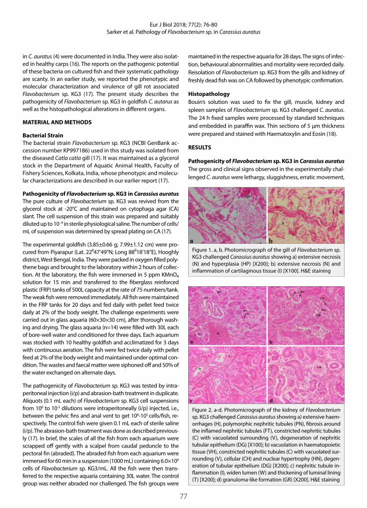
77
Eur J Biol 2018; 77(2): 76-80Sarker et al. Pathology of Flavobacterium sp. in Carassius auratus
in C. auratus (4) were documented in India. They were also isolat-ed in healthy carps (16). The reports on the pathogenic potential of these bacteria on cultured fish and their systematic pathology are scanty. In an earlier study, we reported the phenotypic and molecular characterization and virulence of gill rot associated Flavobacterium sp. KG3 (17). The present study describes the pathogenicity of Flavobacterium sp. KG3 in goldfish C. autarus as well as the histopathological alterations in different organs.
MATERIAL AND METHODS
Bacterial StrainThe bacterial strain Flavobacterium sp. KG3 (NCBI GenBank ac-cession number KP997186) used in this study was isolated from the diseased Catla catla gill (17). It was maintained as a glycerol stock in the Department of Aquatic Animal Health, Faculty of Fishery Sciences, Kolkata, India, whose phenotypic and molecu-lar characterizations are described in our earlier report (17).
Pathogenicity of Flavobacterium sp. KG3 in Carassius auratusThe pure culture of Flavobacterium sp. KG3 was revived from the glycerol stock at -20°C and maintained on cytophaga agar (CA) slant. The cell suspension of this strain was prepared and suitably diluted up to 10−9 in sterile physiological saline. The number of cells/mL of suspension was determined by spread plating on CA (17).
The experimental goldfish (3.85±0.66 g; 7.99±1.12 cm) were pro-cured from Piyarapur (Lat. 22⁰47’49”N; Long 88⁰18’18”E), Hooghly district, West Bengal, India. They were packed in oxygen filled poly-thene bags and brought to the laboratory within 2 hours of collec-tion. At the laboratory, the fish were immersed in 5 ppm KMnO₄ solution for 15 min and transferred to the fiberglass reinforced plastic (FRP) tanks of 500L capacity at the rate of 75 numbers/tank. The weak fish were removed immediately. All fish were maintained in the FRP tanks for 20 days and fed daily with pellet feed twice daily at 2% of the body weight. The challenge experiments were carried out in glass aquaria (60×30×30 cm), after thorough wash-ing and drying. The glass aquaria (n=14) were filled with 30L each of bore-well water and conditioned for three days. Each aquarium was stocked with 10 healthy goldfish and acclimatized for 3 days with continuous aeration. The fish were fed twice daily with pellet feed at 2% of the body weight and maintained under optimal con-dition. The wastes and faecal matter were siphoned off and 50% of the water exchanged on alternate days.
The pathogenicity of Flavobacterium sp. KG3 was tested by intra-peritoneal injection (i/p) and abrasion-bath treatment in duplicate. Aliquots (0.1 mL each) of Flavobacterium sp. KG3 cell suspensions from 100 to 10-3 dilutions were intraperitoneally (i/p) injected, i.e., between the pelvic fins and anal vent to get 108-105 cells/fish, re-spectively. The control fish were given 0.1 mL each of sterile saline (i/p). The abrasion-bath treatment was done as described previous-ly (17). In brief, the scales of all the fish from each aquarium were scrapped off gently with a scalpel from caudal peduncle to the pectoral fin (abraded). The abraded fish from each aquarium were immersed for 60 min in a suspension (1000 mL) containing 6.0×10⁶ cells of Flavobacterium sp. KG3/mL. All the fish were then trans-ferred to the respective aquaria containing 30L water. The control group was neither abraded nor challenged. The fish groups were
maintained in the respective aquaria for 28 days. The signs of infec-tion, behavioural abnormalities and mortality were recorded daily. Reisolation of Flavobacterium sp. KG3 from the gills and kidney of freshly dead fish was on CA followed by phenotypic confirmation.
HistopathologyBouin’s solution was used to fix the gill, muscle, kidney and spleen samples of Flavobacterium sp. KG3 challenged C. auratus. The 24 h fixed samples were processed by standard techniques and embedded in paraffin wax. Thin sections of 5 μm thickness were prepared and stained with Haematoxylin and Eosin (18).
RESULTS
Pathogenicity of Flavobacterium sp. KG3 in Carassius auratusThe gross and clinical signs observed in the experimentally chal-lenged C. auratus were lethargy, sluggishness, erratic movement,
Figure 1. a, b. Photomicrograph of the gill of Flavobacterium sp. KG3 challenged Carassius auratus showing a) extensive necrosis (N) and hyperplasia (HP) [X200]; b) extensive necrosis (N) and inflammation of cartilaginous tissue (I) [X100]. H&E staining
Figure 2. a-d. Photomicrograph of the kidney of Flavobacterium sp. KG3 challenged Carassius auratus showing a) extensive haem-orrhages (H), polymorphic nephritic tubules (PN), fibrosis around the inflamed nephritic tubules (FT), constricted nephritic tubules (C) with vacuolated surrounding (V), degeneration of nephritic tubular epithelium (DG) [X100]; b) vacuolation in haematopoietic tissue (VH), constricted nephritic tubules (C) with vacuolated sur-rounding (V), cellular (CH) and nuclear hypertrophy (HN), degen-eration of tubular epithelium (DG) [X200]; c) nephritic tubule in-flammation (I), widen lumen (W) and thickening of luminal lining (T) [X200]; d) granuloma-like formation (GR) [X200]. H&E staining

78
Eur J Biol 2018; 77(2): 76-80Sarker et al. Pathology of Flavobacterium sp. in Carassius auratus
hanging, anorexia, white patches on the gills, excessive mucus secretion, caudal peduncle lesions, tail rot, cutaneous haemor-rhages, scale loss, ulceration in the abraded area, skin discolor-ation, skin peeling, fluid-filled peritoneal cavity and intestine, pale and discoloured kidney and liver, and haemorrhagic internal organs. About 80-100% mortalities were noted in challenged C. auratus at 106-108 CFU/fish levels. The LD50 dose of Flavobacterium sp. KG3 was determined to be 2.5 × 10⁵ CFU/fish. The abraded fish recorded 60% mortality at a challenge dose of 6.0 × 10⁶ cells/mL.
Histopathology The gills of experimentally challenged C. auratus exhibited extensive necrosis, hyperplasia (Figure 1a) and inflammation of cartilaginous tissue (Figure 1b). In kidney, extensive haem-orrhages, polymorphic nephritic tubules, fibrosis around the inflamed nephritic tubules, constricted nephritic tubules with vacuolated surrounding, degeneration of nephritic tubular ep-ithelium (Figure 2a), vacuolation in haematopoietic tissue, cel-lular and nuclear hypertrophy, degeneration of tubular epithe-lium (Figure 2b), nephritic tubule inflammation, widen lumen and thickening of luminal lining (Figure 2c) and granuloma for-mation (Figure 2d) were observed. The challenged goldfish ex-hibited sarcolysis along with haemocyte infiltration (Figure 3a) and diffused muscle bundle (Figure 3b). Histopathological al-terations such as melanomacrophage aggregate, necrosis and loosely packed white pulp with vacuolated cells were noted in the spleen of challenged C. auratus (Figure 3c).
DISCUSSION
During the surveillance work on the winter diseases in 2014 and 2015 (November-February), increasing incidences of flavobacte-rial infection with mild to moderate mortalities were recorded in the cultured food fish and ornamental fish species from diverse localities of West Bengal, India (4,15,17). The surveillance results established that Flavobacterium spp. are emerging as major fish pathogens of winter disease in freshwater aquaculture and is a major cause for concern. Therefore, further attempts were made to assess the pathogenic potential of these bacteria on cultured fish as well as the tissue level changes in the infected
fish. In the present study, cent percent mortality was observed in goldfish challenged with Flavobacterium sp. KG3 at a level of 108 CFU/fish in 24 h. About 80-90% mortalities were recorded at 106-107 CFU/fish within 60 h of the challenge. The LD50 value of Flavobacterium sp. KG3 was determined to be 2.5×10⁵ CFU/fish, which imply that the tested bacterium was moderately vir-ulent to C. auratus as per the degree of virulence (19). Flavobac-terium sp. KG3 also resulted in 60% mortality within 5 days of challenge in abraded and bath treated C. auratus. These results suggested that physical or mechanical injuries may facilitate the entry of Flavobacterium sp. KG3 to effect significant mortal-ities in goldfish. In our earlier study, the same bacterium, when challenged by abrasion-bath treatment in C. catla fingerlings at 4.7×10⁶ cells/mL, resulted in 2.8 times higher mortality (56.7%) within 5 days of challenge than the unchallenged and abraded catla (20%) at 24-28 °C (17). These results corroborate the earlier observations made in zebrafish Danio rerio with abrasion-bath experiments using F. columnare (5), which recorded LD50 values of 1.1×10⁶ -1.1×10⁷ CFU/mL. However, their challenge experi-ments with the same strain by intramuscular and intraperito-neal injection yielded much higher LD50 values (3.2×10¹⁰ CFU/fish and 4.2×10¹⁰ CFU/fish, respectively) than those observed in the present study by i/p route (2.5×10⁵ CFU/fish). The results im-plied that Flavobacterium spp. have varying degrees of patho-genic potential on fish with challenge routes. Irrespective of the challenge route, our strain Flavobacterium sp. KG3 demonstrat-ed its virulence and pathogenic potential. Contrarily, in suspen-sion challenge study with F. branchiophilum in intact L. rohita fingerlings mortalities ranging from nil at 106 -108 CFU/mL to 80% at 1012 CFU/mL were recorded (12). The results of the pres-ent study, thus, demonstrated that with skin injuries or breach of the immune barrier the Flavobacterium sp. KG3 can cause mortalities in fish as with other potential bacterial pathogens. The above results, thus, indicated that Flavobacterium sp. KG3 may be involved in the pathogenesis of goldfish in union with adverse environmental conditions and/or injuries. The haem-orrhagic lesions on the internal organs of challenged goldfish were indicative of septicemia condition.
Figure 3. a-c. Photomicrograph of the muscle of Flavobacterium sp. KG3 challenged Carassius auratus showing a) sarcolysis (S) along with haemocyte infiltration (IR); b) diffused muscle bundle (DM). c) Photomicrograph of the spleen of Flavobacterium sp. KG3 challenged Carassius auratus showing melanomacrophage aggregate (MA), necrosis (N), loosely packed white pulp (LW) with vacuolated cells (V) [X200]. H&E staining

79
Eur J Biol 2018; 77(2): 76-80Sarker et al. Pathology of Flavobacterium sp. in Carassius auratus
The histopathological observations in Flavobacterium sp. KG3 challenged goldfish also demonstrated extensive damag-es in the gills and internal organs. The gills of experimentally challenged C. auratus had extensive necrosis and hyperplasia, inflammation of cartilaginous tissue similar to those reported earlier (18,20,21). In our earlier study, cartilaginous tissue in-flammation, mucus secretion, loss of gill lamellar structure, ne-crosis of gill filament with the associated reduction in the num-ber of lamellae per filament, obliteration of interlamellar water channels, and fusion of lamellae were noted in naturally infect-ed C. catla exhibiting gill rot, from where the Flavobacterium sp. KG3 was isolated (17). Likewise, columnaris diseased fish recorded congestion of blood vessels, dissociation of surface epithelium of the lamellae from the capillary bed probably due to the accumulation of oedematous fluid and scattered areas of haemorrhage with globose masses of blood cells (20). On the other hand, proliferative branchitis consisting of epithelial hy-perplasia of the gill lamellae and interlamellar space resulting in lamellar fusion was observed during the flavobacterial infection (21). In the present study, the gills of C. auratus exhibited ex-tensive necrosis with other cellular and tissue level alterations, which are histologically similar to those of several earlier stud-ies (13,14,20,22).
In the kidney, severe nephritic cellular and tissue level alter-ations including granuloma formation were observed, thus indi-cating the systemic pathogenic potential of Flavobacterium sp. KG3. Similarly, granulomatous lesions in the kidney of Molliensia sphenops affected with Flavobacterium sp. was reported (23). Contrarily, in an experimental study with F. columnare, kidney lesions were localized in the glomerulus (20). In support of the present study, renal tubular degeneration and proteinaceous casts in the tubular lumen, focal renal tubular degeneration, and necrosis, melanomacrophage, hyperplasia, tubular degenera-tive changes, necrosis and edema within the renal interstitium of the kidney of an injected (i/p) fish with Flavobacterium were noted (21). According to Ferguson (22), inspection of the Hema-toxylin and Eosin or Giemsa stained sections from the affected tissue can reveal typical long and slender bacterial cells, where they appear bluish-purple or blue, respectively. But, the present experiment and also in earlier studies (20,24), the stained tissue sections failed to reveal bacteria in the internal organs. Also, the challenged C. auratus exhibited diffused muscle bundle, sarcol-ysis along with haemocyte infiltration; whereas several earlier studies noted severe haemorrhagic muscle with degeneration of the myofibers (21), and degeneration of muscle fibers and necrosis (20).
Histopathological alterations such as necrosis, loosely packed white pulp with vacuolated cells, melanomacrophage ag-gregate and necrosis were noted in the spleen of challenged C. auratus. The presence of melanomacrophage aggregate in the spleen was indicative of immune reactions to ward-off the bacterial challenge. An early study observed no bacilli or microscopic lesions in the liver, spleen and anterior kidney of Flavobacterium infected koi carp (25). However, in the present
study, microscopic lesions were noted in all internal organs, which confirm that Flavobacterium sp. KG3 has the ability to cause systemic infection. In agreement with this study, a fria-ble and swollen spleen or splenomegaly in columnaris-like dis-eased fish was reported (21).
CONCLUSION
The histopathological observations of the present study, in general, presented the fact that Flavobacterium sp. KG3 can induce pathogenesis both externally and internally in exper-imentally challenged fish. Since the flavobacterial infection is severe during the winter months appropriate preventive or stress mitigation measures such as avoidance of crowding, use of probiotics, immunomodulators, vaccines or development of disease-resistant stocks, etc are recommended to manage the flavobacterial infection or winter diseases.
Ethics Committee Approval: All the experimental protocols with gold-fish as an experimental animal were approved by the Ethical Commit-tee, WBUAFS, Kolkata, India.
Peer-review: Externally peer-reviewed.
Author Contributions: Concept - T.J.A; Design - T.J.A; Supervision – T.J.A., S.B.; Resource - T.J.A., S.S., H.A.; Materials - S.S., H.A.; Data Collec-tion and/or Processing - S.S., S.B., H.A.; Analysis and/or Interpretation - S.S., S.B., H.A., T.J.A.; Literature Search - S.S., S.B.; Writing - S.S., T.J.A.; Critical Reviews - T.J.A.
Acknowledgement: The authors thank the Vice-Chancellor, West Ben-gal University of Animal and Fishery Sciences, Kolkata for providing nec-essary facilities to carry out the work.
Conflict of Interest: The authors have no conflict of interest to declare.
Financial Disclosure: The research work was supported by the Indian Coun-cil of Agricultural Research (Grant F. 10(12)/2012-EPD dated 23.03.2012), Gov-ernment of India, New Delhi under the Niche Area of Excellence program.
REFERENCES
1. Livengood EJ and Chapman FA. The Ornamental Fish Trade: An In-troduction with Perspectives for Responsible Aquarium Fish Own-ership. University of Florida, IFAS Extension, 2011, pp. 1-8.
2. Citarasu T, Alfred DK, Velmurugan S, Thanga VV, Kumaran T, Mi-chael BM, et al. Isolation of Aeromonas hydrophila from infected ornamental fish hatchery during massive disease outbreak. Int J Curr Res 2011; 2(1): 37-41.
3. Sahoo PK, Swaminathan TR, Abraham TJ, Kumar R, Pattanayaka S, Mohapatra A, et al. Detection of goldfish haematopoietic necro-sis herpes virus (Cyprinid herpesvirus-2) with multi-drug resistant Aeromonas hydrophila infection in goldfish: First evidence of any viral disease outbreak in ornamental freshwater aquaculture farms in India. Acta Trop 2016; 161: 8-17.
4. Sarker S, Patra A, Adikesavalu H and Abraham TJ. Flavobacteriosis in cultured freshwater ornamental telescopic eye goldfish Caras-sius auratus. Int J Curr Microbiol Appl Sci 2016; 5(4): 39-46.
5. Moyer TR, Hunnicutt DW. Susceptibility of zebrafish Danio rerio to infection by Flavobacterium columnare and F. johnsoniae. Dis Aquat Organ 2007; 76(1): 39-44.

80
Eur J Biol 2018; 77(2): 76-80Sarker et al. Pathology of Flavobacterium sp. in Carassius auratus
6. Bernardet JF and Bowman JP. Genus I. Flavobacterium Bergey et al. 1923, emend. Bernardet et al. 1996. Whitman W, editor, Bergey’s Manual of Systematic Bacteriology; Vol. 4. 2nd edition, 2011 The Williams and Wilkins Co., Baltimore, MD.
7. Bernardet JF and Bowman JP. The genus Flavobacterium. Dworkin M, Falkow S, editors. The Prokaryotes: A Handbook on the Biol-ogy of Bacteria, Vol. 7. 2006 Springer-Verlag, New York, NY, pp. 481-531.
8. Austin B and Austin DA. Bacterial Fish Pathogens. Diseases of Farmed and Wild Fish. 5th edition. 2012 Springer-Praxis in Aqua-culture in Fisheries, Praxis Publication Ltd., Chichester, UK, p. 457.
9. Starliper CE and Schill WB. Flavobacterial diseases: columnaris dis-ease, coldwater disease, and bacterial gill disease. Woo PTK, Bruno DW, editors. Fish Diseases and Disorders. 2nd edition. Vol. 3. 2012 CAB International, Wallingford, UK, pp. 606-31.
10. Good C, Davidson J, Wiens GD, Welch TJ, Summerfelt S. Flavobac-terium branchiophilum and F. succinicans associated with bacterial gill disease in rainbow trout Oncorhynchus mykiss (Walbaum) in water recirculation aquaculture systems. J Fish Dis 2015; 38(4): 409-13.
11. Loch TP, Faisal M. Emerging flavobacterial infections in fish: A re-view. J Adv Res 2015; 6(3): 283-300.
12. Swain P, Mishra S, Dash S, Nayak SK, Mishra BK, Pani KC, et al. Asso-ciation of Flavobacterium branchiophilum in bacterial gill disease of Indian major carps. Indian J Anim Sci 2007; 77(7): 646-9.
13. Verma DK, Rathore G. Molecular characterization of Flavobacteri-um columnare isolated from a natural outbreak of columnaris dis-ease in farmed fish, Catla catla from India. India. J Gen Appl Micro-biol 2013; 59: 417-24.
14. Verma DK, Rathore G, Pradhan PK, Sood N, Punia P. Isolation and characterization of Flavobacterium columnare from freshwater orna-mental goldfish Carassius auratus. J Environ Biol 2015; 36(2): 433-9.
15. Patra A, Sarker S, Banerjee S, Adikesavalu H, Biswas D, Abraham TJ. Rapid detection of Flavobacterium columnare infection in fish by species-specific polymerase chain reaction. J Aquac Res Dev 2016; 7: 445.
16. Verma DK, Rathore G. New host record of five Flavobacterium spe-cies associated with tropical freshwater farmed fishes from North India. Brazilian J Microbiol 2015; 46(4): 969-76.
17. Sarker S, Abraham TJ, Banerjee S, Adikesavalu H and Patra A. Char-acterization, virulence and pathology of Flavobacterium sp. KG3 associated with gill rot in carp, Catla catla (Ham.). Aquaculture 2017; 468(1): 579-84.
18. Roberts RJ. Fish Pathology. 4th edition, 2012 Wiley-Blackwell, UK, p.581
19. Pu JY, Huang XX, Lu CP. Virulence detection of Streptococcus suis type 2 in zebrafish. Sci Agricult Sin 2007; 40: 2655-8.
20. Pacha RE, Ordal EJ. Histopathology of experimental columnaris disease in young salmon. J Comp Pathol 1967; 77(4): 419-23.
21. Loch TP. Identification of novel flavobacteria from Michigan and assessment of their impacts on fish health. Ph.D. Thesis, Michigan State University, East Lansing 2012, p.270.
22. Ferguson HW. Systemic Pathology of Fish: A Text and Atlas of Nor-mal Tissues in Teleosts and their Responses in Disease. 2nd edition, 2006 Scotian Press, London, p.367.
23. Kluge JP. A granulomatous disease of fish produced by flavobacte-ria. Path Vet 1965; 2(6): 545-52.
24. Morrison C, Cornick J, Shum G, Zwicker B. Microbiology and histo-pathology of saddle-back disease of underyearling Atlantic salm-on, Salmo salar L. J Fish Dis 1981; 4: 243-58.
25. Tripathi NK, Latimer KS, Gregory CR, Ritchie BW, Wooley RE, Walk-er RL. Development and evaluation of an experimental model of cutaneous columnaris disease in koi Cyprinus carpio. J Vet Diagn Invest 2005; 17(1): 45-54.

81
INTRODUCTION
Oncogenes, known as cancer genes, and tumor sup-pressor genes encode the components of the path-ways that regulate the proliferative and social behavior of cells, and a molecular change in these can lead to the development of cancer (1). Natural products are con-sidered a promising approach to cancer therapy with less toxic yet more potent effects. (2-4). Many chemo-therapeutic agents used in clinics are produced from plant and analogs of the compound of it contains. Now-adays, the compounds obtained from plants in natural products (secondary metabolites) which have antican-cer activity, cause apoptosis and regulate angiogene-sis. Additionally, the metastasis potential of tumor cells have been identified (5-8). Some of the compounds and
their derivatives such as paclitaxel, vinblastine, vincris-tine, etoposide, artemisinin, cannabinoid, resveratrol, camptothecin, derivatives of colchicine (Democolcine, N-deacetylcolchicine, and trimethyl colchicinic acid) are secondary metabolites used as chemotherapeutics in the clinic (3,6,8).
PAC, derived from the bark of Taxus brevifolia and having an antimitotic effect, has been used as a drug in some cancer therapy such as cancer of the uterus and breast cancer (9). The drug affects the normal functioning of the interphase as well as the micro-tubules in the mitosis. This effect of PAC has been shown to result from apoptosis of the resulting cell death (10).
Eur J Biol 2018; 77(2): 81-88
RESEARCH ARTICLE
The Antiproliferative Activity of Colchicum umbrosum Plant Extract and Paclitaxel on C-4 I and Vero Cells
Ozlem Dagdeviren Ozsoylemez1* , Gul Ozcan2 1Istanbul University, Institute of Graduate Studies in Science and Engineering, Department of Radiobiology, Istanbul, Turkey2Istanbul University, Faculty of Science, Department of Biology, Istanbul, Turkey
Address for Correspondence: Ozlem Dagdeviren Ozsoylemez E-mail: [email protected] Received: 22.11.2018 Accepted: 12.12.2018© Copyright 2018 by The Istanbul University Faculty of Science • Available online at http://ejb.istanbul.edu.tr • DOI: 10.26650/EurJBiol.2018.0016
Please cite this article as: Dagdeviren Ozsoylemez O, Ozcan G. The Antiproliferative Activity of Colchicum umbrosum Plant Extract and Paclitaxel on C-4 I and Vero Cells. Eur J Biol 2018; 77(2): 81-88.
ORCID IDs of the authors: O.D.O. 0000-0001-9019-0757; G.O. 0000-0003-0682-5065.
ABSTRACT
Objective: This study was conducted to determine the antiproliferative effects of paclitaxel (PAC) and Colchicum umbrosum plant extract on C-4 I and Vero cells. In addition, the apoptotic effects of plant extract on these cells were compared.
Materials and Methods: C-4 I and Vero cells were treated with PAC and corm extract of C. umbrosum for 24 h. The antiproliferative effects of 0.01, 0.1, and 1 mg/mL of plant extract and 7.5, 15, and 30 nM of PAC and the half maximal inhibitory concentration (IC50) values were determined by the MTT method. Morphological changes occurring in the C-4 I and Vero cells were observed under phase-contrast and fluorescence microscopes using DAPI staining.
Results: The IC50 values were found to be 0.01 mg/mL in C-4 I cells and 1 mg/mL in Vero cells for the plant extract and 15 nM in C-4 I and Vero cells for PAC. The apoptotic index (AI) values of the experimental groups of C-4 I and Vero cells were significantly increased compared to those of the control group (p<0.01) after treatment with IC50 concentration of the plant extract. The AI values of the plant extract treatment in C-4 I cells were higher than those in Vero cells at 24 and 48 h.
Conclusion: Treatment with the IC50 concentration of the plant extract induced apoptotic cell death in C-4 I cells. It can worth be considered as a novel promising candidate for the treatment of cancer.
Keywords: C-4 I, Vero, Colchicum umbrosum, paclitaxel, cancer

82
Eur J Biol 2018; 77(2): 81-88Ozsoylemez and Ozcan. The Effect of Plant Extract and PAC in the Cells
Colchicine is another secondary metabolite isolated from the species of Colchicum genus. Colchicine, an antimitotic agent that prevents the accumulation of microtubules, has antiproliferative effects due to its ability to inhibit cell division in the metaphase (11-13). Thus, the cells’ death was actualized by using the apoptotic pathways (14).
Commonly, cancer cells are more susceptible to apoptosis themselves and the understanding of the molecular pathways that regulate apoptosis is beneficial in exploring the chemo-therapeutic targets of cancer, which in turn will allow the dis-covery and development of new drugs. Recent studies have revealed that there are both natural and synthetic anticancer drugs mediating through apoptosis induction in order to pre-vent tumor expression, progression, and the emergence of cellular inflammatory responses other than necrosis (15-18). Apoptosis is a genetically controlled death which is found in both physiological and pathological conditions which are not needed or are dangerous to the organism. Given that apoptotic cells can be morphologically identified and counted by using phase contrast and fluorescence microscopy, they have used in the determination of the apoptotic index in tumor growths as a prognostic marker (19).
In this study, the antiproliferative effects of different doses of Colchicum umbrosum extract alone and PAC alone treated in C-4 I (cancer-acquired cell) and Vero cells (normal cell) were investigated using the MTT [3-(4, 5-dimethylthiazol-2-yl)-2, 5- diphenyltetrazolium bromide] method for 24 hours. Addi-tionally, its IC50 values were determined. In this context, the aim was to determine the different antiproliferative effects of the chemotherapy drug and the plant extract on both the cancer and normal cells. Morphological changes in cells were shown with phase contrast and fluorescence microscopes with 4’-6-diamidino-2-phenylindole (DAPI). Furthermore, the apop-tosis rate was determined using the apoptotic index parameter.
MATERIALS AND METHODS
Colchicum umbrosum Steven (corm, ISTE: 85333) plant was collected from Bolu-Abant in Turkey on 25.06.2008. The plant extract was percolated with pure methanol and lyophilized at Istanbul University Faculty of Pharmacy’s Department of Phar-macognosy. Plant extract and PAC (Ebetaksel®, Bristol Myers Squibb Co.) were prepared at different doses and applied on normal and cancer cells used in this study.
Cell LinesVero (normal cell line, green monkey kidney) and C-4 I (cancer cell line, human cervix carcinoma) cells were obtained from the ATCC (Vero: ATCC® CCL-81; C-4 I: ATCC® CRL-1594) used in the experiments. 10% Fetal Bovine Serum (FBS, Gibco Lab.) was added to the media used for these cells (Vero cells were MEM medium, C-4 I cells were in Waymouth MB medium) and pH was adjusted to7.2 with 4.4% NaHCO3. Cells were incubated in ster-ile 25 cm2 flasks with regular passages twice a week at 37 °C in a 5 % CO2 and 95 % air mixture (20,21).
Passage Processes of CellsThe Vero and C-4 I cells were passaged when they became monolayers in the culture containers where they grew. Cells were washed with Hank’s Buffered Salt Solution and incubated with 2 ml of 0.25% trypsin for 3 minutes to release them from the surface. The suspended cells were taken to the centrifuge tube and 4 ml of the medium was added for the trypsin inac-tivation. Then, it was centrifuged at 500 xg for 5 minutes. After this process, the supernatant part of the precipitated cells was removed (20,21). The cells were palced in a 96 well plate for the antiproliferative experiment at 30,000 cells/well; and to deter-mine the apoptotic index, the cells were seeded in 12 well plate at 200,000 cells per well. After the cultivation of the cells was carried out, incubation was carried out at pH 7.2 for 24 hours at 37 ° C in an incubator containing 5% CO2 and 95% air (21).
Preparation of Concentration of Colchicum umbrosum ExtractThe concentration of extracts prepared from the corm of C. umbrosum plant used in the experiments was determined with reference to the previous studies (13,22). The dried ex-tract was dissolved in a sterile, serum-free medium to prepare the stock solution. Then, 3 different concentrations of the plant extract (0.01, 0.1 and 1 mg/mL) were prepared by diluting the stock solution with a medium containing 10% FBS.
Preparation of Doses of PACThe doses of PAC used were prepared taking into account the clinically applied doses. 30 mg/5 ml of sterile stock solution was diluted with PAC (Ebetaksel®, Bristol Myers Squibb Co.) se-rum-free medium, resulting in an intermediate stock of 7.5 μM. Three different doses (7.5 nM, 15 nM, and 30 nM) to be used in the experiments were prepared sterilely diluting them with the prepared intermediate-staged medium containing 10% FBS and then applied to the cells (23).
Treatment of Plant Extract and PAC in Cells3 different concentrations of the plant extracts alone and 3 dif-ferent doses of PAC alone were applied to the cells for 24 hours. In the control group, the extract-free medium was used. At the end of these periods, the agents were removed and the anal-yses were carried out in accordance with the determined pa-rameters.
Determination of Cell Viability3 different concentrations of the plant extract and 3 different doses of PAC were treated in the cells and the IC50 values were determined for use in the experiments. At the end of the specified experiment period, the cells were removed from the 96-well plate and 40 μL MTT (5mg/mL) was added to the wells and incubated for 4 hours in a shaking incubator. To dissolve the formazan crys-tals which formed after 4 hours of incubation, 160 μL of dimethyl sulfoxide was added to each well and left in the shaker incubator for 1 hour. Then, the absorbance at 570 nm wavelength reference of 690 nm wavelength was determined using ELISA spectrometry (μQuant, Bio-Tek Instruments INC.) In addition, the samples were photographed using phase-contrast microscopy to determine any morphological changes in the experimental groups (23).
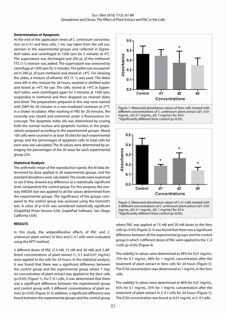
83
Eur J Biol 2018; 77(2): 81-88Ozsoylemez and Ozcan. The Effect of Plant Extract and PAC in the Cells
Determination of ApoptosisAt the end of the application times of C. umbrosum concentra-tion on C-4 I and Vero cells, 1 mL was taken from the cell sus-pension in the experimental groups and collected in Eppen-dorf tubes and centrifuged at 1500 rpm for 5 minutes at 4°C. The supernatant was discharged and 200 μL of the methanol: FTS (1:1) mixture was added. The supernatant was removed by centrifuge at 1500 rpm for 5 minutes. The pellet was resuspend-ed in 200 μL of pure methanol and stored at +4°C. For cleaning the slides, a mixture of ethanol: HCl (1: 1) was used. The slides were left in this mixture for 24 hours, washed in distilled water and stored at +4°C for use. The cells, stored at +4°C in Eppen-dorf tubes, were centrifuged again for 5 minutes at 1500 rpm, suspended in methanol and then dropped on cleaned slides and dried. The preparations prepared in this way were stained with DAPI for 20 minutes in a non-irradiated container at 37°C in a shaker incubator. After washing in PBS for 20 minutes, the coverslip was closed and examined under a fluorescence mi-croscope. The Apoptotic index (AI) was determined by scoring both the normal nucleus and apoptotic nucleus in the prepa-rations prepared according to the experimental groups. About 100 cells were counted in at least 30 sites for each experimental group, and the percentages of apoptotic cells to total cells for each area was calculated. The AI values were determined by av-eraging the percentages of the 30 areas for each experimental group (23).
Statistical AnalysisThe arithmetic mean of the reproduction speed, the AI data de-termined by dose applied in all experimental groups, and the standard deviations were calculated. The results were examined to see if they showed any difference at a statistically significant level compared to the control group. For this purpose, the one-way ANOVA test was applied to all the values determined from the experimental groups. The significance of the groups com-pared to the control group was assessed using the Dunnett’s test. A value of p<0.05 was considered statistically significant (GraphPad Prism Version 6.00, GraphPad Software, San Diego California USA).
RESULTS
In this study, the antiproliferative effects of PAC and C. umbrosum plant extract in Vero and C-4 I cells were evaluated using the MTT method.
3 different doses of PAC (7.5 nM, 15 nM and 30 nM) and 3 dif-ferent concentrations of plant extract (1, 0.1 and 0.01 mg/mL) were applied to the cells for 24 hours. In the statistical analysis, it was found that there was a significant difference between the control group and the experimental group where 1 mg/ml concentration of plant extract was applied to the Vero cells (p<0.05) (Figure 1). For C-4 I cells, it was determined that there was a significant difference between the experimental group and control group with 3 different concentrations of plant ex-tract (p<0.05) (Figure 2). In addition, a significant difference was found between the experimental groups and the control group
where PAC was applied at 15 nM and 30 nM doses to the Vero cells (p<0.05) (Figure 3). It was found that there was a significant difference between all the experimental groups and the control group in which 3 different doses of PAC were applied to the C-4 I cells (p<0.05) (Figure 4).
The viability % values were determined as 94% for 0.01 mg/mL, 75% for 0.1 mg/mL, 68% for 1 mg/mL concentration after the treatment of plant extract in Vero cells for 24 hours (Figure 5). The IC50 concentration was determined as 1 mg/mL in the Vero cells.
The viability % values were determined as 46% for 0.01 mg/mL, 65% for 0.1 mg/mL, 35% for 1 mg/mL concentration after the treatment of plant extract in C-4 I cells for 24 hours (Figure 5). The IC50 concentration was found as 0.01 mg/mL in C-4 I cells.
Figure 1. Measured absorbance values of Vero cells treated with different concentrations of C. umbrosum plant extract (d1: 0.01 mg/mL, d2: 0.1 mg/mL, d3: 1 mg/mL) for 24 h. *Significantly different from control (p<0.05).
Figure 2. Measured absorbance values of C-4 I cells treated with 3 different concentrations of C. umbrosum plant extract (d1: 0.01 mg/mL, d2: 0.1 mg/mL, d3: 1 mg/mL) for 24 h. *Significantly different from control (p<0.05).

84
Eur J Biol 2018; 77(2): 81-88Ozsoylemez and Ozcan. The Effect of Plant Extract and PAC in the Cells
Figure 3. Measured absorbance values of Vero cells treated with 3 different doses of PAC (d1: 7.5 nM, d2: 15 nM, d3: 30 nM) for 24 h. *Significantly different from control (p<0.05).
Figure 4. Measured absorbance values of C-4 I cells treated with 3 different doses of PAC (d1: 7.5 nM, d2: 15 nM, d3: 30 nM) for 24 h. *Significantly different from control (p<0.05).
Figure 5. Viability % values of the cells treated with 3 different concentrations of C. umbrosum plant extract for 24 h.
Figure 6. Viability % values of the cells treated with 3 different dos-es of PAC for 24 h.
Figure 7. a-f. Phase-contrast microscopy image (x100) treated with IC50 values of plant extracts and PAC for 24 h. a) Vero, Control; b) Vero, C. umbrosum; c) Vero, PAC; d) C-4 I, Control; e) C-4 I, C. umbrosum, f ) C-4 I, PAC.
a
d
b
e
c
f

85
Eur J Biol 2018; 77(2): 81-88Ozsoylemez and Ozcan. The Effect of Plant Extract and PAC in the Cells
The viability %values were determined as 76% for 7.5 nM, 52% for 15 nM, 53% for 30 nM doses after the treatment of PAC in Vero cells for 24 hours (Figure 6). The IC50 dose was determined as 15nM.
The viability % values were determined as 68% for 7.5 nM, 53% for 15 nM, 35% for 30 nM doses after the treatment of PAC in C-4 I cells for 24 hours (Figure 6). The IC50 dose was determined as 15nM.
Any morphological changes in the cells were shown using phase-contrast microscopy after the treatment of IC50 concen-tration of plant extracts and IC50 doses of PAC for 24 and 48 hours (Figures 7 and 8).
Nuclear condensation and apoptotic bodies formed during apoptosis induced by the plant extract were shown with a fluorescence microscope at x100 magnification (Figures 9 and 10).
The AI values were determined from the preparations prepared after administration of the IC50 concentration of the plant ex-tract for 24 and 48 hours on the cells. This was done using fluo-rescence microscopy with DAPI, by counting at least 30 apop-totic and normal cells per 100 cells for each test group.
Figure 8. a-f. Phase-contrast microscope image (x100) treated with IC50 values of plant extracts and of PAC for 48 h. a) Vero, Control; b) Vero, C. umbrosum; c) Vero, PAC; d) C-4 I, Control; e) C-4 I, C. umbrosum, f ) C-4 I, PAC.
a
d
b
e
c
f
Figure 9. a-d. Fluorescence microscope image (x100) with DAPI of Vero and C-4 I cells for 24 h. a) Vero, Control; b) Vero, experiment group; c) C-4 I, control; d) C-4 I, experiment group. (→: apoptotic cells).
a
c
b
d
Figure 10. a-d. Fluorescence microscope image (x100) with DAPI of Vero and C-4 I cells for 48 h. a) Vero, Control; b) Vero, experiment group; c) C-4 I, control; d) C-4 I, experiment group. (→: apoptotic cells).
a
c
b
d

86
Eur J Biol 2018; 77(2): 81-88Ozsoylemez and Ozcan. The Effect of Plant Extract and PAC in the Cells
At the end of 24 hours, the AI values were found as 3% for the control group and 58% for the experiment group of Vero cell; 2% for the control group and 74% for the experiment group of C-4 I cell (Figure 11). It was determined that the difference between the experimental groups and the control groups was significant (p<0.01).
At the end of 48 hours, AI values were found as 4% for the con-trol group and 29% for the experiment group of Vero cell; 4% for the control group and 77% for the experimental group of C-4 I cell (Figure 12). The difference between the experimental groups and the control groups was significant (p<0.01).
DISCUSSION
In this study, it has been shown that at 1, 0.1, and 0.01 mg/mL concentration of C. umbrosum plant extract applied on Vero and C-4 I cells during 24 hours caused a further decrease in the via-bility of cancer cells compared to normal cells. It has been de-termined that the IC50 concentration, which caused the death of approximately half of the cells of the plant extract at 1 mg/mL in Vero cells and 0.01 mg/mL in C-4 I cells, between these three concentration, is the optimal concentration to be used at other stages of the study. According to this results, the plant ex-tract more antiproliferative in C-4 I cells according to Vero cells.
MTT, known as one of the viability tests, is a rapid method used to determine the antiproliferative effect of agents on cells (23).
It was shown that PAC, commonly used in cancer chemother-apy, applied to Vero and C-4 I cells over 24 hours, causes a de-crease in the viability of both normal cells and cancer cells at 7.5 nM, 15 nM and 30 nM doses. Of these three doses, the 15 nM
dose appears to be the optimal dose to be used in other stages of the study (called the IC50 dose), which causes the death of half of both cells.
In a study conducted, the antiproliferative effect of PAC on HeLa cells was determined. The optimal dose determination was found to be nM dose of the IC50 value of PAC and related viability values (23).
The Colchicum genus of the Liliaceae (Colchicaceace) family has been used for therapeutic purposes since ancient times. As a re-sult of studies on Colchicum species, the substance named Col-chicine was found in the structure. Colchicine shows anticancer properties because it can stop cell division in the metaphase. However, although it is known that colchicine, which is highly toxic at the desired doses for cancer treatment, has a potent an-tiproliferative effect, the therapeutic index is too low to be used therapeutically for this purpose. Derivatives of colchicine such as Demecolchine, N-deacetylcolchine, and trimethyl colchicinic acid, are less toxic derivatives and have been shown to be more promising in cancer treatment (13).
In one study, the chemical constituents of the different parts of C. baytopiorum plant extract and their antiproliferative effects in K562 and HL60 cells were reported. In this study, colchicine, 2-demethyl-colchicine, 3-demethylcolchicine, 3-demethyl-N-formyl-N-deace-tylcolchicine, colchifoline, 2-demethylcolchifoline, demecolcine, 2-demethyldemecolcine and 2-demethyl-ɣ-lumicolchicine 12 were isolated from all the separated parts of C. baytopiorum. Ad-ditionally, the antiproliferative effects of different concentrations (500, 100, 50, 5,1, and 0.1 μg/mL) of all extracts prepared from the different parts of C. baytopiorum in K562 and HL60 cells were in-vestigated. IC50 values were calculated and the results were com-pared with each other and with those for colchicine and deme-colcine standards. According to this study, all extracts were more active than the demecolcine standard, and they also showed a similar activity to colchicine (13).
In a second report, the apoptotic effect of 0.1 mg/ml of C. baytopiorum plant extract on HeLa cells by using qRT-PCR was researched. According to this report, the regulatory genes’ expression of apoptosis was significantly increased with the plant extract in the treatment group compared to the control group in HeLa cells (24). In other similar studies it was shown that colchicine may be more effective by interacting with other substances found in plant extracts (25-27).
Nowadays, anticancer therapies promote apoptosis using a va-riety of cellular signaling pathways. By elucidating the details of the apoptotic mechanism, new targets can be created in tumor treatment and it will be possible to increase the treatment ef-ficiency. In this study, the apoptotic effect of the IC50 concen-tration of C. umbrosum plant extract in normal and cancer cells was investigated.
In attempts to improve the outcome of traditional cancer ther-apies, intensive investigations have been directed at various models of regulation of P53 and other key molecules involved
Figure 11. a, b. Apoptotic index values (a) Vero; b) C-4 I) of cells treated with IC50 concentration of the plant extract for 24 h. **Significantly different from control (p<0.01).
Figure 12. a, b. Apoptotic index values (a) Vero; b) C-4 I) of cells treated with IC50 concentration of the plant extract for 48 h. **Significantly different from control (p<0.01).
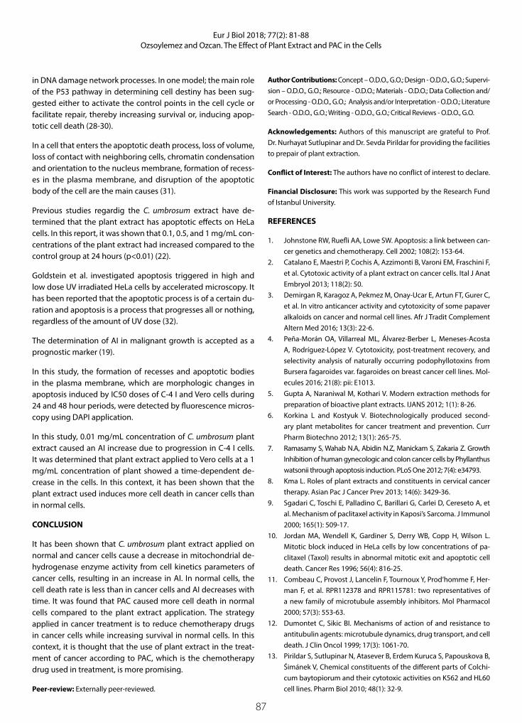
87
Eur J Biol 2018; 77(2): 81-88Ozsoylemez and Ozcan. The Effect of Plant Extract and PAC in the Cells
in DNA damage network processes. In one model; the main role of the P53 pathway in determining cell destiny has been sug-gested either to activate the control points in the cell cycle or facilitate repair, thereby increasing survival or, inducing apop-totic cell death (28-30).
In a cell that enters the apoptotic death process, loss of volume, loss of contact with neighboring cells, chromatin condensation and orientation to the nucleus membrane, formation of recess-es in the plasma membrane, and disruption of the apoptotic body of the cell are the main causes (31).
Previous studies regardig the C. umbrosum extract have de-termined that the plant extract has apoptotic effects on HeLa cells. In this report, it was shown that 0.1, 0.5, and 1 mg/mL con-centrations of the plant extract had increased compared to the control group at 24 hours (p<0.01) (22).
Goldstein et al. investigated apoptosis triggered in high and low dose UV irradiated HeLa cells by accelerated microscopy. It has been reported that the apoptotic process is of a certain du-ration and apoptosis is a process that progresses all or nothing, regardless of the amount of UV dose (32).
The determination of AI in malignant growth is accepted as a prognostic marker (19).
In this study, the formation of recesses and apoptotic bodies in the plasma membrane, which are morphologic changes in apoptosis induced by IC50 doses of C-4 I and Vero cells during 24 and 48 hour periods, were detected by fluorescence micros-copy using DAPI application.
In this study, 0.01 mg/mL concentration of C. umbrosum plant extract caused an AI increase due to progression in C-4 I cells. It was determined that plant extract applied to Vero cells at a 1 mg/mL concentration of plant showed a time-dependent de-crease in the cells. In this context, it has been shown that the plant extract used induces more cell death in cancer cells than in normal cells.
CONCLUSION
It has been shown that C. umbrosum plant extract applied on normal and cancer cells cause a decrease in mitochondrial de-hydrogenase enzyme activity from cell kinetics parameters of cancer cells, resulting in an increase in AI. In normal cells, the cell death rate is less than in cancer cells and AI decreases with time. It was found that PAC caused more cell death in normal cells compared to the plant extract application. The strategy applied in cancer treatment is to reduce chemotherapy drugs in cancer cells while increasing survival in normal cells. In this context, it is thought that the use of plant extract in the treat-ment of cancer according to PAC, which is the chemotherapy drug used in treatment, is more promising.
Peer-review: Externally peer-reviewed.
Author Contributions: Concept – O.D.O., G.O.; Design - O.D.O., G.O.; Supervi-sion – O.D.O., G.O.; Resource - O.D.O.; Materials - O.D.O.; Data Collection and/or Processing - O.D.O., G.O.; Analysis and/or Interpretation - O.D.O.; Literature Search - O.D.O., G.O.; Writing - O.D.O., G.O.; Critical Reviews - O.D.O., G.O.
Acknowledgements: Authors of this manuscript are grateful to Prof. Dr. Nurhayat Sutlupinar and Dr. Sevda Pirildar for providing the facilities to prepair of plant extraction.
Conflict of Interest: The authors have no conflict of interest to declare.
Financial Disclosure: This work was supported by the Research Fund of Istanbul University.
REFERENCES
1. Johnstone RW, Ruefli AA, Lowe SW. Apoptosis: a link between can-cer genetics and chemotherapy. Cell 2002; 108(2): 153-64.
2. Catalano E, Maestri P, Cochis A, Azzimonti B, Varoni EM, Fraschini F, et al. Cytotoxic activity of a plant extract on cancer cells. Ital J Anat Embryol 2013; 118(2): 50.
3. Demirgan R, Karagoz A, Pekmez M, Onay-Ucar E, Artun FT, Gurer C, et al. In vitro anticancer activity and cytotoxicity of some papaver alkaloids on cancer and normal cell lines. Afr J Tradit Complement Altern Med 2016; 13(3): 22-6.
4. Peña-Morán OA, Villarreal ML, Álvarez-Berber L, Meneses-Acosta A, Rodríguez-López V. Cytotoxicity, post-treatment recovery, and selectivity analysis of naturally occurring podophyllotoxins from Bursera fagaroides var. fagaroides on breast cancer cell lines. Mol-ecules 2016; 21(8): pii: E1013.
5. Gupta A, Naraniwal M, Kothari V. Modern extraction methods for preparation of bioactive plant extracts. IJANS 2012; 1(1): 8-26.
6. Korkina L and Kostyuk V. Biotechnologically produced second-ary plant metabolites for cancer treatment and prevention. Curr Pharm Biotechno 2012; 13(1): 265-75.
7. Ramasamy S, Wahab N.A, Abidin N.Z, Manickam S, Zakaria Z. Growth Inhibition of human gynecologic and colon cancer cells by Phyllanthus watsonii through apoptosis induction. PLoS One 2012; 7(4): e34793.
8. Kma L. Roles of plant extracts and constituents in cervical cancer therapy. Asian Pac J Cancer Prev 2013; 14(6): 3429-36.
9. Sgadari C, Toschi E, Palladino C, Barillari G, Carlei D, Cereseto A, et al. Mechanism of paclitaxel activity in Kaposi’s Sarcoma. J Immunol 2000; 165(1): 509-17.
10. Jordan MA, Wendell K, Gardiner S, Derry WB, Copp H, Wilson L. Mitotic block induced in HeLa cells by low concentrations of pa-clitaxel (Taxol) results in abnormal mitotic exit and apoptotic cell death. Cancer Res 1996; 56(4): 816-25.
11. Combeau C, Provost J, Lancelin F, Tournoux Y, Prod’homme F, Her-man F, et al. RPR112378 and RPR115781: two representatives of a new family of microtubule assembly inhibitors. Mol Pharmacol 2000; 57(3): 553-63.
12. Dumontet C, Sikic BI. Mechanisms of action of and resistance to antitubulin agents: microtubule dynamics, drug transport, and cell death. J Clin Oncol 1999; 17(3): 1061-70.
13. Pirildar S, Sutlupinar N, Atasever B, Erdem Kuruca S, Papouskova B, Śimánek V, Chemical constituents of the different parts of Colchi-cum baytopiorum and their cytotoxic activities on K562 and HL60 cell lines. Pharm Biol 2010; 48(1): 32-9.

88
Eur J Biol 2018; 77(2): 81-88Ozsoylemez and Ozcan. The Effect of Plant Extract and PAC in the Cells
14. Jordan MA, Wilson L. Microtubules as a target for anticancer drugs. Nat Rev 2004; 4(4): 253-65.
15. Piccolella M, Crippaa V, Messia E, Tetel MJ, Poletti A. Modulators of estrogen receptor inhibit proliferation and migration of prostate cancer cells. Pharmacol Res 2014; 79: 13-20.
16. Wu CT, Lai JN, Tsai YT. The prescription pattern of Chinese herbal products that contain Dang-Qui and risk of endometrial cancer among Tamoxifen-treated female breast cancer survivors in Tai-wan: A population-based study. PLoS One 2014, 9(12): e113887.
17. Binkhorst L, Mathijssen R.H, Jager A, van Gelder T. Individualization of tamoxifen therapy: Much more than just CYP2D6 genotyping. Cancer Treat Rev 2015; 41(3): 289-99.
18. Bisi A, Gobbi S, Merolle L, Farruggia G, Belluti F, Rampa A, Molnar J, et al. Design, synthesis and biological profile of new inhibitors of multidrug resistance associated proteins carrying a polycyclic scaffold. Eur J Med Chem 2015; 92: 471-80.
19. Chauhan R, Verma N, Sharma SP, Bhargava R, Singh P. Role of apop-totic index, mitotic index and MIB-1 antibody expression as bio-markers in preneoplastic and neoplastic lesions of uterine cervix. Int J Res Med Sci 2016; 4(6): 2093-100.
20. Artun FT, Karagoz A, Ozcan G, Melikoglu G, Anil S, Kultur S, et al. In vitro anticancer and cytotoxic activities of some plant extracts on HeLa and Vero cell lines. Proceedings 2017; 1: 1019.
21. Ozcan G, Dagdeviren Ozsoylemez O, Akman G, Khalilia W, Tezel B.Y, Karagoz A, Melikoglu G, Anil S, Kultur S, Sutlupinar N. Screening for antitumor activity of various plant extracts on HeLa and C 4-I cell lines. J BUON 2016; 21(6): 1552-60.
22. Dağdeviren Ö. Acı Çiğdem (Colchicum umbrosum S. ve Colchi-cum baytopiorum C.D.) ekstrelerinin Hela hücreleri üzerine an-ti-tümöral etkilerinin araştırılması. İÜ Fen Bilimleri Enstitüsü, Yük-sek Lisans Tezi. 2010.
23. Serbes U, Ozsoylemez OD, Ozcan G. Evaluation of paclitaxel effects in the pattern of expression of survival and apoptotic genes regu-lators in HeLa cells. Curr Pharm Biotechnol 2016; 17(12): 1058-67.
24. Ozsoylemez OD, Ozturk M, Sutlupinar N, Kayacan S, Tuncdemir M, Ozan G. The effects of Colchicum baytopiorum on regulatory genes of apoptotic and autophagic cell death in HeLa cells. Curr Pharm Biotechnol 2016; 17(15): 1369-76.
25. Solowey E, Lichtenstein M, Sallon S, Paavilainen H, Solowey E, Lorberboum-Galski H. Evaluating medicinal plants for anticancer activity. ScientificWorldJournal 2014; 2014: 721402.
26. Delebinski C, Georgi S, Kleinsimon S, Twardziok M, Kopp B, Melzig MF, et al. Analysis of proliferation and apoptotic induction by 20 steroid glycosides in 143B osteosarcoma cells in vitro. Cell Prolif 2015; 48(5): 600-10.
27. Radogna F, Dicato M, Diederich M. Cancer-type-specific crosstalk between autophagy, necroptosis and apoptosis as a pharmaco-logical target. Biochem Pharmacol 2015; 94(1): 1-11.
28. Mirzayans R, Andrais B, Scott A, Murray D. New insights into p53 signaling and cancer cell response to DNA damage: implications for cancer therapy. Biomed Res Int 2012; 2012: 170325.
29. Ganapathy S, Xiao S, Yang M, Qi M, Choi DE, Ha CS, et al. A low-dose arsenic-induced p53 protein-mediated metabolic mechanism of radiotherapy protection. J Biol Chem 2014; 289(8): 5340-7.
30. Aubrey BJ, Kelly GL, Janic A, Herold MJ, Strasser A. How does p53 induce apoptosis and how does this relate to p53-mediated tu-mour supression? Cell Death Differ 2018; 25(1): 104-13.
31. Gewies A. Introduction to Apoptosis. ApoReview 2003; 1-26.32. Goldstein JC, Kluck RM, Green DR. A single cell analysis of apop-
tosis: ordering the apoptotic phenotype. Ann N Y Acad Sci 2000; 926(1): 132-41.

89
INTRODUCTION
The “biodiversity hotspots” term is one of the most suc-cessful methods developed to preserve the richest and most endangered areas on the world (1). The Mediter-ranean Basin is one of the most important area for plant diversity (2,3). The plant diversity is excessive, with ap-proximately 30.000 plant species with 13.000 endemics and many of the endemics plants are local (4). The flora of Turkey has approximately 10.000 vascular plants, so it is the richest flora in the temperate zone (5,6). Also, it has special habitats. But, the incomparable flora and habitats of Turkey have been threatened and have di-minished rapidly over the last four decades; Turkey has 144 Important Plant Areas (7) which have contributed much to the origin of many cultivated plant species.
The aim of this study is to reveal the biodiversity of the Başkonuş Mountain and to determine alternatives for the protection of the Small Protected Areas (SPAs).
Kahramanmaraş is a region that is comprised of plains in mountains which is located in the South-East Ana-tolia region of Turkey (8,9) (Figure 1). The flora of Kah-ramanmaraş is very rich, almost 2500 taxa and many endemic species (20%) (10-16).
The main reasons for this richness are due to the tran-sition zone of Irano-Turan and Mediterranean plant ge-ography regions, which began in the South of the Ana-tolian Diagonal and extend to north-eastern Anatolia to the Mediterranean Taurus mountains, this region has been critical for plant diversification (10). The Başkonuş
Eur J Biol 2018; 77(2): 89-96
RESEARCH ARTICLE
“Small Protected Areas” for Conservation Priorities in South Anatolia (Başkonuş Mountain-Kahramanmaraş)
Mine Kocyigit1* , Serpil Demirci Kayıran2 1Istanbul University, Faculty of Pharmacy, Department of Pharmaceutical Botany, Istanbul, Turkey2Cukurova University, Faculty of Pharmacy, Department of Pharmaceutical Botany, Istanbul, Turkey
Address for Correspondence: Mine Kocyigit E-mail: [email protected] Received: 23.10.2018 Accepted: 12.12.2018© Copyright 2018 by The Istanbul University Faculty of Science • Available online at http://ejb.istanbul.edu.tr • DOI: 10.26650/EurJBiol.2018.0013
Please cite this article as: Kocyigit M, Demirci S. “Small Protected Areas” for Conservation Priorities in South Anatolia (Başkonuş Mountain-Kahramanmaraş). Eur J Biol 2018; 77(2): 89-96.
ORCID IDs of the authors: M.K. 0000-0002-7831-0151; S.D.K. 0000-0001-8340-3347.
ABSTRACT
Objective: The Başkonuş Mountain (Kahramanmaraş) is located in the transition zone of Irano-Turan and Mediterranean plant geographical regions and in the southern part of the Anatolian Diagonal. A specific region in the East Mediterranean basin consists of an area that holds great biological importance and is a world hotspot. In this study, the small protected areas of the Başkonuş Mountain were determined.
Materials and Methods: A total of 17 endemic and 9 rare taxa were selected based on their IUCN statutes, altitudes, and habitats. Their GPS coordinates were loaded on a computer and digitized in a map projection system. Distributions of the selected taxa were marked on a topographic map in the scale of 1/25.000. The 26 maps were overlapped so that the areas of plant biodiversity could be observed on the map.
Results: Six small protected areas were defined on the Başkonuş Mountain. Several threats and conservation suggestions were presented in this study.
Conclusion: Local data about biodiversity sources are limited. Thus, conservation biologists and government offices should play active roles in formal educational settings and employ alternative methods to the community in Kahramanmaraş.
Keywords: Başkonuş Mountain, small protected areas, biodiversity, Turkey

90
Eur J Biol 2018; 77(2): 89-96Kocyigit and Demirci. Small Protected Areas of Başkonuş Mountain
Mountain, Nature Conservation Area, and is in Kahramanmaraş province. The highest point of the examined area is Başkonuş Mountain (1775 m). The research area is located within the Mediterranean phytogeographical region. The three types veg-etation basically can be observed in the area are; forest, mac-chie, and steppe vegetation (13-16).
MATERIALS AND METHODS
Study AreaThe study area is located in Kahramanmaraş province. Accord-ing to Equator 37°38’35’’- 37°28’ 32’’ north latitudes, accord-ing to Greenwich 36 028’48’’- 36° 41’ 54’’ east meridians on the topographic map with in scaled to 1/25.000 (Figure 2). This area has a surface area of 203.084 km2 (118.258 km2 forest, 84.826 km2 opened forest) and varies in height from 345 m to the peak of the Başkonuş mountain (1779 m). The Başkonuş Mountain is studied because;
1) Some observations on the vegetation in the Başkonuş Moun-tain (13-16), and detailed floristic studies in this area during the Project of KANBK (Conservation for Rare and Endemic Plants of Kahramanmaraş and Ardahan was carried out in 2007-2011 and funded by BTC-UNDP (SGP) (10-12).
2) The study area is a promenade area.
3) Phytogeographic location of Başkonuş Mountain and Anato-lian Diagonal (17).
Selection of Taxa The Flora of Başkonuş Mountain consists of 66 endemic taxa (18). In the study, 17 endemic and 9 rare taxa (17 threatened en-demic taxa and 9 national threatened rare taxa) were selected according to their IUCN (International Union for Conservation of Nature) statutes (19), altitudes and habitats as listed in Table 1. Endemic which includes endemic taxa found solely in Kahra-manmaraş and in neighbouring areas (e.g., Ahır Mountain, Bin-boğa Mountain, Çimen Mountain) (13-16).
Field InvestigationsThe field work was carried out in 2011 and information of vari-ous data such as GPS (Global Positioning System), altitude, hab-itat, density of plant population and selected taxa for each 10 km, were recorded. Some newly recorded plants for the Flora of Başkonuş Mountain were collected during the field studies; the plants were identified and kept in ISTE (The Herbarium of Istanbul University Faculty of Pharmacy).
In this study, 13 habitats types were defined and presented in a simplified list as Table 2, where the endemics and rare taxa of the Başkonuş Mountain grow. The numbers of endemic and rare plants in the habitats were presented by a chart. Addition-ally altitudinal distributions of the selected taxa were showed with a chart. During the field work, 610 GPS were loaded on the computer and digitized in Map Projection System; ED 1950 UTM Zone 37 N which is a GIS program (Geographic Information Sys-tems). Distributions of the selected taxa were marked on the topographic map in scaled to 1/25.000. Afterwards the 26 maps were overlapped so that the areas of plant biodiversity were oc-curred on the last map could be seen on Figure 3. As the results of there, six SPAs were defined in Başkonuş Mountain.
RESULTS
The results identified six sites which were based on one or more of the following criteria: 1. Presence of rare species, 2. Presence of botanical diversity, 3. Presence of threatened habitats. The six determined SPAs were descripted using altitudes, size (km2), habitats, selected taxa and other plants. Descriptions of the SPAs were presented in Table 3.
Habitat Types and Altitude DifferentiationHabitat types were grouped as rocks, grasslands, meadows, shrubs, woods, cultivated area, waste and then 13 habitats types were defined. Pinus brutia, P. nigra, Cedrus libani, Abies cilicica, Taxus baccata, Alnus glutinosa are the main taxa of for-est vegetation. Additionally Quercus coccifera, Styrax officinalis, Arbutus unedo, Pistacia terebinthus and Cotinus coggyria are the main taxa of shrubby vegetation.
Some endemics and rare plants have been found in sever-al different habitats, these being: Lathyrus laxiflorus subsp. angustifolius, Helleborus vesicarius –in shrubs, screes, cliffs and rocks. Some endemics have been found only in special habitats,
Figure 1. Map of the study area; Başkonuş Mountain
Figure 2. Small Protected Areas of Başkonuş Mountain: 1. Tower of fire observation and around, 2. Locality of Yediardıc, 3. Pond and castle around, 4. Ziyaret Hill, 5. Locality of Rahmacılar, 6. Lo-cality of Topcalı and Sersem

91
Eur J Biol 2018; 77(2): 89-96Kocyigit and Demirci. Small Protected Areas of Başkonuş Mountain
these being: Allium glumaceum- in screes, Fritillaria alfredae subsp. glaucoviridis - in shrubs.
The relationship between the selected taxa and altitudinal zones has been reported (Figure 3). The endemics and rare
plants of Başkonuş Mountain develop densely in 1200-1400 m and in 600-1000 m.
Ziyaret Hill has maximum habitats although it is the smallest SPA. Its altitude zone and the richness of the habitat provide a
Table 1. List of selected taxa and their IUCN statutes (Ekim et al.2000), altitudes and habitats
Taxa IUCN Altitudes Habitats
Threatened Endemic Taxa (17 taxa)
Acer monspessulanum L. subsp. oksalianum Yalt. VU 1000-1200 W3, R3, S2
Allium glumaceum Boiss. & Hausskn. LR (cd) 1500-2200 R2
Astragalus distinctissimus Eig EN 1200-1520 W1
Astragalus leporinus Boiss. var. hirsutus (Post) D.F.Chamb. LR (lc) 600-1500 W2, C1
Centaurea lycopifolia Boiss. & Kotschy LR (nt) 50-2000 R1, S1
Colchicum davisii C.D. Brickell EN 1000-1950 R2, G1
Crataegus aronia (L.) Bosc. ex DC. var. minuta Browicz LR (lc) 600-1300 G1
Cyclamen pseudoibericum Hildebr. EN 550-1500 G1, W1, W2, W3
Fritillaria alfredae Post subsp. glaucoviridis (Turrill) Rix VU 500-1600 S1
Helleborus vesicarius Aucher LR (nt) 550-1300 S1, W4, G2
Kitaibelia balansae Boiss. EN 1900-2000 R1, G1, C2
Lathyrus laxiflorus (Desf.) Kuzntze subsp.angustifolius(Post ex Dinsm.) P.H.Davis
VU 1200-1400 R1, S1, W3
Michauxia tchihatchewii Fisch. et Mey. LR (nt) 500-1800 R1
Rhamnus nitidus P.H.Davis LR (cd) 700-1300 W1
Verbascum amanum Boiss. VU 400-2000 R1, S1, W1
Verbascum pinetorum (Boiss.) Kuntze VU 370-1000 S1,W1
Hyacinthus orientalis L. subsp. chionophilus Wendelbo LR (nt) 1900-2000 R1, R3, G1
National Threatened Rare Taxa, nonendemic (9 taxa)
Anemone blanda Schott & Kotschy VU 150-2600 R1, S1
Allium opacum Rech. fil. EN 800-1700 R1, R2
Allium dodecanasii Karavokyrou & Tzanoud. EN 400-1000 R3, W1
Anacamptis pyramidalis (L.) Rich. LR (lc) 200-1500 W4,G1, C1, C2
Cephalanthera rubra (L.) Rich. LR (lc) 200-1500 W1, W2, S1
Comperia comperiana (Steven) Asch. & Graebn. EN 500-1600 W1, W2, W3
Epipactis condensata Boiss. LR (lc) 600-1600 W1
Eranthis hyemalis (L.) Salisb. VU 1300-1800 G2
Lathyrus variabilis (Boiss. & Ky.) Maly VU 1000-1700 R1, S1, W3
IUCN statutes: VU: Vulnerable; EN: Endangered; LR (cd): Lower Risk/conservation dependent; LR (lc): Lower Risk/least concern; LR (nt): Lower Risk/near threatened. Grasslands and meadows (G), Shrubs (S), Woods (W), Cultivated and waste (C)

92
Eur J Biol 2018; 77(2): 89-96Kocyigit and Demirci. Small Protected Areas of Başkonuş Mountain
high level biodiversity, so the smallest size SPA competes with the largest size SPA (Locality of Yediardıç) about in number of contained endemic and rare taxa.
EndemismThe flora of Başkonuş Mountain has 565 taxa of which 66 are en-demics with an overall endemism of 11.68%. According distri-butions of the selected endemic and rare taxa, Small Protected Area 2 (Locality of Yediardıç) has the highest number of selected endemic and rare taxa (Figure 4). Firstly, the selected endem-ic and rare plants were found in a woody habitat (W1 and W4) with a rate of 29.01%. Secondly, they were found in a rock hab-itat (R1 and R3) with a rate of 20.85% (Figure 5).
Threats and Conservation StateThe main factor responsible for the threats to plants in the Başkonuş Mountain is human pressure. Intensive grazing of sheep and goats in the meadows and grasslands, uncon-trolled plant collection, especially some orchids (Anacamptis pyrimidalis, Comperia comperiana), and working on road and forestry activities (uncontrolled tree cutting or silviculture) are the most important threats to the flora of Başkonuş Mountain.
According to IUCN categories, 7 taxa is EN (endangered), 8 taxa is VU (vulnerable), 4 taxa is LR (nt) (near threatened), 2 taxa is LR (cd) (conservation dependent) and 5 taxa is LR (lc). The en-dangered and vulnerable taxa are distributed in the second SPA (Figure 6). This is not surprising due to the fact that the most numerous endemic taxa observed are in this area.
The Başkonuş Mountain has various types of rock slides and screes. This habitat is a biotope of most numerous endemic geo-phytes (Allium glumaceum, Colchicum davisii, Fritillaria alfredae subsp. glaucoviridis, Cyclamen pseudoibericum, Hyacinthus orientalis subsp. chionophilus).
DISCUSSION
Başkonuş Mountain has specific habitat such as medium-height steep slopes and forest edges. This habitat is also a refuge for endemics. Habitat loss is thought to lead to endemic species extinctions. Many studies use species-area relationships and predict substantial extinctions when levels of habitat loss ex-ceed approximately 70-80% (16).
Table 2. Habitat types of the selected plants from the Başkonuş Mountain
Habitats HabitatsNumber of endemics
Number of rare plants Total %
Rocks (R) 31 23.66
R1 High and Mid altituded cliffs and rocks R1 12 2 14 11.69
R2 Screes R2 10 2 12 9.16
R3 Moist rocks R3 4 1 5 3.82
Grasslands and meadows (G) 13 9.92
G1 Calcareous rocky grasslands at mid and high altitude G1 2 1 3 2.29
G2 Alpine grasslands, snow beds G2 9 1 10 7.63
Shrubs (S) 10 7.63
S1 Shrubby places S1 4 1 5 3.82
S2 River bed shrubs S2 4 1 5 3.82
Woods (W) 61 46.56
W1 Low and mid altitude needle-leaved forest W1 16 5 21 16.03
W2 Low and mid altitude broad-leaved forest W2 7 2 9 6.87
W3 Low and mid altitude mixed forest W3 12 2 14 10.69
W4 Forest edges W4 14 3 17 12.98
Cultivated and waste (C) 16 12.21
C1 Traditionally cultivated fields C1 6 1 7 5.34
C2 Fallows, roadsides C2 8 1 9 6.87
TOTAL 108 23 131 100

93
Eur J Biol 2018; 77(2): 89-96Kocyigit and Demirci. Small Protected Areas of Başkonuş Mountain
Table 3. Descriptions of the Small Protected Areas (Continue)
SPA 1: Tower of fire observation and around
Coordination: 37°33’00”N 36°35’00”E Size: 0.5 km2 Altitude: 1700-1800 m
Habitat types: Cliffs and rocks, screes, calcareous rocky grasslands, alpine grasslands, snowbeds and needle leaved forest
Threatened endemic taxa:
Centaurea lycopifolia, Colchicum davisii, Crataegus aronia var. minuta, Cyclamen pseudoibericum, Helleborus vesicarius, Lathyrus laxiflorus subsp. angustifolius, Verbascum amanum, Verbascum pinetorum,
Threatened rare taxa:
Allium opacum, Anemone blanda
Threats: Working on forest road, uncontrolled tree cutting, and plant collection.
SPA 2: Locality of Yediardic
Coordination: 37°34’00”N 36°35.5’00”E Size: 1 km2 Altitude: 1400-1500 m
Habitat types: Forest, shrubby places, screes and alpine grasslands
Threatened endemic taxa:
Acer monspessulanum subsp. oksalianum, Allium glumaceum, Astragalus distinctissimus, Astragalus leporinus var. hirsutus, Centaurea lycopifolia, Colchicum davisii, Crataegus aronia var. minuta, Cyclamen pseudoibericum, Fritillaria alfredae subsp. glaucoviridis, Helleborus vesicarius, Lathyrus laxiflorus subsp. angustifolius, Rhamnus nitidus, Verbascum amanum, Verbascum pinetorum,
Threatened rare taxa:
Anemone blanda, Cephalanthera rubra, Comperia comperiana, Lathyrus variabilis, Anacamptis pyramidalis, Epipactis condensata
Threats: human pressure, uncontrolled grazing, and construction
SPA 3: Pond and Castle Around
Coordination: 37°34.5’00”N 36°35’00”E Size: 0.83 km2 Altitude: 1300-1400 m
Habitat types: Cliffs and rocks, moist rocks, river bed shrubs, forest
Threatened endemic taxa:
Acer monspessulanum subsp. oksalianum, Allium glumaceum, Astragalus distinctissimus, Centaurea lycopifolia, Crataegus aronia var. minuta, Cyclamen pseudoibericum, Helleborus vesicarius, Kitaibelia balansae, Lathyrus laxiflorus subsp. angustifolius, Verbascum amanum, Verbascum pinetorum,
Threatened rare taxa:
Cephalanthera rubra, Comperia comperiana, Lathyrus variabilis, Eranthis hyemalis
Threats: Working on highway, uncontrolled grazing, and plant collection.
SPA 4: Ziyaret Hill
Coordination: 37°34.5’00”N 36°35’00”E Size: 0.4 km2 Altitude: 1200-1300 m
Habitat types: Cliffs and rocks, moist rocks, river bed shrubs, alpine grasslands, forest
Threatened endemic taxa:
Acer monspessulanum subsp. oksalianum, Astragalus distinctissimus, Centaurea lycopifolia, Colchicum davisii, Crataegus aronia var. minuta, Fritillaria alfredae subsp. glaucoviridis, Helleborus vesicarius, Lathyrus laxiflorus subsp. angustifolius, Rhamnus nitidus, Verbascum amanum, Verbascum pinetorum
Threatened rare taxa:
Comperia comperiana, Lathyrus variabilis, Epipactis condensata
Threats: Uncontrolled grazing and plant collection. human pressure
SPA 5: Locality of Rahmacılar
Coordination: 37°33’00”N 36°35.5’00”E Size 0.7 km2 Altitude 1200-1300 m

94
Eur J Biol 2018; 77(2): 89-96Kocyigit and Demirci. Small Protected Areas of Başkonuş Mountain
Figure 3. Altitudinal distribution of endemic and rare vascular plant taxa in Başkonuş Mountain.
Figure 4. Numbers of endemic and rare taxa in the Small Pro-tected Areas.
Figure 5. Number of endemic and rare vascular plant taxa ac-cording to habitats in Başkonuş Mountain.
Figure 6. IUCN categories of selected taxa
Table 3. Descriptions of the Small Protected Areas (Continue)
Habitat types: Shrubby places and forest
Threatened endemic taxa:
Acer monspessulanum subsp. oksalianum, Allium glumaceum, Centaurea lycopifolia, Crataegus aronia var. minuta, Cyclamen pseudoibericum, Fritillaria alfredae subsp. glaucoviridis, Helleborus vesicarius, Hyacinthus orientalis subsp. chionopphilus, Lathyrus laxiflorus subsp. angustifolius, Rhamnus nitidus, Verbascum amanum
Threatened rare taxa:
Anemone blanda, Lathyrus variabilis.
Threats: Working on road, uncontrolled plant collection. human pressure,
SPA 6: Locality of Topcalı and Sersem
Coordination: 37°33’00”N 36°35.5’00”E Size: 0.79km2 Altitude: 1300-1400 m
Habitat types: Shrubby places, alpine grasslands, snowbeds, screes and forest
Threatened endemic taxa:
Acer monspessulanum subsp. oksalianum, Allium glumaceum, Centaurea lycopifolia, Crataegus aronia var. minuta, Helleborus vesicarius, Lathyrus laxiflorus subsp. angustifolius, Michauxia tchihatchewii, Rhamnus nitidus, Verbascum amanum
Threatened rare taxa:
Allium dodecanesii, Epipactis condensata, Lathyrus variabilis.
Threats: Opening agricultural area, working on road, uncontrolled plant collection. human pressure,

95
Eur J Biol 2018; 77(2): 89-96Kocyigit and Demirci. Small Protected Areas of Başkonuş Mountain
Selected endemic taxa which include endemic plants found only in Kahramanmaraş and in neighbouring areas (e.g., Ahır, Bin-boğa, Çimen Mountains) (17-20). In fact most endemism grow in high stress level populations, such as screes (R2), cliffs and rocks high and mid altitude (R1) and alpine grasslands (G2) (16,21), but it should be emphasized that endemics and rare plants are found in woody habitats, especially coniferous habitats in the study.
The flora of the Başkonuş Mountain is comparable with data from the literature for mountainous areas in the Mediterranean recognized as one of the most important centers of endemism on Earth (22,23). Besides, the relationship is considered biodi-versity, habitats and endemic plants is remarkable compared to other Mediterranean hotspots (24-26). For example, although the Başkonuş Mountain has a smaller area than the Alps and the Pyrenees, its endemism rate is higher than them. Additionally, the ratio of endemic species per unit area is higher than Pyre-nees and the Andalucía Mountains could be seen on Table 4.
Ecologists recognize that the Earth’s biota is now experiencing the sixth great extinction wave (27). The conversion of native for-est and grasslands, to plantation crops is one of the largest threats to terrestrial biodiversity and a key of the global extinction crisis (28). Additionally, intensive grazing causes transformation of the vegetation in scrub communities and in the woods (21).
While people have become more aware of environmental prob-lems recently, most do not get actively involved in movements that support a more liveable future (3). Many people reach limit-ed data about environmental from sources that may be reached, especially the media. Thus ecologists should be more active in educational settings and alternative methods of biodiversity in the community of Kahramanmaraş. In 2010, the tenth meeting of the Conference of Parties to the Convention on Biological Diver-sity led to the adoption of a global strategic plan for biodiversity for the period of 2011-2020. The EU follows a similar policy. The new Biodiversity Strategy purposes to stop the loss of biodiver-
sity and the degeneracy of ecosystem services in the EU by 2020 (29,30). The strategy should be carried out in Turkey.
An agreement among politicians and managers of the local ad-ministrations and parks (City Councils, Government and Nation-al Parks) is necessary in Kahramanmaraş and Turkey.
Major threats to biodiversity include human-associated factors, such as habitat destruction and degradation, over profiteering, invasive species, incidental mortality, pollution, climate change diseases and human population growth (31).
CONCLUSION
Six SPAs were defined in Başkonuş Mountain. Some threats and conservation suggestions were presented in this study. Local people reach limited information about biodiversity issues from sources that may be biased. Thus conservation biologists and government offices should be much more active in formal ed-ucational settings and via alternative methods to a diversity of audiences in Kahramanmaraş.
Peer-review: Externally peer-reviewed.
Author Contributions: Concept – M.K., S.D.K.; Design - M.K.; Supervi-sion – M.K.; Resource - M.K., S.D.K.; Materials - S.D.K.; Data Collection and/or Processing - M.K., S.D.K.; Analysis and/or Interpretation - M.K., S.D.K.; Literature Search - M.K., S.D.K.; Writing - M.K.; Critical Reviews - M.K., S.D.K.
Acknowledgements: We thank Prof. Dr. Neriman Özhatay and Andrew Byfield for their comments on the manuscript and Evrim Tabur and Tol-ga Ok for their support during fieldwork in the province. Also, we thank Mehmet Aydın for helping on the Map Projection System.
Conflict of Interest: The authors have no conflict of interest to declare.
Financial Disclosure: The project was financially supported by the Kahramanmaraş Regional Directorate of Forestry and UNDP (the United
Table 4. Comparison of endemism from Europe and Mediterranean areas
RegionNumber of endemics Endemism
Total number of taxa
Total area (km²)
Endemic taxa/area rate References
Italian Maritime Alps 107 3.40 3137 5000 0.627Casazza et al.
2005
French Maritime Alps 108 3.80 2867 4500 0.637Casazza et al.
2005
Maritime Alps (southwestern France) 115 3.20 3605 9500 0.379
Casazza et al. 2005
Pyrenees (France - Spain)
174 5.00 3480 94300 0.037 Villar and Garcia 1989
Andalucía Mountains (Spain) 125 36.00 347 82800 0.004 Favarger 1972
Peloponnesus (Greece) 300 12.50 2400 5425 0.442 Iatrou 1986
Başkonuş Mountain (Turkey) 66 10.62 565 203 0.325 The present study

96
Eur J Biol 2018; 77(2): 89-96Kocyigit and Demirci. Small Protected Areas of Başkonuş Mountain
Nations Development Program’s). *Project of KANBK (Conservation for Rare and Endemic Plants of Kahramanmaraş and Ardahan) was carried out between 2007-2011, coordinated by N. Özhatay of DHKD and WWF Turkey and funded by BTC-UNDP (SGP).
REFERENCES
1. Myers N, Mittermeier RA, Mittermeier CG, Fonseca GAB, Kent J. Biodiversity hotspots for conservation priorities. Nature 2000; 403(6772): 853-8.
2. Médail F, Quézel P. Biodiversity Hotspots in the Mediterranean Basin: Setting Global Conservation Priorities. Conserv Biol 1999; 13(6): 1510-3.
3. Sekercioglu CH, Anderson A, Akcay E, Bilgin R, Can OE, Semiz G, et al. Turkey’s globally important biodiversity in crisis. Biol Conserv 2011; 144: 2752-69.
4. Abdulmalak D, Al Khader I, Al Jbour S, Arcos J, Ataol M, Azafzaf H, et al. Mediterranean Basin Biodiversity Hotspot. Ankara: Doğa Derneği; 2010.
5. Özhatay N. Important Plant Areas along the BTC Pipeline in Turkey. Istanbul; BTC Şirketi; 2006.
6. Ekim T. Türkiye’nin bitkileri. In: Eken G, Bozdogan M, Isfendiyaroglu S, Kılıc, DT, Lise Y. Eds.), Türkiye’nin Önemli Doğa Alanları. Ankara, Turkey, Doğa Derneği (in Turkish); 2006. pp. 47-8.
7. Özhatay N, Byfield A, Atay S. Important Plant Areas in Turkey: 122 Key Turkish Botanical Sties. İstanbul; WWF-Turkey; 2005. p. 442.
8. Aytac Z, Duman H. The steppic flora of high mounts Ahir, Öksüs and Binboğa (Kahramanmaraş- Kayseri-Turkey). Fl Med 2005; 15: 121-78.
9. Yıldız B. Kahramanmaraş. In: Özhatay N, (Ed.). Important Plant Ar-eas along the BTC Pipeline in Turkey. Istanbul, Turkey; BTC Şirketi; 2006. pp. 241-264.
10. Ozhatay N, İlcim A, Ok, T. Ahır Dağı’ nın sessiz güzelleri “200 Yaba-ni Çiçek”. İstanbul; Kahramanmaraş Valiliği İl Çevre ve Orman Müdürlüğü; 2009.
11. Ozhatay N, Ok T. Doğanın penceresinden Kahramanmaraş. İstan-bul; Kahramanmaraş Valiliği İl Çevre ve Orman Müdürlüğü; 2008.
12. Demirci S, Ozhatay N. An ethnobotanical study in Kahramanmaraş (Turkey); Wild Plants Used For Medicinal Purpose in Andırın, Kahra-manmaraş. Turk J Pharm Sci 2012; 9(1): 75-92.
13. Varol O, Tatlı A. The Vegetation of Çimen Mountain (K.Maraş). Turk J Bot 2001; 25(5): 335-58.
14. Varol O, Tatlı A. Çimen Dağı (Kahramanmaraş) Florası. Ekoloji Çevre Dergisi 2003; 12(46): 17-28.
15. Varol O, Ketenoglu O, Bingol U, Geven F, Guney K. A Phytosociolog-ical research on the coniferous forests of Başkonuş Mountain, An-ti-Taurus, Turkey. Acta Botanica Hungarica 2006; 48(1-2): 195-211.
16. Varol O. Phytosociological research on maquis and steppe vege-tation of Başkonuş Mountain (Kahramanmaraş-Turkey). Thaiszia Journal of Botany 2003; 13(1): 77-88.
17. Varol O, Kara C, İlcim A. New Floristic Records for C6 Grid Square. Turk J Bot 1998; 22: 299-301.
18. Varol O. Flora of Baskonus Mountain. Turk J Bot 2003; 25: 63-102.19. Ekim T, Koyuncu M, Vural M, Duman H, Aytac Z, Adiguzel N. Red
Data Book of Turkish Plants (Pteridophyta & Spermatophyta). An-kara; Turkiye Tabiatini Koruma Dernegi & Van Yuzuncu Yil Universi-tesi; 2000.
20. Casazza G, Barberis G, Minuto L. Ecological characteristics and rara-ity of endemic plants of Italian Maritime Alps. Biol Conserv 2005; 123: 361-71.
21. Nowak A, Nowak S, Nobis M. Distribution pattern, ecological characteristic and conservation status of endemic plants of Tadzhikistan-A global hotspot of diversity. J Nat Conserv 2011; 19: 296-305.
22. Favarger C. Endemism in the montane floras of Europe. In: Valen-tine, D.H. (Ed.), Taxonomy, Phytogeography and Evolution. London and New York; Academic Press; 1972. pp. 191-204.
23. Iatrou G. Contribution to the study of endemism of the flora of Peloponnisos. – Ph. D. thesis, Patras [in Greek with an English sum-mary]; 1986.
24. Villar L, Garcia B. Verse une banque de donne´e`s des plantes vas-culaires ende´miques des Pyre´ne´es. Acta Biol Mont 1989; 9: 261-74.
25. Jones KR, Venter O, Fuller RA, Allan JR, Maxwell SL, Negret PJ, Wat-son JEM. One-third of global protected land is under intense hu-man pressure. Science 2018; 360: 788-91.
26. Piekielek NB, Hansen AJ. Extent of fragmentation of coarse-scale habitats in and around U.S. National Parks. Biol Conserv 2012; 155: 13-22.
27. Mendenhall CD, Daily GC, Ehrlich PR. Improving estimates of biodi-versity loss. Biol Conserv 2012; 151: 32-4.
28. Edwards DP, Laurance SG. Green labeling, sustainability and the expansion of tropical agriculture: Critical issues for certification schemes. Biol Conserv 2012; 151: 60-4.
29. Bladt J, Strange N, Abildtrup J, Svenning JC, Skov F. Conservation efficiency of geopolitical coordination in the EU. J Nat Conserv 2009; 17: 72-86.
30. Maes J, Paracchini ML, Zulian G, Dunbar MB, Alkemade R. Syner-gies and trade-offs between ecosystem service supply, biodiver-sity, and habitat conservation status in Europe. Biol Conserv 2012; 155: 1-12.
31. Brito D, Moreira DO, Coutinho BR, Oprea M. Ill nature: Disease hotspots as threats to biodiversity. J Nat Conserv 2012; 20(2): 72-5.