middle black sea journal of health science (mbsjhs) - DergiPark
41
I MBSJHS, 2(2), 2016 MIDDLE BLACK SEA JOURNAL OF HEALTH SCIENCE
-
Upload
khangminh22 -
Category
Documents
-
view
0 -
download
0
Transcript of middle black sea journal of health science (mbsjhs) - DergiPark
I MBSJHS, 2(2), 2016
MIDDLE BLACK SEA JOURNAL OF HEALTH SCIENCE
0
MIDDLE BLACK SEA JOURNAL OF
HEALTH SCIENCE (MBSJHS)
Owner
On Behalf of Ordu University
NULUFER ERBIL
Director in Charge
ULKU KARAMAN
Editors
AHMET TEVFIK SUNTER
MEHMET HICRI KOSEOGLU
ULKU KARAMAN
VAROL CANAKCI
Ondokuz Mayıs University
Giresun University
Ordu University
Ordu University
Associated Editors
AHMET KAYA, Ordu University
ARIF AYAR, Amasya University
AYDIN HIM, Ondokuz Mayıs University
AYSEGUL CEBI, Giresun University
AYSEGUL TAYLAN OZKAN, Hitit University
AYTAC GUDER, Giresun University
BIRSEN AYDIN KILIC, Amasya University
ENGIN SENEL, Hitit University
KURSAD YAPAR, Giresun University
METE DOLAPCI, Hitit University
MUSTAFA ALISARLI, Ondokuz Mayıs University
MURAT TERZI, Ondokuz Mayıs University
NULUFER ERBIL, Ordu University
SELIM ARICI, Ondokuz Mayıs University
SERDAR GULER, Hitit University
SEVIL ISIK, Medical Park Hospital
SAHIN DIREKEL, Giresun University
TUBA YILDIRIM, Amasya University
YASIN ATAKAN BENKLI, Ordu University

II MBSJHS, 2(3), 2016
MIDDLE BLACK SEA JOURNAL OF HEALTH SCIENCE
Section Editors
(Surgery Medical
Sciences)
Section Editors
(Faculty of Health
Science)
Section Editors
(Internal Medical
Sciences)
Section Editors
(Basic Medical Sciences)
Ali Beytur
Inonu University
Hacer Gök Uğur,
Ordu University
Ahmet Karataş,
Ordu University
Ali Arslan,
Ordu University
Erdal Benli,
Ordu University
Nurgül Bölükbaş,
Ordu University
Ahmet Kaya,
Ordu University
Arzu Şahin,
Ordu University Emine Şamdancı
Inonu University
Sevim Acaröz Candan
Ordu University
Ali Özer
Inonu University
Cheers Emiliano
Milan University
Hakan korkmaz
Ordu University
Katalin Sandor
Karolinska Institutet
Esra Erdoğan
Gulhane Military Medical
Academy
Hamza Çınar
Ordu University Section Editors
(Faculty Of Dentistry)
Özgür Enginyurt,
Ordu University
Fabio Esposito
Milan University
Havva Erdem,
Ordu University
Elif Bahar Çakıcı,
Ordu University
Özlem Özdemir
Ordu University
Funda Doğruman-Al
Gazi University
Mukadder korkmaz
Ordu University Doğu Omur Dede,
Ordu University
Semih Kunak,
Ordu University
Judit Plutzer
National Institute of Environmental Health
Sevil Işık
Ordu University
Fatih Çakıcı,
Ordu University
Yasemin Kaya,
Ordu University
Kosta Y Mumcuoğlu
Hebrew University
Yunus Güzel
Ordu University
Mehmet Melih Ömezli,
Ordu University
Zeki Yüksel Günaydın,
Ordu University
Mustafa Kerem Çalgın
Ordu University
Süleyman Kutalmış
Büyük
Ordu University
Serpil Şener
Inonu University
Orhan Bas,
Ordu University
Pharmacist Editors Ömer Ertürk
Ordu University
Ayşe Baldemir
Erciyes University
Pınar Naile Gürgör,
Ordu University Nilay Ildız
Erciyes University Biostatistical
Consultant Language Inspectors
Serpil Değerli
Cumhuriyet University
Cemil Çolak
Inonu University
Fatih Bayram,
Ordu University
Şahin Direkel
Giresun University
Soner Çankaya,
Ordu University
Ahmet Gökhan Biçer,
Celal Bayar University
Tevfik Noyan,
Ordu University
Zeynep Kolören
Ordu University
Layout Editors Copyeditors Proofreading
Zeynep Taş Cengiz
Yüzüncüyıl University
Arzu Şahin , Ordu University
Arzu Şahin, Ordu University
Elif Bahar Çakıcı, Ordu University
Nülüfer Erbil,
Ordu University
Nülüfer Erbil,
Ordu University
Nülüfer Erbil
Ordu University
Pınar Naile Gürgör,
Ordu University
Orhan Baş,
Ordu University
Özgür Enginyurt
Ordu University
Özgür Enginyurt
Ordu University
Özgür Enginyurt
Ordu University
Pınar Naile Gürgör,
Ordu University
Ülkü Karaman,
Ordu University
Pınar Naile Gürgör,
Ordu University
Ülkü Karaman,
Ordu University
Yasin Atakan Benkli,
Ordu University
Ülkü Karaman,
Ordu University
Yasin Atakan Benkli, Ordu University

III MBSJHS, 2(3), 2016
MIDDLE BLACK SEA JOURNAL OF HEALTH SCIENCE
The Middle Black Sea Journal of Health Science is published by Ordu University Institute of Health
Sciences on behalf of the Middle Black Sea Universities Collaboration Platform
ISSN 2149-7796
Middle Black Sea Journal of Health Science
Editorial Office
Ordu University
Institute of Health Sciences
Cumhuriyet Campus
52200, Ordu, TURKEY
Tel: +90 (452) 226 52 14-5234
Fax: +90 (452) 226 52 28
E-mail: [email protected]
Correspondence Address: PhD, Asst. Prof. Ulku KARAMAN
Institute of Health Sciences, Ordu University,
Cumhuriyet Campus,
52200 Center/ Ordu TURKEY
Phone: +90 452 234 50 10
Fax: +90 452 226 52 55 Email: [email protected]
Web site: http://dergipark.ulakbim.gov.tr/mbsjohs
Sort of Publication: Periodically
Publication Date and Place: 25 / 08 / 2016, ORDU, TURKEY
Publishing Kind: Online
Index: Turkey Citation Index

IV MBSJHS, 2(3), 2016
MIDDLE BLACK SEA JOURNAL OF HEALTH SCIENCE
The Middle Black Sea Journal of Health Science is published by Ordu University Institute of
Health Sciences on behalf of the Middle Black Sea Universities Collaboration Platform
Aims and Scope
The journal publishes clinical and experimental studies, interesting case reports, invited
reviews and letters to the editor. Middle Black Sea Journal of Health Science is an
international journal which is based on independent and unbiased double-blinded peer-review
principles. The publishing language of the journal is English.
The aim of the journal is to publish original articles with highest clinical and scientific quality
at the international level. Middle Black Sea Journal of Health Science also publishes reviews
covering fundamental innovations in health education, editorial articles, case reports and
original images.
The contents of all issues in full text can be accessed free of charge through the web site
http://dergipark.ulakbim.gov.tr/mbsjohs/index
General Rules
Middle Black Sea Journal of Health Science publishes experimental and observational
research articles, clinical reviews, case reports and review articles on health science.
Manuscripts must be submitted online at http://dergipark.ulakbim.gov.tr/mbsjohs/user
All submissions must be accompanied by a signed statement of scientific contributions and
responsibilities of all authors and a statement declaring the absence of conflict of interests.
Any institution, organization, pharmaceutical or medical company providing any financial or
material support, in whole or in part, must be disclosed in a footnote. Manuscripts must be
prepared in accordance with ICMJE-Recommendations for the Conduct, Reporting, Editing
and Publication of Scholarly Work in Medical Journals (updated in December 2013 -
http://www.icmje.org/icmje-recommendations.pdf).
An approval of research protocols by an ethical committee in accordance with international
agreements (Helsinki Declaration of 1975, revised 2002 - available at
http://www.vma.net/e/policy/b3.htm, “Guide for the care and use of laboratory animals -
www.nap.edu/catalog/5140.html/) is required for experimental, clinical and drug studies. A
form stating that the patients have been informed about the study and consents have been
obtained from the patients is also required for experimental, clinical and drug studies. All
submissions must be accompanied by a letter that states that all authors have approved the
publication of the paper in the Middle Black Sea Journal of Health Science.
Submission of the studies requiring ethical committee decision must be accompanied by a
copy of the submission to the ethical committee.

V MBSJHS, 2(3), 2016
MIDDLE BLACK SEA JOURNAL OF HEALTH SCIENCE
SUBMISSION POLICY
Submission of a paper to Middle Black Sea Journal of Health Science is understood to imply
that it deals with original material not previously published, and is not being considered for
publication elsewhere. Manuscripts submitted under multiple authorships are reviewed on the
assumption that all listed Authors concur with the submission and that a copy of the final
manuscript has been approved by all Authors. After acceptation of an article, it should not be
published elsewhere in the same form, in either the same or another language, without the
written consent of the Editors and Publisher. Upon acceptance of an article, Authors will be
asked to transfer copyright (for more information on copyright see. This transfer will ensure
the widest possible dissemination of information. A letter will be sent to the corresponding
Author confirming receipt of the manuscript. A form facilitating transfer of copyright will be
provided.
If excerpts from other copyrighted works are included, the Author(s) must obtain written
permission from the copyright owners and credit the source(s) in the article. Please write your
text in good English (American or British usage is accepted, but not a mixture of these).
Authors in non native speaker of English should check and improve the English of their paper
(before submission).
The layout and style should adhere strictly to the instructions. No revisions or updates will be
incorporated after the article has been accepted and sent to the Publisher (unless approved by
the Editors).
SUBMISSION PROCEDURE
The Middle Black Sea Journal of Health Science welcomes submitted manuscripts online at
http://dergipark.ulakbim.gov.tr/mbsjohs/user Manuscripts submitted online are received on
the day of submission and quickly assigned to reviewers. Through individual Author Centers
on this website, authors can view the status of their manuscripts as they progress through the
review process. Notification of the disposition of each manuscript will be sent by e-mail to the
corresponding author on the day of decision.
To establish your account for online submission, go
to http://dergipark.ulakbim.gov.tr/mbsjohs/user/register Authors are encouraged to check for
an existing account. If you are submitting for the first time, and you do not have an existing
account, then you must create a new account. If you are unsure about whether or not you have
an account, or have forgotten your password, enter your e-mail address into the Password
Help section on the log-in page. If you do not have an account, click on the Create Account
link on the top right of the log-in page. You then will be able to submit and monitor the
progress of your manuscripts.
Once you have logged in, you will be presented with the Main Menu and a link to your
Author Centre. Submit your manuscript from the Author Centre. At the end of a successful
submission and you will receive an e-mail confirming that the manuscript has been received
by the journal. If this does not happen, please send an e-mail to [email protected]
To submit your manuscript online, please prepare the text and illustrations according to the
instructions listed below. You may enter and exit the manuscript submission process at the
completion of each step. After submission of the manuscript, however, you will not be able to
edit it.
Web submission is required- instructions are available for downloading on the
website http://dergipark.ulakbim.gov.tr/mbsjohs/author/submit/1
VI MBSJHS, 2(3), 2016
MIDDLE BLACK SEA JOURNAL OF HEALTH SCIENCE
COPYRIGHT TRANSFER AGREEMENT
A signed COPYRIGHT RELEASE FORM by all authors of the manuscript should be sent
during manuscript submission.
Middle Black Sea Journal of Health Science
Editorial Office
Ordu University
Institute of Health Sciences
Cumhuriyet Campus
52200, Ordu, TURKEY
Tel: +90 (452) 226 52 14-5234
Fax: +90 (452) 226 52 28
E-mail: [email protected]
Where possible, Authors should also include a list of three or more potential reviewers for
their manuscript, with contact information (Full address, telephone and fax numbers, e-mail
address).
PREPARING ELECTRONIC MANUSCRIPTS Author should submit manuscript in both ways as explain in below:
1- Please keep text, tables and graphics as separate files in other word do not import the
figures or tables into the text file. Text files should be supplied in one of the following
formats: Microsoft Word or WordPerfect, Windows or Macintosh formatted. Text files should
be supplied in one of the following formats: Microsoft Word or WordPerfect, Windows or
Macintosh formatted.
2- Please insert all attachments that are tables, figures and graphics into the text file in
appropriate place, than creates the PDF file of this text. During submission submits this PDF
file as a supplementary.
When mentioning parasites in the main text and references, the genus and species names must
be italicized and the genus name must be written with an initial capital letter.
Abbreviations should be expanded at first mention and used consistently thereafter.
Graphic files: Journal only accepts PDF, TIFF and EPS formats for graph. Each figure
should be a separate file and not be embedded in the text.
All graphic files must be submitted in sufficiently high resolution, for grey scale and color
images 250 dpi and 500-800 dpi for line art) to allow for printing.
Electronic submission of articles via the Web
http://dergipark.ulakbim.gov.tr/mbsjohs/login
Full instructions for uploading data and files etc. are given on the website when submitting a
manuscript. It is the responsibility of the Authors to create the proper files as instructed above
for the electronically submitted manuscript. The editorial office cannot make conversions
beyond the supported file types.
After online submission, there is no need sending a hardcopy of manuscript or illustrations to
the Editors. Please note that the electronic files supplied will always be used to produce the
illustrations, including those for the print version of the article; it is the Authors’
responsibility to ensure that these files are of suitable quality

VII MBSJHS, 2(3), 2016
MIDDLE BLACK SEA JOURNAL OF HEALTH SCIENCE
ORGANIZATION OF THE ARTICLE
Manuscripts should be prepared electronically using an appropriate MS Word compatible
word-processing package, formatted for A4 or letter page size, double-spaced throughout with
3 cm margins on all sides, and using 12 point font. Text should not be justified, but flush left.
Words should not be hyphenated to fit on a line. Pages should be numbered sequentially.
Title page: The title page should contain the following items: The title page should include
full and short title English, and meeting and congress presentations of the manuscript must
be stated, if any. Authors’ names and their institutional affiliations must only be provided at
the submission stage, author information must not be included in the main text.
Abstract Page: The second page should include abstracts written both in Turkish and
English, and key words. Structured abstracts, not to exceed 400 words, should consist of four
sections, labeled as Objective, Methods, Results and Conclusion.
Keywords: Keywords: Provide at least 3-6 keywords and avoiding general and plural terms
and multiple concepts. These keywords will be used for indexing purposes. Key words in
should follow the abstract. Please select keywords in Turkish Science Terms
(http://www.bilimterimleri.com).
Research Reports should be divided into numbered sections headed by a caption
1. Introduction, 2. Methods, 3. Results, 4. Discussion, 5. Conclusion, 6. Conflict of Interest
Disclosure, 7. Acknowledgements 8. References, Tables, Figures and Illustrations (with
legends) sections.
Case reports should be divided into the following sections: 1. Introduction, 2. Case(s), 3.
Discussion, 4. Conclusion, 5. References, Tables, Figures and Illustrations (with legends).
Introduction: The objectives of the research should be clearly stated in this section. Relevant
background information and recent published studies should be described concisely, and be
cited appropriately.
Methods: This section should contain all the details necessary to reproduce the experiments.
Avoid re-describing methods already published; only relevant modifications should be
included in the text. Experimental subjects when human subjects are used, manuscripts must
be accompanied by a statement that the experiments were undertaken with the understanding
and written consent of each subject.
When experimental animals are used, the methods section must clearly indicate that adequate
measures were taken to minimize pain or discomfort.
Results and Discussion: These sections should present the results and interpret them in a
clear and concise manner. Results should usually be presented descriptively and be
supplemented by figures. Extensive citations and discussion of published literature should be
not be used.
Literature references:
Care should be taken to cite Turkey-based studies and journal of national during the granting
of resources (www.atifdizini.com).
In the text, references should be cited by authors’ surnames and year of publication. All
references cited in the text (and only those cited in the text) should be included. One or two
authors should be cited by surname; for three or more, the first author is cited followed by et
al.:
… (Yaman, 2003) …
… (Yaman and Erturk, 2001)…
… (Erbil et al., 2003) … … (Yaman and Erturk, 2001; Erbil et al., 2003; Gürgör, 2009; Sahin, 2010) …

VIII MBSJHS, 2(3), 2016
MIDDLE BLACK SEA JOURNAL OF HEALTH SCIENCE
References that are not cited by surname should be included at the end of a phrase or sentence
in parentheses, in chronological order, separated by semicolons, except for two or more
papers by the same authors, which should be separated by commas. References to more than
one paper in the same year should be designated by letters:
... (Yaman and Erturk, 2001; Erbil et al., 2003; Karaman et al., 2007a, 2007b) …
All references cited in the text should be listed at the end of the manuscript on a separate
page, arranged in alphabetical order of first author then year of publication. The accuracy of
references is the responsibility of the author. The references should include only articles that
are published or in press. Unpublished data, submitted manuscripts, or personal
communications should be cited within the text only. Personal communications should be
documented by a letter of permission. All items in the list of references should be cited in the
text and, conversely, all references cited in the text must be presented in the list. The
abbreviations of journal titles should conform to those adopted by the List of Serial Title
Word Abbreviations, CIEPS/ISDS, Paris, 1985 (ISBN 2-904938-02-8).
Please use the following style for references:
Examples
Periodicals Stephane A. Management of Congenital Cholesteatoma with Otoendoscopic Surgery: Case
Report. Turkiye Klinikleri J Med Sci 2010;30(2):803-7.
Chapter in Edited Book
Hornbeck P. Assay for antibody production. Colign JE. Kruisbeek AM, Marguiles DH,
editors. Current Protocols in Immunology. New York: Greene Publishing Associates; 1991. p.
105-32.
Book with a Single Author
Fleiss JL. Statistical Methods for Rates and Proportions. Second Edition. New York: John
Wiley and Sons; 1981.
Editor(s) as Author
Balows A. Mousier WJ, Herramaflfl KL, editors. Manual of Clinical Microbiology. Fifth
Edition. Washington DC: IRL Press. 1990.
Conference Paper
Entrala E, Mascaro C. New structural findings in Cryptosporidium parvum oocysts. Eighth
International Congress of Parasitology (ICOPA VIII); October, 10-14; Izmir-Turkey: 1994. p.
1250-75
Thesis
Erakıncı G. Donörlerde parazitlere karşı oluşan antikorların aranması. İzmir: Ege Üniversitesi
Sağlık Bilimleri Enstitüsü. 1997.
Article in Electronic Format
Morse SS. Factors in the emergence of infectious diseases. Emerg Infect Dis (serial online)
l995 Jan-Mar (cited 1996 June 5): 1(1): (24 screens). Available from: URL: http:/
www.cdc.gov/ncidodlElD/cid.htm.
Review articles are only prepared and published by authors invited by the editorial board.

IX MBSJHS, 2(3), 2016
MIDDLE BLACK SEA JOURNAL OF HEALTH SCIENCE
The explanations given below should be at the end of the article as a separate section
before the references.
Ethics Committee Approval: Ethics committee approval was received for this study from
………. Clinical Research Ethics Committee of ……………… University.
Peer-review: Externally peer-reviewed.
Author Contributions: Concept - ……….. Design ………..; Supervision …………...;
Materials - ……..,; Data Collection and/or Processing - ………………...; Analysis and/or
Interpretation - ………………; Literature Review - ………………...; Writing -
……………………; Critical Review - ……………………………………
Acknowledgements:
Conflict of Interest: No conflict of interest was declared by the authors.
Financial Disclosure: The authors declared that this study has /hasn’t received no financial
support.
ILLUSTRATIONS AND TABLES
Illustrations:
The use of color in illustrations can enhance the effective presentation of results, and we are
pleased to offer free reproduction of color illustrations in the electronic version of MBSJOH.
There is no charge for color reproduction of illustrations in the electronic version of the
journal when the use of color is clearly required to further understanding and communication.
It should be borne in mind that in the journal illustrations will appear either across a single
column (=8.3 cm) or a whole page (=17.6 cm). The illustrations should be numbered in
Arabic numerals according to the sequence of appearance in the text, where they are referred
to as Fig. 1, Fig. 2, etc.
If illustrations (or other small parts) of articles or books already published elsewhere are used
in papers submitted to MBSJOH, the written permission of the authors and publisher
concerned must be included with the manuscript. The original source must be indicated in the
legend of the illustration in these cases.
Color reproduction:
On the Web: If you submit usable color figures with your accepted article, then these figures
will appear in color on the Web, they are reproduced in black-and-white in the printed version
of the article.
Tables: Tables should be so constructed together with their captions and legends. They
should be prepared with minimal reference to the text.
Tables of numerical data should each be typed (with one-spacing) on a separate page and
numbered in sequence in Arabic numerals (Table 1,2, etc.). They are referred to in the text as
Table 1, Table 2, etc. The title of each table should appear above it. A detailed description of
its contents and footnotes should be given below the body of the table.

X MBSJHS, 2(3), 2016
MIDDLE BLACK SEA JOURNAL OF HEALTH SCIENCE
PROOFS, OFFPRINTS, MISCELLANEOUS
Proofs
Proofs will be sent by E-mail, as a pdf. Only printer’s errors may be corrected; no change in,
or additions to, the edited manuscript will be allowed at this stage. It should be kept in mind
that proofreading is solely the Authors’ responsibility. A form with queries from the
copyeditor may accompany the proofs. Please answer all queries and make any corrections or
additions required. Corrections to the proofs must be returned by E-mail or Fax within 48
hours after receipt. If the Publisher receives no response from the Authors after 3 days, it will
be assumed that there are no errors to correct and the article will be published.
Page charges There are no page charges.
Offprints A pdf file of each paper will be provided free of charge to the corresponding Author.
Authorship To be identified as an author, the participant should have contributed to the conception and
design of the project, drafted substantive portions of the paper or edited or revised same, and
taken responsibility for the analysis and conclusions of the paper.
Other participants with less responsibility for example those who merely assisted in carrying
out the research should be identified and acknowledged for their contributions.
Disclosure Statement All authors must disclose any affiliations that they consider to be relevant and important with
any organization that to any author’s knowledge has a direct interest, particularly a financial
interest, in the subject matter or materials discussed. Such affiliations include, but are not
limited to, employment by an industrial concern, ownership of stock, membership on a
standing advisory council or committee, a seat on the board of directors, or being publicly
associated with a company or its products. Other areas of real or perceived conflict of interest
would include receiving honoraria or consulting fees or receiving grants or funds from such
corporations or individuals representing such corporations. This requirement will apply to
every sort of article submitted to the Journal, including original research, reviews, editorials,
letters to the editor, and any others, and should be disclosed at the time of submission.
Authors are required to indicate whether there is any financial or other conflict of interest. If
none, authors should make a positive statement to the effect that “The authors declare that
they have no competing financial interests.”
The editorial board has the authority to make necessary revisions in the format of the
manuscript (without making any revision in the context) that does not comply with the
above-mentioned requirements.

XI MBSJHS, 2(3), 2016
MIDDLE BLACK SEA JOURNAL OF HEALTH SCIENCE
TYPES OF ARTICLES
The studies submitted to the Journal are accepted in Original research, Short papers, Case report,
Review articles, Letter to the Editor, Surgical Technique, Differential Diagnosis, Original images,
what is your diagnosis? And Questions and Answers categories a) Original research: Prospective, retrospective and all kinds of experimental studies
Structure English title, author names and institutions. Abstract (average 200-400 word)
Introduction
Methods
Results Discussion and conclusion
References (most 30)
Whole text should not exceed 4500 words except for resources and English summary.
b) Short papers: Prospective, retrospective and all kinds of experimental studies
Structure English title, author names and institutions.
Abstract (average 200-400 word)
Introduction
Methods Results
Discussion and conclusion
References (most 20) Whole text should not exceed 2700 words except for resources and English summary.
c) Case Report: They are rarely seen articles which differs in diagnosis and treatment. They should be supported by enough photographs and diagrams.
Structure English title, author names and institutions.
Abstract (average 100-300 word) Introduction
Case report
Discussion and conclusion References (most 20)
Whole text should not exceed 2200 words except for resources and English summary.
d) Review articles: should be prepared directly or by the invited authors. It can be prepared can be prepared as to include the latest medical literature for all kinds of medical issues.
Particularly, the authors who have publications about the subject should be the reason of preference.
Structure English title, author names and institutions.
Abstract (average 200-400 word)
Introduction The compilation text also including appropriate sub-headings,
Conclusion
References (most 35)
Whole text should not exceed 4550 words except for resources and English summary.
XII MBSJHS, 2(3), 2016
MIDDLE BLACK SEA JOURNAL OF HEALTH SCIENCE
e) Letter to the Editor English title, author names and institutions. Abstract (average 100-300 word)
There is no need to open sub part in the letter text, it must be written as to include the main text and
results. Discussion and conclusion
References (most 15)
Whole text should not exceed 1200 words except for resources and English summary.
f) Surgical technique: Are the articles in which the surgical techniques are processed in details.
Structure Abstract (average 200-400 word) Surgical technique
Conclusion
References (most 15)
g) Differential Diagnosis: Are the case reports which have current value. Includes reviews for similar
diseases.
Structure Abstract (average 100-150 word)
Topics related to the subject.
Conclusion References (3-5 inter)
h) Original Images: Rarely seen annotated medical images and photographs in the literature.
Structure
300 words of text and original images about the subject
References (3-5 inter)
ı) What is Your Diagnosis?: Are the articles prepared as in questions and answers about rarely seen diseases which differ in the diagnosis and treatment .
Structure Topics related to the subject. References (3-5 inter)
i) Questions and Answers: Are the texts written in form of questions and answers about scientific
educative –instructive medical issues.

XIII MBSJHS, 2(3), 2016
MIDDLE BLACK SEA JOURNAL OF HEALTH SCIENCE
DECEMBER 2016 VOLUME 2 ISSUE 3
CONTENTS
Editorial
Ülkü Karaman………………………………………………………………………………… XIV
Original Articles
Ahmet Yılmaz, Önder Akkaş, Hakan Uslu. Investigation of Cryptosporidium sp. Oocysts in
Erzurum’s Potable Water on Different Months ……………………………………………. 1-6
Atıl Bişgin, Gülşah Kutanis Barbaros. Determining Genetics and Medical Genetics Knowledge
of Students, Physicians and Academics in A Medical Faculty Model…………………….. 7-12
Review
Neriman Mor, Ümit Yener Tekdoğan, Murat Bağcıoğlu. Parasitic Diseases of Urinary Tract… 13-20
Case Report
Erdal Uzun, İlknur Uzun. Insidentally Diagnosed Osteopoikilosis After Knee Pain………. 21-23
Mustafa Deveci, Alper Çıraklı, Erdal Uzun, Fuat Duygulu, Öözhan Pazarcı, Eyyüp Sabri Öncel. Correction of Hallux Valgus Deformity with Distal Suture Anchor; Surgical
Technique……………………………………………………………………………….……... 24-27
Referess index 28

XIV MBSJHS, 2(3), 2016
MIDDLE BLACK SEA JOURNAL OF HEALTH SCIENCE
About the third issue...
We are in happiness as we achieve to perform our goal including publications from all areas of
health sciences which are in our journal plans significantly. In the first two issue, we had publications
in the field of heaths Also, in our third issue we tried to create an internationally respected journal with
a similar editorial policy.
In this issue, two are original articles; two of them are about a case report and a review. The
articles’ branches are Parasitology, Genetics, Orthopedic and Traumatology. While the first original
article was reviewing Cryptosporidium sp. in Erzurum’s Potable Water, the second was about
determining genetics and medical genetics knowledge of students, physicians and academics. In
addition, the case reports are about diagnosed osteopoikilosis after knee pain and correction of hallux
valgus deformity with distal suture anchor. The moreover review presented parasitic diseases of
urinary tract.
In our journal publications process, I extend my thanks to our authors, article assessment referees,
our editorial board members and our technical team for their support.
PhD. Asst. Prof. Ülkü KARAMAN
Director in Charge
See you soon…
EDITORIAL

Middle Black Sea Journal of Health Science December 2016; 2(3): 1-6
RESEARCH ARTICLE
Investigation of Cryptosporidium sp. Oocysts in
Erzurum’s Potable Water on Different Months
Ahmet Yılmaz
1, Önder Akkaş
2, Hakan Uslu
1
1Department Medical Microbiology, Faculty of Medicine, Ataturk University, Erzurum/Turkey 2 Vocational School of Health Services Igdır University, Igdır/Turkey
Received: 01 June 2016, Accepted: 13 September 2016, Published online: 25 December 2016
© Ordu University Institute of Health Sciences, Turkey, 2016
Abstract Objective: Cryptosporidium sp. is a protozoan which is highly resistant to external environmental
conditions and chlorination and can lead to severe diarrhea in immunosuppressed persons. Oocysts of this
parasite, is excreted by human and animal feces, lead to contamination of potable (drinking) water supplies in environments with poor sanitation. Our aim is to investigate the presence of oocysts in some potable
water samples taken from different points of Erzurum city center and around on different months.
Methods: Totally 120 water samples were collected from 40 random different points of Erzurum city
center and around on April, May and July. Of the 120 samples, 45 were from city water system and 75 from fountain and wells water. Water samples collected within 5 liter tanks were filtered by using
membrane filter. From each sample preparation were done by using modified acid fast staining method and
then examined under microscope. Results: Cryptosporidium sp. oocysts were detected on 18 (15.0%) of total 120 water samples. 6 (13.3%)
of these positive samples were from city water system and the other 12 (16.0%) were from fountains and
wells. According to the seasonal distribution of positive samples 9 (22.5%) have been taken on April, 7
(17.5%) on May and 2 (5.0%) on July. Conclusion: Results of this study have shown that source of water supplies in our region are notably
contaminated with Cryptosporidium sp. oocysts, and the rate of contamination is higher on April and May
when compared with July. Key words: Cryptosporidium sp, water, modified acid fast
Address for correspondence/reprints:
Ahmet Yılmaz
Telephone number: +90 5052167107 E-mail: [email protected] DOI: ………………………………..
This study was presented at the 18th National Parasitology Meeting held on 29 September-5 October
2013, Karahayıt, Denizli
Introduction
Cryptosporidium sp. is an intracellular protozoa
which can lead to diarrhea in both human and
animals by ingestion of contaminated food and water (Fayer, 2004). It has a typical coxcidian
(merogony, gametogony, chizogony) life cycle. The
parasite is very common in the world, and currently 26 species are reported. But among them only the
six have a special importance in human
cryptosporidiosis cases (Griffiths et al., 1998;
Clark, 1999; Chalmers et al., 2013). They are C. hominis, C. parvum, C. meleagridis, C. cuniculus,
C. felis and C. canis (Chalmers et al., 2013).
Oocysts of this parasite excreted by human and animal feces leads to contamination of drinking
water sources in bad sanitized environments. On
the other hand being resistant to disinfectants and
heat changes of the oocysts make the wastewater

2 MBSJHS, 2(3), 2016
Cryptosporidium sp. in Erzurum’s Potable Water
treatment processes difficult. Although, boiling of
water for about one minute or treating it with iodine for about 20 minutes or filtering are effective
methods for inactivity of oocysts, none of them are
practical to apply in city system of water (Redlinger
et al., 2002). Additionally chlorinating and ozone-treating of the water can’t provide sufficient
prevention. As a result of this fact, waterborne
epidemics may arise due to the drinking of water contaminated with oocyst (Ekinci, 2012). The
epidemic of Milwaukee, in which about 400.000
people are affected in 1993 in USA, is one of the best examples that the agent may lead to the
outbreaks (Mac Kenzie et al., 1994; Eisenberg et
al., 2005).
The infection dose of the parasite is low, and it is reported that taking 10 to 30 oocysts can lead to
infection (DuPont et al., 1995; Okhuysen et al.,
1999). Since the disease can be seen in animals, infected animals may act as a reservoir. Therefore
some occupation employers, such as veterinarians
and livestock rangers are at risk for this disease. The places such as school, dormitory, water parks,
day light care centers are risky places for
epidemics. Clinical table varies depending on the
age and immune status of the host. It is asymptomatic in immunocompetent individuals
(Egyed et al., 2003). In a number of vertebral
organisms including human being, in particular immunosuppressive individuals, it leads cholera-
like enteritis continuing with extreme dehydration
in which water loss may reach up to 20 ml by the
destruction of microvillus lining of gastrointestinal epithelium. This life threating clinical picture is
commonly seen in developing countries especially
in children at the age of under-five (Inceboz et al., 2002; Dirim et al., 2003). Disease can be diagnosed
by detecting the cryptosporidium in feces and tissue
biopsy using IFAT, DFA, ELISA, PCR methods (Eren, 2011).
This study was planned to investigate the
drinking water supplied from different sources in
Erzurum city center and its surroundings for the presence of Cryptosporidium sp. oocysts, and if
detected, to determine the seasonal distribution of
the parasite.
Methods Selecting the sampling points: People, living in
Erzurum city center and in its surrounding, obtain
their drinking water mainly from two different sources. One of these sources is Çat dam belonging
to metropolitan municipality. The water produced
from this natural source is distributed to the city
water system The second source is ground water which comes from unknown origin. People, living
in villages and in city use this water by means of
wells and fountains. In this study, 15 points at which the water is
supplied from dam by city water system, and 25
points at which water is supplied from wells and fountains were selected by simple randomized
method. Water samples were taken from each
selected points. Sampling from the same city water
system and fountain and wells was repeated three times on April, May and July.
Collection of water samples and laboratory
analyses: The process of all samplings was carried out by using 5 lt sterilized plastic containers. Water
samples taken from the sources are filtered using
vacuum-pumped filtration device with a 0.45 µm cellulose acetate membrane filter (Sortorius AG,
Germany). Then, the particles remaining on
membrane were washed with 20 ml of the same
sample by centrifuging for 15 minutes at 3500 rpm. Supernatant was discharged and the sediments were
taken into 1.5 ml Eppendorf tube. Two preparations
from each sample were done by putting 100 µl samples onto clean slide, then let them to dry. Dried
preparations were kept in pure methanol for three
minutes, and fixed. Modified acid fast staining
method was applied. Stained slides were then examined under microscope at x40, x100
magnification. Due to easy application, being able
to show the fine structure of the oocysts in detail, and the fact that red oocysts can be seen easily on
blue ground, this method was used in the diagnosis
of cryptosporidium (Ok et al., 1997; Cicek et al., 2011).
Statistical analysis: SPSS 17 packet program
was used for statistical analysis. Categorical
variables in the study were expressed as percentage (%) and numeric values (n)
Results Cryptosporidium sp. oocysts were detected in 18
of 120 water samples examined (15.0%). When the distribution is considered according to the sources
in which the oocysts are available, oocysts is
detected in 6 of 45 city water systems (13.3%) and 12 of 75 fountains (16.0%) (table 1).

3 MBSJHS, 2(3), 2016
Cryptosporidium sp. in Erzurum’s Potable Water
Table 1. The distributions of Cryptosporidium
sp. oocysts observed in water samples
according to months and sources
Water sources
City water system Fountain and wells
Months Sample (n)
Positive (n)
% Sample (n)
Positive (n)
%
April 15 3 20 25 6 24
May 15 2 13.3 25 5 20 July 15 1 6.7 25 1 4
Total 45 6 13.3 75 12 16
While Cryptosporidium sp. oocysts was observed in
total 9 drinking water samples in April (22.5%), this was 7 in May (17.5%) and 2 in July (5.0%)
(Graphic 1)
Graphic 1. The distribution of Cryptosporidium sp. oocysts in drinking waters according to the months
When we evaluated the positive results according to the months and the sources of the
samples, it is seen that oocysts were detected in 6 of
25 samples from fountain and wells (24.0%) and 3
of 15 from city water system (20.0%) in April. We also found out that there was oocysts in 2 of 15
(13.3%) samples taken from city water system and
in 5 of 25 (20.0%) samples taken from fountains and wells in May. Also in 1 of 15 (6.7%) samples
taken from city water systems and in 1 of 25 (4.0%)
samples taken from fountain and wells in July oocysts were detected (Table 1). Cryptosporidium
sp. oocysts were detected in exactly different
sources in April, May and July. During microscopic
examinations, we also observed Cyclospora sp. oocysts in some of the samples as well as
Cryptosporidium sp. (2 in April, 2 in May and 2 in
July) (Figure1- 3).
Figure 1. Cryptosporidium sp. oocysts (100X)
Figure 2. Cryptosporidium sp. oocysts (40X)
Figure 3. Cyclospora sp. oocysts (100X)
22,5 17,5
5 0
5
10
15
20
25
April(n=40) May(n=40) July(n=40)
The distrubition of pozitive water samples
according to the months
Cry
pto
spori
diu
m s
p. oocy
sts
%

4 MBSJHS, 2(3), 2016
Cryptosporidium sp. in Erzurum’s Potable Water
Discussion Cryptosporidium sp. is considered to be one of
the three pathogens which is the most common and
causing gastroenteritis, especially in developing
countries. Among the many species C. parvum and
C. hominis are more prevalent than the others (Current et al., 1991; Wilson, 2004). This
protozoon may transmit to the humans by different
ways but waterborne transmission is the most important one. As the time passes, the numbers of
cases in both immunocompromised and
immunocompetent persons increased and this make the disease more important (Juranek, 2000; Fayer et
al., 2000; Ozcel et al., 2007).
In some countries such as USA and Australia,
Cryptosporidium takes place at the upper ranks among the waterborne epidemics, however, in
Europe, frequency and importance of infection is
highly variable. It is reported that even in only one epidemic in USA, while over thousands of people
are affected, this number may be the most 27-575
in Europe (Mac Kenzie et al., 1994; Smith, 1998; Eisenberg et al., 2005). In Turkey one waterborne
epidemic of Cryptosporidium sp. and Cyclospora
sp. which was experienced in a village of Izmir was
reported by Aksoy et al. (2007). In a study carried out in Germany,
Cryptosporidium sp. has been detected in 90% of
sand-filtered portable waters, and in 78 % surface waters (Karanis et al., 1996). Also, in a multicenter
study carried out in Europa, Cryptosporidium sp.
has been detected in one third of drinking waters
(Ward et al., 2002). In the studies carried out in Mediterranean Region particularly in Greece, Spain
and İtaly, high rate of Cryptosporidium sp. oocysts
was detected in lakes, rivers and wastewater treatment pools (Conio et al., 1999; Karanis et al.,
2002). LeChevallier et al. (1991) reported that they
detected Giardia cysts and Cryptosporidium sp. oocysts at the rate of 17% and 27% in filtered
waters, 81% and 87% in non-filtered waters in
wastewater treatment centers at 14 states of USA.
Almeida et al. (2010) reported that they met Giardia cysts in 8.4% of 167 drinking waters coming from
44 sources and Cryptosporidium sp. oocysts in
10.2% of drinking water in Portugal. In another study carried out by Galván et al. (2014) in Spain,
they reported that Cryptosporidium sp. oocysts
were most often encountered in winter and spring season.
The study by Koksal et al. (2002) in which
Giardia sp. and Cryptosporidium sp. oocysts were
searched in 40 untreated water samples obtained from different dams in İstanbul was known to be
the first study in Turkey. But the authors reported
that they couldn't find any parasite. In Mersin, Ceber et al. (2005) investigated Cryptosporidium
sp. oocysts in total 100 water samples including
drinking water, wastewater, sea water and potable
water. In the result of study, they reported to have found Cryptosporidium sp. oocysts in 11.4% of 44
drinking water, in 21.0% of 19 wastewater, in
50.0% of 2 well water and in 2.9% of 35 sea water samples. In Van, Cicek et al. (2011) observed that
there was Cryptosporidium sp. oocysts in 1.1% of
total 440 water samples. In our study, we observed Cryptosporidium sp.
oocysts in 16.0% of 75 samples from fountains and
wells, and in 13.3% of 45 samples from city water
system. When we evaluated the results according to the months, we found out that in 22.5% of 40 water
samples taken in April, 17.5% of 40 samples taken
in May, and 5.0% of 40 samples taken in July were contaminated with oocysts. These results that
Cryptoporidium sp. oocysts are more prevalent in
spring than in summer are accordance with the study results by Galvan et al. (2014) in Spain.
In conclusion, we can say that city water system
and fountain and wells water used as drinking water
in our region is contaminated with Cryptosporidium sp. oocysts. One another conclusion of our study is
that more Cryptosporidium sp. oocysts is seen in
April and May than in July. The reason of this situation may be due to the more fecal
contamination occurred in these months resulting
from melting of snow mass and more rainfall in
these months. In order to obtain healthy drinking water, contamination of water sources with human
and animal fecal wastes should be prevented.
Because the Cryptosporidium sp. oocysts are resistant to chlorine and other disinfectant, using of
filtered water or water which is boiled or heated at
least one at 72 °C will be protective for people especially small children and immunosuppressive
patients.
Acknowledgements: I thank to Erzurum Public Health Directorate managers and workers because
of their contributions. Also, I would like to thank
Prof. Dr. Ahmet Ayyıldız for his contributions in translation.
5 MBSJHS, 2(3), 2016
Cryptosporidium sp. in Erzurum’s Potable Water
Ethics Committee Approval: Ethics committee
approval was received for this study from Clinical
Research Ethics Committee of Atatürk University
Medical Faculty. Author Contributions: İdea- H.U; Design A.Y,
H.U, Ö.A; Supervision- H.U, A.Y; Funding- A.Y.,
H.U; Materials- A.Y., H.U.; Data Collection/Data Process- A.Y., Ö.A.; Analyze or Comment- A.Y.,
Ö.A.; Literature Scanning- A.Y., H.U.; Writer of
Paper-A.Y.; Critical Review- H.U, A.Y.
Conflict of Interest: No conflict of interest was declared by the authors.
Financial Disclosure: The authors declared that
this study has received no financial support.
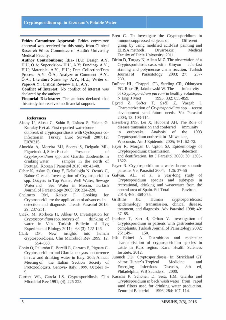
References Aksoy U, Akısu C, Sahin S, Usluca S, Yalcın G,
Kuralay F et al. First reported waterborne
outbreak of cryptosporidiosis with Cyclospora co-
infection in Turkey. Euro Surveill 2007;12: E070215.
Almeida A, Moreira MJ, Soares S, Delgado ML,
Figueiredo J, Silva E et al. Presence of Cryptosporidium spp. and Giardia duodenalis in
drinking water samples in the north of
Portugal. Korean J Parasitol 2010; 48: 43-48. Ceber K, Aslan G, Otag F, Delialioğlu N, Ozturk C,
Babur C et al. Investigation of Cryptosporidium
spp. Oocysts in Tap Water, Well Water, Sewage
Water and Sea Water in Mersin, Turkish Journal of Parasitology 2005; 29: 224-228.
Chalmers RM, Katzer F. Looking for
Cryptosporidium: the application of advances in detection and diagnosis. Trends Parasitol 2013;
29: 237-251.
Cicek, M. Korkoca H, Akkas O. Investigation for
Cryptosporidium spp. oocysts of drinking of water in Van. Turkish Bulletin of Hyg
Experimental Biology 2011; 68 (3): 122-126.
Clark DP. New insights into human cryptosporidiosis. Clin Microbiol Rev 1999; 12:
554–563.
Conio O, Palumbo F, Borelli E, Carraro E, Pignata C. Cryptosporidium and Giardia oocysts occurrence
in raw and drinking water in Italy. 20th Annual
Meeting of the Italian Section Society of
Protozoologists, Genova- İtaly: 1999. October 8–9.
Current WL, Garcia LS. Cryptosporidiosis. Clin
Microbiol Rev 1991; (4): 225-228.
Eren C. To investigate the Cryptosporidium in
immunosuppressed subjects of Different group by using modified acid-fast painting and
ELISA methods, Diyarbakir: Medical
Faculty of Dicle University. 2011.
Dirim D, Turgay N, Alkan M Z. The observation of a Cryptosporidiosis cases with Kinyon acid-fast
staining and polymerase chain reaction. Turkish
Journal of Parasitology 2003; 27: 237-239.
DuPont HL, Chappell CL, Sterling CR, Okhuysen
PC, Rose JB, Jakubowski W. The infectivity of Cryptosporidium parvum in healthy volunteers.
N Engl J Med 1995; 332: 855-859.
Egyed Z, Sréter T, Széll Z, Vargab I.
Characterization of Cryptosporidium spp.—recent development sand future needs. Vet Parasitol
2003; 13: 103-114.
Eisenberg JNS, Lei X, Hubbard AH. The Role of disease transmission and conferred immunity
in outbreaks: Analysis of the 1993
Cryptosporidium outbreak in Milwaukee, Wisconsin. Am J Epidemiol 2005; 161: 62–72.
Fayer R, Morgan U, Upton SJ, Epidemiology of
Cryptosporidium: transmission, detection
and dentification. Int J Parasitol 2000; 30: 1305-1322.
Fayer R. Cryptosporidium: a water–borne zoonotic
parasite. Vet Parasitol 2004; 126: 37-56 Galván, AL., et al. a year-long study of
Cryptosporidium species and subtypes in
recreational, drinking and wastewater from the
central area of Spain. Sci Total Environ 2014; 469: 368-375.
Griffiths JK. Human cryptosporidiosis:
epidemiology, transmission, clinical disease, treatment, and diagnosis. Adv Parasitol 1998; 40:
37–85.
Inceboz T, Sarı B, Orhan V. Investigation of Cryptosporidium in patients with gastrointestinal
complaints. Turkish Journal of Parasitology 2002;
26: 149- 150.
Itik Ekinci A. Distrubition and moleculer characterisation of cryptosporidium species in
cattle in Kars region. Kars: Health Sciences
Institute. 2012. Juranek DD, Cryptosporidiosis. In: Strickland GT
editor. Hunter’s Tropical Medicine and
Emerging Infectious Diseases, 8th ed, Philadelphia, WB Saunders; 2000.
Karanis P, Schonen D, Seitz HM. Giardia and
Cryptosporidium in back wash water from rapid
sand filters used for drinking water production. Zentralbl Bakteriol 1996; 284: 107–114.

6 MBSJHS, 2(3), 2016
Cryptosporidium sp. in Erzurum’s Potable Water
Karanis P, Papadopoulou C, Kimua A, Economou E,
Kourenti C, Sakkas H. Cryptosporidium and Giardia innatural, drinking and recreational
water of Northwestern Greece. Acta
Hydrochim Hydrobiol 2002; 30: 49-58.
Koksal F. Investigation of Source Waters for Giardia and. Journal of Turkish Society of
Microbiology 2002; 32: 275-277.
LeChevallier MW, Norton WD, Lee RG. Giardia and Cryptosporidium sp. in filtered drinking
water supplies. Appl Environ Microbiol 1991; 26:
17-21. Mac Kenzie W R, Hoxie NJ, Proctor ME, Gradus
MS, Blair KA, Peterson DE. A massive
outbreak in Milwaukee of Cryptosporidium
infection transmitted through the public water supply. N Engl J Med 1994; 331: 161–167.
Ok UZ, Girginkardeşler N, Kilimcioğlu A, Limoncu
E. Stool Analysis Methods, Chapter 1, Diagnosis of Parasitic Diseases, Editors: Ozcel
MA, Altintas N, 1st Edition, Izmir, Ege
University Press; 1997. p. 45-50. Okhuysen PC, Chappell CL, Crabb JH, Sterling CR,
DuPont HL. Virulence of three distinct
Cryptosporidium parvum isolates for healthy
adults. J Infect Dis 1999; 180: 1275-1281. Ozcel MA, Ozbel Y, Ak M. Ozcel's Medical
Parasitic Diseases. Medical Parasitology
Association, Publication No: 22 Izmir; 2007; 363-376
Redlinger T, Corella-Barud V, Graham J, Galindo A,
Avita R, Cardenas V. Hyperendemic
Cryptosporidium and Giardia in households lacking municipal sewer and water on the
United States-Mexico Border. Am J Med Trop
Hyg 2002; 66: 784-798 Smith HV. Detection of parasites in the environment.
Parasitology 1998; 117: 113– 141.
Ward PI, Deplazes P, Regli W, Rinder H, Mathis A. Detection of eight Cryptosporidium genotypes
in surface and wastewaters in Europe.
Parasitology 2002; 124: 359–368.
Wilson WR. Diagnosis and Treatment of Infectious Diseases, Nobel Medical Bookstores, 2004;
824-827.

Middle Black Sea Journal of Health Science December 2016; 2(3): 7-12
RESEARCH ARTICLE
Determining Genetics and Medical Genetics
Knowledge of Students, Physicians and Academics in A
Medical Faculty Model
Atil Bisgin1, Gulsah Kutanis Barbaros
1
1 Cukurova University, Faculty of Medicine, Medical Genetics Department of Balcali Clinics and Hospitals, Adana, Turkey.
Received: 21 November 2016, Accepted: 5 December 2016, Published online: 25 December 2016
© Ordu University Institute of Health Sciences, Turkey, 2016
Abstract Objective: Medical Genetics is one of the latest and hot topics in medicine both as a specialty and as the
most recent developments after the human genome project. The aim of this study is to determine the basic
knowledge about genetics and medical genetics through the faculty of medicine that had no medical genetics department before.
Methods: We assessed the knowledge of medical genetics through a questionnaire that was applied to 549
undergraduate students whom 300 of them were at preclinical and 249 were clinical studies, 149 resident
physicians and 86 academic staff in Cukurova University, Adana, Turkey. Results: According to our data, the scores of basic knowledge of genetics ranged from 40% to 100% with
an average of 74%. This significant difference was related to the age and the education level. However, the
scores on the knowledge of medical genetics (as a clinical department) were significantly higher in the academic stuff (54% positivity) than the resident physicians (39% positivity) and medical students. More
interestingly, the lowest rate (9% positivity) was in the students group of 4th, 5th and 6th year of medical
studies at clinics, while it was 17% of positivity in the first 3 years of medical education.
Conclusion: As a conclusion, while the great importance of medical genetics is well-accepted in all medical literature, there was still a big gap between medical education and the implementation of medical
genetics. Key words: Medical genetic, education, medical students
Address for correspondence/reprints:
Atil Bisgin
Telephone number: +90 532 565 2167 E-mail: [email protected] DOI: ……………………………………..
.
Introduction Medical genetics is one of the most recent
developed departments in medical faculties. Over
the last decade, after the human genome project
data released, there was a transformation in all fields of medicine by an explosion of genetic
testing, genetic diseases and genomic data (Zhao
and Grant, 2011). The establishment of medical genetics as a
primary specialty and not a subspecialty of another
fields in medical training in Europe began most
recently, while there were still discrepancies between countries. Genetics is usually thought
together with biology as an obligatory lesson during
the first year of medical education. Unfortunately, these lessons were thought by non-medical
geneticists or by geneticists that has no clinical
attitudes. However, there were huge clinical responsibilities of medical genetics that should be
given through the medical education. So, the

10 MBSJHS, 2(3), 2016
Determining and Medical Genetics Knowledge of Students
genetic literacy via the medical education affect the
attitudes and understanding of basic
genetics/genomics and medical genetics as a clinical specialty.
Medical geneticist has clinical responsibilities of
providing patient care in clinical genetics policlinics that include dysmorphology, rare
diseases clinics/genomics, prenatal diagnosis
clinics, diagnosis laboratory and treatment of rare diseases (Acgme, 2016). Medical geneticists also
participate in undergraduate and graduate level
courses in human and medical genetics,
cytogenetics, biochemical genetics and molecular genetics laboratories.
In this study, the main aim is to focus on the
assessment of medical genetics knowledge level in a medical faculty throughout all its components
starting from undergraduate students to academic
stuff and also to find out the importance of medical
genetics training thought by appropriate department; Department of Medical Genetics
Methods In our study, we measured the participants’ genetic
and medical genetics knowledge. All the
participants enrolled in the study were required to complete two tests; one for screening the baseline
knowledge of genetics and the second for the
knowledge about Department of Medical Genetics
as a primary clinical specialty. Participants for this study were all from the
Cukurova University Faculty of Medicine and this
study was performed before the integration of medical genetics training program to the
undergraduate and graduate education. During the
study, there was also no on-going medical genetics
clinics at the faculty hospital. There were three groups for the comparison; (1)
undergraduate students (n=549) that was also
divided into two groups, one from the first three year (preclinical education) (n=300) and other from
clinical period (n=249); (2) resident physicians
(n=149) and; (3) academic stuff (n=86) at the Balcali Hospital and Clinics of Cukurova
University Faculty of Medicine.
We used the well-validated, previously reported
16-item survey to measure the knowledge about basic genetics (Jallinoja and Aro, 1999). Genetics
literacy knowledge survey was scored according
categorization; 0-40 as inadequate, 41-70 as marginal, or 71-100 as adequate.
To ascertain participants’ knowledge about the
medical genetics, we have asked 2 survey questions:
1) a- Before this survey, had you ever heard of
Department of Medical Genetics?
b- If Yes, where did you know about the Department of Medical Genetics?
2) Have you ever considered to refer any patient to
the Department of Medical Genetics? The answers were classified according to positive
perceived knowledge of the Department of Medical
Genetics.
Results
Our study demonstrates the characterization of a
medical faculty including students, resident physicians and academic stuff about the genetics
and medical genetics knowledge. We used the
descriptive statistics to summarize each study group and the association with knowledge scores.
Overall, 784 participants were enrolled in our
study. The 70% of participants (n=549) were
undergraduate students, 19% were resident physicians (n=149) and 11% were academic stuff
(n=86). Undergraduates group were divided into
two groups according to the medical education system as 55% of preclinical (n=300) and 45% of
clinical period (n=249). The characterization of
participants in this study was showed in table 1.
Table 1: The characterization and number of
participants in this study. Characterization of Participants Number (%)
Undergraduate students 70 (549/784) Preclinical 55 (300/549) Clinical 45 (249/549) Resident physicians 19 (149/784) Academic stuff 11 (86/784)
Genetics Knowledge
According to our findings, the scores of genetic
knowledge were ranging from 40% to 100% (mean score=74%). Overall differences in genetic
knowledge were observed among the education
levels (p=0.0001) with the highest score in academic stuff and lowest in preclinical
undergraduate students. However, no differences
were noted between these groups on questions
pertaining to the inheritance-related questions (mean score 90.6%). But, when we compared to the
scores of questions on genes, chromosomes, and
cells, there was a statistically significant difference between academic stuff and other groups
(p=0.0001). None of the participants had under the
score of 40 in this study. Only the preclinical period
students had a marginal knowledge of genetics (mean=62%).

11 MBSJHS, 2(3), 2016
Determining and Medical Genetics Knowledge of Students
Medical Genetics Knowledge
Only the 22.6% of the participants (n=177)
indicated that that they had heard about the Department of Medical Genetics before our survey.
The significantly lowest proportion was 9% (n=22)
in the group of clinical period students (n=249), followed by the 17% (n=51) in the group of
preclinical period students (n=300) (table 2).
The highest rate was in the academic stuff group (n=86) as 54% (n=46). Most interestingly, the
majority of this group (96.8%) at least once in their
career they referred a patient to the medical
geneticist (table 2). The interesting finding is in resident physicians
group (n=149) with 39% high positivity (n=58)
with only 5% very low rate of referring any patient (table 2).
Table 2: The value of medical genetics
knowledge among participants in this study Participants Medical Genetics Knowledge (%)
Preclinical Students 17 (n=51) Clinical Students 9 (n=22) Resident physicians 39 (n=149) Academic stuff 54 (n=46)
Discussion Medical genetics or clinical genetics in some
countries has been recognized as an EU (European
Union) – wide primary medical specialty since
March 3rd, 2011 by the Commission adopted Regulation (EU) No:213/2011 amending Annexes
II and V to Directive 2005/36/EC of the European
Parliament and of the Council on the recognition of professional qualifications. Since then, education of
medical genetics towards successful
implementation in the medical training becomes
more important. There were studies that underlie the importance of genomic knowledge and its effect
in public debates on genomic issues including
genetic concepts of prenatal diagnosis; newborn screening and genetics research (Catz et al 2005;
McInerney 2002; Lea et al 2011; Levitt et al 2005).
Although given recent and prevalent scientists attention to medical genetics and their role in
scientific explanation, the studies about medical
genetics has still been understudied.
Thus, in our study we studied the status of a well-known university with a medical faculty
without an established Department of Medical
Genetics throughout the knowledge level in genetics and medical genetics.
In summary, in this article we demonstrated the
genetic knowledge to the association of medical
genetics department and an actual comprehension
of all over a medical faculty from students to
academic stuff.
The scientists have already theorized and pratices debated about the medical genetics of
natural development for decades and medical
genetics is quite common for all of scientists to refer to the other medical branches in all the world
(Brown 2016; Matthews 2016).
There are new studies about provide a useful evolutionary lesson about the impact of selection on
spatial patterns of medical genetic variation, when
the environment affects which individuals can
colonize new sites, and on adaptive genetic variation, when environmental heterogeneity
creates divergence at specific loci underlying local
adaptation. After these studies the new branches were development about medical genetics like
immunogenetics, epigenetics (Sork et al., 2016;
Davies et al., 2016). In addition the results of
studies suggest that associations identified between genetic and other medical branches like
dermatology, oncology and microbiology (Eroglu
et al., 2014; Li X et al., 2016; Suh et al., 2016). Overall, while there was a high knowledge of
genetics, the medical genetics knowledge differs
from that. Most of our data indicates that the higher knowledge levels related to the higher education.
However, there was a negative correlation in terms
of medical genetics knowledge; 17% positivity in
preclinical and 9% positivity in clinical students. Interestingly, the 85% of preclinical students with
positive knowledge obtained their knowledge
during the very first few months of the medical school via the internet searches about postgraduate
specialties but not by courses during the education.
Providing clinically meaningful education and
training in genomics is central to enabling every health employee to develop the appropriate
knowledge and skills in genomics in order to
provide optimum care to individuals and families now, and to facilitate the integration of new
information and technology as it becomes available
across mainstream healthcare services (Tonkin et al., 2011). Our study by its concept, points out the
importance of re-defining the medical education by
the novel concepts in medicine such as
medical/clinical genetics, and the imbalance of knowledge between medical education and
scientific concepts.

12 MBSJHS, 2(3), 2016
Determining and Medical Genetics Knowledge of Students
Informed Consent: Written informed consent was
obtained from students who participated in this study.
Ethics Committee Approval: The approval was
obtained from the local Ethical committee Peer-review: Externally peer-reviewed.
Author Contributions: Concept-AB; Design-AB;
Supervision-GKB; Materials-AB, GKB; Data Collection and/or Processing-AB, GKB; Analysis
and/or Interpretation-AB, GKB; Literature Review-
AB, GKB; Literature Review-AB; Writing-AB;
Critical Review-AB. Conflict of Interest: No conflict of interest was
declared by the authors.
Financial Disclosure: This study was supported by the grant from Cukurova University Scientific
Research Projects Unit (TSA-2014-2702).
References
Acgme O. The Accreditation Council for Graduate
Medical Education. www.acgme.org/ac, 2016. Brown SJ. Hand dermatitis in construction workers:
a lesson in genetic epidemiology. Br J
Dermatol. 2016; 174(2):263-265.
Catz N, Dicke PW, Their P. Cerebellar complex
spike firing is suitable to induce as well as to
stabilize motor learning. Curr Biol. 2005; 20:15 (24): 2179-2189.
Davies PF, Manduchi E, Jimenez JM, Jiang YZ.
Biofluids, cell mechanics and epigenetics: Flow-induced epiegentic mechanisms of
endothelial gene expression. J Biomech. 2016;
doi:10.1016/j.jbiomech.
Eroglu F, Uzun S, Koltas IS. Comparison of clinical samples and methods in chronic
cutaneous leishmaniasis. Am J Trop Med Hyg.
2014; 91(5):895-900. Griffiths Anthony JF, Miller Jeffrey H, Suzuki
David T, Lewontin Richard C, Gelbart eds.
Genetics and the organism: Introduction. An introduction to Genetic Analysis (7th ed) New
York; WH Freeman. 2000.
Jallinoja P, Aro Arja R. Knowledge about genes
and heredity among Finns. New genetics and society. 1999; 18: 101-110.
Matthews LJ. On closing the gap between
philosophical concepts and their usage in scientific practice: A lesson from the debate
about natural selection as mechanism. Stud
Hist Philos Biol Biomed Sci. 2016; 55:21-28.
McInerney J. Education in a genomic world. J Med
Philos. 2002; 27(3):369-390.
Sork VL, Werth S. Phylogeography of Ramalina menziesii, a widely distributed lichen-forming
in western North America. Mol Ecol. 2014;
23(9):2326-2339. Suh YJ, Shin J, Kang M, Park HJ, Lee K, Song
YM, Sung J. Genetic and environmental
influences on general skin traits: healthy twins and families in Korea. Twin Res Hum Genet.
2016; 16:1-7.
Lea DH, Skirton H, Read CY, Williams JK.
Implications for educating the next generation of nurses on genetics and genomics in the 21st
century. J Nurs Scholarsh. 2011; 43(1):3-12.
Levitt M, Hayry M. Overcritical, overfriendly? A dialogue between a sociologist and a
philosopher on genetic technology and its
applications. Med Health Care Philos. 2005;
8(3): 377-383. LiX, Zou W, Liu M, Cao W, Jiang Y, An G, Wang
Y, Huang S, Zhao X. Association of multiple
genetic variants with breast cancer susceptibility in the Han Chinese population.
Oncotarget. 2016; doi: 10.18632/oncotarget.
Tonkin E, Calzone K, Jenkins J, Lea D, Prows C. Genomic education resources for nursing
faculty. J Nurs Scholarsh. 2011; 43(4): 330-
340.
Zhao J, Grant SF. Genetics of childhood obesity. J Obes. 2011; 10.1155/2011/845148

Middle Black Sea Journal of Health Science December 2016; 2(3):13-20
REVIEW
Parasitic Diseases of Urinary Tract
Neriman Mor1, Ümit Yener Tekdoğan
2, Murat Bağcıoğlu
2
1Kafkas University, Faculty of Health Sciences, Kars, Turkey;
2Department of Urology, Kafkas University Faculty of Medicine, Kars, Turkey
Received: 20 October 2016 Accepted: 18 December 2016, Published online: 25 December 2016
© Ordu University Institute of Health Sciences, Turkey, 2016
Abstract
Urinary tract infections are among frequent health problems, but parasitic infections are taken no notice. Of urinary system diseases, parasitic diseases such as schistosomiasis (bilharziasis) and trichomoniasis effect
lots of people and often give rise to renal and lower urinary tract diseases. Echinocoocosis and filariasis
rarely effect urinary system. Schistosomiasis gives rise to the disease by finding its way into bladder veins in human who is final host. It leads to unintended consequences such as permanent urogenital problems,
renal failure and malignancy because of its chronicity and tissue damage in the part of body it locates.
Symptomatic involvement in urogenital system is more common in females compared to males.
Trichomoniasis is the most frequent parasitic disease in the world which occurs because of Trichomonas vaginalis trophozoites’ involvement in urogenital system sexually. This disease underlies vaginitis which is
frequent in females, but it gives rise to urethritis and prostatitis in male. Echinococcosis is known as an
endemic zoonotic parasitic disease which infects to humans from animals and grows up in humans evolved in agriculture and stock rising and it may involve in kidney sporadically. Filariasis involves lymphatic
system. Obstruction give rise to elephantiasis which may involve in stratum and legs. In this review was
presented general information about morphological, biological characteristics of urinary system parasites,
diseases they give rise to, clinical symptoms and their prevalence in Turkey. Therefore, helpful information for diagnosis and treatment in humans has been presented by reviewing information in literature about S.
haematobium, E. granulosus, T. vaginalis, W. bancrofti, urogenital myiasis and scabies which are among
urogenital system parasites. Key words: Urogenital parasites, human, Turkey..
Address for correspondence/reprints: Neriman MOR Telephone number: +90 532 728 2360 E-mail: [email protected]
DOI:………………………………
Introduction Parasitic diseases as schistosomiasis
(bilharziasis), trichomoniasis, echinococcosis, and
filariasis (Wuchereria buncrofti) affect lots of
people, and they cause urinary tract diseases. Echinococcosis and filariasis affect the urinary tract
disease less than other parasites (Unat et al., 1995;
Özcel, 2007; Yazar et al., 2016). In this review, the general information about the biological,
morphological features, and clinical symptomps of
significant helminths, protozoons, and arthtoparasides which locate in urinary system, and
the prevalance of diseases in Turkey were reported.
We aimed to present the recent information of
literature in this topic.

14 MBSJHS; 2(3), 2016
REVIEW
Schistosomiasis (Bilharziasis)
Schistosomiasis is an endemic disease in tropical, and subtropical areas. The urinary
schistosomiasis caused by Schistosoma
haematobium (Bilharz,1852) is common in 75
countries in South America, Africa, and The Middle East region. More than 200 million people
are infected, and it is estimated that more than 800
million people are under the risk of infection (WHO Information, 2010). It was reported that
Bulinis, the intermediate host worm, was seen in
our country in Nusaybin and Suruç region (Özcel, 2007b). The frequency of this parasite is unknown
in Turkey. Most of the reported cases have
originated abroad (Alver et al., 2004; Yazar, 2008;
Özvatan, 2011).
Morphology
Females are generally longer and slimmer than males, and they exist in ducts called as ‘canalis
gynecophrus’ which are located in the ventral part
of the male body. Female Schistosoma leave their males while just laying eggs, go into the capillary
veins and and then turn back to ducts of the males
after releasing their eggs. Males are 10-15x0.7-1
mm, and females are 15-20x0.25 mm in size. There are oral suckers surrounding the oral cavity
and the ventral sucker just under it in the front part
of the body. In the digestive system, oral cavity, pharynx, oesephagus and intestinal canal are
available respectively. Two intestinal canals end at
the posterior part of the body by combining with
each other. The eggs of Schistosoma species which are classified in trematodes don’t have valves,
miracidium (larva) exist in Schistosoma eggs as in
many other trematod classes. The sizes of the eggs vary according to the species of Schistosoma and
there is an apophysis spinal in various parts of the
egg. Schistosoma haematobium eggs are oval and 100–160 x 40–60 μm in size. They have a spina on
one of its margins (Yazar, et. al., 2016; Özcel,
2007b).
Biology
Adult male and female parasites live together in
big veins. The males stay in the big veins after copulation and females leave the males nearly
every day and mainly go into the veins surrounding
the urogenital system (urinary bladder veins) by proceeding into small veins and lay their eggs there.
These eggs attach to the blood vessels via blood
flow velocity and spinas on their walls and reach
into the urinary bladder space by penetrating into
the vein wall. These eggs which are passed with
urine need fresh water or saline water with a density of less than 0.7% to be alive. Miracidium
leaves the egg in fresh water, and moves by the
help of cillias and penetrate into the fresh water
snail Bulinis. Then it loses the cillias and sporocyst phase is progressed and female sporocysts take its
place without redi phase. Double–tailed cercarias
evolve out of sporocysts and these are called furcucariaes. Furcucariaes scatter to snail’s tissues
and body cavity by destroying the sporocyst. In
certain circumstances, they move in the fresh water very fast by leaving the snail (Altıntaş, 2002; Yazar
et. al., 2016). There is no metacercaria phase, which
is an infective form in other areas, for them. The
furcocercariae is the infective form of S. haematobium. The infection of the last host occurs
due to furcocercariaes’ burrowing the skin or
contamines by the freshwater. The furcocercariae that goes into the organism from the skin, travels to
the mezenterium vein via the lymphatic vessels,
then lung and systemic circulation, and finally comes to bladder vein. The furcocercariae that
penetrated into the oral epithelia travels to lung via
blood stream and then migrates to the bladder from
the left heart. The prepatent period is six weeks. Schistosoma is a treametode, which has a long
lifetime, and they can be alive during the host life if
a treatment is not given (Altıntaş, 2002; Ross et. al., 2002; Özcel, 2007b; Avcıoğlu, 2007).
Clinical Symptoms, Diagnosis and Treatment
The erythema, papule and itchiness (Swimmer’sitch) can be seen on the host skin
because of the host reaction (Altıntaş, 2002; Şahin
et. al., 2009). The classic signs of urinary schistosomiasis are associated with accumulating
the eggs in the kidney and the kidney damage.
Adult females lay eggs at bladder veins, then migrate to the urinary bladder submucosa and
granulomas occur because of cell infiltration and
inflammation (Hams et. al., 2013). The
accumulation of eggs under mucosa causes mucosal hyperemia and hematuria. The eggs which
penetrate into the mucosa and migrate into the
urinary bladder are excreted through bleeding the urine. The inflammation causes different levels of
cystitis in urinary bladder mucosa. The little
erythema and ulcerative lesions on bladder mucosa can be seen under cystoscopic evaluation.
Ulcerative lesions induce scar tissue, the bladder
wall becomes thicker and loses its elasticity. The

15 MBSJHS; 2(3), 2016
REVIEW
eggs create pseudo tubercules on the bladder
mucosa and then nodules and polipoid masses develop (Gryseels, 2012). Schistosomiasis is also
known as a predisposing factor for adenocarcinoma
of the bladder. It is one of the most important
reasons of urinary bladder carcinoma especially in Africa and The Middle East, although the cancer
pathogenesis, and molecular mechanisms are still
not known well (Koonrungsesomboon, 2015). It is more common in females than males
(Remoue et. al., 2002; Özcel, 2007b; Avcıoğlu,
2007). While vesicula seminalis, prostate, urethra, vas deferences and rarely testes and ureters are
affceted in males, ovary, fallopian tubes, uterus,
and vagina are involved for females (Özcel, 2007).
In addition tiredness, exhaustion, sweating, headache and subfebrile fever (Katayama fever) are
seen in local symptoms (Avcıoğlu, 2007).
The parasite is diagnosed by the detection of Schistosoma eggs in the urine through stool and
biopsy materials. The most appropriate method to
detect the eggs in the urine is by examining the last part of the urine which passed in the afternoon after
being centrifuged. It is recommended that the eggs
examined for at least five different days
microscopically if the urine sample is negative taken from clinically suspicious conditions.
Additionally, antigen and antibody detection tests
can be used alternatively in suspicious conditions (Özcel, 2007b; Şahin et. al., 2009).
The control of the disease is based on large-
scale treatment of risky population, access to safe
water, improved sanitation and hygiene education. The disease should be kept in mind if the visitors
from the endemic countries have symtoms of
infection (Inobaya et. al., 2014). The most effective treatment modality is using
Praziquantel (single dose, orally, 40 miligrams per
kilogram) (Altıntaş, 2002).
Cystic Echinococcosis
Cystic echinococcosis (CE) is a
helminthzoonotic disease and common in the world especially our country. The prevalence of disease in
our country varies according to the region, it is
mostly common in Eastern, South-East, and Central Anatolia (Unat et. al., 1995). The prevalence of
disease is 50-400/100.000, and the incidence is
3,4/100.000 in Turkey (Doğru, 2008). The reported case with CE in Turkey is mainly located in the
liver and lungs (Altıntaş et. al., 2004). Renal cyst
hydatid commonly occurs with systemic diseases
and the isolated form is very rare (Vargas-Serrano
et. al., 1997; Zmerli et. al., 2001; Bozdağ et. al., 2012; Merdin et. al., 2014).
Morphology
The adult form of Echinococcus granulosus (E. granulosus) is 2-11 .mm in height and 0.6 mm in
width. The body of the parasite is formed of 3 or 4
cingulars. It is composed of scolex, neck, immature, mature, and gravid cingulars. The scolex is
typically small, and has four brawny suckers,
rostellum, and 28-50 distichous coronoids. The body (strobila) is generally composed of 3
cingulars, but rarely 4- cingular striae are
encountered. The last cingular, proglottid is gravid
and it is nearly half of the body. The uterus extends across the cingular and there may be 200-800 eggs
in it. The eggs are round and slightly oval, 28-36
µm in size, thick-walled, dark brown, radial lineal and contain 6-coronoid embryo (the oncosphere) .
The eggs of E. granulosus can not be differentiated
under light microscope from the other species of Echinococcus and Taenia (Altıntaş et. al., 2004;
Özbilgin &Kilimcioğlu, 2007).
The cause of the disease is the larva form of E.
granulosus called as hydatid cyst. The cysts are usually filled with clear fluid called rock water,
which is secreted from germinal layer. The fluid is
alcali (base) with 7.2-7.4 Ph, 1007-1015 density, and also it has antigenic features (Özbilgin
&Kilimcioğlu, 2007).
Biology The adult form is located in the small intestine
of dogs or other canidae as a definitive host, the
larva form is located in internal organs of sheep, cow, pig or human as intermediate host. The eggs
of parasite excreted by the definitive host constitute
disease by ingestion and rarely inhalation by intermediate hosts. The oncosphere that releases
from the eggs in the small intestine penetrates into
the intestine wall and enters into the portal system.
Oncosphere invades the liver previously and develops with fluid-filled cysts. Oncospheres rarely
pass over the sinusoids and enter to systemic
circulation, and can migrate to all internal organs (Altıntaş et. al., 2004; Eckert &Deplazes, 2004).
Clinical Symptoms, Diagnosis and Treatment Isolated renal disease is rare, and reported as 2-
4% of all cases (Adayener et. al., 2008). The other
involved organs in the genitourinary system are
urinary bladder, prostate, testes, and seminal
16 MBSJHS; 2(3), 2016
REVIEW
vesicules. Renal cystic echinococcosis is usually a
single and cortical located lesion. The disease can be asymptomatic for several years, and some
symptoms occur when the cyst enlarges. The most
common symptoms are palpable mass, hematuria,
pain, and hydaturia which occur with the connection of cyst and collector system (Göğüş et.
al., 2003). Hydaturia is specific to disease (Angulo
et. al., 1997; Göğüş et. al., 2003). The renal cystic echinococcosis is diagnosed
with radiologic methods, direct diagnostic
microbiologic tests, serologic and molecular tests. Ultrasonography, computed tomography, and
manyetic resonans are used for radiologic imaging.
Imaging technics are useful to see the calcification
of cyst wall. The suspicious cases can be checked by using serologic tests as indirect
hemagglutination test, indirect fluorescent antibody
test, and Enyzme-linked immunosorbent assay (Pawlowski et. al., 2001; Filippou et. al., 2007;
Yılmaz&Babür, 2007).
The renal cystic echinococcosis can be treated with different methods as medical, surgical, and
PAIR (puncture, aspiration, injection, respiration)
(McManus et. al., 2003; Köksal et. al., 2004).
Surgery is the first line treatment modality, and complete resection of the cyst is recommended.
There are several different surgical approaches as
cystectomy, pericystectomy, partial cystectomy, partial and simple nephrectomy as well. The most
serious complication during the surgery is fatal
anaphylaxis due to a rupture of the cyst and
dissemination of the infection. Benzimidazol family such as Albendazol, Mabendazol, and Praziquantel
are the options for the medical treatment. The
purpose of Albendazol and Mabendazol treatment, which is done before and after surgery, is
sterilisation of the cyst, preventing the anaphlaxy
and the recurrence (Pawlowski et. al., 2001; Filippou et. al., 2007). The recommended treatment
period is 3-6 months. The most common adverse
effect of medical treatment is dysfunction of the
liver (Köksal et. al., 2004).
Trichomoniasis
The flagellated parasite Trichomonas vaginalis (T. vaginalis) is the aetiologic agent of
trichomoniasis, which was firstly described by
Donne in 1836. It is a sexually transmitted infection, and a common parasite in the world (Safi
et. al., 2000; Özcel &Zeyrek, 2007). It affects the
urogenital system and it is fed by cells, bacteries,
and sperm cells via fagositing (Unat et. al., 1995).
T. vaginalis is mostly common in patients of ages
between 20-40. It is reported that T. vaginalis is detected between 4% and 73% of women with
vaginal discharge in our country (Suay et. al., 1995;
Tamer et. al., 2008; Değerli et. al., 2011), and it is
reported by 2,8% among men with urethritis (Çulha et. al., 2008), while founded that between 0,8% and
12% among men with nongonococcal urethritis
(Özbilgin et. al., 1992; Ay et. al., 1999).
Morphology
Trichomonas vaginalis has only one form as a trophozoite. The trophozoite is oval and pear
shaped, 10-25 x 5-15 µm in size. It has a nucleus,
four anterior flagella, and a flagella called ‘’wavy
membrane’’ dangling down along the membrane of a parasite. T. vaginalis is a protozoon which turns
around its own axis continually. It feeds by
phagocytosing cell, bacteria and sperm in its location (Unat et. al., 1995; Özcel &Zeyrek, 2007).
Biology It is a monoxenous parasite, and human is the
single host for it. The throphozoite is in infective
form since there isn’t a cyst form of it. The
trophozoite form is not very resistant to external factors; it can be alive for 1 hour in water, and 24
hours in urine. Trichomonas is sexually transmitted
from infected female or male, but rarely contaminated from soiled towels, baths, swimming
pools, toilet paper, and toilet seats (Unat et. al.,
1995; Yazar et. al., 2016).
Clinical Symptoms, Diagnosis and Treatment
The mean incubation period is 6-10 days. The
symptomps of disease vary according to organ and gender. Vaginal discharge occurs during the acute
period of the disease. Frothy, greenish, malodorous
vaginal discharge is characteristic of the disease. Disuria and pollacuria are common with erythema,
tanning, and itchness of the vulva and vagina. The
chronic disease can be commonly present with
semptomatic vaginal discharge or can be asymptomatic. The symptoms have decreased over
time, and disease becomes in latency (Kuman
&Altıntaş, 1996). Most infected men are asymptomatic, and it is
hard to detect the disease. Male patients are
generally presented with urethritis, prostatitis, and rarely epididymitis and cystitis. Occasionally, white
and fuzzy urethral discharge can be seen (Kuman
&Altıntaş, 1996; Skerk et. al., 2002).
The disease is diagnosed with immediate

17 MBSJHS; 2(3), 2016
REVIEW
microscopic evaluation of the vaginal, and urehtral
discharge, prostatic secretion, and urine sediment. Another method of diagnosis is via overnight
culture. The suspicious cases can be checked by
using serologic and molecular tests. More than one
method can be combined to diagnose (Özcel &Zeyrek, 2007).
Infection is treated with oral or local use of
metranidazole (250 miligrams per kilogram, three times a day, a week period). The drug is
contraindicated in the first 3 months of pregnancy.
Secnidazole has taken the place of metronidazole because of its adverse effects and long treatment
period of this drug. Secnidazole has better results
than other nitroimidazole family (2 grams, single
dose, orally). Tinidazole can be useful in case of resistant disease. Partners have to be treated all
together (Kuman&Altıntaş, 1996; Özcel &Zeyrek,
2007).
Filariasis
Filariasis is a parasitic infection in tropical and subtropical areas with high morbidity but low
mortality (Chandy et. al., 2011). There are 7 species
of filariasis parasites which cause disease in
humans, but typically Wuchereria bancrofti, Brugia malayi, and Brugia timori are known as factors for
filiariasis (Melrose, 2002; Palumbo, 2008). The
intermediate host is mosquitos as Aedes, Anopheles and Culex species. The disease is transmitted to
humans through the bite of an infected mosquito.
The disease has a long latency period, and
sometimes, it can also be asymptomatic. This situation generally damages the lymphatic system
and kidneys (Kuman, 2007).
Wuchereria bancrofti
Almost 1 billion people are under the risk of
lymphedema disease known as Wuchereria bancrofti (W. bancrofti). It is estimated that roughly
120 million people are infected with W. bancrofti in
developing countries. Lymphedema is common in
Uganda, Tanzania, Kenya, Rwanda, Burundi, Sudan and Ethiopia. Filariasis cases are very rare in
our country, because of this reason there is no
information about its frequency in Turkey. It is detected sporadically in Alanya, Elazığ, Çubuk and
Samsun region in Turkey (Unat et. al., 1995;
Kuman, 2007).
Morphology
The adult forms are cylinder-shaped. They don’t have lips, and oral cavity is not developed, and it is
fistular going on with oesephagus. The male is 30-
40 mm x 0,1 mm in size, and it has 15 pairs of
postnatal papillas. The female is 50-100 mm x 0,25 mm in size, and transparent, but eggs in the uterus
can be seen. Females are viviparous, and their
larvas are named as microfilariaes which are 300x8 micron in size. After the infection, microfilariaes
can be seen in blood within 6-8 months (Unat et.
al., 1995; Kuman, 2007).
Biology
Human beings serve as the definitive host and
mosquitoes as their intermediate hosts. The major vectors of W. bancrofti are mosquitoes of the genus
Culex (in urban and semi-urban areas), Anopheles
(in rural areas of Africa and elsewhere) and Aedes (in islands of the Pacific). In Turkey, the most
common vector is Culex fatigans. Larvae known as
microfilariaes may live for a few years until they are transferred by an insect vector (Culicidae;
mosquitos) which is an intermediate host in the
blood. When microfilariaes are transferred together
with blood by a suitable vector, L1 larvae develops in the digestive tract of the vector. L1 larvae
migrate to thoracic muscles of vector mosquito
from midgut wall. It molts here twice in 3 weeks and L3 larvae develop within 3 weeks after
metamorphosis (infective filariform). Infective L3
larvae come to the proboscis of the vector and it
goes into the circulator system of the host while sucking blood. A larva molts twice in humans and
it becomes adult with metamorphosis while
migrating to certain infected regions. Microfilariaes are detected in the blood after almost 6 months
(Unat et. al., 1995; Kuman, 2007; Yazar et. al.,
2016).
Clinical Symptoms, Diagnosis and Treatment
Recurrent lymphadenitis, lymphangitis, and
fever can be seen in mild disease, and these cause swelling especially in the legs and feet.
Inflammation in epididymis, testes, scrotum, and
spermatic cord is not very rare, and also hydrocele is common. In severe disease, obstruction of major
lymphatic vessels may cause to chyluria and
elephantiasis (Melrose, 2002; Kuman, 2007; Chandy et. al., 2011).
The final diagnosis for filariasis is established
from a blood sample which is taken during the
night by microscopic examination if microfiliariaes

18 MBSJHS; 2(3), 2016
REVIEW
are seen in blood samples (Melrose, 2002). Urine
and hydrocele fluid may also be seen as factors. The lower parts of the legs, edema and painful
swelling began in the reproductive organs and feet
should be considered of filarial acute attack.
Finger-causing fossa while pressing over swelling is the diagnostic sign of the disease. Over time, skin
thickens and changes colour. It resembles an
elephant’s skin. Apart from swelling, cough and mild temperature are some indications of the
disease. In elephantiasis cases, the diagnosis is
established clinically because microfilaria can not be detected in blood (Simonsen et. al., 2002;
Mendoza et. al., 2009).
The first choice for treatment is
diethylcarbamazine (DEC), despite having some toxic side effects. DEC should be given orally three
times a day in dose of 2 mg per kg during 12 days.
This medicine does not make an end of adult larvae but effective on microfilariaes. The most important
advantage of Ivermectin which was investigated
recently is using a single dose a day. The effectiveness of both medicines on adult parasites is
not enough. Long term DEC treatment is useful to
prevent the spread of new adult parasites.
Physiotherapy is run together with surgical treatment. The legs should be jacked for
detumescence in legs and compression socks
should be put on to prevent the obstruction of lymphatic liquide (Stolk et. al., 2005).
Urogenital Myasis
Urogenital myiasis is an infestasion of humans and vertebrates, which is a member of the diptera
family known as flies larva, and also lives on live
cells or dead tissue. Pathophysiology of infestasion varies on humans, with the location of larva and the
species of it. Urogenital myasis usually limits itself,
but rarely patients are presented with urinary tract obstruction, and genital lesions. Urogenital myasis
cases were rarely reported in our country with the
species of Eristalis tenax (Mumcuoğlu et. al.,
2005), Lucilia sericata (Dinçer et. al., 1995), and Psychoda albipennis (Taylan-Özkan et. al., 2004;
Güven et. al., 2008).
The symptoms of the infestation are commonly itchiness, dysuria, pollacuria, infravesical
obstruction, anuria and hematuria. These symptoms
occur with habitation and development of larva’s into the lower urinary system (Zumpt, 1965;
Dinçer, 1997; Güven et. al., 2008).
Scabies
Scabies called as Sarcoptes scabiei are the other infestasions located on genital organs (Yazar et. al.,
2016).
Acknowledgements
We would like to thank to Andrew Roberts
for English editing
Peer-review: Externally peer-reviewed.
Author Contributions: Concept NM, ÜYT;
Design NM, ÜYT; Supervision NM, ÜYT,
MB; Literature review NM, ÜYT, MB; Writing
NM, ÜYT, MB; Critical review NM, ÜYT,
MB.
Conflict of Interest: No conflict of interest was
declared by the authors. Financial Disclosure: The authors declared that
this study hasn’t received any financial support.
References
Adayener C, Akyol I, Berber U, Senkul T, Baykal
K, Iseri C. A multilocular cystic renal cell carcinoma mimicking hydatid cyst. Turk J Urol
2008;34:266-8.
Altıntas K. Medical Parasitology. Ankara: Nobel
Tıp Kitapevleri, 2002. p. 234. Alver O, Kılıçarslan E, Helvacı S, Töre O. A
Nigerian patient with Schistosoma haematobium
in Turkey. Turk Parazitol Derg’s 2004; 28(4): 197-8.
Angulo JC, Sanchez-Chapodo M, Diego A,
Eseribano J, Tamayo JC, Martin L. Renal echinococcosis: clinical study of 34 cases. J
Urol 1997; 157: 787-94.
Avcıoglu H. Urinary system helmints and
prevalence in Turkey. Atatürk Univ Vet Bil Derg 2007;2(2):44-49.
Ay S, Yalçın O, Ilhan F, Tahmaz L, Yılmaz M.
Chlamydia trachomatis, Ureaplasma urealyticum and Trichomonas vaginalis in cases
of Nongonococcal urethritis (NGU). Infek Derg
1999; 13(1): 55-57.
Bozdağ Z, Turkmen E, Dogan M, Guzel Z, Kırımlıoglu H. Isolated renal hydatid cyst: case
report. Inönü Univ Saslık Bil Derg 2012;1:55-7.

19 MBSJHS; 2(3), 2016
REVIEW
Chandy A, Thakur AS, Singh MP, Manigauha A. A
review of neglected tropical diseases: filariasis. Asian Pac J Trop Med 2011; 4(7): 581-6.
http://dx.doi.org/10.1016/S1995-
7645(11)60150-8
Culha G, Gorur S, Helli A, Akcin S, Kiper AN. The prevalence of Trichomonas vaginalis in male
patients with urethritis who referred to Mustafa
Kemal University Hospital Urology Clinic. Turk Hij Den Biyol Derg 2008;65(1): 37-41.
Degerli S, Salk S, Malatyalı E. Incidence in Sivas
of Trichomonas vaginalis in patients with vaginitis. Turk Parazitol Derg. 2011;35:145-7.
Dincer S, Tanyuksel M, Kucuk T. Human myiasis
caused by Psychoda albipennis (Diptera:
Nematocera) and Sarcophaga spp. (Diptera: Cyclorrhapha) larvae: report of two cases. Turk
Parazitol Derg 1995;19(3):402-8.
Dincer S. Myiasis in humans and animals. Ozcel MA, Daldal N, editors. Arthropod Diseases and
Vectors. İzmir: Turk Parazitol Dern Public
1997. p. 169-233. Dogru U. Hydatid cysts. Güncel Pediatri Derg
2008;3:60-1.
Eckert J, Deplazes P. Biological, epidemiological,
and clinical aspects of echinococcosis, a zoonosis of increasing concern. Clin Microbiol
Rev 2004;17:107-135
Filippou D, Tselepis D, Filippou G, Papadopoulos V. Advances in liver echinococcosis: diagnosis
and treatment. Clin Gastroenterol and Hepatol
2007;5:152-9.
Gögüs C, Safak M, Baltaci S, Turkolmez K. Isolated renal hydatidosis: experience with 20
cases. J Urol, 2003; 169: 186-9.
Gryseels B. Schistosomiasis. Infect Dis Clin North Am 2012;26(2):383–397.
Guven E, Kar S, Doğan N, Karaer Z. Urogenital
myiasis caused by Psychoda albipennis in a woman. Turk Parazitol Derg 2008; 32(2):174-6.
Hams E, Aviello G, Fallon PG. The schistosoma
granuloma: friend or foe? Front Immunol
2013;4:89. Inobaya MT, Olveda RM, Chau TNP, Olveda DU,
Ross AGP. Prevention and control of
schistosomiasis: a current perspective. Research and Reports in Tropical Medicine 2014;5:65–75.
Kilimcioglu A, Ok UZ. Human, epidemiology of
echinococcus species, geographic prevalence and condition of Turkey. Izmir. Altıntas N,
Tınar R, Coker A, editors. Echinococcosis.
Izmir: Hidatidoloji Dern 2004.p.129-40
Koonrungsesomboon N, Wadagni AC, Mbanefo
EC. Molecular markers and Schistosoma-associated bladder carcinoma: A systematic
review and meta-analysis. Cancer epidemiol
2015;39(4):487-496.
Koksal AS, Arhan M, Oguz D. Hydatid cysts. Güncel Gastroenteroloji 2004; 61-67.
Kuman HA, Altıntaş N. Protozoan diseases. İzmir:
Ege Üniversitesi Basımevi; 1996.p. 71-78. Kuman HA. Filariosis. Özcel A, Özbel Y, Ak M,
editors. Ozcel's Medical Parasitic Diseases.
İzmir: Turkiye Parazitoloji Derneği; 2007. p. 601-627.
McManus DP, Zhang W, Li J, Bartley PB.
Echinococcosis. Lancet 2003; 362: 1295-304.
Melrose Wd. Lymphatic filariasis: new insights into an old disease. Int J Parasitol, 2002;32(8):947-
60. http://dx.doi.org/10.1016/S0020-
7519(02)00062-0 Mendoza N, li A, Gill A, Tyring S. Filariasis:
diagnosis and treatment. Dermatol Ther 2009;
22(6): 475-90. http://dx.doi.org/10.1111/j.1529-8019.2009.01271.x PMid:19889133.
Merdin A, Ogur E, Cicek Kolak C, Avcı Merdin F,
Gunseren F, Inan D, et al. Renal cyst hydatid.
Turk Parazitol Derg 2014;38:190-3. Mumcuoglu I, Akarsu GA, Balaban N, Keles I.
Eristalis tenax as a cause of urinary myiasis.
Scand J Infect Dis 2005;37(11-12):942-3. Ozbilgin A, Kilimcioglu AA. Cystic
Echinococcosis. Ozcel A, Ozbel Y, Ak M,
editors. Ozcel's Medical Parasitic Diseases.
İzmir: Turkiye Parazitol Dern 2007. p. 541-61. Ozbilgin A, Nazlı O, Ozbel Y, Tuzcuoglu Y, Ozcel
MA, Mulazımoglu N. Trichomoniasis in
nongonococcal urethritis in males. Turk Parazitol Derg 1992;16:43-48.
Ozcel MA. Schistosomiosis. Özcel A, Özbel Y, Ak
M, editors. Ozcel's Medical Parasitic Diseases. İzmir: Turkiye Parazitol Dern 2007. p. 475-97.
Ozcel MA, Zeyrek FY. Trichomoniosis, Özcel A,
Özbel Y, Ak M, editors. Ozcel's Medical
Parasitic Diseases. İzmir: Turkiye Parazitol Dern 2007. p. 431-45.
Ozvatan TS, Kocak C, Alver O, Mistik R, Aslan E.
Travel related urinary schistosomiasis: case report. Turk Parazitol Derg 2011;35:175-7.
Palumbo E. Filariasis: diagnosis, treatment and
prevention. Acta Biomed 2008; 79(2): 106-9. PMid:18788504

20 MBSJHS; 2(3), 2016
REVIEW
Pawlowski ZS, Eckert J, Vuitton DA, Ammann
RW, Kern P, Craig PS. Echinococcosis in humans: clinical aspects, diagnosis and
treatment. Eckert J, Gemmell MA, Melsin FX,
Pawlowski ZS, editors. WHO/OIE manual on
echinococcosis in humans and animals: a public health problem of global concern. Paris; France:
World Organization for Animal Health 2001.
p.20-68. Remoue F, Mani JC, Pugniere M, Schacht AM,
Capron A, Riveau G. Functional specific
binding of testosterone to Schistosoma haematobium 28-kilodalton glutathione S-
transferase. Infect Immun 2002;70:601-5.
Ross AG, Bartley PB, Sleigh AC, Olds GR, Li Y,
Williams GM, et al. Schistosomiasis. N Engl J Med 2002;346(16):1212–1220.
Safi Z, Demirezen Ş, Beksaç MS. Biological
characteristics and clinically important of Trichomonas vaginalis. Klinik Bilimler ve
Doktor 2000;6 (4):531-8.
Simonsen PE, Bernhard P, Jaoko WG, Meyrowitsch dW, Malecela-lazaro MN,
Magnussen P, et al. Filaria dance sign and
subclinical hydrocoele in two east African
communities with bancroftianfilariasis. Trans R Soc Trop Med Hyg 2002; 96(6): 649-53.
http://dx.doi.org/10.1016/S0035-
9203(02)90342-1 Skerk V, Schonwald S, Krhen I, Markovinovic L,
Beus A, Kuzmanovic NS, et al. Aetiology of
chronic prostatitis. Int J Antimicrob Agents
2002;19(6):471-474. Stolk WA, VAN Oortmarssen GJ, Pani SP, dE Vlas
SJ, Subramanian S, dAS PK, et al. Effects of
ivermectin and diethylcarbamazine on microfilariae and overall microfilaria production
in bancroftian filariasis. Am J Trop Med Hyg
2005;73(5):881-7. PMid:16282298 Suay A, Yayla M, Mete Ö, Elçi S. Investigation of
trichomoniasis due to Trichomonas vaginalis
with direct Microscopy and cultural methods in
300 Life Woman. Turk Parazitol Derg’s 1995;19(2):170-173.
Şahin İ, Yazar S, Yaman O. Schistosomiasis.
Doğanay M, Altıntaş N, editors. Zoonoses. Ankara: Bilimsel Tıp Yayınevi 2009.p. 869-77.
Tamer GS, Dündar D, Çalışkan Ş, Doğer E.
Comparison of direct microscopy and in-vitro
cultures in detection of Trichomonas vaginalis. Turk Hij Den Biyol Derg 2008;65(2):75-80.
Taylan-Özkan A, Babür C, Kiliç S, Nalbantoğlu S,
Dalkılıç I, Mumcuoğlu KY. Urogenital myiasis caused by Psychoda albipennis (Diptera:
Nematocera) in Turkey. Int J Dermatol
2004;43(12):904-5 Unat EK, Yücel A, Atlaş K, Samastı M. Unat's
Medical Parasitology (5. printing). İstanbul: Cer
Tıp Fak Vakfı Yayın No:15, 1995. p. 19-49.
Vargas-Serrano B, Ferreiro-Argüelles C, Rodríguez-Romero R, Marcos del Río N.
Imaging findings in renal hydatid disease. Eur
Radiolb1997;7:548-51. WHO Information schistosomiasis.
http://www.who.int/schistosomiasis/en/ Erişim
tarihi 21.12.2010. Yazar S, Kuk S, Miman Ö, Saygı G. Saygı’s Basic
Medical Parasitology. Kayseri: Erciyes
Üniversitesi Yayınları Yayın No:206, 2016.
Yazar S, Sipahioğlu M, Ünal A, Yaman O, Şahin İ, Utaş C, et al. Schistosoma haematobium
infection in a Ghanaian patient residing for a
period in Turkey. Turk Parazitol Derg 2008;32(2):161-3.
Yılmaz GR, Babür C. Diagnosis of echinococcosis.
Türk Hij Den Biyol Derg 2007;67(3):35-44.
Zmerli S, Ayed M, Horchani A, Chami I, El Quakti M, Ben Slama MR. Hydatid cyst of the kidney:
diagnosis and treatment. World J Surg
2001;25:68-74. Zumpt F. Myiasis in Man and Animals in the
World, Butterworths; London: 1965. pp: 1-
247.Zmerli S, Ayed M, Horchani A, Chami I, El Quakti M, Ben Slama MR. Hydatid cyst of the
kidney: diagnosis and treatment. World J Surg
2001; 25:68-74.
Zumpt F. Myiasis in Man and Animals in the World, Butterworths, London. 1965; pp: 1-247.

Middle Black Sea Journal of Health Science December 2016 ; 2(3): 21-23
CASE REPORT
Insidentally Diagnosed Osteopoikilosis After Knee Pain
Erdal Uzun1, İlknur Uzun
2
1Orthopedic and Traumatology Clinic, Kayseri Research and Training Hospital, Kayseri, Turkey.
2Department of Internal Medicine, Faculty of Medicine, Erciyes University, Kayseri, Turkey.
Received: 25 April 2016 Accepted: 15 June 2016, Published online: 25 December 2016
© Ordu University Institute of Health Sciences, Turkey, 2016
Abstract
Osteopoikilosis is a rare, benign, autosomal dominant disorder characterised by sclerotic bone lesions most
commonly involving the hands, feet, pelvis, and ends of long bones. Lesions are typically found incidentally on imaging studies done for unrelated complaints. It may resemble bone tumor, metabolic
disease, mastocytosis and tuberous sclerosis. Early recognition is essential to prevent unnecessary
emotional distress and invasive testing. Herein we report a case presenting with knee pain whose
radio¬logic investigation revealed typical osteopoikilotic lesions. Key words: Osteopoikilosis, bone dysplasia, radiography, diagnosis.
Address for correspondence/reprints: Erdal Uzun Telephone number: +90 (352) 336 8884 E-mail: [email protected]
DOI: ……………………………………
Introduction
Osteopoikilosis is also named as “spotted bone
disease” an asymptomatic osteosclerotic dysplasia initially described by Albers-Schönberg in 1915
(Resnick et al., 1988). It is a rare, benign,
autosomal dominant disorder characterised by
sclerotic bone lesions most commonly involving the hands, feet, pelvis, and ends of long bones
(Woyciechowsky et al., 2012; Paraskevas et al.,
2009). Skalp, vertebrae, costa and mandibular involvement were less commonly reported
(Resnick and Niwayama, 1995). Prevalence has
been estimated to be 1/50,000, male and female are equally affected, and it may occur at any age
(Woyciechowsky et al., 2012). Lesions are
typically found incidentally on imaging studies
done for unrelated complaints (Carpintero et al., 2004). It may resemble bone tumor, metabolic
disease, mastocytosis and tuberous sclerosis
(Resnick et al., 1988). Therefore, patients with the diagnosis of mass history should be evaluated with
physical examination, labolatuary and radiology
together (Dabak et al., 2012). The symmetric distribution, lack of bone destruction, and location
differentiates osteopoikilosis from other

22 MBSJHS; 2(2), 2016
CASE REPORT
pathologies (Tuncel and Caner, 2012). Early
recognition is essential to prevent unnecessary emotional distress and invasive testing (Carpintero
et al., 2004). No routine follow-up or studies are
necessary
Case Report A 20-year-old female came to our hospital with
right knee pain for about two months after a minor
trauma. On physical examination her right knee
medial joint space was painful with palpation accompanied by mild suprapatellar swelling and no
other joints were affected. All joints had a full
passive range of motion and other pyhsical examination was normal. He was in good health
and had no history of illness or drug use. On X-ray
the affected knee showed that there were circumscribed sclerotic areas near the ends of the
tibia and femur (Figure 1). To explore possible
intraarticular pathologies we considered to look
Magnetic Resonans Imaging (MRI) of the affected knee. MRI showed no intraarticular signs but in T1
sequences there were multiple, well-defined,
millimeter-sized sclerotic areas near the ends of the tibia and femur (osteopoikilosis) as shown on X-
ray (Figure 2). Then bone scan done to explore the
other parts of the body and showed multiple ovoid, radiodense bony lesions in the pelvis and proximal
femurs, consistent with the diagnosis of
osteopoikilosis. The patient used ibuprofen for 10
days and full resolution was achieved with 10 days rest.
Figure 1: On X-ray the affected knee showed that there were circumscribed sclerotic areas near the
ends of the tibia and femur
Figure 2: MRI showed no intraarticular signs but in T1 sequences there were multiple, well-defined,
millimeter-sized sclerotic areas near the ends of the
tibia and femur (osteopoikilosis).
Discussion Osteopoikilosis is a rare, benign, autosomal
dominant disorder characterised by sclerotic bone
lesions most commonly involving the hands, feet,
pelvis and ends of long bones (Woyciechowsky et al., 2012; Paraskevas et al., 2009). Lesions are
typically found incidentally on imaging studies
done for unrelated complaints (Carpintero et al.,
2004). In some patients with osteopoikilosis joint pain and effusions were reported but so far a clear
relationship between osteopoikilosis and joint
symptoms has not shown (Resnick et al., 1988). In this report the patient had medial knee pain with
palpation accompanied by mild suprapatellar
swelling but we thought this pain was not associated with osteopoikilozis. She had moderate
pain for two months, bu she had no similar pain
earlier.
Osteopoikilosis is an important disorder to be kept in mind during differential diagnosis with
osteoblastic metastases, tuberous sclerosis,
mastocytosis, osteopathy, melorheostosis. It can be distinguished by metaphyseal and epiphyseal
involvement, symmetrical distribution and uniform
size of the lesions (Appenzeller et al., 2007). In this report imaging of the affected knee showed
that there were circumscribed sclerotic areas near
the ends of the tibia and femur and also bone scan
showed multiple ovoid, radiodense bony lesions in the pelvis and proximal femurs, consistent with the
diagnosis of osteopoikilosis.
In conclusion, patients with osteopoikilosis remain asymptomatic and diagnosed incidentally
on X-ray. The symmetric distribution, lack of bone
destruction, and location differentiates

23 MBSJHS; 2(2), 2016
CASE REPORT
osteopoikilosis from other pathologies. The
physicians must advise the patients with osteopoikilosis that the disease has benign course,
and similar pathologies can be found with their
family members, In these cases to prevent unnecessary and invasive testing and emotional
distress, early recognition is essential. No routine
follow-up or studies are necessary but it is
important to keep it in mind at differential diagnosis..
Informed Consent: Necessary information using
the patient information form and consent form was
taken from the patients
Peer-review: Externally peer-reviewed.
Author Contributions: Concept- EU, Design-
EU, Supervision EU, Funding- EU, İU, Materials-
EU, İU Data Collection and/or Processing- EU, İU, Analysis and/or Interpretation- EU, Literature
Review- EU, İU, Writing- EU,İU Critical Review-
EU. Conflict of Interest: No conflict of interest was
declared by the authors.
Financial Disclosure: The authors declared that
this study hasn’t received any financial support.
References Appenzeller S, Castro GRW, Coimbra IB.
Osteopoikilosis with abnormal bone scan long-
term follow-up. Journal of Clinical Rheumatology 2007;13(5):291-2.
Carpintero P, Abad JA, Serrano P, Serrano JA,
Rodríguez P, Castro L. Clinical features of ten
cases of osteopoikilosis. Clin Rheumatol 2004;23:505-8.
Dabak N, Çıraklı A. Tumors Originated from
Cartilage. Türkiye Klinikleri J Orthop&Traumatol-SpecialTopics.
2012;5(4):38-46.
Paraskevas G, Raikos A, Stavrakas M, Spanidou S, Papaziogas B. Osteopoikilosis: a case report of
a symptomatic patient. J Radiol Case Rep
2009;3:38-43.
Resnick D, Niwayama G. Enostosis, hyperostosis and periostitis. In: Reznick D, Niwayama G,
editors. Diagnosis of bone and joint disorders.
Philadelphia: WB Saunders; 1988. p. 4084-88. Resnick D, Niwayama G. Enostosis, hyperostosis
and periostitis. In: Resnick D, editor. Diagnosis
of bone and jo¬int disorders. 3rd ed.
Philadelphia: WB Saunders; 1995. p. 4404-11.
Tuncel M, Caner B. Osteopoikilosis: a major
diagnostic problem solved by bone scintigraphy. Rev Esp Med Nucl Imagen Mol
2012;21:93-6.
Woyciechowsky TG, Monticielo, MR, Keiserman B, Monticielo OA. Osteopoikilosis: what does
the rheumatologist must know about it ? Clin
Rheumatol 2012;31:745-8.

Middle Black Sea Journal of Health Science December 2016 ; 2(3): 24-27
CASE REPORT
Correction of Hallux Valgus Deformity with Distal
Suture Anchor; Surgical Technique
Mustafa Deveci1, Alper Çirakli
1, Erdal Uzun
1, Fuat Duygulu
1, Özhan Pazarci
2,
Eyyüp Sabri Öncel2
1Orthopedic and Traumatology Clinic, Kayseri Research and Training Hospital, Kayseri/Turkey.
2Department of Orthopedics and Traumatology, Reyhanlı State Hospital, Hatay, Turkey.
Received: 22 June 2016, Accepted: 26 August 2016, Published online 25 December 2016 © Ordu University Institute of Health Sciences, Turkey, 2016
Abstract
Hallux valgus, which is a frequently encountered foot problem, is a disease causing pain to the patient with difficulties in walking and wearing shoes and cosmetic problems. There are many different conservative
and surgical treatments which are applied according to the degree of deformity. The treatment method of
correction of the deformity with a distal suture anchor without the need for osteotomy, which has not been previously described in literature, is presented in this paper when applied to cases of moderate hallux
valgus as it was considered as an alternative treatment method because of complications occurring in
osteotomy
Key words: Hallux Valgus, Treatment Outcome, Surgery.
Address for correspondence/reprints: Erdal Uzun Telephone number: +90 (352) 336 8884 E-mail: [email protected] DOI: ……………………………………
Introduction
Hallux valgus (HV) deformity was first
identified by Carl Huster in 1871 (Dogan et al.,
2007). HV, which is characterized by external angulation of the big toe, is actually a complex
deformity including various pathologies (Karlı et
al., 1991). Factors such as the type of foot, the foot structure, genetic, systemic and neuromuscular
diseases may play a role separately or in
combination in the etiology (Pehlivan, 2002). Many different conservative and surgical treatments have
been described for this complex deformity which is
seen at varying degrees. Although surgical
treatment includes operations on the soft tissue and bone, complications in the osteotomy area and
associated with the implant used can create
problems in bone operations. The technique of deformity correction with a
distal suture anchor without the need for osteotomy
is presented in literature for the first time in this paper as applied to cases determined with moderate
level symptomatic HV.

25 MBSJHS; 2(2), 2016
CASE REPORT
Case Report Patients and Surgical Technique
The study included 7 feet of 6 patients who were
treated surgically for a diagnosis of hallux valgus
after presenting at our clinic with complaints of foot pain and mis-shapen foot. Informed consent was
obtained from all the patients. Age, gender, affected
side, degree of HV, pre-operative and postoperative hallux valgus angle (HVA) and intermetatarsal
angle (IMA), follow-up period and complications
were examined in all patients.
Surgical technique: With the application of a
tourniquet, an incision of approximately 5cm was
made on the medial surface of the foot in the area including the 1st metatarsal and the proximal of the
proximal phalanx, the capsule was opened
longitudinally and the buniectomy procedure was applied. Then the adductor hallucis tenotomy was
made with a 45˚ oblique incision of approximately
5cm on the dorsal side in the first web space. By stripping the periosteum in the area where the K-
wires were to pass from the 2nd metatarsal medial
and lateral surfaces, the first row was passed. In the
area over the buniectomy, a 5mm anchor suture was placed obliquely so as not to press on the opposite
cortex at approximately 2.5 cm proximal from the
joint. After opening 2 holes approximately 1cm proximal and distal to the placed suture anchor, the
threads of the suture anchor (2-2) were passed
through these holes with 1.5mm K-wire (Figure 1).
In the same way, the threads of the suture anchor were passed through holes opened in the 2nd
metatarsal, thus protecting the nerve and vascular
bundle. The first and second metatarsals were drawn together with a clamp, the deformity was
corrected and the threads were tightly sutured on the
2nd metatarsal lateral surface (Figure 2). Preoperative and postoperative radiograph is shown
in Figure 3.
Figure 1. Intra-operative view of the obliquely placed distal suture anchor and the threads which
have passed through.
Figure 2. Intra-operative view of the fixation after the deformity correction in the lateral of the threads
passed from the 2nd metatarsal
Figure 3. Preoperative and postoperative
radiographs
Results
The patients were 5 females and 1 male with a mean age of 46 years (range, 26-50 years). The affected
foot was right side in 3 cases, left side in 2 and
bilateral in 1. All the cases were moderate HV.
Angulation measurements were determined as preoperative HVA 27.8±8.3, postoperative HVA
17.0±5.0, and amount of HVA correction 10.8±4.4,
preoperative IMA 13.1±1.8, postoperative IMA 9.1±0.8, and amount of IMA correction 4.0±1.5. No

26 MBSJHS; 2(2), 2016
CASE REPORT
recurrence and no complications were observed
throughout the follow-up of mean 10 months (range, 3-12 months). The characteristics of the
Table 1. Characteristics of the cases
cases are shown in Table 1.
Discussion When making decisions about HV treatment, the
aim should be to achieve normal anatomy with an
appropriate technique, taking into account the
difference shown by each deformity (Oztuna et al., 2003). While mild deformities can be treated with
soft tissue operations alone or with distal
osteotomies, in deformities with a higher 1st-2nd
metatarsal angle, it is recommended that proximal metatarsal osteotomy is applied first (Trnka, 2005).
In osteotomies made with many different techniques
and fixation materials, non-union, malunion, delayed union, avascular necrosis and implant-
related problems may develop. In addition, although
high success rates have been reported from all these techniques, there are technical difficulties in
application (Honkamp and Rongstad., 2004; Erkan
et al., 2007). When all these problems are taken into
consideration, deformity correction without the need for an osteotomy provides a great advantage.
When literature is examined, it can be seen that
many osteotomy techniques and results have been reported. Different procedures have been presented
using absorbable screws, endobutton and absorbable
proximal suture anchor (Haddon and Gunzy, 2008;
Haddon et al., 2011; Holme 2011). In procedures made with absorbable implants, there is the
advantage that no second surgical intervention is
required to remove the implant. However, it has been reported in literature that stress fractures have
occurred from the pressure in the lateral of the 2nd
metatarsal of the metal button of the absorbable proximal suture anchor and the endobutton. In this
respect, a great advantage of the new technique
presented here is that the distal suture anchor does
not contain a metal button and as suturing is applied
to the lateral of the 2nd metatarsal, there is less
pressure on the bone.
Limitations of the current study can be said to be that the number of cases was low, only cases of
moderate HV were included, there was a risk of
recurrence because of suturing, values below the normal upper values of the HVA and IMA angles
could not be taken and there was no long-term
follow up. A moderate degree of HV deformity can be
corrected without making a bone osteotomy with
this method which has not been previously
described in literature. Using our novel technique, the above-mentioned complications are not
encountered. In conclusion, deformity correction
with distal suture anchor can be considered a reliable alternative method in the treatment of
moderate level HV.
Informed Consent: Necessary information using
the patient information form and consent form was
taken from the patientS. Peer-review: Externally peer-reviewed.
Author Contributions: Concept- MD, Design-
MD, AÇ, EU, Supervision MD, Funding- FD, ESÖ,
ÖP, Materials- MD,AÇ,EU, Data Collection and/or Processing- EU, AÇ, ESÖ, ÖP Analysis and/or
Interpretation- MD, AÇ, Literature Review- FD,
ESÖ, Writing- EU,ÖP Critical Review- MD, FD. Conflict of Interest: No conflict of interest was
declared by the authors.
Financial Disclosure: The authors declared that this study hasn’t received any financial support.

27 MBSJHS; 2(3), 2016
CASE REPORT
References
Dogan A, Uzumcugil O, Akman YE. Halluks Valgus. TOTBİD J 2007;6(3,4):88-94.
Erkan S, Yercan HS, Okcu G. The clinical and
radiographic results of closed wedge proximal metatarsal osteotomy for the treatment of hallux
valgus. Joint Dis Rel Surg 2007;18(1):29-32.
Haddon TB, Freed L, Gunzy TR. Repositional
bunionectomy: using absorable crew fixation. 2011;22. www.podiatryinstitute.com.
Haddon TB, Gunzy TR. Augmented bunionectomy:
using proximal suture anchor. 2008;26. www.podiatryinstitute.com.
Holme GB. Correction of hallux valgus without
osteotomy allows for faster recovery. 2011. www.healio.com.
Honkamp NJ, Rongstad KM. A technique for
proximal first metatarsal osteotomy for hallux
valgus repair. J Foot Ankle Surg 2004;43:204-5. Karli MU, Mirzanli C, Zorer G, Tatar A, Erturk H.
Proximal metatarsal osteotomy in treatment of
hallux valgus. Acta Orthop Traumatol Turc 1991;25: 191-4.
Oztuna V, Eskandari MM, Milcan A, Golpinar
A,Kuyurtar F. Results of crescentıc proxımal
metatarsal osteotomy ın treatment of hallux valgus. Joint Dis Relat Surg 2003;14(4):214-8.
Pehlivan O. Short-term results of proximal oblique
crescentic osteotomy in hallux valgus. Acta Orthop Traumatol Turc 2002;36:417-22.
Trnka HJ. Osteotomies for hallux valgus correction.
Foot Ankle Clin 2005;10:15-33.

Middle Black Sea Journal of Health Science December 2016 ; 2(3): 24-27
DECEMBER – 2016 REFEREES INDEX
In our journal publications process, extend our thanks to article assessment referees.
Alper ÇIRAKLI Ordu University, Ordu
Hasan GÖÇER Ondokuz MAyıs University, Samsun
Ayşegül TAYLAN ÖZKAN Hitit University, Çorum
Fadime EROĞLU Çukurova University, Adana
Yakup EKİNCİ Develi Public Hospital, Kayseri
Zeynep TAS CENGİZ Yuzuncuyıl University, Van
Muttalip ÇİÇEK Dicle University, Diyarbakır
Mehmet Bertan YILMAZ Çukurova University, Adana