Turkish Journal of Physiotherapy and Rehabilitation - DergiPark
128
www.dergipark.gov.tr/tjpr Volume/Cilt 32, Number/Sayı 3, 2021 ISSN: 2651-4451 • e-ISSN: 2651-446X Sahibi (Owner) Türkiye Fizyoterapistleri Derneği adına (On Behalf of Turkish Physiotherapy Association) Tülin DÜGER Editör ve Yazı İşleri Müdürü (Editor in Chief and Managing Editor) H. Serap İNAL TÜRKİYE FİZYOTERAPİSTLER DERNEĞİ’nin bilimsel yayın organı ve yaygın süreli yayınıdır. (The official scientific journal of Turkish Physiotherapy Association) “Türk Fizyoterapi ve Rehabilitasyon Dergisi”; Web of Science (WOS)-Emerging Sources Citation Index (ESCI), Cumulative Index to Nursing and Allied Health Literature (CINAHL), EBSCO, Excerpta Medica (EMBASE), Türkiye Atıf Dizini ve Ulakbim Türk Tıp Dizini (TR Dizin)’nde yer almaktadır. “Turkish Journal of Physiotherapy and Rehabilitation” is listed in Web of Science (WOS)- Emerging Sources Citation Index (ESCI), Cumulative Index to Nursing and Allied Health Literature (CINAHL), EBSCO, Excerpta Medica (EMBASE), Turkey Citation Index and Ulakbim TR Medical Index (TR Dizin). “Açık Erişim Dergi” yılda 3 kez (Nisan, Ağustos, Aralık) yayınlanır. “Open Access Journal” published 3 times (April, August, December) a year. CC BY - NC Türk Fizyoterapi ve Rehabilitasyon Dergisi Atıf- GayriTicari 4.0 Uluslararası Lisansı (CC BY-NC 4.0) ile lisanslanmıştır. Turkish Journal of Physiotherapy and Rehabilitation is licensed under a Creative Commons Attribution- NonCommercial 4.0 International License (CC BY-NC 4.0). Yönetim Yeri Adresi (Administration Address) Türkiye Fizyoterapistler Derneği Genel Merkezi Adres: Kültür Mah. Mithatpaşa Cad. 71/13, 06420 Kızılay/ANKARA Telefon : (0312) 433 51 71 Faks : (0312) 433 51 71 Gsm : (0507) 251 91 43 [email protected] Tasarım (Design) Merdiven Reklam Tanıtım Telefon: (0312) 232 30 88 www.merdivenreklam.com Baskı (Printing) Merdiven Reklam Tanıtım Mustafa Kemal Mahallesi, 2138 Sokak, No: 6/1 Çankaya- Ankara Tel: 0312 232 30 88 Dergi Basım Tarihi: 21.12.2021 Turkish Journal of Physiotherapy and Rehabilitation Türk Fizyoterapi ve Rehabilitasyon Dergisi Yayın Kurulu (Editorial Board) Editör (Editor) Prof. Dr. H. Serap İNAL İstinye Üniversitesi Önceki Editörlerimiz (Previous Editors) Dr. Nihal Şimşek 1974-Mart 1985 Hacettepe Üniversitesi Prof. Dr. Ayfer Sade Nisan 1985-Mart 1999 Hacettepe Üniversitesi Prof. Dr. Yavuz Yakut Nisan 1999-Mart 2013 Hacettepe Üniversitesi Prof. Dr. Ayşe Karaduman Nisan 2013-Mart 2017 Hacettepe Üniversitesi Prof. Dr. Deniz İnal İnce Nisan 2017-Mart 2021 Hacettepe Üniversitesi Alan Editörleri (Associate Editors) Prof. Dr. Nilgün Bek Lokman Hekim Üniversitesi Prof. Dr. Filiz Can Hacettepe Üniversitesi Prof. Dr. İlkim Çıtak Karakaya Muğla Sıtkı Koçman Üniversitesi Prof. Dr. Mehtap Malkoç Doğu Akdeniz Üniversitesi Prof. Dr. Feryal Subaşı Yeditepe Üniversitesi Prof. Dr. Emine Handan Tüzün Doğu Akdeniz Üniversitesi Doç. Dr. Nuray Alaca Acıbadem Mehmet Ali Aydınlar Üniversitesi Doç. Dr. Ender Angın Doğu Akdeniz Üniversitesi Doç. Dr. Selen Serel Arslan Hacettepe Üniversitesi Doç. Dr. Öznur Büyükturan Kırşehir Ahi Evran Üniversitesi Doç. Dr. Yasemin Buran Çırak İstinye Üniversitesi Doç. Dr. Tuğba Kuru Çolak Marmara Üniversitesi Doç. Dr. Numan Demir Hacettepe Üniversitesi Doç. Dr. Arzu Erden Karadeniz Teknik Üniversitesi Doç. Dr. Zeynep Hoşbay Biruni Üniversitesi Doç. Dr. Burcu Ersöz Hüseyinsinoğlu İstanbul Üniversitesi-Cerrahpaşa Doç. Dr. Rüstem Mustafaoğlu İstanbul Üniversitesi-Cerrahpaşa Doç. Dr. Seher Özyürek Dokuz Eylül Üniversitesi Doç. Dr. Sevtap Günay Uçurum İzmir Katip Çelebi Üniversitesi Doç. Dr. Gül Deniz Yılmaz Yelvar İstinye Üniversitesi Doç. Dr. Sevgi Sevi Subaşı Yeşilyaprak Dokuz Eylül Üniversitesi Dr. Öğr. Üyesi Ayşe Numanoğlu Akbaş Sivas Cumhuriyet Üniversitesi Dr. Öğr. Üyesi Gülay Aras Bayram İstanbul Medipol Üniversitesi Dr. Öğr. Üyesi Pınar Kaya Ciddi İstanbul Medipol Üniversitesi Dr. Öğr. Üyesi Dilber Karagözoğlu Coşkunsu Fenerbahçe Üniversitesi Dr. Öğr. Üyesi Meltem Yazıcı Gülay Çankırı Karatekin Üniversitesi Dr. Fzt. Cemil Özal Hacettepe Üniversitesi Teknik Editörler (Technical Editors) Dr. Öğr. Üyesi Elif Develi Yeditepe Üniversitesi Dr. Öğr. Üyesi Ceyhun Türkmen Çankırı Karatekin Üniversitesi Dr. Fzt. Tansu Birinci İstanbul Medeniyet Üniversitesi Dr. Fzt. Özge Çankaya Kütahya Sağlık Bilimleri Üniversitesi Uzm. Fzt. Deniz Tuğyan Ayhan Kapadokya Üniversitesi Uzm. Fzt. Pınar Baştürk Sağlık Bilimleri Üniversitesi Uzm. Fzt. Çiçek Günday İstinye Üniversitesi Uzm. Fzt. Kübra Köçe İstinye Üniversitesi Uzm. Fzt. Merve Kurt İzmir Kâtip Çelebi Üniversitesi Uzm. Fzt. Şule Okur İstanbul Yeni Yüzyıl Üniversitesi Uzm. Fzt. Cengiz Taşkaya Muş Alparslan Üniversitesi eklenecek Uzm. Fzt. Atahan Turhan Kırşehir Ahi Evran Üniversitesi Uzm. Fzt. Yunus Emre Tütüneken İstinye Üniversitesi Uzm. Fzt. Pınar Van Der Veer İstinye Üniversitesi Uzm. Fzt. Uğur Verep Dokuz Eylül Üniversitesi Uzm. Fzt. Semiha Yenişehir Muş Alparslan Üniversitesi Biyoistatistik Editörleri (Biostatistics Advisors) Prof. Dr. Ahmet Uğur Demir Hacettepe Üniversitesi Doç. Dr. Jale Karakaya Hacettepe Üniversitesi Ulusal Danışma Kurulu (National Advisory Board) Prof. Dr. Candan Algun İstanbul Medipol Üniversitesi Prof. Dr. Berna Arda Ankara Üniversitesi Prof. Dr. Hülya Arıkan Atılım Üniversitesi Prof. Dr. Salih Angın Uluslararası Kıbrıs Üniversitesi Prof. Dr. Erkut Attar Yeditepe Üniversitesi Prof. Dr. Türkan Akbayrak Hacettepe Üniversitesi Prof. Dr. Erhan Akdoğan Yıldız Teknik Üniversitesi Prof. Dr. Duygun Erol Barkana Yeditepe Üniversitesi Prof. Dr. Kezban Bayramlar Hasan Kalyoncu Üniversitesi Prof. Dr. Sinan Beksaç Hacettepe Üniversitesi
-
Upload
khangminh22 -
Category
Documents
-
view
0 -
download
0
Transcript of Turkish Journal of Physiotherapy and Rehabilitation - DergiPark
www.dergipark.gov.tr/tjprVolume/Cilt 32, Number/Sayı 3, 2021
ISSN: 2651-4451 • e-ISSN: 2651-446X
Sahibi (Owner)Türkiye Fizyoterapistleri Derneğiadına(On Behalf of Turkish Physiotherapy Association)Tülin DÜGER
Editör ve Yazı İşleri Müdürü (Editor in Chief and Managing Editor)H. Serap İNAL
TÜRKİYE FİZYOTERAPİSTLER DERNEĞİ’nin bilimsel yayın organı ve yaygın süreli yayınıdır.(The official scientific journal of Turkish Physiotherapy Association)
“Türk Fizyoterapi ve Rehabilitasyon Dergisi”; Web of Science (WOS)-Emerging Sources Citation Index (ESCI), Cumulative Index to Nursing and Allied Health Literature (CINAHL), EBSCO, Excerpta Medica (EMBASE), Türkiye Atıf Dizini ve Ulakbim Türk Tıp Dizini (TR Dizin)’nde yer almaktadır.
“Turkish Journal of Physiotherapy and Rehabilitation” is listed in Web of Science (WOS)-Emerging Sources Citation Index (ESCI), Cumulative Index to Nursing and Allied Health Literature (CINAHL), EBSCO, Excerpta Medica (EMBASE), Turkey Citation Index and Ulakbim TR Medical Index (TR Dizin).
“Açık Erişim Dergi” yılda 3 kez (Nisan, Ağustos, Aralık) yayınlanır.“Open Access Journal” published 3 times (April, August, December) a year.
CC BY - NC
Türk Fizyoterapi ve Rehabilitasyon Dergisi Atıf-GayriTicari 4.0 Uluslararası Lisansı (CC BY-NC 4.0) ile lisanslanmıştır. Turkish Journal of Physiotherapy and Rehabilitation is licensed under a Creative Commons Attribution-NonCommercial 4.0 International License (CC BY-NC 4.0).
Yönetim Yeri Adresi (Administration Address)Türkiye Fizyoterapistler DerneğiGenel Merkezi Adres: Kültür Mah. Mithatpaşa Cad. 71/13, 06420 Kızılay/ANKARA Telefon : (0312) 433 51 71Faks : (0312) 433 51 71Gsm : (0507) 251 91 [email protected]
Tasarım (Design)Merdiven Reklam TanıtımTelefon: (0312) 232 30 88www.merdivenreklam.com
Baskı (Printing)Merdiven Reklam TanıtımMustafa Kemal Mahallesi, 2138 Sokak, No: 6/1 Çankaya- AnkaraTel: 0312 232 30 88
Dergi Basım Tarihi: 21.12.2021
Turkish Journal of Physiotherapyand RehabilitationTürk Fizyoterapi ve Rehabilitasyon Dergisi
Yayın Kurulu (Editorial Board)Editör (Editor)Prof. Dr. H. Serap İNAL İstinye Üniversitesi
Önceki Editörlerimiz (Previous Editors)Dr. Nihal Şimşek 1974-Mart 1985 Hacettepe ÜniversitesiProf. Dr. Ayfer Sade Nisan 1985-Mart 1999 Hacettepe ÜniversitesiProf. Dr. Yavuz Yakut Nisan 1999-Mart 2013 Hacettepe ÜniversitesiProf. Dr. Ayşe Karaduman Nisan 2013-Mart 2017 Hacettepe ÜniversitesiProf. Dr. Deniz İnal İnce Nisan 2017-Mart 2021 Hacettepe Üniversitesi
Alan Editörleri (Associate Editors)Prof. Dr. Nilgün Bek Lokman Hekim ÜniversitesiProf. Dr. Filiz Can Hacettepe ÜniversitesiProf. Dr. İlkim Çıtak Karakaya Muğla Sıtkı Koçman ÜniversitesiProf. Dr. Mehtap Malkoç Doğu Akdeniz ÜniversitesiProf. Dr. Feryal Subaşı Yeditepe Üniversitesi Prof. Dr. Emine Handan Tüzün Doğu Akdeniz Üniversitesi Doç. Dr. Nuray Alaca Acıbadem Mehmet Ali Aydınlar ÜniversitesiDoç. Dr. Ender Angın Doğu Akdeniz Üniversitesi Doç. Dr. Selen Serel Arslan Hacettepe ÜniversitesiDoç. Dr. Öznur Büyükturan Kırşehir Ahi Evran ÜniversitesiDoç. Dr. Yasemin Buran Çırak İstinye ÜniversitesiDoç. Dr. Tuğba Kuru Çolak Marmara ÜniversitesiDoç. Dr. Numan Demir Hacettepe ÜniversitesiDoç. Dr. Arzu Erden Karadeniz Teknik Üniversitesi Doç. Dr. Zeynep Hoşbay Biruni ÜniversitesiDoç. Dr. Burcu Ersöz Hüseyinsinoğlu İstanbul Üniversitesi-CerrahpaşaDoç. Dr. Rüstem Mustafaoğlu İstanbul Üniversitesi-CerrahpaşaDoç. Dr. Seher Özyürek Dokuz Eylül ÜniversitesiDoç. Dr. Sevtap Günay Uçurum İzmir Katip Çelebi Üniversitesi Doç. Dr. Gül Deniz Yılmaz Yelvar İstinye ÜniversitesiDoç. Dr. Sevgi Sevi Subaşı Yeşilyaprak Dokuz Eylül ÜniversitesiDr. Öğr. Üyesi Ayşe Numanoğlu Akbaş Sivas Cumhuriyet ÜniversitesiDr. Öğr. Üyesi Gülay Aras Bayram İstanbul Medipol ÜniversitesiDr. Öğr. Üyesi Pınar Kaya Ciddi İstanbul Medipol ÜniversitesiDr. Öğr. Üyesi Dilber Karagözoğlu Coşkunsu Fenerbahçe ÜniversitesiDr. Öğr. Üyesi Meltem Yazıcı Gülay Çankırı Karatekin Üniversitesi Dr. Fzt. Cemil Özal Hacettepe Üniversitesi
Teknik Editörler (Technical Editors)Dr. Öğr. Üyesi Elif Develi Yeditepe ÜniversitesiDr. Öğr. Üyesi Ceyhun Türkmen Çankırı Karatekin ÜniversitesiDr. Fzt. Tansu Birinci İstanbul Medeniyet ÜniversitesiDr. Fzt. Özge Çankaya Kütahya Sağlık Bilimleri ÜniversitesiUzm. Fzt. Deniz Tuğyan Ayhan Kapadokya ÜniversitesiUzm. Fzt. Pınar Baştürk Sağlık Bilimleri ÜniversitesiUzm. Fzt. Çiçek Günday İstinye ÜniversitesiUzm. Fzt. Kübra Köçe İstinye ÜniversitesiUzm. Fzt. Merve Kurt İzmir Kâtip Çelebi ÜniversitesiUzm. Fzt. Şule Okur İstanbul Yeni Yüzyıl Üniversitesi Uzm. Fzt. Cengiz Taşkaya Muş Alparslan Üniversitesi eklenecekUzm. Fzt. Atahan Turhan Kırşehir Ahi Evran ÜniversitesiUzm. Fzt. Yunus Emre Tütüneken İstinye ÜniversitesiUzm. Fzt. Pınar Van Der Veer İstinye ÜniversitesiUzm. Fzt. Uğur Verep Dokuz Eylül ÜniversitesiUzm. Fzt. Semiha Yenişehir Muş Alparslan Üniversitesi
Biyoistatistik Editörleri (Biostatistics Advisors)Prof. Dr. Ahmet Uğur Demir Hacettepe ÜniversitesiDoç. Dr. Jale Karakaya Hacettepe Üniversitesi
Ulusal Danışma Kurulu (National Advisory Board)Prof. Dr. Candan Algun İstanbul Medipol ÜniversitesiProf. Dr. Berna Arda Ankara ÜniversitesiProf. Dr. Hülya Arıkan Atılım ÜniversitesiProf. Dr. Salih Angın Uluslararası Kıbrıs ÜniversitesiProf. Dr. Erkut Attar Yeditepe ÜniversitesiProf. Dr. Türkan Akbayrak Hacettepe ÜniversitesiProf. Dr. Erhan Akdoğan Yıldız Teknik ÜniversitesiProf. Dr. Duygun Erol Barkana Yeditepe ÜniversitesiProf. Dr. Kezban Bayramlar Hasan Kalyoncu ÜniversitesiProf. Dr. Sinan Beksaç Hacettepe Üniversitesi
Turkish Journal of Physiotherapyand RehabilitationTürk Fizyoterapi ve Rehabilitasyon Dergisi
Prof. Dr. Uğur Cavlak AdanaProf. Dr. Engin Çalgüner Girne ÜniversitesiProf. Dr. Seyit Çıtaker Gazi ÜniversitesiProf. Dr. Arzu Daşkapan Yakın Doğu ÜniversitesiProf. Dr. Rengin Demir İstanbul Üniversitesi-CerrahpaşaProf. Dr. Arzu Demirgüç Sanko ÜniversitesiProf. Dr. Mahmut Nedim Doral Ufuk ÜniversitesiProf. Dr. Bülent Elbasan Gazi ÜniversitesiProf. Dr. Emin Ergen Haliç ÜniversitesiProf. Dr. Nevin Ergun Sanko ÜniversitesiProf. Dr. Nihal Gelecek Dokuz Eylül ÜniversitesiProf. Dr. Arzu Genç Dokuz Eylül ÜniversitesiProf. Dr. Arzu Güçlü Gündüz Gazi ÜniversitesiProf. Dr. Mintaze Kerem Günel Hacettepe ÜniversitesiProf. Dr. Hakan Gür Uludağ ÜniversitesiProf. Dr. Nilgün Gürses Bezmialem ÜniversitesiProf. Dr. İlknur Naz Gürşan İzmir Katip Çelebi ÜniversitesiProf. Dr. Hasan Hallaçeli Hatay Mustafa Kemal ÜniversitesiProf. Dr. Deniz İnal İnce Hacettepe ÜniversitesiProf. Dr. Selim İsbir Acıbadem Mehmet Ali Aydınlar ÜniversitesiProf. Dr. Ayşe Karaduman Lokman Hekim ÜniversitesiProf. Dr. Özgür Kasapçopur İstanbul Üniversitesi-CerrahpaşaProf. Dr. Hülya Kayıhan Biruni ÜniversitesiProf. Dr. Zuhal Kunduracılar Sağlık Bilimleri ÜniversitesiProf. Dr. Gökhan Metin İstanbul Üniversitesi-CerrahpaşaProf. Dr. Fatma Mutluay İstanbul Medipol ÜniversitesiProf. Dr. Piraye Oflazer Koç ÜniversitesiProf. Dr. Deran Oskay Gazi ÜniversitesiProf. Dr. Saadet Otman Biruni ÜniversitesiProf. Dr. Arzu Razak Özdinçler Biruni ÜniversitesiProf. Dr. Sevgi Özalevli Dokuz Eylül ÜniversitesiProf. Dr. Lamia Pınar İstanbul Okan ÜniversitesiProf. Dr. Mine Gülden Polat Marmara ÜniversitesiProf. Dr. Sema Savcı Dokuz Eylül ÜniversitesiProf. Dr. Bilsen Sirmen İstanbul Prof. Dr. Ferhan Soyuer Antalya Bilim ÜniversitesiProf. Dr. Ela Tarakcı İstanbul Üniversitesi-CerrahpaşaProf. Dr. Hanifegül Taşkıran İstanbul Prof. Dr. Haluk Topaloğlu Yeditepe ÜniversitesiProf. Dr. Fatma Uygur Uluslararası Kıbrıs ÜniversitesiProf. Dr. Selda Uzun Marmara ÜniversitesiProf. Dr. Ferda Dokuztuğ Üçsular İstanbulProf. Dr. Özlem Ülger Hacettepe ÜniversitesiProf. Dr. Mehmet Yanardağ Anadolu ÜniversitesiProf. Dr. Fatma Gül Yazıcıoğlu Hacettepe ÜniversitesiProf. Dr. Necmiye Ün Yıldırım Sağlık Bilimleri ÜniversitesiProf. Dr. Sibel Aksu Yıldırım Hacettepe ÜniversitesiProf. Dr. İlker Yılmaz Eskişehir Teknik ÜniversitesiProf. Dr. Zerrin Yiğit İstanbul Üniversitesi-CerrahpaşaDoç. Dr. Sevil Bilgin Hacettepe ÜniversitesiDoç. Dr. Tüzün Fırat Hacettepe ÜniversitesiDoç. Dr. Semra Topuz Hacettepe Üniversitesi
Uluslararası Danışma Kurulu (International Advisory Board)Andrea Aliverti, PhD Politecnico di Milano, MilanoPeter C. Belafsky, MD, PhD University of California, DavisJosette Bettany-Saltikov, PhD Teesside University, MiddlesbroughRichard Wallace Bohannon, DPT Campbell University, Buies CreekMicheal Callaghan, PhD Manchester Metropolitan University, ManchesterPere Clave, MD Universitat Autonoma de Barcelona, BarcelonaBarbara H. Connolly, Ed.D., DPT University of Tennessee, TennesseeVictor Dubowitz, MD Institute of Child Health, LondonMichelle Eagle, PhD, Newcastle Muscle Clinic, NewcastleChrista Einspıeler, PhD Medizinische Universitat Graz, GrazAndre Farasyn, PhD, PT Vrije Universiteit Brussel, BrusselsP. Senthil Kumar, PhD, PT Maharishi Markandeswar University, AmbalaCarole B. Lewis, PhD, DPT George Washington University, WashingtonRusu Ligia, MD, PhD University of Craiova, CraiovaJohn A. Nyland, Ed.D., PT University of Louisville, LouisvilleJarmo Perttunen, PhD, PT Tampere University, TamperePaul Rockar, DPT University of Pittsburg, PittsburgGuy G. Simoneau, PhD, PT Marquette Univeristy, MilwaukeeDeborah Gaebler Spira, MD Northwestern Medicine, ChicagoMartijn A. Spruit, PhD Maastricht University, HornNuray Yozbatıran, PhD, PT University of Texas, Texas
www.dergipark.gov.tr/tjprVolume/Cilt 32, Number/Sayı 3, 2021
ISSN: 2651-4451 • e-ISSN: 2651-446X
Sahibi (Owner)Türkiye Fizyoterapistleri Derneğiadına(On Behalf of Turkish Physiotherapy Association)Tülin DÜGER
Editör ve Yazı İşleri Müdürü (Editor in Chief and Managing Editor)H. Serap İNAL
TÜRKİYE FİZYOTERAPİSTLER DERNEĞİ’nin bilimsel yayın organı ve yaygın süreli yayınıdır.(The official scientific journal of Turkish Physiotherapy Association)
“Türk Fizyoterapi ve Rehabilitasyon Dergisi”; Web of Science (WOS)-Emerging Sources Citation Index (ESCI), Cumulative Index to Nursing and Allied Health Literature (CINAHL), EBSCO, Excerpta Medica (EMBASE), Türkiye Atıf Dizini ve Ulakbim Türk Tıp Dizini (TR Dizin)’nde yer almaktadır.
“Turkish Journal of Physiotherapy and Rehabilitation” is listed in Web of Science (WOS)-Emerging Sources Citation Index (ESCI), Cumulative Index to Nursing and Allied Health Literature (CINAHL), EBSCO, Excerpta Medica (EMBASE), Turkey Citation Index and Ulakbim TR Medical Index (TR Dizin).
“Açık Erişim Dergi” yılda 3 kez (Nisan, Ağustos, Aralık) yayınlanır.“Open Access Journal” published 3 times (April, August, December) a year.
CC BY - NC
Türk Fizyoterapi ve Rehabilitasyon Dergisi Atıf-GayriTicari 4.0 Uluslararası Lisansı (CC BY-NC 4.0) ile lisanslanmıştır. Turkish Journal of Physiotherapy and Rehabilitation is licensed under a Creative Commons Attribution-NonCommercial 4.0 International License (CC BY-NC 4.0).
Yönetim Yeri Adresi (Administration Address)Türkiye Fizyoterapistler DerneğiGenel Merkezi Adres: Kültür Mah. Mithatpaşa Cad. 71/13, 06420 Kızılay/ANKARA Telefon : (0312) 433 51 71Faks : (0312) 433 51 71Gsm : (0507) 251 91 [email protected]
Tasarım (Design)Merdiven Reklam TanıtımTelefon: (0312) 232 30 88www.merdivenreklam.com
Baskı (Printing)Merdiven Reklam TanıtımMustafa Kemal Mahallesi, 2138 Sokak, No: 6/1 Çankaya- AnkaraTel: 0312 232 30 88
Dergi Basım Tarihi: 21.12.2021
YAZARLARIN DİKKATİNEGenel Bilgiler Genel BilgilerTurkiye Fizyoterapistler Derneği’nin resmi yayın organı olan Turk Fizyoterapi ve Rehabilitasyon Dergisi, bağımsız, tarafsız ve çift kör hakemlik ilkelerine uygun bir şekilde elektronik ve basılı olarak yayımlanan açık erişimli, ucretsiz, bilimsel bir yayın organıdır. Dergi, Nisan, Ağustos ve Aralık olmak uzere yılda 3 kez yayımlanır. Yazım dili Turkçe ve İngilizcedir. Bununla birlikte İngilizce gönderilen makalelere yayımlanma aşamasında öncelik verilecektir. Dergi, özgun araştırmalar, çağrılı derlemeler, sistematik derleme ve meta-analiz çalışmaları, ilginç olgu sunumları ve editöre mektupları yayımlamaktadır.Derginin amacı fizyoterapi ve rehabilitasyon ile ilgili en yuksek bilimsel, etik ve klinik değere sahip orijinal çalışmaları yayımlamaktır. Turk Fizyoterapi ve Rehabilitasyon Dergisi, yayımladığı makalelerin daha önce başka bir yerde yayımlanmamış veya yayımlanmak uzere gönderilmemiş olması, ticari kaygılarda olmaması şartını gözetmektedir. Yayımlanacak makalenin tum yazarlar tarafından ve çalışmanın yapıldığı yerdeki sorumlu kişi tarafından dolaylı olarak veya açık bir şekilde onaylandığını ve kabul edilmesi halinde aynı biçimde Turkçe, İngilizce veya başka bir dilde başka bir yerde yayımlanmayacağını taahhut eder. Dergi, bilimsel kalitesi yuksek ve atıf potansiyeline sahip bir yazının yayına kabul edilmesi için en önemli kriter olan özgunluk ilkesini benimsemektedir. Derginin yazım kuralları Uniform Requirements for Manuscripts Submitted to Biomedical Journals - International Committee of Medical Journal Editors (http://www.icmje.org) ve Committee on Publication Ethics (COPE) (https://publicationethics.org) tarafından yayımlanan rehberler ve politikalar dikkate alınarak hazırlanmıştır.Turk Fizyoterapi ve Rehabilitasyon Dergisi (Turk Fizyoter Rehabil Derg / Turk J Physiother Rehabil), dunyanın her yerinden makaleler yayımlamaktadır ve aşağıdaki özelliklere sahip makalelere öncelik vermektedir:• Fizyoterapi ve rehabilitasyon uygulamaları uzerinde etkisi olacak önemli araştırma
sorularını ele alan ve hipotezleri guçlu yöntem ve araştırma tasarımı ile test eden özgun çalışmalar
• Klinik veya saha uygulamaları için temel teşkil edebilecek laboratuvar tabanlı çalışmalar• Rehabilitasyon uygulamaları, politikaları, eğitimleri veya araştırmalarda karar vermeyi
kolaylaştırmaya ve geliştirmeye yardımcı olabilecek çalışmalar.
ETİK SORUMLULUK Editör ve Alan EditörleriEditör ve alan editörleri, açık erişim olarak Committee on Publication Ethics (COPE) tarafından yayımlanan “COPE Code of Conduct and Best Practice Guidelines for Journal Editors” ve “COPE Best Practice Guidelines for Journal Editors” rehberleri temelinde etik görev ve sorumluluklara sahiptirler. Editörler ve alan editörleri:• Dergide yayımlanan her makalenin dergi yayın politikaları ve uluslararası standartlara
uygun olarak yayımlanmasından,• Derginin kalitesini, özgunluğunu ile okunurluğunu geliştirmekten,• Fikri mulkiyet hakları ile etik standartlardan taviz vermeden şeffaf bir şekilde iş sureçlerini
yurutmekten,• Makalelerin tarafsız ve bağımsız olarak değerlendirme sureçlerinin tamamlanması için
yazarlar, hakemler ve uçuncu kişiler arasında oluşabilecek çıkar ilişkisi ve çatışmalarına karşı önlem almaktan sorumludurlar.
Editörler, çalışmaların önemi, özgun değeri, geçerliliği, anlatımın açıklığı ve derginin amaç ve hedeflerine dayanarak olumlu ya da olumsuz karar verirler. Dergi yayın politikalarında yer alan “Kör Hakemlik ve Değerlendirme Sureci” politikalarını uygulamaktadırlar. Bu bağlamda editörler her çalışmanın değerlendirme surecinin çıkar çatışması olmadan, adil, tarafsız ve zamanında tamamlanmasını sağlarlar. Derginin editör veya editör kurulu uyelerinin yazar oldukları makalelerin değerlendirme sureçlerinin yönetilmesi için dışarıdan bağımsız bir editör davet edilebilir.Hakemler Turk Fizyoterapi ve Rehabilitasyon Dergisi’ne gönderilen yazılar çift kör hakem değerlendirme surecinden geçer. Tarafsız bir değerlendirme surecini sağlamak için her gönderi, alanlarında uzman olan en az iki bağımsız hakem tarafından incelenir. Hakemler yazıya ilişkin bilgileri gizli tutmakla yukumludur. Hakemler, çıkar çatışması olması halinde bu konu hakkında Turk Fizyoterapi ve Rehabilitasyon Dergisi’ne bildirimde bulunur. Hakemler kendilerine gönderilen çalışmayı değerlendirme sureci tamamlanıncaya ve yayına verilinceye kadar herhangi bir amaç için kullanamaz. Hakemler makaleyi değerlendirirken nazik ve yapıcı bir dil kullanmalı, kötu yorum ve ifadelerden kaçınmalıdırlar. Hakemler makaleyi zamanında ve etik kurallara dikkat ederek değerlendirmekle sorumludurlar.Yazarlar Yazıların bilimsel içeriği ve etik kurallara uygunluğu yazar/yazarların sorumluluğundadır. Deneysel ve klinik çalışmalar ile olgu sunumlarının araştırma protokollerinin uluslararası anlaşmalara (World Medical Association Declaration of Helsinki “Ethical Principles for Medical Research Involving Human Subjects” www.wma.net) uygun olarak, etik kurul tarafından onaylanması gerekmektedir. Dergiye, etik kurul onayı almış ve Helsinki Bildirgesi’nin en guncel versiyonuna uygun yurutulmuş araştırmalar kabul edilir. Yazarlar, insan öğesi ile yapılmış çalışmalarda makalenin “YÖNTEM” bölumunde bu prensiplere uygun olarak çalışmayı yaptıklarını, kurumlarının etik kurullarından ve çalışmaya katılmış insanlardan “bilgilendirilmiş olur veya onam formlarını” (informed consent) aldıklarını belirtmek zorundadırlar. Yazarlar gerektiğinde hastalara veya katılımcılara ait bilgilendirilmiş olur veya onam formlarını belgeleyebilmelidir. Katılımcının onayı ile ilgili bilgiler, etik kurulun adı ve etik komite onay numarası da yazının “YÖNTEM” bölumunde belirtilmelidir. Etik kurul onayı gerekmeyen çalışmalar için çalışmanın tasarımı ve içeriğine uygun etik kurullardan alınan muafiyet belgesi veya sorumlu yazar tarafından yazılan bilgi amaçlı bir beyanın (meta-analiz, sistematik derleme, çağrılı derleme için) sisteme yuklenmesi gerekir. Çalışmada hayvan öğesi kullanılmış ise yazarlar, makalenin “YÖNTEM” bölumunde Guide for the Care and Use of Laboratory Animals (http://www.nap.edu/catalog/5140.html) prensipleri doğrultusunda çalışmalarında hayvan haklarını koruduklarını ve kurumlarının etik kurullarından onay aldıklarını belirtmek zorundadır.Yazar olarak listelenen her kişi, International Committee of Medical Journal Editors (ICMJE-www.icmje.org) tarafından önerilen ve aşağıda gösterilen yazarlık kriterlerinin dördunu de karşılamalıdır:• Çalışmanın planlanmasına, verilerin toplanmasına veya verilerin analizine ve
yorumlanmasına katkısı olmalıdır,• Makale taslağının hazırlanması veya revize edilmesine katkıda bulunmalıdır,
• Makalenin dergiye gönderilecek ve yayınlanacak son halini okuyup kabul etmelidir,• Çalışmanın herhangi bir bölumunun doğruluğu veya butunluğu ile ilgili soruların uygun
bir şekilde araştırıldığı ve çözumlendiği konusunda diğer yazarlarla hemfikir olmalı ve çalışmadan tum yönleriyle sorumlu olmalıdır.
Makalelerin bilimsel içeriği ve etik kurallara uygunluğu yazarların sorumluluğundadır. Tum çalışmalar lisanslı bir benzerlik tespit yazılımı (CrossCheck tarafından iThenticate/Turnitin vb.) tarafından taranıp ilgili rapor belge olarak başvuru sırasında sisteme yuklenmelidir. Kaynaklar, tablo ve şekil içerikleri haricindeki yazının içeriğinde benzerlik oranı %20’nin uzerinde olmamalı ve yazarların önceki çalışmalarıyla bir benzerliği bulunmamalıdır. Benzerlik oranı %20’nin uzerindeki makaleler hakeme gönderilmeden reddedilir. İntihal, alıntı manipulasyonu ve veri sahteciliği/uydurma gibi durumlardan şuphelenilmesi veya tespit edilmesi halinde yayın kurulu COPE yönergelerini izleyecek ve bunlara göre hareket edecektir.İletişimden sorumlu yazar makalenin sunum aşamasından basımına kadar olan sureçlerde her turlu yazışmaları gerçekleştiren yazardır. İletişimden sorumlu yazar:• Etik kurul onay belgesi,• Telif hakkı devir formu (e-imza veya ıslak imzalı olmalıdır. Bu formda imzası bulunanlar
dışında sonradan yazar ismi eklenemez ve yazar sırası değiştirilemez.) • Yazar katkı formu • Çıkar çatışması formu• Yayın hakları sözleşmesi belgelerini sisteme taratıp yuklemelidir. Makalede, kitaplarda veya dergilerde daha önce yayımlanmış alıntı yazı, tablo, şekil vb. mevcutsa, yazarlar ilgili yazı, tablo, şekil, anket ve ölçeğin (geçerlilik, guvenirlik çalışmaları ile kullanımı için özel izin, sertifika istenen anket/ölçekler) telif hakkı sahibinden ve yazarlarından yazılı izin almak; izin yazısını makale ile birlikte göndermek ve bunu makalede belirtmek zorundadır. Hastaların kimliğini açığa çıkarabilecek fotoğraflar için hasta veya yasal temsilcisinin imzalı izinleri eklenmeli ve “YÖNTEM” bölumunde bu izinlerin alındığı ifade edilmelidir. Bilimsel toplantılarda sunulan bildiler özet şeklinde daha önce sunulmuş ve/veya basılmış ise başlık sayfasında mutlaka belirtilmelidir.Yazım KurallarıMakaleler, ICMJE -Recommendations for the Conduct, Reporting, Editing and Publication for Scholarly Work in Medical Journals (updated in December 2019 - http://www.icmje.org/icmje-recommendations.pdf ) uyarınca hazırlanmalıdır. Yazarların CONSORT’a uygun olarak makale hazırlaması gerekmektedir. Orijinal araştırma çalışmaları için STROBE kılavuzları, sistematik incelemeler ve meta-analiz için PRISMA yönergeleri, deneysel hayvan çalışmaları için ARRIVE yönergeleri kullanılmalıdır. Turkçe makalelerde Turk Dil Kurumu’nun Turkçe Sözluğu esas alınmalıdır. İngilizce makaleler ve İngilizce özetlerin, dergiye gönderilmeden önce dil uzmanı tarafından değerlendirilmesi gerekmektedir. Editör veya alan editörleri gerekli gördukleri hallerde İngilizce makale veya İngilizce özet için redaksiyonun sertifikasını talep edebilirler. Özgün Makale: Guncel ve önemli bir konuda temel veya klinik bilgi sunan, önceki çalışmaları genişletip ilerleten veya klasik bir konuda yeni bir yaklaşım getiren turde araştırmalardan oluşur. Özgun makaleler 4000 kelimeyi ve kaynak sayısı 40’ı aşmamalıdır.Olgu Sunumu: İlginç olguları, yeni fikirleri ve teknikleri tanımlamaktadır. Şekiller, tablolar ve kaynaklar yazıyı açıklamaya ve desteklemeye yetecek en az sayıda olmalıdır. Kelime sayısı 2000’i, kaynak sayısı 20’yi geçmemelidir.Editöryal Yorum: Editörler Kurulu, eğitim ve klinik uygulamalar konusunda uzman bir yazarı belli bir konuda bilgilendirici bir yazı yazmak veya yorum yapmak uzere davet edebilir. Kelime sayısı 1000’i, kaynak sayısı 10’u geçmemelidir.Çağrılı Derleme/Sistematik Derleme/Meta-Analiz: Sistematik derleme ve meta-analizler doğrudan, çağrılı derlemeler ise davet edilen yazarlar tarafından hazırlanmaktadır. Fizyoterapi ve rehabilitasyon bilimi ve klinik uygulamaları hakkında olabilecek her turlu konu için guncel literaturu de içine alacak şekilde hazırlanmalıdır. Yazarların o konu ile ilgili basılmış yayınlarının olması özellikle tercih nedenidir. Kelime sayısı 6000’i, kaynak sayısı 100’u geçmemelidir.Editöre Mektup: Editörler Kurulunun onayı ile yayımlanmaktadır. Mektup, dergide yayımlanmış bir makaleye yorum niteliğinde ise hangi makaleye (sayı, tarih verilerek) ithaf edildiği kaynak olarak belirtilmelidir. Mektuba cevap, editör veya makalenin yazar (ları) tarafından, yine dergide yayımlanarak verilir. Mektuplarda kelime sayısı 500, kaynak sayısı beş ile sınırlıdır. Dergide yayımlanmak üzere gönderilen makaleler;• Yazım sayfası A4 boyutunda olacak şekilde, PC uyumlu Microsoft Word programı ile
yazılmalıdır.• “Times New Roman” yazı tipi kullanılarak 12 punto ve makalenin tum bölumleri 1,5 satır
aralıklı yapılmalıdır.• Sayfanın her kenarında en az 2,5 cm boşluk bırakılmalıdır.• Sayfalar (sağ alt köşede) ve satırlar numaralandırılmalıdır.• Makalenin ana başlıkları (Giriş, Yöntem, Sonuçlar, Tartışma, Kaynaklar) buyuk harf
kullanılarak ve koyu olarak belirtilmelidir.• Alt başlıklar ise baş harf buyuk ve koyu renk olacak şekilde yazılmalıdır.• Metin içinde verilen sayısal değerlerde Turkçe makalelerde virgul (,); İngilizce makalelerde
nokta (.) kullanılmalıdır. Verilen bu sayısal değerlerde virgul veya noktadan sonra p ve r değerleri hariç sayının iki basamağı daha verilmeli (Örnek: 13.31 veya 15,21); p ve r değerleri ise virgulden/noktadan sonra uç basamak olacak şekilde yazılmalıdır.
• Kısaltmalar, kelimenin ilk geçtiği yerde parantez içinde verilir ve tum metin boyunca o kısaltma kullanılır. Uluslararası kullanılan kısaltmalar için ‘Bilimsel Yazım Kuralları” kaynağına başvurulabilir.
Başlık SayfasıMakalenin başlığı kısa fakat içeriği tanımlayıcı ve amaçla uyumlu olmalıdır. Başlıkta kısaltma kullanılmamalıdır. Makale başlığı Turkçe ve İngilizce yazılmalıdır. Turkçe ve İngilizce başlıkların tamamı buyuk harfler ile koyu olarak yazılmalıdır. Ayrıca yazının 40 karakterlik kısa bir başlığı da Turkçe ve İngilizce olarak başlık sayfasında belirtilmelidir. Makalenin kelime sayısı (başlık sayfası, kaynaklar, tablolar, şekiller hariç) yazılmalıdır. Tum yazarların açık adları, soyadları (buyuk harf ile yazılacak) ve akademik unvanları, çalıştıkları kurum, iletişim bilgileri, Open Researcher and Contributor ID (ORCID) numaraları, çalışmanın yurutulduğu kurumun veya kurumların açık adı ve adresi belirtilmelidir. Her yazar için ust numaralandırma kullanılmalıdır. İletişimden sorumlu yazarın iletişim bilgileri ayrıca sunulmalıdır. Başlık sayfası her yazarın iletişim bilgilerini, adres, guncel e-posta adresi ve iş telefon numarasını içermelidir.

ÖzetlerHer makale Turkçe ve İngilizce özet içermelidir. Türkçe Özet ve Anahtar KelimelerTurkçe özet ayrı bir sayfadan başlamalı ve 250 kelimeden fazla olmamalıdır. Turkçe özet bölumu çalışmanın amacını, uygulanan yöntemi, en önemli bulguları ve sonucu içermelidir. Özet, “Öz” başlığını taşımalı ve “Amaç”, “Yöntem”, “Sonuçlar” ve “Tartışma” alt başlıklarına ayrılmalıdır. “Sonuçlar” kısmında p değeri belirtilmelidir. Turkçe makale özetlerinde ondalık sayılarda virgul (,) kullanılmalıdır.Anahtar kelimeler 3’ten az, 5’ten çok olmamalıdır. Anahtar kelimeler “Turkiye Bilim Terimleri” listesinden (http://www.bilimterimleri.com) seçilmelidir. Bu listede henuz yer almayan yeni bir kavram için liste dışı kelimeler kullanılabilir. Anahtar kelimelerin her biri buyuk harf ile başlamalı; virgul ile birbirinden ayrılmalı ve alfabetik sıraya göre yazılmalıdır. Makale Turkçe ise İngilizce özet kısmındaki anahtar kelimeler (keywords) Turkçe anahtar kelimelerin alfabetik sıralamasına uygun sıralanmalıdır.İngilizce Özet (Abstract) ve Anahtar Kelimeler (Keywords) İngilizce özet ayrı bir sayfadan başlamalı ve 250 kelimeden fazla olmamalıdır. İngilizce özette ondalık sayılarda nokta (.) kullanılmalıdır. İngilizce özet “Purpose”, “Methods”, “Results” ve “Conclusion” alt başlıklarına ayrılmalıdır. İngilizce özet ve anahtar kelimeler, Turkçe özet ve anahtar kelimelerin birebir aynısı olmalıdır. Anahtar kelimeler “MeSH (Medical Subject Headings)” terimlerinden seçilmiş olmalıdır. MeSH listesinde henuz yer almamış yeni bir kavram için liste dışı kelimeler kullanılabilir. Anahtar kelimelerin her biri buyuk harf ile başlamalı; virgul ile birbirinden ayrılmalı ve alfabetik sıraya göre yazılmalıdır. Makale İngilizce ise İngilizce anahtar kelimelerin (keywords) alfabetik sıralamasına göre, Turkçe anahtar kelimeler sıralanacaktır.Araştırma Makalesinin Bölümleri Makale metni Turkçe makalelerde “Giriş”, “Yöntem”, “Sonuçlar” ve “Tartışma” bölumlerinden oluşur. İngilizce makalelerde ise “Introduction”, “Methods”, “Results” ve “Discussion” bölumleri yer alır. Metin içinde beş defadan fazla tekrar eden ifadeler için standart kısaltmalar kullanılabilir. Kısaltmanın açıklaması metinde ilk geçtiği yerde belirtilmelidir.Giriş Çalışma konusuyla ilgili önceki yayınlardan elde edilen temel bilgilerin özetini içermelidir. Çalışmanın yapılmasındaki gereklilik ve amaç kısaca belirtilmelidir.YöntemÇalışmadaki klinik, teknik veya deneysel yöntemler açıkça belirtilmelidir. Yöntem için uygun kaynaklar verilmelidir. Bu bölumde yazarlar, insanlar uzerinde yapmış oldukları çalışmaları Helsinki Bildirgesi prensiplerine uygun olarak yuruttuklerini, ilgili etik kuruldan onay aldıklarını (etik kurulun adı, tarih ve protokol numarası yazılmalıdır) ve katılımcılardan bilgilendirilmiş onam alındığını belirtmek zorundadır. Yöntem bölumu “İstatistiksel analiz” alt başlığını içermelidir. Çalışmada hayvan ögesi kullanılmış ise yazarlar, Guide for the Care and Use of Laboratory Animals (http://www.nap.edu/catalog/5140.html) prensipleri doğrultusunda hayvan haklarını koruduklarını ve ilgili etik kuruldan onay aldıklarını belirtmek zorundadırlar. Katılımcıların kimliğini açığa çıkarabilecek fotoğraflar için yayın onayı alındığına yönelik bir ifade bu bölumde yer almalıdır.İstatistiksel analiz için herhangi bir istatistik programı kullanılmış ise kullanılan yazılım programının adı, surum numarası, yer, tarih ve firma bilgileri yazılmalıdır. İstatistiksel analiz yöntemleri ve örneklem buyukluğunun hesaplanması ile ilgili bilgiler gerekçeleri ile birlikte sunulmalı, gerektiğinde kaynaklarla desteklenmelidir.SonuçlarSonuçlar sayısal verilere dayanmayan herhangi bir yorum içermemelidir. Tablolarda sunulan verilerin, metin içinde tekrar edilmesinden kaçınılmalı, en önemli sonuçlar vurgulanmalıdır.Tartışma Tartışma, çalışmada elde edilen en önemli sonuçlara ait bilgiler ile başlamalıdır. Çalışmadan elde edilen sonuçlar yorumlanmalı ve önceki çalışmaların sonuçları ile ilişkilendirilmelidir. Tartışmada çalışmanın kısıtlılıkları, literature ve klinik uygulamalara olan katkısı belirtilmelidir. “Sonuçlar” bölumunde ve tablolarda yer alan bulguların, detayları ile tartışma bölumunde tekrar edilmesinden kaçınılmalıdır. Araştırmada elde edilmeyen veriler tartışılmamalıdır.Aşağıdaki başlıklar tartışma kısmından sonra açıklamalarıyla beraber eklenmelidir:• Destekleyen Kuruluş: Destekleyen kuruluşlar varsa belirtilmelidir. • Çıkar Çatışması: Çıkar çatışması varsa belirtilmelidir. • Yazar Katkıları: Yazarların makaleye yönelik katkıları belirtilmelidir. Katkılar fikir/kavram,
tasarım, denetleme/ danışmanlık, kaynaklar ve fon sağlama, materyaller, veri toplama ve/veya işleme, analiz ve/ veya yorumlama, literatur taraması, makale yazımı, eleştirel inceleme başlıkları altında toplanmalıdır.
• Açıklamalar: Yazı özet ve/veya bildiri şeklinde daha önce sunulmuş ise, sunulduğu bilimsel toplantı, sunum yeri, tarihi ve basılmışsa basımı yapılan yayın organına ilişkin bilgiler “Açıklamalar” kısmında belirtilmelidir.
• Teşekkür: Yazar olma kriterlerini karşılamayan ancak araştırma sırasında destek sağlayan (makaleyi okuma, yazma, teknik destek, dil ve istatistik desteği vb.) bireylere ve/veya kuruluşlara ilişkin bilgiler olabildiğince kısa ve öz bir şekilde “Teşekkur” kısmında belirtilmelidir.
KaynaklarKaynaklar makale ana metinden hemen sonra yer almalıdır. Kaynaklar metinde geçiş sırasına göre, cumle sonunda (noktadan önce), Arabik rakamlarla, parantez içine alınarak numaralandırılmalıdır [Örnek: ....... meydana geldiği bulunmuştur (21).]. Kaynak sayısının 40’ı aşmamasına ve 10 yıldan eski tarihli kaynak kullanımının toplam kaynak sayısının % 15’ini geçmemesine özen gösterilmelidir. Gerekmedikçe kitapların, web sayfalarının, yayınlanmamış gözlem ve kişisel göruşmelerin kaynak olarak kullanımından kaçınılmalıdır. Birden çok kaynağa atıf varsa kaynaklar arasına virgul konulmalı ve virgulden önce ya da sonra boşluk bırakılmamalıdır. Örnek olarak (3,7,15–19) verilebilir; burada “15–19”, 15. kaynaktan 19. kaynağa kadar olan beş yayını kapsamaktadır. Ana metin içinde isim belirtilerek referans gösterilmesi gerektiğinde, makalenin yazım dili İngilizce ise “Yazar adı et al.” (Örnek: Burtin et al.); makalenin yazım dili Turkçe ise “Yazar adı ve diğ.” (Örnek: Burtin ve diğ.) şeklinde yazılmalıdır.Dergi adları Index Medicus’a göre kısaltılmış olarak sunulmalıdır. Standart dergide yayınlanmış bir makalede, yazar sayısı 6 ve daha az ise tum yazarların adı yazılmalıdır. Yazar sayısı 6’dan çok ise, ilk 6 yazar yazılmalı, diğer yazarlar Turkçe makaleler için “ve diğ.”, İngilizce makaleler için “et al.” olarak belirtilmelidir. Endnote, Mendeley gibi program kullanacak yazarlar programların içerisinde bulunan “VANCOUVER” stilini kullanmalıdır. Vancouver stilinde verilen bir referansta mutlaka olması gereken bilgiler aşağıda belirtilmiştir: - Yazar(lar) ad(ları), - Makale adı, - Dergi adı (Index Medicus’a göre kısaltılmış), - Basım yılı, - Dergi volumu ve sayısı, - Sayfa aralığı (Örnek:10-5).
Kaynak yazım örnekleri aşağıdaki gibidir: • Makaleler; Burtin C, Saey D, Saglam M, Langer D, Gosselink R, Janssens W, et al.
Effectiveness of exercise training in patients with COPD: the role of muscle fatigue. Eur Respir J. 2012;40(2):338-44.
• Dergi ilavesinde yayımlanan çalışmalar; Hielkema T, Hadders Algra M. Motor and cognitive outcome after specific early lesions of the brain–a systematic review. Dev Med Child Neurol. 2016;58(Suppl 4):46-52.
• Kitap; Murtagh J. John Murtagh’s general practice. 4th ed. Sydney: McGraw-Hill Australia Pty Ltd; 2007.
• Kitap bölumu; Cerulli G. Treatment of athletic injuries: what we have learned in 50 years. In: Doral MN, Tandogan RN, Mann G, Verdonk R, eds. Sports injuries. Prevention, diagnosis, treatment and rehabilitation. Berlin: Springer-Verlag; 2012: p. 15-9.
• Kongre Bildirisi; Callaghan MJ, Guney H, Bailey D, Reeves N, Kosolovska K, Maganaris K, et al. The effect of a patellar brace on patella position using weight bearing magnetic resonance imaging. 2014 World Congress of Osteoarthritis Research Society International, April 24-27, 2014, Paris. Osteoartr Cartilage; 2014;22(Suppl):S55.
• Web sayfası; Diabetes Australia. Gestational diabetes [Internet]. Canberra (AU): Diabetes Australia; 2015 [updated 2015; cited 2017 Nov 23]. Available from: https://www.diabetesaustralia.com.au/gestational-diabetes.
TablolarTablolar, Microsoft Word dosyası formatında hazırlanmalı, her biri ayrı sayfalarda olacak şekilde makalenin sonunda yer almalı ve ana metinde geçtikleri sıraya göre numaralandırılmalıdır. Toplam tablo ve şekil sayısı en fazla 6 olmalıdır. Tablolarda her sutun başlığına kısa bir başlık yazılmalıdır. Tabloların sutunlarında her kelimenin ilk harfi buyuk olmalıdır. Tablo numara ve başlığı tablonun ust kısmında yer almalı; tablo numarası koyu renk ile yazılmalı, tablo başlığından nokta (.) ile ayrılmalıdır (Örnek: Tablo 1. Katılımcıların Sosyodemografik Özellikleri). Tablolarda dikey çizgi kullanılmamalı sadece ilk satır öncesi ve sonrası ile tablo sonunda yatay çizgiler olmalıdır. Tabloda yer alan p değerleri *, ** ile gösterilmelidir. Notlar ve tabloda kullanılan kısaltmaların açıklamaları tablonun alt kısmında yazılmalıdır. Kısaltmaların açıklamasının yazımında önce kısaltma yazılmalı, iki nokta ust uste (:) işaretinden sonra kısaltmanın açık hali yazılmalıdır. Kısaltmalar birbirinden virgul ile ayrılmalıdır. Tabloda kullanılan değişkenlerin birimleri parantez içinde belirtilmelidir. Belirli bir aralığı kapsayan birimler aralık dilimi ile sayısal olarak ifade edilmelidir. Tabloda verilen ondalık sayılarda, Turkçe makalelerde virgul (,); İngilizce makalelerde nokta (.) kullanılmalıdır. Tablolarda verilen ondalık sayılarda virgul veya noktadan sonra iki basamak yazılmalıdır (Örnek: 31,12 veya 20.10). Ortalama, yuzde ve ortanca değerleri dışındaki değerler (p, r, vb.) virgulden/noktadan sonra uç basamak olarak yazılmalıdır. Tablo örneği aşağıda bulunmaktadır.
Tablo 1. Grupların Bilgi Testi Sonuçları
Bilgi TestiTU Grubu(n=20)
SH Grubu (n=20)
TU-SH Grubu (n=20) t p§
Ön Test 60,50±13,17 69,05±14,11 67,14±14,54 0,002 0,051
Son Test 83,00±14,18 73,50±9,33 83,33±10,17 0,002 0,001
*p<0,05. §Kruskal Wallis Analizi. TU: Teorik/uygulamalı ders grubu, SH: Simule hasta grubu, TU-SH: Teorik/uygulamalı ders ve simule hasta grubu.
Şekiller Şekil başlıkları tablolardan sonra ayrı bir sayfada yer almalıdır. Şekiller ise ayrı bir dosya olarak JPEG, TIFF, PNG formatında yuksek kalitede yuklenmelidir. Makale içinde kullanılan fotoğraflar net olmalıdır. Fotoğraf ve şekiller metin içinde geçiş sırasına göre numaralandırılmalıdır. Yazarlar, insan öğesinin bulunduğu fotoğraflarda, kişiden yazılı izin ve kimliğini gizleyecek önlemler almalıdırlar. İzin metni makale ile birlikte dergiye gönderilmelidir. “YÖNTEM” bölumunun ilk paragrafında yayın onayı alındığına dair bilgi verilmelidir.Makale Gönderme FormatıMakaleler Microsoft Office Word dosyası formatında hem yazar isimleri olan hem de yazar isimleri içermeyen iki kopya şeklide DergiPark (http://dergipark.gov.tr/tjpr) sistemine kullanıcı olarak kayıt olunduktan sonra yuklenecektir. Yazar isimlerinin bulunmadığı Word dosyasında adı geçen tum kurumların (etik kurul onayın alındığı kurum da dahil olmak uzere) “X” ile kapatılması gerekmektedir. Makale Değerlendirme Süreci: Derginin yayın sureci, Uluslararası Tıbbi Dergi Editörleri Komitesi (ICMJE), Dunya Tıbbi Dergi Editörleri Birliği (WAME), Bilim Editörleri Konseyi (CSE), Yayın Etiği Komitesi (COPE), Avrupa Bilim Editörleri Birliği (EASE) ve Ulusal Bilgi Standartları Organizasyonu (NISO) kılavuzları ile uyumludur. Yazar makalenin değerlendirme surecini DergiPark (http://dergipark.gov.tr/tjpr) sisteminden takip edebilmektedir. Dergiye gönderilen yazılar ilk olarak, teknik editör tarafından yazının dergi yönergelerine uygunluğu açısından değerlendirilecektir. Derginin yönergelerine uymayan yazılar, teknik duzeltme talepleriyle birlikte yazara tekrar gönderilecektir. Makaleler ilgili alanda uzman en az iki dış hakem tarafından değerlendirmeye tabi tutulacak ve hakem raporları, iletişimden sorumlu yazara bildirilecektir. Revizyon gerektiren makalelerde yazarın hakem yorumlarını birebir yanıtlaması ve makalenin revize edilmiş versiyonunu yuklemesi gerekir. Bu sureç, yayın kurulu makaleye onay verene kadar tekrarlanır.Telif HakkıDergimizde yayınlanan yazıların tum telif hakları Turkiye Fizyoterapistler Derneği’ne aittir.Sorumluluk ReddiTurk Fizyoterapi ve Rehabilitasyon Dergisi’nde yayımlanan yazılardaki ifadeler veya göruşler, editörlerin, yayın kurulunun veya yayıncının göruşlerini değil yazarların göruşlerini yansıtmaktadır. Editörler, yayın kurulu ve yayıncı bu tur materyaller için herhangi bir sorumluluk veya yukumluluk kabul etmemektedir. Yayınlanan içerikle ilgili nihai sorumluluk yazarlara aittir.

Instructions for AuthorsTurkish Journal of Physiotherapy and Rehabilitation is the official journal of the Turkish Physiotherapy Association. Turkish Journal of Physiotherapy and Rehabilitation is open-access, free, impartial, and employs a double-blind peer-review process published electronically and in print. It is published three times a year, in April, August, and December, in Turkish and English. The manuscripts submitted in English will be given priority in the publication process. We are pleased to receive articles reporting original scientific research, invited reviews, systematic reviews or meta-analyses, rare case studies, and letters to the editor. The journal aims to publish original studies of the highest scientific, ethical, and clinical value on physiotherapy and rehabilitation. Submission of an article implies that the work described has not been published previously, that it is not under consideration for publication elsewhere, that it is not having commercial concerns. The publication of an article is approved by all authors and tacitly or explicitly by the responsible authorities where the work was carried out, and that, if accepted, it will not be published elsewhere in the same form, in Turkish, English or any other language. The journal adopts the principle of originality, which is the most important criterion for an article with high scientific quality and citation potential to be accepted for publication. The editorial rules of the journal are based on the guidelines published by Uniform Requirements for Manuscripts Submitted to Biomedical Journals - International Committee of Medical Journal Editors (http://www.icmje.org) and Committee on Publication Ethics (COPE) (https://publicationethics.org). Turkish Journal of Physiotherapy and Rehabilitation (Turk J Physiother Rehabil) publishes articles from all over the world and gives priority to articles with the following characteristics:• Original studies that address important research questions that will have an impact on
physiotherapy and rehabilitation practices and test hypotheses with a strong method and research design
• Laboratory-based studies that can be the basis for clinical or field applications• Studies that can help facilitate and improve decision-making in rehabilitation practices,
policies, education, or research.
ETHICAL RESPONSIBILITY Editorial Board Editors have ethical duties and responsibilities based on the “COPE Code of Conduct and Best Practice Guidelines for Journal Editors” and “COPE Best Practice Guidelines for Journal Editors” published by the Committee on Publication Ethics (COPE) as open access. Editors:• Every article published in the journal is published by journal publication policies and
international standards,• To improve the quality, originality, and readability of the journal,• To conduct processes transparently without compromising intellectual property rights and
ethical standards,• To complete the impartial and independent evaluation processes of the articles, they are
responsible for taking precautions against conflicts of interest that may arise between the authors, reviewers, and third parties.
Editors make positive or negative decisions based on the importance, original value, and validity, clarity of the narrative, and the journal’s goals and objectives. They apply the “Blind Peer-Review and Evaluation Process” policies included in the publication policies of the journal. In this context, the editors ensure that the evaluation process of each study is completed in a fair, impartial, and timely manner without conflict of interest.An independent external editor may be invited to manage the evaluation processes of the articles in which the editorial board members are the authors.Reviewers Manuscripts submitted to the Turkish Journal of Physiotherapy and Rehabilitation go through a double-blind peer-review process. To ensure an unbiased review process, each submission is reviewed by at least two independent reviewers who are experts in their fields. The reviewers are obliged to keep the information about the article confidential. In case of a conflict of interest, the reviewers notify the Turkish Journal of Physiotherapy and Rehabilitation.The reviewers cannot use the article sent to them for any purpose until the evaluation process is completed and it is published. Reviewers should use kind and constructive language while evaluating the article and avoid bad comments and expressions. The reviewers are responsible for evaluating the article on time and by paying attention to the ethical rules.Authors The scientific content of the manuscripts and their compliance with ethical principles are under the responsibility of the author(s). The ethics committee must approve research protocols of experimental and clinical studies and case reports following international agreements (World Medical Association Declaration of Helsinki “Ethical Principles for Medical Research Involving Human Subjects” www.wma.net). The journal accepts manuscripts which; have been approved by the relevant Ethical Committees and are by ethical principles stated in the Declaration of Helsinki. The authors must state that they conducted the study according to the abovementioned principles in the “METHOD” section for studies conducted on human subjects. They also must express ethical committee approval and obtain “informed consent forms” from volunteers who participated in the study. Authors should document informed consent or consent forms of patients or participants when necessary. Information about the approval of the volunteers, the name of the ethics committee, and the ethics committee approval number should also be stated in the “METHOD” section of the manuscript. For studies that do not require ethics committee approval, letter of an exemption from the ethics committee in accordance with the design and content of the study or an informative statement written by the responsible author (for meta-analysis, systematic review, or invited review) should be uploaded to the system. In studies involving “animals,” the author(s) should state in the “Methods” section that they have protected the rights of the animals by the principles of “Guide for the Care and Use of Laboratory Animals” (http://www.nap.edu/catalog/5140.html) and obtained approval from the relevant Ethical Committees. Each person listed as an author must meet the following 4 criteria for authorship recommended by the International Committee of Medical Journal Editors (ICMJE-www.icmje.org:• Substantial contributions to the conception or design of the work; or the acquisition,
analysis, or interpretation of data for the work; AND• Drafting the work or revising it critically for important intellectual content; AND• Final approval of the version to be published; AND• Agreement to be accountable for all aspects of the work in ensuring that questions related
to the accuracy or integrity of any part of the work are appropriately investigated and resolved.
The scientific content of the articles and their compliance with ethical principles are the responsibility of the authors. All studies must be checked by a licensed plagiarism detection software (iThenticate/Turnitin etc., by CrossCheck) and uploaded to the system as a supplementary document at the time of application.The similarity rate in the content of the article should not be over 20% and should not have any similarity with the previous works of the authors except for the references, table, and figure contents. Articles with a more than 20% similarity rate are rejected without being sent to the referee. In case of suspected or detected plagiarism, citation manipulation, and data forgery/fabrication, the editorial board will follow the COPE guidelines and act accordingly. The corresponding author carries out all kinds of correspondence from the presentation stage to the printing of the article. The corresponding author should scan and upload the following documents to the system.• Ethics committee approval form,• Copyright transfer form (must be e-signed or original signed. Another author’s name
cannot be added later, and the order of authors cannot be changed, except for those whose signatures are on this form.)
• Author contribution form• Conflict of interest form• Publication rights agreement formSuppose there are cited articles, tables, and figures previously published in articles, books, or journals. In that case, the authors must obtain written permission from the copyright holder for the table, figure, survey, and scale (validity, reliability studies and special permission for its use, certificate/scales), send the permission letter together with the article, and indicate this in the article. In addition, the signed permission of the patient or his legal representative should be attached for the photographs that may reveal the identity of the patient, and it should be stated in the “METHOD” section. Finally, if the papers are presented in scientific meetings and presented and/or published in the abstracts book, authors must be stated on the title page.Instructions for AuthorsArticles should be prepared following ICMJE -Recommendations for the Conduct, Reporting, Editing, and Publication for Scholarly Work in Medical Journals (updated in December 2019 - http://www.icmje.org/icmje recommendations.pdf). In addition, authors are required to prepare an article in accordance with the Consolidated Standards of Reporting Trials (CONSORT) Statement. Strengthening the Reporting of Observational Studies in Epidemiology (STROBE) Statement should be used for original research studies, Preferred Reporting Items for Systematic Reviews and Meta-Analyses (PRISMA) Statement should be used for systematic reviews and meta-analysis, and Animal Research: Reporting of In Vivo Experiments (ARRIVE) Statement for experimental animal studies.Turkish dictionary of Turkish Language Institution should be considered in Turkish manuscripts. A native speaker should edit the manuscripts and abstracts in English before being submitted to the journal. Editors or field editors may request proofreading for English articles or English abstracts if they deem necessary.Original Article: It consists of research that provides basic or clinical information on a current and essential topic, extends, and advances previous studies, or introduces a new approach to a classic topic. Original articles should not exceed 4000 words, and the number of references should not exceed 40.Case Report: It describes interesting cases, novel ideas, and techniques. Figures, tables, and references should be as minimal as possible to explain and support the text. The number of words should not exceed 2000, and the number of references should not exceed 20.Editorial Comment: The Editorial Board may invite an author who is an expert in education and clinical practice to write an informative article or comment on a particular subject. The number of words should not exceed 1000, and the number of references should not exceed 10.Invited Review/Systematic Review/Meta-Analysis: Systematic reviews and meta-analyses are prepared directly, while invited authors prepare invited reviews. They should also include the current literature for any subject about physiotherapy and rehabilitation science and clinical applications. It is especially preferred that the authors have published publications on that subject. The number of words should not exceed 6000, and the number of references should not exceed 100.Editorial Letter: It is published with the approval of the Editorial Board. If the letter is a commentary on an article published in the journal, it should be stated as the source to which article (number, date) it is dedicated. The answer to the letter is given by the editor or the author(s) of the article, again by publishing it in the journal. The number of words in the letters is limited to 500, and the number of references is limited to five.Articles submitted for publication in the journal;• The writing page should be A4 size, with a PC-compatible Microsoft Word program.• “Times New Roman” font with a 12-font size should be used, and all parts of the article
should be written with 1.5 line spacing.• At least 2.5 cm of space should be left on each side of the page.• Pages (bottom right corner) and lines should be numbered.• The main headings of the article (Introduction, Method, Results, Discussion, and
References) should be written in capital letters and in bold.• Sub-headings should begin with a capital letter as a sentence case and bold.• In the numerical values given in the text, a comma (,) should be used in Turkish articles
and a period (.) in English articles. In these numerical values given, two more digits of the number should be given after the comma or period, excluding p and r values (Example: 13.31 or 15.21); the p and r values should be written as three digits after the comma/period.
• Abbreviations are given in parentheses at the first occurrence of the word, and that abbreviation is used throughout the text. Reference can be made to the scientific spelling rules for internationally used abbreviations.
Title PageThe title of the manuscript should be brief but descriptive for the content and compatible with the purpose. Article title should be written in Turkish and English. The Turkish and English titles should be written in bold with capital letters. Besides, a short running title (not exceeding 40 characters) should be specified both in Turkish and English on the title page. The number of words (excluding title page, references, tables, and figures) of the article should be written. Full names, surnames (written in a capital letter), academic titles, institutions, and digital identifiers Open Researcher and Contributor ID (ORCID) of the authors, full name and address of the clinic, department, institute, hospital, or university which the study was conducted at
should be declared using superscript numbers for each author. The contact information of the corresponding author should also be specified. The title page should include each author’s contact information, address, current e-mail address, and business phone number.Abstracts Each manuscript should include both Turkish and English abstracts.Turkish Abstract and KeywordsThe Turkish abstract should begin from a separate page and not exceed 250 words. The Turkish summary section should include the purpose of the study, the methods, the primary findings, and the result. The abstract should be titled “Öz” and divided into subheadings of “Purpose,” “Methods,” “Results,” and “Conclusion.” The p-value must be specified in the “Results” section. A comma (,) should be used in decimal numbers in Turkish article summaries.The number of keywords should not be less than 3 or more than 5. Keywords should be selected from the “Turkey Science Terms” list (http://www.bilimterimleri.com). The out-of-list terms may be used for a new concept. Each keyword begins with an uppercase letter, separated by a comma and written in alphabetical order. If the article is in Turkish, the keywords in the English abstract should be written in the alphabetical order of the Turkish keywords.English Abstract and Keywords: The English abstract should begin on a separate page and not exceed 250 words. A period (.) should be used in decimal numbers in the English summary. English abstract must be divided into subheadings of “Purpose,” “Methods,” “Results,” and “Conclusion.” The English abstract and keywords should be the same as the Turkish abstract and keywords. Keywords should be selected from “MeSH (Medical Subject Headings)” terms. The out-of-list terms may be used for a new concept that has not taken place in MeSH yet. Each keyword begins with an uppercase letter, separated by a comma and written in alphabetical order. If the article is in English, the keywords in the Turkish abstract should be sorted according to the alphabetical order of the English keywords.Sections of the Original Research ArticlesThe sections of Turkish Article consist of “Giriş”, “Yöntem”, “Sonuçlar” and “Tartışma”. In English articles, there are “Introduction,” “Methods,” “Results,” and “Discussion” sections. Abbreviations can be used for the expressions repeated more than five times in the manuscript. The explanation of the abbreviation should be stated in the first place in the text.IntroductionThe introduction should summarize the basic knowledge obtained from previous studies related to the study topic. The rationale and purpose of the study should be described briefly.Methods The clinical, technical, or experimental methods in the study should be clearly stated. Appropriate references should be given for the method. In this section, the authors must state that they carried out their studies on humans in accordance with the principles of the Declaration of Helsinki, that they received approval from the relevant ethics committee (name of the ethics committee, date, and protocol number should be written) and informed consent was obtained. The method section should include the subtitle as “Statistical analysis.” If an animal is used in the study, the authors should state that they protect animal rights in line with the principles of the Guide for the Care and Use of Laboratory Animals (http://www.nap.edu/catalog/5140.html) and have obtained approval from the relevant ethics committee. A statement that publication approval has been obtained for photographs that may reveal the identity of the participants should be included in this section.If any statistical program is used, the name of the software program, version number, location, date and company information should be written. Information on statistical analysis methods and the calculation of sample size should be presented and supported with references when necessary.Results The results should not contain any interpretation that is not based on numerical data. In the text, repetition of the data presented in the tables should be avoided, and the most important results should be emphasized.Discussion The discussion should begin with information on the most important results obtained in the study. Results from the study should be interpreted and correlated with the results of previous studies. In the discussion, the limitations of the study, its contribution to the literature, and clinical practice should be stated. It should be avoided to repeat the findings in the “Results” section and the tables with their details in the discussion section. Data not obtained in the study should not be discussed.The following titles should be added after the discussion section with their explanations:• Sources of Support: If there are supporting organizations, it should be specified.• Conflict of Interest: It should be stated if there is a conflict of interest.• Author Contributions: Authors’ contributions to the article should be stated. Contributions
should be gathered under the headings of idea/concept, design, supervision/consulting, resources and funding, materials, data collection and/or processing, analysis and/or interpretation, literature review, article writing, critical review.
• Explanations: If the article has been presented in the form of an abstract and/or a conference proceeding before, information about the scientific meeting, place, and date of the presentation, and if published, the publication organ should be stated in the “Explanations” section.
• Acknowledgement: Information about individuals and/or organizations that do not meet the criteria for being an author but provided support during the research (reading the article, writing, technical support, language, and statistical support, etc.) should be stated in the “Acknowledgements” section as briefly and concisely as possible.
ReferencesReferences should be placed after the main text. References should be numbered in the order of occurrence in the text, at the end of the sentence (before the point), with Arabic numerals, and in parentheses [Example: ....... it was found (21).]. The number of references should not exceed 40, and the use of references older than ten years should not exceed 15% of the total number of references. Unless necessary, the use of books, web pages, unpublished observations, and personal interviews as references should be avoided. If more than one reference is cited, a comma should be placed between them, and no spaces should be left before or after the comma. An example (3,7,15–19) can be given; “15–19” covers five publications from reference 15 to reference 19. If the article is in English, the references that the name will indicate in the text should be specified as “Author’s name et al.” (Example: Burtin et al.); if the text is in Turkish, the references that the name will indicate in the text should be specified as “Yazar adı ve diğ.” (Example: Burtin ve diğ.). Journal names should be presented in abbreviated form as in Index Medicus. All authors should be written if the number of authors is six or less in the standard journal. If the number of authors
is more than 6, the first six authors should be written, and the other authors should be specified as “ve diğ.” for Turkish articles and “et al.” for English articles. Authors who will use programs such as Endnote, Mendeley should use the “VANCOUVER” style. The information that must be included in a reference given in Vancouver style is as follows:- Author(s) name(s), - Article title, - Journal name (abbreviated as in Index Medicus), - Publication year, - Journal volume and issue, - Page range (Example:10-5).Reference writing examples are as follows:• Article; Burtin C, Saey D, Saglam M, Langer D, Gosselink R, Janssens W, et al.
Effectiveness of exercise training in patients with COPD: the role of muscle fatigue. Eur Respir J. 2012;40(2):338-44.
• Studies published as a supplement of the journal; Hielkema T, Hadders Algra M. Motor and cognitive outcome after specific early lesions of the brain–a systematic review. Dev Med Child Neurol. 2016;58(Suppl 4):46-52.
• Book; Murtagh J. John Murtagh’s general practice. 4th ed. Sydney: McGraw-Hill Australia Pty Ltd; 2007.
• Book Section; Cerulli G. Treatment of athletic injuries: what we have learned in 50 years. In: Doral MN, Tandogan RN, Mann G, Verdonk R, eds. Sports injuries. Prevention, diagnosis, treatment and rehabilitation. Berlin: Springer-Verlag; 2012: p. 15-9.
• Congress Papers; Callaghan MJ, Guney H, Bailey D, Reeves N, Kosolovska K, Maganaris K, et al. The effect of a patellar brace on patella position using weight bearing magnetic resonance imaging. 2014 World Congress of Osteoarthritis Research Society International, April 24-27, 2014, Paris. Osteoartr Cartilage; 2014;22(Suppl):S55.
• Web pageı; Diabetes Australia. Gestational diabetes [Internet]. Canberra (AU): Diabetes Australia; 2015 [updated 2015; cited 2017 Nov 23]. Available from: https://www.diabetesaustralia.com.au/gestational-diabetes.
TablesTables should be prepared in Microsoft Word file format, placed at the end of the article on separate pages, and numbered according to the order in which they occur in the main text. The total number of tables and figures should be at most 6. A short title should be written for each column heading in the tables. The first letter of each word in table columns must be capital. Table number and title should be at the top of the table; “table” should be written in bold, separated from the table title by (.) (Example: Table 1. Sociodemographic Characteristics of the Participants). Vertical lines should not be used in tables, and only horizontal lines should be used before and after the first line and at the end of the table. The p values in the table should be indicated with *, **. Notes and explanations of abbreviations used in the table should be written at the bottom of the table. While writing the explanation of the abbreviations, the abbreviation should be written first, and the open version of the abbreviation should be written after the colon (:) sign. Abbreviations should be separated by commas. The units of the variables used in the table should be specified in parentheses. Units covering a certain range should be expressed numerically by the range segment. In decimal numbers given in tables, comma (,) in Turkish articles; point (.) in English articles should be used. In the decimal numbers given in the tables, two digits should be written after the comma or the point (Example: 31,12 or 20.10). Values other than a mean, percent, and median values (p, r, etc.) should be written as three digits after the comma/point (Please see the example table below).
Table 1. Knowledge Test Results of the Groups
Knowledge TestGroup TP (n=20)
Group SP (n=20)
Group TP-SP (n=20) t p§
Pre Test 60.50±13.17 69.05±14.11 67.14±14.54 0.002 0.051
Post Test 83.00±14.18 73.50±9.33 83.33±10.17 0.002 0.001
*p<0,05. §Kruskal Wallis Analysis. TP: Theoretical/practical course group, SP: Simulated patient group, TP-SP: Theoretical/practical course, and simulated patient group.
FiguresA list of figures should be placed on a page after the list of tables. The authors are expected to submit good quality figure(s) in JPEG, TIFF, or PNG versions as separate files. The photographs used in the manuscript should be clear. The photographs and figures should be numbered in the order in which they are referenced. If the manuscript involves humans, written consent of the participants should be collected, and precautions should be taken to disguise individuals’ identities. The text of the consent form should be sent to the journal with the manuscript. It should be indicated in the first paragraph of the “METHOD” section that the written consent was collected from the participants.Manuscript SubmissionTwo copies of the manuscript should be prepared for submission as Word files. One file must have all author details included, and the other must be anonymized. Both versions should include the title, abstract, body, and references. All institutions mentioned in the anonymous file (including the institution where the ethics committee approval was obtained) must be written as “X.” Both copies will be uploaded (after registering as a user) in the DergiPark (http://dergipark.gov.tr/tjpr) system.Peer Review Process: The editorial and publication process of the journal is shaped following the guidelines of the International Committee of Medical Journal Editors (ICMJE), World Association of Medical Journal Editors (WAME), Council of Science Editors (CSE), Committee on Publication Ethics (COPE), European Association of Science Editors (EASE), and National Information Standards Organization (NISO). The author(s) will be able to follow the evaluation process of the article from the DergiPark system (http://dergipark.gov.tr/tjpr). Manuscripts submitted to the journal will first go through a technical evaluation process where the editorial office staff will ensure that the manuscript has been prepared and submitted following the journal’s guidelines. Submissions that do not conform to the journal’s guidelines will be returned to the submitting author with technical correction requests. The articles will be evaluated by at least two external referees who are experts in the relevant field, and the referee reports will be sent to the corresponding author. If a revision is required, the author should respond to all referee comments and upload the revised version of the manuscript. This process will be repeated until the editorial board approves the manuscript.Copyrights Copyrights of all published articles will be held by the publisher: Turkish Physiotherapy Association.DisclaimerThe information, opinions, and views presented in the Turkish Journal of Physiotherapy and Rehabilitation reflect the views of the authors and contributors of the articles and not of the editors, the editorial board, or the publisher. The editors, the editorial board, and the publisher disclaim any responsibility or liability for such materials. The final responsibility regarding the published content rests with the authors.

EDİTÖRDEN Değerli Okuyucular,
Dergimizin 2021 yılına ait son sayısını sizlere sunuyoruz. Bu sayıda da birbirinden değerli on iki araştırma makalesi bulunmakta. Bunların yanı sıra bu sayıda görevi devraldığımız Mart 2020 tarihinden bugüne kadar bize rehberlik yaparak kıymetli zamanlarını veren; bilgi, deneyim ve becerileri ışığında bizleri karar vermede yönlendiren kıymetli hakemlerimizi sizlere sunmaktan onur duyarız. Bugüne kadar her makalemizi en az iki, bazen de üç hakem inceledi. Bilimsel ve etik ilkeler ışığında gerçekleştirdikleri özverili çalışmalarını takdirle karşılıyoruz. Onlar sayesinde bu üç sayıdaki toplam 36 makale ile yazarlarımız bilim dünyasındaki yerlerini aldılar.
2022 yılı itibariyle dergimizin yazım kurallarında değişiklikler yaparak daha geniş bir okuyucu grubuna ulaşmayı hedefledik. Bu amaçla Editörlerimizin yaptığı titiz çalışma ile ortaya çıkan yazım kuralları Nisan 2022 sayısından başlayarak geçerli olacaktır. Dolayısıyla, hali hazırda değerlendirilmekte olan, hatta çalışmaları kabul almış olan yazarlarımızın makalelerini bu kurallara göre yeniden düzenlemeleri sayesinde Nisan 2022 sayısını yeni yazım kurallarına uygun şekilde çıkartabileceğiz. Yazarlarımızın anlayışları için şimdiden teşekkürlerimizi sunarız.
Bu sayının önemli ve anlamlı bir özelliğini daha sizlerle paylaşmak isteriz. Türkiye Fizyoterapistler Derneğinin 8-9 Mayıs 2021 tarihleri arasında düzenlediği 8. Ulusal Fizyoterapi ve Rehabilitasyon Kongresi’nin sunum özetlerini yayımlamaktayız. Güncel fizyoterapi ve rehabilitasyon yaklaşımlarının ne denli geniş bir yelpaze içinde olduğu, Kongre Programı’ndan ve sunum özetlerinden izlenmektedir. Ülkemizde yarım asır içinde mesleğimizin ulaştığı boyutun bir kanıtı olarak bu özetleri literatüre kazandırmanın gururunu yaşıyoruz.
2021’in Aralık ayında Editörler, Teknik Editörler ve Bilimsel Komite olarak güçlü bir kadro ile birinci yılımızı tamamlarken sizlere teşekkürlerimizi sunuyor, yeni yılda sağlık ve başarılar diliyoruz.
Yayın Kurulu adına,
Saygılarımla
Prof. Dr. H. Serap İNAL
Editör


EDITORIAL Dear Readers,
We present to you the last issue of our Journal for 2021. There are twelve valuable research articles in this issue. In addition to these, we are honored to present to you our valuable referees who guide us in decision making in the light of their knowledge, experience and skills since March 2020, when we took over the Editorship of this Journal. To date, at least two and sometimes three referees reviewed each of our articles. We appreciate their devoted work in the light of scientific and ethical principles. Thanks to them, the authors of 36 articles in our three issues took their place in the scientific world.
As of 2022, we aimed to reach a wider readership group by making changes in the Author Guidelines of our Journal. For this purpose, the Authors Guidelines that emerged with the meticulous work of our Editors will be valid starting from the April 2022 issue. Therefore, we will be able to publish the April 2022 issue in accordance with the new Author Guidelines by virtue of our authors’ re-editing their papers to fulfill the requirements of new guidelines, whose manuscripts are currently being evaluated or even has been accepted. We thank our authors in advance for their understanding.
We would like to share another important feature of this issue with you. We are publishing the abstracts of verbal and poster presentations of the 8th National Physiotherapy and Rehabilitation Congress organized by the Turkish Physiotherapists Association between 8-9 May 2021. The wide range of current physiotherapy and rehabilitation approaches can be traced from the Congress Program and the presentation abstracts. We are proud to bring in these abstracts in the literature as evidence of the extent our profession has reached in half a century in this country.
As we complete our first year in December 2021 with a strong staff as Editors, Technical Editors and Scientific Committee, we would like to thank you for your support, and wish you good health and success in the New Year.
On behalf of the Editorial Board,
Sincerely,
H. Serap İNAL, Prof, PT
Editor in Chief


2021 Yılında Yayınlanan Sayılarda Görev Alan HakemlerimizTürk Fizyoterapi ve Rehabilitasyon Dergisi’ne değerlendirilmek üzere gönderilen makalelerin değerlendirme aşamasında hakem olarak yapmış olduğunuz değerli katkılarınız için teşekkür ederiz.
Unvan Ad Soyad Kurum BilgileriProf. Dr. Semin AKEL İstanbul Kültür Üniversitesi Sağlık Bilimleri Fakültesi Fizyoterapi ve Rehabilitasyon Bölümü
Prof. Dr. Filiz ALTUĞ Pamukkale Üniversitesi Fizik Tedavi ve Rehabilitasyon Yüksekokulu
Prof. Dr. Aydan AYTAR Başkent Üniversitesi Sağlık Bilimleri Fakültesi Fizyoterapi ve Rehabilitasyon Bölümü
Prof. Dr. Gül BALTACI Özel Ankara Güven Hastanesi Fizyoterapi ve Rehabilitasyon Departmanı
Prof. Dr. Bilge BAŞAKÇI ÇALIK Pamukkale Üniversitesi Fizik Tedavi ve Rehabilitasyon Yüksekokulu
Prof. Dr. Funda DEMİRTÜRK Tokat Gaziosmanpaşa Üniversitesi Sağlık Bilimleri Fakültesi Fizyoterapi ve Rehabilitasyon Bölümü
Prof. Dr. İrem DÜZGÜN Hacettepe Üniversitesi Fizik Tedavi ve Rehabilitasyon Fakültesi
Prof. Dr. Tüzün FIRAT Hacettepe Üniversitesi Fizik Tedavi ve Rehabilitasyon Fakültesi
Prof. Dr. Arzu GENÇ Dokuz Eylül Üniversitesi Fizik Tedavi ve Rehabilitasyon Fakültesi
Prof. Dr. Mintaze KEREM GÜNEL Hacettepe Üniversitesi Fizik Tedavi ve Rehabilitasyon Fakültesi
Prof. Dr. Rasmi MUAMMER Yeditepe Üniversitesi Sağlık Bilimleri Fakültesi Fizyoterapi ve Rehabilitasyon Bölümü
Prof. Dr. Fatma MUTLUAY İstanbul Medipol Üniversitesi Sağlık Bilimleri Fakültesi Fizyoterapi ve Rehabilitasyon Bölümü
Prof. Dr. Çiğdem ÖKSÜZ Hacettepe Üniversitesi Sağlık Bilimleri Fakültesi Ergoterapi Bölümü
Prof. Dr. Arzu RAZAK ÖZDİNÇLER Biruni Üniversitesi Sağlık Bilimleri Fakültesi Fizyoterapi ve Rehabilitasyon Bölümü
Prof. Dr. Ferhan SOYUER Antalya Bilim Üniversitesi Sağlık Bilimleri Fakültesi Fizyoterapi ve Rehabilitasyon Bölümü
Prof. Dr. Baki Umut TUGAY Muğla Sıtkı Koçman Üniversitesi Sağlık Bilimleri Fakültesi Fizyoterapi ve Rehabilitasyon Bölümü
Prof. Dr. Songül ATASAVUN UYSAL Hacettepe Üniversitesi Fizik Tedavi ve Rehabilitasyon Fakültesi
Prof. Dr. Bayram ÜNVER Dokuz Eylül Üniversitesi Fizik Tedavi ve Rehabilitasyon Fakültesi
Doç. Dr. Eda AKBAŞ Zonguldak Bülent Ecevit Üniversitesi Sağlık Bilimleri Fakültesi Fizyoterapi ve Rehabilitasyon Bölümü
Doç. Dr. Gökşen KURAN ASLAN İstanbul Üniversitesi-Cerrahpaşa Sağlık Bilimleri Fakültesi Fizyoterapi ve Rehabilitasyon Bölümü
Doç. Dr. Birgül BALCI Dokuz Eylül Üniversitesi Fizik Tedavi ve Rehabilitasyon Fakültesi Fizyoterapi ve Rehabilitasyon Bölümü
Doç. Dr. Nilay ÇÖMÜK BALCI Ondokuz Mayıs Üniversitesi Sağlık Bilimleri Fakültesi Fizyoterapi ve Rehabilitasyon Bölümü
Doç. Dr. Buket AKINCI BARUTÇU Biruni Üniversitesi Sağlık Bilimleri Fakültesi Fizyoterapi ve Rehabilitasyon Bölümü
Doç. Dr. Sevil BİLGİN Hacettepe Üniversitesi Fizik Tedavi ve Rehabilitasyon Fakültesi
Doç. Dr. Sevim ACARÖZ CANDAN Ordu Üniversitesi Sağlık Bilimleri Fakültesi Fizyoterapi ve Rehabilitasyon Bölümü
Doç. Dr. Tuğba KURU ÇOLAK Marmara Üniversitesi Sağlık Bilimleri Fakültesi Fizyoterapi ve Rehabilitasyon Bölümü
Doç. Dr. İlkşan DEMİRBÜKEN Marmara Üniversitesi Sağlık Bilimleri Fakültesi Fizyoterapi ve Rehabilitasyon Bölümü
Doç. Dr. Aynur DEMİREL Hacettepe Üniversitesi Fizik Tedavi ve Rehabilitasyon Fakültesi
Doç. Dr. Tomris DUYMAZ İstanbul Bilgi Üniversitesi Sağlık Bilimleri Fakültesi Fizyoterapi ve Rehabilitasyon Bölümü
Doç. Dr. Arzu ERDEN Karadeniz Teknik Üniversitesi Sağlık Bilimleri Fakültesi Fizyoterapi ve Rehabilitasyon Bölümü
Doç. Dr. Yıldız ERDOĞANOĞLU Antalya Bilim Üniversitesi Sağlık Bilimleri Fakültesi Fizyoterapi ve Rehabilitasyon Bölümü
Doç. Dr. İpek GÜRBÜZ Hacettepe Üniversitesi Fizik Tedavi ve Rehabilitasyon Fakültesi
Doç. Dr. Ceren GÜRŞEN Hacettepe Üniversitesi Fizik Tedavi ve Rehabilitasyon Fakültesi
Doç. Dr. Esra DOĞRU HÜZMELİ Hatay Mustafa Kemal Üniversitesi Sağlık Bilimleri Fakültesi Fizyoterapi ve Rehabilitasyon Bölümü
Doç. Dr. Gözde İYİGÜN Doğu Akdeniz Üniversitesi Sağlık Bilimleri Fakültesi Fizyoterapi ve Rehabilitasyon Bölümü
Doç. Dr. Özgün KAYA KARA Akdeniz Üniversitesi Sağlık Bilimleri Fakültesi Fizyoterapi ve Rehabilitasyon Bölümü
Doç. Dr. Gizem İrem KINIKLI Hacettepe Üniversitesi Fizik Tedavi ve Rehabilitasyon Fakültesi
Doç. Dr. Berkiye KIRMIZIGİL Doğu Akdeniz Üniversitesi Sağlık Bilimleri Fakültesi Fizyoterapi ve Rehabilitasyon Bölümü
Doç. Dr. Çiğdem AYHAN KURU Hacettepe Üniversitesi Fizik Tedavi ve Rehabilitasyon Fakültesi
Doç. Dr. Bahar ANAFOROĞLU KÜLÜNKOĞLU Ankara Yıldırım Beyazıt Üniversitesi Sağlık Bilimleri Fakültesi Fizyoterapi ve Rehabilitasyon Bölümü
Doç. Dr. Rüstem MUSTAFAOĞLU İstanbul Üniversitesi-Cerrahpaşa Sağlık Bilimleri Fakültesi Fizyoterapi ve Rehabilitasyon Bölümü
Doç. Dr. Ebru KAYA MUTLU Bandırma Onyedi Eylül Üniversitesi Sağlık Bilimleri Fakültesi Fizyoterapi ve Rehabilitasyon Bölümü
Doç. Dr. Nuriye ÖZENGİN Bolu Abant İzzet Baysal Üniversitesi Sağlık Bilimleri Fakültesi Fizyoterapi ve Rehabilitasyon Bölümü
Doç. Dr. Nihan ÖZÜNLÜ PEKYAVAŞ Başkent Üniversitesi Sağlık Bilimleri Fakültesi Fizyoterapi ve Rehabilitasyon Bölümü
Doç. Dr. Meral SERTEL Kırıkkale Üniversitesi Sağlık Bilimleri Fakültesi Fizyoterapi ve Rehabilitasyon Bölümü
Doç. Dr. Devrim TARAKCI İstanbul Medipol Üniversitesi Sağlık Bilimleri Fakültesi Ergoterapi Bölümü
Doç. Dr. Ferruh TAŞPINAR İzmir Demokrasi Üniversitesi Sağlık Bilimleri Fakültesi Fizyoterapi ve Rehabilitasyon Bölümü
Doç. Dr. Eda TONGA Marmara Üniversitesi Sağlık Bilimleri Fakültesi Fizyoterapi ve Rehabilitasyon Bölümü
Doç. Dr. Sevtap GÜNAY UÇURUM İzmir Kâtip Çelebi Üniversitesi Sağlık Bilimleri Fakültesi Fizyoterapi ve Rehabilitasyon Bölümü
Doç. Dr. Banu ÜNVER Lokman Hekim Üniversitesi Sağlık Bilimleri Fakültesi Ergoterapi Bölümü
Doç. Dr. Gözde YAĞCI Hacettepe Üniversitesi Fizik Tedavi ve Rehabilitasyon Fakültesi
Doç. Dr. Naciye VARDAR YAĞLI Hacettepe Üniversitesi Fizik Tedavi ve Rehabilitasyon Fakültesi
Dr. Öğr. Üyesi Burçin AKÇAY Bandırma Onyedi Eylül Üniversitesi Sağlık Bilimleri Fakültesi Fizyoterapi ve Rehabilitasyon Bölümü
Dr. Öğr. Üyesi Bahar ARAS Kütahya Sağlık Bilimleri Üniversitesi Sağlık Bilimleri Fakültesi Fizyoterapi ve Rehabilitasyon Bölümü
Dr. Öğr. Üyesi Emre Serdar ATALAY Sağlık Bilimleri Üniversitesi Sağlık Bilimleri Fakültesi Fizyoterapi ve Rehabilitasyon Bölümü
Dr. Öğr. Üyesi Emine ATICI İstanbul Okan Üniversitesi Sağlık Bilimleri Fakültesi Fizyoterapi ve Rehabilitasyon Bölümü
Dr. Öğr. Üyesi Gülşah BARĞI İzmir Demokrasi Üniversitesi Sağlık Bilimleri Fakültesi Fizyoterapi ve Rehabilitasyon Bölümü
Dr. Öğr. Üyesi Deniz BAYRAKTAR İzmir Kâtip Çelebi Üniversitesi Sağlık Bilimleri Fakültesi Fizyoterapi ve Rehabilitasyon Bölümü
Dr. Öğr. Üyesi Feyzan CANKURTARAN Nuh Naci Yazgan Üniversitesi Sağlık Bilimleri Fakültesi Fizyoterapi ve Rehabilitasyon Bölümü
Dr. Öğr. Üyesi Ayça AKLAR ÇÖREKÇİ Yeditepe Üniversitesi Sağlık Bilimleri Fakültesi Fizyoterapi ve Rehabilitasyon Bölümü
Dr. Öğr. Üyesi Hanife DOĞAN Yozgat Bozok Üniversitesi Sarıkaya Fizyoterapi ve Rehabilitasyon Yüksekokulu
Dr. Öğr. Üyesi Fatma ERDEO Necmettin Erbakan Üniversitesi Sağlık Bilimleri Fakültesi Fizyoterapi ve Rehabilitasyon Bölümü
Dr. Öğr. Üyesi Özge Ece GÜNAYDIN Aydın Adnan Menderes Üniversitesi Sağlık Bilimleri Fakültesi Fizyoterapi ve Rehabilitasyon Bölümü
Dr. Öğr. Üyesi Gürkan GÜNAYDIN Aydın Adnan Menderes Üniversitesi Sağlık Bilimleri Fakültesi Fizyoterapi ve Rehabilitasyon Bölümü
Dr. Öğr. Üyesi Hasan Erkan KILINÇ Lokman Hekim Üniversitesi Sağlık Bilimleri Fakültesi Fizyoterapi ve Rehabilitasyon Bölümü
Dr. Öğr. Üyesi Gülşah KINALI Sakarya Uygulamalı Bilimler Üniversitesi Sağlık Bilimleri Fakültesi Fizyoterapi ve Rehabilitasyon Bölümü
Dr. Öğr. Üyesi Umut Ziya KOÇAK İzmir Kâtip Çelebi Üniversitesi Sağlık Bilimleri Fakültesi Fizyoterapi ve Rehabilitasyon Bölümü
Dr. Öğr. Üyesi Aynur OTAĞ Sivas Cumhuriyet Üniversitesi Sağlık Bilimleri Fakültesi Fizyoterapi ve Rehabilitasyon Bölümü
Dr. Öğr. Üyesi Sevim ÖKSÜZ Doğu Akdeniz Üniversitesi Sağlık Bilimleri Fakültesi Fizyoterapi ve Rehabilitasyon Bölümü
Dr. Öğr. Üyesi Özgül ÖZTÜRK İstanbul Acıbadem Üniversitesi Sağlık Bilimleri Fakültesi Fizyoterapi ve Rehabilitasyon Bölümü
Dr. Öğr. Üyesi Anıl ÖZÜDOĞRU Kırşehir Ahi Evran Üniversitesi Fizik Tedavi ve Rehabilitasyon Yüksekokulu
Dr. Öğr. Üyesi Seda SAKA Haliç Üniversitesi Sağlık Bilimleri Fakültesi Fizyoterapi ve Rehabilitasyon Bölümü
Dr. Öğr. Üyesi Müberra TANRIVERDİ Bezmialem Vakıf Üniversitesi Sağlık Bilimleri Fakültesi Fizyoterapi ve Rehabilitasyon Bölümü
Dr. Öğr. Üyesi Murat TOMRUK Burdur Mehmet Akif Ersoy Üniversitesi Sağlık Bilimleri Fakültesi Fizyoterapi ve Rehabilitasyon Bölümü
Dr. Öğr. Üyesi Hasan Atacan TONAK Akdeniz Üniversitesi Sağlık Bilimleri Fakültesi Fizyoterapi ve Rehabilitasyon Bölümü
Dr. Öğr. Üyesi Zehra GÜÇHAN TOPÇU Doğu Akdeniz Üniversitesi Sağlık Bilimleri Fakültesi Fizyoterapi ve Rehabilitasyon Bölümü
Dr. Öğr. Üyesi Duygu TÜRKER Sağlık Bilimleri Üniversitesi Gülhane Fizyoterapi ve Rehabilitasyon Fakültesi
Dr. Öğr. Üyesi Naime ULUĞ Atılım Üniversitesi Sağlık Bilimleri Fakültesi Fizyoterapi ve Rehabilitasyon Bölümü
Dr. Öğr. Üyesi Günseli USGU Hasan Kalyoncu Üniversitesi Sağlık Bilimleri Fakültesi Fizyoterapi ve Rehabilitasyon Bölümü
Dr. Öğr. Üyesi Serkan USGU Hasan Kalyoncu Üniversitesi Sağlık Bilimleri Fakültesi Fizyoterapi ve Rehabilitasyon Bölümü
Dr. Öğr. Üyesi Büşra KEPENEK VAROL Nuh Naci Yazgan Üniversitesi Sağlık Bilimleri Fakültesi Fizyoterapi ve Rehabilitasyon Bölümü
Dr. Öğr. Üyesi Nezehat Özgül ÜNLÜER Ankara Yıldırım Beyazıt Üniversitesi Sağlık Bilimleri Fakültesi Fizyoterapi ve Rehabilitasyon Bölümü
Dr. Öğr. Üyesi Hatice YAKUT Süleyman Demirel Üniversitesi Sağlık Bilimleri Fakültesi Fizyoterapi ve Rehabilitasyon Bölümü
Dr. Öğr. Üyesi Yasin YURT Doğu Akdeniz Üniversitesi Sağlık Bilimleri Fakültesi Fizyoterapi ve Rehabilitasyon Bölümü
Öğr. Gör. Irmak ÇAVUŞOĞLU İstanbul Acıbadem Üniversitesi Sağlık Bilimleri Fakültesi Fizyoterapi ve Rehabilitasyon Bölümü
Öğr. Gör. Begüm OKUDAN İstanbul Okan Üniversitesi Sağlık Bilimleri Fakültesi Fizyoterapi ve Rehabilitasyon Bölümü
Öğr. Gör. Elif Esma SAFRAN İstanbul Acıbadem Üniversitesi Sağlık Bilimleri Fakültesi Fizyoterapi ve Rehabilitasyon Bölümü
Öğr. Gör. Bayram Sönmez ÜNÜVAR KTO Karatay Üniversitesi Sağlık Hizmetleri Meslek Yüksekokulu Fizyoterapi Bölümü
Arş. Gör. Dr. Ender AYVAT Hacettepe Üniversitesi Fizik Tedavi ve Rehabilitasyon Fakültesi
Arş. Gör. Dr. Caner KARARTI Kırşehir Ahi Evran Üniversitesi Fizik Tedavi ve Rehabilitasyon Yüksekokulu
Arş. Gör. Esma Nur KOLBAŞI İstanbul Medeniyet Üniversitesi Sağlık Bilimleri Fakültesi Fizyoterapi ve Rehabilitasyon Bölümü
Arş. Gör. Vesile YILDIZ KABAK Hacettepe Üniversitesi Fizik Tedavi ve Rehabilitasyon Fakültesi

ARAŞTIRMA MAKALELERİ (ORIGINAL ARTICLES)
COMPARISON OF THE RUNNING PARAMETERS IN MORNING AND EVENING TRAININGS OF ELITE SOCCER PLAYERS ..........................................................................................................................................................15ELİT FUTBOLCULARIN SABAH VE AKŞAM ANTRENMANLARINDAKİ KOŞU PARAMETRELERİNİN KARŞILAŞTIRILMASIMurat EMİRZEOĞLU, Mesut KABAHASANOĞLU, Ufuk ŞENTÜRK, Volga BAYRAKCI TUNAY
STRUCTURAL EQUATION MODELLING TO EXAMINE THE AFFECTED FACTORS OF KINESIOPHOBIA IN POSTMENOPAUSAL WOMEN WITH CHRONIC LOW BACK PAIN ........................................................................22POSTMENAPOZAL DÖNEMDEKİ KRONİK BEL AĞRILI KADINLARDA KİNEZYOFOBİYİ ETKİLEYENFAKTÖRLERİN YAPISAL EŞİTLİK MODELİ İLE İNCELENMESİArzu ERDEN, Kemal TURHAN, Muammer ALBAYRAK, Emre ŞENOCAK
PERCEIVED EXERCISE BENEFITS AND BARRIERS IN ACTIVE AND INACTIVE UNIVERSITY STUDENTS ....33AKTİF VE AKTİF OLMAYAN ÜNİVERSİTE ÖĞRENCİLERİNDE ALGILANAN EGZERSİZ YARARLARI VE ENGELLERİÇağla ÖZKUL
EFFECTS OF STATIC AND FUNCTIONAL STRETCHING EXERCISES ON LOWER LIMB SPASTICITY AND FUNCTION IN PEOPLE WITH MULTIPLE SCLEROSIS: A RANDOMIZED CONTROLLED TRIAL ......................43MULTİPL SKLEROZLU BİREYLERDE STATİK VE FONKSİYONEL GERME EGZERSİZLERİNİN ALT EKSTREMİTE SPASTİSİTESİ VE FONKSİYONU ÜZERİNE ETKİLERİ: RANDOMİZE KONTROLLÜ BİR ÇALIŞMAMerziye ERGUL, Afsun NODEHI MOGHADAM, Akbar BIGLARIAN
AN INVESTIGATION OF CHARACTERISTICS RELATED TO ORAL MOTOR AND SWALLOWING DISORDERS IN CHILDREN WITH CEREBRAL PALSY: A DESCRIPTIVE STUDY .................................................52SEREBRAL PALSİLİ ÇOCUKLARDA ORAL MOTOR VE YUTMA BOZUKLUKLARI İLE İLİŞKİLİ ÖZELLİKLERİN İNCELENMESİ: TANIMLAYICI BİR ÇALIŞMAÖzgü İNAL, Selen SEREL ARSLAN, Numan DEMİR, Aynur Ayşe KARADUMAN
RELATIONSHIP BETWEEN SITTING POSTURE, SITTING BALANCE AND UPPER EXTREMITY FUNCTIONS IN CHILDREN WITH SPINA BIFIDA .....................................................................................................62SPİNA BİFİDALI ÇOCUKLARDA OTURMA POSTÜRÜ, OTURMA DENGESİ VE ÜST EKSTREMİTE FONKSİYONLARI ARASINDAKİ İLİŞKİGüler ATALAY, Gönül ACAR
GENÇ YETİŞKİNLERDE POSTÜRÜN SAĞLIKLA İLGİLİ YAŞAM KALİTESİ ÜZERİNE ETKİSİNİN ARAŞTIRILMASI ..............................................................................................................................................................70THE INVESTIGATION OF THE EFFECTS OF POSTURE ON HEALTH RELATED QUALITY OF LIFE IN YOUNG ADULT POPULATIONGözde YAĞCI, Nilgün BEK
MOTOR IMAGERY ABILITY IN TURKISH-SPEAKING STROKE PATIENTS: STUDY ON RELIABILITY AND CONSTRUCT VALIDITY STUDY OF TWO IMAGERY QUESTIONNAIRES ...................................................78TÜRKÇE-KONUŞAN İNME HASTALARINDA MOTOR İMGELEME YETENEĞİ: İKİ İMGELEME ANKETİNİN GÜVENİRLİK VE YAPI GEÇERLİK ÇALIŞMASIBurcu DİLEK, Büşra ARSLAN, Lütfü HANOĞLU
TÜRKİYE’DE FİZYOTERAPİ VE REHABİLİTASYON ALANINDA YAPILAN DOKTORA TEZLERİNİN TEMATİK ve METODOLOJİK AÇIDAN İNCELENMESİ ARAŞTIRMA MAKALESİ .................................................87A THEMATIC AND METHODOLOGICAL ANALYSIS OF DOCTORAL DISSERTATIONS IN THE FIELD OFPHYSIOTHERAPY AND REHABILITATION IN TURKEY Murat Ali ÇINAR, Begümhan Turhan, Tuğba GÖNEN, Kezban BAYRAMLAR, Yavuz YAKUT
BIOMOTOR AND TECHNICAL FEATURES OF WHEELCHAIR BASKETBALL PLAYERS BY CLASSIFICATION SCORES: A PILOT STUDY .............................................................................................................95TEKERLEKLİ SANDALYE BASKETBOL OYUNCULARINDA KLASİFİKASYON PUANLARINA GÖRE BİYOMOTOR VE TEKNİK ÖZELLİKLER: PİLOT ÇALIŞMAMehmet Fatih YÜKSEL
INVESTIGATION OF THE EFFECT OF COVID-19 OUTBREAK ON PHYSICAL ACTIVITY, PERCEIVED STRESS, PHYSICAL ACTIVITY AWARENESS AND EXERCISE BARRIERS: A NATIONAL STUDY ..................103COVİD-19’UN FİZİKSEL AKTİVİTE, ALGILANAN STRES, FİZİKSEL AKTİVİTE FARKINDALIĞI VE EGZERSİZ YAPMAMA NEDENLERİ ÜZERİNE ETKİSİNİN ARAŞTIRILMASIBurçin AKÇAY, Gülhan YILMAZ GÖKMEN, Şule KEÇELİOĞLU, Esra KESKİN, Ozan Bahadır TÜRKMEN
EFFECTS OF NEURODEVELOPMENTAL THERAPY ON FUNCTION AND MUSCLE ULTRASOUND PARAMETERS IN CHILDREN WITH CEREBRAL PALSY ........................................................................................113NÖROGELİŞİMSEL TEDAVİNİN SEREBRAL PALSİ’Lİ ÇOCUKLARDA FONKSİYONELLİK VE KAS ULTRASON PARAMETRELERİNE ETKİSİÜlkü ATASOY, Miray BUDAK, Devrim TARAKCI, Ali Rıza BULUT
İÇİNDEKİLER
(CONTENTS)
2021 32(3)
ISSN:2651-4451 • e-ISSN: 2651-446X
www.dergipark.gov.tr/tjpr
Turkish Journal of Physiotherapyand RehabilitationTürk Fizyoterapi ve Rehabilitasyon Dergisi

15
COMPARISON OF THE RUNNING PARAMETERS IN MORNING AND EVENING TRAININGS OF ELITE
SOCCER PLAYERS
ORIGINAL ARTICLE
ABSTRACTPurpose: Although there are many studies in the literature regarding the running of soccer players, there is a need to examine the running asymmetry of the athletes' actual training, regardless of a specific intervention protocol. The aim of this study was to compare the running asymmetry of healthy elite soccer players in training sessions at different times of the day.
Methods: Sixteen healthy male elite soccer players were included in this study. Global Positioning System units (GPSports, SPI Pro, 5 Hz, GPSport, Canberra, Australia) were used to define training and running details. Fourteen (7 morning, 7 evening) training data were evaluated.
Results: There was no statistical difference between morning and evening trainings in terms of training time, heart rate average and total running distance except for high speed running distance. Running asymmetry was 66% greater in evening training sessions than in morning training sessions (p=0.001; 4.13±1.92, 2.49±1.32 respectively). Running asymmetry did not show any significant correlation with training time, heart rate average, running distance, and high speed running distance (p>0.05).
Conclusion: Running asymmetry is higher in evening training sessions than in morning training sessions. Therefore, for athletes who are particularly at high risk of injury or who are in the process of a return to sports, and from whom high performance is not expected, morning trainings may be preferred instead of evening trainings. If training can not be performed in the morning, clinicians should follow the athletes instantly during evening training.
Key Words: Athletes, Lower extremity, Soccer, Technology
ELİT FUTBOLCULARIN SABAH VE AKŞAM ANTRENMANLARINDAKİ KOŞU PARAMETRELERİNİN
KARŞILAŞTIRILMASI
ARAŞTIRMA MAKALESİ
ÖZAmaç: Futbolcuların koşularına yönelik literatürde birçok araştırma olmasına rağmen, belli bir müdahale protokolüne bağlı kalmaksızın sporcuların gerçek antrenmanlarındaki koşu asimetrisinin incelenmesine ihtiyaç vardır. Bu çalışmanın amacı, sağlıklı elit futbolcuların günün farklı saatlerinde yapılan antrenmanlardaki koşu asimetrilerini karşılaştırmaktı.
Yöntem: Çalışmaya 16 sağlıklı erkek elit futbolcu dâhil edildi. Antrenman ve koşu detaylarını belirlemek için Küresel Konumlama Sistemi (GPSports, SPI Pro, 5 Hz, GPSport, Canberra, Australia) kullanıldı. On dört (sabah yedi, akşam yedi) antrenman verisi değerlendirildi.
Sonuçlar: Yüksek hızlı koşu mesafesi dışında sabah ve akşam antrenmanları arasında antrenman süresi, kalp atış hızı ortalaması ve toplam koşu mesafesi açısından istatistiksel olarak fark yoktu. Koşu asimetrisi, akşam antrenmanlarında sabah antrenmanlarına göre %66 daha fazlaydı (p=0,001; sırasıyla: 4,13±1,92; 2,49±1,32). Koşu asimetrisi antrenman süresi, kalp atış hızı ortalaması, koşu mesafesi ve yüksek hızda koşu mesafesi ile anlamlı bir ilişki göstermedi (p> 0,050).
Tartışma: Koşu asimetrisi akşam antrenmanlarında sabah antrenmanlarına kıyasla daha yüksektir. Bu nedenle, özellikle yüksek yaralanma riski olan veya spora geri dönüş sürecinde olup yüksek performans beklenmeyen sporcular için, akşam antrenmanları yerine sabah antrenmanları tercih edilebilir. Eğer antrenmanlar sabah yapılamıyorsa, klinisyenler akşam antrenmanları süresince sporcuları anlık olarak takip etmelidir.
Anahtar Kelimeler: Sporcular, Alt ekstremite, Futbol, Teknoloji
Turkish Journal of Physiotherapy and
Rehabilitation2021 32(3)15-21
Murat EMİRZEOĞLU, MSc, PT1
Mesut KABAHASANOĞLU2
Ufuk ŞENTÜRK, Dr. MD3
Volga BAYRAKCI TUNAY, PhD, PT4
ISSN: 2651-4451 • e-ISSN: 2651-446X
Emirzeoğlu M., Kabahasanoğlu M., Şentürk U., Bayrakcı Tunay V., Comparison of the Running Parameters in Morning and Evening Trainings of Elite Soccer Players, Turk J Physiother Rehabil. 2021; 32(3):15-21. doi: 10.21653/tjpr.775369
1 Karadeniz Technical University, Faculty of Health Sciences, Department of Physiotherapy and Rehabilitation, Trabzon, Turkey
2 Trabzon University, Physical Education and Sport, Trabzon, Turkey
3 Helios Clinic Emil von Behring, Clinic for Orthopedic Surgery and Traumatology, Berlin, Germany
4 Hacettepe University, Faculty of Physical Therapy and Rehabilitation, Ankara, Turkey
Correspondence (İletişim):
Murat Emirzeoğlu, MSc, PTAddress: Karadeniz Technical University, Faculty of Health Sciences, Department of Physiotherapy and
Rehabilitation, 61080, Trabzon, TurkeyPhone number: +90 462 377 8842
E-posta: [email protected]: 0000-0001-6351-7937
Mesut KABAHASANOĞLUE-posta: [email protected]
ORCID: 0000-0003-3981-5592
Ufuk ŞENTÜRKE-posta: [email protected]
ORCID: 0000-0003-0434-3089
Volga BAYRAKCI TUNAYE-posta: [email protected]
ORCID: 0000-0002-0946-9484
Received: 5.08.2020 (Geliş Tarihi)Accepted: 18.02.2021 (Kabul Tarihi)
CC BY - NC
Content of this journal is licensed under a Creative Commons Attribution-NonCommercial 4.0 International License.

TURKISH JOURNAL OF PHYSIOTHERAPY AND REHABILITATION 2021; 32(3)16
Comparison of the Running Parameters in Morning and Evening Trainings of Elite Soccer Players
INTRODUCTION
Soccer is one of the most popular sports in the world with more than 265 million athletes (1). Many soccer players are exposed to risk of injury. The incidence of injuries per 1000 hours of expo-sure is 3.7 in training and 36 in matches (2). These injuries affect team performance, and the health of athletes (3), and can cause economic consequences for the clubs and the players. Not only the health-care costs of the injury, but also the fact that the athlete is not able to play in a match causes eco-nomic expense, even for elite level teams. It has been stated that the monthly cost of injured upper league soccer players is approximately € 500,000 (4). Determining the factors that may present a risk of injury and taking measures against them may reduce the incidence of injury, and contribute to the reduction of such a large economic expenditure for soccer clubs.
Training loads increase the risk of injury to soccer players (5). It is important for athletes, coaches, and the medical staff to reduce the risk of injury in soccer, and to plan the return to sports after injury. Reducing training injuries should be a special focus particularly for elite youth players (6). Factors that may pose a risk for injury should be identified and eliminated as far as possible. As soccer involves running at different intensities and in different di-rections, examining running asymmetry in training can provide significant benefit. Running asymme-try can be defined as the exposure of athletes to loads at different rates according to the right and left sides of their bodies during running. Since al-most all physiological and biochemical processes in the human body follow the circadian rhythm, these asymmetries should be examined according to training at different times of the day.
In the last decade, Global Positioning System (GPS)-based assessments have been used to ex-amine the running of soccer players. This system is field-based technology used to describe the run-ning of athletes in team sports and has an accept-able level of accuracy and reliability for distance, and speed (7). GPS researches in the literature fo-cuses on running distance, high-speed running dis-tance and acute: chronic workload ratio rather than microtechnology sensor studies such as acceler-
ometers (8,9). In these studies, the running perfor-mance in the matches is explained (10), evaluations are made in terms of injury risk (9), different sided games are examined (11) and running parameters are evaluated according to the game sequence and player position (12) However, as far as we know, there is no study in the literature that examines running asymmetry using GPS.
In this study, soccer, running and technology titles were gathered together to examine the training at different times of the day. The primary aim of this study was to compare running asymmetries of elite soccer players in morning and evening train-ings. Secondarily, the correlations between running asymmetry and training time, heart rate average, running distance and high speed running distance parameters were examined.
METHODS
In this study, the data were evaluated retrospec-tively. However, measurements were taken rou-tinely by researchers in the training periods. The independent variables of this study were morning and evening trainings. The dependent variables are running asymmetry, training time, heart rate average, running distance, and high speed running distance. All the study procedures were conducted in accordance with the principles of the Declara-tion of Helsinki. The ethical approval of study was granted by the University of Health Sciences Kanuni Education and Research Hospital Clinical Research Ethics Committee (Approval Date: 07.02.2019, and Approval Number: 2018-32).
Participants
Twenty-five elite male soccer players were included in this study. Four of them were excluded for being goalkeepers. Also, five athletes were not included because they did not have sufficient data selected according to the training criteria in the study. As a result, 16 players (age: 25.31±4.17 years, height: 182.5±6.86 cm, body weight: 79.48±5.73 kg, body mass index: 23.88±0.84 kg/m2) met the required criteria. Before the measurements, the athletes were evaluated by an orthopedist and a sports phy-sician. Athletes with any health problems were not allowed to participate in the training. In addition, the athletes who were in the process of returning
TURKISH JOURNAL OF PHYSIOTHERAPY AND REHABILITATION 2021; 32(3) 17
Emirzeoğlu M., Kabahasanoğlu M., Şentürk U., Bayrakcı Tunay V.
to sports after injury were not allowed to partici-pate directly in the training without fieldwork and evaluation with sport-specific tests. Athletes did not participate in any sporting activity other than trainings and matches.
Trainings
Trainings from September to December were ex-amined. The data of training sessions held between 10.00-12.30 for morning training, and 18.00-20.30 for evening training were used. Players did not par-ticipate in the field training outside these hours. The training session lasted 60-90 minutes (warm up, training, cool down). But, GPS data were not re-corded during warm-up (jogging, limb movements, short passing, passing, and so on) and cooling (jog-ging, stretching, and so on).
The content of all training sessions was determined by the coaches, and the trainings were carried out by them. However, the trainings to be examined within the scope of the current study were deter-mined by the researchers according to the following rules: 1) morning and evening trainings with similar content (game, run, sprint) were included 2) if both morning and evening training sessions were held on the same day, the data for that day were not used so that fatigue caused by morning training would not affect evening training 3) the data of the training sessions performed one day before and af-ter a match were not evaluated because they were generally at a low intensity (foot tennis, passing, jogging, and so on) 4) similarly, first trainings after long rest (>36 hour) were not included as they were not comprehensive, and 5) the trainings performed on the day after the match were not evaluated with the idea that it was regeneration training and the match fatigue could affect the results of the study
Of the 45 training examined, it was decided that 7 morning and 13 evening training sessions were suitable for the study. In order to have similar total duration of morning and evening trainings, seven evening trainings showing maximum homogeneity with seven morning trainings were randomly se-lected regardless of running asymmetries. Training details are shown in table 1.
Procedures
Training and running data were collected by GPS
units (GPSports, SPI Pro, 5 Hz, GPSport, Canberra, Australia) measuring the forces on ground con-tact. According to the GPSsports quick start guide, “GPSports uses integrated GPS and accelerometer data to complete this analysis. The software identi-fies, quantifies, and compares the forces at ground contact (foot strike) on the right and left sides during running.” Although there are some doubts, the validity and reliability of GPS tools have been shown in different studies (13-15).
Runnings above 10 km/h were examined in run-ning asymmetry calculations. Asymmetry of seven morning and seven evening training sessions was recorded as % difference between left and right foot strikes. The positive value of the formula be-low was used for this. [(Right foot strike - Left foot strike)/left foot strike)] x 100 = asymmetry. Accord-ing to the GPSsports quick start guide, “A score of ‘0’ represents a symmetrical running. A ‘5% right’ score reflects an asymmetrical stride, specifically a
5% greater load on the right side, compared with the left.” In our study, the asymmetry between the extremities was recorded without distinguishing between left and right. The final result was ob-tained by taking the mean of the seven values.
Fourteen training session data were used togeth-er to evaluate the correlation between running asymmetry and training time, heart rate average, running distance and high speed running distance parameters.
Before the training session, the GPS units were turned on and held stationary outdoors to receive satellite signals (16), and each athlete was pre-pared by the analyst. The athletes wore a specially designed vest with the GPS fixed on the back of the vest. During the training session, the data of all the athletes were continuously monitored on a laptop. The measurements was followed by physiothera-pist and analyst.
Statistical analysis
The data were evaluated with the SPSS 22.0 pro-gram (Statistical Package for Social Sciences Inc. Chicago, IL, USA). Firstly, the data were evaluated using histogram, coefficient of variation, Kurtosis, Skewness, Detrended plot graph and the Shap-iro-Wilks test to determine whether the obtained

TURKISH JOURNAL OF PHYSIOTHERAPY AND REHABILITATION 2021; 32(3)18
Comparison of the Running Parameters in Morning and Evening Trainings of Elite Soccer Players
data showed normal distribution. As the data were normally distributed, the Paired-Samples T test was used. The relationship between running asym-metry and training time, heart rate avarage, run-ning distance, high-speed running distance was evaluated with the Pearson correlation test. The confidence interval was taken as 95% through-out the statistical analyses. A value of p<0.05 was accepted as statisticlly significant. The power of the current study was calculated as 0.94 (G.Pow-er 3.1.9.4 software; t test, difference between two dependent means; effect size: 0.96; error: 0.05, and sample size: 16).
RESULTS
It was observed that morning and evening training sessions were quite homogeneous regardless of heart rate average. Also, there were no statistical difference between morning and evening training in terms of total running distance. The high-speed running distance of the athletes in the evening
training sessions were significantly higher than in the morning sessions (p=0.032) (Table 2). A sta-tistically significant difference was found between the running asymmetry of morning and evening training sessions (p=0.001). Running asymmetry did not show any significant correlation with train-ing time (r=0.055; p=0.852) heart rate average (r=0.234; p=0.420), total running distance (r=0.150; p=0.609), and high speed running distance (r=0.375 p=0.186).
DISCUSSION
The results of this study demonstrated that elite soccer players had more asymmetry in evening training sessions than the mornings. Also, there were no correlation between running asymmetry and training time, heart rate average, running dis-tance and high speed running distance parameters. The human body experiences different biochemical and cardiovascular changes at different times of the day due to circadian rhythm. It has been previ-
Table 1: Training Details.
Training Time(min)
Heart Rate Average (bpm)
Running Distance(m)
High Speed Running distance (m)
Running Asymmetry (%)
Morning
1 36.90 150.40 3566.18±301.77 143.93±49.02 3.26±3.51
2 57.40 135.13 5704.87±516.29 145.00±66.03 2.20±1.65
3 44.97 132.00 3650.25±239.89 26.86±18.11 1.86±4.08
4 78.00 140.93 6594.25±747.78 189.50±100.26 3.93±2.59
5 64.93 137.13 4885.31±561.10 125.00±83.30 2.81±2.83
6 69.00 138.20 5097.56±599.73 82.20±47.17 1.73±1.86
7 59.90 122.21 2546.31±236.04 3.14±6.09 1.42±1.74
Evening
1 41.90 153.18 4061.43±424.70 213.31±110.34 4.12±3.79
2 57.00 129.33 3728.73±933.85 82.33±26.82 4.86±5.18
3 58.26 147.85 3987.71±363.81 63.00±37.96 3.42±3.79
4 48.15 127.07 2923.30±1062.95 85.07±68.94 4.46±3.07
5 72.58 126.56 4503.56±968.75 101.06±95.83 3.12±4.03
6 70.95 139.60 5171.60±501.85 86.93±69.05 5.53±4.77
7 61.96 134.56 6365.00±1258.39 173.68±108.11 3.87±3.11
Running distance and asymmetry data are reported as mean±standard deviation.
Table 2: Comparison of Morning and Evening Training Data.
Training Training Time (min)
Heart RateAverage
(bpm)
Running distance (m)
High Speed Running Distance (m)
Running asymmetry (%)
Morning 58.89±0.67 136.54±6.67 4577.82±373.00 102.23±34.90 2.49±1.32
Evening 58.89±1.86 136.82±7.92 4445.30±444.85 118.15±40.60 4.13±1.92
p 0.994 0.833 0.129 0.032* 0.001*
*p<0.05. Data are reported as mean±standard deviation.

TURKISH JOURNAL OF PHYSIOTHERAPY AND REHABILITATION 2021; 32(3) 19
Emirzeoğlu M., Kabahasanoğlu M., Şentürk U., Bayrakcı Tunay V.
ously reported that athletes perform better in the evenings than in the mornings in respect of perfor-mance parameters such as agility, sprint, reactive force and jump (16-18). This situation is similar in terms of high speed running distance in our study. Although the total running distance in morning training is higher than in evening training, the high speed running distance is more in the evening in our study. In terms of running asymmetry, high-speed running distance is the only other parameter that differs between morning and evening trainings. Therefore, we consider that running asymmetry may increase with increasing high speed running distance. However, the lack of correlation between these two parameters in our study makes this sit-uation confusing. This is similar for other research results in the literature.
From a biomechanical point of view, high-speed running increases the load on the knee joints (19). This also results in increased ankle plantar flexor moment, and increased biomechanical load in the hip extensor and knee flexor muscle groups in the terminal swing phase (20,21). Increased loads on the lower extremity may reflected in the increase in forces of ground contact measured by the GPS. Even if athletes run in the morning pattern, the load difference between the extremities may in-crease as the load increases in the evening. This may cause an increase in running asymmetry. But, in order to clearly demonstrate this situation, it is necessary to examine the loads on the support, and active feet according to the training tasks.
Studying the literature from a different perspective, we can see studies stating that high speed running does not cause an increase in running asymmetry. Girard et al. (22) compared well-trained runners’ running at different speeds on the treadmill for one minute, and they stated that running speed did not affect lower extremity asymmetry. In a different study conducted on healthy male individuals, it was stated that the preferred running speed and ± 20% of this speed did not affect the asymmetry (23). In a study of Mo et al. (24), they were stated that running asymmetry decreases as speed increases from 8 to 12 km/h. The results of these studies are both similar and different with the results in our study. Considering that these studies evaluate running for a few minutes, and are not conducted
in a chaotic environment such as training, it is un-derstood that this issue should be viewed from a different perspective. Beato and Drust (25) showed that a different acceleration intensity (2.12 m.s-2, 1.66 m.s-2) affects the external and internal train-ing load parameters. According to this research, increasing the intensity of acceleration increases the training load. Given that athletic performance may be higher in evening trainings, this supports why running asymmetry may be higher in evening tranings.
Changes in postural control are effective in terms of running asymmetry. Keeping these changes within certain limits during a dynamic activity such as running can prevent asymmetry between the extremities. However, it may not be possible for athletes to achieve equal postural control during morning and evening training sessions. In support of the current study, Gribble et al. (26) stated that the results obtained in the morning were better in terms of dynamic postural control than those ob-tained in the afternoon or evening. Heinbaugh et al. (27) showed similar results in terms of static bal-ance. This may be due to better cognitive ability in the morning, which plays an important role in bal-ance. The desire to achieve high performance with lower cognitive control in evening training sessions may cause loss of control over balance.
Finally, it may be necessary to mention that running asymmetry is not correlated with other parameters in our study. When examined in terms of running asymmetry and training time, Borghi et al. (12) explained that in the second half of the match, a significant decrease was observed in GPS variables such as running distance in all game positions. This prevents running asymmetry from occurring even if the training time increases. This situation is similar for heart rate average, total running distance and high speed running distance. For this reason, we recommend that researchers examine the trainings in sections for future studies. In addition, factors such as player position (12), playing formation (28), fatigue (29), task/procedures, loading protocol, anthropometry, fitness level, injury history (30) in training should be evaluated.
In conclusion, the findings from this study showed that there was more running asymmetry in evening
TURKISH JOURNAL OF PHYSIOTHERAPY AND REHABILITATION 2021; 32(3)20
Comparison of the Running Parameters in Morning and Evening Trainings of Elite Soccer Players
training sessions. Therefore, especially for athletes who are at high risk of injury or who are in the process of returning to sports after rehabilitation, and from whom high performance is not expect-ed, morning training sessions may be preferred in-stead of evening training sessions. If training can not be performed in the morning, clinicians should follow the athletes instantly during evening train-ing. Considering the relationship between dynamic postural control and injuries, the results obtained in this study can be considered to contribute to the current literature, and programs related to training and rehabilitation. Since the results of this study were obtained from real training sessions of elite soccer players, they are of greater value to clini-cians and researchers.
Sources of Support: None.
Conflict of Interest: There is no conflict of inter-est.
Ethical Approval: The ethical approval of the study was gathered from the Clinical Research Eth-ics Committee of the University of Health Sciences Kanuni Education and Research Hospital (Approval Date: 07.02.2019, Approval Number: 2018/32).
Informed Consent: Since this study is a retrospec-tive archive research, an informed consent was not obtained.
Peer-Review: Externally peer-reviewed.
Author Contributions: Concept – ME, MK, UŞ, VBT; Design – ME, MK, UŞ, VBT; Supervision – ME, MK, UŞ, VBT; Resources and Financial Support –ME, MK, UŞ; Materials- MK; Data Collection and/or Processing – ME, MK, UŞ, VBT; Analysis and/or Interpretation – ME, MK, UŞ, VBT; Literature Re-search – ME, VBT; Writing Manuscript – ME, VBT; Critical Review – ME, MK, UŞ, VBT.
Acknowledgments: Part of this research was pre-sented as a poster presentation at the IX. Interna-tional Congress of Sports Physiotherapists and was published as an abstract in the Journal of Exercise Therapy and Rehabilitation. The authors thanks the soccer players, staff and club.
REFERENCES1. FIFA. FIFA Big Count 2006: 270 million people active in foot-
ball. FIFA Communications Division, Information Services. 2007;31:1–12.
2. López-Valenciano A, Ruiz-Pérez I, Garcia-Gómez A, Vera-Garcia FJ, De Ste Croix M, Myer GD, et al. Epidemiology of injuries in professional football: a systematic review and meta-analysis. Br J Sports Med. 2020;54(12):711-718.
3. Hägglund M, Waldén M, Magnusson H, Kristenson K, Bengtsson H, Ekstrand J. Injuries affect team performance negatively in professional football: An 11-year follow-up of the UEFA Cham-pions League injury study. Br J Sports Med. 2013;47(12):738–42.
4. Ekstrand J. Keeping your top players on the pitch : the key to football medicine at a professional level. Br J Sports Med. 2015;47(12):2013–16.
5. Watson A, Brickson S, Brooks A, Dunn W. Subjective well-being and training load predict in-season injury and illness risk in fe-male youth soccer players. Br J Sports Med. 2017;51(3):194-13.
6. Pfirrmann D, Herbst M, Ingelfinger P, Simon P, Tug S. Analysis of Injury Incidences in Male Professional Adult and Elite Youth Soc-cer Players: A Systematic Review. J Athl Train. 2016;51(5):410-24.
7. Coutts AJ, Duffield R. Validity and reliability of GPS devices for measuring movement demands of team sports. J Sci Med Sport. 2010;13(1):133-35.
8. Cummins C, Orr R, O’Connor H, West C. Global positioning sys-tems (GPS) and microtechnology sensors in team sports: a sys-tematic review. Sports Med. 2013;43(10):1025-42.
9. Kupperman N, Hertel J. Global Positioning System-Derived Work-load Metrics and Injury Risk in Team-Based Field Sports: A Sys-tematic Review. J Athl Train. 2020;55(9):931-943.
10. Palucci Vieira LH, Carling C, Barbieri FA, Aquino R, Santiago PRP. Match Running Performance in Young Soccer Players: A System-atic Review. Sports Med. 2019;49(2):289-318.
11. Clemente FM, Sarmento H, Rabbani A, Van Der Linden CMIN, Kargarfard M, Costa IT. Variations of external load variables be-tween medium- and large-sided soccer games in professional players. Res Sports Med. 2019;27(1):50-59.
12. Borghi S, Colombo D, La Torre A, Banfi G, Bonato M, Vitale JA. Differences in GPS variables according to playing formations and playing positions in U19 male soccer players. Res Sports Med. 2020;1-15. Epub ahead of print.
13. Waldron M, Worsfold P, Twist C, Lamb K. Concurrent validity and test-retest reliability of a global positioning system (GPS) and timing gates to assess sprint performance variables. J Sports Sci. 2011;29(15):1613-19.
14. McLellan CP, Lovell DI, Gass GC. Performance analysis of elite Rugby League match play using global positioning systems. J Strength Cond Res. 2011;25(6):1703-10
15. Petersen C, Pyne D, Portus M, Dawson B. Validity and reliability of GPS units to monitor cricket-specific movement patterns. Int J Sports Physiol Perform. 2009;4(3):381-93.
16. di Cagno A, Battaglia C, Giombini A, Piazza M, Fiorilli G, Calcag-no G et al. Time of Day – Effects on Motor Coordination and Reactive Strength in Elite Athletes and Untrained Adolescents. J Sci Med Sport. 2013;12(1):182-89.
17. López-Samanes Á, Moreno-Pérez D, Maté-Muñoz JL, Domínguez R, Pallarés JG, Mora-Rodriguez R, et al. Circadian rhythm effect on physical tennis performance in trained male players. J Sports Sci. 2017;35(21):2121-28.
18. Mhenni T, Michalsik LB, Mejri MA, Yousfi N, Chaouachi A, Souis-si N, et al. Morning-evening difference of team-handball-relat-ed short-term maximal physical performances in female team handball players. J Sports Sci. 2017;35(9):912–20.
19. Petersen J, Sørensen H, Nielsen RØ. Cumulative Loads Increase at the Knee Joint With Slow-Speed Running Compared to Fast-

TURKISH JOURNAL OF PHYSIOTHERAPY AND REHABILITATION 2021; 32(3) 21
Emirzeoğlu M., Kabahasanoğlu M., Şentürk U., Bayrakcı Tunay V.
er Running: A Biomechanical Study. J Orthop Sports Phys Ther. 2015;45(4):316-22.
20. Petersen J, Nielsen RO, Rasmussen S, Sørensen H. Comparisons of increases in knee and ankle joint moments following an in-crease in running speed from 8 to 12 to 16km·h(-1.). Clin Bio-mech (Bristol, Avon). 2014;29(9):959-64.
21. Schache AG, Blanch PD, Dorn TW, Brown NA, Rosemond D, Pandy MG. Effect of running speed on lower limb joint kinetics. Med Sci Sport Exer. 2011;43(7):1260-71.
22. Girard O, Morin JB, Ryu J, Read P, Townsend N. Running Velocity Does Not Influence Lower Limb Mechanical Asymmetry. Front Sports Act Living. 2019;1:36.
23. Furlong LM, Egginton NL. Kinetic Asymmetry during Running at Preferred and Nonpreferred Speeds. Med Sci Sports Exerc. 2018;50(6):1241-1248.
24. Mo S, Lau FOY, Lok AKY, Chan ZYS, Zhang JH, Shum G, et al. Cheung RTH. Bilateral asymmetry of running gait in competitive, recreational and novice runners at different speeds. Hum Mov Sci. 2020;1-10.
25. Beato M, Drust B. Acceleration intensity is an important con-
tributor to the external and internal training load demands of repeated sprint exercises in soccer players. Res Sports Med. 2021;29(1):67-76.
26. Gribble PA, Tucker WS, White PA. Time-of-day influences on static and dynamic postural control. J Athl Train. 2007;42(1):35-41.
27. Heinbaugh EM, Smith DT, Zhu Q, Wilson MA, Dai B. The effect of time-of-day on static and dynamic balance in recreational athletes. Sport Biomech. 2015;14(3):361-73.
28. Tierney PJ, Young A, Clarke ND, Duncan MJ. Match play demands of 11 versus 11 professional football using Global Positioning System tracking: Variations across common playing formations. Hum Mov Sci. 2016;49:1-8.
29. Garrett JM, Gunn R, Eston RG, Jakeman J, Burgess DJ, Norton K. The effects of fatigue on the running profile of elite team sport athletes. A systematic review and meta-analysis. J Sports Med Phys Fitness. 2019;59(8):1328-1338.
30. Heil J, Loffing F, Büsch D. The Influence of Exercise-Induced Fa-tigue on Inter-Limb Asymmetries: a Systematic Review. Sports Med Open. 2020;6(1):39.

TURKISH JOURNAL OF PHYSIOTHERAPY AND REHABILITATION 2021; 32(3)22
STRUCTURAL EQUATION MODELLING TO EXAMINE THE AFFECTED FACTORS OF KINESIOPHOBIA IN
POSTMENOPAUSAL WOMEN WITH CHRONIC LOW BACK PAIN
ORIGINAL ARTICLE
ABSTRACTPurpose: The pragmatic aim of this study was to show affected factors including pain, disability level, and anxiety of kinesiophobia using structural equation modeling (SEM) in postmenopausal women with chronic low back pain (CLBP).
Methods: The study was conducted with 200 women aged 45–75 years. Tampa Scale for Kinesiophobia 11, Hospital Anxiety and Depression Scale, Roland Morris Disability Questionnaire, and Visual Analog Scale were used. The SEM was also used to analyze the direction and power of complex interactions between kinesiophobia and anxiety, pain intensity, and disability level by using hypothetically designed models. The average pain intensity was 5.98 (2.39).
Results: The average age of the women was 58.00±8.39 years. The average menopausal age was 45.75±5.95. The average kinesiophobia point was 25.97±8.57. Anxiety risk score was 14.74±11.27; depression risk score was 12.39±10.51. The SEM analysis outcome showed that the final model was expository kinesiophobia with pain, anxiety, and disability level (chi-square=21.37; df=28; p=0.810). Anxiety was found as a strong mediator in the relationship between kinesiophobia and pain intensity and disability.
Conclusion: This study showed that SEM was appropriate method to explain relationships between kinesiophobia and pain, anxiety, and disability. The created model also showed that anxiety was a strong mediator in postmenopausal women with CLBP.
Key Words: Chronic Low Back Pain, Kinesiophobia, Structural Equation Modelling.
POSTMENAPOZAL DÖNEMDEKİ KRONİK BEL AĞRILI KADINLARDA KİNEZYOFOBİYİ ETKİLEYEN
FAKTÖRLERİN YAPISAL EŞİTLİK MODELİ İLE İNCELENMESİ
ARAŞTIRMA MAKALESİ
ÖZAmaç: Bu çalışmanın amacı, postmenopozal dönemdeki kronik bel ağrılı (KBA) kadınlarda ağrı, özür, kinezyofobi ve anksiyete düzeyini etkileyen faktörleri yapısal eşitlik modeli (YEM) ile incelemekti.
Yöntem: Çalışma 200 kadınla gerçekleştirildi. Tampa Kinezyofobi Ölçeği Kısa formu 11, Hastane Anksiyete ve Depresyon Ölçeği, Roland Morris Engellilik Anketi ve Vizüel Analog Skalası kullanıldı. YEM hipotetik olarak modeller tasarlanarak kinezyofobi ile anksiyete, ağrı şiddeti ve özür düzeyi arasındaki karmaşık etkileşimlerin yönünü ve gücünü analiz etmek için kullanıldı. Ağrı şiddeti ortalamaları 5,98±2,39 idi.
Sonuçlar: Kadınların yaş ortalaması 58,00±8,39 yıldı. Katılımcıların menapoza girdikleri yaş ortalaması 45,75±5,95 idi. Kinezyofobi puan ortalaması 25,97±8,57. Anksiyete risk ortalaması 14,74±11,27; depresyon risk ortalaması 12,39±10,51 idi. Yapısal eşitlik analizi sonucunda oluşturulan son model; ağrı şiddeti, anksiyete ve özürlülük düzeyiyle kinezyofobi etkileşimini açıklamada yeterli bulundu (ki-kare=21,37; df=28; p=0,810). Anksiyete; kinezyofobi, ağrı şiddeti ve özürlülük arasındaki ilişkide güçlü bir mediatör olarak bulundu.
Tartışma: Çalışma, YEM’in kinezyofobi ile ağrı şiddeti, anksiyete ve özür arasındaki ilişkileri açıklamak için uygun bir yöntem olduğunu gösterdi. Bu çalışmada oluşturulan model, anksiyetenin postmenopozal dönemdeki kronik bel ağrılı kadınlarda güçlü bir mediatör olduğunu ortaya koydu.
Anahtar Kelimeler: Kinezyofobi, Kronik Bel Ağrısı, Yapısal Eşitlik Modeli.
Turkish Journal of Physiotherapy and
Rehabilitation2021 32(3)22-32
Arzu ERDEN, PhD1
Kemal TURHAN, Prof Dr2
Muammer ALBAYRAK, Msc3
Emre ŞENOCAK, MsC, PT4
ISSN: 2651-4451 • e-ISSN: 2651-446X
Erden A., Turhan K., Albayrak M., Şenocak E., Structural Equation Modelling to Examine the Affected Factors of Kinesiophobia in Postmenopausal Women with Chronic Low Back Pain, Turk J Physiother Rehabil. 2021; 32(3):22-32. doi: 10.21653/tjpr.797833
1 Department of Physiotherapy and Rehabilitation, Karadeniz Technical University, Faculty of Health Science, Trabzon, TURKEY
2 Department of Biostatistics and Medical Informatics, Karadeniz Technical University, Faculty of Medicine, Trabzon, TURKEY
3 Department of Biostatistics and Medical Informatics, Karadeniz Technical University, Faculty of Medicine, Trabzon, TURKEY,
4 Department of Physiotherapy and Rehabilitation, Marmara University, Faculty of Health Science, İstanbul, TURKEY
Correspondence (İletişim):
Arzu Erden Karadeniz Technical University,
Faculty of Health Science, Department of Physiotherapy and Rehabilitation,
Trabzon, 61080, TURKEY Gsm: +90506 248 26 80Fax: +90 462 230 04 75
e-mail: [email protected] ORCID: 0000-0002-8698-7648
Kemal TURHANE-mail: [email protected]: 0000-0002-8698-7648
Muammer ALBAYRAKE-mail: [email protected]: 0000-0002-5946-6310
Emre ŞENOCAKE-mail: [email protected]
ORCID: 0000-0003-3677-9813
Received: 21.09.2020 (Geliş Tarihi)Accepted: 21.02.2021 (Kabul Tarihi)
CC BY - NC
Content of this journal is licensed under a Creative Commons Attribution-NonCommercial 4.0 International License.

TURKISH JOURNAL OF PHYSIOTHERAPY AND REHABILITATION 2021; 32(3) 23
Erden A., Turhan K., Albayrak M., Şenocak E.
INTRODUCTION
Chronic low back pain (CLBP) is a common condi-tion in the postmenopausal term. It affects an esti-mated 70 per 1000 women in the world (1). In this period, increased pain over a long period causes fear-avoidance or kinesiophobia behavior during rest and activity (2). Kinesiophobia is an import-ant component of CBP, which leads to challeng-es in the emotional status (3). Responses to pain due to the kinesiophobia, in which negative emo-tional thoughts and beliefs develop about chronic pain depending on the pain experience. Also one of the most negative parameters is anxiety in this period. Anxiety seriously affects quality of life and prevents the fulfillment of functional and social participation. Flores-Ramos et al emphasized that anxiety was very common and related with several factors such as hormonal changes and premeno-pausal condition in the postmenopausal term (4).
Kinesiophobia and related factors are needed to in-vestigate deeply more in postmenopausal women with CLBP. Because CLBP is more common in post-menopausal term than before (5). It is usually fol-lowed by kinesiophobia. Also the CLBP associated with gender, age, life conditions, job issues, genetic factors, musculoskeletal structures, and hormones. Thus it has a specific characteristics and related factors in postmenopausal period. There are few researches which analyze kinesiophobia in this pe-riod. So it is important to establish a new model to explain the effect of kinesiophobia on pain, anxiety, depression, and functional disability in the post-menopausal period.
Structural Equation Modelling (SEM) is a clear and appropriate approach to developing a model. This modelling method did not studied in physiotherapy. Its usage has nearly started to explain underlying causes deeply more. SEM is a statistical method that continues to evolve, allowing the analysis by known statistical methods such as factor analy-sis, regression analysis, and variance analysis. The SEM could be used to develop and test hypotheses using empirical data. The most important feature of SEM is that it provides an easy and illustrative graphical environment by combining the comple-mentary aspects of these statistical methods. Also the modelling could be described as a generaliza-
tion, integration, and extension of these combined known statistical methods (6). It allows us to test the model or models created with the help of hy-potheses that could explain the phenomenon. We selected SEM method since the research subject had multivariate variables and complex interrela-tionships. Another important reason was that the research question included in hypothetical latent variables that could not be obtained with obser-vations.
In our study, we aimed to create a SEM model to explain the roles of related factors for kinesiopho-bia in the postmenopausal women with CLBP and to reveal which variable played a key role in this period.
METHODS
Participants and Study Design
The descriptive study was performed in Private Bağcılar SAFA Hospital in İstanbul in Turkey between January 2018 and March 2019. Study sample was 200 postmenopausal women aged between 45–75 years, with nonspecific CLBP (>3months) in Phys-iotherapy and Rehabilitation Department of the hospital. Non-specific CLBP is a CLBP type where pain cannot be attributed to a specific cause. The participants gave permission to participation in the study and signed the informed consent documents. Ethic committee approval was obtained from Tra-bzon Kanuni Educational and Research Hospital Clinical Research Ethical Committee (Approval Date: 25th October 2017 and Approval Number: 2017/47). Permissions were obtained from the au-thors who made the Turkish validity and reliability of the questionnaires to use the questionnaires.
The women who had been experiencing non-spe-cific CLBP for at least 3 months, had no menstrual bleeding last 12 months, had no visual, verbal, or-thopedic, or neurological problems that could hin-der assessments were included in the study. Those who had undergone surgery because of severe pain, as well as those with radiculopathy, infections, ankylosing spondylitis, rheumatoid arthritis or in-flammatory diseases, scoliosis, fractures, or cauda equine syndrome were excluded from the study.
Sample size was calculated in accordance with ref-erence literature suggestion as “at least 5 per ob-
TURKISH JOURNAL OF PHYSIOTHERAPY AND REHABILITATION 2021; 32(3)24
Structural Equation Modelling to Examine the Affected Factors of Kinesiophobia in Postmenopausal Women with Chronic Low Back Pain
served variable” (7). As the number of implicit vari-ables increases, the required sample size increases. There was one implicit variable and 17 observed variables. So the sample size was calculated as 153 at least. In case of loss of data the study was com-pleted with 200 people. In order to avoid bias, an-other physiotherapist administered the scales and questionnaires. The researchers were not aware of the results until the study ended.
Outcome measurements
Visual Analog Scale (VAS)
The VAS was used to evaluate pain intensity. This scale allows evaluation of pain over a 10 centime-ter line, where 0= no pain and 10= intolerable pain (8).
Tampa Scale of Kinesiophobia-11 (TSK-11)
The kinesiophobia level was assessed using by the TSK-11. The 11 itemed scale was 4-pointed Likert scale (1=strongly disagree; 2=disagree; 3=agree; and 4=strongly agree). The total score ranged from 11 to 44. A high score indicated a high level of ki-nesiophobia. Validity and reliability analyses of the Turkish version of Tampa Scale of Kinesiophobia was previously made by Yılmaz et al. (Intraclass correlation coefficient= 0.806) (9).
Hospital Anxiety and Depression Scale (HADS)
The scale was developed to identify and measure a patient’s risk of anxiety and depression. There were 14 items: 2, 4, 6, 8, 10, 12, 14 items for depression and 1, 3, 5, 7, 9, 11, 13 items for anxiety. It was 3-pointed Likert scale. The cut-off scores of the Turkish version of the HADS were 10 for the anxi-ety subscale and 7 for the depression subscale. The Turkish validity and reliability analysis of the scale was performed by Aydemir et al. (10).
Roland Morris Disability Questionnaire (RMDQ)
The 24 itemed questionnaire was scored as 0=yes, 1=no. The higher the score showed the lower the participation in activities of daily living because of LBP. Total score was used to assessment. The Turk-ish validity and reliability analysis of the scale was performed by Küçükdeveci et al. (11).
Statistical Analysis (SEM analysis)
IBM Statistical Package for the Social Scienc-es (SPSS) 23 and Analysis of Moment Structure (Amos) 24 Software (Amos Development Corpora-tion 3000 Village Run Road Unit 103, #315 Wex-ford, PA 15090 USA) were used for modelling.
The SEM framework was summarized as suggest-ed in the literature that we examined in this study (6). The anxiety complex structure was determined as a latent variable. Four steps were described in below:
1st step- Data acquisition and preparation: Partic-ipants’ sociodemographic data were recorded. Vi-sual Analog Scale (VAS) was used to evaluate pain intensity, Tampa Scale of Kinesiophobia (TSK-11) was used to evaluate kinesiophobia, Roland Mor-ris Disability Questionnaire (RMDQ) was used to evaluate functional disability, Hospital Anxiety and Depression Scale (HADS) were used to evaluate anxiety and depression risk level. Pain duration and Body Mass Index (BMI) were also recorded.
2nd step- Specification and identification: The iden-tification of a SEM model was the less known-un-known parameter balance required to estimate the unknown parameters from the known parameters of the observational variables. If this balance was not achieved, then the model would have identified as unidentified (as in the first model of this study). Chi-square statistic was used to evaluate overall fit of the model to the data. P value was calculated us-ing chi-square and degree of freedom (df) values to show the significance of the model fit. Evaluation of the fit indexes of the current model was used to decide whether to identify a new model. Seven initial hypotheses were determined using observa-tional data and literature. Hypotheses were:
Hypothesis 1. Kinesiophobia has an important ef-fect on the emotional status of women in post-menopausal period.
Hypothesis 2. Emotional status has an important effect on pain intensity in postmenopausal women.
Hypothesis 3. Emotional status has an important effect on the functional disability level of post-menopausal women.
Hypothesis 4. Pain intensity has an important ef-
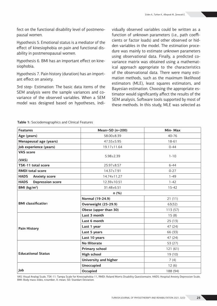
TURKISH JOURNAL OF PHYSIOTHERAPY AND REHABILITATION 2021; 32(3) 25
Erden A., Turhan K., Albayrak M., Şenocak E.
fect on the functional disability level of postmeno-pausal women.
Hypothesis 5. Emotional status is a mediator of the effect of kinesiophobia on pain and functional dis-ability in postmenopausal women.
Hypothesis 6. BMI has an important effect on kine-siophobia.
Hypothesis 7. Pain history (duration) has an import-ant effect on anxiety.
3rd step- Estimation: The basic data items of the SEM analysis were the sample variances and co-variance of the observed variables. When a SEM model was designed based on hypotheses, indi-
vidually observed variables could be written as a function of unknown parameters (i.e., path coeffi-cients or factor loads) and other observed or hid-den variables in the model. The estimation proce-dure was mainly to estimate unknown parameters using observational data. Finally, a predicted co-variance matrix was obtained using a mathemat-ical approach appropriate to the characteristics of the observational data. There were many esti-mation methods, such as the maximum likelihood estimators (MLE), least squares estimators, and Bayesian estimation. Choosing the appropriate es-timator would significantly affect the results of the SEM analysis. Software tools supported by most of these methods. In this study, MLE was selected as
Table 1: Sociodemographics and Clinical Features
Features Mean+SD (n=200) Min- Max
Age (years) 58.00±8.39 40-76
Menapeusal age (years) 47.55±5.95 18-61
Job experience (years) 19.17±11.64 0-44VAS score
(VAS)5.98±2.39 1-10
TSK-11 total score 25.97±8.57 6-44
RMDI total score 14.37±7.91 0-27
HADS Anxiety score 14.74±11.27 1-49
HADS Depression score 12.39±10.51 1-42
BMI (kg/m2) 31.48±6.51 15-42
BMI classification
n (%)
Normal (19-24.9) 21 (11)
Overweight (25-29.9) 63(32)
Obese (upper than 30) 113 (57)
Pain History
Last 3 month 15 (8)
Last 6 month 25 (13)
Last 1 year 47 (24)
Last 5 years 66 (33)
Last 10 years 47 (24)
Educational Status
No Illiterate 53 (27)
Primary school 121 (61)
High school 19 (10)
University and higher 7 (4)
JobUnoccupied 12 (6)
Occupied 188 (94)
VAS: Visual Analog Scale, TSK-11: Tampa Scale for Kinesiophobia-11, RMDI: Roland Morris Disability Questionnaire. HADS: Hospital Anxiety Depression Scale, BMI: Body mass index, n:number, X: mean, SD: Standart Deviation.

TURKISH JOURNAL OF PHYSIOTHERAPY AND REHABILITATION 2021; 32(3)26
Structural Equation Modelling to Examine the Affected Factors of Kinesiophobia in Postmenopausal Women with Chronic Low Back Pain
the most convenient method. NFI, RFI, IFI, TLI, CFI indexes (>0.9) and thresholds (< 0.08) were used as criteria to include and to retain variables for ideal model. For this reason, the statistical significance p value was taken into consideration (p<0.05). The b coefficients are calculated showing the level of relation between variables. These coefficients, also called standardized regression coefficients or beta weights, were the estimates resulting from a regression analysis for standardization. So the variances of dependent and independent variables were 1. In addition, the relationship between all these latent and observed variables were decided
by evaluating the expert knowledge. Because, sta-tistical knowledge of critical values was also im-portant as much as SEM analysis. According to the graphic model of SEM, latent variables were shown with ellipses, observed variables were shown as rectangles, and error terms (e) were shown as cir-cles.
4th step-Evaluation of model fit and re-specifica-tion: SEM tests the statistical validity of the mod-el fit indices based on the parameter estimation derived from the sample data. Here the normed-fit index (NFI), relative fit index (RFI), incremental
Table 2: Analysis of Estimated Parameters’ Significance
Hypothesis Relations Est. S.E. C.R. P
Modified model (Model 2)
Hypothesis 1 HADS Anxiety score ← TSK-11 .02 .00 4.30 <0.001**
Hypothesis 2 Pain intensity ← HADS anxiety score .64 .33 1.92 0.031*
HADS item1 ← HADS anxiety score 1.00
HADS item 3 ← HADS anxiety score 1.31 .19 6.91 <0.001**
HADS item 5 ← HADS anxiety score 1.22 .18 6.65 <0.001**
HADS item 7 ← HADS anxiety score .52 .13 3.84 <0.001**
HADS item 9 ← HADS anxiety score .92 .14 6.37 <0.001**
HADS item 11 ← HADS anxiety score 1.00 .17 5.75 <0.001**
HADS item 13 ← HADS anxiety score 1.00 .16 6.03 <0.001**
Hypothesis 3 RMDI score ← HADS anxiety score 2.98 1.03 2.88 0.004*
Hypothesis 4 RMDI ← VAS 1.42 .20 6.89 <0.001**
Hypothesis 5 RMDI ← TSK-11 -.007 .06 -.12 0.902
VAS TSK-11 .003 .019 .147 0.883
Hypothesis 6 TSK-11 ← BMI .08 .09 .88 0.375
Hypothesis 7 HADS Anxiety score ← Duration of Pain .12 .03 3.05 .002*
Last model (Model 3)
Hypothesis 1 HADS anxiety ← TSK-11 .02 .006 4.28 <0.001**
Hypothesis 2 VAS ← HADS anxiety score .87 .31 2.77 0.005*
HADS item 1 ← HADS anxiety score 1.00
HADS item 2 ← HADS anxiety score 1.32 .19 6.87 <0.001**
HADS item 3 ← HADS anxiety score 1.22 .18 6.68 <0.001**
HADS item 4 ← HADS anxiety score .50 .13 3.77 <0.001**
HADS item 5 ← HADS anxiety score .91 .14 6.35 <0.001**
HADS item 6 ← HADS anxiety score 1.01 .17 5.81 <0.001**
HADS item 7 ← HADS anxiety score 1.00 .16 6.04 <0.001**
Hypothesis 3 RMDI ← HADS anxiety score 2.87 .95 3.02 0.003*
Hypothesis 4 RMDI ← VAS 1.42 .20 6.90 <0.001**
*: p<0.05, **: p<0.001, Est: Estimated regression weights, S.E: Standard Error, C.R: Standardized estimated regression weights,, HADS: Hospital Anxiety and Depression Scale, RMDI: Roland Morris Disability Questionnaire, VAS: Visual Analog Scale, TSK-11: Tampa Scale for Kinesiophobia-11.

TURKISH JOURNAL OF PHYSIOTHERAPY AND REHABILITATION 2021; 32(3) 27
Erden A., Turhan K., Albayrak M., Şenocak E.
fit index (IFI), Tucker-Lewis coefficient (TLI) versus comparative fit index (CFI), goodness-of-fit index (GFI), adjusted GFI, and root mean square error of approximation (RMSEA) were used to evaluate model fitness. Scores of >0.9 were considered as good and >0.95 were considered as excellent for the NFI, RFI, IFI, TLI, and CFI of the compliance in-dices; for RMSEA, values of <0.08 were considered as good and those <0.03 were considered as a per-fect fit (12).
RESULTS
The average age of the women was 58.3±8.53 years. The average menopausal age was 47.55±5.95. The average TSK-11 was above average as 25.97±8.57. Anxiety and depression risks were also above the cut-off score according to HADS. The average anxi-ety risk score was 14.74±11.27 and the average of depression risk score was 12.39±10.51. The socio-demographic and clinical data were shown in Table 1.
Regression analysis to determinate the vari-ables for SEM
Firstly, the variables that were thought to be af-fected by kinesiophobia according to the data ob-tained from the literature, were examined by re-gression analysis. Here, it was aimed to obtain a regression model that best explained the variation in the kinesiophobia variable and then to explain the interactions of the variables in this model with the contribution of latent variables using SEM analysis. As a result of the regression analysis, the variables included in the significant model (p = 0.001, R^2=0.863) were selected to be used in
the SEM analysis phase. The variables were finally determined as: pain intensity, pain duration, BMI, disability, anxiety and depression.
1st model creation results
Functional disability, pain intensity, pain duration, and BMI were considered as observed variables, while kinesiophobia, anxiety, and depression were considered as latent variables (Figure 1A). The first model was created as a result of the analysis. It was not identified in the program due to an insuf-ficient number of observations (preferably at least 10 per variable) according to the number of vari-ables. Since the first model was not identified, so it was modified and a second model was created by modifying the first model.
2nd model creation results
The second model was identified when the de-pression variable was removed, the TSK 11 total score was used instead of 11 items, and the model was re-tested. Anxiety remained a latent variable (Figure 1B). The coefficients on the one-direction-al links between variables were the standardized regression coefficients. Bidirectional arrows be-tween error terms represented covariance and were added to improve model 2 using modification indices. The chi-square statistic for the model was obtained as chi-square=37.58 (df= 43; p=0.700). These values showed that the tested model was significant. According to the analysis results of this model, kinesiophobia had a strong effect on anxiety (b=0.36, p<0.001). Pain had a strong positive ef-fect on functional disability (b=0.43, p<0.001). Anx-iety had a weakly positive effect on pain (b=0.16,
Table 3: Model Fitting Analysis for Primary, Modified and Last Measurement Models Obtained at 60º/s and 180º/s Speeds of Dominant and Non-Dominant Shoulder.
Fit index Modified model
Critical value Last model
Normed-fit index (NFI) 0.92 > 0.9 0.95
Relative fit index (RFI) 0.89 > 0.9 0.92
Incremental fit index (IFI) 1.01 > 0.9 1.01
Tucker-Lewis coefficient (TLI) 1.01 > 0.9 1.02
Comparative fit index (CFI) 1.00 > 0.9 1.00
Root mean square error of approximation (RMSEA) 0.00 > 0.8 0.00
Scores of >0.9 are considered good and >0.95 are considered excellent for the NFI. RFI. IFI. TLI. and CFI of the compliance indices. For RMSEA values of <0.08 were considered good and those <0.03 were considered a perfect fit.

TURKISH JOURNAL OF PHYSIOTHERAPY AND REHABILITATION 2021; 32(3)28
Structural Equation Modelling to Examine the Affected Factors of Kinesiophobia in Postmenopausal Women with Chronic Low Back Pain
p=0.031) and a positive effect on functional dis-ability (b=0.22, p=0.004). While the correlation be-tween kinesiophobia and anxiety (b=0.36, p<0.001) and the correlation between anxiety and functional disability (b=0.23, p=0.004) were significant. The correlation between kinesiophobia and functional disability (b=-0.01, p=0.902) was not significant. Likewise, while the direct correlation between kine-siophobia and anxiety and the correlation between anxiety and pain intensity were significant, the di-rect link between kinesiophobia and pain (b=0.01, p=0.883) was not significant. These results con-firmed the Hypothesis 5. The correlation between BMI and kinesiophobia was not significant (b=0.06, p=0.375) (Figure 1B) (Table 2). Thus, since Hypoth-esis 6 was rejected, the BMI variable was removed
from the model. Goodness-of-fit statistics were: NFI: 0.92, RFI: 0.89, IFI: 1.01, TLI:1.01, CFI:1.00, RMSEA:0.00 (Table 3).
3rd model creation results
Model 2 showed moderate fitness in accordance with RFI; good fitness in accordance with NFI, IFI, TLI, and CFI; and perfect fitness in accordance with RMSEA. To obtain a better fitness, BMI and pain duration variables, which seemed insignificant in model 2, were removed from the model. Thus model 3 was obtained (Figure 1C). The chi-square statistic for the model was found as 1,37 (df =28, p=0.810). These values showed that the analyzed model was significant, and the P values were higher than those in model 2. According to the results of
et1= error term for TSK-11 item 1; et2= error term for TSK-11 item 2; et3= error term for TSK-11 item 3; et4= error term for TSK-11 item 4; et5= error term for TSK-11 item 5; et6= error term for TSK-11 item 6; et7= error term for TSK-11 item 7; et8= error term for TSK-11 item 8; et9= error term for TSK-11 item 9; et10= error term for TSK-11 item 10; et11= error term for TSK-11 item 11; eh1= error term for HADS item 1; eh3= error term for HADS item 3; eh5= error term for HADS item 5; eh7= error term for HADS item 7; eh9= error term for HADS item 9; eh11= error term for HADS item 11; eh13= error term for HADS item 13; eh2= error term for HADS item 2; eh4= error term for HADS item 4; eh6= error term for HADS item 6; eh8= error term for HADS item 8; eh10= error term for HADS item 10; eh12= error term for HADS item 12; eh14= error term for HADS item 14; ek= error term for latent variable TSK-11 total score; eb= error term for BMI; ed= error term for latent variable HADS Depression score; ef= error term for Functional Disability; ev= error term for VAS score; ea= error term for latent variable HADS Anxiety score; eag= error term for Duration of Pain.
Figure 1A: Primary structural model (Model 1)
Figure 1C: Last structural model (Model 3)
Figure 1B: Modified structural model (Model 2)
Figure 1D: Final structural model framework
HADS item 1
HADS item 3
HADS item 5
HADS item 7
HADS item 9
HADS item 11
HADS item 13
HADS item 2
HADS item 4
HADS item 6
HADS item 8
HADS item 10
HADS item 12
HADS item 14
HADS Anxiety
HADS Depression
TSK-11 item 1
TSK-11 item 2
TSK-11 item 3
TSK-11 item 4
TSK-11 item 5
TSK-11 item 6
TSK-11 item 7
TSK-11 item 8
TSK-11 item 9
TSK-11 item 10
TSK-11 item 11
Kinesiophobia(TSK-11) BMI
eh1
eh3
eh5
eh7
eh9
eh11
eh13
eh2
eh4
eh6
eh8
eh10
eh12
eh14
Duration of Pain
et1 et2 et3 et4 et5 et6 et7 et8 et9 et10 et11
ebeag
Pain(VAS)
FunctionalDisability
(RMDI)ev ef
ek
ea
ed
HADS item 1
HADS item 3
HADS item 5
HADS item 7
HADS item 9
HADS item 11
HADS item 13
HADS Anxiety
eh1
eh3
eh5
eh7
eh9
eh11
eh13
Pain(VAS)
FunctionalDisability
(RMDI)
ev
ef
ek
ea
Kinesiophobia(TSK-11)
.32
-,17
-,20
,17
,17
,21
HADS item 1
HADS item 3
HADS item 5
HADS item 7
HADS item 9
HADS item 11
HADS item 13
HADS Anxiety
BMI
eh1
eh3
eh5
eh7
eh9
eh11
eh13
Duration of Pain
eb
eag
Pain(VAS)
Functional Disability
(RMDI)
ev
ef
ek
ea
.06Kinesiophobia(TSK-11)
.26
.33
-,15
-,19
,16
,16
,22
,23
.17

TURKISH JOURNAL OF PHYSIOTHERAPY AND REHABILITATION 2021; 32(3) 29
Erden A., Turhan K., Albayrak M., Şenocak E.
the analysis, there was a strong effect of kinesio-phobia on anxiety (b=0.37, p<0.001) (Table 2). Pain intensity had a strongly positive effect on function-al disability (b=0.43, p<0.001). Anxiety had a weak-ly positive effect on pain (b=0.23, p=0.005). Anxiety also had a positive effect on functional disability (b=0.22, p=0.003). All regression coefficients relat-ed to the model were significant. Goodness-of-fit-ness of the model was given in Table 3 and Figure 1C. The model had good fitness according to RFI and perfect fitness in accordance with NFI, IFI, TLI, CFI, and RMSEA. Thus, a perfect fit index was ob-tained in accordance with all criteria except RFI. This final structural model created as the frame-work (Figure 1D).
DISCUSSION
In the present study, a new SEM model was cre-ated which could explain kinesiophobia with pain intensity, anxiety, and functional disability in post-menopausal women with CLBP. According to our results, pain and functional disability affected the kinesiophobia directly. Anxiety was a strong medi-ator of this relationship. Another finding was BMI and pain duration were not related to the all pa-rameters of the model. Also depression risk factor decreased the fit coefficient of the model. For this reason, these variables were found unrelated with kinesiophobia, so they removed from the model.
Most women experienced much more CLBP during postmenopausal term than before. The reason was emotional symptoms and pain intensity increased in this period. This coincided with the fact that the women in our sample were at risk of developing anxiety and depression. So they started to avoid the movement. Also, Trocoli et al reported that there was a strong correlation between anxiety and kinesiophobia scores in patients with CLBP (13). In our first hypothesis (H1), the relationship between kinesiophobia and anxiety was confirmed in the model. This finding was consistent with pre-vious studies showing an association between ki-nesiophobia and anxiety (3). Erden et al. and Bilgin et al reported positive correlation between anxiety and kinesiophobia in people with CLBP in their studies. (8, 14). Contrarily Branström et al found no relationship between kinesiophobia and anxi-ety in patients with chronic musculoskeletal pain
(15). However, their results were not specific to the postmenopausal women. In our study, we pre-sented a new evidence about relationship between anxiety and kinesiophobia for postmenopausal pe-riod–specifically in our SEM model. Results should be supported by the further studies.
Evidence about effects of anxiety and depression is common in the general population with CLBP but rare in the postmenopausal period. In our second hypothesis (H2), while the effect of anxiety on pain intensity was significant, depression was not fit with the model. So depression was removed from the model. This finding was compatible with the literature (16, 17, 18-20). Kanwaljit et al reported that the pain was affected by emotional status on non-specific CLBP in postmenopausal women (21). In our model, positive correlation between pain intensity and anxiety revealed in postmenopausal women. The negative effects of pain intensity and depressive symptoms on functional status were previously indicated in the literature (22). However, it was understood that anxiety and depression did not affect patients to the same extent. Some stud-ies reported that depression affected pain intensity (17, 22). There were some studies which reject-ed this relationship (23). There was not a current consensus about this field. Kuch et al found no dif-ference in depression risk levels between patients with low back pain (24). Results should be support-ed by the further studies.
In related literature it was known that patients with CLBP had worse physical function and experience more problems in daily life compared to individu-als without CLBP (22). In our third hypothesis (H3), positive correlation was found between anxiety and functional disability. Asama et al emphasized that the catastrophobia and anxiety were relative risk factors for disability in patients with CLBP (25, 26). Dündar et al found high pain and disability levels as well as poor functional and emotional statuses in patients with CLBP versus those without CLBP. They also emphasized that emotional status eval-uation was crucial in patients with CLBP (27). But their sample was not postmenopausal women. As a contribution, the effect of anxiety on functional disability was demonstrated in our model. So the model fit index proved that this effect could not be independent of pain in our model.
TURKISH JOURNAL OF PHYSIOTHERAPY AND REHABILITATION 2021; 32(3)30
Structural Equation Modelling to Examine the Affected Factors of Kinesiophobia in Postmenopausal Women with Chronic Low Back Pain
Studies about CLBP over the past decade have shown that pain could lead to avoidance, kinesio-phobia, decreased physical function capacity, and increased disability (27). In related literature, pain during the activity decreased physical functions (28). Our fourth hypothesis (H4) was confirmed the association between pain intensity with function-al disability. The model also explained that anx-iety was an important factor in this relationship. Kuch et al found that anxiety was associated with functional disability in patients with musculoskel-etal pain (24). Güçlü et al found a weakly positive correlation between pain intensity and disability and kinesiophobia in patients with CLBP (29). Con-versely, Baillie et al emphasized that pain intensi-ty is a strong predictor for disability in CLBP (30). In our study, we also found a positive correlation between kinesiophobia and functional disability in postmenopausal women.
In some studies, anxiety was defined as a mediator between physical functions and social parameters. Korkmaz et al. emphasized that emotional factors were important on the functional disability and kinesiophobia (31). Cederbom et al. showed that there was a relationship between chronic pain and disability. They emphasized that both pain-related disability and emotional state were the mediators of disease in CLBP (32). Helminen et al. found that the change in pain and functional status were as a result of anxiety (33). Anxiety was a strong me-diator on relationship between kinesiophobia, pain intensity and functional disability in our fifth hy-pothesis (H5). Our model revealed the relationship between anxiety, kinesiophobia, disability and pain intensity clearly.
In conclusion, the results of our study indicated that kinesiophobia affected pain intensity, functional disability and anxiety. The key point of the results was the anxiety was as strong mediator of this re-lationship. Kinesiophobia which affected the social participation of postmenopausal women were ex-amined in depth by this model with health-related variables. By this modelling, the underlying reasons could be explained by phenomenon-based hypo-thetical approach rather than roughly examining the relationships. Since it was the first study which was used the structural equation model in post-menopausal women with CLBP. So our study had
an important contribution in terms of encouraging it’s widely used in physiotherapy, women’s health and other fields of health.
There are some limitations. Sociodemographic features and quality of life was also important to explain the kinesiophobia in postmenopausal term. Because poor economic conditions could be caused poor quality of life too. So lower quality of life could affect pain and kinesiophobia in this period. Fur-ther studies are needed to evaluate these parame-ters. Second limitation was the lack of data about presence of another musculoskeletal problems such as osteoporosis. Because osteoporosis is very common in postmenopausal term and triggers the CLBP. More comprehensive and detailed outcome measures of women health could warrant more fruitful results in discerning the role of osteoporo-sis on level kinesiophobia during postmenopausal term. Potential confounders, such as use of alcohol, tobacco, hormonal drugs, and specific disabilities should also be considered in future research. Last limitation was the lack of data about participants’ anxiety experiences which they had before post-menopausal term. It could have been confounders in the evaluation of anxiety in any period of life.
Sources of Support: This study has not received any financial support from any institution.
Conflict of Interest: There is no conflict of interest between the authors.
Ethical Approval: This study was approved by the Trabzon Kanuni Educational and Research Hospi-tal, Clinical Research Ethical Committee (Approval Date: 25th October 2017 and Approval Number: 2017/47).
Informed Consent: The participants read and signed the informed consent document by accept-ing the principle of compliance with the Helsinki Declaration Principles.
Peer-Review: Externally peer-reviewed.
Author Contributions: Concept – AE, EŞ, KT; De-sign – AE, KT, MA; Supervision – AE, KT; Resources and Financial Support - AE, EŞ, KT, MA; Materials – AE, KT; Data Collection and/or Processing – EŞ, MA; Analysis and/or Interpretation – AE, MA; Literature Research – EŞ, AE; Writing Manuscript - AE, EŞ, KT, MA; Critical Review - KT.
TURKISH JOURNAL OF PHYSIOTHERAPY AND REHABILITATION 2021; 32(3) 31
Erden A., Turhan K., Albayrak M., Şenocak E.
Acknowledgments: We thank to Taylor & Fran-cis Editing Services Supporting. The edit was performed by professional editors at Editage, a division of Cactus Communications, in coopera-tion with Taylor & Francis Group (order number: RZERD_1).
REFERENCES1. E Silva AVS, Lacativa PGS, Russo LAT, Gregorio LH, Pinheiro RAC
et al. Association of back pain with hypovitaminosis D in post-menopausal women with low bone mass. BMC Musculoskelet Disord. 2013;14:184. https://doi.org/10.1186/1471-2474-14-184.
2. Uluğ N, Yakut Y, Alemdaroğlu İ, Yılmaz Ö. Comparison of pain, kinesiophobia and quality of life in patients with low back and neck pain. J Phys Ther Sci. 2016;28:665-670. https://doi.org/10.1589/jpts.28.665.
3. Hapidou EG, O’Brien MA, Pierrynowski MR, Las Heras E, Patla T. Fear and avoidance of movement in people with chronic pain: psychometric properties of the 11-Item Tampa Scale for Kine-siophobia (TSK-11). Physiother Can. 2012;64:235-241. https://doi.org/10.3138/ptc.2011-10.
4. Flores-Ramos M, Silvestri Tomassoni R, Guerrero-López JB, Sa-linas M. Evaluation of trait and state anxiety levels in a group of peri-and postmenopausal women. Women Health. 2018;58:305-319. https://doi.org/10.1080/03630242.2017.1296059.
5. Wáng YXJ, Wáng JQ, Káplár Z. Increased low back pain preva-lence in females than in males after menopause age: evidences based on synthetic literature review. Quant Imaging Med Surg. 2016;6:199-206. https://doi.org/10.21037/qims.2016.04.06.
6. Hoyle RH, Editor. Handbook of structural equation modeling. New york: A Division of Guilford Publications. c2012.
7. Wolf EJ, Harrington KM, Clark, SL, Miller MW. Sample Size Require-ments for Structural Equation Models: An Evaluation of Power, Bias, and Solution Propriety. Educ Psychol Meas. 2013;76:913–934. https://doi.org/10.1177/0013164413495237.
8. Erden A, Altuğ F, Malkoç A, Akgül Kocabal A. Investigation Of The Relationship Between Kinesiophobia, Pain, Anxiety-Depression Status And Quality Of Life In Patients With Knee Osteoarthritis Before Surgery. OTSHD. 2016;7:1-17. https://doi.org/10.17372/OTSHD.2016.7.16650.
9. Yilmaz ÖT, Yakut Y, Uygur F, Uluğ N. Turkish version of the Tam-pa Scale for Kinesiophobia and its test-retest reliability. Turk J Physiother Rehabil. 2011;22:44-49.
10. Aydemir Ö, Guvenir T, Kuey L, Kultur S. Validity and reliability of Turkish version of hospital anxiety and depression scale. Turk Physciatry Derg. 1997;8:280-287.
11. Küçükdeveci AA, Tennant A, Elhan AH, Niyazoğlı H. Validation of the Turkish version of the Roland-Morris Disability Question-naire for use in low back pain. Spine. 2001;26:2738-43. https://doi.org/10.1097/00007632-200112150-00024.
12. Hooper D, Coughlan J, Mullen M. Structural equation modelling: Guidelines for determining model fit. EJBRM. 2008;6:53-60.
13. Trocoli TO, Botelho RV. Prevalence of anxiety, depression and ki-nesiophobia in patients with low back pain and their association with the symptoms of low back spinal pain. Rev Bras Reumatol Engl Ed. 2016;56,330–336. doi:10.1016/j.rbre.2016.02.010.
14. Bilgin S, Cetin H, Karakaya J, Kose N. Multivariate Analy-sis of Risk Factors Predisposing to Kinesiophobia in Per-sons With Chronic Low Back and Neck Pain. J Manipulative Physiol Ther. 2019;42(8):565-571. https://doi.org/10.1016/j.jmpt.2019.02.009.
15. Branström H, Fahlström M. Kinesiophobia in patients with
chronic musculoskeletal pain: differences between men and women. J Rehabil Med. 2008;40:375-380. https://doi.org/10.2340/16501977-0186.
16. Durmaz T. Investigation of the factors effect on low back pain in postmenopausal women. 15th World Congress on Meno-pause. 28 September-01 October, 2016. Prague. Climacteric; 2016;19(sup1):p48.
17. Bair MJ, Wu J, Damush TM, Sutherland JM, Kroenke K. Association of depression and anxiety alone and in combination with chronic musculoskeletal pain in primary care patients. Psychosom Med. 2008;70:890. https://doi.org/10.1097/PSY.0b013e318185c510.
18. Meints SM, Mawla I, Napadow V, Kong J, Gerber J, Chan ST, et al. The relationship between catastrophizing and altered pain sen-sitivity in patients with chronic low-back pain. Pain. 2019;160(4), 833–843. doi:10.1097/j.pain.0000000000001461.
19. Subhadra KT. Low back pain: Correlation with anxiety, depres-sion, and well-being and the effect of homeopathic medicines on the physical signs of patients. Int J Med Sci. 2017;6(9), 1427-1432.
20. Comachio J, Magalhães MO, Campos Carvalho E Silva, APDM, Marques AP. A cross-sectional study of associations between kinesiophobia, pain, disability, and quality of life in patients with chronic low back pain. Adv Rheumatol. 2018;58. doi:10.1186/s42358-018-0011-2.
21. Kanwaljit Chopra, Vipin Arora. An intricate relationship between pain and depression: clinical correlates, coactivation factors and therapeutic targets. Expert Opin Ther Targets. 2014;18:2, 159-176, DOI: 10.1517/14728222.2014.855720.
22. Yazıcı K, Tot Ş, Biçer A, Yazıcı A, Buturak V. Anxiety, Depression and Quality of Life in Patients with Lowback Pain and Neck Pair. J Clin Psychol. 2003;6:95-101.
23. Casten RJ, Parmelee PA, Kleban MH, Lawton MP, Katz IR. The relationships among anxiety, depression, and pain in a geriatric institutionalized sample. Pain. 1995;61:271-276. https://doi.org/10.1016/0304-3959(94)00185-h.
24. Kuch K, Cox B, Evans RJ, Watson PC, Bubela C. To what extent do anxiety and depression interact with chron-ic pain? Can J Psychiatry. 1993;38:36-38. https://doi.org/10.1177/070674379303800110.
25. Ranger TA, Cicuttini FM, Jensen TS, Manniche C, Heritier S, Ur-quhart DM. Catastrophization, fear of movement, anxiety, and depression are associated with persistent, severe low back pain, and disability. The Spine Journal. 2020;6:857-865. https://doi.org/10.1016/j.spinee.2020.02.002.
26. Marshall PWM, Schabrun S, Knox MF. Physical activity and the mediating effect of fear, depression, anxiety, and catastrophizing on pain related disability in people with chronic low back pain. PLOS ONE. 2017; 12(7). doi:10.1371/journal.pone.0180788.
27. Dündar Ü, Özlem S, Demirdal ÜS, Toktas H, Kavuncu V. Relation of pain, disability and depression with quality of life in patients with chronic low back pain. Genel Tip Derg. 2009;19:99-104.
28. Erden A, Şenocak E. An analysis on factors affecting back awareness of postmenopausal women with chronic low back pain. JETR. 2020;7(1), 21-27.
29. Güçlü DG, Güçlü O, Ozaner A, Şenormancı O, Konkan R. The relationship between disability, quality of life and fear-avoid-ance beliefs in patients with chronic low back pain. Turk Neu-rosurg. 2012;22:724-731. https://doi.org/10.5137/1019-5149.JTN.6156-12.1.
30. Baillie L, Bacon CJ, Hewitt CM, Moran RW. Predictors of func-tional improvement in people with chronic low back pain follow-ing a graded Pilates-based exercise programme. J Bodyw Mov Ther. 2019;23,211–218. doi:10.1016/j.jbmt.2018.06.007.
31. Korkmaz N, Akinci A, Yörükan S, Sürücü HS, Saraçbaşi O, Ozça-kar L. Validation and Reliability of the Turkish Version of the Fear Avoidance Beliefs Questionnaire in Patients With Low Back Pain. Eur J Phys Rehabil M. 2009;45:527-35.

TURKISH JOURNAL OF PHYSIOTHERAPY AND REHABILITATION 2021; 32(3)32
Structural Equation Modelling to Examine the Affected Factors of Kinesiophobia in Postmenopausal Women with Chronic Low Back Pain
32. Cederbom S, Söderlund A, Denison A, Wagert PH. Chronic pain among older women living alone. A study focusing on disability and morale. Eur J Physiother. 2014;16:139-150. https://doi.org/10.3109/21679169.2014.893448.
33. Helminen EE, Sinikallio SH, Valjakka AL, Väisänen-Rouvali RH, Arokoski JPA. Determinants of pain and functioning in knee osteo-arthritis: a one-year prospective study. Clin Rehabil. 2016;30:890-900. https://doi.org/10.1177/0269215515619660.

33
PERCEIVED EXERCISE BENEFITS AND BARRIERS IN ACTIVE AND INACTIVE UNIVERSITY STUDENTS
ORIGINAL ARTICLE
ABSTRACTPurpose: The aims of this study were to compare the perceived benefits and barriers to exercise in active and inactive university students and to determine the relationship between physical activity level and perceived benefits/barriers in university students.
Methods: The undergraduate students were invited to this cross-sectional survey. A total of 526 students responded to the online survey consisting of the International Physical Activity Questionnaire (IPAQ) and Exercise Benefits/Barriers Scale (EBBS). The participants were divided into two groups: the active group (n=341) and the inactive group (n=185) based on IPAQ.
Results: The most agreed benefit was the item "exercise improves the way my body looks," whereas the most agreed barrier was the item "exercise tires me". Comparison of the active and inactive groups showed that the active group perceived the benefits of exercise higher than the inactive group, especially in terms of life enhancement, physical performance, and psychological outlook (p<0.05). The inactive group perceived more barriers to exercise than the active group, especially in terms of exercise milieu and physical exertion (p<0.05). Moreover, the total physical activity level was positively associated with exercise benefits, especially psychological outlook, while negatively related to barriers, especially exercise milieu and physical exertion (p<0.001).
Conclusion: The physical activity participation of university students can be encouraged by increasing their knowledge and perception of the benefits of exercises and by decreasing the barriers that they felt. Therefore, this study's results may contribute to planning interventions and strategies aiming to promote physical activity participation among university students.
Key Words: Barrier, Benefit, Exercise, Physical Activity, Student.
AKTİF VE AKTİF OLMAYAN ÜNİVERSİTE ÖĞRENCİLERİNDE ALGILANAN EGZERSİZ YARARLARI
VE ENGELLERİ
ARAŞTIRMA MAKALESİ
ÖZAmaç: Bu çalışmanın amaçları, aktif ve aktif olmayan üniversite öğrencilerinde algılanan egzersiz yararları ve engellerini karşılaştırmak ve üniversite öğrencilerinde fiziksel aktivite düzeyi ile algılanan egzersiz yararları ve engelleri arasındaki ilişkiyi belirlemekti.
Yöntem: Bu kesitsel araştırmaya lisans öğrencileri davet edildi. Uluslararası Fiziksel Aktivite Anketi'ni (UFAA) ve Egzersiz Yararları/Engelleri Ölçeği'ni (EYEÖ) içeren çevrimiçi anketi toplam 526 öğrenci cevapladı. Katılımcılar UFAA'ya göre aktif (n=341) ve aktif olmayan grup (n=185) olmak üzere iki gruba ayrılarak değerlendirildi.
Sonuçlar: Üniversite öğrencileri tarafından en çok algılanan yarar "egzersiz vücut görünümümü geliştirme yoludur", en çok karşılaşılan engel ise "egzersizden yoruluyorum" idi. Aktif ve aktif olmayan grupların karşılaştırılması, aktif grubun özellikle yaşam iyileştirme, fiziksel performans ve psikolojik bakışa dair egzersizin faydalarını aktif olmayan gruptan daha yüksek algıladığını (p<0,05), aktif olmayan grubun ise özellikle egzersiz ortamı ve fiziksel efora dair engelleri aktif gruptan daha fazla algıladığını göstermiştir (p<0,05). Ayrıca, toplam fiziksel aktivite düzeyi özellikle psikolojik bakış olmak üzere egzersiz yararları ile pozitif ilişkili iken, egzersiz ortamı ve fiziksel efora dair engeller ile negatif ilişkiliydi (p<0,001).
Tartışma: Üniversite öğrencilerinin fiziksel aktivite katılımları, egzersizlerin faydalarına ilişkin bilgi ve algıları artırılarak ve hissettikleri engeller azaltılarak teşvik edilebilir. Bu nedenle, bu çalışmanın sonuçları, üniversite öğrencileri arasında fiziksel aktivite katılımını teşvik etmeyi amaçlayan müdahalelerin ve stratejilerin planlanmasına katkıda bulunabilir.
Anahtar Kelimeler: Bariyer, Yarar, Egzersiz, Fiziksel Aktivite, Öğrenci.
Turkish Journal of Physiotherapy and
Rehabilitation2021 32(3)33-42
Çağla ÖZKUL, PhD, PT
ISSN: 2651-4451 • e-ISSN: 2651-446X
Özkul Ç., Perceived Exercise Benefits and Barriers in Active and Inactive University Students, Turk J Physiother Rehabil. 2021; 32(3):33-42. doi: 10.21653/tjpr.794911
Gazi University, Faculty of Health Sciences, Department of Physiotherapy and Rehabilitation, Ankara, Turkey.
Correspondence (İletişim):
Çağla ÖZKUL, PhD, PTGazi University,
Faculty of Health Sciences, Department of Physiotherapy and Rehabilitation Emek Mahallesi, Bişkek Caddesi, 6. Sokak, No:2
Çankaya/Ankara, 06490 TURKEYPhone: 0312 216 26 78
E-mail: [email protected] ORCID: 0000-0001-9367-9910
Received: 14.09.2020 (Geliş Tarihi)Accepted: 21.02.2021 (Kabul Tarihi)
CC BY - NC
Content of this journal is licensed under a Creative Commons Attribution-NonCommercial 4.0 International License.

TURKISH JOURNAL OF PHYSIOTHERAPY AND REHABILITATION 2021; 32(3)34
Perceived Exercise Benefits and Barriers in Active and Inactive University Students
INTRODUCTION
Physical activity prevents many chronic diseas-es and improves both physical and psychological health (1). However, it is estimated that 31% of adults worldwide are physically inactive (2). The World Health Organization (WHO) reported that in-activity has become widespread, and about 3.2 mil-lion deaths per year are associated with physical inactivity. For this reason, physical inactivity has also been defined as a global public health problem (3). Therefore, increasing physical activity is among the priorities for public health worldwide (4).
Physical activity habit is generally developed during childhood and young adulthood. It has been report-ed that there is a significant decrease in the phys-ical activity level of students in the transition from high school to university (5). The university period is precious for the development of physical activity habits, as it is a period when individuals start to make their own decisions and develop lifelong hab-its according to their preferences (6).
The studies conducted in many different countries have shown that university students’ physical ac-tivity levels are generally low (6-11). Many studies among university students have shown that there are many different barriers to exercise, primarily the lack of time, lack of motivation, and tiredness (7-16). However, it is not yet clear which motivat-ing factors lack inactive students and encourage exercise in physically active students.
The studies generally focused on barriers to exercise from inactive students’ perspective (7-9,12,13,16). As well as barriers to exercise, lack of motivating factors may also be responsible for inactivity, inso-much that motivating factors encouraging active students can be useful tools to overcome barriers (17). One of the primary motivation sources for ex-ercise is to know the benefits of exercise and to perceive these benefits at a high level. Therefore, investigating both perceived benefits and barriers to exercise in active and inactive university stu-dents will better understand the physical inactivity in university students (18). In this way, it may be possible to develop beneficial interventions to pro-mote physical activity in university students.
This study’s primary aim is to compare the per-
ceived exercise benefits/barriers in active and in-active university students. The secondary aim is to determine the relationship between physical activi-ty level and perceived benefits/barriers in university students.
METHODS
Participants
The study protocol was approved by the Gazi Uni-versity Ethics Commission (No: 2020-371, date: July 14, 2020). The undergraduate students at Gazi University were invited to this cross-sectional sur-vey. The surveys were prepared using Google forms, and the relevant link was sent to 625 students. The students read the informed consent form on the first page, and 563 volunteer students agreed to participate in the survey. Thirty-seven students with chronic disease were excluded. The data were collected between 17 and 21 July 2020. This date range was in the normalization process when the Coronavirus disease (COVID-19) pandemic was ongoing, but restrictions were minimal, and there were no restrictions that prevent physical activity, such as curfews and closure of the gym in Turkey.
The sample size was calculated based on the differ-ence between barrier scores in active and inactive students declared by the study of Blake et al. (8). The total sample size was estimated at a minimum of 458 using the power analysis software (G*Power 3.1.9.2) to achieve 99% power with a two-sided level of 5% (19).
Measurements
The survey consisted of three parts: demographic information, International Physical Activity Ques-tionnaire (IPAQ), and the Exercise Benefits/Barriers Scale (EBBS). The physical activity level was as-sessed using the Short Form of IPAQ (20, 21). The IPAQ measures vigorous-intensity activity, moder-ate-intensity activity, and walking activity levels by calculating physically active time regarding the number of days and average time per day in the last seven days. Scores are calculated for walking, moderate-intensity activities, and vigorous-inten-sity activities using the following formulas: walking MET-minutes/week=3.3 x walking minutes x walk-ing days; moderate MET-minutes/week=4.0xmod-erate-intensity activity minutes x moderate days;
TURKISH JOURNAL OF PHYSIOTHERAPY AND REHABILITATION 2021; 32(3) 35
Özkul Ç.
vigorous MET-minutes/week=8.0 x vigorous-in-tensity activity minutes x vigorous-intensity days. The activity levels are represented as a Metabol-ic Equivalent of Tasks (METs) which is the energy expended during sitting at rest. The total score of IPAQ indicates a low physical activity of fewer than 600 MET-minutes per week, moderate physical ac-tivity of more than 600 MET-minutes per week, and a high level of physical activity of more than 3000 MET-minutes per week. The lowest score is “0”, and the score increases as the activity time increases.
In this study, the participants who met the follow-ing criteria, which is three or more days of vigor-ous activity of at least 20 min per day, or five or more days of moderate-intensity activity or walk-ing at least 30 min per day, or any combination of walking, moderate-intensity and vigorous-intensity activities achieving a minimum of 600 MET-min/week were considered as active. Those not meeting these criteria were considered inactive (21). Thus, the participants were assigned to the active group (n=341) and the inactive group (n=185) based on IPAQ.
The EBBS assessed perceived exercise benefits and barriers. The benefit component consisted of 29 items categorized into five subscales: life enhance-ment, physical performance, psychological outlook, social interaction, and preventative health (22,23). The barrier component consisted of 14 items cat-egorized into four subscales: exercise milieu, time expenditure, physical exertion, and family discour-agement. The scales are scored based on a 4-point Likert scale: “4”strongly agree, “3” agree, “2” dis-agree, and “1” strongly disagree. The benefit sub-scales scores may range between 29 to 116, and the barrier subscales scores may range between 14 and 56. A higher score indicates a greater percep-tion of benefits or barriers to exercise.
The permissions were obtained for using Turkish versions of both questionnaires, and Turkish ver-sions of the questionnaires were used in this study (20, 23).
Statistical analysis
Statistical analysis was performed by using the IBM Statistics SPSS v21.0. (IBM Corp. Armonk. NY, USA). Kolmogorov–Smirnov test was used to
assess the normality of the sample’s distribution. Due to non-normal distribution, a Mann-Whitney U test was used to compare the values between the two groups, and the statistical significance level was p<0.05. Categorical variables were expressed as percentages. A Spearman correlation coefficient was performed to decide the factors associated with all participants’ physical activity levels. The correlation coefficient was classified as negligible (0-0.10), weak (0.10-0.39), moderate (0.40-0.69), strong (0.70-0.89), and very strong (0.90-1.00) (24).
RESULTS
Five hundred sixty-three students answered the survey; however, 37 students with chronic diseas-es were excluded. A total of 526 students (age: 21.66±2.98) were included in this study. The post hoc power analysis showed the statistical power of 83% for the difference between the barrier scores of EBBS in active and inactive students.
Most of the participants were female (81%), did not smoke (90.3%), and were studying at the fac-ulty of health sciences (71.7%). Participants were divided into active (n=341, 64.8%) and inactive groups (n=185, %35.2) based on IPAQ scores. There was no difference between the demographic characteristics of the two groups. The demograph-ic characteristics and physical activity levels of the participants are shown in Table 1.
Difference between active and inactive university students
Table 2 shows exercise benefits and barriers in ac-tive and inactive groups. Expectedly, the groups’ comparison showed that the active group per-ceived more benefits and fewer barriers to exer-cise than the inactive group (p<0.05). In perceived benefits items, both groups’ median values were 3-4, except the item “exercising increases my ac-ceptance by others”. In other words, the majority in both groups replied “strongly agree” or “agree” to all items regarding the benefits of exercise. In all participants, the most agreed benefit was the item “exercise improves the way my body looks,” whereas the least agreed benefit was the item “ex-ercising increases my acceptance by others”. In the life enhancement subscale, the active group had a higher perception of benefits regarding the fol-
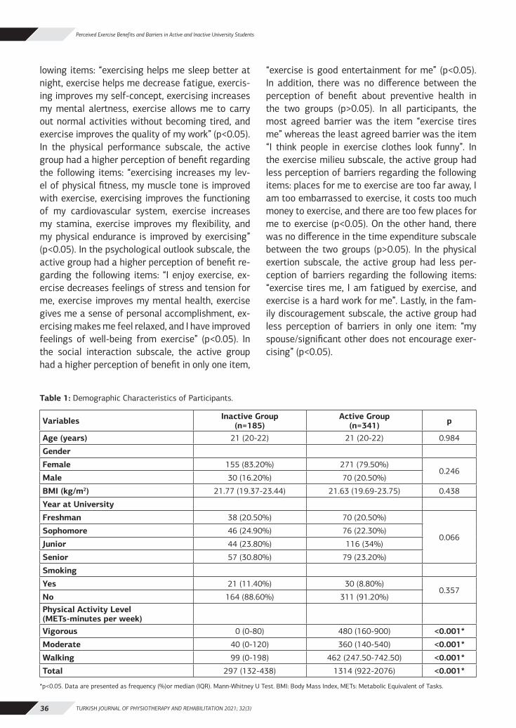
TURKISH JOURNAL OF PHYSIOTHERAPY AND REHABILITATION 2021; 32(3)36
Perceived Exercise Benefits and Barriers in Active and Inactive University Students
lowing items: “exercising helps me sleep better at night, exercise helps me decrease fatigue, exercis-ing improves my self-concept, exercising increases my mental alertness, exercise allows me to carry out normal activities without becoming tired, and exercise improves the quality of my work” (p<0.05). In the physical performance subscale, the active group had a higher perception of benefit regarding the following items: “exercising increases my lev-el of physical fitness, my muscle tone is improved with exercise, exercising improves the functioning of my cardiovascular system, exercise increases my stamina, exercise improves my flexibility, and my physical endurance is improved by exercising” (p<0.05). In the psychological outlook subscale, the active group had a higher perception of benefit re-garding the following items: “I enjoy exercise, ex-ercise decreases feelings of stress and tension for me, exercise improves my mental health, exercise gives me a sense of personal accomplishment, ex-ercising makes me feel relaxed, and I have improved feelings of well-being from exercise” (p<0.05). In the social interaction subscale, the active group had a higher perception of benefit in only one item,
“exercise is good entertainment for me” (p<0.05). In addition, there was no difference between the perception of benefit about preventive health in the two groups (p>0.05). In all participants, the most agreed barrier was the item “exercise tires me” whereas the least agreed barrier was the item “I think people in exercise clothes look funny”. In the exercise milieu subscale, the active group had less perception of barriers regarding the following items: places for me to exercise are too far away, I am too embarrassed to exercise, it costs too much money to exercise, and there are too few places for me to exercise (p<0.05). On the other hand, there was no difference in the time expenditure subscale between the two groups (p>0.05). In the physical exertion subscale, the active group had less per-ception of barriers regarding the following items: “exercise tires me, I am fatigued by exercise, and exercise is a hard work for me”. Lastly, in the fam-ily discouragement subscale, the active group had less perception of barriers in only one item: “my spouse/significant other does not encourage exer-cising” (p<0.05).
Table 1: Demographic Characteristics of Participants.
Variables Inactive Group (n=185)
Active Group (n=341) p
Age (years) 21 (20-22) 21 (20-22) 0.984
Gender
Female 155 (83.20%) 271 (79.50%)0.246
Male 30 (16.20%) 70 (20.50%)
BMI (kg/m2) 21.77 (19.37-23.44) 21.63 (19.69-23.75) 0.438
Year at University
Freshman 38 (20.50%) 70 (20.50%)
0.066Sophomore 46 (24.90%) 76 (22.30%)
Junior 44 (23.80%) 116 (34%)
Senior 57 (30.80%) 79 (23.20%)
Smoking
Yes 21 (11.40%) 30 (8.80%)0.357
No 164 (88.60%) 311 (91.20%)Physical Activity Level (METs-minutes per week)Vigorous 0 (0-80) 480 (160-900) <0.001*
Moderate 40 (0-120) 360 (140-540) <0.001*
Walking 99 (0-198) 462 (247.50-742.50) <0.001*
Total 297 (132-438) 1314 (922-2076) <0.001*
*p<0.05. Data are presented as frequency (%)or median (IQR). Mann-Whitney U Test. BMI: Body Mass Index, METs: Metabolic Equivalent of Tasks.

TURKISH JOURNAL OF PHYSIOTHERAPY AND REHABILITATION 2021; 32(3) 37
Özkul Ç.
Table 2: Exercise Benefits and Barriers in Active and Inactive Groups.
Exercise Benefits and Barriers
All students (n=526)
Inactive Group (n=185)
Active Group (n=341)
pMean±SD
Median (IQR)
Mean±SDMedian (IQR)
Mean±SDMedian (IQR)
Exercise Benefits
Life Enhancement (1-4) 25.24±5.14 25 (23-30) 24.50±4.90 24 (22-29) 25.65±5.23 25 (23-30) 0.001*
My disposition is improved by exercise 2.82±0.91 3 (2-4) 2.77±0.87 3 (2-3) 2.85±0.94 3 (2-4) 0.235
Exercising helps me sleep better at night 3.26±0.80 3 (3-4) 3.17±0.77 3 (3-4) 3.32±0.81 3 (3-4) 0.007*
Exercise helps me decrease fatigue 2.84±0.89 3 (2-4) 2.74±0.81 3 (2-3) 2.91±0.93 3 (2-4) 0.021*
Exercising improves my self-concept 3.15±0.80 3 (3-4) 3.06±0.82 3 (3-4) 3.21±0.79 3 (3-4) 0.038*
Exercising increases my mental alertness 3.32±0.73 3 (3-4) 3.23±0.70 3 (3-4) 3.38±0.74 3 (3-4) 0.005*
Exercise allows me to carry out normal activities without becoming tired
3.29±0.75 3 (3-4) 3.19±0.72 3 (3-4) 3.35±0.77 3 (3-4) 0.004*
Exercise improves the quality of my work 3.29±0.73 3 (3-4) 3.17±0.69 3 (3-4) 3.36±0.75 3 (3-4) <0.001*
Exercise improves overall body functioning for me
3.23±0.75 3 (3-4) 3.16±0.74 3 (3-4) 3.28±0.76 3 (3-4) 0.057
Physical Performance (1-4) 27.66±4.45 29 (25-31) 27.15±4.11 27 (25-31) 27.93±4.61 30 (25-32) 0.003*
Exercise increases my muscle strength 3.46±0.70 4 (3-4) 3.44±0.65 4 (3-4) 3.48±0.73 4 (3-4) 0.161
Exercising increases my level of physical fitness
3.40±0.72 4 (3-4) 3.38±0.68 3 (3-4) 3.48±0.75 4 (3-4) 0.025*
My muscle tone is improved with exercise. 3.40±0.73 4 (3-4) 3.35±0.68 3 (3-4) 3.43±0.77 4 (3-4) 0.035*
Exercising improves functioning of my cardiovascular system
3.44±0.73 4 (3-4) 3.38±0.69 3 (3-4) 3.47±0.75 4 (3-4) 0.030*
Exercise increases my stamina 3.43±0.70 4 (3-4) 3.37±0.67 3 (3-4) 3.48±0.72 4 (3-4) 0.013*
Exercise improves my flexibility 3.44±0.70 4 (3-4) 3.37±0.67 3 (3-4) 3.48±0.72 4 (3-4) 0.016*
My physical endurance is improved by exercising
3.21±0.78 3 (3-4) 3.06±0.80 3 (3-4) 3.29±0.78 3 (3-4) 0.001*
Exercise improves the way my body looks 3.80±0.39 4 (4-4) 3.79±0.41 4 (4-4) 3.82±0.38 4 (4-4) 0.373
Psychological Outlook (1-4) 19.84±3.84 20 (18-23) 19.09±3.56 19 (18-22) 20.25±3.93 21 (18-24) <0.001*
I enjoy exercise 3.19±0.73 3 (3-4) 3.00±0.64 3 (3-3) 3.30±0.76 3 (3-4) <0.001*
Exercise decreases feelings of stress and tension for me
3.32±0.71 3 (3-4) 3.18±0.67 3 (3-4) 3.40±0.73 4 (3-4) <0.001*
Exercise improves my mental health 3.37±0.72 3 (3-4) 3.28±0.69 3 (3-4) 3.43±0.73 4 (3-4) 0.002*
Exercise gives me a sense of personal accomplishment
3.38±0.73 4 (3-4) 3.25±0.74 3 (3-4) 3.45±0.73 4 (3-4) 0.001*
Exercising makes me feel relaxed 3.32±0.75 3 (3-4) 3.22±0.76 3 (3-4) 3.38±0.75 4 (3-4) 0.006*
I have improved feelings of wellbeing from exercise
3.24±0.75 3 (3-4) 3.15±0.73 3 (3-4) 3.29±0.76 3 (3-4) 0.011*
Social Interaction (1-4) 10.89±2.64 11 (9-12) 10.63±2.49 10 (9-12) 11.03±2.71 11 (9-13) 0.060
Exercising lets me have contact with friends and persons I enjoy
2.65±0.90 3 (2-3) 2.62±0.87 3 (2-3) 2.67±0.92 3 (2-3) 0.523
Exercising is a good way for me to meet new people
2.77±0.86 3 (2-3) 2.73±0.84 3 (2-3) 2.80±0.88 3 (2-3) 0.209
Exercise is good entertainment for me 3.11±0.79 3 (3-4) 2.99±0.74 3 (3-3) 3.19±0.82 3 (3-4) 0.001*
Exercising increases my acceptance by others
2.34±0.92 2 (2-3) 2.29±0.87 2 (2-3) 2.37±0.96 2 (2-3) 0.440
Preventive Health (1-4) 9.71±1.94 9 (9-11) 9.55±1.84 9 (9-11) 9.80±1.99 10 (9-12) 0.053
I will prevent heart attacks by exercising 3.28±0.71 3 (3-4) 3.23±0.69 3 (3-4) 3.32±0.72 3 (3-4) 0.051
Exercising will keep me from having high blood pressure
3.30±0.72 3 (3-4) 3.25±0.69 3 (3-4) 3.34±0.74 3 (3-4) 0.060
I will live longer if I exercise 3.11±0.83 3 (3-4) 3.07±0.77 3 (3-4) 3.13±0.87 3 (3-4) 0.162
Total Score (29-116) 93.33±16.09 94 (85-107) 90.92±15.01 88 (83-104) 94.65±16.53 96 (86-108) <0.001*
Exercise Barriers
Exercise Milieu (1-4) 11.51±3.31 12 (9-13) 12.05±3.25 12 (10-14) 11.22±2.71 11 (9-13) 0.005*
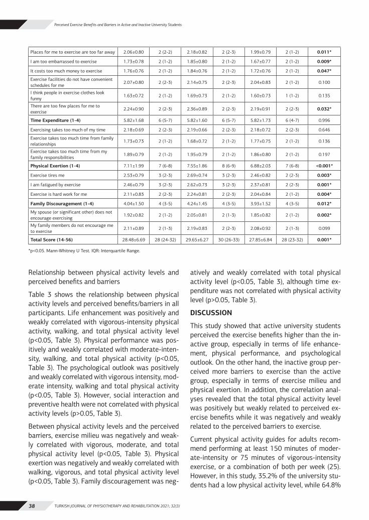
TURKISH JOURNAL OF PHYSIOTHERAPY AND REHABILITATION 2021; 32(3)38
Perceived Exercise Benefits and Barriers in Active and Inactive University Students
Relationship between physical activity levels and perceived benefits and barriers
Table 3 shows the relationship between physical activity levels and perceived benefits/barriers in all participants. Life enhancement was positively and weakly correlated with vigorous-intensity physical activity, walking, and total physical activity level (p<0.05, Table 3). Physical performance was pos-itively and weakly correlated with moderate-inten-sity, walking, and total physical activity (p<0.05, Table 3). The psychological outlook was positively and weakly correlated with vigorous intensity, mod-erate intensity, walking and total physical activity (p<0.05, Table 3). However, social interaction and preventive health were not correlated with physical activity levels (p>0.05, Table 3).
Between physical activity levels and the perceived barriers, exercise milieu was negatively and weak-ly correlated with vigorous, moderate, and total physical activity level (p<0.05, Table 3). Physical exertion was negatively and weakly correlated with walking, vigorous, and total physical activity level (p<0.05, Table 3). Family discouragement was neg-
atively and weakly correlated with total physical activity level (p<0.05, Table 3), although time ex-penditure was not correlated with physical activity level (p>0.05, Table 3).
DISCUSSION
This study showed that active university students perceived the exercise benefits higher than the in-active group, especially in terms of life enhance-ment, physical performance, and psychological outlook. On the other hand, the inactive group per-ceived more barriers to exercise than the active group, especially in terms of exercise milieu and physical exertion. In addition, the correlation anal-yses revealed that the total physical activity level was positively but weakly related to perceived ex-ercise benefits while it was negatively and weakly related to the perceived barriers to exercise.
Current physical activity guides for adults recom-mend performing at least 150 minutes of moder-ate-intensity or 75 minutes of vigorous-intensity exercise, or a combination of both per week (25). However, in this study, 35.2% of the university stu-dents had a low physical activity level, while 64.8%
Places for me to exercise are too far away 2.06±0.80 2 (2-2) 2.18±0.82 2 (2-3) 1.99±0.79 2 (1-2) 0.011*
I am too embarrassed to exercise 1.73±0.78 2 (1-2) 1.85±0.80 2 (1-2) 1.67±0.77 2 (1-2) 0.009*
It costs too much money to exercise 1.76±0.76 2 (1-2) 1.84±0.76 2 (1-2) 1.72±0.76 2 (1-2) 0.047*
Exercise facilities do not have convenient schedules for me
2.07±0.80 2 (2-3) 2.14±0.75 2 (2-3) 2.04±0.83 2 (1-2) 0.100
I think people in exercise clothes look funny
1.63±0.72 2 (1-2) 1.69±0.73 2 (1-2) 1.60±0.73 1 (1-2) 0.135
There are too few places for me to exercise
2.24±0.90 2 (2-3) 2.36±0.89 2 (2-3) 2.19±0.91 2 (2-3) 0.032*
Time Expenditure (1-4) 5.82±1.68 6 (5-7) 5.82±1.60 6 (5-7) 5.82±1.73 6 (4-7) 0.996
Exercising takes too much of my time 2.18±0.69 2 (2-3) 2.19±0.66 2 (2-3) 2.18±0.72 2 (2-3) 0.646
Exercise takes too much time from family relationships
1.73±0.73 2 (1-2) 1.68±0.72 2 (1-2) 1.77±0.75 2 (1-2) 0.136
Exercise takes too much time from my family responsibilities
1.89±0.79 2 (1-2) 1.95±0.79 2 (1-2) 1.86±0.80 2 (1-2) 0.197
Physical Exertion (1-4) 7.11±1.99 7 (6-8) 7.55±1.86 8 (6-9) 6.88±2.03 7 (6-8) <0.001*
Exercise tires me 2.53±0.79 3 (2-3) 2.69±0.74 3 (2-3) 2.46±0.82 2 (2-3) 0.003*
I am fatigued by exercise 2.46±0.79 3 (2-3) 2.62±0.73 3 (2-3) 2.37±0.81 2 (2-3) 0.001*
Exercise is hard work for me 2.11±0.83 2 (2-3) 2.24±0.81 2 (2-3) 2.04±0.84 2 (1-2) 0.004*
Family Discouragement (1-4) 4.04±1.50 4 (3-5) 4.24±1.45 4 (3-5) 3.93±1.52 4 (3-5) 0.012*
My spouse (or significant other) does not encourage exercising
1.92±0.82 2 (1-2) 2.05±0.81 2 (1-3) 1.85±0.82 2 (1-2) 0.002*
My family members do not encourage me to exercise
2.11±0.89 2 (1-3) 2.19±0.83 2 (2-3) 2.08±0.92 2 (1-3) 0.099
Total Score (14-56) 28.48±6.69 28 (24-32) 29.65±6.27 30 (26-33) 27.85±6.84 28 (23-32) 0.001*
*p<0.05. Mann-Whitney U Test. IQR: Interquartile Range.

TURKISH JOURNAL OF PHYSIOTHERAPY AND REHABILITATION 2021; 32(3) 39
Özkul Ç.
had moderate (56.8%) to high (8%) physical activ-ity level. In the previous study, Savci et al. reported that university students’ physical activity levels in Turkey were as follows: 15% low, 68% moderate, and 18% high physical activity (26). Due to the COVID-19 pandemic, the restrictions were declared in Turkey in March 2020, and the normalization process started in June 2020. Although there were no restrictions such as curfews or the gym’s closure that would affect the students’ physical activities, the effects of the pandemic were ongoing on the days when the survey was answered. Therefore, the COVID-19 pandemic may have led to a high-er proportion of students with low physical activity levels in this study.
Promoting physical activity is an essential need among university students. Murphy et al. investi-gated the relationship between physical activity and psychosocial factors in university students in Ireland, and they found that the increase in motiva-tion promoted physical activity (27). The best meth-
od of motivation to increase physical activity may be increased awareness about the exercise bene-fits. Therefore, as the perceived benefits increase, an increase in the individual’s physical activity is expected. This study demonstrated that the active group perceived higher exercise benefits than the inactive group in line with these expectations. To our knowledge, the differences between perceived exercise benefits in active and inactive university students have not been investigated to date. In-terestingly, in this study, most inactive groups re-plied “strongly agree” or “agree” to items regard-ing the exercise benefits like the active group. In other words, the main difference between the two groups was due to the difference in the level of positive perception about exercise benefits rather than knowing these benefits. Most of the students participating in this study were studying in the fac-ulty of health sciences. Therefore, most of the par-ticipants may already have sufficient information about exercise benefits through courses. Since the active group performed vigorous-intensity or mod-
Table 3: Relationship between Physical Activity Levels and Perceived Benefits/Barriers.
Exercise Benefits and BarriersPhysical Activity Level
Vigorous Moderate Walking Total
Exercise Benefits
Life Enhancement r 0.112 0.059 0.111 0.127
p 0.010* 0.179 0.011* 0.004*
Physical performance r 0.070 0.105 0.112 0.130
p 0.107 0.016* 0.010* 0.003*
Psychological Outlookr 0.161 0.156 0.153 0.212
p <0.001* <0.001* <0.001* <0.001*
Social Interactionr 0.070 0.003 0.036 0.059
p 0.111 0.947 0.404 0.178
Preventive Healthr 0.055 0.009 0.036 0.043
p 0.206 0.833 0.406 0.328
Exercise Barriers
Exercise Milieur -0.134 -0.115 -0.073 -0.160
p 0.002* 0.008* 0.095 <0.001*
Time Expenditurer 0.028 -0.037 -0.007 0.001
p 0.527 0.400 0.880 0.991
Physical Exertionr -0.118 -0.080 -0.145 -0.160
p 0.007* 0.068 0.001* <0.001*
Family Discouragementr -0.050 -0.076 -0.071 -0.118
p 0.248 0.083 0.102 0.007*
*p<0.05. Spearman rank correlation coefficient.

TURKISH JOURNAL OF PHYSIOTHERAPY AND REHABILITATION 2021; 32(3)40
Perceived Exercise Benefits and Barriers in Active and Inactive University Students
erate-intensity exercise, their perceptions of exer-cise benefits were based not only on information but also on experience. In other words, as physical activity levels increase through performing exer-cise, positive perceptions about exercise benefits may have increased in participants.
This study demonstrated that the university stu-dents agreed the most with “exercise improves the way my body looks”, whereas they agreed the least with “exercising increases my acceptance by others”. Similarly, Lovell et al. examined perceived exercise benefits among non-exercising female university students in the United Kingdom. They showed that the participants agreed the least with “exercising increases my acceptance by others” while agreeing the most with “exercising increases my level of physical fitness”. Moreover, the most perceived benefit was physical performance fol-lowed by the benefits of psychological outlook, pre-ventive health, life enhancement, and then social interaction (11).
Moreover, the results of correlation analyses in this study showed that the total physical activity level was positively related to the following bene-fits: psychological outlook, life enhancement, and physical performance. In addition, the total phys-ical activity level was not related to the exercise benefit in terms of preventive health in this study. The exclusion of students with chronic disease may have caused this result.
In all participants of this study, the most agreed barriers were the items “exercise tires me”, fol-lowed by “I am fatigued by exercise”, and “there are too few places for me to exercise”. In line with our results, Lovell et al. stated that non-exercising fe-male university students in the UK agreed the most with “exercise tires me”, “places for me to exercise are too far away”, and “exercise is hard work for me” (11). Similarly, Perry et al. found that the most substantial barrier was physical exertion among university students in the UK (14).
The study, which examined university students in Malaysia, showed that lack of time and lack of motivation and physical exertion were among the significant barriers to exercise (12). The previous studies also supported that the lack of time and lack of motivation was the most critical barriers to
exercise among university students in Egypt, Saudi Arabia, the UK, and Spain (7,8,10). In Turkey, Daska-pan et al. investigated barriers to exercise among inactive university students. They found that the most crucial external barrier was lack of time, while the most critical internal barrier was lack of energy. The authors also emphasized that external barriers, including lack of resources, lack of social support, and lack of time, were higher perceived than the internal barriers, including lack of energy, lack of motivation, and lack of self-efficacy (16). This sur-vey was answered during the university’s summer vacation, and the students did not have a course load. Therefore, the participants in this study may have reported that the items related to time expen-diture were not barriers to exercise.
In this study and studies above, perceived exercise benefits and barriers were examined using a survey, including an ordinal scale. However, in a small num-ber of studies, researchers examined the students’ thoughts on barriers to exercise more comprehen-sively through open-ended surveys and interviews. The focus group study by Deliens et al. demon-strated that physical activities in Belgian univer-sity students were affected by factors within the scope of individual factors, social networks, phys-ical environment, and macro-environment (15). In another study, 67 university students in India were questioned about barriers to exercise through fo-cus group interviews, and the students reported that significant barriers were time constraints, tiredness, stress, family control, safety issues (13). Similarly, the study in Brazil showed that the most referred barriers were uncomfortable climate, fam-ily and study obligations, distance to the place of practice, lack of facilities, lack of money to pay pro-fessional and safety issues (9).
All these studies with different assessments in dif-ferent countries have revealed many barriers to ex-ercise in university students. Especially focus group studies also revealed the barriers that could not be reached through questionnaires. There were differ-ences between the importance orders of barriers in these studies. All of these barriers were common to university students.
Furthermore, this study showed that the perceived barriers regarding exercise milieu and physical ex-
TURKISH JOURNAL OF PHYSIOTHERAPY AND REHABILITATION 2021; 32(3) 41
Özkul Ç.
ertion played a role in the difference in active and inactive students’ physical activity levels. Correla-tion analyses demonstrated that the total physical activity level was negatively related to exercise milieu and physical exertion, supporting this result. Measures for the removal of these barriers may di-rectly increase the physical activity level in univer-sity students.
Increasing physical activity levels and develop-ing physical activity habits for students should be among universities’ goals, which is an important place where lifelong habits are shaped. This study showed the possible factors that play a role in dif-ferences between active and inactive students. It could be suggested to add lessons related to ex-ercise based on experience rather than theoreti-cal information to university education programs to increase perceived exercise benefits. It is quite worthy of providing suitable places for exercise to remove the barriers to exercise. In addition, the possibilities of individual exercise training should be offered to all students to overcome the negative perception of physical exertion.
This study has some limitations. First, this cross-sectional study was carried out at Gazi Uni-versity in Turkey. Thus, it may not reflect the over-all student profile worldwide. Second, most of the participants were from the faculty of health sci-ences. They may have more knowledge about ex-ercise benefits. Third, all assessments were made based on participant declaration through question-naires, and so these assessments were not suffi-ciently objective. Forth, the data were collected as the COVID-19 pandemic continued. Last, even if exercise benefits/barriers were assessed using a valid and reliable scale, the scale’s use limited the assessment. Therefore, future studies addressing this issue with interviews such as focus groups may reveal different related factors.
In conclusion, the present study revealed that ac-tive students perceived more exercise benefits than inactive students, whereas they perceived fewer barriers to exercise. The primary factors making a difference between the two groups were life en-hancement, physical performance, and psycholog-ical outlook in exercise benefits, while these were exercise milieu and physical exertion in barriers to
exercise. Furthermore, the total physical activity level was positively related to perceived exercise benefits while negatively related to exercise bar-riers. Therefore, these results may contribute to effective intervention programs aiming to increase physical activity among university students.
Sources of Support: None.
Conflict of Interest: Authors declare no conflict of interest.
Ethical Approval: The study protocol was ap-proved by the Gazi University Ethics Commission (Approval Date: 14.07.2020 and Approval Number: 2020-371).
Informed Consent: All participants approved the informed consent form.
Peer-Review: Externally peer-reviewed.
Author Contributions: Concept – CO; Design – CO; Supervision - CO; Resources and Financial Support – CO; Materials - CO; Data Collection and/or Pro-cessing – CO; Analysis and/or Interpretation– CO; Literature Research - CO; Writing Manuscript –CO; Critical Review – CO.
Acknowledgments: The author is grateful to Gazi University Academic Writing Center employees for revising the English language of the manuscript and to Prof. Dr. Arzu Guclu-Gunduz for her guidance.
REFERENCES1. Bull FC, Al-Ansari SS, Biddle S, Borodulin K, Buman MP, Car-
don G, et al. World Health Organization 2020 guidelines on physical activity and sedentary behaviour. Br J Sports Med. 2020;54(24):1451-1462.
2. Hallal PC, Andersen LB, Bull FC, Guthold R, Haskell W, Ekelund U. Global physical activity levels: surveillance progress, pitfalls, and prospects. Lancet. 2012;380(9838):247-57.
3. Kohl HW, Craig CL, Lambert EV, Inoue S, Alkandari JR, Leetongin G, et al. The pandemic of physical inactivity: global action for public health. Lancet. 2012;380(9838):294-305.
4. Crooke R, Haseler C, Haseler T, Collins J, Crockett A. Physical activity and moving more for health. J R Coll Physicians Edinb. 2020;50(2):173-80.
5. Kwan MY, Cairney J, Faulkner GE, Pullenayegum EE. Physical activity and other health-risk behaviors during the transition into early adulthood: a longitudinal cohort study. Am J Prev Med. 2012;42(1):14-20.
6. Maselli M, Ward PB, Gobbi E, Carraro A. Promoting physical ac-tivity among university students: a systematic review of con-trolled trials. Am J Health Promot. 2018;32(7):1602-12.
7. Blake H, Stanulewicz N, McGill F. Predictors of physical activity and barriers to exercise in nursing and medical students. J Adv Nurs. 2017;73(4):917-29.
TURKISH JOURNAL OF PHYSIOTHERAPY AND REHABILITATION 2021; 32(3)42
Perceived Exercise Benefits and Barriers in Active and Inactive University Students
8. Martínez-Lemos RI, Puig-Ribera AM, García-García O. Perceived barriers to physical activity and related factors in Spanish uni-versity students. Open J. Prev. Med. 2014;04(04):164-74.
9. Sousa TFd, Fonseca SA, Barbosa AR. Barreiras para a prática de atividade física no lazer em estudantes universitários. Rev Bras Cineantropom Desempenho Hum. 2013;15(2):164-73.
10. El-Gilany A, El-Masry R. Physical inactivity among Egyptian and Saudi medical students. TTAF Prev Med Bull. 2011;10(1):35-44.
11. Lovell GP, El Ansari W, Parker JK. Perceived exercise benefits and barriers of non-exercising female university students in the United Kingdom. Int J Environ Res Public Health. 2010;7(3):784-98.
12. Saleem F, Bashaar M, Hassali M. Assessment of barriers to physical activities among university students in Malaysia. Pharm Pharmacol Int J. 2018;6(6):468-73.
13. Sabharwal M. Perceived barriers of young adults for participa-tion in physical activity. Curr Res Nutr Food Sci. 2018;6(2):437-49.
14. Perry J, Stamp E, Crust L, Swann CF. Relationships between mental toughness, barriers to exercise, and exercise behaviour in undergraduate students. Int J Sport Exerc Psychol. 2017;48 (3):262-77.
15. Deliens T, Deforche B, De Bourdeaudhuij I, Clarys P. Determi-nants of physical activity and sedentary behaviour in university students: a qualitative study using focus group discussions. BMC Public Health. 2015;15:201.
16. Daskapan A, Tuzun EH, Eker L. Perceived barriers to physical activity in university students. J Sci Med Sport. 2006;5(4):615.1
17. Sukys S, Cesnaitiene VJ, Emeljanovas A, Mieziene B, Valantine I, Ossowski ZM. Reasons and barriers for university students’ leisure-time physical activity: moderating effect of health edu-cation. Percept Mot Skills. 2019;126(6):1084-100.
18. Bauman AE, Reis RS, Sallis JF, Wells JC, Loos RJF, Martin BW. Correlates of physical activity: why are some people physically active and others not? Lancet. 2012;380(9838):258-71.
19. Faul F, Erdfelder E, Lang A-G, Buchner A. G* Power 3: A flexible statistical power analysis program for the social, behavioral, and biomedical sciences. Behav Res Methods. 2007;39(2):175-91.
20. Saglam M, Arikan H, Savci S, Inal-Ince D, Bosnak-Guclu M, Karabulut E, et al. International physical activity questionnaire: reliability and validity of the Turkish version. Percept Mot Skills. 2010;111(1):278-84.
21. Booth M. Assessment of physical activity: an international per-spective. Res Q Exerc Sport 2000;71(sup2):114-20.
22. Sechrist KR, Walker SN, Pender NJ. Development and psycho-metric evaluation of the exercise benefits/barriers scale. Res Nurs Health. 1987;10(6):357-65.
23. Ortabag T, Ceylan S, Akyuz A, Bebis H. The validity and reliability of the exercise benefits/barriers scale for Turkish Military nurs-ing students. S Afr J Res Sport Ph. 2010;32(2):55-70.
24. Schober P, Boer C, Schwarte LA. Correlation coefficients: appro-priate use and interpretation. Anesth Analg. 2018;126(5):1763-8.
25. Piercy KL, Troiano RP, Ballard RM, Carlson SA, Fulton JE, Galuska DA, et al. The physical activity guidelines for Americans. JAMA. 2018;320(19):2020-8.
26. Savcı S, Öztürk M, Arıkan H, İnal İnce D, Tokgözoğlu L. Physi-cal activity levels of university students. Turk Kardiyol Dern Ars. 2006;34(3):166-72.
27. Murphy JJ, MacDonncha C, Murphy MH, Murphy N, Nevill AM, Woods CB. What psychosocial factors determine the physi-cal activity patterns of university students? J Phys Act Health. 2019;16(5).

43
EFFECTS OF STATIC AND FUNCTIONAL STRETCHING EXERCISES ON LOWER LIMB SPASTICITY AND
FUNCTION IN PEOPLE WITH MULTIPLE SCLEROSIS: A RANDOMIZED CONTROLLED TRIAL
ORIGINAL ARTICLE
ABSTRACTPurpose: Spasticity is a common problem among people with Multiple Sclerosis (MS). This study aimed to compare the effects of static stretching exercise (SSE) and functional stretching exercise (FSE) on lower limb spasticity, function, lower limb pain, active range of motion (ROM), and health-related quality of life (HRQOL) in patient with MS.
Methods: Twenty-six participants were randomly allocated into two groups. SSE group (n=12) completed a four-week (12 sessions) SSE of hamstrings, quadriceps, hip adductors and plantar flexors muscles. FSE group 2 (n=12) completed a four-week (12 sessions) FSE of the same muscles. The outcome measures were Modified Modified Ashworth Scale, Timed Up and Go Test, Timed 25 Foot Walk Test, active ROM assessment, Visual Analogue Scale, and EuroQoL 5-Dimension 5-Level questionnaire.
Results: In each group, decrease of spasticity, improvement of functional tests, decrease of pain, increase of ROM and increase of HRQOL were statistically significant compared to baseline (p<0.05). There were no significant differences between both groups in all variables before and after treatment (p>0.05). A strong correlation was found between decreased spasticity and functional improvement in the SSE group (r=0.793, p=0.002). In the FSE group, there were moderate correlations between decreased spasticity and increased ROM (r=0.689, p=0.013) and between increased ROM and functional improvement (r=0.593, p=0.042). There was also a strong correlation between decreased spasticity and increased HRQOL (r=0.721, p=0.006).
Conclusion: The regular four-weeks of SSE or FSE can decrease lower limb spasticity, improve function, decrease pain, increase active ROM and increase HRQOL in patients with MS.
Key Words: Mobility Limitation, Multiple Sclerosis, Muscle Spasticity, Muscle Stretching Exercises, Quality of Life.
MULTİPL SKLEROZLU BİREYLERDE STATİK VE FONKSİYONEL GERME EGZERSİZLERİNİN ALT EKSTREMİTE SPASTİSİTESİ VE FONKSİYONU ÜZERİNE ETKİLERİ: RANDOMİZE KONTROLLÜ
BİR ÇALIŞMA
ARAŞTIRMA MAKALESİ
ÖZAmaç: Spastisite, Multipl Sklerozlu bireyler arasında yaygın bir sorundur. Bu çalışmanın amacı, Multipl Sklerozlu bireylerde statik germe (SG) ve fonksiyonel germenin (FG) alt ekstremite spastisitesi, fonksiyon, alt ekstremite ağrısı, aktif eklem hareket açıklığı (EHA) ve sağlıkla ilişkili yaşam kalitesi (SİYK) üzerine etkilerini karşılaştırmaktı.
Yöntem: Yirmi altı katılımcı randomize edilerek iki gruba ayrıldı. SG grubuna (n=12) 4 hafta (12 seans) hamstring, quadriseps, kalça adduktor ve plantar fleksor kaslarının statik germe egzersizleri uygulandı. FG grubuna (n=12) aynı kaslar için dört haftalık (12 seans) fonksiyonel germe egzersizleri uygulandı. Sonuç ölçümü olarak Modifiye Modifiye Ashworth Skalası, Zamanlı Kalk ve Yürü Testi, Zamanlı 25 Adım Yürüme Testi, aktif EHA değerlendirilmesi, Görsel Analog Skala ve Avrupa Yaşam Kalitesi Ölçeği-5 Boyut Anketi kullanıldı.
Sonuçlar: Her iki grupta da spastisitede azalma, fonksiyonel test sonuçlarında iyileşme, ağrıda azalma, EHA'nda e SİYK’nde meydana gelen artış başlangıç değerlerine kıyasla istatistiksel olarak anlamlıydı (p<0,05). Gruplar arasında, tüm değişkenlerde tedavi öncesi ve sonrası için anlamlı farklılık yoktu (p>0,05). SG grubunda spastisite azalma ve fonksiyonel iyileşme arasında güçlü bir korelasyon vardı (r=0,793, p=0.002). FG grubunda ise azalmış spastisite ile artmış EHA arasında (r=0,689, p=0.013) ve artmış EHA ile fonksiyonel iyileşme arasında (r=0,593, p=0.042) orta düzeyde korelasyon vardı. Azalan spastisite ile SİYK artışı arasında ise güçlü bir korelasyon vardı (r=0,721, p=0.006)
Tartışma: Düzenli olarak dört hafta boyunca uygulanan SG veya FG, multipl sklerozlu hastalarda alt ekstremite spastisitesini azaltabilir, fonksiyonu iyileştirebilir, ağrıyı azaltabilir, aktif EHA’nı ve SİYK’ni artırabilir.
Anahtar Kelimeler: Mobilite Limitasyonu, Multipl Skleroz, Kas Spastisitesi, Kas Germe Egzersizleri, Yaşam Kalitesi.
Turkish Journal of Physiotherapy and
Rehabilitation2021 32(3)43-51
Merziye ERGUL, MSc, PT1
Afsun NODEHI MOGHADAM, PhD, PT1
Akbar BIGLARIAN, PhD2
ISSN: 2651-4451 • e-ISSN: 2651-446X
Ergul M., Nodehi Moghadam A., Biglarıan A., Effects of Static and Functional Stretching Exercises on Lower Limb Spasticity and Function in People with Multiple Sclerosis: A Randomized Controlled Trial, Turk J Physiother Rehabil. 2021; 32(3):43-51. doi: 10.21653/tjpr.762021
1 University of Social Welfare and Rehabilitation Sciences, Department of Physiotherapy, Tehran, Iran.
2 University of Social Welfare and Rehabilitation Sciences, Department of Biostatistics, Social Determinant of Health Research Center, Tehran, Iran.
Correspondence (İletişim):
Afsun NODEHI MOGHADAM, PhD, PTUniversity of Social Welfare and Rehabilitation
Sciences, Department of Physiotherapy, Postal code: 1985713871, Tehran, Iran
Phone: +98 21 71732889 E-mail: [email protected]: 0000-0001-5843-2330
Merziye ERGULE-mail: [email protected]: 0000-0002-9980-2251
Akbar BIGLARIANE-mail: [email protected]
ORCID: 0000-0002-9776-7085
Received: 13.07.2020 (Geliş Tarihi)Accepted: 24.02.2021 (Kabul Tarihi)
CC BY - NC
Content of this journal is licensed under a Creative Commons Attribution-NonCommercial 4.0 International License.

TURKISH JOURNAL OF PHYSIOTHERAPY AND REHABILITATION 2021; 32(3)44
Effects of Static and Functional Stretching Exercises on Lower Limb Spasticity and Function in People with Multiple Sclerosis: A Randomized Controlled Trial
INTRODUCTION
Sixty to eighty percent of people with multiple scle-rosis (PwMS) have spasticity (1,2). Spasticity was defined by Pandyan et al. as “Disordered senso-rimotor control, resulting, resulting from an upper motor neuron lesion, presenting as intermittent or sustained involuntary activation of muscles” (3). Secondary changes in mechanical muscle fibers, collagen tissues, and tendon properties also con-tribute to spastic muscle tone, resulting in func-tional limitations in upper motor neuron lesions (4). Spasticity causes contracture of muscles and soft tissues, decreased range of motion (ROM) and pain; these components result in limb stiffness, im-paired muscle balance, limited functional mobility, and decreased quality of life (2, 5, 6).
Stretching techniques prevent contractures, in-crease joint ROM, decrease limb stiffness and pain (7). These changes may contribute to improvement in functional movements and quality of life in PwMS (8). Static stretching exercises are generally rec-ommended to reduce spasticity and increase flexi-bility in PwMS (9). Functional stretching is a thera-pist-assisted technique that specifically suggested to recover ROM and joint mobility. This technique is derived from normal daily activities and applied actively by patients in static or dynamic ways (10).
The results of some previous studies (11-17) have indicated positive effects of stretching on func-tional mobility, ambulation, balance, fatigue and quality of life in PwMS. However, in these studies, stretching exercises have been applied with other types of exercises such as balance, coordination, stability, strengthening or resistance exercises and particular parameters (i.e., stretching time, repe-tition, muscles) of the stretching program. In ad-dition, the exact effects of stretching as an inter-vention on PwMS were unclear. Some other studies (18-20) studied the isolated stretching exercises effects on PwMS, but in these studies, stretching has been applied with anti-spasticity drugs, or pa-rameters of the stretching program were unclear. Additionally, the effects of stretching exercises on clinical outcomes such as spasticity or functional mobility were not assessed.
However, stretching is recommended for PwMS, but also there is significant ambiguity about the
type and its unique effects. Therefore, the prima-ry aim was to compare the effects of static and functional stretching on lower limb spasticity and function in PwMS. Secondary aims were to com-pare two types of stretching on lower limb pain, active range of motion (ROM) and health-related quality of life (HRQOL) in PwMS, and to examine the relationship between outcomes.
METHODS
Study Design
This study was a single-center, single-blinded (only patients were blind to their treatment allocation), parallel, randomized, controlled trial investigat-ing the effects of static stretching exercises (SSE) and functional stretching exercises (FSE) on low-er limb spasticity, function, pain, active ROM and health-related quality of life in PwMS. Parameters related to the sample size calculation were taken from a similar study (12). The sample size was es-timated as 11 (n=11 for each group) with an alpha of 0.05, 80% power and effect size of 1.2 using an equation. This study was conducted between Sep-tember-December 2019 at Tehran MS Society Re-habilitation Center in Iran.
Written informed consent was obtained from all study participants. All participants were assessed at baseline for eligibility criteria by a physiothera-pist (researcher). The participants who fulfilled in-clusion criteria were then randomly allocated into either SSE (1) group and FSE (2) group; using ran-dom allocation rule (21) with 1:1 allocation ratio (placing 13 papers of 1 and 13 papers of two in a pocket and drawing them randomly) by an indepen-dent member of Tehran MS Society Rehabilitation Center.
Participants
The Medical Ethics Committee approved this study of the University of Social Welfare and Re-habilitation Sciences, Tehran, Iran (Approval Date: 23.07.2019 and Approval Number: IR.USWR.REC.1398.051) and was registered with the Irani-an Registry of Clinical Trial (Registration number: IRCT20190702044079N1). Thirty-four PwMS from the Tehran MS Society rehabilitation center were screened for eligibility criteria, and 26 PwMS were enrolled in the study following Helsinki’s declara-
TURKISH JOURNAL OF PHYSIOTHERAPY AND REHABILITATION 2021; 32(3) 45
Ergul M., Nodehi Moghadam A., Biglarıan A.
tion. Inclusion criteria were specialist-confirmed MS (22), self-reported lower-extremity spastici-ty in daily activities with limb stiffness or muscle spasms or pain (Modified Modified Ashworth Scale score ≥1 in at least one of the assessed muscles), able to walk a minimum of 25 feet with or without assistive devices such as cane or walker. Exclusion criteria were participation in any yoga, Pilates or exercise programs, using medications used in the treatment of spasticity, Botox injections for lower limb (in the last six months), the use of an orthosis.
Interventions
Static stretching was in a passive form and per-formed by a physiotherapist (researcher). SSE in-clude (23); static stretching of hamstrings in the supine position (hip flexion with knee locked in ex-tension), quadriceps in the prone position (hip ex-tension with full knee flexion and heel in contact with gluteal), hip adductors in the supine position (hip abduction with full knee extension) and plantar flexors muscles in the supine position (ankle dorsi-flexion with the knee in full extension).
In the FSE group, the following exercises were su-pervised by a physiotherapist (researcher) and per-formed actively by participants (24):
Standing wedge plantar flexor stretch: Participant stood in front of a parallel bar, and held the bars for support, placed both feet onto a wedge with ankles are dorsiflexed. The patient then shifted his/her weight forward gradually while the knees were kept in extension.
Seated toe touch hamstring stretch: The participant sat on a chair in front of parallel bars. Both ankles were placed on the bar, which had been adjusted appropriately in height for the patient’s safety and comfort; the patient then gradually reached as if to touch his toes.
Standing single leg quadriceps stretch: Participants stood in front of a parallel bar and held the bars for support while flexing the knee. Then placed his/her foot on a chair adjusted to an appropriate height and located behind the leg. This procedure was re-peated for the other leg.
Standing hip adductors stretch by hip abduction: Participants stood in the parallel bar and hold onto the bars for support before abducting gradually
both legs out to the side.
Each stretch maintained for 30 seconds and re-peated three times with 15 seconds rests in both groups. After each stretching, participants were required to do five times active or active-assisted movements in the same direction of stretching in both groups. The intervention schedule for both groups was three times a week for four weeks, and each session took approximately 25-30 minutes. All participants completed their sessions individu-ally in a separated timetable.
Measurements
The following outcomes were assessed at base-line and after a 4-week stretching program. Mod-ified Modified Ashworth Scale (MMAS) was used to grade the spasticity level of hamstrings, quad-riceps, hip adductors and plantar flexors (25). Func-tional mobility and dynamic balance were assessed using a timed up and go test (TUG) (26). Partici-pant took the time to stand up from a chair at the height of 46 cm from the ground and walk 3 me-ters, turn around, and sit back down on the chair as fast as they could be measured in seconds by a stopwatch. Gait speed and gait capacity were mea-sured using the Timed 25 Foot Walk Test (T25FWT) (27). Participants were asked to walk 25 feet as fast as possible, and the time taken to complete was recorded by stopwatch. Visual Analogue Scale (VAS) (28) was used to measure pain. Patients were asked to determine their general pain level in the lower limb on a 0–10 points scale in which 0 indi-cates no pain, and 10 indicates severe pain.
In a supine position, active hip flexion, abduction and ankle dorsiflexion ROM; and in prone position active hip extension ROM were assessed using a standard goniometer (Stainless Steel goniometer 20cm/180°, SAEHAN, Belgium) (29). The measure-ment was performed bilaterally. All ROM measure-ments were repeated two times, then we have averaged the first and second measurements of hip abduction, hip flexion, hip extension and ankle dorsiflexion. These new values were used for the statistical analyses.
European Quality of Life 5 Dimensions 5 Levels (EQ-5D-5L) questionnaire was allowed to use in this study and used to evaluate health-related qual-

TURKISH JOURNAL OF PHYSIOTHERAPY AND REHABILITATION 2021; 32(3)46
Effects of Static and Functional Stretching Exercises on Lower Limb Spasticity and Function in People with Multiple Sclerosis: A Randomized Controlled Trial
ity of life (HRQOL) (30). Participants were required to describe their HRQOL in five dimensions: Mo-bility, Self-Care, Usual Activities, Pain/Discomfort, and Anxiety/Depression by choosing one of five re-sponse categories as no problems, slight problems, moderate problems, severe problems, and extreme problems. The EQ-VAS part recorded the patient’s self-rated health on a vertical visual analogue scale (0-100 scale).
Statistical Analysis
For data analysis, SPSS version 17.0 (SPSS Inc., Chicago, USA) was used. Descriptive statistics (mean and standard deviation) were computed for all data. The Kolmogorov–Smirnov test was used to determine the normal distribution of variables. The ANOVA was applied to detect baseline differ-ences between both groups, and the paired 𝑡-testwas applied for comparing differences to baseline within the group. The ANCOVA was used to com-
Table 1: The Descriptive and Baseline Measures in Static and Functional Stretching Groups.
Variables Static Stretching (n=12)
Functional Stretching (n=12) p
GenderFemale 5 7
0.436Male 7 5
Dominant FootRight 10 9
0.633Left 2 3
Type of Disease
PPMS 1 -
0.764SPMS 6 6
RRMS 4 6
PRMS 1 -
Age (years) 45.33±11.96 43.75±7.58 0.702
Weight (kg) 68.33±5.44 64.50±21.28 0.552
Height (cm) 168.58±8.96 164±6.99 0.177
Disease Duration (years) 13.42±7.90 18.42±5.48 0.085
MMAS (grade) Hamstrings 1 (0-2) 0 (0-2) 0.109
MMAS Quadriceps 1 (0-3) 0 (0-2) 0.107
MMAS Adductors 1 (0-2) 1 (0-1) 0.103
MMAS Plantar Flexors 2 (1-2) 2 (1-3) 1.000
TUG (s) 19.21±12.73 11.52±5.01 0.065
T25FWT (s) 12.50±8.72 8.40±3.33 0.142
Pain VAS (0-10 cm) 4.75±3.57 3.50±2.97 0.361
AROM (angle) FlexionR 69.95±30.91 82.87±24.22 0.267
L 76.75±26.12 87±21.77 0.308
AROM ExtensionR 10.79±8.57 16.08±7.55 0.123
L 9.41±7.14 14.91±7.82 0.086
AROM AbductionR 33.62±14.96 39.91±12.58 0.277
L 24.66±13.61 35.41±6.67 0.052
AROM Dorsi FlexionR 26.08±8.76 32.04±14.30 0.232
L 23.66±17.13 30.62±12.92 0.274
EQ-5D-5L 12.25±2.95 10±2.25 0.061
EQ-VAS (0-100) 65±19.42 74.58±14.21 0.182
Results expressed as mean±SD. MMAS results expressed as median (min-max). PPMS: Primary Progressive Multiple Sclerosis, SPMS: Secondary Progressive Multiple Sclerosis, RRMS: Relapsing-Remitting Multiple Sclerosis, PRMS: Progressive Relapsing Multiple Sclerosis, MMAS: Modified Modified Ashworth Scale, TUG: Timed Up and Go, T25FWT: Timed 25 Foot Walk Test, VAS: Visual Analogue Scale, AROM: Active Range of Motion, EQ-5D-5L: European Quality of Life 5 Dimensions 5 Levels, EQ-VAS: European Quality of Life Visual Analog Scale, R: Right, L: Left.

TURKISH JOURNAL OF PHYSIOTHERAPY AND REHABILITATION 2021; 32(3) 47
Ergul M., Nodehi Moghadam A., Biglarıan A.
pare pre-post differences between both groups. The standardized treatment effect was calculated to compare static and functional stretching groups for pre-post differences. The Spearman rank test was used to quantify associations among the vari-ables. Correlation coefficients(r) whose magnitudes were ≥0.7, 0.5-0.7, and ≤0.5 were considered as strong, moderate and weak correlation, respective-ly (31). A significant level was considered less than 0.05.
RESULTS
From the 26 PwMS that included in this study, two patients dropped out: one from the SSE group due to personal issues and one from the FSE group due to general pain; and 24 PwMS were included in the analysis. Twelve participants (7 men, 5 wom-en; age=45.33±11.96 years) as the SSE group and 12 participants (5 men, 7 women; age= 43.75±7.58 years) as the FSE group participated in this study. Participants flow shown in Figure 1. The descriptive and baseline characteristic of participants have shown in Table 1.
There was no significant difference between the two groups for any data at the baseline (p>0.05). According to the results of paired t-test, which is shown in Table 2, there were significant improve-ments (p<0.05) in measured outcomes within both groups compared to baseline, except spasticity of hamstrings and quadriceps in the FSE group. There were no significant differences between both groups for all variables compared to baseline (p>0.05) (Table 3).
There was a strong correlation (r=0.793, p=0.002) between decreased spasticity of quadriceps and improved TUG in the SSE group. A strong cor-relation (r=0.733, p=0.007) was found between decreased spasticity of quadriceps and improved T25FWT in the SSE group. There was also a mod-erate correlation (r=0.662, p=0.019) between de-creased spasticity of hip adductors and improved TUG in the SSE group. In the FSE group, a mod-erate correlation (r=0.689, p=0.013) was observed between decreased spasticity of hamstrings and increased right hip extension ROM. There was a moderate negative correlation (r=0.593, p=0.042)
Table 2: The Pre and Post Comparisons of Measured Variables in Static and Functional Stretching Groups.
Group Outcomes Static Stretching (n=12) Functional Stretching (n=12)
Mean Difference (95% CI) p Mean Difference (95% CI) pMMAS (grade) Hamstrings -0.66 (-1.16--0.17) 0.025* -0.25 (-0.74 to 0.24) 0.275
MMAS Quadriceps -0.75 (-1.17--0.32) 0.005* -0.41 (-0.84 to 0.1) 0.054
MMAS Adductors -0.91 (-1.25--0.57) 0.001* -0.75 (-1.09 to -0.4) <0.001*
MMAS Plantar Flexors -1.25 (-1.61--0.88) <0.001* -1 (-1.36--0.63) <0.001*
TUG (s) -3.83 (-5.62--2.04) 0.007* -2.1 (-3.89--0.31) <0.001*
T25FWT (s) -2.23 (-3.41 to -1.06) 0.006* -1.74 (-2.92--0.57); 0.003*
Pain VAS (0-10 cm) -1.83 (-3.06 to -0.59) 0.003* -2 (-3.23--0.76); 0.014*
AROM (angle) –Flexion
R 21.16 (12.88-29.44) <0.001* 16.45 (8.18-24.73) 0.002*
L 18.29 (12.56-24.01) <0.001* 12.70 (6.98-18.43) <0.001*
AROM-ExtensionR 6.70 (2.93-10.48) 0.001* 8.41 (4.64-12.19) 0.002*
L 8.54 (5-12.08) 0.001* 7.75 (4.20-11.29) <0.001*
AROM-AbductionR 14.29 (7.64-20.93) 0.002* 13.83 (7.18- 20.48) <0.001*
L 14.45 (10.25-18.66) <0.001* 8.54 (4.33-12.75) <0.001*
AROM-Dorsi Flexion
R 11.04 (6.52-15.56) 0.002* 6.79 (2.27-11.31) 0.001*
L 14.62 (9.98-19.26) <0.001* 10.04 (5.39-14.68) <0.001*
EQ-5D-5L -2.66 (-3.56--1.76) <0.001* -2.5 (-3.4--1.6) <0.001*
EQ-VAS (0-100) 9.58 (3.78-15.38) 0.024* 5.83 (0.34-11.63) 0.002*
*p<0.05. CI: Confidence Intervals, MMAS: Modified Modified Ashworth Scale, TUG: Timed Up and Go, T25FWT: Timed 25 Foot Walk Test, VAS: Visual Analogue Scale, AROM: Active Range of Motion, EQ-5D-5L: European Quality of Life 5 Dimensions 5 Levels, EQ-VAS: European Quality of Life Visual Analog Scale, R: Right, L: Left.
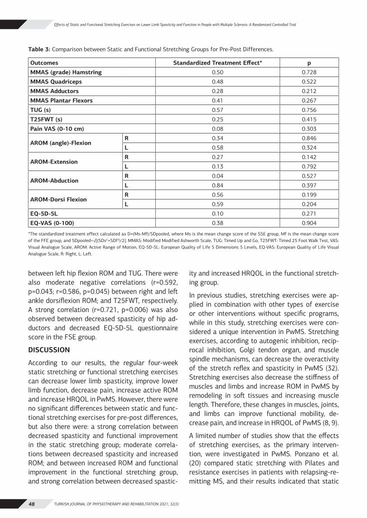
TURKISH JOURNAL OF PHYSIOTHERAPY AND REHABILITATION 2021; 32(3)48
Effects of Static and Functional Stretching Exercises on Lower Limb Spasticity and Function in People with Multiple Sclerosis: A Randomized Controlled Trial
between left hip flexion ROM and TUG. There were also moderate negative correlations (r=0.592, p=0.043; r=0.586, p=0.045) between right and left ankle dorsiflexion ROM; and T25FWT, respectively. A strong correlation (r=0.721, p=0.006) was also observed between decreased spasticity of hip ad-ductors and decreased EQ-5D-5L questionnaire score in the FSE group.
DISCUSSION
According to our results, the regular four-week static stretching or functional stretching exercises can decrease lower limb spasticity, improve lower limb function, decrease pain, increase active ROM and increase HRQOL in PwMS. However, there were no significant differences between static and func-tional stretching exercises for pre-post differences, but also there were: a strong correlation between decreased spasticity and functional improvement in the static stretching group; moderate correla-tions between decreased spasticity and increased ROM; and between increased ROM and functional improvement in the functional stretching group, and strong correlation between decreased spastic-
ity and increased HRQOL in the functional stretch-ing group.
In previous studies, stretching exercises were ap-plied in combination with other types of exercise or other interventions without specific programs, while in this study, stretching exercises were con-sidered a unique intervention in PwMS. Stretching exercises, according to autogenic inhibition, recip-rocal inhibition, Golgi tendon organ, and muscle spindle mechanisms, can decrease the overactivity of the stretch reflex and spasticity in PwMS (32). Stretching exercises also decrease the stiffness of muscles and limbs and increase ROM in PwMS by remodeling in soft tissues and increasing muscle length. Therefore, these changes in muscles, joints, and limbs can improve functional mobility, de-crease pain, and increase in HRQOL of PwMS (8, 9).
A limited number of studies show that the effects of stretching exercises, as the primary interven-tion, were investigated in PwMS. Ponzano et al. (20) compared static stretching with Pilates and resistance exercises in patients with relapsing-re-mitting MS, and their results indicated that static
Table 3: Comparison between Static and Functional Stretching Groups for Pre-Post Differences.
Outcomes Standardized Treatment Effect* p
MMAS (grade) Hamstring 0.50 0.728
MMAS Quadriceps 0.48 0.522
MMAS Adductors 0.28 0.212
MMAS Plantar Flexors 0.41 0.267
TUG (s) 0.57 0.756
T25FWT (s) 0.25 0.415
Pain VAS (0-10 cm) 0.08 0.303
AROM (angle)-FlexionR 0.34 0.846
L 0.58 0.324
AROM-ExtensionR 0.27 0.142
L 0.13 0.792
AROM-AbductionR 0.04 0.527
L 0.84 0.397
AROM-Dorsi FlexionR 0.56 0.199
L 0.59 0.204
EQ-5D-5L 0.10 0.271
EQ-VAS (0-100) 0.38 0.904
*The standardized treatment effect calculated as D=(Ms-Mf)/SDpooled, where Ms is the mean change score of the SSE group, Mf is the mean change score of the FFE group, and SDpooled=√[(SDs2+SDf2)/2]. MMAS: Modified Modified Ashworth Scale, TUG: Timed Up and Go, T25FWT: Timed 25 Foot Walk Test, VAS: Visual Analogue Scale, AROM: Active Range of Motion, EQ-5D-5L: European Quality of Life 5 Dimensions 5 Levels, EQ-VAS: European Quality of Life Visual Analogue Scale, R: Right, L: Left.
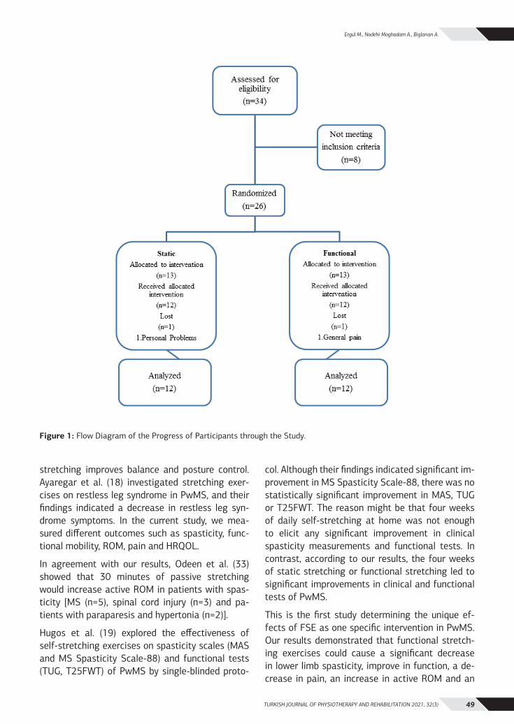
TURKISH JOURNAL OF PHYSIOTHERAPY AND REHABILITATION 2021; 32(3) 49
Ergul M., Nodehi Moghadam A., Biglarıan A.
stretching improves balance and posture control. Ayaregar et al. (18) investigated stretching exer-cises on restless leg syndrome in PwMS, and their findings indicated a decrease in restless leg syn-drome symptoms. In the current study, we mea-sured different outcomes such as spasticity, func-tional mobility, ROM, pain and HRQOL.
In agreement with our results, Odeen et al. (33) showed that 30 minutes of passive stretching would increase active ROM in patients with spas-ticity [MS (n=5), spinal cord injury (n=3) and pa-tients with paraparesis and hypertonia (n=2)].
Hugos et al. (19) explored the effectiveness of self-stretching exercises on spasticity scales (MAS and MS Spasticity Scale-88) and functional tests (TUG, T25FWT) of PwMS by single-blinded proto-
col. Although their findings indicated significant im-provement in MS Spasticity Scale-88, there was no statistically significant improvement in MAS, TUG or T25FWT. The reason might be that four weeks of daily self-stretching at home was not enough to elicit any significant improvement in clinical spasticity measurements and functional tests. In contrast, according to our results, the four weeks of static stretching or functional stretching led to significant improvements in clinical and functional tests of PwMS.
This is the first study determining the unique ef-fects of FSE as one specific intervention in PwMS. Our results demonstrated that functional stretch-ing exercises could cause a significant decrease in lower limb spasticity, improve in function, a de-crease in pain, an increase in active ROM and an
Figure 1: Flow Diagram of the Progress of Participants through the Study.

TURKISH JOURNAL OF PHYSIOTHERAPY AND REHABILITATION 2021; 32(3)50
Effects of Static and Functional Stretching Exercises on Lower Limb Spasticity and Function in People with Multiple Sclerosis: A Randomized Controlled Trial
increase in HRQOL of PwMS. Elshafey et al. (24) explored the effects of functional stretching on neural, mechanical and gait parameters in spastic diplegic children, and their results demonstrated significant improvement in all parameters. Ghase-mi et al. (34) also showed that functional stretch-ing could lead to functional improvement in chronic stroke patients. The FSE is suggested by Lederman et al. (10) as sensory-motor training to maintain, increase or recover ROM, enhancing motor learn-ing and recovery. However, our results show a sig-nificant increase in ROM and indicated a moderate correlation between increased ROM and functional improvement in the FSE group.
Studies showed that limited functional mobility, loss of ROM, and decreased quality of life are spas-ticity consequences (6,35,36). Our results showed that there was a strong association between de-creased spasticity and functional tests improve-ment in the SSE group. In our FSE group, there was a moderate correlation between decreased spasticity and recovered ROM. Also, there was a strong correlation between decreased spasticity and an increase in HRQOL. Our stretching protocol can potentially help reverse the limited functional mobility, loss of ROM and decrease in quality of life in PwMS. However, this study is preliminary to determine the unique effects of stretching exercis-es and shows the importance of stretching exercis-es as an essential intervention in comprehensive rehabilitation programs for PwMS. Stretching is a safe intervention and has fewer side effects than medications, and may increase medications’ ef-fects (37). Therefore, stretching can be considered as a recommended rehabilitation program for MS individuals with severe disabilities.
There were some limitations to this study. First, there was no opportunity to incorporate a follow-up period to examine the treatment’s long-term ef-fects. Second, a double-blinded condition would be ideal for reducing any internal bias in measure-ment. Third, it would be better for two different re-searchers to do the assessment and treatment to avoid any possible Pygmalion effect.
In conclusion, this study’s central finding is that regular four-week static or functional stretch-ing can decrease spasticity and pain and improve
function, ROM, and health-related quality of life in people with multiple sclerosis. Static and function-al stretching exercises by decreasing spasticity and increasing range of motion could lead to improve-ments in functional mobility of lower limbs, and through this could increase health-related quality of life in people with multiple sclerosis.
Sources of Support: None.
Conflict of interest: None.
Ethical Approval: This study was approved by the Medical Ethics Committee of the University of Social Welfare and Rehabilitation Sciences, Teh-ran, Iran (Approval Date: 23.07.2019 and Approval Number: IR.USWR.REC.1398.051).
Informed Consent: A written informed consent was obtained from all study participants.
Peer-Review: Externally peer-reviewed.
Author Contribution: Concept – ME, ANM; Design – ME, ANM; Supervision – ME, ANM; Resources and Financial Support – ME, ANM; Materials – ME, ANM; Data Collection and/or Processing – ME, AB; Analysis and/or Interpretation – ME, ANM, AB; Lit-erature Research – ME, ANM; Writing Manuscript – ME, ANM; Critical Review – ME, ANM, AB.
Acknowledgments: The authors acknowledge their gratitude to all of the participants in this project. We acknowledge especially the members of Tehran MS Society Rehabilitation Center.
REFERENCES1. Sosnoff JJ, Gappmaier E, Frame A, Motl RW. Influence of spastic-
ity on mobility and balance in persons with multiple sclerosis. J Neurol Phys Ther. 2011;35(3):129-32.
2. Flachenecker P, Henze T, Zettl U. Spasticity in patients with mul-tiple sclerosis–clinical characteristics, treatment and quality of life. Acta Neurol Scand. 2014;129(3):154-62.
3. Pandyan AD, Gregoric M, Barnes MP, Wood D, Van Wijck F, Bur-ridge J, et al. Spasticity: clinical perceptions, neurological reali-ties and meaningful measurement. Disabil Rehabil. 2005;27(1-2):2–6.
4. Dietz V. Clinical treatment of spasticity--spastic movement dis-orders. Nervenarzt. 2013;84(12):1508-11.
5. Bethoux F. Gait disorders in multiple sclerosis. Continuum (Min-neap Minn). 2013;19(4):1007-22.
6. Svensson J, Borg S, Nilsson P. Costs and quality of life in mul-tiple sclerosis patients with spasticity. Acta Neurol Scand. 2014;129(1):13-20.
7. Amatya B, Khan F, La Mantia L, Demetrios M, Wade DT. Non pharmacological interventions for spasticity in multiple sclero-sis. Cochrane Database Syst. Rev. 2013(2): CD009974.
TURKISH JOURNAL OF PHYSIOTHERAPY AND REHABILITATION 2021; 32(3) 51
Ergul M., Nodehi Moghadam A., Biglarıan A.
8. Motl RW, Pilutti LA. The benefits of exercise training in multiple sclerosis. Nat Rev Neurol. 2012;8(9):487-97.
9. Halabchi F, Alizadeh Z, Sahraian MA, Abolhasani M. Exercise pre-scription for patients with multiple sclerosis; potential benefits and practical recommendations. BMC Neurol. 2017;17(1):1-11.
10. Lederman E. Therapeutic stretching in physical therapy: towards a functional approach. London: Elsevier Health Sciences; 2013.
11. Shams A, Taheri H. The effect of 8 weeks selective training pro-grams with instructions focus of attention on walking speed of patients with multiple sclerosis. Med J Mashhad Univ Med Sci. 2015;57(9):969-75.
12. Sosnoff JJ, Finlayson M, McAuley E, Morrison S, Motl RW. Home-based exercise program and fall-risk reduction in older adults with multiple sclerosis: phase 1 randomized controlled trial. Clin Rehabil. 2014;28(3):254-63.
13. Ertekin Ö, Özakbaş S, İdiman E, Algün ZC. Quality of life, fatigue and balance improvements after home-based exercise program in multiple sclerosis patients. Noro Psikiyatr Ars. 2012;49(1):33-38.
14. Alaee S, Fathi M, Moazami M. Effect of a six-week concurrent exercise (stretching and aerobic) and the consumption of tetra hydro cannabinol additive on fatigue severity level among fe-male MS patients. Adv Environ Biol. 2014;8(9):924-930.
15. Tarakci E, Yeldan I, Huseyinsinoglu BE, Zenginler Y, Eraksoy M. Group exercise training for balance, functional status, spasticity, fatigue and quality of life in multiple sclerosis: a randomized controlled trial. Clin Rehabil. 2013;27(9):813-822.
16. Pazokian M, Shaban M, Zakerimoghdam M, Mehran A, Sang-laje B. The effect of stretching together aerobic exercises on fatigue level in multiple sclerosis𝑡 patients refer to MS soci-ety of Iran those suffer from fatigue. J Holist Nurs Midwifery. 2012;22(2):18-24.
17. Negahban H, Rezaie S, Goharpey S. Massage therapy and ex-ercise therapy in patients with multiple sclerosis: a randomized controlled pilot study. Clin Rehabil. 2013;27(12):1126-1136.
18. Ayaregar M, Hosseini M, Shakeri N, Ashktorab T, Zohari Anbouhi S. The effect of stretching exercises on the severity of restless legs syndrome symptoms in patients with multiple sclerosis. Ha-yat. 2017;23(1):7-16.
19. Hugos CL, Bourdette D, Chen Y, Chen Z, Cameron M. A group-de-livered self-management program reduces spasticity in people with multiple sclerosis: a randomized, controlled pilot trial. Mult Scler J Exp Transl Clin. 2017;3(1):2055217317699993.
20. Ponzano M, Beratto L, Mazza C, Di Tommaso S, Abate Daga F, Allois R, et al. Benefits of static stretching, Pliates® and elastic bands resistance training on patients with relapsing-remitting multiple sclerosis: a longitudinal study. J Neurol Neurophysiol. 2017;8(4):1-6.
21. 21. Schulz KF, Grimes DA. Generation of allocation se-quences in randomized trials: chance , not choice. Lancet. 2002;359(9305):515-519.
22. McDonald WI, Compston A, Edan G, Goodkin D, Hartung HP, Lub-lin FD, et al. Recommended diagnostic criteria for multiple scle-rosis: guidelines from the International Panel on the diagnosis of multiple sclerosis. Ann Neurol. 2001;50(1):121-127.
23. Gibson BE. Stretching with a helper for people with ms: an il-lustrated manual. New York: National Multiple Sclerosis Society, 2000.
24. Elshafey MA, Abd-Elaziem A, Gouda RE. Functional stretching exercise submitted for spastic diplegic children: a randomized control study. Rehabil Res Pract. 2014;2014(1):128-134.
25. Ghotbi N, Ansari NN, Naghdi S, Hasson S. Measurement of low-er-limb muscle spasticity: intrarater reliability of Modified Mod-ified Ashworth Scale. J Rehabil Res Dev. 2011;48(1):83-88.
26. Sebastião E, Sandroff BM, Learmonth YC, Motl RW. Validity of the timed up and go test as a measure of functional mobili-ty in persons with multiple sclerosis. Arch Phys Med Rehabil. 2016;97(7):1072-1077.
27. Motl RW, Cohen JA, Benedict R, Phillips G, LaRocca N, Hudson DL et al. Validity of the timed 25-foot walk as an ambulatory performance outcome measure for multiple sclerosis. Mult Scler. J. 2017;23(5): 704-710.
28. Young J, Amatya B, Galea MP, Khan F. Chronic pain in mul-tiple sclerosis: a 10-year longitudinal study. Scand J Pain. 2017;16:198-203.
29. Clarkson HM. Musculoskeletal assessment: joint motion and muscle testing. Philadelphia: Wolters Kluwer/Lippincott Williams & Wilkins Health China; 2013.
30. Kuspinar A, Mayo NE. A review of the psychometric properties of generic utility measures in multiple sclerosis. Pharmacoeconom-ics. 2014;32(8):759-773.
31. Cohen J. Statistical power analysis for the behavioral sciences: New York: Academic Press; 2013.
32. Bovend’Eerdt TJ, Newman M, Barker K, Dawes H, Minelli C, Wade DT. The effects of stretching in spasticity: a systematic review. Arch Phys Med Rehabil. 2008;89(7):1395-1406.
33. Odeen I. Reduction of muscular hypertonus by long-term muscle stretch. Scand J Rehabil Med. 1981;13(2-3):93-99.
34. Ghasemi E, Khademi-Kalantari K, Khalkhali-Zavieh M, Rezasol-tani A, Ghasemi M, Baghban AA, et al. The effect of functional stretching exercises on neural and mechanical properties of the spastic medial gastrocnemius muscle in patients with chronic stroke: a randomized controlled trial. J Stroke Cerebrovasc Dis. 2018;27(7):1733-1742.
35. Sandoval AEG. Exercise in multiple sclerosis. Phys Med Rehabil Clin N Am. 2013; 24(4):605-618.
36. Hoang PD, Gandevia SC, Herbert RD. Prevalence of joint con-tractures and muscle weakness in people with multiple sclerosis. Disabil Rehabil. 2014;36(19):1588-1593.
37. Brar SP, Smith MB, Nelson LM, Franklin GM, Cobble ND. Evalua-tion of treatment protocols on minimal to moderate spasticity in multiple sclerosis. Arch Phys Med Rehabil. 1991;72(3):186-189.
TURKISH JOURNAL OF PHYSIOTHERAPY AND REHABILITATION 2021; 32(3)52
AN INVESTIGATION OF CHARACTERISTICS RELATED TO ORAL MOTOR AND SWALLOWING DISORDERS IN CHILDREN WITH CEREBRAL PALSY: A DESCRIPTIVE
STUDY
ORIGINAL ARTICLE
ABSTRACTPurpose: Oral motor problems and swallowing disorders are common in children with cerebral palsy (CP). This study was planned to examine the oral motor and swallowing related descriptive characteristics of children with CP.
Methods: This study was carried out with 90 children with CP (39 girls and 51 boys) with a mean age of 70.35±28.39 months. Gross Motor Function Classification System (GMFCS) was used for functional motor level. The Tongue Thrust Rating Scale (TTRS) for tongue thrust severity, Karaduman Chewing Performance Scale (KCPS) for chewing performance level, Pediatric Eating Assessment Tool (PEDI-EAT-10) for dysphagia symptom severity and Drooling Severity and Frequency Scale for drooling were used.
Results: According to GMFCS, 55.6% of children were in level V. The 87.8% of the children had pathological tongue thrust. The mean PEDI-EAT-10 score was 15.42±11.41. Chewing disorders were detected in 85.6%, and drooling was detected in 87.8% of CP children. The GMFCS level, clinical type of CP, presence of open mouth, food consistency and the number of pneumonia correlated with all oral motor and swallowing assessments (p<0.05).
Conclusion: In this study, various oral motor and swallowing disorders were identified in children with CP, and a Turkish community-specific general profile of these children was obtained.
Key Words: Cerebral Palsy, Deglutition, Eating, Mastication.
SEREBRAL PALSİLİ ÇOCUKLARDA ORAL MOTOR VE YUTMA BOZUKLUKLARI İLE İLİŞKİLİ ÖZELLİKLERİN
İNCELENMESİ: TANIMLAYICI BİR ÇALIŞMA
ARAŞTIRMA MAKALESİ
ÖZAmaç: Serebral Palsili (SP) çocuklarda oral motor problemler ve yutma bozukluğu yaygın olarak görülmektedir. Bu çalışma, SP’li çocukların oral motor ve yutma bozukluğu ile ilgili tanımlayıcı özelliklerini incelemek amacı ile planlandı.
Yöntem: Bu çalışma, yaş ortalaması 70.35±28.30 ay olan 90 SP’li çocuk (39 kız, 51 erkek) ile gerçekleştirildi. Motor fonksiyonel seviye için Kaba Motor Fonksiyon Sınıflandırma Sistemi (KMFSS) kullanıldı. Dil itme şiddetinin değerlendirilmesi için Dil İtme Derecelendirme Ölçeği (TTRS), salya problemi için Salya Şiddet ve Frekans Ölçeği, çiğneme performans seviyesi için Karaduman Çiğneme Performans Ölçeği (KCPS) ve yutma bozukluğu için Pediatrik Yeme Değerlendirme Aracı (PEDI-EAT-10) kullanıldı.
Sonuçlar: KMFSS’ye göre çocukların % 55,6'sı seviye V düzeyindeydi. Çocukların %87,8'inde patolojik dil itme refleksi vardı. Ortalama PEDI-EAT-10 skoru 15,42±11,41 idi. SP’li çocukların % 87,8'inde çiğneme bozukluğu ve % 87,8'inde salya problemi saptandı. GMFCS düzeyi, klinik SP tipi, open mouth varlığı, besin kıvamı ve pnömoni sayısı tüm oral motor ve yutma değerlendirmeleri ile korelasyon gösterdi (p<0.05).
Tartışma: Bu çalışmada SP'li çocuklarda çeşitli oral motor ve yutma bozuklukları tespit edildi ve bu çocuklar için Türk toplumuna özgü genel bir profil elde edildi.
Anahtar Kelimeler: Serebral Palsi, Deglügasyon, Yeme, Çiğneme.
Turkish Journal of Physiotherapy and
Rehabilitation2021 32(3)52-61
Özgü İNAL, Assist Prof1
Selen SEREL ARSLAN, PT, PhD, Assoc Prof2
Numan DEMİR, PT, PhD, Assoc Prof2
Aynur Ayşe KARADUMAN, PT, PhD, Prof3
ISSN: 2651-4451 • e-ISSN: 2651-446X
İnal Ö., Serel Arslan S., Demir N., Karaduman A.A., An Investigation of Characteristics Related to Oral Motor and Swallowing Disorders in Children with Cerebral Palsy: A Descriptive Study, Turk J Physiother Rehabil. 2021; 32(3):52-61. doi: 10.21653/tjpr.751763
1 Health Sciences University, Faculty of Gülhane Health Sciences, Department of Occupational Therapy, Ankara, Turkey.
2 Hacettepe University, Faculty of Physical Therapy and Rehabilitation, Ankara, Turkey.
3 Lokman Hekim University, Faculty of Health Sciences, Department of Physical Therapy and Rehabilitation, Ankara, Turkey.
Correspondence (İletişim):
Özgü İNAL, PhD, PTHealth Sciences University,
Faculty of Gülhane Health Sciences, Department of Occupational Therapy,
06018, Ankara, TurkeyPhone: +90-312-304 6191
E-mail: [email protected]: 0000-0002-0351-1821
Selen SEREL-ARSLANE-mail: [email protected]
ORCID: 0000-0002-2463-7503
Numan DEMİRE-mail: [email protected]: 0000-0001-6308-0237
Aynur Ayşe KARADUMANE-mail: [email protected]
ORCID: 0000-0001-6252-1934
Received: 11.06.2020 (Geliş Tarihi)Accepted: 15.04.2021 (Kabul Tarihi)
CC BY - NC
Content of this journal is licensed under a Creative Commons Attribution-NonCommercial 4.0 International License.
TURKISH JOURNAL OF PHYSIOTHERAPY AND REHABILITATION 2021; 32(3) 53
İnal Ö., Serel Arslan S., Demir N., Karaduman A.A.
INTRODUCTION
Cerebral palsy (CP) is a common childhood disorder that causes impaired sensorimotor functions, in-cluding oral motor and/or swallowing dysfunction. Oral motor problems, feeding problems, drooling, and swallowing disorders (dysphagia) are common in children with CP. The incidence of these problems is up to 90%. These problems are associated with malnutrition, dehydration, incomplete teeth, failure to provide consistency transitions following mat-uration, aspiration pneumonia, and death. These problems also can interrupt typical development at critical periods (1-5). Therefore, early identification of oral motor and swallowing disorders in children is critical.
Many studies focus on feeding and swallowing dis-orders in children with CP (6,7). In these studies, the effect of feeding, swallowing, and nutrition problems on the caregiver’s quality of life were ex-amined in children with CP. Studies have pointed out that these problems are common in children with CP, negatively affecting caregivers’ quality of life. However, no study has been performed to in-vestigate the descriptive characteristics of children with CP who have an oral motor and swallowing disorders in the Turkish population.
There are many different reasons for feeding and swallowing problems (e.g. incorrect posture, an-atomical problems of the oral motor structure). Therefore, it is thought that it is essential to ex-plain the different characteristics that may cause oral motor and swallowing disorders and examine their relationship with them. Determining these re-lationships will be instructive in terms of the scope of the rehabilitation program. It was aimed to obtain objective data about oral motor and swal-lowing problems in children with CP in the Turkish population.
METHODS
This study was conducted at Hacettepe University Faculty of Physical Therapy and Rehabilitation with the Hacettepe University Swallowing Disorders Re-search and Application Center’s cooperation. The Non-Interventional Clinical Research Ethics Com-mittee approved the study protocol (Approval Date: 05.12.2017 and Approval Number: GO17/860-04).
All mothers provided a written consent form to par-ticipate in the study.
Participants
Ninety children referred were included in the study, who were thought to have oral motor problems and swallowing disorders. Inclusion criteria were being an age range between 3 and 12 years, diagnosed with CP by a pediatric neurologist based on inter-national CP criteria (8). Children not in the specified age range were excluded from the study.
Assessments
Descriptive information including age, sex, prena-tal, natal, and postnatal history, sucking and feed-ing history from the birth of the child, feeding dura-tion time, feeding position, first teething time, GIS problem and number of pneumonia in the last one year were obtained from the mothers and noted. Two physiotherapists made all evaluations with more than ten years of clinical experience in oral motor and swallowing disorders. During the evalu-ations, the presence of open mouth and high palate was also noted.
The Gross Motor Function Classification System (GMFCS), a valid and reliable 5-level system, was used to classify the severity of motor functions in children. Level I indicates “Walks without restric-tion,” and Level V means “Children are transported in a manual wheelchair in all settings” (9-12).
The Tongue Thrust Rating Scale (TTRS), which is a valid, reliable, clinically easy to use the instru-ment, was used to determine tongue thrust sever-ity in children (13). The scale defines the severity of tongue thrust in four levels, ranging from 0 to 3. Level 0 indicates ‘No tongue thrust’, and Level 3 indicates ‘severe tongue thrust’.
Drooling Severity and Frequency Scale was used to determine the severity and frequency of drooling in children. The severity of drooling is scored between 1 to 5, and the frequency of drooling is scored be-tween 1 to 4. Increased levels indicate more severe and more frequent drooling (14).
The Karaduman Chewing Performance Scale (KCPS), a valid and reliable instrument, was used to define children’s chewing performance level. The
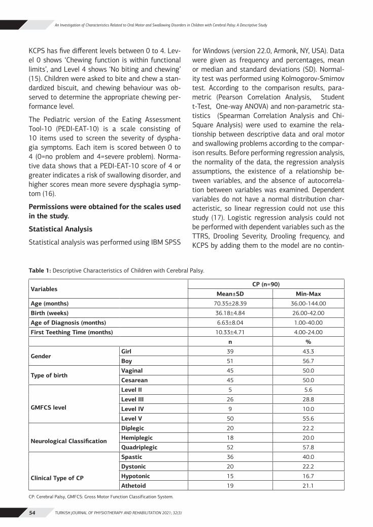
TURKISH JOURNAL OF PHYSIOTHERAPY AND REHABILITATION 2021; 32(3)54
An Investigation of Characteristics Related to Oral Motor and Swallowing Disorders in Children with Cerebral Palsy: A Descriptive Study
KCPS has five different levels between 0 to 4. Lev-el 0 shows ‘Chewing function is within functional limits’, and Level 4 shows ‘No biting and chewing’ (15). Children were asked to bite and chew a stan-dardized biscuit, and chewing behaviour was ob-served to determine the appropriate chewing per-formance level.
The Pediatric version of the Eating Assessment Tool-10 (PEDI-EAT-10) is a scale consisting of 10 items used to screen the severity of dyspha-gia symptoms. Each item is scored between 0 to 4 (0=no problem and 4=severe problem). Norma-tive data shows that a PEDI-EAT-10 score of 4 or greater indicates a risk of swallowing disorder, and higher scores mean more severe dysphagia symp-tom (16).
Permissions were obtained for the scales used in the study.
Statistical Analysis
Statistical analysis was performed using IBM SPSS
for Windows (version 22.0, Armonk, NY, USA). Data were given as frequency and percentages, mean or median and standard deviations (SD). Normal-ity test was performed using Kolmogorov-Smirnov test. According to the comparison results, para-metric (Pearson Correlation Analysis, Student t-Test, One-way ANOVA) and non-parametric sta-tistics (Spearman Correlation Analysis and Chi-Square Analysis) were used to examine the rela-tionship between descriptive data and oral motor and swallowing problems according to the compar-ison results. Before performing regression analysis, the normality of the data, the regression analysis assumptions, the existence of a relationship be-tween variables, and the absence of autocorrela-tion between variables was examined. Dependent variables do not have a normal distribution char-acteristic, so linear regression could not use this study (17). Logistic regression analysis could not be performed with dependent variables such as the TTRS, Drooling Severity, Drooling frequency, and KCPS by adding them to the model are no contin-
Table 1: Descriptive Characteristics of Children with Cerebral Palsy.
VariablesCP (n=90)
Mean±SD Min-Max
Age (months) 70.35±28.39 36.00-144.00
Birth (weeks) 36.18±4.84 26.00-42.00
Age of Diagnosis (months) 6.63±8.04 1.00-40.00
First Teething Time (months) 10.33±4.71 4.00-24.00
n %
GenderGirl 39 43.3
Boy 51 56.7
Type of birthVaginal 45 50.0
Cesarean 45 50.0
GMFCS level
Level II 5 5.6
Level III 26 28.8
Level IV 9 10.0
Level V 50 55.6
Neurological Classification
Diplegic 20 22.2
Hemiplegic 18 20.0
Quadriplegic 52 57.8
Clinical Type of CP
Spastic 36 40.0
Dystonic 20 22.2
Hypotonic 15 16.7
Athetoid 19 21.1
CP: Cerebral Palsy, GMFCS: Gross Motor Function Classification System.
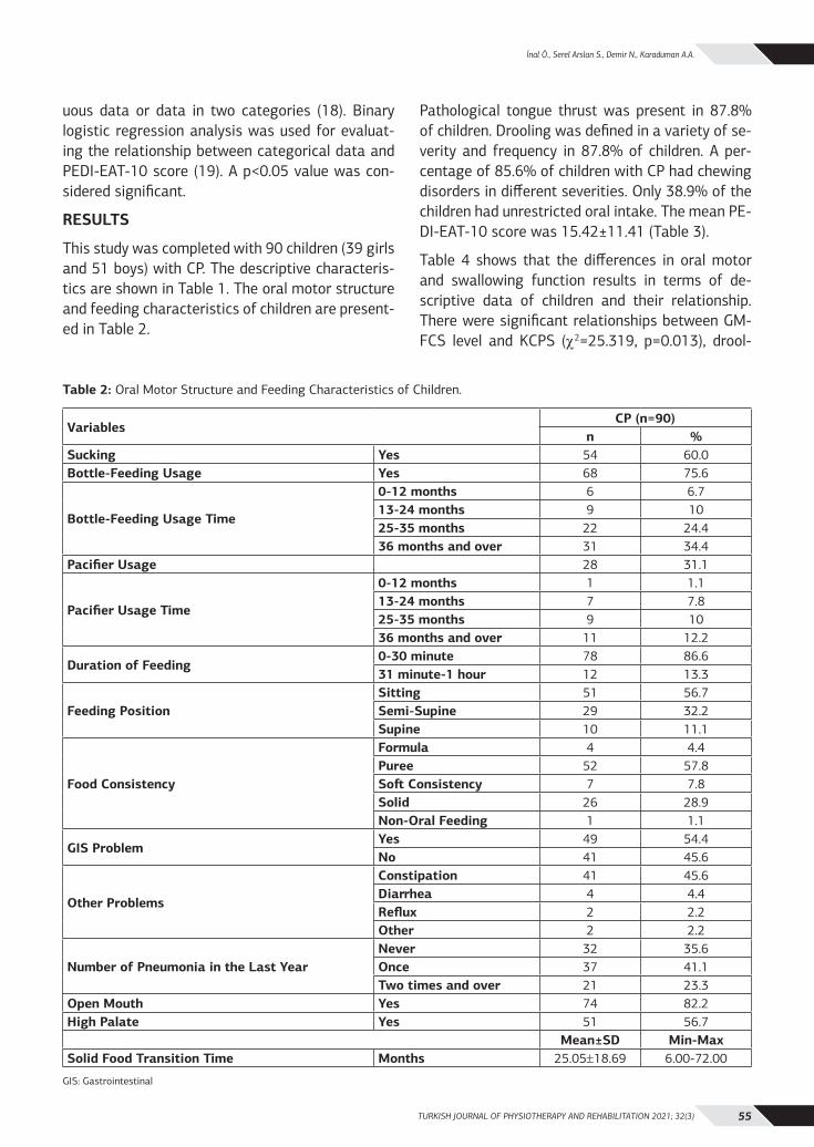
TURKISH JOURNAL OF PHYSIOTHERAPY AND REHABILITATION 2021; 32(3) 55
İnal Ö., Serel Arslan S., Demir N., Karaduman A.A.
uous data or data in two categories (18). Binary logistic regression analysis was used for evaluat-ing the relationship between categorical data and PEDI-EAT-10 score (19). A p<0.05 value was con-sidered significant.
RESULTS
This study was completed with 90 children (39 girls and 51 boys) with CP. The descriptive characteris-tics are shown in Table 1. The oral motor structure and feeding characteristics of children are present-ed in Table 2.
Pathological tongue thrust was present in 87.8% of children. Drooling was defined in a variety of se-verity and frequency in 87.8% of children. A per-centage of 85.6% of children with CP had chewing disorders in different severities. Only 38.9% of the children had unrestricted oral intake. The mean PE-DI-EAT-10 score was 15.42±11.41 (Table 3).
Table 4 shows that the differences in oral motor and swallowing function results in terms of de-scriptive data of children and their relationship. There were significant relationships between GM-FCS level and KCPS (χ2=25.319, p=0.013), drool-
Table 2: Oral Motor Structure and Feeding Characteristics of Children.
Variables CP (n=90)
n %Sucking Yes 54 60.0Bottle-Feeding Usage Yes 68 75.6
Bottle-Feeding Usage Time
0-12 months 6 6.713-24 months 9 1025-35 months 22 24.436 months and over 31 34.4
Pacifier Usage 28 31.1
Pacifier Usage Time
0-12 months 1 1.113-24 months 7 7.825-35 months 9 1036 months and over 11 12.2
Duration of Feeding0-30 minute 78 86.631 minute-1 hour 12 13.3
Feeding PositionSitting 51 56.7Semi-Supine 29 32.2Supine 10 11.1
Food Consistency
Formula 4 4.4Puree 52 57.8Soft Consistency 7 7.8Solid 26 28.9Non-Oral Feeding 1 1.1
GIS ProblemYes 49 54.4No 41 45.6
Other Problems
Constipation 41 45.6Diarrhea 4 4.4Reflux 2 2.2Other 2 2.2
Number of Pneumonia in the Last YearNever 32 35.6Once 37 41.1Two times and over 21 23.3
Open Mouth Yes 74 82.2High Palate Yes 51 56.7
Mean±SD Min-MaxSolid Food Transition Time Months 25.05±18.69 6.00-72.00
GIS: Gastrointestinal
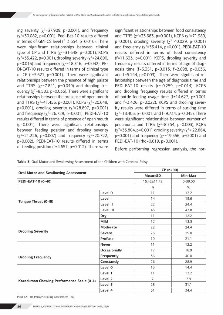
TURKISH JOURNAL OF PHYSIOTHERAPY AND REHABILITATION 2021; 32(3)56
An Investigation of Characteristics Related to Oral Motor and Swallowing Disorders in Children with Cerebral Palsy: A Descriptive Study
ing severity (χ2=37.909, p=0.001), and frequency (χ2=30.082, p=0.001). Pedi-Eat-10 results differed in terms of GMFCS level (f=3.654, p=0.016). There were significant relationships between clinical type of CP and TTRS (χ2=31.648, p=0.001), KCPS (χ2=35.422, p=0.001), drooling severity (χ2=24.890, p=0.015) and frequency (χ2=18.316, p=0.032). PE-DI-EAT-10 results differed in terms of clinical type of CP (f=5.621, p=0.001). There were significant relationships between the presence of high palate and TTRS (χ2=7.841, p=0.049) and drooling fre-quency (χ2=8.583, p=0.035). There were significant relationships between the presence of open mouth and TTRS (χ2=41.456, p=0.001), KCPS (χ2=20.649, p=0.001), drooling severity (χ2=28.897, p=0.001) and frequency (χ2=26.729, p=0.001). PEDI-EAT-10 results differed in terms of presence of open mouth (p=0.001). There were significant relationships between feeding position and drooling severity (χ2=21.226, p=0.007) and frequency (χ2=20.722, p=0.002). PEDI-EAT-10 results differed in terms of feeding position (f=4.657, p=0.012). There were
significant relationships between food consistency and TTRS (χ2=33.683, p=0.001), KCPS (χ2=71.989, p=0.001), drooling severity (χ2=40.029, p=0.001) and frequency (χ2=33.414, p=0.001). PEDI-EAT-10 results differed in terms of food consistency (f=11.633, p=0.001). KCPS, drooling severity and frequency results differed in terms of age of diag-nosis time (f=3.351, p=0.013, f=2.698, p=0.036, and f=5.144, p=0.003). There were significant re-lationships between the age of diagnosis time and PEDI-EAT-10 results (r=-0.259, p=0.014). KCPS and drooling frequency results differed in terms of bottle-feeding usage time (f=14.627, p=0.001 and f=3.426, p=0.022). KCPS and drooling sever-ity results were differed in terms of sucking time (χ2=18.405, p= 0.001, and f=9.734, p=0.045). There were significant relationships between number of pneumonia and TTRS (χ2=8.754, p=0.003), KÇPS (χ2=33.804, p=0.001), drooling severity (χ2= 22.864, p=0.001) and frequency (χ2=19.556, p=0.001) and PEDI-EAT-10 (rho=0.619, p=0.001).
Before performing regression analysis, the nor-
Table 3: Oral Motor and Swallowing Assessment of the Children with Cerebral Palsy.
Oral Motor and Swallowing Assessment CP (n=90)
Mean±SD Min-Max
PEDI-EAT-10 (0-40) 15.42±11.42 0-39.00
n %
Tongue Thrust (0-III)
Level 0 11 12.2
Level I 14 15.6
Level II 22 24.4
Level III 43 47.8
Drooling Severity
Dry 11 12.2
Mild 12 13.3
Moderate 22 24.4
Severe 26 29.0
Profuse 19 21.1
Drooling Frequency
Never 11 12.2
Occasionally 17 18.9
Frequently 36 40.0
Constantly 26 28.9
Karaduman Chewing Performance Scale (0-4)
Level 0 13 14.4
Level 1 11 12.2
Level 2 7 7.9
Level 3 28 31.1
Level 4 31 34.4
PEDI-EAT-10: Pediatric Eating Assessment Tool
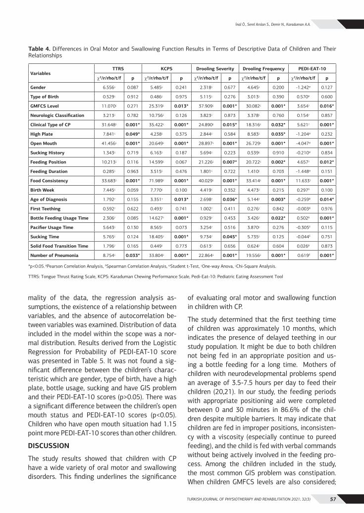
TURKISH JOURNAL OF PHYSIOTHERAPY AND REHABILITATION 2021; 32(3) 57
İnal Ö., Serel Arslan S., Demir N., Karaduman A.A.
mality of the data, the regression analysis as-sumptions, the existence of a relationship between variables, and the absence of autocorrelation be-tween variables was examined. Distribution of data included in the model within the scope was a nor-mal distribution. Results derived from the Logistic Regression for Probability of PEDI-EAT-10 score was presented in Table 5. It was not found a sig-nificant difference between the children’s charac-teristic which are gender, type of birth, have a high plate, bottle usage, sucking and have GIS problem and their PEDI-EAT-10 scores (p>0.05). There was a significant difference between the children’s open mouth status and PEDI-EAT-10 scores (p<0.05). Children who have open mouth situation had 1.15 point more PEDI-EAT-10 scores than other children.
DISCUSSION
The study results showed that children with CP have a wide variety of oral motor and swallowing disorders. This finding underlines the significance
of evaluating oral motor and swallowing function in children with CP.
The study determined that the first teething time of children was approximately 10 months, which indicates the presence of delayed teething in our study population. It might be due to both children not being fed in an appropriate position and us-ing a bottle feeding for a long time. Mothers of children with neurodevelopmental problems spend an average of 3.5-7.5 hours per day to feed their children (20,21). In our study, the feeding periods with appropriate positioning aid were completed between 0 and 30 minutes in 86.6% of the chil-dren despite multiple barriers. It may indicate that children are fed in improper positions, inconsisten-cy with a viscosity (especially continue to pureed feeding), and the child is fed with verbal commands without being actively involved in the feeding pro-cess. Among the children included in the study, the most common GIS problem was constipation. When children GMFCS levels are also considered;
Table 4. Differences in Oral Motor and Swallowing Function Results in Terms of Descriptive Data of Children and Their Relationships
VariablesTTRS KCPS Drooling Severity Drooling Frequency PEDI-EAT-10
χ2/r/rho/t/f p χ2/r/rho/t/f p χ2/r/rho/t/f p χ2/r/rho/t/f p χ2/r/rho/t/f p
Gender 6.556ς 0.087 5.485ς 0.241 2.318ς 0.677 4.645ς 0.200 -1.242ϕ 0.127
Type of Birth 0.529ς 0.912 0.486ς 0.975 5.115ς 0.276 3.013ς 0.390 0.570ϕ 0.600
GMFCS Level 11.070ς 0.271 25.319ς 0.013* 37.909ς 0.001* 30.082ς 0.001* 3.654γ 0.016*
Neurologic Classification 3.213ς 0.782 10.756ς 0.126 3.823ς 0.873 3.378ς 0.760 0.154γ 0.857
Clinical Type of CP 31.648ς 0.001* 35.422ς 0.001* 24.890ς 0.015* 18.316ς 0.032* 5.621γ 0.001*
High Plate 7.841ς 0.049* 4.238ς 0.375 2.844ς 0.584 8.583ς 0.035* -1.204ϕ 0.232
Open Mouth 41.456ς 0.001* 20.649ς 0.001* 28.897ς 0.001* 26.729ς 0.001* -4.047ϕ 0.001*
Sucking History 1.343ς 0.719 6.163ς 0.187 5.694ς 0.223 0.539ς 0.910 -0.210ϕ 0.834
Feeding Position 10.213ς 0.116 14.599ς 0.067 21.226ς 0.007* 20.722ς 0.002* 4.657γ 0.012*
Feeding Duration 0.285ς 0.963 3.515ς 0.476 1.801ς 0.722 1.410ς 0.703 -1.448ϕ 0.151
Food Consistency 33.683ς 0.001* 71.989ς 0.001* 40.029ς 0.001* 33.414ς 0.001* 11.633γ 0.001*
Birth Week 7.445ς 0.059 7.770ς 0.100 4.419ς 0.352 4.473ς 0.215 0.297δ 0.100
Age of Diagnosis 1.792γ 0.155 3.351γ 0.013* 2.698γ 0.036* 5.144γ 0.003* -0.259φ 0.014*
First Teething 0.592γ 0.622 0.493γ 0.741 1.002γ 0.411 0.276γ 0.842 -0.003φ 0.976
Bottle Feeding Usage Time 2.306γ 0.085 14.627γ 0.001* 0.929γ 0.453 3.426γ 0.022* 0.502φ 0.001*
Pacifier Usage Time 5.643ς 0.130 8.565ς 0.073 3.254γ 0.516 3.870ς 0.276 -0.305δ 0.115
Sucking Time 5.765ς 0.124 18.405ς 0.001* 9.734γ 0.045* 5.735ς 0.125 -0.044δ 0.751
Solid Food Transition Time 1.796γ 0.165 0.449γ 0.773 0.613γ 0.656 0.624γ 0.604 0.026φ 0.873
Number of Pneumonia 8.754ς 0.033* 33.804ς 0.001* 22.864ς 0.001* 19.556ς 0.001* 0.619δ 0.001*
*p<0.05. φPearson Correlation Analysis, δSpearman Correlation Analysis, ϕStudent t-Test, γOne-way Anova, ςChi-Square Analysis.
TTRS: Tongue Thrust Rating Scale, KCPS: Karaduman Chewing Performance Scale, Pedi-Eat-10: Pediatric Eating Assessment Tool
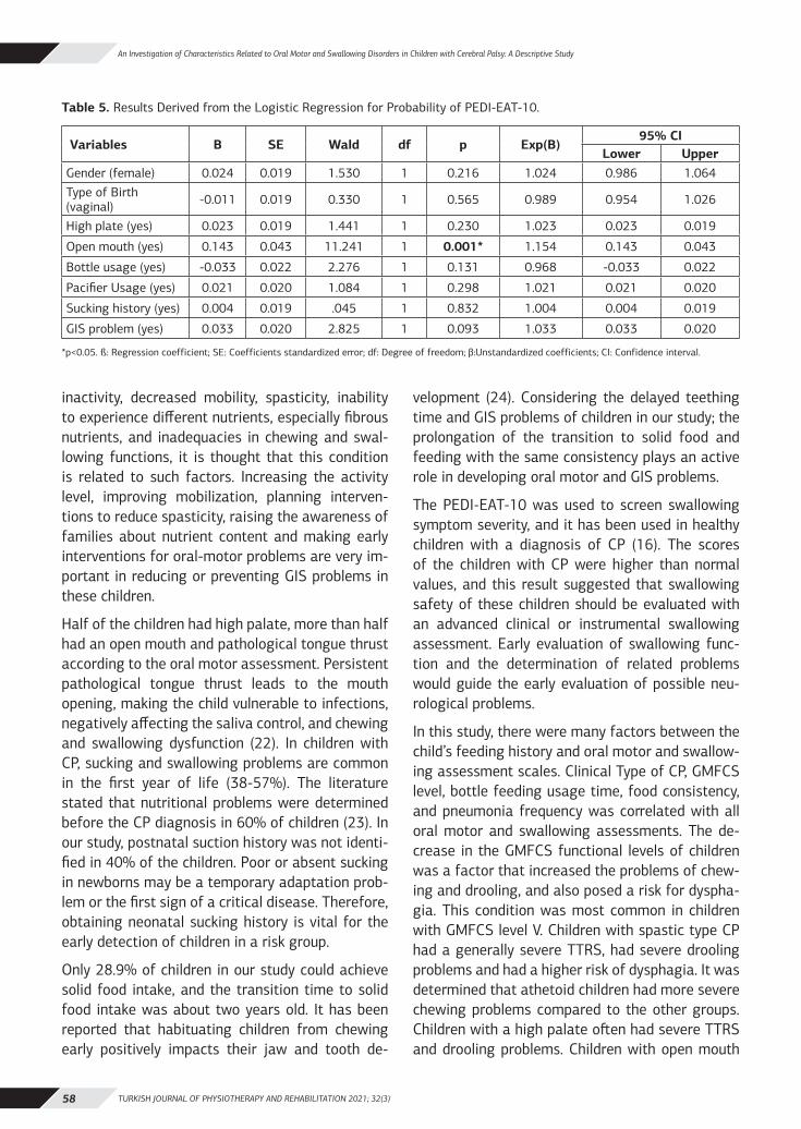
TURKISH JOURNAL OF PHYSIOTHERAPY AND REHABILITATION 2021; 32(3)58
An Investigation of Characteristics Related to Oral Motor and Swallowing Disorders in Children with Cerebral Palsy: A Descriptive Study
inactivity, decreased mobility, spasticity, inability to experience different nutrients, especially fibrous nutrients, and inadequacies in chewing and swal-lowing functions, it is thought that this condition is related to such factors. Increasing the activity level, improving mobilization, planning interven-tions to reduce spasticity, raising the awareness of families about nutrient content and making early interventions for oral-motor problems are very im-portant in reducing or preventing GIS problems in these children.
Half of the children had high palate, more than half had an open mouth and pathological tongue thrust according to the oral motor assessment. Persistent pathological tongue thrust leads to the mouth opening, making the child vulnerable to infections, negatively affecting the saliva control, and chewing and swallowing dysfunction (22). In children with CP, sucking and swallowing problems are common in the first year of life (38-57%). The literature stated that nutritional problems were determined before the CP diagnosis in 60% of children (23). In our study, postnatal suction history was not identi-fied in 40% of the children. Poor or absent sucking in newborns may be a temporary adaptation prob-lem or the first sign of a critical disease. Therefore, obtaining neonatal sucking history is vital for the early detection of children in a risk group.
Only 28.9% of children in our study could achieve solid food intake, and the transition time to solid food intake was about two years old. It has been reported that habituating children from chewing early positively impacts their jaw and tooth de-
velopment (24). Considering the delayed teething time and GIS problems of children in our study; the prolongation of the transition to solid food and feeding with the same consistency plays an active role in developing oral motor and GIS problems.
The PEDI-EAT-10 was used to screen swallowing symptom severity, and it has been used in healthy children with a diagnosis of CP (16). The scores of the children with CP were higher than normal values, and this result suggested that swallowing safety of these children should be evaluated with an advanced clinical or instrumental swallowing assessment. Early evaluation of swallowing func-tion and the determination of related problems would guide the early evaluation of possible neu-rological problems.
In this study, there were many factors between the child’s feeding history and oral motor and swallow-ing assessment scales. Clinical Type of CP, GMFCS level, bottle feeding usage time, food consistency, and pneumonia frequency was correlated with all oral motor and swallowing assessments. The de-crease in the GMFCS functional levels of children was a factor that increased the problems of chew-ing and drooling, and also posed a risk for dyspha-gia. This condition was most common in children with GMFCS level V. Children with spastic type CP had a generally severe TTRS, had severe drooling problems and had a higher risk of dysphagia. It was determined that athetoid children had more severe chewing problems compared to the other groups. Children with a high palate often had severe TTRS and drooling problems. Children with open mouth
Table 5. Results Derived from the Logistic Regression for Probability of PEDI-EAT-10.
Variables B SE Wald df p Exp(B)95% CI
Lower UpperGender (female) 0.024 0.019 1.530 1 0.216 1.024 0.986 1.064Type of Birth (vaginal) -0.011 0.019 0.330 1 0.565 0.989 0.954 1.026
High plate (yes) 0.023 0.019 1.441 1 0.230 1.023 0.023 0.019
Open mouth (yes) 0.143 0.043 11.241 1 0.001* 1.154 0.143 0.043
Bottle usage (yes) -0.033 0.022 2.276 1 0.131 0.968 -0.033 0.022
Pacifier Usage (yes) 0.021 0.020 1.084 1 0.298 1.021 0.021 0.020
Sucking history (yes) 0.004 0.019 .045 1 0.832 1.004 0.004 0.019
GIS problem (yes) 0.033 0.020 2.825 1 0.093 1.033 0.033 0.020
*p<0.05. ß: Regression coefficient; SE: Coefficients standardized error; df: Degree of freedom; β:Unstandardized coefficients; CI: Confidence interval.
TURKISH JOURNAL OF PHYSIOTHERAPY AND REHABILITATION 2021; 32(3) 59
İnal Ö., Serel Arslan S., Demir N., Karaduman A.A.
had severe TTRS and chewing problems. Children with open mouth were found to have severe drool-ing problems, and these children had a higher risk of dysphagia.
The saliva frequency of the children in the sitting position was higher than the other feeding posi-tions. It was determined that children who were fed supine had the highest risk for dysphagia. It was determined that children fed with food, pu-ree and soft consistency had higher TTRS severity than children fed with solids and had more severe drooling problems. Similarly, children who could eat solid foods had better chewing performance and had a lower risk of dysphagia. Children who used bottles for longer had better chewing performance and had fewer drooling problems. In addition, these children had less risk of dysphagia. It was deter-mined that children with longer sucking times had a worse chewing performance. It was determined that increased pneumonia level was a risk factor for all oral motor skills and swallowing disorder.
Oral motor function is associated with head and trunk stability. In general, as the functional level de-teriorates, oral motor functions are negatively af-fected, and there is evidence in the literature that oral motor disorders are associated with the func-tional level (25). This study’s findings support the literature and draw attention to the relationship between functional level and oral motor and swal-lowing problems. In the study, oral motor and swal-lowing problems are more common in spastic type cerebral palsy. This situation is due to the general characteristics (hypertonic musculature, involun-tary movements, chewing and swallowing problems due to stiff facial muscles, delayed jaw movements (26). Presence of an open mouth was found to be associated with all oral motor and swallowing eval-uations. The tonus and muscular imbalance of the oral structures could leave the lips in an open posi-tion. It may result in inadequacy or loss during food manipulation. The study has also drawn attention to the negative effect of long-term bottle feeding on oral motor function. Nutritive and nonnutritive sucking habits (bottle-feeding or pacifier feeding usage), are associated with an atypical swallowing pattern, for example, pathological tongue thrust. The tongue’s continuous anterior-posterior motion during bottle-feeding usage may cause it to take
longer than average tongue-thrust reflex (27,28). In this study, 23.3% of the children had a history of lung infection two or more times in one year. Recurrent lung infection could be an indicator of aspiration (29). A history of lung infection was also associated with all oral motor and swallowing as-sessments in this study. The incidence and frequen-cy of lung infections may be the only indication that aspiration occurs (30). It is a significant and remarkable finding. It draws attention to the im-portance of using these scales as a pre-screening tool for the risk of dysphagia in practice.
This study pointed out many aspects of oral motor and swallowing disorders while also providing in-formation about children’s general profile with CP in Turkey. Oral motor and swallowing problems are common in children with CP. Prolonged feeding or swallowing difficulty in premature babies may rep-resent an early marked of undiagnosed brain injury (3,4). Therefore, early identification of oral motor and swallowing disorders in children is critical.
Instrumental assessment methods such as video fluoroscopy (VFSS) are not available in every insti-tution to evaluate feeding and swallowing disor-ders. Moreover, a multidisciplinary team and equip-ment are required for the implementation of these methods. These applications also take a long time, and it is not always possible to apply them in prac-tice. In this context, it is crucial to increase clini-cians’ awareness about using the evaluation tools (TTRS, KCPS, PEDI-EAT-10, and Drooling Severity and Frequency Scale) in the evaluation and identifi-cation of feeding and swallowing disorders.
The lack of instrumental evaluation methods in the study could be considered as a limitation. The VFSS is the gold standard in swallowing assessment. However, the use of radioactive material during evaluation is a disadvantage. Similarly, the Fiber-optic Endoscopic Swallowing study is an instru-mental assessment that provides objective data on swallowing function. The method’s disadvantages are that the pediatric group cannot tolerate this application and does not provide sufficient infor-mation about the application’s oral phase. Despite all their disadvantages, instrumental evaluations provide reliable data in the assessment of swal-lowing. Instrumental evaluations do not provide

TURKISH JOURNAL OF PHYSIOTHERAPY AND REHABILITATION 2021; 32(3)60
An Investigation of Characteristics Related to Oral Motor and Swallowing Disorders in Children with Cerebral Palsy: A Descriptive Study
sufficient information about chewing function and tongue thrust. In this study, evaluation scales were explicitly used for chewing function, and tongue thrust was used. It could be considered a strength of the study. Although there are many studies in the literature investigating oral motor problems and swallowing problems, including children with CP, it is noteworthy that studies on tongue thrust are inadequate. A valid and reliable assessment tool specific to tongue thrust developed in our country was used in this study. It is thought that the results obtained will guide the studies on the subject in other countries. The second strength is that it is the first study conducted in this context in Turkey in this context.
In this study, we found that GMFCS level, clinical type of CP, presence of open mouth, food consis-tency and the number of pneumonia correlated with tongue thrust, chewing performance, drooling and dysphagia. Oral motor problems and swallow-ing problems should be observed in children with CP from the early period, and individuals should be included in the rehabilitation program at the ear-liest stage. Considering that oral motor problems and swallowing disorders are vital; it is also essen-tial to increase experts’ awareness and work in pe-diatric rehabilitation on this issue.
Sources of Support: This study has received no financial support.
Conflict of Interest: There is no conflict of inter-est.
Ethical Approval: The study’s ethical approval was gathered from the Noninvasive Clinical Research Ethics Committee approved the study protocol (Ap-proval Date 05.12.2017 and Approval Number: GO 17/860-04).
Informed Consent: Written informed consent was obtained from all children and their parents.
Author Contributions: Concept – Öİ, SSA, ND, AAK; Design - Öİ, SSA, ND, AAK; Data Collection and/or Processing - Öİ, SSA, Analysis and/or Interpretation - Öİ, SSA, Literature Research - Öİ, SSA, Critical Review - Öİ, SSA, ND, AAK.
Acknowledgments: We would like to thank chil-dren and their parents who were consented to par-ticipate in this study.
REFERENCES1. Kakooza-Mwesige A, Tumwine JK, Eliasson AC, Namusoke
HK, Forssberg H. Malnutrition is common in Ugandan chil-dren with cerebral palsy, particularly those over the age of five and those who had neonatal complications. Acta Paediatr. 2015;104(12):1259-68.
2. Bingham PM. Deprivation and dysphagia in premature infants. J. Child Neurol. 2009;24(6):743-9.
3. Slattery J, Morgan A, Douglas J. Early sucking and swallowing problems as predictors of neurodevelopmental outcome in chil-dren with neonatal brain injury: a systematic review. Dev Med Child Neurol. 2012;54(9):796-806.
4. Speyer R, Cordier R, Kim JH, Cocks N, Michou E, Wilkes-Gillan SJDM, et al. Prevalence of drooling, swallowing, and feeding problems in cerebral palsy across the lifespan: a systematic re-view and meta-analyses. 2019;61(11):1249-58.
5. Sherman V, Greco E, Moharir M, Beal D, Thorpe K, Martino RJDM, et al. Feeding and swallowing impairment in children with stroke and unilateral cerebral palsy: a systematic review. 2019;61(7):761-9.
6. Polack S, Adams M, O’Banion D, Baltussen M, Asante S, Ker-ac M, et al. Children with cerebral palsy in Ghana: malnutrition, feeding challenges, and caregiver quality of life. Dev Med Child Neurol. 2018;60(9):914-21.
7. Mishra A, Malandraki GA, Sheppard JJ, Gordon AM, Levy ES, Tro-che MS. Voluntary cough and clinical swallow function in chil-dren with spastic cerebral palsy and healthy controls. Dyspha-gia. 2019;34(2):145-54.
8. Hagberg B, Hagberg G. The origins of cerebral palsy. In: David TJ, ed. Recent advances in paediatrics XI. Edinburgh: Churchill Livingstone; 1993: p.67-83.
9. Kerem Günel M, Mutlu A, Livanelioğlu A. Kaba Motor Fonksiy-on Sınıflandırma Sistemi Genişletilmiş ve Yeniden Düzenlen-miş Şekli. https://canchild.ca/system/tenon/assets/attach-ments/000/000/083/original/GMFCS-ER_Translation-Turkish.pdf. Access Date: June 10, 2020.
10. Palisano R, Rosenbaum P, Walter S, Russell D, Wood E, Galuppi B. Gross motor function classification system for cerebral palsy. Dev Med Child Neurol. 1997;39(4):214-23.
11. Wood E, Rosenbaum PJDM, Neurology C. The gross motor func-tion classification system for cerebral palsy: a study of reliability and stability over time. 2000;42(5):292-6.
12. El O, Baydar M, Berk H, Peker O, Kosay C, Demiral Y. Interobserv-er reliability of the Turkish version of the expanded and revised gross motor function classification system. Disabil Rehabil. 2012;34(12):1030-3.
13. Serel Arslan S, Demir N, Karaduman A. Reliability and validity of a tool to measure the severity of tongue thrust in children: the Tongue Thrust Rating Scale. J Oral Rehabil. 2017;44(2):119-24.
14. Thomas-Stonell N, Greenberg J. Three treatment approaches and clinical factors in the reduction of drooling. Dysphagia. 1988;3(2):73-8.
15. Serel Arslan S, Demir N, Barak Dolgun A, Karaduman A. Develop-ment of a new instrument for determining the level of chewing function in children. Oral Rehabil. 2016;43(7):488-95.
16. Serel Arslan S, Demir N, Karaduman AA, Belafsky PC. The pe-diatric version of the eating assessment tool: a caregiver ad-ministered dysphagia-specific outcome instrument for children. Disabil Rehabil. 2018;40(17):2088-92.
17. Chatterjee S, Hadi AS. Regression analysis by example. 5th ed. New Jersey: John Wiley & Sons; 2015; 424.
18. Tranmer M, Elliot M. Binary logistic regression. Cathie Marsh Institute for Social Research, Teaching Paper 2008 (20). http://hummedia.manchester.ac.uk/institutes/cmist/archive-publica-tions/working-papers/2008/2008-20-binary-logistic-regres-sion.pdf. Access date: 18.01.2021
TURKISH JOURNAL OF PHYSIOTHERAPY AND REHABILITATION 2021; 32(3) 61
İnal Ö., Serel Arslan S., Demir N., Karaduman A.A.
19. Çokluk. Ö Lojistik regresyon analizi: kavram ve uygulama. Kuram ve Uygulamada Eğitim Bilimleri 2010;10(3),1357-1407.
20. Clawson EP, Kuchinski KS, Bach R. Use of behavioral interven-tions and parent education to address feeding difficulties in young children with spastic diplegic cerebral palsy. NeuroReha-bilitation. 2007;22(5):397-406.
21. Vargün R, Ulu HÖ, Duman R, Yağmurlu A. Serebral palsili çocuk-larda beslenme problemleri ve tedavisi. Ankara Üniversitesi Tıp Fakültesi Mecmuası. 2004;57(04).
22. Arvedson J, Brodsky L. The airway and swallowing. Pediatric swallowing and feeding: assessment and management. Albany, NY: Singular Publishing Group, a division of Thompson Learning, Inc; 2002: pp. 153-85.
23. Reilly S, Skuse D, Poblete X. Prevalence of feeding problems and oral motor dysfunction in children with cerebral palsy: a commu-nity survey. J Pediatr. 1996;129(6):877-82.
24. Çubukçu ÇE. Prenatal dönemde ve bebeklikte ağız ve diş sağlığı. Güncel Pediatri. 2007;5(2), 77-81.
25. Edvinsson SE, Lundqvist LO. Prevalence of orofacial dysfunction in cerebral palsy and its association with gross motor function and manual ability. Dev Med Child Neurol. 2016;58(4):385-94.
26. Rajendran P, Mahalakshmi K, Anison JJ, Varghese SC. Or-thodontist’s view on cerebral palsy. Biomed Pharmacol J. 2018;11(1):557-63.
27. Ovsenik M. Incorrect orofacial functions until 5 years of age and their association with posterior crossbite. Am J Orthod Dentofa. Orthop. 2009;136(3):375-81.
28. Vázquez-Nava F, Quezada-Castillo J, Oviedo-Trevino S, Saldivar-González A, Sánchez-Nuncio H, Beltrán-Guzmán F, et al. Asso-ciation between allergic rhinitis, bottle feeding, non-nutritive sucking habits, and malocclusion in the primary dentition. Arch Dis Child. 2006;91(10):836-40.
29. Hurley M, Vyas H. Respiratory problems in children with neuro-disability. J Paediatr Child Health. 2015;25(10):463-6.
30. Prasse JE, Kikano GE. An overview of pediatric dysphagia. Clin Pediatr. 2009;48(3):247-51.
TURKISH JOURNAL OF PHYSIOTHERAPY AND REHABILITATION 2021; 32(3)62
RELATIONSHIP BETWEEN SITTING POSTURE, SITTING BALANCE AND UPPER EXTREMITY FUNCTIONS IN
CHILDREN WITH SPINA BIFIDA
ORIGINAL ARTICLE
ABSTRACTPurpose: To evaluate the relationship between the sitting posture, sitting balance and upper extremity functions of children with Spina Bifida (SB).
Methods: Thirty-one children with SB, and aged 5-18 years, were included the study. Their physical and clinical characteristics were recorded. Seated Postural Control Measure (SPCM), Pedalo® Balance Measurement System, Sitting Assessment for Children with Neuromotor Dysfunction (SACND), Modified Functional Reach Test (MFRT) and Jebsen-Taylor Hand Function Test (JTHFT) were used.
Results: There was a significant correlation between JTHFT results and the other test results, i.e. Pedalo® (r = -0.478, p=0.007), SACND (r=0.399, p=0.026) and MFRT (r = -0.598, p<0.01). There was no correlation between JTHFT and SPCM (p𝑡0.05). In Pedalo®, MFRT, SACND, SPCM scores, significant differences were observed between the levels of lumbar and sacral lesions.
Conclusion: In children with SB, upper extremity functions and functional independence can be increased by improving sitting postures and sitting balance starting from the early period. We believe that studies searching for sitting mechanisms and exercises that may improve the sitting abilities and hand functions of children with SB are needed.
Key Words: Balance, Hand, Posture, Sitting Position, Spinal Dysraphism.
SPİNA BİFİDALI ÇOCUKLARDA OTURMA POSTÜRÜ, OTURMA DENGESİ VE ÜST EKSTREMİTE
FONKSİYONLARI ARASINDAKİ İLİŞKİ
ARAŞTIRMA MAKALESİ
ÖZAmaç: Spina Bifida (SB)’lı çocukların oturma postürü, oturma dengesi ve üst ekstremite fonksiyonları arasındaki ilişkiyi değerlendirmek.
Yöntem: Çalışmaya 5-18 yaş arası 31 SB'li çocuk dahil edildi. Fiziksel ve klinik özellikler kaydedildi. Oturarak Postüral Kontrol Ölçümü (SPCM), Pedalo® Denge Ölçüm Sistemi, Nöromotor Disfonksiyonlu Çocuklarda Oturma Değerlendirme (SACND), Modifiye Fonksiyonel Uzanma Testi (MFRT) ve Jebsen-Taylor El Fonksiyon Testi (JTHFT) kullanıldı.
Sonuçlar: Pedalo (r =-0,478; p=0,007), SACND (r = 0,399; p = 0,026) ve MFRT (r =-0,598; p<0,01) ile JTHFT arasında anlamlı bir korelasyon vardı. JTHFT ile SPCM arasında bir ilişki yoktu (p𝑡0,05). Pedalo®, MFRT, SACND ve SPCM skorlarında, lumbal ve sakral lezyon seviyeleri arasında anlamlı farklılık gözlendi.
Tartışma: SB'li çocuklarda erken dönemden başlayarak oturma postürleri ve oturma dengesi iyileştirerek üst ekstremite fonksiyonları ve fonksiyonel bağımsızlık arttırılabilir. SB'li çocukların oturma becerilerini ve el fonksiyonlarını geliştirebilecek oturma mekanizmalarını ve egzersizlerini araştıran çalışmalara ihtiyaç olduğu kanaatindeyiz.
Anahtar Kelimeler: Denge, El, Postür, Oturma Pozisyonu, Spinal Disrafizm.
Turkish Journal of Physiotherapy and
Rehabilitation2021 32(3)62-69
Güler ATALAY1
Gönül ACAR2
ISSN: 2651-4451 • e-ISSN: 2651-446X
Atalay G., Acar G., Relationship Between Sitting Posture, Sitting Balance and Upper Extremity Functions in Children With Spina Bifida, Turk J Physiother Rehabil.2021; 32(3):62-69. doi: 10.21653/tjpr.820740
1 Ministry of Youth and Sports, Istanbul, Turkey.2 Department of Physiotherapy and
Rehabilitation, Marmara University, Istanbul, Turkey.
Correspondence (İletişim):
Güler ATALAYMinistry of Youth and Sports
Kartal District Office, 34876, ISTANBULE-mail: [email protected]
ORCID: 0000-0003-1376-4379
Gönül ACARE-mail: [email protected]: 0000-0002-6964-6614
Received: 05.11.2020 (Geliş Tarihi)Accepted: 30.04.2021 (Kabul Tarihi)
CC BY - NC
Content of this journal is licensed under a Creative Commons Attribution-NonCommercial 4.0 International License.
TURKISH JOURNAL OF PHYSIOTHERAPY AND REHABILITATION 2021; 32(3) 63
Atalay G., Acar G.
INTRODUCTION
Spina bifida (SB) is a disease that causes structural dysfunction in the body, and restrics participation in activities and daily life, depending on the type and level of lesions affecting the spine and low-er extremities. Additionally, joint contractures are very common in SB. Muscle weakness, congenital malformations, and limitation of motion of the joints that accompany the neurological disorder can lead to the formation and progression of these deformities (1).
Children with SB can achieve various ambulation abilities based on their lesion levels but in the ado-lescent period, ambulation could be lost due to obe-sity and patients may become wheelchair-bound. Therefore, maintaining the sitting posture and spine alignment becomes even more important in this period (2).
It is emphasized that the disorder in upper extrem-ity functions of children with SB is caused by such problems as upper extremity weakness, lack of coordination, spasticity, lack of body control and scoliosis (2). A progressive scoliosis deformity can bring a child from walking to sitting or limit bilater-al hand use (3). Children with SB have been shown to have weak hand muscles and difficulty in writing (4).
Rehabilitation of postural impairment caused by muscle weakness and spinal deformities in chil-dren with SB is crucial to improve the effectiveness of using hands. Since these children are generally wheelchair-bound, increasing the sitting balance and alignment, improving upper extremity func-tions and increasing independence in daily living activities should be aimed (5).
There is no study in the literature that evaluates the sitting ability, body control and upper extrem-ity functions of children with SB as a whole with objective data. Our study was performed to de-termine the relationship between upper extremity functions, sitting balance and sitting posture of children with SB. This study evaluates the sitting balance by providing objective data through Peda-lo® balance test and also measures the sitting abil-ity with different scales. We believe that this study will contribute to the literature in these aspects.
METHODS
This study was conducted between March 2016 and March 2017 in Istanbul. The participants were contacted via Turkey Spina Bifida Association and the evaluations were conducted in their home en-vironment. Children with SB, aged between 5 and 18 years, and with independent sitting ability and mental capacity to understand instructions were included in the study (n=31). Children with mental and behavioral problems that impede perceiving the command were excluded from the study. Writ-ten consent was obtained from the participants’ parents. The permission was obtained from Mar-mara University Ethics Committee for the study (22.02.2016-23).
The participants’ socio-demographic characteris-tics, lesion levels, extremity involvement, whether they used assistive devices and an accompanying orthopedic/neurological disease was present were recorded (Table 1).
Pedalo® balance board is a system that records the centre of pressure sway to give information about body balance, response time and possible imbalances. A person can improve his/her perfor-mance by trying to achieve motor coordination on Pedalo®, and the progress achieved with therapy is documented by numerical results. In our study, the patients who could sit unsupported were seated on the platform of Pedalo® balance assessment de-vice and the centre of pressure sway was recorded by asking the subjects to maintain their balance for one minute (6).
Seated Postural Control Measure (SPCM) is a test used in children and adults. This measurement eval-uates postural impairment and the effect of sitting on postural control. SPCM consists of; a) personal information (diagnosis, age, date of birth, etc.), b) alignment section, c) upper limb function. The test evaluates the sitting posture by grading deviations from the base posture (from 1-severe impairment to 4-normal) (7, 8). In our study, the alignment sec-tion of SPCM test was used to evaluate the sitting posture.
The sitting ability of the cases were evaluated with Sitting Assessment for Children with Neuromotor Dysfunction (SACND), which is a scale that evalu-

TURKISH JOURNAL OF PHYSIOTHERAPY AND REHABILITATION 2021; 32(3)64
Relationship Between Sitting Posture, Sitting Balance and Upper Extremity Functions in Children With Spina Bifida
ates the quality of independent seating under four sub-headings: a) proximal stability, b) postural to-nus, c) postural alignment and d) equilibrium. The lowest possible score was-8 and the highest one was 32 points, and a low score indicates a high sitting ability (9).
The dynamic sitting balance of the cases was eval-uated with Modified Functional Reach Test (MFRT). In MFRT, the arm is extended parallel to the wall while in the sitting position. First, the tip of the middle finger is marked on the wall, then the fore-arm reaches forward as far as possible and the tip of the fingertip is marked for a second time. The distance between the two points is recorded in cm using a tape measure (10).
Jebsen-Taylor Hand Function Test (JTHFT) was used to evaluate upper extremity and hand functions. JTHFT is one of the most frequently used objective and standardized tests to evaluate functional hand use. The test includes seven subtests designed to investigate various hand activities: 1) copy a sen-tence, 2) turn index cards, 3) pick up small objects and place them in a can, 4) pick up beans with a spoon and place them in a can, 5) stack checkers, 6) move empty cans onto a board, and 7) move 1-pound cans onto a board (11). All these subtests were used except for the first one, which includes writing, in our study to evaluate the hand functions of the cases.
Statistical Analysis
Statistical analysis was performed using SPSS 11.5 (Statistical Package for Social Sciences Inc. Chica-go, IL, ABD). The significance level was accepted as p < 0.05. Percentage values were used for the variables determined by analysing the demograph-ic information of the cases and mean ± standard deviation values were calculated for the variables determined through measurement. During the data analysis process, One-Sample Kolmogorov-Smirn-ov test was performed in order to select the ap-propriate statistical tests, and it revealed that the data was normally distributed. The factors associ-ated with upper extremity functions were analyzed with Pearson’s Correlation Test because the data was normally distributed and in compliance with parametric conditions. If an r value of 1 is consid-ered perfect, then 1 𝑡 r ≥ 0.8 is very strong, 0.8 𝑡 r ≥ 0.6 is moderate, 0.6 𝑡 r ≥ 0.3 is fair and 0.3 𝑡 r ≥ 0.1 is poor (12). The difference between the variables was investigated via Kruskal-Wallis Test. Differ-entiating groups were determined using Post-hoc Tamhane’s Test (13).
RESULTS
The study included 31 children with SB who ful-filled the inclusion criteria. Of the participants, 18 (58%) were male and 13 (42%) were female and the mean age was 10.25±4.10 (Table 1). Ten of the patients had the ability to walk. Eight children had
Table 1: Demographic Characteristics of Participants.
Characteristics Mean (SD)
Age (year) 10.25 (4.10)
BMI (kg/m2) 20.46 (4.91)
n (%)Gender Male Female
18 (58)13 (42)
Lesion level Cervical-toracal Upper Lumbal Lower Lumbal Sacral
0 (0)11 (35.48)13 (41.93)7 (22.58)
Mobility devices Walks independently Walks with assistance Uses a wheelchair
7 (22.58)3 (9.67)21 (67.74)
SD: Standart Deviation, BMI: Body Mass Index

TURKISH JOURNAL OF PHYSIOTHERAPY AND REHABILITATION 2021; 32(3) 65
Atalay G., Acar G.
a sacral and two children had a lumbar level lesion.
SPCM, SACND, MFRT (cm), Pedalo® (%), JTHFT (sec) test results are given in Table 2.
There was a negative fair correlation between JTHFT results and those of Pedalo® (r=-0.478, p=0.007) and MFRT (r=-0.598, p<0.01) and a pos-itive fair correlation between JTHFT and SACND (r=0.399, p=0.026) (Table 3). Besides, there was
a poor correlation between JTHFT and SPCM (r=-0.334, p=0.066) (Table 3).
MFRT, which gives dynamic sitting balance results, was fairly negatively correlated with lesion level (r=-0.579, p=0.001) and JTHFT (r=-0.598, p<0.01), moderately negatively correlated with SACND (r=-0.653, p<0.01); moderately positively correlated with SPCM (r=0.655, p<0.01) and Pedalo® (r=0.638, p<0.01) (Table 3).
Table 2: Functional assessment results of children with spina bifida according to levels.
Tests Lesion Levels Mean (SD)
Seated Postural Control Measure
Upper Lumbar 54.36 (4.41)Lower Lumbar 58.61 (5.36)Sacral 61.42 (5.15)Total 57.74 (5.56)
Sitting Assessment for Children with Neuromotor Dys-function
Upper Lumbar 17.54 (2.69)Lower Lumbar 12.61 (3.81)Sacral 8.57 (1.51)Total 13.77 (5.06)
Modified Functional Reach Test (cm)
Upper Lumbar 12.31 (8.51)Lower Lumbar 20.69 (9.86)Sacral 28.5 (5.86)Total 19.48 (10.41)
PEDALO® (%)
Upper Lumbar 94.81 (2.56)Lower Lumbar 96.92 (2.05)Sacral 98 (1.41)Total 96.41 (2.43)
Jebsen-Taylor Hand Function Test (sec)
Upper Lumbar 81.09 (36.17)Lower Lumbar 58.31 (36.99)Sacral 51.22 (16.7)Total 64.79 (34.61)
PEDALO®: Pedalo Balance Measurement System, SD: Standart Deviation.
Table 3: Correlations of parameters.
Parameters Lesion level SPCM SACND MFRT PEDALO® JTHFT
Lesion level r=1p<0.01*
r= -0.465p=0.008*
r=0.747p<0.01*
r=-0.579p=0.001*
r=-0.503p=0.004*
r=0.323p=0.076
SPCM r= -0.465p=0.008*
r=1p<0.01*
r=-0.739p=0.01
r=0.655p<0.01*
r=0.363p=0.045*
r=-0.334p=0.066
SACND r=0.747p<0.01*
r=-0.739p<0.01*
r=1p<0.01*
r=-0.653p<0.01*
r=-0.593p<0.01*
r= 0.399p= 0.026*
MFRT r=-0.579p=0.001*
r=0.655p<0.01*
r=-0.653p<0.01*
r=1p<0.01*
r=0.638p<0.01*
r= -0.598p < 0.01*
PEDALO® r=-0.503p=0.004*
r=0.363*p=0.045*
r=-0.593p<0.01*
r=0.638p<0.01*
r=1p<0.01*
r= -0.478p= 0.007*
JTHFT r=0.323p=0.076
r=-0.334p=0.066
r= 0.399p= 0.026*
r= -0.598p<0.01*
r= -0.478p= 0.007*
r=1p<0.01*
SPCM: Seated Postural Control Measure, SACND: Sitting Assessment for Children with Neuromotor Dysfunction, MFRT: Modified Functional Reach Test, PEDALO®: Pedalo Balance Measurement System, JTHFT: Jebsen Taylor Hand Function Test
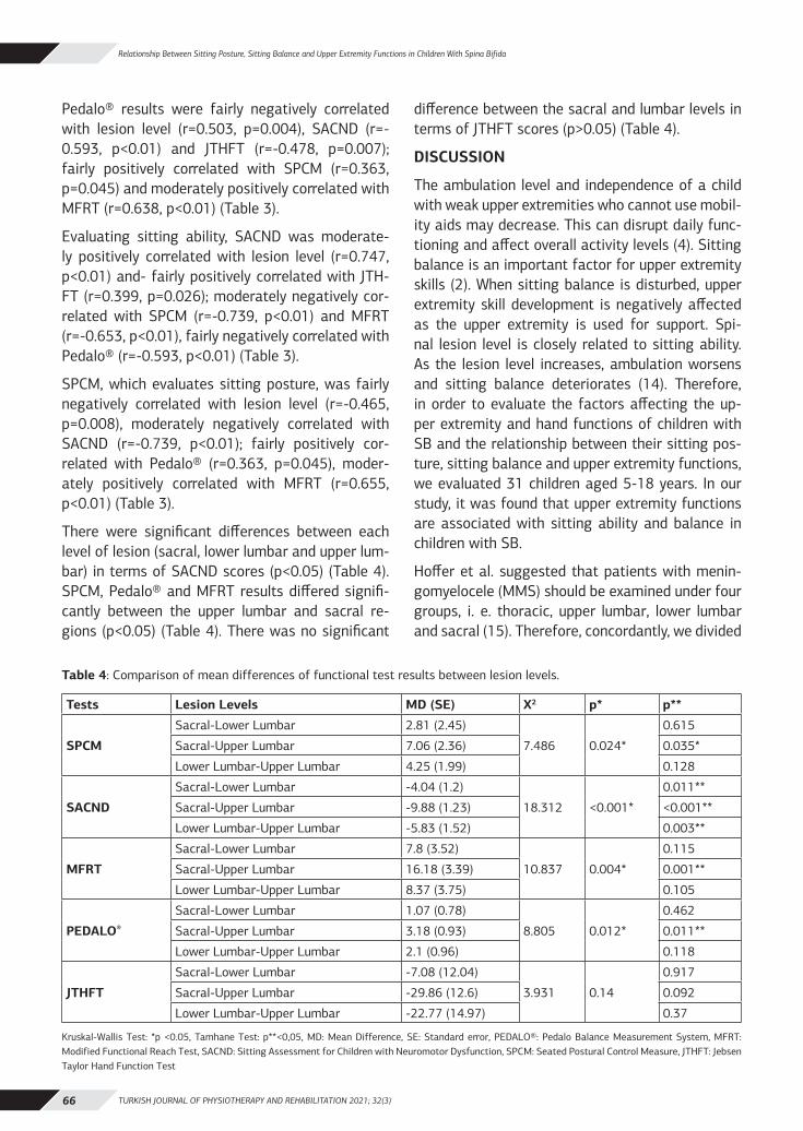
TURKISH JOURNAL OF PHYSIOTHERAPY AND REHABILITATION 2021; 32(3)66
Relationship Between Sitting Posture, Sitting Balance and Upper Extremity Functions in Children With Spina Bifida
Pedalo® results were fairly negatively correlated with lesion level (r=0.503, p=0.004), SACND (r=-0.593, p<0.01) and JTHFT (r=-0.478, p=0.007); fairly positively correlated with SPCM (r=0.363, p=0.045) and moderately positively correlated with MFRT (r=0.638, p<0.01) (Table 3).
Evaluating sitting ability, SACND was moderate-ly positively correlated with lesion level (r=0.747, p<0.01) and- fairly positively correlated with JTH-FT (r=0.399, p=0.026); moderately negatively cor-related with SPCM (r=-0.739, p<0.01) and MFRT (r=-0.653, p<0.01), fairly negatively correlated with Pedalo® (r=-0.593, p<0.01) (Table 3).
SPCM, which evaluates sitting posture, was fairly negatively correlated with lesion level (r=-0.465, p=0.008), moderately negatively correlated with SACND (r=-0.739, p<0.01); fairly positively cor-related with Pedalo® (r=0.363, p=0.045), moder-ately positively correlated with MFRT (r=0.655, p<0.01) (Table 3).
There were significant differences between each level of lesion (sacral, lower lumbar and upper lum-bar) in terms of SACND scores (p<0.05) (Table 4). SPCM, Pedalo® and MFRT results differed signifi-cantly between the upper lumbar and sacral re-gions (p<0.05) (Table 4). There was no significant
difference between the sacral and lumbar levels in terms of JTHFT scores (p>0.05) (Table 4).
DISCUSSION
The ambulation level and independence of a child with weak upper extremities who cannot use mobil-ity aids may decrease. This can disrupt daily func-tioning and affect overall activity levels (4). Sitting balance is an important factor for upper extremity skills (2). When sitting balance is disturbed, upper extremity skill development is negatively affected as the upper extremity is used for support. Spi-nal lesion level is closely related to sitting ability. As the lesion level increases, ambulation worsens and sitting balance deteriorates (14). Therefore, in order to evaluate the factors affecting the up-per extremity and hand functions of children with SB and the relationship between their sitting pos-ture, sitting balance and upper extremity functions, we evaluated 31 children aged 5-18 years. In our study, it was found that upper extremity functions are associated with sitting ability and balance in children with SB.
Hoffer et al. suggested that patients with menin-gomyelocele (MMS) should be examined under four groups, i. e. thoracic, upper lumbar, lower lumbar and sacral (15). Therefore, concordantly, we divided
Table 4: Comparison of mean differences of functional test results between lesion levels.
Tests Lesion Levels MD (SE) X2 p* p**
SPCM
Sacral-Lower Lumbar 2.81 (2.45)
7.486 0.024*
0.615
Sacral-Upper Lumbar 7.06 (2.36) 0.035*
Lower Lumbar-Upper Lumbar 4.25 (1.99) 0.128
SACND
Sacral-Lower Lumbar -4.04 (1.2)
18.312 <0.001*
0.011**
Sacral-Upper Lumbar -9.88 (1.23) <0.001**
Lower Lumbar-Upper Lumbar -5.83 (1.52) 0.003**
MFRT
Sacral-Lower Lumbar 7.8 (3.52)
10.837 0.004*
0.115
Sacral-Upper Lumbar 16.18 (3.39) 0.001**
Lower Lumbar-Upper Lumbar 8.37 (3.75) 0.105
PEDALO®
Sacral-Lower Lumbar 1.07 (0.78)
8.805 0.012*
0.462
Sacral-Upper Lumbar 3.18 (0.93) 0.011**
Lower Lumbar-Upper Lumbar 2.1 (0.96) 0.118
JTHFT
Sacral-Lower Lumbar -7.08 (12.04)
3.931 0.14
0.917
Sacral-Upper Lumbar -29.86 (12.6) 0.092
Lower Lumbar-Upper Lumbar -22.77 (14.97) 0.37
Kruskal-Wallis Test: *p <0.05, Tamhane Test: p**<0,05, MD: Mean Difference, SE: Standard error, PEDALO®: Pedalo Balance Measurement System, MFRT: Modified Functional Reach Test, SACND: Sitting Assessment for Children with Neuromotor Dysfunction, SPCM: Seated Postural Control Measure, JTHFT: Jebsen Taylor Hand Function Test
TURKISH JOURNAL OF PHYSIOTHERAPY AND REHABILITATION 2021; 32(3) 67
Atalay G., Acar G.
the lesion level of the cases into four corresponding groups in our study. In the literature, it is reported that MMS is the most common in lumbosacral re-gion (16). Similarly, in our study, all of the cases had lumbar and sacral region lesions. Because of the clinical status of children with upper-level le-sions with SB, it could be argued that they may have problems in attending to special education and rehabilitation institutions, and therefore, these patients are not frequently encountered in institu-tions.
A statistically significant difference was found be-tween the levels of sacral and upper lumbar lesions in terms of sitting balance and sitting posture. There was no difference between the lower lum-bar and sacral regions in terms of these variables. However, sitting ability (SACND) was found differ-ent at each lesion level. In SACND, any unwanted abnormal postural responses such as involuntary movements which take place during each module (rest and reach) and their duration are recorded (17). Since it is a method that evaluates not only the sitting ability but also the quality of sitting, we think that it differs across all lesion levels. In the present study, upper extremity functions did not differ between sacral, lower lumbar and upper lumbar levels. When the literature is examined, it is seen that the level of the lesion affects the upper extremity functions negatively (4). However, in our study, the participants had only lumbar and sacral lesions, and their upper extremity functions were not affected at these levels.
Improvement of sitting posture in children with SB is aimed to increase sitting tolerance, ensure ap-propriate pelvic-vertebral-head alignment and in-crease the level of functional independence (18). In our study, sitting balance and sitting ability im-proved with the increase in sitting posture (SPCM) results. With the increase in the level of the lesion, the sitting posture was disrupted. Thomson et al. emphasized that sitting posture is related to the level of lesion and ambulation level, which is sim-ilar to the results of our study (18). Glard et al. indicated that the level of lesion was a determi-nant factor in spinal deformities. They stated that spinal deformity was not expected at L5 and be-low, but expected at L2 and above, also indicated that T12 and higher levels are prognostic in the
development of kyphosis (19). Mummareddy et al., Dunn et al., Sibinski et al. stated that scoliosis had a negative effect on sitting balance (20-22). In the present study, no relationship was found between sitting posture and hand functions. The alignment section of the SPCM test was used to evaluate the sitting posture. The test was performed while the children were in a static state. However, evaluation of hand functions is related to dynamic posture. It was an expected result that hand functions were associated with dynamic sitting ability rather than a static sitting posture.
As the sitting ability of the patients increased, the sitting posture and functional reach results, and hand functions improved. The increase in lesion level negatively affected the sitting ability. SACND was also fairly correlated with Pedalo® assess-ment. Compared to Pedalo®, SACND can provide a more advantageous clinical evaluation method as it is an easy-to-use and inexpensive test.
The dynamic sitting balance of children with SB was measured by the Modified Functional Reach Test (MFRT). This test, which can also be used in children with neurological problems, can be applied in standing position or in sitting position when it is not possible to stand due to spinal cord injuries (10). In our study, we applied this test to all cas-es in sitting position. MFRT results of the patients decreased with the increase in the level of the le-sion. The increase of sway on Pedalo® (forward, backward, right, left sway) was also related to the results of the modified reach test of the cases and this result was reflected in the measurements. In addition, MFRT scores increased as sitting ability, speed of hand functions, sitting posture and sit-ting balance improved. In addition, we found that postural stability was an important factor affect-ing sitting activity and upper extremity functions in children with SB.
The number of studies evaluating the sitting bal-ance of children with SB through an objective mea-surement system is insufficient in the literature. In this sense, the use of Pedalo® system, which evaluates the sitting balance with numerical data, contributes to the literature. Studies investigating the balance of sitting in patients with SB are usu-ally performed to evaluate the results of surgical
TURKISH JOURNAL OF PHYSIOTHERAPY AND REHABILITATION 2021; 32(3)68
Relationship Between Sitting Posture, Sitting Balance and Upper Extremity Functions in Children With Spina Bifida
interventions to correct spinal deformities such as scoliosis (23-25). In our study, we evaluated the relationship between sitting balance and upper extremity functions with Pedalo® balance test. An increase in lesion level resulted in increased sway on Pedalo® and reduction in Pedalo® performance percentage. Swank et al. discovered that the lev-el of ambulation is related to sitting balance and stated that it is the most basic clinical finding that determines a patient’s quality of life and future walking potential (26). The findings of our study also support those of this study.
It has been observed that the hand functions of children with SB who showed increased sway on Pedalo® balance board are slower. Improving the sitting ability and upper extremity functions of chil-dren with SB can improve their participation in life and their quality of life. In our study, we evaluat-ed the sitting balance of children with SB using a method that provides objective data such as Peda-lo® Balance Assessment System. Pedalo® is useful in clinical practice to evaluate the dynamic stabil-ity in a short time, as short as one minute (6, 27). However, this method of measurement is insuffi-cient to show how the sitting balance will change during functioning. Therefore, we think it would be more appropriate to use it with other clinical bal-ance tests. We believe that the results of our work will be guided by the development of new seating arrangements for children with SB and of rehabili-tation programs to improve sitting posture, sitting balance and upper extremity functions.
Lack of power analysis for the sample size and the small number of participants are the limitations of our study.
In children with SB, upper extremity functions and functional independence can be increased by im-proving sitting postures and sitting balance start-ing from the early period. We think that studies in-vestigating sitting mechanisms and exercises that will improve the sitting abilities and hand functions of children with SB are needed.
Source of Finance: During this study, no moral and material support was received from any phar-maceutical company that has a direct connection with the research subject, or from a company that provides or produces medical instruments and ma-
terials, which may negatively affect the evaluation process of this study.
Conflict of Interest: The authors declare no con-flicts of interest.
Ethical Approval: The permission was obtained from Marmara University Ethics Committee for the study (22.02.2016-23).
Informed Consent: Consent was obtained from the participants’parents.
Author Contribution: Concept – GA1, GA2; Design – GA1, GA2; Supervision – GA2; Resources and Fi-nancial Support – GA1, GA2; Materials – GA1; Data Collection and/or Processing – GA1, GA2; Analysis and/or Interpretation – GA1, GA2; Literature Re-search – GA1, GA2; Writing Manuscript – GA1, GA2; Critical Review – GA1, GA2.
Acknowledgments: The current study was pre-sented as a poster at the 6th National Physiother-apy and Rehabilitation Congress held in Ankara, in May 2017, and published as an abstract in Turkish Journal of Physiotherapy and Rehabilitation.
REFERENCES1. Ardolino EM, Flores MB, Ferreira G, Jeantete SN, Manella, KM. In-
terrater reliability of the pediatric neuromuscular recovery scale in children with spina bifida. Dev Neurorehabil. 2020; 23(3): 160-165.
2. Pauly M, Cremer R. Levels of mobility in children and adoles-cents with spina bifida-clinical parameters predicting mobility and maintenance of these skills. Eur J Pediatr Surg. 2013; 23(2): 110-114.
3. Patel SK, Staarmann B, Heilman A, Mains A, Woodward J, Bier-brauer KS. Growing up with spina bifida: bridging the gaps in the transition of care from childhood to adulthood. Neurosurg Focus. 2019; 47(4): E16.
4. Saygi EK, Ozsoy T, Baskaya S, Cicek C, Honac O, Devecioglu G et. al. Assessment of sitting abilities and upper extremity func-tions according to lesion level in children with spina bifida/Spina bifidali cocuklarda lezyon seviyesine gore oturma becerisi ve ust ekstremite fonksiyonlarinin incelenmesi. Turk J Phys Med Reha-bil. 2016; 62(4): 303-308.
5. Hoglund A, Norrlin S. Influence of dual tasks on sitting postural sway in children and adolescents with myelomeningocele. Gait Posture. 2009; 30: 424-430.
6. Ahmad I., Noohu MM., Verma S. Validity and responsiveness of balance measures using Pedalo®-Sensomove balance device in patients with diabetic peripheral neuropathy. J Clin Diagn Res. 2019; 13 (6):1-4.
7. Fife S, Roxborough LA, Story M, Field D, Harris SR, Armstrong RW. Reliability of a measure to assess outcomes of adaptive seating in children with neuromotor disabilities. Can J Rehabil. 1993; 7:11–13.
8. McDonald R, Surtees R. Longitudinal study evaluating a seating system using a sacral pad and kneeblock for children with cere-

TURKISH JOURNAL OF PHYSIOTHERAPY AND REHABILITATION 2021; 32(3) 69
Atalay G., Acar G.
bral palsy. Disabil Rehabil. 2007; 29:1041–1047.9. Reid, D. Development and preliminary validation of an instru-
ment to assess quality of sitting in children with neuromotor dysfunction. Phys Occup Ther Pediatr. 1995; 15: 53-81.
10. Abou L, Sung J, Sosnoff JJ, Rice LA. Reliability and validity of the function in sitting test among non-ambulatory individuals with spinal cord injury. J Spinal Cord Med. 2020; 43(6): 846-853.
11. Jebsen R, Taylor N, Trieschman R. An objective and standardized test of hand function. Arch Phys Med Rehabil. 1969; 50: 311 -319.
12. Akoglu H. User’s guide to correlation coefficients. Turk J Emerg Med. 2018;18(3):91-93.
13. Alpar R. Spor sağlık ve eğitim bilimlerinden örneklerle uygu-lamalı istatistik geçerlilik-güvenirlik-SPSS’de çözümleme adım-larıyla birlikte. 3. Baskı. Ankara: Detay Yayıncılık; 2016.
14. Pauly M, Cremer R. Levels of mobility in children and adoles-cents with spina bifida—clinical parameters predicting mobil-ity and maintenance of these skills. Eur J Pediatr Surg. 2013; 23(02): 110-114.
15. Hoffer MM, Feiwell E, Perry R, Perry J, Bonnett C. Functional am-bulation in patients with myelomeningocele. J Bone Joint Surg. 1973; 55: 137–148.
16. Norrlin S, Strinnholm M, Carlsson M, Dahl M. Factors of signifi-cance for mobility in children with myelomeningocele. Acta Pe-diatr. 2003; 92: 204-210.
17. Kim DH, An DH, Yoo WG. Changes in trunk sway and impairment during sitting and standing in children with cerebral palsy. Tech-nol Health Care. 2018; 26(5): 761-768.
18. Thomson JD, Segal LS. Orthopedic management of spina bifida. Dev Disabil Res Rev. 2010; 16:96 -103.
19. Glard Y, Launay F, Viehweger E, Hamel A, Jouve JL, Bollini G. Neu-rological classification in myelomeningocele as a spine deformi-ty predictor. J Pediatr Orthop B. 2007; 16:287–292.
20. Mummareddy N, Dewan MC, Mercier MR, Naftel RP, Wellons JC 3rd, Bonfield CM. Scoliosis in myelomeningocele: epidemiolo-gy, management, and functional outcome. J Neurosurg Pediatr. 2017; 20(1):99–108.
21. Dunn RN, Bomela LN. Kyphectomy in children with severe myelo-meningocele-related kyphosis. Spine Deform. 2016; 4(3):230– 236.
22. Sibinski M, Synder M, Higgs ZCJ, Kujawa J, Grzegorzewski A. Quality of life and functional disability in skeletally mature pa-tients with myelomeningocele-related spinal deformity. J Pediatr Orthop B. 2013; 22:106–109.
23. Bartnicki B, Synder M, Kujuawa J. Sitting stability in skeletally mature patients with scoliosis and myelomeningocele. Orthop Trauma Rehabil. 2012; 4(6) Vol 14: 363-369.
24. Kose KC, Inanmaz ME, Uslu M. Kyphectomy for congenital ky-phosis due to meningomyelocele: a case treated with a modified approach to skin healing. Int Wound J. 2012; 9(3):311-315.
25. Garg S, Matthew O, Rathjen K. Kyphectomy improves sitting and skin problems in patients with myelomeningocele. Clin Orthop Relat Res. 2011; 469:1279–1285.
26. Swank M, Dias L. Walking ability in spina bifida patients: a model for predicting future ambulatory status based on sitting balance and motor level. J Pediatr Orthop. 1994; 14: 715-718.
27. Khan N, Ahmad I, Noohu MM. Association of disease duration and sensorimotor function in type 2 diabetes mellitus: beyond diabetic peripheral neuropathy. Somatosens Mot Res. 2020; 37(4): 326-333.
TURKISH JOURNAL OF PHYSIOTHERAPY AND REHABILITATION 2021; 32(3)70
GENÇ YETİŞKİNLERDE POSTÜRÜN SAĞLIKLA İLGİLİ YAŞAM KALİTESİ ÜZERİNE ETKİSİNİN ARAŞTIRILMASI
ARAŞTIRMA MAKALESİ
ÖZAmaç: Postür, fiziksel ve psikososyal iyilik hali ile ilişkilendirilmektedir. Postüral sapmalar çoğu zaman asemptomatik olduğu için göz ardı edilebilmekle beraber, uzun dönemde kas iskelet sistemi problemlerine yol açabilir. Bu retrospektif çalışmanın amacı, genç yetişkin bireylerde postürün sağlıkla ilgili yaşam kalitesi üzerine etkisini araştırmaktı.
Yöntem: Yaşları 18-34 yıl arasında değişen 90 sağlıklı birey, postür ve yaşam kalitesi açısından değerlendirildi. Postür, postüral dizilim açısından Reedco Postür Skoru (RPS) ve postüral simetri açısından posterior gövde simetri indeksi (POTSI) ile değerlendirildi. Bireylere yaşam kalitesini değerlendirmek için SF-36 Kısa Form uygulandı.
Sonuçlar: Sonuçlar, POTSI gövde simetri indeksi ile SF-36 Kısa Form Anketinin fiziksel fonksiyon (r=-0,317), mental sağlık (r=-0,425), zindelik (r=-0,296), ağrı (r=-0,358), genel sağlık algılaması (r=-0,284) puanları arasında negatif yönlü ve düşük-orta şiddette ilişki olduğunu gösterdi (p<0,05). RPS ile SF-36 Kısa Form fiziksel fonksiyon (r=0,347), fiziksel sorunlara bağlı rol kısıtlılıkları (r=0,228), sosyal fonksiyon (r=0,328), mental sağlık (r=0,515), zindelik (r=0,388), ağrı (r=0,398) ve genel sağlık algılaması (r=0,369) puanları arasında da pozitif yönlü ve düşük-orta-yüksek şiddetlerde ilişki saptandı (p<0,05).
Tartışma: Bu çalışmada, sağlıklı genç yetişkinlerde postüral dizilimde normalden sapmaların ve asimetrilerin olduğu ve bu postüral sapmaların yaşam kalitesini olumsuz etkilediği bulundu. Sağlıklı genç bireylerde, asemptomatik dahi olsa, postüral değişikliklerin yaşam kalitesi üzerindeki bu negatif etkisinin, ileri dönemde karşılaşılabilecek olası kas-iskelet sistemi problemleri açısından dikkate alınmasını önermekteyiz.
Anahtar Kelimeler: Genç Yetişkin; Postür; Yaşam Kalitesi.
THE INVESTIGATION OF THE EFFECTS OF POSTURE ON HEALTH RELATED QUALITY OF LIFE IN YOUNG
ADULT POPULATION
ORIGINAL ARTICLE
ABSTRACTPurpose: Posture is associated with psychosocial well-being. Postural deviations are often underestimated in asymptomatic subject; however, they cause musculoskeletal problem in long term. The purpose of this retrospective study was to investigate effects of posture on health-related quality of life in young adults.
Methods: Ninety healthy adults aged between 18 and 34 years were evaluated for quality of life. Posture was assessed using Reedco Postural Score (RPS) for alignment and Posterior Trunk Symmetry Index (POTSI) for symmetry. SF-36 Short Form was applied for evaluating quality of life.
Results: Results showed negative and low-moderate correlations between POTSI and physical function (r=-0.317), mental health (r=-0.425), vitality (r=-0.296), pain (r=-0.358), and general health perception scores of SF-36 Short Form (r=-0.284) (p<0.05). There was positive and low-moderate-high correlation between RPS and SF-36 Short Form physical function (r=0.347), role limitations due to physical problems (r=0.228), social function (r=0.328), mental health (r=0.515), vitality (r=0.388), pain (r=0.398), and general health perception (r=0.369) (p<0.05).
Conclusion: This study demonstrated that postural deviations and asymmetries in healthy young adults negatively affects quality of life. Even in asymptomatic healthy young adults, negative effects of postural deviations on quality of life should be considered as a provocative factor in future musculoskeletal problems.
Key Words: Young Adult; Posture; Quality of Life.
Turkish Journal of Physiotherapy and
Rehabilitation2021 32(3)70-77
Doç. Dr. Gözde YAĞCI1
Prof. Dr. Nilgün BEK2
ISSN: 2651-4451 • e-ISSN: 2651-446X
Yağcı G., Bek N., Genç Yetişkinlerde Postürün Sağlıkla İlgili Yaşam Kalitesi Üzerine Etkisinin Araştırılması, Turk J Physiother Rehabil. 2021; 32(3):70-77. doi: 10.21653/tjpr.818369
1 Hacettepe Üniversitesi, Fizik Tedavi ve Rehabilitasyon Fakültesi, Altındağ, Ankara
2 Lokman Hekim Üniversitesi, Sağlık Bilimleri Fakültesi, Fizyoterapi ve Rehabilitasyon Bölümü
Correspondence (İletişim):
Gözde YağcıHacettepe Üniversitesi, Fizik Tedavi ve
Rehabilitasyon Fakültesi, Altındağ, Ankara, 06100, Türkiye
Email: [email protected] Phone: 03123051576 Fax: 03123052012
ORCID ID: 0000-0002-4603-7162
Prof. Dr. Nilgün BekEmail: [email protected]
ORCID ID:0000-0002-2243-5828
Received: 30.10.2020 (Geliş Tarihi)Accepted: 28.04.2021 (Kabul Tarihi)
CC BY - NC
Content of this journal is licensed under a Creative Commons Attribution-NonCommercial 4.0 International License.
TURKISH JOURNAL OF PHYSIOTHERAPY AND REHABILITATION 2021; 32(3) 71
Yağcı G., Bek N.
GİRİŞ
Postür vücut parçalarının birbiri ile ilişkili olarak dizilimi veya oryantasyonu olarak tanımlanmakta-dır. Bu dizilim, yer çekimi etkisine, kas gerilimine ve kemik yapıların bütünlüğüne bağlıdır (1). İyi pos-tür, pozisyona (dik duruş, yatış, oturma, öne eğilme vb.) bağlı olmaksızın, yaralanmaya veya progresif deformitelere karşı vücut yapılarını koruyan kas ve iskelet yapılarının dengeli olma durumudur. Böyle bir postürde, kaslar etkin bir şekilde çalışmakta ve torakal ve abdominal organlar için ideal pozisyon-lar oluşmaktadır (2). Kötü postürde vücut parçaları arasındaki denge bozulur ve bu durum birbirleri ile olumsuz bir ilişki oluşmasına neden olur. Destekle-yici yapılarda gerilim artar; daha az etkin bir vücut dengesi oluşur; enerji tüketimi artar ve destekleyen yapılar yetersiz kalır (2).
Literatürde bireylerin postürünü etkileyen intrinsik ve ekstrinsik faktörler tanımlanmaktadır. Bu fak-törler arasında kalıtım, bireyin yaşamındaki çevre-sel veya fiziksel durumlar, sosyo-ekonomik seviye, emosyonel faktörler ve büyüme ve gelişim sırasın-da oluşan fizyolojik değişikliklerden söz edilmekte-dir (2,3).
Postür, fiziksel ve psikososyal iyilik hali ile ilişki-lendirilmektedir. Bu nedenle postürü ve postüral dizilimi geliştirmek, rehabilitasyon programlarının önemli amaçlarındandır (4). İleri yaştaki bireyler üzerinde yapılan çalışmalarda, spinal postüral deği-şikliklerin yaşla arttığı ve iyi spinal postürün, günlük yaşam aktivitelerinde bağımsızlıkla ilişkili olduğu gösterilmiştir (5). Ayrıca gövde deformitesi olan bi-reylerde, subjektif sağlık ve iyilik hissi ile yaşamdan memnuniyet skorlarının daha düşük olduğu bulun-muştur (6). Ancak sağlıklı bireyler üzerinde yapılan çalışmalar yetersizdir ve az da olsa olası postüral asimetrilerin erken yetişkinlikte saptanması, ileri yaşlarda gelişebilecek deformite veya patolojilerin önlenmesinde önem kazanacaktır.
Çocukluk çağında postüral değişikliklerin görülme oranının yüksek olduğu belirtilmektedir (3). Bu pos-türal değişikliklerin bazılarının geçici olduğu ve bü-yüme döneminde normal postüral gelişim ile birlikte düzeldiği; bazı postüral değişikliklerin ve asimetrile-rin ise kalıcı olarak yetişkinlikte de devam ettiği ve sağlıkla ilgili yaşam kalitesini de olumsuz etkilediği ifade edilmektedir (3). Çocukluk döneminde pos-
türal değişikliklerin görülmesi büyüme döneminde normal olarak var olan yüksek mobilite ve fleksi-biliteye dayandırılmaktadır (3). Çocuklardaki artmış eklem hareket genişliği, postüral dizilimde geçici deviasyonlara sebep olabilmektedir (3). Ancak bu deviasyonların yetişkinlik döneminde var olması durumu, anormal olarak nitelendirilmektedir (3,4,7). Dolayısıyla büyüme döneminde olduğu kadar, genç yetişkin dönemde de postürün değerlendirilmesinin ve kötü postür ve/veya postüral asimetrilerin sap-tanması gerekliliği vurgulanmaktadır (3). Czakwari ve ark., postüral hataların genç yetişkinlerde yaygın olduğunu ve sedanter bireylerde olduğu kadar fizik-sel olarak aktif kişilerde de görülebileceğini belirt-mişlerdir (7).
Yetişkin popülasyonda, kas-iskelet sistemindeki bi-yomekanik değişikliklerin ağrı ve fonksiyon bozuk-luklarına yol açabildiği belirtilmektedir (8). Postü-ral anomalilerin, klinik bulgu vermesinin daha çok ileri dekatlarda (orta yaş ve yaşlılık) ortaya çıktığı ifade edilmektedir. Ortaya çıktığı durumlarda ise, eşlik eden bulgular çeşitli olabilmekte, tedavi uzun bir süreci kapsayabilmekte ve bireyin yaşam kali-tesini olumsuz etkileyebilmektedir (9). Bu sebeple, asemptomatik sağlıklı bireylerde, ileri dönemde patolojik bir duruma neden olabilecek ve yaşam kalitesini etkileyecek postüral belirleyicilerin sap-tanması, etkin koruyucu stratejilerin geliştirilmesi açısından önemli olacaktır. Bu çalışmanın amaçla-rından biri, genç popülasyonda postürü ve sağlıkla ilgili yaşam kalitesini araştırmak iken diğeri, genç yetişkin bireylerde postürün yaşam kalitesi üzerine etkilerini incelemekti.
YÖNTEM
Bireyler
Hacettepe Üniversitesi, Fizik Tedavi ve Rehabilitas-yon Fakültesi Ortez ve Biyomekanik Ünitesi’nde 1 Aralık 2018 ile 31 Mayıs 2020 tarihleri arasında değerlendirilmiş sağlıklı genç yetişkin bireylerden, postür değerlendirmeleri yapılmış olan ve yaşam kalitesi anketini dolduran bireylere ait değerlendir-me verileri retrospektif olarak incelenmiş, 18-34 yaşları arasında, 90 birey (63 kadın, 27 erkek) bu araştırmaya dahil edildi. Tüm bireyler, fiziksel ola-rak aktif, herhangi bir hastalığı olmayan, düzenli

TURKISH JOURNAL OF PHYSIOTHERAPY AND REHABILITATION 2021; 32(3)72
Genç Yetişkinlerde Postürün Sağlıkla İlgili Yaşam Kalitesi Üzerine Etkisinin Araştırılması
ilaç kullanmayan, kendi ifadelerine göre belirtildiği üzere ortopedik veya nörolojik bir bozukluğa sahip olmayan ve son altı aydır vücudunun herhangi bir yerinde ağrı olmayan bireylerden seçildi. Herhangi bir konjenital anomalisi olan, skolyozu olan, cerrahi veya gebelik geçirmiş olan bireyler çalışmaya da-hil edilmedi. Bu retrospektif çalışma için Hacettepe Üniversitesi Girişimsel Olmayan Klinik Araştırıl-malar Etik Kurulu tarafından, 6 Ekim 2020 tarihli, 2020/16 toplantısında, GO20/870 kayıt numarası ile onay alındı.
Değerlendirme Yöntemleri
Bireylere ait yaş, boy, vücut ağırlığı, beden kütle in-deksi gibi demografik bilgiler kaydedildi. Bireylerin postürleri, Reedco Postür Skoru (RPS) adı verilen bir postür skorlama skalası kullanılarak, birinci yazar tarafından değerlendirildi. RPS, 1974 yılından bu yana sagital ve koronal planda ayaktan-başa, tüm vücut postürünün değerlendirilmesi için kullanılan standart bir yöntemdir (10). Ayrıca uygulanması ko-lay ve maliyet etkin bir yöntem olarak yaygın olarak kullanılmaktadır. Bu yöntemle 10 postüral özellik açısından birey, lateral ve posteriordan gözlemsel olarak değerlendirilir. Lateral değerlendirme, sagi-tal düzlemde boyun, üst sırt, gövde, karın bölgesi ve bel bölgesini içerirken; posterior değerlendirme, ko-ronal düzlem üzerinde baş, omuzlar, omurga, kalça-lar ve ayak bileklerini içermektedir. RPS skorlanma-sı, postüral dizilimin “0” (kötü postür veya şiddetli deviasyon), “5” (yetersiz postür veya minimal-orta şiddette deviasyon) ve “10” (iyi postür veya nor-mal dizilim) olarak 1 ile 10 arasında puanlanması şeklindedir. Maksimum puan olan 100 iyi postürü işaret ederken, %59 veya altında bir puan, postü-ral disfonksiyonu belirtmektedir (11). Çalışmamız-da RPS için sagital plan postür puanı, koronal plan postür puanı ve toplam puanı hesaplanmış ve ana-lizlere dahil edildi. Önceki bir çalışmada RPS’nin, iyi derecede gözlemciler arası (alfa katsayısı= 0,899-0,015) ve test-tekrar test güvenirliği (ICC=0,85-0,95) olduğu belirtilmiştir (12).
Bireylerin postüral simetrileri, Posterior Gövde Si-metri İndeksi (POTSI) kullanılarak değerlendirildi. POTSI, bireyin gövdesinde herhangi bir giysi yok-ken, ayakta duruş pozisyonunda sırtındaki belirli anatomik noktalardan mezura ile yapılan ölçümler-den vücut şeklini ve simetrisini değerlendiren ob-
jektif bir yöntemdir. Omuz, aksilla ve gövde için her bir bölgeden frontal asimetri indeksi ve yükseklik fark indeksi olmak üzere toplam altı indeks içerir. POTSI total skoru, bu indekslerden elde edilen skor-ların toplamıdır. Yüksek değerler artmış asimetriyi ifade etmektedir (13).
Bireylerin kendi ifadelerine dayalı sağlıkla ilgili ya-şam kalitelerinin değerlendirilmesinde SF-36 Kısa Form Anketi kullanıldı. SF-36 Kısa Form yetişkinler-de yaşam kalitesini ölçmede kullanılan en yaygın öl-çütlerden biridir (14). Bu ölçek fiziksel fonksiyon, rol kısıtlamaları (fiziksel ve emosyonel sorunlara bağ-lı), sosyal fonksiyon, mental sağlık, zindelik (enerji), ağrı ve genel sağlık algılaması gibi alt başlıklarla sağlığın sekiz boyutunu 36 madde ile incelemekte-dir. Ölçek alt başlıklarının toplam puanları elde edil-mektedir. Bu puanlar 0 ile 100 arasında değişmek-tedir. 100 puan iyi sağlık durumunu gösterirken, 0 puan kötü sağlık durumunu göstermektedir.
İstatistiksel Analiz
Çalışma kapsamında değerlendirilen kırk birey üze-rinden yapılan istatistiksel analizlere göre, birincil sonuç ölçümleri olan RPS ve SF-36 Kısa Form ara-sında bulunan, 0,366 değerindeki korelasyon kat-sayısına göre, %80 güç ve %95 güvenirlilik düzeyi ile örneklem büyüklüğü 56 birey olarak tespit edil-di. Bu retrospektif çalışmanın örneklemini, çalışma için planlanan süre içerisinde (Aralık 2018 ile Mayıs 2020 arası) belirtilen değerlendirilme verileri tam olan bireylerin sayısını ifade eden, 90 birey oluş-turdu. Tanımlayıcı istatistikler ortalama ± standart sapma ile ifade edildi. Tüm analizler SPSS versiyon 20.0 programı kullanılarak gerçekleştirildi. Postü-ral değerlendirme parametreleri ile yaşam kalitesi arasındaki ilişki Pearson korelasyon analizi ile test edildi. Cohen’in sınıflamasına göre, korelasyon kat-sayısı 0,10 ile 0,29 arasında ise ilişki düşük; 0,30 ile 0,49 arasında ise ilişki orta, 0,5 veya daha yüksek ise ilişki yüksek olarak kabul edildi (15). İstatistiksel yanılma olasılığı (P değeri) 0,05 olarak belirlendi.
SONUÇLAR
Bireylerin yaş ortalaması 21,63 ± 2,13 yıl idi. Bi-reylerin demografik ve klinik özellikleri Tablo 1’de gösterilmiştir.
Bireylerin %90’ında RPS’ye göre (80 birey) hem sa-gital veya koronal planda postüral sapmaların mev-

TURKISH JOURNAL OF PHYSIOTHERAPY AND REHABILITATION 2021; 32(3) 73
Yağcı G., Bek N.
cutken, POTSI değerlendirmesine göre %86’sında (77 birey) da postüral asimetrilerin bulunduğu tes-pit edildi.
Tablo 2’de POTSI gövde simetrisi ve RPS bulguları ile yaşam kalitesi değerlendirme bulguları arasın-daki ilişki verilmiştir. Korelasyon analizi sonuçları-na göre, POTSI gövde simetri skoru ile SF-36 Kısa Form anketi fiziksel fonksiyon (r=-0,317), men-tal sağlık (r=-0,425), zindelik (r=-0,296), ağrı (r=-0,358), genel sağlık algılaması (r=-0,284) alt başlık-larının puanları arasında istatistiksel olarak anlamlı ilişki bulundu ve bu ilişki negatif yönlü ve düşük-orta şiddetteydi. RPS ile SF-36 Kısa Form fiziksel fonksi-yon (r=0,347), fiziksel sorunlara bağlı rol kısıtlılıkları
(r=0,228), sosyal fonksiyon (r=0,328), mental sağlık (r=0,515), zindelik (r=0,388), ağrı (r=0,398), genel sağlık algılaması (r=0,369) alt başlıklarının arasın-da da ilişki istatistiksel olarak anlamlıydı. Bu ilişki ise pozitif yönlü ve düşük-orta-yüksek şiddetlerdey-di.
RPS’nin sagital plan ve koronal plan postür skorla-rının yaşam kalitesi ile ilişkisi Tablo 3’ de verilmiş-tir. Sagital plan postür skoru ile SF-36 Kısa Form yaşam kalitesi değerlendirme bulguları arasında, emosyonel sorunlara bağlı rol kısıtlılıkları alt başlığı hariç tüm parametrelerde istatistiksel olarak an-lamlı (fiziksel fonksiyon için r=0,314, fiziksel sorun-lara bağlı rol kısıtlılıkları r=0,266, sosyal fonksiyon
Tablo 1: Bireylerin Özellikleri.
Özellik Olgular (n=90)
X±SS Minimum-Maksimum
Yaş (yıl) 21,63±2,13 18-34
Cinsiyet (Kadın/ Erkek) 63/27 %70 / %30
Boy (m) 168,96±8,48 154-190
Vücut Ağırlığı (kg) 63,51±12,02 42-96
Beden Kütle İndeksi (kg/m2) 22,14±3,11 15,62-33,12
POTSI (cm) 14,34±8,78 0-34,71RPS (%) Sagital Plan Koronal Plan Toplam Skor
80,99±15,2185,65±12,6583,32±11,65
50-10040-10060-100
SF-36 Kısa Form (0-100) Fiziksel Fonksiyon Fiziksel Sorunlara Bağlı Rol Kısıtlamaları Emosyonel Sorunlara Bağlı Rol Kısıtlamaları Sosyal Fonksiyon Mental Sağlık Zindelik (Enerji) Ağrı Genel Sağlık Algılaması
88,20±11,4671,70±32,7061,42±30,5081,12±20,6067,98±20,8859,67±21,5175,52±20,4271,84±19,77
55-1000-1000-100
12,50-10020-10015 -100
12,50-10015-100
POTSI: Posterior Gövde Simetri İndeksi; RPS: Reedco Postür Skoru.
Tablo 2: Bireylerin Postür Değerlendirme Bulguları ile Yaşam Kalitesi Değerlendirme Bulguları Arasındaki İlişki.
SF-36 Kısa FormPOTSI RPS
r p r pFiziksel FonksiyonFiziksel Sorunlara Bağlı Rol KısıtlılıklarıEmosyonel Sorunlara Bağlı Rol Kısıtlılıkları Sosyal FonksiyonMental SağlıkZindelik (Enerji)Ağrı Genel Sağlık Algılaması
-0,317-0,127-0,040-0,189-0,425-0,296-0,358-0,284
0,004*0,2610,7290,104
<0,001*0,008*0,001*0,011*
0,3470,2280,1390,3280,5150,3880,3980,369
0,002*0,047*0,233
0,005*<0,001*0,001*
<0,001*0,001*
*p<0,05. POTSI: Posterior Gövde Simetri İndeksi; RPS: Reedco Postür Skoru.

TURKISH JOURNAL OF PHYSIOTHERAPY AND REHABILITATION 2021; 32(3)74
Genç Yetişkinlerde Postürün Sağlıkla İlgili Yaşam Kalitesi Üzerine Etkisinin Araştırılması
için r=0,306, mental sağlık için r=0,400, zindelik için 0,302, ağrı için r=0,340, genel sağlık algılama-sı için r=0,265) düşük-orta şiddette pozitif bir ilişki bulundu. Koronal plan postür skoru ile ise fiziksel ve emosyonel sorunlara bağlı rol kısıtlılıkları paramet-releri hariç, yine tüm parametrelerde (fiziksel fonk-siyon için r=0,260, sosyal fonksiyon için r=0,250, mental sağlık için r=0,477, zindelik için r=0,350, ağrı için r=0,324, ve genel sağlık algılaması için r=0,358) istatistiksel olarak anlamlı düşük-orta şid-dette pozitif bir ilişki bulundu.
TARTIŞMA
Bu çalışmada, sağlıklı genç yetişkinlerde postüral sapma ve asimetri olduğu tespit edildi. Ayrıca bu postüral değişikliklerin sağlıkla ilgili yaşam kalite-sini olumsuz etkilediği bulundu. Postürden, özellikle yaşam kalitesinin fiziksel fonksiyon, sosyal fonksi-yon, mental sağlık, zindelik, ağrı ve genel sağlık al-gılaması gibi parametreleri etkilendi.
İyi postür, her bir vücut segmentinin gravite mer-kezinin altındaki segmentin üzerinde vertikal olarak yerleşmesi durumu olarak tanımlanmaktadır. Seg-mentlerin grative merkezlerinin yer değiştirmesi durumunda postüral sapmalar/anomaliler ortaya çıkmaktadır (16). Bu araştırmada, sağlıklı genç ye-tişkin bireylerde hem sagital veya koronal planda postüral sapmaların (80 birey, bireylerin % 90’ı), hem de postüral asimetrilerin (77 birey, bireylerin % 86’sı) bulunduğu tespit edildi. Sagital plandaki sapmalar başın anteriora tilti, torakal kifozda artış, lumbar lordozda düzleşme veya artış iken; koronal plandaki sapmalar, başın laterale tilti, gövdenin la-terale tilti, omuz ve kalça yükseklik farkları ve ayak-ta pronasyon şeklindeydi. Benzer olarak Czakwari
ve ark. (7), genç yetişkinlerde koronal ve sagital planda postüral sapmaların insidansının yüksek ol-duğunu belirtmiştir. En yaygın postüral sapmaların lumbar hipolordoz (% 71 görülme sıklığı), torakal hiperkifoz (% 58) ve skolyoz (% 58) olduğu ifade edilmiştir. Maslen ve Straker (17), çocuklarda genç yetişkinlere göre artmış omurga fleksiyonu ve asi-metrisi tespit etmişler ve hem çocuklar hem de genç yetişkinlerde postüral değişikliklerin meydana geldiğini vurgulamışlardır.
Omurganın kas iskeletsel patolojileri genel yetişkin popülasyonda ağrıya yol açarak yaşam kalitesini azaltmaktadır (18). Postürde normalden sapma-ların da omurgada patolojik ortopedik durumların oluşmasına önemli bir rol oynadığı belirtilmektedir (19). Sagital ayakta duruş postünün ağrı, fiziksel fonksiyon kaybı ve azalmış yaşam kalitesi ile ilişki-li olduğunu gösteren pek çok klinik çalışma bulun-maktadır (20-22). Genellikle ileri yaşlarda omurga deformitesi oluşturmasıyla fonksiyon kaybına yol açması ile ilişkili bulunan bu postür, anterior sagital denge ve artmış pelvik tilt olarak belirtilmektedir (20). Çalışmamızda, RPS’ye göre, postüral dizilimde meydana gelen sapma arttıkça, yaşam kalitesinin fiziksel fonksiyon, fiziksel sorunlara bağlı rol kısıtlı-lıkları, sosyal fonksiyon, mental sağlık, zindelik, ağrı ve genel sağlık algılaması gibi pek çok bileşeninin olumsuz etkilendiği saptandı. Çalışmamızda ayrıca RPS anketi sonuçları, sagital ve koronal plan olarak ikiye ayrılarak, bu iki planda saptanan postür puan-larının yaşam kalitesi ile ilişkisi tek tek incelendi. Sonuç olarak, sagital ve koronal planda analiz edi-len postür puanlarının da total puana benzer olarak yaşam kalitesi ile ilişkili olduğu bulundu. Bu ilişki total puan için orta-yüksek şiddette iken, sagital ve
Tablo 3: Reedco Postür Skoru Sagital ve Koronal Planda Postürü Analizi Alt Başlıkları ile Yaşam Kalitesi Arasındaki İlişki.
SF-36 Kısa Form
RPS
Sagital Plan Koronal Plan
r p r pFiziksel FonksiyonFiziksel Sorunlara Bağlı Rol KısıtlılıklarıEmosyonel Sorunlara Bağlı Rol Kısıtlılıkları Sosyal FonksiyonMental SağlıkZindelik (Enerji)Ağrı Genel Sağlık Algılaması
0,314 0,2660,2190,3060,4000,3020,3400,265
0,006* 0,020*
0,059 0,010* 0,001* 0,008* 0,003* 0,021*
0,2600,104-0,0010,2500,4770,3500,3240,358
0,023*0,3710,993
0,036*<0,001* 0,002*0,004* 0,001*
*p<0,05. RPS: Reedco Postür Skoru.

TURKISH JOURNAL OF PHYSIOTHERAPY AND REHABILITATION 2021; 32(3) 75
Yağcı G., Bek N.
koronal plan puanları için düşük-orta şiddetteydi. O’Neill ve ark. araştırmalarında RPS’nin % 59 veya daha az olması durumunu postüral disfonksiyon olarak tanımlamışlardır (11). Çalışmamızda bu ka-tegoriye giren herhangi bir bireye rastlanmamıştır. Çalışmamızdaki bireylerin sadece postüral sapma/değişiklik kategorisinde olduğunu söyleyebiliriz. Postür ve yaşam kalitesi ilişkisinin düşük-orta-yük-sek arasında değişen şiddetlerde tespit edilmesinin sebebi, bireylerin sağlıklı olması nedeni ile, postü-ral değişimin düşük miktarda olmasından kaynaklı olabilir. Ancak sadece düşük şiddette bir postüral değişimi ifade eden bu postüral sapmanın dahi, bireylerin yaşam kalitesi üzerinde olumsuz etkileri-nin tespit edildiğini de vurgulamak gerekmektedir. Murphy ve ark. (23) da çalışmamıza benzer olarak, okul çağı çocuklarında artmış fleksiyon postürünün, boyun ve bel ağrısı ile ilişkili olduğunu bulmuşlardır.
Koronal plan gövde simetrisinin klinik olarak değer-lendirmesi, çocuklarda ve adolesanlarda postüral hataların erken dönemde saptanmasında yardımcı bir yöntem olarak önerilmektedir (24). Gövde si-metrisinin iki boyutlu (fotoğraf üzerinden) analizi, anterior ve posterior gövde üzerinden belirli anato-mik noktalar arasındaki mesafelerin ölçülerek, PO-TSI ile oranlanmasıyla gerçekleştirilmektedir. Mat-lega ve ark. sağlıklı çocuklarda POTSI değerlerinin kronolojik yaş ve cinsiyetten bağımsız olduğunu belirtmişler ve çocukluk döneminden yaşlılığa ka-dar geniş bir yaş spektrumunda gövde simetrisinin değerlendirilmesinde kullanılmasının uygunluğunu vurgulamışlardır (25). Daha önceki bir araştırma-da POTSI için 27,5 puanın altındaki değerler için, gövde asimetrisinin normal sınırda olduğunu ifade ettiği belirtilmiştir (26). Çalışmamızdaki bireyleri bu parametre açısından incelediğimizde, 81 (%90) bireyin gövde asimetrisinin normal sınırlarda oldu-ğunu; 9 (%10) bireyin omurgasında laterale sapma olduğunu söyleyebiliriz. Ayrıca çalışmamızda gövde asimetrisi arttıkça, sağlıkla ilgili yaşam kalitesinin azaldığı bulundu. Yine bu iki değişken arasında-ki ilişkinin düşük-orta şiddette bulunması, sağlıklı bireylerde tespit edilen postüral asimetrinin hafif olmasına bağlanabilir. Yaşam kalitesinin özellikle bireylerin fiziksel fonksiyon, mental sağlık, zindelik, ağrı ve genel sağlık algılaması gibi komponentleri-nin etkilendiği tespit edildi. Benzer olarak, Kamitani ve ark. yaşlı bireylerde spinal postür parametrele-
rinin ileri dönemde günlük yaşam aktivitelerinde bağımsızlıkla ilişkili olduğunu bulmuşlardır (27). Ta-kahashi ve ark. araştırmalarında, gövde deformite-si olan yaşlı bireylerin, olmayanlara göre subjektif sağlık ve yaşam memnuniyeti algısı açısından daha düşük skorlara sahip olduğunu belirtmişlerdir (6). Dolayısıyla çalışmamızda tespit edilen genç yetiş-kinlerdeki postüral asimetrilerin yaşam kalitesi ile ilişkisini düşündüğümüzde, postürün ileri yaşlarda yaşam kalitesi üzerine artabilecek olumsuz etkileri-ni önlemek için, erken dönemden itibaren koruyucu yaklaşımlar geliştirilmesini önermekteyiz.
Fizyoterapi ve rehabilitasyon çatısında düzenlenen egzersiz programlarının birincil amacı iyi postür geliştirmektir. Bunun amacı da yetersiz veya kötü postürü olan bireylerin daha az iyi görünmesi, daha kötü öz-imaj ve özgüven geliştirmeye yatkın olması-dır (16). Ayrıca belirli postürlerin belirli yaralanma-lar için zemin hazırladığı belirtilmektedir. Örneğin Cowan ve ark., alt ekstremite postüral deviasyonu olan bireylerin aşırı kullanma kaynaklı yaralanma prevalansının daha yüksek olduğunu bulmuşlardır (28). Sluming ve Scutt, bel ağrısının postüral bo-zukluklar nedeni ile olduğu veya bel ağrısının sonu-cu olarak postüral bozuklukların ortaya çıkabildi-ğini belirtmişlerdir (29). Bu nedenle, çalışmamızın bulgularına dayanarak genç yetişkinlik döneminde saptadığımız, asemptomatik olan postüral değişik-liklerin ve asimetrilerin, ileri yaşlarda yaralanma ve rahatsızlık riskini artırabileceği öngörülerek, ileri yaşlarda da dikkate alınması gerekebilir.
Bu çalışmanın bazı limitasyonları mevcuttur. Ret-rospektif bir çalışma olan çalışmamızda, postürün değerlendirmesinde klinik skalalar kullanılmıştır. Üç boyutlu hareket analizi sistemlerinin kullanılmasıy-la, postürle ilgili daha objektif veri sağlanabilecek-tir. Bu çalışmada postür değerlendirmesi kapsa-mında kullanılan yöntemler gereği, özellikle gövde üzerinde durulmuştur. İleri prospektif çalışmalarda tüm vücudu detaylı inceleyen değerlendirme yön-temlerinin kullanılması ile, konuya ilave bir katkı sağlanabilecektir. Bu çalışmanın sonuçları genç yetişkin bireyler için geçerlidir. Diğer yaş grupları için yapılacak genellemelerde bu konu dikkate alın-malıdır.
Sonuç olarak, retrospektif olarak gerçekleştirilen bu çalışmanın sonuçları, sağlıklı yetişkinlerde de

TURKISH JOURNAL OF PHYSIOTHERAPY AND REHABILITATION 2021; 32(3)76
Genç Yetişkinlerde Postürün Sağlıkla İlgili Yaşam Kalitesi Üzerine Etkisinin Araştırılması
postüral değişikliklerin meydana geldiği ve bu de-ğişikliklerin yaşam kalitesini olumsuz etkilediğini gösterdi. Postürün yaşam kalitesi üzerine etkileri, fiziksel fonksiyon, sosyal fonksiyon, mental sağlık, zindelik, ağrı ve genel sağlık algılaması gibi geniş bir spektrumda gözlendi. Genç yetişkinlerde tespit edilen söz konusu postüral değişimlerin ve asimet-rilerin, uzun dönemde veya ileri yetişkinlikte semp-tomatik olabileceği, postüral disfonksiyona veya kas iskelet sistemi problemlerine yol açabileceği yönünden dikkate alınması önerilmektedir.
Destekleyen Kuruluş: Yok.
Çıkar Çatışması: Herhangi bir çıkar çatışması bu-lunmamaktadır.
Etik Onay: Bu retrospektif çalışma için Hacette-pe Üniversitesi Girişimsel Olmayan Klinik Araştırıl-malar Etik Kurulu tarafından, 6 Ekim 2020 tarihli, 2020/16 toplantısında, GO20/870 kayıt numarası ile onay alındı.
Aydınlatılmış Onam: Rutindeki olguların var olan verilerini retrospektif olarak incelediğimiz için onam alınmasına gerek olmamış ve etik kurul onayı da bu şekilde alınmıştır.
Hakem Degerlendirmesi: Dış bağımsız hakemler tarafından değerlendirilmiştir.
Yazar Katkıları: Konsept – GY, NB, Tasarım - GY, Süpervizyon - NB; Kaynaklar ve Finansal Destek - GY, NB; Materyaller - GY, Veri Toplaması ve/veya İşleme - GY; Analiz ve Yorumlama: GY, NB; Literatür Tarama - GY; Makale Yazımı: GYO; Eleştirel İncele-me: NB.
Açıklamalar: Bulunmamaktadır.
KAYNAKLAR1. Raine C, Twomey L. Attributes and qualities of human posture
and their relationship to dysfunction or musculoskeletal pain. Crit Rev Phys Rehabil Med. 1994;6:409-409
2. Kendall FP, McCreary EK, Provance PG, Abeloff D, Andrews PJ, Krausse CC. Músculos, provas e funções; com Postura e dor: Editora Manole; Thesis, 1995. São Paulo: Editora Manole LTDA, 1995. 453
3. Penha PJ, João SMA, Casarotto RA, Amino CJ, Penteado DC. Pos-tural assessment of girls between 7 and 10 years of age. Clinics. 2005;60(1):9-16.
4. Fortin C, Ehrmann Feldman D, Cheriet F, Labelle H. Clinical met-hods for quantifying body segment posture: a literature review. Disabil Rehabil. 2011;33(5):367-83.
5. Nishiwaki Y, Kikuchi Y, Araya K, Okamoto M, Miyaguchi S, Yoshio-ka N, et al. Association of thoracic kyphosis with subjective poor
health, functional activity and blood pressure in the communit-y-dwelling elderly. Environ. Health Prev Med. 2007;12(6):246-50.
6. Takahashi T, Ishida K, Hirose D, Nagano Y, Okumiya K, Nishinaga M, et al. Trunk deformity is associated with a reduction in out-door activities of daily living and life satisfaction in communit-y-dwelling older people. Osteoporos Int. 2005;16(3):273-9.
7. Czakwari A, Czernicki K, Durmala J. Faulty posture and style of life in young adults. Stud Health Technol Inform. 2008;140:107-10.
8. Vialle R, Levassor N, Rillardon L, Templier A, Skalli W, Guigui P. Radiographic analysis of the sagittal alignment and balance of the spine in asymptomatic subjects. JBJS. 2005;87(2):260-7.
9. Schwab F, Lafage V, Boyce R, Skalli W, Farcy J-P. Gravity line analysis in adult volunteers: age-related correlation with spi-nal parameters, pelvic parameters, and foot position. Spine. 2006;31(25):E959-E67.
10. REEDCO. Reedco posture score. Auburn, NewYork. 1974.11. O’Neil MB, Woodard M, Sosa V, Hunter L, Mulrow CD, Gerety MB,
et al. Physical therapy assessment and treatment protocol for nursing home residents. Phys Ther. 1992;72(8):596-604.
12. Services HT. Reedco posture score sheet 2009 [Available from: Retrieved from https://goo.gl/ZXEwXz. Ekim 2020.
13. Suzuki N, Inami K, Ono T, Kohno K, Asher M. Analysis of posterior trunk symmetry index (POTSI) in scoliosis. Part 1. Stud Health Technol Inform. 1999:59:81-4.
14. Koçyigit H, Aydemir O, Olmez N, Memis A. SF-36’nın Türkçe için güvenilirliği ve geçerliliği. Ege Fiz Ted Reh Derg. 1999;12:102-6.
15. Cohen J. Statistical power analysis. Curr. Dir. Psychol. 1992;1(3):98-101.
16. Watson A, Mac Donncha C. A reliable technique for the assess-ment of posture: assessment criteria for aspects of posture. J Sports Med Phys Fitness. 2000;40(3):260.
17. Maslen B, Straker L. A comparison of posture and muscle acti-vity means and variation amongst young children, older child-ren and young adults whilst working with computers. Work. 2009;32(3):311-20.
18. Hootman JM, Helmick CG, Brady TJ. A public health approach to addressing arthritis in older adults: the most common cause of disability. Am J Public Health. 2012;102(3):426-33.
19. Labelle H, Mac-Thiong J-M, Roussouly P. Spino-pelvic sagittal balance of spondylolisthesis: a review and classification. Eur Spine J. 2011;20(5):641.
20. Lafage V, Schwab F, Patel A, Hawkinson N, Farcy J-P. Pel-vic tilt and truncal inclination: two key radiographic para-meters in the setting of adults with spinal deformity. Spine. 2009;34(17):E599-E606.
21. Neal CJ, McClendon J, Halpin R, Acosta FL, Koski T, Ondra SL. Predicting ideal spinopelvic balance in adult spinal deformity. J Neurosurgery: Spine. 2011;15(1):82-91.
22. Glassman SD, Berven S, Bridwell K, Horton W, Dimar JR. Correla-tion of radiographic parameters and clinical symptoms in adult scoliosis. Spine. 2005;30(6):682-8.
23. Murphy S, Buckle P, Stubbs D. Classroom posture and sel-f-reported back and neck pain in schoolchildren. Appl Ergon. 2004;35(2):113-20.
24. Inami K, Suzuki N, Toshiaki O, Ono T, Kohno K, Asher, M. Analysis of Posteriori Trunk Symmetry Index (POTSI) in scoliosis. Spine Def. 1998;1(13):43-9.
25. Matlęga A, Stępowska J, Wiśniewski A, Gajewski J. Assessment of the coronal plane trunk symmetry in children. Physiother The-ory Pract. 2020:36(12): 1502-1508.
26. Mínguez MF, Buendía M, Cibrián RM, Salvador R, Laguía M, Martín A, et al. Quantifier variables of the back surface defor-mity obtained with a noninvasive structured light method: eva-luation of their usefulness in idiopathic scoliosis diagnosis. Eur Spine J. 2007;16(1):73-82.
27. Kamitani K, Michikawa T, Iwasawa S, Eto N, Tanaka T, Takeba-
TURKISH JOURNAL OF PHYSIOTHERAPY AND REHABILITATION 2021; 32(3) 77
Yağcı G., Bek N.
yashi T, et al. Spinal posture in the sagittal plane is associated with future dependence in activities of daily living: a communit-y-based cohort study of older adults in Japan. J Gerontol A Biol Sci Med Sci. 2013;68(7):869-75.
28. Cowan DN, Jones BH, Frykman PN, Polly Jr DW, Harman EA, Ro-senstein RM, et al. Lower limb morphology and risk of overu-se injury among male infantry trainees. Med Sci Sports Exerc. 1996;28(8):945-52.
29. Sluming VA, Scutt ND. The role of imaging in the diagnosis of postural disorders related to low back pain. Sports Med. 1994;18(4):281-91.

TURKISH JOURNAL OF PHYSIOTHERAPY AND REHABILITATION 2021; 32(3)78
MOTOR IMAGERY ABILITY IN TURKISH-SPEAKING STROKE PATIENTS: STUDY ON RELIABILITY AND CONSTRUCT VALIDITY STUDY OF TWO IMAGERY
QUESTIONNAIRES
ORIGINAL ARTICLE
ABSTRACTPurpose: Motor imagery is considered as a complementary approach for functional recovery after stroke. Thus, applying reliable assessment tools to measure imagery ability in stroke is essential. The aims of this study were to apply Turkish versions of the Movement Imagery Questionnaire-3 (MIQ-3) and the Kinesthetic and Visual Imagery Questionnaire-20 (KVIQ-20) in individuals with stroke and investigate the validity and reliability of both questionnaires.
Methods: Stroke patients with mild functional impairments (n=31) and healthy volunteers who age- and gender-matched were selected as a control group (n=29) were recruited to the study. The test-retest reliability was assessed using intra-class correlation coefficients (ICCs). Spearman’s correlation analysis was performed to assess concurrent validity of the KVIQ-20 with the MIQ-3. Furthermore, the internal consistency (Cronbach’s alpha) and factorial structures of both questionnaires were investigated.
Results: Each sub-score of the MIQ-3 was found statistically different between stroke and control groups (p<0.001). Only visual sub-score of the KVIQ-20 yielded statistically different between stroke and control groups (p<0.001). ICC values were in the acceptable level of reliability (0.571-0.850). Both questionnaires had good internal consistency with high Cronbach’s alpha (Cronbach’s alpha test/retest for MIQ-3=0.941/0.970; test/retest=0.971/0.981 for KVIQ-20.). The concurrent validity between the KVIQ-20 and MIQ-3 was good (r=0.40, p<0.05). Exploratory factor analysis confirmed that MIQ-3 had three-factor and KVIQ-20 had two-factor structure. These obtained factors were explaining 88.99% and 80.87% of the total variance, respectively.
Conclusion: Turkish versions of the MIQ-3 and KVIQ-20 are the tools with good reliability and validity to assess motor imagery ability in stroke patients with mild functional impairments.
Keywords: Body Image; Imagination; Kinesthesis; Stroke.
TÜRKÇE-KONUŞAN İNME HASTALARINDA MOTOR İMGELEME YETENEĞİ: İKİ İMGELEME ANKETİNİN
GÜVENİRLİK VE YAPI GEÇERLİK ÇALIŞMASI
ARAŞTIRMA MAKALESİ
ÖZAmaç: Motor imgeleme, inme sonrası fonksiyonel iyileşme için tamamlayıcı bir yaklaşım olarak kabul edilir. Bu nedenle, inmede imgeleme yeteneğini ölçmek için güvenilir değerlendirme araçlarının uygulanması gereklidir. Bu çalışmanın amacı, inmeli bireylerde Hareket İmgeleme Anketi-3 (HİA-3) ve Kinestetik ve Görsel Görüntüleme Anketi-20'nin (KGİA-20) Türkçe versiyonlarını uygulamak ve her iki anketin geçerlilik ve güvenilirliğini araştırmaktı.
Yöntem: Hafif fonksiyonel bozukluğu olan inme hastaları (n=31) ile yaş ve cinsiyet açısından eşleştirilmiş sağlıklı gönüllü bireyler kontrol grubu olarak (n=29) çalışmaya dâhil edildi. Test-tekrar test güvenilirliği, sınıf içi korelasyon katsayılarıyla (ICC) değerlendirildi. KGİA-20’nin HİA-3 ile eşzamanlı geçerliliğini değerlendirmek için Spearman’ın korelasyon analizi gerçekleştirildi. Ayrıca, her iki anketin iç tutarlılığı (Cronbach alfa) ve faktör yapıları araştırıldı.
Sonuçlar: HİA’nin her alt bölümü, inme ve kontrol grupları arasında istatiksel olarak farklı bulundu (p<0,001). KGİA-20’nin sadece görsel alt skoru, inme ve kontrol grupları arasında istatistiksel olarak farklıydı (p <0,001). ICC değerleri kabul edilebilir güvenirlik seviyesindeydi (0,571-0,850). Her iki anket de yüksek Cronbach alfa ile iyi bir iç tutarlılığa sahipti (Cronbach alfa HİA-3 için test/tekrar test=0,941/0,970; KGİA-20 için test/tekrar test=0,971/0,981). KGİA-20 ve HİA-3 arasındaki eşzamanlı geçerlilik iyiydi (r = 0,40, p <0,05). Açıklayıcı faktör analizi HİA’nin üç faktörlü ve KGİA-20’nin iki faktörlü yapıya sahip olduğunu doğruladı. Elde edilen bu faktörler toplam varyansın sırası ile %88,99 ve %80,87’sini açıklamaktaydı.
Tartışma: HİA-3 ve KGİA-20’nin Türkçe versiyonları, hafif fonksiyonel bozukluğu olan inme hastalarında motor imgeleme yeteneğini değerlendirmek için iyi güvenirlik ve geçerliliğe sahip araçlardır.
Anahtar kelimeler: Vücut imajı; İmgeleme; Kinestezi; İnme.
Turkish Journal of Physiotherapy and
Rehabilitation2021 32(3)78-86
Burcu DİLEK, PT, PhD1,2
Büşra ARSLAN, PT3
Lütfü HANOĞLU, MD, Prof2,4
ISSN: 2651-4451 • e-ISSN: 2651-446X
Dilek B., Arslan B., Hanoğlu L., Motor Imagery Ability in Turkish-Speaking Stroke Patients: Study on Reliability and Construct Validity Study of Two Imagery Questionnaires, Turk J Physiother Rehabil. 2021; 32(3):78-86. doi: 10.21653/tjpr.796362
1 Faculty of Health Sciences, Department of Occupational Therapy, Trakya University, Edirne, Turkey.
2 Istanbul Medipol University, Institute of Health Sciences, Department of Neuroscience, Istanbul, Turkey.
3 Istanbul Medipol University, Institute of Health Sciences, Department of Physical Therapy and Rehabilitation, Istanbul, Turkey.
4 Istanbul Medipol University, Faculty of Medicine, Department of Neurology, Istanbul, Turkey.
Correspondence (İletişim):
Burcu DİLEK PT, PhDFaculty of Health Sciences, Department of Occupational Therapy, Trakya University
Phone: +90 (284) 223 42 10E-mail: [email protected]: 0000-0002-4169-6302
Büşra ARSLANE-mail: [email protected]
ORCID: 0000-0002-9531-7486
Lütfü HANOĞLUE-mail: [email protected]
ORCID: 0000-0003-2629-5305
Received: 13.07.2020 (Geliş Tarihi)Accepted: 29.07.2021 (Kabul Tarihi)
CC BY - NC
Content of this journal is licensed under a Creative Commons Attribution-NonCommercial 4.0 International License.

TURKISH JOURNAL OF PHYSIOTHERAPY AND REHABILITATION 2021; 32(3) 79
Dilek B., Arslan B., Hanoğlu L.
INTRODUCTION
Motor imagery is a cognitive process defined as mental rehearsal of visual and kinesthetic proper-ties of bodily movements without physical activity (1,2). In several neurophysiological studies, it has been claimed that a desire to move a body part, conceptualizing a physical movement or observ-ing a physical action are among a series of mental tasks that activate the sensorimotor area of the brain in the way physical actions do (1,3,4). Motor imagery trainings which have been integrated with several techniques in behavioral and psychological areas lead to make progress in different types of skills in healthy population and also in individu-als with neurological diseases (4-7). Studies have also demonstrated that motor imagery practices improve cognitive parameters and motor perfor-mance in stroke rehabilitation (8-10). It is signif-icant to manage motor imagery strategies with appropriate assessment tools to evaluate patients’ imagery ability in stroke rehabilitation. Since peo-ple with brain damage suffer from several prob-lems such as difficulty in concentrating on a task, performing physical actions, and thinking about abstract concepts, the use of reliable and valid im-agery assessment tools for motor imagery is more crucial for their benefit (6,7,11).
The use questionnaires to measure imagery ability is considered as a relevant topic in the literature (2,7) and they were discussed in detail (6,7,11). Such tools aim to evaluate the vividness or ease/difficulty of an imagination task by using differ-ent imagery strategies (2,5,7). Movement Imag-ery Questionnaire (MIQ) (1983) is the first reliable and widely used tool for measuring imagery ability (5,12) and its first revised version was named as Movement Imagery Questionnaire-Revised (MIQ-R) (1997) (13). The last updated version of the ques-tionnaire is MIQ-3 and it assesses an individual’s ability to imagine four movements with external visual perspective, internal visual perspective, and kinesthetic imagery (14). As a result, a total of twelve movements are evaluated, and imagination of these movements is rated by asking the partic-ipant about the ease or difficulty of the imagery task according to a-seven-point Likert Scale. In the related studies, it has been demonstrated that these questionnaires are reliable and valid tools
in different types of populations such as dancers, athletes, and stroke patients (2,13,14). Kinesthet-ic and Visual Imagery Questionnaire-20 (KVIQ-20) has been developed to assess imagery ability of the disabled individuals who are not able to stand still or perform complex physical movements (7). KVIQ-20, which is suitable for the physically dis-abled people who need guidance in applying imag-ery questionnaires, assesses the vividness of visual and kinesthetic dimensions of motor imagery ac-cording to a-five-point ordinal scale. KVIQ-20 is a valid and reliable tool both in able-body groups and in stroke patients (7,15).
Different features of both questionnaires were determined in the literature (7,11). MIQ-3 does not measure imagery vividness directly. Instead, it is used to score the ease or difficulty of imag-ery. While MIQ-3 includes different perspectives of imagery (external vs internal), KVIQ-20 assesses the movements imagined only from internal per-spective. MIQ-3 has advantages as a self-report-ing questionnaire and includes tasks that demands high physical activity. Therefore, patient safety must be observed if the individuals with physical disabilities take MIQ-3. However, KVIQ-20, which is not a self-administered test, contains of simple, one-joint axis movements of the limbs, head, and trunk in a sitting position. Hence, both imagery questionnaires offer different advantages and dis-advantages in assessing imagery ability.
As far as we know, there are not any studies that evaluate imagery performance with questionnaires in a Turkish-speaking stroke population. Therefore, this study aims to apply the Turkish versions of the KVIQ-20 and the MIQ-3 in a group of Turkish stroke patients with mild functional impairments and to investigate their internal consistency and factorial structure.
METHODS
A cross sectional design was used to assess the psychometric properties of the Turkish versions of the MIQ-3 and KVIQ-20 in stroke patients with mild functional impairments. The study has been conducted in accordance with the principles of the Declaration of Helsinki and written informed con-
TURKISH JOURNAL OF PHYSIOTHERAPY AND REHABILITATION 2021; 32(3)80
Motor Imagery Ability in Turkish-Speaking Stroke Patients: Study on Reliability and Construct Validity Study of Two Imagery Questionnaires, Turk J Physiother Rehabil
sent was obtained from each participant. All par-ticipants were informed about the purpose of the study and written informed consent obtained from all participants. This study was conducted at Medi-pol Mega University Hospital from February 2017 to March 2018. Ethical approval was obtained from Non-Interventional Ethics Committee of Istanbul Medipol University (Approval number: E4262, date: 15.02.2017).
Participants
The sample size was estimated with G*Power 3.1.7 for Windows (G*Power from University of Düssel-dorf, Düsseldorf, Germany)(16). The effect size in this study was 0.30, considered to be a small ef-fect using Cohen’s (1988) criteria (17) and alpha was 0.05. The result showed that a total sample of 50 subjects with two equal-sized groups of n=25 was required to achieve a power of 0.80. All indi-viduals in the stroke group (n=31) had a neurolo-gist-confirmed diagnosis of stroke. Healthy vol-unteers (n=29) whose ages and genders matched with the stroke group were included in the control group. Five criteria were set for the participants in the stroke group. They were included in the stroke group if 1) they were between the ages of 40–80; 2) had a unilateral stroke for the first time; 3) agreed not to attend any therapeutic interven-tions during the study; 4) got 27 or a higher score from the Mini-Mental Status Examination (MMSE)(18); 5) got ≥79 points as a total score from the Fugl-Meyer Assessment (FMA) indicating mild motor impairment (19). The exclusion criteria for stroke group were: 1) severe aphasia and percep-tual impairments (apraxia, hemineglect, etc.); 2) severe cognitive impairments (<27 from MMSE); 3) severe motor impairment (<79 points as a total score from FMA); 4) clinical conditions that involve other neurological diseases (Parkinson’s disease, dementia, etc.) or musculoskeletal impairments (amputation, etc.). The volunteers who did not re-port any neurological disease or cognitive problem were included in the control group.
Evaluation
All participants were asked to complete a socio-de-mographic form about their age, gender, height, and weight. Handedness was determined accord-ing to the Edinburgh Handedness Inventory Ques-
tionnaire (20). To define the characteristics of the stroke group, additional information such as the time elapsed since stroke (days) and the side of stroke lesion (right/left) were obtained.
Fugl-Meyer Assessment (FMA): The FMA evaluates reflex activity, coordination, and voluntary move-ment in and out of synergy patterns (21). Thir-ty-three items are rated on a 3-point ordinal scale and total possible maximum score is 226. Lower scores indicate a higher degree of impairment. The scale has high intra-rater reliability, inter-rater reli-ability, and construct validity (22). FMA was used to determine the degree of motor impairment in the stroke group and the scores of ≥79 in total were accepted as mild motor impairment (according to the relevant study in the literature) (19).
Movement Imagery Questionnaire-3 (MIQ-3): The Turkish version of the MIQ-3 (23) consists of 12 items that assess an individual’s ability to imag-ine four movements (raising legs, jumping, arm abduction-adduction, and bending forward) by us-ing visual imagery from internal or external per-spective and kinesthetic imagery (14). Firstly, the movements in the questionnaire were physically performed and following, imagination of these movements were requested. Later, the participants rated their vividness of imagination from 1 (very hard to see/feel) to 7 (very easy to see/feel) for each item. The subscale scores of MIQ-3 can range from 4 to 28 (23). Higher scores indicate higher movement imagery ability. It took approximately 40 minutes to administer all procedures.
Kinesthetic and Visual Imagery Questionnaire-20 (KVIQ-20): The Turkish version of the KVIQ-20 (15) includes 10 visual and 10 kinesthetic items. The items are presented in the same order as they are in the original version. In administrating the KVIQ-20 the procedures outlined by Malouin et al. were followed (7). Firstly, the participants were asked to assume “start position”. Secondly, they were asked to perform a described movement only once. Thirdly, they returned to “the start position” and imagined the same movement without performing any physical activity. Finally, the participants were asked to rate the clarity of visual image or the in-tensity of sensations associated with the imagined movement according to a 5-point ordinal scale (1:

TURKISH JOURNAL OF PHYSIOTHERAPY AND REHABILITATION 2021; 32(3) 81
Dilek B., Arslan B., Hanoğlu L.
‘‘very hard’’ to 5: ‘‘very easy’’). It took approximately 40 minutes to administer all procedures.
To evaluate the test-retest reliability of the MIQ-3 and KVIQ-20, the participants in the stroke group were assessed twice, and there were seven days between the first and the second assessments. In administrating both questionnaires, participants were guided by a physiotherapist.
Statistical Analyses
The Statistical Package for the Social Sciences (SPSS version 22.0; IBM, Chicago, IL, USA) was used for the statistical analysis in the study. The level of significance was determined to be 0.05. Statistical calculations were done by using arith-metic mean ± standard deviation (SD) for the vari-ables defined by measurement, and by percent (%) values for the variables defined by counting. The Kolmogorov-Smirnov Test was used to verify the normality of the distribution. Mann-Whitney U test was used for the comparisons between the groups. Spearman’s rank correlation coefficient (Spearman rho) was used to find about the relation between demographic information and imagery scores. Cronbach’s alpha was used to assess the internal consistency of the stroke patients’ responses. In-ternal consistency with a coefficient greater than 0.7 was regarded “acceptable”, 0.8 at minimum “good”, and higher than 0.9 “excellent” (24). In-tra-class correlation coefficients (ICCs) and two-way random model (consistency type) were used to estimate the dependent variable reliability: a 95% confidence interval (CI) was used to describe the va-
riety/difference in ICCs. ICC values were considered “very high” if they were higher than 0.90; “high” if they were between 0.70 and 0.89; and “moderate” if they were between 0.50 and 0.69 (25). The con-current validity of the KVIQ-20 with the MIQ-3 was examined with the spearman’s correlation analysis. MIQ-3 was accepted as the gold standard to mea-sure imagery ability (13,14). Finally, Exploratory Factor Analysis (EFA) was performed to examine factor structure of both questionnaires according to the data obtained from stroke group. Based on the expectation of visual and kinesthetic factors to be correlated, oblique rotation was used. Prin-cipal Component Analysis was used to confirm the three-factor structure of the MIQ-3 and two-factor structure of the KVIQ-20. The Kaiser-Meyer-Olkin (KMO) test and Bartlett’s test of sphericity were calculated for sampling adequacy. Eigen value was used to determine the significant components and factorial structure of both questionnaires. Factor loadings that exceeded the value of 0.50 was ac-cepted as satisfactory (26).
RESULTS
Demographic data about the groups was given at Table 1. The relationship between the demographic data and the imagery scores of the groups were analyzed but no statistical difference was found (p>0.05).
Total and sub-scores of the questionnaires for each group were showed at Table 2. Statistically signif-icant differences were found between the groups with regard to each sub-score of the MIQ-3 and the
Table 1: Demographic Characteristics of Participants.
Stroke (n=31)Mean±SD
Control (n=29)Mean±SD
Statistical Value p
Age (years) 59.06±9.06 56.00±9.61 -1.08 0.28
Height (cm) 165.32±8.42 169.03±8.54 -1.71 0.09
Weight (kg) 75.48±10.10 75.48±10.63 -0.55 0.58
Gender (Male/Female) (%) 48.40/51.60 48.3/51.7 0.07 0.80
Handedness (Right/Left) (%) 90.30/9.70 82.8/17.2 0.74 0.38Time Elapsed Since Stroke (min=66, max=1460 days) 365.70±299.35 - - -
Side of Stroke Lesion# (Right/Left) (%) (30/70) - - -Fugl-Meyer Assessment (min=79, max=98 points) 92.30±6.50 - - -
SD: Standard deviation; Min: minimum; Max: maximum #: side of the stroke lesion could not be determined for one participant.

TURKISH JOURNAL OF PHYSIOTHERAPY AND REHABILITATION 2021; 32(3)82
Motor Imagery Ability in Turkish-Speaking Stroke Patients: Study on Reliability and Construct Validity Study of Two Imagery Questionnaires, Turk J Physiother Rehabil
visual sub-scores of the KVIQ-20 (p<0.001).
Results about the internal consistency, the test-re-test reliability, and the correlation coefficients of the imagery questionnaires for stroke group were given at Table 3. Cronbach’s alpha (α) values showed a high internal consistency for both ques-tionnaires (Cronbach’s α test/retest= 0.941/0.970 for the MIQ-3; test/retest=0.971/0.981 for the KVIQ-20). The ICC values of both questionnaires were regarded “acceptable” (the lowest: 0.571 and the highest: 0.850). Overall, the analyses showed that both questionnaires have “good” test-retest reliability for the stroke group.
We found a statistically significant positive correla-tion between the total scores of the MIQ-3 and the KVIQ-20 (r=0.40, p<0.05). Significant statistical cor-
relations between these two questionnaires were also explored based on the visual and kinesthetic aspects (r=0.44, p<0.001 for visual sub-scores and r=0.36, p=0.05 for kinesthetic sub-scores).
KMO and Bartlett’s sphericity test results revealed that both questionnaires had good fit indexes with KMO (0.896 for the MIQ-3 and 0.863 for the KVIQ-20) and Bartlett’s test of sphericity (χ2=1010.60, df = 66, p<0.001 for the MIQ-3; χ2=1887.60, df = 190, p<0.001 for the KVIQ-20). These results demon-strated that the sample size in the study was ad-equate to perform EFA. Three-factor structure of the MIQ-3 and two-factor structure of the KVIQ-20 were confirmed with an eigenvalue higher than 1 (Table 4). The total variance was approximately 88.99% for the MIQ-3 and 80.87% for the KVIQ-20.
Table 2: Imagery Scores for Each Group.
Imagery Sub-Types
Stroke (n=31) Control (n=29)1st
administrationMean±SD
2ndadministration
Mean±SDMean±SD
MIQ-3
Internal Visual 15.87±3.52 15.19±3.30 16.55±3.36*
External Visual 15.97±3.32 15.48±3.34 16.79±3.42*
Kinesthetic 15.97±3.24 15.32±3.52 16.62±3.18*
KVIQ-20
Visual 38.84±8.16 40.39±8.16 42.86±7.67*
Kinesthetic 38.10±8.63 39.61±8.51 40.03±8.84
Total 76.94±16.71 80.00±16.64 82.90±16.02
MIQ-3: Movement Imagery Questionnaire-3, KVIQ-20: Kinesthetic and Visual Imagery Questionnaire-20. , SD: Standard deviation. , *: indicates the statistical differences between stroke and control group (performed by Mann-Whitney U test)
Table 3: Internal Consistency and Test-Retest Reliability for Stroke Group (n=31).
Cronbach’s αICC 95%CI Cronbach’s α if
Item DeletedTest Retest
MIQ-3
Internal Visual 0.914 0.914 0.571 0.433-0.718 0.936
External Visual 0.936 0.935 0.645 0.515-0.775 0.933
Kinesthetic 0.947 0.910 0.690 0.567-0.807 0.932
Total 0.941 0.970 0.665 0.536-0.791 0.970
KVIQ-20
Kinesthetic 0.941 0.949 0.652 0.528-0.778 0.964
Visual 0.944 0.957 0.691 0.573-0.806 0.966
Total 0.971 0.981 0.850 0.767-0.915 0.981
MIQ-3: Movement Imagery Questionnaire-3, KVIQ-20: Kinesthetic and Visual Imagery Questionnaire-20. , ICC: intra-class correlation coefficients, CI: Confidence Interval.
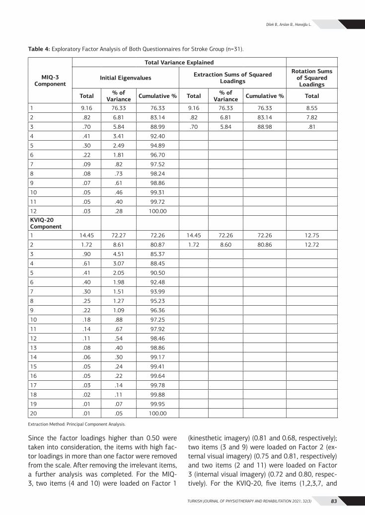
TURKISH JOURNAL OF PHYSIOTHERAPY AND REHABILITATION 2021; 32(3) 83
Dilek B., Arslan B., Hanoğlu L.
Since the factor loadings higher than 0.50 were taken into consideration, the items with high fac-tor loadings in more than one factor were removed from the scale. After removing the irrelevant items, a further analysis was completed. For the MIQ-3, two items (4 and 10) were loaded on Factor 1
(kinesthetic imagery) (0.81 and 0.68, respectively); two items (3 and 9) were loaded on Factor 2 (ex-ternal visual imagery) (0.75 and 0.81, respectively) and two items (2 and 11) were loaded on Factor 3 (internal visual imagery) (0.72 and 0.80, respec-tively). For the KVIQ-20, five items (1,2,3,7, and
Table 4: Exploratory Factor Analysis of Both Questionnaires for Stroke Group (n=31).
MIQ-3 Component
Total Variance Explained
Initial Eigenvalues Extraction Sums of Squared Loadings
Rotation Sums of Squared Loadings
Total % of Variance Cumulative % Total % of
Variance Cumulative % Total
1 9.16 76.33 76.33 9.16 76.33 76.33 8.55
2 .82 6.81 83.14 .82 6.81 83.14 7.82
3 .70 5.84 88.99 .70 5.84 88.98 .81
4 .41 3.41 92.40
5 .30 2.49 94.89
6 .22 1.81 96.70
7 .09 .82 97.52
8 .08 .73 98.24
9 .07 .61 98.86
10 .05 .46 99.31
11 .05 .40 99.72
12 .03 .28 100.00KVIQ-20 Component1 14.45 72.27 72.26 14.45 72.26 72.26 12.75
2 1.72 8.61 80.87 1.72 8.60 80.86 12.72
3 .90 4.51 85.37
4 .61 3.07 88.45
5 .41 2.05 90.50
6 .40 1.98 92.48
7 .30 1.51 93.99
8 .25 1.27 95.23
9 .22 1.09 96.36
10 .18 .88 97.25
11 .14 .67 97.92
12 .11 .54 98.46
13 .08 .40 98.86
14 .06 .30 99.17
15 .05 .24 99.41
16 .05 .22 99.64
17 .03 .14 99.78
18 .02 .11 99.88
19 .01 .07 99.95
20 .01 .05 100.00
Extraction Method: Principal Component Analysis.
TURKISH JOURNAL OF PHYSIOTHERAPY AND REHABILITATION 2021; 32(3)84
Motor Imagery Ability in Turkish-Speaking Stroke Patients: Study on Reliability and Construct Validity Study of Two Imagery Questionnaires, Turk J Physiother Rehabil
9) were loaded on Factor 1 (kinesthetic imagery) (0.72; 0.87; 0.97; 0.93; and 0.89 respectively); and five items (4,5,6,8, and 10) were loaded on Factor 2 (visual imagery) (0.90; 0.98; 0.52; 0.94; and 0.88, respectively).
DISCUSSION
This study demonstrates that the Turkish versions of the Movement Imagery Questionnaire-3 (MIQ-3) and Kinesthetic and Visual Imagery Question-naire-20 (KVIQ-20) are valid and reliable in stroke patients with mild motor impairment. Both ques-tionnaires have been translated into several differ-ent languages and used to for assess the imagery ability in able-body groups and people with physi-cal disabilities (2,7,11,27-30). This study is the first to show the reliability, the internal consistency, and the factorial structure of the imagery question-naires in a Turkish-speaking stroke population.
In our study, significant differences in imagery abilities were found between the stroke group and control group with respect to their imagery abili-ties. Each MIQ-3 sub-score (internal, external, and kinesthetic) was statistically higher in the control group. Likewise, both sub-scores of the KVIQ-20 (visual and kinesthetic) were higher in the control group, however, only visual imagery scores showed a statistically difference between the groups. Al-though the KVIQ-20 was developed for individuals with physical impairments and is more appropriate than the MIQ-3 in the way of discriminating visual and kinesthetic aspects of the imagery, in our study we could not determine the differences between sub-scores via KVIQ-20. It is possible that this lack of difference may have appeared as a result of hav-ing relatively small sample size. Thus, future stud-ies with larger sample sizes are needed to investi-gate the relations between these variables. Since stroke patients may have cognitive problems such as paying attention to a task and concentration on a duty (31), the amount of time used to perform the items in the questionnaires should be concerned. To overcome this problem, the KVIQ-10 which is the short form of the KVIQ-20 may be preferred (7).
Differences between the groups regarding the im-agery perspectives (internal vs external) and types (visual vs kinesthetic) of imagery were also stud-ied. Our study revealed that the visual imagery scores of both questionnaires were slightly higher
than the kinesthetic imagery scores in both groups. These outcomes were in line with the results in the literature. Since visual imagery is easier than kinesthetic imagery, participants are likely to get more scores in visual imagery than kinesthetic im-agery (5,7,8). Therefore, visual imagery technique is considered as a useful strategy to explain the theoretical concept of motor imagery for the peo-ple who are less familiar with motor imagery and have limited attention and concentration skills (2). Additionally, assessing visual imagery at first and then evaluating kinesthetic imagery may be appro-priate for older people with physical disabilities (7).
The MIQ-3 is a more useful tool than the KVIQ-20 to discriminate between the types of visual imag-ery. The MIQ-3 was designed to allow the partic-ipant to choose between first- and third-person visual imagery (11). The scores for the external visual imagery subscale were the highest in both groups. According to this result, third-person per-spective, which means external visual imagery is easier than first-person perspective, which means internal visual imagery (11). Gregg et al suggested that practice complex and functional movements in graded stages by using third-person perspective for stroke survivors. Thus, it could be asserted that third-person perspective might be a more efficient therapeutic application for individuals with stroke (2). Overall, these findings show that the Turkish versions of both questionnaires are sensitive and convenient tools to assess imagery abilities in stroke patients with mild motor impairment.
We confirmed the internal consistency of both questionnaires with high Cronbach alpha values and these results matched with the findings report-ed in the previous studies (2,7). Butler et al. showed the internal consistency of the MIQ-RS was found to be high in able-bodied and stroke groups (11). Malouin et al. developed the KVIQ for the individu-als with physical disabilities and needed guidance in applying imagery questionnaires. According to their results, the internal consistency of the Cron-bach’s alpha values was accepted in the range of 0.87-0.94 in individuals with stroke (7). Additional-ly, our test-retest analysis resembled the outcomes of such studies in the literature with respect to the ICC values of both questionnaires. The ICC values for kinesthetic and visual items were in accept-
TURKISH JOURNAL OF PHYSIOTHERAPY AND REHABILITATION 2021; 32(3) 85
Dilek B., Arslan B., Hanoğlu L.
able levels. The ICC values for the visual imagery subscale were lower than the ICC values for the kinesthetic imagery subscale in the MIQ-3. Simi-larly, Butler et al. (11) reported lower visual imag-ery scores in their stroke group (ranging from 0.54 to 0.80). On the other hand, the ICC values for the kinesthetic imagery subscale were higher than the ICC values for the visual imagery subscale in the KVIQ-20. These outcomes matched with the results of other studies about the issue (7,32). However, there has been no consensus about the correlation coefficient values in the literature (2,7,11). Re-searchers explained that subjective variables might be among the reasons of such results. In addition, distinctions in the way of instructions of the im-agery procedures may affect the scores (2,7,32). Moreover, some items that include movements with different levels of difficulty may be challenging for stroke patients (7). As a result, the methods of applying the procedures of imagery questionnaires needs to be standardized before the assessments. In our study, both questionnaires had good to excel-lent reliability and demonstrated very good consis-tency with respect to their items and purposes in the Turkish-speaking stroke patients.
The exploratory factor analysis was used for the concurrent validity of both questionnaires (2,7,11,12,14,27-30). Previous studies reported the two-factor model of the MIQ-RS (2,27). However, the two-factor model was found to be inappro-priate to distinguish some items and did not have satisfactory adjustment indexes. Alternatively, the three-factor model of the MIQ-3 was accepted as the most appropriate model to evaluate imagery ability comprehensively (14). Our results were in the line with the literature and the factor analysis confirmed the use of three-factor model for MIQ-3 in the Turkish-speaking stroke population. The ex-ploratory factor analysis revealed the two-factor structure of the KVIQ-20 as shown in the literature (7,15). Additionally, a statistically significant cor-relation was found between its items with the MIQ-3’ items and the use of two-dimensional structure of the KVIQ-20 was confirmed for our group. Sim-ilar outcomes were reported in the previous stud-ies (7,13,27). Malouin et al. (7) confirmed the bi-factorial structure of the KVIQ-20 by showing the correlations between visual and kinesthetic factors (0.46). Although our results showed the validity
of both questionnaires, it should be emphasized that either questionnaire is not adequate to as-sess motor imagery in stroke patients with lesions that may disrupt the capacity to perform imagery (2,11). Therefore, researchers who developed these questionnaires suggested that validation studies in imagery questionnaires need further exploration in all types of clinical properties in stroke (2,7,13).
Our study had some limitations. Its sample size was relatively small, and it was performed only in the stroke patients with mild motor impairment. Thus, our findings might not be adequate for the entire clinical conditions stroke patients complain about.
In conclusion, the Turkish versions of the MIQ-3 and the KVIQ-20 have satisfactory reliability and validity to assess motor imagery ability in Turk-ish-speaking stroke patients with mild functional impairments. Therefore, we suggest future stud-ies should investigate the motor imagery ability of moderate to severe stroke patients as well as indi-viduals with different physical disabilities.
Sources of Support: This study did not receive any specific grant from funding agencies in the public, commercial, or not-for-profit sectors.
Conflict of Interest: The authors declare no con-flict of interest.
Ethical Approval: The study protocol was ap-proved by Istanbul Medipol University Non-Inter-ventional Clinical Research Ethics Board (Approval Date: 15.02.2017; Approval Number: E4262).
Informed Consent: Informed consent was ob-tained from all participants.
Peer-Review: Externally peer-reviewed.
Author Contributions: Concept BA, BD, LH; De-sign BA, BD, LH; Supervision BD, LH; Resources and Financial Support –; Materials BA, BD, LH; Data Collection and/or Processing BA,BD; Analysis and/or Interpretation BA, BD, LH ; Literature Research BA, BD, LH; Writing Manuscript BA, BD, LH; Critical Review BD, LH.
Acknowledgments: We appreciate the volunteers and their willingness for the study. We also wish to thank Yavuz Yakut, Prof. for his valuable contribu-tion to the statistical analysis.
TURKISH JOURNAL OF PHYSIOTHERAPY AND REHABILITATION 2021; 32(3)86
Motor Imagery Ability in Turkish-Speaking Stroke Patients: Study on Reliability and Construct Validity Study of Two Imagery Questionnaires, Turk J Physiother Rehabil
REFERENCES1. Decety J, Jeannerod M. Mentally simulated movements in virtual
reality: does Fitts’s law hold in motor imagery? Behav Brain Res. 1995;72(1-2):127-34. doi:10.1016/0166-4328(96)00141-6
2. Gregg M, Hall C, Butler A. The MIQ-RS: a suitable option for examining movement imagery ability. Evid Based Complement Alternat Med. Jun 2010;7(2):249-57. doi:10.1093/ecam/nem170
3. Decety J. The neurophysiological basis of motor imagery. Behav Brain Res. 1996;77(1-2):45-52. doi:10.1016/0166-4328(95)00225-1
4. Warner L, McNeill ME. Mental imagery and its potential for physical therapy. Phys Ther. 1988;68(4):516-21. doi:10.1093/ptj/68.4.516
5. Hall CR. Individual differences in the mental practice and imagery of motor skill performance. Can J Appl Sport Sci. 1985;10(4):17S-21S.
6. Malouin F, Richards CL, Durand A. Normal aging and motor imagery vividness: implications for mental practice training in rehabilitation. Arch Phys Med Rehabil. 2010;91(7):1122-7. doi:10.1016/j.apmr.2010.03.007
7. Malouin F, Richards CL, Jackson PL, Lafleur MF, Durand A, Doyon J. The Kinesthetic and Visual Imagery Questionnaire (KVIQ) for assessing motor imagery in persons with physical disabilities: a reliability and construct validity study. J Neurol Phys Ther. 2007;31(1):20-9. doi:10.1097/01.npt.0000260567.24122.64
8. Braun SM, Beurskens AJ, Borm PJ, Schack T, Wade DT. The ef-fects of mental practice in stroke rehabilitation: a systematic re-view. Arch Phys Med Rehabil. 2006;87(6):842-52. doi:10.1016/j.apmr.2006.02.034
9. Page SJ, Levine P, Sisto SA, Johnston MV. Mental practice com-bined with physical practice for upper-limb motor deficit in subacute stroke. Phys Ther. 2001;81(8):1455-62. doi:10.1093/ptj/81.8.1455
10. Zimmermann-Schlatter A, Schuster C, Puhan MA, Siekierka E, Steurer J. Efficacy of motor imagery in post-stroke rehabil-itation: a systematic review. J Neuroeng Rehabil. 2008;5:8. doi:10.1186/1743-0003-5-8
11. Butler AJ, Cazeaux J, Fidler A, et al. The Movement Imagery Ques-tionnaire-Revised, Second Edition (MIQ-RS) Is a reliable and valid tool for evaluating motor imagery in stroke populations. Evid Based Complement Alternat Med. 2012;2012:497289. doi:10.1155/2012/497289
12. Atienza F, Balaguer I, Garcia-Merita ML. Factor analysis and re-liability of the Movement Imagery Questionnaire. Percept Mot Skills. 1994;78(3Pt2):1323-8. doi:10.2466/pms.1994.78.3c.1323
13. Hall CR, Martin KA. Measuring movement imagery abilities: A revision of the Movement Imagery Questionnaire. J Ment Imag. 1997;21:143-54.
14. Williams SE, Cumming J, Ntoumanis N, Nordin-Bates SM, Ramsey R, Hall C. Further validation and development of the movement imagery questionnaire. J Sport Exerc Psychol. 2012;34(5):621-46. doi:10.1123/jsep.34.5.621
15. Dilek B, Ayhan Ç, Yakut Y. Kinestetik ve Görsel İmgeleme An-keti-20’nin Türkçe versiyonunun geçerlik ve güvenirliği. JETR 2019;6(3):201-10.
16. Faul F, Erdfelder E, Lang AG, Buchner A. G*Power 3: a flexible statistical power analysis program for the social, behavioral, and biomedical sciences. Behav Res Methods. 2007;39(2):175-91. doi:10.3758/bf03193146
17. J. C. Statistical power analysis for the behavioral sciences. Rout-ledge Academic; 1988.
18. Folstein MF, Folstein SE, McHugh PR. “Mini-mental state”. A prac-tical method for grading the cognitive state of patients for the clinician. J Psychiatr Res. 1975;12(3):189-98. doi:10.1016/0022-3956(75)90026-6
19. Duncan PW, Goldstein LB, Horner RD, Landsman PB, Samsa GP,
Matchar DB. Similar motor recovery of upper and lower extrem-ities after stroke. Stroke. 1994;25(6):1181-8. doi:10.1161/01.str.25.6.1181
20. Oldfield RC. The assessment and analysis of handedness: the Edinburgh inventory. Neuropsychologia. 1971;9(1):97-113. doi:10.1016/0028-3932(71)90067-4
21. Fugl-Meyer AR, Jaasko L, Leyman I, Olsson S, Steglind S. The post-stroke hemiplegic patient. 1. a method for evaluation of physical performance. Scand J Rehabil Med. 1975;7(1):13-31.
22. Duncan PW, Propst M, Nelson SG. Reliability of the Fugl-Meyer assessment of sensorimotor recovery following cerebrovascu-lar accident. Phys Ther. 1983;63(10):1606-10. doi:10.1093/ptj/63.10.1606
23. Dilek B, Ayhan C, Yakut Y. Reliability and validity of the Turkish version of the movement imagery questionnaire-3: Its cultural adaptation and psychometric properties. Neurol Sci Neurophysi-ol. 2020;37:221-7.
24. Cronbach LJ. Coefficient alpha and the internal structure of tests. Psychometrika. 1951;16(3):297-334.
25. Shrout PE, Fleiss JL. Intraclass correlations: uses in assessing rater reliability. Psychol Bull. 1979;86(2):420-428.
26. Howell DC. Statistical Methods for Psychology. Wadsworth, Cen-gage Learning; 2009;411-457.
27. Loison B, Moussaddaq AS, Cormier J, et al. Translation and validation of the French Movement Imagery Questionnaire - revised second version (MIQ-RS). Ann Phys Rehabil Med. Apr 2013;56(3):157-73. doi:10.1016/j.rehab.2013.01.001.
28. Schuster C, Lussi A, Wirth B, Ettlin T. Two assessments to eval-uate imagery ability: translation, test-retest reliability and con-current validity of the German KVIQ and Imaprax. BMC Med Res Methodol. 2012;12:127. doi:10.1186/1471-2288-12-127.
29. Paravlic A, Pisot S, Mitic P. Validation of the Slovenian Version of Motor Imagery Questionnaire 3 (MIQ-3): promising tool in modern comprehensive rehabilitation practice. Zdr Varst. Dec 2018;57(4):201-210. doi:10.2478/sjph-2018-0025.
30. Siuda J, Boczarska-Jedynak M, Budrewicz S, et al. Validation of the Polish version of the Movement Disorder Society-Unified Parkinson’s Disease Rating Scale (MDS-UPDRS). Neurol Neuro-chir Pol. 2020;54(5):416-425. doi:10.5603/PJNNS.a2020.0049.
31. das Nair R, Cogger H, Worthington E, Lincoln NB. Cognitive reha-bilitation for memory deficits after stroke. Cochrane Database Syst Rev. Sep 1 2016;9:CD002293. doi:10.1002/14651858.CD002293.pub3.
32. Tabrizi YM, Zangiabadi N, Mazhari S, Zolala F. The reliabil-ity and validity study of the Kinesthetic and Visual Imagery Questionnaire in individuals with multiple sclerosis. Braz J Phys Ther. Nov-Dec 2013;17(6):588-92. doi:10.1590/S1413-35552012005000124.

87
TÜRKİYE’DE FİZYOTERAPİ VE REHABİLİTASYON ALANINDA YAPILAN DOKTORA TEZLERİNİN TEMATİK
ve METODOLOJİK AÇIDAN İNCELENMESİARAŞTIRMA MAKALESİ
ARAŞTIRMA MAKALESİ
ÖZAmaç: Doktora eğitimi, her mesleğin gelişiminde olduğu gibi fizyoterapi mesleğinin gelişimi için de oldukça önemlidir. Bu araştırma Türkiye’de fizyoterapi alanında yapılan doktora tezlerinin tematik ve metodolojik açıdan özelliklerini değerlendirmek amacıyla yapılmıştır.
Yöntem: Bu çalışma kapsamında Türkiye Cumhuriyeti Yüksek Öğretim Kurulu’nun “Ulusal Tez Merkezi” resmi web sitesinde yer alan Fizyoterapi ve Rehabilitasyon alanında yapılan tezler tarandı. 1971 yılı ile 15 Ekim 2020 tarihleri arasında fizyoterapi ve rehabilitasyon alanında yapılan ve Yükseköğretim Kurumu elektronik tez arşivinde kayıtlı olan 422 doktora tezi incelenmiştir. Tezleri incelemede araştırmacılar tarafından geliştirilen tez inceleme formu kullanılmıştır.
Sonuçlar: Türkiye’de fizyoterapi ve rehabilitasyon alanında doktora eğitimi verilen üniversite sayısı 19’dur. Doktora eğitimi veren 4 vakıf üniversitesi bulunmaktadır. İncelenen doktora tezlerinde, tez danışmanlarının %81 (n=341) profesör unvanına sahip olduğu belirlendi. Yapılan tezlerin örneklem sayı aralığının 8-1563 arasında değiştiği gözlendi. Alanlar açısından tezlere bakıldığında, en fazla tezin nöroloji alanında yapıldığı görüldü. Tüm araştırmacıların 329’unun kadın (%78), 93’ünün (%22) de erkek olduğu belirlendi.
Tartışma: Doktora eğitimi alan ya da alacak olan fizyoterapi öğrencilerinin tez konusu seçiminde Yükseköğretim Kurulunun belirlemiş olduğu öncelikli alanlar ile ulusal ve uluslararası kabul gören alanları dikkate almalarının önemli olduğu görüşündeyiz. Sonuç olarak bu çalışmanın ileride yapılacak olan doktora tezlerinin niteliğini arttırmaya katkı sağlayacağı düşünülmektedir.
Anahtar Kelimeler: Doktora Eğitimi, Doktora Tezi, Fizyoterapi, Rehabilitasyon.
A THEMATIC AND METHODOLOGICAL ANALYSIS OF DOCTORAL DISSERTATIONS IN THE FIELD OF
PHYSIOTHERAPY AND REHABILITATION IN TURKEY
ORIGINAL ARTICLE
ABSTRACTPurpose: This research was conducted in order to evaluate the thematic and methodological characteristics of doctoral thesis in the field of physiotherapy in Turkey.
Method: In this study, dissertations in the field of Physiotherapy and Rehabilitation, which are on the official website of the “National Thesis Center” of the Higher Education Council of the Republic of Turkey, were scanned. 422 doctoral dissertations conducted in the field of physiotherapy and rehabilitation between the dates of 1971 and October 15, 2020 were examined, which were registered in the Electronic dissertation archive of the higher education institution.
Results: The number of universities having doctoral training in the field of physiotherapy and rehabilitation is 19 in Turkey. There are four foundation universities where doctoral education is given. It was determined that 81% (n=341) of the thesis consultants in the examined doctoral dissertations had the title of Professor. It was observed that the sample number in the thesis ranges from eight to 1563. When analyzed in terms of fields, it was seen that the most of the dissertations were conducted in the field of Neurology. It was determined that 329 of all researchers were female (78%) and 93 (22%) were male.
Conclusion: We believe that it is important for physiotherapy students who receive or will receive doctoral education to take into account the priority areas, national and internationally accepted areas determined by the Council of Higher Education in the selection of the thesis subject. As a result, it is believed that this study will contribute to improving the quality of future doctoral dissertations.
Key Words: Doctorate Education, Doctoral Dissertation, Physiotherapy, Rehabilitation.
Turkish Journal of Physiotherapy and
Rehabilitation2021 32(3)87-94
Murat Ali ÇINAR Dr. Öğr. Üyesi1
Begümhan Turhan Dr. Öğr. Üyesi1
Tuğba GÖNEN Arş. Gör1
Kezban BAYRAMLAR Prof. Dr.1
Yavuz YAKUT Prof. Dr.1
ISSN: 2651-4451 • e-ISSN: 2651-446X
Çınar .A., Turhan B., Gönen T., Bayramlar K., Yakut Y., Türkiye’de Fizyoterapi ve Rehabilitasyon Alanında Yapılan Doktora Tezlerinin Tematik ve Metodolojik Açıdan İncelenmesi -Araştırma Makalesi, Turk J Physiother Rehabil. 2021; 32(3):87-94. doi: 10.21653/tjpr.824330
1 Hasan Kalyoncu Üniversitesi, Sağlık Bilimleri Fakültesi, Fizyoterapi ve Rehabilitasyon Bölümü, Gaziantep
Correspondence (İletişim):
Murat Ali ÇINAR, Telefon: 0 342 211 8080
E-posta: [email protected]: 0000-0003-2122-3759
Begümhan TurhanORCID: 0000-0003-0842-2059
Tuğba GÖNENORCID: 0000-0002-0484-0221
Kezban BAYRAMLARORCID: 0000-0001-6912-4405
Yavuz YAKUTORCID: 0000-0001-9363-0869
Received: 11.11.2020 (Geliş Tarihi)Accepted: 10.08.2021 (Kabul Tarihi)
CC BY - NC
Content of this journal is licensed under a Creative Commons Attribution-NonCommercial 4.0 International License.

TURKISH JOURNAL OF PHYSIOTHERAPY AND REHABILITATION 2021; 32(3)88
Türkiye’de Fizyoterapi ve Rehabilitasyon Alanında Yapılan Doktora Tezlerinin Tematik ve Metodolojik Açıdan İncelenmesi Araştırma Makalesi
GİRİŞ
Avrupa yükseköğretim sisteminin son aşaması ve araştırma eğitiminin ise ilk aşaması olarak tanım-lanan doktora eğitimi, bireylere, bağımsız araştır-ma yapma, bilimsel olayları geniş ve derin bir bakış açısı ile irdeleyerek yorum yapma ve yeni sentezlere ulaşmak için gerekli adımları belirleme yeteneği ka-zandırmayı amaçlamaktadır (1). Özgün araştırma-lar yapmak ve bilim üretme yetilerini kazandırmak suretiyle bilim insanı ve öğretim üyesi yetiştiril-mesine, ulusal refahın ve kalkınmanın vazgeçilmez unsurları olarak bilinen bilim ve teknolojinin geliş-mesine yapacağı katkılardan dolayı, doktora eğiti-mi yirmi birinci yüzyılın en kritik eğitim kademesi olarak görülmeye başlanmıştır (2).
Doktora eğitimi, bilim insanı yetiştirmenin en önemli basamağıdır. Bu eğitimin sonunda bireyler-den beklenen bilim insanı adayları olarak yeni bilgi-ler üretmeleridir (3). Doktora programlarının temel hedefinin, mesleki eğitime katkıda bulunacak, ileri uygulamaları geliştirecek ve araştırmalar yapmak için gerekli olan becerilere sahip, araştırma yapma-ya yönelmiş olan deneyimli bilim insanları yetiştir-mek olduğuna çok eski kaynaklarda dahi rastlan-maktadır (4). Aynı zamanda doktora programları ekonomik ve toplumsal gereksinimleri belirlemek, sağlıkla ilgili problemleri gidermek için yeni yollar geliştirmek ve yeni teknolojileri ortaya koyan yeni-likçi düşüncelerin geliştirilmesinde harekete geçi-rici bir etkendir (5). Doktora eğitimi, her mesleğin ilerlemesinde önemli olduğu gibi fizyoterapi mesle-ğinin gelişimi için de oldukça önemlidir.
Türkiye’de 1950’lerden sonra yükseköğretime olan talebin artmasıyla lisansüstü eğitim alanında önemli gelişmeler kaydedilmiştir. Lisansüstü eğiti-min yaygınlaşması ve kurumsallaşması İkinci Dün-ya Savaşından sonra daha da hızlanmıştır (6). Aynı süreçte Türkiye’de üniversite sayılarının artmasıyla birlikte lisansüstü düzeyde de bazı düzenlemelere gidildiği görülmektedir. 1946 yılında çıkarılan 4936 sayılı yasa ile üniversitelere, 1959 yılında çıkarılan 7334 sayılı yasa ile de kurulması kararlaştırılan akademilere lisansüstü eğitim ve araştırma yapma görevi verilirken; 1981 yılında çıkarılan 2547 sayı-lı yasa ile lisansüstü eğitim yüksek lisans, doktora, tıpta uzmanlık ve sanatta yeterlik eğitimlerini kap-sayacak şekilde yeniden düzenlenmiştir (7). Türki-
ye’de diğer bilimsel çalışma alanlarında 1950’lere doğru başlayan lisansüstü eğitim, fizyoterapi ve rehabilitasyon alanında doktora eğitimi aşama-sına ilk olarak (1970’li yılların başlarında) Hacet-tepe Üniversitesi’nde başlanmıştır. Fizyoterapinin 1940-1980 yılları arasındaki gelişim döneminin incelendiği bir çalışmada, İngiltere’de 1977 yılın-dan itibaren fizyoterapistlerin özel ilgi alanlarında (ortopedi, nöroloji, pediatri, kardiyoloji, göğüs, geri-atri gibi) özelleşmeleri veya uzmanlık yapmalarının sağlandığı belirtmiştir. Aynı çalışmada, fizyotera-pistlerin Avustralya’da 1980’lerden başlayarak bazı özelleşme alanlarında yaptıkları (ortopedik rehabi-litasyon, nörolojik rehabilitasyon, pediatrik rehabi-litasyon, geriatrik rehabilitasyon, kardiyopulmoner rehabilitasyon, kadın sağlığı, sporcu sağlığı, el-mik-rocerrahi, protez-ortez, onkoloji, ağrı) lisansüstü eğitimlerinin yanı sıra; fizyoloji, anatomi, nörofiz-yoloji, nöroanatomi, biyomekanik, kinezyoloji, biyo-mühendislik ve nörobilim gibi konularda da lisan-süstü eğitim yapmaya başladıkları belirtilmektedir (6). 1980’lerde Dünya Fizyoterapi Konfederasyonu fizyoterapide yüksek lisans ve doktora programları-nın, meslekte bilgi ve beceri kazandıran, profesyo-nel yeterliliği artıran, analitik düşünme yeteneğini geliştiren ve bilimde yeniliklerin takibini sağlayan eğitimler olması gerektiğini bildirmiştir (8).
Türkiye’de lisansüstü eğitimlerin incelendiği bir çalışmada, öğrencilerin en çok tez konusunun se-çiminde zorlandığını ve bu konuda nasıl bir strateji izleyeceklerini bilmedikleri vurgulanmıştır (9). Bu araştırma Türkiye’de fizyoterapi alanında yapılan doktora tezlerinin tematik ve metodolojik açıdan özelliklerini değerlendirmek amacıyla yapılmıştır. Çalışmadan elde edilecek sonuçların, Türkiye’de fiz-yoterapi ve rehabilitasyon alanında yapılan doktora tezlerinin mevcut durumu hakkında bilgi vereceği ve yeni yapılacak doktora çalışmalarının yönünün belirlenmesine katkı sağlayacağı düşünülmektedir.
YÖNTEM
Türkiye Cumhuriyeti Yükseköğretim Kurulu’nun “Ulusal Tez Merkezi” web sitesinin detaylı tarama kısmında bulunan “anabilim dalı” bölümünden: Fiz-yoterapi ve Rehabilitasyon, Fizik Tedavi ve Rehabi-litasyon, Fiziksel Tıp ve Rehabilitasyon, Fizyoterapi ve Rehabilitasyon, başlıklarıyla taramalar yapılmış-

TURKISH JOURNAL OF PHYSIOTHERAPY AND REHABILITATION 2021; 32(3) 89
Çınar .A., Turhan B., Gönen T., Bayramlar K.
tır. Yine aynı detaylı tarama kısmında bulunan bi-lim dalı bölümünde Fiziksel Tıp ve Rehabilitasyon, Fizyoterapi ve Rehabilitasyon, Fizik Tedavi ve Re-habilitasyon anahtar kelimeleri ile de taramalar ya-pılmıştır. Ayrıca enstitü bölümü kısmında bulunan Sağlık Bilimleri Enstitüsü alanlarında yapılan tezler de taranmıştır. Tüm tezler bilgisayara indirilmiştir. Toplamda 440 doktora tezine ulaşılmıştır. Ancak 18 tezin iki ayrı aramada görüldüğü ve tekrar ettiği görüldüğünden inceleme listesinden çıkarılmıştır. X Üniversitesi Sağlık Bilimleri Fakültesi Fizyotera-pi ve Rehabilitasyon Bölümü’nde gerçekleştirilen ve tanımlayıcı nitelikteki bu araştırmada, 1971 yılı ile 15 Ekim 2020 tarihleri arasında fizyoterapi ve rehabilitasyon alanında yapılmış ve Yükseköğre-tim Kurulu elektronik tez arşivinde kayıtlı olan 422 doktora tezine ulaşılmıştır. Bu tezler tematik ve metodolojik açıdan incelenmiştir. Tezlerin incelen-mesinde araştırmacılar tarafından geliştirilen tez inceleme formu kullanılmıştır. Tez inceleme formu aşağıdaki soruları içermektedir (10):
1. Doktora tezleri yürütüldükleri üniversitelere göre nasıl bir dağılım göstermektedir?
2. Doktora tezleri yayın yılına göre nasıl bir dağılım göstermektedir?
3. Doktora tezleri danışman unvanına göre nasıl bir dağılım göstermektedir?
4. Doktora tezleri araştırmacı cinsiyetine göre nasıl bir dağılım göstermektedir?
5. Doktora tezleri örnekleme sayılarına göre dağı-lımı nasıldır?
6. Doktora tezleri konu alanına göre nasıl bir dağı-lım göstermektedir?
7. Doktora tezleri yöntem bölümünde kullanılan araştırma yaklaşımına göre nasıl bir dağılım gös-termektedir?
8. Doktora tezleri sayfa sayılarına göre nasıl bir da-ğılım göstermektedir?
İstatiksel Analiz
Araştırmacılar tarafından geliştirilen tez incele-me formundaki her bir inceleme boyutu Microsoft Excel 2010 programı kullanılarak bir çizelgeye dö-nüştürülmüştür. Öncelikle tezlere ilişkin demogra-fik bilgilere (tez adı, yayın yılı, üniversite, yazar adı,
Tablo 1: Doktora Eğitimi Veren Üniversitelerin Tez Sayılarının Dağılımı.
ÜNİVERSİTE n %
Hacettepe Üniversitesi 257 61
Dokuz Eylül Üniversitesi 51 12
Pamukkale Üniversitesi 31 7
İstanbul Üniversitesi 27 6
İstanbul Medipol Üniversitesi* 15 4
T.C. Gazi Üniversitesi 13 3
Bolu Abant İzzet Baysal Üniversitesi 5 1
Trakya Üniversitesi 5 1
Bezm-i Âlem Vakıf Üniversitesi* 4 1
Marmara Üniversitesi 5 1
Kütahya Sağlık Bilimleri Üniversitesi 3 1
Kocaeli Üniversitesi 2 1
Ankara Yıldırım Beyazıt Üniversitesi 2 1
Ege Üniversitesi 1 0
Dicle Üniversitesi 1 0
Hasan Kalyoncu Üniversitesi* 0 0
Bahçeşehir Üniversitesi * 0 0
Kırşehir Ahi Evran Üniversitesi 0 0
Muğla Sıtkı Koçman Üniversitesi 0 0
TOPLAM 422 100
* Vakıf üniversiteleri

TURKISH JOURNAL OF PHYSIOTHERAPY AND REHABILITATION 2021; 32(3)90
Türkiye’de Fizyoterapi ve Rehabilitasyon Alanında Yapılan Doktora Tezlerinin Tematik ve Metodolojik Açıdan İncelenmesi Araştırma Makalesi
danışman unvanı); tematik bilgilere (çalışma alanı) ve metodolojik bilgilere (tezin araştırma yaklaşımı, çalışma grubu, tez örneklem türü) her satırda yer verilmiştir. Belirlenen bir ölçüte göre (örneğin bir yazara ya da bir üniversiteye ilişkin tezlere ulaş-mak) arama yapabilmek için Excel programının makrolarından faydalanılmıştır. Oluşturulan çizelge ile çalışma, verilerin kaydedilmesine hazır hale ge-tirilmiştir. Araştırma kapsamındaki 422 tez belirle-nen bu ölçütlere göre incelenerek her bir veri ilgili sütuna kaydedilmiştir.
Etik Onay
Yükseköğretim Kurulu Ulusal Tez Merkezi lisan-süstü tezleri, 2547 Sayılı Yükseköğretim Kanunu Ek Madde 40 hükümleri çerçevesinde bilime katkı sağlamak, bilimsel araştırma ve faaliyetleri destek-lemek amacıyla elektronik ortamda erişime açıktır. Erişime açık olan tezler Kişisel verilerin korunması kanununa uygun bir şekilde incelenmiş olduğundan “ Etik Onay” alınmasına gerek duyulmamıştır.
SONUÇLAR
Yükseköğretim Kurumu elektronik tez arşivinde ka-yıtlı olan tezlerin incelenmesi neticesinde en fazla doktora tezinin (257 adet tez), doktora programı-nın da ilk olarak açıldığı Hacettepe Üniversitesi’nde yazıldığı belirlenmiştir. Bu sıralamayı ikinci olarak Dokuz Eylül Üniversitesi (51 adet tez), üçüncü sıra-da ise Pamukkale Üniversitesi (31 adet tez) takip etmektedir. Ayrıca Türkiye’de doktora eğitimi veri-len üniversite sayısı 19’dur (4 vakıf 15 devlet üni-versitesi) (Tablo 1).
Yazılan doktora tezlerinin yıllara göre dağılımları incelendiğinde, en fazla tezin yazıldığı yıl 2019 yılı olarak belirlenmiştir. Yıllara göre doktora tezlerinin dağılımı şekil-1’de verilmiştir.
İncelenen doktora tezlerinde, tez danışmanının aka-demik unvanı tespit edilebilen tezlerde danışmanla-rın 341’i profesör (% 81’), 72’si doçent (%17), 22’si (% 5) ise doktor öğretim üyesi olarak belirlenmiştir. Ayrıca 13 doktora tezinin de çift danışmanı vardır (Tablo 2).
Doktora tezlerini yazan araştırmacıların cinsiyete göre dağılımları incelendiğinde, kadın araştırma-cıların sayısının erkek araştırmacılara göre fazla olduğu görülmüştür. Araştırmacıların 329’u (%78) kadın, 93’ü (%22) erkektir.
Yapılan doktora tezlerinin örneklem sayıları ile il-gili verilerin birçoğuna ulaşılmış olup, 6 adet tezin örneklem sayısına ulaşılamamıştır. Örneklem sayı aralığı 8-1563 arasında değişmektedir.
Yapılan doktora tezlerinin çalışma alanları incelen-diğinde; en fazla tezin nöroloji alanında yapıldığı sonrasında ise en çok sağlıklı bireylerde yapılan çalışmaların tercih edildiği görülmüştür. Konu alan-larının ayrıntılı sınıflandırılmasına Tablo 3’te yer verilmiştir.
Doktora tez çalışmalarının yöntem bölümünde kul-lanılan araştırma yaklaşımına göre incelendiğinde en çok randomize kontrollü çalışmaların tercih edil-diği görülmüştür. Yıllara göre yapılan tez sayılarıyla randomize kontrollü çalışma sayıları karşılaştırıldı-ğında her yıl yapılan çalışmaların yarıdan fazlasının randomize kontrollü çalışma türünde olduğu görül-mektedir (Tablo 4).
TARTIŞMA
Doktora tezleri, bireyin akademik gelişim süreçle-rindeki ilk özgün çalışma aşaması olması nedeniyle oldukça önemlidir. Çeşitli meslek gruplarına ait bu
Tablo 2: Danışmanların Akademik Unvanlarının Dağılımı.
Danışmanın Akademik Unvanı Tez Sayısı (%)
Profesör 341 (% 81)
Doçent 72 (% 17)
Doktor Öğretim Üyesi 22 (% 5)
Çift Danışman 13
Toplam 422
Şekil 1: Tez Sayılarının Yıllara Göre Dağılımı.

TURKISH JOURNAL OF PHYSIOTHERAPY AND REHABILITATION 2021; 32(3) 91
Çınar .A., Turhan B., Gönen T., Bayramlar K.
Tablo 3: Doktora Tez Konularının Dağılımları.
TEZ KONULARI SayılarNörolojik Problemler* 48SAĞLIKLI BİREYLER** 41Omurga Patolojileri *** 33Kardiyopulmoner ve Kardiyovasküler Patolojiler**** 25Serebral Palsi 24Diz Patolojileri***** 24Diğer Kas-İskelet Sistemi Problemleri****** 15Sağlıklı Geriatrik Bireyler 14Diğer Pediyatrik Problemler******* 14Ayak-Ayak Bileği Patolojileri******** 13Amputasyon Cerrahisi Geçiren Hastalar********* 13Omuz Patolojileri********** 11Nöromusküler Hastalıklar*********** 10Diyabet 10Osteoartrit 9Romatizmal Hastalıklar************ 9Çeşitli Üst Ekstremite Yaralanmaları 8Onkoloji Hastaları************* 8Lenf Ödem 7Sporcu ve Sanatçılar 6Riskli Bebekler 6Brakial Pleksus ve Diğer Periferik Sinir Yaralanmaları 6Çeşitli Jinekolojik Problemler 6Engelli Bireyler 6Üriner İnkontinans 5Yutma ve Çiğneme Problemleri 5Skolyoz 4Mental Retardasyonu Olan Bireyler 4Kalça ve Sakroilyak Eklem Problemleri 4Baş Ağrısı ve Migren 4Metabolik Hastalıklar 3Psikolojik Problemler 3Hematolojik Hastalıklar / Hemofili 3Uyku Problemleri 2Osteoporoz 2Alzeimer ve Demans 2Organ Transplantasyonu************** 2RATLAR 2Diğer*************** 12TOPLAM 422
* Svo, hemipleji, hemiparezi, ms, parkinson, als ** Özel bir sınıfa dahil olmayan sağlıklı bireyler dahil edilmiştir *** Lumbal ve servikal herniasyonlar, diskopatiler, mekanik ağrılar, stenozlar dahil edilmiştir ****Astım, koah, pulmoner hipertansiyon, kistik fibrozis, solunum yetmezliği, toraks cerrahileri, derin ven trombüsü, kronik venöz yetmezlik, myokardiyak lezyonlar ve kalp-damar cerrahileri dahil edilmiştir*****Atroplastiler, Patello-femoral ağrı sendromu, kronik diz problemleri ****** Yumuşak doku yaralanmaları, myofasyal ağrı sendromu ******* Sınıflandırılamayan çeşitli pediyatrik problemler ( infantil retinopati, infantil ortopedik problemler vb.)********Pes ekinovarus, inversiyon-eversiyon yaralanmaları, pes planus, plantar fasitis dahil edilmiştir.*********Alt ve üst ekstiremite ampütasyonları, ampute sporcular **********Rotator cuff yırtıkları, impingement, donuk omuz problemler,adheziv kapsülit dahil edilmiştir***********Progresif musküler hastalıklar, duschenne musküler distrofi ************ Ankilozan spondilit, romatoid artrit dahil edilmiştir *************Prostat kanseri, meme kanseri ve akut lösemi hastaları dahil edilmiştir ************** Renal ve akciğer transplantasyonu dahil edilmiştir *************** Sınıflandırılamayan çeşitli tez konularını içermektedir( yanık, vestibüler bozukluklar temporamandibular eklem sorunları vb.)

TURKISH JOURNAL OF PHYSIOTHERAPY AND REHABILITATION 2021; 32(3)92
Türkiye’de Fizyoterapi ve Rehabilitasyon Alanında Yapılan Doktora Tezlerinin Tematik ve Metodolojik Açıdan İncelenmesi Araştırma Makalesi
tip çalışmalar bulunmakla birlikte, Türkiye’de fizyo-terapi ve rehabilitasyon alanında yapılan doktora tezleriyle ilgili tematik ve metodolojik incelemenin olduğu bir çalışmaya rastlanılmamıştır.
Türkiye’de fizyoterapi ve rehabilitasyon eğitimi ve-ren 79 üniversite bulunmaktadır (KKTC üniversite-leri de dahil edilmiştir) (11). Bunlardan sadece 19 üniversitede doktora eğitimi verilmektedir. Daha fazla doktora programının açılması, hem fizyotera-pi ve rehabilitasyon bölümünün gelişmesine hem de Türkiye’de sağlık alanında yapılan çalışmalara katkı sağlayacaktır. Ancak, Yüksek Öğretim Kurumu’nun doktora programı açma kriterleri incelendiğinde, nitelikli doktora eğitiminin temel koşullarını sağla-yabilmek için doktora programı açma kriterlerinin aşamalı bir zorluğu olduğu görülmüştür. Yüksek Öğ-retim Kurumu’nun doktora programı açma ölçütleri arasında, üniversite kadrosunda görev yapmakta olan doktorası veya doçentliği program açılması istenen alandan ya da açılması istenen program disiplinler arası ise o alanla doğrudan ilişkili olmak üzere en az biri profesör, ikisi doçent toplam beş öğretim üyesinin veya en az ikisinin profesör olma-sı durumunda toplam 5 öğretim üyesinin bulunma-sı koşulu bulunmaktadır. Ayrıca, doktora programı açılabilmesi için önemli bir kriter de yüksek lisans programının bulunuyor olmasına ek olarak bu prog-ramdan mezun vermiş olma koşulu vardır (8). Bu kriterleri sağlayamayan pek çok üniversite bulun-maktadır. Doktora eğitimi verilen üniversite sayısı-nın yalnızca 19 olması, doktora programı açabilmek için üniversite kadrolarında yeterli sayıda öğretim üyesinin bulunmamasından kaynaklanabilmektedir.
Sağlık profesyonellerinin eğitimi 3 eğitim döngü-sünü benimsemiştir. Bu eğitim döngüleri lisans, yüksek lisans ve doktora eğitiminden oluşmaktadır (12). Lisansüstü eğitim döngüsünün standartları çe-şitli kurumlar tarafından sağlanmaktadır. Doktora eğitiminin standartlarını belirleyen çeşitli kurumlar olmasına rağmen, doktora eğitiminin ülkenin bilim-sel araştırma kapasitesi ile ilgili olduğu bildirilmiş-tir (13-14). Araştırmamızda Türkiye’deki fizyoterapi ve rehabilitasyon alanındaki doktora tezlerinin ça-lışma alanlarını, örneklem sayıları, danışman dağı-lımlarını inceleyerek bu alandaki birikimleri ortaya koymayı hedefledik.
Sağlık bilimlerinin bazı alanlarında doktora eğitimi, ya doğrudan lisans eğitimi sonrası (yüksek lisans yapmadan direk başvurulabilen bütünleşik doktora programı) ya da yüksek lisans sonrası verilmekte-dir. Lisans derecesiyle doktora programına başvu-rulabilen durumlarda lisans mezuniyet not ortala-malarının 4 üzerinden en az 3 veya muadili bir puan olması gerekir. Türkiye’de fizyoterapi ve rehabili-tasyon anabilim dallarında doktora eğitimi yüksek lisans eğitimi sonrası yapılabilmekte, bütünleşik doktora eğitimi veren bir üniversite bulunmamak-tadır.
2014 yılında yapılan bir çalıştayda Avrupa’da dok-tora eğitiminin geliştirilmesine yönelik en önemli kriterlerden birinin de bütünleşik doktora eğitimi olduğu vurgulanmıştır (15).
Son yıllarda fizyoterapi ve rehabilitasyon alanında yapılan doktora tezleri sayısında önemli bir artış ol-duğu göz önünde bulundurulduğunda, doktora tez-
Tablo 4: Doktora Tezlerinde Kullanılan Araştırma Tiplerinin Dağılımı.
ÇALIŞMA TİPİ n %
Randomize Kontrollü Çalışmalar 187 44,30
Randomize Olmayan Kontrolü Çalışmalar 126 29,80
Ön Test-Son Test 37 8,70
Değerlendirme Çalışmaları 35 8,30
Vaka-Kontrol 24 5,70
Validasyon 5 1,18
Anket Çalışması 3 0,71
Ölçek Geliştirme 2 0,47
Çapraz Geçişli 2 0,47
Self Kontrol 1 0,37
Toplam 422 100
TURKISH JOURNAL OF PHYSIOTHERAPY AND REHABILITATION 2021; 32(3) 93
Çınar .A., Turhan B., Gönen T., Bayramlar K.
lerinin özelliklerinin incelenmesinin, doktora eğitimi sırasındaki bilimsel tutumun gelişmesi, nitelikli bi-limsel araştırma yapılması ve yapılan bilimsel araş-tırmaların uygulanabilir olması, kanıta dayalı tıbbın gelişmesi için gerekli olduğu düşüncesindeyiz.
Çağdaş bilimin her alanda kanıtlar bulmaya çalıştı-ğı bilinmektedir. Günümüzde popülaritesi artan ka-nıta dayalı tıp temelde pek çok güvenilir kaynaktan elde edilen bilgileri organize ederek, protokoller ve rehberler oluşturmayı hedeflemektedir (19). Rando-mize kontrollü çalışmalar klinik kanıta dayalı çalış-malar için altın standart olarak kabul edilmektedir-ler (20). Doktora tezlerinin araştırma yöntemlerini incelememiz neticesinde deneysel çalışmaların ran-domize kontrollü olarak yapıldığı tez çalışmalarında son yıllarda artış görülmüştür. Buradan, randomize kontrollü çalışmalardaki bu artışın fizyoterapi ve rehabilitasyon alanındaki kanıta dayalı bilimin ge-lişmesine katkıda bulunacağı sonucuna varılabilir.
Fizyoterapi ve rehabilitasyon alanında yapılan tez-lerin sayılarının yıllara göre dağılımını incelediği-mizde, 2007 yılından itibaren tez sayılarında istik-rarlı bir artış gözlemlenmiştir. Doktora tez aşaması ders süreci, danışman ve tez konusu seçme, tez izleme komitesi belirleme, yeterlilik, tez önerisi ver-me, materyal geliştirme, tezi uygulama ve yazma gibi birden çok aşamayı içermektedir. Bu süreçler ilerlerken literatür taraması, konunun önceden ça-lışılıp çalışılmadığını araştırmak, daha önce fazlaca çalışılmış konuları ve uygulanan yöntemi belirlemek önemlidir (18). Çalışmamız bu anlamda en fazla ve en az çalışılan konulara ışık tutan niteliktedir. Doktora tez konularının dağılımlarına bakıldığında nörolojik rehabilitasyon alanında fazlaca çalışma yapıldığı belirlenmiştir.
Doktora tez sayılarındaki artışa etken olan durum-lardan biri de fizyoterapi ve rehabilitasyon bölümü mezunlarının son yıllarda istihdam sorunları yaşa-maya başlamaları ve fizyoterapistlerin klinikteki çalışma şartlarının zorluğu olabilir. Bu gibi neden-lerle fizyoterapistler akademisyenliğe yönelebil-mektedirler. Ayrıca bu artışta, 2002 yılında kurulan ve 2016 yılından itibaren uygulamadan kaldırılan Öğretim Görevlisi Yetişme Programının da (ÖYP) etkili olabileceği görüşündeyiz.
21. yy verileri kadın akademisyenlerin akademinin üst basamaklarında yeterince temsil edilmediğini,
kadın öğrencilerin ve akademik kariyer yapmakta olan kadınların çeşitli engellerle karşılaştığını or-taya koymaktadır. Kadınların akademik kadrolar içindeki eksik temsili ve akademik dünyanın içinde var olan eşitsizlikler değişik oranlarda olsa da bu-nun küresel bir sorun olduğu düşünülmektedir. 2015 verilerine göre, Türkiye’deki tüm üniversitelerde çalışan öğretim üyelerinin % 43’ü kadındır. (7,15). İncelememiz sonuçlarına göre, Türkiye’de fizyotera-pi ve rehabilitasyon alanında doktora yapan kadın araştırmacıların yaklaşık % 78’i kadındır. Fizyote-rapi ve rehabilitasyon alanında kadın akademisyen sayısının çok olması Türkiye’deki diğer akademik alanlara da örnek teşkil etmektedir.
Günay tarafından 2014 yılında yayımlanan bir ça-lışmada doktora eğitiminin temel bileşeni olarak “Özgün araştırma yoluyla bilginin geliştirilmesidir. Ayrıca doktora programlarından beklenen bir başka işlev, akademiden daha geniş kapsamı olan istih-dam piyasasının ihtiyaçlarını karşılamasıdır” diye belirtilmiştir (9). Özellikle son yıllarda lisansüstü eğitimde etiketleme ya da akreditasyon yapan or-ganizasyonlar tarafından lisansüstü öğrencilerinin üniversite sanayi işbirliği ile yapılan tezler üret-meye yönlendirilmeleri değerli bulunmaktadır. Ça-lışmamız kapsamında incelediğimiz fizyoterapi ve rehabilitasyon alanında yapılan doktora tezlerinde sanayi iş birliği ile yapılan tezlere rastlanılmamış-tır. Araştırmacıların bu alanlarda doktora tez çalış-maları üretmelerinin mesleki kariyerleri açısından önemli olabileceği görüşündeyiz.
Yükseköğretim kurulu (YÖK) üniversitelerimizdeki enstitülerin, araştırma merkezlerinin girişimci do-ğalarını, disiplinler üstü ve disiplinler arası çalışma-larını desteklemektedir. Bu bağlamda da: “Gelecek 10 Yıl İçin Güçlü Nesiller Yetiştirme Projesi’’, “YÖK Gelecek Projesi” gibi projeler başlatmış ve bu pro-jeler için de öncelikli alanlar belirlemiştir. Belirle-nen öncelikli alanlar ise teknoloji odaklı inovasyon içeren konulardır (20,21). Ayrıca disiplinler arası çalışmaların önemi de ayrıca vurgulanmıştır. So-nuç olarak doktora eğitimi alan ya da alacak olan fizyoterapi öğrencilerinin tez konusu seçiminde bu projelerdeki öncelikli alanları da dikkate almalarının önemli olduğu görüşündeyiz.
Destekleyen Kuruluş: Destek alınan kuruluş bulun-mamaktadır.

TURKISH JOURNAL OF PHYSIOTHERAPY AND REHABILITATION 2021; 32(3)94
Türkiye’de Fizyoterapi ve Rehabilitasyon Alanında Yapılan Doktora Tezlerinin Tematik ve Metodolojik Açıdan İncelenmesi Araştırma Makalesi
Çıkar Çatışması: Yok.
Etik Onay: Yükseköğretim Kurulu Ulusal Tez Mer-kezi lisansüstü tezleri, 2547 Sayılı Yükseköğretim Kanunu Ek Madde 40 hükümleri çerçevesinde bili-me katkı sağlamak, bilimsel araştırma ve faaliyet-leri desteklemek amacıyla elektronik ortamda erişi-me açıktır. Erişime açık olan tezler Kişisel verilerin korunması kanununa uygun bir şekilde incelenmiş olduğundan “Etik Onay” alınmasına gerek duyulma-mıştır.
Aydınlatılmış Onam: Çalışma tematik ve metodo-lojik bir inceleme olduğundan aydınlatılmış onam formu kullanılmamıştır.
Hakem Degerlendirmesi: Bağımsız dış hakemler tarafından değerlendirilmiştir. Hakem değerlendir-mesinden sonra yazar katıkları en sonda da açıkla-malar yer almalıdır.
Yazar Katkıları: Murat Ali ÇINAR: Verilerin top-lanması, makalenin tüm bölümlerinin yazılması. Begümhan TURHAN: Makalenin tüm bölümlerinin yazılması. Tuğba GÖNEN: Verilerin toplanması ve teknik düzeltmeler. Kezban BAYRAMLAR: Çalışma-nın planlanması ve tartışma kısmının yazılması. Yavuz YAKUT: Çalışmanın planlanması ve tartışma kısmının yazılması.
Açıklamalar: Yok
KAYNAKLAR1. Güçlü N, Yılmaz G. Türkiye ve Bazı Avrupa Ülkelerinin Doktora
Programlarının Değerlendirilmesi. Çağdaş Yönetim Bilimleri Der-gisi. 2019;6(1):71-89.
2. Gigliotti RA. An exploratory study of academic leadership educa-tion within the Association of American Universities. Journal of Applied Research in Higher Education. 2017;9(2):196-210.
3. Keskinkılıç SB, Ertürk A. Eğitim bilimleri doktora öğrencilerinin istatistiksel bilgi yeterlilikleri. Ahi Evran Üniversitesi Kırşehir
Eğitim Fakültesi Dergisi. 2015;10(1):141-151.4. Shores LS. Opening a doctoral program in nursing: factors to
consider. Nursing Outlook. 1986;34(6):286-288.5. Kim M.J, McKenna HP, Ketefian S. Global quality criteria, stan-
dards, and indicators for doctoral programs in nursing; literature review and guideline development. International Journal of Nur-sing Studies. 2006;43(4):477–489.
6. Can F. Fizyoterapi ve Rehabilitasyonun Mesleki Gelişim Tarihçe-si. İzmir Katip Çelebi Üniversitesi Sağlık Bilimleri Fakültesi Der-gisi 2016;1(3):1-6.
7. Bıkmaz FH, Aksoy H, Tatar Ö, Altınyüzük CA. Eğitim programları ve öğretim alanında yapılan doktora tezlerine ait içerik çözüm-lemesi (1974-2009). Eğitim ve Bilim Dergisi 2013;38(168):288-303.
8. World Confederation of Physical Therapy (WCPT). Professional issues. Retrieved July 8, 2016, from www.wcpt.org.
9. Günay D. Türkiye’de lisansüstü eğitim ve lisansüstü eğitime fel-sefi bir bakış. Üniversite Araştırmaları Dergisi. 2018;1(2):71-88.
10. Şenyurt S, Özkan YÖ. Eğitimde ölçme ve değerlendirme alanında yapılan yüksek lisans tezlerinin tematik ve metodolojik açıdan incelenmesi. Elementary Education Online. 2017;16(2):628-653.
11. TFD 2019. Erişim adresi: https://fizyoterapistler.org/tfd/index.php/en/. Erişim tarihi: 2 Kasım 2020.
12. Koç, M., Bayar, K. Türkiye’de Fizyoterapi ve Rehabilitasyon Ala-nında Lisans ve Lisansüstü Eğitim Veren Yeterli Sayıda Üniversi-te Var Mıdır, 2018
13. Orer, Hakan S. An overview of the doctoral education in health sciences in Turkey. Turk. J. Biochem, 2011, 36.1: 35-41.
14. Marusic, Matko, et al. Doctoral degree in health professions: Professional needs andlegal requirement. Acta medica acade-mica, 2013, 42.1: 61-70.
15. Acar AC. Ulusal Ve Uluslararası Boyutlarıyla Doktora Eğitimi Ça-lıştayı Raporu. İstanbul Üniversitesi. 2014.
16. Carr KC. Developing an evidence based practice protocol: impli-cations for midwifery practice. Journal of Midwifery & Women’s Health. 2000;45(6):544-551.
17. Gökçe KY. Kanıta Dayalı Tıp. http://www.hutbat.hacettepe.edu.tr/beta/27ar alik2001ygk.php. Alınma tarihi: 15 Kasım 2003.
18. Bahçeci, Ferhat; UŞENGÜL, Leyla. Yüksek lisans ve doktora öğ-rencilerinin tez konusu belirleme kriterlerinin incelenmesi. Tur-kish Journal of Educational Studies, 2018, 5.2: 85-96.
19. Öztan E, Doğan SN. Akademinin Cinsiyeti: Yıldız Teknik Üniversi-tesi Örneği Üzerinden Üniversite ve Toplumsal Cinsiyet. Calisma ve Toplum. 2015;46(3):191-221.
20. https://yuzikibinbursu.yok.gov.tr/Sayfalar/HaberDuyu-ru/100-2000-doktora-projesi-nedir.aspx Erişim Tarihi: 20 Hazi-ran 2021
21. https://www.yok.gov.tr/Sayfalar/Haberler/2020/yok-gele-cek-projesi.aspx. Erişim Tarihi: 20 Haziran 2021
95
BIOMOTOR AND TECHNICAL FEATURES OF WHEELCHAIR BASKETBALL PLAYERS BY
CLASSIFICATION SCORES: A PILOT STUDY
ORIGINAL ARTICLE
ABSTRACTPurpose: This study was conducted to examine the biomotor and technical skills of wheelchair basketball players through their classification scores.
Methods: A total of 22 male athletes, 11 with low trunk control (1 to 2.5 points) and 11 with high trunk control (3 to 4.5 points) from Turkish Wheelchair Basketball First League, voluntarily participated in the research. Athletes were grouped according to the International Wheelchair Basketball Federation functional classification system. Biomotor features of the athletes and wheelchair basketball skill test scores were measured. SPSS 24.0 program was used for data analysis. Group differences were determined by Mann-Whitney U analysis.
Results: The study revealed statistically significant differences were in classification points, trunk balance, modified sit-up, modified abdominal endurance, 20 m speed, slalom without the ball, slalom with the ball and 6-min endurance race test parameters (p <0.05).
Conclusion: It can be argued that athletes with low and high trunk control differ in terms of biomotor abilities. However, they have similar features regarding wheelchair basketball technical skills (throwing basketball, layup, shooting, scoring).
Key Words: Individual with Physically Disability, Paralympic, Team Sports, Technical Skill.
TEKERLEKLİ SANDALYE BASKETBOL OYUNCULARINDA KLASİFİKASYON PUANLARINA
GÖRE BİYOMOTOR VE TEKNİK ÖZELLİKLER: PİLOT ÇALIŞMA
ARAŞTIRMA MAKALESİ
ÖZAmaç: Bu çalışma, tekerlekli sandalye basketbolcularında biyomotor ve teknik becerilerinin klasifikasyon puanlarına göre incelenmesi amacıyla gerçekleştirildi.
Yöntem: Araştırmaya Türkiye Tekerlekli Sandalye Basketbol 1. Liginden düşük gövde kontrolüne sahip (1 ile 2,5 puan) 11 erkek sporcu ve yüksek gövde kontrolüne sahip (3 ile 4,5 puan) 11 erkek sporcu olmak üzere 22 sporcu gönüllü olarak katıldı. Sporcular Uluslararası Tekerlekli Sandalye Basketbol Federasyonu fonksiyonel sınıflama sistemine göre gruplandırıldı. Sporcuların biyomotor özellikleri ve tekerlekli sandalye basketboluna özgü beceri test skorları tespit edildi. Verilerin analizinde SPSS 24,0 programı kullanıldı. Gruplar arası farklar Mann-Whitney U analizi ile tespit edildi.
Sonuçlar: Çalışma, gruplar arasında klasifikasyon puanları, gövde denge, modifiye mekik, modifiye şınav, abdominal dayanıklılık, 20 m sürat, top ile slalom, topsuz slalom ve 6 dakika dayanıklılık test parametrelerinde istatistiki olarak anlamlı düzeyde farklılıklar olduğunu gösterdi (p<0,05).
Tartışma: Düşük ve yüksek gövde kontrolüne sahip sporcuların biyomotor yetiler yönünden farklılaştığı bununla birlikte tekerlekli sandalye basketboluna özgü teknik beceriler (basketbol topu fırlatma, turnike, şut, isabet) yönünden ise benzer özelliklere sahip olduğu söylenebilir.
Anahtar Kelimeler: Bedensel Engelli, Paralimpik, Takım Sporu, Teknik Beceri.
Turkish Journal of Physiotherapy and
Rehabilitation2021 32(3)95-102
Mehmet Fatih YÜKSEL1
ISSN: 2651-4451 • e-ISSN: 2651-446X
Yüksel M.F., Biomotor and Technical Features of Wheelchaır Basketball Players by Classification Scores: A Pilot Study, Turk J Physiother Rehabil. 2021; 32(3):95-102.doi: 10.21653/tjpr.862640
1 Department of Physical Education and Sports, Faculty of Education, Necmettin Erbakan University, 42090, Konya, Turkey
Correspondence (İletişim):
Mehmet Fatih YükselPhone number: +90 505 391 17 39
Fax number: +90 332 323 82 25E-mail address : [email protected]
ORCID: 0000-0001-6481-5098
Received: 16.01.2021 (Geliş Tarihi)Accepted: 28.11.2021 (Kabul Tarihi)
CC BY - NC
Content of this journal is licensed under a Creative Commons Attribution-NonCommercial 4.0 International License.

TURKISH JOURNAL OF PHYSIOTHERAPY AND REHABILITATION 2021; 32(3)96
Biomotor and Technical Features of Wheelchaır Basketball Players by Classification Scores: A Pilot Study
INTRODUCTION
Sports for individuals with disabilities has devel-oped rapidly in recent years. This is thanks to the fact that individuals with disabilities come out of social isolation. Since the importance of the posi-tive effects of sports on the physical and psycho-logical features of people is emphasized, the fact that sports activities are also vital for individuals with disabilities (1) has become more common. Sports for people with disabilities are not limited to actively spending their leisure time or improving their physical fitness. Sport is an important part of active rehabilitation that aims to increase the con-fidence of the individuals with disabilities. Com-petitive sport is a way to satisfy ambitions, offer superior talent, and even compete with strong peo-ple (2, 3). Today, there are several sports branch-es adapted for people with disabilities. Among the Paralympic sports, wheelchair basketball is one of the most popular.
Wheelchair basketball is an exciting and fast sport with high competition with similar game rules as running basketball (4). It has intermittent activi-ties where both aerobic and anaerobic effort are performed at a high level (5, 6). It is characterized by high intensity movements such as turning the wheel, rebounding, passing, shooting, and sudden forward and backward maneuvering, and short sprints (7, 8). These basic skills are important fac-tors in winning a game. Though many sports with disabilities that are performed individually wheel-chair basketball is a team game. It takes place with the participation of individuals with disabilities at different functional levels (4). Functional scoring of individuals with disabilities is performed through the classification system created by the Interna-tional Wheelchair Basketball Federation (IWBF). Persons with permanent locomotor disabilities are divided into five main classes according to their functional abilities. These are Class I (1-1.5 points; lowest functional level), Class II (2-2.5 points), Class III (3-3.5 points), Class IV (4 points) and Class V (4.5 points; those with minimum disability) (9).
The scholarship includes seminal works on upper extremity functional levels in individuals mobilized by wheelchairs (7, 10), trunk strength (11), upper extremity muscle strength, speed and endurance (8,
12). Moreover, studies on the physical fitness levels of wheelchair basketball players (13-15), technical skills assessment (1, 5, 12, 16, 17), physiological responses of athletes (18, 19) had high impacts in the literature. Molik and Kosmol (20) reported in a pilot study that there was no difference between the performances of athletes with disabilities on the 3 and 4-point level. Another study revealed that there was no functional difference between Class 3 and Class 4 and between Class 1 and Class 2 (21). Therefore, several studies in the literature (1, 7, 8, 12-14), categorize the wheelchair basketball play-ers between functionally low trunk control (1 to 2.5) and high trunk control (3 to 4.5). This study focuses on the compliance of the functional body control levels of the athletes with the literature. Moreover, it offers distinct analyses to determine the perfor-mance levels in the Turkish Wheelchair Basketball First League which includes elite athletes. This study was conducted to examine biomotor features and evaluate technical skills of elite athletes with low and high trunk control who compete in Turkish Wheelchair Basketball First League.
METHODS
Subjects and Study Design
The research sample was formed among the volun-teer players. 11 male athletes with low trunk con-trol (1 to 2.5 points) and 11 with high trunk control (3 to 4.5 points) from Turkish Wheelchair Basket-ball First League, participated in the research vol-untarily. The study was conducted in line with the Declaration of Helsinki, and the Social and Human-ities Scientific Research Ethics Committee of Nec-mettin Erbakan University approved the protocol numbered 2020/06. All the volunteers participating in the research signed an informed consent (volun-teer) form and filled a personal information form. Athletes who regularly attend two or more training sessions a week were included in the study. Disabil-ity classification of the players was specified be-fore the tests and measurements. Tests and mea-surements of wheelchair basketball players were performed at Konya Selçuklu Municipality Sports Hall in October 2020. Measurements took a week and participants were invited to the gym in the af-ternoon (03:00 pm—05:30 pm) for testing twice

TURKISH JOURNAL OF PHYSIOTHERAPY AND REHABILITATION 2021; 32(3) 97
Yüksel M.F.
with three days apart. Athletes who did not want to sign the voluntary participation form, did not play at the specified league level and participate in reg-ular training were excluded from the study.
Disability Condition
The disability score distributions of the athletes with low and high trunk control are presented in Table 1.
Table 1 shows that there are four participants with Poliomyelitis/Post-Polio (n = 4), five participants with paraplegia (n = 5), one participant with lower extremity amputation (n = 1), and one participant with Spina Bifida in the low trunk control (A Cate-gory) group. Considering mobilization type, it was determined that two participants used crutch (n = 2) and nine participants used wheelchair (n = 9). Three participants have Poliomyelitis/Post-Polio (n = 3), three participants have paraplegia (n = 3), and five participants have lower extremity amputation (n = 5) in the group with high trunk control (B Cat-egory). Considering the type of mobilization, it was determined that five participants use crutch (n = 5), two participants use wheelchair (n = 2), and four participants use prosthesis (n = 4).
Procedures
Meetings were held with club officials and coach-es, as the necessary permissions were obtained for test and measurements. The players were classi-fied according to the IWBF grading rules. Athletes were grouped in two categories with low trunk con-trol (1 to 2.5, n = 11) and high trunk control (3 to 4.5, n = 11). It was ensured that the athletes did not have any alcoholic and caffeinated beverages before the tests applied on the measurement days. It was also reported that they should avoid intense physical activity in the pre-test period. Tests were performed in two stages. On the first test day, the anthropometric measurements of the athletes and wheelchair basketball field test scores were doc-umented. On the second test day, measurements were conducted to determine the biomotor fea-tures. All participants completed technical tests. However, the test scores of ten athletes were de-termined since one of the athletes with low trunk control hesitated to participate in the tests for the modified sit-up and abdominal endurance test.
Moreover, the test scores of nine athletes were as-sessed because two athletes with low trunk control could not complete the 6-min endurance race test. The measurement and test protocols applied to determine the participants’ biomotor and technical features are as follows:
Trunk balance; a modified functional reaching test was used to assess trunk balance. The players were positioned in a posture that keeps hip-knee section in flexion position, upper body is in a vertical 900 position leaned to back support of the chair with 5 cm in between popliteal fossa and the side of the chair. Lower extremities were fastened to each other on the femur shaft distal. The players sat on high chairs to restrain foot support. The players were asked to do 900 shoulder flexion. The length of the arm was marked on the ulnar styloid level and the player was asked to reach out to the front as much as possible. Compensation mechanisms such as shoulder protraction and neck flexion were avoided during this activity. The distance that ulnar styloid moved was marked on the maximal reach-ing point, as the distance in between the first and the second values were recorded in cm (22).
Modified sit-up test; is performed when the player lies on one’s back on a mat, knees bent with soles fully on the mat, hands on each side of the hips and fingers in extension on the mat. The legs were sup-ported to keep the knees bent. The individual was asked to raise until the scapula bottom level and do as many sit-ups as one could in 30 seconds (23).
Modified abdominal endurance; is performed with a player lying on his back on the mat who tries rais-ing until lower angle of scapula and keep this po-sition as much as possible. The timer was stopped and recorded as the participant touched the scapu-lar end or lost the position (24).
Modified push-up; is performed as a participant, whose knees and elbows were flexed on the mat, pushes the trunk backwards by bringing the elbows to extension without deforming the knee flexion. The number of correct moves during 30 seconds was recorded (24).
Shoulder Flexibility; is measured with the Back Scratch Test. The players were seated in a position that their backs were vertical. The players were

TURKISH JOURNAL OF PHYSIOTHERAPY AND REHABILITATION 2021; 32(3)98
Biomotor and Technical Features of Wheelchaır Basketball Players by Classification Scores: A Pilot Study
asked to tie their hands together at the back while one of their shoulders were respectively in flexion, abduction, external rotation, and elbow flexion as the other shoulder maintains in extension, adduc-tion, and internal rotation and elbow flexion. The distances between the index fingers were recorded in cm in this phase. If the fingers are touching each other the value is 0, otherwise the distance was recorded in minus cm. The measurement was re-peated after the positions of the extremities were changed and the results were also recorded in cm (25).
Pass for distance; athletes placed their wheelchairs behind the baseline. The requirement was throwing the ball as far as possible, using chest passes. Six trials were performed and the best grade was re-corded in meters (1).
20 m speed test; was conducted to assess the speed of wheelchair use of athletes. After the ath-letes were positioned with the front bar of the wheelchair at the end of the field, they were asked to drive the chair as fast as possible with the sig-nal. The 20-meter course times were measured and recorded in seconds (16).
Slalom without the ball test; was performed to measure athletes’ wheelchair use skills. Five cones were placed on the field starting 1.5 meters off the starting line and with a distance of 1.5 meters each. The athletes were asked to perform slaloms between these cones and to complete the course by returning from the last cone, in the same man-ner, crossing the starting line. Track completion times were recorded in seconds (21).
Slalom with the ball test; was conducted to assess athletes’ chair use and dribbling skills. The athletes were asked to make slalom by dribbling between 5 cones with a distance of 1.5 meters from the start-ing line within the framework of the rules set by the IWBF. Track completion times were recorded in seconds (21).
Layup test; was performed to assess athletes’ layup skills. Two cones were placed on the parallels of the foul shot line on the 3-point line. The athletes were asked to make a layup next to the first cone with a signal, take their own rebounds and then go around the cone on the other side and repeat. The
test continued for 2 minutes. The total score was recorded by calculating the accurate shots made by the athletes as 2 points and the missed shots as 1 point (1).
Zone shot test; was performed to assess athletes’ shooting skills. When the athletes were on the foul line, they were asked to shoot the basket in the starting position with a signal and then take their own rebounds. They were allowed to shoot back to the basket once from the point where they took the rebounds and then moving to the foul throw line. The test continued for 2 minutes and the to-tal score was recorded by recording the accurate shots made as 2 points and the missed shots as 1 point (16).
Pass for accuracy test; was carried out to assess athletes’ ability to pass accurately from different distances. A square was drawn on the wall with a center of 120 cm from the ground and 30 cm on each side. The athletes were required passing to the square (excluding bounce pass) from 8 meters and 4 meters, respectively. The test continued for 2 minutes and the total score was recorded by re-cording the correct passes from a distance of 8 meters as 2 points, and the ones from 4 meters as 1 point (14).
6 Minutes endurance race test; was performed to determine the endurance of the athletes. The ath-letes were stood on the starting position with the wheelchair front bar at the edge line. They were asked to take a tour in the basketball court with the first warning and at the end of the 6-minute period, they were made to stand in their position with the second signal. The distance traveled by the athletes was measured and recorded in meters at the end of the period (14).
Statistical Analysis
SPSS 24.0 program was used in the data analy-sis, and minimum, maximum, arithmetic mean, and standard deviation values were calculated. A non-parametric test was applied because the data did not indicate normal distribution accord-ing to Skewness and Kurtosis values. Inter-group differences were determined with Mann-Whitney U analysis. Confidence interval was determined as p <0.05.

TURKISH JOURNAL OF PHYSIOTHERAPY AND REHABILITATION 2021; 32(3) 99
Yüksel M.F.
RESULTS
Table 2 illustrates that there is a significant dif-ference between the average values of wheel-chair basketball players with low trunk control and high trunk control such as classification points (U =.000), trunk balance (U = 22.000), modified sit-up (U = 9.000), modified abdominal endurance (U = 25.500), 20 m speed (U = 24.000), slalom with-out the ball (U = 17.000), slalom with the ball (U = 23.000) and 6 minutes endurance race test (U = 28.500), but there was no statistically significant difference (p> 0.05) in the other parameters.
DISCUSSION
This study was conducted to assess the biomotor and technical features of WB players of Turkish Wheelchair Basketball First League with low (1 to 2.5 points) and high (3 to 4.5 points) trunk control. Several scholars have emphasized the benefits of physical activity for physically handicapped individ-uals. These studies especially focused on the char-acteristics of wheelchair basketball and the phys-ical profiles of players. As it is known, wheelchair basketball is played with the participation individ-uals with disabilities at different functional levels. These score differences are important regarding trunk control and extremities and determining ath-letes’ functional levels. It is aimed that the present study findings will contribute to the literature in this sense.
The classification scores of WB players with low and high trunk control, who participated in the study,
differ statistically in a predictable manner. On the other hand, the players had similar results regard-ing age, training age, height and body weight val-ues. The similarity in these parameters is thought to be significant in examining and comparing the biomotor and technical features of the athletes by their functional levels since the factors such as age and past training level are reflected in test scores as differences between player groups. However, the fact that players with high trunk control have lower body mass index values can be explained through increased independence regarding daily living ac-tivities and mobilization.
As statistically significant differences were ob-served in biomotor abilities between WB players with low and high trunk control, there was no sta-tistically significant difference between the groups in technical features such as pass for accuracy, lay-up, pass for distance, zone shot. These results are similar to several studies found in the literature (1, 7, 12, 13). However, contrary to the contemporary scholarship, Ergun et al. (14) reported that there are significant differences in zone shot test and lay-up test scores. Another study showed that the average pass on target points of Italian young WB players is much lower than this research sample (17). This difference can be explained by the fact that the players in this study are experienced and can use both extremities more harmonically under stress. The fact that some of the athletes in the study group formed by Ergun et al. (14) consisted of amateur athletes may be the reason for the dif-ference with the research results. This finding sup-
Table 1: Disability Distribution of Wheelchair Basketball Players with Low Trunk Control (A Category) and High Trunk Control (B Category).
ParametersA Category B Category
Frequency(n)
Percentage(%)
Frequency(n)
Percentage(%)
A. Disability Cause
Poliomyelitis/Post-Polio 4 36.4 3 27.3
Paraplegia 5 45.4 3 27.3
Lower-extremity amputation 1 9.1 5 45.4
Spina Bifida 1 9.1 -
B. Mobilization Type
Crutch 2 18.2 5 45.4
Wheelchair 9 81.8 2 18.2
Prosthesis - 4 36.4
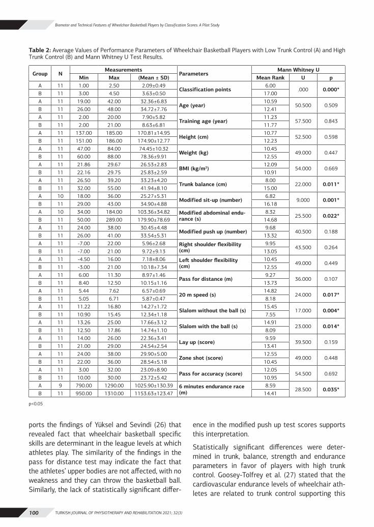
TURKISH JOURNAL OF PHYSIOTHERAPY AND REHABILITATION 2021; 32(3)100
Biomotor and Technical Features of Wheelchaır Basketball Players by Classification Scores: A Pilot Study
ports the findings of Yüksel and Sevindi (26) that revealed fact that wheelchair basketball specific skills are determinant in the league levels at which athletes play. The similarity of the findings in the pass for distance test may indicate the fact that the athletes’ upper bodies are not affected, with no weakness and they can throw the basketball ball. Similarly, the lack of statistically significant differ-
ence in the modified push up test scores supports this interpretation.
Statistically significant differences were deter-mined in trunk, balance, strength and endurance parameters in favor of players with high trunk control. Goosey-Tolfrey et al. (27) stated that the cardiovascular endurance levels of wheelchair ath-letes are related to trunk control supporting this
Table 2: Average Values of Performance Parameters of Wheelchair Basketball Players with Low Trunk Control (A) and High Trunk Control (B) and Mann Whitney U Test Results.
Group NMeasurements
ParametersMann Whitney U
Min Max (Mean ± SD) Mean Rank U pA 11 1.00 2.50 2.09±0.49
Classification points6.00
.000 0.000*B 11 3.00 4.50 3.63±0.50 17.00A 11 19.00 42.00 32.36±6.83
Age (year)10.59
50.500 0.509B 11 26.00 48.00 34.72±7.76 12.41A 11 2.00 20.00 7.90±5.82
Training age (year)11.23
57.500 0.843B 11 2.00 21.00 8.63±6.81 11.77A 11 137.00 185.00 170.81±14.95
Height (cm)10.77
52.500 0.598B 11 151.00 186.00 174.90±12.77 12.23A 11 47.00 84.00 74.45±10.32
Weight (kg)10.45
49.000 0.447B 11 60.00 88.00 78.36±9.91 12.55A 11 21.86 29.67 26.53±2.83
BMI (kg/m2)12.09
54.000 0.669B 11 22.16 29.75 25.83±2.59 10.91A 11 26.50 39.20 33.23±4.20
Trunk balance (cm)8.00
22.000 0.011*B 11 32.00 55.00 41.94±8.10 15.00A 10 18.00 36.00 25.27±5.31
Modified sit-up (number)6.82
9.000 0.001*B 11 29.00 43.00 34.90±4.88 16.18A 10 34.00 184.00 103.36±34.82 Modified abdominal endu-
rance (s)8.32
25.500 0.022*B 11 50.00 289.00 179.90±78.69 14.68A 11 24.00 38.00 30.45±4.48
Modified push up (number)9.68
40.500 0.188B 11 26.00 41.00 33.54±5.31 13.32A 11 -7.00 22.00 5.96±2.68 Right shoulder flexibility
(cm)9.95
43.500 0.264B 11 -7.00 21.00 9.72±9.13 13.05A 11 -4.50 16.00 7.18±8.06 Left shoulder flexibility
(cm)10.45
49.000 0.449B 11 -3.00 21.00 10.18±7.34 12.55A 11 6.00 11.30 8.97±1.46
Pass for distance (m)9.27
36.000 0.107B 11 8.40 12.50 10.15±1.16 13.73A 11 5.44 7.62 6.57±0.69
20 m speed (s)14.82
24.000 0.017*B 11 5.05 6.71 5.87±0.47 8.18A 11 11.22 16.80 14.27±1.72
Slalom without the ball (s)15.45
17.000 0.004*B 11 10.90 15.45 12.34±1.18 7.55A 11 13.26 25.00 17.66±3.12
Slalom with the ball (s)14.91
23.000 0.014*B 11 12.50 17.86 14.74±1.10 8.09A 11 14.00 26.00 22.36±3.41
Lay up (score)9.59
39.500 0.159B 11 21.00 29.00 24.54±2.54 13.41A 11 24.00 38.00 29.90±5.00
Zone shot (score)12.55
49.000 0.448B 11 22.00 36.00 28.54±5.18 10.45A 11 3.00 32.00 23.09±8.90
Pass for accuracy (score)12.05
54.500 0.692B 11 10.00 30.00 23.72±5.42 10.95A 9 790.00 1290.00 1025.90±130.39 6 minutes endurance race
(m)8.59
28.500 0.035*B 11 950.00 1310.00 1153.63±123.47 14.41
p<0.05
TURKISH JOURNAL OF PHYSIOTHERAPY AND REHABILITATION 2021; 32(3) 101
Yüksel M.F.
study. Sprigle et al. (28) reported that a good sit-ting balance and trunk control are necessary to per-form upper extremity movements. As Kerr and Eng (22) stated that proximal stabilization and trunk balance are vital to ensure the smoothness of the players’ distal movement in wheelchairs sports ac-tivities. The significantly higher trunk strength and balance parameters in the player group with low trunk control should stem from the higher number of athletes with paraplegia. Moreover, one athlete had injury at thoracic region and the other athletes had in lumbar region in the player group with high trunk control, while all of the athletes in the group with low trunk control had thoracic region damage. Adegoke et al. (29) found that trunk muscles, es-pecially M. rectus abdominis are innervated at T5-T12 levels and that individuals with spinal cord in-juries above this section cannot balance their trunk. Therefore, it can be asserted that this difference between the two athlete groups regarding trunk balance and strength is due to the differences in the injury level.
Statistically significant differences were found in favor of players with high trunk control for 20 m speed and 6-minute endurance, slalom without the ball, slalom with the ball test and trunk balance for the player group with high trunk control. These re-sults are similar to several studies in the literature (7, 12-14, 21, 30). On the other hand, Yanci et al. (8) argued that there was no statistically significant difference between players with low and high trunk control in speed, direction, strength and endurance parameters. Another study showed that the 20 m speed test scores of players with low and high trunk control were similar (1). Although the players were classified as having low and high trunk control in the abovementioned studies, it was not reported whether there was a statistical difference between their classification scores. Therefore, classification score averages for low and high trunk control may be close to each other and may alter the findings.
The main difference between wheelchair basketball and stand-up basketball is the wheelchair itself. Wheelchair basketball players have to control not only the ball but also the wheelchair. Gagnon et al. (10) asserted that the ball control and player skills may be related to lateralization, but wheelchair propulsion is also dependent on trunk strength. It
can be argued that trunk strength test scores are reflected in the statistical differences observed be-tween groups in with and without ball slalom tests. As Gil et al. (31) revealed that good performance is also highly dependent on wheelchair use in daily life, but the mobilization type findings in this study contradict this finding. As nine players with low trunk control use wheelchairs in their daily lives, while only two of the players with high trunk control use wheelchairs. Therefore, it is thought that prac-tices that improve wheelchair using skills should be integrated into the training with low trunk control starting from the early period. Thus, inter-classifi-cation differences can be avoided.
Although wheelchair basketball players are classi-fied by low and high trunk control, the groups are not homogeneous internally. Even if the disabilities are similar, their functional levels differ. Omitting the separate functional classification scores is one of the limitations. Moreover, it is thought that it will be useful to review similar studies with a pow-er analysis, and higher number of participants, and other performance parameters, for more reliable results. The interest in this sport performed by athletes with disabilities, ongoing development of training methods and materials, advances in tech-nology such as lighter wheelchairs, and efforts to improve functional abilities have gained momen-tum in recent years. Thus, it is recommended to up-date the studies focusing on separate assessment of functional classification score differences in fur-ther studies.
In conclusion, it can be argued that athletes with low and high trunk control differ in terms of biomo-tor abilities. However, they have similar features regarding wheelchair basketball technical skills (throwing basketball, layup, shooting, scoring). It can be concluded that practices aim to improve the biomotor skills of athletes with low trunk control are important and necessary regarding the func-tional levels of the players.
Sources of Support: None.
Conflict of Interest: The authors report no con-flicts of interest.
Ethical Approval: The study was approved by So-cial and Human Scientific Research Ethics Commit-

TURKISH JOURNAL OF PHYSIOTHERAPY AND REHABILITATION 2021; 32(3)102
Biomotor and Technical Features of Wheelchaır Basketball Players by Classification Scores: A Pilot Study
tee of Necmettin Erbakan University the protocol numbered 2020/06.
Informed Consent: A written informed consent statement was given.
Peer-Review: Externally peer-reviewed.
Acknowledgments: The author sincerely thanks İlker Tuzcu, Bünyamin Ünsal and Serkağan Ye-menoğlu for their helps with testing process and language corrections and the volunteers who par-ticipated in this study. This research received no external funding.
REFERENCES1. Zacharakis E, Apostolidis N, Kostopoulos N, Bolatoglou T. Tech-
nical abilities of elite wheelchair basketball players. The Sports J. 2012;15(1):1-8.
2. Di Russo F, Bultrini A, Brunelli S, Delussu AS, Polidori L., Taddei F, et al. Benefits of sports participation for executive function in disabled athletes. J Neurotrauma. 2010;27(12):2309-19.
3. Hutzler Y, Chacham-Guber A, Reiter S. Psychosocial effects of re-verse-integrated basketball activity compared to separate and no physical activity in young people with physical disability. Res Dev Disabil. 2013;34(1):579-87.
4. Plinta R, Saulicz E, Gnat R, Juras G, Waskiewicz Z. Assessment of specific coordination in subjects with handicaped locomotor system. J Hum Kinet. 2005;14:9-50.
5. Goosey-Tolfrey VL, Leicht CA. Field-based physiological testing of wheelchair athletes. Sports Med. 2013;43(2):77-91.
6. Weissland T, Faupin A, Borel B, Leprêtre PM. Comparison be-tween 30-15 intermittent fitness test and multistage field test on physiological responses in wheelchair basketball players. Front Physiol. 2015;6:380.
7. Cömert E, Ün Yıldırım N, Ergun N. Tekerlekli sandalye basketbol oyuncularında üst ekstremite fonksiyonlarının değerlendirilmesi. Turkiye Klinikleri J Sports Sci. 2010;2(2):62-9.
8. Yanci J, Granados C, Otero M, Badiola A, Olasagasti J, Bidaurraza-ga-Letona I, et al. Sprint, agility, strength and endurance capac-ity in wheelchair basketball players. Biol Sport. 2015;32(1):71-8.
9. International Wheelchair Basketball Federation. A Guide to the Functional Classification of Wheelchair Basketball Players. Available from: https://iwbf.org/wp-content/uploads/2020/10/Official-Player-Classification-Manual-2018.pdf, 2018.
10. Gagnon DH, Roy A, Gabison S, Duclos C, Verrier MC, Nadeau S, et al. Effects of seated postural stability and trunk and upper extremity strength on performance during manual wheelchair propulsion tests in individuals with spinal cord injury: an explor-atory study. Rehabil Res Pract. 2016;2016:1-11.
11. da Silva Santos S, Krishnan C, Alonso AC, Greve JMDA. Trunk function correlates positively with wheelchair basketball player classification. Am J Phys Med Rehabil. 2017;96(2):101-8.
12. Molik B, Laskin J, Kosmol A, Marszałek J, Morgulec-Adamowicz N, Frick T. Relationships between anaerobic performance, field tests, and functional level of elite female wheelchair basketball athletes. Hum Mov. 2013;14(4):366-71.
13. Darilgen A, Ün Yıldırım N. Tekerlekli sandalye basketbol oyuncu-larında fiziksel uygunluğun değerlendirilmesi. Turk J Physiother Rehabil. 2008;19(2):64-73.
14. Ergun N, Düzgün İ, Aslan E. Effect of the number of years of experience on physical fitness, sports skills and quality of life in wheelchair basketball players. Turk J Physiother Rehabil. 2008;19(2):55-63.
15. Granados C, Yanci J, Badiola A, Iturricastillo A, Otero M, Ola-sagasti J, et al. Anthropometry and performance in wheelchair basketball. J Strength Cond Res. 2015;29(7):1812-20.
16. Vanlandewijck YC, Daly DJ, Theisen DM. Field test evaluation of aerobic, anaerobic and wheelchair basketball skill performance. Int J Sports Med. 1999;20(8):548–54.
17. Cavedon V, Zancanaro C, Milanese C. Physique and performance of young wheelchair basketball players in relation with classifi-cation. PloS One. 2015;10(11):e0143621.
18. de Witte AM, Hoozemans MJ, Berger MA, van der Woude LH, Veeger D. Do field position and playing standard influence athlete performance in wheelchair basketball? J Sports Sci. 2016;34(9):811-20.
19. Iturricastillo A, Granados C, Camara J, Reina R, Castillo D, Bar-renetxea I, et al. Differences in physiological responses during wheelchair basketball matches according to playing time and competition. Res Q Exerc Sport. 2018;89(4):474-81.
20. Molik B, Kosmol A. Physical ability as a criterion in classifying basketball wheelchair players. Wychowanie Fizyczne I Sport. 1999;43(1/SUPP):471-72.
21. Molik B, Kosmol A, Laskin JJ, Morgulec-Adamowicz N, Skucas K, Dabrowska A, et al. Wheelchair basketball skill tests: differences between athletes’ functional classification level and disability type. Turk J Physiother Rehabil. 2010;21(1):11-19.
22. Kerr HM, Eng JJ. Multidirectional measures of seated postural stability. Clin Biomec. 2002;17(7):555-57.
23. Tomchuk D. Companion Guide to Measurement and Evaluation for Kinesiology, Jones & Bartlett Learning, Canada, 2011.
24. Yüksel MF, Sevindi T. Physical fitness profiles of sitting volleyball players of the Turkish national team. Univers J Educ Res. 2018; 6(3): 556-561.
25. Dewhurst S, Bampouras TM. Intraday reliability and sensitivity of four functional ability tests in older women. J Phys Med Reha-bil. 2014;93(8):703-7.
26. Yüksel MF, Sevindi T. Examination of performance levels of wheelchair basketball players playing in different leagues. Sports. 2018;6:18.
27. Goosey-Tolfrey VL, Batterham AM, Tolfrey K. Scaling behavior of VO2peak in trained wheelchair athletes. Med Sci Sports Exerc. 2003;35(12):2106-11.
28. Sprigle S, Maurer C, Holowka M. Development of valid and reliable measures of postural stability. J Spinal Cord Med. 2007;30(1):40-9.
29. Adegoke BOA, Ogwumike OO, Olatemiju A. Dynamic balance and level of lesion in spinal cord injured patients. Afr J Med Med Sci. 2002;31(4):357-60.
30. Tachibana K, Mutsuzaki H, Shimizu Y, Hotta K, Wadano Y. In-fluence of functional classification on skill tests in elite female wheelchair basketball athletes. Medicina. 2019;55(11):740.
31. Gil SM, Yanci J, Otero M, Olasagasti J., Badiola A., Bidaurraza-ga-Letona I, et al. The functional classification and field test performance in wheelchair basketball players. J Hum Kinet. 2015;46(1):219-30.

103
INVESTIGATION OF THE EFFECT OF COVID-19 OUTBREAK ON PHYSICAL ACTIVITY, PERCEIVED STRESS, PHYSICAL ACTIVITY AWARENESS AND
EXERCISE BARRIERS: A NATIONAL STUDY
ORIGINAL ARTICLE
ABSTRACTPurpose: The aim of this study was to investigate the effect of COVID-19 on physical activity (PA), perceived stress, awareness of physical activity, and exercise barriers nationally.
Methods: The survey was sent online and data were collected from 1087 volunteers between May 8-31, 2020. The physical activity levels of the respondents were evaluated with the International Physical Activity Questionnaire-Short Form (IPAQ-SF) and stress levels with the Perceived Stress Scale. The awareness of exercise and barriers to exercise were questioned with qualitative questions. One-way ANOVA, the Chi-squared test, and Pearson Correlation analysis were used to evaluate the data.
Results: During the COVID-19 pandemic, 70% of adults had inadequate PA. It was observed that the stress level was different in terms of PA levels (p<0.001), and individuals with low-intensity PA levels had higher stress levels. The stress levels and inactivity levels of females were higher than those of males, and the 18-29 age group had a higher stress level (p<0.001).
Conclusion: The precautions taken during the COVID-19 outbreak in Turkey were seen to have a negative effect on physical activity and stress levels, and the most affected groups in this outbreak were females and young adults. Home-based exercises can support the protection of physical and mental health, and avoid the risk of inactivity-related health problems during the COVID-19 outbreak.
Key Words: Awareness, COVID-19, Psychological Stress, Physical activity.
COVİD-19’UN FİZİKSEL AKTİVİTE, ALGILANAN STRES, FİZİKSEL AKTİVİTE FARKINDALIĞI VE
EGZERSİZ YAPMAMA NEDENLERİ ÜZERİNE ETKİSİNİN ARAŞTIRILMASI
ARAŞTIRMA MAKALESİ
ÖZAmaç: Çalışmamızda COVID-19'un ulusal düzeyde fiziksel aktivite (FA), algılanan stres, fiziksel aktivite farkındalığı ve egzersiz yapmama nedenleri üzerindeki etkisini araştırmak amaçlandı.
Yöntem: Anket online olarak gönderildi ve veriler 8-31 Mayıs 2020 tarihleri arasında 1087 gönüllünün katılımı ile toplandı. Katılımcıların fiziksel aktivite düzeyleri Uluslararası Fiziksel Aktivite Anketi (UFAA)-Kısa Formu ile, stres düzeyleri Algılanan Stres Ölçeği ile değerlendirildi. Egzersiz farkındalığı ve egzersiz yapmama nedenleri ise nitel sorularla sorgulandı. Verilerin değerlendirilmesinde tek yönlü ANOVA, Ki-kare testi ve Pearson Korelasyon analizi kullanıldı.
Sonuçlar: COVID-19 salgını sırasında, yetişkinlerin %70'inin yetersiz FA değerlerine sahip olduğu saptandı. Farklı FA düzeylerine sahip bireylerin stres düzeyinin de farklılık gösterdiği (p <0.001) ve düşük şiddetli FA düzeyine sahip bireylerin daha yüksek stres düzeylerine sahip olduğu bulundu. Kadınların erkeklere göre daha yüksek stres ve inaktivite düzeyine, 18-29 yaşları arasındaki bireylerin ise diğer yaş gruplarına göre daha yüksek stres düzeyine sahip olduğu saptandı (p <0.001).
Tartışma: Türkiye'de COVID-19 salgını sırasında alınan önlemlerin fiziksel aktivite ve stres düzeylerini olumsuz etkilediği görüldü ve bu salgından en çok etkilenen grupların kadınlar ve genç yetişkinler olduğu saptandı. COVID-19 salgını sırasında uygulanabilecek ev egzersiz programları, fiziksel ve zihinsel sağlığın korunmasını destekleyebilir ve hareketsizlikle ilişkili sağlık sorunlarının oluşma riskini önleyebilir.
Anahtar Kelimeler: Farkındalık, COVID-19, Psikolojik Stres, Fiziksel Aktivite.
Turkish Journal of Physiotherapy and
Rehabilitation2021 32(3)103-112
Burçin AKÇAY, PhD, PT1
Gülhan YILMAZ GÖKMEN, PhD, PT1
Şule KEÇELİOĞLU, MSc, PT1
Esra KESKİN, MSc, PT1
Ozan Bahadır TÜRKMEN, MSc, PT1
ISSN: 2651-4451 • e-ISSN: 2651-446X
Akçay B., Yılmaz Gökmen G., Keçelioğlu Ş., Keskin E., Türkmen O.B., Investigation of the Effect of Covid-19 Outbreak on Physical Activity, Perceived Stress, Physical Activity Awareness and Exercise Barriers: A National Study, Turk J Physiother Rehabil. 2021; 32(3):103-112. doi: 10.21653/tjpr.856453
1 Bandırma Onyedi Eylül University, Faculty of Healthy Sciences, Department of Physiotherapy and Rehabilitation, Balıkesir, Turkey.
Correspondence (İletişim):
Burçin Akçay, Asst. Prof., PhD, PTBandırma Onyedi Eylül University,
Faculty of Healthy Sciences, Department of Physiotherapy and Rehabilitation,
Balıkesir, TurkeyPhone: +90 505 653 32 73
E-mail: [email protected]: 0000-0002-0883-0311
Gülhan YILMAZ GÖKMENE-mail: [email protected]
ORCID: 0000-0002-0468-7036
Şule KEÇELİOĞLUE-mail: [email protected]
ORCID: 0000-0003-0949-8573
Esra KESKİNE-mail: [email protected]: 0000-0002-4444-5939
Ozan Bahadır TÜRKMENE-mail: [email protected]
ORCID: 0000-0002-1799-3286
Received: 20.01.2021 (Geliş Tarihi)Accepted: 25.05.2021 (Kabul Tarihi)
CC BY - NC
Content of this journal is licensed under a Creative Commons Attribution-NonCommercial 4.0 International License.
TURKISH JOURNAL OF PHYSIOTHERAPY AND REHABILITATION 2021; 32(3)104
Investigation of the Effect of Covid-19 Outbreak on Physical Activity, Perceived Stress, Physical Activity Awareness and Exercise Barriers: A National Study
INTRODUCTION
Coronavirus disease 2019 (COVID-19) appeared in China, in December 2019, rapidly spread worldwide and the World Health Organization (WHO) declared this outbreak a pandemic on March 11, 2020 (1,2). The first COVID-19 cases in Turkey were recorded on March 11, 2020, and as in many other countries measures were taken to prevent the spread of the outbreak. Thus, concepts such as “social distance, staying at home, and quarantine” also became an important part of our daily life.
In a study by Qin et al, the measures taken were seen to seriously change daily routine activities and it was reported that more than half of Chinese adults temporarily lived a sedentary lifestyle with insufficient PA, more sitting time, and poor emo-tional states during the COVID-19 outbreak (3). Many other researchers have emphasized the im-portance of PA in the COVID-19 outbreak and have published studies presenting various recommenda-tions and exercise guidance (4,5).
In societies that have experienced previous out-breaks, negative psychological symptoms have been recorded and recent studies have indicated that the COVID-19 pandemic also has a negative impact not only on physical health but also on men-tal health (4-6). PA has been shown to significantly improve the risk of viral infection, stress, and anx-iety, and it is well known that inadequate PA is an important risk factor for many diseases (8,9).
Physical inactivity has also been defined as a pan-demic and approximately 3.2 million deaths per year are associated with insufficient PA (10). The world is therefore currently experiencing two pan-demics together. Hall et al. warned that the world would recover from the COVID-19 pandemic, nor-mal activities would continue, and then more trou-blingly, the world would be at risk of an inactivity pandemic (11). Therefore, it is important to deter-mine whether the level of PA has been affected nationally and internationally during the pandemic.
The aim of this study was to investigate the effect of COVID-19 pandemic on PA, perceived stress, PA awareness, and exercise barriers. This investiga-tion will provide information on the PA level impact during the pandemic and may be a resource for the
development of outbreak strategies and policies.
METHODS
The study was approved by the IRB and was con-ducted in compliance with the Helsinki Declaration. Informed consent was obtained from all partici-pants online. Individuals who were having been di-agnosed with COVID-19 and went out of the house for more than ten days to go to work in the last one month were not included in the study. Inclusion cri-teria were age ≥18 years and willing to participate.
The sample size of the study was calculated with G-Power software (Version 3.1.9.2, University of Dusseldorf, Dusseldorf, Germany). According to the unknown prevalence formula, the number of indi-viduals in the population was 384 at a 95% con-fidence interval and 50% prevalence. The sample size envisaged for the current study was reached and more was achieved.
Between May 8 and May 31, 2020, the survey was sent online to 1110 individuals. Of these, 23 did not wish to participate, so the study included 1087 re-spondents, comprising 329 males and 758 females (mean:31.30±12.55, range:18-76 years).
The demographic characteristics of the partic-ipants, duration of staying at home were ques-tioned. The age data was divided into 11 groups, and the cities where the participants lived were divided into two groups, as those with and without restrictions.
PA level and sitting time were evaluated using the Turkish version of the International Physical Activ-ity Questionnaire-Short Form (IPAQ-SF) (12). IP-AQ-SF includes items about severe and moderate PA, walking time, and sitting time over the last 7 days. The scoring of the form is calculated by mul-tiplication of the metabolic equivalent task (MET) level of the activity performed, the number of days per week, and for how many minutes per day. In the interpretation of the results a PA level behavior of <600 MET-min/week is low, PA level behavior of 600-3000 MET-min/week is moderate and PA level behavior of >3000 MET-min/week is high (13).
The perceived stress was evaluated with the Turk-ish version of the Perceived Stress Scale (PSS)

TURKISH JOURNAL OF PHYSIOTHERAPY AND REHABILITATION 2021; 32(3) 105
Akçay B., Yılmaz Gökmen G., Keçelioğlu Ş., Keskin E., Türkmen O.B.
(14). This scale consists of 14 items to evaluate stressful situations experienced in the past month with 5-point Likert-type responses. High scores ob-tained from the scale indicate a high level of stress (15).
In order to evaluate the awareness of the partic-ipants about their PA, the level of PA before and during COVID-19 pandemic, the effect of COVID-19 pandemic on PA, and the level of exercise require-ment were questioned. During the pandemic, par-ticipants were asked how they obtained the infor-mation they needed about exercise, whether they found this information useful, whether they did the exercises, and how they changed their PA. To eval-uate the barriers to exercise, the respondents were also asked to select the exercise barriers that suit-ed them with ten reasons given.
Statistical analysis
Data obtained in the study were analyzed statis-tically using IBM SPSS Statistic software for Win-dows (Version 23.0. Armonk, NY: IBM Corp). Descrip-
tive statistics were used for qualitative data. Prior to the statistical evaluation, conformity of the data to normal distribution for all continuous measure-ments in all groups was assessed with the Kolm-ogorov Smirnov test. The Chi-square test was used to examine the relationships between categorical variables, and the one-way ANOVA was used to compare more than two independent groups. Pear-son Analysis was used in the evaluation of the re-lationship between continuous variables. The level of p<0.05 was defined as statistical significance.
RESULTS
The distribution of the participants by age ranges, cities with restrictions, and educational status are given in Table 1. The presence and distribution of diseases of the participants are shown in Figure 1.
It was seen that the participants were able to ac-cess information about PA and exercise practices during the COVID-19 pandemic from social media (47.8%), websites (39.2%), news (8%), television programs (4.3%), and advertisements (0.6%). When
Table 1. Demographic characteristics of the participants.
Femalesn (%)
Malesn (%)
TOTALn (%)
Gender 758 (69.70) 329 (30.30) 1087 (100)Age (years) 31.30±12.55
< 20 108 (9.90) 33 (3.00) 141 (13.00)20-24 223 (20.50) 75 (6.90) 298 (27.40)25-29 117 (10.80) 64 (5.90) 181 (16.70)30-34 67 (6.20) 42 (3.90) 109 (10.00)35-39 58 (5.30) 28 (2.60) 86 (7.90)40-44 66 (6.10) 28 (2.60) 94 (8.60)45-49 46 (4.20) 18 (1.70) 64 (5.90)50-54 19 (1.70) 16 (1.50) 35 (3.20)55-59 25 (2.30) 9 (0.80) 34 (3.10)60-64 19 (1.70) 11 (1.00) 30 (2.80)>64 10 (0.90) 5 (0.50) 15 (1.40)
Cities with Restrictions 679 (62.50) 288 (26.50) 967 (89.00)Cities without Restrictions 79 (7.30) 41 (3.80) 120 (11.00)
Education StatusPrimary school 8 (0.70) 3 (0.30) 11 (1.00)Middle school 24 (2.20) 8 (0.70) 32 (2.90)High school 269 (24.70) 104 (9.60) 373 (34.30)
Associate degree 64 (5.90) 18 (1.70) 82 (7.50)University 244 (22.40) 138 (12.70) 382 (35.10)
Master of Degree 94 (8.60) 34 (3.10) 128 (11.80)Doctor of Philosophy 55 (5.10) 24 (2.20) 79 (7.30)
%: Percentage
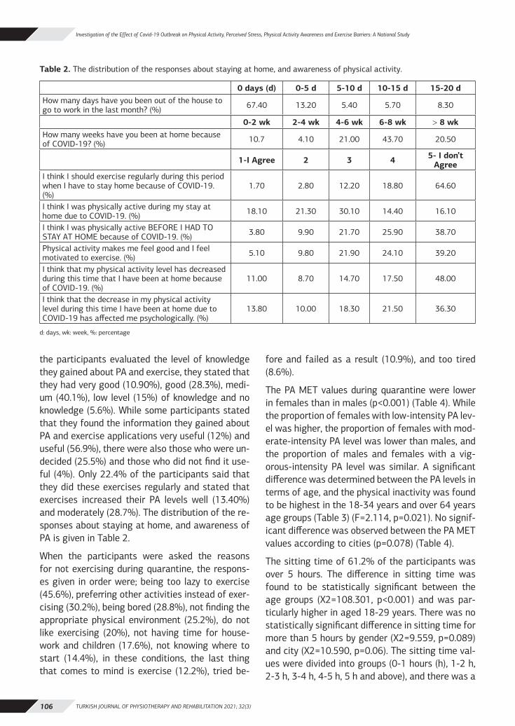
TURKISH JOURNAL OF PHYSIOTHERAPY AND REHABILITATION 2021; 32(3)106
Investigation of the Effect of Covid-19 Outbreak on Physical Activity, Perceived Stress, Physical Activity Awareness and Exercise Barriers: A National Study
the participants evaluated the level of knowledge they gained about PA and exercise, they stated that they had very good (10.90%), good (28.3%), medi-um (40.1%), low level (15%) of knowledge and no knowledge (5.6%). While some participants stated that they found the information they gained about PA and exercise applications very useful (12%) and useful (56.9%), there were also those who were un-decided (25.5%) and those who did not find it use-ful (4%). Only 22.4% of the participants said that they did these exercises regularly and stated that exercises increased their PA levels well (13.40%) and moderately (28.7%). The distribution of the re-sponses about staying at home, and awareness of PA is given in Table 2.
When the participants were asked the reasons for not exercising during quarantine, the respons-es given in order were; being too lazy to exercise (45.6%), preferring other activities instead of exer-cising (30.2%), being bored (28.8%), not finding the appropriate physical environment (25.2%), do not like exercising (20%), not having time for house-work and children (17.6%), not knowing where to start (14.4%), in these conditions, the last thing that comes to mind is exercise (12.2%), tried be-
fore and failed as a result (10.9%), and too tired (8.6%).
The PA MET values during quarantine were lower in females than in males (p<0.001) (Table 4). While the proportion of females with low-intensity PA lev-el was higher, the proportion of females with mod-erate-intensity PA level was lower than males, and the proportion of males and females with a vig-orous-intensity PA level was similar. A significant difference was determined between the PA levels in terms of age, and the physical inactivity was found to be highest in the 18-34 years and over 64 years age groups (Table 3) (F=2.114, p=0.021). No signif-icant difference was observed between the PA MET values according to cities (p=0.078) (Table 4).
The sitting time of 61.2% of the participants was over 5 hours. The difference in sitting time was found to be statistically significant between the age groups (X2=108.301, p<0.001) and was par-ticularly higher in aged 18-29 years. There was no statistically significant difference in sitting time for more than 5 hours by gender (X2=9.559, p=0.089) and city (X2=10.590, p=0.06). The sitting time val-ues were divided into groups (0-1 hours (h), 1-2 h, 2-3 h, 3-4 h, 4-5 h, 5 h and above), and there was a
Table 2. The distribution of the responses about staying at home, and awareness of physical activity.
0 days (d) 0-5 d 5-10 d 10-15 d 15-20 dHow many days have you been out of the house to go to work in the last month? (%) 67.40 13.20 5.40 5.70 8.30
0-2 wk 2-4 wk 4-6 wk 6-8 wk > 8 wkHow many weeks have you been at home because of COVID-19? (%) 10.7 4.10 21.00 43.70 20.50
1-I Agree 2 3 4 5- I don’t Agree
I think I should exercise regularly during this period when I have to stay home because of COVID-19. (%)
1.70 2.80 12.20 18.80 64.60
I think I was physically active during my stay at home due to COVID-19. (%) 18.10 21.30 30.10 14.40 16.10
I think I was physically active BEFORE I HAD TO STAY AT HOME because of COVID-19. (%) 3.80 9.90 21.70 25.90 38.70
Physical activity makes me feel good and I feel motivated to exercise. (%) 5.10 9.80 21.90 24.10 39.20
I think that my physical activity level has decreased during this time that I have been at home because of COVID-19. (%)
11.00 8.70 14.70 17.50 48.00
I think that the decrease in my physical activity level during this time I have been at home due to COVID-19 has affected me psychologically. (%)
13.80 10.00 18.30 21.50 36.30
d: days, wk: week, %: percentage

TURKISH JOURNAL OF PHYSIOTHERAPY AND REHABILITATION 2021; 32(3) 107
Akçay B., Yılmaz Gökmen G., Keçelioğlu Ş., Keskin E., Türkmen O.B.
Table 3. Distribution of physical activity intensity levels in terms of gender, age, city and perceived stress during the COVID-19 outbreak.
Vigorous -Intensity PA
(Mean±SD, %)
Moderate –Intensity PA
(Mean±SD, %)
Low -Intensity PA
(Mean±SD, %)P
Gender p<0.001∗∗
Female 2.60 23.10 74.30
Male 2.10 35.30 62.60
Age (Years) p=0.021∗
< 20 0.00 24.10 75.90
20-24 1.70 28.90 69.50
25-29 1.70 23.80 74.60
30-34 2.80 24.80 72.50
35-39 3.50 30.20 66.30
40-44 2.10 27.70 70.20
45-49 4.7 21.90 73.40
50-54 2.90 28.60 68.60
55-59 11.80 32.40 55.90
60-64 11.80 32.40 50.00
>64 6.70 6.70 86.70
All Ages 4148.31±975.862.50
1185.53±557.4926.80
217.8±181.4570.70
Quarantined Cities 2.80 26.30 70.90
Not Quarantined Cities 0.00 30.80 69.20
Perceived Stress 21.29±7.12 bc 26.26±8.02 ac 28.82±7.15 ab p<0.001∗∗
SD: Standard Deviation, ∗ : p<0.05,∗∗ : p<0.001, a: p<0.05 versus Vigorous-intensity PA, b: p<0.05 versus Moderate-intensity PA, c: p<0.05 versus Low- intensity PA
Figure 1. Distribution of chronic and musculoskeletal diseases of the participants.
CD: Cardiovascular Disease, CRD: Chronic Respiratory Diseases, DM: Diabetus Mellitus, RD: Rheumatic Diseases
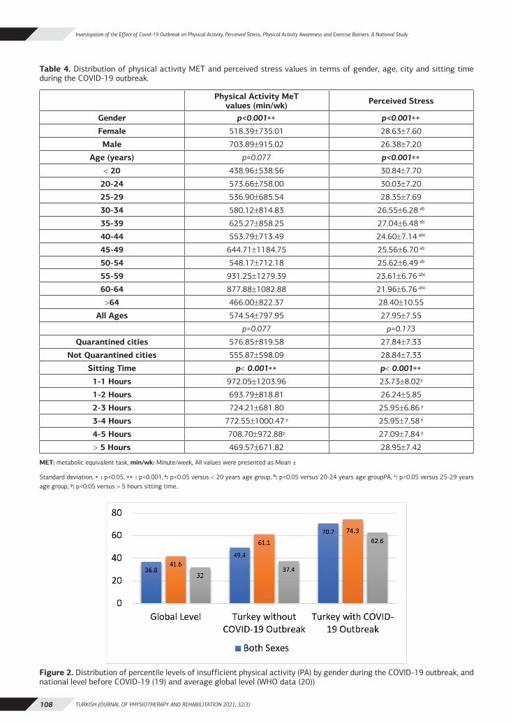
TURKISH JOURNAL OF PHYSIOTHERAPY AND REHABILITATION 2021; 32(3)108
Investigation of the Effect of Covid-19 Outbreak on Physical Activity, Perceived Stress, Physical Activity Awareness and Exercise Barriers: A National Study
Table 4. Distribution of physical activity MET and perceived stress values in terms of gender, age, city and sitting time during the COVID-19 outbreak.
Physical Activity MeTvalues (min/wk) Perceived Stress
Gender p<0.001∗∗ p<0.001∗∗
Female 518.39±735.01 28.63±7.60
Male 703.89±915.02 26.38±7.20
Age (years) p=0.077 p<0.001∗∗
< 20 438.96±538.56 30.84±7.70
20-24 573.66±758.00 30.03±7.20
25-29 536.90±685.54 28.35±7.69
30-34 580.12±814.83 26.55±6.28 ab
35-39 625.27±858.25 27.04±6.48 ab
40-44 553.79±713.49 24.60±7.14 abc
45-49 644.71±1184.75 25.56±6.70 ab
50-54 548.17±712.18 25.62±6.49 ab
55-59 931.25±1279.39 23.61±6.76 abc
60-64 877.88±1082.88 21.96±6.76 abc
>64 466.00±822.37 28.40±10.55
All Ages 574.54±797.95 27.95±7.55
p=0.077 p=0.173
Quarantined cities 576.85±819.58 27.84±7.33
Not Quarantined cities 555.87±598.09 28.84±7.33
Sitting Time p< 0.001∗∗ p< 0.001∗∗
1-1 Hours 972.05±1203.96 23.73±8.02y
1-2 Hours 693.79±818.81 26.24±5.85
2-3 Hours 724.21±681.80 25.95±6.86 y
3-4 Hours 772.55±1000.47 y 25.95±7.58 y
4-5 Hours 708.70±972.88y 27.09±7.84 y
> 5 Hours 469.57±671.82 28.95±7.42
MET: metabolic equivalent task, min/wk: Minute/week, All values were presented as Mean ±
Standard deviation, ∗ : p<0.05, ∗∗ : p<0.001, a: p<0.05 versus < 20 years age group, b: p<0.05 versus 20-24 years age groupPA, c: p<0.05 versus 25-29 years age group, y: p<0.05 versus > 5 hours sitting time.
Figure 2. Distribution of percentile levels of insufficient physical activity (PA) by gender during the COVID-19 outbreak, and national level before COVID-19 (19) and average global level (WHO data (20))

TURKISH JOURNAL OF PHYSIOTHERAPY AND REHABILITATION 2021; 32(3) 109
Akçay B., Yılmaz Gökmen G., Keçelioğlu Ş., Keskin E., Türkmen O.B.
statistically significant difference between groups in terms of PA MET values (F=6.539, p<0.001), and stress levels (F=7.150, p<0.001) (Table 4).
While females had higher stress (F=20.818, p<0.001), there was no statistically significant dif-ference in terms of city (F=1.862, p=0.173). The stress level showed a statistically significant differ-ence between the age groups (F=11.776, p<0.001) and was particularly higher in participants aged 18-29 years and over 64 years. It was also observed that the stress level was different in terms of PA (F=23.948, p<0.001); individuals with vigorous-in-tensity PA levels had lower stress, and those with low-intensity PA levels had higher stress (Table 3).
A weak negative correlation was determined be-tween stress and PA (r=-0.203, p<0.001), and be-tween PA and sitting time (r=-0.147, p<0.001). A weak positive correlation was found between stress and sitting time (r=0.166, p<0.001).
DISCUSSION
These national research data provided four main findings. First, 70% of the participants were found to have insufficient PA levels and the inactivity rate increased dramatically due to the COVID-19 pan-demic and this PA percentage is twice the global value. The second finding was that the stress levels of the respondents increased as the PA level de-creased, and a negative correlation was found be-tween PA level and stress level. Third, females and young adults were seen to be most affected by the COVID-19 outbreak in terms of PA and stress level. Finally, a large proportion of the participants were aware that their level of PA was low and they need-ed to exercise, but the rate of exercising was low and exercise barriers were detected. These findings demonstrate the importance of early awareness and a need for publicity strategies for the develop-ment of PA during the stay at home.
International and intercity travel restrictions were imposed on April 3, 2020 in Turkey. Subsequent-ly, those with chronic disease, individuals over the age of 65 and under 18 years were isolated. En-vironmental preventative measures were taken to reduce spread of the virus and restrictions were imposed on public places. These precautions were applied according to the process of the COVID-19
pandemic and as of June 1, 2020, the restrictions have been largely removed (16). Therefore, data were collected between May 8 and May 31,2020, and this time frame can be considered to be the best time to see lifestyle changes due to the COVID-19 pandemic.
The WHO has stated that regular and adequate PA improves muscle, bone, cardiovascular fitness, and reduces the risk of hypertension, coronary heart disease, stroke, diabetes, cancer and depression and many studies have supported these bene-fits (17, 8, 9). Recent studies have stated that the COVID-19 pandemic has had a negative impact on physical and mental health, due to having to stay at home, less social interaction and inactivity (4, 7). Therefore, studies have emphasized the impor-tance of PA during the COVID-19 pandemic, and have offered suggestions and exercise guidance (4, 5, 7, 18).
In the current study, more than half of the partic-ipants thought that they were more active before COVID-19 pandemic, and that their PA were neg-atively affected and decreased with this outbreak. Participants declared that their knowledge level was not sufficient for exercise, they accessed the information mostly from social media and found this information useful, but did not perform the new exercises they regularly acquired. In line with these results, although participants had high PA awareness during the outbreak, it was observed that the awareness did not result in these individ-uals exercising. New strategies need to be devel-oped to guide individuals to exercise.
When the national pre-pandemic values and the values of this study were compared; it was found that during the outbreak low-intensity PA level in-creased (pre-pandemic:49.40%, during- pandem-ic:70.70%), moderate-intensity PA level did not change (pre-pandemic:26.00%, during-COVID-19 pandemic:26.40%) and vigorous-intensity PA level decreased (pre-pandemic:24.60%, during-COVID-19 pandemic:2.50%) (19). In addition, when the global data are examined, it can be seen that the COVID-19 pandemic has affected the PA rate in Turkey two-fold (Figure 2) (20).
While the low-intensity PA level was higher in fe-males, the moderate-intensity PA level rate was
TURKISH JOURNAL OF PHYSIOTHERAPY AND REHABILITATION 2021; 32(3)110
Investigation of the Effect of Covid-19 Outbreak on Physical Activity, Perceived Stress, Physical Activity Awareness and Exercise Barriers: A National Study
higher in males. Although the pre-pandemic values were similar in both gender for moderate-intensi-ty PA level, a decrease in the moderate-intensity PA level of females was observed in the pandemic process (19). Qin et al. reported an increase in fe-males with low-intensity PA levels, and the current study results showed that the rate of females with low-intensity PA levels was higher than the rate in the Qin et al study (3). Previous studies have indi-cated that females tend to do low-intensity activity (21,22) and the current study findings demonstrate that females have been more affected by COVID-19 pandemic.
In terms of age groups in this study, the lowest PA was determined in young adults aged 18-34 years and in those aged over 64 during the COVID-19 pandemic and these findings were similar to those in the study by Qin et al (3). Compared to the pre-pandemic values, an increase in low-inten-sity PA level was seen in all age groups with the COVID-19 pandemic. There was a higher rate of low-intensity PA level in the 60-64 years age group before COVID-19 pandemic, whereas this changed to the 18-34 years and over 64 years age groups during the COVID-19 pandemic (19). It was expect-ed that the over 65 years age group would have a high level of inactivity because leaving the home was prohibited. The high level of inactivity of young adults may be due to the fact that some of this age group were students and their courses continued at home and others were working at home rather than in an office. However, the state of inactivity, which is evident especially in females and young adults, may pose a high health risk in terms of a sedentary lifestyle and various precautions should be taken to prevent the occurrence of various health problems.
Qin et al. found that the level of inactivity differed between cities in China (3) whereas there was no difference in the current study. These results can be interpreted as the participants taking individual measures irrespective of the city restrictions.
Regardless of leisure PA, a long sitting time has been reported to show a dose-response relation-ship between all causes, deaths, cardiovascular dis-eases, and cancer associated with physical inactivi-ty (23). In this study, while sitting time did not differ in terms of gender and city, higher differences were
observed between age groups, especially with the age group of 18-29 years. It was also found that the MET values of PA decreased as the sitting time increased. Qin et al. reported similar results and stated that PA could be used as an effective way to reduce sitting time, especially for young adults (3).
While 83.30% of the participants stated that they knew that they should exercise during the quaran-tine and PA could make them feel good, they listed the exercise barriers during the pandemic as being too lazy to exercise, preferring other activities in-stead of exercising, and being bored. In pre-pan-demic studies, exercise barriers have been noted as lack of time and facilities having inconvenient schedules and exercise not fitting around study or placement schedules (24). Therefore, this study demonstrates that exercise barriers may change due to the pandemic. In addition to emphasizing the importance of PA for health during the pan-demic, it may be necessary to develop health strat-egies to overcome the barriers to exercise.
Recent studies have stated that the COVID-19 pandemic has caused psychological distress since the start of the outbreak and there has been no change in stress level over time (7,25). Sareen et al concluded that females are more likely to devel-op post-traumatic stress disorders and are more vulnerable to stress (26) and other studies have shown that the COVID-19 pandemic affects fe-males psychologically more than males (3,27), and the current study results are consistent with these findings. Although the precautions taken due to the COVID-19 pandemic differed according to the cit-ies, it was found in this study that the presence of restrictions in the cities did not affect the stress level, which was similar to other studies (3). In ad-dition, the stress level varied by age, and individ-uals aged 18-29 and over 64 years were found to have high stress levels. In a study by Çalışkan et al., the average of stress values between the ages of 17-28 years in the Turkish population was stated to be 19.07, while in the current study, this rate was remarkably higher (28). Recent studies in other populations have also shown that the stress asso-ciated with pandemics was more common in the young population (3, 27). Cheng et al. suggested that greater anxiety among the young population could be a result of greater access to information
TURKISH JOURNAL OF PHYSIOTHERAPY AND REHABILITATION 2021; 32(3) 111
Akçay B., Yılmaz Gökmen G., Keçelioğlu Ş., Keskin E., Türkmen O.B.
through social media (29).
Finally, there was determined to be a negative correlation between stress and PA, and a positive correlation between stress and daily sitting time. It has been stated in studies before COVID-19 pan-demic and during the COVID-19 pandemic that in-dividuals have better emotional status as the level of PA increases (3,30).
Despite the importance of the findings, this study had several limitations. First, as the study was in the form of an online survey, a small number of el-derly respondents was reached, and the response from a young population, who use the internet much more, was much greater. In subsequent stud-ies, the tele-survey method could be used to reach the elderly population. Second, although more par-ticipants were targeted, the study was terminat-ed due to the substantial removal of restrictions on May 31. Third, the study included participants from 65 different cities, but most participants were from the big cities so the results may not reflect the overall situation in Turkey. Another limitation of our study was that demographic characteristics of the participants, such as chronic diseases, the environment they live in, and their socioeconom-ic level affecting their physical activity level were not compared. Despite the limitations mentioned above, the strength of this study is that it is the first national study covering 65 different provinc-es, to examine exercise barriers, and PA awareness during the COVID-19 pandemic. Most importantly, the findings show that early and preventive health promotion and PA guidance are required during a period of quarantine. It can guide the development of preventive health strategies in case of disease outbreaks at the national and international level.
The results of this national research demonstrat-ed that 70% of the respondents had insufficient PA levels and the inactivity rate increased dramat-ically during the COVID-19 pandemic. Stress levels were seen to increase as PA level decreased, and females and young adults were observed to be the most affected by the COVID-19 pandemic in terms of PA and stress level. Although the study partici-pants had high PA awareness, this awareness did not lead these individuals to exercise, and the bar-riers to exercise were seen to change during the
pandemic. Therefore, timely preventive measures should be taken to avoid the risk of many diseases that may result from inactivity.
Sources of Support: There is no funding support for the study.
Conflict of Interest: The authors report no conflict of interest.
Ethical Approval: Ethical approval was received from Bandırma Onyedi Eylül University, ethics com-mittee of non-interventional research in health sci-ences (Date: 08.05.2020, number: 2020-21).
Informed Consent: Informed Consent was at the top of the online form. After the participants ap-proved the informed consent form, they were able to access the answer to the questions page. Thus, informed consent was obtained from all partici-pants.
Peer-Review: Externally peer-reviewed.
Authors Contributors: All authors designed the study, searched literature, collected data, analyzed the data, wrote the first draft of the report, revised and approved the final report.
Acknowledgments: We would like to thank many colleagues and friends for supporting us by partic-ipating in this study.
REFERENCES 1. WHO. WHO Timeline - COVID-19. https://www.who.int/news-
room/detail/27-04-2020-who-timeline---covid-19; 2020 Ac-cessed 10.06.2020
2. Wang C, Horby PW, Hayden FG, Gao GF. A novel coronavirus out-break of global health concern. Lancet. 2020;15;395(10223):514-23. doi:10.1016/S0140-6736(20)301
3. Qin F, Song Y, Nassis GP, Zhao L, Cui S, Lai L et al. Preva-lence of Insufficient Physical Activity, Sedentary Screen Time and Emotional Well-Being During the Early Days of the 2019 Novel Coronavirus (COVID-19) Outbreak in China: A National Cross-Sectional Study. SSRN Electron J. Published online 2020. doi:10.2139/ssrn.3566176
4. Chen P, Mao L, Nassis GP, Harmer P, Ainsworth BE, Li F. Coro-navirus disease (COVID-19): The need to maintain regular physical activity while taking precautions. J Sport Heal Sci. 2020;9(2):103-104. doi:10.1016/j.jshs.2020.02.001
5. Jiménez-Pavón D, Carbonell-Baeza A, Lavie CJ. Physical exer-cise as therapy to fight against the mental and physical con-sequences of COVID-19 quarantine: Special focus in older people [published online ahead of print, 2020 Mar 24]. Prog Cardiovasc Dis. 2020;S0033-0620(20)30063-3. doi:10.1016/j.pcad.2020.03.009
6. Brooks SK, Webster RK, Smith LE, Woodland L, Wesse-ly S, Greenberg N et al. The psychological impact of quar-antine and how to reduce it: rapid review of the evidence.

TURKISH JOURNAL OF PHYSIOTHERAPY AND REHABILITATION 2021; 32(3)112
Investigation of the Effect of Covid-19 Outbreak on Physical Activity, Perceived Stress, Physical Activity Awareness and Exercise Barriers: A National Study
Lancet. 2020;395(10227):912-920. doi:10.1016/S0140-6736(20)30460-8
7. Wang C, Pan R, Wan X, Tan Y, Xu L, Ho CS et al. Immediate Psy-chological Responses and Associated Factors during the Initial Stage of the 2019 Coronavirus Disease (COVID-19) Epidemic among the General Population in China. Int J Environ Res Public Health. 2020;17(5):1729. Published 2020 Mar 6. doi:10.3390/ijerph17051729
8. Sanchis-Gomar F, Lucia A, Yvert T, Ruiz-Casado A, Pareja-Ga-leano H, Santos-Lozano A et al. Physical inactivity and low fit-ness deserve more attention to alter cancer risk and prognosis. Cancer Prev Res (Phila). 2015;8(2):105-110. doi:10.1158/1940-6207.CAPR-14-0320
9. Mikkelsen K, Stojanovska L, Polenakovic M, Bosevski M, Apostol-opoulos V. Exercise and mental health. Maturitas. 2017;106:48-56. doi:10.1016/j.maturitas.2017.09.003
10. Kohl 3rd HW, Craig CL, Lambert EV, Inoue S, Alkandari JR, Leetongin G et al. The pandemic of physical inactivity: glob-al action for public health. Lancet. 2012;380(9838):294-305. doi:10.1016/S0140-6736(12)60898-8
11. Hall G, Laddu DR, Phillips SA, Lavie CJ, Arena R. A tale of two pan-demics: How will COVID-19 and global trends in physical inactiv-ity and sedentary behavior affect one another? [published online ahead of print, 2020 Apr 8]. Prog Cardiovasc Dis. 2020;S0033-0620(20)30077-3. doi:10.1016/j.pcad.2020.04.005
12. Saglam M, Arikan H, Savci S, Inal-Ince D, Bosnak-Guclu M, Karabulut E et al. International physical activity questionnaire: reliability and validity of the Turkish version. Percept Mot Skills. 2010;111(1):278-284. doi:10.2466/06.08.PMS.111.4.278-284
13. IPAQ Research Committee. Guidelines for data processing and analysis of the International Physical Activity Questionnaire (IP-AQ)-short and long forms. http://www. ipaq. ki. se/scoring. pdf. 2005.
14. Eskin M, Harlak H, Demirkıran F, Dereboy Ç. Algılanan stres ölçeğinin Türkçeye uyarlanması: güvenirlik ve geçerlik analizi. Yeni Symposium Journal. 2013;51-3:132-140.
15. Cohen S, Kamarck T, Mermelstein R. A global measure of per-ceived stress. J Health Soc Behav. 1983;24(4):385-396.
16. T.C. Sağlık Bakanlığı. Sağlık Bakanı Basın Bildirileri. T.C. Sağlık Bakanlığı. https://www.saglik.gov.tr/TR,3187/basin-merkezi.html; 2020 Accessed 14.06.20.
17. WHO. Physical activity. https://www.who.int/news-room/fact-sheets/detail/physical-activity; 2020 Accesses 14.06.20.
18. Jurak G, Morrison SA, Leskošek B, Kovač M, Hadžić V, Vodičar J et al. Physical activity recommendations during the coronavirus disease-2019 virus outbreak [published online ahead of print, 2020 May 16]. J Sport Health Sci. 2020;S2095-2546(20)30062-4. doi:10.1016/j.jshs.2020.05.003
19. Üner S, Balcılar M, Ergüder T. Türkiye hanehalkı sağlık araştır-
ması: bulaşıcı olmayan hastalıkların risk faktörleri prevalansı 2017 (STEPS). Dünya Sağlık Örgütü Türkiye Ofisi, Ankara. 2018.
20. WHO. Prevalence of insufficient physical activity among adults Data by World Bank income groups. WHO. https://apps.who.int/gho/data/view.main.2487?lang=en); 2018
21. Guthold R, Stevens GA, Riley LM, Bull FC. Worldwide trends in insufficient physical activity from 2001 to 2016: a pooled anal-ysis of 358 population-based surveys with 1·9 million partici-pants [published correction appears in Lancet Glob Health. 2019 Jan;7(1):e36]. Lancet Glob Health. 2018;6(10):e1077-e1086. doi:10.1016/S2214-109X(18)30357-7
22. Mielke GI, da Silva ICM, Kolbe-Alexander TL, Brown WJ. Shifting the Physical Inactivity Curve Worldwide by Closing the Gender Gap. Sports Med. 2018;48(2):481-489. doi:10.1007/s40279-017-0754-7
23. Katzmarzyk PT, Church TS, Craig CL, Bouchard C. Sitting time and mortality from all causes, cardiovascular disease, and can-cer. Med Sci Sports Exerc. 2009;41(5):998-1005. doi:10.1249/MSS.0b013e3181930355
24. Blake H, Stanulewicz N, Mcgill F. Predictors of physical activity and barriers to exercise in nursing and medical students. J Adv Nurs. 2017;73(4):917-929. doi:10.1111/jan.13181
25. Tang W, Hu T, Hu B, Jin C, Wang G, Xie C et al. Prevalence and correlates of PTSD and depressive symptoms one month after the outbreak of the COVID-19 epidemic in a sample of home-quarantined Chinese university students. J Affect Disord. 2020;274:1-7. doi:10.1016/j.jad.2020.05.009
26. Sareen J, Erickson J, Medved MI, Asmundson GJ, Enns MW, Stein M et al. Risk factors for post-injury mental health problems. De-press Anxiety. 2013;30(4):321-327. doi:10.1002/da.22077
27. Mazza C, Ricci E, Biondi S, Colasanti M, Ferracuti S, Napoli C et al. A Nationwide Survey of Psychological Distress among Italian People during the COVID-19 Pandemic: Immediate Psychologi-cal Responses and Associated Factors. Int J Environ Res Public Health. 2020;17(9):3165. Published 2020 May 2. doi:10.3390/ijerph17093165
28. Çalışkan S, Aydoğan S, Işıklı B, Metintaş S, Yenilmez F, Yenilmez Ç. Üniversite Öğrencilerinde Algilanan Stres Düzeyi ve İlişkili Olabilecek Faktörlerin Değerlendirilmesi-Assessment Of Per-ceived Stress Level And Related Factors In University Students. Eskişehir Türk Dünyası Uygulama ve Araştırma Merkezi Halk Sağlığı Dergisi. 2018; 31:3(2). (ISSN: 2564-6311)
29. Cheng C, Jun H, Liang B. Psychological health diathesis assess-ment system: a nationwide survey of resilient trait scale for Chi-nese adults. Stud Psychol Behav. 2014;12(6):735-742.
30. Stults-Kolehmainen MA, Sinha R. The effects of stress on physical activity and exercise. Sports Med. 2014;44(1):81-121. doi:10.1007/s40279-013-0090-5
113
EFFECTS OF NEURODEVELOPMENTAL THERAPY ON FUNCTION AND MUSCLE ULTRASOUND PARAMETERS
IN CHILDREN WITH CEREBRAL PALSY
ORIGINAL ARTICLE
ABSTRACTPurpose: This study aimed to evaluate the effectiveness of neurodevelopmental therapy (NDT) for improving lower extremity functions, ankle dorsiflexion range of motion (ROM), popliteal angle, medial gastrocnemius (GCM) muscle thickness (MT), and functionality of lower extremity on children with hemiparetic Cerebral Palsy (CP).
Methods: Eighteen children with hemiparetic CP aged between 6-15 years were included in the study. Structured NDT (s-NDT) was applied 40 minutes per session, three times a week over 12 weeks. Children were evaluated with Gross Motor Function Measurement-88 (GMFM-88) for motor function level; with goniometer for ankle dorsiflexion ROM and popliteal angle; with ultrasound for medial GCM MT, and with ultrasound Lower Extremity Function Test (LEFT) for the functionality of lower extremity. Children were evaluated before and after the intervention.
Results: After the treatment, statistically significant differences were obtained in mean values of GMFM-88, ankle dorsiflexion ROM, medial GCM MT, and LEFT (p<0.05). There was no significant difference in popliteal angle (p>0.05).
Conclusion: In conclusion, it was observed that s-NDT might be effective on motor function level, ankle dorsiflexion, medial GCM MT, and lower extremity functionality in children with hemiparetic CP. These improvements will make significant positive contributions to the mobility of children.
Key Words: Cerebral Palsy, Hemiparesis, Lower Extremity, Neurodevelopmental Therapy, Ultrasonographic Imaging.
NÖROGELİŞİMSEL TEDAVİNİN SEREBRAL PALSİ’Lİ ÇOCUKLARDA FONKSİYONELLİK VE KAS ULTRASON
PARAMETRELERİNE ETKİSİ
ARAŞTIRMA MAKALESİ
ÖZAmaç: Bu çalışmanın amacı; hemiparetik serebral palsi (SP)’li çocuklarda nörogelişimsel tedavinin (NGT) alt ekstremite fonksiyonları, ayak bileği dorsifleksiyon eklem hareket açıklığı (EHA), popliteal açı, medial gastroknemius (GCM) kas kalınlığı (MT) ve alt ekstremiye fonksiyonelliğini iyileştirmedeki etkinliğini değerlendirmekti.
Yöntem: Çalışmaya yaşları 6-15 yıl aralığında on sekiz hemiparetik SP’li çocuk dahil edildi. Yapılandırılmış NGT haftada 3 seans, her seans 40 dakika olacak şekilde 12 hafta boyunca uygulandı. Çocukların motor fonksiyon seviyelerini değerlendirmek için Kaba Motor Fonksiyon Ölçütü-88 (KMFÖ-88), ayak bileği dorsifleksiyon açısı ve popliteal açının değerlendirmesi için universal gonyometre, medial gastroknemius kasının kalınlığı ölçmek için ultrasanografik ölçüm ve alt ekstremite fonksiyonlarını değerlendirmek için Alt Ekstremite Fonksiyon Testi (AEFT) kullanıldı.
Sonuçlar: Tedavi sonrasında GMFM-88, ayak bileği dorsifleksiyon EHA, medial GCM MT ve AEFT değerlerinde istatistiksel olarak anlamlı gelişme olduğu belirlendi (p<0,05). Popliteal açı değerlerinde ise anlamlı değişiklik yoktu (p>0,05).
Tartışma: Sonuç olarak, hemiparetik SP'li çocuklarda yapılandırılmış NGT'nin motor fonksiyon düzeyi, ayak bileği dorsifleksiyonu, medial GCM- MT değerleri ve alt ekstremite fonksiyonelliği üzerine olumlu etkileri olabileceği gözlendi. Elde edilen bu gelişmeler çocukların mobilitesine önemli pozitif katkılar sağlayacaktır.
Anahtar Kelimeler: Serebral Palsi, Hemiparezi, Alt Ekstremite, Nörogelişimsel Tedavi, Ultrasonografik Görüntüleme.
Turkish Journal of Physiotherapy and
Rehabilitation2021 32(3)113-120
Ülkü ATASOY, MSc1, 2
Miray BUDAK, PhD3
Devrim TARAKCI, PhD3 Ali Rıza BULUT, MD1
ISSN: 2651-4451 • e-ISSN: 2651-446X
Atasoy Ü., Budak M., Tarakcı D., Bulut A.R., Effects of Neurodevelopmental Therapy on Function and Muscle Ultrasound Parameters in Children with Cerebral Palsy, Turk J Physiother Rehabil. 2021; 32(3):113-120. doi: 10.21653/tjpr.950714
1 Ümran Medical Center, Istanbul, Turkey.2 Istanbul Medipol University, Institute of Health
Sciences, Department of Physiotherapy and Rehabilitation, Istanbul, Turkey.
3 Istanbul Medipol University, Faculty of Health Sciences, Department of Occupational Therapy, Istanbul, Turkey.
Correspondence (İletişim):
Ülkü ATASOYÖzel Ümran Tıp Merkezi, Bulgurlu mah.
İzzettin bey sok. 6/3.ÜsküdarPhone: + 90 543 579 6261
E-mail: [email protected] ORCID: 0000-0002-7356-1074
Miray BUDAK, PhDE-mail: [email protected]
Devrim TARAKCI, PhDE-mail: [email protected]
Ali Rıza BULUT, MDE-mail: [email protected]
Received: 11.06.2021 (Geliş Tarihi)Accepted: 8.10.2021 (Kabul Tarihi)
CC BY - NC
Content of this journal is licensed under a Creative Commons Attribution-NonCommercial 4.0 International License.
TURKISH JOURNAL OF PHYSIOTHERAPY AND REHABILITATION 2021; 32(3)114
Effects of Neurodevelopmental Therapy on Function and Muscle Ultrasound Parameters in Children with Cerebral Palsy, Turk J Physiother Rehabil
INTRODUCTION
Cerebral palsy (CP) is a non-progressive develop-mental disorder characterized by a lifelong mobil-ity and posture deficit caused by a lesion during the prenatal, natal, or postnatal phase during brain development (1, 2). In low- and middle-income countries, it is the most common physical disability in children, with a 2.0 to 2.9 percent (3). Hemipa-retic CP is a type of CP that affects one-half of the body’s lower and upper extremities and the trunk and can be accompanied by visual sensory impairments, convulsions, cognitive problems, and motor problems (4). There is a tendency to fall to one side due to poor balance reactions. The unaf-fected side’s stability and balance reactions are hyperactive, and the weight is carried more on the unaffected side while standing, while the affected side is compensated accordingly (5, 6). Basic motor abilities such as standing, walking, and standing on one leg are challenging for children with hemipa-retic CP (1).
Neurodevelopmental Therapy (NDT)/Bobath is a problem-solving technique used to evaluate and treat people who have functional, motor, and pos-tural impairments resulting from central nervous system injuries (7). Dr. Karel and Bobath developed the Bobath method, which has been utilized by therapists worldwide in CP rehabilitation for over 80 years. By promoting muscle activity through key control points guided by the therapist, the Bobath concept attempts to improve gross motor function and postural control(7). The Bobaths renamed their method to neurodevelopmental treatment (NDT) in 1960. Both names are used to describe the same intervention in the literature (8).
Even though physiotherapists have been using the NDT approach for decades, a recent systematic re-view suggests that the impact of NDT on children with cerebral palsy is still unclear (9). Because of methodological and intervention variations among studies and a lack of standards for outcome cri-teria, clinical data supporting the benefits of NDT in children with Hemiparetic CP is inadequate. Re-habilitative ultrasonography (RUSG) is a technique for providing objective feedback on the efficacy of NDT (10). In the review by Yeşilyaprak et al, there was a direct relationship between muscle volume
and cross-sectional area of the muscle (10). The muscle thickness (MT) of the gastrocnemius mus-cle (GCM) is thinner in children with CP than in chil-dren who are developing typically, and there is a significant relationship between MT and functional level when evaluated by muscle volume, GMFM-88, GMFCS, and mobility area (11). Only a few studies have evaluated the effect of NDT on muscle thick-ness using US techniques (12). The purpose of this study was to evaluate the effects of NDT in terms of motor function level, ankle dorsiflexion range of motion (ROM), popliteal angle, medial gastrocne-mius muscle thickness, and quality of life in chil-dren with hemiparetic CP.
METHODS
Participants
Participants were recruited among children with hemiparetic CP attending the Umran Medical Cen-ter, Istanbul, Turkey. The study was approved by the Istanbul Medipol University, Non-Interventional Clinical Research Ethics Committee (Approval num-ber: 10840098-604.01.01-E.66352). All parents had signed a written informed consent form, and the study was conducted according to the princi-ples of the Declaration of Helsinki. This study was concluded between April 2020-January 2021.
The inclusion criteria are defined as having been diagnosed with hemiparetic CP, being between 6-15 years old, having sufficient communication skills (Communication Function Classification Sys-tem level I-IIl), and having GMFCS level I-II. The exclusion criteria are defined as having congenital malformations, having botulinum toxin (BOTOX) injection on GCM in the last six months, having a history of orthopedic surgery (lengthening the GCM and hamstring muscles) in the last six months, and having severe convulsion situations that cannot be controlled with drugs.
Outcome Measurements
All children were evaluated in terms of motor func-tion level, ankle dorsiflexion ROM, popliteal angle, medial gastrocnemius MT, and quality of life be-fore and after the intervention. A physiotherapist performed all evaluations and interventions, while a physiatrist provided ultrasound measurements.

TURKISH JOURNAL OF PHYSIOTHERAPY AND REHABILITATION 2021; 32(3) 115
Atasoy Ü., Budak M., Tarakcı D., Bulut A.R.
Patient Assessment Form: Age, gender, affected bodyside (left-right), time of birth, use of assistive device-orthosis, Gross Motor Function Classifica-tion System (GMFCS) level, and family history were questioned and recorded in the ‘Patient Assess-ment Form’.
Gross Motor Function Measurement-88: GMFM-88 standing (13 items), walking (19 items climbing stairs (5 items), subsections are used. The gross motor functions in these items were evaluated ac-cording to the degree of achievement. Each item was given scores between 0 and 3; 0 means that the activity cannot be performed at all, and three means the activity is performed 90-100%. The Turkish validity and reliability study of the GMFM-88 was conducted (13).
Range of motion: Passive lower extremity range of motion was measured with a universal goniometer with Kendall protocol. The ankle dorsiflexion ROM was measured with the hip and knee joints fully extended in the supine position. The pivot point was placed in the lateral malleolus, and the fixed arm was kept parallel to the lateral midline of the fibula. The movable arm was placed to follow the lateral midline of the fifth metatarsal bone. Mea-surements were repeated three times, and average values were recorded (14).
For popliteal angle: The knee was extended as much as possible while the children were lying in supine table-tilt positions (in 900 flexions of hip and knee). The lateral femoral epicondyle was chosen as the pivot point, and the fixed arm was held perpendicu-lar to the floor lateral to the femur, while the mov-able arm followed the fibula laterally. The “popliteal angle” was defined as the missing angle from full extension (14).
MT measuring method: Medial GCM thickness was measured by ultrasound (GE logiq E 9, 2D- 3.5-15 MHz sound waves). The MT was determined by measuring the distance between the upper and lower muscular fascias that could be seen on the image (10). The thickness of the medial GCM was measured using transverse imaging. The GCM transverse images were acquired from the poplite-al fossa at a distance of 25% of the tibial length. The children were placed prone, with their legs hanging over the edge, enabling for a resting ankle
position. (Figure 2).
Lower Extremity Function Test (LEFT): This test includes activities in daily living that require us-ing the lower extremities. Doing these activities is scored between 0 (not difficult) and 4 (unable); as the score of the test approaches 0 (zero), the functional level of the child increases (15). The to-tal score was calculated by adding the scores from each item. Some test items were scored by testing the parents’ responses (walking distance, cycling) and the remaining items personally.
Intervention
Structured NDT (s-NDT) was implemented to chil-dren with hemiparetic CP for 40 minutes per ses-sion, three times a week over 12 weeks. Children were treated only in our center during this process because rehabilitation centers and pools were closed owing to the pandemic.
NDT/Bobath: All of the children received neurode-velopmental therapy based on Bobath principles. Stretching and strengthening exercises are used to help regulate muscle tone, provide bilateral body image, assist sensory-perception-motor develop-ment, facilitate normal movements, and support the regulation of the agonist-antagonist muscle relationship. The exercises were tailored to the children’s motor and cognitive levels and their sen-sory sensitivities. The following exercises were per-formed:
• Trunk elongation exercise (Myofacial releasing techniques and active reaching exercise)
• Balance exercises (Balance exercises were per-formed with one foot on the floor and the other on the physiotherapist’s knee, with tactile and proprio-ceptive stimuli used to reach for the object.
• Weight transfer exercises (It was applied in every position from supine to standing in neurodevel-opmental order, utilizing Bobath’s handling tech-niques without allowing the tone to rise on the af-fected side,
• Pelvic stabilization exercises (Bridge exercises performed at different angles for pelvic control),
• The difficulty level of the therapy was changed in 3-week periods according to the adaptive response of the children. Families were advised to encourage
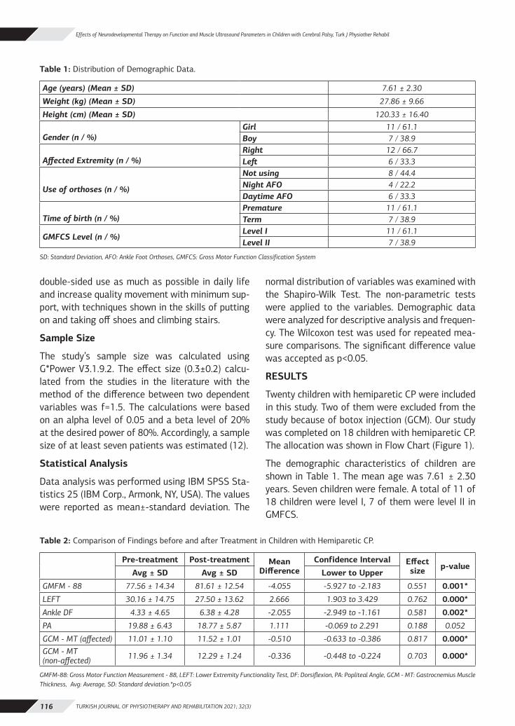
TURKISH JOURNAL OF PHYSIOTHERAPY AND REHABILITATION 2021; 32(3)116
Effects of Neurodevelopmental Therapy on Function and Muscle Ultrasound Parameters in Children with Cerebral Palsy, Turk J Physiother Rehabil
double-sided use as much as possible in daily life and increase quality movement with minimum sup-port, with techniques shown in the skills of putting on and taking off shoes and climbing stairs.
Sample Size
The study’s sample size was calculated using G*Power V3.1.9.2. The effect size (0.3±0.2) calcu-lated from the studies in the literature with the method of the difference between two dependent variables was f=1.5. The calculations were based on an alpha level of 0.05 and a beta level of 20% at the desired power of 80%. Accordingly, a sample size of at least seven patients was estimated (12).
Statistical Analysis
Data analysis was performed using IBM SPSS Sta-tistics 25 (IBM Corp., Armonk, NY, USA). The values were reported as mean±-standard deviation. The
normal distribution of variables was examined with the Shapiro-Wilk Test. The non-parametric tests were applied to the variables. Demographic data were analyzed for descriptive analysis and frequen-cy. The Wilcoxon test was used for repeated mea-sure comparisons. The significant difference value was accepted as p<0.05.
RESULTS
Twenty children with hemiparetic CP were included in this study. Two of them were excluded from the study because of botox injection (GCM). Our study was completed on 18 children with hemiparetic CP. The allocation was shown in Flow Chart (Figure 1).
The demographic characteristics of children are shown in Table 1. The mean age was 7.61 ± 2.30 years. Seven children were female. A total of 11 of 18 children were level I, 7 of them were level II in GMFCS.
Table 2: Comparison of Findings before and after Treatment in Children with Hemiparetic CP.
Pre-treatment Post-treatment Mean Difference
Confidence Interval Effect size p-value
Avg ± SD Avg ± SD Lower to Upper
GMFM - 88 77.56 ± 14.34 81.61 ± 12.54 -4.055 -5.927 to -2.183 0.551 0.001*
LEFT 30.16 ± 14.75 27.50 ± 13.62 2.666 1.903 to 3.429 0.762 0.000*
Ankle DF 4.33 ± 4.65 6.38 ± 4.28 -2.055 -2.949 to -1.161 0.581 0.002*
PA 19.88 ± 6.43 18.77 ± 5.87 1.111 -0.069 to 2.291 0.188 0.052
GCM - MT (affected) 11.01 ± 1.10 11.52 ± 1.01 -0.510 -0.633 to -0.386 0.817 0.000*GCM - MT(non-affected) 11.96 ± 1.34 12.29 ± 1.24 -0.336 -0.448 to -0.224 0.703 0.000*
GMFM-88: Gross Motor Function Measurement - 88, LEFT: Lower Extremity Functionality Test, DF: Dorsiflexion, PA: Popliteal Angle, GCM - MT: Gastrocnemius Muscle Thickness, Avg: Average, SD: Standard deviation.*p<0.05
Table 1: Distribution of Demographic Data.
Age (years) (Mean ± SD) 7.61 ± 2.30
Weight (kg) (Mean ± SD) 27.86 ± 9.66
Height (cm) (Mean ± SD) 120.33 ± 16.40
Gender (n / %)Girl 11 / 61.1Boy 7 / 38.9
Affected Extremity (n / %)Right 12 / 66.7Left 6 / 33.3
Use of orthoses (n / %)
Not using 8 / 44.4Night AFO 4 / 22.2Daytime AFO 6 / 33.3
Time of birth (n / %)Premature 11 / 61.1Term 7 / 38.9
GMFCS Level (n / %)Level I 11 / 61.1Level II 7 / 38.9
SD: Standard Deviation, AFO: Ankle Foot Orthoses, GMFCS: Gross Motor Function Classification System

TURKISH JOURNAL OF PHYSIOTHERAPY AND REHABILITATION 2021; 32(3) 117
Atasoy Ü., Budak M., Tarakcı D., Bulut A.R.
Assessment values of motor function levels, ankle dorsiflexion ROM and popliteal angle, Muscle thick-ness values, and lower extremity functionality ob-tained before and after intervention were shown in Table 2. Statistically significant improvements were found in terms of GMFM-88 (p=0.001), Low-er Extremity Function Test (p=0.000), ankle dorsi-flexion ROM (p=0.002), and GCM muscle thickness (p=0.000). There was no statistically significant dif-ference in popliteal angle (p=0.052).
DISCUSSION
The results supported the hypothesis, demonstrat-ing that s-NDT contributes to children with hemi-paretic CP improving the medial GCM MT, ankle dorsiflexion ROM, and functionality. The Bobath/NDT approach normalizes muscle tone, prevents primitive and abnormal reflexes, and facilitates normal movements. Bobath concept is continuous-ly developing based on neuroplasticity and motor
Figure 1: Flow chart of the study
Assessed for eligibility (n=20)
Excluded (n=2)
(Botox injection)
First assesment (n=18)
(Gross Motor Function Measurement-88, dorsiflexion range of motion and popliteal angle, medial
Gastrocnemius muscle thickness, Lower Extremity Function Test)
Intervention (n=18)
Structures Neurodevelopmental Therapy
3 days/week for 12 week
Final assesment (n=18)
(Gross Motor Function Measurement-88, dorsiflexionrange of motion and popliteal angle, medial
Gastrocnemius muscle thickness, Lower Extremity Function Test)
Analysis (n=18)

TURKISH JOURNAL OF PHYSIOTHERAPY AND REHABILITATION 2021; 32(3)118
Effects of Neurodevelopmental Therapy on Function and Muscle Ultrasound Parameters in Children with Cerebral Palsy, Turk J Physiother Rehabil
control principles (16). According to studies, NDT approaches tend to play an essential role in devel-oping postural control in children with CP. Although the Bobath method’s evidence level has not been clarified in the literature, physicians have indicat-ed that it is a practical approach in clinical prac-tice for children with CP (9). Researchers appear to have included studies with CP children with an average age of 6 years in a systematic review that uncovered evidence for the Bobath method (9). We assume that the age of the patients, the practi-tioner’s experience, the level of family participation in therapy, and the child’s motivation may have af-fected the results.
It is critical to adopt physiotherapy approaches that optimize the child’s potential by focusing on function and movement in CP rehabilitation. The importance of 2-8 weeks of strengthening exercis-es in healthy children to increase EMG activation by training nervous mechanisms and motor unit firings is emphasized (5, 7). Vercuren et al. empha-sized that exercise training should be continued for at least 12 weeks to be effective in children with CP (17). A 12-week neurodevelopmental treatment program for children with hemiparetic CP was ap-plied in this study.
The Gross Motor Function Scale (GMFM) and the GMFM-88 are functional assessment tools com-monly used as primary outcome measurements in studies evaluating the effectiveness of NDT on motor function in children with CP (13). GMFM-88, the gold standard, is an observational assessment scale developed to evaluate gross motor function in children with CP. It is widely used to evaluate mo-tor function in children with CP aged five months to 16 years (13). Van den Broeck et al. (18) eval-uated the effectiveness of an individually defined 6-week physiotherapy program on functionality and gait patterns in 16 children aged 3-12 years with GMFCS I-II level. They reported that GMFM-88 significantly improved walking, running, and jumping movements. Knox et al. (19) observed a significant increase in the GMFM-88 total score at 6-week intervals (baseline, before and after Bobath treatment, and follow-up) of the functional bene-
fits of Bobath treatment in 15 children with CP. In this study, a 12-week s-NDT program was applied in children with hemiparetic CP, and our results in-dicated that a 12-week s-NDT program provides significant improvements in GMFM-88 (walking, climbing, running mode) scores in children with di-paretic CP.
Hemiparetic children can usually walk independent-ly between 18-24 months. Children with hemipa-retic CP who gain the ability to walk at an early age need to reduce their energy consumption while walking, improve their walking quality and boost their level of participation in life. Foot deformities are seen in approximately 70-90% of children with CP (20,21). The severity of foot deformity can have important effects on the overall walking ability of children with CP. McDowell et al. (22) reported that the ankle dorsiflexion angle was 3.7° while in the knee extension position in 50 hemiparetic CP chil-dren aged between 4-10. In this study, we found the ankle dorsiflexion angle value in the knee extension position was 4.33°, similar to this study. An increas-ing popliteal angle value is typically interpreted in clinical practice to suggest decreased hamstring length, tightness, or contracture, indicating chang-es in mechanical properties. In our study, there was a significant difference in ankle dorsiflexion ROM, and we found that the change in popliteal angle was not significant. While popliteal angle values were observed as 30-40 degrees in children with CP above in the literature, in our study, the mean popliteal angle value was 19.88. We think this re-sult is related to the fact that most of the children in the study group (n=11) were at the GMFM I level and had a lower average age than the literature sample (23). It has been shown that changes in the pelvis and hip joint affect all joints in CP, including the ankle (24).
We propose that with the Bobath principle-based treatment, the ankle ROM angles improved as the child’s proximal body awareness and pelvic control improved. Muscle weakness and motor disorder in children with spastic CP are linked to their func-tionality, and skeletal muscle architecture can sig-nificantly influence their muscle functions. Ultraso-nography is the most objective and useful tool for determining muscle size changes related to muscu-lar strength and function (11).
Figure 2: Ultrasonographic measurement of the gastrocnemius muscle.
TURKISH JOURNAL OF PHYSIOTHERAPY AND REHABILITATION 2021; 32(3) 119
Atasoy Ü., Budak M., Tarakcı D., Bulut A.R.
GCM is critical for maintaining anti-gravity pos-ture in the standing position, the pushing phase of walking, and the most efficient energy consump-tion throughout walking (25). Rose and McGill (26) emphasized that the GCM lost 50% strength, and the motor unit activation rate was lower in chil-dren with CP during the walking push phase than healthy controls. They also stated that children with CP cannot activate the high-threshold mo-tor unit groups required for maximum voluntary contraction, and those low-threshold motor units cannot change the firing speed. Barret et al. In a systematic study examining muscle morphology in children with spastic CP, it was emphasized that there was a significant difference between paret-ic and typically developing muscle thicknesses and that TCM muscle thickness was less in children with CP compared to their peers (27). Lee et al. applied 18 sessions of NDT to a group of 13 pa-tients for six weeks and reported a non-significant increase in the thickness of the GCM muscle (12). Furthermore, researchers found that progressive functional training improved the pennation angle of GCM substantially, and they suggested that functional training be combined with convention-al therapies. Improvements in GCM thickness were shown to be statistically significant in this study. We believe that variations in the substance and du-ration of the treatments utilized may have resulted in varied outcomes.
Although the increase in muscle thickness caused by the rise in adipose tissue in muscle tissue may not always support the gain in strength in children with cerebral palsy, our hypothesis was in this di-rection. We also examined their functionality be-cause we didn’t think isolated muscle strength assessment was appropriate in our cases, and we believe that improving functionality also enhances muscular strength.
Hemiparetic children’s functionality is impaired by asymmetry, increased tone, loss of muscle strength, and sensory problems. Although the lower extrem-ity function test is a commonly used functional test in the literature in children with idiopathic toe walking and musculoskeletal injuries, no study in-vestigating the validity and reliability of the lower extremity function test in children with CP has been published to our knowledge. (28). After 12 weeks of
s-NDT treatment, we observed significant improve-ments in LEFT scores in our study. We believe that the substantial gain in lower extremity function tests is attributable to the treatment program’s positive impression of balance-based structured neurodevelopmental therapy.
The limitations of this study were that the LEFT test was not previously used in children with cere-bral palsy who were GMFCS 1 and 2 levels, children with a wide age range were included in the study, age-related changes in motor function, and short treatment duration. The strengths of our study are to evaluate the effectiveness of NDT with objective (muscle ultrasound) and functional measurement (GMFM-88 and LEFT) criteria and include only chil-dren with hemiparetic type CP.
We recommend that more studies be done using various rehabilitation methods with a large sample size and a control group.
In conclusion, s-NDT was found to be effective in improving motor function, ankle dorsiflexion, me-dial GCM MT, and lower extremity functionality in children with hemiparetic CP in this pilot study. These enhancements will have a substantial posi-tive impact on children’s mobility.
Sources of Support: The authors received no fi-nancial support for the research and authorship of this article.
Conflict of Interest: The authors report no conflict of interest
Ethical Approval: Istanbul Medipol Universi-ty Non-Interventional Clinical Research Ethics Committee approved the study. (Approval Date: 27.12.2019 and Approval Number 10840098-604.01.01-E.66352)
Informed Consent: Written informed consent was obtained from all the study participants’ parents or legal guardians.
Peer-Review: Externally peer-review
Author contributions: U.A. and D.T. designed the study and wrote the manuscript. U.A. collected and analyzed data. All authors read and approved the final manuscript.
Acknowledgments: The author thanks the children and their parents for participating in our study.
TURKISH JOURNAL OF PHYSIOTHERAPY AND REHABILITATION 2021; 32(3)120
Effects of Neurodevelopmental Therapy on Function and Muscle Ultrasound Parameters in Children with Cerebral Palsy, Turk J Physiother Rehabil
REFERENCES1. Topçu Y, Aydın K. Cerebral Palsy-epidemiology, etiology and pa-
thology. TOTBID. 2018;52(17):402-4.2. Oskoui M, Coutinho F, Dykeman J, Jette N, Pringsheim T. An up-
date on the prevalence of cerebral palsy: a systematic review and meta-analysis. Dev Med Child Neurol. 2013;55(6):509–19.
3. Fidan F, Baysal Ö. Epidemiologic characteristics of patients with cerebral palsy. OJTR. 2014;2(3):126-32.
4. Ebtehal A. Abdel R.Comparison of different physical therapy interventions for upper extremity in children with hemiparetic cerebral palsy. JCR. 2020;7(11):1217-22.
5. Williams SA, Stott NS, Valentine J, Elliott C, Reid SL. Measuring skeletal muscle morphology and architecture with imaging mo-dalities in children with cerebral palsy. Dev Med Child Neurol. 2021;63(3):263-73.
6. Martins E, Cordovil R, Oliveira R, Pinho J, Diniz A, Vaz JR. The immediate effects of a dynamic orthosis on gait patterns in chil-dren with unilateral spastic cerebral palsy: A kinematic analysis. Front Pediatr. 2019;21(7): 42.
7. Tekin F, Kavlak E, Cavlak U, Altug F. Effectiveness of Neuro-De-velopmental Treatment (Bobath Concept) on postural control and balance in cerebral palsied children. Musculoskelet Rehabil. 2018;31(2):397-403.
8. Graham JV, Eustace C, Brock K. The Bobath concept in contem-porary clinical practice. Top Stroke Rehabil. 2009;16(1):57-68
9. Zanon M, Pacheco R, Latorraca C, Martimbianco A, Pachito, D, Riera R. Neurodevelopmental Treatment (Bobath) for chil-dren with cerebral palsy: A Systematic Review. J Child Neurol. 2019;34(11):679-86.
10. Yeşilyaprak S, Çeliker Tosun Ö, Angın S. Kas ultrasonu ve fi-zyoterapi. Turkiye Klinikleri. 2015;1(1) :43-53.
11. Choe YR, Kim JS, Kim KH, Yi TI. Relationship between functional level and muscle thickness in young children with cerebral palsy. Ann Rehabil Med. 2018; 42(2): 286–95.
12. Lee M, Ko Y, Shin MMS, Lee W. The effects of progressive functional training on lower limb muscle architecture and motor function in children with spastic cerebral palsy. JPTS. 2015;27(5):1581–84.
13. Alotaibi M, Long T, Kennedy E, Bavishi S. The efficacy of GMFM-88 and GMFM-66 to detect changes in gross motor function in children with cerebral palsy (CP): a literature review. Disabil Rehabil. 2014;36(8):617-27.
14. Otman S. Köse N. Tedavi hareketlerinde temel değerlendirme prensipleri (11. Baskı). Hipokrat Kitabevi: Ankara, 2019:S79.
15. Mehta SP, Fulton A, Quach C, Thistle M. Measurement proper-ties of the lower extremity functional scale: a systematic review.
JOSPT. 2016;46(3):200-16.16. Michielsen M, Vaughan-Graham J, Holland A, Magri A, Suzuki M.
The Bobath concept - a model to illustrate clinical practice. Dis-abil Rehabil. 2019;41(17):2080-92.
17. Verschuren O, Ada L, Désirée B. Maltais, Gorter JW, Scianni A, Ketelaar M. Muscle strengthening in children and adolescents with spastic cerebral palsy: considerations for future resistance training protocols. Phys Ther. 2011;91(7):1130–39.
18. Van den Broeck C, De Cat J, Molenaers G, Franki I, Himpens E, Severijns D, Desloovere K. The effect of individually defined physiotherapy in children with cerebral palsy (CP). Eur J Paediatr Neurol.2010;14(6):519–25.
19. Knox V, Evans AL. Evaluation of the functional effects of a course of bobath therapy in children with cerebral palsy: a preliminary study. Dev Med Child Neurol .2002; 44(07):447-60
20. Şimşek İ, Serebral Palsi İn: Beyazova M, Kutsal YG. Fiziksel Tıp ve Rehabilitasyon Cilt 2. Güneş Kitabevi: Ankara; 2000: S2395- 439.
21. Kedem P, Scher DM. Foot deformities in children with cerebral palsy. Opin Pediatr. 2015; 27(1):67-74.
22. McDowell BC, Salazar-Torres JJ, Kerr C, Cosgrove AP. Passive range of motion in a population-based sample of children with spastic cerebral palsy who walk. Phys Occup Ther Pediatr. 2012;32(2): 139–50.
23. Manikowska F, Chen BP J, Jóźwiak M, Lebiedowska MK. The popliteal angle tests in patients with cerebral palsy. J Pediatr Orthop B. 2019;28(4):332-6.
24. Papageorgiou E, Simon-Martinez C, Molenaers G, Ortibus E, Van Campenhout A, Desloovere K. Are spasticity, weakness, selec-tivity, and passive range of motion related to gait deviations in children with spastic cerebral palsy? A statistical parametric mapping study. PLoS ONE. 2019;14(10): e0223363.
25. Benard MR, Harlaar J, Becher JG, Huijing PA, Jaspers RT. Effects of growth on geometry of gastrocnemius muscle in children: a three-dimensional ultrasound analysis. J Anat. 2011;219(3):388-402
26. Rose J, McGill KC. Neuromuscular activation and motor-unit firing characteristics in cerebral palsy. Dev Med Child Neurol. 2005;47(5):329-36.
27. Barrett RS, Lichtwark GA. Gross muscle morphology and struc-ture in spastic cerebral palsy: a systematic review. Dev Med Child Neurol. 2010;(52):794-804.
28. Numanoğlu Akbaş A. Assessments and outcome measures of ce-rebral palsy, cerebral palsy - current steps, Mintaze Kerem Gunel, IntechOpen, 2016: Doi: 10.5772/64254.

TURKISH JOURNAL OF PHYSIOTHERAPY AND REHABILITATION 2021; 32(1) 121
TELİF HAKKI DEVİR FORMU
Biz aşağıda imzası bulunan kişiler,………………………….……………………………………………………….…………………………………….…… …………….…………………………………………………………………………………………………………………………………………………………………………………………….……………………………………………………………………………………………………………………………………………….……………………isimli makalenin tüm yayın haklarını Türk Fizyoterapi ve Rehabilitasyon Dergisi’ne devrediyoruz.
Aşağıda imzası olan yazarlar makaleyi dikkatlice okumuşlardır ve içeriği, dili ve biçimi konusunda fikir birliği içindedirler. Makalenin özgün olduğunu, başka bir dergide yayımlanmadığını ve başka bir dergiye yayımlanmak üzere gönderilmediğini beyan ederler.
(LÜTFEN BÜTÜN YAZARLARIN İSİMLERİNİ MAKALEDEKİ İSİM SIRALAMASINA GÖRE YAZINIZ. YAZARLARIN TAMAMININ İMZASI GEREKMEKTEDİR.)
İsim: _______________________________________ İmza:________________ Tarih:_______________İsim: _______________________________________ İmza:________________ Tarih:_______________İsim: _______________________________________ İmza:________________ Tarih:_______________İsim: _______________________________________ İmza:________________ Tarih:_______________İsim: _______________________________________ İmza:________________ Tarih:_______________İsim: _______________________________________ İmza:________________ Tarih:_______________İsim: _______________________________________ İmza:________________ Tarih:_______________İsim: _______________________________________ İmza:________________ Tarih:_______________
ÇIKAR ÇATIŞMASI FORMU:
Yazarlar bu çalışmada, herhangi bir kişi, kurum veya kuruluşla, sonuçlarında ve ifade edilen görüşlerde ön- yargılı davranmaya neden olabilecek bir mali yarar veya çıkar ilişkisinin olmadığını bildirirler. (Not: Böyle bir yarar veya ilişki var ise, ayrıca mutlaka beyan edilmelidir.)
(LÜTFEN BÜTÜN YAZARLARIN İSİMLERİNİ MAKALEDEKİ İSİM SIRALAMASINA GÖRE YAZINIZ. YAZARLARIN TAMAMININ İMZASI GEREKMEKTEDİR.)
İsim: _______________________________________ İmza:________________ Tarih:_______________İsim: _______________________________________ İmza:________________ Tarih:_______________İsim: _______________________________________ İmza:________________ Tarih:_______________İsim: _______________________________________ İmza:________________ Tarih:_______________İsim: _______________________________________ İmza:________________ Tarih:_______________İsim: _______________________________________ İmza:________________ Tarih:_______________İsim: _______________________________________ İmza:________________ Tarih:_______________İsim: _______________________________________ İmza:________________ Tarih:_______________

TURKISH JOURNAL OF PHYSIOTHERAPY AND REHABILITATION 2021; 32(1)122

TURKISH JOURNAL OF PHYSIOTHERAPY AND REHABILITATION 2021; 32(1) 123
COPYRIGHT ASSIGNMENT
We, the undersigned, transfer all copyright ownership of the manuscript entitled: ………………………………………………………………………….……………………………………………………….………………………….…………….……….…….……………………………………………………………………………………………………………………………………………………………………………………………..…………………………….....to Turkish Journal of Physiotherapy and Rehabilitation.
The undersigned authors carefully read the article and agree with all its contents, language and style. The undersigned authors state that the article is original, is not under consideration by another journal, and has not been previously published.
(PLEASE TYPE OR PRINT THE NAMES OF ALL AUTHORS BY NAME ORDER.)
Name: ______________________________________ Signature:_______________ Date:____________Name: ______________________________________ Signature:_______________ Date:____________Name: ______________________________________ Signature:_______________ Date:____________Name: ______________________________________ Signature:_______________ Date:____________Name: ______________________________________ Signature:_______________ Date:____________Name: ______________________________________ Signature:_______________ Date:____________Name: ______________________________________ Signature:_______________ Date:____________Name: ______________________________________ Signature:_______________ Date:____________
CONFLICT OF INTEREST FORM:
Authors of this study report no financial interests or connections that might raise the question of bias in the work reported or the conclusions, implications, or opinions stated including pertinent commercial or other sources of funding. (P.S.: If a conflict of interest exists, it should also be reported.)
(PLEASE TYPE OR PRINT THE NAME OF ALL AUTHORS BY NAME ORDER.)
Name: ______________________________________ Signature:_______________ Date:____________Name: ______________________________________ Signature:_______________ Date:____________Name: ______________________________________ Signature:_______________ Date:____________Name: ______________________________________ Signature:_______________ Date:____________Name: ______________________________________ Signature:_______________ Date:____________Name: ______________________________________ Signature:_______________ Date:____________Name: ______________________________________ Signature:_______________ Date:____________Name: ______________________________________ Signature:_______________ Date:____________

TURKISH JOURNAL OF PHYSIOTHERAPY AND REHABILITATION 2021; 32(1)124

TURKISH JOURNAL OF PHYSIOTHERAPY AND REHABILITATION 2021; 32(1) 125
YAZAR KATKI FORMU
“Yazar”, yayımlanmış bir çalışmaya bağımsız entelektüel katkı sağlayan kişi olarak kabul edilir. Telif Hakkı Devir Formu’nda isimleri belirtilen yazarların dergiye gönderilen makaleye doğrudan katkı vermiş olması gerekir. Yazar olarak belirlenen isim aşağıdaki özelliklerin tümüne sahip olmalıdır:• Çalışmanın planlanmasına ve verilerin toplanmasına veya verilerin analizine ve yorumlanmasına katkısı
olmalıdır.• Makale taslağının hazırlanması veya revize edilmesine katkıda bulunmalıdır.• Makalenin dergiye gönderilecek ve yayınlanacak son halini okuyup kabul etmelidir.Yazarların sıralaması yardımcı yazarların ortak kararı olmalıdır. Yazarlar, ihtiyaç halinde yazar sıralamasını açıklamaya hazırlıklı olmalıdır. Sorumlu yazar, çalışmanın yayımlanmasından sonra, ihtiyaç halinde veri ve ek bilgi sağlamalıdır.Yazarlık kriterlerini sağlamayan her katkıdan makalenin “Açıklamalar” bölümünde bahsedilmelidir. Fon sağlamak, veri toplamak, araştırma grubunun genel danışmanlığını yapmak, yazınsal ve teknik düzenleme, dil redaksiyonu ve düzeltmeler tek başına yazarlık hakkı sağlamadığından, “Açıklamalar” bölümünde bahsedilecek başlıklardır.Bu formda belirtilen koşullar, Bilim Editörleri Konseyi (Council of Science Editors [CSE]) ve Uluslararası Tıp Dergi Editörleri Komitesi (International Committee of Medical Journal Editors [ICMJE]) kılavuzlarına göre düzenlenmiştir (www.cse.org, www.icmje.org).Başlık: ............................................................................................................................................................................................................................................................................................................................................................................................................................
KATKI TÜRÜ AÇIKLAMA KATKIDA BULUNANLAR
FİKİR/KAVRAM Araştırma hipotezini veya fikrini oluşturmak
TASARIM Sonuçlara ulaşılmasını sağlayacak yöntemi tasarlamak
DENETLEME/DANIŞMANLIK Araştırmanın yürütülmesini organize etmek, ilerlemesini gözetmek ve sorumluluğunu almak
KAYNAKLAR VE FON SAĞLAMA Çalışma için gerekli personel, mekan, finansal kaynak ve araç-gereçleri sağlamak
MATERYALLER Materyaller ile ilgili sorumluluk almak
VERİ TOPLAMA VE/VEYA VERİ İŞLEME Verilerin toplanması, düzenlenmesi ve raporlanması için sorumluluk almak
ANALİZ VE/VEYA YORUMLAMA Bulguların değerlendirilerek sonuçlandırılmasında sorumluluk almak
LİTERATÜR TARAMASI Çalışma için gerekli literatür taramasında sorumluluk almak
MAKALE YAZIMI Çalışmanın tamamının veya önemli bölümlerinin yazılmasında sorumluluk almak
ELEŞTİREL İNCELEME Çalışmanın raporlanmasından sonra, dil ve yazınsal düzeltmelerden bağımsız olarak bilimsel anlamda çalışmayı yeniden değerlendirmek

TURKISH JOURNAL OF PHYSIOTHERAPY AND REHABILITATION 2021; 32(1)126

TURKISH JOURNAL OF PHYSIOTHERAPY AND REHABILITATION 2021; 32(1) 127
AUTHOR CONTRIBUTION FORM
The “author” is considered to be an independent intellectual contributor to published work. The authors, whose names were specified in the Copyright Agreement Form, should have had a direct contribution to the manuscript submitted to the journal. Authorship requires all three of the following:• Substantial contributions to conception and design of the study, and acquisition of data or analysis and
interpretation of data;• Contributions to drafting or revising the manuscript critically for valuable intellectual content, and• Final approval of the version to be submitted and published. The ranking of the authors should be the joint decision of the co-authors. The authors should be prepared to explain the author’s rank, if needed. The corresponding author should provide data and additional information if necessary after the publication of the work.Every contribution that does not meet the criteria of the authorship should be mentioned in the “Acknowledgements” section of the manuscript. Funding, collecting data, general counseling of the research group, literary and technical editing, language proofreading and corrections are the titles that will be mentioned in the “Acknowledgements” section, as it does not provide authorization alone. The conditions stated in this form are regulated according to the guidelines of the Council of Science Editors (CSE) and the International Committee of Medical Journal Editors (ICMJE) (www.cse.org, www.icmje.org). Title: ..............................................................................................................................................................................................................................................................................................................................................................................................................................
CONTRIBUTION TYPE DESCRIPTION CONTRIBUTORS
CONCEPT Formulating the research hypothesis or idea
DESIGN Designing the method to achieve the results
SUPERVISION Organizing the conduct of the research, overseeing its progress, and taking responsibility
RESOURCES AND FINANCIAL SUPPORT Providing necessary staff, space, financial resources, and equipment for the study
MATERIALS Taking responsibility for the materials
DATA COLLECTION AND/OR PROCESSING Taking responsibility for collecting, organizing, and reporting data
ANALYSIS AND/OR INTERPRETATION Taking responsibility in evaluating and finalizing the findings
LITERATURE SEARCH Taking responsibility in the literature review required for the study
WRITING MANUSCRIPT Taking responsibility for the writing of all important parts of the study
CRITICAL REVIEW After the report of the study, re-evaluating the study in a scientific sense regardless of language and literary corrections

TURKISH JOURNAL OF PHYSIOTHERAPY AND REHABILITATION 2021; 32(1)128