Leitura em Ambientes Cíbridos: a Cosmo(A)Gonia Geométrica de Patrick Burgaud
Upload
independentCategory
view
3download
0
ORIGINAL ARTICLE
Italian multicentre observational study of the prevalenceof CCSVI in multiple sclerosis (CoSMo study): rationale, design,and methodology
Giancarlo Comi • Mario Alberto Battaglia • Antonio Bertolotto • Massimo Del Sette •
Angelo Ghezzi • Giovanni Malferrari • Marco Salvetti • Maria Pia Sormani •
Luigi Tesio • Erwin Stolz • Gianluigi Mancardi
Received: 10 November 2012 / Accepted: 5 December 2012
� The Author(s) 2013. This article is published with open access at Springerlink.com
Abstract Chronic cerebro-spinal venous insufficiency
(CCSVI) has been proposed as a ‘‘congenital malformation’’
implicated in the pathogenesis of multiple sclerosis (MS).
However, numerous studies failed to confirm its presence in
MS patients. This paper presents the rationale, design, and
methodology adopted in the CoSMo study, conducted with
the aim of verifying whether or not CCSVI is linked to MS.
The primary endpoint of the CoSMo study is to compare the
prevalence of CCSVI in patients with MS versus patients
affected by other neurodegenerative diseases (OND) and
healthy volunteers. CoSMo is a multicenter, blinded, prev-
alence study recruiting 2,000 adult subjects, involving 43
MS centers across Italy. Assessment of the presence or
absence of CCSVI is performed by color-coded duplex
(CCD) sonography and two out of the five criteria according
to Zamboni are necessary for the diagnosis of CCSVI. Local
CCD examination carried out by a certified sonologist and
the central image readings performed by experts in the field
are blinded. An advanced protocol is also described in this
paper. The application of a rigorous methodological design
will definitively confirm whether an association exists
between CCSVI and MS. Should an association be observed,
this study also further examines the link between CCSVI and
the severity of MS. The addition of subgroups without MS
and OND also provides information on whether CCSVI is
specific to MS only. Results from the CoSMo study will play
a crucial role in the possible studies concerning the potential
treatment of CCSVI in MS.
G. Comi
Division of Neurology and Neurophysiology Service,
Ospedale ‘‘San Raffaele’’, Milan, Italy
M. A. Battaglia (&)
Fondazione Italiana Sclerosi Multipla, Via Operai 40,
16149 Genoa, Italy
e-mail: [email protected]
A. Bertolotto
Multiple Sclerosis Center, Ospedale ‘‘San Luigi’’,
Orbassano, Italy
M. Del Sette
Neurology Unit, Ospedale ‘‘Sant’ Andrea’’, La Spezia, Italy
A. Ghezzi
Multiple Sclerosis Research Center, Ospedale ‘‘Sant’ Antonio
Abate’’, Gallarate, Italy
G. Malferrari
Neurology Unit-Stroke Unit, Department of Neuromotor
Physiology, Istituto di Ricovero e Cura a Carattere Scientifico,
Arcispedale Santa Maria Nuova, Reggio Emilia, Italy
M. Salvetti
Center for Experimental Neurological Therapies,
Universita ‘‘La Sapienza’’, Rome, Italy
M. P. Sormani
Department of Health Sciences, Universita di Genova,
Genoa, Italy
L. Tesio
Department of Biomedical Sciences for Health, Universita degli
Studi ed Istituto Auxologico Italiano, Milan, Italy
E. Stolz
Department of Neurology, Justus-Liebig-Universitat,
Giessen, Germany
G. Mancardi
Department of Neuroscience, Rehabilitation, Ophthalmology,
Genetics, Maternal and Child Health, Universita di Genova,
Genoa, Italy
123
Neurol Sci
DOI 10.1007/s10072-012-1269-5

Neurol Sci
123

Keywords Multiple sclerosis � CCSVI � Color-coded
duplex sonography � Observational � Multicenter � CoSMo
Abbreviations
ADEM Acute disseminated encephalomyelitis
BFV Blood flow volume
BVR Basal vein of Rosenthal
CCSVI Chronic cerebro-spinal venous insufficiency
CIS Clinically isolated syndrome
CNS Central nervous system
CRO Contract research organization
CSA Cross-sectional area
DVC Double check valves
CCD Color-coded duplex
EDV End diastolic velocity
FISM Fondazione Italiana Sclerosi Multipla
HC Healthy control
IJV Internal jugular vein
EDSS Expanded disability status score
MS Multiple sclerosis
NMO Neuromyelitis optica
NYHA New York heart association
OND Other neurodegenerative disease
ONDi Other neurodegenerative inflammatory disease
PP Primary progressive - MS
PSV Peak systolic velocity
RR Relapsing remitting - MS
SP Secondary progressive - MS
TAV Time averaged velocity
TCCD Transcranial color-coded duplex sonography
VV Vertebral veins
Background
Multiple sclerosis (MS) is an inflammatory demyelinating
disease of the central nervous system (CNS) resulting from
a complex combination of genetic and environmental fac-
tors [1]. Evidence of the immunological pathogenesis of
the disease is derived from animal models of the disease,
the experimental allergic encephalomyelitis, and from the
efficacy of treatments targeting the presumed immunolog-
ical dysfunctions at different levels [2, 3]. However, in
recent years, a new potential mechanism has been proposed
to contribute to the pathogenesis of MS: the presence of
presumably abnormal venous hemodynamics leading to
cerebral venous congestion. This condition has been named
‘‘chronic cerebro-spinal venous insufficiency’’ (CCSVI). It
is hypothesized that extracranial venous obstruction may
lead to inadequate cerebral drainage, raising the venous
pressure and stretching the vein walls sufficiently to sep-
arate the tight junctions between the endothelial cells that
form the blood–brain barrier. Colloids and erythrocytes
may then pass through the exposed porous basement
membranes and participate in the inflammatory process [4].
CCSVI has been initially reported to be strongly associated
with MS [5, 6]. Several subsequent studies have been
performed with the aim of reproducing Zamboni’s results,
with contradicting outcomes. While some studies confirm
Zamboni’s findings [7–11], others claim to have found
compelling evidence against a significant contribution of
CCSVI to the pathogenesis of MS [12–16]. A systematic
review of CCSVI findings in MS has been compiled by
Thapar and coworkers [17], who eloquently concluded that
‘‘there is substantial variation in the strength of association
between CCSVI and MS beyond that explained by demo-
graphic differences or sonographer training. Reliable evi-
dence on which to base decisions requires sonographic
consensus and assessment of the reproducibility of indi-
vidual criteria between trained sonographers’’ [17]. This
controversial problem, therefore, clearly needs to be
addressed by adopting the highest possible scientific stan-
dards and thereby bypassing all limitations inherent in
previous studies, such as the limited number of patients [4,
5, 7, 9, 11–13, 15, 16, 18], lack of blinding [5, 7, 8, 10, 11,
13–16], lack of appropriate controls [7–13, 15, 18], lack of
multicentric design [4, 5, 7–16, 18], and lack of inter-
observer variability assessment [4, 5, 7, 9–16, 18]. The aim
of the CoSMo study, named after the Italian ‘‘CCSVI:
studio Osservazionale Sclerosi Multipla e OND’’, that is
‘‘Observational Study of the prevalence of CCSVI in
Multiple sclerosis and in other neurodegenerative diseases
(OND)’’, is to conclude the heated debate that has arisen in
recent years and provide an appropriate answer as to
whether or not CCSVI is associated to MS. This study
evaluates the prevalence of CCSVI and of the irregularities
of the extracranial cerebro-spinal veins in patients with
different MS courses as well as in patients with OND of
different origin such as degenerative, vascular inflamma-
tory, and autoimmune of the central and peripheral nervous
system, and compare it with the prevalence of CCSVI in
healthy controls. The addition of these control subjects and
non-MS disorders will help determine whether CCSVI is
specific to MS or not. Promoted by the non-profit organi-
zation FISM (Fondazione Italiana Sclerosi Multipla, Italian
Multiple sclerosis Foundation), this is a multicentric,
Fig. 1 Basic operative protocol for the color-coded duplex sonogra-
phy in supine position. IJV internal jugular vein, J1, J2, J3 distal
(from the valve plane to 0.5 cm from it), middle (at the level of the
thyroid gland) and proximal (at the level of the carotid bifurcation)
jugular vein segments, CSA cross-sectional area, VV vertebral veins,
V1, V2 distal and proximal vertebral vein segments, TCCD transcra-
nial color-coded duplex sonography, BVR basal vein of Rosenthal, TStransverse sinus. a Time window, axial scan, mesencephalic and
diencephalic plane; examination has to be performed on both sides, in
order to insonate both right and left temporal windows
b
Neurol Sci
123

Neurol Sci
123

observational study which adopts Echo Color Doppler
(ECD) examinations performed in a blinded manner, and
centralized readings for remote visualization of the radio-
logical images using a network of special devices. The
large sample size (estimated 2,000 enrolled patients, 1,200
with MS, 400 with OND and 400 healthy controls), the
blinding procedures, the multicentric design, and the vali-
dation of the diagnostic criteria are what distinguish this
study from those previously performed.
Methods/design
Study population
Subjects included in the study population are aged between
18 and 55 years. Three patient groups are included in this
study. The first group (MS group) includes patients diag-
nosed with MS, either with relapsing remitting (RR), sec-
ondary progressive (SP), or primary progressive (PP)
course, according to McDonald’s criteria and subsequent
updates [19, 20] with disease duration between 1 month
and 25 years before screening visit and patients with
clinically isolated syndrome (CIS) with disease duration of
maximum 5 years. Patients must not be in clinical relapse
of the disease (at least 30 days since the last relapse). The
second group includes healthy controls (HC group),
namely subjects without any relevant disease and without
any family history of MS or family relation to another MS
patient. The third group includes patients with OND. Two
subtypes of patients belong to the OND group: patients
with neurodegenerative diseases (ONDn), such as Parkin-
son’s disease or amyotrophic lateral sclerosis and patients
with inflammatory CNS disorders (ONDi), such as neuro-
myelitis optica (NMO), acute disseminated encephalomy-
elitis (ADEM), encephalitis, and neurolupus. Exclusion
criteria for all groups include the presence of acute or
chronic invalidating diseases which could interfere with the
design or objective of the study, cardiac dysfunction
(NYHA class C1), previous episodes of venous thrombo-
embolism, neoplasms, thrombophilia, diabetes, primary or
secondary pulmonary hypertension and under treatment for
this condition, systemic steroid treatment within the past
30 days, past or present cerebrovascular diseases, episodes
of transient global amnesia, pregnancy, and previous
diagnosis of/treatment for CCSVI. This study plans to
recruit at least 1,200 MS patients, 400 HCs, and 400
patients with OND.
Study design and duration
This is an observational, case–control, cross sectional,
multicentric study, for which 43 Italian MS centers and the
related sonologists’ groups, evenly distributed through the
Italian territory, are participating. Physicians performing
the CCD examination, examiners reading the CCD images,
the Contract Research Organization (CRO) collecting all
data as well as the statistician performing all analyses, are
blinded throughout the whole study. Each centre is invited
to recruit 60 MS patients (30 RR, 15 SP, 10 CIS, 5 PP), 20
HC and 20 OND patients, but the recruitment is competi-
tive. The study was undertaken in November 2010 and
results are expected in 2 years.
Procedures
A training course with a final examination of proficiency
that certifies that the sonologist can perform ultrasound
evaluations according to the sonological protocol of the
CoSMo study, in its basic or advanced part, is required by
the examiners, in order to increase the homogeneity of the
diagnostic procedures. The sonologists are physicians
already expert in CCD examination of the arteries of the
neck and brain. The training course for the CoSMo study
lasts at least 2 months. The educational and training
pathway of the CoSMo study has been designed and per-
formed with the contribution of two Italian neurosonology
societies, the SINV (Societa Italiana Interdisciplinare
NeuroVascolare, Italian Interdisciplinary NeuroVascular
Society) and the SINSEC (Societa Italiana di Neurosono-
logia ed Emodinamica Cerebrale, Italian Society of Neur-
osonology and Cerebral Hemodynamics). The two
societies cooperated in the organization and management
of the theoretical and practical training of the proposed
sonologists, identifying six centers tutoring each sonologist
during the educational phase. They also arranged the final
exam to give the certification for performing the ultrasound
examination for the CoSMo study, for the basic or for the
advanced protocol, depending on the expertise and level of
knowledge of each sonologist.
Prior to any assessment, an informed consent must be
signed by each study participant. For each patient, a
screening visit is performed, by a non-blinded neurologist,
which provides data on patient’s personal details, medical
history, vital signs, and concomitant medications. All
patients undergo a physical examination, only MS and
OND patients undergo a neurological examination which
Fig. 2 Basic operative protocol for the color-coded duplex sonogra-
phy in sitting position. IJV internal jugular vein, J1, J2, J3 distal
(from the valve plane to 0.5 cm from it), middle (at the level of the
thyroid gland) and proximal (at the level of the carotid bifurcation)
jugular vein segments, CSA cross-sectional area, VV vertebral veins,
V1, V2 distal and proximal vertebral vein segments, TCCD transcra-
nial color-coded duplex sonography, BVR basal vein of Rosenthal, TStransverse sinus. a Time window, axial scan, mesencephalic and
diencephalic plane; examination has to be performed on both sides, in
order to insonate both right and left temporal windows
b
Neurol Sci
123

Neurol Sci
123

uses the Barthel Index [21] and, only for MS patients, the
expanded disability status score (EDSS) [22]. After the
screening visit, the investigator leads the subject to the
examiner in charge of performing the CCD blinded
examination (CCD Basic protocol; Figs. 1, 2). In order to
maintain blinding, subjects are instructed not to commu-
nicate with the examiner and are covered to avoid reveal-
ing any evidence of medication by injection. The
sonologist enters the examination room only after the
patient is positioned on the bed. Sonologists who are cer-
tified for the advanced protocol, may subsequently perform
it in a subgroup of patients, at their discretion. This pro-
tocol includes, on top of standard measurements, some
additional ones (Figs. 3, 4). The examination is initially
performed with the patient in the supine position (Figs. 1,
3) and next in the sitting position (Figs. 2, 4), with a 2 min
minimum break between the two positions. At the end of
the procedure, the examiner immediately performs his
diagnosis (presence or absence of CCSVI) based on the five
criteria (described in detail below) and fill the entire online
case report form with the required hemodynamic and
morphological parameters. The CCD examination images
and video recordings are then uploaded for central read-
ings, using the ‘‘Black-Box Linkverse�’’ network (Fig. 5).
In brief, the CCD evaluation consists in examining the
internal jugular veins (IJV) in J1 (distal segment, at the
level of the valve plane), J2 (middle segment, at the level
of the thyroid gland), and J3 (proximal segment, at the
level of the carotid bifurcation); then the vertebral veins
(VV) segments V1 (distal segment) and V2 (proximal
segment) through axial and longitudinal scans. The mean
number of images/video clips per patient is about 100–120.
Images are randomly sent to one of three designated central
examiners (ES, MDS, GM) who in turn performs a second
reading and a diagnosis, blinded to the group affiliation,
diagnosis of the local sonologist, and the center. If there is
an agreement between the local and the first central
examiner a final diagnosis is definitely established. If the
two readings have contrasting results, the other two central
examiners are asked to read the images and perform a
definitive diagnosis. If no consensus is reached, then the
diagnosis is accepted according to two out of three central
examiners.
CCSVI diagnostic criteria
In order to diagnose CCSVI, at least two out of the five
criteria described in the literature [4] should be met, and
the satisfied criteria should be the same as those among the
different (local and central) examiners. The criteria are
listed below:
1. Constantly present reflux ([0.88 s duration) in internal
jugular veins (IJV) and/or vertebral veins (VV) in both
seated and supine positions in at least one of the
segments J1, J2, J3, V1 or V2, through the color-mode
evaluation. Reflux is also defined as flow directed in
the vertebral axis, from the extra-rachidian plexus
toward the VV (i.e., reflux or reflux on VV and normal
or inverted flow on the extra-rachidian plexus).
2. Reflux in the intracranial veins (ICVs, such as the
basal vein of Rosenthal or the transverse sinus),
analyzed by transcranial color-coded duplex (TCCD)
sonography.
3. The presence of anatomical alterations with docu-
mented hemodynamic relapses - septa, valve malfor-
mations, double channel (one without flow), cross-
sectional area (CSA) B0.3 cm2 - through the B-mode
evaluation and Color-mode examination (also by
Valsalva maneuver).
4. The absence of flow in IJVs and/or VVs after
numerous deep inspirations in both seated and supine
positions in at least one of the reference vein segments,
through the Color-mode evaluation.
5. Negative difference in the IJV CSA in the J2 segment,
that is the area in the upright position subtracted to the
area in the supine position (DCSA).
The advanced protocol also includes arterial and venous
blood flow volume (BFV) measurements [23–25]. Inflow is
analyzed in the following districts: common carotid (J2
segment), internal carotid 1 cm after its origin and between
V1 and V2 segments. To calculate outflow, CSA and TAV
values are measured on the IJV J2 and J3 segments, while
the diameter and time averaged velocity (TAV) is mea-
sured in the venous segment between V1 and V2.
Statistical analysis
Sample size calculation
A sample size of 1,200 subjects with MS, 400 HC and 400
subjects with OND guarantees a power of 80 % (at a 5 %
level of significance) to detect an odds ratio (OR) of 2
Fig. 3 Advanced operative protocol for the color-coded duplex
sonography in supine position. IJV internal jugular vein, J1, J2, J3distal, middle and proximal jugular vein segments, CSA cross-
sectional area, VV vertebral veins, V1, V2 distal and proximal
vertebral vein segments, TCCD transcranial color-coded duplex
sonography, BVR basal vein of Rosenthal, TS transverse sinus.a Doppler waveform spectrum includes PSV peak systolic velocity,
EDV end diastolic velocity and TAV time averaged velocity
measurements through the automatic or manual selection of at least
three cardiac consecutive cycles; blood flow (BF) rate is calculated
according to the following formula: BF = CSA 9 TAV; b Time
window, axial scan, mesencephalic and diencephalic plane; exami-
nation has to be performed on both sides, in order to insonate both
right and left temporal windows; c Doppler waveform spectrum
includes PSV and EDV measurements
b
Neurol Sci
123

Neurol Sci
123
(between MS patients and HC or between MS patients and
subjects with OND) assuming a prevalence of CCSVI in
the reference group (HC) of 5 %, and an OR of 1.50
assuming a prevalence of CCSVI in the reference group of
30 %.
Primary end-points
The primary aim of the CoSMo study is to examine the
association between CCSVI and MS. Therefore, the null
hypothesis of the CoSMo study is the lack of any associ-
ation between CCSVI and MS. To reject this null
hypothesis, the CCSVI prevalence is compared between
MS patients and HC and between MS patients and patients
with OND by a Chi square test. The null hypothesis is
rejected if both tests are significant at a level of 5 %.
A Fisher exact test is used in case of observed low cell
frequencies (expected counts of \10). The prevalence of
CCSVI is calculated along with its 95 % CI in the three
study groups (MS, OND and HC) and the strength of the
association is evaluated by ORs and their 95 % CI.
Secondary end-points
The prevalence of CCSVI is evaluated in CIS, RR, PP, and
SP subgroups of MS patients, by Chi square test for het-
erogeneity and Chi square test for trend.
The impact of other factors such as age, local examiner,
and type of scanner on CCSVI, is evaluated by logistic
regression analysis. Differences among study groups (MS,
HC, and OND) is evaluated by the same model, adjusting
for the aforementioned parameters. The same analyses
described for CCSVI diagnosis are run for each of the five
criteria for CCSVI (prevalence of each criterion, differ-
ences among disease groups, adjusted analysis). Inter-rater
agreement in CCSVI diagnosis and in each criterion is
evaluated between the local and central reading (and
among the three central readers when applicable) by Cohen
kappa statistic. Also positive and negative agreement will
be calculated.
Trial registration ClinicalTrials.gov Identifier:
NCT01384825.
Discussion
The very recent claims that CCSVI could be a variable
combination of vascular abnormalities playing a role in MS
has opened a completely new perspective in the patho-
genesis of the disease, with potential therapeutic implica-
tions [26, 27]. The topic of CCSVI and MS has gained
widespread media attention that is ultimately due to unclear
and uncontrolled news. This, in turn, leads to serious dis-
comfort, especially for patients who experience the drama
of a pathological condition which has become the subject
of biased speculation. Some epidemiological studies,
mostly limited by the low number of patients involved,
produced quite variable results, from a very strict associ-
ation between CCSVI and MS [7–11] to the complete
absence of association [12–16]. Very soon it became clear
that the ultrasound assessment of cerebro-spinal venous
abnormalities were highly operator-dependent and influ-
enced by many factors, such as head position, hydration
state, respiration, etc. The high level of noise in the ultra-
sound examination and the subjectivity of the judgment of
the examiner must be compensated by a strict training and
application of criteria as well as blindness to the diagnosis.
The ultimate aim of the CoSMo study is to respond, with
a rigorous study design, to the question as to whether (or
not) CCSVI is linked to MS. The stringent methodology
adopted, the blinding procedures, the multicentric design,
the large sample size, the appropriate controls and the
extensive training that has been given to sonologists, are
what distinguishes this study from all preceding ones. It is
important to underline that only after the demonstration of
an association between CCSVI and MS can one consider
performing controlled trials to assess the efficacy of
interventional treatment as additional therapy for MS.
However, a simple association (simultaneous presence of
CCSVI and MS) does not equate to causality (CCSVI
causing MS).To date, no scientific evidence is available
that fulfills the nine causality criteria (strength, consis-
tency, specificity, temporality, biological gradient, plausi-
bility, coherence, experiment, analogy) universally
accepted and considered essential by the scientific com-
munity to causally correlate a condition and/or a factor
with a given disease [28]. Of these nine elements, only
‘‘plausibility’’ and ‘‘coherence’’ could be fulfilled since
they require that the alleged cause (CCSVI) is likely to be
framed in the context of knowledge and pathogenesis of the
disease.
If results from CoSMo support Zamboni’s theory, time
and resources can be dedicated to developing or optimizing
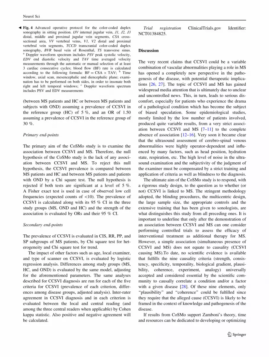
Fig. 4 Advanced operative protocol for the color-coded duplex
sonography in sitting position. IJV internal jugular vein, J1, J2, J3distal, middle and proximal jugular vein segments, CSA cross-
sectional area, VV vertebral veins, V1, V2 distal and proximal
vertebral vein segments, TCCD transcranial color-coded duplex
sonography, BVR basal vein of Rosenthal, TS transverse sinus.a Doppler waveform spectrum includes PSV peak systolic velocity,
EDV end diastolic velocity and TAV time averaged velocity
measurements through the automatic or manual selection of at least
3 cardiac consecutive cycles; blood flow (BF) rate is calculated
according to the following formula: BF = CSA 9 TAV; b Time
window, axial scan, mesencephalic and diencephalic plane; exami-
nation has to be performed on both sides, in order to insonate both
right and left temporal windows; c Doppler waveform spectrum
includes PSV and EDV measurements
b
Neurol Sci
123

a cure: this certainly requires controlled studies and it
encompasses endovascular techniques and device devel-
opment to insure durable efficacy of CCSVI surgical
treatment.
In order to address some of the controversies on the
Zamboni criteria, the CoSMo study also includes an
advanced sonological protocol, applied in some centers.
These further sonological examinations contribute to a
better definition of the intracranial and extracranial path-
ways of the cerebral venous hemodynamics. This peculiar
methodology has been extensively described in a recent
publication [29].
Acknowledgments The authors wish to thank Dr Claudio Barac-
chini (Department of Neurological Sciences, Scuola di Medicina
dell’Universita di Padova, Padua, Italy), Dr Nicola Carraro (Depart-
ment of Clinical Medicine and Neurology, Universita di Trieste,
Trieste, Italy), Dr Marcello Mancini (Scuola di Medicina dell’Uni-
versita ‘‘Federico II’’, Naples, Italy) and Dr Sandro Sangugni (Neu-
rology Department, Ospedale ‘‘Madonna del Soccorso’’, San
Benedetto del Tronto, Italy) for their expert contribution in the defi-
nition of the sonological protocol and training of the sonologists of
the study.
Conflict of interest MAB has no conflicts of interest to declare. AB
has been on steering committees in clinical trials sponsored by Bio-
gen-Idec and Roche, has received speaker’s honoraria from Biogen-
Idec, Merck-Serono, TEVA, Bayer-Schering, Sanofi-Aventis and
Novartis, has received research support from Biogen-Idec, Bayer-
Schering, Merck-Serono, Sanofi-Aventis, the Italian Multiple sclero-
sis Society, and the European Union Sixth Framework Programme.
GC has received consulting fees for participating on advisory boards
from Novartis, TEVA, Sanofi-Aventis, Merck-Serono, and Bayer-
Schering, lecture fees from Novartis, TEVA, Sanofi-Aventis, Merck-
Serono, Biogen-Dompe, Bayer-Schering, and Serono Symposia
International Foundation, he is a member of the Board of the Italian
MS Foundation. MDS has no conflicts of interest to declare. AG
received speaker’s honoraria from Bayer-Schering, Biogen-Dompe,
Merck-Serono, Novartis, Sanofi-Aventis and Allergan, for consul-
tancy from Actelion, Merck-Serono, TEVA, and Novartis, received
support for participating to the National and International Congresses
from Bayer-Schering, Biogen-Dompe, Merck-Serono, Novartis, and
Sanofi-Aventis. GM has no conflicts of interest to declare. GLM
received honoraria for lecturing, travel expenses for attending meet-
ings, and financial support for research from Bayer-Schering, Biogen-
Idec, Sanofi-Aventis, Novartis, and Merck-Serono. He is a member of
the Board of the Italian MS Foundation. MS declares grant support
from Bayer-Schering, Merck-Serono, Sanofi-Aventis, and Biogen-
Idec. MPS has received consulting fees or honoraria from Biogen-
Idec, Merck-Serono, Actelion, Synthon, and TEVA. ES has no
Fig. 5 Black-box Linkverse� system. Schematic chart describing the
flow of images and video recordings from the local examiner
performing the CCD examination to the experts performing the
centralized readings for CCSVI diagnosis. The Black-box Linkverse�
uses the universal DICOM format (Digital Imaging and COmmuni-
cations in Medicine) and a system for consultation, temporary storage
and transmission of images stored in native format and sent as
compressed files, without loss of information
Neurol Sci
123

conflicts of interest to declare. LT is a member of the Board of the
Italian MS Foundation.
Open Access This article is distributed under the terms of the
Creative Commons Attribution License which permits any use, dis-
tribution, and reproduction in any medium, provided the original
author(s) and the source are credited.
References
1. Compston A, Coles A (2008) Multiple sclerosis. Lancet
372:1502–1517. doi:10.1016/S0140-6736(08)61620-7
2. Chen S-J, Wang Y-L, Fan H-C et al (2012) Current status of the
immunomodulation and immunomediated therapeutic strategies
for multiple sclerosis. Clin Dev Immunol 2012:970789. doi:
10.1155/2012/970789
3. Fontoura P (2010) Monoclonal antibody therapy in multiple
sclerosis: paradigm shifts and emerging challenges. MAbs
2:670–681. doi:10.4161/mabs.2.6.13270
4. Zamboni P, Galeotti R, Menegatti E et al (2009) Chronic cere-
brospinal venous insufficiency in patients with multiple sclerosis.
J Neurol Neurosurg Psychiatr 80:392–399. doi:10.1136/
jnnp.2008.157164
5. Zamboni P, Menegatti E, Weinstock-Guttman B et al (2009) The
severity of chronic cerebrospinal venous insufficiency in patients
with multiple sclerosis is related to altered cerebrospinal fluid
dynamics. Funct Neurol 24:133–138
6. Zamboni P, Consorti G, Galeotti R et al (2009) Venous collateral
circulation of the extracranial cerebrospinal outflow routes. Curr
Neurovasc Res 6:204–212
7. Al-Omari MH, Rousan LA (2010) Internal jugular vein mor-
phology and hemodynamics in patients with multiple sclerosis.
Int Angiol 29:115–120
8. Ciccone MM, Galeandro AI, Scicchitano P et al (2012) Multigate
quality Doppler profiles and morphological/hemodynamic alter-
ations in multiple sclerosis patients. Curr Neurovasc Res
9:120–127
9. McTaggart RA, Fischbein NJ, Elkins CJ et al (2012) Extracranial
venous drainage patterns in patients with multiple sclerosis and
healthy controls. AJNR Am J Neuroradiol. doi:10.3174/ajnr.
A3097
10. Zaniewski M, Kostecki J, Kuczmik W et al (2012) Neck duplex
Doppler ultrasound evaluation for assessing chronic cerebrospi-
nal venous insufficiency in multiple sclerosis patients. Phlebol-
ogy. doi:10.1258/phleb.2011.011070
11. Zivadinov R, Poloni GU, Marr K et al (2011) Decreased brain
venous vasculature visibility on susceptibility-weighted imaging
venography in patients with multiple sclerosis is related to
chronic cerebrospinal venous insufficiency. BMC Neurol 11:128.
doi:10.1186/1471-2377-11-128
12. Mayer CA, Pfeilschifter W, Lorenz MW et al (2011) The perfect
crime? CCSVI not leaving a trace in MS. J Neurol Neurosurg
Psychiatr 82:436–440. doi:10.1136/jnnp.2010.231613
13. Doepp F, Paul F, Valdueza JM et al (2010) No cerebrocervical
venous congestion in patients with multiple sclerosis. Ann Neurol
68:173–183. doi:10.1002/ana.22085
14. Worthington V, Killestein J, Eikelenboom MJ et al (2010) Nor-
mal CSF ferritin levels in MS suggest against etiologic role of
chronic venous insufficiency. Neurology 75:1617–1622. doi:
10.1212/WNL.0b013e3181fb449e
15. Centonze D, Floris R, Stefanini M et al (2011) Proposed chronic
cerebrospinal venous insufficiency criteria do not predict multiple
sclerosis risk or severity. Ann Neurol 70:51–58. doi:10.1002/ana.
22436
16. Baracchini C, Perini P, Causin F et al (2011) Progressive multiple
sclerosis is not associated with chronic cerebrospinal venous
insufficiency. Neurology 77:844–850. doi:10.1212/WNL.0b013e
31822c6208
17. Thapar A, Lane T, Nicholas R et al (2011) Systematic review of
sonographic chronic cerebrospinal venous insufficiency findings
in multiple sclerosis. Phlebology 26:319–325. doi:10.1258/phleb.
2011.011098
18. Floris R, Centonze D, Fabiano S et al (2012) Prevalence study of
chronic cerebrospinal venous insufficiency in patients with mul-
tiple sclerosis: preliminary data. Radiol Med 117:855–864. doi:
10.1007/s11547-011-0767-5
19. McDonald WI, Compston A, Edan G et al (2001) Recommended
diagnostic criteria for multiple sclerosis: guidelines from the
international panel on the diagnosis of multiple sclerosis. Ann
Neurol 50:121–127
20. Polman CH, Wolinsky JS, Reingold SC (2005) Multiple sclerosis
diagnostic criteria: three years later. Mult Scler 11:5–12
21. Mahoney FI, Barthel DW (1965) Functional evaluation: the
barthel index. Md State Med J 14:61–65
22. Kurtzke JF (1983) Rating neurologic impairment in multiple
sclerosis: an expanded disability status scale (EDSS). Neurology
33:1444–1452
23. Schreiber SJ, Lurtzing F, Gotze R et al (2003) Extrajugular
pathways of human cerebral venous blood drainage assessed by
duplex ultrasound. J Appl Physiol 94:1802–1805. doi:10.1152/
japplphysiol.00782.2002
24. Scheel P, Ruge C, Petruch UR, Schoning M (2000) Color duplex
measurement of cerebral blood flow volume in healthy adults.
Stroke 31:147–150
25. Scheel P, Ruge C, Schoning M (2000) Flow velocity and flow
volume measurements in the extracranial carotid and vertebral
arteries in healthy adults: reference data and the effects of age.
Ultrasound Med Biol 26:1261–1266
26. Dake MD (2012) Chronic cerebrospinal venous insufficiency and
multiple sclerosis: history and background. Tech Vasc Interv
Radiol 15:94–100. doi:10.1053/j.tvir.2012.02.002
27. Lugli M, Morelli M, Guerzoni S, Maleti O (2012) The hypothesis
of patho-physiological correlation between chronic cerebrospinal
venous insufficiency and multiple sclerosis: rationale of treat-
ment. Phlebology 27(Suppl 1):178–186. doi:10.1258/phleb.2012.
012S24
28. Hill AB (1965) The environment and disease: association or
causation? Proc R Soc Med 58:295–300
29. Malferrari G, Del Sette M, Zedde M et al (2012) Italian multicenter
study on venous hemodynamics in multiple sclerosis: advanced
sonological protocol. Perspect Med. doi:10.1016/j.permed.
2012.03.013
Neurol Sci
123
Copyright © 2022 FDOKUMEN