Gametophyte interaction and sexual reproduction: how plants make a zygote
Upload
independentCategory
view
7download
0
Plant Molecular Biology40: 857–872, 1999.© 1999Kluwer Academic Publishers. Printed in the Netherlands.
857
Isolation of rapeseed genes expressed early and specifically duringdevelopment of the male gametophyte
Agnes Fourgoux-Nicol, Jan Drouaud, Najat Haouazine, Georges Pelletier and PhilippeGuerche∗Station de G´enetique et d’Am´elioration des Plantes, Institut National de la Recherche Agronomique, Route de SaintCyr, 78026 Versailles cedex, France (∗author for correspondence)
Received 18 December 1998; accepted in revised form 8 May 1999
Key words: Arabidopsis, gene-specific expression, GUS staining,in situhybridization, microspore, rapeseed
Abstract
A cDNA subtraction and differential hybridization strategy was used to isolate cDNAs expressed early duringmale gametophyte development in the important crop speciesBrassica napus. Three cDNAs, corresponding togenes highly and specifically expressed at the tetrad and microspore stages, are presented here. The analysis ofone of them, namedBnM3.4, by in situhybridization, showed that it is expressed specifically and at a high level inthe rapeseed microspore. The specificity in its profile of expression is most likely transcriptionally controlled asasimilar pattern of expression was also observed inArabidopsis thalianaplants transformed by theBnM3.4promoterfused to the reporter GUS-coding sequence. The putativeBnM3.4promoter contains three dispersed copies of amotif described previously in the promoters of several genes expressed in the male gametophyte. TheBnM3.4geneencodes a predicted novel proline-rich protein of 23.4 kDa which may interact with cytoskeletal components orhave a structural role in the cell wall.
Introduction
Pollen development in angiosperm plants occurs in aspecialized floral organ, the anther. It starts by a se-ries of mitotic divisions of archeosporial cells from theprimary sporogenous layer leading to the formation ofmeiocytes. Each meiocyte undergoes meiosis to gen-erate a tetrad of four haploid microspores surroundedby a callose wall, whose digestion then liberates theminto the anther locule. A complex extracellular ma-trix composed of intine and exine is built around themale gametophyte after microspore differentiation. Anasymmetric cell division of each microspore producesthe bicellular pollen grain in which the large vegetativecell encloses the smaller generative cell. This lattercell has a condensed nucleus and a reduced cytoplasm.In the majority of plant species, the second mitoticdivision of the generative cell occurs after pollination
The nucleotide sequence data reported will appear in theEMBL, GenBank and DDBJ Nucleotide Sequence Databases underthe accession number AF136223 (BnM3.4).
within the growing pollen tube producing two spermcells. In some genera such asBrassica this secondpollen mitosis occurs earlier during pollen maturation.The pollen grain is hence released from the antherlocule in a tricellular form. This developmental pro-gramme is necessary to prepare the pollen grain forefficient interaction with the stigma, for rapid ger-mination and pollen tube growth, and for successfuldelivery of sperm cells to the ovules (Bedingeret al.,1994).
Despite the wealth of descriptive studies on thestructural and physiological aspects of pollen forma-tion, our understanding of the molecular events re-mains rather limited. Our main goal is to extend ourknowledge on microspore differentiation and develop-ment through the identification and characterizationof novel genes specifically expressed during earlystages of microgametogenesis. Such genes may alsoprovide alternative methods for controlling male fer-tility in economically important plants, such as in ourcaseBrassica napus. To identify genes that might be
858
implicated in the developmental process, differentialscreening of cDNA libraries constructed from wholeanthers has been the most commonly used approach.A number of genes have been shown to be expressedin developing pollen, as well as in the tapetum orother sporophytic tissues of the anther (Ursinet al.,1989; Koltunowet al., 1990; Nackenet al., 1991;Theerakulpisutet al., 1991; Paulet al., 1992; Aguirreand Smith, 1993; Robertet al., 1993; Bucciaglia andSmith, 1994; Ross and Murphy, 1996). Some pollen-specific genes expressed after the first mitotic divisionand presumably playing a role in pollen development,maturation, germination or pollen tube growth havealso been described (Hansonet al., 1989; Brown andCrouch, 1990; Shen and Hsu, 1992; Weteringset al.,1992; Brander and Kuhlemeier, 1995; Stanchevet al.,1996; Yuet al., 1998). Finally, so far, a single tobaccogene has been identified that shows a microspore-specific expression (Oldenhofet al., 1996),
We report the isolation of cDNAs fromB. napusthat are expressed during early pollen development.During this work, we developed a novel combinedapproach by differential screening of a microsporecDNA library with a subtracted probe enriched formicrospore-specific sequences. We describe here indetail the characterization of the structure and ex-pression of theBnM3.4 gene corresponding to themicrospore-specific cDNA M3.
Materials and methods
Plant material
Plants ofB. napusL. cv. Brutor were grown in anopen field or under standard greenhouse conditionsand were used for RNA and DNA isolation.Arabidop-sis thaliana(Wassilevskija ecotype) plants used fortransformation (Bechtoldet al., 1993) were grown ina greenhouse under standard conditions. TransgenicA. thalianaT1 seedlings were selected in a greenhouseon sand, sub-irrigated with water containing Bastaherbicide (7.5 mg/l phosphinothricine). Two monthslater, T2 seeds were harvested individually.In vitroculture of seedlings for segregation analysis was donein a culture chamber onA. thaliana medium (Es-telle and Sommerville, 1987) containing 5 mg/l ofphosphinothricine as selective agent.
RNA isolation and poly(A)+ RNA purification
Several grams of rapeseed flower buds from 0.2 mm tomore than 4 mm in length were harvested in 4 differ-ent classes according to bud length. Bud length wasmeasured from the base to the tip of the outermostsepal. Male gametophytes from graded floral budswere isolated and purified as previously described (Al-bani et al., 1990) with a few modifications. Afterdisruption of the buds in a blender with a solution of10% sucrose (pH 7), the resulting suspension was fil-tered through 80µm nylon mesh. Only the suspensioncontaining microspores (from 2 to 3 mm buds) wasfiltered through 45µm nylon mesh. After two washes,as described by Albaniet al. (1990), the pellet wasfrozen in liquid nitrogen and stored at−80 ◦C if notimmediately used for RNA extraction. The purifiedmale gametophytes from the different classes of budwere disrupted by 3 cycles of pressurization (at 110bar) depressurization in a mini-bomb (Bioblock Sci-entific, Illkirch, France) in 50% RNA extraction bufferand 50% phenol before performing RNA extraction aspreviously reported (Deanet al., 1985).
Poly(A)+ RNAs were purified from total RNAusing the mRNA purification kit from PharmaciaBiotech.
Total RNA was extracted from young rapeseedseedlings, total fertile and male-sterile buds, pistils,sepals and petals, roots, leaves and stems as describedabove.
cDNA library construction and cDNA subtraction
The procedure used for the isolation of organ-specificcDNA was based on an unpublished protocol from S.Lok and D.C. Baulcombe (Sainsbury Laboratory, JohnInnes Centre for Plant Science Research, Norwich,UK). It is the result of several reports on ligation-mediated PCR (Mueller and Wold, 1989), direct in-corporation of biotin nucleotide during PCR (Loet al.,1988) and differential removal of biotinylated DNAby a streptavidin/phenol extraction procedure (Siveand St John, 1988; Wang and Brown, 1991) and wasmodified as follows.
The blunted ‘tracer’ cDNA (T) and the ‘driver’cDNA (D) were synthesized from 3µg of poly(A)+
RNA from microspores and male-sterile buds (Ogu-INRA) (Gourret et al., 1992), respectively, using akit from Pharmacia Biotech. An aliquot of the tracercDNA was ligated to theEcoRI/NotI adaptor ac-cording to the kit’s instructions and inserted into the

859
EcoRI site ofλgt10 and plated on non-permissiveEs-cherichia coli strain C600 Hfl− using Gigapack IIIpackaging extract (Stratagene). The titre of the librarywas estimated at 1.7× 10⁷ pfu/µg of λgt10 DNA.
A 200 ng portion of T and D cDNA (0.5 to 1 pmolof ends) was ligated to 280 pmol of the T (AATTCGC-CATGGATCTAGACC / pGGTCTAGATCCATG) andD (ATCAGTGATCGGCATGAGCTCG / pCGAGCT-CATGCCGA) specific linkers. A 50 ng portion of Tand D DNA was then amplified for 12 cycles of PCRfor the T (using the top T primer) and 30 cycles forthe D using 50 nM Bio-11-dUTP (Sigma) and the topT primer. A 150 ng portion of amplified T was thensubtracted with a 20-fold excess of biotinylated D for24 h at 68◦C. The biotinylated DNA was then re-moved by the streptavidin/phenol extraction method.The subtraction step was repeated 2 times under thesame conditions. 1/6 of the total subtracted tracer-driver (T-D) DNA was then amplified for 35 cyclesusing the T primer and the result was quantified on agel. We chose to not directly clone the subtracted prod-uct as done by Rubinelliet al. (1998) but to use it as aprobe to screen the microspore cDNA library in orderto compensate the representational bias introduced byPCR.
Differential screening
A 20 ng portion of amplified subtraction prod-uct (T-D) and unsubtracted T cDNA were labelledat high specific activity with [32P] dCTP with anoligolabelling kit (Pharmacia Biotech) (Feinberg andVogelstein, 1985) and hybridized on duplicate fil-ters (colony/plaque Screen filters, NEN ResearchProducts, DuPont) containing approximately 20 000plaques of the microspore cDNA library at 65◦C in6× SSC for 24 h. The filters were washed in threesteps of 30 min in 2×, 1× and 0.1× SSC with 0.1%SDS at 65◦C.
Only the clones showing a higher signal when hy-bridized with the T-D cDNA probe than with the TcDNA probe were selected and isolated. The accu-racy of this choice was confirmed by a second roundof hybridization realised in the same conditions onfilters containing only the selected clones. PositivecDNA clones were purified andλDNA was extractedas previously described (Albaniet al., 1990). cDNAinserts were isolated afterNotI or EcoRI digestion andsubcloned in pBluescript SK− plasmid (Stratagene)digested byNotI or EcoRI.
Northern blot hybridization
A 10 µg portion of denatured total RNA was loadedin each lane of a 1.5% agarose gel containingformaldehyde and transferred onto Hybond-N mem-brane (Amersham) according to the manufacturer’sprotocol. Prehybridization and hybridization were car-ried out in a buffer containing 5× SSPE, 5× Den-hardt’s solution, 100µg/ml sonicated salmon spermDNA, 1% SDS and 50% deionized formamide at42 ◦C for 6 h and 20 h, respectively. The selectedcDNA fragments were gel-purified, labelled with [32P]dCTP and used as probes for hybridization. The filterswere washed at 65◦C in three 15 min steps in 0.1%SDS and decreasing salt concentrations (5×, 2× and0.1× SSPE).
Genomic DNA library screening
350 000 recombinant clones (average size 15 kb)(three B. napusgenome equivalents) of a genomicDNA library of B. napuscv. Bridger in bacterio-phageλ EMBL-3 (Clontech, USA) were plated withE. coli LE392 as host. The entire M3 cDNA was usedas a [32P] dCTP-labelled probe for screening. Hy-bridization and washing were carried out with standardtechniques (Sambrooket al., 1989).
Determination of the 5′ end of mRNA
The method was based on PCR using the 5′-AmpliFINDER RACE kit from Clontech.
Two antisense primers were designed from theBnM3.4coding sequence: P1, 5′-TGATGACTCTAGTTCTGTTGCTGTG-3′ and P2, 5′-TGGAATTCGTTCTGTTGCTGTGACTTTGGATGT-3′, and we opti-mized the annealing temperature at 55◦C for the stepof PCR amplification. The experiment was performedwith 2 µg of mRNA extracted fromB. napusmi-crospores, fromB. napusfloral buds and fromB.napusleaves for the negative controls. Thereafter PCRproducts were digested byEcoRI and cloned intopBluescript SK− for sequencing.
Genomic DNA isolation and Southern blothybridization
Genomic DNA was purified from seedling leaves us-ing a standard procedure (Dellaportaet al., 1983)followed by standard CsCl gradient centrifugation.Southern analysis was performed by digestion of10 µg of genomic DNA, electrophoresis of DNA
860
fragments on a 0.8% agarose gel and blotting ontoHybond-N membrane using standard methods (Sam-brook et al., 1989). The M3 cDNA fragment wasgel-purified and labelled with [32P] dCTP as describedpreviously. A Southern blot was hybridized overnightat 65 ◦C in 6× SSC and washed at 65◦C in three15 min steps in 0.1% SDS and decreasing salt con-centrations (6×, 2× and 0.1× SSC).
Plasmid construction
Recombinant DNA techniques were carried out asdescribed by Sambrooket al. (1989) to construct atranscriptional fusion bringing the expression of theuidA (commonly namedgus) reporter gene under thecontrol of theBnM3.4promoter in binary vector.
A ‘promoterless’gusgene cassette, containing theguscoding sequence (Jeffersonet al., 1986) and thenopaline synthase polyadenylation site (nos-ter; Be-van et al., 1983) was cloned into pBluescript SK−
(Stratagene, USA), leading to pAF1opt. A 2056 bpBamHI-NspV fragment (Figure 3) from the genomiccloneBnM3.4was cloned intoBamHI-ClaI sites of theplasmid pAF1opt, generating pJD51. The chimaericgene was then excised from pJD51 and introduced intothe binary plasmid pEC2 (Carteaet al., 1998) in in-verse orientation with respect to the Basta resistancegene, generating pJD101. The promoterlessgusgenecassette was also transferred into pEC2 to generatethe negative control binary plasmid pAF100. The bi-nary plasmids pAF100 and pJD101 were introducedinto Agrobacterium tumefaciensstrain C58C1 (Konczand Schell, 1986) by electroporation (Nagelet al.,1990; Singhet al., 1993). The recombinant genes wereintroduced intoA. thaliana by in planta infiltration(Bechtoldet al., 1993).
In situhybridization
TheBnM3.4coding sequence was cloned in both ori-entation in pGEM-3Zf(+) (pJD6 and 7). AfterSmaIlinearization, these two plasmids were used to synthe-size digoxigenin-11-rUTP-labelled probes using theRiboprobe Combination System T7 kit from Promega.
B. napusfloral buds were fixed in 4% formalde-hyde, embedded in wax, and 8µm-sections wereprepared forin situhybridization according to Jackson(1991).
Full-length RNA probes were alkaline-hydrolysedto 150 nt fragments and hybridized to sections at aconcentration of 0.5 ng/ml per kb of probe. Hybridiza-tion was carried out in 210µl for a sandwich of
two slides at 50◦C in 50% formamide, 10% dex-tran sulfate, 1× Denhardt’s solution, 0.3 M NaCl,10 mM Tris-HCl pH 8, 1 mM EDTA, 1 mg/ml yeasttransfer RNA. Slides were washed in several baths of0.2× SSC at 55◦C for 1 h, followed by two rinsesof 5 min each with 0.5 M NaCl, 10 mM Tris-HCl(pH 7.5), 1 mM EDTA (NTE buffer), and treatedwith 20 mg/ml RNase A in this buffer at 37◦C for30 min. The slides were then washed again in NTEbuffer and 0.2× SSC as described above, and finallywashed in PBS (130 mM NaCl, 7 mM Na2HPO4,3 mM NaH2PO4, pH 7) for 5 min. Immunologi-cal detection of the hybridized probe was carried outas described in the Boehringer digoxigenin-nucleicacid detection kit with some modifications. Slideswere incubated with gentle agitation for 1 h in 0.5%blocking agent (Boehringer) in buffer 1 (100 mMTris-HCl, 150 mM NaCl, pH 7.5) followed by 1 hin 1% bovine serum albumin, 0.3% Triton X-100in buffer 1 (buffer 2). This was followed by a 1 hincubation in dilute antibody conjugate (Boehringer)(1:1250) in buffer 2 and 4 washes of 15 min eachin buffer 2. Slides were briefly washed in 100 mMTris-HCl pH 9.5, 100 mM NaCl, 50 mM MgCl2and incubated for 12–48 h in 0.34 mg/ml nitrobluetetrazolium salt and 0.175 mg/ml 5-bromo-4-chloro-3-indoyl phosphate toluidinium salt in 100 mM Tris-HClpH 9.5, 100 mM NaCl, 50 mM MgCl2. The colourreaction was stopped with 10 mM Tris-HCl pH 8.0,1 mM EDTA, and sections were passed through anethanol series before mounting in mounting mediumfrom Sigma.
Histochemical GUS assays
Histochemical assays for GUS activity were con-ducted according to the protocol described previouslyby Jeffersonet al. (1987) with some modifications.Fresh tissue samples were fixed in a solution con-taining chloroform, 95% ethanol and water in pro-portions 3:6:1 and 0.1% Triton X-100 under vacuum(−93.3 kPa) for 1 min. Then, tissue samples werewashed twice in 50 mM potassium phosphate buffer,pH 7.0. GUS staining was performed by vacuum in-filtration (3× 10 min at−93.3 kPa) in the GUS stainsolution (Jeffersonet al., 1987) and by incubating at37 ◦C overnight. The plant material was then clearedby rinsing with 70% ethanol and the samples wereexamined and photographed under a Diaplan type307-148.002 microscope (Leitz, Wetzlar, Germany).

861
Figure 1. Correlation between the size of rapeseed flower buds and microgametophytic stage of development. Expression pattern of the M 21,M3 and M25 clones isolated by subtraction and differential screening of the microspore cDNA library. A. DAPI staining of the four differentdevelopmental stages during male gametogenesis (in the lower part) and the corresponding buds (in the upper part). a, DAPI-stained tetrads andearlier stages (not shown) contained in buds of length strictly below 2 mm; b, microspores contained in buds of 2–3 mm; c, bicellular pollencontained in buds of 3–4 mm; d, tricellular pollen contained in buds of 4.5 mm and more. Each square is 1 mm× 1 mm. B. Northern blotexperiments hybridized with M21, M3 and M25 cDNA clones as probes. Total RNA (10µg) from (a) meiocyte/tetrads, (b) microspores, (c)young (binucleate) and (d) mature (trinucleate) pollen, (e) sepals, (f) petals, (g) wild type and (h) CMS buds, and (i)in vitro grown seedlingswere loaded on the gel. The picture of the corresponding ethidium bromide stained gel is presented underneath.
The pistil length was used as the unique criterionfor identification of the stages of microsporocyte de-velopment inA. thaliana. Based on results of Bowman(1994), the correlation between the pistil length (pl)and the stages of microsporocyte development wasfurther studied (D. Vezon, personal communication)and we found that a pl of 0.3-0.4 mm corresponds tomeiosis; pl 0.5 mm corresponds to the end of meiosisand tetrad microspores; pl 0.75 corresponds to free mi-crospores; pl 1.25 mm corresponds to the first pollenmitosis and bicellular pollen; pl 1.50 mm correspondsto the second pollen mitosis and tricellular pollen.
Results
Isolation ofB. napuscDNAs encoding sequencesexpressed early during pollen development
Cytological studies (Scottet al., 1991) have shownthat the stages of male gametogenesis within the an-ther are highly correlated to bud length and that game-togenesis is synchronised in all anthers of one flowerbud.
The cytological studies allowed us to establish that,in the growth conditions we used, rapeseed buds oflength strictly below 2 mm contained mainly meio-cytes and tetrads, 2 to 3 mm long buds contained
862
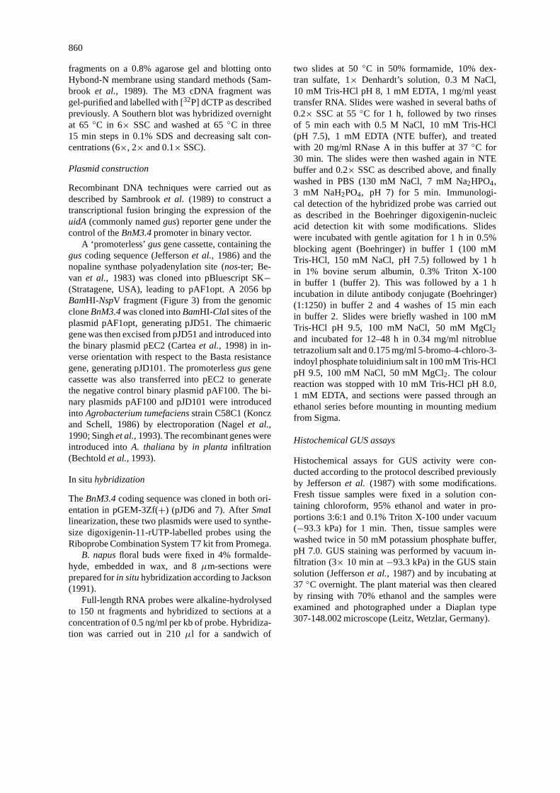
Figure 2. Comparison of M3 and M3.21 cDNA sequences. Numbers at the right margin refer to the nucleotide position. Dashed lines indicategaps and underlined sequences represent imperfect repeated motifs found in the M3 and M3.21 sequences. The stop codons are shown in boldtype.
mainly microspores, 3 to 4 mm long buds containedmainly bicellular pollen grains and buds of 4.5 mmand more in length contained mature tricellular pollengrains (Figure 1A). RNAs were prepared from malegametophytes purified from buds at these four stages.To achieve this we had to develop a new technique (seeMaterials and methods) to lyse the microspores whichhave extremely resistant cell walls. To obtain a driverfor the subtraction procedure, we isolated RNA fromwhole male-sterile flower buds (Ogu-INRA). Thesebuds are identical to those of wild type except thatmale gametophyte development is arrested before thetetrad stage and consequently they do not contain anymicrospores (Gourretet al., 1992).
Poly(A)+ RNAs from microspores were convertedto cDNA, of which a fraction was used to constructa cDNA library. To generate probes enriched formicrospore-specific transcripts, another fraction of theabove microspore cDNA was then subtracted sev-
eral times by hybridization with cDNA made frommale-sterile buds (see Materials and methods). Wethen screened the microspore cDNA library usingsubtracted and primary probes. After two rounds ofsuch successive screens, we retained 32 potential can-didates that showed increased hybridization to thesubstracted probe with respect to the unsubtractedmicrospore cDNA probe.
The expression pattern of selected clones was com-pared in more detail by slot blot (data not shown) andby northern blot analyses with total RNA isolated fromthe four same developmental stages of male game-tophytyte as above, entire wild-type and male-sterilebuds, sepals, petals andin vitro grown seedlings orleaves. These expression studies allowed us to classifythese genes into four groups.
The first group consists of 13 clones whose expres-sion is strictly confined to the male gametophyte andhigh in the microspore. The second group contains 13

863
Figure 3. Analysis of theBnM3.4 gene. Partial restriction mapof the BnM3.4genomic clone. The striped box represents the se-quenced region of theBnM3.4genomic clone; the black one thecoding sequence of theBnM3.4gene. The twoSalI sites border the10 kb insert.
clones whose expression is high in the microspores butnot restricted to the male gametophyte since they arealso expressed in other sporophytic tissues. The thirdand fourth groups correspond respectively to 2 cloneshaving a weak expression restricted to microsporesand 4 clones showing weak expression in all tissuestested.
We characterized further three cDNAs belong-ing to the first group of genes expressed early andspecifically during male gametophyte development.Northern blot analyses of these clones are shown inFigure 1B. The M21 gene detected a mRNA of 1.4 kb(encodes potentially a novel protein), which is ex-pressed at the meiocyte and tetrad stages but its mRNAlevel remains high in the microspore. The M25 cDNAdetected a 0.6 kb mRNA (encoding a putative SKP1-like protein (Connelly and Heiter, 1996; Krek, 1998)),mainly in the microspore but expression was alsodetected at meiocyte/tetrad stages and in bicellularpollen. The M3 cDNA detected a 1.0 kb mRNA inmicrospores but expression was also evident in meio-cyte/tetrad stages. We have chosen to focus on thisparticular clone for further studies because extensivehybridization studies using a variety of sporophytictissues (data not shown) suggested that the M3 mRNAis highly early stage microspore-specific.
Analysis of the nucleotide sequences of M3 andM3.21 cDNAs
The M3 cDNA has a length of 497 bp. It was smallerthan the 1.0 kb mRNA detected on northern blots, soM3 is likely to be a partial cDNA. A second screeningof the microspore cDNA library was performed usingM3 as probe. A longer but still partial clone, namedM3.21, of 674 bp was then isolated. The expressionpattern of the M3.21 cDNA was checked by northernblot (data not shown) and appeared to be identical tothe M3 pattern. Both cDNA clones, M3 and M3.21,were subcloned into pBluescript SK− and sequenced.Alignment of their nucleotide sequences shows thatthe M3 cDNA differs essentially from M3.21 by an
insertion of 99 bp constituted by three repeats of thesame motif (Figure 2).
M3 cDNA and the correspondingBnM3.4genomicclone: DNA sequence comparisons and analyses
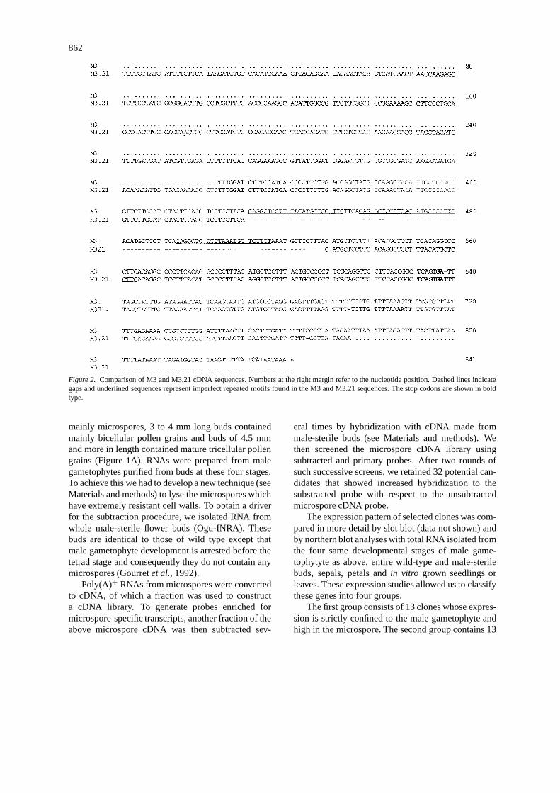
The M3 cDNA was used as a probe to screen aB. napusgenomic DNA library in order to isolatethe corresponding gene. One of the isolated clones,BnM3.4, with an insert size of 10 kb, was selectedfor further characterization (Figure 3). A 2.9 kb se-quence surrounding theBnM3.4gene was deposited inthe EMBL, GenBank and DDBJ nucleotide sequencedatabases under the accession number AF136223.Two putative in-frame translation start codons, sepa-rated by 114 bp and leading to 2 ORFs of 712 bp and598 bp, were detected (respectively at 1971 and 2085nucleotides from the start of the sequence). Compar-ison of the M3 cDNA sequence with the sequence ofthe shortest ORF of the genomic clone revealed 1 dif-ference out of 293 bp of aligned coding sequence and7 differences out of 203 bp of aligned 3′-downstreamregion. These sequence differences between genomicand cDNA clones were repeatedly found in inde-pendent sequencing experiments and might be dueto allelic variations between the differentB. napusvarieties used in the construction of the cDNA andgenomic libraries. No intron was detected in the regioncovered by the cDNA and no intron/exon junctionswere found in the rest of the ORF using a splice siteprediction program (Hebsgaardet al., 1996). A puta-tive polyadenylation signal (Joshi, 1987b) was found197 bp downstream of the stop codon. Because we areultimately interested in the regulation of pollen andanther-specific genes, we first determined by 5′ RACEwhich of the two potential start codons in M3/M3.21are used exclusively or preferentially. Sequences of12 PCR fragments revealed two groups of products.The first group contained seven sequences identicalto theBnM3.4gene (PCR1 in Figure 4). The longestPCR product showed that the transcription start of theBnM3.4gene is a G (underlined on Figure 4), localizedbetween the two potential translation starts. Primerextension experiments (data not shown) indicated ap-proximately the same nucleotide for the transcriptionalstart site. The length between the transcriptional startsite and the second ATG (64 bp), is in agreement withthe predominant length of leader sequences (40–80nucleotides) in plant genes (Joshi, 1987a). Moreover,the context of the second ATG agrees well with theconsensus sequence (AAorCAAUGGC) proposed for
864
Figure 4. Alignment of theBnM3.4gene sequence and the longest representatives of the two types of products PCR1 and PCR2 from the 5′
RACE PCR on mRNA fromB. napusmicrospores. Dashed lines indicating gaps and nucleotide differences are reported in bold type in PCR2.The transcription start site (G) is underlined (inBnM3.4and PCR1) and the ATG codon is in bold type.
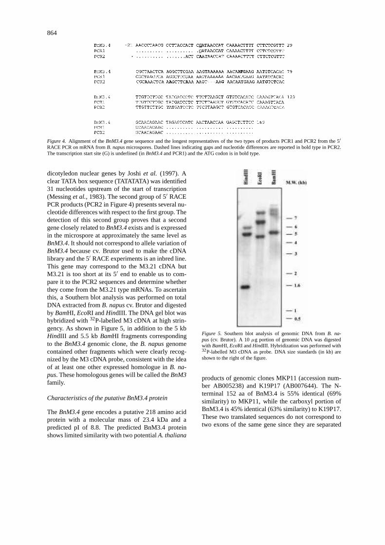
dicotyledon nuclear genes by Joshiet al. (1997). Aclear TATA box sequence (TATATATA) was identified31 nucleotides upstream of the start of transcription(Messinget al., 1983). The second group of 5′ RACEPCR products (PCR2 in Figure 4) presents several nu-cleotide differences with respect to the first group. Thedetection of this second group proves that a secondgene closely related toBnM3.4exists and is expressedin the microspore at approximately the same level asBnM3.4. It should not correspond to allele variation ofBnM3.4because cv. Brutor used to make the cDNAlibrary and the 5′ RACE experiments is an inbred line.This gene may correspond to the M3.21 cDNA butM3.21 is too short at its 5′ end to enable us to com-pare it to the PCR2 sequences and determine whetherthey come from the M3.21 type mRNAs. To ascertainthis, a Southern blot analysis was performed on totalDNA extracted fromB. napuscv. Brutor and digestedby BamHI, EcoRI andHindIII. The DNA gel blot washybridized with32P-labelled M3 cDNA at high strin-gency. As shown in Figure 5, in addition to the 5 kbHindIII and 5.5 kbBamHI fragments correspondingto theBnM3.4genomic clone, theB. napusgenomecontained other fragments which were clearly recog-nized by the M3 cDNA probe, consistent with the ideaof at least one other expressed homologue inB. na-pus. These homologous genes will be called theBnM3family.
Characteristics of the putative BnM3.4 protein
TheBnM3.4gene encodes a putative 218 amino acidprotein with a molecular mass of 23.4 kDa and apredicted pI of 8.8. The predicted BnM3.4 proteinshows limited similarity with two potentialA. thaliana
Figure 5. Southern blot analysis of genomic DNA fromB. na-pus (cv. Brutor). A 10µg portion of genomic DNA was digestedwith BamHI, EcoRI andHindIII. Hybridization was performed with32P-labelled M3 cDNA as probe. DNA size standards (in kb) areshown to the right of the figure.
products of genomic clones MKP11 (accession num-ber AB005238) and K19P17 (AB007644). The N-terminal 152 aa of BnM3.4 is 55% identical (69%similarity) to MKP11, while the carboxyl portion ofBnM3.4 is 45% identical (63% similarity) to K19P17.These two translated sequences do not correspond totwo exons of the same gene since they are separated

865
Figure 6. BnM3.4 protein characteristics. A. Cysteine pattern alignment of the BnM3.4 protein with sequences of seven stamen-specific genes.The number of amino acids between cysteine residues is given, but the spacing is not proportional. Le 5B,Lycopersicon esculentumTomA5B(Aguirre and Smith, 1993); Bn A9,Brassica napusA9; At A9, Arabidopsis thalianaA9 (Paulet al., 1992); Am FIL1,Antirrhinum majusFIL1 (Nackenet al., 1991); Le 108,Lycopersicon esculentum108 (Chen and Smith, 1993); Os YY1,Oryza sativaYY1 (Hihara et al.,1996), Ll LIM1, Lilium longiflorumLIM1 (Kobayashiet al., 1994). The amino acid sequences were deduced from DNA sequences. B. HCA(Hydrophobic Cluster Analysis) representation of the BnM3.4 protein. Amino acids are represented with one-letter symbols except for: proline(stars); glycine (diamonds); threonine (open squares); and serine (squares with dot). Hydrophobic clusters are endorsed by a black line.
by 16 Mb on the chromosome. The functions of thesetwo putativeA. thalianagene products are not known.
The putative BnM3.4 protein, however, has threerecognizable structural domains. The residues from1 to 23 are hydrophobic, characteristic of a signalpeptide, and a cleavage site between the alanine atposition 23 and the threonine at position 24 can bepredicted according to the rules of von Heijne (Nielsenet al., 1997). From these features, the protein maybe secreted and this N-segment may help the pro-tein to cross or to interact with the membrane. Thecentral part of the protein (residues 25–143) is hy-drophilic. The spacing of six particular cysteines inthis segment resembles that found in various stamen-specific proteins: TomA5B (Aguirre and Smith, 1993),FIL1 (Nackenet al., 1991), A9 (Paulet al., 1992),108 (Chen and Smith, 1993), YY1 (Hiharaet al.,1996) and LIM1 (Kobayashiet al., 1994). The pos-sible relationship among these cysteine-rich proteinsand the BnM3.4 protein is best illustrated by show-ing only their pattern of cysteine residues (Figure 6A).Although the BnM3.4 protein presents a shorter pat-tern than the others, the position and spacing of thecysteine residues are very similar.
The C-terminal domain consists of 14 repeats re-lated to the motif P-[SL]-[HNQ]-A. Analysis of thisdomain by the Hydrophobic Cluster Analysis program(available at: www.lmcp.jussieu.fr/∼soyer/www_hca/hca-form.html) suggests that the proline residues inthe motifs could form the backbone of a rigid helicalstructure (Figure 6B).
In situ analysis of the expression of theBnM3 genesfamily
The pattern ofBnM3expression during the four stagesof gametogenesis (see above) was examined in budtissues ofB. napusby in situ hybridization using theBnM3.4antisense transcript as a probe. As at least twohomologous genes are present in theB. napusgenome,the hybridization pattern may reflect the expressionpattern of (at least) these two genes. TheBnM3 tran-scripts were not detectable in anthers with pollenmother cells undergoing meiosis (data not shown), norin tetrads (Figure 7A), nor in bicellular pollen (Fig-ure 7G), nor in tricellular pollen (Figure 7I). However,at the developmental stage where rapeseed buds are2.5 mm, a strong hybridization signal was detectedin the microspore (Figure 7C). At high magnification

866

867
Figure 7. BnM3.4expression in floral buds ofB. napus. In situ hybridization of DIG-labelled antisense (A, C, E, G and I) or sense (B, D, F, Hand J)BnM3.4RNA probe toB. napusfloral bud sections. A and B. Transverse sections of 1.5 mm long buds containing tetrads (bar= 4 µm).C and D. Longitudinal sections of 2.5 mm long buds containing microspores (bar= 10µm). E and F. Magnification of anthers from transversesections of 2.5 mm long buds (bar= 0.8µm). G and H. Transverse sections of 3.5 mm long buds containing bicellular pollen (bar= 10µm). Iand J. Transverse sections of anthers from 4.5 mm long buds containing tricellular pollen (bar= 4 µm). Abbreviations: an, anther; lo, locule;mi, microspore; pi, pistil; po, mature pollen; se, sepal; ta, tapetal cell.
(Figure 7E), theBnM3transcripts are clearly visible bya deep staining inside the microspores. No hybridiza-tion signal was detected in any male gametophytetissue at any stage of development with the sense probe(Figure 7B, D, F, H, J). The lack of hybridization ofsome microspores with the antisense probe is probablyan artefact caused by a partial section of the pollengrain or a loss of cytoplasm during the pretreatment ofslides. Both the sense and the antisense probes allowedus to detect a weak hybridization signal in tapetal cellsthat could be considered as a background signal notspecific to theBnM3.4transcripts.
TheBnM3.4promoter: regions of homology andfunctional analysis
A detailed analysis of theBnM3.4promoter sequencerevealed the presence of several regions having pos-sible regulatory functions. Two 14 bp repeats (tt-taaaagtggaac) are located from−1852 to−1780 fromthe transcription start. A 14 bp palindromic sequence(ttagaatattctaa) is located at−307 while another 12 bppalindrome (ttttaattaaaa) starts at−213. These motifshave not been detected in any other pollen-specificgene. However, the motif ‘tcattt’, located between−593 and−517 and the reverse motif ‘aaatga’, locatedat −1566 in BnM3.4upstream regions were alreadydescribed for other pollen-specific genes likeNTP303(Weteringset al., 1995), NTM19 (Oldenhof et al.,1996) andBp10(Albani et al., 1992). A 2 kb regionof this 5′-flanking sequence, containing these differ-ent regions was transcriptionally fused with the re-porterguscoding sequence. The resulting construction(pJD101, see Materials and methods) was introducedinto A. thaliana. This species was chosen for theseexperiments because it belongs to the same botanicalfamily as B. napus, but it can be more easily trans-formed and has a shorter life cycle. GUS staining wasfirst performed on the inflorescences of fifteen inde-pendent transgenicA. thalianaT1 lines (JD101 series)transformed with this gene. They all showed qualita-tively the same expression patterns in the course ofgametophyte development (not shown). The progeny(T2) of T1 plants with insertion of the transgene(s)
presumably at a single locus was chosen for furtheranalysis. The results of histochemical analysis ofgusexpression for individual hemizygous and homozy-gous progeny of one of them, JD101.42, are shownin Figure 8. This transformant presents one insertionaccording to the genetic test (275 phosphinotricineresistant plants out of 380 seedlings;χ2
= 1.40;P < 0.05).
In all the transgenic JD101 plants and theirprogeny studied, no GUS activity was detected inflower organs other than the stamen as shown in Fig-ure 8A for one hemizygote progeny of the JD101.42plant. GUS activity was not detected in any organ ofseedling (Figure 8I) or older plants (not shown), orin siliques (Figure 8J). In comparison, plants trans-formed with the control construction pAF100 (guswithout promoter) showed no GUS activity (Fig-ure 8A, inset). The presence of 50% of blue pollen and50% of unstained pollen in anthers of the hemizygousJD101.42 plant (Figure 8H) confirms that theBnM3.4promoter is only activated in gametophytic cells i.eonly the transgenic pollen shows GUS activity.
The temporal pattern ofgus expression was de-termined with homozygous JD101 progeny with theaim of precisely determining the developmental stagewhere the reporter gene is activated. At the tetrad stageof male gametophyte, no GUS activity was detected(Figure 8B); it only appears at a high level at the begin-ning of the microspore stage (Figure 8C). As shown inFigure 8E, high levels of GUS activity were detectedin pollen. There seems to be a low level of the GUSproduct in the locule which may be the result of dif-fusion, but no activity was present in the sporophytictissues of the anther such as the epidermis, the en-dothecium and the tapetum. The GUS expression levelstays constant until the mature tricellular pollen stage(Figure 8D, 8F and 8G) but it is difficult to know if thisis due to the longevity of the GUS protein or transgeneactivity in Arabidopsis.
At least for the first stages of male gametophyte de-velopment, the reporter gene has the same expressionpattern as intact genes.

868

869
Figure 8. Histochemical analysis ofgusexpression in transgenicA. thalianaJD101 plants. The following plant materials from homozygousand hemizygous JD101.42 transgenic plants and from a AF100 transgenic plant were examined for GUS activity as described in Materialsand methods. A. Inflorescence from a 5-week old homozygous pJD101 plant and (in the inset) the inflorescence from a 5-week old AF100plant (negative control plant). B–G. Developmental series of buds from the stained inflorescence presented above. Sepals and petals wereremoved from the bud in order to measure the pistil length more easily and to improve the visualization of GUS in anthers. The stages ofmale gametophyte development were determined according to the pistil length as described in Materials and methods. B, tetrad stage; C, earlymicrospore stage; D, end of microspore stage; E, locule of one of the anthers of the bud shown in D, magnified 6× relative to Figure D (barrepresents 27µm); F, bicellular pollen. g, tricellular pollen in anthers at anthesis. In B, C, D, F and G, bars represent 90µm. H. Antherof a hemizygous plant, at the microspore stage (bar represents 70µm). I. Ten-day old seedling. J. Open siliques and seeds at two differentmagnifications.
Discussion
This paper describes a method in order to isolate cD-NAs from rapeseed that are preferentially expressedduring early male gametophyte development. Expres-sion of several of these genes appears to be highlyspecific to microspores and we have analysed one ofthese, calledBnM3.4, in detail. This gene correspondsto the M3 cDNA isolated, but another cDNA was iso-lated from the microspore cDNA library using M3as a probe. This longer cDNA (M3.21) is homolo-gous to M3 except that it shows a deletion of 99 bpin the putative coding region. As these two cDNAcross-hybridize in the hybridization conditions used,we can conclude that at least two genes (theBnM3family=BnM3.4+ related gene(s)) are present in theB. napusgenome. This is also supported by the resultsof 5′ RACE and Southern blot experiments.
The expression of these genes was studied bothby northern blot andin situ hybridization. By north-ern blot analysis, the transcripts were observed mainlyin microspores and also in total RNA extracted frombuds of length strictly below 2 mm, that contain amajority of tetrads and meiocytes. The developmentalsynchrony between all the male gametophytes of ananther is not perfect and some free microspores al-ready appear among tetrads in buds below 2 mm, sothe hybridization of M3 probe to RNAs from pollenextracted from buds below 2 mm may be due to theearly presence of microspores in these buds. Thishypothesis was confirmed byin situ hybridization ex-periments. In individual 1.5 mm long buds, whereno microspores were found, no signal was detected.On the other hand, in 2.5 mm long buds, where mi-crospores are the only developmental stage present,a strong signal was detected. In later gametogenesisstages, no further signal was detected. According tothese results, we can conclude that the homologousgenes ofBnM3family are expressed specifically in themicrospore ofB. napus.
Like the other early-expressed gene isolated fromtobacco, NTM19 (Oldenhof et al., 1996), BnM3.4does not show any homology to known genes. Al-though the deduced BnM3.4 protein does not showhomology with the deduced NTM19 protein, they areboth proline-rich proteins and contain a putative sig-nal peptide. We can only speculate on the functionand cellular localisation of the BnM3.4 protein withthe help of prediction programs. Several stamen pro-teins of unknown function contain similar proline-richdomains (Chenet al., 1993) that, despite differencesin amino-acid composition, are structured in a verysimilar way. The role of the proline-rich motifs is notknown, but one may speculate that they serve as somekind of anchoring structures in interaction with ei-ther components of the cytoskeleton or with structuralcomponents of the cell wall (Woessneret al., 1994;Krögeret al., 1996).
The comparison analysis of theNTM19 and theBnM3.4 promoter regions did not show any clearcommon domains which could indicate a role in thespecificity of expression of these genes. Only the box‘tcattt’ and its reverse sequence, ‘aaatga’, which hasalready been shown to be important for pollen-specificexpression are present in both promoters. Even if thenumber and the precise location of these motifs arenot conserved between the different promoter regionsdescribed (tobacco forNTM19 and NTP303, rape-seed forBp10), functional analysis using deletion andtargeted mutagenesis experiments in microprojectile-mediated transient expression assays (Weteringset al.,1995) showed clearly that one of these elements of theNTP303promoter constitutes a positivecis-regulatoryelement and functions by specifically enhancing tran-scription in pollen. This is the reason why we kept thispromoter region of 2 kb for the heterologous expres-sion experiments. Both analysis of more microspore-specific genes and promoter deletions using a reportergene will be necessary to evaluate the influence of thedifferent motifs in the spacial and temporal regulationof transcription of these genes.
870
The histochemical analysis ofgus expressiondriven by theBnM3.4promoter (pJD101 construction)in transgenicA. thalianaplants showed a high level ofGUS activity in male gametophytes but not in otherfloral and seedling tissues. Furthermore, GUS activ-ity appeared at the microspore stage and stayed at aconstant level during pollen development until pollenmaturity. GUS being a very stable protein, the detec-tion of GUS activity allowed us to define precisely thestart point ofBnM3.4promoter activity inA. thalianabut not its stop point. The spatial and temporal pat-tern ofgusexpression driven by theBnM3.4promoterin A. thaliana is in accordance with the establishedexpression pattern of the gene inB. napus. These re-sults suggest the presence oftrans-acting factors inA. thalianaable to recognise and interact with thecis-regulatory elements ofBnM3.4in the same manner asin B. napus.
Several of the genes isolated by this procedurebelong to gene families (BnM3.4, SKP1-like genes)as seen with other transcriptional approaches (Twell,1994). This strategy is complementary to the muta-genesis being undertaken inA. thaliana (Feldmannet al., 1997; Bonhommeet al., 1999). We nowplan to compare the 5′-flanking regions of the othermicrospore-specific genes we have isolated and to tryBnM3.4promoter deletion analysis in order to findcis-regulatory elements required for microspore-specificexpression.
Acknowledgements
This research was supported in part by the Associa-tion pour la Promotion de la Sélection des Oléagineux(PROMOSOL), France. The authors wish to thankJ.M. Camadro for protein analysis expertise and help-ful discussion, O. Renoir for her help for GUS stain-ing, S. Bonhomme, H. Höfte, J. Leung and I. Small forhelpful discussions and correction of the manuscript.
References
Aguirre, P.J. and Smith, A.G. 1993. Molecular characterization of agene encoding a cysteine-rich protein preferentially expressed inanthers ofLycopersicon esculentum. Plant Mol. Biol. 23: 477–487.
Albani, D., Robert, L.S., Donaldson, P.A., Altosaar, I., Ar-nison, P.G. and Fabijanski, S.F. 1990. Characterization of apollen-specific gene family fromBrassica napuswhich is acti-vated during early microspore development. Plant Mol. Biol. 15:605–622.
Albani, D., Sardana, R., Robert, L.S., Altosaar, I., Arnison, P.G.and Fabijanski, S.F. 1992. ABrassica napusgene family whichshows sequence similarity to ascorbate oxidase is expressedin developing pollen. Molecular characterization and analysisof promoter activity in transgenic tobacco plants. Plant J. 2:331–342.
Bechtold, N., Ellis, J. and Pelletier, G. 1993.In planta Agrobac-teriummediated gene transfer by infiltration of adultArabidopsisthaliana plants. C.r. Acad. Sci. (Sér. III: Sci. Vie) 316: 1194–1199.
Bedinger, P.A., Hardeman, K.J. and Loukides, C.A. 1994. Trav-elling in style: the cell biology of pollen. Trends Cell Biol. 4:132–138.
Bevan, M., Barnes, W.M. and Chilton, M.-D. 1983. Structure andtranscription of the nopaline synthase gene region of T-DNA.Nucl. Acids Res. 11: 369–385.
Bonhomme, S., Horlow, C., Vezon, D., de Laissardière, S., Guyon,A., Férault, M., Marchand, M., Bechtold, N. and Pelletier,G. 1999. T-DNA mediated disruption of essential gametophyticgenes inArabidopsisis unexpectedly rare and cannot be inferredfrom segregation distortion alone. Mol. Gen. Genet, in press.
Bowman, J. (Ed.) 1994.Arabidopsis. An Atlas of Morphology andDevelopment, Springer-Verlag New York, pp. 133–332.
Brander, K.A. and Kuhlemeier, C. 1995. A pollen-specific DEAD-box protein related to translation initiation factor eIF-4A fromtobacco. Plant Mol. Biol. 27: 637–649.
Brown, S.M. and Crouch, M.L. 1990. Characterization of a genefamily abundantly expressed inOenothera organensispollen thatshows sequence similarity to polygalacturonase. Plant Cell 2:263–274.
Bucciaglia, P.A. and Smith, A.G. 1994. Cloning and characteriza-tion of Tag1, a tobacco antherβ-1,3-glucanase expressed duringtetrad dissolution. Plant Mol. Biol. 24: 903–914.
Cartea, M.E., Migdal, M., Galle, A.M., Pelletier, G. and Guerche,P. 1998 Comparison of sense and antisense methodologies formodifying the fatty acid composition ofArabidopsis thalianaoilseed. Plant Sci 136: 181–194.
Chen, C.G., Mau, S.L. and Clarke, A.E. 1993. Nucleotide sequenceand style-specific expression of a novel prolin-rich protein genefrom Nicotiana alata. Plant Mol. Biol. 21: 391–395.
Connelly, C. and Heiter, P. 1996. Budding yeast SKP1 encodesan evolutionarily conserved kinetochore protein required for cellcycle progression. Cell 86: 275–285.
Chen, R. and Smith, A.G. 1993. Nucleotide sequence of a stamen-and tapetum-specific gene fromLycopersicon esculentum. PlantPhysiol. 101: 1413.
Dean, C., van den Elzen, P., Tamaki, S., Dunsmuir, P. and Beds-brook, J. 1985. Differential expression of eight genes of petu-nia ribulose bisphosphate carboxylase small subunit multi-genefamily. EMBO J. 4: 3055–3061.
Dellaporta, J.L., Wood, J. and Hicks, J.B. 1983. A plant DNAminipreparation: Version II. Plant Mol. Biol. Rep. 1: 19–21.
Estelle, M.A. and Sommerville, C.R. 1987. Auxin-resistant mutantsof Arabidopsis thalianawith an altered morphology. Mol. Gen.Genet. 206: 200–206.
Feinberg, A.P. and Vogelstein, B. 1985. A technique for radiola-belling DNA fragments to high specificity. Anal. Biochem. 137:266–267.
Feldmann, K., Coury, D. and Christianson, M. 1997. Exceptionalsegregation of a selectable marker (KanR) inArabidopsisidenti-fies genes important for gametophytic growth and development.Genetics 147: 1411–1422.

871
Gourret, J.P., Delourme, R. and Renard, M. 1992. Expression ofogucytoplasmic male sterility in cybrids ofBrassica napus. Theor.Appl. Genet. 83: 549–556.
Hanson, D.D., Hamilton, D.A., Travis, J.L., Bashe, D.M. and Mas-carenhas, J.P. 1989. Characterization of a pollen-specific cDNAclone fromZea maysand its expression. Plant Cell 1: 173–179.
Hebsgaard, S.M., Korning, P.G., Tolstrup, N., Engelbrecht, J.,Rouze, P. and Brunak, S. 1996. Splice site prediction inAra-bidopsis thalianapre-mRNA by combining local and globalsequence information. Nucl. Acids Res. 24: 3439–3452.
Hihara, Y., Hara, C. and Uchimiya, H. 1996. Isolation and charac-terization of two cDNA clones for mRNAs that are abundantlyexpressed in immature anthers of rice (Oryza sativaL.). PlantMol. Biol. 30: 1181–1193.
Jackson, D.P. 1991.In situ hybridization in plants. In: D.J. Bowles,S.J. Gurr and M. McPhereson (Eds.), Molecular Plant Pathol-ogy: A Practical Approach, Oxford University Press, Oxford,pp. 163–174.
Jefferson, R.A., Burgess, S.M. and Hirsh, D. 1986. ß-glucuronidasefrom Escherichia colias a gene-fusion marker. Proc. Natl. Acad.Sci. USA 83: 8447–8451.
Jefferson, R.A., Kavanagh, T.A. and Bevan, M.W. 1987. GUS fu-sions: ß-glucuronidase as a sensitive and versatile gene fusionmarker in higher plants. EMBO J. 6: 3901–3907.
Joshi, C.P. 1987a. An inspection of the domain between putativeTATA box and translation start site in 79 plant genes. Nucl. AcidsRes. 15: 6643–6653.
Joshi, C.P. 1987b. Putative polyadenylation signals in nuclear genesof higher plants: a compilation and analysis. Nucl. Acids Res.15: 9627–9640.
Joshi, C.P., Zhou, H., Huang, X. and Chiang, V.L. 1987. Contextsequences of translation initiation codon in plants. Plant Mol.Biol. 35: 993–1001.
Kobayashi, T., Kobayashi, E., Sato, S., Hotta, Y., Miyajima, N.,Tanaka, A. and Tabata, S. 1994. Characterization of cDNAs in-duced in meiotic prophase in lily microsporocytes. DNA Res. 1:15–26.
Koltunow, A.M., Truettner, J., Cox, K.H., Wallroth, M. andGoldberg, R.B. 1990. Different temporal and spatial gene ex-pression patterns occur during anther development. Plant Cell 2:1201–1224.
Koncz, C. and Schell, J. 1986. The promoter of TL-DNA gene5′ controls the tissue specific expression of chimeric genes car-ried by a novel type ofAgrobacteriumbinary vector. Mol. Gen.Genet. 204: 383–396.
Krek, W. 1998. Proteolysis and the G1-S transition: the SCFconnection. Curr. Opin. Genet. Devel. 8: 36–42.
Kröger, N., Bergsdorf, C. and Sumper, M. 1996. Frustulins: do-main conservation in a protein family associated with diatom cellwalls. Eur. J. Biochem. 239: 259–264.
Lo, Y.-M.D., Mehal, W.Z. and Kleming, K.A. 1988. Rapid pro-duction of free biotinylated probes using the polymerase chainreaction. Nucl. Acids Res. 16: 8719–8722.
Messing, J., Geraghty, D., Heidecker, G., Hu, N.T., Kridl, J.and Rubenstein, I. 1983. Plant gene structure. In: T. Kosuge,C.P. Meredith and A. Hollaender (Eds.), Genetic Engineering ofPlants, Plenum, New York, pp. 211–227.
Mueller, P.R. and Wold, B. 1989. In vivo footprinting of a muscle-specific enhancer by ligation mediated PCR. Science 246: 780–786.
Nacken, W.K.F., Huijser, P., Beltran, J.-P., Saedler, H. and Som-mer, H. 1991. Molecular characterization of two stamen-specificgenes,tap1andfil1, that are expressed in the wild type, but not
in thedeficiensmutant ofAntirrhinum majus. Mol. Gen. Genet.229: 129–136.
Nagel, R., Masel, E.A., Birch, R.G. and Manners, J.M. 1990.Electroporation of binary Ti plasmid vector intoAgrobacteriumtumefaciensand Agrobacterium rhizogenes. FEMS Microbiol.Lett. 67: 325–328.
Nielsen, H., Engelbrecht, J., Brunak, S., Von Heijne, G. 1997.Identification of prokaryotic and eukaryotic signal peptides andprediction of their cleavage sites. Prot. Engng. 10: 1–6.
Oldenhof, M.T., de Groot, P.F.M., Visser, J.H., Schrauwen, J.A.M.and Wullems, G.J. 1996. Isolation and characterization of amicrospore-specific gene from tobacco. Plant Mol. Biol. 31:213–225.
Paul, W., Hodge, R., Smartt, S., Draper, J. and Scott, R. 1992. Theisolation and characterisation of the tapetum-specificArabidop-sis thalianaA9 gene. Plant Mol. Biol. 19: 611–622.
Robert, L.S., Allard, S., Gerster, J.L., Cass, L. and Simmonds, J.1993. Isolation and characterization of a polygalacturonase genehighly expressed inBrassica napuspollen. Plant Mol. Biol. 23:1273–1278.
Ross, J.H.E. and Murphy, D.J. 1996. Characterization of anther-expressed genes encoding a major class of extracellular oleosin-like proteins in the pollen coat of Brassicaceae. Plant J. 9:625–637.
Rubinelli, P., Hu, Y. and Ma, H. 1998. Identification, sequenceanalysis and expression studies of novel anther-specific genes ofArabidopsis thaliana. Plant Mol. Biol. 37: 607–619.
Sambrook, J., Fritsch, E.F. and Maniatis, T. 1989. MolecularCloning: A Laboratory Manual, 2nd ed. Cold Spring HarborLaboratory Press, Cold Spring Harbor, NY.
Scott, R., Dagless, E., Hodge, R., Paul, W., Soufleri, I. and Draper,J. 1991. Patterns of gene expression in developing anthers ofBrassica napus. Plant Mol. Biol. 17: 195–207.
Shen, J.B. and Hsu, F.C. 1992.Brassicaanther-specific genes: char-acterization andin situ localization of expression. Mol. Gen.Genet. 234: 379–389.
Singh, A., Kao, T. and Lin, J.J. 1993. Transformation ofAgrobac-terium tumefacienswith T-DNA vectors using high-voltageelectroporation. Focus 15: 84–87.
Sive, H.L. and St John, T. 1988. A simple subtractive hybridiza-tion technique employing photoactivatable biotin and phenolextraction. Nucl. Acids Res. 16: 10937.
Stanchev, B.S., Doughty, J., Scutt, C.P., Dickinson, H. and CroyR.R.D. 1996. Cloning ofPCP1, a member of a family of pollencoat protein (PCP) genes fromBrassica oleraceaencoding novelcysteine-rich proteins involved in pollen-stigma interactions.Plant J. 10: 303–313.
Theerakulpisut, P., Xu, H., Singh, M.B., Pettitt, J.M. and Knox,R.B. 1991. Isolation and developmental expression of Bcp1, ananther-specific cDNA clone inBrassica campestris. Plant Cell 3:1073–1084.
Twell, D. 1994. The diversity and regulation of gene expression inthe pathway of male gametophyte development. In: R.J. Scottand A.D. Stead (Eds.), Molecular and Cellular Aspects of PlantReproduction, Society for Experimental Biology, CambridgeUniversity, Press, Cambridge, UK, pp. 83–135.
Ursin, V.M., Yamaguchi, J. and McCormick, S. 1989. Gameto-phytic and sporophytic expression of anther-specific genes indeveloping tomato anthers. Plant Cell 1: 727–736.
Wang, Z. and Brown, D.B. 1991. A gene expression screen. Proc.Natl. Acad. Sci. USA 88: 11505–11509.
Weterings, K., Reijnen, W., van Aarssen, R., Kortstee, A., Spijk-ers, J., van Herpen, M., Schrauwen, J. and Wullems, G. 1992.Characterization of a pollen-specific cDNA clone fromNicotiana

872
tabacumexpressed during microgametogenesis and germination.Plant Mol. Biol. 18: 1101–1111.
Weterings, K., Schrauwen, J., Wullems, G. and Twell, D. 1995.Functional dissection of the promoter of the pollen-specificgene NTP303 reveals a novel pollen-specific, and conservedcis-regulatory element. Plant J. 8: 55–63.
Woessner, J.P., Molendijk, A.J., van Egmond, P., Klis, F.M., Good-enough, U.W. and Haring, M.A. 1994. Domain conservationin several volvocalean cell wall proteins. Plant Mol. Biol. 26:947–960.
Yu, L.-X., Nasrallah, J., Valenta, R. and Parthasarathy, M.V. 1998.Molecular cloning and mRNA localization of tomato pollenprofilin. Plant Mol. Biol. 36: 699–707.
Copyright © 2022 FDOKUMEN