The influence of alkalising amines on the film formation by ...
Upload
independentCategory
view
2download
0
1 3
DOI 10.1007/s10337-014-2731-8Chromatographia (2014) 77:1451–1459
ORIGINAL
Isolation of Biogenic Amines Using Paramagnetic Microparticles Off‑Line Coupled with Ion Exchange Liquid Chromatography
Natalia Cernei · Zbynek Heger · Pavel Kopel · Jiri Skladanka · Ondrej Zitka · Vojtech Adam · Rene Kizek
Received: 12 February 2014 / Revised: 22 June 2014 / Accepted: 25 June 2014 / Published online: 17 July 2014 © Springer-Verlag Berlin Heidelberg 2014
cadaverine, and histamine in the case of MAN35; and his-tamine, tyramine, and putrescine in the case of MAN8. Finally, we carried out the analyses of real samples obtained from patients suffering from prostate carcinoma, where histamine was determined as the most abundant biogenic amine (10.456–13.654 µg mL−1). The prepared PMPs were able to isolate the biogenic amines from real samples, and thus they may be helpful in construction of biosensors, or Lab-on-a-Chip platforms, enabling less painful, and more rapid diagnosis of prostate cancer.
Keywords Biogenic amines · Biosensor · Ion-exchange liquid chromatography · Magnetic-particles-based separation
Introduction
Biogenic amines are formed as products of physiologi-cal metabolic activities of microorganisms, plants and ani-mals [1]. They are produced by amino acid decarboxylase, in the enzymatic driven process called decarboxylation, which contains pyridoxal phosphate as the cofactor [2]. The decarboxylase cleaves the carboxyl to form basic amine [3]. The produced polyamines spermine (Spm) and spermidine (Spd) and diamines putrescine (Put) and cadaverine (Cad) are constituents of eukaryotic and prokaryotic cells, hav-ing multiple functions in living organisms, in the binding and precipitation of DNA or inhibition of neuronal nitric oxide synthase, or stabilization of macromolecules [4]. On the other hand, increased levels of these substances above physiological concentration are toxic to cells, and can facili-tate cell death based on the enhanced oxidative stress [5, 6]. Thus, their pathological presence could be utilized for con-ducting more precise diagnostics. In particular, in urine,it
Abstract This study aims at the possibility of single struc-tured paramagnetic microparticles (PMPs), composed of maghemite (γ-Fe2O3) core modified with chitosan called MAN8, or tetraethyl orthosilicate covered with Dowex called MAN35, to be helpful for isolation of biogenic amines prior to their further analysis. Primarily, we synthe-sized and characterized PMPs. To obtain the information about bead morphology, scanning electron microscopy was employed. Furthermore, X-ray fluorescence was employed to carry out the elemental composition analyses. To obtain further insight into interaction between PMP surface and biogenic amines, scanning electron microscope was employed. It was shown that binding of biogenic amines causes increase of relative current response of deproto-nated microparticles. We tested the specificity of PMPs to bind biogenic amines on histamine, tyramine, spermine, spermidine, putrescine, and cadaverine. We found that two types of our PMPs were able to selectively bind spermidine,
Published in the topical collection Advances in Chromatography and Electrophoresis & Chiranal 2014 with guest editor Jan Petr.
N. Cernei · Z. Heger · P. Kopel · O. Zitka · V. Adam · R. Kizek (*) Department of Chemistry and Biochemistry, Faculty of Agronomy, Mendel University in Brno, Zemedelska 1, 613 00 Brno, Czech Republice-mail: [email protected]
N. Cernei · P. Kopel · O. Zitka · V. Adam · R. Kizek Central European Institute of Technology, Brno University of Technology, Technicka 3058/10, 616 00 Brno, Czech Republic
J. Skladanka Department of Animal Nutrition and Forage Production, Faculty of Agronomy, Mendel University in Brno, Zemedelska 1, 613 00 Brno, Czech Republic

1452 N. Cernei et al.
1 3
has been previously shown that increased levels of biogenic amines are associated with various types of tumors [7, 8], and the recovery of urinary diacetylpolyamines to nor-mal levels accompanied with remission of cancer has been described [9]. For diagnostics, putrescine, spermidine, sper-mine and cadaverine could be interesting, because of their presence in many malignant diseases [10]. On the other hand, histamine can affect numerous processes, including anaphylaxis, dermatosis, phlogistic reactions, rheumatoid arthritis and many others [11]. Moreover, biogenic amines dopamine (Da), norepinephrine (Ne), and serotonin (5-HT) are important neurotransmitters in the central and peripheral nervous system [12]. Concentrations of norepinephrine and dopamine in urine specimens above 80 and 400 μg per 24 h were reported to be linked with pheochromocytoma [13].
In order to determine the concentrations of biogenic amines in biological matrices like urine, techniques provid-ing high resolution and sensitivity are demanded. Deter-mination of biogenic amines is usually carried out using thin-layer chromatography [14], gas chromatography, cap-illary zone electrophoresis [15], ion exchange liquid chro-matography (IEC) [16], enzyme immunoassay [17] and/or high-performance liquid chromatography [18, 19]. In our study, we decided to use IEC with post-column ninhy-drin derivatization and Vis detection for biogenic amines separation and detection, as they minimize the sample pre-cleaning requirement [20]. The main aim of the present study consisted of synthesis and characterization of two types of paramagnetic microparticles (PMPs), composed of nanomaghemite (γ-Fe2O3) core, modified with chitosan, or tetraethyl orthosilicate (TEOS) covered with Dowex. Sub-sequently, our prepared microparticles were tested for their ability to bind with biogenic amines (spermine, spermidine,
putrescine, cadaverine, histamine, and tyramine, Fig. 1), and finally, these were applied as a first part of our two-dimensional (2D) separation approach, consisting of PMPs and IEC for isolation of biogenic amines in a sample with-out time-consuming pre-treatment.
Experimental Section
Chemicals
Standards of biogenic amines (Tyr, Put, His, Cad, Spm, Spd) with a purity of 99 % were obtained from Sigma-Aldrich (St. Louis, MO, USA). Solutions of biogenic amines were prepared with a dilution buffer called sodium cycle, com-posed of 1.5 mM N3Na, 197 mM NaCl and 73 mM C6H8O7 in MilliQ H2O. Furthermore, we used citric acid, sodium citrate, isopropanol, potassium hydroxide, potassium bro-mide, hydrochloric acid and ninhydrine, all purchased from Sigma-Aldrich. Methyl cellosolve was purchased from Ingos (Prague, Czech Republic), as well as tin chloride. All buffer solutions were prepared with deionized water obtained using reverse osmosis Aqual 25 (Aqual s.r.o., Brno, Czech Repub-lic). The deionized water was further purified by using a Direct-Q 3 UV Water Purification System equipped with a UV lamp (Millipore, Billerica, MA, USA). The resistance was established to 18 MΩ cm−1. The pH was measured using a pH meter WTW inoLab (Weilheim, Germany).
Real Samples of Urine
For purpose of this study, urine samples from patients suf-fering from carcinoma of the prostate (n = 5), obtained from St. Anne’s University Hospital, Department of Urology, Brno were used. Average age of patients was 69–55 years. In all cases, different types of acinar adenocarcinoma were diagnosed. For a control measurement, urine samples from volunteers (n = 10) with average age of 24–69 years were used. The enlistment of patients into the clinical study was approved by the Ethics Committee of the Faculty of Medi-cine, Masaryk University, Brno, Czech Republic.
Immunoenzymometric Assay (IEMA)
For analysis of prostate specific antigen (PSA) and free pros-tate specific antigen (fPSA) in the sample of urine, IEMA was used. Measurement was carried out using an automated analyzer AIA 600 II (Tosoh Bioscience, Tokyo, Japan). Sev-enty microliters of urine sample were pipetted into the testing cup ST AIA-PACK PSAII, obtained from Tosoh Bioscience (Tokyo, Japan), and containing lyophilized reagent (mag-netic microbeads with murine anti-PSA and mouse anti-PSA conjugated with bovine alkaline phosphatase). Subsequently,
NH2
OH
NH2
NH
NH2NH 2
N H 2
a
b
c
NH2
NH2
d
NH
N
NH2
e
NH2
NH
NH
NH2
f
Fig. 1 The chemical structures of the studied biogenic amines, a spermidine, b cadaverine, c tyramine, d putrescine, e histamine, and f spermine

1453Isolation of Biogenic Amines Using Paramagnetic Microparticles
1 3
the sample was incubated at 37 °C for 10 min. Non-bonded antibodies were removed by washing solution (Tosoh Biosci-ence). Finally, fluorogenic substrate (4-methylumbellipheryl phosphate) was added and the intensity of fluorescence for determination the activity of enzyme was measured.
Synthesis of Paramagnetic Microparticles
Two types of PMPs were employed in this study. Both were based on nanometric maghemite particle cores, crafted according to our previous study [21].
Nanomaghemite obtained in this way was further modi-fied. (I) In the case of PMPs, MAN8 20 mL of nanomagh-emite solution was added to 2 mL of 1 % chitosan (v/v). The mixture was stirred overnight with a Biosan OS-10 (Biosan, Riga, Latvia). The resulting product was separated using an external magnet and washed with water. Finally, the prod-uct was dried at 40 °C. (II) In the case of PMPs MAN35, 20 mL of nanomaghemite solution was mixed with 25 mL of isopropanol, and washed with water using the magnetic force of an external magnetic field. Furthermore, 50 mL of isopropanol was added again with 7 mL of 28 % ammonia (v/v). Into this mixture, 1.5 mL of TEOS was slowly added. The mixture was stirred overnight at 40 °C using the Biosan OS-10. Subsequently, 0.8 g of Dowex was added. The result-ing product was stirred for 2 h and separated with an external magnetic field. Finally, the product was dried at 40 °C.
Scanning Electron Microscopy (SEM) of Paramagnetic Microparticles
The morphology of PMPs was characterised using an elec-tron microscope (FEG-SEM MIRA XMU, Tescan, a.s., Brno, Czech Republic). This model is equipped with a high brightness Schottky field emitter for low noise imaging at fast scanning rates. The SEM was fitted with an Everhart-Thronley type of SE detector, a high-speed YAG scintil-lator-based BSE detector, panchromatic CL Detector and EDX spectrometer. Samples were coated with 10 nm of carbon to prevent sample charging. A carbon coater K950X (Quorum Technologies, Grinstead, United Kingdom) was used for this purpose. Different conditions were optimized in order to reach either minimum analysis time or maxi-mum detail during overnight automated analysis. An accel-erating voltage of 15 kV and beam currents of about 1 nA gave satisfactory results regarding maximum throughput.
Scanning Electrochemical Microscope for Characterization of Paramagnetic Microparticles
Identification of relative current response before and after biogenic amine binding to PMP surface was performed using scanning electrochemical microscope Model 920D
(CH instruments, Inc. USA). We used an electrochemi-cal microscope with a 10-mm measuring platinum disc probe electrode with a potential of 0.2 V, and another platinum disc electrode with an O-ring as the conducting substrate,with a potential of 0.3 V. During scanning, the particles were attached to the substrate platinum electrode by magnetic force from a neodyme magnet, which was situ-ated below the electrode. The platinum measuring electrode was moving from 150 µm above the surface. The scanning was carried out in a solution consisting of 5 % ferrocene in methanol mixed in a 1:1 ratio with 0.05 % KCl in water (v/v). Measurements were performed in a 1.5 mL Teflon cell, according to the following parameters: amperometric mode, and vertical scan was carried out in a 500 × 500 µm area with a scan rate of 30 µm s−1.
X-Ray Fluorescence Elemental Analysis
X-ray fluorescence (XRF) elemental analysis of PMPs was carried out on a Xepos (SPECTRO analytical instruments GmbH, Kleve, Germany) fitted with three detectors: Barkla scatter-aluminium oxide, Barkla scatter-HOPG and Comp-ton/secondary molybdenum. Analyses were conducted by the Turbo Quant cuvette method of measurement. Experi-mental parameters were set to a measurement duration of 300 s, with a tube voltage from 24.81 to 47.72 kV, tube cur-rent from 0.55 to 1.0 mA, with zero peak at 5000 cps and the vacuum switched off.
Sample Preparation
To obtain information about behaviour of our own prepared PMPs, biogenic amines (volume 250 µL) in concentrations of 100 µg mL−1 were bound to them, according to isola-tion conditions optimized in our preliminary study dealing with amino acids [21]. For isolation, we employed 250 µL of dispersion, comprising 10 mg mL−1 PMPs in PBS. After isolation, the sample was dissolved in 3 M hydrochloric acid (250 µL) and evaporated using nitrogen evaporator Ultravap RC (Porvair Sciences, Leatherhead, UK). Finally, the evaporated sample was resuspended with dilution buffer (250 µL) and analysed using IEC. In addition, real urinary samples (250 µL) were prepared similarly, to be analysed by PMPs.
Ion-Exchange Liquid Chromatography
As a second part of our 2D separation approach, an AAA 400 (Ingos, Prague, Czech Republic) IEC apparatus was used. The system consisted of a glass filling chromato-graphic column and steel precolumn, two chromatographic pumps for transport of elution buffers and derivatization reagent, cooled carousel for 25 eppendorf tubes, dosing

1454 N. Cernei et al.
1 3
valve, heat reactor, Vis detector, and cooled chamber for derivatization reagent. The volume of the injected sample was 100 µL, with an RSD 1 % accuracy of application. We used a two-channel Vis detector with a 5 µL flow volume cuvette operated at wavelengths of 440 and 570 nm. A solution of ninhydrin was prepared with 75 % methyl cel-losolve (v/v) and 25 % 4 M acetic buffer (v/v). Tin chloride was used as a reducing reagent. Prepared solution of ninhy-drin was stored under an inert atmosphere (N2) with cool-ing at 4 °C. Flow rate was 0.25 mL min−1 under a pressure ranging from 4.5 to 6.0 MPa. Reactor temperature was set to 120 °C. For elution, two buffers were employed: buffer A was composed of 5.5 mM C6H8O7, 81 mM Na3C6H5O7, 257 mM NaCl, 350 mM KBr, and 250 mL of C3H8O per 1 L of MiliQ water, with a final pH of 5.78. Buffer B con-sisted of 73 mM C6H8O7, 3 M NaCl, and 10.0 mL of 50 % solution of KOH (w/w) per 1 L of MilliQ water, with a final pH of 3.27. For pH measurements, the WTW inoLab pH meter (Weilheim, Germany) was employed.
Recovery
Recoveries of biogenic amines were evaluated from urine samples, spiked with an internal standard of concentra-tion of 1 mM. Before isolation, 100 µL of biogenic amine standards and 100 µL of water were added to urine sam-ples. Homogenates were assayed blindly and biogenic amine concentrations were derived from the calibration curves. The spiking of biogenic amines was determined as a standard measured without the presence of real sample. The calculation of recovery was performed according to Causon [22] and Bugianesi et al. [23].
Descriptive Statistics
Mathematical analysis of the data and their graphical inter-pretation were realized by Microsoft Excel®, Microsoft Word® and Microsoft PowerPoint®. Results are expressed as mean ± standard deviation (SD), unless otherwise noted. The detection limits (three signal/noise, S/N) were calcu-lated according to Long and Winefordner [24], whereas N was expressed as S.D. of noise determined in the signal domain unless stated otherwise.
Results and Discussion
Ion-Exchange Liquid Chromatography with Vis Detection of Biogenic Amines
Determination of biogenic amines is not easy task, regard-ing the demands of sensitivity and accuracy of meas-urements and the influence of the matrix on the sample
pre-treatment steps. In this study, we attempted to combine advantages of PMP-based isolation of the amines with their subsequent analysis using IEC.
Primarily, we had to optimize the conditions for bio-genic amine separation and detection using IEC with ninhydrin postcolumn derivatization and dual channel Vis detection (λ = 570 nm, and 440 nm). As representa-tives of biogenic amines, we selected the most commonly occurring biogenic amines, such as spermine, spermidine, putrescine, cadaverine, histamine and tyramine. Besides their abundance, they have been found as the potential can-cer biomarkers [7, 8]. Based on the optimization of sepa-ration of biogenic amines, the conditions were as follows: elution of biogenic amines was performed using gradient elution with buffers of different ionic strength and pH, as well as a temperature gradient. Finally, two elution buffers were used (composition is mentioned in the Materials and Methods–Ion-Exchange Liquid Chromatography section), and biogenic amines were eluted under the following pro-gram: 0–60 min elution with buffer A, and 60–86 min elu-tion with buffer B. After separation, the column was regen-erated using 0.2 mol L−1 NaOH for 15 min, and stabilized for 19 min using buffer A. The column temperature was set to 76 °C. Typical IE chromatograms of various concentra-tions of cadaverine, spermidine, tyramine, spermine, hista-mine, and putrescine are shown in Fig. 2a–f. In insets of these figures, calibration curves measured under the opti-mal conditions are also shown. Correlation coefficients equal to or greater than 0.995 were obtained for peak area-based calibration curves, indicating strictly linear depend-encies. LODs were shown to be within the range of 59 to 110 ng mL−1 for the studied biogenic amines. These val-ues were comparable with other reported UV and fluores-cence detection levels obtained using various derivatization agents [25, 26] or with the derivatization electrochemical method [27].
Paramagnetic Particles Based Isolation of Biogenic Amines
Generally, derivatization methods suffer from various drawbacks, such as labourious sample preparation, inter-ference from by-products, long analysis times and the risk of indeterminate errors [28]. Hence, we decided to include the PMP separation approach, with the ability to selectively isolate the analyte, and thus increase the sensi-tivity of detection. Our PMPs integrate the specificity for biogenic amines, as well as the perfect PMPs provided by nanometric maghemite particles, forming the core of the beads. Nanomaghemite synthesis was carried out by reduc-tion of iron chloride, and subsequent modifications with TEOS, and covered with Dowex 50X4400, comprising ion-exchange resins with nuclear sulfonic acid as an active group (MAN35). In the case of the PMPs called MAN8,

1455Isolation of Biogenic Amines Using Paramagnetic Microparticles
1 3
polysaccharide chitosan was applied for surface modifica-tion of nanomaghemite, comprising in its structure a func-tional amino group at C-2, ready to react with electrophilic reagents [29]. Both approaches offer high repeatability of microparticle synthesis when the protocol of production is strictly followed.
To obtain further insight into our beads morphology, the SEM was employed. As is shown in micrographs in Fig. 3a for MAN35, or Fig. 3b for MAN8, respectively, the size of microparticles ranged in μm. This phenomenon is caused by modification with various materials (TEOS, Dowex, Chitosan), making the nanomaghemite core (commonly dozens of nanometers) much larger without influence on the perfect paramagnetic properties of a complex important for subsequent workflow process. Hence, it clearly follows from the results obtained that nanomaghemite’s surface provides many active groups to establish a binding with biogenic amines, due to its extensive modification.
Additionally, XRF was employed to reveal the elemen-tal composition of PMPs, because of the fact that elemental composition affects specificity and other binding attributes of PMPs; thus, elemental characterization of microparticles may show very important information about their binding and/or magnetic behaviour. As shown in Fig. 3c, d, in both cases, iron was determined as the most abundant element (Fe represented in 45.85 % in the case of MAN35, and in 52.73 % in the case of MAN8). That fact was interesting with regards to the results obtained from electron micros-copy, pointing at vastness of modification, but it is partly caused by limits of instrumentation, because XRF cannot determine elements like hydrogen, oxygen, or nitrogen. Hence, these elements forming the largest part of sub-stances used for modification remain unquantified. Despite this, the results (iron) from XRF confirm the PMPs of microparticles. Further, in the case of MAN35, sulphur was determined as the second most represented element
1.5 µg.mL-1
Peak
are
a (A
U)
y = 0.3959x + 0.1497R² = 0.9961
0.0
20.0
40.0
60.0
80.0
0 50 100 150
y = 0.285xR² = 0.9986
0.0
10.0
20.0
30.0
0 50 100 150
y = 0.2846xR² = 0.9972
0.0
10.0
20.0
30.0
0 50 100 150
a
Concentration (µg.mL-1)
Concentration (µg.mL-1)
Concentration (µg.mL-1)
y = 0.3247x - 0.0844R² = 0.9978
0.0
10.0
20.0
30.0
40.0
0 50 100 150
y = 1.0856x - 2.1991R² = 0.9985
0.0
50.0
100.0
0 50 100 150
Peak
are
a (A
U)
Abs
orba
nce
(AU
)
Retention time (min)
Abs
orba
nce
(AU
)
RT
:71.
33
Retention time (min)
Retention time (min)
0.1
0.2
0.3
0.4
100 30 40 50 60 70 80 90 100
0.5
0.6
0.020
RT
:52.
58
Abs
orba
nce
(AU
)
RT
:26.
80
y = 0.7381x - 1.0375R² = 0.9958
0.0
20.0
40.0
60.0
80.0
0 50 100 150
Retention time (min)
c
e
b
Concentration (µg.mL-1)
Peak
are
a (A
U)
Peak
are
a (A
U)0.7
0.1
0.2
0.3
0.4
100 30 40 50 60 70 80 90 100
0.5
0.6
0.020
0.1
0.2
0.3
0.4
100 30 40 50 60 70 80 90 100
0.5
0.6
0.020
0.1
0.2
0.3
0.4
100 30 40 50 60 70 80 90 100
0.5
0.6
0.020
RT
:25.
7
Abs
orba
nce
(AU
)
RT
:85.
94
Retention time (min)
Abs
orba
nce
(AU
)A
bsor
banc
e (A
U)
Retention time (min)
Concentration (µg.mL-1)
Peak
are
a (A
U)
0.1
0.2
0.3
0.4
100 30 40 50 60 70 80 90 100
0.5
0.6
0.020
Concentration (µg.mL-1)
0.1
0.2
0.3
0.4
100 30 40 50 60 70 80 90 100
0.5
0.6
0.0
f
Peak
are
a (A
U)
RT
:47.
25
d
3 µg.mL-1
1.5 µg.mL-1
0.7 µg.mL-1
3 µg.mL-1
1.5 µg.mL-1
0.7 µg.mL-1
3 µg.mL-1
0.7 µg.mL-1
5 µg.mL-1
2.5 µg.mL-1
1.2 µg.mL-1
5 µg.mL-1
2.5 µg.mL-1
1.2 µg.mL-1
3 µg.mL-1
1.5 µg.mL-1
0.7 µg.mL-1
Fig. 2 Typical chromatograms of biogenic amines and calibration curves measured within a range of 0.7 to 100 µg mL−1 obtained using IELC with post-column derivatization with ninhydrin and Vis detec-
tion for a cadaverine, b spermidine, c tyramine, d spermine, e hista-mine, and f putrescine

1456 N. Cernei et al.
1 3
(3.29 %), and that fact confirmed to us that sulfonic func-tional groups provided by Dowex are present in the micro-structure. MAN8 PMPs were shown to contain the residues of cobalt (0.1206 %) and chlorine (0.0485 %) (Fig. 3d), but in levels naturally occurring in PMP mixture, with a negli-gible effect on microparticles behaviour.
Coupling of Paramagnetic Particles Based Isolation of Biogenic Amines with IEC-Vis
To determine the binding specificity of the prepared PMPs, we carried out IEC-Vis analyses. Biogenic amines were immobilized onto microparticle surfaces, following the optimized conditions used in our pre-vious experiments with amino acids [21], but it was shown that isolation of biogenic amines exhibits higher yields when interaction time is increased to 30 min (data not shown). Prior to instrumental analysis, bio-genic amine PMPs were dissolved in 3 M HCl, and the sample was evaporated using a nitrogen blow-down evaporator Ultravap 96 with spiral needles (Porvair Sci-ences limited, Leatherhead, United Kingdom). After resuspension, the analyses were carried out. As shown
in Fig. 3e, where the specificity of MAN35 micropar-ticles is expressed, spermidine was determined to have the largest affinity to PMPs surface (extraction recov-ery 37.1 %). Cadaverine (9.3 %) and histamine (1.8 %) were isolated too, but with lower yields. In the case of MAN8 (Fig. 3f), the largest yields were determined for histamine (extraction recovery 31.3 %), tyramine (8.9 %), putrescine (7.2 %), and cadaverine (4.1 %). Recoveries for both PMPs are summarized in Table 1.
0
10
20
30
40
Spermine Spermidine Cadaverine Putrescine Tyramine Histamine
RT
: 26.
8
RT
:25.
7
RT
: 52.
58
RT
:47.
25
71.3
3
0
10
20
30
40
Tyramine Putrescine Spermine Histamine Cadaverine Spermidine
RT
:71.
33
RT
:52.
58
RT
:25.
7
0
90000
180000
S 3.
29 %Zer
o pe
ak
Fe
45.8
5 %
0
90000
180000
1 500
1 500
Zer
o pe
ak
Fe 5
2.73
%
Co
0.12
06 %
Cl 0
.048
5 %
Inte
nsity
(co
unts
)In
tens
ity (
coun
ts)
Channel (Kev)
Ext
ract
ion
reco
very
(%
)
f
c
d
Ext
ract
ion
reco
very
(%
)
Channel (Kev)
e
1000
1000 1500
RT
:71.
33
RT
:52.
58
RT
:25.
7R
T: 2
6.8
RT
:25.
7
RT
: 52.
58
RT
:47.
25
71.3
3
Fig. 3 Basic characterization of our synthesized PMPs. Micrographs expressing microparticles surface and morphology are highlighted as a for MAN35 and b for MAN8. The XRF elemental composition of the particles, c for MAN35 and d for MAN8, measured using XRF. Chromatograms showing various retention times of biogenic amines
immobilized on PMPs, with expression of their extraction recov-ery (%) representing the amount specifically bound on PMPs, e for MAN35 and f for MAN8. The concentration of biogenic amines used was 50 µg mL−1
Table 1 The expression of chosen biogenic amines (spermine, sper-midine, tyramine, putrescine, cadaverine, histamine), extraction recoveries for both types of PMPs (MAN8 modified with chitosan and MAN35 modified with TEOS)
Values are expressed in %
MAN8 MAN35
Spermine 0.04 0.05
Spermidine 0.7 37.1
Tyramine 8.9 0.03
Putrescine 7.2 0.01
Cadaverine 4.1 9.3
Histamine 31.3 1.8
1457Isolation of Biogenic Amines Using Paramagnetic Microparticles
1 3
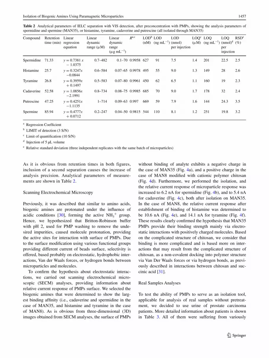
As it is obvious from retention times in both figures, inclusion of a second separation causes the increase of analysis precision. Analytical parameters of measure-ments are shown in Table 2.
Scanning Electrochemical Microscopy
Previously, it was described that similar to amino acids, biogenic amines are protonated under the influence of acidic conditions [30], forming the active NH3
+ group. Hence, we hypothesized that Britton-Robinson buffer with pH 2, used for PMP washing to remove the unde-sired impurities, caused molecule protonation, providing the active sites for interaction with surface of PMPs. Due to the surface modification using various functional groups providing different current of beads surface, selectivity is offered, based probably on electrostatic, hydrophobic inter-actions, Van der Waals forces, or hydrogen bonds between microparticles and molecules.
To confirm the hypothesis about electrostatic interac-tions, we carried out scanning electrochemical micro-scopic (SECM) analyses, providing information about relative current response of PMPs surface. We selected the biogenic amines that were determined to show the larg-est binding affinity (i.e., cadaverine and spermidine in the case of MAN35, and histamine and tyramine in the case of MAN8). As is obvious from three-dimensional (3D) images obtained from SECM analyses, the surface of PMPs
without binding of analyte exhibits a negative charge in the case of MAN35 (Fig. 4a), and a positive charge in the case of MAN8 modified with cationic polymer chitosan (Fig. 4d). Furthermore, we performed the isolation, and the relative current response of microparticle response was increased to 6.2 nA for spermidine (Fig. 4b), and to 5.4 nA for cadaverine (Fig. 4c), both after isolation on MAN35. In the case of MAN8, the relative current response after establishment of binding of histamine was determined to be 10.6 nA (Fig. 4e), and 14.1 nA for tyramine (Fig. 4f). These results clearly confirmed the hypothesis that MAN35 PMPs provide their binding strength mainly via electro-static interactions with positively charged molecules. Based on the complicated structure of chitosan, we consider that binding is more complicated and is based more on inter-actions that may result from the complicated structure of chitosan, as a non-covalent docking into polymer structure via Van Der Waals forces or via hydrogen bonds, as previ-ously described in interactions between chitosan and suc-cinic acid [31].
Real Samples Analyses
To test the ability of PMPs to serve as an isolation tool, applicable for analysis of real samples without pretreat-ment, we decided to use urine of prostate carcinoma patients. More detailed information about patients is shown in Table 3. All of them were suffering from variously
Table 2 Analytical parameters of IELC separation with VIS detection, after preconcentration with PMPs, showing the analysis parameters of spermidine and spermine (MAN35), or histamine, tyramine, cadaverine and putrescine (all isolated through MAN35)
a Regression Coefficientb LIMIT of detection (3 S/N)c Limit of quantification (10 S/N)d Injection of 5 µL volumee Relative standard deviation (three independent replicates with the same batch of microparticles)
Compound Retention time (min)
Linear regression equation
Linear dynamic range (μM)
Linear dynamic range (μg mL−1)
Ra,c LODb (nM)
LOD (ng mL−1)
LOD (nmol) per injection
LOQc (μM)
LOQ (ng mL−1)
LOQ (nmol)d per injection
RSDe (%)
Spermidine 71.33 y = 0.7381 x − 1.0375
0.7–482 0.1–70 0.9958 627 91 7.5 1.4 201 22.5 2.5
Histamine 25.7 y = 0.3247x −0.0844
0.6–584 0.07–65 0.9978 495 55 9.0 1.3 149 28 2.6
Tyramine 26.8 y = 0.3959x + 0.1497
0.5–583 0.07–80 0.9961 450 62 6.5 1.1 160 19 2.3
Cadaverine 52.58 y = 1.0856x −2.1991
0.8–734 0.08–75 0.9985 685 70 9.0 1.7 178 32 2.4
Putrescine 47.25 y = 0.4251x −1.1135
1–714 0.09–63 0.997 669 59 7.9 1.6 144 24.3 3.5
Spermine 85.94 y = 0.4777x + 0.0712
0.2–247 0.04–50 0.9815 544 110 8.1 1.2 251 19.8 3.2

1458 N. Cernei et al.
1 3
graded acinar adenocarcinoma with increased levels of PSA, a common marker of prostate carcinoma deter-mined from serum. We applied both of our PMPs directly to urine samples (250 µL), and carried out the optimized isolation steps. After incubation, the amounts of biogenic amines were determined using IEC-Vis. In all patients, histamine was determined as the most abundant biogenic amine (10.456–13.654 µg mL−1), as well as spermine and tyramine. When compared to controls as urine of healthy people, where biogenic amines were not determined, it can be concluded that these molecules offer a possibility to
enhance the diagnostic possibilities of prostate carcinoma diagnosis.
Here, we confirmed that isolation process can be performed directly in samples containing the analyte, eliminating the need for centrifugation or filtration [32]. Moreover, the pres-ence of chosen biogenic amines in tumour patients was con-firmed. Magnetic microparticles can connect two selective processes in bioanalysis: the specific binding of analytes to the magnetic particle surface based on detection, and the specific isolation of magnetic objects from complex sample mixtures [33].
a c
fed
b
4.95.05.45.65.86.06.26.46.66.87.0
4.04.55.05.56.06.57.07.58.08.7
6.26.57.07.58.08.59.09.5
10.010.210.5
9.09.5
10.010.511.011.512.012.513.013.514.014.515.0
3.84.04.24.44.65.05.25.45.65.86.0
5.15.45.86.06.46.87.07.47.88.08.28.6R
elat
ive
curr
ent r
espo
nse
(nA
)
Field of scanning (µm)
Rel
ativ
e cu
rren
t res
pons
e (n
A)
Field of scanning (µm)
Rel
ativ
e cu
rren
t res
pons
e (n
A)
Field of scanning (µm)
Rel
ativ
e cu
rren
t res
pons
e (n
A)
Field of scanning (µm)
Rel
ativ
e cu
rren
t res
pons
e (n
A)
Field of scanning (µm)
Rel
ativ
e cu
rren
t res
pons
e (n
A)
Field of scanning (µm)
-5.0 nA
-7.1 nA -5.0 nA
6.2 nA
-5.0 nA
5.4 nA
-5.0 nA
4.1 nA10.6 nA
-5.0 nA -5.0 nA
14.1 nA
Fig. 4 Scanning electrochemical microscopy 3D images charac-terizing the PMP electrochemical surface changes after binding of biogenic amines. Relative current response of PMP surface without biogenic amines bound on their surface: a for MAN35 and d for MAN8. b Changes of PMPs MAN35 surface current after binding of
spermidine. c Changes of PMPs MAN35 surface current after bind-ing of cadaverine. In case of MAN8, e changes of PMPs surface cur-rent after binding of histamine, and f surface changes after binding of tyramine
Table 3 Concentrations of biogenic amines (µg mL−1) in the urine samples of patients with cancer of prostate, determined using PMP isolation, with subsequent IELC-VIS analysis, and their diagnosis, including histology, GS (Gleason score), tumour stage and PSA levels
For isolation of histamine, tyramine, putrescine and cadaverine, we employed PMPs MAN8; and for isolation of spermine and spermidine,we employed PMPs MAN35
Age Histology GS Stage PSA (ng mL−1)
Histamine (µg mL−1)
Tyramine (µg mL−1)
Putrescine (µg mL−1)
Cadaverine (µg mL−1)
Spermidine (µg mL−1)
Spermine (µg mL−1)
55 Acinar adenocarcinoma 6(3 + 3) pT2c cN0cM0 8.89 12.615 0.365 1.242 0.282 4.397 0.398
64 Acinar adenocarcinoma (3 + 2) pT2c cN0cM0 14.6 11.214 0.258 1.254 0.731 4.417 0.480
56 Low acinar adenocarcinoma (4 + 3) pT3b cN0cM0 8.8 10.456 0.469 1.546 0.652 4.408 0.321
69 Medium acinar adenocarcinoma
6(3 + 3) pT2c cN0cM0 7.34 13.654 0.254 1.587 0.590 4.497 0.412
64 Medium acinar adenocarcinoma
2 + 3(5) pT2c cN0cM0 15.13 11.254 0.654 1.987 0.662 3.491 0.287

1459Isolation of Biogenic Amines Using Paramagnetic Microparticles
1 3
Conclusion
In this study, the design and performance of the method used for isolation and quantification of biogenic amines, namely histamine, spermidine, cadaverine, putrescine and tyramine PMPs, was described. These PMPs are able to improve biogenic amine isolation from different matrices such as urine, as was shown on the real samples obtained from patients with prostate carcinoma. We decided to include IEC as the second separation step to achieve a good resolution of analytes. Furthermore, these PMPs may serve as an effective tool for application in biosensors, or Lab-on-a-Chip platforms, where PMPs should be helpful for isolation and immobilization of biogenic amines from vari-ous matrices. Moreover, the finding that biogenic amines were not determined in the urine of healthy individuals, as opposed to prostate carcinoma patients, makes our mag-netic separation approach interesting for diagnostic pur-poses. PMPs may thus be a platform for the development of a low-cost, non-invasive and painless diagnostic approach.
Acknowledgments This work was financially supported by project MAG-AMINO (TR9140018). The authors wish to express thanks to Michal Zurek for perfect technical assistance.
Conflict of interest The authors have declared no conflict of interest.
References
1. Gosetti F, Mazzucco E, Gennaro MC, Marengo E (2013) Anal Bioanal Chem 405:907–916
2. Fadhlaoui-Zid K, Curiel JA, Landeta G, Fattouch S, Reve-ron I (2012) de las Rivas B, Sadok S, Munoz R. Food Control 25:89–95
3. Agostinelli E, Tempera G, Molinari A, Salvi M, Battaglia V, Toninello A, Arancia G (2007) Amino Acids 33:175–187
4. Pegg AE (1988) Cancer Res 48:759–774 5. Russell DH, Russell SD (1975) Clin Chem 21:860–863 6. Brunton VG, Grant MH, Wallace HM (1990) Biochem Pharma-
col 40:1893–1900
7. Min JZ, Yano H, Matsumoto A, Yu HF, Shi Q, Higashi T, Inagaki S, Toyo’oka T (2011) Clin Chim Acta 412:98–106
8. Soda K (2011) J Exp Clin Cancer Res 30:1–9 9. Kawakita M, Hiramatsu K (2006) J Biochem 139:315–322 10. Gerner EW, Meyskens FL (2004) Nat Rev Cancer 4:781–792 11. van Laarhoven AIM, Kraaimaat FW, Wilder-Smith OH, van
Riel P, van de Kerkhof PCM, Evers AWM (2013) Exp Dermatol 22:530–534
12. Nguyen L, Rigo JM, Rocher V, Belachew S, Malgrange B, Rogis-ter B, Leprince P, Moonen G (2001) Cell Tissue Res 305:187–202
13. Lucon AM, Pereira MAA, Mendonca BB, Halpern A, Wajchen-beg BL, Arap S (1997) J Urol 157:1208–1212
14. Tao ZH, Sato M, Han YL, Tan ZJ, Yamaguchi T, Nakano T (2011) Food Control 22:1154–1157
15. Tang WR, Ge SL, Gao F, Wang G, Wang QJ, He PG, Fang YZ (2013) Electrophoresis 34:2041–2048
16. Palermo C, Muscarella M, Nardiello D, Iammarino M, Centonze D (2013) Anal Bioanal Chem 405:1015–1023
17. Kim TK, Lee JI, Kim JH, Mah JH, Hwang HJ, Kim YW (2011) Food Sci Biotechnol 20:1747–1750
18. Deng YH, Zhang HS, Du XL, Wang H (2008) J Sep Sci 31:990–998
19. Liu R, Jia Y, Cheng WM, Ling JH, Liu LL, Bi KS, Li Q (2011) Talanta 83:751–756
20. Cernei N, Zitka O, Ryvolova M, Adam V, Masarik M, Hubalek J, Kizek R (2012) Int J Electrochem Sci 7:4286–4301
21. Zitka O, Cernei N, Heger Z, Matousek M, Kopel P, Kynicky J, Masarik M, Kizek R, Adam V (2013) Electrophoresis 34:2639–2647
22. Causon R (1997) J Chromatogr B 689:175–180 23. Bugianesi R, Serafini M, Simone F, Wu DY, Meydani S, Ferro-
Luzzi A, Azzini E, Maiani G (2000) Anal Biochem 284:296–300 24. Long GL, Winefordner JD (1983) Anal Chem 55:A712–A724 25. Busto O, Guasch J, Borrull F (1995) J Chromatogr A
718:309–317 26. Oguri S, Watanabe S, Abe S (1997) J Chromatogr A 790:177–183 27. Draisci R, Giannetti L, Boria P, Lucentini L, Palleschi L, Cavalli
S (1998) J Chromatogr A 798:109–116 28. Sadain SK, Koropchak JA (1999) J Chromatogr A 844:111–118 29. Kenawy ER, Abdel-Hay FI, Abou El-Magd A, Mahmoud Y
(2005) J Bioact Compat Polym 20:95–111 30. De Robertis A, De Stefano C, Gianguzza A, Sammartano S
(1998) Talanta 46:1085–1093 31. Mitra T, Sailakshmi G, Gnanamani A, Mandal AB (2013) Res-
Ibero-am. J Mater 16:755–765 32. Karamani AA, Douvalis AP, Stalikas CD (2013) J Chromatogr A
1271:1–9 33. Pamme N (2012) Curr Opin Chem Biol 16:436–443
Copyright © 2022 FDOKUMEN