Glut5 and Fructose Metabolism Inhibition in a Simulated Small ...
Vol. 143, No. 3, 1987 BIOCHEMICAL AND BIOPHYSICAL RESEARCH COMMUNICATIONS
March 30, 1987 Pages 1092-1096
ISOLATION OF A cDNA CLONE FOR RAT LIVER GPHOSPHOFRUCTO 2-KINASE/FRUCTOSE 2,6-BISPHOSPHATASE
Ann D. Colosia*. Mark Lively+, M. Raafat El-Maghrabi, and Simon J. Pilkis
*Department of Molecular Physiology & Bio h Vanderbilt University, Nashville. TN 37 lx
sits, 3
Department of Ph Schoo Y
siology and Biophysics, of Medicine,
State University of New York, Stony Brook, NY I 1794
+Department of Biochemistry, Wake Forest University. Bowman Gra School of Medicine
Winston-Salem, NC 7 103 l
Received February 12, 1987
==?. -. I * A cDNA clone for rat liver 6-phosphofructo Z-kinase/fructose 2.6bisphosphatase
was ISO. ated horn a Xgt I I rat liver expression library by antibody screening. The clone was approximately I 100 bases in len residues at the carbox
th and the derived amino acid sequence contained 303 e subunit. This derived amino acid se
exactly with the actua amino acid sequence of the enzyme determined 73 uence corresponded
of the protein. y direct sequencing
0 1987 Academic Press. Inc.
The bifunctional enzyme 6-phosphofructo 2-kinase/fructose 2.6-bisphosphatase
catalyzes both the synthesis and degradation of Fru 2.6-P, (I -3). The enzyme, a dimer with
a 55,000 dalton subunit (4). is phosphorylated by the CAMP-dependent protein kinase on a
single seryl residue per subunit with a concomitant decrease in the kinase and an increase in
the bisphosphatase activity (5). The phosphorylation state of the enzyme determines the
hepatic steady-state level of Fru 2,6-P, and ipso facto glycolytic and gluconeogenic flux (6).
Inclirect, circumstantial evidence has suggested that the enzyme’s two reactions are catalyzed
at two discrete sites on the enzyme subunit (I .2,6). For example, the bisphosphatase
reaction has been shown to proceed via a phosphoenzyme intermediate involving a histidyl
residue whereas no such intermediate has been detected for the kinase reaction (7.8) and
numerous protein modifying reagents have differential effects on the two activities. In
addition. the total activities of the enzyme in liver are also under hormonal control (9). In
order to elucidate the molecular basis for the enzyme’s activities and the regulation of its
The abbreviations used are: 6-PF2-K/F2.6-P,ase. 6-phosphofructo 2-kinase/fructose 2.6- bisphosphatase; R-u-2,6-P,, fructose-2,6-bisphosphate; IPTG, isopropylthiogalactoside .
0006-291X/87 $1.50
Copyright 0 1987 by Academic Press, Inc. AN rights of reproduction in any form reserved. 1092
Vol. 143, No. 3, 1987 BIOCHEMICAL AND BIOPHYSICAL RESEARCH COMMUNICATIONS
flanking 7 DNA and sequences within the 7 gene are sufficient
for expression of hybrid genes containing these sequences in
K562 cells (6-8). To further explore the sequences involved in
this regulation, plasmids have been constructed containing
either the 7 or B globin gene promoter linked to a neoR gene.
These genes were then transfected into both K562 and HeLa cells,
and G418-resistant clones analyzed.
METHODS
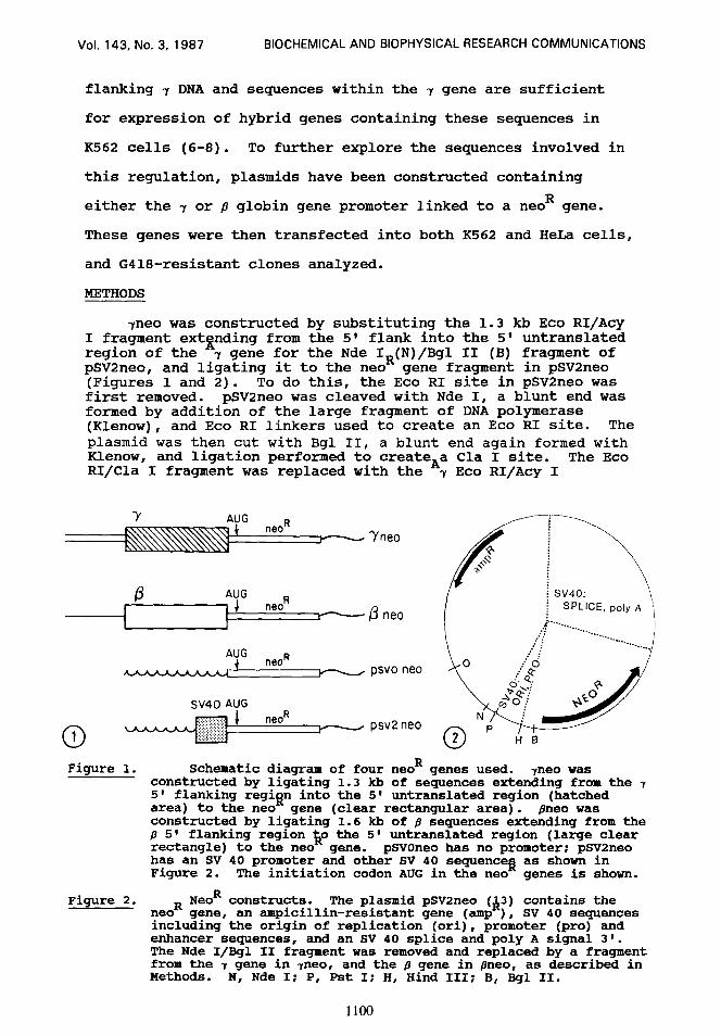
7neo was constructed by substituting the 1.3 kb Eco RI/Acy I fragment extgnding from the 5' flank into the 5' untranslated region of the 7 gene for the Nde I*(N)/Bgl II (B) fragment of pSV2neo, and ligating it to the neo gene fragment in pSV2neo (Figures 1 and 2). To do this, the Eco RI site in pSV2neo was first removed. pSV2neo was cleaved with Nde I, a blunt end was formed by addition of the large fragment of DNA polymerase (Klenow), and Eco RI linkers used to create an Eco RI site. The plasmid was then cut with Bgl II, a blunt end again formed with Klenow, and ligation performed to createAa Cla I site. The Eco RI/Cla I fragment was replaced with the 7 Eco RI/Acy I
AUG neo I?
r -Yneo
(3 AUG neoR
v /3 neo
AUG neo R
y - psvo neo
sv40 AUG neoR
0 1
r - psv2neo
Figure 1. Schematic diagram of four neoR genes used. qeo was constructed by ligating 1.3 kh of sequences extending from the 7 5' flanking regifin into the 5 * untranslated region (hatched area) to the neo gene (clear rectangular area). pneo was constructed by ligating 1.6 kb of f3 sequences extending from the fl 5' flanking region 50 the 5' untranslated region (large clear rectangle) to the neo gene. pSVOneo has no promoter: pSV2neo has an SV 40 promoter and other SV 40 sequencea as shown in Figure 2. The initiation codon ATJG in the neo genes is shown.
Figure 2. NeoR constructs. neoR gene, an ampicillin-resistant gene (amp ),
The plasmid pSV2neo (&3) contains the SV 40 sequences
including the origin of replication (ori), promoter (pro) and enhancer sequences, and an SV 40 splice and poly A signal 3'. The Nde I/Bgl II fragment was removed and replaced by a fragment from the 7 gene in vneo, and the fl gene in flneo, as described in Methods. N, Nde I; P, Pst I; H, Hind III; B, Bgl II.
1100
Vol. 143, No. 3, 1987 BIOCHEMICAL AND BIOPHYSICAL RESEARCH COMMUNICATIONS
I I
EcoRl Digest
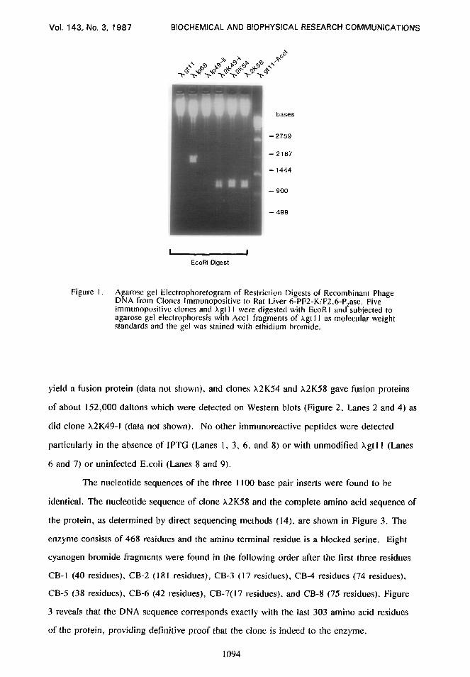
Figure I. Agarose gel Electrophoretogram of Restriction Digests of Recombinant Phage DNA from Clones lmmunopositive to Rat Liver 6-PF2-K/F2.6-P,ase. Five immunopositive clones and Xgt I I were digested with EcoR I and subjected to agarose gel electrophoresis with Act I fragments of xgt I I as molecular weight standards and the gel was stained with ethidium bromide.
yield a fusion protein (data not shown), and clones X2K54 and X2K58 gave fusion proteins
of about 152,000 daltons which were detected on Western blots (Figure 2, Lanes 2 and 4) as
did clone X2K49-I (data not shown). No other immunoreactive peptides were detected
particularly in the absence of IPTG (Lanes I, 3, 6, and 8) or with unmodified xgt I I (Lanes
6 and 7) or uninfected E.coli (Lanes 8 and 9).
The nucleotide sequences of the three I 100 base pair inserts were found to be
identical. The nucleotide sequence of clone X2K58 and the complete amino acid sequence of
the protein, as determined by direct sequencing methods (14). are shown in Figure 3. The
enzyme consists of 468 residues and the amino terminal residue is a blocked serine. Eight
cyanogen bromide fragments were founcl in the following order after the first three residues
CB-I (40 residues), CB-2 (I81 residues), CB-3 (I7 residues), CB-4 residues (74 residues),
CB-5 (38 residues), CB-6 (42 residues), CB-7( I7 residues). and CB-8 (75 residues). Figure
3 reveals that the DNA sequence corresponds exactly with the last 303 amino acid residues
of the protein, providing definitive proof that the clone is indeed to the enzyme.
1094

Vol. 143, No. 3. 1987 BIOCHEMICAL AND BIOPHYSICAL RESEARCH COMMUNICATIONS
. _ KDa
- 200
us
-116
-92
IPTG - + - +std - + - + 3456789 Lane 1 234567 89
Figure 2. SDS-polyacrylamide Gel and Western Blot of Fusion Proteins from Putative Clones of 6-PF2-K/F2.6-P,ase. Fusions proteins from clones X2K49-I and X2K58 were prepared as described under ” Materials and Methods” and subjected to electrophoreses on 6% polyacrylamide gels in SDS. IAl is the Coomassie Blue-stained gel and IB] is a Western blot. Lanes I and 2. X2K49-I: lanes 3 and 4. X2K58; lanes 6 and 7. xgt I I: lanes 8 and 9, uninfected E.coli: lane 5. protein molecular weight markers and purified 6-PF2-K/F2.6-P+se (5og in A and IOOng in B). The presence and absence of IPTG are denoted by (+) and (-). respectively.
Immediately after the COOH-terminal codon is a termination codon followed by 171 bases
of 3 ’ untranslated region.
A small number of peptide sequences from the protein have been reported
previously and can now be placed in the sequence. A thermolytic fragment containing the
CAMP-dependent phosphorylation site (VLQRRRGSSIPQ) (5) is in the CB-I fragment in the
NH,-terminal region of the subunit corresponding to residues 25-36. The carboxy terminal
residues of the enzyme have been reported to be (PAHY) (4) and these residues are found at
the carboxyl terminus by protein sequencing (M. Lively et al, manuscript submitted) and
from the deduced nucleotide sequence. The bisphosphatase active site histidyl group is
found in CB 4 at residue 256 (S. Pilkis et al. manuscript submitted). Three cysteinyl
residues. which are critical for kinase activity (14). are located at residues 158. ISI and 196
(M. R. El-Maghrabi et al, manuscript submitted). Two pyridoxal-P reactive lysyl residues
which. from the sequence presented in this manuscript. are at positions I I4 and 354 were
postulated to be at the Fru 2.6-P, and ATP binding sites of the kinase by Kitajima et al.
1095
Vol. 143, No. 3, 1987 BIOCHEMICAL AND BIOPHYSICAL RESEARCH COMMUNICATIONS
1 5 10 15 20 *S R E M G E L T Q T R L 0 K I W I P H S
21 25 30 35 40 SSSSVLQRRRGSSIPQFTNS
41 45 50 55 60 PTMVIMVGLPARGKTYISTKLT
61 65 70 75 80 R Y L N W I G T P T K V F N L G Q Y R R
81 85 90 95 100 E A V S Y R N Y E F F R P D N T E A Q L
101 105 110 115 120 I R K Q C A L A A L K D V H K Y L S R E
121 125 130 135 140 EGHVAVFDATNYTRFRRSLI
141 145 150 155 160 L Q F A K E H G Y K V F F I E S I C N D
161 165 170 175 180 PEIIAENIKQVKLGSPDYID
GCA GAA AAC ATC AAG CAA GTG AAA CTT GGT AGT CCT GAT TAC ATA GAC
181 185 190 195 200 C D Q E K V L E D F L K R I E C Y E I N
TGT GAC CAA GAA AAG GTT TTG GAA GAC TTT CTA AAG AGA ATA GAG TGC TAT GAG ATC AAC
201 205 210 215 220 YQPLDEELDSHLSYIKIFDV
TAC CAA CCT TTG GAT GAG GAA TTG GAC AGC CAC CTG TCC TAC ATC AAG AX TTC GAC GTG
221 225 230 235 240 G T R Y M V N R V Q D H V Q S R T A Y Y
GGC ACA CGC TAC ATG GTA AAT CGA GTG CAG GAC CAC GTT CAG AGC CGT ACA GCC TAC TAC
241 245 250 255 260 '2 M N I H V T P R S I Y L C R H G E S E
CT6 ATG AAC ATC CAT GTC ACA CCT CGA TCT ATC TAC CTA TGC CGC CAT GGT GAG AGT GAA
261 265 270 275 280 L N L R G R I G G D S G L S A R G K Q Y
CTC AAC CTT AGA GGC CGC ATT GGA GGT GAC TCT GGC CTC TCA GCT CGG GGC AAG CAG TAT
281 285 290 295 300 AYALANFIRSQGISSLKVWT
GCC TAT GCA CTA GCC AAC TTC ATC CGG TCT CAA GGC ATC AGC TCC CTG AAA GTA TGG ACT
301 305 310 315 320 S H M K R T I Q T A E A L G V P Y E Q W
AGC CAC ATG AAG AGG ACC ATT CAG ACC GCT GAA GCC CTA GGT GTC CCC TAT GAA CAG TGG
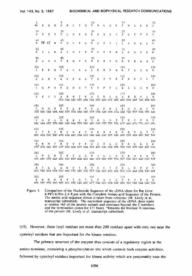
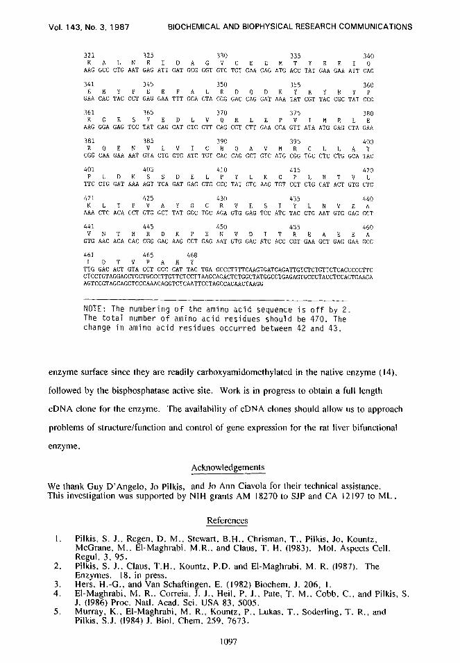
Figure 3. Comparison of the Nucleotide Sequence of the cDNA clone for Rat Liver 6-PF2-K/Fru-2,6-P,ase with the Complete Amino acid Sequence of the Protein. The amino acid sequence shown is taken from reference (M. Lively et al. manuscript submittecl). The nucleotide sequence of the cDNA clone starts at residue 165 of the protein subunit and continues beyond the C-terminus and the termination codon for 171 bases. *Denotes the blocked N-terminus of the protein (M. Lively et al, manuscript submitted).
(15). However, these lysyl residues are more than 200 residues apart with only one near the
cysteinyl residues that are important for the kinase reaction.
The primary structure of the enzyme thus consists of a regulatory region at the
amino terminus, containing a phosphorylation site which controls both enzyme activities,
followed by cysteinyl residues important for kinase activity which are presumably near the
1096
Vol. 143, No. 3, 1987 BIOCHEMICAL AND BIOPHYSICAL RESEARCH COMMUNICATIONS
321 325 330 335 340 KALNEIDAGVCEEMTYEEIQ
AAG GCC CTG AAT GAG ATT GAT GCG GGT GTC TGT GAA GAG ATG ACC TAT GAA GAA ATT CAG
341 345 350 355 360 E H Y P E E F A L R D Q D K Y R Y R Y P
GAA CAC TAC CCT GAG GAA TTT GCA CTA CGG GAC CAG GAT AAA TAT CGT TAC CGC TAT CCC:
361 365 370 375 380 KGESYEDLVQRLEPVIMELE
AAG GGA GAG TCC TAT GAG GAT CTG GTT CAG CGT CTT GAA CCA GTT ATA ATG GAG CTA GAA
381 385 390 395 400 R Q E N V L V I C H Q A V M R C L L A Y
CGG CAA GAA AAT GTA CTG GTG ATC TGT CAC CAG GCT GTC ATG CGG TGC CTC CTG GCA TAC
401 405 410 415 420 F L D K S S D E L P Y L K C P L H T V L
TTC CTG GAT AAA AGT TCA GAT GAG CTG CCC TAT CTC AAG TGT CCT CTG CAT ACT GTG CTC
421 425 430 435 440 K L T P V A Y G C R V E S I Y L N V E A
AAA CTC ACA CCT GTG GCT TAT GGC TGC AGA GTG GAG TCC ATC TAC CTG AAT GTG GAG GCT
441 445 450 455 460 V N T H R D K P E N V D I T R E A E E A
GTG AAC ACA CAC CGG GAC AAG CCT GAG AAT GTG GAC ATC ACC CGT GAA GCT GAG GAA GCC
461 465 468 L D T V P A H Y
TTG GAC ACT GTA CCT GCC CAT TAC TGA GCCCTTTTCAAGTGATCAGATTGTCTCTGTTCTCACCCCCTTC CTCCTGTAGGAGCTGCTGCCCTTGTTCTCCTTAAGCAGACTCTGGCTATGGCCTGAGAGTGCCCTACCTCCAGTGAAGA AGTCCGTAGCAGCTCCCAAACAGGTCTCAATTCCTAGCCACAACTAAGG
NOTE: The numbering of the amino acid sequence is off by 2. The total number of amino acid residues should be 470. The change in amino acid residues occurred between 42 and 43.
enzyme surface since they are readily carboxyamidomethylated in the native enzyme (14).
followed by the bisphosphatase active site. Work is in progress to obtain a full length
cDNA clone for the enzyme. The availability of cDNA clones should allow us to approach
problems of structure/function and control of gene expression for the rat liver bifunctional
enzyme.
Acknowledgements
We thank Guy D’Angelo, Jo Pilkis, and Jo Ann Ciavola for their technical assistance. This investigation was supported by NIH grants AM I8270 to SJP and CA I2 I97 to ML.
I.
2.
3. 4.
5.
References
Pilkis, S. J., Regen, D. M., Stewart. B.H.. Chrisman, T., Pilkis, Jo, Kountz, McCrane, M., El-Maghrabi. M.R., and Claus, T. H. (1983). Mol. Aspects Cell. Regul. 3, 95. Pilkis. S. J.. Claus, T.H., Kountz. P.D. and El-Maghrabi, M. R. (1987). The Enzymes. IS. in press. Hers, H.-G., and Van Schaftingen, E. (1982) Biochem. J. 206: I. El-Maghrabi. M. R.. Correia. J. J., Heil. P. J.. Pate, T. M.. Cobb. C., and Pilkis, S. J. (1986) Proc. Natl. Acad. Sci. USA 83, 5005. Murray, K., El-Maghrabi, M. R.. Kountz, P., Lukas. T., Soderling. T. R., and Pilkis, S.J. (1984) J. Biol. Chem. 259. 7673.
1097

Vol. 143, No. 3, 1987 BIOCHEMICAL AND BIOPHYSICAL RESEARCH COMMUNICATIONS
6.
7.
8.
9.
IO. I I. 12. 13.
14.
IS.
Claus, T. H., El-Maghrabi, M.R. Regen, D.M.. Stewart, H.B., McCrane. M.. Kountz, P.D., Nyfeler, F., Pilkis, J., and Pilkis, S. J. (1984). Curr. Top. Cell. Regul. 2357. Pilkis. S.J. Regen. D.M., Stewart. H.B., Pilkis, J.. Pate, T.M., and El-Maghrabi, M. R. (1984). J. Biol. Chem. 259. 949. Pilkis. S.J.. Walderhaug, M.. Murray. K., Beth. A., Venkataramu, S. D.. Pilkis, J., and El-Maghrabi. M. R. (1983). J. Biol. Chem. 258, 6135. Pilkis, S. J.. Fox. E., Wolfe. L., Rothbarth. L., Colosia, A., Stewart. H. B. and El- Maghrabi, M. R. (1986). Ann. NY. Acad. Sci. 478. I. Young, R. A., and Davis, R. W. (1983). Science 222.778. Morrison. D. K.. Carter, J. K., and Moyer. R. W. (1985). J. Virol. 55, 670. Dente, L., Cesareni. G.. and Cortese, R. (1983). Nut. Acids Res. I I. 1645. Sanger, F.. Nicklen. S. and Coulson, A. R. (1977) Proc. Natl. Acad. Sci. USA 74, 5463. El-Maghrabi, M.R.. Pate, T., Pilkis, 3.. and Pilkis, S.J. (1984). J. Biol. Chem. 259, 13104. Kitajima. S.. Thomas, H.. and Uyeda. K. (1985). J. Biol. Chem. 260, 13995.
1098
Copyright © 2022 FDOKUMEN