papillomas and cancers of the bladder, ureter, and renal - NCBI
Upload
independentCategory
view
0download
0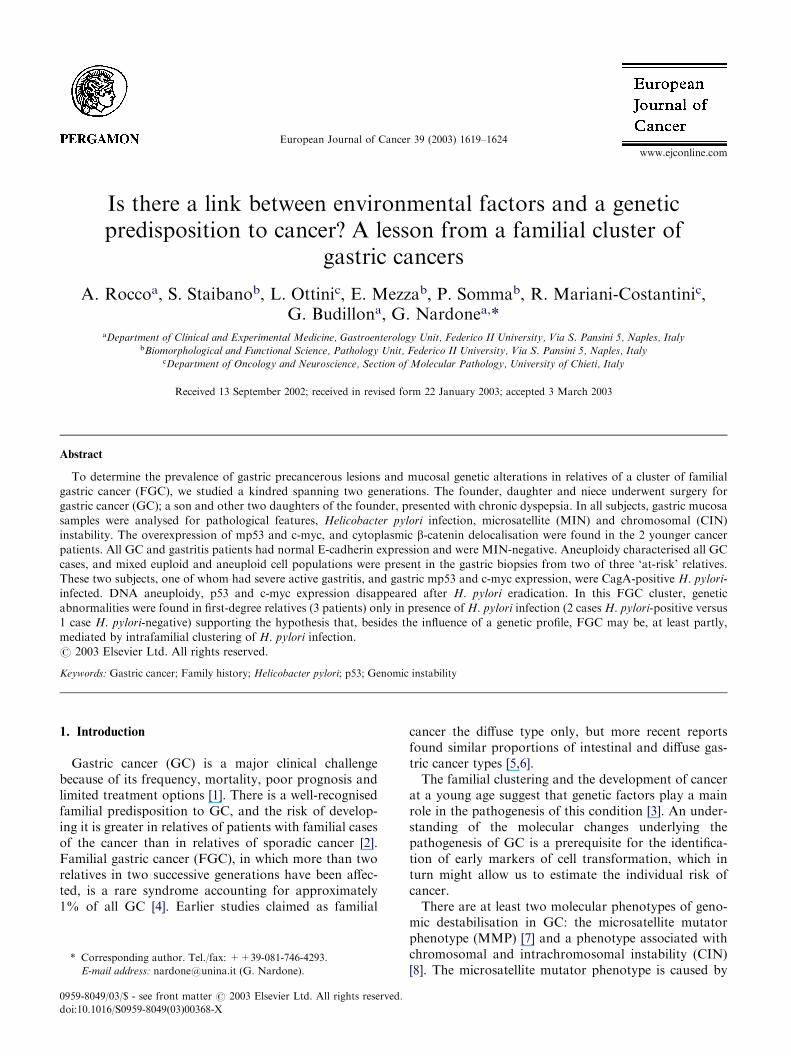
Is there a link between environmental factors and a geneticpredisposition to cancer? A lesson from a familial cluster of
gastric cancers
A. Roccoa, S. Staibanob, L. Ottinic, E. Mezzab, P. Sommab, R. Mariani-Costantinic,G. Budillona, G. Nardonea,*
aDepartment of Clinical and Experimental Medicine, Gastroenterology Unit, Federico II University, Via S. Pansini 5, Naples, ItalybBiomorphological and Functional Science, Pathology Unit, Federico II University, Via S. Pansini 5, Naples, Italy
cDepartment of Oncology and Neuroscience, Section of Molecular Pathology, University of Chieti, Italy
Received 13 September 2002; received in revised form 22 January 2003; accepted 3 March 2003
Abstract
To determine the prevalence of gastric precancerous lesions and mucosal genetic alterations in relatives of a cluster of familialgastric cancer (FGC), we studied a kindred spanning two generations. The founder, daughter and niece underwent surgery forgastric cancer (GC); a son and other two daughters of the founder, presented with chronic dyspepsia. In all subjects, gastric mucosa
samples were analysed for pathological features, Helicobacter pylori infection, microsatellite (MIN) and chromosomal (CIN)instability. The overexpression of mp53 and c-myc, and cytoplasmic b-catenin delocalisation were found in the 2 younger cancerpatients. All GC and gastritis patients had normal E-cadherin expression and were MIN-negative. Aneuploidy characterised all GC
cases, and mixed euploid and aneuploid cell populations were present in the gastric biopsies from two of three ‘at-risk’ relatives.These two subjects, one of whom had severe active gastritis, and gastric mp53 and c-myc expression, were CagA-positive H. pylori-infected. DNA aneuploidy, p53 and c-myc expression disappeared after H. pylori eradication. In this FGC cluster, genetic
abnormalities were found in first-degree relatives (3 patients) only in presence of H. pylori infection (2 cases H. pylori-positive versus1 case H. pylori-negative) supporting the hypothesis that, besides the influence of a genetic profile, FGC may be, at least partly,mediated by intrafamilial clustering of H. pylori infection.# 2003 Elsevier Ltd. All rights reserved.
Keywords: Gastric cancer; Family history; Helicobacter pylori; p53; Genomic instability
1. Introduction
Gastric cancer (GC) is a major clinical challengebecause of its frequency, mortality, poor prognosis andlimited treatment options [1]. There is a well-recognisedfamilial predisposition to GC, and the risk of develop-ing it is greater in relatives of patients with familial casesof the cancer than in relatives of sporadic cancer [2].Familial gastric cancer (FGC), in which more than tworelatives in two successive generations have been affec-ted, is a rare syndrome accounting for approximately1% of all GC [4]. Earlier studies claimed as familial
cancer the diffuse type only, but more recent reportsfound similar proportions of intestinal and diffuse gas-tric cancer types [5,6].The familial clustering and the development of cancerat a young age suggest that genetic factors play a mainrole in the pathogenesis of this condition [3]. An under-standing of the molecular changes underlying thepathogenesis of GC is a prerequisite for the identifica-tion of early markers of cell transformation, which inturn might allow us to estimate the individual risk ofcancer.There are at least two molecular phenotypes of geno-mic destabilisation in GC: the microsatellite mutatorphenotype (MMP) [7] and a phenotype associated withchromosomal and intrachromosomal instability (CIN)[8]. The microsatellite mutator phenotype is caused by
0959-8049/03/$ - see front matter # 2003 Elsevier Ltd. All rights reserved.
doi:10.1016/S0959-8049(03)00368-X
European Journal of Cancer 39 (2003) 1619–1624
www.ejconline.com
* Corresponding author. Tel./fax: ++39-081-746-4293.
E-mail address: [email protected] (G. Nardone).
mismatch repair (MMR) deficiency and is associatedwith mutational inactivation of one of at least fiveMMR genes, i.e. hMLH1, hMSH2, hMSH6, hPMS1and hPMS2 [7]. This condition, identified as micro-satellite instability (MIN), is reported in GC, particu-larly in the diffuse types, and in FGC [7,9].CIN is characterised by chromosomal rearrangementsand losses or gains of chromosomes, which in turn caninduce oncogene activation and/or tumour-suppressor-gene inactivation, thereby causing the development andprogression of GC [8,9].Mounting evidence suggests that MMP alterations,DNA aneuploidy and expression of the products ofcancer-related genes are early markers of cell transfor-mation, and may serve to identify the genetic pathwayof gastric carcinoma in patients with and without afamilial background of the disease [10,11].As yet, there are no generally agreed upon recommen-dations for relatives of FGC patients, and the relation-ship between their genetic predisposition andenvironmental factors is obscure. We have studied gastricdisease findings, mucosal genetic alterations (MIN andCIN status), and Helicobacter pylori infection in affectedand unaffected relatives from a well-characterised FGCkindred. Our data suggest an interplay between thegenetic profile of the host andH. pylori gastric infection.
2. Patients and methods
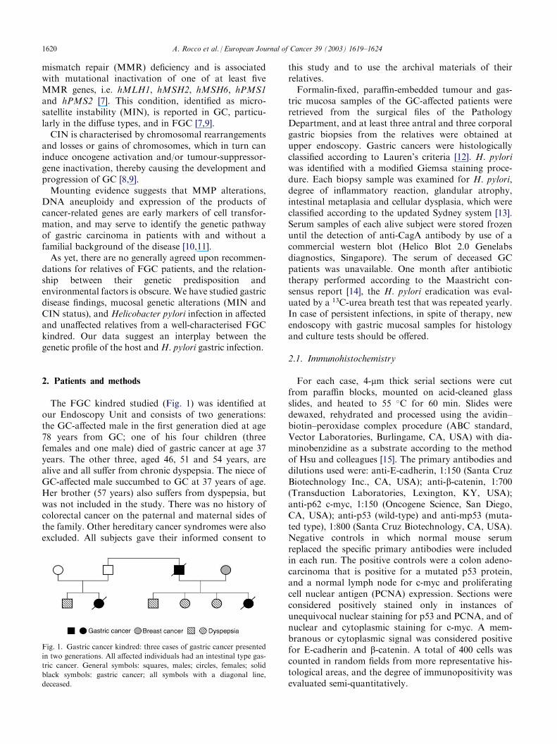
The FGC kindred studied (Fig. 1) was identified atour Endoscopy Unit and consists of two generations:the GC-affected male in the first generation died at age78 years from GC; one of his four children (threefemales and one male) died of gastric cancer at age 37years. The other three, aged 46, 51 and 54 years, arealive and all suffer from chronic dyspepsia. The niece ofGC-affected male succumbed to GC at 37 years of age.Her brother (57 years) also suffers from dyspepsia, butwas not included in the study. There was no history ofcolorectal cancer on the paternal and maternal sides ofthe family. Other hereditary cancer syndromes were alsoexcluded. All subjects gave their informed consent to
this study and to use the archival materials of theirrelatives.Formalin-fixed, paraffin-embedded tumour and gas-tric mucosa samples of the GC-affected patients wereretrieved from the surgical files of the PathologyDepartment, and at least three antral and three corporalgastric biopsies from the relatives were obtained atupper endoscopy. Gastric cancers were histologicallyclassified according to Lauren’s criteria [12]. H. pyloriwas identified with a modified Giemsa staining proce-dure. Each biopsy sample was examined for H. pylori,degree of inflammatory reaction, glandular atrophy,intestinal metaplasia and cellular dysplasia, which wereclassified according to the updated Sydney system [13].Serum samples of each alive subject were stored frozenuntil the detection of anti-CagA antibody by use of acommercial western blot (Helico Blot 2.0 Genelabsdiagnostics, Singapore). The serum of deceased GCpatients was unavailable. One month after antibiotictherapy performed according to the Maastricht con-sensus report [14], the H. pylori eradication was eval-uated by a 13C-urea breath test that was repeated yearly.In case of persistent infections, in spite of therapy, newendoscopy with gastric mucosal samples for histologyand culture tests should be offered.
2.1. Immunohistochemistry
For each case, 4-mm thick serial sections were cutfrom paraffin blocks, mounted on acid-cleaned glassslides, and heated to 55 �C for 60 min. Slides weredewaxed, rehydrated and processed using the avidin–biotin–peroxidase complex procedure (ABC standard,Vector Laboratories, Burlingame, CA, USA) with dia-minobenzidine as a substrate according to the methodof Hsu and colleagues [15]. The primary antibodies anddilutions used were: anti-E-cadherin, 1:150 (Santa CruzBiotechnology Inc., CA, USA); anti-b-catenin, 1:700(Transduction Laboratories, Lexington, KY, USA);anti-p62 c-myc, 1:150 (Oncogene Science, San Diego,CA, USA); anti-p53 (wild-type) and anti-mp53 (muta-ted type), 1:800 (Santa Cruz Biotechnology, CA, USA).Negative controls in which normal mouse serumreplaced the specific primary antibodies were includedin each run. The positive controls were a colon adeno-carcinoma that is positive for a mutated p53 protein,and a normal lymph node for c-myc and proliferatingcell nuclear antigen (PCNA) expression. Sections wereconsidered positively stained only in instances ofunequivocal nuclear staining for p53 and PCNA, and ofnuclear and cytoplasmic staining for c-myc. A mem-branous or cytoplasmic signal was considered positivefor E-cadherin and b-catenin. A total of 400 cells wascounted in random fields from more representative his-tological areas, and the degree of immunopositivity wasevaluated semi-quantitatively.
Fig. 1. Gastric cancer kindred: three cases of gastric cancer presented
in two generations. All affected individuals had an intestinal type gas-
tric cancer. General symbols: squares, males; circles, females; solid
black symbols: gastric cancer; all symbols with a diagonal line,
deceased.
1620 A. Rocco et al. / European Journal of Cancer 39 (2003) 1619–1624
2.2. DNA ploidy
We used the Feulgen (sulphuric fucsin-acid) techniquefor nuclear DNA staining of paraffin-embedded sec-tions. Briefly, sections were deparaffinised in xylene andrehydrated through decreasing ethanol concentrations.Cellular DNA was quantified with a microprocessor-controlled image analysis system (Leica-Quantimet500IW analyser, Sony 3CCD camera mod. DXC-950Pand Leica DMLB microscope). The Leica-QWINV0200A software was used for data analysis. At least400 cells from each patient were examined in non-con-secutive random fields of representative areas of thelesions. The following indexes were determined for eachmeasurement: normal diploid DNA content (2c), 2c-deviation index (2cDI), 5c exceeding events (5cEE) andDNA-malignancy grade (DNA-MG) [16].
2.3. DNA extraction and microsatellite analysis
Five-mm-thick paraffin-embedded sections were col-lected on microscope slides. Areas representative oftumour and of normal tissue (muscularis propria and/ormicroscopically normal mucosa, with no evidence ofintestinal metaplasia or dysplasia) were identified withinsingle unstained sections and microdissected into 1.5-mlpolypropylene vials, using an haematoxylin–eosin-stained step section as a guide. DNA was extracted aspreviously reported in Ref. [17]. Genomic DNA wasextracted from peripheral blood lymphocytes according
to standard procedures [17]. Microsatellite instabilitywas evaluated at the BAT 25, BAT 26 and SMAD2mononucleotide repeats using a polymerase chain reac-tion (PCR)-based assay. These three loci are consideredsensitive indicators of MIN [17]. Primers, PCR mixture,cycling conditions, electrophoretic separation and auto-radiography are described elsewhere in Ref. [17]. Pairedgenotypings of cases positive for microsatellite altera-tions were confirmed in triplicate experiments.
3. Results
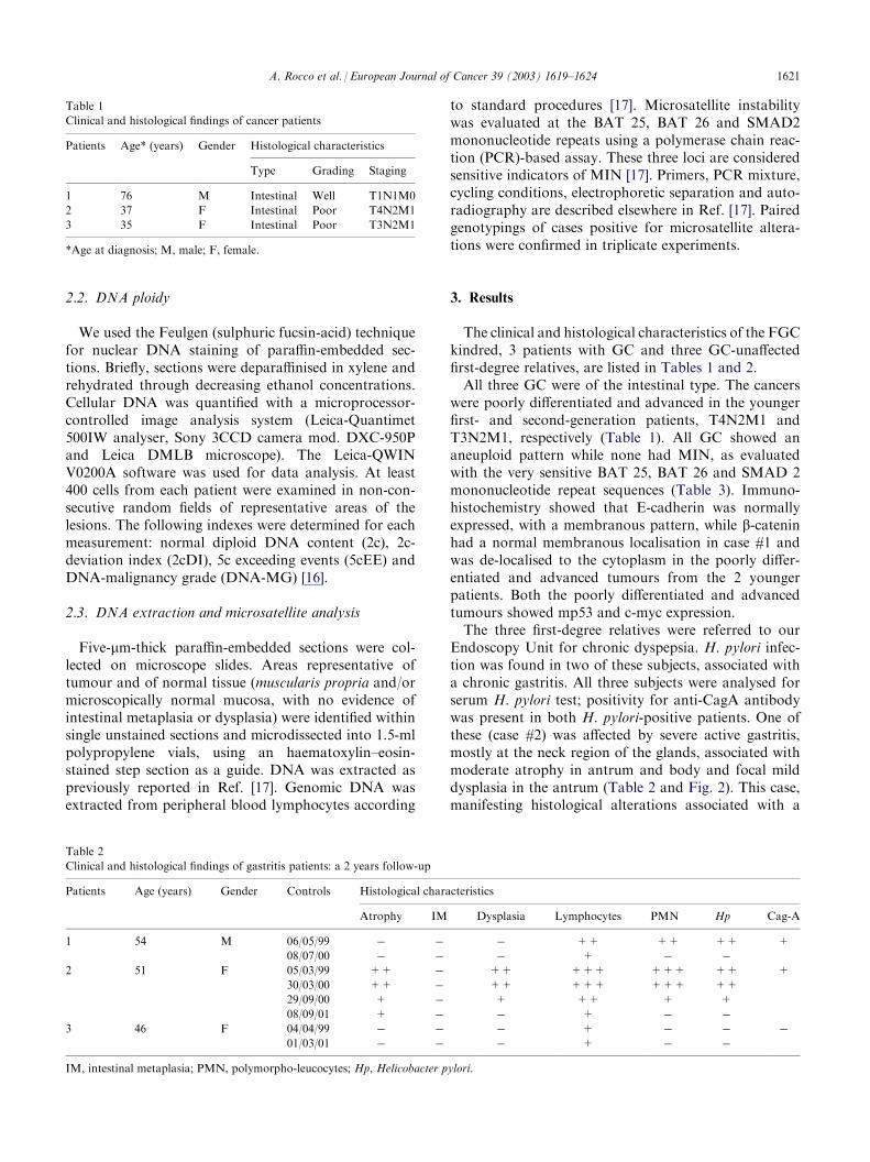
The clinical and histological characteristics of the FGCkindred, 3 patients with GC and three GC-unaffectedfirst-degree relatives, are listed in Tables 1 and 2.All three GC were of the intestinal type. The cancerswere poorly differentiated and advanced in the youngerfirst- and second-generation patients, T4N2M1 andT3N2M1, respectively (Table 1). All GC showed ananeuploid pattern while none had MIN, as evaluatedwith the very sensitive BAT 25, BAT 26 and SMAD 2mononucleotide repeat sequences (Table 3). Immuno-histochemistry showed that E-cadherin was normallyexpressed, with a membranous pattern, while b-cateninhad a normal membranous localisation in case #1 andwas de-localised to the cytoplasm in the poorly differ-entiated and advanced tumours from the 2 youngerpatients. Both the poorly differentiated and advancedtumours showed mp53 and c-myc expression.The three first-degree relatives were referred to ourEndoscopy Unit for chronic dyspepsia. H. pylori infec-tion was found in two of these subjects, associated witha chronic gastritis. All three subjects were analysed forserum H. pylori test; positivity for anti-CagA antibodywas present in both H. pylori-positive patients. One ofthese (case #2) was affected by severe active gastritis,mostly at the neck region of the glands, associated withmoderate atrophy in antrum and body and focal milddysplasia in the antrum (Table 2 and Fig. 2). This case,manifesting histological alterations associated with a
Table 2
Clinical and histological findings of gastritis patients: a 2 years follow-up
Patients
Age (years) Gender Controls Histological characteristicsAtrophy
IM Dysplasia Lymphocytes PMN Hp Cag-A1
54 M 06/05/99 � � � ++ ++ ++ +08/07/00
� � � + � �2
51 F 05/03/99 ++ � ++ +++ +++ ++ +30/03/00
++ � ++ +++ +++ ++29/09/00
+ � + ++ + +08/09/01
+ � � + � �3
46 F 04/04/99 � � � + � � �01/03/01
� � � + � �IM, intestinal metaplasia; PMN, polymorpho-leucocytes; Hp, Helicobacter pylori.
Table 1
Clinical and histological findings of cancer patients
Patients
Age* (years) Gender Histological characteristicsType
Grading Staging1
76 M Intestinal Well T1N1M02
37 F Intestinal Poor T4N2M13
35 F Intestinal Poor T3N2M1*Age at diagnosis; M, male; F, female.
A. Rocco et al. / European Journal of Cancer 39 (2003) 1619–1624 1621
cancer risk, showed an increase in cell proliferation, aswitnessed by PCNA expression, and was positive for c-myc expression and for the expression of wild and
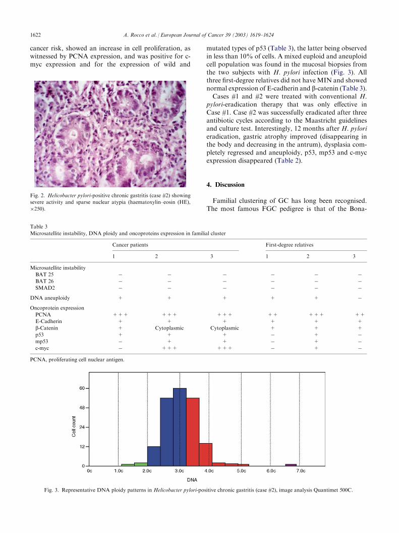
mutated types of p53 (Table 3), the latter being observedin less than 10% of cells. A mixed euploid and aneuploidcell population was found in the mucosal biopsies fromthe two subjects with H. pylori infection (Fig. 3). Allthree first-degree relatives did not have MIN and showednormal expression of E-cadherin and b-catenin (Table 3).Cases #1 and #2 were treated with conventional H.
pylori-eradication therapy that was only effective inCase #1. Case #2 was successfully eradicated after threeantibiotic cycles according to the Maastricht guidelinesand culture test. Interestingly, 12 months after H. pylorieradication, gastric atrophy improved (disappearing inthe body and decreasing in the antrum), dysplasia com-pletely regressed and aneuploidy, p53, mp53 and c-mycexpression disappeared (Table 2).
4. Discussion
Familial clustering of GC has long been recognised.The most famous FGC pedigree is that of the Bona-
Fig. 3. Representative DNA ploidy patterns in Helicobacter pylori-positive chronic gastritis (case #2), image analysis Quantimet 500C.
Table 3
Microsatellite instability, DNA ploidy and oncoproteins expression in familial cluster
Cancer patients
First-degree relatives1
2 3 1 2 3Microsatellite instability
BAT 25
� � � � � �BAT 26
� � � � � �SMAD2
� � � � � �DNA aneuploidy
+ + + + + �Oncoprotein expression
PCNA
+++ +++ +++ ++ +++ ++E-Cadherin
+ + + + + +b-Catenin
+ Cytoplasmic Cytoplasmic + + +p53
+ + + � + �mp53
� + + � + �c-myc
� +++ +++ � + �PCNA, proliferating cell nuclear antigen.
Fig. 2. Helicobacter pylori-positive chronic gastritis (case #2) showing
severe activity and sparse nuclear atypia (haematoxylin–eosin (HE),
�250).
1622 A. Rocco et al. / European Journal of Cancer 39 (2003) 1619–1624
parte family: the Emperor, two sisters, one brother, thefather and, probably, a paternal aunt died from sto-mach cancer [18]. Predictive genetic testing is an essen-tial step for the clinical management of affected families.MIN and E-cadherin are the most frequent geneticalterations associated with a family history of GC[17,19,20]. At present, the International Gastric CancerConsortium recommends testing for E-cadherin altera-tions in patients with diffuse GC and suggests prophy-lactic gastrectomy in carriers of E-cadherin germlinemutations belonging to FGC [4]. In our FGC kindred,neither GC cases nor unaffected first-degree relativeshad MIN or deregulated E-cadherin expression in gas-tric tissues (Table 3). Unfortunately, fresh blood sam-ples from the GC-affected members of the kindred werenot available for germline mutation analysis. However,it is unlikely that alterations in the E-cadherin gene areinvolved in our FGC kindred. In fact, germline muta-tions of the E-cadherin gene, responsible for hereditarydiffuse gastric cancer [4], have not been reported inintestinal type FGC kindreds, while somatic E-cadherinmutations have been frequently detected only in spora-dic diffuse-type GC [21–23].The lack of a clear genetic basis for FGC lends weightto the notion that familial aggregations might reflect notonly a hereditary predisposition, but also exposure torisk factors in the familial or community environment[24,25]. H. pylori infection is one of the most importantenvironmental risk factors associated with sporadic GC[26]. The disease risk can be further modulated byH. pylori genotypes. In Western populations, subjectswith CagA-positive strains of H. pylori have anenhanced risk of developing atrophic gastritis, pepticdiseases and GC compared with individuals with CagA-negative strains [27]. Exposure of gastric epithelial cellsto H. pylori results in the generation of reactive oxygenspecies and an increased level of inducible nitric oxidesynthase; both in turn may cause genetic alterationswhich may lead to cancer [28,29]. Alterations indicativeof a CIN phenotype during H. pylori infection are notrare. Altered expression of p53, APC, K-ras, c-myc andan aneuploidy status have been described in chronicatrophic gastritis, intestinal metaplasia and dysplasia,and in populations at high risk of GC [10,11,28–30].Huang and colleagues in a meta-analysis of 19 cohortand case–control studies found an approximately 2-foldrisk of GC among infected individuals [31], while Bren-ner and colleagues found a 16-fold risk of non-cardiacGC in patients with H. pylori infection and a familialaggregation of gastric cancer compared with uninfectedsubjects [32]. Therefore, familial aggregation of GCcould be mediated by H. pylori infection, which suggestsan interplay between H. pylori and the genetic profile ofthe host. In a Caucasian population, a DNA poly-morphism in the interleukin-1 (IL-1) gene cluster resul-ted in atrophy of the gastric corpus and an increased
risk for GC in response to H. pylori infection [33]. Theabsence of the DQA10102 allele is associated with agreater incidence of glandular atrophy and GC in aJapanese population with H. pylori infection [34].In this study, we describe, for the first time, a familialcluster of FGC in which unaffected first-degree relatives(3 cases, 2 H. pylori-positive and 1 H. pylori-negative)with chronic gastritis have CIN alterations only in pre-sence of CagA-positive H. pylori. One of these patients(case #2) was affected by severe active gastritis asso-ciated with moderate atrophy and mild dysplasia(Table 2 and Fig. 2). Initially, this patient did notrespond to three cycles of H. pylori eradication therapy.Interestingly, in this patient, CIN alterations disappeared12 months after H. pylori eradication, which indicates aclose relationship between the genetic predisposition ofthe host and H. pylori infection. Alterations of CINhave been reported by several authors in gastric mucosaof patients with H. pylori-positive chronic gastritis[10,11,28–30]. At present in FGC kindred, no data con-cerning CIN changes in relation to H. pylori status areavailable, probably because hereditaryGC is a rare disease.In conclusion, the results reported herein, even ifobtained in one FGC cluster (3 GC cases and three GC-unaffected relatives), support the hypothesis that besidesthe influence of genetic profile, hereditary GC may be,at least partly, mediated by intrafamilial clustering of H.pylori infection. Therefore, screening for and, even-tually, treatment of H. pylori infection should be offeredto reduce the risk of GC in first-degree relatives ofpatients with FGC and, taking into account the rate ofH. pylori infection recurrence, these procedures shouldbe repeated yearly. Finally, our finding provides scien-tific backing for the recommendation of the Maastricht2-2000 Consensus Report that patients with a familyhistory of GC should undergo H. pylori-eradicationtherapy [14].
Acknowledgements
We are grateful to Antonella Coppeto and EleonoraEfficie for Endoscopic assistance and to Jean AnnGilder for revising and editing the manuscript. Thisstudy has been supported by a grant from MIUR 2002(Ministero Italiano Universita e Ricerca).
References
1. Hanahan D, Weinberg RA. The hallmarks of cancer. Cell 2000,
100, 57–70.
2. La Vecchia C, Negri E, Franceschi S, Gentile A. Family history and
the risk of stomach and colorectal cancer. Cancer 1992, 70, 50–55.
3. Shinmura K, Kohno T, Takahashi M, et al. Familial gastric
cancer: clinicopathological characteristics, RER phenotype and
germline p53 and E-cadherin mutations. Carcinogenesis 1999, 20,
1127–1131.
A. Rocco et al. / European Journal of Cancer 39 (2003) 1619–1624 1623
4. Caldas C, Carneiro F, Linch HT, et al. Familial gastric cancer:
overview and guidelines for management. J Med Genet 1999, 36,
873–880.
5. Lehtola J. Family study of gastric carcinoma with special reference
to histological types. Scand J Gastroenerol 1978, 13(Suppl. 50), 3–54.
6. Palli D, Galli M, Caporaso NE, et al. Family history and risk of
stomach cancer in Italy. Cancer Epidemiol Biomark Prev 1994, 3,
15–18.
7. Furuya T, Uchiyama T, Murakami T, et al. Relationship between
chromosomal instability and intratumoral regional DNA ploidy
heterogeneity in primary gastric cancer. Clin Cancer Res 2000, 6,
2815–2820.
8. Simpson AJ, Cabalero OL, Pena SD. Microsatellite instability as
tool for the classification of gastric cancer. Trends Mol Med 2001,
7, 76–80.
9. Lengauer C, Kinzler KW, Vogelstein B. Genetic instabilities in
human cancers. Nature 1998, 396, 399–643.
10. Tahara E. Molecular mechanism of stomach carcinogenesis. J
Canc Res Clin Oncol 1993, 119, 265–272.
11. Sepulveda AR. Molecular testing of Helicobacter pylori-asso-
ciated chronic gastritis and premalignant gastric lesions: clinical
implications. J Clin Gastroenterol 2001, 32, 377–382.
12. Lauren P. The two histological main types of gastric carcinoma:
diffuse and so-called intestinal-type carcinoma. Acta Path Micro-
biol Scand 1965, 64, 31–49.
13. Dixon MF, Genta RM, Yardley JH, Correa P, the Participants in
the International Workshop on the Histopathology of Gastritis,
Houston 1994. Classification and grading of gastritis. Am J Surg
Pathol 1996, 20, 1161–1181.
14. Malfertheiner P, Megraud F, O’Morain C, et al. Current con-
cepts in the management of Helicobacter pylori infection. The
Maastricht 2-2000 Consensus Report. Aliment Pharmacol Ther
2002, 16, 167–180.
15. Hsu SM, Raine L, Faine L, Fanger H. Use of avidin-biotin-per-
oxidase complex (ABC) in immunoperoxidase technique: a com-
parison between ABC and unlabeled antibody (PAP) procedures.
J Histochem Cytochem 1981, 29, 577–580.
16. Bocking A, Adler CP, Common HH, Hilgarth M, Gransen B,
AuffermannW. Logarithm for a DNA-photocytometric diagnosis
and grading of malignancy. Anal Quant Cytol Histol 1984, 6, 1–8.
17. Ottini L, Palli D, Falchetti M, et al. Microsatellite instability in
gastric cancer is associated with tumor location and family his-
tory in a high risk population from Tuscany. Cancer Res 1997,
57, 4523–4529.
18. Sokoloff B. Predisposition to cancer in the Bonaparte family. Am
J Surg 1938, 40, 673–678.
19. Guilford P, Hopkins J, Harraway J, et al. E-cadherin germ-
line mutations in familial gastric cancer. Nature 1998, 26,
402–405.
20. Semba S, Yokozaki H, Yamamoto S, Yasui W, Tahara E.
Microsatellite instability in precancerous lesions and adenocarci-
nomas of the stomach. Cancer 1996, 77, 1620–1627.
21. Becker KF, Atkinson MJ, Reich U, et al. E-cadherin gene muta-
tions provide clues to diffuse type gastric carcinomas. Cancer Res
1994, 54, 3845–3852.
22. Muta H, Noguchi M, Kanai Y, Ochiai A, Nawata H, Hirohashi
S. E-cadherin gene mutations in signet ring cell carcinoma of the
stomach. Jpn J Cancer Res 1996, 87, 843–848.
23. Tamura G, Sakata K, Nishizuka S, et al. Inactivation of the
E-cadherin gene in primary gastric carcinomas and gastric carci-
noma cell lines. Jpn J Cancer Res 1996, 87, 1153–1159.
24. Parsonnet J. When heredity is infectious. Gastroenterology 2000,
118, 222–224.
25. EL-Omar EM, Oien K, Murray LS, et al. Increased prevalence of
precancerous changes in relatives of gastric cancer patients: crit-
ical role of H. pylori. Gastroenterology 2000, 118, 22–30.
26. Goldstone AR, Quirke P, Dixon MF. Helicobacter pylori infec-
tion and gastric cancer. J Pathol 1996, 179, 129–137.
27. Blaser MJ, Berg DE. Helicobacter pylori diversity and risk of
human disease. J Clin Inv 2001, 107, 767–773.
28. Correa P, Shiao YH. Phenotypic and genotypic events in gastric
carcinogenesis. Cancer Res 1994, 54(Suppl.), 19641–19643.
29. Kuipers EJ, Meuwissen GM. Helicobacter pylori and gastric car-
cinogenesis. Scand J Gastroenterol 1996, 31, 103–105.
30. Nardone G, Staibano S, Rocco A, et al. The effect of Helico-
bacter pylori infection and its eradication on cell proliferation,
DNA status, and oncogene expression in patients with chronic
gastritis. Gut 1999, 44, 789–799.
31. Huang JQ, Sridar S, Chen Y, Hunt RH. Meta-analysis of the
relationship between Helicobacter pylori seropositivity and gastric
cancer. Gastroenterology 1998, 114, 1169–1179.
32. Brenner H, Arndt V, Sturmer T, Stegmaier C, Ziegler H, Dhom
G. Individual and joint contribution of family history and Helico-
bacter pylori infection to the risk of gastric carcinoma. Cancer
2000, 88, 274–279.
33. El-Omar EM, Carrington M, Chow WH, et al. Interleukin-1
polymorphisms associated with increased risk of gastric cancer.
Nature 2000, 404, 398–402.
34. Azuma T, Ito S, Sato F, et al. The role of the HLA-DQA1 gene
in resistance to atrophic gastritis and gastric adenocarcinoma
induced by Helicobacter pylori infection. Cancer 1998, 82, 1013–
1018.
1624 A. Rocco et al. / European Journal of Cancer 39 (2003) 1619–1624
Copyright © 2022 FDOKUMEN