Ironing out the issues: Integrated approaches to understanding iron homeostasis in plants
10
Click here to load reader
Transcript of Ironing out the issues: Integrated approaches to understanding iron homeostasis in plants

G
P
R
Ih
RQ1
a
b
ARRAA
KFFFQ3SEPMP
C
Q2
0h
1
2
3
4
5
6
7
8
9
10
11
12
13
14
15
16
17
18
19
20
21
22
23
24
25
26
27
28
29
30
31
32
33
34
35
36
37
38
39
40
41
42
43
44
45
ARTICLE IN PRESS Model
SL 8818 1–10
Plant Science xxx (2013) xxx– xxx
Contents lists available at SciVerse ScienceDirect
Plant Science
jo ur nal home p age: www.elsev ier .com/ locate /p lantsc i
eview
roning out the issues: Integrated approaches to understanding ironomeostasis in plants
ozalynne Samiraa,1, Anna Stallmanna,1, Lynnicia N. Massenburgb, Terri A. Longa,∗
Department of Plant Biology, North Carolina State University, Raleigh, NC 27695, USADepartment of Plant Biology and Pathology, Rutgers University, New Brunswick, NJ 08901, USA
a r t i c l e i n f o
rticle history:eceived 27 February 2013eceived in revised form 7 June 2013ccepted 7 June 2013vailable online xxx
eywords:e homeostasis
a b s t r a c t
Plants initialize responses to environmental changes at all levels, from signaling to translation and beyond.Such is the case for fluctuations in the availability of iron (Fe), one of the most critical micronutrients forplants. The results of these responses are physiological and morphological changes that lead to increasediron uptake from the rhizosphere, and recycling and reallocation of Fe, which must be properly localizedwithin specific cells and cellular compartment for use. The use of reductionist approaches, in combinationwith in vivo and in situ Fe localization tools, has been able to shed light on critical signaling molecules,transcriptional regulators, transporters and other proteins involved in Fe homeostasis. Recent advances
e transporte translocationystems biologylemental analysis
in elemental distribution and speciation analysis now enable detection and measurement of Fe andother elements at resolutions never seen before. Moreover, increasing use of systems biology approachesprovide a substantially broader perspective of how Fe availability affects processes at many levels. Thisreview highlights the latest in vivo and in situ iron localization approaches and some of the recent advances
roteomicsetabolomics
redictive model
in understanding mechanisms that control Fe translocation. A broad perspective of how Fe localizationdata might one day be integrated with large-scale data to create models for Fe homeostasis is presented.
© 2013 Published by Elsevier Ireland Ltd.
ontents
1. Introduction . . . . . . . . . . . . . . . . . . . . . . . . . . . . . . . . . . . . . . . . . . . . . . . . . . . . . . . . . . . . . . . . . . . . . . . . . . . . . . . . . . . . . . . . . . . . . . . . . . . . . . . . . . . . . . . . . . . . . . . . . . . . . . . . . . . . . . . . . . 002. In vivo and in situ technologies to visualize and quantify Fe . . . . . . . . . . . . . . . . . . . . . . . . . . . . . . . . . . . . . . . . . . . . . . . . . . . . . . . . . . . . . . . . . . . . . . . . . . . . . . . . . . . . . . . . 00
2.1. Histochemical staining . . . . . . . . . . . . . . . . . . . . . . . . . . . . . . . . . . . . . . . . . . . . . . . . . . . . . . . . . . . . . . . . . . . . . . . . . . . . . . . . . . . . . . . . . . . . . . . . . . . . . . . . . . . . . . . . . . . . . . . 002.2. Transmission electron microscopy–electron spectroscopic imaging. . . . . . . . . . . . . . . . . . . . . . . . . . . . . . . . . . . . . . . . . . . . . . . . . . . . . . . . . . . . . . . . . . . . . . . . 002.3. Energy-dispersive X-ray analysis . . . . . . . . . . . . . . . . . . . . . . . . . . . . . . . . . . . . . . . . . . . . . . . . . . . . . . . . . . . . . . . . . . . . . . . . . . . . . . . . . . . . . . . . . . . . . . . . . . . . . . . . . . . . 002.4. Particle induced X-ray emission . . . . . . . . . . . . . . . . . . . . . . . . . . . . . . . . . . . . . . . . . . . . . . . . . . . . . . . . . . . . . . . . . . . . . . . . . . . . . . . . . . . . . . . . . . . . . . . . . . . . . . . . . . . . . 002.5. Synchrotron X-ray fluorescence microtomography . . . . . . . . . . . . . . . . . . . . . . . . . . . . . . . . . . . . . . . . . . . . . . . . . . . . . . . . . . . . . . . . . . . . . . . . . . . . . . . . . . . . . . . . . 002.6. RPE, a turn-on fluorescent Fe3+ sensor . . . . . . . . . . . . . . . . . . . . . . . . . . . . . . . . . . . . . . . . . . . . . . . . . . . . . . . . . . . . . . . . . . . . . . . . . . . . . . . . . . . . . . . . . . . . . . . . . . . . . . . 002.7. Secondary ionization mass spectrometry . . . . . . . . . . . . . . . . . . . . . . . . . . . . . . . . . . . . . . . . . . . . . . . . . . . . . . . . . . . . . . . . . . . . . . . . . . . . . . . . . . . . . . . . . . . . . . . . . . . . 002.8. Laser ablation inductively coupled plasma mass spectrometry . . . . . . . . . . . . . . . . . . . . . . . . . . . . . . . . . . . . . . . . . . . . . . . . . . . . . . . . . . . . . . . . . . . . . . . . . . . . . 00
3. Application of Fe localization tools: Recent advances in understanding Fe translocation. . . . . . . . . . . . . . . . . . . . . . . . . . . . . . . . . . . . . . . . . . . . . . . . . . . . . . . . . . 003.1. Vascular uptake and translocation to the shoot . . . . . . . . . . . . . . . . . . . . . . . . . . . . . . . . . . . . . . . . . . . . . . . . . . . . . . . . . . . . . . . . . . . . . . . . . . . . . . . . . . . . . . . . . . . . . 003.2. Xylem unloading and Fe translocation to seeds and beyond . . . . . . . . . . . . . . . . . . . . . . . . . . . . . . . . . . . . . . . . . . . . . . . . . . . . . . . . . . . . . . . . . . . . . . . . . . . . . . . . 003.3. Practical application of elemental analysis: A look ahead . . . . . . . . . . . . . . . . . . . . . . . . . . . . . . . . . . . . . . . . . . . . . . . . . . . . . . . . . . . . . . . . . . . . . . . . . . . . . . . . . . . 00
4. Understanding Fe homeostasis in the–omics era . . . . . . . . . . . . . . . . . . . . . . . . . . . . . . . . . . . . . . . . . . . . . . . . . . . . . . . . . . . . . . . . . . . . . . . . . . . . . . . . . . . . . . . . . . . . . . . . . . . 004.1. Building the Fe deficiency transcriptional regulatory network. . . . . . . . . . . . . . . . . . . . . . . . . . . . . . . . . . . . . . . . . . . . . . . . . . . . . . . . . . . . . . . . . . . . . . . . . . . . . . 00
Please cite this article in press as: R. Samira, et al., Ironing out the issues:
Plant Sci. (2013), http://dx.doi.org/10.1016/j.plantsci.2013.06.004
4.2. Posttranscriptional and posttranslational response to Fe deficienc4.3. Metabolic responses to Fe deficiency: Shedding new light on Fe li
5. Concluding remarks and future perspectives . . . . . . . . . . . . . . . . . . . . . . . . . . . . . .Acknowledgments . . . . . . . . . . . . . . . . . . . . . . . . . . . . . . . . . . . . . . . . . . . . . . . . . . . . . . . . .
References . . . . . . . . . . . . . . . . . . . . . . . . . . . . . . . . . . . . . . . . . . . . . . . . . . . . . . . . . . . . . . . . . .
∗ Corresponding author. Tel.: +1 919 515 0478.E-mail address: [email protected] (T.A. Long).
1 These authors contributed equally to this work.
168-9452/$ – see front matter © 2013 Published by Elsevier Ireland Ltd.ttp://dx.doi.org/10.1016/j.plantsci.2013.06.004
Integrated approaches to understanding iron homeostasis in plants,
y . . . . . . . . . . . . . . . . . . . . . . . . . . . . . . . . . . . . . . . . . . . . . . . . . . . . . . . . . . . . . . . . . . . . . . . . 00gands and respiration responses . . . . . . . . . . . . . . . . . . . . . . . . . . . . . . . . . . . . . . 00
. . . . . . . . . . . . . . . . . . . . . . . . . . . . . . . . . . . . . . . . . . . . . . . . . . . . . . . . . . . . . . . . . . . . . . . . . 00. . . . . . . . . . . . . . . . . . . . . . . . . . . . . . . . . . . . . . . . . . . . . . . . . . . . . . . . . . . . . . . . . . . . . . . . . . 00
. . . . . . . . . . . . . . . . . . . . . . . . . . . . . . . . . . . . . . . . . . . . . . . . . . . . . . . . . . . . . . . . . . . . . . . . . 00

ING
P
2 Scienc
1
tbircosipi
rioapceahimasRfu
umsraemtftiamtttccFrlwraks
2F
rio
46
47
48
49
50
51
52
53
54
55
56
57
58
59
60
61
62
63
64
65
66
67
68
69
70
71
72
73
74
75
76
77
78
79
80
81
82
83
84
85
86
87
88
89
90
91
92
93
94
95
96
97
98
99
100
101
102
103
104
105
106
107
108
109
110
111
112
113
114
115
116
117
118
119
120
121
122
123
124
125
126
127
128
129
130
131
132
133
134
135
136
137
138
139
140
141
142
143
144
145
146
147
148
149
150
151
152
153
154
155
156
157
158
159
160
161
162
ARTICLE Model
SL 8818 1–10
R. Samira et al. / Plant
. Introduction
Iron (Fe) is a critical nutrient for plants, essential for respira-ion, photosynthesis, and a number of other required functions. Feioavailability is determined, in part, by proper uptake and local-
zation throughout various organelles and cell types. Thus, theegulation of mechanisms involved in these processes is tightlyontrolled. Additionally, redox reactivity between Fe2+ and Fe3+
xidation states make Fe a useful cofactor in respiration and photo-ynthesis, yet capable of creating damaging reactive oxygen speciesf not chelated or stored. Based on these realities, the total soil orlant Fe content is not particularly informative without considering
ts more precise location and state.Until recently, most Fe homeostasis studies describe gene
egulatory processes, the activity of Fe transport mechanismsnvolved in the root, and the eventual use of Fe in specific cells andrganelles, such as leaf chloroplasts. A number of newer studiesre shedding more light on how Fe is translocation throughout thelant, and on how it is localized and reallocated between cellularompartments, cells and organs in response to Fe deficiency. How-ver, there is still much that we do not know about Fe homeostasisnd response to Fe deficiency, and current knowledge about Feomeostasis mechanisms is limited by the nature of available tools
n plant research. The ability to determine the mechanisms of Feovement (intracellular and intercellular) and use throughout
plant hinges on the ability to differentiate between various Feources and pools, and to visualize Fe localization in vivo and in situ.ecent advances in imaging techniques could provide momentum
or this field and will allow for a more broad and quantitativenderstanding of Fe pathways.
An increase in the use of systems biology approaches, or these of large-scale high-resolution transcriptomics, proteomics,etabolomics data to create predictive models [1], is also set to
ubstantially enhance our understanding of Fe homeostasis andeveal emergent properties that lead to proper localization andvailability of Fe. Today, whole genome transcriptional profilesxist for a wide range of species, for wild type and Fe homeostasisutants, for single time points, for whole roots across multiple
ime points, for specific root developmental zones, and evenor specific cell types [2–7]. Proteomic profiles for Arabidopsishaliana (Arabidopsis), tomato, cucumber, sugar beet, and Med-cago truncatula, and metabolomics profiles for a number of speciesre all revealing system-wide responses to Fe deprivation thatight even be conserved in animals [8,9]. How are plants able
o integrate these responses throughout the system? How doranscriptional, posttranscriptional and metabolic responses leado increased Fe concentration in specific tissues, cells, and cellularompartments? These and many other questions are yet to beompletely answered. However, a more holistic understanding ofe homeostasis is now emerging (Fig. 1). Excellent comprehensiveeviews of Fe homeostasis are found elsewhere [10–14]. Here, theatest in vivo and in situ Fe localization technologies are discussed
ith a focus on their current use and future utility. Additionally,ecent findings about Fe localization and what controls localizationre presented along with discussion of how we can advance thisnowledge by better integration of Fe localization tools withystems biology approaches.
. In vivo and in situ technologies to visualize and quantifye
Please cite this article in press as: R. Samira, et al., Ironing out the issues:Plant Sci. (2013), http://dx.doi.org/10.1016/j.plantsci.2013.06.004
The mechanisms that regulate physiological and molecularesponses to Fe deficiency all ultimately lead to the proper local-zation and availability of Fe within specific organs, cells andrganelles. The diverse set of pathways controlling Fe acquisition
PRESSe xxx (2013) xxx– xxx
and localization are difficult to resolve without spatial andtemporal understanding. Recent imaging advances that enablehigh-resolution detection, and, in some cases, measurement, of ele-mental concentration in vivo and in situ are presented. While adetailed review of many of these techniques is presented elsewhere[15], we describe and compare methods that have been utilizedto advance knowledge of Fe homeostasis (Table 1). Unless other-wise indicated most methods described below require the use ofinstrumentation often available, with moderate training, at manyresearch institutions.
2.1. Histochemical staining
Perls staining is one of the oldest methods for visualizing Fein plants. Briefly, in the presence of acid and potassium ferro-cyanide, acid hydrolysis releases ferric Fe from tissues, which canthen react with ferrocyanide ions, creating an insoluble, immo-bile, and visible potassium Fe–ferrocyanide complex known asPrussian blue [16]. Because of its specificity, low-cost, and sim-plicity, this method has been widely used in plant samples tovisualize ferric Fe. The use of this technique has been somewhatlimited due to poor sensitivity and penetration of ferrocyanide inhydrophobic tissues, making embryos with starchy endospermssuch as wheat grains difficult to stain [16]. Perls staining is also dif-ficult to detect in heavily pigmented plant samples, such as seedcoats [17]. Perls/diaminobenzidine (DAB) staining was recentlydeveloped to overcome these issues. In the presence of hydro-gen peroxide and DAB, the redox active Perls complex causesDAB to undergo oxidative polymerization, forming a brown col-ored pigment [16]. Implementation of tissue fixation and thinsectioning along with Perls–DAB stain can overcome the abovestated resolution and penetration problems, increasing resolu-tion and sensitivity [18]. Perls/DAB staining is also inexpensiveand does not require specialized instrumentation or expertise,making it one of the most popular in situ methods for detect-ing Fe in plants today. However, this technique does not enablequantification of elements and requires fixation for higher resolu-tion.
2.2. Transmission electron microscopy–electron spectroscopicimaging
Transmission electron microscopy (TEM) is a technique inwhich the differential interaction of a beam of electrons withan ultra-thin section of a specimen forms an image. Electronspectroscopic imaging (ESI) couples an electron energy loss spec-trometer with a conventional transmission electron microscopeto conduct quantitative imaging of the elements within thespecimen directly. Transmission electron microscopy–electronspectroscopic imaging takes advantage of the fact that when anelectron beam hits the specimen, the dwelling time, loss of energyand scattering of the beam will be different depending on theelemental composition of the specimen [19]. This method hasbeen used to create a 2-dimensional representation of organelleposition in relation to Fe dispersion [20], resulting in high res-olution in situ elemental maps. A downside to this method isthat thin sectioning and thus destructive sample manipulation isrequired.
2.3. Energy-dispersive X-ray analysis
Integrated approaches to understanding iron homeostasis in plants,
Energy-dispersive X-ray analysis (EDX or EDAX) is based onthe principle that each element has a unique atomic structureresulting in a distinct and detectible emitted X-ray spectrum whenbombarded with high-energy electron beams [21]. It has beenused to detect Fe in thin embryonic sections of Avena sativa and
163
164
165
166
167

ARTICLE IN PRESSG Model
PSL 8818 1–10
R. Samira et al. / Plant Science xxx (2013) xxx– xxx 3
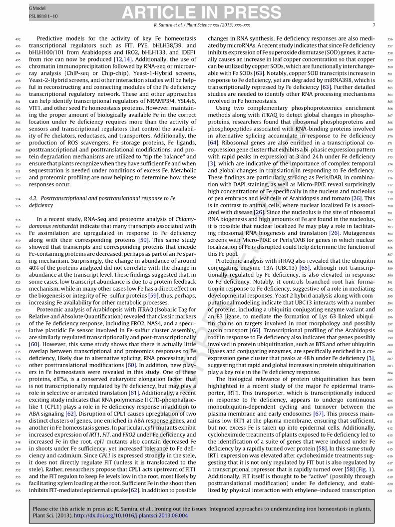
Fig. 1. Examples of how proper localization of Fe (purple) requires control at many different levels, although much is still unknown (?). Interaction with ethylene-regulatedtranscription factors Ethylene Insensitive3 (EIN3) and EIN3-LIKE1 (EIL1), and possibly ubiquitination [68,69] control FER-Like Iron Deficiency Induced Transcription Factor(FIT) stability. Iron-Regulated Transporter 1 (IRT1) is transcriptionally repressed by an unknown repressor susceptible to rapid turnover [58], yet transcriptionally activatedby FIT, which forms heterodimers with related bHLH proteins (BHLH38 and BHLH39) [56]. IRT transports Fe into the epidermal cells, but it can be post-translationallymodified by polyubiquitination and targeted for degradation in the vacuole (V) [67]. NAS4 (Nicotianamine Synthase 4) is transcriptionally repressed by PYE, which may formhetermodimers with homologs (PYE-like (PPL)) that may be degraded by Fe deficiency-responsive BRUTUS (BTS) [3]. NAS proteins synthesize nicotianamine from SAM, ap privatr
Ctpc[tb
168
169
170
171
172
173
174
roduct of the methionine cycle. Proteins involved in this cycle increases under Fe deed dots represent ubiquitination.
asuarina littoralis embryos [22]. More recently it was utilizedo detect decreased Fe–phosphate complexes in the vacuoles of
Please cite this article in press as: R. Samira, et al., Ironing out the issues:
Plant Sci. (2013), http://dx.doi.org/10.1016/j.plantsci.2013.06.004
hosphate starved Arabidopsis plants, likely due to decreased Fehelation to phosphate and subsequent transport into chloroplasts23]. Similar to transmission electron microscopy–electron spec-roscopic imaging, fixation is required for this method and Fe cane detected at subcellular resolution.
ion [60]. NA2+–Fe is transported from root to shoot [36]. Gray indicates degradation,
2.4. Particle induced X-ray emission
Integrated approaches to understanding iron homeostasis in plants,
When using particle induced X-ray emission (PIXE), elementsare exposed to ion beams (usually protons) that emit X-ray elec-tromagnetic radiation specific to the element of interest. PIXE isa non-destructive technique used to identify and quantify Fe intissues at the surface or near-surface layers [24]. Fixation of dry
175
176
177
178
179

ARTICLE IN PRESSG Model
PSL 8818 1–10
4 R. Samira et al. / Plant Science xxx (2013) xxx– xxx
Table 1Comparison of elemental analysis techniques currently utilized to detect Fe in plants. References for examples of usage are indicated.
Technique Detectionmethod/resolutiona
Advantages Disadvantages Findings
Perls/DAB Light microscopyimage/subcellular
Facile Fixation required for highestresolution
Fe distribution duringembryogenesis, mature seeds,germination, and throughoutdevelopment [18]
Does not require expensiveinstrumentation ortechnical expertise
Quantification not possible Role of many Fe homeostasisgenes
Transmission electronmicroscopy/Electronspectroscopic imaging
Electron energy lossspectra/subcellular
High resolution Requires fixation Role of NRAMP3/4 [20]Role of pH in seed ferritindisassociation [51]
Energy-dispersive X-rayanalysis
X-rays/subcellular High resolution Requires fixation Fe–phosphate complexes invacuoles [23]Fe mislocalization inchloronerva [75]
Micro-particle induced X-rayemission
X-rays/subcellular Does not require fixation ofdry samples
Wet samples must be treatedwith cryo-fixation or chemicalfixation
Fe in bean seeds [17]
3D elemental mapconstruction
Nuclear localization of Fe [26]
Synchrotron X-rayfluorescence microtomography
X-ray fluorescence/cellular Does not require fixation ofdry samples
Instrument inaccessibility andexpertise
Role of VIT1, YSL1 and YSL3 inFe localization [45,50]
3D elemental mapconstruction
Turn-on fluorescent Fe3+ sensor:RPE
Fluorophore/N/A In vivo imaging capability Not yet used in plants N/A
Secondary ionization massspectrometry
Secondary ions/cellular High sensitivity Instrument inaccessibility andexpertise
Affect of Fe plaques on Asabsorption in rice roots [34]
Laser ablation inductively coupled ICP-MS/cellular High sensitivity Sample ablated Fe detection in tobacco cells
tmbiMstPa
2
tctdqawecftseip
2
aiP
180
181
182
183
184
185
186
187
188
189
190
191
192
193
194
195
196
197
198
199
200
201
202
203
204
205
206
207
208
209
210
211
212
213
214
215
216
217
218
219
220
221
222
223
224
225
226
227
228
229
230
231
232
233
234
235
236
237
238
plasma mass spectrometry(LA–ICP-MS)
a Resolution in published plant studies.
issue is not required prior to analysis, which reduces the event ofetal leaching during fixative soak. However, a wet sample must
e treated either by cryofixation or chemical fixation to prepare formaging and maintain sample integrity [25]. Recently developed
icro-PIXE (in which tightly focused beams are utilized for micro-copic detection of elements) substantially increased resolution ofhis technique, allowing for subcellular visualization [25]. Micro-IXE was useful in imaging Fe in pea (Pisum sativum) embryos [26]nd bean embryos [17].
.5. Synchrotron X-ray fluorescence microtomography
Synchrotron X-ray fluorescence (SXRF) visualized with micro-omography relies upon the unique absorption and fluorescenceharacteristics of elements exposed to synchrotron-based X-rayso create high-resolution 3D elemental maps [27]. Elementalistribution maps can be created from image data to obtain semi-uantitative metal concentrations. This method is non-invasive,nd particularly sensitive due to the high energy, tunability andeak scattering of the synchrotron-based X-ray beams [27]. How-
ver, since the process requires use of beam time on a synchrotron,ost to travel to and use a facility can be prohibitive. Seeds are aavorable choice for X-ray analysis, based on seed stability due toheir low water content [28]. However, recent improvements inample preparation, detection speeds, and data acquisition havenabled detection of nickel and zinc in hydrated cowpea roots [29],ncreasing the likelihood of using this method to detect Fe in freshlant samples in the near future.
.6. RPE, a turn-on fluorescent Fe3+ sensor
Please cite this article in press as: R. Samira, et al., Ironing out the issues:Plant Sci. (2013), http://dx.doi.org/10.1016/j.plantsci.2013.06.004
Nonspecific fluorescent probes for ions are commercially avail-ble, and can be used to detect ions and ion movement in vivon plants [30]. For example, use of the Fe2+-sensitive fluorophore,hen Green SK, was used to show that ferrous Fe movement
[35]
into the chloroplast is due to a uniport-mechanism caused bythe membrane potential of the inner envelope [31]. Recently,a highly sensitive, selective, and reversible turn-on fluorescentFe3+ sensor was developed to detect intracellular Fe3+ levelsand sub-cellular Fe3+ pools in human live neuronal cells [32]. Inthis technique methyl 6-(3′,6′-bis(diethylamino)-3-oxospiro-[iso-indoline-1,9′-xanthen]-2-yl)picolinate (RPE) was incubated withcells for 25 min allowing visualization of the Fe3+ pool with confo-cal microscopy. One advantage of this technique is that the visiblesignal disappears when tissue is treated with an Fe chelator, allow-ing for facile study of Fe dynamics without the need for expertiseand expensive detection equipment beyond a fluorescence micro-scope. As Fe localization in plants is associated with its transitionand chelation state, the possibility of using this reagent in plant cellsfor detecting Fe3+ levels, transition, and sub-cellular concentrationis particularly exciting.
2.7. Secondary ionization mass spectrometry
Secondary ionization mass spectrometry (SIMS) is a method inwhich an accelerated primary ion beam is used to bombard a sam-ple surface, generating secondary ions that are detected by a massspectrometer [33]. SIMS is utilized primarily on hard samples, in thefields of geology or material sciences, with optimal resolving powerof 1 �m. However, the more recent development of NanoSIMStechnology by CAMECA (http://www.cameca.com/) allows resolu-tion, and sensitivity between 50 and 100 nm, enabling cellular andsubcellular resolution, high sensitivity, and high mass resolutionanalysis of samples after ultrarapid cryofixation. NanoSIMS wasrecently used to determine how Fe plaques (Fe oxide/hydroxides
Integrated approaches to understanding iron homeostasis in plants,
precipitates formed by the oxidation of ferrous Fe to ferric Fe)affect arsenic absorption in rice roots [34]. Use of this methodin the area of Fe research is hindered due to rarity of availableNanoSIMS instruments and the requirement for extensive train-ing.
239
240
241
242
243

ING
P
Scienc
2
ebsIhysdiaa
3u
sol(dfiai
3
waFaxopit(dEsxlistFoi[
taFFpmgsats
244
245
246
247
248
249
250
251
252
253
254
255
256
257
258
259
260
261
262
263
264
265
266
267
268
269
270
271
272
273
274
275
276
277
278
279
280
281
282
283
284
285
286
287
288
289
290
291
292
293
294
295
296
297
298
299
300
301
302
303
304
305
306
307
308
309
310
311
312
313
314
315
316
317
318
319
320
321
322
323
324
325
326
327
328
329
330
331
332
333
334
335
336
337
338
339
340
341
342
343
344
345
346
347
348
349
350
351
352
353
354
355
356
357
358
359
360
361
362
ARTICLE Model
SL 8818 1–10
R. Samira et al. / Plant
.8. Laser ablation inductively coupled plasma mass spectrometry
With laser ablation inductively coupled plasma mass spectrom-try (LA–ICP-MS) a sample area is scanned with a focused laseream according to a specified raster area, as low as 5 �m. Theelected area is evaporated by the laser beam and transported forCP-MS analysis by a carrier gas such as Argon [15]. This methodas been used to detect Fe and other metals in tree bark and seeds,et there are few examples of use of this method on fresh plantamples because laser ablation takes place under conditions whereehydration may occur, affecting metal distribution and sample
ntegrity [15]. Cryo-sectioning facilitates the use of this methodnd allowed researchers to produce a 2D image of Fe distributionnd other metals in fixed tobacco roots, shoots and leaves [35].
. Application of Fe localization tools: Recent advances innderstanding Fe translocation
Like most minerals, Fe is obtained primarily from the rhizo-phere, where it is absorbed first into the root epidermis. A numberf kinetic studies indicate that while in the epidermal symplast Fe isikely chelated to the nonproteinogenic amino acid nicotianamineNA) [36,37]. Fe travels symplastically through the cortex and endo-ermis, into the outer layer of the vasculature (the pericycle), andnally into xylem parenchyma cells before entering the apoplastnd being loaded into xylem cells for translocation and utilizationn the shoot.
.1. Vascular uptake and translocation to the shoot
Using an integrated mass spectrometry approach, researchersere able to determine that once loaded into the xylem from the
poplast, Fe is predominantly found as Fe3+-citrate [38]. Thus, whilee travels symplastically from the epidermis to the vasculatures Fe2+-NA, a ligand exchange is required for localization in theylem. In Arabidopsis, Ferric Reductase Defective (FRD3), a memberf the multidrug and toxin efflux family of transmembrane effluxroteins, is thought to mediate citrate efflux from the root per-
cycle and xylem parenchyma cells, while efflux of Fe itself intohe xylem is likely facilitated by the Fe transporter, Feroportin-1FPN1) [39,40]. frd3 mutants are chlorotic, exhibit constitutive Feeficiency responses, and accumulate a range of metals [41,42].legant work with Perls/DAB staining on frd3 mutants has recentlyhown striking accumulation of Fe deposits surrounding pericycle,ylem, and phloem cells, indicating that FRD3-mediated solubi-ization of Fe is required for xylem loading [39]. Once loadednto the xylem, Fe3+-citrate is translocated from the root to thehoot via the transpiration stream. Perls/DAB staining revealedhat Fe3+-citrate may be unloaded into the shoot apoplast byRD3 [39]. This is likely a conserved function as the expressionf soybean GmFRD3b was also implicated as a major contribut-ng factor to differences in Fe efficiency between soybean cultivars43].
Fe in the phloem is found primarily as Fe2+-NA [36,37]. However,he mechanism by which Fe3+-citrate is converted back to Fe2+-NAnd localized in the phloem is still an open question. Apoplastice3+-citrate is likely disassociated, allowing Fe3+ to be reduced toe2+ and then chelated to NA in the apoplast before uptake into thehloem. Analysis of quadruple nicotianamine synthase (nas4x-2)utants that do not synthesize NA revealed that lack of NA causes
Please cite this article in press as: R. Samira, et al., Ironing out the issues:
Plant Sci. (2013), http://dx.doi.org/10.1016/j.plantsci.2013.06.004
rowth inhibition and chlorosis in young leaves but not in older andenescing leaves, and that there is an increase in FRD3 expressionnd citrate content in xylem sap in nas4x-2 mutants [44]. Addi-ionally, both Perls staining of whole leaves and atomic absorptionpectroscopy of leaf extracts indicate that frd3 nas4x-2 quintuple
PRESSe xxx (2013) xxx– xxx 5
mutants contain less Fe in older and senescing leaves than nas4x-2mutants [44]. These and other data suggest that while NA operatesprimarily in young leaves, FRD3-mediated citrate loading into theapoplast facilitates Fe transport to aging leaves. Moreover, citrateand NA may play partially redundant roles in the translocation ofFe from root to shoot [44].
Members of the Yellow Stripe Like (YSL) family of transportersfacilitate the movement of the nicotianamine–Fe complex fromapoplast to symplast in the shoot following xylem unloading[45–48]. Thus, it is likely that FRD3 facilitates citrate extrusion intothe apoplast of maturing leaves, which, by an unknown mecha-nism, facilitates Fe2+-NA uptake into phloem via YSL1 and YSL3proteins, allowing for translocation into reproductive tissues. Theuse of turn-on fluorescent Fe3+ sensors, such as RPE, could begin toanswer questions about how ligand exchange between the xylemand phloem is mediated and help explain how pH or specific Fereductases play a role in ligand exchange. Moreover, further studyof the interplay between citrate and NA, using metabolic profiling,could provide new insight into how Fe is mobilized within long andshort distance pathways, and how citrate could compensate for lossof NA.
3.2. Xylem unloading and Fe translocation to seeds and beyond
Perls/DAB staining has been used to show that in addition to itsrole in Fe3+-citrate xylem loading and unloading, FRD3 also playsa role in solubilizing and loading Fe3+-citrate into pollen, devel-oping embryos, and germinating seedlings. Without FRD3, citrateexcretion is decreased, insoluble Fe deposits are found outsidedeveloping pollen, and exogenous citrate is required for properseed germination [39]. The likelihood that FRD3-mediated citrateexcretion solubilizes Fe in the apoplast for uptake into various tis-sues throughout the plant is exciting, leading to new questionsabout how Fe3+ is reduced in the apoplast, what the predomi-nant forms of Fe and Fe ligands in pollen and developing embryosare, and what transporters facilitate the movement of Fe intoembryos and pollen. For example, while grafting experiments withysl1ysl3 double mutants indicate YSL1 and YSL3 are both requiredfor seed formation, synchrotron X-ray fluorescence microtomog-raphy confirms that Fe, while not mislocalized, is decreased indouble mutants. This result prompted researchers to suggest thatYSL1/3 proteins play an essential role in the short-distance translo-cation of Fe2+-NA from cauline leaves and inflorescence stems intoseeds [45]. Notably, YSL proteins are members of the oligopeptidetransporter (OPT) family. OPT3 has been shown to play a role inlong distance movement of Fe, as Perl staining revealed substan-tial build up of Fe3+ in opt3 leaves causing necrosis [49]. Althoughseeds from opt3 mutants were viable and germinated similar towild type, Fe content was reduced and seedlings germinated in Fedeficient growth media were chlorotic. This finding suggests thatalthough OPT3 affects seed Fe uptake from senescing plants, othertransporters are involved in Fe uptake from growth media upongermination [49].
Where is Fe localized during embryo development and at seedmaturity and how is it remobilized from seed stores during germi-nation? These questions have huge implications, as remobilizationof Fe from seed stores plays a critical role in seed germinationacross species. Moreover, seeds are an important Fe source in thehuman diet. Perls/DAB staining of Arabidopsis seeds reveals Fe inall cells during early stages of embryogenesis, yet as seeds reachmaturity Fe is localized primarily in the endodermal vacuolar com-
Integrated approaches to understanding iron homeostasis in plants,
partment [16]. Studies utilizing synchrotron X-ray fluorescencemicrotomography and Perls/DAB staining indicate that VacuolarIron Transporter 1 (VIT1) facilitates Fe movement into vacuoles,and that without VIT1 Fe is still localized to vacuoles but detectedprimarily in cells external to the endodermal cells in the radical,
363
364
365
366
367

ING
P
6 Scienc
hcwtFAsRdiagPoa[
sofUfsetpiF
bgtaaipfssiiw[
3
umlccimlaatfnaFcri
368
369
370
371
372
373
374
375
376
377
378
379
380
381
382
383
384
385
386
387
388
389
390
391
392
393
394
395
396
397
398
399
400
401
402
403
404
405
406
407
408
409
410
411
412
413
414
415
416
417
418
419
420
421
422
423
424
425
426
427
428
429
430
431
432
433
434
435
436
437
438
439
440
441
442
443
444
445
446
447
448
449
450
451
452
453
454
455
456
457
458
459
460
461
462
463
464
465
466
467
468
469
470
471
472
473
474
475
476
477
478
479
480
481
482
483
484
485
ARTICLE Model
SL 8818 1–10
R. Samira et al. / Plant
ypocotyl, and cotyledons [16,50]. vit1 seeds are viable, and Feoncentration, germination, and growth are indistinguishable fromild type under normal growing conditions. However, disrup-
ion of Fe location in vit1 mutants inhibits Fe availability undere deficient conditions, affecting growth after germination [50].dditionally, use of transmission electron microscopy–electronpectroscopic imaging (TEM–ESI) on mutant seeds in which Naturalesistance Associated Macrophage Proteins (NRAMP) transporters areisrupted, namely nramp3nramp4, provided the high-resolution
mages needed to conclude that these transporters, similar to VIT1,re required to mobilize Fe from vacuolar stores to seeds duringermination, specifically under Fe deficiency [20]. Likewise, Micro-IXE along with Perls staining was used to show that, dependingn the genotype, the cells surrounding the provascular cells are
primary source of Fe during germination of Phaseolus seeds17].
In addition to the vacuoles, Fe is also stored in ferritin cages ineed amyloplast. TEM was used to compare the Fe storage capacityf ferritin in wild type and mutant pea lines in which the phyto-erritin specific domain, or “extension peptide,” was deleted [51].nlike animal ferritin, the association of phytoferritin subunits is
acilitated by low pH and this association results in long term Fetorage in plant seeds. During seed germination, the phytoferritinxtension peptide facilitates auto degradation, causing disassocia-ion and consequently release of Fe from the phytoferritin cavity inlastids [51]. Thus, Fe is released from multiple compartments dur-
ng germination although, once germinated, seedlings then acquiree from the rhizosphere.
A recent study has indicated that there are also checks andalances that prohibit excessive accumulation of free Fe duringermination. YSL4/6 may play a role in this process. Comparedo wild type, growth of ysl4ysl6 seedlings is impaired immedi-tely after germination on Fe deficient media, and YSL4/6 proteinccumulates in mature seeds yet decreases upon germination, sim-lar to the seed ferritin protein, FER2 [52]. Why? Since embryonichotosynthesis stops at seed maturity, excess Fe may be releasedrom the photosynthetic machinery by the activity of YSL4/6 andequestered by FER2 [52]. Since YSL6 is strongly expressed inenescing leaves and Fe accumulates in the chloroplast of senesc-ng leaves in ysl4/ysl6 mutants according to Perls/DAB staining,t is likely that this mechanism is mirrored in senescing leaves,
here photosynthesis decreases due to chloroplast degradation52].
.3. Practical application of elemental analysis: A look ahead
Due to ease of use, Perls/DAB staining is the most commonlysed method for cellular and subcellular detection of Fe. Since thisethod has led to significant advances in our understanding of Fe
ocalization mechanisms, why use other methods? Based on oururrent understanding of Fe homeostasis, Fe pools are dynamic,hanging rapidly in response to Fe availability. Thus, to gain morensight into how plants uptake, translocate, and reallocate Fe, Fe
ust be quantified and visualized with high accuracy at the cellu-ar and subcellular resolution. Elemental analysis techniques suchs micro-PIXE, synchrotron X-ray fluorescence microtomography,nd NanoSIMS allow for such measurements and, in some cases,he construction of 3D images of metal content (Table 1). There-ore, the challenges that lie ahead for researchers are to learn
Please cite this article in press as: R. Samira, et al., Ironing out the issues:Plant Sci. (2013), http://dx.doi.org/10.1016/j.plantsci.2013.06.004
ew methods for elemental analysis, collaborate with chemistsnd bioengineers to optimize the detection of elements such ase in plant tissues in vivo, and, finally, to work with mathemati-ians and systems biologists to create models for how the molecularesponses to Fe deficiency lead to dynamic changes in Fe movementn plants.
486
PRESSe xxx (2013) xxx– xxx
4. Understanding Fe homeostasis in the–omics era
Given that Fe is acquired through the root and localized andutilized in all cells and various cellular compartments, regulatoryresponses must be coordinated for both environmental availabilityand metabolic demands. Identification of a master Fe sensor anddetermining how this sensor triggers signaling and transcriptionalregulatory networks that lead to proteomic and metabolomicsresponses is critical for developing a greater understanding of Fehomeostasis.
4.1. Building the Fe deficiency transcriptional regulatory network
For non-graminaceous plants, specifically, Arabidopsis, com-parisons to existing Fe-binding sensor proteins in bacteria andmammals hint at the possibility of putative E3 ligase BRUTUS (BTS)fulfilling the role of an Fe sensor, although further studies are nec-essary [3]. BTS is upregulated under Fe deficiency and knockoutmutants are embryo lethal [3,53], indicating that this protein mayhave critical functions in Fe transport to seeds or in Fe homeo-stasis during embryo development. However, bts-1 knockdownmutants exhibit increased tolerance of Fe deprivation [3]. Basedon yeast-2-hybrid assays, BTS interacts with bHLH proteins closelyrelated to POPEYE, a transcriptional regulator that appears to reg-ulate (directly and indirectly) a number of known Fe homeostasisgenes. Notably, pye-1 mutants have decreased tolerance to Fe depri-vation, and altered root and shoot Fe concentrations compared towild type [3]. Since BTS encodes a protein with several potentialFe binding sites, it is tempting to speculate that BTS affects Fehomeostasis via interaction with bHLH proteins related to PYE aftersensing Fe. In rice, the transcription factor IDEF1 (IDE-BINDINGFACTOR 1) regulates Fe homeostasis genes and also binds divalentcations such as Fe [54]. Notably, transcriptional activation does notappear to act through metal binding, and IDEF1 is not transcrip-tionally responsive to Fe deficiency. IDEF1 regulates expression ofIRO2, a bHLH transcription factor that is expressed in response toFe deficiency, which, in turn, positively regulates the Fe deficiencyresponse [54]. IDEF1 may also regulate the most recently identifiedbHLH component of the Fe regulatory network in rice, OsbHLH133.OsbHLH133 is transcriptionally induced under Fe deficiency, andrepresses translocation of Fe from root to shoot [5].
In most non-graminaceous plants the bHLH transcriptionalregulator, FER-Like Iron Deficiency Induced Transcription Factor(FIT) is one of the most important regulators of the Fe deficiencyresponse. In response to Fe deprivation FIT is produced and formsheterodimers with related bHLH proteins, bHLH38 and bHLH39.These heterodimers bind to and activate the expression of FerricReduction Oxidase 2 (FRO2), thus inducing reduction of Fe3+ toFe2+ and transport of Fe2+ from the rhizosphere into Arabidopsisroot epidermal cells through Iron-Regulated Transporter 1 (IRT1)[55–57]. For this reason FIT is thought to play an essential rolein facilitating Fe uptake from the rhizosphere in response to Fedeprivation.
Recently, researchers took advantage of a glucocorticoid (GR)inducible version of FIT (FIT-GR) to identify direct FIT targets. In thisstudy, researchers applied dexamethasone (a GR ligand) to induceFIT activity, followed by cycloheximide (a translational inhibitor),which enabled researchers to identify only FIT direct target genesby preventing protein synthesis of secondary targets [58]. Whileknown targets were identified, new targets such as MYB72, and
Integrated approaches to understanding iron homeostasis in plants,
genes that are not FIT targets such as BTS and PYE were revealed.Using microarray analysis, this study also provided support forthe role of bHLH100/101 as a new and critical component of theFe deficiency regulatory network that regulates genes involved inrelocation of Fe independent of FIT [58].
487
488
489
490
491
ING
P
Scienc
tbfcrYftcVilsipptesar
4d
dFasFi4asmti
Rola[odoepirelAdaiiicisafi
492
493
494
495
496
497
498
499
500
501
502
503
504
505
506
507
508
509
510
511
512
513
514
515
516
517
518
519
520
521
522
523
524
525
526
527
528
529
530
531
532
533
534
535
536
537
538
539
540
541
542
543
544
545
546
547
548
549
550
551
552
553
554
555
556
557
558
559
560
561
562
563
564
565
566
567
568
569
570
571
572
573
574
575
576
577
578
579
580
581
582
583
584
585
586
587
588
589
590
591
592
593
594
595
596
597
598
599
600
601
602
603
604
605
606
607
608
609
610
611
612
613
614
615
616
ARTICLE Model
SL 8818 1–10
R. Samira et al. / Plant
Predictive models for the activity of key Fe homeostasisranscriptional regulators such as FIT, PYE, bHLH38/39, andHLH100/101 from Arabidopsis and IRO2, bHLH133, and IDEF1rom rice can now be produced [12,14]. Additionally, the use ofhromatin immunoprecipitation followed by RNA-seq or microar-ay analysis (ChIP-seq or Chip-chip), Yeast-1-Hybrid screens,east-2-Hybrid screens, and other interaction studies will be help-
ul in reconstructing and connecting modules of the Fe deficiencyranscriptional regulatory network. These and other approachesan help identify transcriptional regulators of NRAMP3/4, YSL4/6,IT1, and other seed Fe homeostasis proteins. However, maintain-
ng the proper amount of biologically available Fe in the correctocation under Fe deficiency requires more than the activity ofensors and transcriptional regulators that control the availabil-ty of Fe chelators, reductases, and transporters. Additionally, theroduction of ROS scavengers, Fe storage proteins, Fe ligands,osttranscriptional and posttranslational modifications, and pro-ein degradation mechanisms are utilized to “tip the balance” andnsure that plants recognize when they have sufficient Fe and whenequestration is needed under conditions of excess Fe. Metabolicnd proteomic profiling are now helping to determine how theseesponses occur.
.2. Posttranscriptional and posttranslational response to Feeficiency
In a recent study, RNA-Seq and proteome analysis of Chlamy-omonas reinhardtii indicate that many transcripts associated withe assimilation are upregulated in response to Fe deficiencylong with their corresponding proteins [59]. This same studyhowed that transcripts and corresponding proteins that encodee-containing proteins are decreased, perhaps as part of an Fe spar-ng mechanism. Surprisingly, the change in abundance of around0% of the proteins analyzed did not correlate with the change inbundance at the transcript level. These findings suggested that, inome cases, low transcript abundance is due to a protein feedbackechanism, while in many other cases low Fe has a direct effect on
he biogenesis or integrity of Fe–sulfur proteins [59], thus, perhaps,ncreasing Fe availability for other metabolic processes.
Proteomic analysis of Arabidopsis with iTRAQ (Isobaric Tag forelative and Absolute Quantification) revealed that classic markersf the Fe deficiency response, including FRO2, NAS4, and a specu-ative plastidic Fe sensor involved in Fe-sulfur cluster assembly,re similarly regulated transcriptionally and post-transcriptionally60]. However, this same study shows that there is actually littleverlap between transcriptional and proteomics responses to Feeficiency, likely due to alternative splicing, RNA processing, andther posttranslational modifications [60]. In addition, new play-rs in Fe homeostasis were revealed in this study. One of theseroteins, eIF5a, is a conserved eukaryotic elongation factor, that
s not transcriptionally regulated by Fe deficiency, but may play aole in selective or arrested translation [61]. Additionally, a recentxciting study indicates that RNA polymerase II CTD-phosphatase-ike 1 (CPL1) plays a role in Fe deficiency response in addition toBA signaling [62]. Disruption of CPL1 causes upregulation of twoistinct clusters of genes, one enriched in ABA response genes, andnother in Fe homeostasis genes. In particular, cpl1 mutants exhibitncreased expression of IRT1, FIT, and FRO2 under Fe deficiency andncreased Fe in the root. cpl1 mutants also contain decreased Fen shoots under Fe sufficiency, yet increased tolerance to Fe defi-iency and cadmium. Since CPL1 is expressed strongly in the stele,
Please cite this article in press as: R. Samira, et al., Ironing out the issues:
Plant Sci. (2013), http://dx.doi.org/10.1016/j.plantsci.2013.06.004
t does not directly regulate FIT (unless it is translocated to thetele). Rather, researchers propose that CPL1 acts upstream of FIT1nd the FIT regulon to keep Fe levels low in the root, most likely byacilitating xylem loading at the root. Sufficient Fe in the shoot thennhibits FIT-mediated epidermal uptake [62]. In addition to possible
PRESSe xxx (2013) xxx– xxx 7
changes in RNA synthesis, Fe deficiency responses are also medi-ated by microRNAs. A recent study indicates that since Fe deficiencyinhibits expression of Fe superoxide dismutase (SOD) genes, it actu-ally causes an increase in leaf copper concentration so that coppercan be utilized by copper SODs, which are functionally interchange-able with Fe SODs [63]. Notably, copper SOD transcripts increase inresponse to Fe deficiency, yet are degraded by miRNA398, which istranscriptionally repressed by Fe deficiency [63]. Further detailedstudies are needed to identify other RNA processing mechanismsinvolved in Fe homeostasis.
Using two complementary phosphoproteomics enrichmentmethods along with iTRAQ to detect global changes in phospho-proteins, researchers found that ribosomal phosphoproteins andphosphopeptides associated with RNA-binding proteins involvedin alternative splicing accumulate in response to Fe deficiency[64]. Ribosomal genes are also enriched in a transcriptional co-expression gene cluster that exhibits a bi-phasic expression patternwith rapid peaks in expression at 3 and 24 h under Fe deficiency[3], which are indicative of the importance of complex temporaland global changes in translation in responding to Fe deficiency.These findings are particularly striking as Perls/DAB, in combina-tion with DAPI staining, as well as Micro-PIXE reveal surprisinglyhigh concentrations of Fe specifically in the nucleus and nucleolusof pea embryos and leaf cells of Arabidopsis and tomato [26]. Thisis in contrast to animal cells, where nuclear localized Fe is associ-ated with disease [26]. Since the nucleolus is the site of ribosomalRNA biogenesis and high amounts of Fe are found in the nucleolus,it is possible that nuclear localized Fe may play a role in facilitat-ing ribosomal RNA biogenesis and translation [26]. Mutagenesisscreens with Micro-PIXE or Perls/DAB for genes in which nuclearlocalization of Fe is disrupted could help determine the function ofthis Fe pool.
Proteomic analysis with iTRAQ also revealed that the ubiquitinconjugating enzyme 13A (UBC13) [65], although not transcrip-tionally regulated by Fe deficiency, is also elevated in responseto Fe deficiency. Notably, it controls branched root hair forma-tion in response to Fe deficiency, suggestive of a role in mediatingdevelopmental responses. Yeast 2 hybrid analysis along with com-putational modeling indicate that UBC13 interacts with a numberof proteins, including a ubiquitin conjugating enzyme variant andan E3 ligase, to mediate the formation of Lys 63-linked ubiqui-tin chains on targets involved in root morphology and possiblyauxin transport [66]. Transcriptional profiling of the Arabidopsisroot in response to Fe deficiency also indicates that genes possiblyinvolved in protein ubiquitination, such as BTS and other ubiquitinligases and conjugating enzymes, are specifically enriched in a co-expression gene cluster that peaks at 48 h under Fe deficiency [3],suggesting that rapid and global increases in protein ubiquitinationplay a key role in the Fe deficiency response.
The biological relevance of protein ubiquitination has beenhighlighted in a recent study of the major Fe epidermal trans-porter, IRT1. This transporter, which is transcriptionally inducedin response to Fe deficiency, appears to undergo continuousmonoubiquitin-dependent cycling and turnover between theplasma membrane and early endosomes [67]. This process main-tains low IRT1 at the plasma membrane, ensuring that sufficient,but not excess Fe is taken up into epidermal cells. Additionally,cycloheximide treatments of plants exposed to Fe deficiency led tothe identification of a suite of genes that were induced under Fedeficiency by a rapidly turned over protein [58]. In this same studyIRT1 expression was elevated after cycloheximide treatments sug-
Integrated approaches to understanding iron homeostasis in plants,
gesting that it is not only regulated by FIT but is also regulated bya transcriptional repressor that is rapidly turned over [58] (Fig. 1).Additionally, FIT itself is thought to be “active” (possibly throughposttranslational modification) under Fe deficiency, and stabi-lized by physical interaction with ethylene–induced transcription
617
618
619
620
621

ING
P
8 Scienc
fLuaosdslF
mppTaSbaTarfig
4F
niaaamrtittt
aarstsor
simlrIaitssbt
622
623
624
625
626
627
628
629
630
631
632
633
634
635
636
637
638
639
640
641
642
643
644
645
646
647
648
649
650
651
652
653
654
655
656
657
658
659
660
661
662
663
664
665
666
667
668
669
670
671
672
673
674
675
676
677
678
679
680
681
682
683
684
685
686
687
688
689
690
691
692
693
694
695
696
697
698
699
700
701
702
703
704
705
706
707
708
709
710
711
712
713
714
715
716
717
718
719
720
721
722
723
724
725
726
727
728
729
730
731
732
733
734
735
736
737
738
739
740
741
742
743
744
745
ARTICLE Model
SL 8818 1–10
R. Samira et al. / Plant
actors Ethylene Insensitive3 (EIN3) and Ethylene Insensitive3-ike1 (EIL1) [68,69]. However, FIT is subject to degradation, likelypon ubiquitination, when in an “inactive” state [68]. Finally, FRO2lso appears to be a target for posttranscriptional regulation, sinceverexpression of FRO2 causes a constitutive increase in tran-cript and an increase in reductase activity is only seen under Feeficiency [70]. Thus, we now have a more comprehensive under-tanding of how transcription control, ubiquitination, membraneocalization, and vesicular trafficking act synergistically to controle uptake in epidermal cells (Fig. 1).
Using two complementary phosphoproteomic enrichmentethods along with iTRAQ to detect global changes in phospho-
roteins, specific, and sometimes unique, Fe deficient-responsivehosphopeptides were identified within 425 distinct proteins [64].wenty-one phosphorylation motifs, which could potentially acts Fe deficiency signaling molecules in roots, were also described.urprisingly, plasma membrane H+-ATPases, which had previouslyeen shown to be activated by phosphorylation [71], were notmong the phosphoproteins found to be increased in the study.his finding is particularly striking considering that before FRO2nd IRT1 can reduce and transport Fe into the root epidermis inesponse to Fe deprivation, respectively, H+-ATPases, must firstacilitate acidification of the rhizosphere, increasing Fe solubil-ty [72]. Thus, further advances in methods that accurately detectlobal posttranslational modifications are needed.
.3. Metabolic responses to Fe deficiency: Shedding new light one ligands and respiration responses
Using the iTRAQ system to identify changes in the proteome, aumber of proteins involved in the methionine cycle appear to be
ncreased under Fe deficiency [60]. S-adenosyl methionine (SAM), product of the methionine cycle, is a precursor for the Fe lig-nd, NA. NAS, an NA synthase enzyme, which is transcriptionallyctivated by Fe deficiency [73], synthesizes NA from SAM. Recentetabolic profiling of sugar beet also indicates that increases in
oot NA, among other substances, occur in response to Fe fluctua-ions [74]. Additionally, energy-dispersive X-ray analysis detectedncreased insoluble Fe inclusion particles in specific organelles ofomato chloronerva NA-free mutants [75]. These studies show thathe synthesis of NA is regulated at multiple levels to properly con-rol the availability of soluble Fe (Fig. 1).
Proteins associated with the production of phenolic compoundsre also upregulated under Fe deficiency [60]. Phenolic compoundsnd organic acids are commonly reported in root exudates inesponse to Fe deficiency, presumably to chelate ferric Fe for sub-equent uptake from the rhizosphere [76]. In a more recent studyo determine whether such mechanisms are conserved betweenpecies, researchers confirmed that the production and secretionf phenolics and flavins from Arabidopsis and Medicago truncatula,espectively, is critical for Fe uptake under Fe deficiency [77].
Metabolic profiling of xylem sap and leaf extract of tomato,ugar beet, lupine, and peach tree exposed to Fe deficiency revealedncreases in TCA cycle metabolites, specifically organic acids that
ay play a role in Fe chelation [9]. In red clover these pheno-ic compounds chelate apoplastic Fe from cell walls and facilitateeutilization of Fe, independent of rhizosphere acidification [78].n addition, phenolic effluxers play a critical role in reutilization ofpoplastic Fe from rice stele cell walls [79]. Thus, metabolic profil-ng indicates that the synthesis of Fe ligands increases in response
Please cite this article in press as: R. Samira, et al., Ironing out the issues:Plant Sci. (2013), http://dx.doi.org/10.1016/j.plantsci.2013.06.004
o Fe deprivation to facilitate the translocation of Fe from differentources. Further use of such methods on Fe homeostasis mutantsuch as frd3, fpn1 and ysl1/ysl3 could indicate on how the interplayetween NA and citrate affects Fe localization and bioavailabilityhroughout growth and development.
PRESSe xxx (2013) xxx– xxx
Global transcriptional profiling of whole Arabidopsis rootsexposed to Fe deficiency for three days revealed increased expres-sion of genes involved in mitochondrial electron transport, inaddition to the TCA cycle. Researchers also reported increasedexpression of genes involved in anaerobic respiration underFe deficiency [80,81]. Phosphoproteomic profiling reveals thatphosphorylation of key carbohydrate metabolism enzymes, includ-ing phosphoenolpyruvate carboxylase and glucose-6-phosphatedehydrogenase, and altered accumulation of phosphopeptidesassociated with invertase, provide a molecular mechanism forincreased glycolysis under Fe deficiency [64]. Likewise, metabolicprofiling of sugar beet in response to Fe deficiency and Fe resupplyreveals increases in proteins associated with glycolysis, the TCAcycle, and anaerobic respiration, along with increases in organicacids, sugars, as well as NA [74]. Since Fe is a critical componentof many mitochondrial electron transport proteins, these find-ings suggest that plant roots respond to Fe deficiency stress withincreased fermentation rates, which provide sufficient NADH tocomplete oxidative phosphorylation [80]. Additionally, researchersconcluded from the study of sugar beet that anaplerotic reactionsfix carbon in the root, which is transported to the shoot [82,83]. Amore recent metabolic study of the xylem sap and leaf extract frompeach tree, lupine and tomato, and sugar beet indicated that notonly do these anaplerotic reactions take place in roots, but shootsas well [9]. While xylem sap contains decreased levels of aminoacids and carbohydrates, leaf extracts contain more [9], suggest-ing that anaplerotic reactions, using amino acids as carbon sources,occur in Fe-deficient leaves also to maintain respiration.
These metabolic studies are particularly exciting from a systemsbiology perspective. Flux analysis of enzymatic activity and themetabolites produced from enzymes involved in metabolic pro-cesses can be used to create mathematical models that describehow enzymatic processes change under conditions of fluctuatingFe availability. Such models could also include transcriptional reg-ulatory components, signaling response, such as phosphorylationstatus, and even Fe movement rates, similar to that seen with auxinflux in roots [84].
Interestingly, many metabolic changes appear to be reflective ofcell-specific response to Fe deprivation revealed by cell-specific andgenome-wide transcriptional profiling. Although there is a core setof genes that are impervious to stress response, similar cell-specifictranscript profiling under salt stress indicated that a number genes,particularly those involved in carbohydrate metabolism, respondto different stress conditions in a cell specific manner [2]. Forexample, compared to other cell types, the cortex transcriptomeis enriched in genes associated with photosynthesis, regardless ofenvironmental conditions, and chloroplasts accumulate in light-grown cortex cells [2]. However, in Fe-deprived root cortex cellsthere is an increase in expression of these same genes yet deceasedexpression of carbohydrate transmembrane transporters, perhapsto inhibit carbohydrate translocation from the shoot to the root[2,3]. Compared to other cell types, the endodermis exhibits strik-ing decreases in the expression of genes encoding fructokinase,glucokinase, and hexokinases in response to Fe deficiency, whichmay play a role in carbohydrate metabolism as well. These findingssuggest that although Fe is primarily detected in the vasculature inroots, as it traverses from the epidermis to the vasculature someof it is captured, utilized, and stored within cells internal to theepidermis such as the cortex and endodermis for metabolism. Fetransporter Ferroportin 2 (FPN2) is thought to move Fe from thecytoplasm into the vacuole in cortex and epidermal cells under Fe
Integrated approaches to understanding iron homeostasis in plants,
deficiency, presumably in order to buffer excess Fe intake [40], butit may also play a role in metabolic requirements specific to theendodermis and cortex. Metabolic and proteomic profiles are nowavailable for specific root cells under nutrient sufficient conditions[85,86]. Querying and comparing data from these studies to similar
746
747
748
749
750
ING
P
Scienc
dsd
5
ureasiptepo
octobteartstotUtmsbeip
kcftsmtciFtmmu
atiiaana
[
[
[
[
[
[
[
[
[
[
[
[
[
[
[
[
[
[
[
751
752
753
754
755
756
757
758
759
760
761
762
763
764
765
766
767
768
769
770
771
772
773
774
775
776
777
778
779
780
781
782
783
784
785
786
787
788
789
790
791
792
793
794
795
796
797
798
799
800
801
802
803
804
805
806
807
808
809
810
811
812
813
814
815
816
817
818
819
820
821
822
823
824
825
826
827
828
829
830
831
832
833
834
835
836
837
838
839
840
841
842
843
844
845
846
847
848
849
850
851
852
853
854
855
856
857
858
859
860
861
862
863
864
865
866
867
868
869
870
871
872
873
874
875
876
877
878
879
880
881
882
883
884
885
ARTICLE Model
SL 8818 1–10
R. Samira et al. / Plant
ata obtained under Fe deficiency could allow researchers to recon-truct high-resolution transcription and metabolic maps of the Feeficiency response.
. Concluding remarks and future perspectives
Today, large-scale datasets have been produced and can besed in combination to begin to make testable and predictiveegulatory models of Fe homeostasis [12,13]. The most recent mod-ling efforts consist mainly of co-expression networks, which onlyllow researchers to predict regulatory relationships between tran-cription factors and their targets, and provide candidate genesnvolved in these networks. Exploration of small and large-scalerotein–DNA and protein–protein interaction data should increasehe strength of correlations between interactors identified in co-xpression networks. Proteome and metabolome profiling datarovide an even richer source of information about signaling andther posttranscriptional processes involved.
Moreover, recent advances in localization methods for Fe andther metals now enable researchers to link large-scale data tohanges in Fe translocation. While the most desirable detectionools would enable in vivo detection and measurement of Fe andther metals, such tools are currently lacking in the field of plantiology. Instead, histochemical methods and advanced ionic quan-ification techniques have led to the creation of high-resolutionlemental maps at specific time points, which enable visualizationnd measurement of Fe and other metals at cellular and subcellularesolution. While there are advantages and disadvantages to each ofhese methods, continued advances that increase resolution, sen-itivity, and usability are sure to contribute to rapid advances inhe field. Indeed the use of such methods has already increasedur understanding of where Fe is localized in plants and whatransporters and ligands contribute to this localization (Table 1).tilizing techniques such as synchrotron X-ray fluorescence micro-
omography or micro-PIXE on known regulatory, signaling, andetabolic mutants in combination with large-scale datasets, or for
creening mutants of genes derived from large-scale data, can helpuild more inclusive, dynamic Fe homeostasis models. Such mod-ls could then include mathematical descriptors of how changesn Fe availability affect Fe translocation and the many biologicalrocesses that require Fe.
There are a number of caveats to this approach that should beept in mind, one being that the identification of network nodesan be confounded by redundancy, making study of single mutantsutile. This could possibly be alleviated with miRNA approaches thatarget gene families. Also, more accurate predictive models requirepatial resolution due to the distinct identity of cell types. Finally,etabolic flux data, which would likely provide the most predic-
ive power for generating models for Fe homeostasis, is still quitehallenging to obtain in the area of plant biology [1,87]. However,nclusion of metabolic rates for key enzymes such as H+ ATPases,e reductases, and phosphoenolpyruvate carboxylase could lead tohe reconstruction of testable regulatory and metabolic network
odels that couple Fe uptake, translocation, and assimilation toetabolic processes such as photosynthesis and respiration, and,
ltimately, growth.Indeed, the long-term goals of many research efforts in this field
re increased growth and nutrient content of agriculturally impor-ant crops. These goals are particularly urgent due to predictedncreases in global food demand in the coming years. The increas-
Please cite this article in press as: R. Samira, et al., Ironing out the issues:
Plant Sci. (2013), http://dx.doi.org/10.1016/j.plantsci.2013.06.004
ng use of non-model species indicated in many studies above is huge step toward those goals. Additionally, although other met-ls are not featured in this review, many studies listed above doot consider Fe in isolation. This is critical, given that Fe bioavail-bility affects and is affected by homeostasis of other metals and
[
[
[
PRESSe xxx (2013) xxx– xxx 9
metabolic processes. Perhaps then, the use of systems biology isparticularly appropriate, as it will enable us to maintain a globalperspective of the side effects produced through genetic manip-ulation of Fe homeostasis. Such a perspective, along with the useof high-resolution imaging tools, makes for exciting times as wesearch for ways to affect Fe bioavailability in a sustainable way.
Acknowledgments
We thank Dr. Devarshi Selote, Dr. Jeff Gillikin, Dr. RosangelaSozzani for critical review of the manuscript.
References
[1] G.W. Bassel, et al., Systems analysis of plant functional, transcriptional, physicalinteraction, and metabolic networks, Plant Cell 24 (2012) 3859–3875.
[2] J.R. Dinneny, et al., Cell identity mediates the response of Arabidopsis roots toabiotic stress, Science 320 (2008) 942–945.
[3] T.A. Long, et al., The bHLH transcription factor POPEYE regulates response toiron deficiency in Arabidopsis roots, Plant Cell 22 (2010) 2219–2236.
[4] E.P. Colangelo, M.L. Guerinot, The essential basic helix–loop–helix protein FIT1is required for the iron deficiency response, Plant Cell 16 (2004) 3400–3412.
[5] L. Wang, et al., Identification of OsbHLH133 as a regulator of iron distributionbetween roots and shoots in Oryza sativa, Plant Cell Environ. 36 (2013) 224–236.
[6] M. Schuler, et al., Transcriptome analysis by GeneTrail revealed regulation offunctional categories in response to alterations of iron homeostasis in Ara-bidopsis thaliana, BMC Plant Biol. 11 (2011) 87.
[7] A. Zamboni, et al., Genome-wide microarray analysis of tomato roots showeddefined responses to iron deficiency, BMC Genom 13 (2012) 101.
[8] G. Vigani, Does a similar metabolic reprogramming occur in Fe-deficient plantcells and animal tumor cells? Front. Plant Sci. 3 (2012) 47.
[9] R. Rellan-Alvarez, et al., Metabolite profile changes in xylem sap and leafextracts of strategy I plants in response to iron deficiency and resupply, Front.Plant Sci. 2 (2011) 66.
10] S.S. Conte, E.L. Walker, Transporters contributing to iron trafficking in plants,Mol Plant 4 (2011) 464–476.
11] M.N. Hindt, M.L. Guerinot, Getting a sense for signals: regulation of the plantiron deficiency response, Biochim. Biophys. Acta 1823 (2012) 1521–1530.
12] W. Schmidt, T.J. Buckhout, A hitchhiker’s guide to the Arabidopsis ferrome,Plant Physiol. Biochem. 49 (2011) 462–470.
13] R. Ivanov, T. Brumbarova, P. Bauer, Fitting into the harsh reality: regulation ofiron-deficiency responses in dicotyledonous plants, Mol Plant 5 (2012) 27–42.
14] T. Kobayashi, N.K. Nishizawa, Iron uptake, translocation, and regulation inhigher plants, Annu. Rev. Plant Biol. 63 (2012) 131–152.
15] B. Wu, J.S. Becker, Imaging techniques for elements and element species inplant science, Metall: Integr Biometal Sci 4 (2012) 403–416.
16] H. Roschzttardtz, G. Conejero, C. Curie, S. Mari, Identification of the endoder-mal vacuole as the iron storage compartment in the Arabidopsis embryo, PlantPhysiol. 151 (2009) 1329–1338.
17] C. Cvitanich, et al., Iron and ferritin accumulate in separate cellular locations inPhaseolus seeds, BMC Plant Biol. 10 (2010) 26.
18] H. Roschzttardtz, G. Conejero, C. Curie, S. Mari, Straightforward histochemicalstaining of Fe by the adaptation of an old-school technique: identification ofthe endodermal vacuole as the site of Fe storage in Arabidopsis embryos, PlantSignal. Behav. 5 (2010) 56–57.
19] U. Lutz-Meindl, Use of energy filtering transmission electron microscopy forimage generation and element analysis in plant organisms, Micron 38 (2007)181–196.
20] V. Lanquar, et al., Mobilization of vacuolar iron by AtNRAMP3 and AtNRAMP4is essential for seed germination on low iron, EMBO J. 24 (2005) 4041–4051.
21] M.J. Dykstra, L.E. Reuss, Biological Electron Microscopy Theory: Techiques andTroublesh, Kluwer Acad./Plenum Publ., New York, 2003.
22] M.S. Buttrose, Manganese and iron in globoid crystals of protein bodies fromAvena and Casuarina, Aust J Plant Physiol 5 (1978) 631–639.
23] J. Hirsch, et al., Phosphate deficiency promotes modification of iron distributionin Arabidopsis plants, Biochimie 88 (2006) 1767–1771.
24] W.J. Przybyłowicz, et al., Micro-PIXE in ecophysiology, X-ray Spectrom. 34(2005) 285–289.
25] J.M. Przybyłowicz, W.J. Przybyłowicz, Micro-PIXE in plant sciences: presentstatus and perspectives, Nucl Instrum Methods Phys Res B 189 (2002) 470–481.
26] H. Roschzttardtz, et al., Plant cell nucleolus as a hot spot for iron, J. Biol. Chem.286 (2011) 27863–27866.
27] E. Lombi, J. Susini, Synchrotron-based techniques for plant and soil science:opportunities, challenges and future perspectives, Plant Soil 320 (2009) 1–35.
28] M.D. de Jonge, S. Vogt, Hard X-ray fluorescence tomography: an emerging toolfor structural visualization, Curr. Opin. Struct. Biol. 20 (2010) 606–614.
Integrated approaches to understanding iron homeostasis in plants,
29] E. Lombi, et al., Fast X-ray fluorescence microtomography of hydrated biologicalsamples, PLoS One 6 (2011) e20626.
30] M. Morel, et al., AtHMA3, a P1B-ATPase allowing Cd/Zn/Co/Pb vacuolar storagein Arabidopsis, Plant Physiol. 149 (2009) 894–904.
31] R. Shingles, M. North, R.E. McCarty, Ferrous ion transport across chloroplastinner envelope membranes, Plant Physiol. 128 (2002) 1022–1030.
886
887
888
889
890
891

ING
P
1 Scienc
[
[
[
[
[
[
[
[
[
[
[
[
[
[
[
[
[
[
[
[
[
[
[
[
[
[
[
[
[
[
[
[
[
[
[Q4
[
[
[
[
[
[
[
[
[
[
[
Q5[
[
[
[
[
[
[
892
893
894
895
896
897
898
899
900
901
902
903
904
905
906
907
908
909
910
911
912
913
914
915
916
917
918
919
920
921
922
923
924
925
926
927
928
929
930
931
932
933
934
935
936
937
938
939
940
941
942
943
944
945
946
947
948
949
950
951
952
953
954
955
956
957
958
959
960
961
962
963
964
965
966
967
968
969
970
971
972
973
974
975
976
977
978
979
980
981
982
983
984
985
986
987
988
989
990
991
992
993
994
995
996
997
998
999
1000
1001
1002
1003
1004
1005
1006
1007
1008
1009
1010
1011
1012
1013
1014
1015
1016
1017
1018
1019
1020
1021
1022
1023
1024
1025
1026
1027
1028
1029
1030
1031
1032
1033
1034
1035
1036
1037
1038
ARTICLE Model
SL 8818 1–10
0 R. Samira et al. / Plant
32] Y. Wei, Z. Aydin, Y. Zhang, Z. Liu, M. Guo, A turn-on fluorescent sensor for imag-ing labile Fe(3+) in live neuronal cells at subcellular resolution, Chembiochem13 (2012) 1569–1573.
33] S.G. Boxer, M.L. Kraft, P.K. Weber, Advances in imaging secondary ion massspectrometry for biological samples, Ann Rev Biophys 38 (2009) 53–74.
34] K.L. Moore, et al., High-resolution secondary ion mass spectrometry reveals thecontrasting subcellular distribution of arsenic and silicon in rice roots, PlantPhysiol. 156 (2011) 913–924.
35] J.S. Becker, R.C. Dietrich, A. Matusch, D. Pozebon, V.L. Dressier, Quantitativeimages of metals in plant tissues measured by laser ablation inductively cou-pled plasma mass spectrometry, Spectrochim Acta B Atom Spectrosc 63 (2008)1248–1252.
36] C. Curie, et al., Metal movement within the plant: contribution of nicotianamineand yellow stripe 1-like transporters, Ann Bot 103 (2008) 1–11.
37] R. Rellan-Alvarez, J. Abadia, A. Alvarez-Fernandez, Formation of metal-nicotianamine complexes as affected by pH, ligand exchange with citrate andmetal exchange. A study by electrospray ionization time-of-flight mass spec-trometry, Rapid Commun. Mass Spectrom. 22 (2008) 1553–1562.
38] R. Rellan-Alvarez, et al., Identification of a tri-iron(III), tri-citrate complex inthe xylem sap of iron-deficient tomato resupplied with iron: new insights intoplant iron long-distance transport, Plant Cell Physiol. 51 (2010) 91–102.
39] H. Roschzttardtz, M. Seguela-Arnaud, J.F. Briat, G. Vert, C. Curie, The FRD3 citrateeffluxer promotes iron nutrition between symplastically disconnected tissuesthroughout Arabidopsis development, Plant Cell 23 (2011) 2725–2737.
40] J. Morrissey, et al., The ferroportin metal efflux proteins function in iron andcobalt homeostasis in Arabidopsis, Plant Cell 21 (2009) 3326–3338.
41] E.E. Rogers, M.L. Guerinot, FRD3 a member of the multidrug and toxin effluxfamily, controls iron deficiency responses in Arabidopsis, Plant Cell 14 (2002)1787–1799.
42] E. Delhaize, A metal-accumulator mutant of Arabidopsis thaliana, Plant Physiol.111 (1996) 849–855.
43] E.E. Rogers, X. Wu, G. Stacey, H.T. Nguyen, Two MATE proteins play a role iniron efficiency in soybean, J. Plant Physiol. 166 (2009) 1453–1459.
44] M. Schuler, R. Rellan-Alvarez, C. Fink-Straube, J. Abadia, P. Bauer, Nicotianaminefunctions in the Phloem-based transport of iron to sink organs, in pollen devel-opment and pollen tube growth in Arabidopsis, Plant Cell 24 (2012) 2380–2400.
45] H.H. Chu, et al., Successful reproduction requires the function of ArabidopsisYellow Stripe-Like1 and Yellow Stripe-Like3 metal-nicotianamine transportersin both vegetative and reproductive structures, Plant Physiol. 154 (2010)197–210.
46] R.J. DiDonato, L.A. Roberts Jr., T. Sanderson, R.B. Eisley, E.L. Walker, Arabidop-sis Yellow Stripe-Like2 (YSL2): a metal-regulated gene encoding a plasmamembrane transporter of nicotianamine-metal complexes, Plant J. 39 (2004)403–414.
47] M. Le Jean, A. Schikora, S. Mari, J.F. Briat, C. Curie, A loss-of-function mutationin AtYSL1 reveals its role in iron and nicotianamine seed loading, Plant J. 44(2005) 769–782.
48] B.M. Waters, et al., Mutations in Arabidopsis yellow stripe-like1 and yellowstripe-like3 reveal their roles in metal ion homeostasis and loading of metalions in seeds, Plant Physiol. 141 (2006) 1446–1458.
49] M.G. Stacey, et al., The Arabidopsis AtOPT3 protein functions in metal homeo-stasis and movement of iron to developing seeds, Plant Physiol. 146 (2008)589–601.
50] S.A. Kim, et al., Localization of iron in Arabidopsis seed requires the vacuolarmembrane transporter VIT1, Science 314 (2006) 1295–1298.
51] H. Yang, et al., Protein association and dissociation regulated by extension pep-tide: a mode for iron control by phytoferritin in seeds, Plant Physiol. 154 (2010)1481–1491.
52] F. Divol, et al., The Arabidopsis Yellow Stripe Like4 and 6 transporters controliron release from the chloroplast, Plant Cell 25 (2013) 1040–1055.
53] J. McElver, et al., Insertional mutagenesis of genes required for seed develop-ment in Arabidopsis thaliana, Genetics 159 (2001) 1751–1763.
54] T. Kobayashi, et al., The rice transcription factor IDEF1 directly binds to iron andother divalent metals for sensing cellular iron status, Plant J. 69 (2012) 81–91.
55] G. Vert, et al., IRT1, an Arabidopsis transporter essential for iron uptake fromthe soil and for plant growth, Plant Cell 14 (2002) 1223–1233.
56] Y. Yuan, et al., FIT interacts with AtbHLH38 and AtbHLH39 in regulating ironuptake gene expression for iron homeostasis in Arabidopsis, Cell Res. 18 (2008)385–397.
57] N.J. Robinson, C.M. Procter, E.L. Connolly, M.L. Guerinot, A ferric-chelate reduc-tase for iron uptake from soils, Nature 397 (1999) 694–697.
Please cite this article in press as: R. Samira, et al., Ironing out the issues:Plant Sci. (2013), http://dx.doi.org/10.1016/j.plantsci.2013.06.004
58] A.B. Sivitz, V. Hermand, C. Curie, G. Vert, Arabidopsis bHLH100 and bHLH101control iron homeostasis via a FIT-independent pathway, PLoS One 7 (2012)e44843.
59] E.I. Urzica, et al., Systems and trans-system level analysis identifies con-served iron deficiency responses in the plant lineage, Plant Cell 24 (2012)3921–3948.
[
[
[
PRESSe xxx (2013) xxx– xxx
60] P. Lan, et al., iTRAQ protein profile analysis of Arabidopsis roots reveals newaspects critical for iron homeostasis, Plant Physiol. 155 (2011) 821–834.
61] P. Lan, W. Schmidt, The enigma of eIF5A in the iron deficiency response ofArabidopsis, Plant Signal. Behav. 6 (2011) 528–530.
62] E. Aksoy, I.S. Jeong, H. Koiwa, Loss of function of Arabidopsis C-terminal domainphosphatase-like1 activates iron deficiency responses at the transcriptionallevel, Plant Physiol. 161 (2013) 330–345.
63] B.M. Waters, S.A. McInturf, R.J. Stein, Rosette iron deficiency transcript andmicroRNA profiling reveals links between copper and iron homeostasis in Ara-bidopsis thaliana, J. Exp. Bot. 63 (2012) 5903–5918.
64] P. Lan, W. Li, T.N. Wen, W. Schmidt, Quantitative phosphoproteome profilingof iron-deficient Arabidopsis roots, Plant Physiol. 159 (2012) 403–417.
65] W. Li, W. Schmidt, A lysine-63-linked ubiquitin chain-forming conjugase.UBC13, promotes the developmental responses to iron deficiency in Arabidop-sis roots, Plant J. 62 (2010) 330–343.
66] W. Li, W. Schmidt, Non-proteolytic protein ubiquitination is crucial for irondeficiency signaling, Plant Signal Behav 5 (2010).
67] M. Barberon, et al., Monoubiquitin-dependent endocytosis of the iron-regulated transporter 1 (IRT1) transporter controls iron uptake in plants, Proc.Natl. Acad. Sci. U. S. A. 108 (2011) E450–E458.
68] J. Meiser, S. Lingam, P. Bauer, Posttranslational regulation of the iron defi-ciency basic helix-loop-helix transcription factor FIT is affected by iron andnitric oxide, Plant Physiol. 157 (2011) 2154–2166.
69] S. Lingam, et al., Interaction between the bHLH transcription factor FIT andEthylene Insensitive3/Ethylene Insensitive3-Like1 reveals molecular linkagebetween the regulation of iron acquisition and ethylene signaling in Arabidop-sis, Plant Cell 23 (2011) 1815–1829.
70] E.L. Connolly, N.H. Campbell, N. Grotz, C.L. Prichard, M.L. Guerinot, Overex-pression of the FRO2 ferric chelate reductase confers tolerance to growth onlow iron and uncovers posttranscriptional control, Plant Physiol. 133 (2003)1102–1110.
71] E.L. Rudashevskaya, J. Ye, O.N. Jensen, A.T. Fuglsang, M.G. Palmgren, Phos-phosite mapping of P-type plasma membrane H+-ATPase in homologous andheterologous environments, J. Biol. Chem. 287 (2012) 4904–4913.
72] S. Santi, W. Schmidt, Dissecting iron deficiency-induced proton extrusion inArabidopsis roots, New Phytol 183 (2009) 1072–1084.
73] M. Klatte, et al., The analysis of Arabidopsis nicotianamine synthase mutantsreveals functions for nicotianamine in seed iron loading and iron deficiencyresponses, Plant Physiol. 150 (2009) 257–271.
74] R. Rellan-Alvarez, et al., Changes in the proteomic and metabolic profiles ofBeta vulgaris root tips in response to iron deficiency and resupply, BMC PlantBiol. 10 (2010) 120.
75] R. Becker, E. Fritz, R. Manteuffel, Subcellular Localization, Characterization ofexcessive iron in the nicotianamine-less tomato mutant chloronerva, PlantPhysiol. 108 (1995) 269–275.
76] H. Marschner, V. Romheld, M. Kissel, Different strategies in higher-plants inmobilization and uptake of iron, J Plant Nutr 9 (1986) 695–713.
77] J. Rodriguez-Celma, et al., Mutually exclusive alterations in secondarymetabolism are critical for the uptake of insoluble iron compounds by Ara-bidopsis and Medicago truncatula, Plant Physiol (2013).
78] C.W. Jin, et al., Iron deficiency-induced secretion of phenolics facilitates thereutilization of root apoplastic iron in red clover, Plant Physiol. 144 (2007)278–285.
79] Y. Ishimaru, et al., A rice phenolic efflux transporter is essential for solubiliz-ing precipitated apoplasmic iron in the plant stele, J. Biol. Chem. 286 (2011)24649–24655.
80] O. Thimm, B. Essigmann, S. Kloska, T. Altmann, T.J. Buckhout, Response of Ara-bidopsis to iron deficiency stress as revealed by microarray analysis, PlantPhysiol. 127 (2001) 1030–1043.
81] L. Espen, M. Dell’Orto, P. De Nisi, G. Zocchi, Metabolic responses in cucum-ber (Cucumis sativus L.) roots under Fe-deficiency: a 31P-nuclear magneticresonance in-vivo study, Planta 210 (2000) 985–992.
82] A.F. Lopez-Millan, F. Morales, A. Abadia, J. Abadia, Effects of iron deficiencyon the composition of the leaf apoplastic fluid and xylem sap in sugar beet.Implications for iron and carbon transport, Plant Physiol. 124 (2000) 873–884.
83] A.F. Lopez-Millan, et al., Responses of sugar beet roots to iron deficiency.Changes in carbon assimilation and oxygen use, Plant Physiol. 124 (2000)885–898.
84] A. Cruz-Ramirez, et al., A bistable circuit involving Scarecrow-Retinoblastomaintegrates cues to inform asymmetric stem cell division, Cell 150 (2012)1002–1015.
Integrated approaches to understanding iron homeostasis in plants,
85] A. Moussaieff, et al., High-resolution metabolic mapping of cell types in plantroots, Proc. Natl. Acad. Sci. U. S. A. 110 (2013) E1232–E1241.
86] J.J. Petricka, et al., The protein expression landscape of the Arabidopsis root,Proc. Natl. Acad. Sci. U. S. A. 109 (2012) 6811–6818.
87] L.J. Sweetlove, R.G. Ratcliffe, Flux-balance modeling of plant metabolism, Front.Plant Sci. 2 (2011) 38.
1039
1040
1041
1042
1043
1044