Iron metabolism and iron disorders revisited in the hepcidin era
13
260 haematologica | 2020; 105(2) Received: November 4, 2019. Accepted: December 18, 2019. Pre-published: January 16, 2020. ©2020 Ferrata Storti Foundation Material published in Haematologica is covered by copyright. All rights are reserved to the Ferrata Storti Foundation. Use of published material is allowed under the following terms and conditions: https://creativecommons.org/licenses/by-nc/4.0/legalcode. Copies of published material are allowed for personal or inter- nal use. Sharing published material for non-commercial pur- poses is subject to the following conditions: https://creativecommons.org/licenses/by-nc/4.0/legalcode, sect. 3. Reproducing and sharing published material for com- mercial purposes is not allowed without permission in writing from the publisher. Correspondence: CLARA CAMASCHELLA [email protected] Haematologica 2020 Volume 105(2):260-272 CENTENARY REVIEW ARTICLE doi:10.3324/haematol.2019.232124 Check the online version for the most updated information on this article, online supplements, and information on authorship & disclosures: www.haematologica.org/content/105/2/260 Ferrata Storti Foundation I ron is biologically essential, but also potentially toxic; as such it is tightly controlled at cell and systemic levels to prevent both deficien- cy and overload. Iron regulatory proteins post-transcriptionally con- trol genes encoding proteins that modulate iron uptake, recycling and storage and are themselves regulated by iron. The master regulator of systemic iron homeostasis is the liver peptide hepcidin, which controls serum iron through degradation of ferroportin in iron-absorptive entero- cytes and iron-recycling macrophages. This review emphasizes the most recent findings in iron biology, deregulation of the hepcidin-ferroportin axis in iron disorders and how research results have an impact on clinical disorders. Insufficient hepcidin production is central to iron overload while hepcidin excess leads to iron restriction. Mutations of hemochro- matosis genes result in iron excess by downregulating the liver BMP- SMAD signaling pathway or by causing hepcidin-resistance. In iron- loading anemias, such as β-thalassemia, enhanced albeit ineffective ery- thropoiesis releases erythroferrone, which sequesters BMP receptor lig- ands, thereby inhibiting hepcidin. In iron-refractory, iron-deficiency ane- mia mutations of the hepcidin inhibitor TMPRSS6 upregulate the BMP- SMAD pathway. Interleukin-6 in acute and chronic inflammation increases hepcidin levels, causing iron-restricted erythropoiesis and ane- mia of inflammation in the presence of iron-replete macrophages. Our improved understanding of iron homeostasis and its regulation is having an impact on the established schedules of oral iron treatment and the choice of oral versus intravenous iron in the management of iron deficien- cy. Moreover it is leading to the development of targeted therapies for iron overload and inflammation, mainly centered on the manipulation of the hepcidin-ferroportin axis. Iron metabolism and iron disorders revisited in the hepcidin era Clara Camaschella, 1 Antonella Nai 1,2 and Laura Silvestri 1,2 1 Regulation of Iron Metabolism Unit, Division of Genetics and Cell Biology, San Raffaele Scientific Institute, Milan and 2 Vita Salute San Raffaele University, Milan, Italy ABSTRACT Introduction Research advances in understanding the biological functions and homeostasis of iron have clarified its role in physiology and disease. Iron, essential for hemoglobin synthesis, is indispensable to all cells for the production of heme and iron-sulfur (Fe/S) clusters, which are components of proteins/enzymes involved in vital biolog- ical processes such as respiration, nucleic acid replication and repair, metabolic reactions and host defense. While essential for life, excess iron is toxic. The ability to accept/release electrons explains the propensity of iron to damage cell compo- nents and is the reason why body iron must be tightly regulated. The two-faced nature of iron is also evident in its disorders, which span from iron excess to iron deficiency and maldistribution, when some tissues are iron-loaded and others are iron-deficient. In the new millennium studies of genetic and acquired iron disorders and the development of their corresponding murine models have identified novel iron genes, proteins and pathways and unveiled the central role of the hepcidin-ferro- portin axis in systemic iron homeostasis. This review summarizes recent advances in the understanding of iron trafficking, utilization and regulation, emphasizing the implications for iron disorders of hematologic interest; for further insights readers are directed to specific reviews. 1-3
-
Upload
khangminh22 -
Category
Documents
-
view
1 -
download
0
Transcript of Iron metabolism and iron disorders revisited in the hepcidin era

260 haematologica | 2020; 105(2)
Received: November 4, 2019.
Accepted: December 18, 2019.
Pre-published: January 16, 2020.
©2020 Ferrata Storti FoundationMaterial published in Haematologica is covered by copyright.All rights are reserved to the Ferrata Storti Foundation. Use ofpublished material is allowed under the following terms andconditions: https://creativecommons.org/licenses/by-nc/4.0/legalcode. Copies of published material are allowed for personal or inter-nal use. Sharing published material for non-commercial pur-poses is subject to the following conditions: https://creativecommons.org/licenses/by-nc/4.0/legalcode,sect. 3. Reproducing and sharing published material for com-mercial purposes is not allowed without permission in writingfrom the publisher.
Correspondence: CLARA [email protected]
Haematologica 2020Volume 105(2):260-272
CENTENARY REVIEW ARTICLE
doi:10.3324/haematol.2019.232124
Check the online version for the most updatedinformation on this article, online supplements,and information on authorship & disclosures:www.haematologica.org/content/105/2/260
Ferrata Storti Foundation
Iron is biologically essential, but also potentially toxic; as such it istightly controlled at cell and systemic levels to prevent both deficien-cy and overload. Iron regulatory proteins post-transcriptionally con-
trol genes encoding proteins that modulate iron uptake, recycling andstorage and are themselves regulated by iron. The master regulator ofsystemic iron homeostasis is the liver peptide hepcidin, which controlsserum iron through degradation of ferroportin in iron-absorptive entero-cytes and iron-recycling macrophages. This review emphasizes the mostrecent findings in iron biology, deregulation of the hepcidin-ferroportinaxis in iron disorders and how research results have an impact on clinicaldisorders. Insufficient hepcidin production is central to iron overloadwhile hepcidin excess leads to iron restriction. Mutations of hemochro-matosis genes result in iron excess by downregulating the liver BMP-SMAD signaling pathway or by causing hepcidin-resistance. In iron-loading anemias, such as β-thalassemia, enhanced albeit ineffective ery-thropoiesis releases erythroferrone, which sequesters BMP receptor lig-ands, thereby inhibiting hepcidin. In iron-refractory, iron-deficiency ane-mia mutations of the hepcidin inhibitor TMPRSS6 upregulate the BMP-SMAD pathway. Interleukin-6 in acute and chronic inflammationincreases hepcidin levels, causing iron-restricted erythropoiesis and ane-mia of inflammation in the presence of iron-replete macrophages. Ourimproved understanding of iron homeostasis and its regulation is havingan impact on the established schedules of oral iron treatment and thechoice of oral versus intravenous iron in the management of iron deficien-cy. Moreover it is leading to the development of targeted therapies foriron overload and inflammation, mainly centered on the manipulation ofthe hepcidin-ferroportin axis.
Iron metabolism and iron disorders revisitedin the hepcidin eraClara Camaschella,1 Antonella Nai1,2 and Laura Silvestri1,2
1Regulation of Iron Metabolism Unit, Division of Genetics and Cell Biology, San RaffaeleScientific Institute, Milan and 2Vita Salute San Raffaele University, Milan, Italy
ABSTRACT
Introduction
Research advances in understanding the biological functions and homeostasis ofiron have clarified its role in physiology and disease. Iron, essential for hemoglobinsynthesis, is indispensable to all cells for the production of heme and iron-sulfur(Fe/S) clusters, which are components of proteins/enzymes involved in vital biolog-ical processes such as respiration, nucleic acid replication and repair, metabolicreactions and host defense. While essential for life, excess iron is toxic. The abilityto accept/release electrons explains the propensity of iron to damage cell compo-nents and is the reason why body iron must be tightly regulated. The two-facednature of iron is also evident in its disorders, which span from iron excess to irondeficiency and maldistribution, when some tissues are iron-loaded and others areiron-deficient.In the new millennium studies of genetic and acquired iron disorders and the
development of their corresponding murine models have identified novel irongenes, proteins and pathways and unveiled the central role of the hepcidin-ferro-portin axis in systemic iron homeostasis. This review summarizes recent advancesin the understanding of iron trafficking, utilization and regulation, emphasizing theimplications for iron disorders of hematologic interest; for further insights readersare directed to specific reviews.1-3

Iron trafficking
Iron trafficking is an example of circular economy. Only1-2 mg iron are absorbed daily in the gut, compensating foran equal loss; most iron (20-25 mg/daily) is recycled bymacrophages upon phagocytosis of erythrocytes. The siteof regulated non-heme iron uptake is the duodenum: non-heme iron is imported from the lumen by the apical diva-lent metal transporter 1 (DMT1) after reduction from ferricto ferrous iron by duodenal cytochrome B reductase(DCYTB). Absorption of heme exceeds that of non-hemeiron, though the mechanisms remain obscure. In entero-cytes non-utilized iron is stored in ferritin - and lost withmucosal shedding - or exported to plasma by basolateralmembrane ferroportin according to the body’s needs(Figure 1).
The role of transferrin and its receptorsThe plasma iron pool is only 3-4 mg and must turn over
several times daily to meet the high (20-25 mg) demand oferythropoiesis and other tissues. The iron carrier transferrinis central to iron trafficking. Binding to its ubiquitous recep-tor TFR1, transferrin delivers iron to cells through the well-known endosomal cycle.1 This function is crucial not onlyfor erythropoiesis, but also for muscle4 and for B- and T-lymphocytes, as highlighted by a TFR1 homozygous muta-tion that causes combined immunodeficiency with onlymild anemia.5 TFR1 is also essential in the gut to maintainepithelial homeostasis independently of its function of aniron importer;6 in hepatocytes TFR1 is dispensable for basaliron uptake, but essential in iron loading to finely tune thehepcidin increase.7Transferrin is emerging as a key regulator of iron home-
ostasis through binding to its second receptor TFR2, whichhas a lower binding affinity than TFR18 and whose expres-sion is restricted to hepatocytes and erythroblasts. Whenplasma iron concentration is high, diferric transferrin bindsTFR2 inducing upregulation of hepcidin in hepatocytes anda reduction of erythropoietin responsiveness in erythroidcells9 where TFR2 binds erythropoietin receptors.10 Thereverse occurs in iron deficiency. The dual function of trans-ferrin as an iron cargo and regulator seems to be dependenton the unequal ability of iron binding of the N and C termi-nal lobes and operates through the differential interactionof monoferric transferrin with the two receptors.11
Iron recyclingMacrophages phagocytize senescent and damaged ery-
throcytes, recover iron from heme through heme oxyge-nase (HMOX) 1 and may utilize, conserve or recycle themetal. The relevance of their role is strengthened by theseverity of conditions in which recycling is altered.HMOX1 mutations in children cause a rare, severe disor-der12 and reduced recycling in inflammation causes anemia.Macrophage ferroportin is crucial for iron balance. Itsexpression is upregulated by heme and downregulated byinflammatory cytokines contributing to iron sequestrationand its translation is repressed by iron. The protein is ulti-mately controlled at the post-translational level by hep-cidin.13
Cell iron import Intracellular iron is used for multiple functions; if not uti-
lized it is stored in ferritin, or exported by ferroportin, inorder to maintain the labile iron pool within narrow limits
to avoid toxicity. Although all cells may import, export orstore iron, some have specific functions:1 e.g., erythroblastsare specialized in iron uptake, macrophages and entero-cytes in iron export, and hepatocytes in iron storage. Withincells most iron is transferred to mitochondria for heme andFe/S cluster production. Heme is indispensable for hemo-globin, cytochromes and enzyme activity. Biogenesis ofFe/S clusters is a process conserved from yeast to humans:this prosthetic group is essential to proteins involved ingenome maintenance, energy conversion, iron regulationand protein translation.14,15 In erythroblasts >80% iron isdirected to mitochondria through a “kiss and run” mecha-nism between endosomes and mitochondria.16 Mitoferrin 1and 2 are iron transporters of the inner mitochondrial mem-brane, the former being essential for zebrafish and murineerythropoiesis.17Ferritin may store up to 4,500 iron atoms in a shell-like
structure formed by 24 chains, comprising both heavy (H)chains, with ferroxidase activity, and light (L) chains.18Ferritin storage of iron provides protection from oxidativedamage, and also saves an essential element for futureneeds. H-ferritin deletion is incompatible with life and itsconditional deletion in the gut deregulates the fine mecha-nism of iron absorption causing iron overload.19 L-ferritinheterozygous mutations are rare and limited to the 5’ ironregulatory element (IRE) - leading to escape from iron regu-latory protein (IRP) control and constitutive high ferritinsynthesis in hyperferritinemia-cataract syndrome.20 Raredominant mutations lead to elongated proteins and neuro-ferritinopathies, a type of neurodegeneration caused byabnormal ferritin aggregates in the basal ganglia and otherareas of the brain21 (Table 1). In the clinical setting serum ferritin is a marker of iron
deficiency when its level is low, and of ironoverload/inflammation when its level is increased, reflect-ing macrophage ferritin content. However, both the originand the function of serum ferritin remain largely unex-plored. One hypothesis is that the secreted ferritin22 may bere-uptaken by cells as an alternative mechanism of ironrecycling, e.g., when iron release from macrophages is lim-ited in inflammation.The cytosolic chaperon Poly (rC) binding protein 1
(PCBP1) delivers iron to ferritin,23 and Pcbp1 null mice havemicrocytic anemia.24 Ferritin turnover occurs through “fer-ritinophagy”, an autophagic process orchestrated bynuclear receptor co-activator (NCOA)4, a cargo moleculethat directs ferritin to lysosomal degradation, to recoveriron when needed.25,26 PCBP1 also delivers iron to prolyl-hydroxylase (PHD2) the enzyme that induces degradationof hypoxia inducible factors (HIF), one of the several linksbetween iron and the hypoxia pathways.27
Iron exportThe ubiquitous iron exporter ferroportin cooperates with
the oxidases ceruloplasmin or hephaestin, to release ferriciron to transferrin. Enterocytes, macrophages, hepatocytesand trophoblasts express high ferroportin levels for theirspecific functions in iron homeostasis. Blocking iron exportmay be dangerous in some cells. For example, conditionalferroportin deletion in murine cardiomyocytes leads tolocal iron overload and cardiac failure;28 furthermore, specif-ic deletion of ferroportin in erythroblasts and erythrocytesleads to hemolytic anemia, due to the toxicity of ironderived from hemoglobin oxidation in an environment (redblood cells) with limited antioxidant capacity.29,30
Hepcidin and iron disorders
haematologica | 2020; 105(2) 261

Erythroblasts may also export heme through felineleukemia virus C receptor (FLVCR).31,32 The latter two exportmechanisms seem counterintuitive in cells that neediron/heme for the production of hemoglobin, but are likelybiological safeguard mechanisms that protect erythroidcells from iron/heme excess.
Iron homeostasis
Maintaining iron balance requires tight regulation at cel-lular, systemic and tissue levels.
Cell iron homeostasisThe IRP-IRE systemThis system is based on the post-transcriptional control
of iron genes mediated by the interaction of IRP with IREof their mRNA untranslated regions. In iron-deficiencystates, IRP1 and 2 increase iron uptake by stabilizing TFR1mRNA and blocking iron storage and export by suppress-ing ferritin and ferroportin translation. In iron-replete cells,Fe/S clusters convert IRP1 into cytosolic aconitase, whileIRP2 undergoes iron-dependent proteasomal degradation.The IRP1/aconitase interconversion on the one hand linksiron to tricarboxylic acid and cell metabolism, and on the
C. Camaschella et al.
262 haematologica | 2020; 105(2)
Figure 1. The iron cycle. Iron (Fe) circulates bound to transferrin to be released to all organs/tissues through transferrin receptor 1. Most iron (20-25 mg) recycledby macrophages, which phagocytize senescent red blood cells (RBC), is supplied to the bone marrow for RBC production. The daily uptake of dietary iron by duodenalenterocytes is 1-2 mg; the same amount is lost through cell desquamation and blood loss. Excess iron is stored in the liver and macrophages as a reserve. Arrowsindicate directions. Numbers (in mg) are a mean estimate. (A) Focus on intestinal iron absorption. The metal transporter DMT1 takes up ferrous iron, reduced byDCYTB, on the luminal side of the enterocyte. Iron not used inside the cell is either stored in ferritin (FT) or exported to circulating transferrin (TF) by ferroportin (FPN),after ferrous iron is oxidized to ferric iron by hephaestin (HEPH).1 Hypoxia inducible factor (HIF)-2α, stabilized by local hypoxia, stimulates the expression of the apical(DMT1) and basolateral (FPN) transporters.63 Heme, after entering the cell through an unknown mechanism, is converted to iron by heme oxygenase. (B) Focus onthe iron recycling process. Macrophages recover iron from phagocytized RBC after heme is degraded by heme oxygenase. They also recover heme from hemoglobin(Hb)-haptoglobin (HP) or heme-hemopexin (HPX) complexes.2 Iron not used inside the cells is either stored in FT or exported to the circulation by FPN with the coop-eration of ceruloplasmin (CP). The latter is the preferential route in normal conditions.
A
A B
B

other hand highlights that, through Fe/S clusters, iron con-trols its own availability. IRP2 binds IRE at normal tissueoxygen, IRP1 acts in hypoxic tissues, such as the duode-num and kidneys. Murine models of total and tissue-specific IRP inactiva-
tion are providing further insights into the local functionof these proteins. Deletion of both Irps in mice is incom-patible with life; loss of Irp2 results in mild anemia, ery-thropoietic protoporphyria and adult-onset neurodegener-ation in mice33 and in patients,34 likely because of function-
Hepcidin and iron disorders
haematologica | 2020; 105(2) 263
Table 1. Genetic and acquired iron disorders. Inheritance Gene Phenotype
Genetic iron overload without anemiaHH type 1 AR HFE Inappropriate low hepcidin Adult-onset iron overloadHH type 2 AR HJV Low hepcidin HAMP Juvenile iron overloadHH type 3 AR TFR2 Low hepcidin Early-onset iron overloadHH type 4 AD SCL40A1 Hepcidin resistancegain-of-function FPN mutations Severe iron overloadFerroportin disease AD SCL40A1 Macrophage iron overloadloss-of-function FPN mutations
Iron-loading genetic anemiasThalassemia syndromes α−thalassemia AR HBA Microcytic anemiaβ−thalassemia AR HBB + iron overloadCongenital sideroblastic anemia (non-syndromic)* X-linked ALAS2 Microcytic anemia AR SLC25A38 Ringed sideroblasts GLRX5 Iron overload HSPA9Congenital sideroblastic anemia (syndromic)*SA and ataxia X-linked ABCB7 SA and ataxiaSIFD AR TRNT1 SA, immunodeficiency and development delayCongenital dyserythropoietic anemia Type 1 AR CDAN1 Anemia, splenomegaly, jaundice, erythroblasts C15orf41 multinuclearity, iron overloadType 2, HEMPAS AR SEC23BType 3 AR KIF23 Hypotransferrinemia AR TF Microcytic anemia, iron overloadDMT1mutations AR SLC11A1 Microcytic anemia, iron overloadGenetic iron deficiency IRIDA AR TMPRSS6 Iron-deficiency anemia Refractoriness to oral ironGenetic regional iron-FT accumulationHyperferritinemia-cataract syndrome AD FTL promoter (IRE) High serum ferritin in the absence of iron overload Congenital cataract due to FT deposition in the lensFerritinopathy AD FTL Brain iron accumulation AR FRDA Neurodegeneration + cardiac iron overloadAcquired iron overloadChronic blood transfusions Iron overload requiring chelation therapyAcquired iron-loading anemiasRS MDS Clonal disorder with SF3B1 Macrocytic anemia. Ringed sideroblasts. somatic mutations Iron overloadAcquired absolute iron deficiency Iron deficiency Low body iron ± microcytic anemiaAcquired functional iron deficiency Anemia of inflammation Low serum iron. Normocytic anemia Macrophage iron accumulationHH: hereditary hemochromatosis; AR: autosomal recessive; AD: autosomal dominant; FPN: ferroportin; SA: sideroblastic anemia; *only forms of hematologic interest are shown.SIFD: congenital sideroblastic anemia, B-cell immunodeficiency, periodic fevers, and developmental delay;. HEMPAS: hereditary erythroblastic multinuclearity with positive acid-ified serum lysis test; DMT1: divalent metal transporter 1; IRIDA: iron-refractory, iron-deficiency anemia; FT: ferritin; RS MDS: ringed sideroblast myelodysplastic syndrome.

al iron deficiency. Adult Irp1-knockout mice have a nor-mal phenotype in basal conditions. Intestinal epitheliumIrp1 and 2 deletion in adult mice leads to impaired ironabsorption and local iron retention because of ferritin-mediated mucosal block.35To escape IRP control, both enterocytes and erythroid
cells also have a ferroportin isoform that, lacking the 5’IRE, ensures iron export in iron deficiency while remain-ing sensitive to degradation by hepcidin.36
FerritinophagyIn iron deficiency, cells may recover iron through fer-
ritinophagy, a process mediated by the multifunctionalprotein NCOA4.25,26 First described as a transcriptional co-activator of androgen nuclear receptor, this protein is iron-regulated at the post-translational level. In iron-repletecells NCOA4 is bound by the E3 ubiquitin ligase HERC2and degraded by the proteasome.37 In iron-deficient cellsNCOA4 binds ferritin inducing its degradation. NCOA4also controls DNA duplication origins and its loss in vitropredisposes cells to replication stress and senescence,38coupling cell duplication and iron availability. Ncoa4-knockout mice accumulate ferritin in the liver and spleen,have reduced iron recycling and demonstrate increasedsusceptibility to iron-deficiency anemia.39 The highNCOA4 expression in erythroblasts24 suggested a role forferritinophagy in hemoglobinization in vitro,24 in zebrafishembryos37 and in mice.40 However, the major relevance ofthe process is in iron-storing macrophages (Nai A. et al.,unpublished data) contributing to systemic homeostasis.
Systemic iron homeostasis: the hepcidin-ferroportin axisThe identification of hepcidin was a breakthrough in
understanding how the liver is the central regulator ofiron homeostasis and how its deregulation leads to irondisorders. The 25 amino acid mature hepcidin peptidecontrols iron export to the plasma by inducing lysosomaldegradation of the iron exporter ferroportin in entero-cytes, macrophages and hepatocytes;13 moreover, hep-cidin also occludes the central cavity that exports iron inferroportin.41Hepcidin transcription is upregulated in hepatocytes by
circulating and tissue iron, through a crosstalk with liversinusoidal endothelial cells, which produce the ligands(BMP6 and 2) that activate the hepatocyte BMP-SMADpathway. BMP6 expression is regulated by iron,42 possiblyin the context of an antioxidant response, controlled byNRF2.43 BMP2 is less iron sensitive and is highlyexpressed in basal conditions.44,45Inflammatory cytokines such as interleukin (IL)-6
upregulate hepcidin expression by activating the IL-6R-JAK2-STAT3 pathway. High hepcidin levels induce ironretention in macrophages, high serum ferritin levels andiron-restricted erythropoiesis, all features of anemia ofinflammation. For full hepcidin activation the IL-6 path-way requires functional BMP-SMAD signaling.46Hepcidin expression is inhibited by iron deficiency,
expansion of erythropoiesis, anemia/hypoxia, testos-terone and possibly other factors.1,47 The most powerfulinhibitor is the liver transmembrane serine proteasematriptase 2, encoded by TMPRSS6,48 which cleaves theBMP co-receptor hemojuvelin,49 thereby attenuatingBMP-SMAD signaling and hepcidin transcription. Therelevance of TMPRSS6 is highlighted by iron-refractory,iron-deficiency anemia (IRIDA), which results from
TMPRSS6 mutations in patients50 and inactivation inmice.48 Deregulated, persistently high hepcidin blocksiron entry into the plasma and leads to iron deficiency.Another local inhibitor, the immunophillin FKBP12, bindsthe BMP receptor ALK2, suppressing the pathway activa-tion.51Erythroferrone (ERFE) is released by erythroid precursors
stimulated by erythropoietin to suppress hepcidin expres-sion and favor iron acquisition for hemoglobin synthesis.52In hypoxia hepcidin is also suppressed in vitro by solublehemojuvelin, released by furin,53 an effect unclear in vivo,and by platelet-derived growth factor-BB in volunteersexposed to hypoxia.54 Proposed models of hepcidin regula-tion in different conditions are depicted in Figure 2A-D.Macrophages produce hepcidin in inflammation,
potentiating the systemic effect on iron sequestration.55
Local effects of hepcidinAs an antimicrobial peptide hepcidin is induced in the
skin of patients with necrotizing fasciitis caused by groupA streptococcal infections. Mice without hepcidin in ker-atinocytes fail to block the spread of infection because ofa reduction of the neutrophils recruiting chemokineCXCL1.56Cardiomyocytes produce hepcidin with local effect on
ferroportin. Conditional cardiomyocyte hepcidin deletionin mice does not affect systemic iron homeostasis butleads to excess iron export, severe contractile dysfunctionand heart failure.57Emerging evidence links iron with lipid and glucose
metabolism. Genome-wide association studies foundoverlapping associations for iron and lipid traits.58Adipocytes produce hepcidin in severe obesity59 and hep-cidin and gluconeogenesis are concomitantly upregulatedin conditions of insulin-resistance.60 Finally Tmprss6-knockout mice with high hepcidin levels are protectedfrom obesity induced by a high-fat diet.61
Crosstalk between iron, oxygen and erythropoiesis The hepcidin-ferroportin axis intersects other biological
systems, such as IRP, hypoxia responsive pathways anderythropoietin signaling.
Iron absorption revisitedIron absorption is a physiological example of crosstalk
between IRP-hypoxia and the hepcidin-ferroportin axis. Inthe hypoxic duodenal environment, IRP1 controls transla-tion of hypoxia-inducible factor 2α (HIF-2α) which, stabi-lized by prolyl hydroxylase, upregulates the expression ofapical (DMT1) and basolateral (ferroportin) enterocyteiron transporters.62 In iron deficiency, absorption isenhanced by hepcidin downregulation, which, favoringexport, depletes enterocytes of iron, further stabilizingHIF-2α63 (Figure 1). In iron overload, high hepcidin increas-es enterocyte iron and impairs luminal uptake. In addition,the rapid cell turnover with shedding of ferritin-loadedenterocytes further limits iron absorption. In this way theinteraction between local (hypoxia, IRP) and systemic(hepcidin) mechanisms optimizes iron balance.
Crosstalk between iron and erythropoiesisIron and erythropoiesis are interconnected at multiple
levels and are reciprocally regulated. First, iron tunes renalproduction of erythropoietin, the growth factor essentialfor proliferation and differentiation of erythroid cells, The
C. Camaschella et al.
264 haematologica | 2020; 105(2)
Hepcidin and iron disorders
haematologica | 2020; 105(2) 265
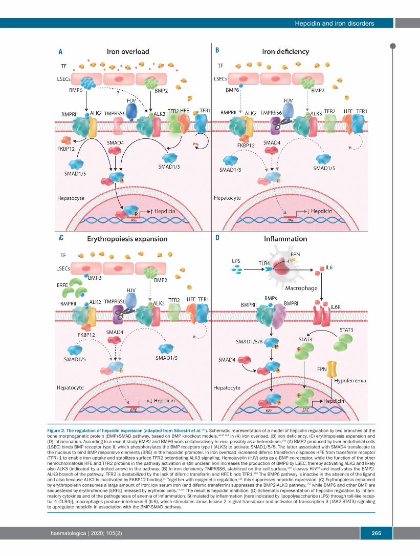
Figure 2. The regulation of hepcidin expression (adapted from Silvestri et al.147). Schematic representation of a model of hepcidin regulation by two branches of thebone morphogenetic protein (BMP)-SMAD pathway, based on BMP knockout models,44,45,148 in (A) iron overload, (B) iron deficiency, (C) erythropoiesis expansion and(D) inflammation. According to a recent study BMP2 and BMP6 work collaboratively in vivo, possibly as a heterodimer.153 (A) BMP2 produced by liver endothelial cells(LSEC) binds BMP receptor type II, which phosphorylates the BMP receptors type I (ALK3) to activate SMAD1/5/8. The latter associated with SMAD4 translocate tothe nucleus to bind BMP responsive elements (BRE) in the hepcidin promoter. In iron overload increased diferric transferrin displaces HFE from transferrin receptor(TFR) 1 to enable iron uptake and stabilizes surface TFR2 potentiating ALK3 signaling. Hemojuvelin (HJV) acts as a BMP co-receptor, while the function of the otherhemochromatosis HFE and TFR2 proteins in the pathway activation is still unclear. Iron increases the production of BMP6 by LSEC, thereby activating ALK2 and likelyalso ALK3 (indicated by a dotted arrow) in the pathway. (B) In iron deficiency TMPRSS6, stabilized on the cell surface,149 cleaves HJV49 and inactivates the BMP2-ALK3 branch of the pathway. TFR2 is destabilized by the lack of diferric transferrin and HFE binds TFR1.150 The BMP6 pathway is inactive in the absence of the ligandand also because ALK2 is inactivated by FKBP12 binding.51 Together with epigenetic regulation,151 this suppresses hepcidin expression. (C) Erythropoiesis enhancedby erythropoietin consumes a large amount of iron: low serum iron (and diferric transferrin) suppresses the BMP2-ALK3 pathway,152 while BMP6 and other BMP aresequestered by erythroferrone (ERFE) released by erythroid cells.72,154 The result is hepcidin inhibition. (D) Schematic representation of hepcidin regulation by inflam-matory cytokines and of the pathogenesis of anemia of inflammation. Stimulated by inflammation [here indicated by lipopolysaccharide (LPS) through toll-like recep-tor 4 (TLR4)], macrophages produce interleukin-6 (IL6), which stimulates Janus kinase 2–signal transducer and activator of transcription 3 (JAK2-STAT3) signalingto upregulate hepcidin in association with the BMP-SMAD pathway.
A B
C D

synthesis of erythropoietin is mediated by HIF-2α. In irondeficiency, IRP1 binding to HIF-2α 5’IRE represses the lat-ter’s translation and decreases erythropoietin production,to limit erythropoiesis and iron consumption. When themechanism fails, as in Irp1-knockout mice, a transientpolycythemia occurs in the relatively iron-deficientyoung animals, which reverts in adult mice with iron suf-ficiency.64,65 In this context prolyl hydroxylase, theenzyme that induces degradation of HIF-2α, is iron-dependent and is thus inactive in iron deficiency.Second, in in vitro studies, iron deprivation induced a
block of early erythroid progenitors, by inactivating mito-chondrial aconitase;66 this block could be overcome byisocitrate supplementation.67 Finally the erythroidresponse to iron restriction is optimized by the iron sen-sor TFR2, a partner of erythropoietin receptor.10 Loss ofbone marrow TFR2 in mice increases the sensitivity oferythroblasts to erythropoietin, which causes erythrocy-tosis, especially in iron deficiency.68 Liver TFR2 upregu-lates hepcidin and TFR2mutations cause hemochromato-sis.69 As a sensor of iron-bound transferrin, erythroidTFR2 regulates erythropoiesis, while liver TFR2, control-ling hepcidin, modulates iron acquisition according toerythropoietic needs. The recently demonstrated TFR2expression in osteoclasts and osteoblasts70 places this ironsensor at the crossroads of red cell production, ironhomeostasis and bone turnover.While iron regulates erythropoiesis, the reverse is also
true. The old hypothesis that the erythropoietic drivecontrols iron absorption through an erythroid regulator71was confirmed by the discovery of ERFE, the best exam-ple of tissue-mediated regulation of hepcidin. ERFE is amember of the tumor-necrosis factor (TNF)-α family, pro-duced by several tissues, but increased in response to ery-thropoietin only in erythroid precursors. ERFE sequestersBMP receptor ligands, especially BMP6,72 inhibiting BMP-SMAD signaling and hepcidin. However, ERFE fails tosuppress hepcidin when the BMP pathway is overactive.73Erfe-knockout mice are not anemic, indicating that ERFEhas a modest effect on hepcidin repression in steadystate. However, ERFE contributes to iron loading in micewith β-thalassemia.74
Iron disorders
The improved understanding of iron physiology has pro-foundly changed the modern approach to iron disorders,known historically for centuries as iron deficiency (chloro-sis) in young females and hemochromatosis (bronze dia-betes) in middle-age males. We now suspect hemochro-matosis based on iron parameters and confirm the diagno-sis by genetic testing well before the development of ironoverload and organ damage. We are aware that anemia isa complication of iron deficiency, though not the only one,since other tissues/organs may be iron-depleted beforeanemia develops, as occurs in chronic heart failure.75Hepcidin is tightly controlled to maintain body iron
balance. Loss of this control leads to opposite genetic oracquired disorders (Table 1).
Genetic disorders Hereditary hemochromatosisThe pathophysiology and diagnosis of hemochromato-
sis were profoundly influenced by the cloning of the HFE
gene76 and the definition of the genetic heterogeneity ofthe disease (Table 1). Overall hemochromatosis is due to“insufficient hepcidin production” or exceptionally to“hepcidin resistance”.77 Mutations in genes of the hep-cidin-ferroportin axis disrupt iron homeostasis, leading toincreased iron absorption, high transferrin saturation andincreased toxicity from non-transferrin bound iron (NTBI)species.78 The commonest form of hemochromatosis inCaucasians is due to homozygous HFE(C282Y) muta-tions. Genetic tests allow early diagnosis so that individ-uals with the affected genotypes show high biochemicalpenetrance (increased transferrin saturation ± increasedserum ferritin) but low clinical expression. Loss-of-func-tion mutations of HJV, TFR2 and HAMP (encoding hep-cidin) lead to more severe diseases, collectively called“non-HFE hemochromatosis”. In all the recessive formshepcidin is inappropriately low in comparison with ironexcess and the onset and severity of iron overload corre-late with the hormone deficiency.79 The implementationof next-generation sequencing in familial and isolatedcases of iron overload80 has enabled the identification ofmutations in more than one gene and provided examplesof digenic inheritance. Both genetic and acquired modi-fiers contribute to the penetrance of HFE-hemochromato-sis, interfering with hepcidin expression: e.g., alcoholaggravates the iron burden, whereas blood donationsattenuates it.77Ferroportin mutations are inherited in a dominant man-
ner (Table 1). The different effects of these mutationsaccount for the controversy in disease nomenclature.Loss-of-function mutations impair iron export, are associ-ated with iron accumulation in Küpffer cells and requireno or reduced phlebotomy therapy, representing the true“ferroportin disease”.81 Gain-of-function mutations leadto hepcidin resistance and the release of too much iron, asoccurs in hemochromatosis. The clinical severity of hemochromatosis is related to
NTBI, a toxic iron species bound to low molecular weightmolecules, easily taken up by hepatocytes and pancreaticcells via ZIP14 transporter82 and by cardiomyocytesthrough other transporters.83 While iron uptake by trans-ferrin receptor is tightly regulated, the uptake of NTBI isnot and persists in iron overload. NTBI leads to the gen-eration of reactive oxygen species and cell damage, caus-ing liver fibrosis (which may progress to cirrhosis andhepatocellular carcinoma), chronic heart failure, diabetes,hypopituitarism and other complications of iron load-ing.77,79
Iron-refractory iron-deficiency anemia This form of iron deficiency anemia (IRIDA) was recog-
nized after the discovery of hepcidin as being due tomutations of its inhibitor TMPRSS6.50 High hepcidin levelslead to a phenotype opposite to that of hemochromatosis,reminiscent of anemia of inflammation.84 The anemia isrefractory to oral iron and may require intravenous thera-py, especially when iron demand is high.85 TMPRSS6genetic variants modulate iron and hematologic traits inseveral genome-wide association studies,58 alter hepcidinlevels in normal subjects86 and might confer susceptibili-ty/resistance to iron deficiency, as observed in blooddonors.87Other rare recessive disorders of the transferrin receptor
pathway – such as hypotransferrinemia and DMT1 muta-tions – lead to “atypical microcytic anemia” with
C. Camaschella et al.
266 haematologica | 2020; 105(2)

increased transferrin saturation and iron stores, because ofdecreased iron utilization by blunted erythropoiesis.85
Congenital sideroblastic anemia Ringed sideroblasts are erythroblasts with iron-loaded
mitochondria that, clustering around the nucleus, conferthe appearance of a ring at Perls iron staining. Hereditarysideroblastic anemias are usually due to heme deficiency:X-linked sideroblastic anemia is caused by mutations inALAS2, the first, rate-limiting enzyme of heme biosynthe-sis, while recessive forms are associated with mutations ofmitochondria glycine importer solute carrier family 25member 38.88 Rare severe cases result from mutations ofFe/S cluster proteins, such as GLRX589 or HSPA9,90 whichdecrease Fe/S groups and the activity of ferrochelatase, thelast enzyme of the heme pathway. Another cause of lowheme in GLRX5 deficiency is the overactive IRP1 that, notbeing converted to aconitase because of the Fe/S clusterdeficit, blocks ALAS2 translation, thereby preventingheme formation. These disorders reveal the tight connec-tion of heme-Fe/S metabolism. Among syndromic forms,the X-linked ABCB7 deficiency reduces export of Fe/Sclusters to the cytosol,91 while others are associated withimmunodeficiency92 strengthening the need of Fe/S clus-ters in other cell types (Table 1). Those due to mitochon-drial protein mutations91 are not discussed here.
Acquired iron disordersIron deficiencyIron deficiency, both isolated and associated with ane-
mia, represents one of the five major causes of disabilityburden worldwide, especially in women.93 For discussionsof the etiology, clinical presentation and treatment of irondeficiency with or without anemia readers are directed tospecific reviews.94-96 In absolute iron deficiency low totalbody and serum iron fully suppress hepcidin, a mecha-nism of adaptation to increase iron absorption. In func-tional iron deficiency (e.g., in inflammation) total bodyiron is not decreased, but iron is sequestered in stores bythe high hepcidin levels.84,97 This distinction strongly influ-ences the route of iron administration required to treatiron deficiency, as discussed below.
Anemia of inflammationProinflammatory cytokines such as IL-6 and IL-1β, pro-
duced in chronic infections, autoimmunity, cancer, renalfailure and other chronic disorders activate hepcidinexpression leading to iron-restricted erythropoiesis andanemia of inflammation, once named anemia of chronicdiseases.84,97,98 By withholding iron in macrophages, extra-cellular Gram-negative microorganisms are deprived ofthis essential nutrient.99,100 This is an innate defense mech-anism known as 'nutritional immunity'.101 A recent inter-pretation is that hypoferremia prevents the generation ofNTBI that potentiates the pathogenicity of Gram-nega-tive bacteria.102 Anemia, usually moderate and normocyt-ic, is multifactorial, because of concomitant insufficienterythropoietin production and impaired early erythroidcommitment.98 Microcytosis occurs in longstandingsevere inflammation such as in Castleman disease, a lym-phoproliferative disorder in which high IL-6 productionstrongly enhances hepcidin synthesis103 or in patientswith ectopic hepcidin expression by liver adenomas.104Anemia reverts after anti-IL6 receptor treatment inCastleman disease or after surgical removal of the tumor
in the case of adenoma.Anemia of inflammation regresses with control of the
disease. In selected cases intravenous iron or erythro-poiesis-stimulating agents are used. Since treatment isoften unsatisfactory, manipulation of the hepcidin path-way (blocking either its production or function) is pro-posed as a novel therapeutic opportunity.97
Iron-loading anemiasLow hepcidin levels explain the iron overload that
develops in the absence of blood transfusions in “iron-loading anemias”, i.e., anemias with ineffective erythro-poiesis (Table 1). ERFE, released by erythropoietin-stimu-lated erythroblasts, inhibits hepcidin, despite iron over-load. In non-transfusion-dependent β-thalassemiapatients, serum ERFE levels are high,105 to ensure ironacquisition for the expanded erythropoiesis.106 However,since the erythropoiesis is inefficient, excess iron inter-feres with erythroblast maturation aggravating anemia ina vicious cycle.107 In patients with transfusion-dependentthalassemia, hepcidin increases following transfusionswhich partially suppress erythropoiesis.ERFE contributes to the iron loading of some clonal
myelodysplastic syndromes. Patients with the ringedsideroblasts subtype of myelodysplastic syndrome (oncecalled refractory anemia with ringed sideroblasts) carry asomatic mutation in the spliceosome gene SF3B1.108Among other abnormally spliced products, an elongatedvariant of ERFE is more efficient than the wildtype hor-mone in hepcidin repression.109
Diagnostic implications
Notwithstanding spectacular advances in our under-standing of iron metabolism and homeostasis our diag-nostic approach to iron disorders still relies mainly onthree historical tests: serum iron, transferrin (or total ironbinding capacity) and ferritin. Transferrin saturation(Tsat), i.e. the ratio of serum iron/total iron binding capac-ity and serum ferritin coupled with genetic testing andnon-invasive magnetic resonance imaging measurementsof liver iron content, define the nature and severity of ironloading in both hemochromatosis77 and thalassemia.110Other useful markers are the level of serum soluble trans-ferrin receptor (sTFRC), related to the expansion of ery-thropoiesis or iron deficiency, the sTFRC/log ferritin ratiofor the diagnosis of iron deficiency in inflammation98 andthe Tsat/log hepcidin ratio to suspect IRIDA.111Enzyme-linked immunosorbent assay kits can measure
serum hepcidin levels. However, this does not provide anyinformation additional to serum ferritin, since the twovariables are tightly related.112,113 Some researchers proposedetermining hepcidin levels in order to choose the bettertherapeutic route of administration of iron supplementa-tion (oral vs. intravenous),114 as well as its correct timing115and schedule.116 However, besides being subject to circadi-an oscillations, hepcidin levels change rapidly in responseto activating and inhibitory signals, making their measure-ment useful in only a limited number of conditions.47 A kitto measure human serum ERFE concentration is availablefor research purposes. Whether the elongated ERFE iden-tified in individuals with SF3B1 mutations will become abiomarker of ringed sideroblast myelodysplastic syn-drome109 remains to be tested.
Hepcidin and iron disorders
haematologica | 2020; 105(2) 267

Therapeutic implications
Hepcidin levels favor response (when low) or resistance(when high) to oral iron administration, explaining part ofiron refractoriness.117 The dynamics of the increase in hep-cidin levels after oral iron therapy have suggested that alter-nate-day administration of iron salts is an alternative todaily refracted doses, with the former being a protocol thatincreases both efficacy and tolerability, at least in womenwith iron deficiency without or with mild anemia.116,118,119The availability of more tolerated, iron-stable and effica-cious preparations has increased the use of intravenousiron, especially of the high-dose single-injection com-pounds.120 However, when used to correct iron deficiency ininflammation, intravenous iron may lead to macrophageiron accumulation whose long-term effects are unknown.Manipulation of the hepcidin-ferroportin axis is the
most logical experimental approach to iron disorders. Therationale is to use hepcidin agonists for iron overload dis-orders caused by inappropriate/low hepcidin and hep-cidin antagonists to release sequestered iron in IRIDA andin anemia of inflammation (Table 2).
Increasing hepcidin levels /decreasing ferroportinactivityIn preclinical studies, increasing hepcidin levels prevent-
ed iron overload or redistributed iron to sites of safe stor-
age. Potentially useful in hemochromatosis, whose treat-ment is still based on phlebotomy,77 hepcidin agonists areof interest in disorders with ineffective erythropoiesis,such as β-thalassemia.107 Agonists include hepcidin ana-logues, minihepcidins, inhibitors of hepcidin repressorssuch as anti-TMPRSS6 molecules or compounds thatblock ferroportin activity. By inducing iron restriction hep-cidin agonists ameliorated anemia and iron overload inpreclinical studies of thalassemia models;106,121 a few hep-cidin agonists are currently being investigated in phase I-IIclinical trials (Table 2). Hepcidin mimics could also be use-ful to induce iron restriction in polycythemia.122 Accordingto recent findings hepcidin might have a role as an antimi-crobial peptide in the treatment of Gram-negative sepsis102and streptococcal necrotizing fasciitis.56
Other approachesIn non-transfusion-dependent β-thalassemia (Hbbth1/th1
and Hbbth3/+) mice transferrin infusions improve the pheno-type, increasing hepcidin and hemoglobin levels, improv-ing erythrocyte survival and limiting splenomegaly,123,124effects similar to those observed when Tfr1 expression isdecreased.125 Selective inactivation of bone marrow Tfr2improves anemia in a non-transfusion-dependent Hbbth3/+model, enhancing the sensitivity of erythroid cells to ery-thropoietin.126Short interfering RNA against DMT1, administered in
C. Camaschella et al.
268 haematologica | 2020; 105(2)
Table 2. Targeted therapeutic approaches for disorders with low and high hepcidin.Compounds Mechanism Effect
IA. TO INCREASE HEPCIDIN OR REDUCE FERROPORTIN ACTIVITY122
Hepcidin analogues and minihepcidin121 Replacement therapyBMPs Activating the hepcidin signaling pathway Increased hepcidinAnti-TMPRSS6 (ASO, siRNA)137 Counteracting hepcidin inhibition Reduced iron overload FPN inhibitor VIT-2763155 Blocking the hepcidin receptor Increased Hb in ineffective erythropoiesis
IB. OTHER APPROACHESTransferrin injections123 Decreasing transferrin receptor 1 Reduction of iron uptakeProtoporphyrin IX138 Inhibiting heme oxygenase 1 Reduction of iron recycling
IIA. TO DECREASE HEPCIDIN OR INCREASE FERROPORTIN ACTIVITY97
Anti cytokines (IL-6, IL-6R)103
Anti-BMP6 MoAb139
BMP receptor inhibitors140 Reducing the hepcidin signaling pathway Reduced hepcidinAnti-hemojuvelin MoAb141 Reduced macrophage iron sequestrationHeparins142 Correction of hypoferremiaAnti-hepcidin MoAb143 (Partial) correction of anemiaAnti-hepcidin Spiegelmer144 Hepcidin bindersAnti-hepcidin anticalin145
Anti-ferroportin MoAb139 Interfering with hepcidin-ferroportin interactionGDP146 Blocking iron export and decreasing Stat3 activation
IIB. OTHER APPROACHESProlylhydroxylase inhibitors134 Increasing EPO Correction of EPO defect Increasing iron absorption Correction of hypoferremiaI. Compounds potentially useful in hereditary hemochromatosis and β-thalassemia; II. Compounds potentially useful in anemia of inflammation. Compounds tested in clinicaltrials are indicated in bold. BMP: bone morphogenetic protein; ASO: antisense specific oligonucleotides; siRNA: short interfering RNA; FPN: ferroportin; VIT 2763: small moleculeoral ferroportin inhibitor; Hb: hemoglobin; IL: interleukin; MoAb: monoclonal antibodies; GDP: guanosine 5'-diphosphate encapsulated in lipid vesicle; EPO: erythropoietin.

nanoparticles to target intestinal absorption,127 establisheda proof of principle of reducing dietary iron uptake.Another approach might be to block intestinal HIF-2α byspecific antagonists.Clinical trials are showing that correcting ineffective
erythropoiesis by activin ligand traps128 not only improvesanemia but, in the long-term, also iron loading in bothnon-transfusion-dependent and transfusion-dependentthalassemia129 and ringed sideroblast myelodysplastic syn-drome.130 Some thiazolidinones have been shown to stim-ulate hepcidin activity in preclinical studies.131 The use ofproton pump inhibitors reduced the need for phlebotomyin patients with hemochromatosis.132
Decreasing hepcidin levels/increasing ferroportin functionIn preclinical models of anemia of inflammation, hep-
cidin antagonists decreased hepcidin expression, an effectverified in clinical trials for some compounds.133 Anotheroption is to interfere with the hepcidin-ferroportin inter-action (Table 2). However, targeting the hepcidin-ferro-portin axis may not fully correct this multifactorial anemiacharacterized by low erythropoietin and a blunted ery-thropoietic response.97,98 Another approach is based onmanipulation of the hypoxia-responsive pathway.63 Prolylhydroxylase inhibitors or HIF stabilizers, now tested inchronic kidney disease, by increasing HIF-2α might targettwo abnormal processes enhancing both erythropoietinsynthesis and iron absorption.134
Unresolved issues
Notwithstanding significant advances many questionsabout iron metabolism and homeostasis remain unan-
swered. The mechanisms of intestinal heme absorptionare mysterious, as are the roles of secreted ferritin and sol-uble transferrin receptor. We have just started exploringthe autonomous regulation of iron in the heart and vascu-lar wall; the role of iron (deficiency or excess) as a cofactorof metabolic disorders, chronic liver disease, heart failure,pulmonary hypertension and neurodegeneration stillrequires elucidation. We need to be able to diagnose iso-lated tissue iron deficiency better and to increase the lim-ited number of iron status markers. In hematology we need to clarify the relationship
between iron and platelet production considering that irondeficiency directs the common erythroid-megakaryocyteprecursor towards the platelet lineage.135 More informa-tion is required on the role of iron in B-lymphocyte devel-opment and function, in B-cell malignancies, such as mul-tiple myeloma,136 and in response to infectious diseases.We have to explore better how iron/TFR2 intersects theerythropoietin signaling pathway and bone metabolism.We need novel protocols of iron supplementation and
clear indications regarding high-dose intravenous iron tooptimize iron therapy. Targeted approaches, now in clin-ical trials, have the potential to change traditional treat-ment – such as the time-honored phlebotomy-based reg-imen – for disorders such as hemochromatosis.Repurposing commercially available compounds, devel-oped for other conditions, to iron/erythroid disorders isanother option. All these approaches will, it is hoped,enable a more personalized treatment of iron disorders inthe near future.
AcknowledgmentsThis work was supported in part by an ASH Global Research
Award 2017 to AN.
Hepcidin and iron disorders
haematologica | 2020; 105(2) 269
References
1. Hentze MW, Muckenthaler MU, Galy B,Camaschella C. Two to tango: regulation ofMammalian iron metabolism. Cell.2010;142(1):24-38.
2. Muckenthaler MU, Rivella S, Hentze MW,Galy B. A red carpet for iron metabolism.Cell. 2017;168(3):344-361.
3. Wang CY, Babitt JL. Liver iron sensing andbody iron homeostasis. Blood. 2019;133(1):18-29.
4. Barrientos T, Laothamatas I, Koves TR, et al.Metabolic catastrophe in mice lacking trans-ferrin receptor in muscle. EBioMedicine.2015;2(11):1705-1717.
5. Jabara HH, Boyden SE, Chou J, et al. A mis-sense mutation in TFRC, encoding transfer-rin receptor 1, causes combined immunode-ficiency. Nat Genet. 2016;48(1):74-78.
6. Chen AC, Donovan A, Ned-Sykes R,Andrews NC. Noncanonical role of transfer-rin receptor 1 is essential for intestinal home-ostasis. Proc Natl Acad Sci U S A.2015;112(37):11714-11719.
7. Fillebeen C, Charlebois E, Wagner J, et al.Transferrin receptor 1 controls systemic ironhomeostasis by fine-tuning hepcidin expres-sion to hepatocellular iron load. Blood.2019;133(4):344-355.
8. Kawabata H, Yang R, Hirama T, et al.
Molecular cloning of transferrin receptor 2.A new member of the transferrin receptor-like family. J Biol Chem. 1999;274(30):20826-20832.
9. Camaschella C, Pagani A, Nai A, Silvestri L.The mutual control of iron and erythro-poiesis. Int J Lab Hematol. 2016;38 (Suppl1):20-26.
10. Forejtnikova H, Vieillevoye M, Zermati Y, etal. Transferrin receptor 2 is a component ofthe erythropoietin receptor complex and isrequired for efficient erythropoiesis. Blood.2010;116(24):5357-5367.
11. Parrow NL, Li Y, Feola M, et al. Lobe speci-ficity of iron-binding to transferrin modu-lates murine erythropoiesis and iron home-ostasis. Blood. 2019;134(17):1373-1384.
12. Yachie A, Niida Y, Wada T, et al. Oxidativestress causes enhanced endothelial cellinjury in human heme oxygenase-1 deficien-cy. J Clin Invest. 1999;103(1):129-135.
13. Nemeth E, Tuttle MS, Powelson J, et al.Hepcidin regulates cellular iron efflux bybinding to ferroportin and inducing its inter-nalization. Science. 2004;306(5704):2090-2093.
14. Lill R, Broderick JB, Dean DR. Special issueon iron-sulfur proteins: Structure, function,biogenesis and diseases. Biochim BiophysActa. 2015;1853(6):1251-1252.
15. Braymer JJ, Lill R. Iron-sulfur cluster biogen-esis and trafficking in mitochondria. J BiolChem. 2017;292(31):12754-12763.
16. Hamdi A, Roshan TM, Kahawita TM,Mason AB, Sheftel AD, Ponka P. Erythroidcell mitochondria receive endosomal iron bya "kiss-and-run" mechanism. BiochimBiophys Acta. 2016;1863(12):2859-2867.
17. Shaw GC, Cope JJ, Li L, et al. Mitoferrin isessential for erythroid iron assimilation.Nature. 2006;440(7080):96-100.
18. Arosio P, Carmona F, Gozzelino R,Maccarinelli F, Poli M. The importance ofeukaryotic ferritins in iron handling andcytoprotection. Biochem J. 2015;472(1):1-15.
19. Vanoaica L, Darshan D, Richman L,Schumann K, Kuhn LC. Intestinal ferritin His required for an accurate control of ironabsorption. Cell Metab. 2010;12(3):273-282.
20. Castiglioni E, Soriani N, Girelli D, et al. Highresolution melting for the identification ofmutations in the iron responsive element ofthe ferritin light chain gene. Clin Chem LabMed. 2010;48(10):1415-1418.
21. Luscieti S, Santambrogio P, Langloisd'Estaintot B, et al. Mutant ferritin L-chainsthat cause neurodegeneration act in a domi-nant-negative manner to reduce ferritin ironincorporation. J Biol Chem. 2010;285(16):11948-11957.
22. Truman-Rosentsvit M, Berenbaum D,Spektor L, et al. Ferritin is secreted via 2 dis-tinct nonclassical vesicular pathways. Blood.2018;131(3):342-352.
23. Leidgens S, Bullough KZ, Shi H, et al. Eachmember of the poly-r(C)-binding protein 1

(PCBP) family exhibits iron chaperone activ-ity toward ferritin. J Biol Chem.2013;288(24):17791-17802.
24. Ryu MS, Zhang D, Protchenko O,Shakoury-Elizeh M, Philpott CC. PCBP1and NCOA4 regulate erythroid iron storageand heme biosynthesis. J Clin Invest.2017;127(5):1786-1797.
25. Mancias JD, Wang X, Gygi SP, Harper JW,Kimmelman AC. Quantitative proteomicsidentifies NCOA4 as the cargo receptormediating ferritinophagy. Nature.2014;509(7498):105-109.
26. Dowdle WE, Nyfeler B, Nagel J, et al.Selective VPS34 inhibitor blocks autophagyand uncovers a role for NCOA4 in ferritindegradation and iron homeostasis in vivo.Nat Cell Biol. 2014;16(11):1069-1079.
27. Nandal A, Ruiz JC, Subramanian P, , et al.Activation of the HIF prolyl hydroxylase bythe iron chaperones PCBP1 and PCBP2. CellMetab. 2011;14(5):647-657.
28. Lakhal-Littleton S, Wolna M, Carr CA, et al.Cardiac ferroportin regulates cellular ironhomeostasis and is important for cardiacfunction. Proc Natl Acad Sci U S A.2015;112(10):3164-3169.
29. Zhang DL, Ghosh MC, Ollivierre H, Li Y,Rouault TA. Ferroportin deficiency in ery-throid cells causes serum iron deficiency andpromotes hemolysis due to oxidative stress.Blood. 2018;132(19):2078-2087.
30. Zhang DL, Wu J, Shah BN, et al.Erythrocytic ferroportin reduces intracellulariron accumulation, hemolysis, and malariarisk. Science. 2018;359(6383):1520-1523.
31. Keel SB, Doty RT, Yang Z, et al. A hemeexport protein is required for red blood celldifferentiation and iron homeostasis.Science. 2008;319(5864):825-828.
32. Chiabrando D, Marro S, Mercurio S, et al.The mitochondrial heme exporter FLVCR1bmediates erythroid differentiation. J ClinInvest. 2012;122(12):4569-4579.
33. Zhang DL, Ghosh MC, Rouault TA. Thephysiological functions of iron regulatoryproteins in iron homeostasis - an update.Front Pharmacol. 2014;5:124.
34. Costain G, Ghosh MC, Maio N, et al.Absence of iron-responsive element-bindingprotein 2 causes a novel neurodegenerativesyndrome. Brain. 2019;142(5):1195-1202.
35. Galy B, Ferring-Appel D, Becker C, et al. Ironregulatory proteins control a mucosal blockto intestinal iron absorption. Cell Rep.2013;3(3):844-857.
36. Zhang DL, Hughes RM, Ollivierre-WilsonH, Ghosh MC, Rouault TA. A ferroportintranscript that lacks an iron-responsive ele-ment enables duodenal and erythroid pre-cursor cells to evade translational repression.Cell Metab. 2009;9(5):461-473.
37. Mancias JD, Pontano Vaites L, Nissim S, etal. Ferritinophagy via NCOA4 is required forerythropoiesis and is regulated by irondependent HERC2-mediated proteolysis.Elife. 2015;4.
38. Bellelli R, Castellone MD, Guida T, et al.NCOA4 transcriptional coactivator inhibitsactivation of DNA replication origins. MolCell. 2014;55(1):123-137.
39. Bellelli R, Federico G, Matte A, et al.NCOA4 deficiency impairs systemic ironhomeostasis. Cell Rep. 2016;14(3):411-421.
40. Gao X, Lee HY, Li W, et al. Thyroid hor-mone receptor beta and NCOA4 regulateterminal erythrocyte differentiation. ProcNatl Acad Sci U S A. 2017;114(38):10107-10112.
41. Aschemeyer S, Qiao B, Stefanova D, et al.Structure-function analysis of ferroportin
defines the binding site and an alternativemechanism of action of hepcidin. Blood.2018;131(8):899-910.
42. Kautz L, Meynard D, Monnier A, et al. Ironregulates phosphorylation of Smad1/5/8 andgene expression of Bmp6, Smad7, Id1, andAtoh8 in the mouse liver. Blood.2008;112(4):1503-1509.
43. Lim PJ, Duarte TL, Arezes J, et al. Nrf2 con-trols iron homeostasis in haemochromatosisand thalassaemia via Bmp6 and hepcidin.Nat Metab. 2019;1(5):519-531.
44. Koch PS, Olsavszky V, Ulbrich F, et al.Angiocrine Bmp2 signaling in murine livercontrols normal iron homeostasis. Blood.2017;129(4):415-419.
45. Canali S, Wang CY, Zumbrennen-BulloughKB, Bayer A, Babitt JL. Bone morphogeneticprotein 2 controls iron homeostasis in miceindependent of Bmp6. Am J Hematol.2017;92(11):1204-1213.
46. Theurl I, Schroll A, Sonnweber T, et al.Pharmacologic inhibition of hepcidinexpression reverses anemia of chronicinflammation in rats. Blood.2011;118(18):4977-4984.
47. Girelli D, Nemeth E, Swinkels DW.Hepcidin in the diagnosis of iron disorders.Blood. 2016;127(23):2809-2813.
48. Du X, She E, Gelbart T, et al. The serine pro-tease TMPRSS6 is required to sense irondeficiency. Science. 2008;320(5879):1088-1092.
49. Silvestri L, Pagani A, Nai A, De Domenico I,Kaplan J, Camaschella C. The serine pro-tease matriptase-2 (TMPRSS6) inhibits hep-cidin activation by cleaving membranehemojuvelin. Cell Metab. 2008;8(6):502-511.
50. Finberg KE, Heeney MM, Campagna DR, etal. Mutations in TMPRSS6 cause iron-refrac-tory iron deficiency anemia (IRIDA). NatGenet. 2008;40(5):569-571.
51. Colucci S, Pagani A, Pettinato M, et al. Theimmunophilin FKBP12 inhibits hepcidinexpression by binding the BMP type I recep-tor ALK2 in hepatocytes. Blood.2017;130(19):2111-2120.
52. Kautz L, Jung G, Valore EV, Rivella S,Nemeth E, Ganz T. Identification of erythro-ferrone as an erythroid regulator of ironmetabolism. Nat Genet. 2014;46(7):678-684.
53. Silvestri L, Pagani A, Camaschella C. Furin-mediated release of soluble hemojuvelin: anew link between hypoxia and iron home-ostasis. Blood. 2008;111(2):924-931.
54. Sonnweber T, Nachbaur D, Schroll A, et al.Hypoxia induced downregulation of hep-cidin is mediated by platelet derived growthfactor BB. Gut. 2014;63(12):1951-1959.
55. Theurl I, Theurl M, Seifert M, et al.Autocrine formation of hepcidin inducesiron retention in human monocytes. Blood.2008;111(4):2392-2399.
56. Malerba M, Louis S, Cuvellier S, et al.Epidermal hepcidin is required for neu-trophil response to bacterial infection. J ClinInvest. 2019 Dec 3. [Epub ahead of print]
57. Lakhal-Littleton S, Wolna M, Chung YJ, etal. An essential cell-autonomous role forhepcidin in cardiac iron homeostasis. Elife.2016;5.
58. Benyamin B, Esko T, Ried JS, et al. Novel lociaffecting iron homeostasis and their effectsin individuals at risk for hemochromatosis.Nat Commun. 2014;5:4926.
59. Bekri S, Gual P, Anty R, et al. Increased adi-pose tissue expression of hepcidin in severeobesity is independent from diabetes andNASH. Gastroenterology. 2006;131(3):788-796.
60. Vecchi C, Montosi G, Garuti C, et al.
Gluconeogenic signals regulate iron home-ostasis via hepcidin in mice.Gastroenterology. 2014;146(4):1060-1069.
61. Folgueras AR, Freitas-Rodriguez S, RamsayAJ, et al. Matriptase-2 deficiency protectsfrom obesity by modulating iron homeosta-sis. Nat Commun. 2018;9(1):1350.
62. Mastrogiannaki M, Matak P, PeyssonnauxC. The gut in iron homeostasis: role of HIF-2 under normal and pathological conditions.Blood. 2013;122(6):885-892.
63. Schwartz AJ, Das NK, Ramakrishnan SK, etal. Hepatic hepcidin/intestinal HIF-2alphaaxis maintains iron absorption during irondeficiency and overload. J Clin Invest.2019;129(1):336-348.
64. Ghosh MC, Zhang DL, Jeong SY, et al.Deletion of iron regulatory protein 1 causespolycythemia and pulmonary hypertensionin mice through translational derepressionof HIF-2alpha. Cell Metab. 2013;17(2):271-281.
65. Anderson SA, Nizzi CP, Chang YI, et al. TheIRP1-HIF-2alpha axis coordinates iron andoxygen sensing with erythropoiesis and ironabsorption. Cell Metab. 2013;17(2):282-290.
66. Bullock GC, Delehanty LL, Talbot AL, et al.Iron control of erythroid development by anovel aconitase-associated regulatory path-way. Blood. 2010;116(1):97-108.
67. Richardson CL, Delehanty LL, Bullock GC,et al. Isocitrate ameliorates anemia by sup-pressing the erythroid iron restrictionresponse. J Clin Invest. 2013;123(8):3614-3623.
68. Nai A, Lidonnici MR, Rausa M, et al. Thesecond transferrin receptor regulates redblood cell production in mice. Blood.2015;125(7):1170-1179.
69. Camaschella C, Roetto A, Cali A, et al. Thegene TFR2 is mutated in a new type ofhaemochromatosis mapping to 7q22. NatGenet. 2000;25(1):14-15.
70. Rauner M, Baschant U, Roetto A, et al.Transferrin receptor 2 controls bone massand pathological bone formation via BMPand Wnt signaling. Nat Metab. 2019;1(1):111-124.
71. Finch CA. Erythropoiesis, erythropoietin,and iron. Blood. 1982;60(6):1241-1246.
72. Arezes J, Foy N, McHugh K, et al.Erythroferrone inhibits the induction of hep-cidin by BMP6. Blood. 2018;132(14):1473-1477.
73. Nai A, Rubio A, Campanella A, et al.Limiting hepatic Bmp-Smad signaling bymatriptase-2 is required for erythropoietin-mediated hepcidin suppression in mice.Blood. 2016;127(19):2327-2336.
74. Kautz L, Jung G, Du X, et al. Erythroferronecontributes to hepcidin suppression and ironoverload in a mouse model of beta-tha-lassemia. Blood. 2015;126(17):2031-2037.
75. Anker SD, Comin Colet J, Filippatos G, et al.Investigators F-HT. Ferric carboxymaltose inpatients with heart failure and iron deficien-cy. N Engl J Med. 2009;361(25):2436-2448.
76. Feder JN, Gnirke A, Thomas W, et al. Anovel MHC class I-like gene is mutated inpatients with hereditary haemochromatosis.Nat Genet. 1996;13(4):399-408.
77. Brissot P, Pietrangelo A, Adams PC, deGraaff B, McLaren CE, Loreal O.Haemochromatosis. Nat Rev Dis Primers.2018;4:18016.
78. Le Lan C, Loreal O, Cohen T, et al. Redoxactive plasma iron in C282Y/C282Yhemochromatosis. Blood. 2005;105(11):4527-4531.
79. Sandhu K, Flintoff K, Chatfield MD, et al.Phenotypic analysis of hemochromatosis
C. Camaschella et al.
270 haematologica | 2020; 105(2)

subtypes reveals variations in severity ofiron overload and clinical disease. Blood.2018;132(1):101-110.
80. McDonald J, Wooderchak-Donahue W,VanSant Webb C, Whitehead K, StevensonDA, Bayrak-Toydemir P. Hereditary hemor-rhagic telangiectasia: genetics and moleculardiagnostics in a new era. Front Genet.2015;6:1.
81. Pietrangelo A. Ferroportin disease: patho-genesis, diagnosis and treatment.Haematologica. 2017;102(12):1972-1984.
82. Jenkitkasemwong S, Wang CY, Coffey R, etal. SLC39A14 is required for the develop-ment of hepatocellular iron overload inmurine models of hereditary hemochro-matosis. Cell Metab. 2015;22(1):138-150.
83. Oudit GY, Sun H, Trivieri MG, et al. L-typeCa2+ channels provide a major pathway foriron entry into cardiomyocytes in iron-over-load cardiomyopathy. Nat Med.2003;9(9):1187-1194.
84. Weiss G, Ganz T, Goodnough LT. Anemiaof inflammation. Blood. 2019;133(1):40-50.
85. Camaschella C. How I manage patientswith atypical microcytic anaemia. Br JHaematol. 2013;160(1):12-24.
86. Nai A, Pagani A, Silvestri L, et al. TMPRSS6rs855791 modulates hepcidin transcriptionin vitro and serum hepcidin levels in normalindividuals. Blood. 2011;118(16):4459-4462.
87. Kiss JE, Vassallo RR. How do we manageiron deficiency after blood donation? Br JHaematol. 2018;181(5):590-603.
88. Guernsey DL, Jiang H, Campagna DR, et al.Mutations in mitochondrial carrier familygene SLC25A38 cause nonsyndromic auto-somal recessive congenital sideroblastic ane-mia. Nat Genet. 2009;41(6):651-653.
89. Camaschella C, Campanella A, De Falco L,et al. The human counterpart of zebrafishshiraz shows sideroblastic-like microcyticanemia and iron overload. Blood.2007;110(4):1353-1358.
90. Schmitz-Abe K, Ciesielski SJ, Schmidt PJ, etal. Congenital sideroblastic anemia due tomutations in the mitochondrial HSP70homologue HSPA9. Blood. 2015;126(25):2734-2738.
91. Ducamp S, Fleming MD. The moleculargenetics of sideroblastic anemia. Blood.2019;133(1):59-69.
92. Chakraborty PK, Schmitz-Abe K, KennedyEK, et al. Mutations in TRNT1 cause con-genital sideroblastic anemia with immunod-eficiency, fevers, and developmental delay(SIFD). Blood. 2014;124(18):2867-2871.
93. Disease GBD, Injury I, Prevalence C. Global,regional, and national incidence, prevalence,and years lived with disability for 328 dis-eases and injuries for 195 countries, 1990-2016: a systematic analysis for the GlobalBurden of Disease Study 2016. Lancet.2017;390(10100):1211-1259.
94. Camaschella C. Iron-deficiency anemia. NEngl J Med. 2015;372(19):1832-1843.
95. Lopez A, Cacoub P, Macdougall IC, Peyrin-Biroulet L. Iron deficiency anaemia. Lancet.2016;387(10021):907-916.
96. Camaschella C. Iron deficiency. Blood.2019;133(1):30-39.
97. Ganz T. Anemia of inflammation. N Engl JMed. 2019;381(12):1148-1157.
98. Weiss G, Goodnough LT. Anemia of chronicdisease. N Engl J Med. 2005;352(10):1011-1023.
99. Drakesmith H, Prentice AM. Hepcidin andthe iron-infection axis. Science. 2012;338(6108):768-772.
100.Nairz M, Dichtl S, Schroll A, et al. Iron andinnate antimicrobial immunity-Depriving
the pathogen, defending the host. J TraceElem Med Biol. 2018;48:118-133.
101.Hood MI, Skaar EP. Nutritional immunity:transition metals at the pathogen-host inter-face. Nat Rev Microbiol. 2012;10(8):525-537.
102. Stefanova D, Raychev A, Arezes J, et al.Endogenous hepcidin and its agonist medi-ate resistance to selected infections by clear-ing non-transferrin-bound iron. Blood.2017;130(3):245-257.
103.Song SN, Tomosugi N, Kawabata H,Ishikawa T, Nishikawa T, Yoshizaki K.Down-regulation of hepcidin resulting fromlong-term treatment with an anti-IL-6 recep-tor antibody (tocilizumab) improves anemiaof inflammation in multicentric Castlemandisease. Blood. 2010;116(18):3627-3634.
104.Weinstein DA, Roy CN, Fleming MD, LodaMF, Wolfsdorf JI, Andrews NC.Inappropriate expression of hepcidin is asso-ciated with iron refractory anemia: implica-tions for the anemia of chronic disease.Blood. 2002;100(10):3776-3781.
105.Ganz T, Jung G, Naeim A, et al.Immunoassay for human serum erythrofer-rone. Blood. 2017;130(10):1243-1246.
106.Rivella S. Iron metabolism under conditionsof ineffective erythropoiesis in beta-tha-lassemia. Blood. 2019;133(1):51-58.
107.Camaschella C, Nai A. Ineffective erythro-poiesis and regulation of iron status in ironloading anaemias. Br J Haematol. 2016;172(4):512-523.
108.Papaemmanuil E, Cazzola M, Boultwood J,et al. Chronic Myeloid Disorders WorkingGroup of the International Cancer GenomeC. Somatic SF3B1 mutation in myelodyspla-sia with ring sideroblasts. N Engl J Med.2011;365(15):1384-1395.
109.Bondu S, Alary AS, Lefevre C, et al. A vari-ant erythroferrone disrupts iron homeosta-sis in SF3B1-mutated myelodysplastic syn-drome. Sci Transl Med. 2019;11(500).
110.Taher AT, Weatherall DJ, Cappellini MD.Thalassaemia. Lancet. 2018;391(10116):155-167.
111.Heeney MM, Guo D, De Falco L, et al.Normalizing hepcidin predicts TMPRSS6mutation status in patients with chronic irondeficiency. Blood. 2018;132(4):448-452.
112.Traglia M, Girelli D, Biino G, et al.Association of HFE and TMPRSS6 geneticvariants with iron and erythrocyte parame-ters is only in part dependent on serum hep-cidin concentrations. J Med Genet.2011;48(9):629-634.
113.Galesloot TE, Vermeulen SH, Geurts-Moespot AJ, et al. Serum hepcidin: referenceranges and biochemical correlates in thegeneral population. Blood. 2011;117(25):e218-225.
114.. Bregman DB, Morris D, Koch TA, He A,Goodnough LT. Hepcidin levels predict non-responsiveness to oral iron therapy inpatients with iron deficiency anemia. Am JHematol. 2013;88(2):97-101.
115.Prentice AM, Doherty CP, Abrams SA, et al.Hepcidin is the major predictor of erythro-cyte iron incorporation in anemic Africanchildren. Blood. 2012;119(8):1922-1928.
116.Stoffel NU, Zeder C, Brittenham GM,Moretti D, Zimmermann MB. Iron absorp-tion from supplements is greater with alter-nate day than with consecutive day dosingin iron-deficient anemic women.Haematologica. 2019 Aug 14. [Epub aheadof print]
117.Hershko C, Camaschella C. How I treatunexplained refractory iron deficiency ane-mia. Blood. 2014;123(3):326-333.
118.Moretti D, Goede JS, Zeder C, et al. Oral
iron supplements increase hepcidin anddecrease iron absorption from daily ortwice-daily doses in iron-depleted youngwomen. Blood. 2015;126(17):1981-1929.
119.Stoffel NU, Cercamondi CI, Brittenham G,et al. Iron absorption from oral iron supple-ments given on consecutive versus alternatedays and as single morning doses versustwice-daily split dosing in iron-depletedwomen: two open-label, randomised con-trolled trials. Lancet Haematol. 2017;4(11):e524-e533.
120.Auerbach M, Deloughery T. Single-doseintravenous iron for iron deficiency: a newparadigm. Hematology Am Soc HematolEduc Program. 2016 ;2016(1):57-66.
121.Casu C, Chessa R, Liu A, et al.Minihepcidins improve ineffective erythro-poiesis and splenomegaly in a new mousemodel of adult beta-thalassemia major.Haematologica. 2019 Oct 3. [Epub ahead ofprint]
122.Casu C, Nemeth E, Rivella S. Hepcidin ago-nists as therapeutic tools. Blood. 2018;131(16):1790-1794.
123.Li H, Rybicki AC, Suzuka SM, et al.Transferrin therapy ameliorates disease inbeta-thalassemic mice. Nat Med. 2010;16(2):177-182.
124.Gelderman MP, Baek JH, Yalamanoglu A, Pet al. Reversal of hemochromatosis by apo-transferrin in non-transfused and transfusedHbbth3/+ (heterozygous B1/B2 globin genedeletion) mice. Haematologica. 2015;100(5):611-622.
125.Li H, Choesang T, Bao W, et al. DecreasingTfR1 expression reverses anemia and hep-cidin suppression in β-thalassemic mice.Blood. 2017;129(11):1514-1526.
126.Artuso I, Lidonnici MR, Altamura S, et al.Transferrin receptor 2 is a potential noveltherapeutic target for β-thalassemia: evi-dence from a murine model. Blood.2018;132(21):2286-2297.
127.Wang X, Zhang M, Flores SRL, et al. Oralgavage of ginger nanoparticle-derived lipidvectors carrying Dmt1 siRNA blunts ironloading in murine hereditary hemochro-matosis. Mol Ther. 2019;27(3):493-506.
128.Dussiot M, Maciel TT, Fricot A, et al. Anactivin receptor IIA ligand trap corrects inef-fective erythropoiesis in β-thalassemia. NatMed. 2014;20(4):398-407.
129.Piga A, Perrotta S, Gamberini MR, et al.Luspatercept improves hemoglobin levelsand blood transfusion requirements in astudy of patients with β-thalassemia. Blood.2019;133(12):1279-1289.
130.Platzbecker U, Germing U, Gotze KS, et al.Luspatercept for the treatment of anaemia inpatients with lower-risk myelodysplasticsyndromes (PACE-MDS): a multicentre,open-label phase 2 dose-finding study withlong-term extension study. Lancet Oncol.2017;18(10):1338-1347.
131.Liu J, Liu W, Liu Y, et al. New thiazolidi-nones reduce iron overload in mouse mod-els of hereditary hemochromatosis and β-thalassemia. Haematologica. 2019;104(9):1768-1781.
132.Vanclooster A, van Deursen C, Jaspers R,Cassiman D, Koek G. Proton pumpinhibitors decrease phlebotomy need inHFE hemochromatosis: double-blind ran-domized placebo-controlled trial.Gastroenterology. 2017;153(3):678-680.e2.
133.Crielaard BJ, Lammers T, Rivella S. Targetingiron metabolism in drug discovery anddelivery. Nat Rev Drug Discov.2017;16(6):400-423.
134.Chen N, Hao C, Peng X, et al. Roxadustat
Hepcidin and iron disorders
haematologica | 2020; 105(2) 271

for anemia in patients with kidney diseasenot receiving dialysis. N Engl J Med.2019;381(11):1001-1010.
135.Xavier-Ferrucio J, Scanlon V, Li X et al. Lowiron promotes megakaryocytic commitmentof megakaryocytic-erythroid progenitors inhumans and mice. Blood. 2019;134(18):1547-1557.
136.Bordini J, Galvan S, Ponzoni M, et al.Induction of iron excess restricts malignantplasma cells expansion and potentiatesbortezomib effect in models of multiplemyeloma. Leukemia. 2017;31(4):967-970.
137.Guo S, Casu C, Gardenghi S, et al. ReducingTMPRSS6 ameliorates hemochromatosisand β-thalassemia in mice. J Clin Invest.2013;123(4):1531-1541.
138.Garcia-Santos D, Hamdi A, Saxova Z, et al.Inhibition of heme oxygenase amelioratesanemia and reduces iron overload in a β-tha-lassemia mouse model. Blood. 2018;131(2):236-246.
139.Sheetz M, Barrington P, Callies S, et al.Targeting the hepcidin-ferroportin pathwayin anaemia of chronic kidney disease. Br JClin Pharmacol. 2019;85(5):935-948.
140.Asshoff M, Petzer V, Warr MR, et al.Momelotinib inhibits ACVR1/ALK2,decreases hepcidin production, and amelio-rates anemia of chronic disease in rodents.Blood. 2017;129(13):1823-1830.
141.Kovac S, Boser P, Cui Y, et al. Anti-hemoju-velin antibody corrects anemia caused byinappropriately high hepcidin levels.
Haematologica. 2016;101(5):e173-176. 142. Poli M, Asperti M, Naggi A, et al. Glycol-split
nonanticoagulant heparins are inhibitors ofhepcidin expression in vitro and in vivo.Blood. 2014;123(10):1564-1573.
143.Vadhan-Raj S, Abonour R, Goldman JW, etal. A first-in-human phase 1 study of a hep-cidin monoclonal antibody, LY2787106, incancer-associated anemia. J Hematol Oncol.2017;10(1):73.
144.Boyce M, Warrington S, Cortezi B, et al.Safety, pharmacokinetics and pharmacody-namics of the anti-hepcidin Spiegelmerlexaptepid pegol in healthy subjects. Br JPharmacol. 2016;173(10):1580-1588.
145.Hohlbaum AM, Gille H, Trentmann S, et al.Sustained plasma hepcidin suppression andiron elevation by anticalin-derived hepcidinantagonist in cynomolgus monkey. Br JPharmacol. 2018;175(7):1054-1065.
146.Angmo S, Tripathi N, Abbat S, et al.Identification of guanosine 5'-diphosphateas potential iron mobilizer: preventing thehepcidin-ferroportin interaction and modu-lating the interleukin-6/Stat-3 pathway. SciRep. 2017;7:40097.
147. Silvestri L, Nai A, Dulja A, Pagani A. Hepcidinand the BMP-SMAD pathway: an unexpect-ed liaison. Vitam Horm. 2019;110: 71-99.
148.Latour C, Besson-Fournier C, Meynard D, etal. Differing impact of the deletion ofhemochromatosis-associated moleculesHFE and transferrin receptor-2 on the ironphenotype of mice lacking bone morpho-
genetic protein 6 or hemojuvelin.Hepatology. 2016;63(1):126-137.
149.Zhao N, Nizzi CP, Andrerson SA, et al. Lowintracellular iron increases the stability ofmatriptase-2. J Biol Chem. 2015;290(7):4432-4446.
150.Schmidt PJ, Toran PT, Giannetti AM,Bjorkman PJ, Andrews NC. The transferrinreceptor modulates Hfe-dependent regula-tion of hepcidin expression. Cell Metab.2008;7(3):205-214.
151.Pasricha SR, Lim PJ, Duarte TL, et al.Hepcidin is regulated by promoter-associat-ed histone acetylation and HDAC3. NatCommun. 2017;8(1):403.
152.Artuso I, Pettinato M, Nai A, et al. Transientdecrease of serum iron after acute erythro-poietin treatment contributes to hepcidininhibition by ERFE in mice. Haematologica.2019;104(3):e87-e90.
153.Xiao X, Dev S, Canali S, et al. EndothelialBmp2 knockout exacerbates hemochro-matosis in Hfe knockout mice but not Bmp6knockout mice. Hepatology. 2019 Nov 28.[Epub ahead of print]
154.Wang CY, Xu Y, Traeger L, et al.Erythroferrone lowers hepcidin by seques-tering BMP2/6 heterodimer from binding tothe BMP type I receptor ALK3. Blood. 2019Dec 4. [Epub ahead of print]
155. Manolova V, Nyffenegger N, Flace A, et al.Oral ferroportin inhibitor ameliorates inef-fective erythropoiesis in a model of β-tha-lassemia. J Clin Invest. 2019;130(1):491-506
C. Camaschella et al.
272 haematologica | 2020; 105(2)