Tree bark suber-included particles: A long-term accumulation site for elements of atmospheric origin
Upload
independentCategory
view
1download
0
ARTICLE IN PRESS
0885-5765/$ - se
doi:10.1016/j.pm
�CorrespondE-mail addr
Physiological and Molecular Plant Pathology 69 (2006) 62–72
www.elsevier.com/locate/pmpp
Involvement of a cinnamyl alcohol dehydrogenase of Quercus suber inthe defence response to infection by Phytophthora cinnamomi
A.C. Coelhoa,b, M. Hortaa, D. Nevesc, A. Cravadora,�
aUniversidade do Algarve, Faculdade de Engenharia dos Recursos Naturais, Campus de Gambelas, 8005-139 FARO, PortugalbUniversidade do Algarve, Escola Superior de Educac- ao, Campus da Penha, 8005-139 FARO, Portugal
cUniversidade do Algarve, Instituto Transfronteiric-o Universitario para a Ciencia, a Cultura e o Ambiente, Campus de Gambelas, 8005-139 FARO, Portugal
Accepted 9 January 2007
Abstract
A gene encoding a potential NADPH-dependent cinnamyl alcohol dehydrogenase (QsCAD1) (GenBank accession no: AY362455)
was identified in Quercus suber (cork oak). Its complete cDNA sequence was obtained by RACE-PCR, starting from total
RNA extracted from roots of seedlings of Q. suber, infected with Phytophthora cinnamomi, the causal agent of the decline and
sudden death of Q. suber and Quercus ilex subsp. rotundifolia in the Iberian Peninsula. Sequence information to perform the RACE-PCR
was acquired from a polymorphic fragment (C9), specifically identified by cDNA-AFLP, in leaves of epicormic shoots of a cork oak
tree that suffered sudden death. RT-PCR and hybridization analysis showed that the QsCAD1 gene is up-regulated in root seedlings
of Q. suber infected with P. cinnamomi. QsCAD1 has a high structural homology with VR-ERE (Vigna radiata), an enzyme
that detoxifies eutypine (produced by Eutypa lata, the causal agent of eutypa dieback of grapevines), to eutypinol, and with
QrCAD1 (Q. ilex subsp. rotundifolia), EgCAD1 (Eucalyptus gunnii), MdCAD1 (Malus x domestica). Taken together, these results
suggest that these enzymes, and namely QsCAD1 belong to a new group of CAD potentially involved in deactivation of toxins produced
by phytopathogens.
r 2007 Elsevier Ltd. All rights reserved.
Keywords: Oak tree; Phytophthora cinnamomi; Cinnamyl alcohol dehydrogenase; Defence response
1. Introduction
The involvement of Phytophthora cinnamomi in thedecline disease of cork oak (Quercus suber) and holm oak(Quercus ilex subsp. rotundifolia) in the Iberian Peninsulawas hypothesized 10 years ago [1,2]. Recent extensive fieldsurveys and greenhouse experiments provided evidencethat P. cinnamomi infects the roots of these evergreen oaks.The reduction of the amount of roots available for waterand nutrient uptake, caused by Phytophthora root rot,leads to widespread deaths of cork oak and holm oak,either directly, or by predisposing the trees to other bioticand abiotic stresses, factors usually nondeleterious tohealthy plants [1,3,4]. In affected areas, some corkoaks exhibit a range of symptoms and a variable rate
e front matter r 2007 Elsevier Ltd. All rights reserved.
pp.2007.01.001
ing author. Tel.: +351 289800935; fax: +351 289818419.
ess: [email protected] (A. Cravador).
of disease expression, while others die suddenly. Thosethat show evidence of chronic disease show a gradualdeterioration of the crown that starts with leaf chlorosisand dieback of leaf bearing branches. As disease pro-gresses, anomalous leaf fall occurs. New leaves, formed onapparently dead shoots, have reduced size, resulting inthinning of the crown and reduction of leaf area. Someaffected trees show stem fluxes indicative of root malfunc-tion. Trees showing such symptoms may survive for severalyears, depending on their resistance to disease progressionand adaptation to environmental conditions. In contrast tochronic disease decline, the sudden death process ischaracterized by a quick drying of the leaves and deathof the tree, within few months. The causes of thesecontrasting behaviours, slow decline and sudden death,are not known. Until now, no efficient methods have beenfound that will limit the threat of the disease to the oakecosystem.

ARTICLE IN PRESSA.C. Coelho et al. / Physiological and Molecular Plant Pathology 69 (2006) 62–72 63
Toxins produced by some fungi and bacteria play animportant role in the expression of disease in plants. Toxintolerance or resistance has been often associated with thecapacity of the plant to metabolically detoxify the toxin[5–7]. As association between toxin tolerance and resistanceto the disease has been frequently described [8,9], it isimportant to explain the mechanism underlying detoxifica-tion, and characterize the enzymes involved in this process.
A novel NADPH-dependent aldehyde reductase gene(VR-ERE) from Vigna radiata, that confers resistance tothe grapevine fungal toxin eutypine [4-hydroxy-3-(3-methyl-3-butene-1-ynyl) benzyl aldehyde], was recentlydescribed [10]. Eutypine is produced by the mycelium ofEutypa lata, the causal agent of eutypa dieback ofgrapevines [11–14]. It induces a number of symptoms,including dwarfed and withered shoots, marginal necrosisof the leaves, desiccation of the inflorescences and death ofbranches. It was suggested [15] that eutypine is transportedfrom the trunk through the sap to the herbaceous parts ofthe vine, where it exerts its toxic action. The recombinantform of VR-ERE was shown to have a high affinity for 3-substituted benzaldehydes, reducing them to alcohols. Itsreducing activity was namely confirmed with eutypine, thatit converts into the nontoxic corresponding benzyl alcohol,eutypinol [16]. The tolerance of some cultivars to thisdisease was correlated with their capacity to achieve thisconversion.
The aim of the present work is to disclose cork oak genesinvolved in the defence response to infection by P.
cinnamomi, namely in connection with the sudden deathdisease expression. As a first step in attempting thisobjective, we looked for polymorphic fragments presentin mRNA profiles of leaves from adult asymptomatic treesand from trees that either exhibited symptoms of slowdecline or underwent sudden death. In a second step, wehave looked for the corresponding genes in roots of corkoak seedlings. The outcome of this approach was theidentification of four genes, namely one that encodes acinnamyl alcohol dehydrogenase (CAD), highly homolo-gous to VR-ERE. The expression of CAD was analysed ininfected and noninfected roots of Q. suber seedlings. Thepotential involvement of the uncovered CAD in thedetoxification of a putative toxin produced by P. cinna-
momi is discussed.
2. Material and methods
2.1. cDNA-AFLP analysis
2.1.1. Plant material
Fully expanded young leaves were collected in May, fromfive adult cork oak trees (BS222A-4, BS250HL-0, BS110HL-3, AS17BV-0 and AS21BV-3) located in infested sites fromdecline areas of Alentejo and Algarve regions of Portugal,and immediately stored at �80 1C. P. cinnamomi hadpreviously been isolated from roots and associated soil ofoak trees located in those sites [3]. Trees were selected on the
basis of the defoliation degree (DG), ranging from 0 (healthytree) to 4 (dead tree), according to the scale proposed byCadahia et al. [17]. The last figure, included in the designationof the trees, corresponds to the DG attributed at the momentof the collection of the leaves. Leaves from a cork oak treeaffected by sudden death (BS222A-4) were collected fromepicormic shoots present in the trunk.
2.1.2. Extraction and purification of total RNA
Total RNA was extracted from 50mg of leaves with theRNeasy Kit from Qiagen, according to the instructionssupplied by the manufacturer [18]. Total RNA was thentreated with DNase I (1U/mL; Gibco), in the presence of2 mL of RNaseout (40U/mL; Gibco) [19]. The reactionoccurred in a final volume of 100 mL, for 30min at 37 1C.After DNA digestion, the total RNA was purified withthe RNeasy Kit. The quality and the quantity of totalRNA present in the samples were evaluated by UVspectrophotometry and by electrophoresis in denaturingagarose gel.
2.1.3. cDNA synthesis
The synthesis of cDNA was accomplished with thecDNA synthesis system (Roche), with modifications to theoriginal protocol. Synthesis of the first cDNA strand wasinitiated with the degenerate primer [50AGTGAATTCT12V (V ¼ A; C; G], comprised of an equimolar mixtureof the three oligonucleotides [20]. Synthesis of the secondcDNA strand and digestion of residual RNA wereperformed according to the kit protocol [21,22]. Thedouble strand cDNA was then purified with the QiaquickPCR purification kit (Qiagen). This methodology isdesigned to purify double-stranded DNA fragments fromenzymatic reaction mixtures. Fragments ranging from100bp to 10kb were separated from polymerases, RNases,ligases, nucleotides and salts. In the process DNA binds tothe QIAquick column (silica-gel membrane), and proteinsor compounds resulting from the enzymatic reactions areeliminated by washing the column with buffers. In the laststep of the protocol, the DNA is eluted from the columnwith 50mL of the elution buffer (10mM Tris–Cl, pH 8.5).The volume of cDNA samples, eluted from the column,was reduced to a final volume of 20mL by centrifugationunder vacuum. cDNA samples (20mL) were then subjectedto the standard AFLP template production [23]. ThecDNA was first digested with EcoRI and MseI (2.5U/mLeach) in a restriction buffer (50mM Tris–HCl pH—7.5,50mM magnesium acetate, 250mM potassium acetate), ina total volume of 25mL for 2h at 37 1C (AFLP CoreReagent Kit, GIBCO BRL). After enzyme inactivation at70 1C for 15min, EcoRI (5 pmol) and MseI (50 pmol)adaptors were ligated to cDNA digested fragments. Thereaction was performed for 2 h at 37 1C in ligase buffer, 1Uof T4 DNA ligase and ATP (0.4mM) (AFLP CoreReagent Kit, GIBCO BRL). The cDNA ligated to theadaptor was pre-amplified using the following cyclingparameters: 28 cycles of 30 s at 94 1C; 60 s at 56 1C and 60 s

ARTICLE IN PRESSA.C. Coelho et al. / Physiological and Molecular Plant Pathology 69 (2006) 62–7264
at 72 1C. The pre-amplification reaction was carried out with15pmol of primer EcoRI+0, 15pmol of primerMseI+0.5mL of cDNA template, 0.2mL of Taq DNApolymerase in an appropriate Taq buffer. Aliquots of thepre-amplification reaction were run on a 1.5% agarose gel,to verify the presence of a smear of cDNA, beforeproceeding. The pre-amplified cDNA was diluted in water,in a ratio of 1:10 and was used as a template for the selectiveamplification which involved the use of 20 primers pairswith 30 extensions of one, two or three bases. Primer EcoRIwas end-radiolabelled (g33P dATP; Redivue, AmershamPharmacia) with standard protocols [24,25]. The followingcycling parameters were applied: 13 cycles with denaturationof 30 s at 94 1C, primer annealing of 30 s at 65 1C. Theannealing temperature was lowered by 0.7 1C/cycle duringthe first 12 cycles, until reaching 56 1C. Polymerization of60 s at 72 1C, and then 18 cycles of 30 s at 94 1C, 30 s at 56 1Cand 60 s at 72 1C. Labelled selective amplification productswere separated on standard 6% polyacrylamide sequencinggels. After electrophoresis, the gel was dried on filter paper(3MM paper; Whatmann) and exposed to X-ray film for30h. The cDNA fragments were visualised by autoradio-graphy, after positional marking the gel and the film.
2.2. Isolation and sequencing of fragments
Polymorphic gene fragments, present only in the mRNAprofile of cork oak tree affected by sudden death, wereexcised from the gel. A piece of the dried gel, containing theband of interest, was cut out and soaked in 40 mL of H2Ofor 10min on ice. The sample was then heated for 15min at95 1C and cooled again on ice. After a brief centrifugation(30 s; 13,000 rpm), 5 mL of the supernatant were transferredto another tube. Re-amplification of the recoveredfragment was performed under the same conditions, andwith the same primer combination used in the reactionwhich generated the product of interest. The re-amplifiedPCR product was run on a 2% agarose gel, cut out andpurified with the Qiaquick PCR Purification Kit (Qiagen)and finally cloned into the pCRII Topo vector, with the TACloning Kit (Invitrogen). Manufacturer’s instructions forthese kits were followed throughout. The fragments weresequenced with an Applied Biosystems PRISM ReadyReaction DyeDeoxy Terminator cycle sequencing kit andan automated sequencer.
2.3. Rapid amplification of 50 and 30 complementary cDNA
ends (RACE-PCR)
2.3.1. Plant material
Seeds of cork oak and holm oak were germinated atroom temperature (25 1C) in a mixture of vermiculite andsand. When the main root reached 8–10 cm, the seedlingswere separated from the sand, washed with water andtransferred to Petri dishes to be infected with P. cinnamomi.Roots still attached to the seeds, were inoculated with slicesof agar containing P. cinnamomi (isolate PA20) mycelium
grown for 1 week in V8 solid medium [26]. Roots in contactwith slices of mycelium were covered with aluminium foil,and placed at 25 1C for 24 h. The infected roots were thencut and stored at �80 1C.
2.3.2. Extraction and purification of total RNA
Total RNA was extracted from 50mg of roots of corkoak seedlings with RNeasy Kit from Qiagen, according tothe instructions supplied by the manufacturer [18]. DNasetreatment of total RNA was performed as described above(cDNA-AFLP analysis).
2.3.3. RACE-PCR
RACE-PCR (30/50) was performed to obtain full lengthcDNA of the gene comprising the C9 cDNA-AFLPfragment. Three and five prime ends were obtained witha GeneRacer Kit (Invitrogen) following the manufacturer’sinstructions [27,28].DNase treated total RNA (1 mg) extracted from roots of
cork oak seedlings, infected with P. cinnamomi, was used astemplate in the following steps:
(1)
50 phosphates were removed from truncated mRNAand from non mRNA with calf intestinal phosphatase(CIP). The reaction was performed for 1 h at 50 1C,in a total volume of 15 mL, comprised of 1 mL CIP(10U/mL), 1 mg of total RNA, 1 mL of RNase inhibitor(10U/mL), 1 mL of CIP buffer (0.5M Tris–HCl, pH 8.5;1mM EDTA). RNA was precipitated and washed withethanol and resuspended in 7 mL DEPC water asdescribed in the instruction manual of GeneRacerTMKit (Invitrogene).
(2) Dephosphorylated RNA (6 mL) was treated withtobacco acid pyrophosphatase (TAP) to remove 50
cap structure from intact, full-length RNA. Thereaction was carried out for 1 h at 37 1C in a totalvolume of 10 mL, comprised of 1 mL of TAP (0.5U/mL),1 mL of TAP buffer (0.5M sodium acetate, pH 6;10mM EDTA; 1% b-mercaptoethanol; 0.1% TritonX-100), 1 mL of RNase inhibitor (10U/mL). RNA wasprecipitated and washed with ethanol and resuspendedin 6 mL DEPC water, as described in the instructionmanual of GeneRacerTM Kit (Invitrogene).
(3)
For selective ligation of an RNA oligonucleotide (RNAOligo) to the 50 ends of decapped mRNAs 6mL ofdephosphorylated decapped RNA were added to0.25mg of lyophilized RNA Oligo (50CGACUGGAGCACGAGGACACUGACAUGGACUGAAGGAGUAGAAA30), the mixture was incubated for 5min at65 1C and chilled on ice for 2min. After a briefcentrifugation (5 s, 13,000 rpm), 1mL of RNase Inhibi-tor (10U/mL), 1mL of ligase buffer (330mM Tris-acetate, pH 7.8; 660mM potassium acetate; 100mMmagnesium acetate; 5mM DTT), 1mL ATP (10mM),1mL of T4 RNA ligase (5U/mL) were added.The reaction mixture was incubated for 1 h at 37 1C.RNA was precipitated and washed with ethanol and
ARTICLE IN PRESSA.C. Coelho et al. / Physiological and Molecular Plant Pathology 69 (2006) 62–72 65
resuspended in 13mL DEPC water, as described in theinstruction manual of GeneRacerTM Kit (Invitrogene).
(4)
The synthesis of cDNA was primed by an oligonucleo-tide composed of 18 thymidine and 36 additionalnucleotides (50GCTGTCAACGATACGCTACGTAA-CGGCATGACAGTGT1830) (oligo-dT). SuperscriptTM
II RNase H� (GIBCO) was used as reverse transcrip-tase. One microlitre of oligo-dT (37.5 mM), 2 mLdNTPs (10mM) and water were added to totalRNA, to a final volume of 31 mL. The reaction mixturewas incubated at 65 1C for 5min and then chilled on icefor 2min. Then, 4 mL of First Strand Buffer (250mMTris–HCl, pH 8.3; 375mM KCl; 15mM MgCl2), 2 mL0.1M of DTT, 1 mL RNaseOUT (40U/ml), 2 mL ofSuperscript (200U/mL) were added to the reactionmixture that was further incubated at 42 1C for 1 h.The enzyme was inactivated at 70 1C for 15min, then1 mL (2U) of RNase H was added and the reaction wasperformed at 37 1C for 20min.
The obtained single strand cDNA was used as templatefor the amplification of 30 and 50 ends.
Amplification of the 30 end was carried out withGeneRacer 30 primer (50GCTGTCAACGATACGCTACGTAACG30) that anneals with oligo-dT and with a primer,specific to the gene comprising the C9 fragment (C9SP4;50CAAAGCAAATCTACTGGAAG30). Amplification wasperformed in a total volume of 50 mL, comprised of 5 ml ofPCR buffer (200mM Tris–HCl, pH 7.5, 1M KCl; 15mMMgCl2, 10mM DTT, 1mM EDTA, 1% v/v Tween 20, 1%v/v Nonidet P40), 5 mL dNTPs (2mM each), 2 mL ofprimers (10mM), 1 mL of cDNA solution, 0.75 mL ofExpand High Fidelity Polymerase (3.5U/mL). The follow-ing cycling parameters were applied: 2min at 95 1C; 30cycles with denaturation at 95 1C for 30 s, primer annealingat 50 1C for 30 s, polymerisation at 72 1C for 60 s; one cycleat 72 1C for 7min.
Amplification of the 50 end was carried out withGeneRacer 50 primer (50CGACTGGAGCACGAGGACACTGA30) that anneals with the DNA complementary toRNA Oligo and with a primer, specific to the genecomprising the C9 fragment (C9SP2; 50TCACAGCCCTCAACAGCAGA30). Amplifications conditions were identi-cal to those above described for the 30 end amplification.
The products of the amplification reactions were clonedinto the pCRII Topo vector, with the TA Cloning Kit(Invitrogen), and sequenced.
2.4. Qualitative analysis by RT-PCR and hybridization
2.4.1. Plant material
Seeds of cork oak and holm oak were germinated atroom temperature (25 1C) in a mixture of vermiculite andsand. When the main root reached 8–10 cm, the seedlingswere separated from the sand and washed with water.Some roots were cut and stored at �80 1C and otherswere transferred to Petri dishes, to be infected with
P. cinnamomi. The infection of the root seedlings wasperformed as described above.
2.4.2. RT-PCR and hybridization
Total RNA (2 mg) was extracted from noninfected rootsof cork oak seedlings, and from roots that were put incontact with P. cinnamomi mycelium for 24 h. cDNA wassynthesized with Superscript II RNase H-reverse transcrip-tase, in a total volume of 40 mL. Two microliters of thecDNA synthesis solution were diluted 50, 100 and 200times, and 10 mL of these dilutions were used as cDNAtemplate for amplification reactions. Fragments greaterthan 1000 bp from QsCAD1 transcripts were amplified witha specific primer, C9SP4 for the known sequence and anoligo-dT primer. The following primers were used for theamplification of the actin fragment: 50GCCGGTGACGACGCCCCGCG30 and 50CCACGCTCCGTCAGGTACTTC30. PCR products were separated on 1% agarose gelsand blotted to membrane (Hybond-N+) on a Trans-BlotSD semi-dry electrophoretic transfer cell. The specificity ofthe amplified products was confirmed by hybridization.A DNA fragment (75 bp) from QsCAD1 gene, obtained byamplification with C9SP2 and C9SP4 primers, labelled withdigoxigenin, was used as probe. Hybridization of digox-igenin-labelled probes, with the target DNA, was achievedat 42 1C in the presence of DIG Easy Hybridization buffer(Roche). Digoxigenin-labelled nucleic acid hybrids weredetected with a Dig Luminescent Detection Kit (Roche).
2.5. Phylogenetic analysis
The amino acid sequences of CAD 1 and 2, identified inQ. suber, were aligned with those of their homologues inplants by the CLUSTAL W algorithm [29]. Phylogeneticanalysis was conducted with the neighbour-joining method[30].
2.6. Sequence analysis
The cDNA sequences from RACE-PCR and cDNA-AFLP analysis were aligned and compared through theNational Centre for Biotechnology Information (NCBI)GenBank (http://www.ncbi.nlm.nih.gov/BLAST/) with thebasic local alignment search tool (BLAST) algorithm [31],the fastA programme from the GCG software (GeneticsComputer Group, University of Wisconsin, Madison,1981) and the Vector NTI 6 softwareTM (InforMax. Inc).
3. Results
3.1. Identification of a gene from Q. suber encoding a
potential cinnamyl alcohol dehydrogenase
cDNA-AFLP methodology was used to identify poly-morphisms in genes that are expressed in leaves of cork oaktrees presenting symptoms of the decline disease, mani-fested as slow decline or sudden death [36]. The severity of

ARTICLE IN PRESSA.C. Coelho et al. / Physiological and Molecular Plant Pathology 69 (2006) 62–7266
the disease was evaluated according to different DGs basedon the scale proposed by Cadahia et al. [17]. The pattern ofmRNAs extracted from young leaves of cork oak treesexhibiting various DGs, and growing in P. cinnamomi
infested soil, showed a high polymorphism. Twenty-sevenout of 122 up-regulated cDNA-AFLP fragments werecloned and sequenced (not shown). One was shown to havea high homology with CAD1 gene sequence and used tocharacterize this gene in Q. suber as described below. ThemRNA was extracted from young leaves of five cork oaktrees: BS222A-4, BS250HL-0, BS110HL-3, AS17BV-0 andAS21BV-3, with DGs of 4, 0, 3, 0 and 3, respectively,located in decline areas where P. cinnamomi had alreadybeen isolated with success from roots and associated soils.
Fig. 1. Autoradiogram electrophoretic separation of cDNA-AFLP
fragments produced with the primer combination EcoRI-ACC/MseI-
ACC. RNA was isolated from young leaves collected from five adult trees
(BS222A-4; BS250HL-0; BS110HL-3; AS17BV-0 and AS21BV-3) located
in infested sites from decline areas of Alentejo and Algarve regions of
Portugal. Lanes 1–5: cDNA-AFLP patterns of cork oak trees: healthy
trees (lanes 2 and 4); trees affected by sudden death (lane 1) or by slow
decline (lanes 3 and 5). M: DNA molecular marker. The 200bp gene
fragment C9 (arrow) produced specifically in reactions containing cDNA
from BS222A-4 tree is indicated.
In the mRNA profile obtained with primer combination I2(EcoRI-ACC; MseI-ACC), a polymorphic fragment with200 bp (named C9) was identified. This fragment wasexclusively present in the cork oak tree BS222A-4 that wasaffected by sudden death (Fig. 1). The mRNA, used for thecDNA-AFLP analysis, was extracted from leaves ofepicormic shoots present in the trunk. The comparison ofC9 fragment nucleotide sequence with GenBank data,showed similarities of 85% and 77% with sequences ofgenes that code for CADs identified in Malus x domestica1
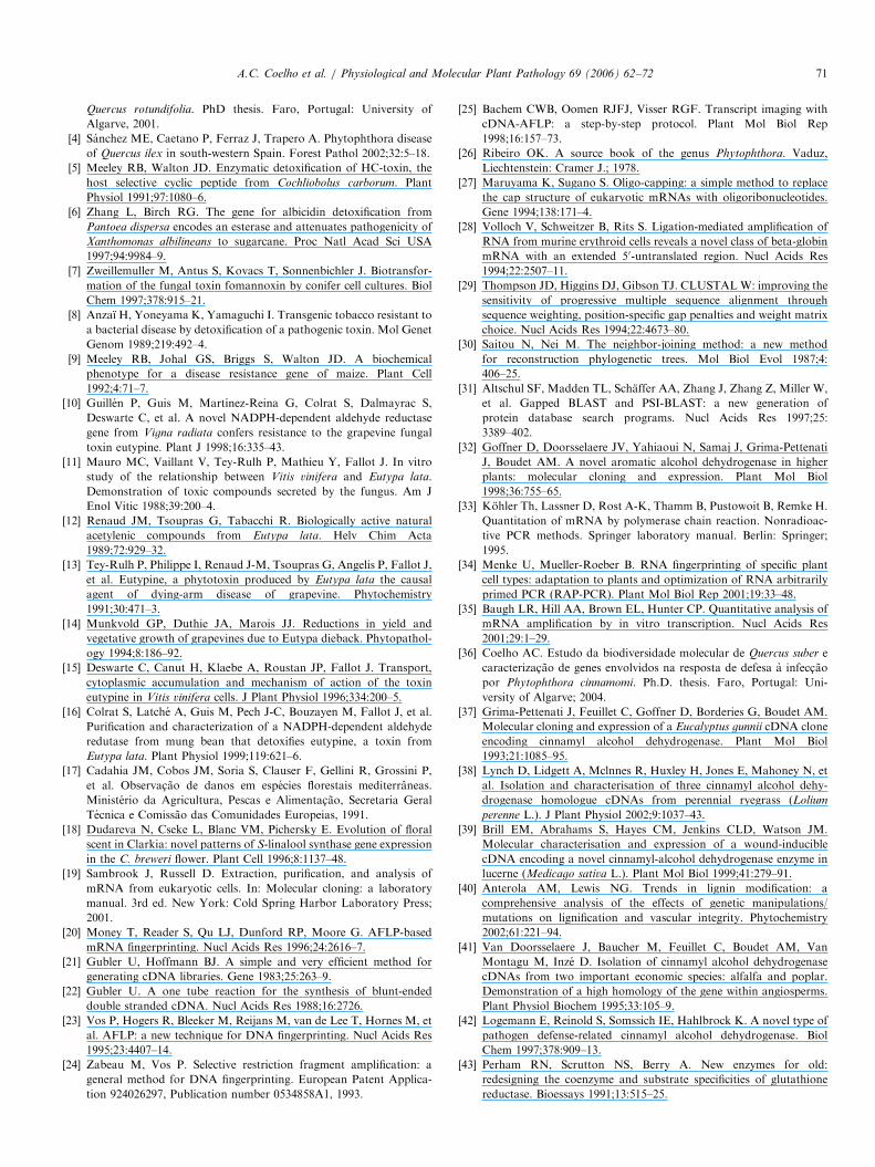
and in Eucalyptus gunnii,2 respectively. The completecDNA sequence of the gene corresponding to the C9fragment was obtained by RACE-PCR, starting from totalRNA extracted from roots of seedlings of Q. suber infectedwith P. cinnamomi. The amplification was performed withprimers defined in regions of the C9 fragment that areconserved in respect to the above referred to CADs. Theresulting cDNA was found to be 1399 bp long. It isdesignated henceforth as QsCAD1. The open readingframe (978 bp) encoded a protein of 326 amino acids witha calculated molecular mass of 35 922Da, and a calculatedisoelectric point of 7.72. The 50-UTR contained 54nucleotides and the 30-UTR contained 361 nucleotides(Fig. 2). Three putative polyadenylation signal sequencesappeared at 33, 181 and 313 nt downstream of the TAAtermination codon. QsCAD1 open reading frame codes fora protein that is 80.7% and 76.5% homologous to theCADs identified in M. x domestica and E. gunnii,respectively. QsCAD1 and C9 sequences have almostcomplete homology until nucleotide 111 of C9. Down-stream of this nucleotide, C9 and QsCAD1 sequencesdiverge significantly from each other (Fig. 3).
3.2. Increased expression of QsCAD1 gene in roots of Q.
suber infected with P. cinnamomi analysed by RT-PCR and
hybridization
To observe the expression pattern of QsCAD1, totalRNA extracted from roots of Q. suber seedlings, eithernoninfected or infected with P. cinnamomi was analysed byRT-PCR and hybridization. The synthesis of completecDNAs was primed with a 54 bp oligonucleotide (Gene-RacerTM Oligo dT Primer) containing a dT tail of 18nucleotides and a priming site of known sequence forspecific primers. In order to select specific QsCAD1
sequences, an amplification reaction was performed withserial dilutions of cDNA solution (1:50; 1:100; 1:200), andspecific primers to the QsCAD1 and to the oligonucleotideused to prime the cDNA synthesis (vide Section 2).Simultaneously, control primers for the constitutivelyexpressed gene actin were also used. These primers weredesigned in the more conserved regions of plant actin genesand amplified a 500 bp fragment. The amplificationproducts were separated on an agarose gel electrophoresis,
1GenBank accession no: AF053084.2GenBank accession no: X88797.
ARTICLE IN PRESS
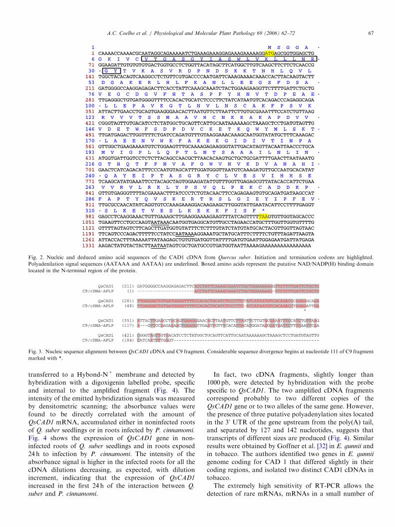
Fig. 2. Nucleic and deduced amino acid sequences of the CAD1 cDNA from Quercus suber. Initiation and termination codons are highlighted.
Polyadenilation signal sequences (AATAAA and AATAA) are underlined. Boxed amino acids represent the putative NAD/NADP(H) binding domain
located in the N-terminal region of the protein.
Fig. 3. Nucleic sequence alignment between QsCAD1 cDNA and C9 fragment. Considerable sequence divergence begins at nucleotide 111 of C9 fragment
marked with *.
A.C. Coelho et al. / Physiological and Molecular Plant Pathology 69 (2006) 62–72 67
transferred to a Hybond-N+ membrane and detected byhybridization with a digoxigenin labelled probe, specificand internal to the amplified fragment (Fig. 4). Theintensity of the emitted hybridization signals was measuredby densitometric scanning; the absorbance values werefound to be directly correlated with the amount ofQsCAD1 mRNA, accumulated either in noninfected rootsof Q. suber seedlings or in roots infected by P. cinnamomi.
Fig. 4 shows the expression of QsCAD1 gene in non-infected roots of Q. suber seedlings and in roots exposed24 h to infection by P. cinnamomi. The intensity of theabsorbance signal is higher in the infected roots for all thecDNA dilutions decreasing, as expected, with dilutionincrement, indicating that the expression of QsCAD1
increased in the first 24 h of the interaction between Q.
suber and P. cinnamomi.
In fact, two cDNA fragments, slightly longer than1000 pb, were detected by hybridization with the probespecific to QsCAD1. The two amplified cDNA fragmentscorrespond probably to two different copies of theQsCAD1 gene or to two alleles of the same gene. However,the presence of three putative polyadenylation sites locatedin the 30 UTR of the gene upstream from the poly(A) tail,and separated by 127 and 142 nucleotides, suggests thattranscripts of different sizes are produced (Fig. 4). Similarresults were obtained by Goffner et al. [32] in E. gunnii andin tobacco. The authors identified two genes in E. gunnii
genome coding for CAD 1 that differed slightly in theircoding regions, and isolated two distinct CAD1 cDNAs intobacco.The extremely high sensitivity of RT-PCR allows the
detection of rare mRNAs, mRNAs in a small number of

ARTICLE IN PRESS
Fig. 4. Expression of QsCAD gene from Q. suber non-infected (R) and infected (RI) roots. The infection was performed by putting the roots in contact
with P. cinnamomi mycelia for 24 h. RT-PCR products from total RNA were separated by agarose gel electrophoresis, transferred to a Hybond N+
membrane, denaturated and hybridized with a digoxigenin labelled probe. The intensity of the hybridization signals was measured by densitometric
scanning. Integration values of absorbance curve’s areas are shown at the bottom. Lanes 1 and 2: molecular markers; lanes 3–8: cDNA synthesis solutions
were diluted 50, 100 and 200 times as indicated and 10mL of these dilutions were used as cDNA template for amplification reactions; lane 9: actin cDNA.
A.C. Coelho et al. / Physiological and Molecular Plant Pathology 69 (2006) 62–7268
cells or in small amounts of tissue as well as mRNAsexpressed in mixed-cell populations [33–35]. The method isadequate to study the expression of genes in roots ofwoody plants, because the amount of mRNA extracted isalways very small, and usually insufficient for analysis byNorthern blot. Furthermore, the analysis by RT-PCR andhybridization can be performed in highly diluted mRNAextracts, so that the concomitant dilution of acids andphenols present in necrotic roots prevents them frominterfering with the enzymatic reactions. The specificity ofthe amplification reaction was tested by hybridization witha probe directed to the QsCAD1 cDNA. Efficiency of PCRand absence of contamination were confirmed by analysisof actin gene expression (not shown).
The ability to amplify highly diluted cDNA confirms theeffectiveness and sensitivity of the method.
3.3. Phylogenetic analysis
The amino acid sequence deduced from the QsCAD1
gene was compared with a partial sequence coding foranother potential cinnamyl alcohol dehydrogenase(QsCAD2) identified in Q. suber 36 and with 34 CADsidentified in other plant species present in the GenBank. Todetermine the phylogenetic relationship between them theamino acid sequences were aligned by the CLUSTAL Walgorithm [29] and the phylogenetic analysis was conductedwith the neighbour-joining method [30].
The phylogenetic tree shows that CAD sequencesgrouped in three distinct clusters (A–C) reflecting probablytheir biological similarities (Fig. 5). Cluster A includedCADs isolated from E. gunnii (EgCAD2; [37], Lolium
perenne (LpCAD3), [38] and Medicago sativa (MsaCAD2),[39] among others. These CAD homologues share morethan 70% identity and are implicated in the synthesis oflignin and/or suberin that use specifically cinnamylaldehydes (p-coumaryl, coniferyl and sinapyl) as sub-strates. They catalyse the NADPH-dependent reductionof these aldehydes to the corresponding alcohols [40].Eucalyptus CAD2 was classified as a zinc-containing long-chain alcohol dehydrogenase, based on the presence of theconsensus sequence GHEXXGXXXXXGXXV that in-cludes a histidine residue known to be the second ligand ofthe catalytic zinc atom [37] and on the motif GXGXXGcharacteristic of the NADP-binding domain of CADs. TheQsCAD2 partial sequence forms a subgroup with Popt-CAD from Populus tremuloides [41].The cluster B grouped CADs such as MsaCAD1
(M. sativa), [39] and PcELI3 (Petroselinum crispum), [42]whose expression is induced in response to wounding or byelicitor molecules secreted in plant–pathogen incompatibleinteractions. These enzymes accept a more diversifiedspectrum of substrates than CADs of group A such ascinnamyl, benzyl and aliphatic aldehydes, and use NADPHas co-substrate. The amino acid sequences of MsaCAD1and PcELI3 have in common with their homologue CADsfrom cluster A, the characteristic domains present in allalcohol dehydrogenases mentioned above.Cluster C grouped proteins with sequence identity higher
than 68% with VR-ERE, an NADPH-dependent reduc-tase, involved in the deactivation of eutypine, 4-hydroxy-3-(3-methyl-3-butene-1-ynyl) benzyl aldehyde, a toxin pro-duced by E. lata, the causal agent of eutypa dieback ofgrapevines (Fig. 5). In particular, QsCAD1 is highly

ARTICLE IN PRESS
Fig. 5. Phylogenetic tree constructed from amino acid sequences of plant CADs including VR-ERE. The corresponding GenBank accession numbers are
noted in the phylogenetic tree. CADs belong to the following plant species: Pinus radiata (PrCAD); Pinus taeda (PtCAD); Picea abies (PaCAD); Lolium
perenne (LpCAD1, LpCAD2, LpCAD3); Festuca arundinacea (FaCAD); Zea mays (ZmCAD); Saccharum officinarum (SoCAD); Arabidopsis thaliana
(AtCAD, AtCAD1, AtCAD2, AtCAD3, AtCAD4; AtEli3-1); Zinnia elegans (ZeCAD); Medicago sativa (MsaCAD1, MsaCAD2); Populus tremuloides
(PoptCAD); Aralia cordata (AcCAD); Eucalyptus globulus (EgbCAD); Eucalyptus gunnii (EgCAD1, EgCAD2); Eucalyptus botryoides (EbCAD);
Nicotiana tabacum (NtCAD14); Stylosanthes humilis (ShCAD1, ShCAD2); Mesembryanthemum crystallinum (McELI3); Fragaria x ananassa (FrCAD);
Petroselinum crispum (PcEli3); Malus domestica (MdCAD); Quercus suber (QsCAD1; QsCAD2); Quercus ilex subsp. rotundifolia (QrCAD1); Vigna radiata
(VR-ERE). Substrate range and motifs of coenzyme binding domains are features that differentiate clusters A–C and are explained in the text. Distances
are proportional to evolutionary divergences expressed in substitution per 100 sites and are indicated on the tree branches.
3GenBank accession nos.: CAA56103; CAA66063.
A.C. Coelho et al. / Physiological and Molecular Plant Pathology 69 (2006) 62–72 69
homologous (496%) to QrCAD1 deduced from thecDNA of Q. ilex subsp. rotundifolia (our unpublishedresults) and to MdCAD1 (480%), the protein deducedfrom the MdCAD1 gene of Malus x domestica.
EgCAD1 (E. gunnii), MdCAD1, QsCAD1, QrCAD1have completely divergent primary sequences from CADspresent in clusters A and B, lacking all the conserveddomains typical of alcohol dehydrogenases. Instead, the N-
terminal part of proteins from cluster C, is the mostconserved among all these proteins and VR-ERE, andincludes a block of 21 amino acids corresponding to theputative NAD/NADP(H) binding site [43]. This motif isalso found in proteins including cinnamoyl-CoA reductases(CCR) from Eucalyptus gunnii3 and dihydroflavonol

ARTICLE IN PRESSA.C. Coelho et al. / Physiological and Molecular Plant Pathology 69 (2006) 62–7270
reductases (DFR) from several plant species.4 It is alsopresent in the protein encoded by the drought-induciblegene CPRD14 isolated from Vigna unguiculata [44] and inthe NADPH-HC toxin reductase gene HM1 of maize [45].
4. Discussion
We propose that EgCAD1, MdCAD1, QsCAD1,QrCAD1 and AtCAD3 belong to a group of proteins thatdiverged from the CADs involved in lignification andsuberization. Like VR-ERE, they are probably enzymeswhose up-regulation is induced in plant–pathogen compa-tible interactions with or without expression of diseasesymptoms. Initially, it was suggested, on the basis of theenzymatic activity of EgCAD1, that CAD1 could functionas an alternative enzyme in the lignin biosynthetic pathway[32]. Thus, the possibility that the substrate for QsCAD1 isof host origin can not be entirely rejected. The elevatedexpression of the QsCAD1 could eventually be induced bycompounds derived from aromatic polymers releasedthrough the action of degradative enzymes produced bythe pathogen during infection. However, more recently, itwas described [46] that plants down-regulated for CAD1do not exhibit any changes in their lignin profiles butpresent significant modifications of their morphology andpostulate that CAD1 could thus control a metabolicpathway involved in the production of aromatic substancesplaying a role in morphogenesis.
EgCAD1 [32] and VR-ERE [10] share a strong affinitytowards a larger range of substrates than proteins groupedin cluster A and B, converting benzaldehydes moreefficiently than cinnamyl aldehyde derivatives. Neverthe-less, these enzymes may also have developed specifically forprocessing certain substances like toxins, for which theyshould present a higher catalytic efficiency.
The high structural homology between QsCAD1,QrCAD1, EgCAD1 and VR-ERE, suggests that they havea common enzymatic activity and a similar function. Therecombinant form of VR-ERE was shown to have thecapacity to reduce aldehydic compounds, namely eutypinein eutypinol [10]. Eutypine, 4-hydroxy-3-(3-methyl-3-bu-tene-1-ynyl) benzylaldehhyde is a toxin produced byE. lata, the causal agent of dieback of grapevines. Thetolerance of some cultivars to this disease has been shownto be correlated with their capacity to convert eutypine intothe corresponding alcohol, eutypinol, which lacks phyto-toxicity. Previous studies have shown that eutypine playsan important role in the expression of the disease. It hasbeen suggested [15] that during the growth period ofgrapevine, eutypine is transported from the trunk throughthe sap to the herbaceous parts of the vine where it inducesa number of symptoms leading to the dead of the wholebranch.
4GenBank accession nos.: AAA32783; CAA78930; CAA79154;
CAA28734.
Dieback symptoms of grapevines resemble those ex-hibited by cork oak and holm oak suffering fromP. cinnamomi infection. We propose that a mechanismsimilar to that involving eutypine in the dieback ofgrapevines caused by E. lata may apply to the suddendeath of Quercus spp. involving P. cinnamomi. Theoccurrence of leaf chlorosis, wilting and death of branches,root necrosis, and sometimes tarry exudations on trunks, isfollowed by sudden death of crowns. Trees often diesuddenly following one or two seasons of decline. Thesesymptoms observed in the field suggest that, followinginfection, P. cinnamomi produces toxins that migratethrough the sap from the roots to the leaves. Treesexhibiting sudden death symptoms are unable to detoxifythese toxins. The homology between VR-ERE andQsCAD1, and our evidence for up-regulation of the genein infected roots of cork oak seedlings, suggests adetoxifying role for the latter that may be inoperative insusceptible hosts, but partially or fully operative in hostsexhibiting chronic decline symptoms. The fragment C9partially homologous to QsCAD1, isolated and identifiedexclusively from a cork oak tree with symptoms of suddendeath, could derive from a gene coding for an inactive formof QsCAD1. The involved toxins should harbour abenzylaldehyde moiety, a common motive to substratesof VR-ERE. This is not the case of elicitins, polypeptideshaving toxic properties secreted by almost all Phytophthora
species [47]. As a matter of fact no other toxins have beenuntil now identified in Phytophthora species. Should,however these putative toxins be secreted by P. cinnamomi
they could account for the remarkably wide host rangeexhibited by this species. We are presently identifying andinvestigating the phytotoxic nature of the substancesdetected in the culture medium of P. cinnamomi.
Acknowledgements
This work was financed by the EC—III FrameworkProgramme for Research and Technological Development,co-financed by the European Social Fund (ESF) and bynational funding from Ministerio da Ciencia e do EnsinoSuperior (MCES) (POCTI/AGR/34389/2000). Horta,M. thanks Fundac- ao para a Ciencia e a Tecnologia(FCT) and ESF (EC—III Framework Programme) forher Ph.D. Grant SFRH/BD/1249/2000. The authors aregrateful to Professor C.M. Brasier for critically reading themanuscript.
References
[1] Brasier CM, Robredo F, Ferraz JFP. Evidence for Phytophthora
cinnamomi involvement in iberian oak decline. Plant Pathol
1993;42:140–5.
[2] Brasier CM. Phytophthora cinnamomi associated oak decline:
environmental constraints including climate change. Ann Sci Forest-
ieres 1996;53:347–58.
[3] Moreira-Marcelino AC. Aspectos da interacc- ao entre Phyto-
phthora cinnamomi e a doenc-a do declınio em Quercus suber e
ARTICLE IN PRESSA.C. Coelho et al. / Physiological and Molecular Plant Pathology 69 (2006) 62–72 71
Quercus rotundifolia. PhD thesis. Faro, Portugal: University of
Algarve, 2001.
[4] Sanchez ME, Caetano P, Ferraz J, Trapero A. Phytophthora disease
of Quercus ilex in south-western Spain. Forest Pathol 2002;32:5–18.
[5] Meeley RB, Walton JD. Enzymatic detoxification of HC-toxin, the
host selective cyclic peptide from Cochliobolus carborum. Plant
Physiol 1991;97:1080–6.
[6] Zhang L, Birch RG. The gene for albicidin detoxification from
Pantoea dispersa encodes an esterase and attenuates pathogenicity of
Xanthomonas albilineans to sugarcane. Proc Natl Acad Sci USA
1997;94:9984–9.
[7] Zweillemuller M, Antus S, Kovacs T, Sonnenbichler J. Biotransfor-
mation of the fungal toxin fomannoxin by conifer cell cultures. Biol
Chem 1997;378:915–21.
[8] Anzaı H, Yoneyama K, Yamaguchi I. Transgenic tobacco resistant to
a bacterial disease by detoxification of a pathogenic toxin. Mol Genet
Genom 1989;219:492–4.
[9] Meeley RB, Johal GS, Briggs S, Walton JD. A biochemical
phenotype for a disease resistance gene of maize. Plant Cell
1992;4:71–7.
[10] Guillen P, Guis M, Martınez-Reina G, Colrat S, Dalmayrac S,
Deswarte C, et al. A novel NADPH-dependent aldehyde reductase
gene from Vigna radiata confers resistance to the grapevine fungal
toxin eutypine. Plant J 1998;16:335–43.
[11] Mauro MC, Vaillant V, Tey-Rulh P, Mathieu Y, Fallot J. In vitro
study of the relationship between Vitis vinifera and Eutypa lata.
Demonstration of toxic compounds secreted by the fungus. Am J
Enol Vitic 1988;39:200–4.
[12] Renaud JM, Tsoupras G, Tabacchi R. Biologically active natural
acetylenic compounds from Eutypa lata. Helv Chim Acta
1989;72:929–32.
[13] Tey-Rulh P, Philippe I, Renaud J-M, Tsoupras G, Angelis P, Fallot J,
et al. Eutypine, a phytotoxin produced by Eutypa lata the causal
agent of dying-arm disease of grapevine. Phytochemistry
1991;30:471–3.
[14] Munkvold GP, Duthie JA, Marois JJ. Reductions in yield and
vegetative growth of grapevines due to Eutypa dieback. Phytopathol-
ogy 1994;8:186–92.
[15] Deswarte C, Canut H, Klaebe A, Roustan JP, Fallot J. Transport,
cytoplasmic accumulation and mechanism of action of the toxin
eutypine in Vitis vinifera cells. J Plant Physiol 1996;334:200–5.
[16] Colrat S, Latche A, Guis M, Pech J-C, Bouzayen M, Fallot J, et al.
Purification and characterization of a NADPH-dependent aldehyde
redutase from mung bean that detoxifies eutypine, a toxin from
Eutypa lata. Plant Physiol 1999;119:621–6.
[17] Cadahia JM, Cobos JM, Soria S, Clauser F, Gellini R, Grossini P,
et al. Observac- ao de danos em especies florestais mediterraneas.
Ministerio da Agricultura, Pescas e Alimentac- ao, Secretaria Geral
Tecnica e Comissao das Comunidades Europeias, 1991.
[18] Dudareva N, Cseke L, Blanc VM, Pichersky E. Evolution of floral
scent in Clarkia: novel patterns of S-linalool synthase gene expression
in the C. breweri flower. Plant Cell 1996;8:1137–48.
[19] Sambrook J, Russell D. Extraction, purification, and analysis of
mRNA from eukaryotic cells. In: Molecular cloning: a laboratory
manual. 3rd ed. New York: Cold Spring Harbor Laboratory Press;
2001.
[20] Money T, Reader S, Qu LJ, Dunford RP, Moore G. AFLP-based
mRNA fingerprinting. Nucl Acids Res 1996;24:2616–7.
[21] Gubler U, Hoffmann BJ. A simple and very efficient method for
generating cDNA libraries. Gene 1983;25:263–9.
[22] Gubler U. A one tube reaction for the synthesis of blunt-ended
double stranded cDNA. Nucl Acids Res 1988;16:2726.
[23] Vos P, Hogers R, Bleeker M, Reijans M, van de Lee T, Hornes M, et
al. AFLP: a new technique for DNA fingerprinting. Nucl Acids Res
1995;23:4407–14.
[24] Zabeau M, Vos P. Selective restriction fragment amplification: a
general method for DNA fingerprinting. European Patent Applica-
tion 924026297, Publication number 0534858A1, 1993.
[25] Bachem CWB, Oomen RJFJ, Visser RGF. Transcript imaging with
cDNA-AFLP: a step-by-step protocol. Plant Mol Biol Rep
1998;16:157–73.
[26] Ribeiro OK. A source book of the genus Phytophthora. Vaduz,
Liechtenstein: Cramer J.; 1978.
[27] Maruyama K, Sugano S. Oligo-capping: a simple method to replace
the cap structure of eukaryotic mRNAs with oligoribonucleotides.
Gene 1994;138:171–4.
[28] Volloch V, Schweitzer B, Rits S. Ligation-mediated amplification of
RNA from murine erythroid cells reveals a novel class of beta-globin
mRNA with an extended 50-untranslated region. Nucl Acids Res
1994;22:2507–11.
[29] Thompson JD, Higgins DJ, Gibson TJ. CLUSTALW: improving the
sensitivity of progressive multiple sequence alignment through
sequence weighting, position-specific gap penalties and weight matrix
choice. Nucl Acids Res 1994;22:4673–80.
[30] Saitou N, Nei M. The neighbor-joining method: a new method
for reconstruction phylogenetic trees. Mol Biol Evol 1987;4:
406–25.
[31] Altschul SF, Madden TL, Schaffer AA, Zhang J, Zhang Z, Miller W,
et al. Gapped BLAST and PSI-BLAST: a new generation of
protein database search programs. Nucl Acids Res 1997;25:
3389–402.
[32] Goffner D, Doorsselaere JV, Yahiaoui N, Samaj J, Grima-Pettenati
J, Boudet AM. A novel aromatic alcohol dehydrogenase in higher
plants: molecular cloning and expression. Plant Mol Biol
1998;36:755–65.
[33] Kohler Th, Lassner D, Rost A-K, Thamm B, Pustowoit B, Remke H.
Quantitation of mRNA by polymerase chain reaction. Nonradioac-
tive PCR methods. Springer laboratory manual. Berlin: Springer;
1995.
[34] Menke U, Mueller-Roeber B. RNA fingerprinting of specific plant
cell types: adaptation to plants and optimization of RNA arbitrarily
primed PCR (RAP-PCR). Plant Mol Biol Rep 2001;19:33–48.
[35] Baugh LR, Hill AA, Brown EL, Hunter CP. Quantitative analysis of
mRNA amplification by in vitro transcription. Nucl Acids Res
2001;29:1–29.
[36] Coelho AC. Estudo da biodiversidade molecular de Quercus suber e
caracterizac- ao de genes envolvidos na resposta de defesa a infecc- ao
por Phytophthora cinnamomi. Ph.D. thesis. Faro, Portugal: Uni-
versity of Algarve; 2004.
[37] Grima-Pettenati J, Feuillet C, Goffner D, Borderies G, Boudet AM.
Molecular cloning and expression of a Eucalyptus gunnii cDNA clone
encoding cinnamyl alcohol dehydrogenase. Plant Mol Biol
1993;21:1085–95.
[38] Lynch D, Lidgett A, Mclnnes R, Huxley H, Jones E, Mahoney N, et
al. Isolation and characterisation of three cinnamyl alcohol dehy-
drogenase homologue cDNAs from perennial ryegrass (Lolium
perenne L.). J Plant Physiol 2002;9:1037–43.
[39] Brill EM, Abrahams S, Hayes CM, Jenkins CLD, Watson JM.
Molecular characterisation and expression of a wound-inducible
cDNA encoding a novel cinnamyl-alcohol dehydrogenase enzyme in
lucerne (Medicago sativa L.). Plant Mol Biol 1999;41:279–91.
[40] Anterola AM, Lewis NG. Trends in lignin modification: a
comprehensive analysis of the effects of genetic manipulations/
mutations on lignification and vascular integrity. Phytochemistry
2002;61:221–94.
[41] Van Doorsselaere J, Baucher M, Feuillet C, Boudet AM, Van
Montagu M, Inze D. Isolation of cinnamyl alcohol dehydrogenase
cDNAs from two important economic species: alfalfa and poplar.
Demonstration of a high homology of the gene within angiosperms.
Plant Physiol Biochem 1995;33:105–9.
[42] Logemann E, Reinold S, Somssich IE, Hahlbrock K. A novel type of
pathogen defense-related cinnamyl alcohol dehydrogenase. Biol
Chem 1997;378:909–13.
[43] Perham RN, Scrutton NS, Berry A. New enzymes for old:
redesigning the coenzyme and substrate specificities of glutathione
reductase. Bioessays 1991;13:515–25.

ARTICLE IN PRESSA.C. Coelho et al. / Physiological and Molecular Plant Pathology 69 (2006) 62–7272
[44] Iuchi S, Yamaguchi-Shinozaki K, Urao T, Terao T, Shinozaki K.
Novel drought-inducible genes in the highly drought-tolerant Cow-
pea: cloning of cDNAs and analysis of the expression of the
corresponding genes. Plant Cell Physiol 1996;37:1037–82.
[45] Johal GS, Briggs SP. Reductase activity encoded by the HM1 disease
resistance gene in maize. Science 1992;258:985–7.
[46] Boudet A-M. Lignins and lignification: selected issues. Plant Physiol
Biochem 2000;38:81–96.
[47] Pernollet J-C, Sallantin M, Salle-Tourne M, Huet J-C. Elicitin
isoforms from seven Phytophthora species: comparison of their
physicochemical properties and toxicity to tobacco and other plant
species. Physiol Mol Plant Pathol 1993;42:53–67.
Copyright © 2022 FDOKUMEN