Intronic CA-repeat and CA-rich elements: a new class of regulators of mammalian alternative splicing
11
Intronic CA-repeat and CA-rich elements: a new class of regulators of mammalian alternative splicing Jingyi Hui 1,3 , Lee-Hsueh Hung 1,3 , Monika Heiner 1 , Silke Schreiner 1 , Norma Neumu ¨ ller 1 , Gregor Reither 1,4 , Stefan A Haas 2 and Albrecht Bindereif 1, * 1 Institut fu ¨r Biochemie, Justus-Liebig-Universita ¨t Gieen, Gieen, Germany, and 2 Department of Computational Molecular Biology, Max Planck Institute for Molecular Genetics, Berlin, Germany We have recently identified an intronic polymorphic CA-repeat region in the human endothelial nitric oxide synthase (eNOS) gene as an important determinant of the splicing efficiency, requiring specific binding of hnRNP L. Here, we analyzed the position requirements of this CA-repeat element, which revealed its potential role in alternative splicing. In addition, we defined the RNA binding specificity of hnRNP L by SELEX: not only regular CA repeats are recognized with high affinity but also certain CA-rich clusters. Therefore, we have systematically searched the human genome databases for CA-repeat and CA-rich elements associated with alternative 5 0 splice sites (5 0 ss), followed by minigene transfection assays. Surprisingly, in several specific human genes that we tested, intronic CA RNA elements could function either as splicing enhancers or silencers, depending on their proximity to the alternative 5 0 ss. HnRNP L was detected specifically bound to these diverse CA elements. These data demonstrated that intronic CA sequences constitute novel and widespread regulatory elements of alternative splicing. The EMBO Journal (2005) 24, 1988–1998. doi:10.1038/ sj.emboj.7600677; Published online 12 May 2005 Subject Categories: RNA Keywords: alternative splicing; hnRNP L; repetitive sequence; splicing enhancer; splicing silencer Introduction Messenger RNA splicing is essential for generating and regulating highly complex proteomes from a relatively small number of human genes. Tissue- and development-specific regulation of mRNA splicing can occur either on the level of splice site (ss) choice (alternative splicing) or splicing effi- ciency. Considerable efforts have been undertaken to eluci- date the cis-acting elements on the pre-mRNA that are responsible for these regulatory processes; as a result, in particular in the exon regions both positively and negatively acting regulatory elements have been described, called exonic splicing enhancers (ESE) and silencers (ESS), respectively. Among the exonic elements, several classes can be distin- guished that function as potent splicing enhancers, including both purine-rich and AC-rich sequences (Coulter et al, 1997; Fairbrother et al, 2002; reviewed by Black, 2003). In contrast to exonic elements, we know much less about characteristics and mechanisms of intronic splicing enhan- cers and -silencers (ISE and ISS elements); there are only few and diverse examples, such as uridine-rich enhancer elements downstream of alternative 5 0 ss, the UGCAUG hexa- nucleotide enhancer, or CUCUCU as an intronic splicing repression element (reviewed by Ladd and Cooper, 2002; Black, 2003). Finally, both positive and negative splicing- regulatory elements often occur clustered, assembling a regulatory complex. In general, regulatory proteins can function as activators or repressors, and mediate—by their tissue-specific expression and by interacting with the general splicing machinery— regulation of alternative splicing pathways. Among the trans-acting protein factors, which recognize either exonic or intronic elements, are members of the SR (serine-arginine- rich) protein family, members of the hnRNP (heterogenous nuclear ribonucleoprotein) family, but also other specific proteins such as TIA-1, CUGBP, PTB, ETR-like factors (CELF) proteins, and the U1 snRNP (see references in Black, 2003). In particular, RNA recognition and function of the SR proteins have been well-characterized (reviewed by Graveley, 2000); less is known about other splicing regulators. Alternative splicing is in many cases essential for under- standing human genetic diseases: Many mutations have been found to result in splicing defects and map to splicing- relevant sequences, most often to the splice site regions (Krawczak et al, 1992; Cooper and Mattox, 1997). Moreover, mutations affecting ESE or ESS have been sug- gested as a novel disease mechanism on the level of pre- mRNA splicing (Blencowe, 2000; Cartegni et al, 2002). Examples include alternative splicing of the BRCA1 gene (Liu et al, 2001) and the human SMN locus, where mutations cause spinal muscular atrophy (SMA, Cartegni and Krainer, 2002; Kashima and Manley, 2003). We have recently studied an unusual case of splicing regulation, involving the human gene for the endothelial NO synthase (eNOS); its intron 13 contains a polymorphic CA-repeat region, (CA) 14–44 , proximal to the 5 0 ss. eNOS plays a key role in vascular homeostasis, and there is an interesting correlation between the number of CA-repeat units and the risk of coronary artery disease (Stangl et al, 2000). We have established that this intronic CA-repeat element acts as an important sequence determinant for the efficiency of intron Received: 12 October 2004; accepted: 14 April 2005; published online: 12 May 2005 *Corresponding author. Institut fu ¨r Biochemie, Fachbereich Biologie, Justus-Liebig-Universita ¨t Giessen, Heinrich-Buff-Ring 58, 35392 Gieen, Germany. Tel.: þ 49 641 9935 420; Fax: þ 49 641 9935 419; E-mail: [email protected] 3 These authors contributed equally to this work 4 Present address: Institut fu ¨r Molekulare Zellbiologie, Universita ¨t des Saarlandes, 66421 Homburg, Germany The EMBO Journal (2005) 24, 1988–1998 | & 2005 European Molecular Biology Organization | All Rights Reserved 0261-4189/05 www.embojournal.org The EMBO Journal VOL 24 | NO 11 | 2005 & 2005 European Molecular Biology Organization EMBO THE EMBO JOURNAL THE EMBO JOURNAL 1988
Transcript of Intronic CA-repeat and CA-rich elements: a new class of regulators of mammalian alternative splicing

Intronic CA-repeat and CA-rich elements: a newclass of regulators of mammalian alternativesplicing
Jingyi Hui1,3, Lee-Hsueh Hung1,3,Monika Heiner1, Silke Schreiner1,Norma Neumuller1, Gregor Reither1,4,Stefan A Haas2 and Albrecht Bindereif1,*1Institut fur Biochemie, Justus-Liebig-Universitat Gie�en, Gie�en,Germany, and 2Department of Computational Molecular Biology,Max Planck Institute for Molecular Genetics, Berlin, Germany
We have recently identified an intronic polymorphic
CA-repeat region in the human endothelial nitric oxide
synthase (eNOS) gene as an important determinant of the
splicing efficiency, requiring specific binding of hnRNP L.
Here, we analyzed the position requirements of this
CA-repeat element, which revealed its potential role in
alternative splicing. In addition, we defined the RNA
binding specificity of hnRNP L by SELEX: not only regular
CA repeats are recognized with high affinity but also
certain CA-rich clusters. Therefore, we have systematically
searched the human genome databases for CA-repeat and
CA-rich elements associated with alternative 50 splice
sites (50ss), followed by minigene transfection assays.
Surprisingly, in several specific human genes that we
tested, intronic CA RNA elements could function either
as splicing enhancers or silencers, depending on their
proximity to the alternative 50ss. HnRNP L was detected
specifically bound to these diverse CA elements. These
data demonstrated that intronic CA sequences constitute
novel and widespread regulatory elements of alternative
splicing.
The EMBO Journal (2005) 24, 1988–1998. doi:10.1038/
sj.emboj.7600677; Published online 12 May 2005
Subject Categories: RNA
Keywords: alternative splicing; hnRNP L; repetitive
sequence; splicing enhancer; splicing silencer
Introduction
Messenger RNA splicing is essential for generating and
regulating highly complex proteomes from a relatively small
number of human genes. Tissue- and development-specific
regulation of mRNA splicing can occur either on the level of
splice site (ss) choice (alternative splicing) or splicing effi-
ciency. Considerable efforts have been undertaken to eluci-
date the cis-acting elements on the pre-mRNA that are
responsible for these regulatory processes; as a result, in
particular in the exon regions both positively and negatively
acting regulatory elements have been described, called exonic
splicing enhancers (ESE) and silencers (ESS), respectively.
Among the exonic elements, several classes can be distin-
guished that function as potent splicing enhancers, including
both purine-rich and AC-rich sequences (Coulter et al, 1997;
Fairbrother et al, 2002; reviewed by Black, 2003).
In contrast to exonic elements, we know much less about
characteristics and mechanisms of intronic splicing enhan-
cers and -silencers (ISE and ISS elements); there are only
few and diverse examples, such as uridine-rich enhancer
elements downstream of alternative 50ss, the UGCAUG hexa-
nucleotide enhancer, or CUCUCU as an intronic splicing
repression element (reviewed by Ladd and Cooper, 2002;
Black, 2003). Finally, both positive and negative splicing-
regulatory elements often occur clustered, assembling a
regulatory complex.
In general, regulatory proteins can function as activators or
repressors, and mediate—by their tissue-specific expression
and by interacting with the general splicing machinery—
regulation of alternative splicing pathways. Among the
trans-acting protein factors, which recognize either exonic
or intronic elements, are members of the SR (serine-arginine-
rich) protein family, members of the hnRNP (heterogenous
nuclear ribonucleoprotein) family, but also other specific
proteins such as TIA-1, CUGBP, PTB, ETR-like factors
(CELF) proteins, and the U1 snRNP (see references in Black,
2003). In particular, RNA recognition and function of the SR
proteins have been well-characterized (reviewed by Graveley,
2000); less is known about other splicing regulators.
Alternative splicing is in many cases essential for under-
standing human genetic diseases: Many mutations have been
found to result in splicing defects and map to splicing-
relevant sequences, most often to the splice site regions
(Krawczak et al, 1992; Cooper and Mattox, 1997).
Moreover, mutations affecting ESE or ESS have been sug-
gested as a novel disease mechanism on the level of pre-
mRNA splicing (Blencowe, 2000; Cartegni et al, 2002).
Examples include alternative splicing of the BRCA1 gene
(Liu et al, 2001) and the human SMN locus, where mutations
cause spinal muscular atrophy (SMA, Cartegni and Krainer,
2002; Kashima and Manley, 2003).
We have recently studied an unusual case of splicing
regulation, involving the human gene for the endothelial
NO synthase (eNOS); its intron 13 contains a polymorphic
CA-repeat region, (CA)14–44, proximal to the 50ss. eNOS plays
a key role in vascular homeostasis, and there is an interesting
correlation between the number of CA-repeat units and the
risk of coronary artery disease (Stangl et al, 2000). We have
established that this intronic CA-repeat element acts as an
important sequence determinant for the efficiency of intronReceived: 12 October 2004; accepted: 14 April 2005; publishedonline: 12 May 2005
*Corresponding author. Institut fur Biochemie, Fachbereich Biologie,Justus-Liebig-Universitat Giessen, Heinrich-Buff-Ring 58, 35392 Gie�en,Germany. Tel.: þ 49 641 9935 420; Fax: þ 49 641 9935 419;E-mail: [email protected] authors contributed equally to this work4Present address: Institut fur Molekulare Zellbiologie, Universitat desSaarlandes, 66421 Homburg, Germany
The EMBO Journal (2005) 24, 1988–1998 | & 2005 European Molecular Biology Organization | All Rights Reserved 0261-4189/05
www.embojournal.org
The EMBO Journal VOL 24 | NO 11 | 2005 &2005 European Molecular Biology Organization
EMBO
THE
EMBOJOURNAL
THE
EMBOJOURNAL
1988

13 splicing. Interestingly, the activity of this CA-repeat ele-
ment correlates with the CA-repeat length and depends on the
trans-acting factor hnRNP L (Hui et al, 2003; reviewed by
Bilbao and Valcarcel, 2003).
In the human genome, 19.4 CA repeats occur per Mb,
representing by far the most common simple-sequence repeat
(SSR) motif (Waterston et al, 2002). The functional signifi-
cance of these polymorphic sequence elements has remained
largely unclear. Together with our initial findings on the CA-
repeat element in the human eNOS gene, important questions
are raised: Do CA repeats function as widespread regulatory
elements, and is there a general role of the abundant hnRNP L
protein as a splicing regulator? Although identified already 15
years ago (Pinol-Roma et al, 1989), it was not clear so far,
what sequence determinant in the pre-mRNA is recognized
by hnRNP L.
To address these issues, we have initially used mutational
analysis of the intronic CA-repeats in the human eNOS gene;
a SELEX approach was applied to define the RNA binding
specificity of hnRNP L; sets of candidate genes were selected
from human sequence databases as potential hnRNP L tar-
gets; and alternative splicing of such CA-element containing
genes was analyzed by minigene transfections. In sum, we
demonstrate here for the first time that (1) hnRNP L recog-
nizes—in addition to regular CA-repetitive sequences—cer-
tain CA-rich clusters with high affinity; (2) such CA elements
can act—depending on their 50ss proximity—as either en-
hancers or silencers in alternative splicing regulation; and (3)
hnRNP L is involved in mediating these regulatory processes.
Thus, CA-repeat and -rich sequences have a high potential
to function as important splicing-regulatory elements in the
intronic regions of many human genes.
Results and discussion
Position requirements of the CA-repeat element:
role in 50ss activation
In the human eNOS gene, the polymorphic CA-repeat region
that functions as a length-dependent sequence determinant of
splicing efficiency is located close to the 50ss of intron 13,
starting after position þ 80 (Hui et al, 2003). To obtain more
insight into the mechanism of this unusual regulatory ele-
ment, we tested the position requirements of the enhancer
function (Figure 1). Our normal minigene derivative,
CA32(þ 80), contains eNOS exons 13 and 14 interrupted by
a shortened version of intron 13 with (CA)32. RT–PCR assays
of in vitro splicing reactions showed that splicing activity of
the normal eNOS minigene construct could be detected, as
previously described (panel A); no activity could be observed
with pre-mRNAs, where the CA repeats had been deleted or
replaced by a 64-nucleotide nonspecific sequence (data not
shown; see Hui et al, 2003).
We first moved the (CA)32 sequence to a position closer to
the 50ss, resulting in construct CA32(þ 23), which contains 32
CA repeats after position þ 23 in intron 13. Splicing of
CA32(þ 23) pre-mRNA did not alter the splicing pattern
compared with the normal substrate (compare panels A and
B). Next, we moved the (CA)32 sequence further downstream,
giving construct CA32(þ 175) (panel C). Surprisingly, the
normal spliced product was only very weakly detectable,
but instead, a larger RT–PCR product appeared during the
time course of the splicing reaction. Cloning and sequence
analysis of this additional product revealed that it reflects
usage of a cryptic 50ss mapping 37 nucleotides upstream of the
CA repeats (50-GAG/GTACAG-30; the slash marking the 50ss).
Why was the normal 50ss used only weakly in comparison
to the cryptic 50ss? Since there are three deviations from the
consensus (positions �3, þ 3, and þ 6) in the normally used
50ss, we converted it to the consensus sequence by three
point mutations (G�3-C, Gþ 3-A, Aþ 6-T; opt 50ss; see
panel D). In vitro splicing analysis for this construct,
CA32(þ 175) opt 50ss, showed that the change to the con-
sensus 50ss did reactivate the normal 50ss, but only partially;
the cryptic 50ss was still used at a similar efficiency (panel D).
These RT–PCR based results were confirmed by direct RNA
analysis with 32P-labeled RNA, using the most important
constructs, CA32(þ 80), CA32(þ 175), and CA32(þ 175)
opt50ss (panel F). In an additional control construct, the
nonspecific sequence was inserted at position þ 175: No
significant in vitro splicing activity could be observed, con-
firming that the activation of the cryptic 50ss depended on the
specific CA-repeat sequence and was not caused by changes
in the relative distances of other elements (data not shown).
We conclude that moving the CA repeats to a more down-
stream position in intron 13 resulted in activation of a novel
cryptic 50ss, mapping 37 nucleotides upstream of the CA
repeats. This implies that the CA-repeat region functions in
activating a 50ss located directly upstream of the CA repeats.
It further suggests that CA repeats can determine not only
splicing efficiency but might also be involved in 50ss selection
during alternative splicing (see further below).
Position requirements of the CA-repeat element:
intron specificity
To further study the position requirements, we also made two
constructs with the CA-repeat enhancer in an exonic context.
First, in CA32(�21) the (CA)32 sequence was inserted up-
stream of position �21 in the eNOS exon 13 (Figure 1E).
In vitro splicing of this construct demonstrated that with CA
repeats in an exonic context, no splicing stimulation could be
detected. Second, this was confirmed in a different, hetero-
logous construct consisting of exons 3 and 4 of the Drosophila
dsx gene; as previously characterized, splicing of this reporter
depends on an exonic splicing enhancer in exon 4 (Tanaka
et al, 1994; Woerfel and Bindereif, 2001). In this context, both
in the absence of an insert, with 32 CA repeats, or with a
nonspecific sequence of identical length inserted, only very
low background levels of in vitro splicing activity were
detected. In contrast, a positive control, which carries a
known strong exonic enhancer derived from the IgM exon
M2, strongly enhanced splicing activity (data not shown). In
conclusion, we have demonstrated that in two different
exonic contexts no splicing stimulation could be detected.
Therefore, the (CA)32 enhancer appears to function in an
intron-specific manner.
Intronic CA repeats are sufficient for splicing
enhancement
Next, the CA repeats were placed into a heterologous intronic
context, to test whether they are sufficient for splicing
activation. We used the T7 DUP4-1 construct, which is
derived from the human b-globin gene and contains three
exons and two introns (Figure 2A). The first and last exons
are b-globin exons 1 and 2, respectively. In between, an
Intronic elements regulating alternative splicingJ Hui et al
&2005 European Molecular Biology Organization The EMBO Journal VOL 24 | NO 11 | 2005 1989

Figure 1 Position requirements of the CA-repeat element in eNOS intron 13. (A–E) Pre-mRNAs are schematically shown on the right and arederived from exons 13–14 of the human eNOS gene with a shortened version of intron 13 containing the (CA)32 splicing enhancer (gray box).The (CA)32 enhancer is located at its normal intronic position (þ 80; A), moved upstream (þ 23; panel B), downstream (þ 175; C and D), orinto exon 13 (�21; E). In addition, for CA32(þ 175) opt 50ss, the 50 splice site (50ss; see A) was converted to the consensus sequence (opt 50ss;see D, sequence as shown with three mutated positions underlined). Each eNOS pre-mRNA derivative was spliced in vitro, and the reaction attime 0, and after 30, 60, 90, and 120 min was analyzed by RT–PCR. The positions corresponding to pre-mRNA and product mRNAs areschematically indicated on the left, predominant splicing patterns also on the right. Note that a cryptic 50ss is activated with the (CA)32
enhancer at intron position þ 175 (C and D). The band marked by the asterisk (D) represents a nonspecific PCR artifact, as determined bysequence analysis. (F) Direct RNA analysis of in vitro splicing of 32P-labeled eNOS pre-mRNA CA32(þ 80) and derivatives CA32(þ 175) andCA32(þ 175) opt50ss (compare with panels A, C, and D). Time points of 45 and 90 min have been analyzed, as indicated. The positions of pre-mRNAs, spliced products, and first-exon intermediates are indicated on the right. The gray box represents the additional exon sequenceincorporated as a result of use of the cryptic 50ss. The asterisk marks most likely a degradation product of the splicing substrate, since itoccurred splicing-independently and varied in intensity when different preparations of nuclear extract were used. M, pBR322/HpaII markers(sizes in nucleotides).
Intronic elements regulating alternative splicingJ Hui et al
The EMBO Journal VOL 24 | NO 11 | 2005 &2005 European Molecular Biology Organization1990

additional 33-nucleotide hybrid exon containing the dupli-
cated 30ss of b-globin exon 2 and the duplicated 50ss of
b-globin exon 1 had been inserted. Thus, this clone contains
two identical 50ss, two identical 30ss, and two identical
introns (intron A and intron B). In vivo, the hybrid central
exon is skipped due to its short length (Modafferi and Black,
1997), and the combination of introns A and B and the hybrid
central exon was named ‘intron C’.
Two new clones were derived from the DUP4-1 construct:
T7 DUP-CA32 and T7 DUP-control, which contain 32 CA
repeats or a 64-nucleotide control sequence, respectively,
inserted in intron B after position þ 45. Splicing activities
of these constructs were tested in vitro, using RT–PCR
analysis and primer combinations that monitor separately
the excision of the entire unit (intron C, primers 5/6.1;
Figure 2B), of intron A (primers 5/10; Figure 2C), and of
intron B (primers 9/6.1; Figure 2D).
In the original construct, T7 DUP 4-1, intron C was spliced
out in vitro only inefficiently (to B10%; see Figure 2B, lanes
DUP 4-1). Inserting the (CA)32 sequence into intron B greatly
stimulated splicing activity (to almost 30%; see Figure 2B,
lanes CA32). This effect was sequence-specific, since splicing
activity of the control construct was similar to the starting
construct (Figure 2B; compare lanes control and DUP4-1). Using
an intron A-specific primer pair showed there were no signifi-
cant differences in intron A removal between the three con-
structs (Figure 2C); in contrast, the CA-repeat sequence strongly
and specifically stimulated intron B excision (Figure 2D;
compare lanes DUP 4-1 and CA32). Moreover, insertion of
the control sequence appears to suppress splicing activity of
intron B, possibly because of the length increase of intron B in
comparison to the construct without insert (Figure 2D, lanes
control). The result that CA repeats are sufficient for specifying
splicing enhancement of the intron they reside in was repro-
Figure 2 Intronic CA repeats are sufficient for splicing enhancement. (A) Schematic representation of heterologous constructs: T7 DUP 4-1 isderived from exons 1 and 2 (white boxes) of the human b-globin gene, with an artificial short exon (gray box) between two identical introns Aand B. T7 DUP-CA32 contains in intron B the (CA)32 sequence, T7 DUP-control a nonspecific sequence (shaded boxes). The positions of theprimers used for RT–PCR assays are shown by arrows with primer names labeled. (B–D) Each of these three pre-mRNAs was spliced in vitro,and aliquots were assayed by RT–PCR at time points between 0 and 240 min (as indicated above the lanes), using primer combinationsindicative of splicing of the entire intron C unit (B), of intron A (C), and of intron B (D). Positions of products corresponding to pre-mRNAs andspliced mRNAs are schematically shown on the right. Splicing efficiencies for removing different introns were quantitated and diagrammed onthe right. In panel B, the efficiency of removing intron C was quantitated as the ratio of the mRNA signal without the central exon to the sum ofpre-mRNA and mRNA signals. In panels C and D, the efficiencies of removing intron A and B, respectively, were quantitated as the ratio of themRNA signal to the sum of pre-mRNA and mRNA signals (expressed as percentages). M, 100-bp ladder (Fermentas).
Intronic elements regulating alternative splicingJ Hui et al
&2005 European Molecular Biology Organization The EMBO Journal VOL 24 | NO 11 | 2005 1991
duced several times (for two repeats in different nuclear extract
preparations, see Supplementary Figure S1).
Taken together, these results confirm that (CA)32 has the
properties of a strong splicing-regulatory element. More im-
portantly, the CA repeats are sufficient for activating splicing
in a heterologous context, acting specifically on excision of
the intron they reside in.
Defining the RNA binding specificity of hnRNP L
by SELEX
We had previously established that hnRNP L represents the
major CA-repeat RNA binding protein (Hui et al, 2003). To
test whether repetitive CA sequences are the only target RNA
of hnRNP L, we applied an in vitro SELEX approach (sys-
tematic evolution of ligands by exponential enrichment;
Tuerk and Gold, 1990). Baculovirus-expressed GST-hnRNP L
was immobilized on glutathione-Sepharose and presented
with a large molar excess of RNA containing a randomized
20-nucleotide region flanked by two specific sequences. RNA
was bound and—after extensive washing—recovered and
amplified by RT–PCR. Following T7 transcription, this selec-
tion was repeated five times, monitoring the enrichment of
hnRNP L-bound sequences by a GST pull-down assay. After
the final amplification round, products were cloned, resulting
in 108 distinct sequences (see Supplementary Materials and
methods). From these sequences we extracted a consensus
(see Figure 3A for a graphic representation of a 10-nucleotide
consensus, and Supplementary Materials and methods for
details): Clearly, there is a high enrichment of CA dinucleo-
tides. To obtain a minimal binding motif, the occurrence of all
possible 256 tetranucleotides in the 108 selected sequences
was analyzed (see Figure 3B for the 20 most commonly
occurring tetranucleotides): As a result, ACAC and CACA
were found most frequently, representing together B16% of
all tetranucleotides in the selected sequences (high-score
motifs); these were followed by TACA and CACC, each repre-
senting B3% (low-score motifs; compared to 0.39% for any
tetranucleotide in a random sequence). In sum, this indicates
that not only regular CA repeats are among the winner
sequences but also sequences with certain deviations, most
of which maintain an alternating purine–pyrimidine pattern.
Next, we picked 11 SELEX-derived specific RNA sequences
for a more detailed analysis (Figure 3C); in addition, two
sequences of the original starting pool were included (#20 and
15). These 13 RNAs were T7-transcribed and their binding
affinities to hnRNP L determined by filter binding assays.
There are four sequences (#51, 55, 1, and 63) with the highest
apparent KD values (between 7 and 18 nM), three more with
intermediate values (#33, 105, and 26; between 32 and 71 nM),
and four others with relatively low KD values (#72, 49, 23, and
6; above 300 nM). The affinities of the two RNAs of the control
group (#20 and 15) were close to the millimolar range.
To test whether the RNA binding specificity of hnRNP L
might be based on this minimal tetranucleotide consensus,
we counted how often the high- (ACAC, CACA) and low-score
motifs (TACA, CACC) occur in these sequences (Figure 3C).
Significantly, the number of tetranucleotide motifs correlates
well with the apparent affinities: Each of the four sequences
within the top group (KD below 18 nM) carries between two
and four motifs, mostly of the high-score type; the three
sequences in the intermediate group (KD between 32 and
71 nM) contain only one or two high-score motifs each, and
the members of the low-affinity group only a single, low-score
motif. In contrast, the two control RNAs contain none of
these tetranucleotide motifs (#15 or 20). In addition, we note
that C/A-richness does not suffice for high hnRNP L binding
affinity: For example, sequence #23 consists entirely of
cytidine and adenosine nucleotides, yet shows only a single
low-score motif and binds hnRNP L with very low affinity.
Finally, this newly defined RNA-binding specificity of
hnRNP L also explains older data in the literature, where
hnRNP L had been crosslinked to specific RNAs, but where
the binding motif had not yet been recognized (see Table I).
First, Liu and Mertz (1995) had identified an RNA element in
the HSV-TK gene, called pre-mRNA processing enhancer
(PPE), to which hnRNP L could be crosslinked. The wild-
type sequence contains three closely spaced high-score motifs
(see double-underlining). In contrast, three mutant deriva-
tives, LS0, LS1, and LS2, which are defective or reduced in
hnRNP L binding (relative crosslinking efficiencies given in
parentheses), carry only one (mutants LS0 and LS1) or two
high-score motifs (mutant LS2). Therefore, there is a clear
correlation between the number of motifs and the hnRNP L
crosslinking efficiency.
Second, hnRNP L had been identified as a factor binding to
the 30 UTR region of the vascular endothelial growth factor
(VEGF) gene (Shih and Claffey, 1999). The relevant region
contains a striking cluster of two high- and three low-score
motifs (indicated by double- and single-underlining).
Third, Hahm et al (1998) identified hnRNP L as a protein
binding to the 50 end of HCV IRES element. Progressive
deletions from the 30 end (boundaries indicated by the
slashes) gradually abolished hnRNP L binding, concomi-
tantly with the loss of binding motifs.
In conclusion, hnRNP L recognizes not only regular CA
repeats with high affinity but also certain CA-rich clusters that
can be described as high- (ACAC, CACA) or low-score motifs
(TACA, CACC); those can be organized as direct tandem
repeats, or closely spaced within a short RNA region. The
number and strength of these SELEX-derived motifs appear to
be of high predictive value for hnRNP L binding. Further
experimental evidence for this was derived from analyzing
hnRNP L binding to our candidate genes (see below).
Selection of candidate genes
Using the NCBI Human Genome Maps and UniGene cluster
data, we built a data set of 31188 gene-based clusters, which
have at least one mRNA/complete cds sequence. Transcribed
sequences within each cluster were selected only if, first, all
splice sites had GT–AG, GC–AG, or AT–AC boundaries, and
second, if the quality of the alignment with the genomic
sequence (NCBI contig annotation) was greater than 95%.
Based on our SELEX analysis of the hnRNP L recognition
sequence (see above), we searched within the data set for CA-
repeat regions that contained a minimum of two CACA or
ACAC high-score motifs, allowing a spacer in between of
maximally eight nucleotides. Based on the UniGene Build
#172, a total of 155 325 CA-repeat regions were found, of
which 142 542 (92%) were located exclusively in introns,
9067 (6%) exclusively in exons, the remaining rest of 3716
(2%) in alternatively spliced regions, that is in intron or exon
regions, depending on the splicing pattern. Of the total
155 325 CA-repeat regions, 1955 (1.3%) are located within
100 bp downstream of a 50ss. Among these, there are 431
Intronic elements regulating alternative splicingJ Hui et al
The EMBO Journal VOL 24 | NO 11 | 2005 &2005 European Molecular Biology Organization1992

elements with evidence of alternative usage of the upstream
50ss. After further restricting the analysis to exon skipping
cases, we identified 147 candidate genes, for which there is
evidence of alternative inclusion or skipping of the upstream
exon, but where the distal 50ss and the downstream 30ss are
constitutive (data not shown).
Intronic CA repeats and CA-rich sequences:
role in alternative splicing
Based on our mutational studies on the CA repeats of the
human eNOS gene (see above), we tested whether CA repeats
and CA-rich intronic elements can determine alternative
splicing patterns in vivo. Therefore, we selected from the
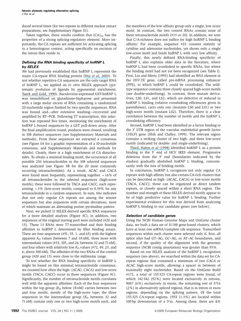
Figure 3 Defining the RNA binding specificity of hnRNP L by SELEX. (A) The 10-nucleotide consensus sequence. The frequency of each of thefour nucleotides at any position in the consensus is represented by the letter height. Boxes mark the two highly conserved core tetranucleotides.(B) Tetranucleotide frequency in selected sequences. The first 20, most common tetranucleotide sequences, are given, in the order of theirfrequencies in the 108 selected sequences (heavy line, SELEX) and in the 20 sequences taken from the initial pool (thin line, control; bothdiagrammed as percentage of the total). (C) Characteristics of 11 SELEX-derived (clone numbers on the left) and two control sequences (withasterisks; #20 and 15). Given are the individual sequences (with high-score motifs in red, low-score motifs underlined) and the KD values (innM; with standard deviations, Po0.05).
Intronic elements regulating alternative splicingJ Hui et al
&2005 European Molecular Biology Organization The EMBO Journal VOL 24 | NO 11 | 2005 1993

data sets described above four human genes, MAPK10,
GSTZ1, SLC2A2, and RFXANK, representing different lengths
of the elements and different distances to the alternatively
used 50ss, all cases of exon skipping, where CA-repeat or CA-
rich elements occur within a 100-nucleotide distance to the
upstream, alternatively used 50ss. Each of these three-exon
minigene constructs was made, using the pcDNA3 vector
under CMV promoter control. The minigene units are com-
prised of the alternatively used exon and the upstream and
downstream flanking exons (Figure 4). Only in the case of
MAPK10 and SLC2A2, the two introns had to be shortened for
experimental reasons. After transient transfection into HeLa
cells, alternative splicing patterns were analyzed by RT–PCR
from total RNA, using primers specific for the two flanking
exons. Mock controls carried out in the absence of transfected
DNA demonstrated that the splicing patterns observed in
each case were due to the transfected constructs, and that
there was no interference from endogenous mRNA.
The mitogen-activated protein kinase 10 (MAPK10) gene of
approximately 343 kb is responsible for phosphorylation of
Table I Correlation of hnRNP L crosslinking data and binding motifs
Genes Sequences Reference
HSV-TK Wild type (1X) 50-TCGCGAACATCTACACCACACAACACCGCCTCGA-30 Liu and Mertz (1995)
LS0 (0.05X) TAG G T TAGA
LS1 (0.45X) G TCT
LS2 (0.6X) AGATCT
VEGF 50-AGACACACCCACCCACATACATACAT-30 Shih and Claffey (1999)
HCV 50-AGCACAAATCCT/AAACCTCAAAGAAAAACC/AAAAGAAACACAAACCGCCGCCCACAGG-30 Hahm et al (1998)
High- and low-score motifs are indicated by double- and single-underlining, respectively. The relative crosslinking efficiencies for wild type andmutant sequences of the HSV-TK gene are given in parentheses. The sequence for HCV IRES element corresponds to the 50 end of HCV mRNA(nucleotides 345–402; Hahm et al, 1998). The slashes indicate the boundaries of different 30 truncations (after positions 356 and 374).
Figure 4 CA-repeat and CA-rich sequences function as regulatory elements of alternative splicing. Alternative splicing of four representativecandidate genes that contain intronic CA-repeat elements (MAPK10, SLC2A2, RFXANK) or a CA-rich sequence (GSTZ1) has been characterized,using minigenes diagrammed on the right. The alternatively used exon is indicated by the dark box, and intron and exon parts are shown inscale. Wild type (WT) and substituted (sub) sequence elements are given, with boundaries relatively to the 50ss of the second intron. Verticallines within the introns mark the positions where introns have been shortened. Minigenes were transfected into HeLa cells, and alternativesplicing tested by RT–PCR (lanes WT, sub); the control transfections (lanes mock) were carried out in the absence of DNA. The positionscorresponding to pre-mRNA and alternative mRNA products are indicated to the right of the gel pictures. For the GSTZ1, SLC2A2, and RFXANKminigenes, the percentages of exon inclusion with standard deviations (n¼ 3) are given below the corresponding lanes.
Intronic elements regulating alternative splicingJ Hui et al
The EMBO Journal VOL 24 | NO 11 | 2005 &2005 European Molecular Biology Organization1994
transcription factor Jun. Alternative splicing through skip-
ping or inclusion of exon 6 (NM_002753) is documented, and
our minigene covers exons 5–7, with shortened introns in
between (Figure 4). There is an extended CA-repeat region,
(CA)3C(CA)25, starting 61 nucleotides downstream from the
50ss of exon 6, which in the substitution was replaced by a
nonspecific sequence of identical length.
Substituting the long CA-repeat element resulted in com-
plete suppression of the normal splice form (exons 5–6–7);
instead, the complete intron 6 was retained, indicating that
the CA repeats are necessary for efficient splicing of intron 6.
In addition, we observed with the altered minigene a minor
splice variant, which originated from activation of a cryptic
50ss within intron 6, downstream of the substituted sequence
(as determined by cloning and sequence analysis; data
not shown). In sum, this provides evidence for an intronic
CA-repeat element functioning as a splicing enhancer.
GSTZ1 encodes a multifunctional enzyme with the glu-
tathione S-transferase (Zeta-1 type) activity, conjugating
glutathione to a wide variety of substrates, as well as with
maleylacetoacetate isomerase activity, which is important in
the catabolism of phenylanaline and tyrosine. The 10.6-kb
gene of nine exons (NM_145870) carries in its intron 5 a CA-
rich region downstream of the alternatively used exon 5
(positions þ 14 to þ 57; Figure 4). To analyze a potential
role of the CA-rich region in alternative splicing, we con-
structed a minigene with exons 4–6; in the substitution
construct, the CA-rich sequence was replaced by a non-
specific sequence of corresponding length.
Transfection of the GSTZ1 minigene gave almost exclu-
sively the constitutive splicing pattern (exons 4–5–6; 9970.6%). However, after substitution the skipped form (exons
4–6) became the predominant form, and only 3071.4% were
included. Therefore, we conclude that a CA-rich element in
50ss proximity can also act as a splicing enhancer.
Next we analyzed two cases, SLC2A2 and RFXANK, where
CA-repeat elements reside in the immediate neighbourhood
of 50ss. First, the SLC2A2 gene of approximately 30 kb con-
tains 11 exons (NM_000340), coding for a specialized glucose
transporter molecule, GLUT2, which is expressed in the
b-cells of pancreatic islets and likely functions as a glucose
sensor. Its exon 4 is included or skipped, and the 50ss of exon
4 is followed by a (CA)19 sequence starting at position þ 7 of
intron 4 (Figure 4). In this minigene the two introns had to be
shortened, and in the substitution derivative, (CA)19 was
replaced by a nonspecific sequence.
For the wild-type version of this minigene, we detected
mostly the mRNA with exons 3–4–5 joined with each other
(8172.2%), and only a small fraction of skipped mRNA
(exons 3–5). In case of the altered minigene, inclusion of
exon 4 increased to 8970.9%, and the skipped form was
hardly detectable, indicating a negative effect of the CA-repeat
region on splicing (silencer effect).
Second, the RFXANK (regulatory factor X-associated an-
kyrin-containing) gene, which codes for a transactivator of
MHC class II genes, is organized in 10 exons spanning 9.7 kb
(NM_003721). Exon 5 is alternative, and a short CA-repeat
region [C(CA)4] starts at position þ 9 of the downstream
intron (Figure 4). Our minigene construct comprises exons
4–6 and the full-length introns. In the substitution derivative
the wild-type CA-repeat sequence, [C(CA)4], was replaced
by 50-AGCTCGATC-30.
For the RFXANK minigene, a similar effect was detected as
for SLC2A2: The distribution of the included (exons 4–5–6)
versus the skipped form (exons 4–6) was clearly shifted
towards exon inclusion by the substitution (percentage of
exon inclusion: 51712% for the wild type, 8376.3% for the
substitution); this indicates a splicing silencer role of the
short CA-repeat region, C(CA)4.
HnRNP L binds specifically to CA-rich elements
of candidate genes
As previously demonstrated by UV-crosslinking experiments
(Hui et al, 2003), hnRNP L binds directly and with high
affinity to CA-repeat RNAs ((CA)32, (CA)20, and (CA)10).
Since we have shown here for the first time, that CA-repeat
and CA-rich RNA elements can regulate alternative splicing,
we next determined whether hnRNP L directly interacts also
with CA-rich (GSTZ1) and with very short CA-repeat se-
quences (RFXANK).
First, we tested this by GST pull-down assays with recom-
binant GST-hnRNP L protein (Figure 5A). 32P-labeled RNAs of
44 nucleotides each were transcribed, covering the CA-ele-
ments of GSTZ1 and RFXANK, as well as their corresponding
substituted sequences of identical length; (CA)20 served as
positive control. These RNAs were incubated with baculo-
virus-expressed GST-hnRNP L, which had been immobilized
on glutathione-Sepharose. After extensive washing, bound
RNAs were recovered and analyzed by denaturing gel elec-
trophoresis. As expected, more than 50% of the input (CA)20
RNA bound to hnRNP L (lanes CA20). The CA-rich element of
the wild-type GSTZ1 sequence bound very efficiently as well,
whereas no significant bound material could be recovered
from the corresponding substituted sequence (compare lanes
wild type (WT) and substitution (S) for GSTZ1). This can be
nicely explained by the presence of five high- and one low-
score motifs (underlined) in the GSTZ1 wild-type element; in
contrast, the GSTZ1 substitution sequence carries none of
these motifs:
GSTZ1, wild type:
ACACTTGCACCCTTGCACACCTGACACACTCTTACACTCACACA
GSTZ1, substitution:
CTAGTAACGGCCGCCAGTGTGCTGGAATTCTGCAGATATCCATC
The RFXANK element also bound hnRNP L in a sequence-
specific manner (compare lanes wild type (WT) and substitu-
tion (S) for RFXANK), although much less efficiently than the
GSTZ1 element and (CA)20 did. Consistent with the high
predictive value of the binding motifs described (see above
under our SELEX analysis), the RFXANK wild-type sequence
contains only two high-score motifs in direct tandem orienta-
tion (CCACACACA); the RFXANK substitution derivative
(AGCTCGATC), however, carries no motif.
Second, we did in vitro binding assays in nuclear extract,
using immunoprecipitations of the endogenous hnRNP L
protein (Figure 5B). Short 32P-labeled RNAs of between 44
and 50 nucleotides in length were incubated in nuclear
extract, followed by anti-hnRNP L immunoprecipitation. In
addition to GSTZ1 wild type and substituted sequences, we
included the CA-repeat element of SLC2A2 (wild type versus
substitution), as well as CA20 as a control. The MAPK10
element was not tested, since its long CA-repeat stretches are
Intronic elements regulating alternative splicingJ Hui et al
&2005 European Molecular Biology Organization The EMBO Journal VOL 24 | NO 11 | 2005 1995

very similar to the SLC2A2 element. These more stringent
assays clearly showed that endogenous hnRNP L associates
very efficiently with the wild-type sequences of both GSTZ1
and SLC2A2 elements, as well as with CA20; in contrast,
binding to the substitution derivatives was undetectable.
Binding of hnRNP L to the RFXANK element under these
conditions was not significant and similar for wild type and
altered versions (data not shown).
Third, we studied whether hnRNP L contacts the RNA
elements directly, using a combined crosslinking/immuno-
precipitation protocol (Figure 5C): Again, both GSTZ1 and
SLC2A2 elements were incubated in nuclear extract, resulting
in crosslinking of an hnRNP L-sized protein band for both the
GSTZ1 and SLC2A2 element, as well for CA20; the identity
of hnRNP L was subsequently confirmed by anti-hnRNP L
immunoprecipitation from the crosslinking reactions.
In sum, hnRNP L directly binds in extract both the CA-rich
element of GSTZ1, which functions as an enhancer, and the
CA-repeat silencer element of SLC2A2. Thus, the same protein
can recognize both CA-repeat and CA-rich RNA elements and
may participate in both enhancer and silencer functions.
Although the short CA element in RFXANK has a clear silencer
role (see Figure 4), hnRNP L binding could be detected only
with purified recombinant protein (see Figure 5A), and only at
a very low level. However, this is not surprising, since in our
earlier study we had found that an RNA containing only 10 CA
repeats crosslinked only very weakly to hnRNP L (Hui et al,
2003). Therefore, in this case hnRNP L binding is probably
functionally not significant, and for such short CA elements
proteins in addition to or alternatively to hnRNP L may
assemble to enhancer/silencer complexes.
We conclude that CA-rich or CA-repeat elements can
regulate alternative splice site choice either positively or
negatively, and hnRNP L may participate in both processes.
Based on the selection of genes analyzed, it appears that
short distances of the CA-sequence to the alternative 50ss
correlate with a silencer role (SLC2A2, RFXANK), longer
distances with enhancer function (MAPK10, GSTZ1, eNOS).
A likely explanation would be that hnRNP L (or alternative
proteins) can work as an activator of the 50ss only from a
certain distance (as demonstrated for the eNOS CA-repeat
enhancer; Hui et al, 2003); in contrast, bound at a 50ss-
proximal position, proteins may sterically block recogition
of the 50ss by the general machinery. What specifies positive
or negative regulation, whether additional proteins or inter-
actions are responsible and sensitive to the 50ss proximity, is
of particular interest and remains to be investigated by
detailed mechanistic studies.
Materials and methods
Oligonucleotides, constructsThe sequences of all oligonucleotides are listed in SupplementaryMaterials and methods. All constructions were confirmed bysequence analysis.
pT7 DUP-CA32 and pT7 DUP-control. The parent plasmid fromwhich these two clones were made was pT7 DUP4-1, which is a T7version of pDUP4-1 (Modafferi and Black, 1997). It contains threeexons and two introns derived from human b-globin sequence. Afragment containing 32 CA repeats was generated by annealing twooligonucleotides, DUP1 and DUP2, and cloned into the BglII site in thesecond intron of pT7 DUP4-1, resulting in pDUP-CA32. Clone pDUP-control was constructed by insertion of a 64-nucleotide nonspecificsequence into pT7 DUP4-1 at the BglII site. The 64-nucleotidesequence was amplified from the polylinker region of pGEM5Zf(þ )(Promega, USA), using oligonucleotides DUP3 and DUP4.
pGEM-CA32(�21), pGEM-CA32(þ 23), pGEM-CA32(þ 175), andpGEM-opt 50ss. A fragment containing 32 CA repeats was generated
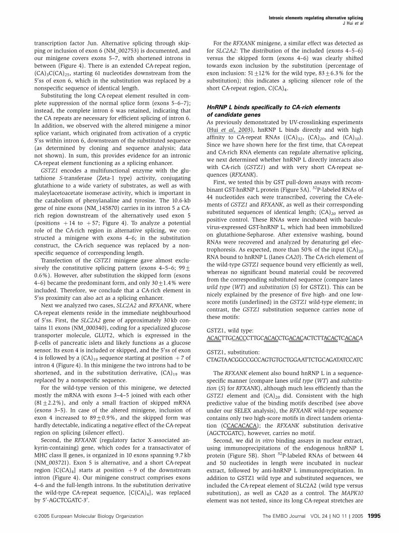
Figure 5 HnRNP L binds specifically to intronic CA-repeat and CA-rich elements of candidate genes. (A) GST-hnRNP L pull-downassays. 32P-labeled short RNAs containing the CA-repeat and CA-rich elements of candidate genes GSTZ1 and RFXANK (wild type,lanes WT, and substitution version, lanes S) as well as the positivecontrol (CA)20 (lanes CA20) were incubated with GST-hnRNP L,which had been immobilized on glutathione-Sepharose. BoundRNAs were recovered and analyzed by denaturing gel electrophor-esis. For each pull-down assay, 10% of the input was loaded (lefthalf of figure) and the total amount of recovered material (righthalf). M, markers (36, 67, and 76 nucleotides). (B) Anti-hnRNP Limmunoprecipitation assays from HeLa nuclear extract. 32P-labeledshort RNAs containing the CA-repeat and CA-rich elements ofcandidate genes GSTZ1 and SLC2A2 (wild type, lanes WT, andsubstitution version, lanes S) as well as the positive control (CA)20
(lanes CA20) were incubated in HeLa nuclear extract. Followingimmunoprecipitation with anti-hnRNP L antibodies, coprecipitatedRNAs were analyzed by denaturing gel electrophoresis. For eachassay, 10% of the input was loaded (left half of figure) and the totalof immunoprecipitated material (right half). M, markers (36, 67,and 76 nucleotides). (C) Coupled UV-crosslinking/anti-hnRNP Limmunoprecipitation assays. Incubations as described in panel Bwere subjected to UV-crosslinking, followed by anti-hnRNP L im-munoprecipitation. For each assay, 10% of the reaction after cross-linking was loaded (left half of figure) and the total of crosslinked/immunoprecipitated material (right half). The arrow indicates themobility of crosslinked hnRNP L (protein markers in kDa).
Intronic elements regulating alternative splicingJ Hui et al
The EMBO Journal VOL 24 | NO 11 | 2005 &2005 European Molecular Biology Organization1996

by annealing two oligonucleotides, DUP1 and DUP2, and clonedinto the BamHI site in the eNOS exon 13 of pGEM-CA32 (Huiet al, 2003), resulting in pGEM-CA32(�21). To construct pGEM-CA32(þ 23) and pGEM-CA32(þ 175), a BglII site was introduced inpGEM-CA32 at positions þ 23 and þ 175 of intron 13, using a two-step PCR mutagenesis method and oligonucleotides, EN1, EN2,þ 23up, þ 23dw, þ 175up, and þ 175dw. The annealed DUP1 andDUP2 oligonucleotides containing 32 CA repeats were subsequentlyinserted into the introduced BglII site. To make pGEM-opt 50ss, aPCR fragment containing three point mutations at the 50ss of intron13 was amplified using oligonucleotides css and EN2 with pGEM-CA32(þ 175) as template, and cloned between the BamHI and XbaIsites of pGEM-CA32.
pDSX-CA32. A fragment containing 32 CA repeats was generated byannealing two oligonucleotides, DSX1 and DSX2, and cloned intothe XbaI and HindIII sites of pDSX-XH (Woerfel and Bindereif,2001), resulting in pDSX-CA32.
Minigene constructs. All minigene constructs are composed of threeexons and two introns. The genomic sequences of the minigene unitwere amplified by using genomic DNA isolated from primaryHUVEC cells as template (kindly provided by Dr Karl Stangl,Charite, Berlin). To construct the wild-type GSTZ1 and RFXANKminigenes, PCR fragments carrying the complete minigene unitwere generated by primer pairs GSTZ-C1/-C2, and RFXANK-C1/-C2,and inserted into pcDNA3 vector (Invitrogen, USA). In the case ofMAPK10 and SLC2A2, most of the intron sequences was deletedfrom 100 nt downstream of the 50ss to 100 nt upstream of the 30ss,except in the second intron of MAPK10 minigene, where the deletedsequence starts 150 nt downstream of the 50ss to 100 nt upstream ofthe 30ss. A series of PCRs were performed, using oligonucleotidesMAPK-C1 to -C6 and SLC2A2-C1 to -C6 as primers. A completefragment was assembled by a second-step PCR, using the previousPCR products as templates, and inserted into the correspondingsites of pcDNA3 vector. In the substituted minigene constructs, theCA repeats or C/A-rich sequences in the wild-type minigeneconstructs were replaced by nonspecific control sequences, usinga similar two-step PCR mutagenesis method and oligonucleotidesMAPK-C1, -C2, and -C7 to -C10; GSTZ-C1, -C2, and -C5 to -C8;SLC2A2-C1, -C2, -C7, and -C8; RFXANK-C1 to -C4. The nonspecificsequences in SLC2A2, GSTZ1, and MAPK10 substitution constructswere amplified from the polylinker region of pcDNA3.
In vitro transcription and splicingpDUP and pGEM derivatives were cut with XbaI and transcribed inthe presence of m7GpppG cap analog; 10 ng of pre-mRNA per 25-mlreaction was spliced in HeLa nuclear extract (Bindereif and Green,1987). Splicing activity was detected by denaturing polyacrylamidegel electrophoresis or by RT–PCR analysis, using oligonucleotidesEN2 and DUP6.1 as primers for RT, and oligonucleotides EN1, EN2,DUP5, DUP6.1, DUP9, and DUP10 as primers for PCR. 32P-labeledshort RNAs for GST pull-down assays were in vitro transcribed asabove, except that [32P]CTP was used. DNA templates fortranscription were obtained by PCR, using primers GSTZ-U1 to-U4, and RFXANK-U1 to -U3, and the corresponding minigeneconstructs as templates, or alternatively, by annealing oligonucleo-tides SLC2A2-U1/-U2 and -U3/U4.
Recombinant hnRNP L-GST fusion proteinTo construct pFASTBAC HTb-hnRNP L-GST, the KpnI–HindIIIcleavage product, which contains the 30 end and 30-UTR of hnRNPL in pFASTBAC HTb-hnRNP L (Hui et al, 2003), was substituted bya fragment obtained by a two-step PCR method. For the first set ofPCRs, the DNA fragments encoding the C-terminus of hnRNP L andGST protein were amplified using oligonucleotide pairs LGST-1/-2,LGST-3/-4, respectively. A second-step PCR was performed withprimers LGST-1 and -4, and the above fragments as templates. Theresulting PCR product was subsequently digested by KpnI andHindIII, and cloned into pFASTBAC HTb-hnRNP L. Recombinantbaculoviruses were generated to express C-terminally GST-taggedhnRNP L in Sf21 cells according to the protocols of Invitrogen,followed by purification through glutathione-Sepharose (AmershamBiosciences, Sweden) and dialysis against buffer D (20 mM HEPES,pH 8.0, 20% glycerol, 100 mM KCl, 0.2 mM EDTA, 1 mM DTT, and0.5 mM PMSF) at 41C for 3 h.
SELEXThe SELEX procedure and the analysis of the consensus sequenceare described in the Supplementary Materials and methods.
Filter binding assayThe filter-binding assay was carried out on a 96-well vacuumfiltration apparatus (BioRad, USA). Approximately 50 fmol in vitrotranscribed, [32P]CTP-labeled RNA was incubated with differentamounts of baculovirus-expressed hnRNP L-GST protein in a finalvolume of 50ml of binding buffer (10 mM Tris–Cl pH 8.0, 100 mMKCl, 2.5 mM MgCl2, 0.01% NP40, 10% glycerol, and 0.2 mg/mlBSA) at room temperature for 20 min. The binding reaction wasapplied onto the nitrocellulose membrane (0.45 mm, Macherey-Nagel, Germany), which had been preincubated with washingbuffer (10 mM Tris–Cl pH 8.0, 100 mM KCl, and 2.5 mM MgCl2) atroom temperature for 30 min. After each well was washed five timeswith washing buffer, the filter was subsequently dried andquantitated by a phosphorimager system (Biorad, USA).
Cell transfection, RNA isolation, RT–PCR analysisThe day before transfection, 5�105 HeLa cells were seeded on a6-cm tissue culture dish. Minigene constructs (8mg) was transfectedby the calcium phosphate method (Sambrook et al, 1989). Cellswere harvested 2 days after transfection. Total RNA was isolatedusing guanidinium thiocyanate (Xie and Rothblum, 1991). TotalRNA (1mg) was annealed to 2 pmol vector-specific primer bGH Rev(Invitrogen, USA) and reverse-transcribed by SuperScriptTM IIIRNase-H-minus reverse transcriptase (Invitrogen, USA), accordingto the manufacturer’s instruction. The resulting first-strand cDNAwas further amplified by PCR, using primers against the flankingexons. The PCR conditions were optimized for each minigene.
GST pull-down assayBaculovirus-expressed hnRNP L-GST fusion protein (5 pmol)immobilized on glutathione-Sepharose beads was incubated with120 fmol in vitro transcribed, [32P]CTP-labeled short RNAs in a finalvolume of 200 ml of binding buffer (20 mM HEPES pH 7.5, 100 mMKCl, 10 mM MgCl2, 1 mM DTT, and 0.01% NP40). The short RNAscarry the CA sequences or the corresponding substituted sequencesderived from the GSTZ1 and RFXANK minigene constructs. Afterbinding at room temperature for 20 min, the beads were washedfour times with 1 ml each of washing buffer (20 mM HEPES pH 7.5,300 mM KCl, 10 mM MgCl2, 1 mM DTT, and 0.01% NP40), followedby treatment with 80mg of proteinase K in 200 ml PK buffer (100 mMTris–Cl pH 7.5, 12.5 mM EDTA, 150 mM NaCl, and 1% SDS) at 371Cfor 30 min. Bound RNA was recovered by extraction with phenol/chloroform and ethanol precipitation, and separated on a 15%denaturing polyacrylamide gel.
In vitro RNA binding/immunoprecipitationIn total, 4 ng of in vitro transcribed, [32P]CTP-labeled short RNAswas incubated at 301C for 15 min in HeLa nuclear extract understandard splicing conditions (Bindereif and Green, 1987). Forimmunoprecipitation experiments, 100ml packed volume of proteinA Sepharose CL-4B (Amersham Pharmacia Biotech) in N100 buffer(100 mM NaCl, 50 mM Tris–Cl pH 8.0, 0.05% NP-40) were firstincubated overnight with 10ml of anti-hnRNP L antibody 4D11 at41C, and washed five times with 1 ml of N100. In total, 20mlpacked volume of beads in 175ml of N100 were then incubatedtogether with 25ml of splicing reaction at 41C for 2 h with rotation.The beads were washed three times with 1 ml of N100 buffer,and heated at 801C for 10 min in 200ml of PK buffer. Immuno-precipitated RNA was extracted by phenol/chloroform followedby ethanol precipitation, and separated on a 12% denaturingpolyacrylamide gel.
UV-crosslinking/immunoprecipitationUV-crosslinking was performed as described (Hui et al, 2003).Briefly, 8 ng of in vitro transcribed, [32P]CTP-labeled short RNAswas incubated at 301C for 15 min in HeLa nuclear extract (withtRNA added to a final concentration of 0.2 mg/ml). UV-crosslinkingwas carried out on ice for 20 min with 254 nm UV light at anintensity of 2700mW/cm2. Unprotected RNA was digested withRNase A (1 mg/ml) at 371C for 20 min. For immunoprecipitation,20ml of protein A Sepharose beads (prepared as above) in 175 ml ofN100 were incubated with the UV-crosslinking reaction at 41C for 2 h
Intronic elements regulating alternative splicingJ Hui et al
&2005 European Molecular Biology Organization The EMBO Journal VOL 24 | NO 11 | 2005 1997

with rotation. The beads were then washed three times with 1 ml ofN100 buffer. Immunoprecipitated proteins were fractionated on a12% SDS–polyacrylamide gel.
Supplementary dataSupplementary data are available at The EMBO Journal Online.
Acknowledgements
We thank Karl Stangl (Charite, Berlin) for providing genomic DNAsamples, Doug Black for construct pDUP4-1, and Gideon Dreyfuss
for anti-hnRNP L monoclonal antibody 4D11. We also thank UweKuhn (University of Halle) and Peter Friedhoff (University ofGiessen) for help with filter binding assay, Martin Vingron (MPIfor Molecular Genetics, Berlin) for discussions, and AndreyDamianov (University of Giessen) for comments on the manuscript.This work was supported by the Deutsche Forschungsgemeinschaft(Grants Bi 316/10-1, 10-2, and 10-3 to AB).Note added in proof:We note that hnRNP L has recently been found to repress exonsplicing via an exonic splicing silencer (CR Rothrock, AE House,KW Lynch, manuscript submitted).
References
Bilbao D, Valcarcel J (2003) Getting to the heart of a splicingenhancer. Nat Struct Biol 10: 6–7
Bindereif A, Green MR (1987) An ordered pathway of snRNPbinding during mammalian pre-mRNA splicing complex assem-bly. EMBO J 6: 2415–2424
Black DL (2003) Mechanisms of alternative pre-messenger RNAsplicing. Annu Rev Biochem 72: 291–336
Blencowe BJ (2000) Exonic splicing enhancers: mechanism ofaction, diversity and role in human genetic diseases. TrendsBiochem Sci 25: 106–110
Cartegni L, Chew SL, Krainer AR (2002) Listening to silence andunderstanding nonsense: exonic mutations that affect splicing.Nat Rev Genet 3: 285–298
Cartegni L, Krainer AR (2002) Disruption of an SF2/ASF-dependentexonic splicing enhancer in SMN2 causes spinal muscular atro-phy in the absence of SMN1. Nat Genet 30: 377–384
Cooper TA, Mattox W (1997) The regulation of splice-site selection,and its role in human disease. Am J Hum Genet 61: 259–266
Coulter LR, Landree MA, Cooper TA (1997) Identification of a newclass of exonic splicing enhancers by in vivo selection. Mol CellBiol 17: 2143–2150
Fairbrother WG, Yeh RF, Sharp PA, Burge CB (2002) Predictiveidentification of exonic splicing enhancers in human genes.Science 297: 1007–1013
Graveley BR (2000) Sorting out the complexity of SR proteinfunctions. RNA 6: 1197–1211
Hahm B, Kim YK, Kim JH, Kim TY, Jang SK (1998) Heterogeneousnuclear ribonucleoprotein L interacts with the 30 border ofthe internal ribosomal entry site of hepatitis C virus. J Virol 72:8782–8788
Hui J, Stangl K, Lane WL, Bindereif A (2003) HnRNP L stimulatessplicing of the eNOS gene by binding to variable-length CArepeats. Nat Struct Biol 10: 33–37
Kashima T, Manley JL (2003) A negative element in SMN2 exon 7inhibits splicing in spinal muscular atrophy. Nat Genet 34: 460–463
Krawczak M, Reiss J, Cooper DN (1992) The mutational spectrumof single base-pair substitutions in mRNA splice junctions ofhuman genes: causes and consequences. Hum Genet 90: 41–54
Ladd AN, Cooper TA (2002) Finding signals that regulate alter-native splicing in the post-genomic era. Genome Biol 3,reviews 0008
Liu HX, Cartegni L, Zhang MQ, Krainer AR (2001) A mechanism forexon skipping caused by nonsense or missense mutations inBRCA1 and other genes. Nat Genet 27: 55–58
Liu X, Mertz JE (1995) HnRNP L binds a cis-acting RNA sequenceelement that enables intron-independent gene expression. GenesDev 9: 1766–1780
Modafferi EF, Black DL (1997) A complex intronic splicing enhancerfrom the c-src pre-mRNA activates inclusion of a heterologousexon. Mol Cell Biol 17: 6537–6545
Pinol-Roma S, Swanson MS, Gall JG, Dreyfuss G (1989) A novelheterogeneous nuclear RNP protein with a unique distribution onnascent transcripts. J Cell Biol 109: 2575–2587
Sambrook J, Fritsch EF, Maniatis T (1989) Molecular Cloning: ALaboratory Manual. Cold Spring Harbour, New York, USA: ColdSpring Harbour Laboratory Press
Shih SC, Claffey KP (1999) Regulation of human vascular endo-thelial growth factor mRNA stability in hypoxia by heterogeneousnuclear ribonucleoprotein L. J Biol Chem 274: 1359–1365
Stangl K, Cascorbi I, Laule M, Klein T, Stangl V, Rost S, WerneckeKD, Felix SB, Bindereif A, Baumann G, Roots I (2000) High CArepeat numbers in intron 13 of the endothelial nitric oxidesynthase gene and increased risk of coronary artery disease.Pharmacogenetics 10: 133–140
Tanaka K, Watakabe A, Shimura Y (1994) Polypurine sequenceswithin a downstream exon function as a splicing enhancer. MolCell Biol 14: 1347–1354
Tuerk C, Gold L (1990) Systematic evolution of ligands byexponential enrichment: RNA ligands to bacteriophage T4 DNApolymerase. Science 249: 505–510
Waterston RH, Lindblad-Toh K, Birney E, Rogers J, Abril JF, AgarwalP, Agarwala R, Ainscough R, Alexandersson M, An P, AntonarakisSE, Attwood J, Baertsch R, Bailey J, Barlow K, Beck S, Berry E,Birren B, Bloom T, Bork P, Botcherby M, Bray N, Brent MR,Brown DG, Brown SD, Bult C, Burton J, Butler J, Campbell RD,Carninci P, Cawley S, Chiaromonte F, Chinwalla AT, ChurchDM, Clamp M, Clee C, Collins FS, Cook LL, Copley RR,Coulson A, Couronne O, Cuff J, Curwen V, Cutts T, Daly M,David R, Davies J, Delehaunty KD, Deri J, Dermitzakis ET, DeweyC, Dickens NJ, Diekhans M, Dodge S, Dubchak I, Dunn DM, EddySR, Elnitski L, Emes RD, Eswara P, Eyras E, Felsenfeld A, FewellGA, Flicek P, Foley K, Frankel WN, Fulton LA, Fulton RS, FureyTS, Gage D, Gibbs RA, Glusman G, Gnerre S, Goldman N,Goodstadt L, Grafham D, Graves TA, Green ED, Gregory S,Guigo R, Guyer M, Hardison RC, Haussler D, HayashizakiY, Hillier LW, Hinrichs A, Hlavina W, Holzer T, Hsu F, HuaA, Hubbard T, Hunt A, Jackson I, Jaffe DB, Johnson LS, JonesM, Jones TA, Joy A, Kamal M, Karlsson EK, Karolchik D,Kasprzyk A, Kawai J, Keibler E, Kells C, Kent WJ, Kirby A,Kolbe DL, Korf I, Kucherlapati RS, Kulbokas EJ, Kulp D,Landers T, Leger JP, Leonard S, Letunic I, Levine R, Li J, Li M,Lloyd C, Lucas S, Ma B, Maglott DR, Mardis ER, Matthews L,Mauceli E, Mayer JH, McCarthy M, McCombie WR, McLaren S,McLay K, McPherson JD, Meldrim J, Meredith B, Mesirov JP,Miller W, Miner TL, Mongin E, Montgomery KT, Morgan M, MottR, Mullikin JC, Muzny DM, Nash WE, Nelson JO, Nhan MN,Nicol R, Ning Z, Nusbaum C, O’Connor MJ, Okazaki Y, Oliver K,Overton-Larty E, Pachter L, Parra G, Pepin KH, Peterson J,Pevzner P, Plumb R, Pohl CS, Poliakov A, Ponce TC, PontingCP, Potter S, Quail M, Reymond A, Roe BA, Roskin KM, RubinEM, Rust AG, Santos R, Sapojnikov V, Schultz B, Schultz J,Schwartz MS, Schwartz S, Scott C, Seaman S, Searle S, SharpeT, Sheridan A, Shownkeen R, Sims S, Singer JB, Slater G,Smit A, Smith DR, Spencer B, Stabenau A, Stange-Thomann N,Sugnet C, Suyama M, Tesler G, Thompson J, Torrents D, TrevaskisE, Tromp J, Ucla C, Ureta-Vidal A, Vinson JP, Von NiederhausernAC, Wade CM, Wall M, Weber RJ, Weiss RB, Wendl MC,West AP, Wetterstrand K, Wheeler R, Whelan S, Wierzbowski J,Willey D, Williams S, Wilson RK, Winter E, Worley KC, WymanD, Yang S, Yang SP, Zdobnov EM, Zody MC, Lander ES, MouseGenome Sequencing Consortium (2002) Initial sequencingand comparative analysis of the mouse genome. Nature 420:520–562
Woerfel G, Bindereif A (2001) In vitro selection of exonic splicingenhancer sequences: identification of novel CD44 enhancers.Nucleic Acids Res 29: 3204–3211
Xie WQ, Rothblum LI (1991) Rapid, small-scale RNA isolation fromtissue culture cells. BioTechniques 11: 326–327
Intronic elements regulating alternative splicingJ Hui et al
The EMBO Journal VOL 24 | NO 11 | 2005 &2005 European Molecular Biology Organization1998