Interventional neurorehabilitation for promoting ... - Nature
11
1 Vol.:(0123456789) Scientific Reports | (2022) 12:3039 | https://doi.org/10.1038/s41598-022-06766-8 www.nature.com/scientificreports Interventional neurorehabilitation for promoting functional recovery post‑craniotomy: a proof‑of‑concept Anujan Poologaindran 1,2 , Christos Profyris 3,4 , Isabella M. Young 4 , Nicholas B. Dadario 4,5 , Syed A. Ahsan 4 , Kassem Chendeb 4 , Robert G. Briggs 6 , Charles Teo 4 , Rafael Romero‑Garcia 1 , John Suckling 1,2 & Michael E. Sughrue 1,4* The human brain is a highly plastic ‘complex’ network—it is highly resilient to damage and capable of self‑reorganisation after a large perturbation. Clinically, neurological deficits secondary to iatrogenic injury have very few active treatments. New imaging and stimulation technologies, though, offer promising therapeutic avenues to accelerate post‑operative recovery trajectories. In this study, we sought to establish the safety profile for ‘interventional neurorehabilitation’: connectome‑based therapeutic brain stimulation to drive cortical reorganisation and promote functional recovery post‑craniotomy. In n = 34 glioma patients who experienced post‑operative motor or language deficits, we used connectomics to construct single‑subject cortical networks. Based on their clinical and connectivity deficit, patients underwent network‑specific transcranial magnetic stimulation (TMS) sessions daily over five consecutive days. Patients were then assessed for TMS‑related side effects and improvements. 31/34 (91%) patients were successfully recruited and enrolled for TMS treatment within two weeks of glioma surgery. No seizures or serious complications occurred during TMS rehabilitation and 1‑week post‑stimulation. Transient headaches were reported in 4/31 patients but improved after a single session. No neurological worsening was observed while a clinically and statistically significant benefit was noted in 28/31 patients post‑TMS. We present two clinical vignettes and a video demonstration of interventional neurorehabilitation. For the first time, we demonstrate the safety profile and ability to recruit, enroll, and complete TMS acutely post‑craniotomy in a high seizure risk population. Given the lack of randomisation and controls in this study, prospective randomised sham‑controlled stimulation trials are now warranted to establish the efficacy of interventional neurorehabilitation following craniotomy. e human brain is a highly plastic ‘complex’ network 1,2 : it self-organises without a hard blueprint, it adapts to evolving circumstances, and can withstand external insults. Our thoughts and behaviour are directly governed by how our brain networks handle, orchestrate, and execute various internal and external demands 3 . Neverthe- less, similar to other naturally-occurring networks, brain networks can only endure a finite amount of damage before becoming maladaptive and fragmented 4 . e practice of neurosurgery is based on therapeutically altering the brain’s global workspace to improve clinical outcomes with resective or stimulation surgery 5,6 . However, since the antiquity of neurosurgery, few strategies have been employed to directly address neurological deficits due to iatrogenic injury. In fact, the usual approach is to send patients to physiotherapy and hope they improve over time in a sufficiently stimulating environment. Moreover, rehabilitation is further complicated when surgical pathology implicates critical areas for motor initiation, alertness, motivation, and consciousness 7 . Furthermore, advanced neurocomputational models suggest the capacity for neuroplasticity greatly varies based on the type of cortical damage which has occurred 8 . Ideally, a fundamental goal of neuro-oncological surgery should be to drive cortical reorganisation OPEN 1 Brain Mapping Unit, Department of Psychiatry, University of Cambridge, Cambridge, UK. 2 The Alan Turing Institute, British Library, London, UK. 3 Netcare Linksfield Hospital, Johannesburg, South Africa. 4 Department of Neurosurgery, Prince of Wales Private Hospital, Sydney, Australia. 5 Rutgers Robert Wood Johnson Medical School, New Brunswick, NJ, USA. 6 Department of Neurosurgery, University of Southern California, Los Angeles, CA, USA. * email: [email protected]
-
Upload
khangminh22 -
Category
Documents
-
view
1 -
download
0
Transcript of Interventional neurorehabilitation for promoting ... - Nature

1
Vol.:(0123456789)
Scientific Reports | (2022) 12:3039 | https://doi.org/10.1038/s41598-022-06766-8
www.nature.com/scientificreports
Interventional neurorehabilitation for promoting functional recovery post‑craniotomy: a proof‑of‑conceptAnujan Poologaindran1,2, Christos Profyris3,4, Isabella M. Young4, Nicholas B. Dadario4,5, Syed A. Ahsan4, Kassem Chendeb4, Robert G. Briggs6, Charles Teo4, Rafael Romero‑Garcia1, John Suckling1,2 & Michael E. Sughrue1,4*
The human brain is a highly plastic ‘complex’ network—it is highly resilient to damage and capable of self‑reorganisation after a large perturbation. Clinically, neurological deficits secondary to iatrogenic injury have very few active treatments. New imaging and stimulation technologies, though, offer promising therapeutic avenues to accelerate post‑operative recovery trajectories. In this study, we sought to establish the safety profile for ‘interventional neurorehabilitation’: connectome‑based therapeutic brain stimulation to drive cortical reorganisation and promote functional recovery post‑craniotomy. In n = 34 glioma patients who experienced post‑operative motor or language deficits, we used connectomics to construct single‑subject cortical networks. Based on their clinical and connectivity deficit, patients underwent network‑specific transcranial magnetic stimulation (TMS) sessions daily over five consecutive days. Patients were then assessed for TMS‑related side effects and improvements. 31/34 (91%) patients were successfully recruited and enrolled for TMS treatment within two weeks of glioma surgery. No seizures or serious complications occurred during TMS rehabilitation and 1‑week post‑stimulation. Transient headaches were reported in 4/31 patients but improved after a single session. No neurological worsening was observed while a clinically and statistically significant benefit was noted in 28/31 patients post‑TMS. We present two clinical vignettes and a video demonstration of interventional neurorehabilitation. For the first time, we demonstrate the safety profile and ability to recruit, enroll, and complete TMS acutely post‑craniotomy in a high seizure risk population. Given the lack of randomisation and controls in this study, prospective randomised sham‑controlled stimulation trials are now warranted to establish the efficacy of interventional neurorehabilitation following craniotomy.
The human brain is a highly plastic ‘complex’ network1,2: it self-organises without a hard blueprint, it adapts to evolving circumstances, and can withstand external insults. Our thoughts and behaviour are directly governed by how our brain networks handle, orchestrate, and execute various internal and external demands3. Neverthe-less, similar to other naturally-occurring networks, brain networks can only endure a finite amount of damage before becoming maladaptive and fragmented4.
The practice of neurosurgery is based on therapeutically altering the brain’s global workspace to improve clinical outcomes with resective or stimulation surgery5,6. However, since the antiquity of neurosurgery, few strategies have been employed to directly address neurological deficits due to iatrogenic injury. In fact, the usual approach is to send patients to physiotherapy and hope they improve over time in a sufficiently stimulating environment. Moreover, rehabilitation is further complicated when surgical pathology implicates critical areas for motor initiation, alertness, motivation, and consciousness7. Furthermore, advanced neurocomputational models suggest the capacity for neuroplasticity greatly varies based on the type of cortical damage which has occurred8. Ideally, a fundamental goal of neuro-oncological surgery should be to drive cortical reorganisation
OPEN
1Brain Mapping Unit, Department of Psychiatry, University of Cambridge, Cambridge, UK. 2The Alan Turing Institute, British Library, London, UK. 3Netcare Linksfield Hospital, Johannesburg, South Africa. 4Department of Neurosurgery, Prince of Wales Private Hospital, Sydney, Australia. 5Rutgers Robert Wood Johnson Medical School, New Brunswick, NJ, USA. 6Department of Neurosurgery, University of Southern California, Los Angeles, CA, USA. *email: [email protected]

2
Vol:.(1234567890)
Scientific Reports | (2022) 12:3039 | https://doi.org/10.1038/s41598-022-06766-8
www.nature.com/scientificreports/
and promote functional recovery in the immediate post-operative period. To advance this viewpoint, we coin a new concept called ‘interventional neurorehabilitation’: connectome-based therapeutic brain stimulation to promote network plasticity and functional recovery.
Over the past few years, monumental advancements have been made in neuroimaging and neurostimulation technologies. Today, state-of-the art connectome methods enable neuroscientists to make highly accurate single-subject predictions on cognition9–11. In addition, we are beginning to non-invasively stimulate focally at-depth without perturbing overlaying cortical structures12. However, before leveraging the most advanced technologies for interventional neurorehabilitation13, applying well-studied existing stimulation approaches is sensible. Repeti-tive transcranial magnetic stimulation (rTMS) is a Food and Drug Administration (FDA)-approved stimulation therapy routinely performed at hospitals across the world14. Given its relative ease and non-invasiveness, the field of TMS has flourished to treat a range of neurological and psychiatric illnesses. In acute and chronic stroke patients, rTMS facilitates cortical reorganization leading to functional preservation or compensation in motor and language abilities15. Unfortunately, prognosis is still poor in many these patients, which may be explained by the limited capacity for effective cerebral plasticity following some acute injuries compared to slow growing tumors8. While meta-analyses highlight the remarkable safety of rTMS in ischemic stroke patients with extremely low-risks for seizures16,17, there remains limited descriptions on the safety and efficacy of this treatment modal-ity in tumor patients in the acute post-operative period. Given the striking advances in fields outside neuro-oncology, individualised TMS therapy merits investigation to accelerate recovery trajectories post-craniotomy.
In this proof-of-concept study, we sought to establish the safety profile and ability to recruit, enroll, and complete connectome-guided TMS to enhance network plasticity and promote functional recovery following glioma surgery.
MethodsThis study was approved by the Human Research Ethics Committee of the South Eastern Sydney Local Health District (SESLHD). Patients provided written informed consent prior to enrolling in our study. All methods were performed in accordance with relevant guidelines and regulations Declaration of Helsinki.
Patient population. Patients with supratentorial gliomas who developed a significant post-operative neu-rological deficit related to motor or language function were invited to take part in an off-label treatment of FDA-approved rTMS. Subjects were only included in this study if TMS was initiated within two weeks post-surgery. Assessments for motor dysfunction were made using the standard Medical Research Council (MRC) 5-point scale18. To be eligible for rTMS therapy, weakness in an arm or leg needed to be 4−/5 or worse in the hand, proximal arm, foot or proximal leg at the time of treatment. Language dysfunction was defined using the Aphasia Rapid Test (ART) with a score greater than 3 considered evidence of significant language disturbance19.
Clinical assessment and definition of outcomes. Neurological assessments were performed imme-diately prior to treatment with rTMS and one week following the last rTMS session by a blinded team member. Improvement in motor function was defined as grade strength to at least 4+/5 in the affected limb, with either functional hand control or the ability to walk with assistance in the leg. In cases of hemiplegia, improvement in either hand or leg function was considered improvement. Finally, reduction in the patient’s pre-treatment ART score by 3 or more points was considered improvement in language.
Connectome‑based TMS target selection in neurosurgical patients. Once recruited, participants underwent a T1-weighted MPRAGE and resting-state fMRI (rsfMRI) scan. The cortical target was selected based on the patient’s primary deficit (i.e. motor or language), our interpretation of any network fragmenta-tion, and our experience with network topology from normative connectomes (i.e. Human Connectome Project [HCP] data)7,20,21.
Imaging acquisition and pre‑processing parameters. The resting-state fMRI was performed on a Phillips 3 T Achieva which was acquired as a T2-star EPI sequence, with 3 × 3 × 3-mm voxels, 128 volumes/run, a TE = 27 ms, a TR = 2.8 s, a field of view = 256 mm, a flip angle = 90° and an 8 min total run time. Resting-state and diffusion pre-processing was performed using in-house custom machine learning algorithms in Python. Standard image processing steps included skull stripping, motion correction with a 6-dimensional rigid body registration, cor-recting for physiological noise (CompCor), slice time correction, spatial smoothing (6 FWHM Gaussian kernel), high-pass filtering, and co-registration to the patient’s structural space1. Of critical importance, we do not warp the brain into a standard space like Montreal Neurological Institute (MNI) or Talairach space at any stage of the processing. The patients then had a diffusion sequence acquired for subsequent connectivity analyses from patient-specific multi-modal imaging data.
Machine‑learning aided parcellation for brain tumours. A fundamental challenge for interventional neurorehabilitation post-craniotomy is to apply a parcellation scheme to highly-distorted anatomical brains. The Glasser HCP parcellation scheme is a state-of-the art multi-modal neurobiological division of the cerebral cortex22. However, it was not designed to be applied to brains with large lesions and oedema. We aimed to directly address this challenge by determining new HCP parcellation locations by using a proprietary machine learning algorithm (Omniscient Technologies)—Fig. 1 is the connectome construction pipeline and Fig. 2 repre-sents sample outputs. Using a supervised machine learning approach, we first trained our algorithms to identify each HCP parcel using network connectivity from a normative dataset. Then, we applied our machine to identify

3
Vol.:(0123456789)
Scientific Reports | (2022) 12:3039 | https://doi.org/10.1038/s41598-022-06766-8
www.nature.com/scientificreports/
the most appropriate HCP parcels in brains after supratentorial tumour surgery based on the same input imag-ing data. To our knowledge, this approach is unique in that previous studies have resolved this issue by applying the HCP parcellation derived from healthy brains without any adjustment to cortical topology.
Comparative connectome analyses. To gain additional insight into network connectivity, we processed n = 300 HCP connectomes to serve as a reference of healthy canonical brain network organisation. Using this normative data, we qualitatively compared healthy networks to those observed in patients with lesions in par-ticular areas. For example, we compared the normative visual areas to a patient with hemianopia (Fig. 3a) or normative language network topology with that of a patient with aphasia (Fig. 3b). This intra-network analysis enabled us to perform a hypothesis-driven neuro-navigated rTMS target selection.
rTMS treatment paradigm. The rTMS treatment was initiated within 1–2 weeks after standard awake glioma surgery. We utilized theta burst stimulation (TBS) protocols in all patients. Details of the TMS proto-col and rationale available in the SI. We performed treatment five times per day over five consecutive days. In between TMS sessions, patients underwent rehabilitative therapy.
Complications and adverse events. All complications and side effects were systematically noted after each rTMS session and 1-week post-treatment. Seizures were defined as any observable seizure or possible sei-zure-like activity during the course of treatment. Neurological complications included any new or worsening of neurological dysfunction measured by the ART and MRC Motor scale.
ResultsRecruitment data regarding rTMS treatment in neurosurgical patients. We successfully recruited 31/34 (91%) patients within two-weeks after glioma surgery and treated them with rTMS. The median partici-pant age was 58 years with 20 females and 14 males. 30 patients had World Health Organization (WHO) grade II–IV gliomas, while four patients had low grade gliomas. Three patients had a history of seizures prior to surgery and were on anti-seizure medications. Of all the participants, n = 23 began rTMS therapy within a week of surgery, and n = 31 began within 2 weeks of surgery. The remaining 3 participants underwent treatment at 2 months, 4 months and 12 months and excluded in the recruitment rate citing logistic concerns. In total, 31
Figure 1. The connectome construction pipeline used in this study. (A) A standard Glasser atlas was established using 300 healthy individuals from the Human Connectome Project (HCP). A supervised machine learning algorithm was employed to recognise connectivity patterns for each of the 360 HCP parcels in a healthy cohort. (B) Using diffusion sequences, we applied constrained spherical deconvolution (CSD) tractography to our patient cohort. Using these images, our algorithm was applied to recognise and adjust the locations of HCP parcels in highly atypical brains. (C) After establishing maximal likely structural connectivity, we used this data to inform and constrain functional connectivity using resting-state fMRI. (D) Finally, structural and functional anomaly matrices were generated to compare network connectivity differences (i.e. language) between our patient and a normative atlas. Adopted with permission from Reference23.

4
Vol:.(1234567890)
Scientific Reports | (2022) 12:3039 | https://doi.org/10.1038/s41598-022-06766-8
www.nature.com/scientificreports/
Figure 2. Demonstration of proprietary machine learning algorithm (Omniscient) that assigns parcellations to very distorted brains. Patient with a frontal lobe GBM and resected regions resulting in total anterior brain shift. (a) Displays the modified location of the caudate nucleus and the putamen. (b) Displays the modified location of the GP. (c) Displays the modified location of the basal forebrain. (d) Displays the modified location of right 55b parcellation. (e) Displays the modified location of the right PBelt. This allows for the creation of a connectivity matrix of any brain despite.

5
Vol.:(0123456789)
Scientific Reports | (2022) 12:3039 | https://doi.org/10.1038/s41598-022-06766-8
www.nature.com/scientificreports/
participants completed all planned treatment sessions with one participant missing one rTMS session due to a rehabilitation bed becoming available the day of their last scheduled treatment. No participant stopped therapy due to treatment intolerance. In 21 participants with a motor deficit, rTMS was applied to the sensorimotor net-work with an improvement noted in 19 patients after 1-week following the last TMS session. In 13 participants with a language deficit, rTMS was applied to the frontoparietal network with an improvement in 12 patients after 1-week of the final stimulation session.
Safety and preliminary efficacy of rTMS in neurosurgical patients. No participants reported any general or par-tial seizures or seizure-like events during the course of treatment and follow-up. Four patients reported transient headaches which resolved at the end of each individual session. Light headedness (n = 1) and nausea (n = 1) was also reported but resolved before the start of the next session. Transient tingling was reported at the site of stimulation during stimulation onset, but also resolved immediately. These results are consistent with well-
Figure 3. (a) compares connectivity matrices of the left and right visual networks in a patient with hemianopia. The left visual network is dotted mostly blue, which means that areas of the visual system are not well synchronized to one another. By comparison, the right visual network displays strong intra-network connectivity. (b) compares the connectivity matrix of the language area of a healthy control on the left with the language area connectivity of an aphasic patient on the right. This aphasic matrix has the parcellations within the language system anticorrelated, therefore, predominantly blue, suggestion loss of connectivity within the language network. Note that columns 55b, 45 and STSdp are blue representing that they are isolated. We hypothesized that this is in part due to problems with the superior longitudinal fasciculus/ arcuate fasciculus system which links different components of the language system7. Conducting connectomics analysis by comparing connectivity matrices enables us to generate potential targets for TMS treatment.

6
Vol:.(1234567890)
Scientific Reports | (2022) 12:3039 | https://doi.org/10.1038/s41598-022-06766-8
www.nature.com/scientificreports/
documented side-effects during rTMS of non-craniotomy patients13,17,24. We noted no worsening of neurological deficits and no other obvious side effects. While our study was not designed to answer timing questions, there were no differences in safety outcomes between the groups who started rTMS therapy one week versus two weeks post-surgery. The Supplemental Digital Content is a video of a typical procedure; the participants con-sented to publication of his/her image. Two brief clinical vignettes are presented below.
Statistical analyses of neurological scores following rTMS. Neurological scores from patients in this study who experienced a post-operative language or motor deficit is presented in Fig. 4. Statistical analysis was performed using the non-parametric Wilcoxon signed ranked test given that the observations were paired ordinal data. Figure 4A demonstrates a significant decrease in the ART 1-week following the last TMS session (p = 1.48 × 10⁻³); these patients experienced post-operative language deficits and underwent frontoparietal TMS stimulation. Fig-ure 4B demonstrates a significant increase in the MRC motor strength scale 1-week following the last TMS ses-sion (p = 8.0 × 10⁻⁵); these patients experience post-operative motor deficits and underwent sensorimotor TMS stimulation. Finally, Fig. 4C-E represents the data with patients with only lower-limb (Fig. 4C) or upper-limb (Fig. 4D), or both (Fig. 4E) limb deficits.
Clinical vignettes. Case 1. A 63-year-old woman with a left parietal glioblastoma presented with preop-erative aphasia and near complete hemiplegia. Following resection, she developed complete expressive apha-sia and right hemiplegia. Connectome analyses revealed that her sensorimotor networks were fragmented as two independent parcellations, likely due to the destruction of the callosal fibers. Specifically, the injured side demonstrated satellite areas anterior to the dysfunctional sensorimotor networks (Fig. 5). Additionally, the left frontoparietal network revealed a clear component of Broca’s area, area 55b, and an Supplementary Motor Area (SMA) component. However, the temporal component appeared to be less organized, appearing abnormal com-pared to normative data. Thus, to potentially enhance functional recovery and address both delocalised net-works, we sought to select a stimulation target that would lead to enhanced network recruitment.
Beginning on post-operative day five, we performed five days of daily continuous TBS (cTBS) to both the middle of the right sensorimotor network and the posterior frontal component of the right frontoparietal network (both targets treated once per day). We then performed intermittent TBS (iTBS) to the areas of scattered activa-tion in the posterior left temporal lobe and the areas near the abnormal sensorimotor regions. This treatment was well tolerated, and by the end of the treatment, the patient was able to ambulate with a cane and speak in full sentences. There were no serious complications, however, she had some persistent arm weakness.
Figure 4. Neurological data (both language and motor) pre-rTMS treatment and 1-week post-rTMS treatment. (A) demonstrates a significant decrease in the ART 1-week following frontoparietal rTMS (p = 1.48 × 10⁻³. (B) demonstrates a significant increase in the MRC motor strength scale 1-week following sensorimotor rTMS (p = 8.0 × 10⁻⁵). (C–E) represents the data with patients with only lower-limb (C) or upper-limb (D), or both (E) limb deficits.

7
Vol.:(0123456789)
Scientific Reports | (2022) 12:3039 | https://doi.org/10.1038/s41598-022-06766-8
www.nature.com/scientificreports/
Case 2. A 71-year-old man with a posterior left insular glioblastoma had moderate pre-operative expressive aphasia that persisted post-surgery. Connectome analyses demonstrated that his posterior temporal region was appropriately organized but did not co-activate within the same network as Broca’s area (Fig. 6)6. Thus, we hypothesized that this was the result of inactivation of the arcuate fasciculus fibers by the tumor or related to oedema. We also noted that he was recruiting the right analog of Broca’s area, as both regions were function-ally co-activated. As a result, we chose to perform accelerated (spaced-delivery of stimulation25) iTBS to the left posterior temporal site to enhance the recruitment of additional connections for speech improvement. This treatment began on post-operative day four. At the end day five of rTMS, his speech markedly improved with no complications to report. Nevertheless, he persisted with residual paraphasia after his therapy.
DiscussionIn this study, we demonstrate the safety of rTMS post-craniotomy with the goal of promoting functional recovery. Specifically, we demonstrate that no seizures were induced in 31 patients post-craniotomy and transient side effects were reported in 6 patients. This work complements safety data from dozens of rTMS studies completed in non-craniotomy individuals26,27. Despite the uncontrolled and open-label nature of the study, we cautiously interpret that rTMS can potentially facilitate functional recovery post-craniotomy.
Similar results have been illustrated in acute and chronic stroke patients suggesting the possible role of TMS as a therapeutic modality for a variety of clinical conditions to facilitate motor and language improvement15. Given the widely demonstrated safety profile of TMS, it would be a disservice not to further investigate the efficacy of technology to optimize post-surgical clinical outcomes. To fully harness interventional neurorehabilitation’s potential for neuro-oncological care, additional research is required in two areas: target engagement and simula-tion protocol. Here, we elucidate the role of individualized TMS in standard inpatient rehabilitation and discuss implications for future study on rTMS to optimize clinical outcomes.
Importance of target engagement and stimulation protocol. There is a growing body of evidence suggesting that effective TMS targeting is critical for success. For example, using image guidance to target rTMS improves efficacy28. Furthermore, targeting brain networks affected by disease-related processes is crucial for
Figure 5. TMS strategy for patient presenting with aphasia and near complete hemiplegia secondary to glioblastoma. (a) Postoperative MRI of patient demonstrating resection cavity. (b) Independent right sided (green) and left sided (orange) sensorimotor networks. Although presented on the same image, these networks appeared as separate networks on connectomic analysis. The anterior satellite areas in the left (orange) dysfunctional sensorimotor network. (c) Left frontoparietal network demonstrating clear Broca’s area and area 55b. The temporal component of the network is disorganized. (d) cTBS was administered to both the middle of the sensorimotor network and the right posterior frontal component of the right frontoparietal network. (e) iTBS was administered to the disorganized temporal component of the left frontoparietal network and the (f) anterior areas of the pathological left sensorimotor network.

8
Vol:.(1234567890)
Scientific Reports | (2022) 12:3039 | https://doi.org/10.1038/s41598-022-06766-8
www.nature.com/scientificreports/
functional improvement. Recently, Momi and colleagues delivered TMS pulses to two frontoparietal nodes (prefrontal and parietal) to enhance fluid intelligence tasks24—adding another research consideration on multi-nodal, rather than uni-nodal, stimulation. In addition, it is likely that different patients with the same clinical deficit may need different target(s)29. Hence, there are many ways to interpret these observations, however, we advocate establishing “the right target for the right patient” as being critical to successful interventional reha-bilitation.
Similar to target engagement, stimulation protocol is another important variable to consider. There are numer-ous different TMS protocols available for use. However, TBS protocols are better suited for neurosurgical patients. First, the lower stimulus intensities used in TBS likely have a lower seizure risk30. Second, TBS protocols achieve similar effects with shorter treatment times (typically 8 min per session) compared to standard 30 min with 10 Hz TMS protocols. This enables the use of accelerated protocols (spaced-delivery of stimulation sessions25) which are useful in treating patients in a subacute paradigm. Finally, the stimulation effects of TBS is believed to last 45–60 min which may fit better when coordinating inter-session rehabilitation31,32. Our view is that while seizures are a concern, given our clinical experience in managing this problem concomitant with the low occur-rence rate of this complication, there is now sufficient evidence to justify offering neurosurgical patients rTMS.
Connectome‑based stimulation for cognitive rehabilitation. In this study, we primarily focused on ameliorating motor and language deficits post-surgery in glioma patients. However, many patients experi-ence cognitive deficits post-surgery and there are no clear guidelines on how to help these patients. The multiple demand (MD) system is a domain-general cognitive control network that acts as a skeleton for executing cogni-tive tasks3,7. Systematically studying this system and the implications of its removal during surgery would be use-ful for predicting post-operative cognitive trajectories33. More broadly, despite motor or language deficits in our cohort, the qualitative fundamental motivation to rehab greatly varied between our individuals34. An increasing line of evidence suggests that increasing the motivation to expend cognitive effort, rather than enhancing cogni-tive networks themselves, would be more effective in bolstering goal-directed behaviour35. Thus, if frontostriatal circuitry can be mapped and effectively modulated post-craniotomy, this would be a significant advancement and become important for other areas of neurosurgery, such as psychiatric surgery36,37.
More broadly, accelerating cognitive rehabilitation with connectome-based stimulation requires precision mapping and timely intervention38. This is particularly challenging in the context of neurosurgery given the highly atypical anatomy routinely encountered. Traditional approaches to this problem have typically involved parcellating the anatomically distorted brain with templates from standard space. While this approach is a
Figure 6. TMS strategy for patient presenting with moderate expressive aphasia secondary to glioblastoma. (a) Preoperative MRI (left) demonstrating left insula glioblastoma and postoperative MRI (right) demonstrating complete resection. (b) Network analysis demonstrating a strongly organized posterior temporal region that is not in in the same network as Broca’s area. This was the area that was selected for treatment with iTBS. (c). Further network analysis demonstrating Broca’s area with bilateral representation that is not in the same network as the posterior temporal region.
9
Vol.:(0123456789)
Scientific Reports | (2022) 12:3039 | https://doi.org/10.1038/s41598-022-06766-8
www.nature.com/scientificreports/
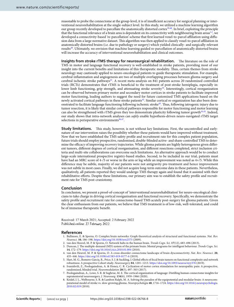
reasonable to probe the connectome at the group-level, it is of insufficient accuracy for surgical planning or inter-ventional neurorehabilitation at the single-subject level. In this study, we utilized a machine learning algorithm our group recently developed to parcellate the anatomically distorted cortex39,40. In brief, exploiting the principle that the functional relevance of a brain area is dependent on its connectivity with neighbouring brain areas1,2, we developed a connectivity-based ‘re-parcellation’ scheme that first learned voxel-to-parcel affiliation using diffu-sion data from a large normative dataset. This algorithm was then applied to classify voxel-to-parcel affiliation in anatomically distorted brains (i.e. due to pathology or surgery) which yielded clinically- and surgically-relevant results40. Ultimately, we envision that machine learning guided re-parcellation of anatomically distorted brains will increase the accuracy of interventional neurorehabilitation and clinical outcomes.
Insights from stroke rTMS therapy for neurosurgical rehabilitation. The literature on the role of TMS in motor and language functional recovery is well-established in stroke patients, providing most of our insight into the current benefits and limitations of this therapeutic modality. Thus, certain themes from stroke neurology may cautiously applied to neuro-oncological patients to guide therapeutic stimulation. For example, cerebral inflammation and angiogenesis are two of multiple overlapping processes between glioma surgery and cerebral ischemic stroke pathways41. A recent meta-analysis on 841 patients across 20 randomized controlled trials (RCTs) demonstrates that rTMS is beneficial to the treatment of post-stroke hemiplegia, especially in: lower limb functioning, grip strength, and attenuating stroke severity16. Interestingly, cortical reorganization can be observed between primary motor and secondary motor cortices in stroke patients to facilitate improved motor functioning, leading authors to suggest the need for future customized TMS applications based on the newly activated cortical pathways in these stroke patients42. Similar cortical re-organization has also been dem-onstrated to facilitate language functioning following ischemic stroke43. Thus, following iatrogenic injury due to tumor resection, it is likely that similar cortical pathways responsible for motor functioning and motor learning can also be strengthened with rTMS given they too demonstrate plasticity following tumor growth44,45. Indeed, our study shows that intra-network analyses can safely enable hypothesis-driven neuro-navigated rTMS target selections in perioperative environments46,47.
Study limitations. This study, however, is not without key limitations. First, the uncontrolled and early-nature of our intervention raises the possibility whether these patients would have improved without treatment. Now that we have established the TMS safety profile and recruitment rate for this complex patient population, future trials should employ prospective, randomised, double-blinded active- and sham-controlled TMS to deter-mine the efficacy of improving recovery trajectories. While glioma patients are highly heterogenous given differ-ent tumors, different degrees of cortical reorganisation, and different resections completed, strict inclusion cri-teria and multi-site collaborations can overcome such limitations. An alternative approach would be to conduct large-scale international prospective registry-based studies. Second, to be included in our trial, patients must have had an MRC score of 4-/5 or worse in the arm or leg while an improvement was noted as 4+/5. While this difference may be subtle, majority of our patients were not antigravity pre-treatment and hence improvement was not subtle in most cases. Finally, we did not acquire long-term outcome data in these patients. Nevertheless, qualitatively, all patients reported they would undergo TMS therapy again and found that it assisted with their rehabilitative efforts. Despite these limitations, our primary aim was to establish the safety profile and recruit-ment rate for TMS post-craniotomy.
ConclusionIn conclusion, we present a proof-of-concept of ‘interventional neurorehabilitation’ for neuro-oncological clini-cians to take charge in driving cortical reorganisation and functional recovery. Specifically, we demonstrate the safety profile and recruitment rate for connectome-based TMS acutely post-surgery for glioma patients. Given the clear enthusiasm from our patients, we believe that TMS treatment is of low-risk, well-tolerated, and could be of immense therapeutic benefit.
Received: 17 March 2021; Accepted: 2 February 2022
References 1. Bullmore, E. & Sporns, O. Complex brain networks: Graph theoretical analysis of structural and functional systems. Nat. Rev.
Neurosci. 10, 186–198. https:// doi. org/ 10. 1038/ nrn25 75 (2009). 2. van den Heuvel, M. P. & Sporns, O. Network hubs in the human brain. Trends Cogn. Sci. 17(12), 683–696 (2013). 3. Duncan, J. The multiple-demand (MD) system of the primate brain: Mental programs for intelligent behaviour. Trends Cogn. Sci.
14, 172–179. https:// doi. org/ 10. 1016/j. tics. 2010. 01. 004 (2010). 4. van den Heuvel, M. P. & Sporns, O. A cross-disorder connectome landscape of brain dysconnectivity. Nat. Rev. Neurosci. 20,
435–446. https:// doi. org/ 10. 1038/ s41583- 019- 0177-6 (2019). 5. Hart, M. G., Romero-Garcia, R., Price, S. J. & Suckling, J. Global effects of focal brain tumors on functional complexity and network
robustness: A prospective Cohort study. Neurosurgery 84, 1201–1213. https:// doi. org/ 10. 1093/ neuros/ nyy378 (2019). 6. Ivanishvili, Z., Poologaindran, A. & Honey, C. R. Cyclization of motor cortex stimulation for neuropathic pain: A prospective,
randomized, blinded trial. Neuromodulation 20(5), 497–503 (2017). 7. Poologaindran, A., Lowe, S. R. & Sughrue, M. E. The cortical organization of language: Distilling human connectome insights for
supratentorial neurosurgery. J. Neurosurg. 134(6), 1959–1966 (2020). 8. Keidel, J. L., Welbourne, S. R. & Lambon Ralph, M. A. Solving the paradox of the equipotential and modular brain: A neurocom-
putational model of stroke vs. slow-growing glioma. Neuropsychologia 48, 1716–1724. https:// doi. org/ 10. 1016/j. neuro psych ologia. 2010. 02. 019 (2010).

10
Vol:.(1234567890)
Scientific Reports | (2022) 12:3039 | https://doi.org/10.1038/s41598-022-06766-8
www.nature.com/scientificreports/
9. Cui, Z. et al. Individual variation in functional topography of association networks in youth. Neuron 106, 340-353 e348. https:// doi. org/ 10. 1016/j. neuron. 2020. 01. 029 (2020).
10. Finn, E. S. et al. Functional connectome fingerprinting: identifying individuals using patterns of brain connectivity. Nat. Neurosci. 18(11), 1664–1671 (2015).
11. Beaty, R. E. et al. Robust prediction of individual creative ability from brain functional connectivity. Proc. Natl. Acad. Sci. U. S. A. 115, 1087–1092. https:// doi. org/ 10. 1073/ pnas. 17135 32115 (2018).
12. Grossman, N. et al. Noninvasive deep brain stimulation via temporally interfering electric fields. Cell 169, 1029-1041 e1016. https:// doi. org/ 10. 1016/j. cell. 2017. 05. 024 (2017).
13. Honey, C. R. et al. Thalamic deep brain stimulation for spasmodic dysphonia: A phase I prospective randomized double-blind crossover trial. Neurosurgery 89(1), 45–52 (2021).
14. Brunoni, A. R. et al. Repetitive transcranial magnetic stimulation for the acute treatment of major depressive episodes: A systematic review with network meta-analysis. JAMA Psychiat. 74, 143–152. https:// doi. org/ 10. 1001/ jamap sychi atry. 2016. 3644 (2017).
15. Hoyer, E. H. & Celnik, P. A. Understanding and enhancing motor recovery after stroke using transcranial magnetic stimulation. Restor. Neurol. Neurosci. 29, 395–409. https:// doi. org/ 10. 3233/ RNN- 2011- 0611 (2011).
16. He, Y., Li, K., Chen, Q., Yin, J. & Bai, D. Repetitive transcranial magnetic stimulation on motor recovery for patients with stroke: a PRISMA compliant systematic review and meta-analysis. Am J Phys Med Rehabil 99(2), 99–108 (2020).
17. Lefaucheur, J. P. et al. Evidence-based guidelines on the therapeutic use of repetitive transcranial magnetic stimulation (rTMS). 18. Brain, M. R. C. G. O. Aids to the Examiantion of the Peripheral Nervous System. (Balliere Tindall, 1986). 19. Azuar, C. et al. The aphasia rapid test: An NIHSS-like aphasia test. J Neurol 260, 2110–2117. https:// doi. org/ 10. 1007/ s00415- 013-
6943-x (2013). 20. Romero-Garcia, R. et al. Practical application of networks in neurosurgery: Combined 3-dimensional printing, neuronavigation,
and preoperative surgical planning. World Neurosurg. 137, e126–e137. https:// doi. org/ 10. 1016/j. wneu. 2020. 01. 085 (2020). 21. Briggs, R. G. et al. A connectomic atlas of the human cerebrum-chapter 18: The connectional anatomy of human brain networks.
Oper. Neurosurg. (Hagerstown) 15, S470–S480. https:// doi. org/ 10. 1093/ ons/ opy272 (2018). 22. Glasser, M. F. et al. A multi-modal parcellation of human cerebral cortex. Nature 536, 171–178. https:// doi. org/ 10. 1038/ natur
e18933 (2016). 23. Ren, H. et al. Application of structural and functional connectome mismatch for classification and individualized therapy in
alzheimer disease. Frontiers in Public Health 8, 720. https:// doi. org/ 10. 3389/ fpubh. 2020. 584430 (2020). 24. Momi, D. et al. Cognitive enhancement via network-targeted cortico-cortical associative brain stimulation. Cereb. Cortex 30,
1516–1527. https:// doi. org/ 10. 1093/ cercor/ bhz182 (2020). 25. Cole, E. J. et al. Stanford Accelerated intelligent neuromodulation therapy for treatment-resistant depression. Am. J. Psychiatry
177, 716–726. https:// doi. org/ 10. 1176/ appi. ajp. 2019. 19070 720 (2020). 26. Di Iorio, R. & Rossini, P. M. In Navigated Transcranial Magnetic Stimulation in Neurosurgery (ed Sandro M. Krieg) 67–83 (Springer,
2017). 27. Rossi, S., Hallett, M., Rossini, P. M., Pascual-Leone, A. & Safety of TMS Consensus Group. Safety, ethical considerations, and
application guidelines for the use of transcranial magnetic stimulation in clinical practice and research. Clin. Neurophysiol 120(12), 2008–2039 (2009).
28. Ahdab, R., Ayache, S. S., Brugieres, P., Goujon, C. & Lefaucheur, J. P. Comparison of “standard” and “navigated” procedures of TMS coil positioning over motor, premotor and prefrontal targets in patients with chronic pain and depression. Neurophysiol. Clin. 40, 27–36. https:// doi. org/ 10. 1016/j. neucli. 2010. 01. 001 (2010).
29. Drysdale, A. T. et al. Erratum: Resting-state connectivity biomarkers define neurophysiological subtypes of depression. Nat. Med. 23, 264. https:// doi. org/ 10. 1038/ nm0217- 264d (2017).
30. Oberman, L., Edwards, D., Eldaief, M. & Pascual-Leone, A. Safety of theta burst transcranial magnetic stimulation: A systematic review of the literature. J. Clin. Neurophysiol. 28, 67–74. https:// doi. org/ 10. 1097/ WNP. 0b013 e3182 05135f (2011).
31. Wischnewski, M. & Schutter, D. J. Efficacy and time course of theta burst stimulation in healthy humans. Brain Stimul. 8, 685–692. https:// doi. org/ 10. 1016/j. brs. 2015. 03. 004 (2015).
32. Wilson, M. T. et al. Biophysical modeling of neural plasticity induced by transcranial magnetic stimulation. Clin. Neurophysiol 129, 1230–1241. https:// doi. org/ 10. 1016/j. clinph. 2018. 03. 018 (2018).
33. Briggs, R. G. et al. The frontal aslant tract and supplementary motor area syndrome: Moving towards a connectomic initiation axis. Cancers https:// doi. org/ 10. 3390/ cance rs130 51116 (2021).
34. Satterthwaite, T. D., Xia, C. H. & Bassett, D. S. Personalized neuroscience: Common and individual-specific features in functional brain networks. Neuron 98, 243–245. https:// doi. org/ 10. 1016/j. neuron. 2018. 04. 007 (2018).
35. Westbrook, A. et al. Dopamine promotes cognitive effort by biasing the benefits versus costs of cognitive work. Science 367, 1362–1366. https:// doi. org/ 10. 1126/ scien ce. aaz58 91 (2020).
36. Hurwitz, T. A., Honey, C. R., McLeod, K. R., Poologaindran, A. & Kuan, A. J. Hypoactivity in the paraterminal gyrus following bilateral anterior capsulotomy. Can. J. Psychiatry 65, 46–55. https:// doi. org/ 10. 1177/ 07067 43719 874181 (2020).
37. Hemmings, W. et al. Deep brain stimulation for refractory obsessive-compulsive disorder (OCD): Emerging or established therapy?. Mol. Psychiatry 26(1), 60–65 (2021).
38. Volz, L. J. et al. Shaping early reorganization of neural networks promotes motor function after stroke. Cereb. Cortex 26, 2882–2894. https:// doi. org/ 10. 1093/ cercor/ bhw034 (2016).
39. Doyen, S. et al. Connectivity-based parcellation of normal and anatomically distorted human cerbral cortex. Hum. Brain Mapp. Published online November 26, 2021.
40. Yeung, J. et al. Using quicktome for intracerebral surgery: Early retrospective study and proof of concept. World Neurosurg. (2021) (Online ahead of Print)
41. Ghosh, M. K., Chakraborty, D., Sarkar, S., Bhowmik, A. & Basu, M. The interrelationship between cerebral ischemic stroke and glioma: A comprehensive study of recent reports. Signal Transduct. Target. Ther. 4, 42. https:// doi. org/ 10. 1038/ s41392- 019- 0075-4 (2019).
42. Sharma, N., Baron, J.-C. & Rowe, J. B. Motor imagery after stroke: Relating outcome to motor network connectivity. Ann. Neurol. 66, 604–616. https:// doi. org/ 10. 1002/ ana. 21810 (2009).
43. Hamilton, R., Keenan, J. P., Catala, M. & Pascual-Leone, A. Alexia for Braille following bilateral occipital stroke in an early blind woman. Neuroreport 11(2), 237–240 (2000).
44. Kong, N. W., Gibb, W. R. & Tate, M. C. Neuroplasticity: Insights from patients harboring gliomas. Neural Plast. 2016, 2365063–2365063. https:// doi. org/ 10. 1155/ 2016/ 23650 63 (2016).
45. Kawashima, A. et al. Plastic reshaping of cortical language areas evaluated by navigated transcranial magnetic stimulation in a surgical case of glioblastoma multiforme. (2013).
46. Dadario, N. B., Brahimaj, B., Yeung, J. & Sughrue, M. E. Reducing the cognitive footprint of brain tumor surgery. Front. Neurol 1342 (2021).
47. Samuel, N. et al. A network-based approach to glioma surgery: Insights from functional neurosurgery. Cancers 13(23), 6127 (2021).

11
Vol.:(0123456789)
Scientific Reports | (2022) 12:3039 | https://doi.org/10.1038/s41598-022-06766-8
www.nature.com/scientificreports/
AcknowledgementsAP is funded through a Turing Doctoral Scholarship from the Alan Turing Institute and the National Science and Engineering Research Council of Canada. RRG is supported by a Guarantors of Brain Fellowship.
Author contributionsConceptualization: A.P., I.M.Y. and M.E.S. Data curation: C.P., I.M.Y. Formal analysis: A.P., I.M.Y. Methodology, A.P., R.G.B., M.E.S.; Project administration, I.M.Y., C.T. and M.E.S.; Software S.A.A., K.C., R.R.G.; Writing—original draft, A.P.; Writing—review and editing, N.B.D., J.S., M.E.S.
Competing interests IMY is an employee of Cingulum Health. MES is the chief medical officer of Omniscient and a shareholder of Cingulum Health. CT is a consultant for Aesculap. All other authors report no conflict of interest related to this study.
Additional informationSupplementary Information The online version contains supplementary material available at https:// doi. org/ 10. 1038/ s41598- 022- 06766-8.
Correspondence and requests for materials should be addressed to M.E.S.
Reprints and permissions information is available at www.nature.com/reprints.
Publisher’s note Springer Nature remains neutral with regard to jurisdictional claims in published maps and institutional affiliations.
Open Access This article is licensed under a Creative Commons Attribution 4.0 International License, which permits use, sharing, adaptation, distribution and reproduction in any medium or
format, as long as you give appropriate credit to the original author(s) and the source, provide a link to the Creative Commons licence, and indicate if changes were made. The images or other third party material in this article are included in the article’s Creative Commons licence, unless indicated otherwise in a credit line to the material. If material is not included in the article’s Creative Commons licence and your intended use is not permitted by statutory regulation or exceeds the permitted use, you will need to obtain permission directly from the copyright holder. To view a copy of this licence, visit http:// creat iveco mmons. org/ licen ses/ by/4. 0/.
© The Author(s) 2022