Diagnostic neuroradiology for the interventional neuroradiologist
13
EDITORIAL Diagnostic neuroradiology for the interventional neuroradiologist Vitor Mendes Pereira, Maria Isabel Vargas, Ana Marcos, Philippe Bijlenga, Ana Paula Narata, Sven Haller, Karl-Olof Lövblad Vitor Mendes Pereira, Maria Isabel Vargas, Ana Marcos, Ana Paula Narata, Sven Haller, Karl-Olof Lövblad, Service Neuro-diagnostique et Neuro-interventionnel �eneva �niver- �eneva �niver- sity Hospitals 1211 �eneva Switzerland Philippe Bijlenga, Service de Neurochirurgie H�� �eneva �niversity Hospitals 1211 �eneva Switzerland Author contributions: All the authors contributed to this work. Correspondence to: Karl-Olof Lövblad, MD, Service Neuro- diagnostique et Neuro-interventionnel �eneva �niversity Hos- �eneva �niversity Hos- pitals rue �abrielle-�erret �entil 1211 �eneva rue �abrielle-�erret �entil 1211 �eneva rue �abrielle-�erret �entil 1211 �eneva 1211 �eneva 1211 �eneva Switzerland. [email protected] Telephone: +1-22-3727033 Fax: +1-22-3727072 Received: September 11 2013 Revised: October 8 2013 Accepted: November 1 2013 Published online: November 28 2013 Abstract In order to further improve the impact of the continu- ously evolving neurointerventional techniques, the interventional neuroradiologist needs to have at his disposal more powerful techniques to image the cen- tral nervous system. With the recent development of diagnostics techniques that are computed tomography and magnetic resonance based we are now able to assess not just morphology, but also physiology, phys- iopathology and function. This review discusses the place of diagnostic techniques in the evaluation that the interventional neuroradiologist hast to make when confronted with patients. We provide an overview of current techniques for the brain and spine. © 2013 Baishideng Publishing Group Co., Limited. All rights reserved. Key words: Neuroradiology; Brain; Imaging; Magnetic resonance; Interventional radiology Core tip: Minimally invasive neuroradiology techniques are taking progressively a more and more important place in the management of patients with diseases of the nervous system. It is thus necessary for the physi- cian who is going to perform them to both understand and master fully standard and advanced neuroimaging techniques that provide detailed insights into the func- tioning of the brain. Mendes �ereira V Vargas MI Marcos A Bijlenga � Narata A� Haller S Lövblad KO. Diagnostic neuroradiology for the interventional neuroradiologist. World J Radiol 2013; 5(11): 386-397 Available from: �RL: http://www.wjgnet.com/199-870/ full/v5/i11/386.htm DOI: http://dx.doi.org/10.329/wjr.v5.i11.386 INTRODUCTION Interventional neuroradiology, the cornerstone of pa- tient management in many neurovascular diseases cur- rently, has known a recent period of groundbreaking milestones with the improvement of techniques devices available [1,2] . These devices and procedures have rendered possible the treatment of stroke, vascular malformations as aneurysms and AVMs with a degree of precision and security that was previously unthinkable. As interven- tional techniques are evolving, it is imperative to dispose of more and more accurate and powerful imaging tech- niques that will allow us to not just perform imaging but also assess any pathological changes [3] . Also, due to the fact that sometimes neurointerventional training does not always encompass a full training in some of these new techniques, it is required that the neurointerven- tionalist immerses himself somewhat more in them in order to fully comprehend them. These new imaging techniques have made possible by the recent parallel developments of techniques such as spiral scanning and multi-slice computed tomography (CT) scanners [4] as well as fast magnetic resonance (MR) techniques [5,6] . World Journal of Radiology WJR Online Submissions: http://www.wjgnet.com/esps/ bpgoffi[email protected] doi:10.4329/wjr.v5.i11.386 World J Radiol 2013 November 28; 5(11): 386-397 ISSN 1949-8470 (online) © 2013 Baishideng Publishing Group Co., Limited. �ll rights reserved �ll rights reserved. 386 November 28, 2013|Volume 5|Issue 11| WJR|www.wjgnet.com
-
Upload
independent -
Category
Documents
-
view
1 -
download
0
Transcript of Diagnostic neuroradiology for the interventional neuroradiologist
EDITORIAL
Diagnostic neuroradiology for the interventional neuroradiologist
Vitor Mendes Pereira, Maria Isabel Vargas, Ana Marcos, Philippe Bijlenga, Ana Paula Narata, Sven Haller, Karl-Olof Lövblad
Vitor Mendes Pereira, Maria Isabel Vargas, Ana Marcos, Ana Paula Narata, Sven Haller, Karl-Olof Lövblad, Service Neuro-diagnostique et Neuro-interventionnel�� �eneva �niver-�� �eneva �niver-sity Hospitals�� 1211 �eneva�� SwitzerlandPhilippe Bijlenga, Service de Neurochirurgie�� H���� �eneva �niversity Hospitals�� 1211 �eneva�� SwitzerlandAuthor contributions: All the authors contributed to this work.Correspondence to: Karl-Olof Lövblad, MD, Service Neuro-diagnostique et Neuro-interventionnel�� �eneva �niversity Hos-�eneva �niversity Hos-pitals�� �� rue �abrielle-�erret �entil�� 1211 �eneva���� �� rue �abrielle-�erret �entil�� 1211 �eneva���� rue �abrielle-�erret �entil�� 1211 �eneva�� 1211 �eneva��1211 �eneva�� Switzerland. [email protected]: +��1-22-3727033 Fax: +��1-22-3727072Received: September 11�� 2013 Revised: October 8�� 2013Accepted: November 1�� 2013Published online: November 28�� 2013
AbstractIn order to further improve the impact of the continu-ously evolving neurointerventional techniques, the interventional neuroradiologist needs to have at his disposal more powerful techniques to image the cen-tral nervous system. With the recent development of diagnostics techniques that are computed tomography and magnetic resonance based we are now able to assess not just morphology, but also physiology, phys-iopathology and function. This review discusses the place of diagnostic techniques in the evaluation that the interventional neuroradiologist hast to make when confronted with patients. We provide an overview of current techniques for the brain and spine.
© 2013 Baishideng Publishing Group Co., Limited. All rights reserved.
Key words: Neuroradiology; Brain; Imaging; Magnetic resonance; Interventional radiology
Core tip: Minimally invasive neuroradiology techniques are taking progressively a more and more important
place in the management of patients with diseases of the nervous system. It is thus necessary for the physi-cian who is going to perform them to both understand and master fully standard and advanced neuroimaging techniques that provide detailed insights into the func-tioning of the brain.
Mendes �ereira V�� Vargas MI�� Marcos A�� Bijlenga ��� Narata A��� Haller S�� Lövblad KO. Diagnostic neuroradiology for the interventional neuroradiologist. World J Radiol 2013; 5(11): 386-397 Available from: �RL: http://www.wjgnet.com/19��9-8��70/full/v5/i11/386.htm DOI: http://dx.doi.org/10.��329/wjr.v5.i11.386
INTRODUCTIONInterventional neuroradiology, the cornerstone of pa-tient management in many neurovascular diseases cur-rently, has known a recent period of groundbreaking milestones with the improvement of techniques devices available[1,2]. These devices and procedures have rendered possible the treatment of stroke, vascular malformations as aneurysms and AVMs with a degree of precision and security that was previously unthinkable. As interven-tional techniques are evolving, it is imperative to dispose of more and more accurate and powerful imaging tech-niques that will allow us to not just perform imaging but also assess any pathological changes[3]. Also, due to the fact that sometimes neurointerventional training does not always encompass a full training in some of these new techniques, it is required that the neurointerven-tionalist immerses himself somewhat more in them in order to fully comprehend them. These new imaging techniques have made possible by the recent parallel developments of techniques such as spiral scanning and multi-slice computed tomography (CT) scanners[4] as well as fast magnetic resonance (MR) techniques[5,6].
World Journal of RadiologyW J R
Online Submissions: http://www.wjgnet.com/esps/[email protected]:10.4329/wjr.v5.i11.386
World J Radiol 2013 November 28; 5(11): 386-397ISSN 1949-8470 (online)
© 2013 Baishideng Publishing Group Co., Limited. �ll rights reserved �ll rights reserved.
386 November 28, 2013|Volume 5|Issue 11|WJR|www.wjgnet.com
Mendes Pereira V et al . Diagnostic neuroradiology for the interventional neuroradiologist
Indeed, this gives imaging a more and more important role whenever an intervention and/or operation are be-ing planned. Indeed, the physician will need not just to obtain optimal 3D visualization of the lesion but also to assess any damage to the surrounding brain tissue. CT imaging is very often the examination of choice for the acute investigation of patients: indeed it is widely used in traumatology due to its superior capacity to image bone lesions but is also a technique that is more easily used for the detection of any intracranial blood. Indeed, the detection of blood is absolutely necessary if one is to treat for ischemic stroke since its presence will be a contra-indication, and also for any kind of treatable cerebral hemorrhage such as subarachnoid hemorrhage. Together with CT perfusion and CT angiography[7-11] these techniques have become easily available and very powerful tools for the neurodiagnostician and neuroin-terventionalist. MR on the other hand disposes of more sensitive techniques such as diffusion-weighted MR[12-17] or susceptibility-weighted MR[18,19] that are able to de-tect lesions that may not be seen on CT. In addition to perfusion, additional techniques such as diffusion tensor imaging, MR spectroscopy, and 3D imaging and more, makes this is superior technique when it comes to the non-urgent evaluation of patients where exact lesion characterization is necessary. Over the last decade there has been a continuous increase in the field strength of clinical MR scanners: this has led to significant improve-ments in imaging; on the one hand signal increases over-all with magnetic field so that an increase in resolution and a decrease in scanning time has been achieved[20]. The aim of this review is to provide an overview of established and new neuroimaging techniques that are increasingly being used in the evaluation of patients un-dergoing interventional neuroradiology procedures.
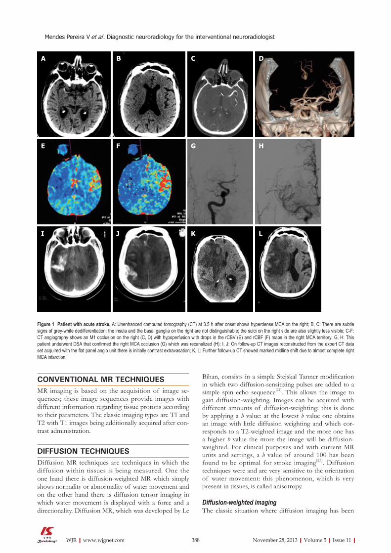
CTCT, since its introduction in the 1970’s remains the work-horse of neuroradiological diagnostic workup. While it has clearly remained the emergency radiology method of choice due to its intrinsic better bone detail, it has regained interest due to the development for spiral CT scanning and multi-slice scanners that allow obtaining additionally angiographic images as well as perfusion im-ages of the brain. The plain unenhanced CT remains the workhorse for any vascular work-up since it will allow on the one hand todemonstrate or exclude easily hem-orrhage. When considering stroke the early signs such as the dense artery, the disappearance of localized grey matter -white matter differentiation and hypodensity are subtle but reliable signs that can be used to detect isch-emia (Figure 1).
CT angiographyCT angiography now allows covering the vasculature from the aortic arch up to the circle of Willis in just a few seconds. Very often two imaging strategies are taken:
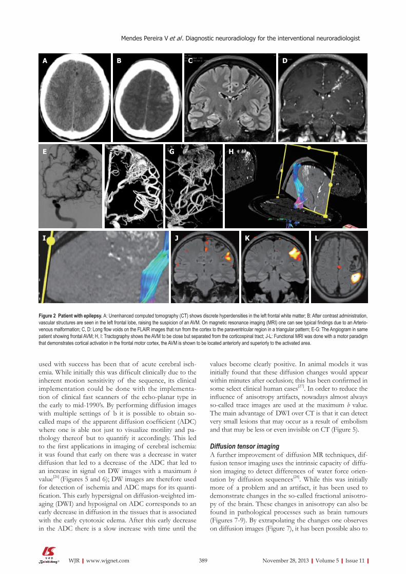
for ischemic stroke, very often, the vasculature is done from the arch, even if sometimes this will cause venous superposition. For dedicated questions such as in pa-tients with suspected aneurysms, it is very often decided to perform only and angio-CT at the level of the circle of Willis in order to have optimal aneurysmal filling; this has the disadvantage that if an endovascular approach is being discussed, the anatomy of the vessels as they arise from the arch is not known[21]. Also for the demonstra-tion of vascular malformations, CT angiography can well demonstrate lesions that will be visible only well after contrast administration (Figure 2).
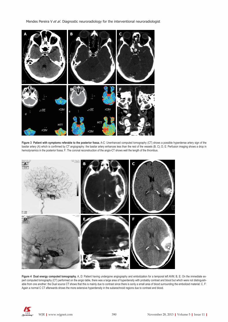
CT perfusionCT perfusion techniques, which had been available for a long time have also become more widely accepted due to their capacity to detect ischemia very early on (i.e., at the same time as the initial emergency CT); while the techniques have somewhat suffered from not initially providing enough brain coverage in cases with extensive strokes or in cases with multiple areas being affected, more CT units are now providing coverage of almost or of the whole brain. Reconstruction algorithms are avail-able that provide maps of cerebral perfusion parameters (CBV, CBF, MTT) that can help understand better the physiopathological processes and help determining tis-sue outcome[22] (Figures 1-3).
Dual energy CTThis is another rather recent development in CT tech-nology that has quite some interesting applications for patients with neurovascular diseases. By using different kVp it is possible to visualize various tissue components that have different X ray absorption rates. It has been used to differentiate bone from other tissues and remove the skull as well as for removing vascular calcifications: More recently it has been used with success after throm-bolysis to differentiate iodinated contrast extravasation from blood[23] or even after other endovascular proce-dures if a complication is suspected (Figure 4).
MR IMAGINGMR imaging, due to its intrinsic improved tissular con-trast with many sequences allowing performing tissue characterization is the method of choice for most situa-tions that do not necessitate an emergency treatment. In-deed, despite the development of some really powerful MR techniques due to the implementation of both high field scanners as well as fast imaging, the technique is still somewhat more time-consuming and requires some more patient cooperation to be performed and has a few disadvantages (claustrophobia inducing, magnetic field). However due to the development of a multitude of vari-ous techniques, called sequences it is now possible to perform advanced tissue characterization in many cases. Also the development of high field scanners has been a recent important step in this direction.
387 November 28, 2013|Volume 5|Issue 11|WJR|www.wjgnet.com
CONVENTIONAL MR TECHNIQUESMR imaging is based on the acquisition of image se-quences; these image sequences provide images with different information regarding tissue protons according to their parameters. The classic imaging types are T1 and T2 with T1 images being additionally acquired after con-trast administration.
DIFFUSION TECHNIQUESDiffusion MR techniques are techniques in which the diffusion within tissues is being measured. One the one hand there is diffusion-weighted MR which simply shows normality or abnormality of water movement and on the other hand there is diffusion tensor imaging in which water movement is displayed with a force and a directionality. Diffusion MR, which was developed by Le
Bihan, consists in a simple Stejskal Tanner modification in which two diffusion-sensitizing pulses are added to a simple spin echo sequence[24]. This allows the image to gain diffusion-weighting. Images can be acquired with different amounts of diffusion-weighting: this is done by applying a b value: at the lowest b value one obtains an image with little diffusion weighting and which cor-responds to a T2-weighted image and the more one has a higher b value the more the image will be diffusion-weighted. For clinical purposes and with current MR units and settings, a b value of around 100 has been found to be optimal for stroke imaging[25]. Diffusion techniques were and are very sensitive to the orientation of water movement: this phenomenon, which is very present in tissues, is called anisotropy.
Diffusion-weighted imagingThe classic situation where diffusion imaging has been
A B C D
E F G H
I J K L
Figure 1 Patient with acute stroke. A: Unenhanced computed tomography (CT) at 3.5 h after onset shows hyperdense MCA on the right; B, C: There are subtle signs of grey-white dedifferentiation: the insula and the basal ganglia on the right are not distinguishable; the sulci on the right side are also slightly less visible; C-F: CT angiography shows an M1 occlusion on the right (C, D) with hypoperfusion with drops in the rCBV (E) and rCBF (F) maps in the right MCA territory; G, H: This patient underwent DSA that confirmed the right MCA occlusion (G) which was recanalized (H); I, J: On follow-up CT images reconstructed from the expert CT data set acquired with the flat panel angio unit there is initially contrast extravasation; K, L: Further follow-up CT showed marked midline shift due to almost complete right MCA infarction.
388 November 28, 2013|Volume 5|Issue 11|WJR|www.wjgnet.com
Mendes Pereira V et al . Diagnostic neuroradiology for the interventional neuroradiologist
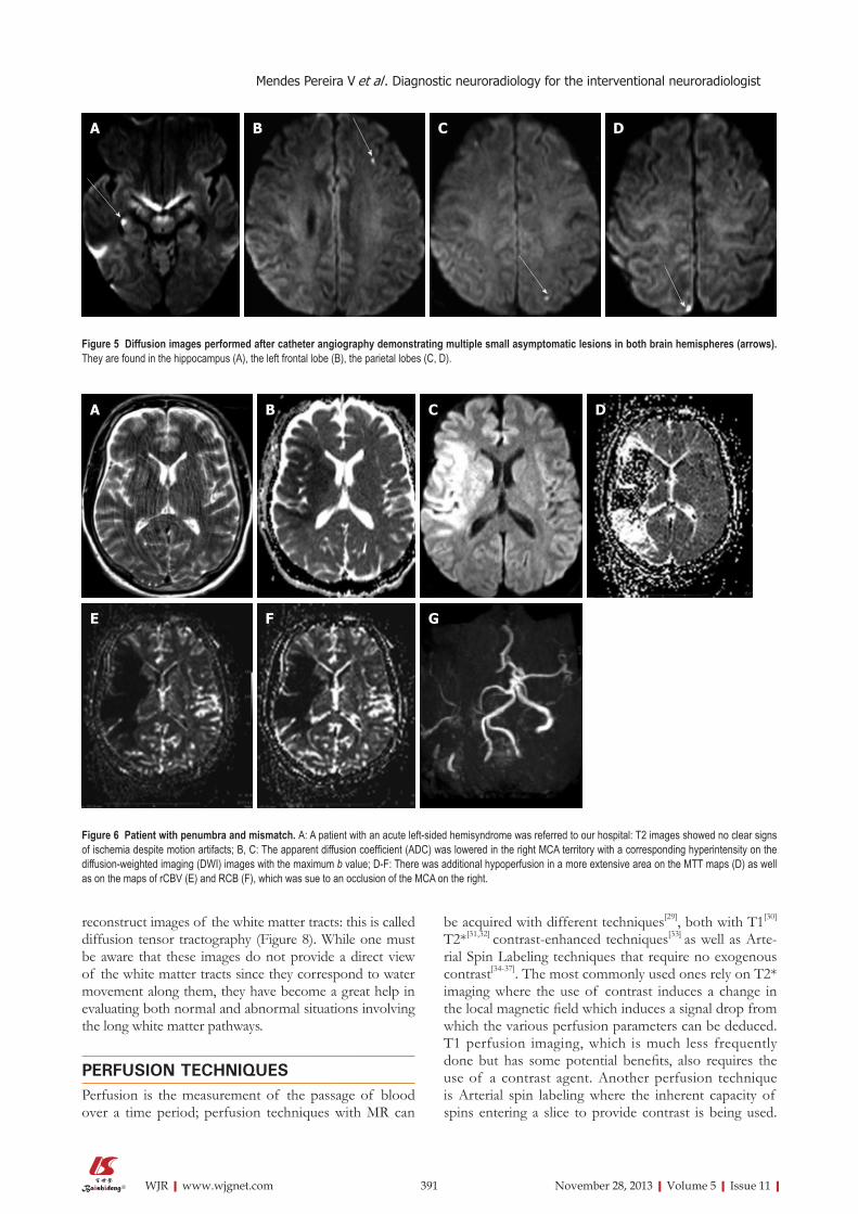
used with success has been that of acute cerebral isch-emia. While initially this was difficult clinically due to the inherent motion sensitivity of the sequence, its clinical implementation could be done with the implementa-tion of clinical fast scanners of the echo-planar type in the early to mid-1990’s. By performing diffusion images with multiple settings of b it is possible to obtain so-called maps of the apparent diffusion coefficient (ADC) where one is able not just to visualize motility and pa-thology thereof but to quantify it accordingly. This led to the first applications in imaging of cerebral ischemia: it was found that early on there was a decrease in water diffusion that led to a decrease of the ADC that led to an increase in signal on DW images with a maximum b value[26] (Figures 5 and 6); DW images are therefore used for detection of ischemia and ADC maps for its quanti-fication. This early hypersignal on diffusion-weighted im-aging (DWI) and hyposignal on ADC corresponds to an early decrease in diffusion in the tissues that is associated with the early cytotoxic edema. After this early decrease in the ADC there is a slow increase with time until the
values become clearly positive. In animal models it was initially found that these diffusion changes would appear within minutes after occlusion; this has been confirmed in some select clinical human cases[27]. In order to reduce the influence of anisotropy artifacts, nowadays almost always so-called trace images are used at the maximum b value. The main advantage of DWI over CT is that it can detect very small lesions that may occur as a result of embolism and that may be less or even invisible on CT (Figure 5).
Diffusion tensor imagingA further improvement of diffusion MR techniques, dif-fusion tensor imaging uses the intrinsic capacity of diffu-sion imaging to detect differences of water force orien-tation by diffusion sequences[28]. While this was initially more of a problem and an artifact, it has been used to demonstrate changes in the so-called fractional anisotro-py of the brain. These changes in anisotropy can also be found in pathological processes such as brain tumours (Figures 7-9). By extrapolating the changes one observes on diffusion images (Figure 7), it has been possible also to
Figure 2 Patient with epilepsy. A: Unenhanced computed tomography (CT) shows discrete hyperdensities in the left frontal white matter; B: After contrast administration, vascular structures are seen in the left frontal lobe, raising the suspicion of an AVM. On magnetic resonance imaging (MRI) one can see typical findings due to an Arterio-venous malformation; C, D: Long flow voids on the FLAIR images that run from the cortex to the paraventricular region in a triangular pattern; E-G: The Angiogram in same patient showing frontal AVM; H, I: Tractography shows the AVM to be close but separated from the corticospinal tract; J-L: Functional MRI was done with a motor paradigm that demonstrates cortical activation in the frontal motor cortex, the AVM is shown to be located anteriorly and superiorly to the activated area.
A B C D
E F G H
I J K L
389 November 28, 2013|Volume 5|Issue 11|WJR|www.wjgnet.com
Mendes Pereira V et al . Diagnostic neuroradiology for the interventional neuroradiologist
rCBV rCBV
MTT
d
rCBVe
rCBV
MTT
A B C
D E F
Figure 3 Patient with symptoms referable to the posterior fossa. A-C: Unenhanced computed tomography (CT) shows a possible hyperdense artery sign of the basilar artery (A) which is confirmed by CT angiography: the basilar artery enhances less than the rest of the vessels (B, C); D, E: Perfusion imaging shows a drop in hemodynamics in the posterior fossa; F: The coronal reconstruction of the angio-CT shows well the length of the thrombus.
A B C
D E F
Figure 4 Dual energy computed tomography. A, D: Patient having undergone angiography and embolization for a temporal left AVM; B, E: On the immediate ex-pert computed tomography (CT) performed on the angio table, there was a large area of hyperdensity with probably contrast and blood but which were not distinguish-able from one another: the Dual source CT shows that this is mainly due to contrast since there is sonly a small area of blood surrounding the embolized material; C, F: Again a normal C CT afterwards shows the more extensive hyperdensity in the subarachnoid regions due to contrast and blood.
390 November 28, 2013|Volume 5|Issue 11|WJR|www.wjgnet.com
Mendes Pereira V et al . Diagnostic neuroradiology for the interventional neuroradiologist
reconstruct images of the white matter tracts: this is called diffusion tensor tractography (Figure 8). While one must be aware that these images do not provide a direct view of the white matter tracts since they correspond to water movement along them, they have become a great help in evaluating both normal and abnormal situations involving the long white matter pathways.
PERFUSION TECHNIQUESPerfusion is the measurement of the passage of blood over a time period; perfusion techniques with MR can
be acquired with different techniques[29], both with T1[30] T2*[31,32] contrast-enhanced techniques[33] as well as Arte-rial Spin Labeling techniques that require no exogenous contrast[34-37]. The most commonly used ones rely on T2* imaging where the use of contrast induces a change in the local magnetic field which induces a signal drop from which the various perfusion parameters can be deduced. T1 perfusion imaging, which is much less frequently done but has some potential benefits, also requires the use of a contrast agent. Another perfusion technique is Arterial spin labeling where the inherent capacity of spins entering a slice to provide contrast is being used.
A B C D
Figure 5 Diffusion images performed after catheter angiography demonstrating multiple small asymptomatic lesions in both brain hemispheres (arrows). They are found in the hippocampus (A), the left frontal lobe (B), the parietal lobes (C, D).
A B C D
E F G
Figure 6 Patient with penumbra and mismatch. A: A patient with an acute left-sided hemisyndrome was referred to our hospital: T2 images showed no clear signs of ischemia despite motion artifacts; B, C: The apparent diffusion coefficient (ADC) was lowered in the right MCA territory with a corresponding hyperintensity on the diffusion-weighted imaging (DWI) images with the maximum b value; D-F: There was additional hypoperfusion in a more extensive area on the MTT maps (D) as well as on the maps of rCBV (E) and RCB (F), which was sue to an occlusion of the MCA on the right.
391 November 28, 2013|Volume 5|Issue 11|WJR|www.wjgnet.com
Mendes Pereira V et al . Diagnostic neuroradiology for the interventional neuroradiologist
This very promising technique was for a long time only available in single-slice mode but with the implementa-tion of high field scanners was able to provide perfusion maps covering the whole brain. While the technique has the advantage of being repeatable without any contrast administration, it does not provide much signal and is prone to artifacts. The first widely accepted application for perfusion imaging with magnetic resonance imaging (MRI) was of course stroke[38] (Figure 6). The combined use of diffusion and perfusion allowed early stroke re-searchers to try and develop a working model of the penumbra where it was postulated that the initial diffu-sion lesion was the central core and the hypoperfused area beyond it the penumbra or tissue at risk for further infarction[39]. While this model does not really correspond to the traditional model of the penumbra from a physio-pathological point of view (it being a hemodynamically based model and not an energetically-based one)[40], it was at least a model that worked in a certain number of cases. While it seems that not all acute cases of stroke may have the same typical mismatch[41] it seems that at least the central diffusion lesion does correspond to the core[42]. The use of ASL techniques could be of further inter-est since it seems able to better demonstrate reperfusion and collateral flow than contrast techniques[43]. Perfusion
techniques have also found progressive acceptance in the grading of brain tumours: together with spectroscopy they can help to grade brain tumours in a superior way[44].
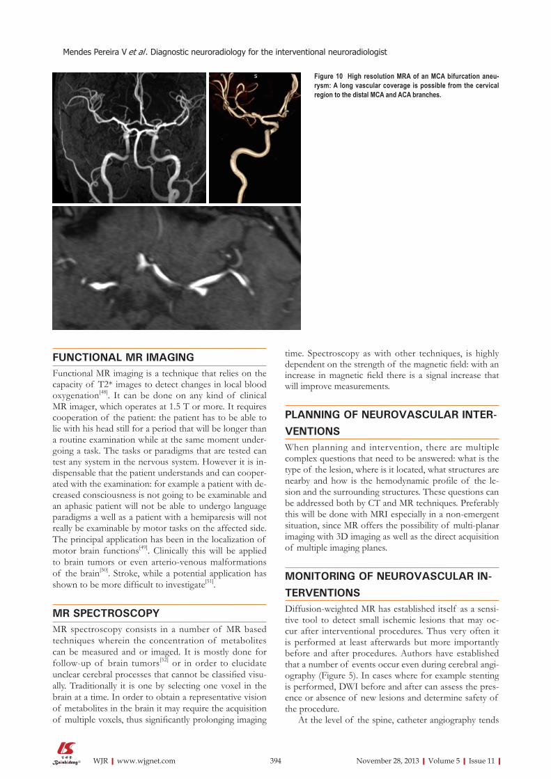
MR ANGIOGRAPHY TECHNIQUESMost MR techniques are very sensitive intrinsically to flow so a natural development was that of MR angiographic techniques. Many various techniques have been developed that allow reconstructing angiographic images, based on time-of flight effects, or phase-contrast images or even contrast-enhanced images. Brain MRA are done predomi-nantly by a time of flight technique at the level of the brain[45]; the TOF techniques can be also done with con-trast, especially when imaging aneurysms in the follow-up since it improves visualization of any aneurysms remnant. Due to the presence of flow-related signal changes inher-ent to MR imaging, one does not currently advocate the use of MR sequences for the diagnosis of aneurysms (Figure 10) but more for their follow-up.
Contrast-enhanced techniques are also used at the level of the neck for the carotid arteries where imag-ing has now been improved so much that coverage is provided that goes from the aortic arch to the circle of Willis[46]. Phlebographic techniques are also possible by
A B C D E
F G H I J
K L M N O
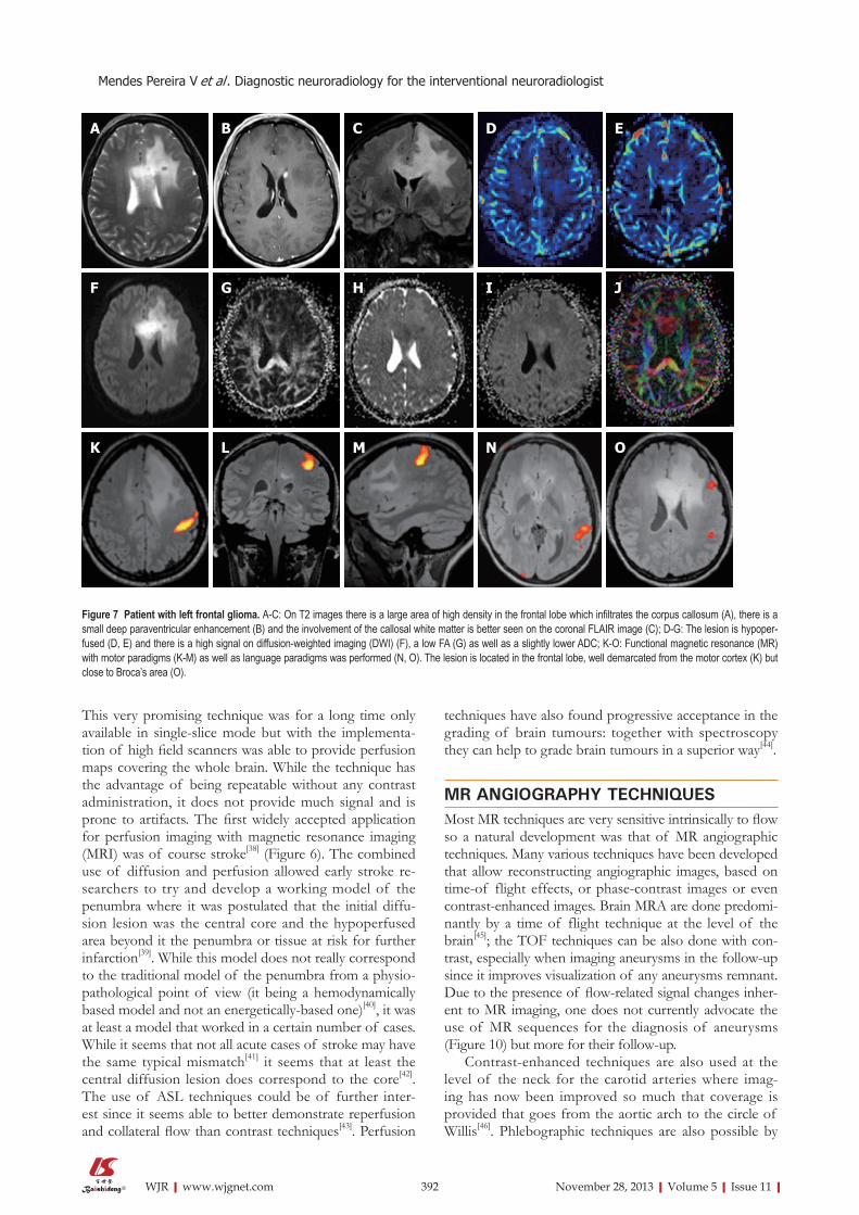
Figure 7 Patient with left frontal glioma. A-C: On T2 images there is a large area of high density in the frontal lobe which infiltrates the corpus callosum (A), there is a small deep paraventricular enhancement (B) and the involvement of the callosal white matter is better seen on the coronal FLAIR image (C); D-G: The lesion is hypoper-fused (D, E) and there is a high signal on diffusion-weighted imaging (DWI) (F), a low FA (G) as well as a slightly lower ADC; K-O: Functional magnetic resonance (MR) with motor paradigms (K-M) as well as language paradigms was performed (N, O). The lesion is located in the frontal lobe, well demarcated from the motor cortex (K) but close to Broca’s area (O).
392 November 28, 2013|Volume 5|Issue 11|WJR|www.wjgnet.com
Mendes Pereira V et al . Diagnostic neuroradiology for the interventional neuroradiologist
using either time-of-flight techniques or also contrast-enhanced techniques[47].
SUSCEPTIBILITY-WEIGHTED MR IMAGINGThis is also a technique that has been available for some
time but which became implementable clinically only with the use of high-field scanners. This is a technique that provides very high T2 and T2* contrast images that have very little anatomic detail but very high contrast. This makes it a sequence that will be of high use to de-tect calcifications or hemorrhagic lesions.
A B C D
E F G H
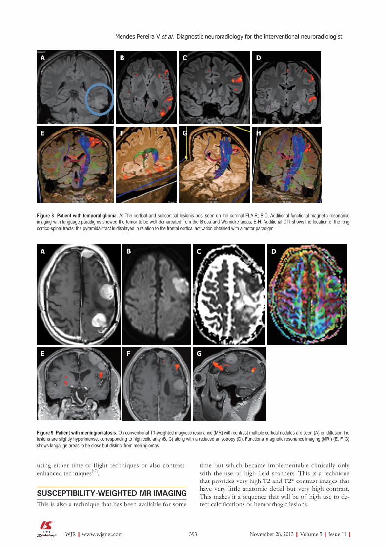
Figure 8 Patient with temporal glioma. A: The cortical and subcortical lesionis best seen on the coronal FLAIR; B-D: Additional functional magnetic resonance imaging with language paradigms showed the tumor to be well demarcated from the Broca and Wernicke areas; E-H: Additional DTI shows the location of the long cortico-spinal tracts: the pyramidal tract is displayed in relation to the frontal cortical activation obtained with a motor paradigm.
B C DA
E F G
Figure 9 Patient with meningiomatosis. On conventional T1-weighted magnetic resonance (MR) with contrast multiple cortical nodules are seen (A) on diffusion the lesions are slightly hyperintense, corresponding to high cellularity (B, C) along with a reduced anisotropy (D). Functional magnetic resonance imaging (MRI) (E, F, G) shows langauge areas to be close but distinct from meningiomas.
393 November 28, 2013|Volume 5|Issue 11|WJR|www.wjgnet.com
Mendes Pereira V et al . Diagnostic neuroradiology for the interventional neuroradiologist
FUNCTIONAL MR IMAGINGFunctional MR imaging is a technique that relies on the capacity of T2* images to detect changes in local blood oxygenation[48]. It can be done on any kind of clinical MR imager, which operates at 1.5 T or more. It requires cooperation of the patient: the patient has to be able to lie with his head still for a period that will be longer than a routine examination while at the same moment under-going a task. The tasks or paradigms that are tested can test any system in the nervous system. However it is in-dispensable that the patient understands and can cooper-ated with the examination: for example a patient with de-creased consciousness is not going to be examinable and an aphasic patient will not be able to undergo language paradigms a well as a patient with a hemiparesis will not really be examinable by motor tasks on the affected side. The principal application has been in the localization of motor brain functions[49]. Clinically this will be applied to brain tumors or even arterio-venous malformations of the brain[50]. Stroke, while a potential application has shown to be more difficult to investigate[51].
MR SPECTROSCOPYMR spectroscopy consists in a number of MR based techniques wherein the concentration of metabolites can be measured and or imaged. It is mostly done for follow-up of brain tumors[52] or in order to elucidate unclear cerebral processes that cannot be classified visu-ally. Traditionally it is one by selecting one voxel in the brain at a time. In order to obtain a representative vision of metabolites in the brain it may require the acquisition of multiple voxels, thus significantly prolonging imaging
time. Spectroscopy as with other techniques, is highly dependent on the strength of the magnetic field: with an increase in magnetic field there is a signal increase that will improve measurements.
PLANNING OF NEUROVASCULAR INTER-VENTIONSWhen planning and intervention, there are multiple complex questions that need to be answered: what is the type of the lesion, where is it located, what structures are nearby and how is the hemodynamic profile of the le-sion and the surrounding structures. These questions can be addressed both by CT and MR techniques. Preferably this will be done with MRI especially in a non-emergent situation, since MR offers the possibility of multi-planar imaging with 3D imaging as well as the direct acquisition of multiple imaging planes.
MONITORING OF NEUROVASCULAR IN-TERVENTIONSDiffusion-weighted MR has established itself as a sensi-tive tool to detect small ischemic lesions that may oc-cur after interventional procedures. Thus very often it is performed at least afterwards but more importantly before and after procedures. Authors have established that a number of events occur even during cerebral angi-ography (Figure 5). In cases where for example stenting is performed, DWI before and after can assess the pres-ence or absence of new lesions and determine safety of the procedure.
At the level of the spine, catheter angiography tends
394 November 28, 2013|Volume 5|Issue 11|WJR|www.wjgnet.com
Mendes Pereira V et al . Diagnostic neuroradiology for the interventional neuroradiologist
Figure 10 High resolution MRA of an MCA bifurcation aneu-rysm: A long vascular coverage is possible from the cervical region to the distal MCA and ACA branches.
s
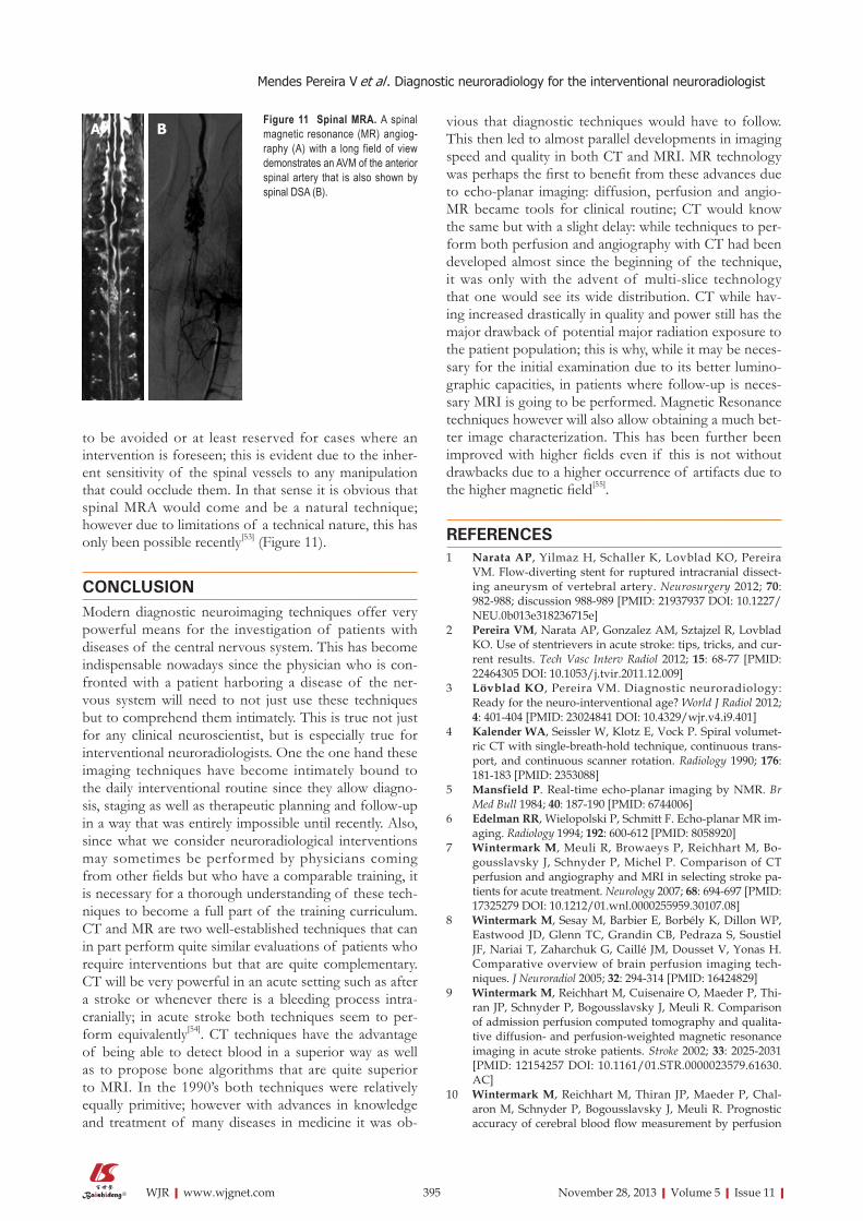
to be avoided or at least reserved for cases where an intervention is foreseen; this is evident due to the inher-ent sensitivity of the spinal vessels to any manipulation that could occlude them. In that sense it is obvious that spinal MRA would come and be a natural technique; however due to limitations of a technical nature, this has only been possible recently[53] (Figure 11).
CONCLUSIONModern diagnostic neuroimaging techniques offer very powerful means for the investigation of patients with diseases of the central nervous system. This has become indispensable nowadays since the physician who is con-fronted with a patient harboring a disease of the ner-vous system will need to not just use these techniques but to comprehend them intimately. This is true not just for any clinical neuroscientist, but is especially true for interventional neuroradiologists. One the one hand these imaging techniques have become intimately bound to the daily interventional routine since they allow diagno-sis, staging as well as therapeutic planning and follow-up in a way that was entirely impossible until recently. Also, since what we consider neuroradiological interventions may sometimes be performed by physicians coming from other fields but who have a comparable training, it is necessary for a thorough understanding of these tech-niques to become a full part of the training curriculum. CT and MR are two well-established techniques that can in part perform quite similar evaluations of patients who require interventions but that are quite complementary. CT will be very powerful in an acute setting such as after a stroke or whenever there is a bleeding process intra-cranially; in acute stroke both techniques seem to per-form equivalently[54]. CT techniques have the advantage of being able to detect blood in a superior way as well as to propose bone algorithms that are quite superior to MRI. In the 1990’s both techniques were relatively equally primitive; however with advances in knowledge and treatment of many diseases in medicine it was ob-
vious that diagnostic techniques would have to follow. This then led to almost parallel developments in imaging speed and quality in both CT and MRI. MR technology was perhaps the first to benefit from these advances due to echo-planar imaging: diffusion, perfusion and angio-MR became tools for clinical routine; CT would know the same but with a slight delay: while techniques to per-form both perfusion and angiography with CT had been developed almost since the beginning of the technique, it was only with the advent of multi-slice technology that one would see its wide distribution. CT while hav-ing increased drastically in quality and power still has the major drawback of potential major radiation exposure to the patient population; this is why, while it may be neces-sary for the initial examination due to its better lumino-graphic capacities, in patients where follow-up is neces-sary MRI is going to be performed. Magnetic Resonance techniques however will also allow obtaining a much bet-ter image characterization. This has been further been improved with higher fields even if this is not without drawbacks due to a higher occurrence of artifacts due to the higher magnetic field[55].
REFERENCES1 Narata AP, Yilmaz H, Schaller K, Lovblad KO, Pereira
VM. Flow-diverting stent for ruptured intracranial dissect-ing aneurysm of vertebral artery. Neurosurgery 2012; 70: 982-988; discussion 988-989 [PMID: 21937937 DOI: 10.1227/NEU.0b013e318236715e]
2 Pereira VM, Narata �P, Gonzalez �M, Sztajzel R, Lovblad KO. Use of stentrievers in acute stroke: tips, tricks, and cur-rent results. Tech Vasc Interv Radiol 2012; 15: 68-77 [PMID: 22464305 DOI: 10.1053/j.tvir.2011.12.009]
3 Lövblad KO, Pereira VM. Diagnostic neuroradiology: Ready for the neuro-interventional age? World J Radiol 2012; 4: 401-404 [PMID: 23024841 DOI: 10.4329/wjr.v4.i9.401]
4 Kalender WA, Seissler W, Klotz E, Vock P. Spiral volumet-ric CT with single-breath-hold technique, continuous trans-port, and continuous scanner rotation. Radiology 1990; 176: 181-183 [PMID: 2353088]
5 Mansfield P. Real-time echo-planar imaging by NMR. Br Med Bull 1984; 40: 187-190 [PMID: 6744006]
6 Edelman RR, Wielopolski P, Schmitt F. Echo-planar MR im-aging. Radiology 1994; 192: 600-612 [PMID: 8058920]
7 Wintermark M, Meuli R, Browaeys P, Reichhart M, Bo-gousslavsky J, Schnyder P, Michel P. Comparison of CT perfusion and angiography and MRI in selecting stroke pa-tients for acute treatment. Neurology 2007; 68: 694-697 [PMID: 17325279 DOI: 10.1212/01.wnl.0000255959.30107.08]
8 Wintermark M, Sesay M, Barbier E, Borbély K, Dillon WP, Eastwood JD, Glenn TC, Grandin CB, Pedraza S, Soustiel JF, Nariai T, Zaharchuk G, Caillé JM, Dousset V, Yonas H. Comparative overview of brain perfusion imaging tech-niques. J Neuroradiol 2005; 32: 294-314 [PMID: 16424829]
9 Wintermark M, Reichhart M, Cuisenaire O, Maeder P, Thi-ran JP, Schnyder P, Bogousslavsky J, Meuli R. Comparison of admission perfusion computed tomography and qualita-tive diffusion- and perfusion-weighted magnetic resonance imaging in acute stroke patients. Stroke 2002; 33: 2025-2031 [PMID: 12154257 DOI: 10.1161/01.STR.0000023579.61630.�C]
10 Wintermark M, Reichhart M, Thiran JP, Maeder P, Chal-aron M, Schnyder P, Bogousslavsky J, Meuli R. Prognostic accuracy of cerebral blood flow measurement by perfusion
Figure 11 Spinal MRA. A spinal magnetic resonance (MR) angiog-raphy (A) with a long field of view demonstrates an AVM of the anterior spinal artery that is also shown by spinal DSA (B).
A B
395 November 28, 2013|Volume 5|Issue 11|WJR|www.wjgnet.com
Mendes Pereira V et al . Diagnostic neuroradiology for the interventional neuroradiologist
26 Schlaug G, Siewert B, Benfield A, Edelman RR, Warach S. Time course of the apparent diffusion coefficient (�DC) abnormality in human stroke. Neurology 1997; 49: 113-119 [PMID: 9222178 DOI: 10.1212/WNL.49.1.113]
27 Lövblad KO, Laubach HJ, Baird �E, Curtin F, Schlaug G, Edelman RR, Warach S. Clinical experience with diffusion-weighted MR in patients with acute stroke. AJNR Am J Neu-roradiol 1998; 19: 1061-1066 [PMID: 9672012]
28 Le Bihan D, Mangin JF, Poupon C, Clark C�, Pappata S, Molko N, Chabriat H. Diffusion tensor imaging: concepts and applications. J Magn Reson Imaging 2001; 13: 534-546 [PMID: 11276097 DOI: 10.1002/jmri.1076]
29 Wu O, Ostergaard L, Sorensen �G. Technical aspects of per-fusion-weighted imaging. Neuroimaging Clin N Am 2005; 15: 623-637, xi [PMID: 16360593 DOI: 10.1016/j.nic.2005.08.009]
30 Heid O. T1-gewichtete MR Perfusion. [Dissertation]. Swit-zerland: University of Bern, 2000
31 Ostergaard L, Sorensen �G, Kwong KK, Weisskoff RM, Gyldensted C, Rosen BR. High resolution measurement of cerebral blood flow using intravascular tracer bolus passages. Part II: Experimental comparison and preliminary results. Magn Reson Med 1996; 36: 726-736 [PMID: 8916023 DOI: 10.1002/mrm.1910360511]
32 Ostergaard L, Weisskoff RM, Chesler D�, Gyldensted C, Rosen BR. High resolution measurement of cerebral blood flow using intravascular tracer bolus passages. Part Ⅰ: Mathemati-cal approach and statistical analysis. Magn Reson Med 1996; 36: 715-725 [PMID: 8916022 DOI: 10.1002/mrm.1910360510]
33 Rosen BR, Belliveau JW, Vevea JM, Brady TJ. Perfusion im-aging with NMR contrast agents. Magn Reson Med 1990; 14: 249-265 [PMID: 2345506 DOI: 10.1002/mrm.1910140211]
34 Detre JA, �lsop DC. Perfusion magnetic resonance imaging with continuous arterial spin labeling: methods and clinical ap-plications in the central nervous system. Eur J Radiol 1999; 30: 115-124 [PMID: 10401592 DOI: 10.1016/S0720-048X(99)00050-9]
35 Alsop DC, Detre JA. Multisection cerebral blood flow MR imaging with continuous arterial spin labeling. Radiology 1998; 208: 410-416 [PMID: 9680569]
36 Edelman RR, Siewert B, Darby DG, Thangaraj V, Nobre �C, Mesulam MM, Warach S. Qualitative mapping of cerebral blood flow and functional localization with echo-planar MR imaging and signal targeting with alternating radio frequency. Radiology 1994; 192: 513-520 [PMID: 8029425]
37 Siewert B, Schlaug G, Edelman RR, Warach S. Comparison ofEdelman RR, Warach S. Comparison of EPIST�R and T2-weighted gadolinium-enhanced perfusion imaging in patients with acute cerebral ischemia. Neurology 1997; 48: 673-679 [PMID: 9065546 DOI: 10.1212/WNL.48.3.673]
38 Sorensen AG, Copen W�, Ostergaard L, Buonanno FS, Gon-zalez RG, Rordorf G, Rosen BR, Schwamm LH, Weisskoff RM, Koroshetz WJ. Hyperacute stroke: simultaneous mea-surement of relative cerebral blood volume, relative cerebral blood flow, and mean tissue transit time. Radiology 1999; 210: 519-527 [PMID: 10207439]
39 Sorensen AG, Buonanno FS, Gonzalez RG, Schwamm LH, Lev MH, Huang-Hellinger FR, Reese TG, Weisskoff RM, Da-vis TL, Suwanwela N, Can U, Moreira J�, Copen W�, Look RB, Finklestein SP, Rosen BR, Koroshetz WJ. Hyperacute stroke: evaluation with combined multisection diffusion-weighted and hemodynamically weighted echo-planar MR imaging. Radiology 1996; 199: 391-401 [PMID: 8668784]
40 Astrup J, Siesjö BK, Symon L. Thresholds in cerebral isch-emia-the ischemic penumbra. Stroke 1981; 12: 723-725 [PMID: 6272455 DOI: 10.1161/01.STR.12.6.723]
41 Ogata T, Nagakane Y, Christensen S, Ma H, Campbell BC, Churilov L, Olivot JM, Desmond PM, �lbers GW, Davis SM, Donnan G�. � topographic study of the evolution of the MR DWI/PWI mismatch pattern and its clinical im-pact: a study by the EPITHET and DEFUSE Investigators. Stroke 2011; 42: 1596-1601 [PMID: 21512174 DOI: 10.1161/STROKE�H�.110.609016]
computed tomography, at the time of emergency room admission, in acute stroke patients. Ann Neurol 2002; 51: 417-432 [PMID: 11921048 DOI: 10.1002/ana.10136]
11 Wintermark M, Thiran JP, Maeder P, Schnyder P, Meuli R. Simultaneous measurement of regional cerebral blood flow by perfusion CT and stable xenon CT: a validation study. AJNR Am J Neuroradiol 2001; 22: 905-914 [PMID: 11337336]
12 Le Bihan D, Breton E, Lallemand D, Grenier P, Cabanis E, Laval-Jeantet M. MR imaging of intravoxel incoherent mo-tions: application to diffusion and perfusion in neurologic disorders. Radiology 1986; 161: 401-407 [PMID: 3763909]
13 Warach S, Chien D, Li W, Ronthal M, Edelman RR. Fast magnetic resonance diffusion-weighted imaging of acute human stroke. Neurology 1992; 42: 1717-1723 [PMID: 1513459 DOI: 10.1212/WNL.42.9.1717]
14 Lövblad KO, Baird AE, Schlaug G, Benfield A, Siewert B, Voetsch B, Connor �, Burzynski C, Edelman RR, Warach S. Ischemic lesion volumes in acute stroke by diffusion-weighted magnetic resonance imaging correlate with clini-cal outcome. Ann Neurol 1997; 42: 164-170 [PMID: 9266725 DOI: 10.1002/ana.410420206]
15 Baird AE, Benfield A, Schlaug G, Siewert B, Lövblad KO, Edelman RR, Warach S. Enlargement of human cerebral ischemic lesion volumes measured by diffusion-weighted magnetic resonance imaging. Ann Neurol 1997; 41: 581-589 [PMID: 9153519 DOI: 10.1002/ana.410410506]
16 Taleb M, Lövblad KO, El-Koussy M, Guzman R, Bassetti C, �rnold M, Oswald H, Remonda L, Schroth G. Reperfusion demonstrated by apparent diffusion coefficient mapping after local intra-arterial thrombolysis for ischaemic stroke. Neuroradiology 2001; 43: 591-594 [PMID: 11512594 DOI: 10.1007/s002340100555]
17 Lövblad KO, Plüschke W, Remonda L, Gruber-Wiest D, Do DD, Barth �, Kniemeyer HW, Bassetti C, Mattle HP, Schroth G. Diffusion-weighted MRI for monitoring neurovascu-lar interventions. Neuroradiology 2000; 42: 134-138 [PMID: 10663492 DOI: 10.1007/s002340050032]
18 Haacke EM, Xu Y, Cheng YC, Reichenbach JR. Suscepti-bility weighted imaging (SWI). Magn Reson Med 2004; 52: 612-618 [PMID: 15334582 DOI: 10.1002/mrm.20198]
19 Viallon M, �ltrichter S, Pereira VM, Nguyen D, Sekoranja L, Federspiel �, Kulcsar Z, Sztajzel R, Ouared R, Bonvin C, Pfeuffer J, Lövblad KO. Combined use of pulsed arterial spin-labeling and susceptibility-weighted imaging in stroke at 3T. Eur Neurol 2010; 64: 286-296 [PMID: 20980761 DOI: 10.1159/000321162]
20 Lövblad KO, Haller S, Pereira VM. Stroke: high-field mag-netic resonance imaging. Neuroimaging Clin N Am 2012; 22: 191-205, x [PMID: 22548928 DOI: 10.1016/j.nic.2012.02.002]
21 Knoepfli AS, Sekoranja L, Bonvin C, Delavelle J, Kulcsar Z, Rüfenacht D, Yilmaz H, Sztajzel R, �ltrichter S, Lövblad KO. Evaluation of perfusion CT and TIBI grade in acute stroke for predicting thrombolysis benefit and clinical out-come. J Neuroradiol 2009; 36: 131-137 [PMID: 19062093 DOI: 10.1016/j.neurad.2008.10.003]
22 Pereira VM, Bijlenga P, Marcos �, Schaller K, Lovblad KO. Di-agnostic approach to cerebral aneurysms. Eur J Radiol 2013; 82: 1623-1632 [PMID: 23158462 DOI: 10.1016/j.ejrad.2012.10.014]
23 Gupta R, Phan CM, Leidecker C, Brady TJ, Hirsch J�, Nogueira RG, Yoo �J. Evaluation of dual-energy CT for dif-ferentiating intracerebral hemorrhage from iodinated con-trast material staining. Radiology 2010; 257: 205-211 [PMID: 20679449 DOI: 10.1148/radiol.10091806]
24 Stejskal EO, Tanner JE. Spin diffusion measurements: spin echoes in the presence of a time-dependent field gradient. J Chem Phys 1965; 42: 288-292 [DOI: 10.1063/1.1695690]
25 Benfield A, Prasad PV, Edelman RR, Warach S. On the opti-mal b value for measurement of lesion volumes in acute hu-man stroke by diffusion-weighted imaging. In: Proceedings of the 4th �nnual Meeting of ISMRM, New York, 1996: 1344
396 November 28, 2013|Volume 5|Issue 11|WJR|www.wjgnet.com
Mendes Pereira V et al . Diagnostic neuroradiology for the interventional neuroradiologist
42 Campbell BC, Purushotham �, Christensen S, Desmond PM, Nagakane Y, Parsons MW, Lansberg MG, Mlynash M, Straka M, De Silva D�, Olivot JM, Bammer R, �lbers GW, Donnan G�, Davis SM. The infarct core is well repre-sented by the acute diffusion lesion: sustained reversal is infrequent. J Cereb Blood Flow Metab 2012; 32: 50-56 [PMID: 21772309 DOI: 10.1038/jcbfm.2011.102]
43 Altrichter S, Kulcsar Z, Sekoranja L, Rüfenacht D, Viallon M, Lovblad KO. �rterial spin labeling demonstrates early recanalization after stroke. J Neuroradiol 2009; 36: 109-111 [PMID: 19056124 DOI: 10.1016/j.neurad.2008.10.002]
44 Law M, Yang S, Wang H, Babb JS, Johnson G, Cha S, Knopp EA, Zagzag D. Glioma grading: sensitivity, specificity, and pre-dictive values of perfusion MR imaging and proton MR spec-troscopic imaging compared with conventional MR imaging. AJNR Am J Neuroradiol 2003; 24: 1989-1998 [PMID: 14625221]
45 Wedeen VJ, Meuli R�, Edelman RR, Geller SC, Frank LR, Brady TJ, Rosen BR. Projective imaging of pulsatile flow with magnetic resonance. Science 1985; 230: 946-948 [PMID: 4059917 DOI: 10.1126/science.4059917]
46 Remonda L, Heid O, Schroth G. Carotid artery stenosis, occlusion, and pseudo-occlusion: first-pass, gadolinium-enhanced, three-dimensional MR angiography--preliminary study. Radiology 1998; 209: 95-102 [PMID: 9769818]
47 Lövblad KO, Schneider J, Bassetti C, El-Koussy M, Guzman R, Heid O, Remonda L, Schroth G. Fast contrast-enhanced MR whole-brain venography. Neuroradiology 2002; 44: 681-688 [PMID: 12185546 DOI: 10.1007/s00234-002-0751-9]
48 Belliveau JW, Kennedy DN, McKinstry RC, Buchbinder BR, Weisskoff RM, Cohen MS, Vevea JM, Brady TJ, Rosen BR. Functional mapping of the human visual cortex by mag-
netic resonance imaging. Science 1991; 254: 716-719 [PMID: 1948051 DOI: 10.1126/science.1948051]
49 Yousry TA, Schmid UD, Jassoy �G, Schmidt D, Eisner WE, Reulen HJ, Reiser MF, Lissner J. Topography of the cortical motor hand area: prospective study with functional MR im-aging and direct motor mapping at surgery. Radiology 1995; 195: 23-29 [PMID: 7892475]
50 Ozdoba C, Nirkko �C, Remonda L, Lövblad KO, Schroth G. Whole-brain functional magnetic resonance imaging of cerebral arteriovenous malformations involving the motor pathways. Neuroradiology 2002; 44: 1-10 [PMID: 11942492 DOI: 10.1007/s002340100664]
51 Cramer SC. Functional imaging in stroke recovery. Stroke 2004; 35: 2695-2698 [PMID: 15388899 DOI: 10.1161/01.STR.0000143326.36847.b0]
52 Law M. MR spectroscopy of brain tumors. Top Magn Reson Imaging 2004; 15: 291-313 [PMID: 15627004 DOI: 10.1097/00002142-200410000-00003]
53 Vargas MI, Nguyen D, Viallon M, Kulcsár Z, Tessitore E, Rilliet B, Rufenacht D, Lovblad K. Dynamic MR angiography (MR�) of spinal vascular diseases at 3T. Eur Radiol 2010; 20: 2491-2495 [PMID: 20473612 DOI: 10.1007/s00330-010-1815-6]
54 Chalela JA, Kidwell CS, Nentwich LM, Luby M, Butman J�, Demchuk �M, Hill MD, Patronas N, Latour L, Warach S. Magnetic resonance imaging and computed tomography in emergency assessment of patients with suspected acute stroke: a prospective comparison. Lancet 2007; 369: 293-298 [PMID: 17258669 DOI: 10.1016/S0140-6736(07)60151-2]
55 Vargas MI, Delavelle J, Kohler R, Becker CD, Lovblad K. Brain and spine MRI artifacts at 3Tesla. J Neuroradiol 2009; 36: 74-81 [PMID: 18835643 DOI: 10.1016/j.neurad.2008.08.001]
P- Reviewers: Hiwatashi �, Leonardi M, Sener RN S- Editor: Song XX L- Editor: � E- Editor: Liu XM
397 November 28, 2013|Volume 5|Issue 11|WJR|www.wjgnet.com
Mendes Pereira V et al . Diagnostic neuroradiology for the interventional neuroradiologist

© 2013 Baishideng Publishing Group Co., Limited. All rights reserved.
Published by Baishideng Publishing Group Co., LimitedFlat C, 23/F., Lucky Plaza,
315-321 Lockhart Road, Wan Chai, Hong Kong, China
Fax: +852-65557188Telephone: +852-31779906
E-mail: [email protected]://www.wjgnet.com