International Journal of Medical Students
38
IJMS eISSN 2076-6327 Year 2013 | Volume 1 | Issue 2 www.ijms.info Editorial - Training Future Scientists - Enhancing Re- search Capabilities in Developing Nations Original Articles - Health Care Expenditure of Rural House- holds in Pondicherry, India Original Articles - Knowledge, Attitudes and Practices towards Medication Use among Health Care Students in King Saud University - Survival and Prognostic Factors in Adult Acute Myeloid Leukemia. A Retrospective Analysis of 119 Cases Review - The Risk of Contact Lens Wear and the Avoi- dance of Complications Case Reports - Brugada ECG Sign & Chest Pain Mimicking ST Elevation Myocardial Infarction - A Case of Hyperbaric Oxygen as Adjunct to Fasciotomies in Compartment Syndrome, Ischemia-Reperfusion Injury, and Delayed Secondary Infection Experience - “How Can I help you?”: Perspectives from a Patient with a Hearing Loss
-
Upload
khangminh22 -
Category
Documents
-
view
3 -
download
0
Transcript of International Journal of Medical Students
IJMS
eISSN 2076-6327
Year 2013 | Volume 1 | Issue 2 www.ijms.info
International Journal of Medical Students
Editorial
- Training Future Scientists - Enhancing Re-
search Capabilities in Developing Nations
Original Articles
- Health Care Expenditure of Rural House-
holds in Pondicherry, India
Original Articles
- Knowledge, Attitudes and Practices
towards Medication Use among Health Care
Students in King Saud University
- Survival and Prognostic Factors in Adult
Acute Myeloid Leukemia. A Retrospective
Analysis of 119 Cases
Review
- The Risk of Contact Lens Wear and the Avoi-
dance of Complications
Case Reports
- Brugada ECG Sign & Chest Pain Mimicking
ST Elevation Myocardial Infarction
- A Case of Hyperbaric Oxygen as Adjunct
to Fasciotomies in Compartment Syndrome,
Ischemia-Reperfusion Injury, and Delayed
Secondary Infection
Experience
- “How Can I help you?”: Perspectives from a
Patient with a Hearing Loss
IJMS
International Journal of Medical StudentsThe International Journal of Medical Students (IJMS) is a peer-reviewed open-access journal, created to share the scientific production and experiences of medical students worldwide.
CadeususBy Rebecca Plummer Rohloff, PhD (with authorization).Assistant Professor, Salem State University.Email: [email protected] at: www.rprgallery.com/gallery/v/aim/
International Journal of Medical Students

Editor-in-ChiefJuliana Bonilla-Velez, MD
University of Valle, Cali, ColombiaMassachusetts Eye and Ear Infirmary, Harvard Medical School, Boston, MA, United States
Deputy EditorFrancisco Javier Bonilla-Escobar, MD, MSc(c).
University of Valle, Cali, Colombia
Scientific EditorIlyas Sahin, MD
Hacettepe University, Ankara, TurkeyDana-Farber Cancer Institute, Harvard Medical School, Boston, MA, United States
Rahul Kashyap, MBBSMayo Clinic, Rochester, MN, USA.
Sofia Oliveira, PhDUniversity of Lisbon, Lisbon, Portugal.
Keval Chandarana, MBBS, PhD, BSc(Hons)Royal Free and University College London Medical School, London, UK.
Francisco Beca, MD, MSc, PhD(c)University of Porto, Porto, Portugal.
Herney Andres Garcia-Perdomo, MD, MSc, PhD(c)University of Valle, Cali, Colombia.
Associate Editors
International Journal of Medical StudentsYear 2013 - Volume 1 - Issue 2
Student EditorsAbhishekh Hulegar AshokBangalore Medical College and Research Institute, Bangalore, India.
Aisha Khlaif Mustafa GharaibehJordan University of Science and Technology, Ar Ramtha, Jordan.
Asad J. ChoudhryUniversity of Health Sciences - Services Institute of Medical Sciences, Lahore, Pakistan.
Gonçalo BoletoUniversity of Lisbon, Lisbon, Portugal.
Ioana SusUniversity of Medicine and Pharmacy Targu-Mures, Targu-Mures, Romania.
Jasmine GrenierMcGill University, Montréal, Canada.
Jatinder NarangSt. George’s University School of Medicine, St. George’s, Gre-nada.
Jimmy Tam Huy PhamMidwestern University, Glendale, AZ, USA.
Matthew BensonUniversity of Alberta, Edmonton, Canada.
Rodolfo MoralesAutonomous University of Nuevo León, Monterrey, Mexico.
Sahil KoppikarQueen’s University School of Medicine, Kingston, Canada.
Stuart MiresSt John’s College - Oxford University, Oxford, UK.
Sujoy RayKasturba Medical College - Manipal University, Manipal, India.
EDITORIAL STAFF
Mentors
EDITORIAL BOARD
Martín E. Fernández-Zapico, MDMayo Clinic, Rochester, MN, United States
Jorge E. Gómez-Marín, MD, MSc, PhDUniversity of Quindio, Armenia, Colombia
Abdel Kareem Azab, PhDWashington University in St Louis, St Louis, MO, USA.
Juan Carlos Puyana, MD, FRCSC, FACS, FACCPUniversity of Pittsburgh Medical Center, Pittsburgh, PA, USA
Ancillary Positions
Support Committee of Public Relations and Communications
DirectorWhitney CordobaUniversity of Valle, Cali, Colombia.
Ambassadors
Asia
Akhtar AminDow University of Health Sciences, Karachi, Pakistan.
Divyansh BajajMaulana Azad Medical College, New Dheli, India.
Ligaya Marie Sanchez-WilsonUniversity of Santo Tomas, Balanga City, Philippines.
Makhyan Jibril Al-FarabiBrawijaya University, Malang, Indonesia.
Muhammad Farhan KhaliqDow University of Health Sciences, Karachi, Pakistan.
Muhammad Usman ShahArmy Medical College, National University of Science and Te-chnology, Peshawar,Pakistan.
Quratulain Zamir Army Medical College, National University of Science and Te-chnology, Rawalpindi, Pakistan.
Raheel MehranIsra University, Hyderabad, Pakistan.
Rekha Jiswant KumarDow University of Health Sciences, Karachi, Pakistan.
Sarthak SachdevaMaulana Azad Medical College, New Dheli, India.
Shoukat Ali BaigDow University of Health Sciences, Karachi, Pakistan.
Soumya SachdevaVardhman Mahavir Medical College & Safdarjung Hospital, Gurgaon, India.
Suprokash SarkarShaheed Suhrawardy Medical College and Hospital, Dhaka, Bangladesh.
Swati SharmaVeer Chandra Singh Garhwali Government Medical Science & Research Institute, Uttarakhand, India.
Wunna TunUniversity of Medicine, Yangon, Myanmar.
Africa
Ala’addin SalihUniversity of Khartoum, Khartoum, Sudan.
Malik Yousef GhannamFaculty of Medicine of Ain Shams University, Cairo, Egypt.
Ruwida AshourAlarab Medical University, Benghazi, Libya.
America
Igor PadilhaFederal University of Alagoas, Sao Paulo, Brazil.
Jorge Eduardo JaramilloTechnological University of Pereira, Pereira, Colombia.
Juan Carlos Viloria-DoriaUniversity of Norte, Barranquilla, Colombia.
Manuel Sebastian Paez-AlvarezPedagogical and Technological University of Colombia, Tunja, Colombia.
Omar Yousef Sulaiman Mousa, MDState University of New York, NY, USA.
Roya YavariBates College, Lewiston, ME, USA.
Europe
Abdellah HedjoujeParis Descartes University, Paris, France.
Mariana Guimaraes AdamsUniversity of Lisbon, Lisbon, Portugal.
Design EditorRoberto Salas-Fragomeni, MDMassachusetts General Hospital, Harvard Medical School, Boston, MA, USA
Raquel CorreiaUniversity of Lisbon, Lisbon, Portugal.
Project Manager
The International Journal of Medical Students (IJMS) is a peer-reviewed open-access journal, crea-ted to share the scientific production and experiences of medical students worldwide. Our objecti-
ve is to be the primary diffusion platform for medical students, using standards that follow the process of scientific publication.
The Journal recieves contributions of previously unpublished Original Articles, Short Communications, Reviews, Case Reports, Interviews, Experiences and Letters, which are reviewed by experts (Peer-Reviewers) who have previously published similar research. This supports the quality and validity of the manuscripts. The review time delay in most cases has been two to four months depending on the diligence of peer-reviewers and authors.
The Journal and the Editorial Board are not responsible for the opinions expressed by the Authors of all published material, nor do these represent the official policy or medical opinion of the Journal or the institutions with which they are affiliated, unless otherwise stated.
The International Journal of Medical Students is published triannually on behalf of the Executive Com-mittee of the International Journal of Medical Students. Any publication, dissemination or distribution of the information included in the Journal is permitted only if the source is cited (Int J Med Students).
The International Journal of Medical Students is indexed or abstracted in: DOAJ (Directory of Open Access Journals), Dialnet (Dialnet Foundation), Google Scholar, IMBIOMED (Mexican Index of Latin-American Biomedical Journals) and J-Gate (The e-Journal Gateway).
All full-text articles are available at:
www.ijms.info
e-ISSN 2076-6327 (Online)
The International Journal of Medical Students is licensed under a Creative Commons Attribution-NonCommercial-NoDerivs 3.0 Unported License.
Printed in Rochester, MN, USA.
The International Journal of Medical Students www.ijms.info • 2013 | Vol 1 | Issue 2 62
IJMSInternational Journal of Medical Students

The International Journal of Medical Studentswww.ijms.info • 2013 | Vol 1 | Issue 2 63
International Journal of Medical StudentsYear 2013 • Volume 1 • Issue 2
Int J Med Students 2013;1(2)
Table of Contents Page
Editorial
Training Future Scientists - Enhancing Research Capabilities in Developing Nations 64 Emmanuel Coronel, Lorenzo Anez-Bustillos, Felipe Fregni
Original Articles
Knowledge, Attitudes and Practices towards Medication Use among Health Care Students 66in King Saud University Abdullah T. Eissa
Survival and Prognostic Factors in Adult Acute Myeloid Leukemia: 70A Retrospective Analysis of 119 CasesAlina M. Gridjac, Cristian D. Pirlog, Anca S. Bojan
Health Care Expenditure of Rural Households in Pondicherry, India 74Poornima Varadarajan, Lopamudra Moharana, Murugan Venkatesan
Review
The Risk of Contact Lens Wear and the Avoidance of Complications 80Farihah Tariq, Peter Koay
Case Reports
Brugada ECG Sign & Chest Pain Mimicking ST Elevation Myocardial Infarction 86Omar Mousa, Haroonur Rashid
A Case of Hyperbaric Oxygen as Adjunct to Fasciotomies in Compartment Syndrome, 91Ischemia-Reperfusion Injury, and Delayed Secondary Infection Dawnielle Endly, Joan Eggert
Experience
“How Can I help you?”: Perspectives from a Patient with a Hearing Loss 94Rachel Kolb

The International Journal of Medical Students
IJMSInternational Journal of Medical Students
64 www.ijms.info • 2013 | Vol 1 | Issue 2
PPCR - Clinical Research Fe-llow Practice website:
www.clinicalresearchlearning.org
Editorial
1 Jackson Memorial Hospital, Leonard M. Miller School of Medicine, Miami, FL2 Beth Israel Deaconess Medical Center, Harvard Medical School, Boston, MA. 3 Spaulding Rehabilitation Hospital and Massachusetts General Hospital, Harvard Medical School, Boston, MA* These authors contributed equally to this work.
CorrespondenceFelipe Fregni, MD, MPH, PhD.Address: Spaulding Rehabilitation Hospital. 125 Nashua Street #725, Boston, MA 02214Email: [email protected].
“Give a man a fish and you will feed him for a day. Teach a man how to fish and you will feed him for a lifetime”
Chinese proverb
Not surprisingly, the main bulk of scientific contributions currently present in the literature come from research per-formed in developed nations. The lack of government fun-ding, unclear health research priorities, and problems retai-ning scientific researchers, are among the reasons why this disparity has historically held true.1 These arguments lead underdeveloped countries to blindly accept the erroneous fact that patient care and scientific research do not belong in the same equation. Medical research has not been a health reform priority in most of these nations, and thus has been traditionally underfunded. The allocation of go-vernment funds to this field and the priority it is given re-flects its unequal development among many regions of the world.1 Consequently, resources destined to train scientists and improve the research infrastructure and equipment are nowhere near the top of the priority list.
In the past few years, regional and global changes have been developed with the purpose of enhancing health research opportunities in non-traditional countries. There seems to be a growing consensus that many answers to common health problems can be provided by performing research. In light of this, the idea of creating knowledge-ba-sed economies in many emerging nations (i.e. Brazil, India, Russia, and China) has made research development one of its critical components. Similarly, the globalization pheno-menon has motivated these nations to understand the role of research as a potential driving force for their economic growth, as has happened with many other industries in the past half century.1,2
Having experienced these challenges first hand, we have previously proposed how they can be translated into op-portunities to increase the scientific contributions of other nations.3 For many years, our commitment to promote this change in paradigm has focused on education through the development of initiatives aimed to promote medical re-search in developing countries. A few years after introdu-cing a novel program on global clinical research training using innovative web tools,*4 we decided to enhance our offering and go beyond the books and lectures by crea-ting the Principles and Practice of Clinical Research Fellow Practice (formerly known as the Latin American Initiative). Through this program, we coupled our theoretical training with hands-on research experience by giving medical scien-tists from developing nations the chance of working as re-search fellows in world-renowned laboratories. Our goal: to train the future investigators that will become regional and global leaders for international collaboration in clinical re-search and medical education. As our training program has grown steadily throughout the years, so has our class of fe-llows that have had the opportunity to join us in Boston. In only three years, more than forty young doctors from many parts of the world have become part of this ever-growing family of professionals committed to adding their contribu-tion to the advancement of their field. After going through a laborious application process, which commonly starts every June, each February we welcome and assign fellows to either basic science, clinical, or translational research laboratories in Harvard Medical School-affiliated hospitals.
Thanks to the outstanding performance of past fellows, we have been able to increase, year after year, the number and variety of specialties offered. We are proud to be part of those changes that are taking place worldwide that will bring forward the scientific advancement of developing countries. By planting the seed through means of education and training, we feel certain that the drive force to scientific development will reverberate and expand globally.
Training Future Scientists – Enhancing Research Capabilities in Developing NationsEmmanuel Coronel,1* Lorenzo Anez-Bustillos,2* Felipe Fregni3
The International Journal of Medical Students 65www.ijms.info • 2013 | Vol 1 | Issue 2
Editorial
AcknowledgementsNone.
Conflict of Interest Statement & FundingThe Authors have no funding, financial relationships or conflicts of interest to disclose.
Cite as:Coronel E, Anez-Bustillos L, Fregni F. Training Future Scientists – Enhancing Research Capabilities in Developing Nations. Int J Med Students. 2013;1(2):64-5.
We commend the editorial board of the International Jour-nal of Medical Students on their noble initiative of provi-ding the much needed resources to promote the scientific growth and contribution of doctors-in-training from around the world. We proudly share the same motivating force that looks for the development of well-rounded professionals committed to the progress of the medical sciences.
* Principles and Practice of Clinical Research. Collaborative and Distance
Learning Program in Clinical Research. Department of Continuing Medical
Education. Harvard Medical School. Boston, Massachusetts, United States.
(www.clinicalresearchlearning.org).
References1. Moloney A. Latin America faces hurdles in health research. Lancet
2009;374(9695):1053-4.
2. Glickman SW, McHutchison JG, Peterson ED, et al. Ethical and scientific
implications of the globalization of clinical research. N Engl J Med
2009;360(8):816-23.
3. Coronel E, Halstead D, Fregni F. Clinical research in Latin America:
obstacles and opportunities. Clinical Investigation 2011;1(7):911-3.
4. Carvas M, Imamura M, Hsing W, Dewey-Platt L, Fregni F. An innovative
method of global clinical research training using collaborative learning
with Web 2.0 tools. Med Teach 2010;32(3):270.

The International Journal of Medical Students
IJMSInternational Journal of Medical Students
Knowledge, Attitudes and Practices towards Medication Use among Health Care Students in King Saud UniversityAbdullah T. Eissa.1,2
AbstractBackground: Health sciences students are expected to have appropriate knowledge and attitudes toward medication use. However, literary
evidence of such expertise among health sciences students of King Saud University is unknown. This study was completed to assess the
knowledge about medicines and behavior of health science students towards safe use of medications. It also aims to assess the health
knowledge, attitude and practices of the students. Methods: This cross-sectional study used a questionnaire consisting of 24 questions.
This was administered by the researcher between October and December 2009 in the colleges of medicine, dentistry, pharmacy, applied
medical science and nursing of the King Saud University. The survey consisted of three parts: Ten questions assessed the students’
knowledge on drug safety (Part 1). Four questions assessed student attitude toward medication consultations by the pharmacist (Part
2) and ten questions involved medication use practices and consultation with pharmacists (Part 3). A stratified sampling method was
used to select participants. Results: Pharmacy students had better medication knowledge compared to other health sciences students
especially regarding antihypertensive drugs, antibiotics, paracetamol and antacids (p<0.05). Pharmacy students showed a positive attitude
regarding the trustworthiness of a pharmacist to give a consultation. Nearly all other health science students showed a negative attitude
about dispensing and consultation concerning nutritional supplements by a pharmacist. All health sciences students had a similar per-
ception toward medication use and practice. Conclusion: Pharmacy students had better knowledge about medication practice compared
to other health sciences students. All other health sciences students lacked the appropriate attitude and practice related to the safe use
of medications.
Key Words: Health Knowledge, Attitudes, Practice, Students, Health Occupations (Source: MeSH-NLM)
66 www.ijms.info • 2013 | Vol 1 | Issue 2
Original Article
IntroductionKing Saud is one of the largest universities in Middle East, from which a large number of health care providers gra-duate. It is ranked first in the Arab and Islamic world, the Middle East and Africa, according to the international Spa-nish webometrics ranking system1 and Academic Ranking of World Universities.2 Health sciences students’ knowledge about over-the-counter prescription medications and herbal products use is very important.3
An American national survey showed that medical students have huge gaps in knowledge towards the health care sys-tem, and the students feel that these deficiencies are not appropriately treated in the medical school curriculum.4
A questionnaire-based survey for first-year medical stu-dents of the Arabian Gulf University, Bahrain (including some Saudi students) suggested that these students had a poor knowledge about adequate self-medication whereas
the knowledge of medication usefulness and harms was adequate. The attitude towards self-medication was positi-ve and although the practice of self-medication was com-mon, it was in most cases inadequate.5
About 83% of American medical schools include teaching on complementary and alternative medicine in their curricula, mostly in the form of electives.6 In addition, the Centers for Disease Control and Prevention have identified that colle-ges and universities are essential settings to deliver health education and services.7
The growing direct-to-consumer advertising of medicinal products targets the young population, a generation ex-posed to large amounts of media directing them to self-medicate.3 Lack of correct knowledge about medicines may directly lead to dangerous outcomes, such as overuse or non-compliance to treatment programs.8 For example, early self-discontinuation of antibiotics can lead to drug resis-
Submission: Feb 26, 2012. Accepted: Mar 1, 2013.Process: Peer-Reviewed.
About the Author:Abdullah Talat Eissa is a me-dical student at the King Saud bin Abdulaziz University For Health Sciences.
1 College of Pharmacy, King Saud University, Riyadh, Saudi Arabia.2 College of Medicine, King Saud bin Abdulaziz University For Health Sciences.
CorrespondenceAbdullah T. EissaAddress: College of Pharmacy King Saud University, Riyadh 12371, Saudi Arabia.Email: [email protected]

The International Journal of Medical Students 67www.ijms.info • 2013 | Vol 1 | Issue 2
Original Article
tance and challenges to effective therapeutics for future
infections.7
Health science students, including medical, dental, pharma-
cy and nursing students, are expected to have appropriate
knowledge and attitudes toward medication use to prevent
inappropriate and harmful use of the medications. For this
reason, the study objective was to assess the knowledge,
practice and attitude of health care students of King Saud
University (KSU).
Materials and MethodsThis cross-sectional study utilized a structured, self-ad-
ministrated questionnaire that intended to evaluate the
knowledge and behavior of medical, dental, pharmacy and
nursing students at King Saud University toward common
medication in Saudi Arabia. The survey was validated to
ensure clear questions and simple and comprehensible
language.
The questionnaire was distributed in paper and electronic
format and was divided in three parts. (1) Questions to
evaluate health science students’ knowledge of drug safety,
(2) questions to assess students’ attitude toward medication
consultations by the pharmacist, and (3) questions to
identify medication use practices and consultation with
pharmacists.
Part one utilized true and false questions to assess drug
safety knowledge in five critical areas in our country: (1)
anti-hypertensive drugs and regimens, (2) antibiotic use,
(3) antacid use, (6) medication storage, and (7) vitamins,
non-prescription drugs and herbal product use.
Part two included the items of the attitude assessment that
were four questions answered to indicate how students
trust pharmacists regarding the consulting and dispensing
of medications, herbals and food. Items were scored on a
five-point Likert scale, where higher scores indicated more
positive attitudes toward medication consultation provided
by pharmacists.
Part three included ten items of the practice questionnaire
that were divided into three subscales: two items on self-
care management, four items on appropriate use, and four
items on prescription filling and medication consultation
with a pharmacist. Items were scored on a five-point
Likert scale. The coding for medication consultation with
a pharmacist was the reverse, where (1) was never, (2)
seldom, (3) sometimes, (4) usually, and (5) always. A
higher total score represented better practices associated
with safer medication use.
Table 1. Distribution of pharmacy, nursing, applied medical science, medical, and dentistry students in self-administered questionnaire by percent and frequency.
An overall internal consistency reliability coefficient
(Spearman’s rho) was calculated for the questionnaire.
Respondents were classified into two groups, which
included the pharmacy students and other health students.
ResultsOf 450 surveys distributed to five different health college
students of King Saud University, including the colleges of
medicine, pharmacy, nursing, applied medical science, and
dentistry, 204 surveys were completed and returned to the
researcher. Table 1 shows the distribution of respondents
among different health students to compare the responses
of 76 (37.3%) pharmacy students with 128 (62.7%) other
health sciences students which includes 56 (27.5%) me-
dical students, 31 (15.2%) nursing students, 37 (18.1%)
applied medical science students and 4 (2.0%) dentistry
students. Table 2 shows health sciences students’ respon-
ses to questions regarding knowledge of medication use.
Although pharmacy students had better knowledge about
medication use in the majority of the questions, the main
difference between both groups was knowledge about anti-
hypertensive drugs, antibiotics, paracetamol, antacids and
drug interactions (p<0.05). In the question relating to the
antihypertensive medication, pharmacy students perfor-
med significantly better than other colleges (p=0.000). In
the question relating to the antibiotics, pharmacy students
demonstrated a significantly higher level of knowledge
compared to other health college students (p=0.013). In
addition, the question regarding the toxicity of paraceta-
mol and antacids and the interference of drugs with other
substances showed significant differences between the two
groups (p=0.013, p=0.000, p=0.000, respectively). In the re-
maining questions, which were related to antacid adminis-
tration, vitamins, storing of ointment, storing of syrup and
dosage of cough syrup, there were no significant differen-
ces between the two groups.
FrequencyPercentStudents
7637.3%Pharmacy students
3115.2%Nursing students
3718.1%Applied medical science students
5627.5%Medical students
42.0%Dentistry students
204100.0%Total
7637.3%Pharmacy students
12862.7%Non-pharmacy health students

The International Journal of Medical Students
IJMSInternational Journal of Medical Students
68 www.ijms.info • 2013 | Vol 1 | Issue 2
prescription medicine. Other related questions provided similar responses between both groups.
DiscussionPharmacy students demonstrated higher levels of knowledge about medication practices compared to other health sciences students according to the significance of the results of many questions. Results shown in Table 2 suggest that patients should seek the appropriate management from licensed physicians or pharmacists. Despite the fact that nurses, dentists, and applied science specialists are
Table 2: Responses of pharmacy, nursing, applied medical science, medical, and dentistry students regarding knowledge of medication use by percent and frequency (N=204)
Table 3 shows health college students’ attitudes toward me-dication consultation with a pharmacist. Pharmacy students showed a positive attitude and agreed with the perception that a pharmacist is a trusted person to provide consul-tation about drugs while other health sciences students tended to neither agree nor disagree. Regarding dispen-sing medication and consultation by a pharmacist, other health sciences students showed negative attitudes, while pharmacy students did not. Other health sciences students also showed negative attitudes regarding herbal medicine consultation by pharmacists. Both pharmacist and other health sciences students showed negative attitudes about dispensing and consultation of nutritional supplements by the pharmacist.
Table 4 shows medication use and practice among pharmacy and health sciences college students. Both pharmacy students and other health sciences students had the same practice level toward seeking non-prescription medications from community pharmacies, giving prescription medicine to others and combining herbal medication with
Table 3. Attitudes of pharmacy, nursing, applied medical science, medical, and dentistry by mean and standard deviation towards medication consulta-tion by pharmacists (N=204).
Table 4. Medication use or practice among pharmacy and other health co-llege students.
P value
Other health studentsPharmacy students
QuestionFalseTrueFalseTrue
0.00073(59.3%)
50(40.7%)
66(86.8%)
10(13.2%)
1 -Antihypertensive drugs could be discontinued when blood pressure returns to a normal range.
0.013102(80.3%)
25(19.7%)
71(93.4%)
5(6.6%)
2 – You can discontinue the use of antibiotics by yourself when the symptoms of fever or sore throat are relieved.
0.01329(23.2%)
96(76.8%)
7(9.2%)
69(90.8%)
3 – Overdose of Panadol® (acetaminophen) will cause liver toxicity.
0.46173(59.8%)
49(40.2%)
41(53.9%)
35(46.1%)
4 – Antacids should be chewed before swallowing to achieve a better effect.
0.00080(64.5%)
44(35.5%s)
69(90.8%)
7(9.2%)
5 – Antacids should be added into all prescriptions to avoid GI upset.
0.29297(75.8%)
31 (24.2%)
62(82.7%)13(17.3%)
6 – Vitamins are a health food, so overusing them will not cause negative effects to the human body.
0.87980(65.0%)
43(35.0%)
48(63.2%)
28(36.8%)
7 – Storing ointment or gel in the refrigerator could extend the expiration date.
0.28485(68.5%)
39(31.5%)
46(60.5%)
30(39.5)
8 – Storing syrup in the refrigerator could extend the expiration date.
0.87290(72.6%)
34(27.4%)
56(73.7%)20(26.3)
9 – Dosage of cough syrup is one bottle per use.
0.000
34
(27.4%)
86
(69.4%)
6
(7.9%)70(92.1%)
10 – Taking some medicines with food, drink or tea will interfere with the effect of medicine.
Other health studentsMean (SD)
Pharmacy studentsMean (SD)
Question
3.2 (1.1)4.1 (1.1)1 –Do you trust the pharmacist as a consultant of drug information?
2.9 (1.0)4.0 (1.1)2 – Is there a need to consult a pharmacist for dispensing and using medications?
2.8 (1.1)3.5 (1.2)3 – Is there a need to consult a pharmacist for dispensing and using herbal medicines?
2.7 (1.2)2.8 (1.1)4 – Is there a need to consult a pharmacist for dispensing and using healthy food?
Other health
studentsMean
Pharmacy students
Mean Question
2.8 (1.3)2.1 (0.9)1 – When you have a cold, will you seek non-prescription medicine in the community pharmacy?
2.8 (1.3)2.2 (1.0)2 – When you have a cold, will you ask a community pharmacist for medication without a prescription?
3.6 (11.3)4.0 (1.2)3 – Have you ever given your prescription medicines to others?
3.8 (1.2)4.0 (1.0)4 – Will you combine herbal medicine when you take usual medicine?
3.7 (1.2)4.1 (0.9)5 – Will you try medicines according to your friend’s suggestion?
3.1 (1.3)3.7 (1.3)6 – When your symptoms are relieved, will you discontinue your prescription medicine by yourself?
3.0 (1.2) 2.9 (1.2)7 – Did you ever receive your prescription from a hospital and have it dispensed in the community pharmacy?
3.0 (1.1)2.4 (1.1)
8 – Will you consult your pharmacist when you receive a special or uncommon dosage form of medicine (eg. nasal spray or suppository)?
3.3 (1.4)3.4 (1.4)9 – When you visit your pharmacist, will you bring all medications you are currently taking?
2.9 (1.3)2.7 (1.4)10 – Will you check with your pharmacist before taking medicines that you have never used before?
Original Article
Legend: (1) always, (2) usually, (3) sometimes, (4) seldom, and (5) never.
Legend: (1) always, (2) usually, (3) sometimes, (4) seldom, and (5) never.
The International Journal of Medical Students 69www.ijms.info • 2013 | Vol 1 | Issue 2
considered health care providers, they cannot prescribe and consult patients regarding medication-related concerns. In addition, Table 2 suggests that the curricula of these health colleges does not provide appropriate teaching of pharmacology. Undergraduate pharmacy students seem to still be unable to deal with all medications appropriately because there were some questions that were not solved correctly by the majority of them. Dental students’ responses suggested little knowledge about the items of the questionnaire, leading them to be classified similarly to the general public.
Table 3 shows that other health college students express neutral responses when asked about the perceived need to consult a pharmacist to adequately use and dispense medications, herbals, and food products. This is considered an inappropriate attitude because the use of medications, herbals, and food products to some extent is considered to fall under the responsibilities of pharmacists. Pharmacy college students agreed that the pharmacists are knowledgeable and can dispense drugs but the students lacked an appropriate attitude towards consulting pharmacists for the use of herbal and food products.
Table 4 shows that both pharmacy students and other health students lacked appropriate practices related to the safe use of medications. That may be because the curricula emphasize gaining knowledge more than clinical practice. Further training and interventions are warranted to improve the attitude and practice related to the safe use of medications in both student groups.
References1. Ranking Web of World Universities [internet]. 2010 [cited2010 July] Available
from: http://www.webometrics.info/top12000.asp.
2. Academic Ranking of World Universities [internet]. 2010[cited 2010].
Available from: http://www.arwu.org/ARWU2010.jsp#.
3. Hsio F, Lee J, Huang W, Chen S, Chen H. Survey of medication knowledge and
behaviors among college students in Taiwan. Am J Pharm Educ 2006;70(2):30.
4. Agrawal J, Huebner J, Hedgecock J et al. Medical Students’ Knowledge of
the U.S. Health Care System and Their Preferences for Curricular Change: A
National Survey Academic Medicine. Academic Medicine 2005;80(5); 484–488
5. James H, Handu SS, Al Khaja KA, Otoom S, Sequeira RP. Evaluation of the
knowledge, attitude and practice of self-medication among first-year medical
students. Med Princ Pract 2006;15(4):270-5
6. Halterman-Cox M, Sierpina V, Sadoski M, Sanders C. CAM Attitudes in
first- and second-year medical students: A pre- and post-course survey.
Integrative Medicine 2009;7(6):34-42.
7. Centers for Disease Control and Prevention. Youth risk behavior
surveillance: National College health risk behavior survey - United States.
MMWR. 1997;46(SS-6):1-54.
8. Azmi M, Hassali A, Stewart K, Kong D. A national survey on knowledge and
perceptions of senior medical students in Australia about generic medicines.
Med J Aust 2008;188(2):120-128.
AcknowledgementsThanks to Mansour Adam for his advice.
Conflict of Interest Statement & FundingThe Authors have no funding, financial relationships or conflicts of interest to disclose.
Cite as:Eissa AT. Knowledge, attitudes and practices towards medication use among health care students in King Saud University. Int J Med Students 2013;1(2):66-9.
Original Article

The International Journal of Medical Students
IJMSInternational Journal of Medical Students
70 www.ijms.info • 2013 | Vol 1 | Issue 2
1 Faculty of General Medicine, University of Medicine and Pharmacy “Iuliu Hatieganu”, Cluj-Napoca, Romania 2 Faculty of Dental Medicine, University of Medicine and Pharmacy “Iuliu Hatieganu”, Cluj-Napoca, Romania, 3 Department of Hematology, Oncology Institute “Prof. Dr. Ion Chiricuta”, Cluj-Napoca, Romania
CorrespondenceAlina Maria Gridjac Address: 25 Main Street, Rona de Sus, Maramures, Romania, 437250.Email: [email protected]
IntroductionAcute myeloid leukemia (AML) is a clonal disease cha-racterized by the proliferation and accumulation of myeloid progenitor cells in bone marrow, which ulti-mately leads to hematopoietic failure.1,2 The inciden-ce of AML increases with age, and older patients ty-pically have worse treatment outcomes than younger patients.3 Diagnosis of AML is based on cellular morpho-logy, immunology, cytogenetics, and molecular features.4
A number of prognostic factors have been identified in AML, including age, performance status, organ dys-function, secondary AML, white blood cell and blast count at presentation, karyotype, and molecular ab-normalities.5,6 Most relevant studies, based on large multicenter trials have definitively demonstrated that age and cytogenetics at diagnosis are the most signi-ficant prognostic determinants for patients with AML.7
This study focuses on survival rate, prognostic factors and the evolution of AML in patients. The purpose of this stu-dy was to formulate an Assessment Scheme for Prognosis of AML. Even if these prognostic factors were previously
studied our goal was to develop a score, which is easy
to calculate, for early prognostic assessment at diagnosis.
Especially in Romania or other less-highly developed cou-
ntries, this scheme would be beneficial particularly when
cytogenetic testing is unavailable or time-intensive.
MethodsWe performed a retrospective longitudinal study on 119
patients, aged 21-85 years with a median age of 60. Using
statistical tests such as Excel and Epi Info we managed to
associate, analyze and compare the variables. Additionally,
this study was analytical and observational.
The length of the study was five years, from January 2007
to November 2011. The main feature of all patients was the
presence of AML. The demographic characteristics included
the patients admitted at the Department of Hematology
of the Oncology Institute “Prof. Dr. Ion Chiricuta”, Cluj-
Napoca, Romania. Although it was a single-center study, it
comprised a whole region of Romania, because Cluj-Napoca
is the most important medical centre in Transilvania.
Survival and Prognostic Factors in Adults Acute Myeloid Leukemia: A Retrospective Analysis of 119 Cases
Alina Maria Gridjac,1 Cristian Daniel Pirlog,2 Anca Simona Bojan3
AbstractBackground: Acute myeloid leukemia (AML) is a malignant disease with significant identified prognostic factors. Therefore our aim was to
develop an Assessment Scheme of Prognosis in AML based on prognostic factors. In some counties, such as Romania or other less-highly
developed countries, this scheme would be beneficial particularly when cytogenetic testing is unavailable or time-intensive. Methods:
We analyzed 119 adult patients with AML during a five year-period from a single-center in Romania. We retrospectively collected and
analyzed data with Epi Info and Excel using patient medical records. Results: According to age, the group A1 (<60 years) had a 40 months
survival, in contrast with the group B1 (≥60 years) with a survival of 19 months (p=0,0063). The group A2 (secondary AML) survived 15
months, whereas the group B2 (AML de novo) survived 40 months (p=0.0021). Additionally, the group A3 (mild comorbidities) achieved a
40 months survival, the group B3 (moderate comorbidities) survived 19 months, whereas the group C3 (severe comorbidities) survived 7
months (p=0,0059). According to WBC and blast number, the group A4 (high levels) had a 25 months survival, whereas the group B4 (low
levels) survived 40 months (p=0,0057). Conclusion: The prognostic factors studied are useful to identify the risk level of AML disease for
each patient at diagnosis. We developed an assessment scheme of prognosis with three risk groups according to age, secondary AML,
comorbidity, WBC and blasts and cytogenetic examination.
Keywords: Acute Myeloid Leukemia, Survival Analysis, Prognosis, Risk Assessment, Age Factor (Source: MeSH-NLM)
Submission: Apr 30, 2012. Accepted: Mar 2, 2013.Process: Peer-Reviewed.
Original Article
About the Author:Alina Gridjac is a 6th year me-dical student at Faculty of Ge-neral Medicine, University of Medicine and Pharmacy “Iu-liu Hatieganu”, Cluj-Napoca, Romania
The International Journal of Medical Students 71www.ijms.info • 2013 | Vol 1 | Issue 2
Original Article
We aimed to identify the most important prognostic factors,
therefore to perform a risk assessment for patient with
AML. According to Kaplan-Meier’s method, we analysed the
patient’s survival rate to illustrate the differences between
specific variables and the characteristics of the patients.
We formulated five research hypotheses, which confirmed
a shorter survival for 60 year olds, secondary AML patients,
patients with severe comorbidities and patients with
elevated levels of WBC and blasts.
The data used in the study comprised: gender, age, place
of origin, date of admission, FAB type, WHO classification,
cytogenetic examination, immunophenotyping, laboratory
data (hemoglobin, erythrocytes, leukocytes, platelets,
LDH, blasts), symptoms, comorbidity, other malignancies,
dysplasia, treatment, complications, date of death and
status censorship. Status of censorship coding included
two groups of patients. The first group was “censored” if
at the end of study, some patients were lost, whereas the
second group was “complete” if during the study, some
patients died.
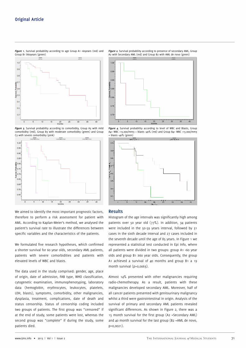
ResultsHistogram of the age intervals was significantly high among
patients over 50 year old (77%). In addition, 34 patients
were included in the 50-59 years interval, followed by 31
cases in the sixth decade interval and 27 cases included in
the seventh decade until the age of 85 years. In Figure 1 we
represented a statistical test conducted in Epi Info, where
all patients were divided in two groups: group A1 <60 year
olds and group B1 ≥60 year olds. Consequently, the group
A1 achieved a survival of 40 months and group B1 a 19
month survival (p=0,0063).
Almost 14% presented with other malignancies requiring
radio-chemotherapy. As a result, patients with these
malignancies developed secondary AML. Moreover, half of
all cancer patients presented with genitourinary malignancy
whilst a third were gastrointestinal in origin. Analysis of the
survival of primary and secondary AML patients revealed
significant differences. As shown in Figure 2, there was a
15 month survival for the first group (A2 =Secondary AML)
and 40 month survival for the last group (B2 =AML de novo,
p=0,0021).
Figure 1. Survival probability according to age Group A1 <60years (red) and Group B1 ≥60years (green)
Figure 2. Survival probability according to presence of secondary AML, Group A2 with Secondary AML (red) and Group B2 with AML de novo (green)
Figure 3. Survival probability according to comorbidity, Group A3 with mild comorbidity (red), Group B3 with moderate comorbidity (green) and Group C3 with severe comorbidity (pink)
Figure 4. Survival probability according to level of WBC and Blasts, Group A4= WBC <15.000/mm3 + blasts <40% (red) and Group B4= WBC >15.000/mm3 + blasts >40% (green)

The International Journal of Medical Students
IJMSInternational Journal of Medical Students
72
Original Article
www.ijms.info • 2013 | Vol 1 | Issue 2
A proportion of 64% of cases had associated comorbidities as they were present especially within the elderly patients. The most frequent were the heart diseases, which comprised 60 cases. Digestive and respiratory illnesses ranked next with 28 and 21 cases respectively. The next frequent disease was diabetes with 19 cases and the less frequent diseases were renal and neurological disorders with 10 and 9 cases respectively. We separated patients, according to comorbidities into three groups seen in Figure 3. Group A3 comprised patients with mild comorbidity with a survival of 40 months. Group B3 covered patients with moderate comorbidities who survived 19 months, whereas group C3 included patients with serious comorbidities who survived only 7 months (p=0,0059).
As shown in the Figure 4, in Epi Info, we analyzed two categories: group A4 =WBC <15.000/mm3 + blasts <40% and group B4 =WBC >15.000/mm3 + blasts >40%. Survival in group A4 was up to 40 months, whereas the group B4 had a survival of 25 months (p=0,0057).
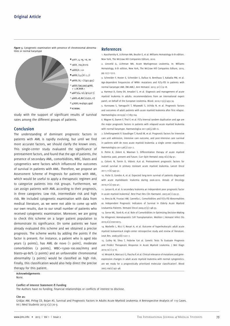
Only 26% of patients received cytogenetic examination. The prognosis distribution of results followed the WHO classification, therefore 71% of patients correspond to intermediate risk, whereas 29% of patients correspond to unfavorable risk with chromosomal abnormalities (Figure 5).
DiscussionVarious clinical and biological features identified previously as useful factors in the prediction of clinical outcomes, could help guide therapy choices.8 Large multicenter trials have demonstrated that: patient’s age, presence of secondary AML, WBC count, cytogenetic and comorbidities at diagnosis were important prognostic factors for AML patients.9
Age remained one of the most significant prognostic factors and its importance has increased among the elderly population.10 Our study shows a clear difference of survival period between the two age groups: <60, >60 years. We decided to define a 60 year-limit because this age is the limit between adults and elderly people. We were not able to find the sample size variable due to the small number of patients. We recognized age as a vital prognostic factor in AML, so we included it in our prognosis scheme.
We highlighted the importance of prior malignancies and their treatment that had a major impact on our patients
survival rate.11 Furthermore, patients with previous malignancies developed secondary AML, which in our study illustrated a surprisingly low life expectancy. As a result, patients with AML de novo lived twice as long as the patients with secondary AML.
Comorbidities affect the therapeutic plan and the post-therapeutic outcome of the index disease. Examples include patients with cancer and multiple studies have demonstrated the relevance of comorbidities in prognosis.12,13 Consequently, we stratified comorbidities after the Sorror Comorbidity Index in mild, moderate and severe comorbidities. Kaplan-Meier survival illustrated a significant difference between the groups in our study. As a result, comorbidities are an important prognostic factor, which affects the survival of patients.
Besides cytogenetic and molecular abnormalities, classically, a high WBC count at presentation was considered an independent prognostic factor. This illustrated a poor outcome in AML, especially for the cytogenetic intermediate risk group.13 A subgroup of patients with hyperleucocytosis was identified with a shorter rate of survival in these cases.14 In our study, many laboratory analyses were performed, but we chose to combine WBC and blast count for a significant difference on our patients survival. Patients were divided into groups according to low or high levels of WBC and blasts. Patients with values of WBC and blasts >40% had much shorter survival than those with low values.
Karyotype abnormalities are probably the most important prognostic determinants in AML.15 However, in the literature it is reported that half of adult AML patients present with a normal karyotype.16 In our study, a proportion of 71 per cent presented a normal karyotype and 29 per cent had unfavorable cytogenetic abnormalities. In our study, we were unable to demonstrate the significance of cytogenetic testing due to the small number of patients who benefited from cytogenetic examination. Finally, we propose that all AML patients should receive cytogenetic examination for a better management of AML.
Our single-center study evaluated and confirmed the significant predictors of outcome of AML. The age of patients, the presence of secondary AML, comorbidities and WBC count and blasts were the most important prognostic parameters. Furthermore, we demonstrated this in our
Table 1. Prognosis assessment scheme.
Points Age (years) Secondary AML Comorbidity WBC (cells/mm3) Blasts (%) Cytogenetics
1p <40 - Mild <15000 <40% t (15,17), t (8,21), inv16*
2p 40-59 Present Moderate 15,000-100,000 40-80% Normal karyotype*
3p >59Present with other
malignanciesSevere >100,000 >80%* del (7q),+8,del(9q), t(v,11)(v,23)*
*from Medical Literature. Low Risk: 5-6p; Intermediate Risk: 7-9p; High Risk: 10-15p.
The International Journal of Medical Students 73
Original Article
www.ijms.info • 2013 | Vol 1 | Issue 2
study with the support of significant results of survival
rates among the different groups of patients.
ConclusionThe understanding of dominant prognostic factors in
patients with AML is rapidly evolving, but until we find
more accurate factors, we should clarify the known ones.
This single-center study evaluated the significance of
pretreatment factors, and found that the age of patients, the
presence of secondary AML, comorbidities, WBC, blasts and
cytogenetics were factors which influenced the outcomes
of survival in patients with AML. Therefore, we propose an
Assessment Scheme of Prognosis for patients with AML,
which would be useful to apply a therapeutic regimen and
to categorize patients into risk groups. Furthermore, we
can assign patients with AML according to their prognosis,
in three categories: Low risk, intermediate risk and high
risk. We included cytogenetic examination with data from
medical literature, as we were not able to come up with
our own results, due to our small number of patients who
received cytogenetic examination. Moreover, we are going
to check this scheme on a larger patient population to
demonstrate its significance. On some patients we have
already evaluated this scheme and we obtained a precise
prognosis. The scheme works by adding the points if the
factor is present. For instance, a patient who is aged ≥60
years (3 points), has AML de novo (1 point), moderate
comorbidities (2 points), WBC=15000-100.000/mm3 and
blasts=40-80% (2 points) and an unfavorable chromosomal
abnormality (3 points) would be classified as high risk.
Finally, this classification would also help direct the precise
therapy for this patient.
References1. Kaushansky K, Lichtman MA, Beutler E, et al. Williams Hematology 8-th edition.
New York, The McGraw Hill Companies Editure, 2010.
2. Liesveld JL, Lichtman MA. Acute Myelogenous Leukemia, In: Williams
Hematology, 8-th edition, New York, The McGraw Hill Companies Editure, 2010,
pp.1277-1312.
3. Schneider F, Hoster E, Schneider S, Dufour A, Benthaus T, Kakadia PM, et al.
Age-dependent frequencies of NPM1 mutations and FLT3-ITD in patients with
normal karyotype AML (NK-AML). Ann Hematol. 2012; 91(1):9-18.
4. Hartmut D, Estey EH, Amadori S, et al. Diagnosis and management of acute
myeloid leukemia in adults: recommendations from an international expert
panel, on behalf of the European Leukemia. Blood. 2010;115(3):453-74.
5. Kurosawa S, Yamaguchi T, Miyawaki S, Uchida N, et al. Prognostic factors
and outcomes of adult patients with acute myeloid leukemia after first relapse.
Haematologica 2010;95(11)1857-64.
6. Wagner K, Damm F, Thol F, et al. FLT3-internal tandem duplication and age are
the major prognostic factors in patients with relapsed acute myeloid leukemia
with normal karyotype. Haematologica 2011;96(5):681-6.
7. Schellongowski P, Staudinger T, Kundi M, et al. Prognostic factors for intensive
care unit admission, intensive care outcome, and post-intensive care survival
in patients with de novo acute myeloid leukemia: a single center experience.
Haematologica 2011;96(2):231-7.
8. Petrie K, Zelent A, Waxman S. Differentiation therapy of acute myeloid
leukemia: past, present and future. Curr Opin Hematol 2009;16(2):84–91.
9. Colovic N, Tomin D, Vidovic A,et al. Pretreatment prognostic factors for
overall survival in primary resistant acute myeloid leukemia. Lancet Oncol
2011;11(6):543-52.
10. Pulte D, Gondos A, et al. Expected long-term survival of patients diagnosed
with acute myeloblastic leukemia during 2006–2010. Annals of Oncology
2010;21(2):335–41.
11. Larson R, et al. Is secondary leukemia an independent poor prognostic factor
in acute myeloid leukemia?. Best Pract Res Clin Haematol. 2007;20(1):29-37.
12. Breccia M, Frustaci AM, Cannella L. Comorbidities and FLT3-ITD Abnormalities
as Independent Prognostic Indicators of Survival in Elderly Acute Myeloid
Leukaemia Patients. Hematol Oncol 2009;27(3):148-53.
13. Sorror ML, Storb R, et al. Role of Comorbidities in Optimizing Decision-Making
for Allogeneic Hematopoietic Cell Transplantation. Mediterr J Hematol Infect Dis
2010;2(2):e2010015.
14. Marbello L, Ricci F, Nosari A, et al. Outcome of hyperleukocytic adult acute
myeloid leukaemia:A single-center retrospective study and review of literature.
Leuk Res. 2008;32(8):1221-7.
15. Gulley M, Shea T, Fedoriw Y,et al. Genetic Tests To Evaluate Prognosis
and Predict Therapeutic Response in Acute Myeloid Leukemia. J Mol Diagn
2010;12(1):3-16.
16. Mrozek K, Marcucci G, Pascha P, et al. Clinical relevance of mutations and gene-
expression changes in adult acute myeloid leukemia with normal cytogenetics:
are we ready for a prognostically prioritized molecular classification?. Blood
2007;109(2):431-48.
Figure 5. Cytogenetic examination with presence of chromosomal abnorma-lities or normal karyotype
AcknowledgementsNone.
Conflict of Interest Statement & FundingThe Authors have no funding, financial relationships or conflicts of interest to disclose.
Cite as:Gridjac AM, Pirlog CD, Bojan AS. Survival and Prognostic Factors in Adults Acute Myeloid Leukemia: A Retrospective Analysis of 119 Cases. Int J Med Students 2013;1(2):70-3.
The International Journal of Medical Students74
IJMSInternational Journal of Medical Students
www.ijms.info • 2013 | Vol 1 | Issue 2
1 Sri Manakula Vinayagar Medical College and Hospital, Pondicherry, India
CorrespondenceLopamudra MoharanaAddress: Sri Manakula Vinayagar Medical College and Hospital. Kalitheerthalkuppam, Madagadipet, Puducherry, 605 107, India.Email: [email protected]
IntroductionThe promotion of health is of fundamental value in itself. It is a vital public good and a basic human right. In this re-gard, delivery of healthcare is very important for providing preventive, promotive and curative services to the com-munity.1
There have been substantial achievements in healthcare in past few decades. However, technological innovation in the health sector has improved the quality of life but has also increased costs especially in middle and low income coun-tries. Shortcomings in healthcare delivery have been largely designated as fragmented care, misdirected care and impo-verishing care.2 In countries that have no social insurance and where the role of the state is limited, people spend a substantial proportion of their incomes on seeking medical treatment, and in the process get impoverished, thus wide-ning disparities in the health status.3 The unpredictability of illness, the lumpiness of health consumption, and the irregular and seasonal nature of incomes make it virtually impossible for the poor to finance their health needs, resul-ting in a denial of care and poverty.1
According to the World Health Organization (2005) estima-tes, every year 25 million households (more than 100 mi-llion people) are forced into poverty by illness and struggle to pay for healthcare.4 The decline in public investment in health and the absence of any form of social insurance have heightened insecurities. Considering the Indian sce-nario, a report by the National Health Accounts reveals that 71% of the health budget is contributed by the private sec-tor; of which households alone spend about 69%.1 It is well known that health expenditure in India is dominated by pri-vate spending and this is a reflection of inadequate public spending. The relationship between poverty and ill-health is indisputable. Even relatively small expenditure on health can be financially disastrous for poor households. High out of pocket payment, an absence of risk pooling mechanism in health financing systems, and high level of poverty can result in catastrophic health expenditure.5 Thus, the pre-sent study was conducted to quantify the health care ex-penditure of households in rural Pondicherry with these objectives; i) to note the health care expenditure of rural households and ii) to assess if any family is undergoing catastrophic health expenditure.
Health Care Expenditure of Rural Households in Pondicherry, India
Poornima Varadarajan,1 Lopamudra Moharana,1 Murugan Venkatesan1
AbstractBackground: Shortcomings in healthcare delivery has led people to spend a substantial proportion of their incomes on medical treatment.
World Health Organization (2005) estimates reveal that every year 25 million households are forced into poverty by illness and the stru-
ggle to pay for healthcare. Thus we planned to calculate the health care expenditure of rural households and to assess the households
incurring catastrophic health expenditure. Methods: A cross-sectional study was conducted in the service area of Sri Manakula Vinayagar
Medical College and Hospital from May to August 2011. A total of 100 households from the 4 adjoining villages of our Institute were selec-
ted for operational and logistic feasibility. The household’s capacity to pay, out of pocket expenditure and catastrophic health expenditure
were calculated. Data collection was done using a pretested questionnaire by the principal investigator and the analysis was done using
SPSS (version 16). Results: The average income in the highest income quintile was Rs 51,885 but the quintile ratio was 14.98. The median
subsistence expenditure was Rs 4,520. About 18% of households got impoverished paying for health care. About 81% of households were
incurring out of pocket expenditure and 66% were facing catastrophic health expenses of 40%. Conclusion: There was very high out of
pocket spending and a high prevalence of catastrophic expenditure noted. Providing quality care at affordable cost and appropriate risk
pooling mechanism are warranted to protect households from such economic threats.
Key Words: Health expenditures, health services needs and demand, India (Source: MeSH-NLM).
Submission: July 11, 2012. Accepted: Nov 17, 2012.Process: Peer-Reviewed.
Original Article
About the Author:Poornima Varadarajan is a 4th year medical student at Sri Manakula Vinayagar Me-dical College and Hospital, Pondicherry, India

The International Journal of Medical Students 75www.ijms.info • 2013 | Vol 1 | Issue 2
Original Article
Materials and MethodsStudy SettingPondicherry (Puducherry), district of India has a total population of 946,600 (census 2011) of which the rural population is about 292,208 (30. 87%). Per-capita income of Pondicherry at 2009-10 current prices is in indian rupee (Rs) 72,917 (draft annual plan 2011-12, Pondicherry) and about 22% are below poverty line (2004-05 Pondicherry estimate).6 Pondicherry ranks quite high compared to India in terms of fulfillment of several health infrastructure indicators. The total health care expenditure is Rs 80 but the per-capita expenditure is Rs 783 (budget estimate, 2003-04).7
Study Design and SamplingWe planned to study the total health spending so that the burden on households can be commented on. Moreover, to get a better picture of rural areas we studied the rural households. A cross-sectional study was designed and conducted from May to August 2011. We covered four villages, situated within 2-4 kilometers of our Institute (Sri Manakula Vinayagar Medical College and Hospital), for operational and logistic feasibility. The villages were Madagadipet (1174), Kalitheerthalkuppam (1120), Kuchipalayam (158) and PS palayam (441) which constituted our sampling frame and the list of houses of the respective villages were collected from the PHC registers (2011). We planned to survey a total of one hundred households. From the total number of households of the four villages (2893), all sampled households were included in the study. A proportionate sampling method was adopted to draw the sample from the household numbers of each village. The streets in each village were selected by simple random technique. Systematic random sampling method was adopted to select every third household in each street. Initially, a pilot study was conducted to assess and modify the logistic problems expected during the main study.
ParametersThe total household income and expenses were calculated. For expenditure on health, both direct and indirect expenses were assessed. Direct expenses were costs incurred for the defined medical problem (consultations, investigations, medicines etc.) and the indirect cost included collateral expenses due to the illness (travel, food, loss of wages etc.). We considered the expenses incurred by the households both for their outpatient and inpatient consultations. To avoid recall bias in expenditure, a one month recall period for any OPD consultations and 3 months for any in-patient admissions was considered. For our calculations, we used equivalent household size (household size 0.56)8 instead of average household size. The various heads of expenses of the households were determined and for accuracy of food expenditure, equivalized food expenditure was calculated. The subsistence expenditure per (equivalent) capita or the poverty line was determined and the subsistence
expenditure (poverty line*equivalent household size) was calculated. The household’s capacity to pay (non subsistence effective income of households), out of pocket expenditure (OOP), burden of health payment and catastrophic health expenditure (CHE) was calculated adopting the methodology described by Xu K et al.8 OOP expenditure is defined as the payment made by families for health care and include out of pocket spending on deductibles and other forms of cost sharing such as co-payments and co-insurance and direct expenditure of health care services equipments and supplies not covered by insurance. OOP in our study was the net of insurance reimbursements and did not include indirect expenses (health-related travel and food). CHE is defined as the level of OOP expenditure that exceeds some fixed proportion of household income or household capacity to pay.9,10 For the purpose of our study, if a household’s total OOP equaled or exceeded 40% of the household’s capacity to pay (non subsistence effective income of the household or income available after basic needs have been met), it was considered to be facing CHE. We also calculated the households that are poor (total household expenditure less than its subsistence spending) and the non-poor households that were impoverished by health payments.
ResultsThe socio-demographic profile of the total households sur-veyed is described in Table 1. We noted that the majority belonged to the Hindu religion (94%). The various castes were OBC (50%), MBC (44%) and the forward casts (6%). About 81% houses were pucca houses and the majority (93%) were nuclear families. The median ‘total’ and ‘per-capita’ income of the households were Rs 10,000 and Rs 2,333 respectively. The average income in the highest (5th) quintile was Rs 51,885 but the “quintile ratio” (richest to poorest) was 14.98. A majority (72%) possessed pink ration cards and 8% did not have any ration card. However, those who participated in different income-generating activities and in self-help groups were 7% and 2% respectively. None of the households had health Insurance. The “equivalent household size” was 2.26 and the median ‘equivalized per capita household expenditure” was Rs 2323 where as the median “equivalized food expenditure’ was Rs 1379. From the food expenditure, the subsistence expenditure per (equivalent) capita or the poverty line was calculated as Rs 2,080 and thus the median “subsistence expenditure” was calculated to be Rs 4,520.
The health facilities preferred by the majority of the hou-seholds were governmental (24%) and private (74%). About 1% preferred both private and governmental facilities and the remaining 1% preferred chemist shops. About 69% had reported outpatient illnesses and 35% had inpatient illnes-ses within our specified period of recall. The common out-patient illnesses were fever, cough and cold, diarrhea, body and joint pains, gastritis, poor vision, pregnancy, allergy, TB,

The International Journal of Medical Students
IJMSInternational Journal of Medical Students
76
Characteristic % n = 100
Religion
Hindu 94
Muslim 5
Christian 1
Housing type
Pucca 81
Kutcha 8
Semipucca 11
Family type
Nuclear 93
Joint 7
Possession of ration cards
Pink 72
Yellow 20
No card 8
Income generating activities
No 93
Yes 7
Participation in SHG
No 98
Yes 2
Health Insurance
No 100
Yes 0
Income Quintile
1st Quintile (Rs 3462)* 26
2nd Quintile (Rs 6875)* 18
3rd Quintile (Rs 10,188)* 16
4th Quintile (Rs 16,781)* 20
5th Quintile (Rs 51,885)* 20
Quintile Ratio (Richest to poorest) 14.98
Total income (Median) 10,000
Per capita monthly income (Rs, median) 2333
Equivalent household size† (Mean ± SD) 2.26 ± 0.4
Equivalized household expenditure (Rs per capita, median) 2323
Equivalized food expenditure‡ (Rs, median) 1379
Subsistence expenditure (Rs, median) 4520
Original Article
www.ijms.info • 2013 | Vol 1 | Issue 2
Table 2. Reported illness, capacity to pay and consequence of health expen-ses incurred per households.
Catastrophic Threshold (OOPCTP)
No of Households with illness
(n = 81)
% of Households with Illness†
Household Expenditure/m
(Median)
< 20% 6 7 520
20-40% 15 19 600
>40% 60 74 8025
Table 3. Prevalence of Catastrophic Expenditure by Cut off Levels (of OOPCTP)
* CHE, i.e, OOPCTP ≥ 40% was incurred by 66%.† Illnesses include both outpatient and inpatient categories.Abbreviations: OOP: Out of Pocket expenditure, OOPCTP: OOP share of capacity to pay.
Table 1. Socio-demographic characteristics of the study participants
* Figures in parentheses are average Income in the corresponding income quintile.† Equivalent household size 0.56 (Adopted from Xu K etal.).‡ Equivalized food exp = Food expenditure / equivalent household size.Abbreviations: OOP: Out of Pocket expenditure, Rs: Indian rupee, SHG: Self-help group,SD: Standard deviation.
* Note: Median OOP spending per household was Rs 3000. Median subsistence spending (expenditure) of the households was Rs. 4520. The households’ capacity to pay is calculated as the non-subsistence effective Income of households. CHE is calculated as OOPCTP ≥40%. About 18% are impoverished paying for their healthcare expenses. Abbreviations: OOP: Out of Pocket expenditure, Rs: Indian rupee.
Characteristic % n = 100
Health facility preferred
Private 74
Governmental 24
Both 1
Chemist shop 1
Outpatient illness (Reported within 2 months) 69
Inpatient illness (Reported within 3 months) 35
Household spending on health (n=84, who reported any illness, as % of their total Income)
<20% 35
20-50% 14
> 50% 35
Household’s capacity to pay
Rs 10,000 71
Rs 10,000-30,000 18
Rs >30,000 11
OOP incurred 81
OOP share of total income
≤50% 48
>50% 33
OOP share of total expenses
≤50% 33
>50% 48
OOP share of capacity to pay
<20% 6
20-40% 15
>40% 60
Poor (Below poverty line) 29
Impoverishment (Due to health expenses) 18

The International Journal of Medical Students 77
Original Article
www.ijms.info • 2013 | Vol 1 | Issue 2
diabetes and hypertension. The common inpatient illnes-
ses were accidents, abdominal pathologies and major ab-
dominal surgeries, obstetric and gynecological complaints,
hernias, fractures, tumors and non healing ulcers. Out of
the households who reported some illness (84%), about
49% were spending about 50% of their total income and
35% were spending even >50% of their incomes (direct and
indirect expenses) on health (Table 2).
Analysis of the households’ capacity to pay showed that
the majority had the capacity to pay up to Rs 10,000 (71%)
while the remaining had the capacity to pay more. About
81% of the households were incurring OOP expenses (only
direct health expenses were considered) and the median
OOP spending per household was Rs 3,000 (Table 2). For
33% of households, the OOP incurred was 50% of their to-
tal expenses but for 48% of households, OOP incurred was
even >50% of their total expenses. About 29% of the hou-
seholds whose total household expenditure was less than
their subsistence spending were already poor, but about
18% were impoverished by paying for health expenses (Ta-
ble 2).
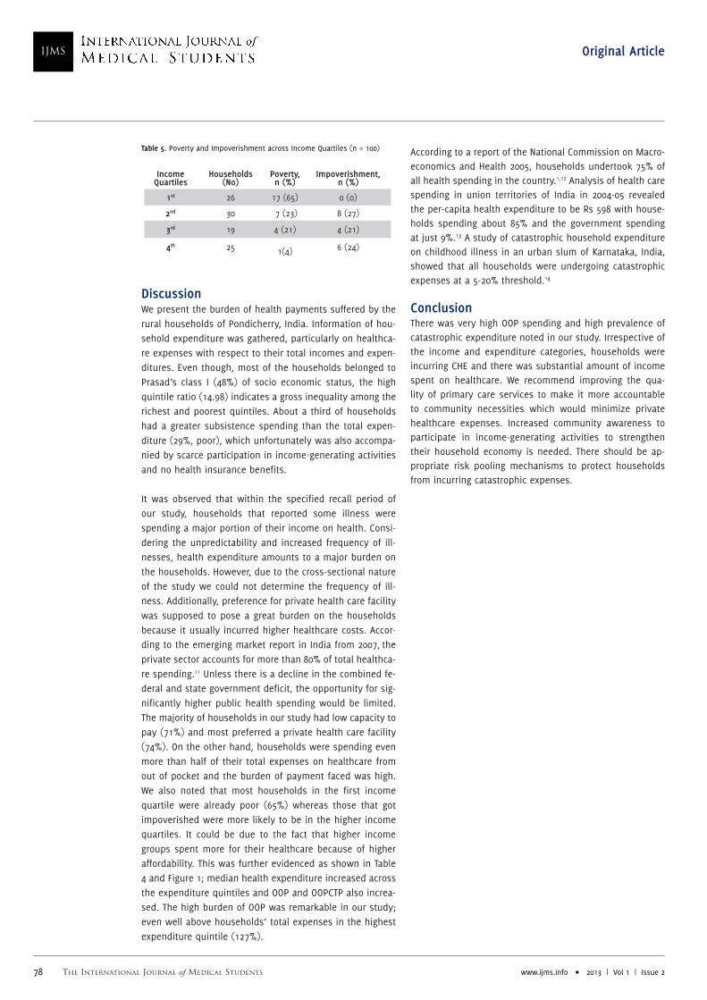
The OOPCTP of ≥40%, i.e., CHE was incurred by 66% of households (Table 2). The percentage of households at catastrophic threshold of <20%, 20-40% and >40% cut off levels were 6%, 15% and 60% respectively (Table 3). We noted that households with higher proportion of reported illnesses and those belonging to higher median household expenditure categories were incurring high CHE. Across the different expenditure quintiles (Table 4, Figure 1), the median health expenditures and median OOP were noted to increase gradually. The OOPCTP and the OOPEXP were highest in the higher expenditure quintiles (4th and 5th quintiles). The OOPEXP was even more than 100% in the highest (5th) expenditure quintiles. We also noted that the-re was impoverishment in the middle expenditure quintiles (3rd and 4th); whereas lowest and highest ones did not show impoverishment. Table 5 represents a picture of the households under poverty and those who got impoverished by paying for health care expenses across income quarti-les. We noted that the highest number of poor households (65%) were in the first quartile and the tendency gradually decreased towards the highest quartile. On the contrary, households that faced impoverishment (paying for health care expenses) were 27%, 21% and 24% in the 2nd, 3rd and 4th quartile respectively. There was no impoverishment no-ted in the first quartile.
Figure 1. Health Expenses Incurred across the Expenditure Quintiles (n = 100)
Expenditure Quintiles
Median Health Expenditure (Rs) OOP in Rs (Median) OOPCTP (%) OOPEXP (%) Impoverished (%)
1st 926 725 55 37.5 Nil
2nd 625 610 46 20.1 Nil
3rd 1775 1330 49 22.7 55
4th 5710 5110 68 86.9 35
5th 11900 10900 69 127.3 Nil
Table 4. Health Expenses Incurred across the Expenditure Quintile (n = 100)
Note: Health Expenditure included both direct and indirect expenses but the OOP takes only direct expenses into consideration
The International Journal of Medical Students
IJMSInternational Journal of Medical Students
78 www.ijms.info • 2013 | Vol 1 | Issue 2
Original Article
DiscussionWe present the burden of health payments suffered by the rural households of Pondicherry, India. Information of hou-sehold expenditure was gathered, particularly on healthca-re expenses with respect to their total incomes and expen-ditures. Even though, most of the households belonged to Prasad’s class I (48%) of socio economic status, the high quintile ratio (14.98) indicates a gross inequality among the richest and poorest quintiles. About a third of households had a greater subsistence spending than the total expen-diture (29%, poor), which unfortunately was also accompa-nied by scarce participation in income-generating activities and no health insurance benefits.
It was observed that within the specified recall period of our study, households that reported some illness were spending a major portion of their income on health. Consi-dering the unpredictability and increased frequency of ill-nesses, health expenditure amounts to a major burden on the households. However, due to the cross-sectional nature of the study we could not determine the frequency of ill-ness. Additionally, preference for private health care facility was supposed to pose a great burden on the households because it usually incurred higher healthcare costs. Accor-ding to the emerging market report in India from 2007, the private sector accounts for more than 80% of total healthca-re spending.11 Unless there is a decline in the combined fe-deral and state government deficit, the opportunity for sig-nificantly higher public health spending would be limited. The majority of households in our study had low capacity to pay (71%) and most preferred a private health care facility (74%). On the other hand, households were spending even more than half of their total expenses on healthcare from out of pocket and the burden of payment faced was high. We also noted that most households in the first income quartile were already poor (65%) whereas those that got impoverished were more likely to be in the higher income quartiles. It could be due to the fact that higher income groups spent more for their healthcare because of higher affordability. This was further evidenced as shown in Table 4 and Figure 1; median health expenditure increased across the expenditure quintiles and OOP and OOPCTP also increa-sed. The high burden of OOP was remarkable in our study; even well above households’ total expenses in the highest expenditure quintile (127%).
According to a report of the National Commission on Macro-economics and Health 2005, households undertook 75% of all health spending in the country.1,12 Analysis of health care spending in union territories of India in 2004-05 revealed the per-capita health expenditure to be Rs 598 with house-holds spending about 85% and the government spending at just 9%.13 A study of catastrophic household expenditure on childhood illness in an urban slum of Karnataka, India, showed that all households were undergoing catastrophic expenses at a 5-20% threshold.14
ConclusionThere was very high OOP spending and high prevalence of catastrophic expenditure noted in our study. Irrespective of the income and expenditure categories, households were incurring CHE and there was substantial amount of income spent on healthcare. We recommend improving the qua-lity of primary care services to make it more accountable to community necessities which would minimize private healthcare expenses. Increased community awareness to participate in income-generating activities to strengthen their household economy is needed. There should be ap-propriate risk pooling mechanisms to protect households from incurring catastrophic expenses.
Income Quartiles
Households (No)
Poverty, n (%)
Impoverishment, n (%)
1st 26 17 (65) 0 (0)
2nd 30 7 (23) 8 (27)
3rd 19 4 (21) 4 (21)
4th 25 1(4) 6 (24)
Table 5. Poverty and Impoverishment across Income Quartiles (n = 100)

The International Journal of Medical Students 79www.ijms.info • 2013 | Vol 1 | Issue 2
Original Article
Klavus J, Knaul F, Murray CMJ, Ortiz JP, Zeramdini R, Annan S, Doorslear EV.
Distribution of Health Payments and Catastrophic Expenditures Methodology:
Discussion Paper. World Health Organization, Department of Health System
Financing and Cluster Evidence and Information for Policy, 2005.
9. Pal R. Analysing Catastrophic OOP Health Expenditure in India: Concepts,
Determinants and Policy Implications, Indira Gandhi Institute of Development
Research, Mumbai, 2010; 1-25. Available online at http://www.igidr.ac.in/pdf/
publication/WP-2010-001.pdf
10. Su TT, Kouyate B, Flessa S. Catastrophic Household expenditure for
Health Care in a Low Income Society: A Study from Nouna District, Burkina
Faso. Bulletin of the World Health Organization 2006; 84 (1); 21-27.
11. Healthcare in India: Emerging market report. 2007; 1-22. Available online
at http://www.pwc.com/en_GX/gx/healthcare/pdf/emerging-market-report-
hc-in-india.pdf
12. National Health Accounts. National Health Accounts Cell, Ministry of
Health and Family Welfare, Govt. of India (in collaboration with WHO country
office for India). 2004-2005.
13. Gupta I. Draft of Out-of-pocket Expenditures and Poverty: Estimates
from NSS 61st Round. Paper presented for consideration of the Expert
Group on Poverty, Planning Commission.12 May 2009. Available online from
planningcommission.nic.in/reports/genrep/indrani.pdf
14. Patil SS, Berad AS, Angadi MM. A Study to Assess Catastrophic Household
Expenditure on Childhood Illness in an Urban Slum in Bijapur. Indian J
Community Med 2009;34(4):335-7.
References1. Report of the National Commission on Macroeconomics and Health,
Ministry of Health and Family Welfare, Government of India, New Delhi,
August, 2005. Available online at www.who.int/macrohealth/action/
Report%20of%20the%20National%20Commission.pdf
2. The World Health Report 2008: Primary Health Care, Now more than Ever,
The World Health Organization, 2008. Available online at www.who.int/entity/
whr/2008/08_overview_en.pdf
3. Government Health Expenditure in India: A Benchmark Study Undertaken
for the MacArthur Foundation, India by Economic Research Foundation, New
Delhi, August 2006; 42-2.
4. World Health Report 2005. Make every mother and child count. Geneva:
World Health Organization; 2005. Available online at http://www.who.int/
whr/2005/whr2005_en.pdf
5. Xu KD, Evans D, Karrin G, Aguilar-Rivera AM. Technical Brief for Policy
Makers. Designing health financing systems to reduce catastrophic health
expenditure. World Health Organization, Department of Health Systems
Financing, Health Financing Policy, 2005; 1-4.
6. Draft Annual Plan, Puducherry. 2011-2012. Available online at http://pandr.
puducherry.gov.in/Plan%20Formulation/Plan%20Document/DAP%202011-12/
pdffiles/int/int.pdf
7. Profile of Union Territory of Pondicherry, 2005; 1-90. Available online at
http://www.ncw.nic.in/pdfreports/Gender%20Profile-Pondicherry.pdf
8. Xu K, Anguliar A, Carrin G, Evans DB, Hanvoravongchai P, Kawabata K,
AcknowledgementsNone.
Conflict of Interest Statement & FundingThe Authors have no funding, financial relationships or conflicts of interest to disclose.
Cite asPoornima V, Lopamudra M, Murugan V. Health Care Expenditure of Rural Households in Pondicherry, India. Int J Med Students 2013;1(2):74-9.

The International Journal of Medical Students www.ijms.info • 2013 | Vol 1 | Issue 280
IJMSInternational Journal of Medical Students
1 School of Medicine, University of Aberdeen, Foresterhill, Aberdeen, UK.2 St John’s Hospital, NHS Lothian, Howden Road West Howden Livingston, UK.
CorrespondenceFarihah TariqAddress: School of Medicine and Dentistry, 3rd Floor Polwarth Building, University of Aberdeen. Foresterhill, Aberdeen, UK, AB25 2ZD.Email: [email protected]
IntroductionAmetropic disorders of vision affect between 800 million to
2.3 billion individuals globally.1 Around 140 million users
worldwide, including 3.3 million in the United Kingdom,
wear contact lenses for the correction of refractive errors.2,3
The British contact lens market value has risen from £33
million in 1992 to £198 million in 2009.3 They are becoming
increasing popular because of the clearer vision achieved,
for cosmetic reasons, for sports and convenience. Contact
lenses are, however a medical device and wearing contact
lenses incurs risks with an estimated 6% of users develo-
ping complications.4 We will discuss the pathophysiology
of contact lens-associated complications and their avoi-
dance.
Search strategy and selection criteriaSoft contact lenses are the focus of this paper. In depth dis-
cussion on other types of lenses such as rigid gas permeable,
PMMA lenses were out of the scope of this paper. We identified
the papers in this review by a computerised search of the Pub-
Med database using the queries “contact lens complications”
and “contact lens keratitis”. We gathered other information
from contact lens manufacturers’ data sheets and used evi-
dence from published abstracts, major international scienti-
fic meetings and textbooks as well as reference collections.
How do contact lenses affect the ocular surface?Contact lens wearers are sixty times more likely to deve-lop ocular disorders than the general population, with the users of extended wear at greatest risk.5-8 An estimated 1 per 2500 persons per year using daily wear and 1 per 500 persons per year using extended wear will develop presu-med microbial keratitis.9 Incidences of complications com-piled by Morgan and colleagues is presented in Table 1.7
Contact lenses influence the allergic and inflammatory res-ponses, alter the ocular microbiota, cause metabolic and mechanical trauma, reduce ocular surface wetting and can exacerbate pre-existing ophthalmic disorders.10,11
Contact lenses alter the natural ocular environmentContact lenses are foreign objects in the eye, altering the natural environment by introducing a bio-burden of micro-organisms to the ocular surface from contaminated hands, lens and lens-care solution.8,12 Insertion of the lens initiates the formation of a biofilm which not only attracts pathoge-nic flora but increases antibiotic resistance by almost one thousand fold.13 Bacteria adhere to the contact lens; this propensity is stimulated by deposits on the lens surface.14 Within 30 minutes of insertion, approximately 50% of the lens accumulates materials on or into the lens matrix.15 Such spoilage by the constituents of the tear film is not
The Risk of Contact Lens Wear and the Avoidance of Complications
Farihah Tariq,1 Peter Koay2
AbstractContact lenses are lenses placed on the surface of the cornea to correct refractive errors such as myopia (short-sightedness), hyper-
metropia (far-sightedness) and astigmatism. Lens-related complications are becoming a greater health concern as increasing number of
individuals are using them as an alternative to spectacles. Contact lenses alter the natural ocular environment and reduce the efficacy
of the innate defences. Although many complications are minor, microbial keratitis is potentially blinding and suspected cases should be
rapidly diagnosed and referred to an ophthalmologist for treatment. Several risk factors have been identified with extended wear, poor
hand hygiene, inadequate lens and lens-case care being the most significant. Promotion of good contact lens hygiene and practices are
essential to reduce the adverse effects of contact lens wear.
Key Words: Contact Lenses, Complications, Keratitis, Patient Compliance (Source: MeSH-NLM)
Submission: Sept 26, 2011. Accepted: Sept 9, 2012.Process: Peer-Reviewed.
Review
About the Author:Farihah Tariq is a 5th year medical student at School of Medicine, University of Aber-deen, Foresterhill, Aberdeen, UK.

The International Journal of Medical Students 81www.ijms.info • 2013 | Vol 1 | Issue 2
only involved in generalised irritation but also contributory
towards complications such as giant papillary conjunctivitis
(GPC), contact lens-induced acute red eye (CLARE), contact
lens-related peripheral ulcer (CLPU) and infiltrative keratitis
(IK) (Figure 1).10,16,17
Additionally, the innate humoral ocular defence mecha-
nisms are reduced by the contact lens limiting tear exchan-
ge as well as altering the quantity and quality of the tear
film.2,18 The lens interferes with the protective function of
the mucin layer (resistant to bacteria adherence) and it hin-
ders the release of anti-microbial factors.2,8,19,20 Coupled with
reduced blinking, these ultimately augment the retention
of potential pathogens onto the ocular surface facilitating
infection.2
The contact lens directly impedes oxygen transmissionContact lenses cause micro-trauma attributed to hypoxia. The cornea receives oxygen fundamental to cellular function primarily through the atmosphere and a small quantity from the limbal and aqueous vasculature.10
Hypoxia causes oedema, altering the epithelial and en-dothelial morphology predisposing the cornea to cellular breakdown.10,21 Reduced oxygen permeability correlates with diminished corneal sensation and increased risk of keratitis. The greater oxygen permeable silicone hydrogel lenses have a five-fold reduced risk of severe keratitis com-pared with hydrogels.7
Scarce distribution of oxygenated tear film due to reduced blinking whilst users are performing visual tasks like wor-
Review
Figure 1. Contact lens-associated complications
A
B
C
D
E
A. Giant Papillary Conjunctivitis: Delayed hypersensitivity inflammatory reaction due to repeated mechanical irritation to residue on lens surface or toxic reaction to cleaning solutions, characterised by papillary changes in the tarsal conjunctiva (cobblestone appearance), itchiness and reduces lens tolerance.10,*
B. Microbial Keratitis: Cornea infection by bacteria, protozoa or fungus, characterised by excavation of the corneal epithelium with infiltration, odeama, necrosis and neovascularisation. There is significant pain, discharge, photophobia and reduced visual acuity.9,10,†
C. Contact Lens Induced Peripheral Ulcer: Corneal inflammation characterised by a small circular full thickness epithelial lesion and infiltration.9,†
D. Contact Lens-Associated Red Eye: Inflammatory reaction of the cornea and the conjunctiva to toxins produced by bacteria on lens surface particularly in those over wearing or sleeping in lenses. It is associated with severe hyperemia, pain and corneal infiltration (indicated by arrow) with minimal or no epithelial involvement.9,†
E. Infiltrative Keratitis (IK)- Inflammatory process charactersised by corneal infiltration (indicate by arrow).28,†* Adapted with permission from emedicine.com, 2011. Available at: http://emedicine.medscape.com/article/1228681-overview.† Adapted with permission from the Guide to Corneal Infiltrative Conditions from the Brien Holden Vision Institute, Sydney, Australia, 2011. To obtain a full scale copy of the Guide, please contact the Brien Holden Vision Institute via http://www.brienholdenvision.org.

The International Journal of Medical Students
IJMSInternational Journal of Medical Students
82 www.ijms.info • 2013 | Vol 1 | Issue 2
Review
king at computers for a prolonged period can also lessen the oxygen availability.22 Closing of the eyelid, for example, when sleeping is also known to lower the amount of oxygen reaching the tear film causing the cornea to swell.23 Slee-ping in lenses may lead to nocturnal hypoxia and deposits may build up on lenses fostering a risk of infection. There is an eight-fold increased incidence of corneal infiltrative events and four-fold increased risk of microbial keratitis in those who sleep wearing lenses compared with users of waking hours only.24,25
Acute hypoxia can lead to overwear syndrome whilst chro-nic hypoxia can instigate corneal neovascularisation contri-buting to decreased visual acuity, particularly if the central visual axis is involved.10,22 However, with the availability of more permeable lenses such problems have been reduced.8
Contact lenses introduce pathogensThe corneal surface is under a constant threat of infection from a barrage of pathogens and at any instance up to 63% of contact lenses yield a positive culture consisting of normal commensals.26 Reduced efficacy of the defence mechanisms coupled with change in the concentration and variety of bacteria can contribute towards pathogenic pro-cesses.8 With the natural barriers threatened, damage to the intact cornea allows bacteria to adhere to the cell mem-brane; a vital step in the infectious process as it aids colo-nisation.13 Recent research has shown there is upregulation of surface-binding receptors further augmenting bacterial adherence.20 Contact lenses, particularly soft non-silicone hydrogel lenses, potentiate their infiltration by inducing changes in corneal epithelium (e.g. reduced desquamation and mitotic activity) making it thinner and increasing the risk of infection.2,20
Although a variety of organisms have been isolated from corneal infections, gram negative infections are most com-mon and sight threatening.9,27,28 Infectious keratitis arising due to the ubiquitous Pseudomonas aeruginosa has the greatest associated morbidity.29 This is attributed to a large number of genes dedicated to virulence regulation, envi-ronmental adaption and resistance to antimicrobial drugs
(e.g. aminoglycosides).2,30 Although rare, 5% of contact lens-related microbial keratitis is attributed to Acanthamoeba.31 This opportunistic pathogen is found in soil and air; but the main perpetrator is contaminated water (e.g. swim-ming pools, hot tubs, water tanks, lakes and contaminated cleaning solution).32-34 Acanthamoeba exists in two forms; a feeding and replicating trophozite which can form antimi-crobial-resistant dormant cysts.32,33 Acanthamoeba keratitis was associated with a poor prognosis before the introduc-tion of topical polyhexamethylenebiguanide (PHMB), pro-pamidine isethionate, and chlorhexidine; 30% of patients had reduced visual acuity (6/18 or less), 50% underwent surgery whilst enucleation was performed in resistant ca-ses.31-34 More recently, early diagnostic techniques and ti-mely treatment with anti-amoebics have improved progno-sis; 90% of patient retain visual acuity of 6/12 or better and less than 2% become blind.34
Correctly differentiating microbial keratitis from the less se-rious sterile corneal infiltrates is crucial.35 Sterile infiltrates tend to be present on the periphery and may be symptoma-tic or asymptomatic.28 They may be the consequence of lens wear itself, from bacterial endotoxins present in conditions such as Staphylococcus aureus–associated blepharitis, or an amalgamation of the two.10,36 Insults from corneal infil-trates is thought be an aetiological factor in CLPU, CLARE and, IK.28 Efron and colleagues have suggested that such inflammatory events can either develop or potentiate the risk of microbial keratitis.28
What are the risk factors for developing contact lens wear complications? There are a range of modifiable and non-modifiable risk factors involved in the development of complications.9 Non-modifiable risk factors are younger age (<25 years), older age (> 50 years), male gender, diabetes mellitus, low socioe-conomic class and late winter months.6,8,9,28,37,38 Modifiable risk factors are those which can be influenced or altered and includes improper lens and case care, poor hand hy-giene, smoking, swimming and showering wearing lenses, as well as extended and overnight wear.8,9,28
Figure 2. Relative risks and non-compliance for a range of compliance and usage factors
Adapted with permission from from Baush & Lomb, 2010.40
Contact lens typeComplications (per 10,000)
Non-severe Severe
Daily wear hydrogel 5 9
Extended wear hydrogel 96 99
Extended wear silicon hydrogel 20 48
Table 1. Incidence of contact lens-associated complications.7
1. Regular review by contact lens provider
2. Take hygiene instructions seriously
3. Follow and understand the care protocol and regime
4. Avoid overnight wear unless extended wear lenses
5. Never shower or swim wearing contact lenses
Table 2. Avoiding contact lens-related complications

The International Journal of Medical Students 83www.ijms.info • 2013 | Vol 1 | Issue 2
Review
Non-ComplianceDissenting behaviour amongst contact lens wearers is pa-ramount when considering the main reason for complica-tions.39 A large well conducted study undertaken on behalf of Bausch & Lomb across Europe highlighted that 98% of all lens wearers were non-compliant in at least one aspect of their lens-care regime (Figure 2).8,40,41
(1) Hand Hygiene: Although inadequate handwashing be-fore lens handling has been associated with a significant increase in risk of infection, the effect is not instantaneous as it takes weeks to remove micro-organisms embedded on the hands.8,42 Perhaps as Morgan suggested, formal training should be provided as this has proven to improve infection control in hospital settings.8
(2) Care Regime & Solutions: One in three lens-related com-plications arise arise directly from inadequate lens care.35,43 Cleaning regimes are either hydrogen peroxide or multipur-pose solution based. Multipurpose solution, dubbed as the ‘no rub’ solution is the most widely used. However, rubbing and rinsing is an imperative step as it removes up to 99.9% of bacteria, thereby adding a safety margin of up to 100,000 times.8,35 Interestingly, recent studies have demonstrated, hydrogen peroxide based cleaning regimes have superior disinfecting capabilities than using multipurpose solution alone.20,44 They reduce the risk of corneal inflammation by ten-fold and disinfects against amoebic cysts.45,46 However, for maximal benefit lenses must be exposed to the peroxide solution for a longer time and must be neutralised before wear to avoid ocular toxicity.35,47,48
(3) Personal Habits: Other unsafe practises include using lenses beyond their recommended replacement schedule, inadequate lens-case care and topping up contaminated solution.8,35,49 The risk increases four-fold compared with appropriately discarded lenses.8
Unsupervised wear Another recent social trend was the use of zero-powered or plano tinted cosmetic lenses designed to change the colour of the eye. They were being bought from unlicensed vendors over the internet without prescription, proper fitting, inade-quate information on use, hygiene and complications and no ongoing supervision.50 Complications associated with the use of such lenses were first reported in 2003.51 In 2005, further cases reported users sharing lenses between multi-ple wearers without adequate cleaning.52 Subsequently, in 2006, Food and Drug Administration (FDA) introduced gui-dance in the USA, whereby plano lenses could only be pur-chased under the supervision of a registered practitioner.53
OrthokeratologyOrthokeratology is the practice of temporary reduction in myopia by the programmed application of rigid gas-per-meable contact lenses, usually at night whilst sleeping.54
Of late, there has been a resurgence of this phenomenon particularly in East Asia and there are growing concerns about the risk of microbial keratitis and loss of vision.2,34,55 Findings of fifty case studies showed 30% had Acanthamoe-ba keratitis from nocturnal orthokeratology compared with 5% from regular lens wear.56
What are the implications?Each year 0.02% to 0.04% of lens wearers can lose up to two lines of best correct visual acuity measured using the snellen chart.24,57 As well as the risk of losing sight, other significant morbidity associated includes hospital admis-sion and/or intensive treatment, cost of therapy, visiting a health care provider, taking time off from work and ina-bility to wear lenses.28,29 An Australian study estimated the median direct costs at Aus $760 [interquartile range $1859] and indirect median costs at Aus $468 [interquartile range $1810].29 Not to mention, patients may claim compensation for negligence.58
How to reduce the risk of complicationsEducationPatient education, particularly regarding the handling and maintenance of contact lenses, is vital in improving overall
Figure 3. The Traffic Light Compliance Model, developed by Dr. Philip Morgan
Adapted with permission from from Baush & Lomb, 2010.41
The International Journal of Medical Students
IJMSInternational Journal of Medical Students
84 www.ijms.info • 2012 | Vol 1 | Issue 2
Review
compliance.59 There is no statistically significant difference
between patients receiving both verbal and written instruc-
tions and those receiving oral only.39 However, intense ini-
tial education has shown improvements in handwashing.60
Morgan and colleagues reported that although, 88% were
given lens care information, 23% were unable to recollect
seeing any information regarding the risks and complica-
tions associated with lens wear.41 Thus, the practitioner
must ensure the patient understands the associated risks,
how these are best avoided, as well as early recognition of
the signs and symptoms and how to proceed in an emer-
gency.10,61
A degree of non-compliance will always be present despi-
te education.61 A small study amongst medical students in
Malaysia showed that although 88% were aware of compli-
cations, only 84% were fully compliant with hygiene and
lens-care, and 14% continued use despite experiencing eye
symptoms.62
To help the practitioners identify individuals with poor
compliance Morgan has developed the “Traffic Light Model”
(Figure 3).8,40,41 Green behaviour is equated to a fully com-
pliant user whilst the red behaviour user is considered non-
compliant.8 To maximize compliance both verbal and writ-
ten information should be given and key aspects reinforced
during follow-ups.61 Any literature disseminated should be
clearly illustrated with sequential steps.61 Table 2 highlights
some key aspects that should be reinforced.
Public awareness
Bausch & Lomb launched a novel and invigorating on-
line campaign “Eyegiene” to promote the importance of
maintaining good eye health and aid compliance. Their
website (http://www.thinkeyegiene.com) features a multi-
lingual virtual optician. Patients can further enhance their
knowledge by playing ‘Defeat the Enemy,’ a game modelled
after the Space Invaders, where users combat the virtual
bacteria using multipurpose ReNu® solution. To optimise
eye care whilst travelling, “On-The-Go-Flight-Pack” was also
introduced.63 Such programmes help publicise good lens
care to a wider audience.
ConclusionContact lens-associated complications can range from self-
limiting to potentially sight-threatening, yet they are avoi-
dable. The eye has various defense mechanisms to protect
itself; however, the presence of contact lenses alters the
natural environment increasing the risk of infection. The in-
cidence of adverse effects of contact lens wear can be redu-
ced by promoting good contact lens hygiene and practices.
References1. Dunaway D, Berger I. Worldwide distribution of visual refractive errors and
what to expect at a particular location. Presentation to the International society
for Geographic and Epidemiologic Ophthalmology. In focus Center for Primary Eye
Care Development. 2006. Available at: http://www.infocusonline.org/worldwide%20
distribution%20of%20visual%20refractive%20error1.doc. 2010 (10/9/2010).
2. Fleiszig SMJ. The Glenn A. Fry Award Lecture 2005. The pathogenesis of contact
lens-related keratitis. Optom Vis Sci 2006;83(12):e866-e873.
3. The Association of Contact Lens Manufacturers. ACLM Market Report 2009:
Technical Summary.
4. Stamler JF. The complications of contact lens wear. Curr Opin Ophthalmol
1998;9(4):66-71.
5. Brennan NA, Coles ML. Proposed performance criteria for extended wear contact
lenses. Cont Lens Anterior Eye 2000;23(4):135-9.
6. Morgan PB, Efron N, Brennan NA, Hill EA, Raynor MK, Tullo AB. Risk factors for the
development of corneal infiltrative events associated with contact lens wear. Invest
Ophthalmol Vis Sci 2005;46(9):3136-43.
7. Morgan PB, Efron N, Hill EA, Raynor MK, Whiting MA, Tullo AB. Incidence of keratitis
of varying severity among contact lens wearers. Br J Ophthalmol 2005;89(4):430-6.
8. Morgan PB. Contact lens compliance and reducing the risk of keratitis. Optician
2007;234:20-5.
9. Stapleton F, Keay L, Jalbert I, Cole N. The epidemiology of contact lens related
infiltrates. Optom Vis Sci 2007;84(4):257-72.
10. Kara-Jose N, Coral-Ghanem C, Joslin CE. Complications Associated with Contact
Lens Use. In: Mannis MJ, Zadnik K, Kara-Jose N, Coral-Ghanem C, ed. Contact Lenses
in Ophthalmic Practice. 1st ed. New York: Springer-Verlag, 2003:243-266.
11. Turturro MA, Paris PM, Arffa R, Wilcox D. Contact lens complications. Am J Emerg
Med 1990;8(3):228-33.
12. Stapleton F, Willcox MD, Fleming CM, Hickson S, Sweeney DF, Holden BA. Changes
to the ocular biota with time in extended- and daily-wear disposable contact lens
use. Infect Immun 1995;63(11):4501-5.
13. Slusher MM, Myrvik QN, Lewis JC, Gristina AG. Extended-wear lenses, biofilm, and
bacterial adhesion. Arch Ophthalmol 1987;105(1):110-5.
14. Miller MJ, Wilson LA, Ahearn DG. Effects of protein, mucin, and human tears on
adherence of Pseudomonas aeruginosa to hydrophilic contact lenses. J Clin Microbiol
1988;26(3):513-7.
15. Fowler SA, Allansmith MR. Evolution of soft contact lens coatings. Arch Ophthalmol
1980;98(1):95-9.
16. Sorbara L, Jones L, Williams-Lyn D. Contact lens induced papillary conjunctivitis
with silicone hydrogel lenses. Cont Lens Anterior Eye 2009;32(2):93-6.
17. Sankaridurg PR, Sharma S, Willcox M, et al. Bacterial colonization of disposable soft
contact lenses is greater during corneal infiltrative events than during asymptomatic
extended lens wear. J Clin Microbiol 2000;38(12):4420-4.
18. Chen Q, Wang J, Shen M, et al. Tear menisci and ocular discomfort during daily
contact lens wear in symptomatic wearers. Invest Ophthalmol Vis Sci 2011;52(5):2175-80.
19. Maltseva IA, Fleiszig SM, Evans DJ, et al. Exposure of human corneal epithelial
cells to contact lenses in vitro suppresses the upregulation of human beta-defensin-2
in response to antigens of Pseudomonas aeruginosa. Exp Eye Res 2007;85(1):142-53.
20. Cavanagh HD, Robertson DM, Petroll WM, Jester JV. Castroviejo Lecture 2009: 40
years in search of the perfect contact lens. Cornea 2010;29(10):1075-85.
21. Jones LW, Jones DA. Non-inflammatory corneal complications of contact lens wear.
Cont Lens Anterior Eye 2001;24(2):73-9.
22. Millis E. Contact lenses and the red eye. Cont Lens Anterior Eye 1997;20 Suppl
1:S5-10.
23. Efron N, Carney LG. Oxygen levels beneath the closed eyelid. Invest Ophthalmol
Vis Sci 1979;18(1):93-5.

The International Journal of Medical Students 85www.ijms.info • 2012 | Vol 1 | Issue 2
Review
24. Efron N, Morgan PB, Hill EA, Raynor MK, Tullo AB. Incidence and morbidity of
hospital-presenting corneal infiltrative events associated with contact lens wear. Clin
Exp Optom 2005;88(4):232-9.
25. Radford CF, Minassian DC, Dart JK. Disposable contact lens use as a risk factor for
microbial keratitis. Br J Ophthalmol 1998;82(11):1272-5.
26. Szczotka-Flynn L, Pearlman E, Ghannoum M. Microbial Contamination of Contact
Lenses, Lens Care Solutions, and Their Accessories: A Literature Review. Eye Contact
Lens 2010; 36(2): 116–29.
27. Cheng KH, Leung SL, Hoekman HW, et al. Incidence of contact-lens-associated
microbial keratitis and its related morbidity. Lancet 1999;354(9174):181-5.
28. Efron N, Morgan PB. Rethinking contact lens associated keratitis. Clin Exp Optom
2006;89(5):280-98.
29. Keay L, Edwards K, Naduvilath T, Forde K, Stapleton F. Factors affecting the
morbidity of contact lens-related microbial keratids: A population study. Invest
Ophthalmol Visual Sci 2006;47(10):4302-8.
30. Stover CK, Pham XQ, Erwin AL, et al. Complete genome sequence of Pseudomonas
aeruginosa PAO1, an opportunistic pathogen. Nature 2000;406(6799):959-64.
31. Butler TK, Males JJ, Robinson LP, et al. Six-year review of Acanthamoeba keratitis in
New South Wales, Australia: 1997-2002. Clin Experiment Ophthalmol 2005;33(1):41-6.
32. Lindsay RG, Watters G, Johnson R, Ormonde SE, Snibson GR. Acanthamoeba
keratitis and contact lens wear. Clin Exp Optom 2007;90(5):351-60.
33. Kilvington S, Gray T, Dart J, et al. Acanthamoeba keratitis: the role of domestic tap
water contamination in the United Kingdom. Invest Ophthalmol Vis Sci 2004;45(1):165-9.
34. Dart JK, Saw VP, Kilvington S. Acanthamoeba keratitis: diagnosis and treatment
update 2009. Am J Ophthalmol 2009;148(4):487-499.e2.
35. Sweeney D, Holden B, Evans K, Ng V, Cho P. Best practice contact lens care: a
review of the Asia Pacific Contact Lens Care Summit. Clin Exp Optom 2009;92(2):78-89.
36. Zhu H, Willcox MD. Detection of staphylococcal superantigens from contact-lens-
induced inflammatory diseases. Aust N Z J Ophthalmol 1999;27(3-4):237-40.
37. Dart JK, Radford CF, Minassian D, Verma S, Stapleton F. Risk factors for microbial
keratitis with contemporary contact lenses: a case-control study. Ophthalmology
2008;115(10):1647-54, 1654.e1-3.
38. O’Donnell C, Efron N. Contact lens wear and diabetes mellitus. Cont Lens Anterior
Eye 1998;21(1):19-26.
39. Cardona G, Llovet I. Compliance amongst contact lens wearers: comprehension
skills and reinforcement with written instructions. Cont Lens Anterior Eye
2004;27(2):75-81.
40. Morgan PB. The Science of Compliance. Visions 2008;8-9.
41. Morgan PB. The Science of Compliance: a guide for the eye care professional.
2007.
42. Larson E, Aiello A, Lee LV, Della-Latta P, Gomez-Duarte C, Lin S. Short- and long-
term effects of handwashing with antimicrobial or plain soap in the community. J
Community Health 2003;28(2):139-50.
43. ECP Research Report. Research Links. 2007.
44. Robertson DM, Cavanagh HD. The Clinical and Cellular Basis of Contact Lens-
related Corneal Infections: A Review. Clin Ophthalmol 2008;2(4):907-17.
45. Shoff ME, Joslin CE, Tu EY, Kubatko L, Fuerst PA. Efficacy of contact lens systems
against recent clinical and tap water Acanthamoeba isolates. Cornea 2008;27(6):713-9.
46. Carnt N, Keay L, Naduvilath T, Holden BA, Willcox MDP. Risk factors associated
with corneal inflammation in contact lens wear. Invest Ophthalmol Vis Sci 2007;48:
E-Abstract 4326.
47. Hughes R, Kilvington S. Comparison of hydrogen peroxide contact lens
disinfection systems and solutions against Acanthamoeba polyphaga. Antimicrob
Agents Chemother 2001;45(7):2038-43.
48. Memarzadeh F, Shamie N, Gaster RN, Chuck RS. Corneal and conjunctival toxicity
from hydrogen peroxide: a patient with chronic self-induced injury. Ophthalmology
2004;111:(8)1546-9.
49. Yung MS, Boost M, Cho P, Yap M. Microbial contamination of contact lenses and
lens care accessories of soft contact lens wearers (university students) in Hong Kong.
Ophthalmic Physiol Opt 2007;27(1):11-21.
50. Fogel J, Zidile C. Contact lenses purchased over the internet place individuals
potentially at risk for harmful eye care practices. Optometry 2008;79(1):23-35.
51. Steinemann TL, Pinninti U, Szczotka LB, Eiferman RA, Price FW,Jr. Ocular
complications associated with the use of cosmetic contact lenses from unlicensed
vendors. Eye Contact Lens 2003;29(4):196-200.
52. Steinemann TL, Fletcher M, Bonny AE, et al. Over-the-counter decorative contact
lenses: Cosmetic or Medical Devices? A Case Series. Eye Contact Lens 2005;31(5):194-200.
53. U.S. Department of Health and Human Services. Guidance for Industry, FDA Staff,
Eye Care Professionals, and Consumers. Decorative, Non-corrective Contact Lenses.
November 2006.
54. Nichols JJ, Marsich MM, Nguyen M, Barr JT, Bullimore MA. Overnight orthokeratology.
Optom Vis Sci 2000;77(5):252-9.
55. Watt KG, Swarbrick HA. Trends in microbial keratitis associated with orthokeratology.
Eye Contact Lens 2007;33(6 Pt 2):373-7; discussion 382.
56. Watt K, Swarbrick HA. Microbial keratitis in overnight orthokeratology: review of
the first 50 cases. Eye Contact Lens 2005;31(5):201-8.
57. Schein OD, McNally JJ, Katz J, et al. The incidence of microbial keratitis among
wearers of a 30-day silicone hydrogel extended-wear contact lens. Ophthalmology
2005;112(12):2172-9.
58. White P, Cho P. Legal issues in contact lens practice with special reference to the
practice of orthokeratology. Ophthalmic Physiol Opt 2003;23(2):151-61.
59. Collins MJ, Carney LG. Compliance with care and maintenance procedures
amongst contact lens wearers. Clinical and Experimental Optometry 1986;69(5):174-7.
60. Claydon BE, Efron N, Woods C. A prospective study of the effect of education on non-
compliant behaviour in contact lens wear. Ophthalmic Physiol Opt 1997;17(2):137-46.
61. Efron N. The truth about compliance. Cont Lens Anterior Eye 1997;20(3):79-86.
62. Tajunisah I, Ophth M, Reddy SC, Phuah SJ. Knowledge and practice of contact
lens wear and care among medical students of University of Malaya. Med J Malaysia
2008;63(3):207-10.
63. Bausch & Lomb. Bausch & Lomb Launches New Bottle Innovation in Europe
Supported By Eye-Catching Online Campaign. London, UK. Bausch & Lomb
Incorporated 2009.
AcknowledgementsThe authors wish to thank Bousch & Lomb, eMedicine and Brien Holden Vision Institute for providing the images.
Conflict of Interest Statement & FundingThe authors have no funding, financial relationships or conflicts of interest to disclose.
Cite as:Tariq F, Koay P. The Risk of Contact Lens Wear and the Avoidance of Complications. Int J Med Students 2013;1(2):80-5.

The International Journal of Medical Students
IJMSInternational Journal of Medical Students
86 www.ijms.info • 2013 | Vol 1 | Issue 2
Brugada ECG Sign & Chest Pain Mimicking ST Elevation Myocardial InfarctionOmar Mousa,1 Haroonur Rashid2
AbstractBackground: Management of patients with the brugada ECG sign who have no previous history of syncope is still negotiable. We present
a case of a 57 year-old Caucasian lady who presented to the emergency department with substernal chest pain. Results: Her past medical
history showed that she had two previous episodes of lightheadedness, but no syncope. She had a family history of sudden death secon-
dary to unknown cause in her aunt at the age of 61. Physical exam was unremarkable except for diaphoresis. Electrocardiography (ECG)
showed ST elevation in the right precordial leads (V1-V2) with T inversion, mimicking a STEMI. Emergent cardiac catheterization revealed
normal coronary arteries. Echocardiogram was normal. Again, interpretation of ECG revealed a Brugada type 1 pattern, characterized by
coved-type, gradually descending ST-T segment, elevated J point of more than 2 mm and T wave inversion. Electrophysiological (EPS)
testing with a Sodium channel blocker challenge showed a persistent Brugada type 1 pattern with non-inducible ventricular tachycardia.
This patient had Brugada type 1 ECG pattern with no previous history of syncope (asymptomatic). Thus she was considered at low risk
of developing a serious arrhythmogenic event in the future. Conclusion: A history of syncope remains the best available predictor for
arrhythmogenic events. EPS testing in such patients, to stratify the risk and predict for any future events, is still controversial. It is still
unjustified to place an implantable cardioverter-defibrillator in asymptomatic non-inducible individuals with the Brugada pattern. These
patients should follow up closely with a cardiologist and be aware of the risk of possible triggers of ventricular arrhythmias.
Key Words: Brugada syndrome, sudden cardiac death, myocardial infarction, sodium channels, cardiac electrophysiologic technique (Sou-
rce: MeSH-NLM)
1 Internal Medicine, State University of New York – Upstate Medical University, Syracuse, NY, USA2 Cardiology, St. Luke’s Episcopal Hospital, Texas Heart Institute, Houston, TX, USA.
CorrespondenceHaroonur RashidAddress: 6624 Fannin, 20th Floor, Houston, TX 77030.Email: [email protected]
IntroductionThe Brugada pattern is characterized by ECG changes alone,
without any of the clinical features of Brugada Syndrome.
The ECG changes can be one of three types and can be
dynamic. However, the patient should have the type 1 ECG
pattern “coved-type” to make the diagnosis of Brugada.
The following case report and discussion focuses on the
presentation, diagnosis, management and prognosis of the
asymptomatic patient with a Brugada ECG pattern.
The CaseA 57-year-old Caucasian lady presented to the emergency
department (ED) with substernal chest pain of one day du-
ration. It was associated with headache, backache and mild
dyspnea. She reported 2 episodes of lightheadedness in
the past month when she felt she was about to pass out.
The episodes lasted for a few seconds and were relieved
spontaneously. She denied syncope, dizziness, cough, pal-
pitations, nausea or vomiting.
Her past medical history included hypertension, migraine, depression, attention deficit hyperactivity disorder, chronic sinusitis and asthma. Her home medications included lisinopril, gabapentin, trazodone, adderall, celebrex and flovent diskus. She has history of allergy to codeine sulfate. Her family history included hypertension and coronary artery disease, however it was significant for sudden death of unknown cause in her aunt at the age of 61. She also had a 48-pack-year history of smoking, but quit 7 years ago. She used to work as a hairstylist and consumed alcohol occasionally.
On physical examination, the patient was diaphoretic and in moderate distress in the ED. The ECG was interpreted by the on-call ER physician as a STEMI (Figure 1) and the cardiac catheterization team was activated. Coronary angiography revealed normal coronary arteries. Laboratory results revealed hypokalemia K = 2.9, and otherwise normal CBC, cardiac enzymes and other electrolytes. Transthoracic echocardiography showed a normal left ventricular ejection fraction and no structural heart disease.
Submission: May 4, 2012. Accepted: Aug. 28, 2012.Process: Peer-Reviewed.
Case Report
About the Author:Omar Mousa is an Internal Medicine resident at State University of New York – Upstate Medical University, Syracuse, NY, USA

The International Journal of Medical Students 87www.ijms.info • 2013 | Vol 1 | Issue 2
Case Report
Figure 1. Electrocardiography of 57 years-old lady showing a Brugada type 1 pattern, characterized by the coved-type, gradually descending ST-T segment elevation, J wave and T inversion in leads V1-V2 and incomplete RBBB in the emergency department.
Figure 2. Electrocardiography of the same lady showed a persistent Brugada type 1 ECG pattern on the 5th day of admission. The patient was afebrile and recovering from a community acquired pneumonia on antibiotics.
The electrophysiology service was consulted and reevaluation of the ECG revealed coved-type J point and ST elevation in the right precordial leads (V1-V2) with incomplete right bundle branch block (RBBB), illustrating Brugada type 1 pattern. Cardiac MRI study showed no evidence of arrhythmogenic right ventricular dysplasia (ARVD). Genetic testing for SCN5A mutation wasn’t performed.
The patient was transferred to telemetry for observation.
Electrophysiological study (EPS) was planned for the next
day due to a Brugada type 1 pattern on ECG with non-
sustained ventricular tachycardia, when the patient was
found to have fever (101.2 F), tachycardia, shortness of
breath and cough.
The International Journal of Medical Students
IJMSInternational Journal of Medical Students
88 www.ijms.info • 2013 | Vol 1 | Issue 2
Case Report
Physical examination and chest X-ray showed left lower lobe infiltrate with left pleural effusion. Labs showed leukopenia and a positive urine test for the Streptococcus pneumonia antigen. She was diagnosed with community acquired pneumonia and antibiotics were started.
On the 6th day of admission, the patient was doing well and afebrile. A persistent Brugada type 1 ECG pattern is shown in (Figure 2). EPS was performed with provocative testing using procainamide. It revealed Brugada type 1 ECG pattern but no VT was induced with up to triple ventricular extra stimuli from right ventricular apex and base, on and off isuprel. The patient was asymptomatic and no implantable cardioverter defibrillator (ICD) was implanted. She was discharged home and scheduled to follow up closely with her cardiologist.
Discussion Sudden cardiac death (SCD) in patients with normal hearts is an uncommon occurrence.1 Brugada syndrome (BrS) was described in 1992 as one of the causes of SCD in patients with normal heart structure.2,3 This syndrome of high incidence of life-threatening ventricular arrhythmias is associated with ECG changes consistent with ST segment elevation in leads V1 to V3 and incomplete right bundle branch block.2,4-6 Due to the availability of a preventable mode of therapy, an implantable cardioverter-defibrillator (ICD) should be considered when definitive diagnosis of BrS is made.7
The Brugada pattern is characterized solely by the typical Brugada ECG changes, excluding the following clinical features: documented ventricular fibrillation (VF), self-terminating polymorphic ventricular tachycardia (VT), family history of SCD at the age <45 years, type 1 ST segment elevation in family members, electrophysiologic inducibility of VT, unexplained syncope suggestive of tachyarrhythmia or nocturnal agonal respiration. The Brugada ECG pattern has a coved-type, gradually descending ST-T segment, J wave amplitude of more than 2mm and T wave inversion.8,9 Characteristics of patients with the Brugada ECG pattern included higher prevalence in the Asian population.6 However, the exact prevalence varied among studies from different countries.13,14,18-21 It was mainly observed in adult males19,22 and a mutation in SCN5A gene on chromosome 3p21-24, that codes for the alpha-subunit of cardiac sodium channels, was found in about 18-30% of families with BrS.11,15,23,24
The relation between the Brugada ECG sign in asymptomatic patients (no previous history of syncope) and the future risk of developing arrhythmogenic events has been investigated, especially over the past decade. One study revealed that the cardiac event rate per year in asymptomatic patients was 0.5%, compared to 1.9% in patients with a history of syncope and 7.7% in patients with aborted SCD.10 Further
reports of extended follow up of the asymptomatic population over a period of 3 to 7 years showed none to a very low arrhythmogenic event rate.4,9,11-15 Variations in the baseline characteristics of asymptomatic patients over a mean follow up of 2 to 3 years, revealed different rates of arrhythmias, with events arising in 8% of the asymptomatic population studied by Brugada et al,16,17 compared to 0.8% in a later study by Eckardt et al.17
Multiple predictors of future arrhythmic events in patients with the Brugada pattern were studied, with male gender, mutation of the SCN5A gene and a positive family history of SCD found to be non-predictive.10,16,30 The prognostic value of the clinical, ECG and EPS variables was analyzed in a population of spontaneous type 1 Brugada ECG patterns and no previous cardiac arrest. The cohort with negative EPS (non-inducibility of VT/VF) had a 1.8% risk of developing arrhythmic events (SCD or documented VF), compared to 14% in those with a positive EPS.30 Further studies revealed a 0.9% rate of significant cardiac events in patients with non-inducible arrhythmias.16,31 A recent prospective multicenter study showed that a positive EPS is not predictive for arrhythmic events, and data reported that the spontaneous ECG pattern and the history of syncope are the best available predictors of such events.10,32 It also showed that QRS fragmentation and a ventricular effective refractory period of less than 200ms are risk indicators. These findings are imperative indicators for the need of an implantable cardioverter defibrillator in such populations.32 Additional literature review revealed the low significance of EPS in stratifying the risk of future arrhythmic events, and that its role remains an area of investigation and debate.9,11,15-17,26,33-39
Currently, pharmacological therapy for the prevention of SCD in Brugada patients is not well established, however some reports showed that quinidine and hydroquinidine may be beneficial in such patients.40-43 The ICD has multiple complications, including the relatively high rate of inappropriate shocks (6-9% per year), thus it seems to be unjustified to give an ICD to the asymptomatic non-inducible individual with an abnormal ECG pattern.16,23,44-46 There is no definite evidence to suggest that individuals who have no personal history of syncope or any family history of SCD are at a higher risk of SCD than the general population.4,9,11-13,15
In our case, genetic testing was deferred as only a small percentage of patients with the BrS do test positive for the SCN5A gene mutation,9,11 and recent reports showed that it will not guide the management of such cases.25 We excluded other important causes of SCD, including ARVD, where the Brugada ECG pattern can be an early subclinical manifestation of this genetic disorder.26 Moreover, our patient was afebrile on presentation and didn’t have any of the known provoking factors that induce a Brugada-like

The International Journal of Medical Students 89www.ijms.info • 2013 | Vol 1 | Issue 2
Case Report
ECG pattern.27,28,29 There was no previous history of syncope along with non-inducible VT on provocative EPS testing. Lack of such possible predictive factors reflects a low risk of developing arrhythmic events and SCD.30 Thus ICD therapy wasn’t recommended in this lady as the weight of evidence didn’t support it.7 To conclude, our patient was advised to follow up carefully with the cardiologist and to be aware of the risks of possible triggers of ventricular arrhythmias.
References 1. Chugh SS, Kelly KL, Titus JL. Sudden cardiac death with apparently normal
heart. Circulation 2000;102(6):649-54
2. Brugada P, Brugada J. Right bundle branch block, persistent ST segment
elevation and sudden cardiac death: a distinct clinical and electrocardiographic
syndrome. A multicenter report. J Am Coll Cardiol 1992; 20(6):1391-6.
3. Scherr D, Brunner G, Kaufmann P, et al. Aborted sudden death in a patient
with a structurally normal heart: the Brugada syndrome. Intensive Care Med
2002;28(6):789–92.
4. Atarashi H, Ogawa S, Harumi K, et al. Characteristics of patients with right
bundle branch block and ST-segment elevation in right precordial leads.
Idiopathic Ventricular Fibrillation Investigators. Am J Cardiol 1996;78(5):581-3.
5. Brugada J, Brugada P. What to do in patients with no structural heart disease
and sudden arrhythmic death? Am J Cardiol 1996;78(5A):69-75
6. Alings M, Wilde A. “Brugada” syndrome: clinical data and suggested
pathophysiological mechanism. Circulation 1999;99(5):666-73.
7. Epstein AE, DiMarco JP, Ellenbogen KA, et al. ACC/AHA/HRS 2008 Guidelines
for Device-Based Therapy of Cardiac Rhythm Abnormalities: a report of the
American College of Cardiology/American Heart Association Task Force on
Practice Guidelines (Writing Committee to Revise the ACC/AHA/NASPE 2002
Guideline Update for Implantation of Cardiac Pacemakers and Antiarrhythmia
Devices): developed in collaboration with the American Association for Thoracic
Surgery and Society of Thoracic Surgeons. Circulation 2008; 117(21):e350-480.
8. Wilde AA, Antzelevitch C, Borggrefe M, et al. Proposed diagnostic criteria for
the Brugada syndrome. Eur Heart J 2002;23(21):1648-54.
9. Littmann L, Monroe MH, Kerns WP, Svenson RH, Gallagher JJ. Brugada
syndrome and “Brugada sign”: clinical spectrum with a guide for the clinician.
Am Heart J 2003;145(5):768–78.
10. Probst V, Veltmann C, Eckardt L, et al. Long-term prognosis of patients
diagnosed with Brugada syndrome. Results from the FINGER Brugada Syndrome
Registry. Circulation 2010;121(5):635-43.
11. Priori SG, Napolitano C, Gasparini M, et al. Clinical and genetic heterogeneity
of right bundle branch block and ST-segment elevation syndrome: A prospective
evaluation of 52 families. Circulation 2000;102(20):2509-15.
12. Takenaka S, Kusano KF, Hisamatsu K, et al. Relatively benign clinical course
in asymptomatic patients with Brugada-type electrocardiogram without family
history of sudden death. J Cardiovasc Electrophysiol 2001;12(1):2–6
13. Miyasaka Y, Tsuji H, Yamada K, et al. Prevalence and mortality of the Brugada-
type electrocardiogram in one city in Japan. J Am Coll Cardiol 2001;38(3):771-4.
14. Atarashi H, Ogawa S, Harumi K, et al. Three-year follow-up of patients with
right bundle branch block and ST segment elevation in the right precordial
leads: Japanese Registry of Brugada Syndrome. Idiopathic Ventricular Fibrillation
Investigators. J Am Coll Cardiol 2001;37(7):1916-20.
15. Priori SG, Napolitano C, Gasparini M, et al. Natural history of Brugada
syndrome: insights for risk stratification and management. Circulation
2002;105(11):1342-7.
16. Brugada J, Brugada R, Antzelevitch C, et al. Long-term follow-up of individuals
with the electrocardiographic pattern of right bundle-branch block and ST-
segment elevation in precordial leads V1 to V3. Circulation 2002;105(1):73-8.
17. Eckardt L, Probst V, Smits JP, et al. Long-term prognosis of individuals
with right precordial ST-segment-elevation Brugada syndrome. Circulation
2005;111(3):257-63.
18. Matsuo K, Akahoshi M, Nakashima E, et al. Clinical characteristics of
subjects with the Brugada-type electrocardiogram. J Cardiovasc Electrophysiol
2004;15(6):653-7.
19. Matsuo K, Akahoshi M, Nakashima E, et al. The prevalence, incidence and
The International Journal of Medical Students
IJMSInternational Journal of Medical Students
90
Case Report
www.ijms.info • 2013 | Vol 1 | Issue 2
published data. Eur Heart J 2007;28(17):2126-33.
34. Gussak I, Bjerregaard P, Hammill SC. Clinical diagnosis and risk stratification
in patients with Brugada syndrome. J Am Coll Cardiol 2001;37(6):1635–8.
35. Takenaka S, Emori T, Koyama S, et al. Asymptomatic form of Brugada
syndrome. Pacing Clin Electrophysiol 1999;22(8):1261–3.
36. Brugada P. Brugada syndrome: an electrocardiographic diagnosis not to be
missed. Heart 2000;84(1):1–2
37. Brugada P, Brugada R, Brugada J, Geelen P. Use of the prophylactic
implantable cardioverter defibrillator for patients with normal hearts. Am J
Cardiol 1999;83(5B):98D–100D
38. Brugada P, Brugada R, Brugada J. Should patients with an asymptomatic
Brugada electrocardiogram undergo pharmacological and electrophysiological
testing? Circulation 2005;112(2):279-92.
39. Priori SG, Napolitano C. Should patients with an asymptomatic Brugada
electrocardiogram undergo pharmacological and electrophysiological testing?
Circulation 2005;112(2):279-92.
40. Belhassen B, Glick A, Viskin S. Efficacy of quinidine in high-risk patients
with Brugada syndrome. Circulation 2004;110(13):1731-7.
41. Belhassen B, Viskin S, Fish R, Glick A, Setbon I, Eldar M. Effects of
electrophysiologic-guided therapy with Class IA antiarrhythmic drugs on the
long-term outcome of patients with idiopathic ventricular fibrillation with or
without the Brugada syndrome. J Cardiovasc Electrophysiol 1999;10(10):1301-
12.
42. Viskin S, Wilde AA, Tan HL, Antzelevitch C, Shimizu W, BelhassenB. Empiric
quinidine therapy for asymptomatic Brugada syndrome: Time for a prospective
registry. Heart Rhythm 2009;6(3):401-4.
43. Rosso R, Glick A, Glikson M, et al. Outcome after implantation of ICD in
patients with Brugada syndrome: a multicenter Israeli study (ISRABRU) Isr Med
Assoc J 2008;10(6):435–9.
44. Brugada J, Brugada R, Brugada P. Right bundle-branch block and ST-segment
elevation in leads V1 through V3: a marker for sudden death in patients
without demonstrable structural heart disease. Circulation 1998;97(5):457-60.
45. Sacher F, Probst V, Iesaka Y, et al. Outcome after implantation of a
cardioverter-defibrillator in patients with Brugada syndrome: a multicenter
study. Circulation 2006;114(22):2317-24.
46. Sarkozy A, Boussy T, Kourgiannides G, et al. Long-term follow-up of
primary prophylactic implantable cardioverter-defibrillator therapy in Brugada
syndrome. Eur Heart J 2007;28(3):334-44.
prognostic value of the Brugada-type electrocardiogram: a population-based
study of four decades. J Am Coll Cardiol 2001;38(3):765-70.
20. Junttila MJ, Raatikainen MJ, Karjalainen J, et al. Prevalence and prognosis
of subjects with Brugada-type ECG pattern in a young and middle-aged Finnish
population. Eur Heart J 2004;25(10):874-8.
21. Monroe MH, Littmann L. Two-year case collection of the Brugada
syndrome electrocardiogram pattern at a large teaching hospital. Clin Cardiol
2000;23(11):849-51.
22. Di Diego JM, Cordeiro JM, Goodrow RJ, et al. Ionic and cellular basis for
the predominance of the Brugada syndrome phenotype in males. Circulation
2002;106(15):2004-11.
23. Antzelevitch C, Brugada P, Borggrefe M, et al. Brugada syndrome: report of
the second consensus conference: endorsed by the Heart Rhythm Society and
the European Heart Rhythm Association. Circulation 2005;111(5):659-70.
24. Chen Q, Kirsch GE, Zhang D, et al. Genetic basis and molecular mechanism
for idiopathic ventricular fibrillation. Nature 1998;392(6673):293-6.
25. Gehi AK, Duong TD, Metz LD, Gomes JA, Mehta D. Risk stratification of
individuals with the brugada electrocardiogram: A meta-analysis. J Cardiovasc
Electrophysiol 2006;17(6):577-83.
26. Gussak I, Antzelevitch C, Bjerregaard P, et al. The Brugada syndrome: clinical,
electrophysiologic and genetic aspects. J Am Coll Cardiol 1999;33(1):5-15.
27. Amin AS, Meregalli PG, Bardai A, Wilde AA, Tan HL. Fever increases the risk
for cardiac arrest in the Brugada syndrome. Ann Intern Med 2008;149(3):216-8.
28. Rouleau F, Asfar P, Boulet S, et al. Transient ST segment elevation in right
precordial leads induced by psychotropic drugs: relationship to the Brugada
syndrome. J Cardiovasc Electrophysiol 2001;12(1):61-5.
29. Goldgran-Toledano D, Sideris G, Kevorkian JP. Overdose of cyclic
antidepressants and the Brugada syndrome. N Engl J Med 2002;346(20):1591-2.
30. Brugada J, Brugada R, Brugada P. Determinants of sudden cardiac death in
individuals with the electrocardiographic pattern of Brugada syndrome and no
previous cardiac arrest. Circulation 2003;108(25):3092-6.
31. Ikeda T. Brugada syndrome: current clinical aspects and risk stratification.
Ann Noninvasive Electrocardiol 2002;7(3):251–62.
32. Priori SG, Gasparini M, Napolitano C, et al. Risk stratification in Brugada
syndrome: results of the PRELUDE (PRogrammed ELectrical stimUlation
preDictive valuE) registry. J Am Coll Cardiol 2012;59(1):37-45.
33. Paul M, Gerss J, Schulze-Bahr E, et al. Role of programmed ventricular
stimulation in patients with Brugada Syndrome: a meta- analysis of worldwide
AcknowledgementsNone.
Conflict of Interest Statement & FundingThe Authors have no funding, financial relationships or conflicts of interest to disclose.
Cite as:Mousa O, Rashid H. Brugada ECG Sign & Chest Pain Mimicking ST Elevation Myocardial Infarction. Int J Med Students. 2013;1(2):86-90.
The International Journal of Medical Students 91www.ijms.info • 2013 | Vol 1 | Issue 2
1 Midwestern University College of Osteopathic Medicine, Glendale, AZ, USA 2 Intermountain Dixie Regional Health Center, St. George, UT, USA
CorrespondenceDawnielle EndlyAddress: Midwestern University College of Osteopathic Medicine. 19555 N 59th Ave, Glendale, AZ, USA.Email: [email protected]
IntroductionAcute compartment syndrome of an extremity often oc-curs after a traumatic injury. As pressure builds within the compartment, the arterioles collapse rendering the tissue hypoxic.1 A fasciotomy can be limb saving as it allows de-compression of the involved compartments.2 Paradoxically, however, the decompression allows return of blood flow which can result in further tissue damage known as ische-mia-reperfusion injury. Hyperbaric oxygen therapy (HBOT) is useful as an adjunct to fasciotomies where it is likely that ischemia-reperfusion injury will result.3 We present a case where HBOT was utilized eight days after a traumatic injury when progressive myonecrosis and necrotizing fasciitis oc-curred secondary to ischemia-reperfusion injury.
The CaseEight days after a high speed motorcycle accident, a healthy 52-year-old male had deteriorating signs of myonecrosis and evidence of a rapidly ascending necrotizing fasciitis of the left lower extremity. He had been revascularized after tran-section of the proximal popliteal artery approximately seven hours after injury with prophylactic fasciotomies performed at time of initial revascularization. Extensive irrigation and debridement was performed on three occasions due to the wound being highly contaminated and the presence of leu-kocytosis of 13.5 K/uL on day one. He was placed on Peni-cillin and Gentamicin prophylactically the day of the injury.
Five days after injury, negative pressure wound therapy
(V.A.C® Therapy, KCl, San Antonio, TX) was utilized to
control the drainage and Piperacillin-tazobactam was added
for continued fevers and a white blood cell (WBC) count
that began increasing again. He was extubated on day six,
but it became evident that he could not move the foot
on command. On day seven, an infectious disease (ID)
consult was ordered as the patient had continued fever,
WBC count of 14.0 K/uL, a foul odor coming from the leg,
and dirty dishwater like secretions exceeding the wound
V.A.C. canister capacity. ID recommended adding Linezolid
and Ciprofloxacin given the clinical course and wound
cultures grew Escherichia coli, Pseudomonas aeruginosa,
and Stenotrophomonas maltophilia. By day eight, the WBCs
increased to 18.8 K/uL and tissue continued to devitalize
and dissolve causing a clinical suspicion of a life threatening
necrotizing fasciitis and the surgeon adamantly advised an
above knee amputation.
The patient had an altered mental status and refused
to consent for amputation. The surgeon requested a
psychiatric consult as family members weren’t comfortable
with consenting for amputation and requested that he be
transported to his home state for a second opinion before
deciding that the leg was non-viable.
A Case of Hyperbaric Oxygen as Adjunct to Fascio-tomies in Compartment Syndrome, Ischemia-Re-perfusion Injury, and Delayed Secondary InfectionDawnielle C. Endly,1 Joan V. Eggert2
AbstractBackground: Fasciotomies are a therapeutic treatment for compartment syndrome, but they also allow reperfusion to tissues that have
been hypoxic. We report a case study of a 52-year-old male with an ischemic leg. Despite prophylactic fasciotomies, ischemia-reperfusion
injury resulted in delayed myonecrosis and progressive necrotizing fasciitis. Results: After two hyperbaric oxygen treatments, edema was
markedly reduced and all visible wound tissues were well perfused without evidence of ascending infection, allowing the patient more
time for further evaluation. The patient did opt for an above the knee amputation in a non-emergent setting and now successfully utilizes
a prosthesis for ambulation. Conclusion: Ischemia-reperfusion injury may result in delayed tissue loss in spite of appropriate fascioto-
mies. Hyperbaric oxygen may be a useful adjunct therapy even when initiated days after the initial injury.
Key Words: Reperfusion Injury, Hyperbaric Oxygenation, Surgical Decompression. (Source: MeSH-NLM).
Submission: Sept. 9, 2012. Accepted: Feb. 2, 2013.Process: Peer-Reviewed.
About the Author:Dawnielle Endly is a 4th year medical student at Midwestern University Co-llege of Osteopathic Medi-cine, Glendale, AZ, USA

The International Journal of Medical Students
IJMSInternational Journal of Medical Students
92 www.ijms.info • 2013 | Vol 1 | Issue 2
HBOT was initiated as an alternative to immediate amputa-tion. The patient received two hyperbaric oxygen treatments at 2.8 atmospheres absolute (ATA) and 2.4 ATA respectively within 12 hours in an attempt to slow the myonecrosis and ascending infection. After HBOT, all visible wound tissues were pink, well perfused, and viable (Figures 1a and 1b).
Edema was reduced and there was no evidence of a pro-gressive ascending infection so he was immediately trans-ferred by air to the care of a trauma surgeon at a specialty hospital closer to his home. After further assessment, it was determined the sciatic nerve was completely seve-red above the knee, and the leg would be incapable of regaining motor activity. The patient did opt for an above the knee amputation in a non-emergent setting with fa-mily support and full informed consent. The limb healed well after amputation and he now successfully utilizes a prosthesis for ambulation.
DiscussionFasciotomies have long been the standard of care in pa-tients suffering from acute compartment syndrome. Howe-ver, this case illustrates that resulting ischemia-reperfusion injury may cause delayed tissue necrosis in spite of appro-priate fasciotomies and HBOT can be utilized as a viable therapeutic adjunct. The mechanisms behind ischemia-reperfusion injury begin with cellular hypoxia which crea-tes a shift to anaerobic metabolism and accumulation of anaerobic byproducts. Fasciotomies re-introduce oxygen into this distorted environment. As a result, free radical formation increases, inflammatory mediators aggregate, the mitochondria sustain structural damage, the sodium-calcium pumps are disrupted, and the complement system is activated.1, 4
In addition, endothelial cells increase the expression of ad-hesion molecules, such that re-perfusion increases neutro-phil attachment. The resulting release of reactive oxygen species from bound neutrophils activates complement and ultimately induces tissue necrosis.4
While HBOT does not replace a fasciotomy, it has several mechanisms of action that are useful to compromised and contaminated tissue. First, the hyperoxia improves oxygen delivery to tissues in need and prevents infections due to anaerobic microorganisms. Leukocytes also utilize hype-roxia for the neutrophil’s oxygen dependent bacterial ki-lling. Additional oxygen in reperfused tissues results in the generation of free radical scavengers such as superoxide dismutase to neutralize the waste products of anaerobic metabolism.5 Moreover, hyperbaric oxygen inhibits the dia-pedesis of neutrophils on post-capillary venules by antago-nizing the Beta 2 integrin system.6 Overall, HBOT maintains oxygenation of the tissues that have become compromised, reduces risk of infection, and down-regulates the inflam-matory response.5-6
Ideally HBOT is started within hours of injury, but this requires the patient to be stable enough to tolerate the treatment. Most hyperbaric centers in the United States, including ours, primarily treat stable outpatients, not in-tubated and critically ill patients. Thus, we could not have treated this patient before he was extubated. In this case HBOT was utilized to treat a secondary progressive myone-crosis and necrotizing fasciitis to allow the patient and fa-mily time to make an educated decision on moving forward with amputation of the limb. Further discussion about the use of HBOT as an adjunct to fasciotomies is needed befo-re conclusions regarding the expected clinical course and outcome can be made.
Figure 1. The medial calf wound post-HBOT still shows obvious edema, but appears well perfused and shows no signs of necrosis or infection.
Case Report
Figure 2. The lateral calf wound post-HBOT shows improvement in the edema and evidence of tissue viability.
The International Journal of Medical Students
References1. Wattel F, Mathieu D, Neviére R, Bocquillon N. Acute peripheral ischaemia
and compartment syndromes: a role for hyperbaric oxygenation.
Anaesthesia 1998;53Suppl2:63.
2. Masquelet AC. Acute compartment syndrome of the leg: pressure
measurement and fasciotomy. Orthop Traumatol Surg Res 2010;96(8):913-
17.
3. Greensmith JE. Hyperbaric oxygen therapy in extremity trauma. J Am
Acad Orthop Surg 2004;12(6):376-84.
4. Zamboni WA, Wong HP, Stephenson LL. Effect of hyperbaric oxygen on
neutrophil concentration and pulmonary sequestration in reperfusion
injury. Arch Surg 1996;131(7):756-60.
5. Zamboni WA, Browder LK, Martinez J. Hyperbaric oxygen and wound
healing. Clin Plastic Surg 2003;30(1):67-75.
6. Thom SR. Hyperbaric oxygen: its mechanisms and efficacy. Plast Reconstr
Surg 2011;127Suppl1:131S.
93www.ijms.info • 2013 | Vol 1 | Issue 2
Case Report
AcknowledgementsNone.
Conflict of Interest Statement & FundingThe Authors have no funding, financial relationships or conflicts of interest to disclose.
Cite as:Endly DC, Eggert JV. A Case of Hyperbaric Oxygen as Adjunct to Fasciotomies in Compartment Syndrome, Ischemia-Reperfusion Injury, and Delayed Secondary Infection. Int J Med Students. 2013;1(2):91-3.

The International Journal of Medical Students
IJMSInternational Journal of Medical Students
94 www.ijms.info • 2012 | Vol 1 | Issue 2
IJMS
1 Stanford University. Stanford, CA, USA.
CorrespondenceRachel KolbAddress: Stanford University Department of English. Building 460, Margaret Jacks Hall. Stanford, CA, 94305. Email: [email protected]
The Experience“So, how are you feeling today?” I retrieve the question off the doctor’s lips and compose myself before launching into an explanation. She is a stranger to me, and though she can easily see on my medical records that I am deaf, I wonder if she knows what that entails for our conversation. I wonder if she knows that, for me, deafness is not just a medical condition, a malady to be treated with prescriptions and therapeutic remedies. It is a dominant force that shapes the way I communicate, the way I understand my world. It has influenced my life experiences as much as any physical characteristic can, even while in another sense it is no different than, say, wearing contact lenses. I see through the lens of my deafness, and I use a particular set of strategies to surmount any barriers it sets before me, even while it is a deep and inescapable part of who I am.
I also wonder if my doctor knows that, despite the “profound hearing loss” designation that must dominate my medical file, I come in today just as anyone else would, wanting to have a direct conversation about my body and my condition. I am sure she does, since she is treating me as she would any other patient. She listens, speaks normally, and looks down at her clipboard from time to time. I appreciate this; I am simply myself, and I am here for a routine physical. Yet difficulties seep in when she treats me like she would treat any other person. This is the paradoxical reality of my life: in many ways I feel and function just like anyone else, but I must often behave differently if I am to communicate on an equal plane.
My doctor continues looking down to write notes as I speak, but sometimes she throws me off balance by saying something when she is not looking at me. Sometimes, later in the examination, she moves around the room to where I cannot see her as clearly, often to retrieve a new implement, but does not think to stop talking. The part of the physical when she stands behind me with a stethoscope and asks me to breathe deeply never works
too well. My strategy, in the lack of any visual cues, is to take deep breaths at random and hope they give her some useful information. In dentist’s offices or other medical situations, she places a surgical mask over her face, cutting me off from communication entirely.
There is an easy solution for this: I can politely interrupt what she is doing and say, “I’m reading your lips. Could you please slow down, look at me, and speak more clearly?” In most cases, this works well. She adjusts her mannerisms and I breathe a sigh of relief as the words flow off her clear, articulate lips. Still, lipreading is less of a fix-all solution than it initially appears, for it is not quite like reading the words in a book. Years of speech therapy and immersion in the hearing world have taught me to work with the facial features of most people I meet, to contrive the appearance of functioning like a hearing person myself, but work is the key word in this statement: lipreading is incredibly hard work.
When I lipread, in the medical setting or elsewhere, I settle down and fixate on my companion’s face, observing the minute ways its muscles move. Some people’s features are more naturally expressive than others, and their words reach my eyes more clearly. What to do with the others? The men who have facial hair that obstructs what they are saying, the people who mumble or speak quickly or who have thin lips that hardly move at all? What about the people whose accents are foreign to me, whether they hail from Boston or somewhere farther away like Scotland or Singapore, and whose way of articulating their words clashes with my mental model of what speech looks like? When I meet people whose physiques challenge my ability to adapt, I resort to guesswork. While still attempting to pay attention to what they are saying in real time, I process what they have just said, what it could have meant according to the context and according to what I’m seeing right now. But guesswork doesn’t only figure into conversations with people whose faces make them hard to lipread. It is an inherent part of lipreading itself.
“How Can I Help You?”: Perspectives from a Patient with a Hearing Loss
Rachel Kolb1
Submission: Apr 15, 2013. Accepted: Apr 23, 2013.
Experience
About the Author:Rachel Kolb is an a. M.A. can-didate in English literature at Stanford University and was awarded the Rhodes Scholar-ship at Oxford University.
The International Journal of Medical Students 95www.ijms.info • 2013 | Vol 1 | Issue 2
Experience
When I lipread, I enter the difficult territory of trying to use one sense to retrieve the information that was meant for another. My sight informs me of what my hearing cannot grasp, but spoken words were not meant for sight alone. I have read that only thirty percent of spoken English is clearly discernable from lipreading alone.1,2 Beyond that threshold, whatever accuracy I achieve is due to guesswork, context, determining whether that word I saw contained a “b” or a “p,” a “t” or a “d.” (Their only difference: one is voiced and one is not.) I use information I know, or preexisting clues I’ve been given, to try to figure out what I did not catch. But how am I supposed to guess what word I’m seeing when my doctor is using medical terminology I do not know, or when she prescribes a medication I have never heard of before and cannot even pronounce?
Unfamiliar terminology surfaces in all situations, making lipreading more difficult, but in the medical setting I feel heightened pressure to get it right. The conversations I have with my doctor are not like the small talk I might have at a café. When I visit her office, I enter an intimate conversation about my body and its condition; I yearn to grasp her insights. My failure to communicate well because I am deaf is not only potentially hazardous for my health. It also fosters a mindset in which I feel far less empowered to look after my own physical well-being. Or, to ask a broader question: if I cannot understand a doctor’s insights about my body, what happens to my sense of autonomy and control over my life?
One might ask, at this point, why I do not use a sign language interpreter to help me function better in the medical setting. At one point, I did. When I was a child, my mother always came along to my doctor’s appointments to be my interpreter. But even with her support, learning to communicate for myself was paramount: I longed after the independence that comes with direct communication. I still do. My speech and lipreading skills enable me to communicate well one-on-one without a professional interpreter, although I know the option is there. I prefer to take my communication into my own hands, although I realize that this is a matter of personal choice.
Some other deaf and hard-of-hearing people do not (or cannot) make this choice – and, in any case, when I or anyone else speaks of “hearing loss,” we speak of a dizzying spectrum of experiences and abilities, from the deaf to the hard-of-hearing, from those born with their hearing loss to those whose hearing fades later in life. Nearly one in eight Americans has a bilateral hearing loss, and among people aged 70 and older the number rises to three in five.3,4 Some of those individuals sign; others might prefer to lipread. Some might strongly identify as culturally Deaf; others might view themselves as hearing people who have difficulty communicating in certain situations. Many people with hearing loss lipread to some extent, though
when one’s deafness is profound enough, using sign
language is often a far clearer mode of communication.
The diversity among people with hearing loss is staggering,
making flexibility in the medical setting all the more crucial.
When I was young, I relied heavily on sign language support,
and the medical system could sometimes seem ill-equipped
to accommodate this need. When I was in first grade, my
family left a reputable local pediatric dentist because her
office would not allow parents to be present during medical
procedures – even for a child with a disability. My mother
explained my situation: I would be sitting in the dentist’s
chair unable to understand who was poking around in
my mouth and what she was about to do to me. Couldn’t
the office make an exception and allow her to interpret?
When they refused, we went elsewhere. I have heard about
other deaf people who have also struggled with medical
professionals’ seeming unwillingness to provide full
communication access. Refusing to write things down or to
provide an interpreter is only the worst of it. Other behaviors,
like failing to maintain eye contact or treating the patient
as invisible while conversing with the interpreter instead,
can feel nearly as demeaning. With or without interpreters,
and medical procedure notwithstanding, visiting a doctor’s
office needs to be something that is done for me, not to
me.
Sometimes, it is not even my conversation with the doctor
herself that poses the biggest challenge in my medical visit.
I arrive in the office and check in at the receptionist’s desk,
with someone who is usually sitting in front of a computer
and who is usually paying more attention to the screen
than she is to me. Her words can be fleeting and difficult to
understand, but fortunately experience has taught me what
will happen: I will say my name and the name of the doctor
I am here to see and when, and she will thank me and tell
me to have a seat, perhaps after asking me to update my
insurance information. After checking in, I ask her to please
wave and let me know when the nurse calls my name and
then settle down in an empty chair.
While in the waiting room, I take care to position myself
where I can see the nurse when she comes out and calls
my name. I do the same in airports and other public places:
even when I ask the people staffing the front desk to alert
me when something important happens, more often than
not they forget. Sometimes I pick up a magazine to pass
the time, but always my eye lingers on the doorway. The
process of calling patients back to the exam room is an
auditory rather than a visual one, and more often than not
I do not catch my name the first time the nurse says it, or
erroneously respond when she has called someone else.

The International Journal of Medical Students
IJMSInternational Journal of Medical Students
96 www.ijms.info • 2012 | Vol 1 | Issue 2
Experience
When I reach the exam room, the nurse, even though I have told her I read lips, sometimes turns to her notepad or computer and asks me a rapid succession of questions – Your height? Your weight? Have your medications changed? – without bothering to look at me. Then she catches me off-guard by asking for my arm to test my blood pressure, or asking me to sit up so she can listen to my heart. I have had this happen even in otolaryngology and audiology clinics, where hearing loss is prevalent. Often the situation improves when I stop the nurse and explain my situation, but already before I meet my doctor I feel embarrassed, worried about my ability to understand, and generally ill at ease. Even for hearing patients, for whom communication is not such a barrier, must it not feel disorienting to be treated in this brisk, laboratory-object manner? Slow down, I want to say. Look into my face and let me tell you what I need.
The doctors who do this – the ones who walk into the room and pay instant attention to my needs, who take care to maintain the connection, who ask how they can make communication easier – are the best ones. I have been fortunate enough to have many doctors like this. Even if I have never met a doctor who knew sign language, their good communication skills can help surpass the language barrier that tends to isolate people with hearing loss. And, even since I received a cochlear implant several years ago, which has made lipreading easier, I continue to believe that the doctors who exhibit such keen awareness of their patients’ overarching needs tend to have excellent medical insights as well. After all, a good doctor ought not to be simply an ivory tower of knowledge, to whom patients must make a pilgrimage. He or she ought to be someone willing, humble, and flexible enough to ask those patients, quite clearly and directly, How can I help you?
References1. Barnett S. A hearing problem. Am Fam Physician. 2002;66(5):911-915.
2. Ebert DA, Heckerling PS. Communication with deaf patients: knowledge,
beliefs, and practices of physicians. JAMA. 1995;273(3):227-229.
3. Lin FR, Thorpe R, Gordon-Salant S, Ferrucci L. Hearing loss prevalence and
risk factors among older adults in the United States. J Gerontol A Biol Sci Med
Sci. 2011;66(5):582-590.
4. Lin FR, Niparko JK, Ferrucci L. Hearing loss prevalence in the United States.
Arch Intern Med. 2011;171(20):1851-1853.
AcknowledgementsSome parts adapted from “Seeing at the Speed of Sound,” Stanford magazine, March/April 2013, p.50-56.
Conflict of Interest Statement & FundingThe authors have no funding, financial relationships or conflicts of interest to disclose.
Cite as:Kolb R. How Can I Help You?: Perspectives from a Patient with a Hearing Loss. Int J Med Students 2013;1(2):94-6.