Annual Medical Student Abstract Journal - URMC
147
Medicine of the Highest Order 2011 Annual Medical Student Abstract Journal Sponsored by: Center for Advocacy, Community Health, Education and Diversity Offices for Medical Education Medical Student Research Faculty Advisory Committee Community Outreach Faculty Advisory Committee International Medicine Faculty Advisory Committee Medical Humanities Faculty Advisory Committee
-
Upload
khangminh22 -
Category
Documents
-
view
4 -
download
0
Transcript of Annual Medical Student Abstract Journal - URMC
Medicine of the Highest Order
2011 Annual
Medical Student Abstract Journal
Sponsored by:
Center for Advocacy, Community Health, Education and Diversity
Offices for Medical Education
Medical Student Research Faculty Advisory Committee
Community Outreach Faculty Advisory Committee
International Medicine Faculty Advisory Committee
Medical Humanities Faculty Advisory Committee
Jarrod Bogue, MS2 Frederick J. Flo, MS2 Preceptor: Arthur Moss, M.D. University of Rochester Medical Center Heart Research Follow Up Program
K897T as a Genetic Modifier in LQTS
Congenital long QT syndrome (LQTS) is an inherited channelopathy characterized by an increased risk for syncope and sudden cardiac death (SCD). LQTS is associated with variable penetrance and prolongation of the heart-rate corrected QT interval (QTc). Genetic mutations leading to LQTS have been identified in 13 genes and genetic testing proves to be an important tool for clinical diagnosis, risk stratification, and patient management. Currently, more than 600 mutations have been identified, with 95% of genotype-positive LQTS patients exhibiting LQT1, LQT2, and LQT3 mutations. Furthermore, mutations in these three genes make up approximately 75% of all patients with LQTS. Genetic testing of family members of identified LQTS probands is a key component of clinical diagnosis. However, there is currently limited data on the clinical course of patients who are LQTS positive, yet negative for the LQTS-causing family mutations. Thus, single nucleotide polymorphisms (SNP) have been identified as a potential risk factor for patients who are genotype negative for their family’s (probands) mutation. K897T is a common SNP found in the KCNH2 gene (LQT2). Accordingly, the present study was carried out using SNP data from genotype-negative patients who are family members of genotype-positive families and are enrolled in the Rochester-based LQTS Registry. To gain an understanding of the molecular mechanisms associated with predisposition for cardiac events in genotype-negative patients, we are carrying out cellular expression studies in Xenopus embryonic cells using the common SNP K897T.
The LQTS mutations were identified with the use of standard genetic tests performed in academic molecular-genetic laboratories (including the Functional Genomics Center, University of Rochester Medical Center, Rochester, NY; Baylor College of Medicine, Houston, TX; Mayo Clinic College of Medicine, Rochester, MN; and Boston Children’s Hospital, Boston, MA) and by commercial genetic laboratories (including GeneDx, Gaithersburg, MD; and PGx Health [FAMILION], New Haven, CT). Data regarding the co-existence of LQTS-related gene polymorphisms, in addition to the LQTS-causing mutation, were collected for first degree family members of 243 study subjects. LQTS-gene polymorphisms were evaluated in the 5 most common LQTS genes: KCNQ1, KCNH2, SCN5A, KCNE1, and KCNE2. K897T of KCNH2 was the SNP of interest and was found in 77 of the 243 patients. Statistical methods included the use of Kaplan Meier survival and multiple regression model analysis. Patients who tested negative for their family mutation, yet were positive for the K897T SNP (n=83), had a meaningfully higher cardiac event rate relative to those with the normal allele (K897K). To further explore this correlation, cellular expression studies in Xenopus cells are currently in progress. We hypothesize that the SNP K897T SNP may act as a genetic modifier, providing an explanation for the clinical heterogeneity in LQTS patients attributed to variable penetrance.
Basic Science, Clinical & Translational Research
Jacqueline Brown, MS2 Preceptor: Catherine Cerulli, J.D., Ph.D. Associate Professor University of Rochester Medical Center Department of Psychiatry
An Examination of the Effects of Polyvictimization on PTSD Among Abused Women
Introduction: Posttraumatic stress disorder (PTSD) affects thousands of Americans each year and occurs as an abnormal response to a threatening and traumatic experience.1 PTSD is often experienced with depressive symptoms simultaneously, creating a quagmire of mental health problems for victims.2 PTSD is more prevalent in women than men, likely due to their heightened probability of being a victim of physical violence at the hands of an intimate partner or rape.3 Both intimate partner violence (IPV), physical and sexual, and rape outside of a relationship are potent causes of the development of PTSD and depression. With 1.3 million American women becoming victims of intimate partner violence annually and 17.4% of women experiencing a lifetime history of rape, 54% of whom were under the age of 18 at first assault, the bounds of these issues as causes of public health problems are expansive.4 Unfortunately, women who are victimized in adulthood were often victims of one or more forms of abuse as a child, allowing for continuation of a cycle of abuse that compounds the mental health consequences of each offense. The sparse but current literature on the topic indicates that childhood trauma, whether sexual or physical, can increase the incidence of adult sexual and physical victimization, although not necessarily in the context of IPV, leading to intensified PTSD symptomatology.5-9 However, the role of sexual assault within the abusive relationship as a moderator of PTSD and depressive symptoms in the context of a history of childhood trauma has yet to be explored. Thus, the following examination of the roles of sexual assault and the milieu of physical violence in which it often occurs has important implications for the development of unique clinical approaches to treatment and prevention of PTSD in women for whom such a history of victimization is a reality.
Objective: To determine whether women who experience childhood trauma and sexual victimization within the confines of an adult relationship are more likely to suffer from mental health problems such as increased PTSD symptomatology or more prevalent comorbid major depressive disorder (MDD) than those who experience childhood trauma and adult physical violence or adult physical violence alone. Methods: Surveys were administered to female Family Court petitioners at the Hall of Justice in Rochester, NY, who were seeking an order of protection against an intimate partner as the result of a recent attack. Participants provided informed consent and completed pencil and paper measures at that time. Any packet that not finished at presentation was completed at a later date in person at court or by phone. Each woman completed the Epidemiologic Studies Depression
Basic Science, Clinical & Translational Research
Scale (CES-D), the Modified PTSD Symptom Scale, the Traumatic Life Events Scale, the Danger Assessment and the Short Form of the Revised Conflict Tactics Scale (CTS-2). A path analysis will be performed to assess potential causal relationships between childhood trauma, adult physical abuse, adult sexual abuse, and PTSD and MDD symptomatology. Results: Preliminary data indicated that the sample population was composed of 190 participants with a mean age of the sample population was 33.90 years (s.d. 9.98). Of those, 94 women were white, 67 were African-American and 26 specified “other.” Educational attainment by geographical location was used as a proxy for socioeconomic status; the majority of women lived in communities with primarily a high school educational level and few college graduates. The Epidemiologic Studies Depression Scale (CES-D) is scored between 2 and 52 with any score over 15 indicative of depression. With a mean score of 25.82 (s.d. 12.11), depression was very prevalent in this population. Alternatively, potential scores on the Modified PTSD Symptom Scale range from 0 to 117, with any score over 34 suggestive of PTSD. This sample of women had a mean score of 48.82 (s.d. 30.49), solidifying the presence of PTSD among these abused women. The victims also perceived significant danger in their lives, as a mean of 9.49 (s.d. 3.87) on a scale of 1-18 was attained on the Danger Assessment. Completion of the path analysis will elucidate whether a connection exists between childhood trauma, adult abuse, both sexual and physical, and PTSD and MDD in these women. Conclusion: Prior to the undertaking of this study, the relationship between childhood trauma, sexual victimization in adulthood concurrent with emotional or physical IPV, and PTSD and depression has yet to be elucidated. The preliminary results suggest that PTSD and depression are widespread among a population of victimized women. The results of the upcoming path analysis will supplement these findings by identifying direct and indirect causal relationships between childhood trauma, adult victimization in the context of an intimate relationship and depressive and dissociative symptoms at presentation. The outcome of this piece of research will have profound treatment implications, as women who experience polyvictimization over a lifetime may require both rape and domestic violence counseling as part of an integrated treatment regimen that differs from the provision of either modality alone. References: 1. American Psychiatric Association. (2000). Diagnostic and statistical manual of mental disorders (4th ed., text rev.). Washington, DC: Author. 2. Elhai, J. D., Grubaugh, A. L., Kashdan, T. B., & Frueh, B. C. Empirical examination of a
proposed refinement to DSM-IV posttraumatic stress disorder symptom criteria using the national comorbidity survey replication data. Journal of Clinical Psychiatry. 2008;69(4):597-602.
3. Kessler, R. C., Chiu, W. T., Demler, O., & Walters, E. E. Prevalence, severity, and comorbidity of 12-month DSM-IV disorders in the national comorbidity survey replication. Archives of General Psychiatry. 2005;62(6):617-627.
4. Tjaden P, Thoennes N. Full Report of the Prevalence, Incidence, and Consequences of Intimate Partner Violence Against Women: Findings from the National Violence Against Women Survey. Report for grant 93-IJ-CX-0012, funded by the National Institute of Justice and the Centers for Disease Control and Prevention. Washington (DC): NIJ; 2000.
5. Becker, K. D., Stuewig, J., & McCloskey, L. A. Traumatic stress symptoms of women exposed to different forms of childhood victimization and intimate partner violence. Journal of Interpersonal Violence. 2010;25(9):1699-1715.
6. Fogarty, C. T., Fredman, L., Heeren, T. C., & Liebschutz, J. Synergistic effects of child abuse and intimate partner violence on depressive symptoms in women. Preventive Medicine: An International Journal Devoted to Practice and Theory. 2008;46(5):463-469.
7. Nishith, P., Mechanic, M. B., & Resick, P. A. Prior interpersonal trauma: The contribution to current PTSD symptoms in female rape victims. Journal of Abnormal Psychology. 2000;109(1):20-25.
8. Noll, J. G., Horowitz, L. A., Bonanno, G. A., Trickett, P. K., & Putnam, F. W. Revictimization and self-harm in females who experienced childhood sexual abuse: Results from a prospective study. Journal of Interpersonal Violence. 2003;18(12): 1452-1471.
9. Schaaf, K. K., & McCanne, T. R. Relationship of childhood sexual, physical, and combined sexual and physical abuse to adult victimization and posttraumatic stress disorder. Child Abuse & Neglect. 1998;22(11):1119-1133.
Oluwateniola Brown, MS2 Tina Jensen, MS2 Preceptor: Loralei Thornburg, M.D. University of Rochester Medical Center Maternal Fetal Medicine, Department of Obstetrics and Gynecology
Time of Delivery of the Anomalous Fetus: Effects on Neonatal Outcomes
Introduction: While there have been multiple studies in the US and Europe on the relationship between time of birth and neonatal mortality, few studies specifically separate term from preterm infants, or healthy neonates from those with anomalies. To our knowledge, there have been no studies that specifically look at infants with non-lethal congenital anomalies and differences in neonatal mortality based on time and day of birth. As these infants require a multidisciplinary team and are often cared for in the NICU, staffing in this unit also affects neonatal outcomes, as does the availability of pediatric surgeons and operating room staff. Anomalies are most often known prior to birth and therefore deliveries are usually planned during the daytime, when there is adequate support staff. As there is minimal information available on this population subset, it would be useful to determine if there is a difference in neonatal mortality between daytime deliveries and those that occur at night and on the weekends.
Objective: The theorized advantage of daytime delivery to increase personnel and resource availability and shorten time to surgical repair for anomalous fetuses leads to high rates of scheduled daytime deliveries. We evaluated whether neonatal outcomes for non-lethal anomalous fetuses differed by timing of delivery. Background: Prior studies in Europe in the 1990s noted that there was an increase in neonatal mortality at night; however, as the structure and work hours likely have undergone major changes in the last two decades, it may be inappropriate to expand that data to hospitals with level III NICUs in the present-day United States. 5,6 In Greece, a study published in 2010 did not find a difference in neonatal morbidity between the night shift and the rest of day; however, emergent cesarean and NICU admission were significantly increased in the first half of the night shift period.4 A recent study focusing on infants in California hospitals greater than 500 g and without lethal congenital anomalies showed there was a 12% increase in the odds of neonatal death in early night births and 16% increase in late night births as compared to during the day.3 A second study by the same investigators noted that mortality increased from 2.80 per 1000 for weekday births as compared to 3.12 per 1000 for weekend births. However, this study found that the proportion of deliveries of very low birth weight (VLBW) infants increased on the weekends and was likely the cause of an increase in neonatal mortality, as opposed to the time of the birth.2
Furthermore, a second set of investigators in California found no difference in the rate of neonatal morbidity or mortality by time of delivery.1
Basic Science, Clinical & Translational Research
Methods: Retrospective evaluation of non-lethal anomalous fetuses, delivered >23 weeks gestation and >500 grams at single institution with level III NICU between 1/2000 and 12/2010. Time of delivery was stratified into day (7am to 6pm Monday-Friday) and nights and weekends (6pm to 7am or weekend). Anomalies were stratified by primary affected organ system: cardiac (N= 118), central nervous system (N=20), facial (N=21), gastrointestinal (GI) (N=42), genitourinary (N=13), and musculoskeletal (N=6). For all groups, infant 5-min Apgar score, NICU admission, perinatal mortality, immediate ventilation, ventilation >6 hours, antibiotic administration, length of stay (LOS) and time from birth to initial operative repair (TTR) were assessed. Chi-square, Mann-Whitney U, and t-testing were used for univariate analysis with p<0.05 for significance. Results: Of the 220 anomalous fetuses, 101 were daytime deliveries, while 119 were at night and/or on the weekend. There was no difference in 5-min Apgar score, NICU admission, perinatal mortality, immediate ventilation, ventilation >6 hours, or antibiotic administration for any group. TTR was significantly lower for day deliveries in all patients, but not for gastrointestinal or cardiac groups. LOS was significantly lower for day deliveries in the GI group but not for all anomalous fetuses or the cardiac group. Post hoc power indicates an 80% power to detect a difference in LOS >12 days. Conclusion: There were no statistically significant differences in neonatal outcomes for most anomalies based on time of delivery. However, day delivery did decrease time to surgery for all patients and LOS for GI anomalies. Daytime delivery may shorten the interval to initial operative repair for patients with fetal anomalies and LOS for GI anomalies. References: 1. Caughey etal. Time of delivery and neonatal morbidity and mortality. Am J Obstet Gynecol. 2008 Nov;199(5):496.e1-5. Epub 2008 May 23. 2. Gould JB, Qin C, Marks A, Chavez G. Neonatal Mortality in Weekend vs Weekday Births. JAMA 2003;289:2958-2962. 3. Gould JB, Qin C, Chavez G. Time of Birth and Risk of Neonatal Death. Obstet Gynecol 2005;106:352-8. 4. Kalogiannidis et al. Infant births during the internal night are at increased risk for operative delivery and NICU admission. Arch Gynecol Obstet 2010 Jul 23. 5. Luo ZC, Karlberg J. Timing of birth and infant and early neonatal mortality in Sweden 1973-95: longitudinal birth register study. BMJ 2001;323:1-5. 6. Stewart JH, Andrews J, Cartlidge PHT. Numbers of death related to intrapartum asphyxia and timing of births in all Wales perinatal survey, 1993-5. BMJ 1998;316:657-60.
Jaclyn Burch, MS2
Preceptors: David Korones, M.D. & OJ Sahler, M.D. Department of Pediatric Palliative Care University of Rochester Medical Center
Growth of a Pediatric Supportive Care Team 2005-2010
Introduction: Although the number of childhood deaths in the United States has decreased greatly over the course of the last century, a significant number of children still die every year (Mathews, Minino, Osterman, Strobino, & Guyer, 2008). Additionally, hundreds of thousands of children are dealing with serious diseases that cause pain and suffering (Browning & Solomon, 2005). Pediatric palliative care is a relatively new discipline designed to support children and their families in order to ease the suffering that they may experience throughout the course of disease (Vadeboncoeur, Splinter, Rattray, Johnston, & Coulombe, 2009).
Background: In 2000, the American Academy of Pediatrics issued a policy statement supporting the creation of palliative care programs for children (American Academy of Pediatrics, 2000). In line with this policy, the Pediatric Palliative Care Service was developed at Golisano’s Children Hospital at Strong in 2005 in order to provide palliative care to children and their families, to include palliative care education in pediatric training, and to develop research into this field. (Korones & Sahler, 2008). Since its inception, the Pediatric Palliative Care Service has sought to fulfill the objectives detailed above, expanding its services from a handful of consultations in its first year, to 125 consultations in 2010. To reflect its expanded role, the Pediatric Palliative Care Service has changed its name to Pediatric Supportive Care Team. As the Team is relatively new, little formal data has been collected on the Team and the role it plays, which is especially important as palliative care is provided in many different contexts, with referrals coming from a number of different services for a variety of diagnoses and goals of care.
Objectives: Primary objective: The purpose of this project is to examine trends in the Pediatric Supportive Care Team with regard to the program characteristics, as well as the demographics and outcomes of the consultations for those it serves. Secondary objectives: This analysis will allow for the identification of potential areas for the future direction of the Supportive Care Team. It will help identify referral sources to target for the building of stronger connections to encourage greater use of the Team’s services. In addition, the study will provide greater detail on the services that are provided in order to ensure that they are state of the art. For example, it may help determine if the Team is effective with their pain management protocols. Finally, some physicians may be hesitant to call the Pediatric Supportive Care Team because of the fear that the patients’ families have the misconception that consulting with palliative care signifies “giving up” on the patient. Examining the reason for consult will better illustrate the goals of the Supportive Care Team and help to allay those fears.
Basic Science, Clinical & Translational Research
Methods: The study is a retrospective chart review. It will include every patient that received a consultation from the Pediatric Supportive Care Team since its development in 2005. There will be 530 consultations included in the data set from the years 2005 – 2010. At this point, data has been collected for 192 consultations. Results and Conclusions: The study is not yet completed, so final results and conclusions are still pending. However, the following results have been taken from the data collected to this point.
Of the patients receiving supportive care consults, about one-third were less than one month in age (28.37%), while patients aged 16-20 years made up about one-fifth of the consultations (19.15%). The remaining age groups each constituted about 10% of the consultations (1 month – 1 year: 9.22%; 1-5 years: 12/06%; 6-10 years: 11.35%; 11-15 years: 9.22% and those 20 years and older: 10.64%). The patients were split relatively evenly between females and males (51.77 and 48.23% respectively). The vast majority of patients received their care in an inpatient setting (92.52%). The majority of patients (56.03%) were still living at the end of the date-range included in the study (December 31, 2010). Only 26.95% of patients had completed Medical Orders of Life Sustaining Treatment (MOLST) forms. However, of those patients that are deceased, a much larger percentage (58%) had MOLST forms that were completed.
The majority of patients were referred by the NICU (24.48%) and the Oncology service (16.15%). The most common reasons for consultation included goals of care (36.97%), pain (32.35%), and support (21.01%).
References: American Academy of Pediatrics (2000). Palliative care for children policy statement. Pediatrics, 106(2). 351-357. Browning, D.M., & Solomon, M.Z. (2005). The initiative for pediatric calliative Care: An interdisciplinary educational approach for health care professionals. Journal of Pediatric Nursing, 20(5), 326-334. Korones, D., & Sahler, O.J. (2008). The Pediatric Palliative Care Program. Golisano Children’s Hospital at Strong, Pediatric Palliative Care Program 5 year plan. Mathews, T. J., Minino, A.M., Osterman, M.J.K., Strobino, D.M., & Guyer, B. (2011). Annual summary of vital statistics. Pediatrics 127(1), 146-157. Vadeboncoeur, C.M., Splinter, W.M., Rattray, M., Johnston, D.L., & Coulombe, L. (2010). A paediatric palliative care programme in development: Trends in referral and location of death. Archives of Disease in Childhood, 95(9), 686-689.
Aaron Butler, MS2
Preceptor: Gregg Nicandri, M.D. University of Rochester Medical Center Department of Orthopaedics and Rehabilitation
Simulation-based Education in Orthopaedic Surgery-Does Prior Training with Anatomic Models Decrease the Number of Trials Required to Reach Proficiency in Performing a
Simulated Arthroscopic Task?
Hypothesis: Training students to perform diagnostic knee arthroscopies on dry-model knees should decrease the number of trials it takes to reach proficiency performing the same procedure on cadaveric specimens.
Introduction/Objective: The objective of this study is two-fold. One goal is to evaluate whether dry-models currently used in arthroscopic skills training such as the Donnie Knee (Sawbones, Vashon WA) can be adequate simulators for performing diagnostic knee arthroscopy on cadaveric specimens. The second goal is to introduce the Cumulative Transfer Effective Ratio (CTER) as a method for evaluating the effectiveness of various surgical skills training techniques in orthopaedic surgery.
Background: Surgical education has traditionally combined didactic lessons with apprenticed training in the operating room.1 Although supervised, having inexperienced surgeons practice on patients increases the risk of patient harm and extends operating room times.2 To decrease these concerns, In 2008 the Residency Review Committee (RRC) in surgery stated that there needed to be an increased emphasis on simulation in surgical training, and the American College of Surgeons (ACS) recently endorsed the effort to “move the learning curve outside of the operating room.”3,4
Currently, cadaveric skills training is the gold standard simulation modality for orthopaedic arthroscopy.5 This method of training does have its limitations. Using human tissues exposes trainees to a risk of disease transmission, and significant preparatory time as well as appropriate storage and disposal protocols are required. In addition, the ability to perform simulated surgeries is dependent on the supply of cadaveric specimens. Sawbones dry-models such as the Donnie Knee have been promoted as adequate simulators for arthroscopic knee surgeries and are currently utilized by various training programs and technical skills courses in orthopaedic surgery.6 Anatomic dry model simulation removes the drawbacks associated with using human tissues and is a renewable resource. However, it may not provide the same realistic environment as cadaveric training thereby limiting its effectiveness as a simulator. It seems that the basic arthroscopic skills acquired during the early part of the arthroscopic learning curve (diagnostic arthroscopy) can be adequately learned using dry-model simulation and may not require the realistic soft tissue environment of cadaveric specimens. To our knowledge, no study has
Basic Science, Clinical & Translational Research
evaluated the effectiveness of dry-model arthroscopic simulation for the task of diagnostic arthroscopy. The aviation industry evaluates the effectiveness of their simulation techniques using the CTER.7 In this study, this same algorithm was used to measure the effectiveness of the Donnie Knee. Methods: Medical students at the University of Rochester were randomly placed into two groups. None of the students who participated had any previous training or experience using an arthroscope. The control group (5 students), was trained and graded using only a cadaveric model. The experimental group (5 students) was trained and completed 9 trials on the Donnie Knee then trained on a cadaveric model on a later date. The students were graded by one of two graders, who were blinded to students’ group assignments. All groups were graded using the Basic Arthroscopic Knee Scoring System (BAKSS) Global Rating Scale.6 Minimal proficiency was defined as a score of 30 out of 45. Scores from the two groups were compared and a CTER was calculated. To calculate CTER the difference between the average number of trials to proficiency for the control group and average number of trials to proficiency for the experimental group is divided by the number of trials performed on the dry-model. Scores and times were also compared for the two groups. Results: The measured CTER was 0.22, meaning that every trial performed on the dry-model saved 0.22 trials learning on the cadaveric specimen. The average number of trials to reach proficiency for the control and experimental groups was 4.8 and 2.8 (p=0.02). Average scores were 29.33 and 35.85 for the control and experimental groups (p= <0.01). Total time spent training on the cadavers was also compared for both groups showing that cumulatively the experimental group was 2.12 hrs. more efficient (25.4 min. per student). Conclusion: Prior training on dry-model knees decreases both the number of trials it takes to reach proficiency and the amount of time required to complete 8 trials on cadaveric specimens. Training also increases average scores. Further study is needed to find the optimal number of trials on the dry-model to achieve the greatest CTER. Based on the data collected, it is estimated that 4 trials on the dry-model may produce a greater CTER. This study suggests that CTER is a useful measurement for grading simulators. References: 1. Michelson JD. Simulation in orthopaedic education: an overview of theory and practice. J Bone
Joint Surg Am. Jun 2006;88(6):1405-1411. 2. Scott DJ. Patient safety, competency, and the future of surgical simulation. Simul Healthc. Fall
2006;1(3):164-170. 3. Scott DJ, Dunnington GL. The new ACS/APDS Skills Curriculum: moving the learning curve out
of the operating room. J Gastrointest Surg. Feb 2008;12(2):213-221. 4. Aggarwal R, Darzi A. From scalpel to simulator: a surgical journey. Surgery. Jan 2009;145(1):1-
4. 5. Mabrey JD, Gillogly SD, Kasser JR, et al. Virtual reality simulation of arthroscopy of the knee.
Arthroscopy. Jul-Aug 2002;18(6):E28. 6. Insel A, Carofino B, Leger R, Arciero R, Mazzocca AD. The development of an objective model
to assess arthroscopic performance. J Bone Joint Surg Am. Sep 2009;91(9):2287-2295. 7. Roscoe SN. Incremental transfer effectiveness. Human Factors. 1971;13(6):561-567.
Anthony Carnicelli, MS2 Preceptor: Michael Singh, M.D. University of Rochester Medical Center Division of Vascular Surgery
Abdominal Aortic Aneurysm Repair in a Patient with a Horseshoe Kidney: A Hybrid Approach
Introduction: The presence of a horseshoe kidney (HSK) often indicates aberrant renal
vasculature, which poses a unique set of challenges in a patient with an abdominal aortic aneurysm (AAA)1. Conventional treatment of such patients has employed open transabdominal, retroperitoneal, or endovascular approaches. However, prior cases have demonstrated considerable risks associated with these procedures including ischemic infarction and collecting system disruption2,3. We present a novel “hybrid” surgical approach to treat a patient with HSK, aberrant renal vasculature, and AAA.
Objective: To employ a new surgical technique in the repair of an infrarenal abdominal aortic aneurysm with horseshoe kidney. To evaluate the efficacy, feasibility, risks, and outcome of a new surgical technique in comparison with several common surgical approaches. Background: Horseshoe kidney (HSK) is the most common congenital renal fusion anomaly, with a prevalence that may be as high as 1 in 4004. Up to 80% of patients with HSK have aberrant renal vasculature consisting of 1 or more accessory renal arteries taking their origins directly off the abdominal aorta1. In fact, a previous review of 176 patients with both AAA and HSK revealed the mean number of renal arteries to be 3.2 with a range of 2-165. It follows that in patients with AAA, the origins of these accessory branches may be found at the site of the aneurysm. This scenario provides an added layer of difficulty when attempting to treat the aneurysm while maintaining proper renal perfusion. Conventional treatment of AAA in patients with HSK employs either an open transabdominal or retroperitoneal approach with the use of a reimplantation or ligation. Though effective, open repair has been associated with significant procedural risks. Adequate exposure of the aneurysm and renal vasculature often poses the greatest challenge. Ezzet et al have documented the potential need to bisect the kidney at the renal isthmus in order to gain access to the aorta, which may produce a disruption in the collecting system and resultant urinoma2,3. Neuralgia and retrograde ejaculation resulting from operative trauma to the pelvic neural plexus have also been reported after isthmus bisection3. Additionally, particular care must be taken to avoid renal insufficiency and renal infarct as a result of accessory renal artery ligation. Endovascular aortic aneurysm repair (EVAR) has provided an effective means of AAA treatment in appropriately selected patients. Successful use of EVAR requires adequate proximal and distal seal zones. In patients with HSK, the accessory renal arteries are often difficult to navigate as they may compromise the proximal seal zone. Several methods may be
Basic Science, Clinical & Translational Research
implemented to overcome this obstacle including the use of snorkels, fenestrations, and coverage; however the multiple accessory renal branches that are often present with HSK preclude the use of snorkels and fenestrations. Furthermore, accessory renal artery coverage is not ideal as renal ischemia and/or endoleak may result. In addition to procedural challenges, EVAR may also produce postoperative morbidity. Kaplan et al describe a series of 12 HSK patients whose EVAR procedures resulted in a total of 17 occluded accessory renal arteries6. 6 of these 12 patients were found to have small (<20%) post-operative segmental renal infarcts, which may actually represent an underestimate in both size and number due to limitations in CT sensitivity6. Other analyses confirm the potential for larger infarcts up to 1/3 the size of the HSK7. The contemporary “hybrid” approach to thoracic and thoracoabdominal aortic aneurysm repair involving aortic arch and visceral/renal debranching has resulted in acceptable rates of mortality and paraplegia8. With the demonstrated feasibility of hybrid visceral debranching, it is reasonable to consider this as an alternative approach to reduce post-operative complications in AAA patients with HSK and aberrant renal vasculature. Methods: First, a transabdominal approach was utilized to isolate and ligate the two aberrant renal arteries on the left side. A bifurcated Dacron graft was then sewn in via end-to-side anastomosis to the left external iliac artery. The bifurcated ends of the graft were then tunneled through the mesocolon and sewn in end-to-end anastomosic fashion to the aberrant renal arteries. After closure of the abdomen, transarterial access was gained through the bilateral groin. A Cook Zenith graft was inserted in the aorta and positioned below the lowest main renal artery. Iliac extension limbs were then placed in each common iliac artery. Fixation was achieved with a Coda balloon. Results: Postoperative graft patency with normal velocities were confirmed via duplex ultrasonography of the single right renal artery, both left renal arteries, the superior mesenteric artery, and the left external iliac artery bypass graft. Positioning of the endograft was confirmed with absence of endoleak. In-hospital recovery was uneventful and discharge was achieved on post-operative day 5. Follow up visits on post-operative days 29 and 43 were performed. Stent graft and debranching vessel patency were confirmed with ultrasonography. The residual sac was decreasing in size and was without evidence of endoleak. Renal function was unchanged from baseline. The patient remained without evidence of urinoma, neuralgia, or anejaculation. Conclusion: A “hybrid” procedure consisting of visceral debranching followed by endovascular repair proved to be a safe and effective surgical strategy for treatment of AAA in patients with HSK. References:
1. Bauer SB, Perlmutter AD, Retik AB. Anomalies of the upper urinary tract. In: Walsh PC, Retik AB, Stamey TA, Vaughan ED Jr, eds. Campbell’s Urology 6th ed. Philadelphia, Pennsylvania: WB Saunders, 1992:1357-442.
2. Ezzet F, Dorazio R, Herzberg R. “Horseshoe and Pelvic Kidneys Associated with Abdominal Aortic Aneurysms.” American Journal of Surgery 134.2 (1977): 196-98.
3. Zumsteg J, Roberts WW, Wolf Jr JS. “Laparoscopic Heminephrectomy for Beingn Renal Anomalies.” Journal of Endourology 24.1 (2010): 41-47.
4. Yohannes P, Smith AD. “The Endourological Management of Complications Associated with Horseshoe Kidney.” Journal of Urology 168 (2002): 5-8.
5. Stroosma OB, Kootstra G, Schurink GW. “Management of Aortic Aneurysm in the Presence of a Horseshoe Kidney.” British Journal of Surgery 88 (2001): 500-509.
6. Kaplan DB, Kwon CC, Marin ML, Hollier LH. “Endovascular Repair of Abdominal Aortic Aneurysms in Patients with Congenital Renal Vascular Anomalies.” Journal of Vascular Surgery 30.3 (1999): 407-416.
7. Rubbert V, Umscheid T, Rieger J, Schmedt CG, Mussack T, Steckmeier B, Stelter WJ. “Endovascular Aneurysm Repair: Treatment of Choice for Abdominal Aortic Aneurysm Coincident with Horseshoe Kidney? Three Case Reports and Review of Literature.” Journal of Vascular Surgery 40.2 (2004): 367-370.
8. Ham SW, Chong T, Moos J, Rowe VL, Cohen RG, Cunningham MJ, Wilcox A, Weaver FA. “Arch and Visceral/Renal Debranching Combined with Endovascular Repair for Thoracic and Thoracoabdominal Aortic Aneurysms.” Journal of Vascular Surgery (2011) In press.
Harris Chengazi, MS2 Preceptor: Anthony Petraglia, M.D. University of Rochester Medical Center Department of Neurosurgery
Leber’s Congenital Amaurosis Associated with Chiari I Malformation: Two Cases and Review of the LiteratureIntroduction:
Objective: To present the incidence and new findings of Chiari I Malformations (CMI) never before described in patients diagnosed with Leber’s Congenital Amaurosis (LCA). Background: Leber’s congenital amaurosis (LCA) is a rare, clinically and genetically heterogeneous, autosomal recessive inherited disorder that affects approximately 3,000 people in the United States.5 First described by Theodor Leber in 1869, patients with LCA usually present with a rapid loss of vision, early in the first year of life, however no concrete diagnostic standard has been established.2 The genetics of the disease are poorly understood, but upwards of 12 genes have been implicated and further loci are being investigated. LCA is often associated with various ocular and systemic abnormalities, and various neurologic abnormalities are reported in up to 78% of LCA patients.9 Chiari I is a malformation of the brain resulting in the displacement of the cerebellar tonsils through the foramen magnum. Several reports have suggested familial aggregation (often the first characteristic trait found in a disease with underlying genetic basis), with Chiari I malformations.8 CMI has been found in association with several genetic syndromes, but have never before been described with LCA. Methods: We retrospectively report prior history, clinical presentation, management, outcomes and imaging studies of two sisters referred to the pediatric neurosurgery clinic at the URMC Department of Neurosurgery. Results: LCA has been seen in association with a series of cerebral anomalies shown by neuroradiological studies, as well as other findings like Joubert Syndrome, however CMI in LCA is a novel finding. Conclusion: Leber’s congenital amaurosis (LCA) is the earliest and most severe form of all inherited retinal dystrophies responsible for 10-18% of cases of congenital blindness.1 It’s incidence is about 1 per 80,000-100,000 births, with only approximately 3,000 people in the U.S. affected.5 While no universally agreed-upon diagnostic criteria exist, the following features are highly suggestive:
Basic Science, Clinical & Translational Research
nystagmus, sluggish or absent papillary responses, blindness/severe visual impairment, extinguished or severely reduced electroretinogram (ERG) testing, oculo-digital sign (poking, rubbing, pressing of the eyes), and family history. Infants will typically present with a normal fundoscopic exam, occasionally with subtle retinal pigment granularity.2,6,7 The genetics are still poorly understood but there are 9-14 known genes, which have been associated with LCA, with further loci being investigated.3 These genes account for ~70% of cases, meaning 30% of cases are still “unknown.” There has been a documented association between neurological and neurodevelopmental (MR) features and LCA. CNS abnormalities are common in patients with LCA; and neuroradiological studies have revealed a series of cerebral anomalies in association with LCA such as microgyria, polygyria, porencephaly and ventricular dilatation; the only consistent finding has been hypoplasia of the cerebellar vermis – seen in 10% of infants with LCA.1,7 This condition has been seen in association with Joubert syndrome as well, however the relationship is unclear.9 The variable presentation of the disease and its associated anomalies may be a manifestation of the lack of understanding of its genetics. Chiari I is a malformation of the brain resulting in the displacement of the cerebellar tonsils through the foramen magnum. CMI can cause obstruction of CSF flow, resulting in non-communicating hydrocephalus, as well as headaches, fatigue, difficulty swallowing, dizziness, nausea, impaired coordination, and in severe cases, paralysis. CMI has been described in association with many different genetic disorders of established inheritance patterns (on OMIM), such as Klippel-Feil syndrome, Carpenter’s syndrome and Hadju-Cheney syndrome, but has never before been described with Leber’s Congenital Amaurosis. Syndromic CMI only accounts for <1% of CMI prevalence, with most occuring as isolated phenomena. Several studies have pointed towards a genetic basis of Chiari malformations, referring to familial aggregation patterns exhibited by affected families. Although no extensive twin cohort studies have been published, there have been reported cases of monozygotic triplets concordant for tonsillar ectopia and CMI as well as female monozygotic twins with an affected mother, and each with an affected daughter as well.4 Pedigree analysis of various family clusters has suggest patterns of mendelian inheritance (autosomal dominant, X-linked or complex inheritance), and this data is further supported by cosegregation studies of CMI with known genetic disorders and relative risk analyses of relatives of CMI affected individuals in comparison to the general population. 4,8 The sisters presented here both have an incredibly rare genetically based disease, known to present with CNS abnormalities. These patients represent the first reported cases of CMI in LCA, and suggest an additional potential CNS anomaly with Leber’s Congenital Amaurosis. The described familial aggregation pattern of Chiari I malformation, and its unique occurrence in these siblings with a known inherited disorder further support the notion of a genetic basis to CMI. The potential overlap between the two conditions necessitates further investigation into both the association of CMI and LCA, as well as the genetic basis of these diseases. References: 1. Fazzi E, Signorini SG, Scelsa B, Bova SM, Lanzi G. “Leber’s Congenital Amaurosis: An Update”. European Journal of Paediatric Neurology 2003; 7(1): 13-22. 2. Leber T. “ Über Retinitis pigmentosa und angeborene Amaurose”. Archiv für Ophthalmologie 1869; 15(3): 1-25. 3. Preising MN, Paunescu K, Friedburg C, Lorenz B. “[Genetic and Clinical Heterogeneity in LCA Patients. The End of Uniformity]” (In German). Der Opthalmologe:
Zeitschrift der Deutschen Ophthalmologischen Gesselschaft 2007; 104(6): 490-498. 4. Speer MC, George TM, Enterline DS, Franklin A, Wolpert CM, Milhorat TH. “A Genetic Hypothesis for Chiari I Malformation With or Without Syringomyelia. Neurosurg Focus 2000; 8(3): Article 12. 5. Stone EM. “Leber Congenital Amaurosis - A Model for Efficient Genetic Testing of Heterogeneous Disorders: LXIV Edward Jackson Memorial Lecture”. Am J Opthalmol 2007; 144(6): 791-811. 6. Vaizey MJ, Sanders MD, Wybar KC, Wilson J. “Neurological Abnormalities in Congenital Amaurosis of Leber”. Archives of Disease in Childhood 1977; 52: 399-402. 7. Weinstein JM, Gleaton M, Weidner WA, Richard SKY. “Leber’s Congenital Amaurosis: Relationship of Structural CNS Anomalies to Psychomotor Retardation”. Arch Neurol 1984; 41: 204-206. 8. Weisfeld-Adams JD, Carter MR, Likeman MJ, Rankin J. “Three Sisters with Chiari I Malformation With and Without Associated Syringomyelia”. Ped Neurosurg 2007; 43: 533-538. 9. Yang HK, Hwang J, Park SS, Yu YS. “Brain Imaging studies in Leber’s Congenital Amaurosis: New Radiologic Findings Associated with the Complex Trait”. Korean J. Opthalmol 2010;24(6): 360-363.
Jesse Doran, MS2 Preceptor: Jeff Bazarian, M.D., MPH Associate Professor Center for Neural Development and Disease Department of Emergency Medicine, University of Rochester School of Medicine and Dentistry
Evaluation of the Wii Balance Board as a Test of Postural Stability Post-Concussion
Return-to-play decisions for post-concussive athletes are currently made based on patient symptoms, Immediate Post-Concussion Assessment and Cognitive Testing (ImPACT®) score, and Balance Error Scoring System (BESS) score. Each method has inherent flaws in sensitivity, specificity, and practicality for on-field decisions. The purpose of this research was to evaluate the use of the Wii Balance Board (WBB) as an objective measuring tool of balance in post-concussive athletes. Recently concussed patients seen in the concussion clinic (n=17) were administered the ImPACT®, BESS, and WBB tests. Controls with no recent history of concussion (n=4) were also given the three tests. Total BESS and WBB scores were correlated with the five (verbal memory, visual memory, visual motor speed, reaction time, and cognitive efficiency) ImPACT® scores using Spearman’s correlation coefficient. Only the total BESS score and ImPACT® cognitive efficiency were significantly correlated (p=0.027). Holistically, these results are inconclusive due primarily to the limited number of patients evaluated. The significant correlation between the BESS and cognitive efficiency suggests that the BESS is a viable technique for evaluating post-concussive athletes, which has already been shown in the literature. In general, BESS correlated better with ImPACT® than the WBB, although the differences were not statistically significant. Furthermore, given that the WBB has previously been established as a viable tool for balance measurement, it would be premature to conclude that the WBB is not useful in the post-concussive evaluation of athletes. In order to establish the efficacy of the WBB both in the clinic and on the playing field, a greater number of both concussed athletes and controls must be evaluated. An intriguing explanation of the lack of significant data for either the BESS or WBB is that balance testing may be a measurement of brain function that is not captured by cognitive testing (i.e., ImPACT®), and thus provides potentially unique information for clinical decisions.
Basic Science, Clinical & Translational Research
Meena Elanchenny, MS2 Preceptors: Arthur J. Moss, M.D., Ilan Goldenberg, M.D., Mehmet Aktas, M.D. Professor of Medicine (Cardiology) University of Rochester Medical Center Heart Research Follow-up Program
Effectiveness of Cardiac Resynchronization Therapy with Defibrillator (CRT-D) in At-Risk Black and White Cardiac Patients
Introduction and Background: Conducted between 2004 and 2008, the Multicenter Automatic Defibrillator Implantation Trial with Cardiac Resynchronization Therapy (MADIT-CRT) assessed the ability of cardiac resynchronization therapy with defibrillator (CRT-D) to reduce the risk of the primary end point (death or heart failure, whichever came first) in asymptomatic or mildly symptomatic heart failure patients with a left ventricular ejection fraction (LVEF) ≤ 0.30 and QRS duration ≥ 130ms. The study found that CRT-D conferred a 34% reduction in heart failure or death, whichever came first, compared to implantable cardioverter defibrillator (ICD) therapy alone. Other studies have also provided evidence for the effectiveness of CRT-D in reducing the risk of death or heart failure in subgroups of the MADIT-CRT population, but no study has yet addressed racial differences in CRT-D response. In this MADIT-CRT substudy, we evaluated the effectiveness of CRT-D therapy compared to ICD-only therapy in 1638 White patients and 143 Black patients in terms of death or heart failure, heart failure only, death at any time, ventricular tachycardia or ventricular fibrillation (VT/VF), and ventricular tachycardia, ventricular fibrillation, or death (VT/VF/death). Only White and Black patients were included in this analysis due to limited numbers of patients from other races. Objective: To assess the relationship between race and clinical response to CRT-D therapy. Methods: Of the 1820 study participants, 1638 (90%) White patients and 143 (7.9%) Black patients enrolled in MADIT-CRT were included in this retrospective outcome analysis. White and Black patients were compared based on response to CRT-D vs. ICD therapy in terms of prespecified end points, using Cox proportional-hazards regression models. Two-dimensional echocardiography was performed at baseline and one-year follow-up in 1354 Black and White patients to determine changes in cardiac volumes (left atrial volume, left ventricular end diastolic volume, and left ventricular end systolic volume) and changes in ejection fraction in patients with CRT-D compared to ICD alone. Results: Black and White patients were followed for an average of 2.4 years. During this period, 28% of Blacks and 20% of Whites experienced the primary end point. Cox regression adjusted for relevant covariates showed that Blacks were at more than two times greater risk than Whites of all end points except death at any time. Cox regression revealed that Blacks and Whites in the study population and in the left bundle branch block (LBBB) subgroup received equal benefit
Basic Science, Clinical & Translational Research
from CRT-D. In the study population, Blacks and Whites with CRT-D had significant 59% (hazard ratio 0.41) and 52% (hazard ratio 0.48) reductions in heart failure only, respectively. In the LBBB subgroup, Blacks and Whites with CRT-D had significant 61% (hazard ratio 0.39) and 63% (hazard ratio 0.37) reductions in the primary end point, significant 62% (hazard ratio 0.38) and 42% (hazard ratio 0.58) reductions in VT/VF/death, and even greater reductions in heart failure only, respectively. Race x treatment interactions for these end points were not significant (p>0.05) in all analyses. Echocardiographic parameters also did not significantly differ between Blacks and Whites at baseline or 1-year follow-up. Conclusion: In the MADIT-CRT trial, Blacks and Whites with CRT-D experienced equal reductions in death or heart failure, heart failure only, and VT/VF/death, despite Blacks being at significantly higher risk for these end points. Black and White patients with left bundle branch block derived greater benefit from CRT-D relative to ICD than any other subgroup. References: 1. Moss AJ, Hall WJ, Cannom DS, Klein H, Brown MW, Daubert JP, Estes NA 3rd, Foster E, Greenberg H, Higgins SL, Pfeffer MA, Solomon SD, Wilber D, Zareba W; MADIT-CRT Trial Investigators. Cardiac-resynchronization therapy for the prevention of heart-failure events. N Engl J Med 2009; 361:1329-1338. 2. Zareba W, Klein H, Cygankiewicz I, Hall J, Goldberger J, Daubert JP, Gold MR, Rashtian M, Viskin S, Kargul W, Pitschner H, McNitt S and Moss AJ. CRT-D effectiveness by QRS duration and morphology in the MADIT-CRT patients. Heart Rhythm 2010; 7:S24–S25. 3. Arshad A, Moss AJ, Foster E, Padeletti L, Barsheshet A, Goldenberg I, Greenberg H, Hall WJ, McNitt S, Zareba W, Solomon S, Steinberg JS; MADIT-CRT Trial Investigators. Cardiac Resynchronization Therapy is More Effective in Women Than Men: The MADIT-CRT Trial. J Am Coll Cardiol. 2011; 57:813-20. 4. Bahrami H, Kronmal R, Bluemke DA, Olson J, Shea S, Liu K, Burke GL, Lima JAC. Differences in the incidence of congestive heart failure by ethnicity: the multi-ethnic study of atherosclerosis. Arch Intern Med 2008; 168:2138-45. 5. Dries DL, Exner DV, Gersh BJ, et al. Racial differences in the outcome of left ventricular dysfunction. N Engl J Med 1999; 340:609-16. 6. Alexander M, Grumbach K, Selby J, Brown A, Washington E. Hospitalization for congestive heart failure: explaining racial differences. JAMA 1995; 274:1037-1042. 7. Weir MR, Gray JM, Paster R, et al. Differing mechanisms of action of angiotensin-converting enzyme inhibition in black and white hypertensive patients. Hypertension 1995; 25:124-130. 8. Materson BJ, Reda DJ, Cushman WC, et al. Single-drug therapy for hypertension in men. A comparison of six antihypertensive agents with placebo. N Engl J Med 1993; 328:914-921.

9. Mathew, J, Wittes J, McSherry F, et al. Racial differences in outcome and treatment effect in congestive heart failure. Am Heart J. 2005; 150:968-76. 10. Mitchell, JE, Hellkamp AS, Mark DB, Anderson J, Poole JE, Lee KL, Bardy GH; SCD-HeFT Investigators. Outcomes in African Americans and other minorities in the Sudden Cardiac Death in Heart Failure Trial (SCD-HeFT). Am Heart J. 2008; 155:501-6. 11. Vorobiof G, Goldenberg I, Moss AJ, et al. Effectiveness of The Implantable Cardioverter Defibrillator in Blacks Versus Whites (from MADIT-II). AJC 2006; 98:1383-6. 12. Bristow MR, Saxon LA, Boehmer J, Krueger S, Kass DA, De Marco T, Carson P, DiCarlo L, DeMets D, White BG, DeVries DW, Feldman AM. Cardiac-resynchronization therapy with or without an implantable defibrillator in advanced chronic heart failure. N Engl J Med 2004; 350:2140–50.
Theron Fussell, MS2 Preceptor: Dr. Roman Eliseev Research Assistant Professor University of Rochester School of Medicine and Dentistry Department of Orthopedics
Intermittent Hypoxia Induces Warburg Effect in Osteosarcoma Cells
Introduction: Twenty percent of patients who present with osteosarcoma present with metastatic disease, with thirty to forty percent of those with osteosarcoma eventually developing metastases and only a ten year survival rate of twenty to thirty percent for those with metastases. Investigation into the metabolic changes within osteosarcoma cells will help elucidate potential targets for future therapies. Objective: To determine the length of hypoxia needed to induce the Warburg Effect in Saos2 osteosarcoma cells and measure the amount of change in the mitochondria. We also wanted to attempt to determine the amount of progression of the benign cells to invasiveness and malignancy. Background: The Warburg Effect, characterized by an increase in glycolysis coupled to a decrease in mitochondrial function and respiration, is a common metabolic hallmark of malignant tumors. Hypoxia, a condition common to all cancers and especially prevalent in tumors, is a hypothesized mechanism of generation of the Warburg Effect. Our lab has previously shown that subjecting Saos2 osteosarcoma cells to intermittent hypoxia, like would be experienced in vivo, induces the Warburg effect and increases the aggressiveness of the tumor cells. Methods: We grew Saos2 osteosarcoma cells in 1% O2 at 37°C for a length of 0, 1, 3, 5 or 7 reoxygenation cycles, with a length of 3-4 days between reoxygenations. Some Saos2 cells were transfected with a vector controlled by a hypoxia inducible factor containing a GFP/Fluc fusion protein with the intent to grow these cells in vivo in a mouse model. Other Saos2 cells with a mutant Iκ-B gene were grown to investigate the role NF-κB plays in cancer progression. A Seahorse XF24 was used to measure extracellular acidification rate (glycolysis) and oxygen consumption rate (oxidative phosphorylation). RT-PCR was used to measure levels of gene expression. Soft agar, invasion, clonogenic, and scratch assays were performed to measure invasiveness and aggressiveness Results: Glycolysis was upregulated and oxidative phosphorylation was suppressed in all Saos2 hypoxic cells with at least one reoxygenation event. Currently, RNA, protein, soft agar, invasion, clonogenic, and scratch assays are being analyzed. Early data is showing an increase in the aggressiveness of the hypoxic cells. Mutant Iκ-B cells are currently being analyzed. The
Basic Science, Clinical & Translational Research
transfected Saos2 cells are having difficulty with expressing a functional GFP but are producing functional Fluc. Mitochondria have been isolated from Saos2 and mIκ-B cells for further analysis. Conclusion: The hypoxic Soas2 osteosarcoma cells undergo the metabolic changes of increased glycolysis and suppressed mitochondrial function that are characteristic of the Warburg Effect in as few as 1 reoxygenation event after hypoxia. Further study into the gene expression and the role of NF-κB in the progression of Saos2 cells to malignancy is needed. Further development of the hypoxia inducible vector is needed to be able to develop an in vivo model in mice to know physiologic effects and confirm findings. References: Messerschmitt, P. J. et al. Osteosarcoma. J Am Acad Orthop Surg 17, 515-527 (2009). Lisle, J. W. et al. Metastatic Osteosarcoma Gene Expression Differs In Vitro and In Vivo. Clin Orthop Relat Res 466, 2071–2080 (2008). Gogvadze, V. et al. The Warburg effect and mitochondrial stability in cancer cells. Molecular Aspects of Medicine 31, 60–74 (2010).
Mykael L. García, MS2 Preceptor: Dr. Brian M. Ward The University of Rochester School of Medicine and Dentistry Department of Microbiology and Immunology
Investigating the Role of Vaccinia Virus F12 Protein Through Yeast Two-Hybrid Screening
Introduction:
The Orthopoxvirus genus of the family Poxviridae contains over ten members including variola virus, the causative agent of smallpox, and vaccinia virus, the live vaccine used in its eradication. Vaccinia virus is the best-characterized Orthopoxvirus due to its extensive use as a smallpox vaccine, wide use as an expression vector, and potential use as a recombinant vaccine vector against other diseases. Vaccinia virus research has provided insight into several cellular processes including transcription, DNA replication, mRNA 5’ cap and 3’ poly (A) structure, and the nucleation of actin polymerization. Further studies will result in a better understanding of Orthopoxvirus envelopment and egress, which can ultimately lead to better viral diagnosis, therapeutics, and effective vaccines.
The major goal of our work with this project is to better understand the molecular mechanisms by which envelope viral proteins form, transport, and release infectious enveloped virions. Specifically for this application we want to understand the role F12 and the cellular protein MyosinV (MyoV), play during morphogenesis. Clinically, vaccinia virus has great potential for use as a live expression vector for both gene therapy and as a live vaccine against a wide range of viral targets. In summary, further studies will result in a better understanding of Orthopoxvirus envelopment and egress, which can ultimately lead to better viral diagnosis, therapeutics, and effective vaccines. Objective:
Our hypothesis is that F12 interacts with both A36 and the cell's actin motor protein MyosinV (MyoV) sequentially to facilitate the transport of IEV across the thick cortical actin network to deliver them to the plasma membrane for release. This is based on results reported that in the absence of F12, normal amounts of IEV are produced but their release from the cell is greatly reduced (3,6). In addition, the results of these studies will lead to the inquiry of where these types of interactions are occurring in the host cell and how they are regulated. In summary, these studies will not only provide a better understanding of F12, but will also provide a better understanding of virion egress. Background:
The Orthopoxvirus genus of the family Poxviridae contains over 10 members including variola virus, the causative agent of smallpox, and vaccinia virus, the live vaccine used in its eradication. Orthopoxviruses produce both enveloped and unenveloped virions and remarkably
Basic Science, Clinical & Translational Research
both types are infectious. While unenveloped virions make up the majority of mature progeny virions, enveloped virions (EV) are required for systemic infection and spreading of the disease. EV are formed by the double membrane wrapping of unenveloped virions with post-Golgi membranes found within the host cell. Presently only six viral proteins (A33, A34, A36, B5, F12, and F13) are required for proper intracellular membrane wrapping and EV production (3). Of these specific proteins, only F13 is predicted to have enzymatic function, therefore the rest are likely involved in protein-protein interactions to coordinate intracellular envelopment and egress.
After wrapping, intracellular EV (IEV) are transported through the cytoplasm and released from the cell by budding through the host plasma membrane. Two viral proteins, F12 and A36, are found exclusively on IEV and are not present on EV released. For this reason, these two proteins are thought to be involved in IEV egress. While A36 has been intensely studied, little is known about F12 outside of deletion of the gene reduces the size of plaques in cell monolayers, indicating it is required for EV production. Previous work has shown that residues 81-111 of A36 interact with the microtubule motor protein kinesin (2,4). Furthermore, it has been shown that the interaction of A36 with kinesin and A33 is mutually exclusive and have shown that this same region of A36 interacts with F12 for its association with IEV (1). It is hypothesized that these interactions are therefore sequential in fashion and the interactions provide a spatial regulatory system for interactions with cellular motor proteins during IEV egress. Methods: Mapping F12 Interactions Using Yeast Two-Hybrid Screening (Y2Hyb):
The utility of the Y2Hyb assay for looking at interactions amongst the IEV proteins has been demonstrated in previous work (1,5). Utilizing a panel of four previously constructed F12 fragments from the lab to start mapping the region of F12 that interacts with various HeLa cell proteins using the Y2Hyb system. Y2Hyb assays will be carried out and positive interactions will be further explored (1,4).
Results:
Preliminary results using a previously constructed fragment of F12 corresponding to amino acids 351-458 generated over 800 positive results using the Y2Hyb system. Positive results are now being further evaluated for the stringency of their interaction and HeLa cell library inserts will be sequenced and examined using genomic databases as possible F12 targets for interaction during viral morphogenesis. Conclusion:
The continued studies of F12-host cell interactions will not only provide a better understanding of F12, but will also provide a better understanding of virion egress. Preliminary results using the Yeast Two-Hybrid system of one of the F12 constructs has definitively shown that portions of F12 can interact with host cell proteins. Further work will be directed toward screening the remaining three previously constructed F12 fragments as well as sequencing positive results to better understand possible interaction partners for F12 during vaccinia virus morphogenesis.
References: 1. Ichihashi, Y., S. Matsumoto, and S. Dales. 1971. Biogenesis of poxviruses: role of A-type inclusions and host cell membranes in virus dissemination. Virology 46:507-32. 2. Johnston, S. C., and B. M. Ward. 2009. Vaccinia virus protein F12 associates with intracellular enveloped virions through an interaction with A36. J. Virol. 83:1708-17. 3. Tooze, J., M. Hollinshead, B. Reis, K. Radsak, and H. Kern. 1993. Progeny vaccinia and human cytomegalovirus particles utilize early endosomal cisternae for their envelopes. Eur. J. Cell Biol. 60:163-178. 4. Ward, B. M. 2004. Pox, dyes, and videotape: making movies of GFP-labeled vaccinia virus. Methods Mol. Biol. 269:205-18. 5. Ward, B. M., and B. Moss. 2001. Vaccinia virus intracellular movement is associated with microtubules and independent of actin tails. J. Virol. 75:11651-63. 6. Wu, X., X. Xiang, and J. A. Hammer, 3rd. 2006. Motor proteins at the microtubule plus-end. Trends Cell Biol. 16:135-43.
Lee Gerwitz, MS2 Preceptor: Joseph Miano, PhD Associate Professor Aab Cardiovascular Research Institute Department of Medicine The effects of reduced Leiomodin1 (Lmod1) expression on the differentiated smooth muscle cell (SMC) phenotype and the effects of serum response factor/myocardin (SRF/MYOCD)
levels on zinc finger (ZNF) 281 and 322B expression.
Introduction: Smooth muscle cells (SMCs) are key contractile and structural components of blood vessels and other vertebrate tissues. SMC differentiation is essential for proper cellular function [1], and several pathological conditions such as heart failure, Alzheimer’s disease, vascular occlusive disorders, asthma, and cancer have been associated with phenotypic modulation, a process whereby SMCs fail to maintain a normal differentiated SMC phenotype. Specifically, controlled and sustained expression of a transcription factor known as serum response factor (SRF) [2] and its coactivator, myocardin (MYOCD), mediates the transcription of many SMC-restricted genes [3, 4] and is vital in contributing to the differentiated SMC contractile phenotype [4-7].
Objective: To assess the effects of reduced Lmod1 expression on the differentiated SMC phenotype and to assess the transcriptional regulation of SRF/MYOCD on ZNFs 281 and 322B. Background: Preliminary data show that a newly discovered gene, Leimoden1 (Lmod1), is under transcriptional regulation by SRF and MYOCD, suggesting the protein product, LMOD1, is a component contributing to the differentiated SMC phenotype. Currently, nothing is known about the function of LMOD1, but it is hypothesized to encode a putative actin cytoskeletal protein and may therefore have functions related to SMC motility or contractile activity. Additionally, the sequences of zinc finger transcription factors (ZNFs) 281 and 322B reveal the presence of CArG boxes, suggesting that their expression may be influenced by SRF/MYOCD. The role of SRF/MYOCD in the transcriptional regulation of ZNFs 281 and 322B has not yet been established. Methods: In order to determine the effects of LMOD1 expression on the differentiated SMC state, we utilized loss-of-function tissue culture rat SMC lines. Loss-of-function lines were generated using a siRNA construct shown to effectively knockdown endogenous LMOD1. Following transfection of cells with these constructs and associated empty vector control plasmids, we performed cell proliferation (by automated cell counting), migration (by measuring wound closure following a scratch wound to a monolayer of cells), and actin cytoskeletal arrangement assays (e.g., phalloidin staining) to detect any deviation from the differentiated SMC phenotype. To assess the effects of SRF/MYOCD on the transcription of ZNFs 281 and
Basic Science, Clinical & Translational Research
322B, we performed RTPCR on RNA obtained from SMCs with either increased MYOCD or decreased SRF expression. Results: Our data showed no significant differences in proliferation, migration, or cytoskeletal architecture between SMCs with reduced Lmod1 expression and wild-type. Additionally, no significant differences were observed in the transcription of ZNF 281 or 322B expression following increased MYOCD or reduced SRF expression. Conclusion: Although LMOD1 is transcriptionally regulated by SRF/MYOCD, it may not be a major contributor to the differentiated SMC phenotype. Perhaps compensatory pathways exist that rescue the differentiated SMC phenotype. We could not detect a difference in ZNF 281 or 322B levels upon increased MYOCD or decreased SRF expression at the level of stringency provided by RTPCR, suggesting that the presence of CArG boxes around a gene may not always indicate function or transcriptional regulation by SRF/MYOCD. References: 1. Owens GK, Kumar MS and Wamhoff BR. Molecular regulation of vascular smooth
muscle cell differentiation in development and disease. Physiol. Rev. 84: 767-801, 2004. 2. Miano JM. Serum response factor: toggling between disparate programs of gene
expression. J. Mol. Cell. Cardiol. 35: 577-593, 2003. 3. Wang D, Chang PS, Wang Z, Sutherl and L, Richardson JA, Small E, Kreig PA and
Olson EN. Activation of cardiac gene expression by myocardin, a transcriptional cofactor for serum response factor. Cell. 105: 851-862, 2001.
4. Chen J, Kitchen CM, Streb JW and Miano JM. Myocardin: a component of a molecular
switch for smooth muscle differentiation. J. Mol. Cell. Cardiol. 34:1345-56, 2002. 6. 5. Du K, Ip HS, Li J, Chen M, Dandre F, Yu W, Lu MM, Owens GK, Parmacek MS.
Myocardin is a critical serum response factor cofactor in the transcriptional program regulating smooth muscle cell differentiation. Mol. Cell. Biol., 23: 2425-2437, 2003. 7.
6. Yoshida T, Sinha S, Dandre F, Wamhoff BR, Hoofnagle MH, Kremer BE, Wang D-Z,
Olson EN, Owens GK. Myocardin is a key regulator of CArG-dependent transcription of multiple smooth muscle marker genes. Circ. Res., 92: 856-864, 2003. 8.
7. Wang Z, Wang D-Z, Pipes GCT, Olson EN. Myocardin is a master regulator of smooth
muscle gene expression. Proc. Natl. Acad. Sci., USA, 100: 7129-7134, 2003.
Jenny Horowitz MS2 Preceptor: Larissa G. Duncan, Ph.D. Assistant Professor Department of Family and Community Medicine Osher Center for Integrative Medicine, UCSF
Improving Father Well-Being During the Transition to Parenthood: Results from a Pilot Study of an Integrative Stress Reduction Intervention
Introduction: There is increasing awareness that fathers, like mothers, can suffer from increased stress and decreased well-being following the birth of their children.1,2 Recent studies suggest that paternal postpartum depression affects 4.9-10% of new fathers3-8 and is associated with negative outcomes in child development.3,4,6,7,9-11 There is a dearth of support mechanisms for new fathers who may face elevated depression and stress.12,13
Objective: To analyze the paternal data collected in a pilot study of a mindfulness based childbirth and parenting program and describe changes in affect, mindfulness, depression, and stress coping in fathers. Fathers were expected to show similar gains to mothers. Background: Mindfulness-Based Childbirth and Parenting (MBCP) is a 10 session program developed by Nancy Bardacke, RN, CNM, MA in 1998 as a formal adaption of the Mindfulness-Based Stress reduction program (MBSR; Kabat-Zinn 1990, 2003). The program is offered to pregnant women and their partners during the 3rd trimester of pregnancy to decrease stress related to the challenges of pregnancy, childbirth and early parenting, as well as promote family health and well-being through the use of mindfulness meditation practices. Analysis of results from pilot study data collected from maternal participants enrolled in an MBCP program indicated a statistically significant increase in mindfulness, positive affect, and decreases in pregnancy anxiety, depression and negative affect.14
Methods: Four cohorts of expectant couples participating in MBCP in 2008 in the San Francisco Bay Area completed self-report questionnaires. MBCP is held for 3 hours once a week for 9 weeks with an additional 7-hour silent retreat day and reunion class 12 weeks after the women have given birth. Formal mindfulness meditation instruction is practiced in each class. Participants commit to practicing techniques at home using guided CDs (30min/day). Pre- and post-course questionnaires were designed to evaluate (1) Perceived stress (Perceived Stress Scale, Cohen 1998), (2) Positive and negative affect – Frequency (DES, Izard 1977 modified by Fredrickson et al. 2008) and Intensity (PANAS, Watcon et al. 1988) (3) Mindfulness (FFMQ, Baer et al, 2006) (4) Depression (CES-D Radloff 1977) (5) Use of mindfulness to cope with pregnancy, labor, delivery and postpartum (open-ended). Quantitative analyses were conducted in SAS, with the use of paired sample t-tests to examine pre- to post-course change on variables of interest. Open-ended data were reviewed to determine key themes regarding fathers’ use of mindfulness to cope with stresses of the perinatal period.
Basic Science, Clinical & Translational Research
Results: Data from N= 30 paternal participants were analyzed. Fathers showed statistically significant decreases in mean levels of the intensity (p < .05) and frequency (p< .05) of past week negative affect from pre- to post-intervention. Although patterns of means were in the expected direction, no statistically significant changes were observed for perceived stress, mindfulness, or depression. In response to open-ended items fathers reported a high level of the use mindfulness skills taught in MBCP. In particular, they reported using the skills in the early postpartum period, as evidenced by the following quotes from two fathers: (1) “Early on I would meditate and focus on my breath and do walking meditation to not get agitated or upset by my baby's crying when I couldn't comfort him. That helped me get through the first few weeks;” and (2) “Just stopping and slowing down while holding him to appreciate his presence or when he's hysterical stopping and slowing down and taking a breath to recognize that the stressful moment will not last forever.” Conclusions: MBCP appears to expand the repertoire of adaptive strategies for coping with postpartum parenting stress for new fathers, however, immediate post-course benefits were attenuated compared to the impact seen for mothers.14 Fathers did perceive a reduction in both the frequency and intensity of negative affect, which may be protective against developing later post-partum depression. The qualitative findings illustrate the ways fathers used mindfulness skills to weather potentially stressful moments of caring for a new infant. Conclusions and generality are limited by the lack of comparison group, selection effect, and reliance solely on self-report data in a relatively high SES group of participants. Promoting health coping skills in partners, as well as mothers, has the potential to improve the behavioral and cognitive development of children. Teaching expectant parents mindfulness skills may further benefit family functioning, by providing both parents with tools to weather the stresses that occur at later developmental periods. Future research will utilize a more rigorous randomized control trial design to test the effects of MBCP on mothers, fathers, and their children. References: 1. Cowan, C. P., Cowan ,P. A., et al. (1985). Transitions to parenthood: his, hers, and theirs. Journal of Family Issues, 6(4): 451-81. 2. Doss, B. D., Rhoades, G. K., et al. (2009). The Effect of the Transition to Parenthood on Relationship Quality: An 8-Year Prospective Study.
Journal of Personality and Social Psychology, 96(3): 601-619. 3. Davé, S., et al., (2005). The association of paternal mood and infant temperament: A pilot study. British Journal of Developmental
Psychology, 23(4): 609-621. 4. Davis, R. N., Davis, M. M., et al. (2011). Fathers' depression related to positive and negative parenting behaviors with 1-year-old children.
Pediatrics 127(4): 612-618. 5. Paulson, J.F., & Bazemore ,S.D. (2010). Prenatal and postpartum depression in fathers and its association with maternal depression: A meta-
analysis. JAMA, 303(19): 1961-1969. 6. Paulson, J. F., Keefe , H. A., et al. (2009). Early parental depression and child language development. Journal of Child Psychology and
Psychiatry 50(3): 254-262. 7. Paulson, J.F., S. Dauber, & Leiferman , J.A. (2006). Individual and combined effects of postpartum depression in mothers and fathers on
parenting behavior. Pediatrics, 118(2): 659-68. 8. Thombs, B.D., Roseman, M., & Arthurs , E. (2010). Prenatal and postpartum depression in fathers and mothers. JAMA, 304(9): p. 961-2. 9. Wilson, S., & Durbin, C.E. (2010). Effects of paternal depression on fathers' parenting behaviors: A meta-analytic review. Clinical
Psychology Review, 30(2):167-180. 10. Kane, P., & Garber, J. (2004). The relations among depression in fathers, children's psychopathology, and father-child conflict: a meta-
analysis. Clinical Psychology Review, 24(3): p. 339-60. 11. Ramchandani, P.G., et al. (2008). Depression in men in the postnatal period and later child psychopathology: A population cohort study.
Journal of the American Academy of Child and Adolescent Psychiatry, 2008. 47(4):390-398. 12. Deave, T. and Johnson, D. (2008). The transition to parenthood: what does it mean for fathers? Journal of Advanced Nursing, 63(6):626-633. 13. Nazareth, I. (2011) Should men be screened and treated for postnatal depression? Expert Review of Neurotherapeutics. 11(1): 1-3. 14. Duncan, L.G. & Bardacke, N. (2010). Mindfulness-based childbirth and parenting education: Promoting family mindfulness during the
perinatal period. Journal of Child and Family Studies. 19(2):190-202.
Justin Houman, MS2 Preceptors: James V. Luck, Jr., M.D. Professor & Executive Vice Chair Department of Orthopaedics Orthopaedic Hospital/UCLA-Santa Monica Medical Center Mauricio Silva, M.D. Associate Clinical Professor Department of Orthopaedics Orthopaedic Hospital/UCLA-Santa Monica Medical Center
Ankle Fusions in the Hemophilic Population Introduction: The ankle is the third most commonly affected joint in hemophilia, with advanced ankle arthropathy being common in severely hemophilic patients by early adulthood. It is common to see new bone formation (exostoses) on the anterior borders of the tibia and talus. This resulting bony growth asymmetry may result in the lateral tilt of the distal tibia and valgus misalignment. In addition to the tibio-talar joint, the sub-talar joint is involved in approximately half of the symptomatic cases, with the earliest sub-talar erosions occurring at the posterior margin of the posterior facet. Patients with advanced hemophilic ankle arthropathy, who are symptomatic, usually require fusion. However, there is limited available information on the outcomes of ankle fusions in the hemophilic patient population. Objective: To determine if ankle fusions at the tibio-talar and sub-talar joints are an acceptable means in which to treat hemophilic patients with painful ankle arthropathies. Background: Hemophilia is an inherited sex-linked recessive trait that occurs equally in all races and ethnicities. Hemophilia has two forms, Hemophilia A (Factor VIII deficiency, accounts for 85% of cases) and Hemophilia B (Factor IX deficiency, accounts for the other 15%). The prevalence of hemophilic arthropathy correlates with the level of circulating clotting factor. The clinical hallmark of hemophilia is intra-articular hemorrhage. Following the first hemarthrosis, a minor form of synovitis occurs which predisposes the individual to further hemarthrosis. Thus, a cycle of chronic synovitis, inflammatory arthrisis, and progressive arthropathy is initiated. Methods: From 1971 to 2010, 39 ankle fusions in 30 hemophilic patients were performed at the Orthopaedic Hospital Los Angeles and the UCLA-Santa Monica Medical Center, and were followed for at least 6 months or until fusion. After IRB approval, clinical information on these patients was retrospectively reviewed. Data collected included age, type and severity of hemophilia, HIV status, and post-operative complications. The development of infection and/or the requirement of additional surgeries were recorded. The types of surgical procedure performed were isolated tibio-talar fusion (TT), combined tibio-talar and sub-talar fusion, and
Basic Science, Clinical & Translational Research
isolated sub-talar fusion (ST). Fixation techniques that were performed included cast, Charnley compression device, staples, T-plate and screws, fibular osteotomy and screws, and 2 screws via the anterior approach. Results: No intra-operative or immediate post-operative complications were observed. There were two late infections (5%): one being a pin-site infection in a patient in whom a Charnley compression device was used, and the other a tibial osteomyelitis that developed 10 years after the ankle fusion. Non-union was a relatively common complication. It was observed in 20% of TT fusions and in 27% of ST fusions. Most of these were non-painful and did not required additional surgery. Before 1995, the incidence of non-unions in isolated TT fusions was 27%, as compared to 11% after 1995 when newer fixation techniques were used (p=0. 2). The incidence of ST non-unions decreased after 1995 (50.0% vs. 22.2%), when T-plates were introduced as part of the surgical technique. A second surgery was required in 6 cases (15.4%): 3 required screw removal due to pain, one required a talo-navicular fusion, one required a ST fusion, and one patient required a below-the-knee amputation due to osteomyelitis that developed 10 years after the ankle fusion. Conclusion: From a radiological standpoint, fusions are an excellent alternative for end-stage hemophilic arthropathy of the ankle. However, the incidence of non-union in the hemophilic population is high, especially for sub-talar fusions. The peri-operative use of NSAIDs might play a role in this phenomenon. In order to reduce the incidence of these non-unions, improvements in surgical techniques need to be pursued. Further research on patient outcomes, including follow-up visits to monitor patient satisfaction and quality of life changes, are needed in order to understand the functional benefits of ankle fusions. References:
1. Luck JV, Silva M, Rodriguez-Merchan EC, Ghalambor N, Zahiri C, Finn R, Hemophilic Arthropathy. Journal of American Academy of Orthopaedic Surgery. 2004 Jul-Aug;12(4):234-45.
2. Luck JV, Lin JC, Kasper CK, Logan LJ: Orthopaedic management of hemophilic arthropathy, in Chapman MW: Chapman’s Orthopaedic Surgery, edition 3. Philadelphia, PA: Lippincott Williams & Wilkins, 2001, p. 3595-3616.
3. Luck JV, Kasper CK: Surgical management of advanced hemophilic arthropathy: An overview of 20 years’ experience. Clinical Orthopaedics 1989;242:60-82.
4. Gamble JG, Bellah J, Rinsky LA, Glader B: Arthropathy of the ankle in hemophilia. Journal of Bone & Joint Surgery 1991;73: 1008-1015.
5. Kasper CK, Boylen AL, Ewing NP, Luck JV Jr, Dietrich SL: Hematologic management of hemophilia A for surgery. JAMA 1985;253:1279-1283.
6. Lachiewicz PF, Inglis AE, Insall JN, Sculco TP, Hilgartner MW, Bussel JB: Total knee arthroplasty in hemophilia. Journal of Bone & Joint Surgery. 1985;67:1361-1366.
7. Eickhoff HH, Koch W, Raderschadt G, Brackmann HH: Arthroscopy for chronic hemophilic synovitis of the knee. Clinical Orthopaedics 1997;343:58-62.
8. Lofqvist T, Petersson C, Nilsson IM: Radioactive synoviorthesis in patients with hemophilia with factor inhibitor. Clinical Orthopaedics 1997;343:37-41.
9. Stein H, Duthie RB: The pathogenesis of chronic haemophilic arthropathy. Journal of Bone & Joint Surgery. 1981;63:601-609.

Bradley Hunter, MS2 Preceptor: Dr. Louis Constine University of Rochester School of Medicine and Dentistry Department: Radiation Oncology
The Staging and Treatment of Extranodal Hodgkin’s Lymphoma
Introduction: For over 40 years, the accurate staging of extranodal Hodgkin’s lymphoma has eluded the field. While these patients represent a minority of all Hodgkin’s patients, an incorrect diagnosis can have drastic effects on the patient, especially when considering the high cure rate of Hodgkin’s lymphoma and the fact that more patients now die from treatment side effects than die of the disease itself. It is beneficial to patients to increase the accuracy of staging.
Objective: To determine the accuracy of staging for Hodgkin’s lymphoma at an academic medical center over the last decade. Background: The staging of extranodal Hodgkin’s Lymphoma has been a source of debate and controversy for over 40 years. Prosnitz remarked that the original case scenarios provided as examples for staging extranodal Hodgkin’s disease under the Ann Arbor system were ambiguous, especially in respect to differentiating Stage IIE from Stage IV.1 This is troubling considering that extranodal involvement from primary nodal sites (Stage IIE) is a relatively common finding in patients with lymphomas.2 While some reports have suggested little impact of an E lesion on prognosis, many have delineated a significantly poorer prognosis in patients diagnosed with an E lesion.3-5 Studies have suggested that Hodgkin’s disease tends to manifest at a single lymph node, and then progress not only to regional lymph nodes, but also to adjacent organs prior to metastatic dissemination. Common sites of extranodal extension include heart, bone, spleen, lung, thymus, and liver. The prognostic significance of extranodal involvement in Hodgkin’s lymphoma is still controversial, as previous studies have yielded conflicting evidence on this issue, which may be due to inconsistencies in staging. Therefore, both the implicit controversy in regards to extranodal extension in Hodgkin’s disease, as well as the relative lack of studies investigating extranodal extension in non-Hodgkin’s disease, underscore the contribution that research that assesses and attempts to reconcile the possibility of inconsistency in clinical staging of an E lesion would make to the literature. This inconsistency continues to be identified as a potentially significant flaw in clinical staging.3 Connors and Klimo performed a small survey among leading oncologists in regards to staging cases of Hodgkin’s disease with extranodal involvement.7 The results of this survey showed that even among experts (many of whom sent their own book chapters and publications along as verification of their diagnoses), there was a great deal of variability in the assignment of Stage IIE disease, designating extranodal involvement, and Stage IV disease, designating frank metastatic disease. The authors concluded that studies were needed to build consensus. Over 25 years later, such a consensus continues to elude the field.
Basic Science, Clinical & Translational Research

The implications of staging ambiguity in Hodgkin’s disease is striking. Patients who are arbitrarily staged higher receive treatment that is too aggressive and artificially improve survival outcomes in both the new staging group they were advanced to (due to the addition of relatively lower risk patients) and the old staging group that they were advanced from (due to the deletion of relatively higher risk patients). The opposite problems arise from patients who are staged too low. In both cases, chemotherapy and particularly radiotherapy may be suboptimal. Studies that explore these ambiguities may enable greater clarification in staging and treating lymphomas and may enhance the reliability of data in clinical trials involving patients with lymphoma. Methods: A review was performed of all patients (273) seen at Strong Memorial Hospital from January 2001-December 2010 with a diagnosis of Hodgkin’s lymphoma. χ2 tests were used to compare incidences between staging groups. Results: Of the 273 reviewed patients, () contained records complete enough for analysis. 18 patients were diagnosed with IIE lesions, and 40 with IV lesions. Of the 18 patients with IIE lesions, 13 of them presented with pericardial or pleural effusions. Of the 40 Stage IV patients seen at Strong, 39 of them were diagnosed correctly, and one of them presented with a Stage II lesion which was overstaged. Conclusion: This research demonstrates that academic medical centers that place an emphasis on staging criteria can achieve a high degree of accuracy across a relatively large time period. Such accuracy assures that fewer Hodgkin’s patients will be overtreated for their disease and experience an unduly high number of short-term and long-term sequelae. Further research is underway to better determine the incidence and significance of effusions in patients with Hodgkin’s lymphoma. References: 1. Prosnitz LR. The Ann Arbor staging system for Hodgkin’s disease: does E stand for
error? Int J Rad Onc Bio Phys 1977; 2, 1039. 2. Mauch PM, Armitage JO, Diehl V, et al. Hodgkin’s Disease. Philadelphia, Lippincott
Williams & Wilkins, 1999. 3. Abeloff. Clinical Oncology, 3rd ed. <http://www.mdconsult.com> Accessed on 08 Jan
2011. 4. Liew KH, et al. Bulky mediastinal Hodgkin’s disease and prognosis. Hematol Oncol,
1984; 2, 45-59. 5. Hodgson DC, Tsang RW, Pintilie M, et al. Impact of chest wall and lung invasion on
outcome of stage I–II Hodgkin's lymphoma after combined modality therapy. Int J Rad Onc Bio Phys 2003; 57(5),1374-1381.
6. Levi JA, Wiernik PH. Limited extranodal Hodgkin’s disease. Unfavorable prognosis and therapeutic implications. Am J Med 1977; 63, 365-372.
7. Connors JM, Klimo P. Is it an E lesion or stage IV?. An unsettled issue in Hodgkin's disease staging. J Clin Oncol 1984; 2(12),1421-1423.
DeAndra Jamerson, MS2 Preceptors: Arthur Moss, MD; Wojciech Zareba, MD/ PhD; Christine Tompkins, MD University of Rochester Medical Center Heart Research Follow-up Program Procedure-related Adverse Events by Gender in the MADIT-CRT Resynchronization Trial Introduction: The most common known early complications of ICD therapy are pocket hematoma (most common), microdislodgement requiring lead repositioning, pneumothorax, and fever of unknown etiology. Common late complications are lead failure (most common), thrombo-emboli events, and electrical storms.1 However, little is known about differences in complications between men and women. While Peterson's (2009) study analyzed gender differences with regards to complications as a result of ICD therapy, it did not include any complications that occurred post-discharge nor did it attempt to identify variables that could serve as predictors for a given complication occurring.2 Nuances in biology between men and women have become an increasingly crucial area of research in cardiology. It has been established that women benefit more than men from CRT.3 To date, there have been no published analyses of the relationship between gender and procedure-related adverse events as a result of CRT-D therapy. We sought to evaluate the differences in procedure-related adverse events in MADIT-CRT according to gender. Objective: To study the factors related to gender-specific relationships between procedure-related adverse events, heart failure events, and death in the MADIT-CRT trial. Background ICD Therapy Complications, Predictors and Gender: Patients undergo ICD implantation often have significant noncardiac comorbidites in addition to their cardiac conditions that may enhance their risk of procedure-related complications.4 In-hospital adverse events occurred in nearly 1 in 25 patients that underwent initial ICD implantation.2 Early ICD complications extend hospital stays and can lead to re-hospitalization or the need for re-operations. It has been shown that patients that undergo ICD implantation at high volume hospitals are less likely to have an adverse event associated with the procedure in comparison to low volume centers. 5 Among patients in the Onatario ICD Database, patient characteristics that were associated with risk of complications included pre-existing coronary artery disease and severity of heart failure. System characteristics associated with risk included a more complex device system, such as CRT and a decreased operator procedure volume. Two risk factors independently associated with major complications from ICD implantation were Canadian Cardiovascular Society angina class and increased number of previous procedures. Risk factors independently associated with minor
Basic Science, Clinical & Translational Research

complications were the use of antiarrhythmic therapy, implanter volume, and Canadian Cardiovascular Society angina class.6 It has been shown that gender does affect the success of ICD therapy, with more adverse events among females. Females exhibit a higher rate of adverse events with a nearly 2-fold increase in major adverse events in comparison to males. After adjusting for demographic, clinical, and procedural differences between males and females, women had 32% higher odds of experiencing an adverse event and 71% higher odds of experiencing a major adverse event than men. 2 Coronary Sinus Dissection and CRT: Patients with left bundle branch block, LV systolic dysfunction, cardiac dyssynchrony, and heart failure benefit from cardiac resynchronization therapy (CRT). However, it presents with a host of complexities, including anatomic challenges associated with implantation of the left ventricular lead. This involves finding and entering the coronary sinus, performing coronary sinus angiography to identify appropriate branches, choosing a target vein and securing the lead within the vein while avoiding phrenic nerve stimulation. Anatomic variations in these tributaries are particularly challenging and can be exacerbated by the pathophysiology of heart failure patients.7 Possible explanations for coronary sinus dissection include inappropriate cannulation of a side branch, the presence of intravascular obstructions, severe manipulation to reach a distal lead position, tortuous vessels, and unusual anatomy. 8 Methods: All 1820 patients (453 female and 1367 male) enrolled in the MADIT-CRT trial were included in this gender-specific outcome analysis that compared differences in the rate of procedure- related adverse events (defined as an adverse event that occurred ≤ 30 days from index procedure) associated with ICD and CRT-D therapy and the effect of major procedure-related adverse events (includes complications deemed potentially life-threatening or requiring re-operation) on death or heart failure (whichever came first), heart failure only, death at any time, and cardiovascular hospitalizations. Major procedure- related adverse events included cardiac arrest (VT/VF); respiratory arrest; coronary sinus dissection; tamponade; pneumothorax/hemothorax; infection requiring reoperation; arterial perforation; CVA or MI. Minor procedure- related adverse events included hematoma/hemorrhage; lead dislodgements; extracardiac stimulation; lead fracture/insulation fail; and upper extremity DVT. Best subset regression analysis was performed to identify clinical factors associated with procedure-related adverse events that have a statistically significant difference between males and females. Preliminary Findings: Women experience more procedure-related adverse events when compared to men who undergo ICD or CRT implantation. Females were more likely to have likely to have a major procedure-related averse event overall (p <0.001), pneumothorax (p <0.001), and an infection in need of reoperation (p =0.019). There was not a statistically significant difference in the rates of minor procedure-related complications between males and females. The paradox that women are more likely to experience complications, yet more likely to benefit from CRT solidifies identifying risk factors for CRT implantation and related adverse events based on gender as a crucial step towards increasing the safety and successful outcome of the procedure.
References: 1. Yap SC, Roos-Hesselink JW, Hoendermis ES, et al. Outcome of implantable cardioverter defibrillators in adults with congenital heart disease: a multi-centre study. Eur Heart J 2007;28:1854-61. 2. Peterson PN, Daugherty SL, Wang Y, et al. Gender differences in procedure-related adverse events in patients receiving implantable cardioverter-defibrillator therapy. Circulation 2009;119:1078-84. 3. Arshad A, Moss AJ, Foster E, et al. Cardiac resynchronization therapy is more effective in women than in men: the MADIT-CRT (Multicenter Automatic Defibrillator Implantation Trial with Cardiac Resynchronization Therapy) trial. J Am Coll Cardiol 2011;57:813-20. 4. Lee DS, Tu JV, Austin PC, et al. Effect of cardiac and noncardiac conditions on survival after defibrillator implantation. J Am Coll Cardiol 2007;49:2408-15. 5. Freeman JV, Wang Y, Curtis JP, Heidenreich PA, Hlatky MA. The relation between hospital procedure volume and complications of cardioverter-defibrillator implantation from the implantable cardioverter-defibrillator registry. J Am Coll Cardiol 2010;56:1133-9. 6. Krahn AD, Lee DS, Birnie D, et al. Predictors of short-term complications after implantable cardioverter-defibrillator replacement: results from the Ontario ICD Database. Circ Arrhythm Electrophysiol 2011;4:136-42. 7. Niu HX, Hua W, Wang FZ, Zhang S, Chen KP, Chen X. Complications of cardiac resynchronization therapy in patients with congestive heart failure. Chin Med J (Engl) 2006;119:449-53. 8. de Cock CC, van Campen CM, Visser CA. Major dissection of the coronary sinus and its tributaries during lead implantation for biventricular stimulation: angiographic follow-up. Europace 2004;6:43-7.

Sabina Q. Khan, MS2 2011 Caldwell Vascular Surgery Fellow Preceptor: Louis Nguyen, MD, MBA, MPH Harvard Medical School Brigham and Women’s Hospital Division of Vascular Surgery
Assessing Vascular Outcomes: A Review of Quality of Life Instruments
Introduction: Outcomes research studies the end results of medical care and assesses the effects of the health care process on the well-being of patients and populations. These studies address the effectiveness of procedures and treatments, as well as the impact of health policies. Outcomes research asks the question “What works best for whom?” while taking into account the cost and quality of health care in a real world setting. While physiologic measurements are commonly used as endpoints in outcomes research, health-related quality of life is a non-physiologic dimension that acts as a comprehensive, patient-centered outcome. Health-related quality of life incorporates the patient’s functional status, well-being, and satisfaction with care. A number of valid, reliable instruments have been constructed for use in patient-assessed quality of life. One of the primary goals of vascular medicine is to improve health-related quality of life for patients. Quality of life instruments are commonly used in vascular care before, during, and after treatment. This summer, I explored the development, scoring, advantages and disadvantages of four quality of life instruments commonly used in vascular patients: the Short-Form Series 36, the EuroQol 5-D, the VasculQol, and the Walking Impairment Questionnaire. Objective: To review and compare the development, scoring, advantages, and disadvantages of quality of life instruments commonly used in vascular care in order to gain a better understanding of how these instruments may be utilized. Background: Quality of life instruments seek to quantify and measure non-discrete, qualitative aspects of patient health. These measurement systems must be evaluated for specific characteristics, including validity, reliability, responsiveness, breadth, depth, and practicality, to determine their usefulness in a clinical setting. The Short-Form Series 36 was developed initially as part of the Medical Outcomes Study, a multi-year, multi-site study conducted by the RAND Corporation in the 1980’s that sought to explain variations in patient outcomes. It measures eight scales of health. The EuroQol 5-D was developed as a standardized instrument by the EuroQol Group in 1987. Both of these instruments are non-specific instruments that are prevalently used in research settings and in clinical care.
Basic Science, Clinical & Translational Research
The VascuQol was developed at King’s College Hospital in 2000 for use as a disease-specific quality of life instrument for use in all patients with lower limb ischemia. The Walking Impairment Questionnaire was developed by Regensteiner and Steiner, et al in 1990 for use as an independent measure of patient-perceived walking performance for patients with peripheral arterial disease and claudication. Both the VascuQol and the Walking Impairment Questionnaire are considered vascular-specific quality of life instruments. Methods: A literature search of PubMed was conducted with key words relating to the Short Form Series 36, the EuroQol 5-D, the VascuQol, and the Walking Impairment Questionnaire. Results and Conclusion: Among non-specific quality of life instruments, the Short-Form Series 36 has been found to have superior validity as compared to the EuroQol 5-D in a number of comparative studies. It was also found to be more responsive to changes in physical activity, pain, and physiological status as compared to the EuroQol 5-D and other non-specific quality of life instruments. The EuroQol 5-D, however, was found to be more practical than the Short Form Series, and its scores may be converted into Quality Adjusted Life Years (QALYs) for cost-effectiveness analysis. Of the vascular-specific quality of life instruments, comparative studies have found the VascuQol to be the best at evaluating the effectiveness of medical outcome and the successes of therapeutic treatment for patients with vascular disease. The Walking Impairment Questionnaire is most effective in producing a quantitative assessment of functionality. With a better understanding of the development, scoring, validation, and advantages of health-related quality of life instruments, investigators and clinicians can make informed choices about which instruments best suit their needs. References: Chetter IC, Spark JI, Dolan P, Scott DJ, Kester RC. "Quality of life analysis in patients with lower limb ischaemia: suggestions for European standardisation." Eur J Vasc Endovasc Surg 13, no. 6 (Jun 1997): 597-604. de Vries M, et al. “Comparison of generic and disease-specific questionnaires for the assessment of quality of life in patients with peripheral arterial disease.” J Vasc Surg 41 no. 2 (Feb 2005): 261-8. Health Outcomes Research: A Primer. Foundation for Health Services Research, 1994. Knipfer E, et al. “Assessment of generic and disease-specific health-related quality of life instruments in peripheral arterial disease.” Vasa 37 no. 2 (May 2008): 99-115. Morgan MBF, Crayford T, Murrin B, Fraser SCA. “Developing the Vascular Quality of Life Questionnaire: A new disease-specific quality of life measure for use in lower limb ischemia.” J Vasc Surg 33 no. 4 (2001):679-687 Regensteiner JG Steiner JF et al. Evaluation of walking impairment by questionnaire in patients with peripheral arterial disease. J Vascular Medicine Biology. 1990; 2: 142-152. (Appendix I pages 150-151).
Shadab A. Khan, MS2 Preceptor: Andrei Ivanov, PhD University of Rochester Medical Center Department of Medicine, Division of Gastroenterology and Hepatology
Role of Protein Kinase D in cytokine induced disassembly of pancreatic cell apical junctions and the implication for pancreatitis and progression of pancreatic ductal
adenocarcinoma Introduction: Integrity of the pancreatic duct epithelium is critical for normal exocrine function of the pancreas. Loss of the pancreatic epithelial barrier is an important pathophysiologic manifestation of pancreatitis that exaggerates severity of this inflammatory disease. Along with pancreatitis, disruption of cell-cell adhesions in pancreatic epithelium may significantly contribute to progression and dissemination of pancreatic ductal adenocarcinoma (PDAC). PDAC is a highly metastatic drug resistant cancer with poor survival rates. Protein Kinase D (PKD) signaling pathways have been previously implicated in promoting mitogenesis and angiogenesis in PDAC, but exact roles of PKD in regulating the integrity and barrier function of pancreatic epithelium remain unknown. Objective: To determine the role of PKD in disassembly of apical junctions in the model pancreatic epithelium exposed to proinflammatory cytokines tumor necrosis factor alpha (TNF-) and interferon gamma (IFN-). This will permit us to gain a better understanding of signaling pathways that lead to disruption of epithelial cell-cell adhesion during pancreatic inflammation and progression of PDAC. Methods: Cell culturing and pharmacological manipulation of junctional disassembly- HPAF-II human pancreatic epithelial cells were grown in a RPMI medium supplemented with 10% fetal bovine serum. For immunofluorescence labeling experiments, epithelial cells were grown for 7–10 days on either collagen-coated permeable Transwell filters or on collagen-coated cover-slips. For biochemical experiments, cells were cultured on either Transwell filters, or 6-well plastic plates. Junctional disassembly in HPAF-II cell monolayers was induced by incubation with either TNF-, IFN- or their combination for 24 and 48 hour time intervals. For pharmacological inhibition experiments, control or cytokine-treated cells were treated for 24 h with different PKD inhibitors. To test effects of PKD activation on the integrity of epithelial junctions, HPAF-II cells were exposed to chemical stimulators of PKD, phorbol ester or octylindolactam-V. Western blots were performed to measure levels of total and phosphorylated (active) PKD. Trans-epithelial electrical resistance measurement-
Basic Science, Clinical & Translational Research
Effect of cytokines and PKD inhibitors on trans-epithelial electrical resistance (TEER) was measured using EVOMX voltohmmeter. TEER allowed us to quantitatively measure integrity of the epithelial barrier. Immunofluorescence labeling and imaging- Cell monolayers were fixed in 100% methanol (-20°C for 20 min) and immunolabeled using primary antibodies against different adherens and tight junctional proteins followed by fluorescently-labeled secondary antibodies. Immunolabeled monolayers were imaged using a laser scanning confocal microscopy. Results: Exposure of HPAF-II human pancreatic cell monolayers to a combination of TNF- and IFN- caused a potent drop in TEER and structural abnormalities of both adherens and tight junctions. Such junctional disruption was accompanied by activation of PKD. Pharmacological inhibition of PKD significantly attenuated breakdown of the epithelial barrier and junctional disassembly in HPAF-II cells. Furthermore, PKD activation by phorbol ester or octylindolactam-V was sufficient to disassemble pancreatic epithelial junctions. Conclusion: Our study reveals for the first time that increased PKD activity mediates disassembly of adherens and tight junctions and disruption of the barrier function in model pancreatic epithelium exposed to proinflammatory cytokines. This mechanism is likely to contribute to exaggerated tissue inflammation during pancreatitis and can also mediate tumor cell dissociation that leads to tumor metastasis in PDAC. Continued research is needed to characterize biological activities of PKD inhibitors that may have a potential for therapeutic interventions in pancreatitis and pancreatic cancers. References: 1. Guha, Sushovan, Suebpong Tanasanvimon, James Sinnett-Smith, and Enrique Rozengurt. "Role of Protein Kinase D Signaling in Pancreatic Cancer." Biochemical Pharmacology 80.12 (2010): 1946-954. Print. 2. Harikumar, KB. "A Novel Small-molecule Inhibitor of Protein Kinase D Blockspancreatic Cancer Growth in Vitro and in Vivo." Molecular Cancer Therapy 1136.46 (2010). Web. 3. Ivanov, Andrei I., Stanislav N. Samarin, Moshe Bachar, Charles A. Parkos, and Asma Nusrat. "Protein Kinase C Activation Disrupts Epithelial Apical Junctions via ROCK-II Dependent Stimulation of Actomyosin Contractility." BMC Cell Biology 10.1 (2009): 36. Print. 4. Ivanov, Andrei. "The Epithelium in Inflammatory Bowel Disease: Potential Role of Endocytosis of Junctional Proteins in Barrier Disruption." Inflammatory Bowel Disease: Crossroads of Microbes, Epithelium and Immune Systems (2008). Print. 5. Kisfalvi, Krisztina, Cliff Hurd, Sushovan Guha, and Enrique Rozengurt. "Induced Overexpression of Protein Kinase D1 Stimulates Mitogenic Signaling in Human Pancreatic Carcinoma PANC-1 Cells." Journal of Cellular Physiology (2010): N/a. Print. 6. Ochi, Nobuo, Suebpong Tanasanvimon, Yoichi Matsuo, Zhimin Tong, Bokyung Sung, Bharat B. Aggarwal, James Sinnett-Smith, Enrique Rozengurt, and Sushovan Guha. "Protein Kinase D1 Promotes Anchorage-independent Growth, Invasion, and Angiogenesis by Human Pancreatic Cancer Cells." Journal of Cellular Physiology (2010): N/a. Print.
Jagger Koerner, MS2 Preceptor: Deborah Friedman, M.D. University of Rochester Medical Center Department of Neuro-Ophthamology
Analysis of Inpatient and Emergency Service Utilization in Patients with Idiopathic Intracranial Hypertension
Introduction: Idiopathic intracranial hypertension (IIH), or pseudotumor cerebri, is
defined as increased intracranial pressure (ICP) in the absence of an intracranial mass or hydrocephalus. The syndrome is characterized by headache, papilledema, no localizing neurologic signs, and normal cerebrospinal fluid (CSF) composition. The incidence of IIH seems to parallel the prevalence of obesity in a given population (1) and is thus increasing in western countries. Many patients with IIH, particularly those with the most severe symptoms, are initially diagnosed and treated in an emergency department or inpatient setting. However, the use of inpatient and emergency services by patients with IIH has not been previously examined.
Objectives: Determine the coding validity of the ICD-9 code for IIH (348.2), record the use of inpatient and emergency services, determine what procedures and tests are provided at those encounters, and evaluate how these variables changed over the study period Background: Idiopathic intracranial hypertension (IIH) is a neurological disease that has been reported from most parts of the world with an annual incidence of 1 to 2 per 100,000 (21). The condition occurs most commonly in obese women of childbearing age, in whom the incidence may be at least 19 per 100,000 (21). The incidence of IIH seems to parallel the prevalence of obesity in a given population (1) and is thus increasing in western countries. Recent studies in the United States suggest that the incidence of IIH has doubled over the past decade (2, 3).
The pathophysiology of intracranial hypertension is controversial. In the absence of a space-occupying lesion or hydrocephalus, a number of mechanisms have been suggested as possible explanations of increased ICP. The two prevailing hypotheses of the pathophysiologic mechanisms are increased brain water content or increased resistance to CSF outflow. While intracellular or interstitial edema is an attractive hypothesis to explain IIH phenomenology, no confirmatory evidence of brain edema exists by either MR imaging (MRI) or autopsy studies (4, 5).
Methods: Our protocol was approved by the institutional review board at the University of Rochester (RSRB 15754). Inpatient and ED medical records were obtained for patients treated at Strong Memorial Hospital likely to have IIH. Patient charts coded with a diagnosis of IIH (ICD-9 code 348.2), optic nerve decompression, or lumboperitoneal shunting, were selected for review. Pediatric patients were not included (age <18 years).
Basic Science, Clinical & Translational Research

Results: Fifty-one patients were identified, accounting for 137 visits. The majority of patient encounters were in the ED (68%), with 40% of ED visits resulting in admission. Nearly all of the patients (94%) were seen in the ED at least once, and the use of ED services increased from fewer than five visits per year from 2001-2003 to more than ten per year from 2007-2010. Most patient encounters were the result of recurrent problems from IIH (43%), followed by surgical complications (26%), and initial presentation (12%). The positive predictive value (PPV) for the ICD-9 code was low, 50%. Patients averaged nearly three visits each over the ten year study period, and the average length of stay for inpatients was 3.9 days. The average the age was 33 years and the average BMI was 40.3. Conclusion: Patients with IIH do use inpatient and emergency services on a regular basis. IIH Use of the ED is quickly increasing, the number of visits per year doubled over the study period. This was expected given the increase in obesity; the BMI of the patients identified (40.3) further supports this hypothesis. The low PPV for the ICD-9 code from IIH makes it difficult to compare data across institutions. The cause of the low PPV and variability between institutions needs to be researched further. References: 1) Carta A, Bertuzzi, Fl, Cologno, D, Giorgi C, Montanari E, Tedesco S. Idiopathic intracranial hypertension (pseudotumor cerebri): descriptive epidemiology, clinical features and visual outcomes in Parma, Italy, 1990-1999. Eur J Ophthalmol 2004; 14:48-54. 2) Garrett JH, Corbett JJ, Braswell R. Incidence of idiopathic intracranial hypertension in Mississippi. Presented at the North American Neuro-Ophthalmology Society meeting, Orlando, Florida, 2004. 3) Jacobs DA, Corbett JJ, Balcer LJ. Annual incidence of idiopathic intracranial hypertension (IIH) in the Philadephia area. Presented at the North American Neuro-Ophthalmology Society meeting, Orlando, Florida 2004. 4) Bastin ME, Sinha S, Farrall AJ, Wardlaw J, Whittle IR. Diffuse brain oedema in idiopathic intracranial hypertension: a quantitative magnetic resonance imaging study. J Neurol Neurosurg Psychiatry 2003;74:1693-1696. 5) Wall M, Dollar JD, Sadun AA. Idiopathic intracranial hypertension. Lack of histological evidence for cerebral edema. Arch Neurol 1995;52:141-145.
Daniel Lee, MS2 Preceptors: John Cullen, Ph.D. and David Gillespie, M.D. University of Rochester School of Medicine and Dentistry Department of Surgery, Division of Vascular Surgery
Biomarkers of Atherosclerotic Plaque Instability
Introduction
Atherosclerotic plaque stability is an important prognostic indicator of future cardiovascular events such as stroke and myocardial infarction. Molecular processes such as inflammation, lipid accumulation, and thrombosis are intimately tied with atherosclerotic plaque stability. As such, identification of intraplaque and serum biomarkers reflecting these processes may aid in the risk assessment of acute clinical complications. At present, such identification markers are poorly defined, and conventional clinical tools, such as physical examination and electrocardiogram, are insensitive for diagnosing atherosclerotic plaque instability and identifying patients at high risk for future events. In turn, this project attempts to identify circulating serum markers of plaque instability as well as intraplaque proteins responsible for instability that may lead to the development of therapeutic agents to aid in plaque stabilization. Background
Atherosclerosis involves the formation of lesions that result from complex cellular interactions in the intima of arteries, which take place between resident cells of the vessel wall (smooth muscle cells and endothelial cells) and cells of the immune system (macrophages). These lesions are characterized by inflammation, lipid accumulation, cell death and fibrosis. Pathological studies have shown that plaques with large lipid cores, thin fibrous caps, prominent inflammation within the shoulder region of the cap, and a paucity of smooth muscle cells and collagen are most likely to rupture4. The majority of cardiovascular deaths are most commonly the result of disruption or rupture of the fibrous cap of a lipid laden vulnerable atherosclerotic plaque1-4. Consequently, atherosclerotic plaque stability is an important prognostic indicator of future cardiovascular events and therefore has become the focus of novel treatment strategies. Inflammation in the vessel wall is considered to be instrumental in the initiation and progression of atherosclerotic plaques and their destabilization. The vulnerable atherosclerotic plaque has a high content of macrophages that have accumulated intracellular lipid, known as foam cells5-6. Numerous inflammatory mediators have been shown to play a role in the progression of atherosclerotic plaques, including TNF-α. TNF-α has emerged as an important contributor to the development of atherosclerotic lesions by promoting the expression of adhesion molecules on endothelial cells, the recruitment and activation of inflammatory cells, and the initiation of the inflammatory cascade inside the arterial wall7-8. Recently, the inflammatory players the tissue
Basic Science, Clinical & Translational Research
factor (TF) and TSP-1 have also been studied. In addition, a number of proteolytic enzymes have been implicated in the progression of vulnerable plaques, including the matrix metalloproteinases or MMPs, the main physiological regulators of the extracellular matrix. Large amounts of MMPs have been found in atherosclerotic plaques, in particular within the shoulder region of stable and unstable plaques9. Of particular interests are MMP-2 and MMP-9, whose levels have been shown to be higher in unstable plaques. Other proteolytic enzymes of note have included the plasminogen activators, uPA and tPA, and PAI-1. Objective
The aim of this study was to investigate the cellular and molecular structure of the plaque in relation to its level of stability and identify novel serum and intraplaque biomarkers of atherosclerotic plaque instability. Methods
12 patients (8 men, 4 women; mean age, 70 years) presenting at our institution were entered into this study after informed consent was obtained. All patients presented for carotid endarterectomy and, for the purposes of our study, were divided into two groups: symptomatic and asymptomatic. Symptomatic patients had the following diagnoses: stroke, transient ischemic attack, and transient monocular blindness. All plaques were obtained and processed immediately after endarterectomy. The plaques were frozen at -80°C and protein was extracted for analysis of the expression of proteolytic enzymes (UPA, TPA, PAI-1, MMP-2 and MMP-9) and inflammatory markers (TF, TSP-1, and TNF-α). Additionally, blood samples were also drawn from each patient to determine serum levels of MMP-2 and MMP-9. Zymography. Serum protein concentrations were determined with the use of the Bradford protein assay and gelatin zymography was performed to determine the presence of MMP-2 and MMP-9 in the serum. Western blot analysis. Protein concentrations were determined by the Bradford assay and the samples were separated by 10% or 12.5% SDS-PAGE and blotted into nitrocellulose. uPA, tPA, PAI-1, MMP-2, MMP-9, TF, TSP-1, and TNF-α were detected by their respective monoclonal antibodies. GAPDH and β-actin antibodies were used as loading controls. Results
The levels of MMP-2 and MMP-9 detected in the serum from symptomatic patients were significantly higher when compared to the asymptomatic patients: 1.8- and 2.3-fold increase in MMP-2 and MMP-9, respectively. The levels of the proteolytic enzyme uPA detected in the plaque were found to be 8.4 times higher in the symptomatic group. Similarly, there was an 11-fold increase in the level of TPA in the symptomatic patients. The expression of the matrix metalloproteinases was also elevated in the symptomatic cohort, with a 9.4 and 19-fold increase in MMP-2 and MMP-9, respectively. The expression of PAI-1, a plasminogen activator inhibitor, was found to be lower in the symptomatic group.
The expression of the inflammatory markers TF, TSP-1 and TNF-α were all elevated in the symptomatic patients. Specifically, there was a 5.3-, 8.8- and 4.4-fold increase in TF, TSP-1 and TNF-α, respectively. Conclusion
The present study demonstrates that an imbalance exists in the levels of matrix metalloproteinases, proteolytic enzymes and inflammatory markers in the serum and plaques of patients with symptomatic carotid atherosclerosis and those with asymptomatic carotid atherosclerosis. Testing for these serum biomarkers may help diagnose atherosclerotic plaque instability and identify patients at high risk for future events, and deciphering the intraplaque proteins mediating atherosclerotic plaque instability may provide significant new information to allow effective interventions to prevent mortality and morbidity associated with cardiovascular disease. References
1. Davies MJ. The pathophysiology of acute coronary syndromes. Heart.
2000;83(3):361-366. 2. Libby P. Molecular bases of the acute coronary syndromes. Circulation.
1995;91(11):2844-2850. 3. Shah PK. Mechanisms of plaque vulnerability and rupture. J Am Coll Cardiol.
2003;41(4 Suppl S):15S-22S. 4. Shah PK. Inflammation and plaque vulnerability. Cardiovasc Drugs Ther. 2009;23(1):31-
40. 5. Lendon CL, Davies MJ, Born GV, Richardson PD. Atherosclerotic plaque caps are
locally weakened when macrophages density is increased. Atherosclerosis. 1991;87(1):87-90.
6. Moreno PR, Falk E, Palacios IF, Newell JB, Fuster V, Fallon JT. Macrophage infiltration in acute coronary syndromes. Implications for plaque rupture. Circulation. 1994;90(2):775-778.
7. Ross R. Atherosclerosis--an inflammatory disease. N Engl J Med. 1999;340(2):115- 126.
8. Skoog T, Dichtl W, Boquist S, Skoglund-Andersson C, Karpe F, Tang R, Bond MG, de Faire U, Nilsson J, Eriksson P, Hamsten A. Plasma tumour necrosis factor-alpha and early carotid atherosclerosis in healthy middle-aged men. Eur Heart J. 2002;23(5):376-383.
9. Charo IF, Myers SJ, Herman A, Franci C, Connolly AJ, Coughlin SR. Molecular cloning and functional expression of two monocyte chemoattractant protein 1 receptors reveals alternative splicing of the carboxyl-terminal tails. Proc Natl Acad Sci U S A. 1994;91(7):2752-2756.
Jared R. Lunkenheimer, MS2 Colin A. Lyness, MS2 Preceptor: Norma Price, MD Medical Advisor No More Deaths/No Más Muertes, Tucson, Arizona
A Medical Profile of Migrant Travelers in the Sonoran Desert, Arizona Introduction The 2800 mile US/Mexico border is the site of a chronic human rights crisis. Economic and political conditions in Latin America are forcing thousands of people to travel north in search of safety and life-sustaining work. US law usually recognizes these travelers as criminals regardless of the circumstances that have forced them to move from their homes. Significant resources are expended by the government to control their mass movement. Ultimately, these policies have been unsuccessful at limiting migration and instead force migrants into the most dangerous corridors of travel. No Más Muertes/No More Deaths (NMD) is a humanitarian aid organization “committed to provide water, food, and medical assistance to migrants traveling through the Arizona desert; to monitor U.S. operations on the border; and to bring the plight of migrants to public attention” (Allen et al., 2008). Objective We sought to describe the most pressing medical needs experienced by migrants assisted by NMD aid workers in or near Arivaca, AZ during June and July of 2011, the hottest months of the summer. In particular, we intended to document travelers’ lack of access to clean water during their journey, a necessity for life in the brutally arid conditions of the American southwest. Background The Sonoran Desert in Southern Arizona is the most deadly region of travel for migrants moving north from the border (Allen et al., 2008). At least 350 migrants have died crossing this region over the last two years (McCombs et al., 2011). Deaths of undocumented migrants crossing the border are most commonly caused by heat exposure and vehicle crashes (Sapkota et al., 2006). Migrants themselves have reported risks such as dehydration, hiking-related injuries, bandits, and abandonment by coyote guides (DeLuca, McEwen, and Keim, 2008). NMD operations are centered at an area approximately 11 miles from the border near many heavily used trails mapped since the organization’s founding in 2004 (Unitarian Universalist Church of Tucson, 2011). Aid workers hike the trails, dropping water and food and looking for lost or abandoned individuals. Many migrants also stop through the NMD camp in Arivaca for medical treatment and nourishment.
Basic Science, Clinical & Translational Research
Methods In order to describe a medical profile of migrants traveling through the Sonoran Desert, NMD volunteers administered our survey to all willing participants at least 18 years of age encountered near Arivaca, AZ. The survey consisted of demographic information (sex and age), vital signs (pulse, respiratory rate, blood pressure, capillary refill time, and assessment of skin turgor, pallor, mucous membranes, and mental status), hydration assessment questions, and commentary on injuries, needed medications, and any other pertinent information. All quantitative and qualitative data categories were analyzed on individual and aggregate bases. Additionally, vital signs data were compared to normal values for indication of possible dehydration (Bickley and Szilagyi, 2009). Many individuals also shared other details of their journeys with us. Results Data were collected from a total of 17 migrants, and most surveys were only partially completed in the interest of convenience for the participant. Many migrants were encountered for whom we have no data; many were younger than 18 and others declined participation. Most people indicated that they began their journey on foot many miles south of the border. They usually had been traveling for 5-6 days when encountered by NMD 11 miles from the border. Some only had another 20 miles to hike from the area near camp to get past the government checkpoints on the highways and be picked up by car. Others planned to continue on foot for another 60 miles to Tucson, and some another 170 miles to Phoenix. The average migrant was a 33 year old male (range 19-50) who had begun his journey with a little less than 1 gallon of water. Two participants were female. The average migrant was not tachycardic, tachypneic, or hypotensive by our criteria (means of 85 beats/min, 20 breaths/min, and 121/79 mm Hg respectively). However, 2 were tachycardic and another 3 were tachypneic. One displayed a capillary refill time of >2 seconds, 1 exhibited skin tenting, and 3 had pallor. All 17 participants were alert, but 2 were not fully oriented. 16 were thirsty upon encounter. Five migrants had experienced lightheadedness and one had had a syncopal episode. Two people had brown urine. Nine had muscle cramps, 5 had nausea and/or diarrhea, 7 had blisters on their feet, and 8 were in pain (most commonly in the lower limbs, back, or digits). Two were in need of their usual medications (asthma medicine and an iron supplement for anemia). Eight had consumed water from cattle tanks. Two had resorted to drinking their own urine. Two people had to be evacuated by paramedics to a hospital, one of whom was subsequently admitted and required hemodialysis. He experienced visual hallucinations in the field. Conclusion The data paint a painful description of the average migrant’s journey. The one gallon of water with which a migrant begins his journey is not nearly enough, especially in the 105°F heat of summer. It is physically impossible to carry enough water to make the journey without any symptoms of dehydration. People are therefore forced to drink from cattle tanks along the way. These pools of water intended for free range cattle are rife with feces and dead animals. Ingestion of this contaminated water may lead to nausea, vomiting, abdominal pain and diarrhea, which exacerbate dehydration. Many experience extreme thirst, lightheadedness, and muscle cramps, and a significant number exhibit mental status changes, syncope, or rhabdomyolysis, even to the point of requiring hospitalization. The physical demands of the mountainous terrain (along with inappropriate footwear) lead to blisters, pain and/or traumatic injuries.
The blood pressure data is difficult to interpret without knowledge of participants’ medical histories. Although the average migrant’s blood pressure was 121/79, and no individual’s blood pressure was hypotensive by our criteria, one could be relatively hypotensive if his usual blood pressure is, say, 160/100. Furthermore, hypertension has a particularly high prevalence in Latin American populations (Rubinstein, Alcocer, and Chagas, 2009). Although no conclusions can be drawn from our data, one could hypothesize that the seemingly normotensive blood pressures of the migrants we assessed could in fact be hypotensive compared to their day-to-day fluid status. The migrant trail is a long, dangerous journey. Some might make it to their destinations unharmed, but the desert poses a serious threat to even the most physically fit. Our data demonstrate a common pattern of events. A dehydrated traveller drinks contaminated cattle tank water, which leads to GI upset and then further, possibly life-theatening dehydration. Regardless of one’s political opinions, one must agree that those who decide to cross suffer immensely. We cannot deny our shared humanity and look away as this suffering continues. No More Deaths and others hope to help end such suffering and death in the desert through direct action. REFERENCES Allen, J., Alvarado, D., Hafter, H., Roberts, S., Sellz, S., and Vallet, M. (2008). Crossing the line: human rights abuses of migrants in short-term custody on the Arizona/Sonora border. No More Deaths. Retrieved from http://www.nomore deaths.org/Download-document/9-Crossing-The-Line-Human-Rights-Abuses-of-Migrants-in-Short-term-Custody.html Bickley, L. S. and Szilagyi, P. G. (2009). Bates’ Guide to Physical Examination and History Taking (10th ed.). New York: Wolters Kluwer Health/Lippincott Williams & Wilkins. DeLuca, L. A., McEwen, M. M., and Keim, S. M. (2008). United States-Mexico border crossing: experiences and risk perceptions of undocumented male immigrants. Journal of Immigrant and Minority Health. 12: 113-123. McCombs, B., Ramon, J., Rumore, K., Marizco, M., and Satter, A. (2011). Arizona Daily Star Border Deaths Database. Retrieved from http://azstarnet.com/news/local/border html_c104ad38-3877-11df-aa1a-001cc4c002e0.html Rubinstein, A., Alcocer, L., and Chagas, A. (2009). High blood pressure in Latin America: a call to action. Therapeutic Advances in Cardiovascular Disease. 3: 259-285. Sapkota, S., Kohl, H. W., Gilchrist, J., McAuliffe, J., Parks, B., England, B., Flood, T., Sewell, C. M., Perrotta, D., Escobedo, M., Stern, C. E., Zane, D., and Nolte K. B. (2006). Unauthorized border crossings and migrant deaths: Arizona, New Mexico, and El Paso, Texas, 2002-2003. American Journal of Public Health. 96: 1282-1287. Unitarian Universalist Church of Tucson. (2011). History and mission of No More Deaths. Retrieved from http://www.nomoredeaths.org/Information/history-and-mission-of-no-more-deaths.html
Matthew Merriman, MS2 Preceptors: Katia Noyes, PhD, MPH Associate Professor and Chief Division of Health Policy and Outcomes Research University of Rochester Medical Center James Sanders, MD Professor of Orthopedics and Pediatrics Department of Orthopedics and Rehabilitation University of Rochester and the Golisano Children's Hospital Calvin Hu, MD Resident Department of Orthopedics and Rehabilitation University of Rochester Medical Center
Systemic review of pain after spinal fusion surgery for adolescent idiopathic scoliosis Introduction: Scoliosis is a complex medical condition whose primary presentation is a curvature of the spine. Idiopathic scoliosis with adolescent onset, hereafter referred to as AIS, is a particular classification for which spinal fusion surgery is a common treatment. Selection of which vertebral levels are to be included in such a fusion is generally based on expert opinion with the goal being a harmonious balance of improvement in appearance and curvature. Current literature is inconsistent in recommendation for fusion level and no thorough review of the available information has been published. Objective: The purpose of this systematic review is to identify studies that stratify pain outcomes after spinal fusion surgery for AIS based on the distal extent of fusion and to combine these results to determine if there is truly an increased incidence of back pain in patients treated with fusion to the lower lumbar spine. Background: As mentioned previously, the selection of surgical fusion level is currently left to expert opinion which can often be varied.1 This is an important decision as the more levels are included, the straighter the spine can become. The issue is that including more levels may also lead to stress on the remaining open vertebral discs causing chronic pain. Several studies have attempted to determine the effects of fusion into different levels of the lumbar spine.2-9 A wide variety of outcome measurements could have been chosen but all these studies included some reference to
Basic Science, Clinical & Translational Research
pain. Some used a previously defined scale by Moskowitz but others used different scales, not all of which were well described within the report and some of which were completely arbitrary.10 There was also heterogeneity with regard to length of follow up and use of controls, among other factors. The results of these studies are largely inconsistent and only occasionally reach statistical significance in the comparison of their outcomes. Using systematic review methods it was possible to combine data from multiple reasonably similar studies to determine if a lack of subjects in individual studies was the cause of diverse outcomes and failure to reach significance. Methods: Several a priori eligibility criteria were determined that studies were required to meet to be included in this analysis. Studies had to include patients fused to the upper lumbar spine (L1-3) as well as the lower lumbar spine (L4-5) and when not reported according to individual vertebral level, groupings had to be sufficient to allow calculation of odds ratios for fusion to L1-3 versus L4-5. Data extracted included number of cases fused to L1-3 and L4-5 and the number of those cases experiencing pain. Further, when possible, the pain variable was stratified into severe or mild categories, still according to distal fusion level, to allow a more specific sub-analysis. All statistical analyses were performed using the Review Manager (RevMan) Version 5.1 software package. Results: Eight studies met the inclusion criteria and were analyzed.2-9 The meta-analysis for occurrence of pain, the primary outcome, resulted in a final odds ratio of 1.29 (95% CI 0.91 – 1.82). The total number of patients included was 1155. The I2 statistic was calculated to be 15% and τ2 was 0.04 suggesting that heterogeneity played a minimal factor in the results of the analysis. When pain was stratified according to severity and only those cases in the severe category considered for meta-analysis the resulting odds ratio was 1.43 (95% CI 0.72 – 2.82). The total number of patients included was 900. The I2 statistic was calculated to be 25% and τ2 was 0.12 suggesting that heterogeneity was more of a factor than in the primary analysis but still was not great enough to warrant further sensitivity analysis. Conclusion: The results of this analysis indicate that while there was a trend suggesting increased pain in patients whose fusion extended to L4 or below there is no statistically significant association between the distal level of fusion and subsequent back pain. When only severe back pain was considered as an outcome the results trended in the same direction and still failed to reach statistical significance. The secondary analysis was performed under the assumption that severe, disabling pain was a more clinically relevant outcome and warranted independent consideration. Based on these results it can be concluded that, while still a relevant issue when planning a surgical course to correct adolescent scoliosis, pain does not need to be the deciding factor in determining fusion extent. References: 1. Sanders JO, Haynes R, Lighter D, Niederpruem M, Hollenback C, Johnson L, et al. Variation in care among spinal deformity surgeons: results of a survey of the Shriners hospitals for children. Spine (Phila Pa 1976.) 2007;32(13):1444-49.

2. Gotze C, Liljenqvist UR, Slomka A, Gotze HG, Steinbeck J. Quality of life and back pain: outcome 16.7 years after Harrington instrumentation. Spine (Phila Pa 1976) 2002;27(13):1456-63; discussion 63-4. 3. Poitras B, Mayo NE, Goldberg MS, Scott S, Hanley J. The Ste-Justine Adolescent Idiopathic Scoliosis Cohort Study. Part IV: Surgical correction and back pain. Spine. 1994;19(14):1582-88. 4. Paonessa KJ, Engler GL. Back pain and disability after Harrington rod fusion to the lumbar spine for scoliosis. Spine (Phila Pa 1976) 1992;17(8 Suppl):S249-53. 5. Takahashi S, Delecrin J, Passuti N. Changes in the unfused lumbar spine in patients with idiopathic scoliosis. A 5- to 9-year assessment after cotrel-dubousset instrumentation. Spine 1997;22(5):517-23. 6. Hayes, MA, Tompkins, SF, Herndon, WA, Gruel, CR, Kopta, JA, Howard, TC. Clinical and radiological evaluation of lumbosacral motion below fusion levels in idiopathic scoliosis. Spine 1988; 13(10):1161-67. 7. Krismer, M, Wimmer, C, Bauer, R, Frischhut, B, Kerber, W. Long-term results after Harrington instrumentation: increased translation and pain. Eur. Spine J. 1993;2:153-8. 8. Fabry G, Van Melkebeek J, Bockx E. Back pain after Harrington rod instrumentation for idiopathic scoliosis. Spine (Phila Pa 1976) 1989;14(6):620-4. 9. Danielsson AJ, Nachemson AL. Back pain and function 22 years after brace treatment for adolescent idiopathic scoliosis: a case-control study-part I. Spine (Phila Pa 1976) 2003;28(18):2078-85; discussion 86. 10. Moskowitz A, Moe JH, Winter RB, Binner H. Long-term follow-up of scoliosis fusion. J Bone Joint Surg Am 1980;62(3):364-76.
Aimee Morris, MS2 Preceptor: Lei Xu, Ph.D. Assistant Professor of Genetics University of Rochester School Department of Biomedical Genetics
Role of TG2 and GPR56 in Melanoma Tumor Development in Mice
Introduction Patients with metastatic melanoma have a 10-year survival rate of less than 10%. There
are many challenges to its treatment including low response rates, limited or no improvement in survival, and toxicity (1). Elucidating the genetic regulation of tumor progression in melanoma is essential to developing novel, effective treatment strategies. Objective Our objective was to examine how GPR56, an atypical G protein-coupled receptor interacts with tissue transglutaminase, TG2, to regulate tumor progression by transplanting transformed mouse melanocytes into mice and examining tumor growth. We anticipated that GPR56+/+ TG2+/+ tumors would be small relative to GPR56-/- TG2+/+ tumors and GPR56-/- TG2-/- melanocytes would not form tumor. Background GPR56 was previously shown to inhibit VEGF production from human melanoma cell lines and inhibit melanoma angiogenesis and tumor progression in xenograft mouse models. TG2 has been shown to bind GPR56 and appears to mediate GPR56’s inhibition of angiogenesis and tumor growth. Deletion of its binding segment in GPR56 causes increased angiogenesis and tumor growth in xenograft models (2). Methods: Melanocytes were isolated from one-day-old GPR56+/+ TG2+/+, GPR56-/- TG2+/+, and GPR56-/- TG2-/- mouse pups, cultured in a selective melanocyte growth media, and passaged to obtain a pure culture. We planned to transform these melanocytes by using siRNA to knockdown P53, then transplanting them subcutaneously or intravenously into mice and measuring primary and metastatic tumor growth. Results: Primary melanocyte isolation yielded cultures of melanocytes clustered around islands of keratinocytes. Repeated passages yielded pure melanocyte cultures; however, these melanocytes had decreased proliferation, increased senescence, and increased death. Morphologically healthy and proliferative melanocytes were present only around keratinocytes.
Basic Science, Clinical & Translational Research
Conclusion: Establishing a healthy melanocyte culture may require interaction with keratinocytes and in the absence of keratinocytes, melanocytes fail to proliferate. A successful culture that could be used for melanocyte transformation might be achieved by culturing melanocytes with mitotically-inhibited keratinocytes in a non-selective growth media. References: 1. Bhatia S, Tykodi SS, Thompson JA. Treatment of metastatic melanoma: an overview. Oncology (Williston Park). 2009 May;23(6):488-96. 2. Yang L, Chen G, Mohanty S, Scott G, Fazal F, Rahman A, Begum S, Hynes RO, Xu L. GPR56 Regulates VEGF Production and Angiogenesis during Melanoma Progression. Cancer Res. 2011 Aug 15;71(16):5558-68. Epub 2011 Jul 1.
Dylan Morris, MS2 Preceptor: Manish N. Shah, MD, MPH Associate Professor, Department of Emergency Medicine University of Rochester Medical Center
A Qualitative Evaluation of Patient, Provider and Caregiver Comfort and Satisfaction with
Telemedicine-Enhanced Acute Care for Older Adults Introduction: Older adults are the demographic most frequently in need of acute illness care, but are poorly served by the medical system. The shortage of primary care physicians, difficulty in accessing same-day outpatient care, and risks inherent in emergency department (ED) visits contribute to a deficit of high quality, patient-centered acute care. Telemedicine is a novel solution being tested in the Rochester community to effectively and efficiently provide older adults residing in senior living communities (SLCs) in-home care for acute illnesses and injuries. Objectives: The objectives of the study were to: 1) understand the experiences of the patients and caregivers in requesting and undergoing telemedicine-based care; 2) understand the experiences of Certified Telehealth Assistants (CTAs), physicians, and advanced practice providers in delivering care; 3) understand the barriers and facilitators to delivering telemedicine-enhanced care to older adults in SLC. Methods: Between June and July 2011, a qualitative study was performed to evaluate a newly developed program that provides residents of SLCs telemedicine-enhanced care for acute illnesses and injuries. For each care episode, the CTA was accompanied by a researcher to observe the visit and collect field notes. After the visit, a researcher interviewed the patient, caregivers, CTA, and telemedicine provider. We used a semi-structured interview guide with questions that sought to elicit opinions regarding telemedicine and comparisons to standard care. Base demographic and clinical information was collected on all patient subjects. Discrete statements from interviews and field notes were coded and arranged into themes. Where possible, concordance or discordance in responses between stakeholders for a single interview or a given code were grouped for specific analysis. Field notes and observations were linked to interview responses as a means of triangulation. Results: After 10 telemedicine visits and 35 interviews redundancy was achieved. 196 discrete statements were arranged into 31 codes, 10 larger themes and 3 domains (Table 1). All patient subjects were White females with a median age of 92 (range: 77-97). Patients and their families overwhelmingly reported being satisfied with the care they received, remarking particularly on the convenience of in-home care and the speed with which they were seen. Providers were split on the issue of efficiency (time required per visit) but agreed that the
Basic Science, Clinical & Translational Research
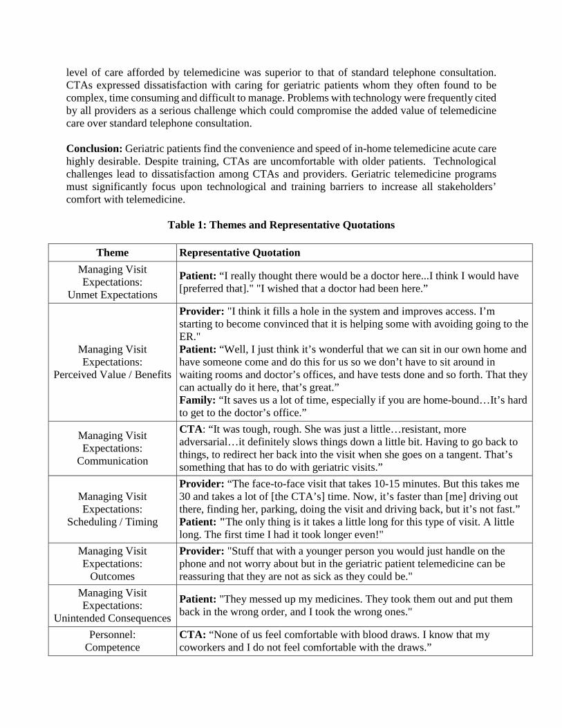
level of care afforded by telemedicine was superior to that of standard telephone consultation. CTAs expressed dissatisfaction with caring for geriatric patients whom they often found to be complex, time consuming and difficult to manage. Problems with technology were frequently cited by all providers as a serious challenge which could compromise the added value of telemedicine care over standard telephone consultation. Conclusion: Geriatric patients find the convenience and speed of in-home telemedicine acute care highly desirable. Despite training, CTAs are uncomfortable with older patients. Technological challenges lead to dissatisfaction among CTAs and providers. Geriatric telemedicine programs must significantly focus upon technological and training barriers to increase all stakeholders’ comfort with telemedicine.
Table 1: Themes and Representative Quotations
Theme Representative Quotation Managing Visit Expectations:
Unmet Expectations
Patient: “I really thought there would be a doctor here...I think I would have [preferred that]." "I wished that a doctor had been here.”
Managing Visit Expectations:
Perceived Value / Benefits
Provider: "I think it fills a hole in the system and improves access. I’m starting to become convinced that it is helping some with avoiding going to the ER." Patient: “Well, I just think it’s wonderful that we can sit in our own home and have someone come and do this for us so we don’t have to sit around in waiting rooms and doctor’s offices, and have tests done and so forth. That they can actually do it here, that’s great.” Family: “It saves us a lot of time, especially if you are home-bound…It’s hard to get to the doctor’s office.”
Managing Visit Expectations:
Communication
CTA: “It was tough, rough. She was just a little…resistant, more adversarial…it definitely slows things down a little bit. Having to go back to things, to redirect her back into the visit when she goes on a tangent. That’s something that has to do with geriatric visits.”
Managing Visit Expectations:
Scheduling / Timing
Provider: “The face-to-face visit that takes 10-15 minutes. But this takes me 30 and takes a lot of [the CTA’s] time. Now, it’s faster than [me] driving out there, finding her, parking, doing the visit and driving back, but it’s not fast.” Patient: "The only thing is it takes a little long for this type of visit. A little long. The first time I had it took longer even!"
Managing Visit Expectations:
Outcomes
Provider: "Stuff that with a younger person you would just handle on the phone and not worry about but in the geriatric patient telemedicine can be reassuring that they are not as sick as they could be."
Managing Visit Expectations:
Unintended Consequences
Patient: "They messed up my medicines. They took them out and put them back in the wrong order, and I took the wrong ones."
Personnel: Competence
CTA: “None of us feel comfortable with blood draws. I know that my coworkers and I do not feel comfortable with the draws.”

Personnel: Confidence
CTA: “Geriatrics is an art and I’m not a master” Provider: "And the CTAs are uncomfortable with geriatric patients, so there is some learning curve there." Patient: “I had the feeling it was sort of new to [the CTA]. He was a little slow, but someone had to learn.”
Non-Personnel: Equipment / Workspace
CTA: “Well, first of all it’s too much. Our bags are packed full and overflowing some of the time. And the cases are like 60lbs and it’s just not worth it to carry it all in there because once you get in there not everything is going to work…It's a pain in the butt."
Non-Personnel: Functionality / Quality of
Information
Provider: "I think that [the technology] is kind of a chronic problem because it’s always some equipment piece, or some upload or some connection that continually does not work on every single visit I’ve done. And that’s really frustrating." CTA: “It’s nice when [the medical technology] works, but it doesn’t always work. I’d say it’s a 50/50 shot.”
Tyson Olson, MS2 Preceptor: Gregg Nicandri, MD University of Rochester Medical Center Department of Orthopaedics and Rehabilitation
Simulation-based Education in Orthopaedic Surgery-What is the Optimum Number of Simulation Training Sessions Required to Maximize Cumulative Transfer Effectiveness When Training Novice Learners the Technique of Diagnostic Arthroscopy on Cadaveric
Specimens? Hypothesis: Training students four times using dry-model knee arthroscopic trainers will result in a higher cumulative transfer effectiveness ratio than training students nine times on the same simulator when performing diagnostic arthroscopy on cadaveric specimens.
Introduction/Objective: The objective of this study is to determine whether four or nine trials of arthroscopic simulation using dry knee models (Donnie Knee - Sawbones, Vashon WA) maximizes cumulative transfer effectiveness for novice surgeons learning to perform diagnostic knee arthroscopy on cadaveric specimens. Background: Surgical education has traditionally combined didactic lessons with apprenticed training in the operating room.1 Although supervised, having inexperienced surgeons practice on patients increases the risk of patient harm and extends operating room times.2 To decrease these concerns, In 2008 the Residency Review Committee (RRC) in surgery stated that there needed to be an increased emphasis on simulation in surgical training, and the American College of Surgeons (ACS) recently endorsed the effort to “move the learning curve outside of the operating room.”3,4
Currently, cadaveric skills training is the gold standard simulation modality for orthopaedic arthroscopy.5 Sawbones dry models such as the Donnie Knee have been promoted as adequate simulators for arthroscopic knee surgeries and are currently utilized by various training programs and technical skills courses in orthopaedic surgery.6 The optimal place for these simulators within the orthopaedic surgical training curricula is still being determined. Butler et al. evaluated the cumulative transfer effectiveness ratio (CTER) of training 9 times on a dry-model knee simulator and demonstrated that each trial on the dry-model led to a decrease of .22 trials to reach proficiency when performing diagnostic arthroscopy on a cadaveric knee. The number of training sessions on the dry model simulator required to achieve the maximum CTER is unknown, however based on their raw data, Butler et al. suggested that this number was four. Methods: Medical students at the University of Rochester were placed into two groups. The control group (6 students), completed eight diagnostic knee arthroscopies on a cadaveric specimen. The experimental group (6 students) was trained and completed 4 trials on the Donnie Knee and then later performed eight diagnostic knee arthroscopies on a cadaveric specimen. The
Basic Science, Clinical & Translational Research
students’ proficiency was evaluated by one of three graders who were blinded to students’ group assignments. Proficiency was determined using the Basic Arthroscopic Knee Scoring System (BAKSS) Global Rating Scale.6 Minimal proficiency was defined as a score of 30 out of 45*. The number of trials required to reach minimum proficiency was determined for each subject and a CTER was calculated. The CTER from the group trained 4 times using the Donnie knee was then compared to the CTER calculated in a previous study where the same methodology was used but where the experimental group trained 9 times using the same simulator. Results: The measured CTER was -0.03 for the group training 4 times using the Donnie Knee, meaning that every trial performed on the dry-model added 0.03 trials on the cadaveric specimen. The average score for the control group was 29.9 and the average score for the experimental group was 28.1(p value=0.36). The average time spent training on the cadaver was 61.6 minutes for the control group and 47.2 minutes for the experimental group(p value=0.059). The measured CTER for the group trained 9 times using the Donnie Knee was 0.22. Conclusion: Being trained 4 times on dry-model knees did not significantly decrease but instead increased the number of trials it took to reach proficiency on cadaveric specimens. Based on these results, it seems that training 9 times on dry-model knees results in a better CTER when compared to training 4 times. This study was limited by several factors. Though all raters were blinded as to whether subjects had received prior training, raters 1 and 2 graded the control group whereas rater 3 exclusively graded the experimental group. It is possible that rater 3 interpreted the BAKSS Global Rating Scale differently than raters 1 and 2. Further studies need to be performed assessing the inter-observer reliability of the BAKSS Global Rating Scale. It is also possible that due to the small number of students evaluated the participants in the experimental group may have been less skilled to begin with than the participants in the previous study. Although the number of trials to reach proficiency did not improve with 4 trials on the dry-model, the average time spent to complete eight trials on the cadaveric specimens was greatly reduced for the experimental group. It appears that 4 trials on the dry-model knee will lead to a decrease in the total time spent training on cadaveric specimens, though this increased efficiency does not seem to result in increased competency overall. Studies evaluating the effect of dry model and cadaveric simulation on the technical proficiency of resident trainees performing live surgery are needed to determine the optimum place for these simulators in the orthopaedic surgery curriculum. *One student did not reach 30 so 8 was used as the number of trials to proficiency. References
1. Michelson JD. Simulation in orthopaedic education: an overview of theory and practice. J Bone Joint Surg Am. Jun 2006;88(6):1405-1411.
2. Scott DJ. Patient safety, competency, and the future of surgical simulation. Simul Healthc. Fall 2006;1(3):164-170.
3. Scott DJ, Dunnington GL. The new ACS/APDS Skills Curriculum: moving the learning curve out of the operating room. J Gastrointest Surg. Feb 2008;12(2):213-221.
4. Aggarwal R, Darzi A. From scalpel to simulator: a surgical journey. Surgery. Jan 2009;145(1):1-4. 5. Mabrey JD, Gillogly SD, Kasser JR, et al. Virtual reality simulation of arthroscopy of the knee.
Arthroscopy. Jul-Aug 2002;18(6):E28. 6. nsel A, Carofino B, Leger R, Arciero R, Mazzocca AD. The development of an objective model to assess
arthroscopic performance. J Bone Joint Surg Am. Sep 2009;91(9):2287-2295.
David A. Paul, MS2 Preceptors: G. Edward Vates, M.D. Ph.D. Assistant Professor, Department of Neurosurgery University of Rochester Medical Center Bradford Mahon, Ph.D. Assistant Professor, Department of Brain and Cognitive Sciences, Department of Neurosurgery University of Rochester Medical Center Zoe Williams, MD Assistant Professor, Department of Ophthalmology University of Rochester Medical Center Eric Hintz, MD Resident Physician, Department of Neurosurgery University of Rochester Medical Center
Using DTI to measure changes in occipital lobe white matter after decompression of the optic chiasm
Introduction:
Pituitary macroadenomas and other parasellar lesions that compress the optic chiasm and cause bitemporal hemianopsia impair dioptic visual signals from reaching the striate cortex. Although these lesions have varied etiologies and treatment regimens, the goal is to decompress the optic chiasm and restore vision.1 At present, there are no biomarkers to predict whether or to what degree a patient’s vision will recover after decompression, except tumor size2 and retinal nerve fiber layer thickness.3 This has led to a renewed effort to build upon the well-documented stages of peripheral vision recovery,4 in search of white matter structural changes associated with chiasmatic compression. Background: Through the measurement water’s unique diffusion properties in tissue, Diffusion Tensor Imaging (DTI) is now a widely accepted imaging modality capable of providing novel biological and clinical information on white matter tracts. The movement of water molecules in nerve fibers is constrained by the myelin sheath to a direction parallel the nerve axis. The strength of this motion constraint at the voxel level is referred to as fractional anisotropy (FA); with a range of values extending from 0 to 1, expressing unconstrained and complete unidirectional motion respectively. Average motion in all directions is defined as the apparent diffusion coefficient (ADC), with higher values representing unconstrained motion. These intrinsic properties of water have been exploited in previous studies to successfully characterize the entire visual system using DTI tractography.5 There is also some evidence that nerve compression can result in measurable tractographic changes along the visual pathway. However, specific pathologic changes to the visual pathways, particularly posterior the optic chiasm, remain poorly defined. Objective: To investigate the plasticity of white matter tracts in response to optic chiasm compression and evaluate how the trajectory of neural plasticity may support visual recovery after resection of a parasellar lesion.
Basic Science, Clinical & Translational Research
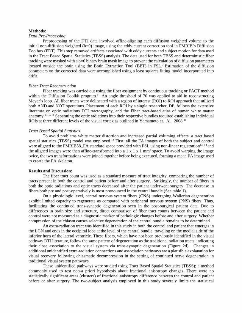
Methods: Data Pre-Processing Preprocessing of the DTI data involved affine-aligning each diffusion weighted volume to the initial non-diffusion weighted (b=0) image, using the eddy current correction tool in FMRIB’s Diffusion Toolbox (FDT). This step removed artifacts associated with eddy currents and subject motion for data used in the Tract Based Spatial Statistics (TBSS) analysis. The data used for both TBSS and deterministic fiber tracking were masked with a b=0 binary brain mask image to prevent the calculation of diffusion parameters located outside the brain using the Brain Extraction Tool (BET) in FSL.7 Estimation of the diffusion parameters on the corrected data were accomplished using a least squares fitting model incorporated into dtifit. Fiber Tract Reconstruction
Fiber tracking was carried out using the fiber assignment by continuous tracking or FACT method within the Diffusion Toolkit program.8 An angle threshold of 70 was applied to aid in reconstructing Meyer’s loop. All fiber tracts were delineated with a region of interest (ROI) to ROI approach that utilized both AND and NOT operations. Placement of each ROI by a single researcher, DP, follows the extensive literature on optic radiations DTI tractography, and the Fiber tract-based atlas of human white matter anatomy.9, 10, 11 Separating the optic radiations into their respective bundles required establishing individual ROIs at three different levels of the visual cortex as outlined in Yamamoto et. Al. 2008.11 Tract Based Spatial Statistics
To avoid problems white matter distortion and increased partial voluming effects, a tract based spatial statistics (TBSS) model was employed.12 First, all the FA images of both the subject and control were aligned to the FMRIB58_FA standard space provided with FSL using non-linear registration13, 14 and the aligned images were then affine-transformed into a 1 x 1 x 1 mm3 space. To avoid warping the image twice, the two transformations were joined together before being executed, forming a mean FA image used to create the FA skeleton.
Results and Discussion:
The fiber tract count was used as a standard measure of tract integrity, comparing the number of tracts present in both the control and patient before and after surgery. Strikingly, the number of fibers in both the optic radiations and optic tracts decreased after the patient underwent surgery. The decrease in fibers both pre and post-operatively is most pronounced in the central bundle (See table 1).
On a physiologic level, central nervous system fibers (CNS) undergoing Wallerian degeneration exhibit limited capacity to regenerate as compared with peripheral nervous system (PNS) fibers. Thus, facilitating the continued trans-synaptic degeneration seen in the post-surgical patient data. Due to differences in brain size and structure, direct comparison of fiber tract counts between the patient and control were not measured as a diagnostic marker of pathologic changes before and after surgery. Whether compression of the chiasm causes selective degeneration of the central bundle remains to be determined.
An extra-radiation tract was identified in this study in both the control and patient that emerges in the LGN and ends in the occipital lobe at the level of the central bundle, traveling on the medial side of the inferior horn of the lateral ventricle. These fibers, which have not been previously identified in the visual pathway DTI literature, follow the same pattern of degeneration as the traditional radiation tracts; indicating their close association to the visual system via trans-synaptic degeneration (Figure 2d). Changes in additional unidentified extra-radiation connections and association pathways are a plausible explanation for visual recovery following chiasmatic decompression in the setting of continued nerve degeneration in traditional visual system pathways.
These unidentified pathways were studied using Tract Based Spatial Statistics (TBSS); a method commonly used to test non-a priori hypothesis about fractional anisotropy changes. There were no statistically significant areas (clusters) of fractional anisotropy difference between the control and patient before or after surgery. The two-subject analysis employed in this study severely limits the statistical
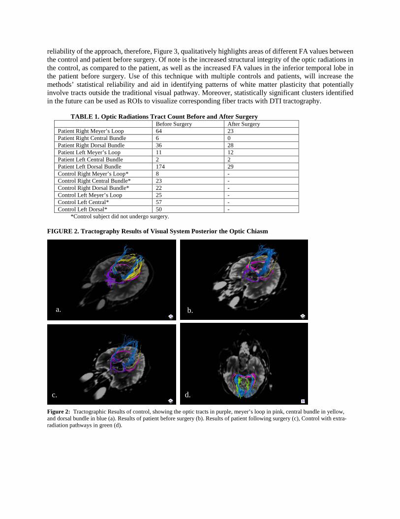
reliability of the approach, therefore, Figure 3, qualitatively highlights areas of different FA values between the control and patient before surgery. Of note is the increased structural integrity of the optic radiations in the control, as compared to the patient, as well as the increased FA values in the inferior temporal lobe in the patient before surgery. Use of this technique with multiple controls and patients, will increase the methods’ statistical reliability and aid in identifying patterns of white matter plasticity that potentially involve tracts outside the traditional visual pathway. Moreover, statistically significant clusters identified in the future can be used as ROIs to visualize corresponding fiber tracts with DTI tractography.
TABLE 1. Optic Radiations Tract Count Before and After Surgery
Before Surgery After Surgery Patient Right Meyer’s Loop 64 23 Patient Right Central Bundle 6 0 Patient Right Dorsal Bundle 36 28 Patient Left Meyer’s Loop 11 12 Patient Left Central Bundle 2 2 Patient Left Dorsal Bundle 174 29 Control Right Meyer’s Loop* 8 - Control Right Central Bundle* 23 - Control Right Dorsal Bundle* 22 - Control Left Meyer’s Loop 25 - Control Left Central* 57 - Control Left Dorsal* 50 -
*Control subject did not undergo surgery. FIGURE 2. Tractography Results of Visual System Posterior the Optic Chiasm
Figure 2: Tractographic Results of control, showing the optic tracts in purple, meyer’s loop in pink, central bundle in yellow, and dorsal bundle in blue (a). Results of patient before surgery (b). Results of patient following surgery (c), Control with extra-radiation pathways in green (d).
a. b.
c. d.
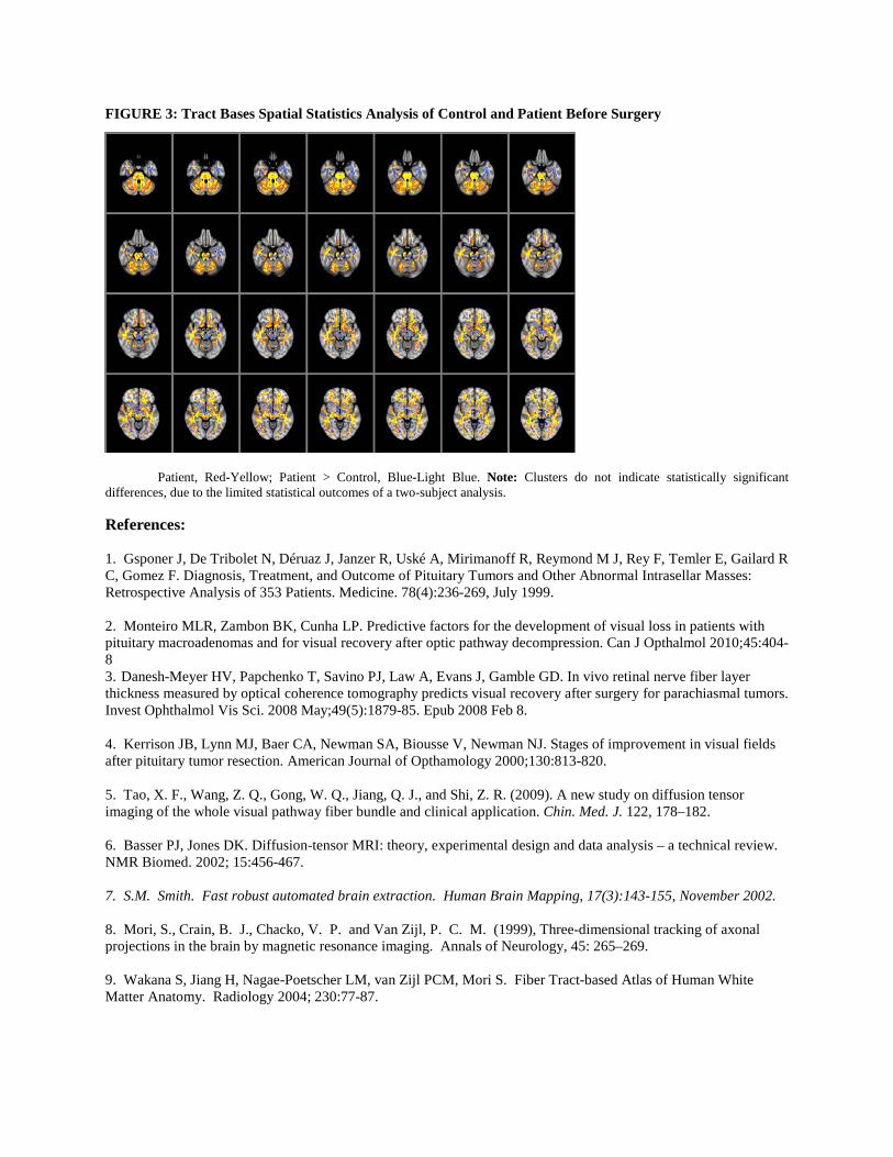
FIGURE 3: Tract Bases Spatial Statistics Analysis of Control and Patient Before Surgery
Figure 3: TBSS Analysis between the control and patient before surgery. Control >
Patient, Red-Yellow; Patient > Control, Blue-Light Blue. Note: Clusters do not indicate statistically significant
differences, due to the limited statistical outcomes of a two-subject analysis.
References:
1. Gsponer J, De Tribolet N, Déruaz J, Janzer R, Uské A, Mirimanoff R, Reymond M J, Rey F, Temler E, Gailard R C, Gomez F. Diagnosis, Treatment, and Outcome of Pituitary Tumors and Other Abnormal Intrasellar Masses: Retrospective Analysis of 353 Patients. Medicine. 78(4):236-269, July 1999. 2. Monteiro MLR, Zambon BK, Cunha LP. Predictive factors for the development of visual loss in patients with pituitary macroadenomas and for visual recovery after optic pathway decompression. Can J Opthalmol 2010;45:404-8 3. Danesh-Meyer HV, Papchenko T, Savino PJ, Law A, Evans J, Gamble GD. In vivo retinal nerve fiber layer thickness measured by optical coherence tomography predicts visual recovery after surgery for parachiasmal tumors. Invest Ophthalmol Vis Sci. 2008 May;49(5):1879-85. Epub 2008 Feb 8. 4. Kerrison JB, Lynn MJ, Baer CA, Newman SA, Biousse V, Newman NJ. Stages of improvement in visual fields after pituitary tumor resection. American Journal of Opthamology 2000;130:813-820. 5. Tao, X. F., Wang, Z. Q., Gong, W. Q., Jiang, Q. J., and Shi, Z. R. (2009). A new study on diffusion tensor imaging of the whole visual pathway fiber bundle and clinical application. Chin. Med. J. 122, 178–182. 6. Basser PJ, Jones DK. Diffusion-tensor MRI: theory, experimental design and data analysis – a technical review. NMR Biomed. 2002; 15:456-467. 7. S.M. Smith. Fast robust automated brain extraction. Human Brain Mapping, 17(3):143-155, November 2002. 8. Mori, S., Crain, B. J., Chacko, V. P. and Van Zijl, P. C. M. (1999), Three-dimensional tracking of axonal projections in the brain by magnetic resonance imaging. Annals of Neurology, 45: 265–269. 9. Wakana S, Jiang H, Nagae-Poetscher LM, van Zijl PCM, Mori S. Fiber Tract-based Atlas of Human White Matter Anatomy. Radiology 2004; 230:77-87.

10. Winston, G. P., Yogarajah, M., Symms, M. R., McEvoy, A. W., Micallef, C. and Duncan, J. S. (2011), Diffusion tensor imaging tractography to visualize the relationship of the optic radiation to epileptogenic lesions prior to neurosurgery. Epilepsia. doi: 10.1111/j.1528-1167.2011.03088.x 11. Yamamoto T, Yamada K, Nishimura T, Kinoshita S. Tractography to Depict Three Layers of Visual Field Trajectories to the Calcarine Gyri. American Journal of Ophthalmology , 140(5);781-785 November 2005 12. S.M. Smith, M. Jenkinson, H. Johansen-Berg, D. Rueckert, T.E. Nichols, C.E. Mackay, K.E. Watkins, O. Ciccarelli, M.Z. Cader, P.M. Matthews, and T.E.J. Behrens. Tract-based spatial statistics: Voxelwise analysis of multi-subject diffusion data. NeuroImage, 31:1487-1505, 2006. 13. J.L.R. Andersson, M. Jenkinson and S. Smith. Non-linear optimisation. FMRIB technical report TR07JA1 from www.fmrib.ox.ac.uk/analysis/techrep 14. J.L.R. Andersson, M. Jenkinson and S. Smith. Non-linear registration, aka Spatial normalisation FMRIB technical report TR07JA2 from www.fmrib.ox.ac.uk/analysis/techrep
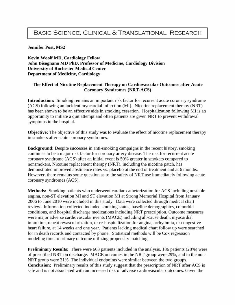
Jennifer Post, MS2
Kevin Woolf MD, Cardiology Fellow John Bisognano MD PhD, Professor of Medicine, Cardiology Division University of Rochester Medical Center Department of Medicine, Cardiology
The Effect of Nicotine Replacement Therapy on Cardiovascular Outcomes after Acute Coronary Syndromes (NRT-ACS)
Introduction: Smoking remains an important risk factor for recurrent acute coronary syndrome (ACS) following an incident myocardial infarction (MI). Nicotine replacement therapy (NRT) has been shown to be an effective aide in smoking cessation. Hospitalization following MI is an opportunity to initiate a quit attempt and often patients are given NRT to prevent withdrawal symptoms in the hospital.
Objective: The objective of this study was to evaluate the effect of nicotine replacement therapy in smokers after acute coronary syndromes. Background: Despite successes in anti-smoking campaigns in the recent history, smoking continues to be a major risk factor for coronary artery disease. The risk for recurrent acute coronary syndrome (ACS) after an initial event is 50% greater in smokers compared to nonsmokers. Nicotine replacement therapy (NRT), including the nicotine patch, has demonstrated improved abstinence rates vs. placebo at the end of treatment and at 6 months. However, there remains some question as to the safety of NRT use immediately following acute coronary syndromes (ACS). Methods: Smoking patients who underwent cardiac catheterization for ACS including unstable angina, non-ST elevation MI and ST elevation MI at Strong Memorial Hospital from January 2006 to June 2010 were included in this study. Data were collected through medical chart review. Information collected included smoking status, baseline demographics, comorbid conditions, and hospital discharge medications including NRT prescription. Outcome measures were major adverse cardiovascular events (MACE) including all-cause death, myocardial infarction, repeat revascularization, or re-hospitalization for angina, arrhythmia, or congestive heart failure, at 14 weeks and one year. Patients lacking medical chart follow up were searched for in death records and contacted by phone. Statistical methods will be Cox regression modeling time to primary outcome utilizing propensity matching. Preliminary Results: There were 663 patients included in the analysis. 186 patients (28%) were of prescribed NRT on discharge. MACE outcomes in the NRT group were 29%, and in the non-NRT group were 31%. The individual endpoints were similar between the two groups. Conclusion: Preliminary results of this study suggest that the prescription of NRT after ACS is safe and is not associated with an increased risk of adverse cardiovascular outcomes. Given the
Basic Science, Clinical & Translational Research
high rate of recurrent tobacco use in smokers after an MI, the ability to provide pharmacologic assistance is critical. Further study is necessary to confirm that NRT is safe to use immediately following incident MI. References 1 CDC. Vital Signs: current cigarette smoking among adults aged ≥18 Years --- United States, 2009. MMWR 2010;59:1135-40. 2 Services USDoHaH. The health consequences of smoking: nicotine addiction. A report of the Surgeon General. In: Services USDoHaH, ed. Rockville, MD; 1988. 3Schane RE, Ling PM, Glantz SA. Health effects of light and intermittent smoking: a review. Circulation 2010;121:1518-22. 4 Erhardt L. Cigarette smoking: an undertreated risk factor for cardiovascular disease. Atherosclerosis 2009;205:23-32. 5Fiore MC, Smith SS, Jorenby DE, Baker TB. The effectiveness of the nicotine patch for smoking cessation. A meta-analysis. JAMA 1994;271:1940-7. 6Nicotine replacement therapy for patients with coronary artery disease. Working Group for the Study of Transdermal Nicotine in Patients with Coronary artery disease. Arch Intern Med 1994;154:989-95. 7Ford CL, Zlabek JA. Nicotine replacement therapy and cardiovascular disease. Mayo Clin Proc 2005;80:652-6.
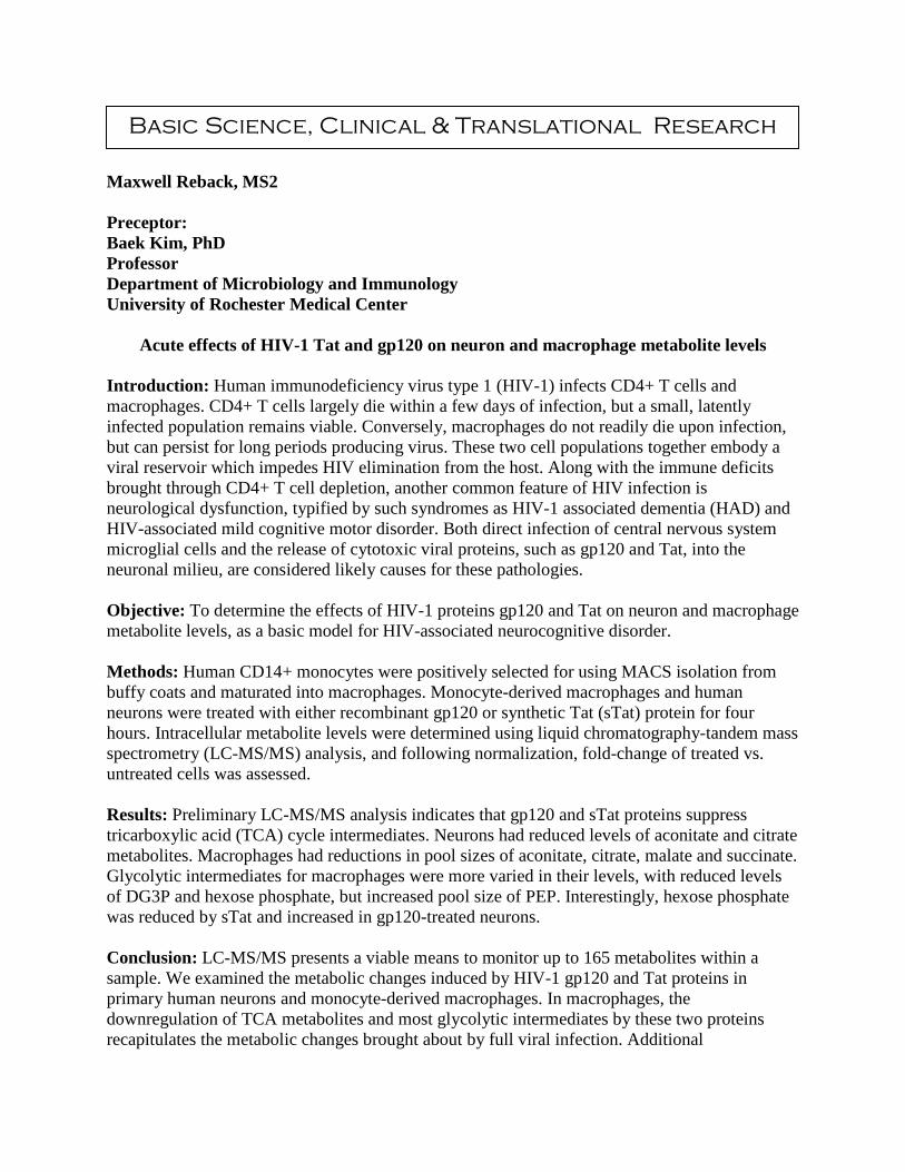
Maxwell Reback, MS2 Preceptor: Baek Kim, PhD Professor Department of Microbiology and Immunology University of Rochester Medical Center
Acute effects of HIV-1 Tat and gp120 on neuron and macrophage metabolite levels
Introduction: Human immunodeficiency virus type 1 (HIV-1) infects CD4+ T cells and macrophages. CD4+ T cells largely die within a few days of infection, but a small, latently infected population remains viable. Conversely, macrophages do not readily die upon infection, but can persist for long periods producing virus. These two cell populations together embody a viral reservoir which impedes HIV elimination from the host. Along with the immune deficits brought through CD4+ T cell depletion, another common feature of HIV infection is neurological dysfunction, typified by such syndromes as HIV-1 associated dementia (HAD) and HIV-associated mild cognitive motor disorder. Both direct infection of central nervous system microglial cells and the release of cytotoxic viral proteins, such as gp120 and Tat, into the neuronal milieu, are considered likely causes for these pathologies.
Objective: To determine the effects of HIV-1 proteins gp120 and Tat on neuron and macrophage metabolite levels, as a basic model for HIV-associated neurocognitive disorder. Methods: Human CD14+ monocytes were positively selected for using MACS isolation from buffy coats and maturated into macrophages. Monocyte-derived macrophages and human neurons were treated with either recombinant gp120 or synthetic Tat (sTat) protein for four hours. Intracellular metabolite levels were determined using liquid chromatography-tandem mass spectrometry (LC-MS/MS) analysis, and following normalization, fold-change of treated vs. untreated cells was assessed. Results: Preliminary LC-MS/MS analysis indicates that gp120 and sTat proteins suppress tricarboxylic acid (TCA) cycle intermediates. Neurons had reduced levels of aconitate and citrate metabolites. Macrophages had reductions in pool sizes of aconitate, citrate, malate and succinate. Glycolytic intermediates for macrophages were more varied in their levels, with reduced levels of DG3P and hexose phosphate, but increased pool size of PEP. Interestingly, hexose phosphate was reduced by sTat and increased in gp120-treated neurons. Conclusion: LC-MS/MS presents a viable means to monitor up to 165 metabolites within a sample. We examined the metabolic changes induced by HIV-1 gp120 and Tat proteins in primary human neurons and monocyte-derived macrophages. In macrophages, the downregulation of TCA metabolites and most glycolytic intermediates by these two proteins recapitulates the metabolic changes brought about by full viral infection. Additional
Basic Science, Clinical & Translational Research
experimental replicates using different primary human donors are required to validate these preliminary trends. References:
1. Cassol E, et al. Monocyte-derived macrophages and myeloid cell lines as targets of HIV-1 replication and persistence. J Leukoc Biol. 80: 1018-1030 (2006).
2. Chugh P, et al. Infection of human immunodeficiency virus and intracellular viral Tat protein exert a pro-survival effect in a human microglial cell line. J Mol Biol. 366: 67-81 (2007).
3. Hollenbaugh JA, et al. Metabolite profiles of human immunodeficiency virus infected CD4+ T cells and macrophages using LC-MS/MS analysis. Virol. 415: 153-159 (2011).
4. Kraft-Terry SD, et al. A coast of many colors: neuroimmune crosstalk in human immunodeficiency virus infection. Neuron. 64: 133-144 (2009).
Joshua Reuss, MS2 Lack of β-cell regeneration following pancreatic duct ligation in a rodent model of extreme
β-cell loss Joshua Reuss, Claudia Cavelti-Weder, Marianne Stemann-Andersen, Cristina Aguayo-Mazzucato, Susan Bonner-Weir, and Gordon Weir, Joslin Diabetes Center, Harvard Medical School, Boston, Massachusetts β-cell regeneration is a fascinating area of study that has the potential to have widespread therapeutic impact on diabetes. An interesting finding that has recently emerged from this research is that extreme β-cell loss appears to trigger β-cell regeneration via α- to β-cell transdifferentiation. In order to investigate the ability of pancreatic duct ligation (PDL) to stimulate β-cell regeneration following extreme β-cell loss, we treated 6-week old Lewis rats with the β-cell specific toxin streptozotocin (STZ). Upon confirmation of induced extreme β-cell loss, islets were transplanted into the animals to regulate glucose and PDL was performed. Total pancreatic insulin content, taken as a surrogate for β-cell mass, was then quantified two weeks after ligation in both the ligated “distal” pancreas and unligated “proximal” pancreas using an enzyme-linked immunosorbent assay (ELISA). Insulin content was also analyzed in rats 5 months post-PDL in order to assess any long-term effects of the procedure. Results revealed no significant difference between the very low insulin content of ligated pancreas (800 ± 153ng/g; n=12) and unligated pancreas (758 ± 59ng/g; n=12) in short-term animals. Similar results were observed for rats subjected to PDL 5 months prior to content determination. These data, which support our histology findings, suggest β-cell regeneration in our model of extreme β-cell loss, be it by neogenesis, α- to β-cell transdifferentiation, or β-cell replication, does not occur following PDL. Future studies should seek to confirm these findings in different ages, physiological conditions, and species. Additionally, our data cast doubt on the value of PDL as a method for inducing β-cell regeneration.
Basic Science, Clinical & Translational Research
Amber Robins, MS2 Dr. Sajeena Thomas Assistant Professor University of Rochester School of Medicine and Dentistry Gynecologic Oncology
Comparison of the Risk of Endometrial Cancer Recurrence in Robotic and Abdominal Surgery
Introduction: Endometrial cancer originates in the innermost uterine layer, the endometrium. As the most common gynecologic malignant neoplasm in the United States, it makes up 6% of cancers in women (1). For women who have this type of cancer, surgery to remove the entire uterus is considered the mainstay of treatment. However, surgery poses risks of complications. These complications are more frequent in patients that are obese which is one of the risk factors for this cancer. Objective: The differences will be compared between open and robotic surgery in terms of the operative time, blood loss, hospital readmissions, wound and other infections, and various types of surgical morbidity, as well as treatment outcome with recurrence risk and survival. Background: A recent study reported that 68% of women with early stage endometrial cancer are obese. In addition to increasing the risk of endometrial cancer, the risk of death for morbidly obese women with endometrial cancer was 6.25 times higher than for women who were not obese (2). Because of this increased risk, minimally invasive surgical alternatives, utilizing a laparoscopic approach, or supplanting the traditional open approach are the procedures used for a hysterectomy. The advantages of a minimally invasive approach include fewer operative complications, shorter operative time, and faster recovery. Such surgeries are now preferred more over open surgery.
Both laparoscopic and robotic-assisted laparoscopic techniques are advantageous in reducing surgical risks, such as blood loss and wound infections (3). Data exists that compares laparoscopic surgery to that of robotic-assisted laparoscopic surgery. These studies have shown that robotic-assisted laparoscopic surgery is associated with less blood loss and shorter operative time than laparoscopic surgery (2). Although preliminary data is supportive of robotic surgery, the safety and efficacy of this approach have not been validated in large series.
Hypothesis: Robotic-assisted laparoscopic surgery will be equivalent to open surgery in terms of cure rates, but will decrease the risk of surgical complications and operative morbidity. Methods: A retrospective chart review will be performed on patients admitted to Highland Hospital for treatment of endometrial cancer between January 1, 2008 and December 31, 2010. Patient demographic information, including age, weight, and co-morbid conditions will be reviewed. Operative reports will be assessed for the type of procedure performed, as well as
Basic Science, Clinical & Translational Research
blood loss, operative time, and any surgical complications. Surgical pathology will be reviewed to determine the type of endometrial cancer, as well as the surgical stage. Hospital records will be reviewed to determine any postoperative morbidity, as well as any readmissions for complications related to the surgery. Lastly, office records will be assessed to determine the patient’s current status. Results: Results are currently pending. Conclusion: The results will give more information on the efficacy of robotic versus abdominal surgery in regards to operative time, blood loss, hospital readmissions, wound and other infections, and various types of surgical morbidity, as well as treatment outcome with recurrence risk and survival. References:
1. Peiretti M, Zanagnolo V, Bocciolone L, Landoni F, Colombo N, Minig L, Sanguineti F, Maggioni A. Robotic surgery: changing the surgical approach for endometrial cancer in a referral cancer center. J Minim Invasive Gynecol. 2009 Jul-Aug;16(4):427-31. Epub 2009 May 21. PubMed PMID: 19467930.
2. Gehrig PA, Cantrell LA, Shafer A, Abaid LN, Mendivil A, Boggess JF. What is the optimal minimally invasive surgical procedure for endometrial cancer staging in the obese and morbidly obese woman? Gynecol Oncol. 2008 Oct;111(1):41-5. Epub 2008 Aug 9. PubMed PMID: 18694588.
3. Kong TW, Lee KM, Cheong JY, Kim WY, Chang SJ, Yoo SC, Yoon JH, Chang KH, Ryu HS. Comparison of laparoscopic versus conventional open surgical staging procedure for endometrial cancer. J Gynecol Oncol. 2010 Jun;21(2):106-11. Epub 2010 Jun 30. PubMed PMID: 20613901; PubMed Central PMCID: PMC2895709.
4. Holloway RW, Ahmad S, DeNardis SA, Peterson LB, Sultana N, Bigsby GE 4th, Pikaart DP, Finkler NJ. Robotic-assisted laparoscopic hysterectomy and lymphadenectomy for endometrial cancer: Analysis of surgical performance. Gynecol Oncol. 2009 Dec;115(3):447-52. Epub 2009 Sep 17. PubMed PMID: 19765807.
5. Krizova A, Clarke BA, Bernardini MQ, James S, Kalloger SE, Boerner SL, Mulligan AM. Histologic artifacts in abdominal, vaginal, laparoscopic, and robotic hysterectomy specimens: a blinded, retrospective review. Am J Surg Pathol. 2011 Jan;35(1):115-26. PubMed PMID: 21164295.
Marabel D. Schneider, MS2 Preceptor: H. Todd Massey, MD Surgical Director, Program in Heart Failure and Transplantation Director, Artificial Heart Program Associate Professor, Cardiac Surgery University of Rochester Medical Center Department of Surgery
Treatment of Cardiogenic Shock Following Acute Myocardial Infarction: Improved Survival with HeartMate II Left Ventricular Assist Device
Introduction:
Cardiogenic shock effects 6-8% of patients following acute myocardial infarction (AMI) and has an in-hospital mortality rate approaching 50% despite technological advances.1 Implantable ventricular assist devices (VADs) offer a valuable treatment option to rescue patients from circulatory failure following acute myocardial infarction, acute myocarditis and post-cardiotomy shock. The outcomes of these patients are heavily reliant on management at initial presentation, time to intervention with a mechanical circulatory support device (MCSDs) when appropriate and, potentially, choice of treatment device. Objective:
The objective of this study is to examine the institutional experience in treatment of cardiogenic shock (CS) with implantation of VADs and evaluate the use of the Thoratec HeartMate II Left Ventricular Assist Device (HM-II LVAD) as a primary MCSDs treatment of this population compared to other MCSDs. The Artificial Heart Program at the URMC has published and implemented a protocol for the management of CS following acute myocardial infarction (AMI) with the belief that time from presentation to implantation is critical to the successful reversal of end-organ dysfunction through mechanical circulatory support devices (MCSDs).1 Longitudinally, we aim to propose a protocol for management of CS following AMI based on the institutional treatment outcome experience.
Background:
Cardiogenic shock is defined as end-organ hypoperfusion resulting from cardiac dysfunction and low cardiac output. The diagnosis is generally characterized by hypotension (baseline systolic pressure < 90 mm Hg) with reduction in cardiac index (<1.8 L min-1 m-2
without support, and adequate or elevated filling pressure (left ventricular [LV] end-diastolic pressure >18 mm Hg or right ventricular [RV] end-diastolic pressure >10 to 15 mm Hg.1
Most commonly, coronary occlusion resulting in a > 40% of left ventricular muscle leads to CS and thus the standard treatment aim is revascularization of the heart and restoration of normal perfusion to the organs and body tissues. Though even with revascularization and blood flow restoration, some patients remain in or return to a state of CS. It is suspected that this may
Basic Science, Clinical & Translational Research

be related to a time delay between initial presentation and reperfusion.2 It is thought that efforts to restore blood flow and reperfuse the body more quickly may improve treatment outcomes in this CS population.
The use of MCSDs aims to restore physiologic blood flow to the body and present end-organ dysfunction. In doing this, preload is decreased reducing myocardial workload and oxygen demand. Currently, MCSDs are FDA-approved for the purpose of bridge-to-recovery, bridge-to-transplant and permanent lifetime therapy. The effectiveness of MCSDs as bridge-to-recovery treatments of acute cardiomyopathy is well established.2 Of critical importance to the successful treatment of CS following AMI, is the standardization of treatment protocols to ensure the best pre-treatment clinical parameters for implantation of MCSDs, if chosen as a method of treatment.
Patients and Methods:
A retrospective review of a single, tertiary-care facility experience in treatment of patients treated for acute CS following AMI with implantation of a MCSD between January 2001 and April 2010. The primary end-points for the study included mortality at 30 days, 90 days and 6 months. Secondary outcomes include survival at discharge, length of ICU stay, length of hospitalization and duration of intubation.
A total of 107 patients received MCSDs in the form of left-ventricular assist devices (LVAD) or biventricular assist devices (BiVAD). Patients were stratified based on type of primary (first-line therapy) MCDS: 16 patients received HM-II LVAD and 91 patients received other MCSDs (Abiomed BVS 5000, Thoratec PVAD, Levitronix CentriMag, HeartMate XVE, ECMO, and Abiomed Impella 2.5 cardiac assist device).
Continuous variables between the two groups were analyzed using nonparametric Wilcoxon rank-sum test. Categorical variables were evaluated with chi-square test or Fisher’s exact test, as appropriate. Results: Baseline characteristics of the two cohorts were similar, accounting for demographics (age, gender, race), clinical history (BMI, cardiac arrest, CAD, DM) and cardiac index, sodium and creatinine levels. Mortality at 30-days, 90-days and 6 months was significantly lower in the HM-II LVAD group compared to the other VAD group (6.2% vs. 43.3%, 6.2% vs. 52.8% and 6.2% vs. 61.6% respectively). Survival at discharge was also significantly greater in the HM-II LVAD recipients (93.8%) compared to recipients of other VADs (41.1%). Secondary outcomes of ICU stay, duration of intubation and hospitalization duration were not significantly different between the two cohorts. Conclusions: Implantation of the HM-II LVAD as a primary mechanical circulatory support device in the treatment of acute cardiogenic shock is associated with lower mortality and improved survival. The HM-II LVAD is a good primary treatment option for patients with CS requiring mechanical circulatory support. The longitudinal aim is to include this recommendation in the proposal of guidelines for improved regional management of acute cardiogenic shock.
References: 1 Seyfarth M, Sibbing D, Baurer I, et al. A Randomized Clinical Trial to Evaluate the Safety and Efficacy of a Percutaneous Left Ventricular Assist Device Versus Intra-Aortic Balloon Pumping for Treatment of Cardiogenic Shock Caused by Myocardial Infarction. JACC 2008; 52: 1584-8. 1 J.D. Bisognano et al. (eds.), Manual of Heart Failure Management, DOI: I0.I007/978-I-84882-I85-9_8, Springer-Verlag London Limited 2009. 1 Reynolds, H and J Hochman. Cardiogenic Shock: Current Concepts and Improving Outcomes. Circulation 2008; 117:686-697.
Thomas Segerson, MS2 Preceptors: Dr. Ilya Voloshin, Associate Professor Dr. Michael Stanton, R3 University of Rochester School of Medicine and Dentistry Department of Orthopaedics
Results of Repeat Meniscal Repair Introduction/Background Meniscal injury ranks as one of the most common knee injuries. While many treatments exist to deal with meniscal injuries, meniscus repair, when indicated, remains the standard of care.1 As the menisci have been shown to function as the knees’ shock absorbers2 and aid in load transmission3 as well as joint stabilization4, meniscectomies have been associated with a 235-335% increase in contact forces on articular cartilage of the knee.5 This increase in forces on the knee joint have been shown to be a cause of osteoarthritis.6 Meanwhile, meniscus repair studies in canines have demonstrated restoration of biomechanical properties of the femoral-tibial articulation and the meniscus in terms of contact area and energy dissipation.7 A separate study demonstrated that meniscal repair compared with partial meniscectomy decreases osteoarthritic changes and reduces the effect on sports activity in the long-term follow-up.8 It is clear that there is strong evidence in favor of meniscus repair over meniscal resection in the treatment of meniscus tears. Current indications for meniscal repair include vertical longitudinal tears in the outer 25%-30% of the meniscus. This is usually within 3 to 5 mm from the meniscocapsular junction and corresponds to the “red-red” (vascular) zone.9 While it is clear that correct repair of a torn meniscus can greatly improve outcome, little work has gone into investigating the efficacy of repeat repair of retorn menisci. Preliminary studies have demonstrated relief of symptoms and high levels of function in repeat-repair patients with a mean follow-up of 7.33 years.10 However, it remains to be seen whether or not these outcomes are long-term. Objective The objective of this study is to assess the long-term efficacy of repeat repair of retorn menisci. Methods Fourteen patients who underwent repeat meniscal repair at Strong Memorial Hospital were recruited for the study. The average follow-up from the time of repeat repair was 15 years. Patient records and office notes were examined for each participant. A questionnaire derived from the Lysholm II rating scale and the Tegner activity score were mailed and completed by each patient. Patients who were available for an office visit were assessed by a physician and scored based on range of motion, thigh circumference, and manual ligament examination. Bilateral anterior-posterior radiographs were taken of the knees in extension and 45° flexion.
Basic Science, Clinical & Translational Research

Radiographs were then interpreted and scored independently by the physician and a musculoskeletal radiologist. Data was compiled and evaluated using standard statistical techniques. Results The mean IKDC score was 86.51 (range 74.71-97.70). The mean Lysholm score was 85.0 (range 66-100) and the mean Tegner score was 4.8 (range 3-6). Radiographs revealed either grade 0 changes (normal) or grade I changes (sclerosis or mild narrowing measuring 1-2mm) in the involved knee compartments. Conclusion When compared with the results of follow-up at 7.33 years (mean IKDC= 78.5, mean Lysholm=82.1, mean Tegner= 5.6, grade 0 and I changes), the results gathered from the 15 year follow-up demonstrate a high survival rate and functional preservation of the repeat meniscal repair over an extended period of time. We conclude that the repair of suitable torn menisci that have previously been repaired can successfully provide long-term relief of symptoms and allow resumption of high levels of activity. References 1.) Gu YL, Wang WB. Treatment of meniscal injury: a current concept review. Chin J Traumatol. 2010;13(6):370-6. 2.) Krause WR, Pope MH, Johnson RJ, et al. Mechanical changes in the knee after meniscectomy. J Bone Joint Surg. 1974;56A:719-729. 3.) Renstrom P, Johnson RJ. Anatomy and biomechanics of the menisci. Clin Sports Med. 1990;9:523-538. 4.) Markolf KL, Bargar WL, Shoemaker SC, et al. The role of joint load in knee stability. J Bone Joint Surg. 1981:63A;570-585. 5.) Paletta GA, Jr., Manning T, Snell E, et al. The effect of allograft meniscal replacement on intraarticular contact area and pressures in the human knee. A biomechanical study. Am J Sports Med. 1997;25:692-8. 6.) Petrosini AV, Sherman OH. A historical perspective on meniscal repair. Clin Sports Med. 1996:15(3);445-453. 7.) Newman AP, Anderson DR, Daniels AU, Dales MC. Mechanics of the healed meniscus in a canine model. Am J Sports Med. 1989:17(2);164-175. 8.) Stein T, Mehling AP, Welsch F, et al. Long-term outcome after arthroscopic meniscal repair versus arthroscopic partial meniscectomy for traumatic meniscal tears. Am J Sports Med. 2010;28(8):1542-1548. 9.) DeHaven KE. Decision-making factors in the treatment of meniscus lesions. Clin Orthop Relat Res. 1990;(252):49-54. 10.) Voloshin I, Schmitz MA, Adams MJ, DeHaven KE. Results of Repeat Meniscal Repair. Am J Sports Med. 2003;31:874-880.
Allison Shatz, MS2 Dr. Jason Schwalb Director of Movement Disorder and Behavioral Neurosurgery Henry Ford Hospital Department of Neurosurgery Introduction: Normal Pressure Hydrocephalus, NPH, is a syndrome characterized by a triad of symptoms: gait disturbances, cognitive dysfunction, and urinary symptoms. Little is known about the pathology of this disease and it is often difficult to distinguish it clinically from other conditions causing similar symptoms. The only treatments available, shunt placement or endoscopic third ventriculostomy, notably improve gait symptoms but have little effect on the other triad symptoms. Historically shunt treatment has not been offered to patients with co-morbid Alzheimer’s disease, because it was thought that the potential benefits were minimal and did not outweigh the potential risks. Recent investigations show that the gait of patients with co-morbid AD do improve with treatment of NPH. However, there have been no detailed analyses of the different aspects of gait that characterize those with “pure” NPH and those with co-morbid Alzheimer’s disease. Objective: The NPH program at Henry Ford Hospital has been rigorously and systematically testing all patients with NPH. In addition to extensive cognitive testing and urinary symptom tracking, specific gait parameters are measured in patients preceding and following shunt placement to track changes. The data from the computerized Gaitrite testing was analyzed to determine which gait components changed in patients with co-morbid Alzheimer’s disease following shunt placement in comparison to those with “pure” NPH.
Background: In 1965, Hakim and Adams identified a triad of gait, cognitive and urinary abnormalities associated with large ventricles but normal intracranial pressure that could be ameliorated by shunting cerebrospinal fluid to the peritoneum, pleura, or venous system. They termed it “normal pressure hydrocephalus” (NPH)1. Despite decades of clinical investigation, this syndrome still is clinically difficult to recognize and the pathophysiology undetermined2-4. In spite of the availability of clinical guidelines for the treatment of idiopathic NPH, this disorder is both underrecognized and overrecognized. The likelihood of other conditions or combinations of conditions causing the NPH triad is not insignificant. For example, it is estimated that 2.1 to 4.5 million Americans have Alzheimer’s disease (AD)5. Many of these patients will have ventriculomegaly due to brain atrophy. In addition, it has been theorized that both NPH and AD have overlapping mechanisms with regards to impaired CSF clearance of toxic metabolites6. Indeed, part of the difficulty in differentiating between NPH and other dementias is that they may co-exist. In a clinic-pathologic study of all patients with dementia, 80% had mixed pathologies despite a clinical presentation that seemed to suggest one dominant disorder5.
While some authors have felt that patients with AD pathology identified on frontal lobe biopsy should not be offered shunting on the basis of higher risk of surgical complications and lack of improvement in standard measures, the results of shunting patients with concomitant
Basic Science, Clinical & Translational Research
pathological signs of AD are not consistent. Historically, reports of improvements with shunting are most impressive in the domains of gait and urinary symptoms domains, with studies showing a more conservative improvement in cognition following surgery7,8. Studies also show that patients with AD pathology co-existing with NPH did not benefit as much as patients with NPH symptoms alone, even in the gait domain8-10. However, in these studies specific domains of gait were not analyzed. A longitudinal study of gait found domain-specific deficits in Alzheimer’s patients compared with normal controls. AD gait characteristics include decreased velocity, shorter stride length, increased variability in stride length, and increased double support time11. This pattern can be distinguished from the NPH gait, described as shuffling and magnetic, which has decreased stride length and increased double support time, but also increased base width12. By distinguishing specific components of gait, a more detailed analysis of the effects of shunting on NPH patients with AD may reveal a potential therapeutic effect of shunting in this population.
Methods: The data from 13 NPH patients who had undergone shunt placement surgery was collected and analyzed. The sample included three co-morbid Alzheimer’s patients. Specifically investigated were the changes in gait following shunt placement. The gait parameters, as measured by the computerized GaitRite system, included velocity, mean normalized velocity, stride length, stride length variance, step length, cadence, double support time, base width and functional ambulation performance. The percent change from pre- and post-surgery measurements was taken and compared across all patients and across all patients without co-morbid AD. This data was analyzed with respect to expected gait pattern for each condition. Results: Twelve of the thirteen patients improved in their global score, as measured by the functional ambulation performance. In comparing the average percentage change of all patients versus only patients without AD, differences emerged in the change in stride length variance and in base width. Conclusion: The AD-patient-specific pattern suggests a different mechanism for gait dysfunction may be involved in patients with NPH and co-morbid AD. Additionally, despite historical reluctance to treat this group, shunting may still be of benefit. However, the benefit seen in co-morbid AD patients may operate by a different mechanism that those with NPH alone. This has implications for the treatment of future patients with NPH and AD as well as for the understanding of the pathology of the disease. References:
1. Hakim S, Adams RD. The special clinical problem of symptomatic hydrocephalus with normal cerebrospinal fluid pressure. Observations on cerebrospinal fluid hydrodynamics. Journal of the neurological sciences. Jul-Aug 1965;2(4):307-327.
2. Adams RD, Fisher CM, Hakim S, Ojemann RG, Sweet WH. SYMPTOMATIC OCCULT HYDROCEPHALUS WITH "NORMAL" CEREBROSPINAL-FLUID PRESSURE.A TREATABLE SYNDROME. N Engl J Med. Jul 15 1965;273:117-126.
3. Fisher CM. Hydrocephalus as a cause of disturbances of gait in the elderly. Neurology. Dec 1982;32(12):1358-1363.
4. Tarnaris A, Toma AK, Kitchen ND, Watkins LD. Ongoing search for diagnostic biomarkers in idiopathic normal pressure hydrocephalus. Biomarkers in medicine. Dec 2009;3(6):787-805.
5. Plassman BL, Langa KM, Fisher GG, et al. Prevalence of dementia in the United States: the aging, demographics, and memory study. Neuroepidemiology. 2007;29(1-2):125-132.

6. Silverberg GD, Mayo M, Saul T, Rubenstein E, McGuire D. Alzheimer's disease, normal-pressure hydrocephalus, and senescent changes in CSF circulatory physiology: a hypothesis. Lancet neurology. Aug 2003;2(8):506-511.
7. Brean A, Fredo HL, Sollid S, Muller T, Sundstrom T, Eide PK. Five-year incidence of surgery for idiopathic normal pressure hydrocephalus in Norway. Acta neurologica Scandinavica. Nov 2009;120(5):314-316.
8. Marmarou A, Young HF, Aygok GA. Estimated incidence of normal pressure hydrocephalus and shunt outcome in patients residing in assisted-living and extended-care facilities. Neurosurgical focus. 2007;22(4):E1.
9. Graff-Radford NR, Godersky JC. Normal-pressure hydrocephalus. Onset of gait abnormality before dementia predicts good surgical outcome. Arch Neurol. Sep 1986;43(9):940-942.
10. Hamilton R, Patel S, Lee EB, et al. Lack of shunt response in suspected idiopathic normal pressure hydrocephalus with Alzheimer disease pathology. Ann Neurol. Oct 2010;68(4):535-540.
11. Wittwer JE, Webster KE, Menz HB. A longitudinal study of measures of walking in people with Alzheimer's Disease. Gait & posture. May 2010;32(1):113-117.
12. Williams MA, Thomas G, de Lateur B, et al. Objective assessment of gait in normal-pressure hydrocephalus. American journal of physical medicine & rehabilitation / Association of Academic Physiatrists. Jan 2008;87(1):39-45.

Ashley Slaughter, MS2 Preceptor: Carol Miller-Graziano, Ph.D. Professor of Surgery, Research Department of Surgery University of Rochester Medical Center Relationship of Altered Co-inhibitory/Co-stimulatory Receptor Balance to T Cell Function
in a CD47 Treated System Introduction: In trauma patients, post-surgical immunosuppression and T lymphocyte (T cell) unresponsiveness has been well-documented and is associated with infection, sepsis, and multiple organ failure. Previous studies have demonstrated that post-trauma T cell hyporesponsiveness (decreased proliferation and lymphokine production) is correlated to increased activation of inhibitory signaling molecules, including Src homology domain containing phosphatase-1, Thrombospodin-1 (TSP-1) and its inhibitory receptor, CD47. Binding of TSP-1 to CD47 activates an inhibitory signaling cascade that results in SHP-1 phosphorylation and inhibition of a number of proteins in the T cell signaling cascade, including Lick and Zap-70. This ultimately leads to the prevention of phosphorylation and translocation of NFкB, a necessary nuclear transcription factor for T cell proliferation. Decreased NFкB activation has been shown to occur in hyporesponsive T cells. Additionally, previous studies have shown that control T cells stimulated through CD47 demonstrated decreased proliferation and a state of hyporesponsiveness similar to that induced by increased SHP-1 and decreased NFкB activation. Objective: To generate hyporesponsive T cells from immunocompetent control T cells stimulated through CD47 and determine altered expression of costimulatory/coinhibitory receptor profiles. We will then compare the ratio of co-inhibitory to co-stimulatory receptors to alterations in receptor-mediated T cell signaling and receptor changes seen in patients who developed T cell hyporesponsiveness and infection. Background: Altered expression of costimulatory or coinhibitory receptors on T cells can increase or decrease immune responses stimulated by bacteria or other antigen challenges. Association of patients’ immune functional alterations to changes in co-stimulatory or coinhibitory expression has the potential to provide a means for rapid and specific identification of patients developing immune dysfunction, as well as a possibility for targeted treatment. Current methods of identification of surgical patients with increased sensitivity to infection rely on multiday functional assays. These assays cannot provide patient information in a timely fashion, which would allow patient-specific immune-intervention. One-day flow cytometric assessment of patient receptor
Basic Science, Clinical & Translational Research

expressions is possible. However, a clear connection of receptor expression profiles and immune dysfunction is needed. Method: Whole blood was collected from patients and age- and sex-matched control donors into syringes containing 2mM EDTA. Peripheral blood mononuclear cells (PBMC) were then isolated using density gradient centrifugation. The PBMC were incubated with neuraminidase-treated sheep red blood cells (SRBC). Using this e-rosetting method, T cells were then separated out using a second density gradient centrifugation. T cell purity was determined by flow cytometry using CD2 staining (FACSCalibur; BD Bioscience). Isolated T cells were cultured with immobilized αCD3 in the presence or absence of αCD47 (15 μg/ml) for 3 days (6 wells/treatment). On day three, 3 wells/treatment were harvested, counted and checked for viability using Trypan blue. Surface and intra-cellular staining was then carried out with pre-titrated fluorescent antibodies (CD47, PD-1, CD152, LAG-3, CD28, CD154, CD25). The cells were analyzed using nine-color flow cytometry (Cyan; Coulter), and receptor expression was determined on gated CD2 positive cells. Expression of co-stimulatory (CD28, CD154, CD25) and co-inhibitory (CD47, PD-1, CD152, LAG-3) receptors was measured on the gated populations, and changes in the ratio of these between the T cells and CD47-T cells was measured. The remaining 3 wells/treatment were pulsed with tritiated thymidine and incubated for a further 20 hours. Remaining cells were then harvested and counted to determine the level of proliferation. Results: CD47 treatment of normal T lymphocytes inhibited T cell proliferation and cytokine production and increased inhibitory receptor expression. Comparison to hyporesponsive patient T cells’ altered receptor expression is ongoing and pending completion of the project. References: Avice MN, Rubio M, Sergerie M, Delespesse G, and Sarfati M. Role of CD47 in the induction of human naïve T cell anergy. J Immunol. 2001; 167: 2459-2468. De, AK, Kodys KM, Pellegrini J, Yeh B, Furse RK, Miller-Graziano C. Induction of global anergy rather than inhibitory Th2 lymphokines mediates posttrauma T cell immunodepression. Clin Immunol. 2000; 96: 52-66. Krzysztof L, De A, Pellegrini J, Miller-Graziano C. Simultaneous aberrations in MØ and T cell function adversely affect trauma patients' clinical outcome: A possible faulty IL-13 feedback loop. Clin Immunol. 2006; 118: 332-341. Kelly J, O’Suilleabhain C, Soberg C, Mannick J, Lederer J. Severe injury triggers antigen-specific T helper cell dysfunction. Shock. 1999; 12: 39-45. Schwartz, RH. T cell anergy. Annu Rev Immunol. 2003; 21: 305-334.
Katie Somers, CC4 PSORIASIS, PSYCHOSOCIAL BURDEN AND PSYCHOLOGICAL COMORBIDITIES: AN INTERIM ANALYSIS Kathryn Somers, BA.‡ Medical student member of the NYACP. Marc Jambon, MA.‡‡ Francisco Tausk, MD.‡ ‡Department of Dermatology, University of Rochester Medical Center, Rochester, NY. ‡‡Department of Clinical and Social Sciences in Psychology, University of Rochester, Rochester, NY. Purpose: To learn if severity of psoriasis as measured objectively by the physician correlates to a patient’s subjective experience of the disease, and if either the objective or subjective measure correlates to depression, anxiety or loneliness. Methods: Participants were drawn from an ongoing study by the International Psoriasis and Arthritis Research Team (IPART), a multicenter collaboration between the United States and Canada. The data included from IPART are those collected at the Rochester, NY site. Data from additional subjects were provided by the ongoing Mindfulness Based Stress Reduction study in Rochester, NY (total N = 75, 64.5% female, Mage = 52.58). Objective severity of psoriasis was determined by the physician-based Psoriasis Area and Severity Index (PASI) score, which takes into account affected body surface area, along with erythema, induration, and desquamation of the lesions (range 0-72). The Psoriasis Life Stress Inventory (PLSI) was self-administered to determine the psychosocial burden and subjective experience of psoriasis, such as feelings of stigmatization due to the condition. Three psychological surveys were also self-administered: the UCLA Loneliness Scale 7 (RCMBR version), the State-Trait Anxiety Inventory, and the Center for Epidemiological Studies Depression Scale 20. Results: PASI scores indicated a somewhat mild psoriasis cohort (m = 4.57, SD = 4.89). PASI and PLSI scores were positively correlated (r = .36, p < 0.01). PASI scores were not significantly correlated with any psychological comorbidities. PLSI scores were significantly positively correlated with depression (r = 0.37, p < 0.01) and loneliness (r = .31, p < 0.01), but not anxiety (r = .14, p = ns). Two hierarchical multiple regression analyses revealed that the PLSI score was still a significant predictor of depression when controlling for age, gender, PASI score, loneliness, and anxiety (ß = .28, p < .01); however, the association between the PLSI score and loneliness was no longer significant when controlling for age, gender, PASI score, anxiety, and depression (ß = .09, p = ns). Conclusions: In this cohort of people with relatively mild psoriasis, patients’ subjective interpretations of their condition (PLSI) were reflective of their physician’s objective ratings of disease severity (PASI). However, the patients’ subjective experience of the psychosocial burden of psoriasis seems to be a more important predictor of psychological comorbidities than the PASI score, especially for depression. This finding has important implications for how physicians judge the effect of psoriasis on a patient’s life, and could further the discussion of costs and benefits of varying levels of treatment options. Acknowledgements: Brandon VonBuskirk, Iwona Juskiewicz, Jeff Swan, Sharon Moorehead.
Basic Science, Clinical & Translational Research
Mercedes Szpunar, MS/PhD student Preceptors: Edward Brown, PhD; Kelley Madden, PhD Associate Professor; Research Assistant Professor University of Rochester Medical Center Department of Biomedical Engineering
Sympathetic nervous system innervation and function in a beta-adrenergic receptor negative breast cancer model
Cancer patients often experience chronic emotional stress with diagnosis and successive treatment. Psychological stressors can activate the sympathetic nervous system (SNS) to release the catecholamine norepinephrine (NE), which stimulates beta-adrenergic receptors. When stimulated by NE, some, but not all, breast cancer cell lines produce increased vascular endothelial growth factor (VEGF), interleukin-6 (IL-6), and matrix metalloproteinases (MMPs), which are known to induce angiogenesis and facilitate metastasis. Our objective is to delineate the role of the SNS and NE signaling in breast cancer growth, angiogenesis and metastasis in a tumor model that does not express beta-adrenergic receptors. The murine mammary adenocarcinoma cell line 4T1 does not express beta-adrenergic receptors, yet 4T1 tumors grown in the mammary fat pad are innervated by sympathetic nerve fibers. To assess the impact of increased NE release, mice were treated with desipramine, a NE reuptake inhibitor that increases NE availability. Though tumor NE concentration was not altered, spleen NE concentration doubled, demonstrating systemic effects of the treatment. 4T1 tumor growth was significantly increased, and although there were no significant changes in tumor IL-6 or MMP-9 concentrations, reduced tumor VEGF concentration was observed at the time of sacrifice. Studies are underway to further dissect the relative importance of tumor cell versus tumor stroma in dictating a tumor’s response to elevated NE. By understanding the impact of SNS activation on breast cancer pathogenesis, we will identify biobehavioral or pharmacological means that block sympathetic activation effects and will be of therapeutic benefit for breast cancer patients.
Basic Science, Clinical & Translational Research
Jarett Thelen, MS2 Preceptor: John Cullen, PhD Department of Surgery University of Rochester Medical Center
Effects of Acetaldehyde on HLA-DR and IL-6R Expression in THP-1 Monocytes
Introduction: Alcohol is the most commonly abused substance in the United States, with more than one fifth, or approximately 58.1 million, of the adult population participating in binge drinking1. Alcohol abuse has been linked with an increased incidence of infection following surgical, infectious, or injury mediated challenges as well, with several studies describing an association between hazardous alcohol intake and an increase in postoperative morbidity2. In addition to increasing both the likelihood and severity of injury, alcohol intoxication and abuse compromise the patient’s immune response after trauma, leading to increased risk of mortality, a greater need for intensive care, and more acute medical complications3,4. Alcohol abuse has been linked with an increased incidence of infection following surgical, infectious, or injury mediated challenges, as well as an overall increase in postoperative morbidity2. The primary step in the metabolism of alcohol is the oxidation of ethanol to acetaldehyde by alcohol dehydrogenase5. The balance between alcohol and acetaldehyde oxidation rates and their resultant plasma concentrations may impact an individual’s response to pathogenic or traumatic insult. However, it is unclear whether ethanol or its metabolic product acetaldehyde play different roles in these responses.
Objective: To determine the effects of acetaldehyde on THP-1 monocyte activation by the analysis of the expression of HLA-DR and IL-6R inflammatory markers. Background: Shortly after a traumatic insult, monocyte activation plays a key role in the initiation of inflammation6. Depending on the type and degree of activation, monocytes produce various cytokines which then act on other cells of the immune system to induce innate and adaptive upregulation or downregulation of the immune response. Thus, patterns of atypical monocyte activation influence and sustain disorders of chronic inflammation that can eventually lead to sepsis, organ failure, acute respiratory distress syndrome, and mortality6. Two important markers of monocyte activation are HLA-DR, a protein involved in antigen presentation, and IL-6R, which plays a role in monocyte chemotaxis. If alcohol and/or acetaldehyde differentially control monocyte activation, their respective concentrations will affect the degree and nature of the response in a hospitalized patient. Acute alcohol exposure is directly immunosuppressive, increasing the risk for post-injury infections, acute respiratory distress syndrome, sepsis and multiple organ failure6. Studies have demonstrated that alcohol exposure results in abnormal immune responses, as well as increased release of pro-inflammatory cytokines and chemokines7. However, despite the large number of studies investigating the actions of alcohol on the immune
Basic Science, Clinical & Translational Research
system, it is still unclear whether these effects are due to elevated levels of ethanol, or caused by the increase in its metabolite, acetaldehyde. Methods: THP-1 cells – THP-1 cells, a human monocytic cell line, were obtained from American Type Culture Collection (ATCC) (Manassas, VA). Growth medium consisted of RPMI-1640, 10% FCS, 0.05mM 2-mercaptoethanol, 10mM HEPES, 1mM sodium pyruvate, 4.5 g/l glucose and 1.5 g/l bicarbonate. To avoid differentiation, cultures were maintained at a density of ∼2×10⁵ cells/ml. Treatment – LPS treated THP-1 cells were incubated with LPS at 20ng/ml for 24 hours prior to staining. Acetaldehyde treated THP-1s were incubated with respective concentrations for 24 hours. For AA+LPS treatments, cells were primed for 2 hours in AA alone, after which LPS was added at a 20ng/ml concentration and incubated for 24 hours. FACS Analysis - For surface staining, cells were incubated for 30 min on ice with human IgG (MP Biomedicals) before staining with either an Alexa Fluoro 647 labeled anti-human Il-6R monoclonal antibody (Biolegend), a Phycoerythrin labeled anti-human HLA-DR (Santa Cruz Biotechnology), or a matching isotype control at 4 ◦C for 30 min. Cells were then washed and fixed using 150 µl Cytofix (BD Biosciences) for 30 min followed by a 2% FACS Buffer (2% FBS, 0.05% NaN3, PBS) wash. Protein expression was assessed on a Becton Dickinson FACS Array Bioanalyzer System, with 5000 total events collected for each sample. Gating for fluorescence intensity was determined by manually gating in the isotype control cell sample, and maintaining that gating for subsequent samples. Data are represented as percent of positive cells or net mean fluorescence intensity (MFI) (as calculated by subtraction of isotype control value from that of the positive antibody). Results: Effect of acetaldehyde HLA-DR - Acetaldehyde treatment increased the number of HLA-DR positive THP-1 monocytes, by 50% and 65% for 5µM and 25µM, respectively, when compared to untreated control. Mean fluorescent intensity (MFI) was also increased, by ~90% for both 5µM and 25µM treatments. IL-6R - Acetaldehyde increased the percentage of IL-6R positive THP-1 monocytes by ~20% at both 5µM and 25µM, when compared to untreated control. Mean fluorescent intensity (MFI) was also increased in these cells, with a 76% increase in the presence of 5µM and a 53% increase in the presence of 25µM acetaldehyde. Effects of acetaldehyde on the THP-1 response to LPS HLA-DR – When THP-1 monocytes were stimulated with LPS there was a 2.5 fold increase in the both the number of HLA-DR positive cells and the HLA-DR MFI. Coincubation with acetaldehyde significantly reduced the LPS-mediated increase in HLA-DR positive cells by ~35% (5 or 25 µM) and the MFI response was inhibited by 40% and 35% in the presence of 5 and 25 µM acetaldehyde, respectively. IL-6R - There was a decrease in the number of IL-6R positive cells and IL-6R MFI when THP-1 monocytes were exposed to LPS. This downregulation was further exacerbated when THP-1
monocytes were pretreated with acetaldehyde. There was an additional 15% and 35% decrease in the number of IL-6R positive cells in the presence of LPS and 5 µM or 25 µM acetaldehyde, respectively, when compared to LPS alone. IL-6R MFI was also reduced by 35% and 50% when THP-1s were coincubated with 5 µM and 25 µM acetaldehyde, respectively. Conclusion: Acetaldehyde, at physiologically relevant concentrations, differentially affected both the basal and LPS-mediated levels of HLA-DR and IL-6R in THP-1 monocytes. These effects of acetaldehyde may contribute, in part, to the abnormal immune response observed in patients following alcohol intoxication or abuse. References: 1. United States. Dept. of Health and Human Services. Substance Abuse and Mental Health
Services Administration. National Survey on Drug Use and Health. Washington: GPO, 2009. Web. 1 Feb. 2011. <http://oas.samhsa.gov/nsduh/2k8nsduh/2k8Results.cfm#3.1>
2. Sorensen LT, Jorgensen T, Kirkeby LT, Skovdal J, Vennits B, Wille-Jorgensen P. Smoking and alcohol abuse are major risk factors for anastomotic leakage in colorectal surgery. Br J Surg. 1999;86(7):927-931.
3. Jurkovich GJ, Rivara FP, Gurney JG, Fligner C, Ries R, Mueller BA, Copass M. The effect of acute alcohol intoxication and chronic alcohol abuse on outcome from trauma. JAMA. 1993;270(1):51-56.
4. von Heymann C, Langenkamp J, Dubisz N, von Dossow V, Schaffartzik W, Kern H, Kox WJ,Spies C. Posttraumatic immune modulation in chronic alcoholics is associated with multiple organ dysfunction syndrome. J Trauma. 2002;52(1):95-103.
5. Isse T, Matsuno K, Oyama T, Kitagawa K, Kawamoto T. Aldehyde dehydrogenase 2 gene targeting mouse lacking enzyme activity shows high acetaldehyde level in blood, brain, and liver after ethanol gavages. Alcohol Clin Exp Res. 2005;29(11):1959-1964.
6. Moore F, Moore E. Postinjury multiple organ failure. In: Moore E, Mattox K, Feliciano D, eds. Trauma. 5th. ed: McGraw-Hill.; 2004:1397-1423.
7. Ramaiah S, Rivera C, Arteel G. Early-phase alcoholic liver disease: an update on animal models, pathology, and pathogenesis. Int J Toxicol. 2004;23(4):217-231.
Kimberley Thoms, MS2 Preceptor: Dr Tony Godfrey University of Rochester School of Medicine and Dentistry Department of Surgery The use of Fluorescent in situ Hybridization to validate genomic changes in the progression
of Barrett’s Esophagus and Esophageal Adenocarcinoma
Introduction: Relatively little is known about the genomic metaplastic changes that enable normal esophageal epithelium to become Barrett’s esophagus, or further progress to esophageal adenocarcinoma. However, using genomic and expression studies in a cohort study of 189 patient samples, the Godfrey laboratory has identified several genomic changes in p16, BLM, and the 9q region that may contribute to their pathogenesis. Objective: To develop an in house Flourescent in situ Hybridization (FISH) protocol to validate the genomic changes using an independently collected cohort from the University of Rochester Medical Center. Background: FISH is a cytogenetic technique in which a fluorescently labels probes are used to detect and localize the presence or absence gene regions. FISH is used extensive in research and commercially to identify genomic aberrations and is both sensitive and specific for the target gene being probed. Methods: Identified and evaluated research articles that used of developed FISH probes for the gene target areas of interest and validated them in Barrett’s esophageal and esophageal adenocarcinoma formalin-fixed paraffin embedded (FFPE) sections. These protocols were then compared the suggested protocols with current methods in use by the Cytogenetics department at the University of Rochester and Abbott Vysis. Results: The FISH protocol for both metaphase samples and FFPE sections were completed and validated on control metaphase and FFPE samples. In the control, there were two distinct signals of different colors for each probe. Conclusion: The in-house FISH protocols for FFPE and metaphase samples were successfully created and validated. The protocols are expected to continue to produce excellent results provided the guidelines of the reagent temperature, pH, and environment protocols are abided. References:
1. Arnaudo J, Blount PL, Galipeau PC, Gunderson KL, Li X, Maley CC, Peiffer DA, Pokholok D, Sanchez CA, & Reid,B.J (2008) Single nucleotide polymorphism-based genome-wide chromosome copy change, loss of heterozygosity, and aneuploidy in
Basic Science, Clinical & Translational Research

Barrett's esophagus neoplastic progression, Cancer Prev.Res.(Phila Pa), 1: 413-423 2. Blount PL, Galipeau PC, Kissel HD, Li X, Odze RD, Paulson TG, Reid BJ, Sanchez CA
& Xu L (2008) p16 mutation spectrum in the premalignant condition Barrett's esophagus, PLoS One, 3:3809
3. Abbott Molecular Vysis® LSI® (Locus Specific Identifier) DNA Probes, Metaphase, and FFPE protocols.

Michael C. Topf, MS2 Preceptor: Gail V.W. Johnson-Voll, PhD Professor of Anesthesiology, of Pharmacology & Physiology, and of the Center for Neural Development and Disease Institution Department of Anesthesiology
Developing Alzheimer disease therapeutics: Does the activation of the Nrf2 pathway protect against tau toxicity?
Introduction: Alzheimer disease (AD), the leading cause of dementia, affects 5.3 million Americans and millions more worldwide. Tau, the protein that accumulates as neurofibrillary tangles (NFT) in AD brain, is a microtubule-associated protein that stabilizes microtubules and regulates axonal transport. In AD, the ability of the mitochondria to buffer increases in cytosolic calcium, regulate oxidative stress, and produce ATP is impaired which results in neuronal cell death. To regulate oxidative stress and eliminate reactive oxygen species (ROS) the cell activates the Nrf2 pathway. Because the Nrf2 pathway is responsive to a broad range of electrophiles, the pharmacological prophylactic activation of the Nrf2 pathway is a real possibility for treatment of AD. With upregulation of the Nrf2 pathway the negative effects from the ROS can be countered, thus limiting damage to the cell. Objective: The focus of this project will be to determine if the cellular compromise that occurs as a result of the presence of pathological tau can be ameliorated by the use of drugs (curcumin and sulforaphane) that activate signaling pathways which increase the expression of mitochondrial and antioxidant genes and to determine the exact mechanism by which this occurs. Background: Tau is a microtubule-associated protein that plays a role in stabilizing microtubules and regulating axonal transport. In the early stages of AD tau undergoes abnormal posttranslational processing, that are central to its toxicity. Concurrent with these changes in tau is the development of mitochondrial abnormalities. The coincidence of these events have lead to the hypothesis that tau exerts its toxicity by negatively impacting mitochondrial function.
To eliminate ROS, the cell activates the Nrf2 pathway for antioxidant defense. The Nrf2 pathway is involved in various protective actions within the cell such as production of antioxidants, glutathione, and mitochondrial proteins. This large group of proteins protects the cell from the toxic effects induced by cellular stress. Recent research suggests that curcumin, and other electrophiles such as sulforaphane may have some neuroprotective effects by upregulating the Nrf2 pathway. With upregulation of the Nrf2 pathway the negative effects from the ROS within the cell can be countered by the production of ARE-responsive proteins, thus limiting damage to the cell. This is important because curcumin and its derivatives are currently being considered as therapeutics for the treatment of a variety of neurodegenerative conditions and limited clinical trials have already begun. Methods: I cultured two different cell lines T4 (normal tau) and T4C3 (caspase-cleaved tau frequently found in AD) and induced the expression of tau with doxcycline. Once the cells were cultured I incubated them in the presence of curcumin, sulforaphane, or control (DMSO) and
Basic Science, Clinical & Translational Research
looked at cell viability using a resazurin assay. In addition, I added thapsigargin, a stressor known to increase ROS, and determined if the electrophiles were able to rescue the neurons. To determine how they activate the Nrf2 pathway, I transfected the cells with an ARE-luciferase reporter and a Renilla luciferase as a control and measured luciferase activity using a luminometer. Results: I determined that cell viability increases in the presence of sulforaphane or curcumin in the presence or absence of a stressor (thapsigargin). In addition, I determined that both sulforaphane and curcumin appear to be working at least partially through the Nrf2 pathway. Conclusion: Sulforaphane and curcumin both increased cell viability of normal T4 neurons and caspase-cleaved T4C3 neurons found in AD brain when treated with a known stressor. In addition, it appears that both these drugs appear to be working at least partially through the Nrf2 pathway, which is a known anti-oxidant response pathway. This research suggests that the prophylactic administration of curcumin is a real possibility in the treatment of Alzheimer disease. Although further research is needed, there is hope that a patient may be able to change their diet as one of the ways to treat and prevent Alzheimer disease. References: 1. Quintanilla RA, Matthews-Roberson TA, Dolan PJ, Johnson GVW. Caspase-cleaved tau expression induces mitochondrial dysfunction in immortalized cortical neurons. J Biol Chem. 2009 Jul 10;284(28):18754-66. 2. Pritchard SM, Dolan PJ, Vitkus A, Johnson GVW. The toxicity of tau in Alzheimer disease: turnover, targets, and potential therapeutics, J Cell Mol Med. 2011, in press. 3. Hur W, Gray N. Small modulators of antioxidant response pathway. Curr Opin Chemical Biology. 2010 Dec 29. 4. Greco T, Fiskum G. Neuroprotection through stimulation of mitochondrial antioxidant protein expression. J Alzheimers Dis. 2010;20 Suppl 2:S427-37.
David Valentine, MS2 Preceptor: Jun J. Mao, MD, MSCE Assistant Professor University of Pennsylvania Perelman School of Medicine Family Medicine and Community Health
Complementary and Alternative Medicine (CAM) Use and Benefit Finding Following Cancer Diagnosis
Introduction: Physical and emotional states for cancer patients are often negatively affected by a cancer diagnosis1 but, perhaps surprisingly, they often find positive changes and personal growth, called benefit finding, from their diagnosis2, 3. Examples include greater life purpose and hopefulness2, greater acceptance, and more emphasis on important relationships3. Little quantitative data, however, is available concerning the association between complementary and alternative medicine (CAM) use and benefit finding in cancer patients. Objective: It was the goal of this study to quantitatively determine if CAM use and benefit finding are correlated post-cancer diagnosis. Background: Approximately 40% of cancer patients4 choose to use CAM. Limited research shows that CAM might promote positive change, also called benefit finding, for cancer patients. Rosmarin et al. 2010 suggests that spiritually integrated therapy, for instance, is correlated to an increased used of positive religious coping, less worry and more tolerance of uncertainty. Likewise, Mao et al. 2010 found that cancer patients who reported greater enhancements in hopefulness, positive changes, and purpose in life were also more likely to take part in CAM. Benefit finding is important for cancer patients because of its potential association with enhanced quality of life following a cancer diagnosis. The evidence, however, is still inconsistent, but could be clarified if benefit finding were to result in more positive outcomes only if it occurs within a supportive intervention4, such as CAM. Lu et al. 2009 support this possibility, suggesting that quality of life in cancer survivors may be enhanced by post-diagnosis and/or post-treatment strategies targeting negative feelings, reduced social/interpersonal support, and severe changes in living environment. CAM could work as a support structure to empower patients and help them to find benefit in their illness. Especially in the first year post-diagnosis5, the patient-centered care of CAM, along with its integrative patient-physician relationship6, can help the patient to perceive more control over
Basic Science, Clinical & Translational Research

their situation7, which can lead to greater symptom relief7, enhanced benefit finding8, and higher eventual quality of life9. Methods: This was a cross-sectional study of 300 medical oncology patients using a self-report survey containing questions on CAM use and a validated benefit finding scale. Therapeutic use of CAM was organized into twelve categories: acupuncture, chiropractic, diet, energy healing, herbs, homeopathy, massage, relaxation techniques, non-daily vitamins, yoga, tai chi, and other. These data were used to create a continuous variable that reflects each category formed (0-12) along with a dichotomous variable (use/no use). Data were analyzed via linear regression using two-sided univariate and multivariate analysis. Results: CAM use was reported by 59% of participants, and an average benefit finding score of 2.592 was formulated. In a multivariate linear regression model, CAM was significantly associated with greater benefit finding (0.200, 0.041-0.359, p 0.014). We also found non-white race/ethnicity (0.417, 0.236-0.598, p<0.01) and being more than 36 months out of diagnosis (0.317, 0.132-0.501, p<0.05) to be associated with higher benefit finding scores. Conclusion: Patients using CAM report higher levels of benefit finding, an association that could be used to help populations that tend to not find benefit. However, further prospective research is required to understand the reasons for this correlation. References: 1. Lu, W., Cui, Y., Zheng, Y., Gu, K., Cai, H., Li, Q., et al. (2007). Impact of newly diagnosed
breast cancer on quality of life among Chinese women. Breast Cancer Research and Treatment, 102, 201-210.
2. Dow, K., Ferrell, B., Leigh, S., Ly, J., & Gulaseka, P. (1996). An evaluation of the quality of life among long-term survivors of breast cancer. Breast Cancer Research and Treatment, 39(3), 261-273.
3. Tomich, P., & Helgeson, V. (2004). Is finding something good in the bad always good? Benefit finding among women with breast cancer. Health Psychology, 23, 16-23.
4. Ernst, E., & Cassileth, B. R. (1998). The Prevalence of Complementary/Alternative Medicine in Cancer. Cancer, 83(4), 777-782.
5. Epplein, M., Zheng, Y., Zheng, W., Chen, Z., Gu, K., Penson, D., et al. (2010). Quality of Life after Breast Cancer Diagnosis and Survival. Journal of clinical oncology, 30, xx.
6. Busato, A., & Künzi, B. (2010). Differences in the quality of interpersonal care in complementary and conventional medicine. BMC Complementary and Alternative Medicine, 10(63), 14.
7. Bann, C., Sirois, F., & Walsh, E. (2010). Provide Support in Complementary and Alternative Medicine: Exploring the Role of Patient Empowerment. The Journal of Alternative and Complementary Medicine, 16, 745-752.
8. Tomich, P., & Helgeson, V. (2006). Breast cancer reoccurence and cognitive adaptation therapy: Are there limits?. Journal of Consulting and Clinical Psychology, 74, 980-987.
9. Lehto, U., Ojanen, M., & Kellokumpu-Lehtinen, P. (2005). Predictors of quality of life in newly diagnosed melanoma and breast cancer patients. Annals of Oncology, 16, 805-816.

10. Rosmarin, D., Pargament, K., Pirutinsky, S., & Mahony, A. (2010). A randomized controlled evaluation of a spiritually integrated treatment for subclinical anxiety in the Jewish community, delivered via the Internet. Journal of Anxiety Disorders, 24, 799-808.
11. Mao, J. J., Cronholm, P. F., Stein, E., Straton, J. P., Palmer, S. C., & Barg, F. K. (2010). Positive Changes, Increased Spiritual Importance, and Complementary and Alternative Med-icine (CAM) Use Among Cancer Survivors. Integrative Cancer Therapies , 9(4), 339-347.
12. Lu, W., Cui, Y., Chen, X., Zheng, Y., Gu, K., Cai, H., et al. (2009). Changes in quality of life among breast cancer patients three years post-diagnosis. Breast Cancer Research and Treatment, 114, 357-369.
Corey Walker, MS2 Preceptor: Berislav Zlokovic, MD, PhD Director, Center for Neurodegenerative & Vascular Brain Disorders Director, Interdisciplinary Program in Dementia Research University of Rochester Medical Center Department of Neurology; Department of Neurosurgery
Hypoxia decreases pericyte coverage and increases pericyte detachment in Alzheimer’s disease (AD) models
Abstract: Alzheimer’s disease (AD) is a chronic, debilitating dementia characterized by early and progressive neurovascular dysfunction. Recently, it has been reported that a deficiency in brain pericytes in the murine central nervous system leads to blood-brain barrier (BBB) breakdown and toxic extravasation of plasma proteins as well as microvascular regression and brain hypoxia which may synergistically interact at the neuronal interface resulting in cell death and degenerative changes. New evidence from our laboratory suggests that cortical and hippocampal pericyte populations are significantly reduced in AD patients when compared to age-matched neurologically intact controls. Furthermore, reductions in brain pericyte populations significantly correlate with the magnitude of BBB breakdown, extra-vascular plasma protein accumulations and the number of microhemorrhages in both the human cortex and hippocampus, thus suggesting that a pericyte deficiency may indeed contribute to progressive neuronal and vascular injuries during AD pathogenesis. Here, we attempt to deduce the molecular mechanisms underlying the AD-associated loss of brain pericytes by examining the effects of chronic hypoxia on transgenic mice overexpressing amyloid precursor protein (Tg2576) and age-matched wild-type C57Bl6 mice. Our results suggest that hypoxia reduces pericyte number and coverage in both Tg2576 and wild-type mice with two independent pericyte markers, N-aminopeptidase (CD13) and platelet-derived growth factor receptor beta (PDGFRß). Moreover, we show that the average distance between PDGFRß-positive pericytes and the adjacent endothelial capillary wall was significantly greater for hypoxia-treated animals. Preliminarily this is suggestive of increased pericyte detachment thus leading to speculation that anoikis may be a possible cause of pericyte cell death in vivo. Additionally, hypoxia led to a time-dependent increase of TUNEL-positive pericytes in vitro. Therefore, our work suggests that brain hypoxia as observed in AD may contribute to pericyte cell death both via disruption of endothelial-pericyte interactions and direct pericyte toxicity.
Basic Science, Clinical & Translational Research
Jason P. Zlotnicki, MS2 Preceptors: Benedict DiGiovanni, MD and Deborah Nawoczenski, PT, PhD University of Rochester School of Medicine and Dentistry Department of Orthopaedics
The Effects of Gastrocnemius Recession on Plantarflexion Strength in Patients with Achilles Tendinopathy and Plantar Fasciitis
Introduction: Achilles tendinopathy and plantar fasciitis affect the entire adult population. Observational data suggests lifetime incidence rates as high as 24% in athletes and 6% in sedentary adults for Achilles tendinopathy. Plantar fasciitis (PF), has a lifetime incidence of greater than 10%, and is estimated to be responsible for about one million patient visits to the doctor per year in the U.S.
Recent research shows that decreased ankle dorsiflexion due to an isolated gastrocnemius contracture (ICG) is associated with these prevalent and debilitating foot and ankle pathologies. However, conventional operative treatment for PF and Achilles injury remains controversial and widely ineffective due to long recovery times, post-operative pain, and frequent wound-healing complications. Given the need for alternative surgical treatment, gastrocnemius recession (GR) surgery has emerged as a minimally invasive procedure for treating the underlying ICG, with significant reduction of pain, faster recovery rate, and decreased wound-healing complications. Though initial reports regarding GR show consistent reduction of pain and symptoms with improvements in range of motion, outcomes related to strength and calf atrophy have been inconclusive. Objective: Evaluate isokinetic plantarflexion strength, measure calf circumference on affected and unaffected extremities, and assess the relationship between calf muscle circumference (as a measure of muscle atrophy) and muscle performance in patients with plantar fasciitis or Achilles tendinopathy who received a gastrocnemius recession procedure. Background: Decreased ankle dorsiflexion due to either an isolated gastrocnemius contracture (ICG) or a global contracture of the gastroc-soleus complex has been linked to a number of debilitating foot and ankle pathologies, including plantar fasciitis and Achilles tendinopathy, among others.1,4 As the link between decreased ankle dorsiflexion and clinical pathologies emerge, a resurgence of gastrocnemius recession procedure has occurred in attempt to treat the underlying ICG.6 Although initial reports suggest improvement in range of motion and faster return to normal function, there have been few studies to evaluate the long-term outcomes of this procedure.3,7 Literature to date is limited and conflicting on whether there is an increase or decrease in muscle strength and endurance following a gastrocnemius recession procedure.5,6 Despite the procedure becoming increasingly popular to treat a large variety of foot and ankle disorders, many of these important have not been adequately investigated.
Basic Science, Clinical & Translational Research

Methods: Muscle strength was analyzed using peak isokinetic ankle plantarflexion torque measured with the Biodex Multi-Joint Testing and Exercise Dynamometer® at speeds of 60 and 120 o/sec. A total of three sets of five repetitions were performed on each extremity with the uninvolved extremity used as a control. For each subject, the mean of the middle three repetitions of each set were used for analysis. Calf circumference was measured in a standing position at the widest point. Paired t-tests were used to compare isokinetic data at 60o/sec and 120o/sec between involved and uninvolved limbs, and Pearson correlations were used to relate differences in calf circumference to differences in plantarflexion strength. Results: Peak isokinetic ankle plantarflexion strength measurements at 60o/sec showed mean torque of 38.3 ± 9.4 N-m and 47.3 ± 15.3 N-m (p = .003) on involved and uninvolved extremities, respectively. At 120o/sec, mean torques of 29.4 ± 10.8 N-m and 37.1 ± 13.1 N-m (p = .0005) were observed on involved and uninvolved extremities, respectively. Calf measurements showed a mean circumference of 38.4 ± 3.5 cm and 39.6 ± 3.4 cm on involved and uninvolved extremities. Pearson coefficients comparing change in strength to change in calf circumference were 0.61 (r2 = 0.38) at 60o/sec and 0.20 (r2 = 0.04) at 120o/sec. Conclusion: Patients receiving GR surgery for Achilles tendinopathy/plantar fasciitis show 24 – 26% strength deficit on the involved extremity and a decrease in calf circumference of 1.2 cm. Approximately 38% of the strength deficit at 60o/sec is explained by the difference in calf circumference. The absence of any similar comparative data for GR or alternative operative measures highlight the importance of collected data and the need for continued study. This surgery continues to gain popularity amongst physicians and patients alike for its significant reduction of pain, lower rate of complication, and faster recovery. More patients, with pre-operative strength assessment, are needed for a more complete description of the effects of GR surgery on isokinetic plantarflexion strength and calf atrophy in patients with chronic, debilitating pathologies of the foot and ankle. References: 1. Chen L, Greisberg J. Achilles lengthening procedures. Foot & Ankle Clinics. 2009;14:627-637. 2. Cheung JT-M, Zhang M, An K-N. Effect of Achilles tendon loading on plantar fascia tension in the standing foot. Clinical biomechanics (Bristol, Avon). 2006;21:194-203. 3. Chimera NJ, Castro M, Manal K. Function and strength following gastrocnemius recession for isolated gastrocnemius contracture. Foot & Ankle International. 2010;31:377-384. 4. DiGiovanni CW, Kuo R, Tejwani N, et al. Isolated Gastrocnemius Tightness. J Bone Joint Surg Am. 2002;84:962-970. 5. Herzenburg JE, Lamm BM, Corwin C, Sekel J. Isolated Recession of the Gastrocnemius Muscle: The Bauman Procedure. Foot & Ankle International. 2007;28:1154-1159. 6. Maskill JD, Bohay DR, Anderson JG. Gastrocnemius Recession to Treat Isolated Foot Pain. Foot & Ankle International. 2010 7. Sammarco GJ, Bagwe MR, Sammarco VJ, Magur EG. The effects of unilateral gastrocsoleus recession. 2006.
Kevin A. Laroche, MS2 Preceptor: Edwin R. Cadet, MD Assistant Professor of Clinical Orthopaedic Surgery Center for Shoulder, Elbow & Sports Medicine Columbia University Medical Center Department of Orthopaedic Surgery
Injury Induced Inflammation at the Rat Rotator Cuff Enthesis Introduction: Rotator cuff repair is one of the most common procedures in the field of orthopaedic surgery.1 Despite advances in surgical technique, numerous studies have shown rotator cuff tears persist after surgical repair leading to a high failure rate of surgery.2 This has been hypothesized to be the result of poor tendon-to-bone healing. The rotator cuff tendon enthesis has a highly structured morphology consisting of 4 zones with varying mechanical properties, which efficiently transfer load from the flexible tendon to the more rigid bone. This complex insertion consisting of tendon, fibrocartilage, mineralized fibrocartilage, and bone is not recreated by surgical repair and instead is characterized by disorganized scar formation.3-6The optimal healing environment for rotator cuff tendon-to-bone healing remains unknown. In the present study, we seek to describe both the expression of inflammatory mediators and Indian Hedgehog (IHH) and parathyroid related protein (PTHrP) in the healing rotator cuff enthesis after acute injury in a full thickness tear and partial thickness tear in an animal model. Rotator cuff healing may be optimized by decreasing inflammation and recreating the natural signaling molecules which occur during primary development. Before developing new strategies to encourage development of a more natural enthesis during healing, it is necessary to describe the healing environment of current repair techniques. Objective: The purpose of the current study is to investigate: 1) the mechanisms responsible for the reconstitution of the rotator cuff enthesis in a pathologic and repaired state and 2) the expression of certain developmental proteins in repair of the rotator cuff architecture in an injured and repaired state. Background: During development, Indian Hedgehog (IHH) is an important signaling protein in endochondral ossification which directs the differentiation of mesenchymal stem cells into chondrocytes which form cartilage, fibrocartilage, and calcified fibrocartilage and is regulated by a feedback loop by parathyroid related protein (PTHrP).7,8The rotator cuff tendon enthesis is characterized by a transition zone of tendon to bone which also contains these same types of cartilage and is regulated by IHH during development.9 The expression of IHH and PTHrP has not been described in any studies examining the healing rotator cuff enthesis after acute injury. Methods: Sixty, 6-month-old Sprague-Dawley rats were purchased and housed at a Columbia University-approved animal vendor (Hilltop Lab Animals, Inc., Scottdale, PA). All protocols were approved by the Institutional Animal Care and Use Committee of Columbia University. The study design included three groups: control, partial thickness tear, and full thickness tear. For the operative procedures, each rat was anesthetized with isoflurane (1-5%) in oxygen and a 2-3 cm skin incision was made over the dorsal aspect of the shoulder and scapula. The control
Community Health Research

group was a sham surgery, incision only and closure, on the contralateral limb of the experimental group animals. With the experimental group animals, the scapular spine was visualized and portions of the trapezius and deltoid was released from the acromion. With the acromion retracted, either the full thickness or partial thickness tear of the supraspinatus tendon was created. Finally, a suture bone tunnel was drilled through the proximal part of the humerus in order to affix the supraspinatus tendon back to its footprint. The rotator cuff enthesis was examined in all the groups with histology and immunohistochemistry to quantify expression of Ihh, PTHrP, and inflammatory mediators. Results/Conclusion: Pending completion of project. References:
1. Iannotti JP, Naranja RJ, et al 1994. Surgical treatment of the intact cuff and repairable cuff defect: arthroscopic and open techniques. In: Norris TR, editor. Orthopaedic knowledge update: shoulder and elbow. Rosemont, IL: American Academy of Orthopaedic Surgeons; p 151–155.
2. Galatz LM, Bali CM, Teefey SA, et al 2004. The outcome and repair integrity of completely arthroscopically repaired large and massive rotator cuff tears. J Bone Joint Surg 86- A:219–224.
3. Liu SH, Panossian V, et al 1997. Morphology and matrix composition during early tendon to bone healing. Clinical Orthopaedics & Related Research 339:253-60.
4. Fujioka H, Thakur R, et al 1998. Comparison of surgically attached and nonattached repair of the rat Achilles tendon-bone interface. Cellular organization and type X collagen expression. Connective Tissue Research 37:205-18.
5. Thomopoulos S, Williams GR, et al 2003. Tendon to bone healing: differences in biomechanical, structural, and compositional properties due to a range of activity levels. Journal of Biomechanical Engineering 125:106-13.
6. St Pierre P, Olson EJ, et al 1995. Tendon-healing to cortical bone compared with healing to a cancellous trough. A biomechanical and histological evaluation in goats. Journal of Bone & Joint Surgery - American Volume 77:1858-66.
7. Juppner H, 2000. Role of parathyroid hormone-related peptide and Indian hedgehog in skeletal development. Pediatr Nephrol 14:606-611.
8. Kronenberg H, 2006. PTHrP and Skeletal Development. Ann. N.Y. Acad. Sci. 1068: 1-13.
9. Thomopoulos S, Genin GM, et al 2010. The development and morphogenesis of the tendon-to-bone insertion –What development can teach us about healing. J Musculoskelet Neuronal Interact 10(1): 35-45.

Rebecca Levinn, MS2 Kyle MacLean, MS2 Andrew Prigge, MS2 Preceptors: Adrienne Morgan University of Rochester School of Medicine and Dentistry Senior Director, Center for Advocacy, Community Health, Education and Diversity Sister Elaine Englert St. Joseph’s Neighborhood Center
“Serving the Underserved: A Growing Role for UR Well in the Rochester Community”
UR Well is a student-run clinic that provides a mutually beneficial environment where students have access to a primary care experience and uninsured patients have access to affordable health care. The clinic was founded in 2003 by students at the University of Rochester School of Medicine and Dentistry in Rochester, NY. Through the generous support of the school’s CACHED office, the space donated by St. Joseph’s Neighborhood Center and the First Methodist Asbury Church, and the time donated by local physicians, clinics are held twice a week to provide both acute and long term medical care. The primary objectives of UR Well are to foster the health of the Rochester community by providing high-quality preventative and health maintenance services to uninsured and underserved families and individuals and to help connect this population with a PCP or medical office where they can go for consistent and reliable health care. UR Well has developed partnerships with community programs to address the disparities in access to health care and prescription medications. Applying for state and federally-funded health insurance programs can be overwhelming. At both of the UR Well clinic sites, patients are able to meet with a facilitated enroller who is trained in navigating the Medicare and Medicaid systems and application processes. For those who do not qualify, there is a local program called Charity Care that assists patients in covering medical costs. Additionally, patients have access to experts in the pharmaceutical programs that offer discounted or free medications to qualifying patients; these programs are known collectively as the Prescription Assistance Program. UR Well is a continually evolving organization. Each year, new leaders and new minds work to expand our efforts and to fulfill our mission. In this vein, several projects are in motion to identify ways to improve the efficiency and effectiveness of the services currently offered, as well as to determine how to better meet the needs of the patients. This year patients will be given intake surveys to explicitly define the patient population demographics, any obstacles they may face (such as transportation or availability during clinic hours), and if there are additional social services that would further benefit their overall well-being. UR Well also hopes to implement a tracking system to follow referral compliance and understand the obstacles patients face in procuring a PCP. It is our hope that this information will allow us to better serve the underserved in the Rochester community.
Community Health Research

Emma Lo, MS2 Preceptor: Adrienne Morgan, MS Senior Director, Center for Advocacy, Community Health, Education and Diversity University of Rochester School of Medicine
UR HOME: Homeless Outreach Medicine and Education - Bringing Street Medicine to Rochester-
Introduction: The purpose of this project was to initiate a Street Medicine program in Rochester that addressed the medical and psychosocial needs of the local homeless population. Considering the major barriers to health care access for homeless people, I began by practicing direct outreach to the homeless to assess their needs, continued with regular outreach and relationship-building, and networked within the community of service providers in order to develop a sustainable Street Medicine program. The initiative, UR HOME (Homeless Outreach Medicine and Education), now operates as a branch of UR Well student-run free clinics and collaborates with Unity HealthReach for the Homeless, St. Mary’s Church, House of Mercy, and the Center for Youth. Objectives: UR HOME is a Street Medicine program whose mission is to ensure access to quality medical care for Rochester’s unsheltered homeless population, operating under the principle that health care is a basic human right. UR HOME seeks to bridge gaps between the homeless and medical communities through direct street outreach and engagement with homeless people where they live, building relationships and trust, and offering companionship and respect. The goals and objectives of this project are as follows:
1. Improve access to quality health care for the homeless population by providing direct, continuous medical care on the streets, advocating for patients, and ensuring appropriate follow-up, with the goal of linking them with a primary medical home.
2. Address the unique problems of the homeless through a psychosocial model of care that looks at each person as a whole and focuses on the individual’s own goals and needs.
3. Promote health literacy, healthier lifestyles, and general well being. 4. Break down stigmas and confront stereotypes by creating a space of positive dialogue
and an environment of dignity and respect between the homeless population and the medical world.
5. Create a valuable educational and community service opportunity for students to practice Street Medicine.
Background: Rochester is a city with over 8,000 homeless people (i). Homelessness presents a significant barrier to accessing health care as well as contributing directly to poor health. Whether it is due to geographical barriers, lack of knowledge about how to obtain insurance and contact a physician, the inability to keep an appointment, the homeless face extraordinary barriers to accessing health care. They may prioritize basic daily survival over health care such that care is significantly delayed until a crisis arises (ii). Other barriers are general hopelessness, fear, and
Community Health Research
feeling alienated, stigmatized, and invisible due to disrespect and judgment by doctors. This promotes a lack of trust in the health care system. These psychological, social, and economic barriers to accessing care, combined with substance abuse and mental health issues make the population particularly difficult to reach. Furthermore, due to lack of hygiene, improper nutrition, and constant exposure to the elements, the homeless are physically vulnerable patients. Street Medicine consists of providing health care to unsheltered individuals where they reside in order to cater to their specific needs. It works toward helping homeless individuals live healthier lives through sustained access to quality health care, as well as health education, housing assistance, substance abuse rehabilitation, mental health services, and case management. Street Medicine organizations throughout the country deliver care through a number of successful models (iii). Governed by the belief that health care is a basic human right, Street Medicine operates under several guiding principles: respect for individuals, personalization of care based on their realities and goals, engagement and companionship through relationship-building and trust, and the humanization and integration of the individual into the greater community (iv). Methods: This project is modeled off of several successful Street Medicine programs throughout the country that were investigated. Best practices were gathered from the following programs:
Operation Safety Net: Pittsburgh, PA MUSHROOM and CHASM: University of West Virginia, WV Outside In and Janus Youth: Portland, OR MEDiC and HEALTH: Madison, WI UCSD Student-Run Free Clinic Project: San Diego, CA
Local health care and social services providers as well as homeless individuals were surveyed about the health care needs of the homeless population and methods of addressing their specific problems. Physicians were contacted regarding the sustainability, efficacy, and value of the program. The local police department was contacted to develop a collaboration in order to ensure the safety of the outreach teams. Resources were assessed and referral lists were developed. Budgets, material lists, and protocols were defined. Logistics of locations, times, and leadership roles were discussed among team members, the homeless, and the administration in order to get the project off the ground. Outcomes: UR HOME currently will continue to operate as a component of UR Well student-run free clinics and to advocate for the local homeless community. Volunteers go out weekly to various campsites, city parks, and other locations to meet homeless people where they reside, providing socks, food, and medical care, as well as social support and companionship. Teams are composed of an outreach guide, a physician, medical students, and social workers. Other initiatives connect medical students to the sheltered homeless and teen and runaway youth. UR HOME provides both a service to the homeless population and contribute to the medical training of students at the University of Rochester School of Medicine. i Healthcare for the Homeless Directory, http://www.nhchc.org/hchirc/directory/NY.html ii Martin, Diane Cocozza. Experiences of Homeless People in the Health Care Delivery System: A Descriptive Phenomenological Study. Public Health Nursing. Vol. 25 No. 5, 2008. pp. 420–430. iii Howe, Evan, David Buck, Jim Withers. Delivering Health Care on the Streets: Challenges and Opportunities for Quality Management. Q Manage Health Care. Vol. 18, No. 4, 2009. pp. 239–246. iv Withers, James. Street Medicine Operations Manual.
Marguerite Maguire, MS2 Preceptor: Sarah J. Betstadt, MD Women’s Health Clinic, Strong Memorial Hospital Director of Family Planning Clinic
Reproductive Health Externship
Medical Students for Choice (MS4C) is a non-profit organization started in the 1990’s by medical students in response to increasing violence against abortion providers. With the aging of the provider community, the founders of MSFC recognized that if the freedoms won in Roe v. Wade are to be realized, we must properly train tomorrow’s doctors, the future providers of abortions. Today the organization has more than 10,000 members and is working to augment comprehensive reproductive education in medical schools.
MS4C has found that the best way to influence a medical student’s decision to become an abortion provider is to have that student witness and partake in the abortion procedure itself. The organization funded me to follow at least 50 abortion cases this summer in preparation for my role as the leader of University of Rochester’s MS4C chapter. I wanted to better understand the barriers individual women face while deciding to terminate a pregnancy and learn the skill of performing the procedure myself.
In all of Western New York State, Strong Memorial Hospital’s Family Planning Clinic is the only location that will provide second trimester abortions for patients with Medicaid. This clinic is run by one physician and a small nursing and office staff. I chose to work in this clinic this summer because I felt that the barriers facing women with unwanted pregnancies would be most clear in this population.
As a medical student I was educated about the prevalence of abortion and the epidemiology of unwanted pregnancy. Monday mornings I took histories and did options and contraceptive counseling for patients who were having a first trimester abortion on Tuesday. Monday afternoon I did the same for patients who were having a second trimester abortion Wednesday. While a “first tri” case is relatively simple and can be performed in the office with mild sedation, a “second tri” is a sterile surgery that must be performed under general anesthesia in the Operating Room. Tuesday mornings I performed the first tri cases under the supervision of my attending and Tuesday afternoon I placed Laminaria tents (cervical dilators) for patients having their 2nd tri cases Wednesday. Wednesday I spent in the OR assisting with 2nd tri cases. Thursday I went to Planned Parenthood to assist with procedures in a different setting, one in which they use no sedation whatsoever. Fridays I often shadowed on Labor and Delivery to get exposure to other veins of the OB/GYN field.
Over the summer I got much more comfortable with the patient interview, the pelvic exam, STD testing, placing Laminaria tents, and with my own surgical skills. I am extremely proud of my summer spent restoring women to health. I am more committed than ever to providing comprehensive women’s healthcare.
Community Health Research

Sabrina MacDuff, MS2 Mary Walsh, MS2 Preceptor: Joseph Lanning PhD student, Anthropology, University of Georgia Malawi Immersion Seminar Director, University of Rochester
Health Concerns in Rural Malawi: How Health Knowledge and Education Impact Perceptions of Disease
Introduction: Countries in sub-Saharan Africa face a unique set of health problems. Malawi is a densely populated, land-locked country. Fifty percent of Malawians live in poverty and 80% in rural areas. With a life expectancy of only 51 years, health issues remain a serious concern. While the adult prevalence rate of HIV/AIDS is 12%, other diseases such as malaria, cholera, tuberculosis, and mental illness also plague the population of Malawi (1). While disease constitutes biological processes within an individual, illness represents individual reactions and perceptions to disease. Illness may be shaped by cultural, social, religious or economic background of an individual (2, 3). Understanding local constructs of disease and illness may provide guidance for future public health interventions by giving insight into the most effective modes of health education, resource availability, and gaps in health knowledge. Objective: The purpose of this project was to assess the local health knowledge, modes of health education and perceptions of severity and frequency of disease in rural Malawi. Background: In the Ntcheu District of southwest Malawi lies the Gowa mission, which is an epicenter for 26 surrounding villages. Previous studies by University of Rochester medical students aimed to understand local perceptions of disease among residents of Gowa. Results showed that HIV/AIDS was perceived as the most severe disease in the area, while malaria was perceived as the most frequently occurring. Qualitative reports suggest that HIV/AIDS is perceived as the most severe disease because it has no cure (4). Methods: Individual interviews were conducted to explore perceptions of five diseases: malaria, cholera, tuberculosis, HIV/AIDS, and mental illness. Questions about knowledge of disease and risk exposure were developed from prior studies (5 - 6). The survey was translated into Chichewa and then back-translated by a different interpreter. The survey was piloted with several community members before finalization. Administration of the survey was performed at the homes of individual subjects. Standardization and translation of the survey ensured comparability, but additional qualitative questions were asked to clarify subjects perspective. Data collection was hand-written using a standardized form at the time of the interview, stored in a secure location on-site, and later computerized upon return to the United States. Results: Sixty-three participants were enrolled in the study between June 21, 2011 and July 21, 2011, among which 61 completed the health survey. Twenty-five participants lived in Daudi Village, 6 lived in Diwiza Village and 30 lived in Kamwaza Village. Most participants were from the Ngoni tribe (93%). The most common source of food and income was from farming (62%), and the remaining participants ran small agricultural-based businesses. The Chichewa
International Medicine Research
and English literacy rate among participants was 77% and 20%, respectively. Participant’s education level ranged from 0-11 years, with 85% of participants having received less than 8 years of education, including 5 who had never attended school. Knowledge of disease was assessed for 5 diseases, including questions about individual disease susceptibility, transmission, prevention, treatment/cure, and seasonality. For all diseases, questions about disease transmission and prevention were most often answered incorrectly. Most notably, 43% of participants did not know the correct treatment for malaria. The range of knowledge within mental illness varied greatly, with 97% of participants answering seasonality and transmission questions correctly and few patients understanding susceptibility and treatment/cure for mental illness (56% and 38%, respectively). HIV/AIDS was perceived to be the most severe disease, while malaria was perceived to be the most frequent and most burdensome disease. The most commonly used source of health information was the health clinic (reported by 22 of 61 participants). The top three most use sources of health information were health clinics, the radio and mobile clinics (37%, 27% and 23%, respectively). Fifty of 61 participants reported that they never use a traditional healer for health information, frequently describing them as untrustworthy. Conclusion: This study provides insight into local knowledge and perceptions of five common diseases in the Gowa Catchment area in rural Malawi. We found the greatest gap in health knowledge was in understanding disease transmission and prevention strategies. In addition, participants most frequently missed questions regarding effective and available treatment for malaria. There were large discrepancies when evaluating knowledge of mental health, suggesting that mental illnesses are poorly defined within the study population. Participant’s perception of the most severe disease, HIV/AIDS, is incongruous with the most frequent and burdensome disease, malaria. More exploration into knowledge of diseases and severity versus burden of disease is necessary to better understand the relationship between local health knowledge and perceived effects of disease on livelihood. Furthermore, our results on sources of health information show that future public health efforts should utilize health clinics, mobile clinics, and to effectively reach rural Malawian villagers. References: 1. US CIA, "The World Factbook: Malawi," 2011(2011). 2. A. Kleinman, L. Eisenberg, B. Good, Ann. Intern. Med. 88, 251 (1978). 3. A. Kleinman, P. Benson, PLoS Med. 3, e294 (2006). 4. H. Lin, M. Brockway, "Understanding Local Perceptions of Relevant Diseases in Rural Malawi," (2010). 5. MT Ajayi, O Solomon, IO Ajayi “Farmer’s perception and knowledge of health problems affecting their agricultural productivity in Edo state, Nigeria.” Journal of Agricultural and Social Research. 7:2. 2007. p38 – 45. 6. Ashis Das, TK Sundari Ravindran. “Community Knowledge on malaria among febrile patients in an endemic district of Orissa, India”. J Vector Borne Dis. 48; March 2011. p46 – 51.
Dara Mendelsohn, MS2 Preceptor: Douglas Stockamn, M.D. Clinical Assistant Professor Department of Family Medicine University of Rochester School of Medicine
Cross-Sectional Analysis of Contraception Availability and Cost in Santiago District, Cusco, Peru
Introduction: A choice of contraceptive methods is an essential component of the right to reproductive and sexual health. The Programme of Action adopted at the 1994 International Conference on Population and Development (ICPD) in Cairo recognized that there is a need for a diverse range of contraceptive methods and that “appropriate methods for couples and individuals vary according to their age, parity, family size-preference and other factors.” The ICPD, therefore, recommended that “information and access to the widest possible range of safe and effective family planning methods” should be made available.1 It has been argued that access to a variety of contraceptive methods can lower unmet need for family planning, which is defined as the unmet desire of women aged 15-49 to either space or limit their births.2 According to the most recent UN statistics, the unmet need for family planning in Peru is 8.4%.3 Objective: This study aimed to assess the cost and availability of various methods of contraception in pharmacies (aka boticas) of the Santiago district in Cusco, Peru. Background: Retail pharmacies are widely used as a source of advice about health concerns and medications, especially among patients from lower socioeconomic classes.4,5,6 Several studies have been done revealing that women are more likely to obtain contraceptive family planning products and services from the commercial sector (pharmacies and private providers) as opposed to the public sector.2,7 The availability and cost of contraceptive methods in pharmacies, therefore, has the potential to influence the prevalence and diversity of contraception used in their communities.
Methods: Data was collected from approximately 30 boticas in the Santiago district. The investigator interviewed the first available pharmacy technician or attendant at each botica. The investigators inquired about which contraceptive methods were currently in stock and the prices of contraceptives in stock were recorded. Contraceptive methods of interest included the pill, emergency contraceptive pills (ECPs), injectables, male condoms, female condoms, IUD, tha patch, vaginal rings, diaphragms, spermicides, and alternative herbal contraceptives. The location of each pharmacy was noted on a map of the district, but no identifying information about the pharmacy was recorded. Results: 33 pharmacies were surveyed in Santiago district to reveal a relatively uniform profile of contraception availability and pricing from pharmacy to pharmacy. Male condoms ($USD 0.91-1.09 per package of 3), the pill ($2.80-3.16 per month), and ECPs ($5.26-7.84) were available in roughly 100% of the pharmacies, spermicide suppositories ($1.13-1.92 per 2) in 75% and injectables ($6.72-
International Medicine Research
6.90 per 1-month or 3-month injection) in 66%. Female condoms, the patch, vaginal rings, and diaphragms were not sold at any of the pharmacies. Conclusion: Patients who rely solely on pharmacies for family planning may be at a disadvantage due to a limited selection of products. In general, 5 methods of contraception were widely available – male condoms, the pill, ECPs, injectables, and spermicide suppositories. The average income of a Santiago resident is estimated to be between $USD 111.11-185.19 per month, which means that more effective methods that cost more (i.e. injectables) may be out of the price range for many local residents . Though family planning counseling, procedures (vasectomy, tubal ligation, and IUD placement), and products (male condoms, the pill, and spermicide suppositories) are available free-of-charge at government-sponsored Ministerio de Salud and ESSalud facilities in Cusco, there are other historical, cultural, and organizational barriers to these services. It is interesting to note that emergency contraceptive pills are not offered at government-sponsored facilities, but can be purchased at private pharmacies. References: 4De Valverde C. “The Pharmacy: A Health Resource.” Arch Latinoam Nutr 1989; 39; 365-381. Fleischman Forfeit, Karen G., “Source of Maternal and Child Health Care as an Indicator of Ability to Pay for Family Planning.” International Family Planning Perspectives, Vol. 28, No. 3 (Sep., 2002), pp. 167-169. 5Goel, P. “Retail Pharmacies in Developing Countries: A Behavior and Intervention Framework.” Soc Sci Med 1996; 42; 1155-1161. Gribble, J. et al. “Family Planning Policies and the Impacts on the Poor: Peru’s Experience.” International Family Planning Perspectives, Vol. 33, No. 4 (Dec., 2007), pp. 176-181. 7Instituto Nacional de Estadisticas y Informática, Asociación Benéfica PRISMA and Macro International, Perú Encuesta Demográfica de Salud Familiar, 1991/1992, Columbia, MD, USA: MacroInternational, 1992. 6Jacobs B, Pool R, Whitworth J. “Incorporating drug shops in the management of sexually transmitted diseases: suggestions from a study in Uganda.” International Journal of STD & AIDS 2005;16(7):518-9. 2Seiber, Eric et al. “Changes in Contraceptive Method Mix in Developing Countries.” International Family Planning Perspectives, Vol. 33, No. 3 (Sep., 2007), pp. 117-123. 3United Nations, Department of Economic and Social Affairs, Population Division (2011). World Contraceptive Use 2010 (POP/DB/CP/Rev2010). http://data.un.org/DocumentData.aspx?q=Peru+and+Unmet+Need+for+Family+Planning&id=248. Accessed on July 13, 2011. 1United Nations Population Fund (UNFPA), Programme of Action Adopted at the International Conference on Population and Development, Cairo, New York: UNFPA, 1996, p. 53.
Kate Munger, MS2 Preceptors: Cynthia Angel, M.D. Department of Obstetrics and Gynecology University of Rochester Medical Center C. Luis Vasquez, M.D. Yantalo Peru Foundation
Participation of Women in Cervical Cancer Screening in Yantalo, Peru: a Longitudinal
Study INTRODUCTION: Among women, cervical cancer is the second most common cancer and more than 80% of the global burden of cervical cancer is found in developing countries, despite the fact that it is a preventable cancer with efficacious screening methods (1). In South America, incidence rates of cervical cancer are about 24 per 100,000 while that in most developed countries are less than 10 per 100,000. In Peru, cervical cancer is one of the leading causes of cancer deaths in women. The incidence rate is estimated to be 34.5 per 100,000 and the mortality rate is estimated to be 16.3 per 100,000 (2). For cervical cancer, screening participation compliance is essential for detecting early changes in cervical epithelium. Conventional cytology (Pap smear), liquid based cytology, HPV testing and visual screening with acetic acid can detect changes in the cervix years before it develops into invasive cancer (1). For early detection and management of invasive cancer, public education and awareness on the sign and symptoms is critical. In 2000-2003, a study conducted in San Martin, Peru demonstrated that the presence of and contact with health services played a role in increasing the participation in screening of women not previously screening or not screened in the past five years (3). OBJECTIVE: To assess the number of women screened in Yantalo, Peru, given the availability of screening provided by the easily accessible local clinics, as well as whether or not women continued getting screening. The study also aimed to understand the reasons why certain women have not obtained screening, as well as identify potential sub-populations with risk factors that are not being screened to help target such populations for future education programs. This follow-up study also allowed us to assess any changes from the 2010 results, as an indication of educational efforts. METHODS: This study was conducted in the northern Peruvian Amazon town of Yantalo, located in the Province of Moyobamba in the San Martin Region. Surveys were read to participants in Spanish. Information was collected in a door-to-door fashion in participants’ homes. After the interview, applicants were verbally educated about cervical cancer and were given a chance to ask any questions they had. The survey was comprised of the same 27 questions used in the 2010 study in Yantalo. Several of these questions were adopted from the World Health Organization health survey from 2002 as well as a similar previous study (4,5). In addition to demographics and health care behavior, specific questinos about cervical cancer included whether they had ever obtained a pelvic exam or Pap smear, where they obtained that test, how many times they had obtained a Pap in
International Medicine Research
their lifetime, if anyone in their family had cervical cancer, if they had heard of the Human Papilloma Virus (HPV), and whether they would be willing to receive a HPV vaccine. If participants had never gotten a Pap smear or other test for cervical cancer, they were asked if they had ever heard of such tests, and if so, why they had not had a Pap smear done previously. RESULTS: We aimed to survey the 378 women over 18 years old living within the District of Yantalo, among 38 blocks, according to the door-to-door census study conducted by a staff member of the Yantalo Foundation. We were able to survey 160 of those women (42%). When asked if they had ever received a pelvic exam, 39.4% of women never had. For the women that had received a pelvic exam, 43.1% had obtained it in the last 3 years, 6.2% had obtained it within the last 4-5 years, and 11.2% had obtained it more than 5 years ago. Out of all women surveyed, 49.4% of women responded that they had received a test for cervical cancer (PAP smear), and 26.9% of women claimed that they had never heard about cervical cancer testing. For those that had received a PAP smear, 37.5% have only had 1 in their lifetime, 27.5% have had 2, 17.5% have had 3, and 18.8% have had 4 or more or were screening annually. When asked if they had a family member who suffered from cervical cancer, 21.2% of women have had an afflicted family member. Of all women surveyed, 59.4% of women have never heard of the Human Papilloma Virus, although 88.1% of them would be willing to be vaccinated against it. Also, 75.6% of women claimed they were sexually active, but only 19.3% use some sort of non-condom contraception and only 6.25% report using condoms. All other results are pending statistical analysis. CONCLUSIONS: Only 49.4% of women over 18 are obtaining PAP smears. This number is better than a previously reported 40% in other areas of the country, most likely due to a health care clinic within walking distance; however, this percentage is much too low in a country with such a high prevalence of cervical cancer. Also there has been no improvement since last year in the number of women who have ever obtained a Pap smear (p=0.87). However it appears that there has been a significant increase in the number of women who have heard of the Human Papilloma virus (p=0.03). Therefore, the efforts implemented in the past year have raised awareness of HPV but have not been enough to encourage more women to get Pap smears. Given that many women in Yantalo have had family members who have had cervical cancer, education and awareness is imperative. Also, given anecdotal evidence and narratives of the women interviewed, it would probably be helpful to emphasize the availability of a safe, painless, and respectful environment for an admittedly uncomfortable and potentially embarrassing procedure. Lastly, the majority of women would be willing to be vaccinated against HPV, and thus it may be beneficial in trying to get vaccines into the clinic and administered to women as early as possible. REFERENCES: 1) Boyle P, Bernard L. World Cancer Report. Lyon, France: IARC Press, 2008. 2) Ferlay J, Bray F, Pisani P, and Parkin DM. “GLOBOCAN 2008: Cancer incidence, mortality and prevalence worldwide.” IARC CancerBase (5), IARCPress: Lyon, 2004. 3) Robles SC, FerreccioC, TsuV, Winkler J, AlmonteM, BinghamA, LewisM, Sasieni P. “Assessing participation of women in a cervical cancer screening program in Peru.” Pan Am J Public Health 25 (2009): 189-195. 4) Winkler J, Bingham A, Coffey P, Handwerker WP. “Women's participation in a cervical cancer screening program in northern Peru.” Health Educ Res. 23 (2008): 10–24. 5) World Health Organization World Health Survey 2002. <http://www.who.int/healthinfo/survey/whsshortindividualc.pdf>. Accessed January 20, 2010. 6) “Yantalo International Clinic.” Yantalo Peru Foundation. <http://www.yantalo.org/clinic/yantalo_clinic.html>. Accessed Jan 29, 2010.
Alexis Pilato, MS2 Preceptors: Manish Shah, MD MPH, Luis Vasquez, MD University of Rochester Medical School, Yantaló Foundation, Peru Emergency Medicine
Prevalence of Depression in Yantaló Peru
Objective: This study aims to quantitatively explore the prevalence of depression in adults in the small, rural community of Yantaló, Peru. Specifically, I addressed the following questions: 1) What proportion of adults (ages 18 and up to age 89) residing in Yantaló screen positive for depression? 2) Does the prevalence of depression differ between men and women? 3) Does the prevalence of depression differ between people 18-65 years old and older adults (age> 65 up to 89)? Background: Depression is common in the United States and is associated with significant economic burden (1). Depression has major emotional consequences such as sadness, fatigue, isolation, hopelessness, inability to enjoy life, self-criticism, and sometimes suicide.
In developing countries, depression is also reported to be a significant health concern and one of the most common causes of morbidity and disability (2). Because of the absolute poverty, gender inequality, and limited healthcare services that exist in developing nations, depression has gone undiagnosed and untreated and continues to affect millions. To combat depression, it is of utmost importance to detect and evaluate the relative prevalence of depression in developing countries.
Yantaló is a small, rural community in the north-central region of Peru with approximately 3,000 inhabitants. It sits in the middle of the Peruvian Amazon jungle and can only be reached by car, moped, horse or foot. Besides the Yantaló Foundation, (www.yantalo.org) there are no specialized medical professionals, programs, or care in this community. Thus, in order to better understand, diagnose, and effectively treat depression in Peru, it is imperative to obtain information about the prevalence of depression in the population. Methods:
I surveyed a convenience sample of Yantaló residents aged 18 years or older using the Patient Health Questionnaire for Depression (PHQ-9). The PHQ-9 is a survey that is used to screen, diagnose, and monitor the severity of depression. A score of 10 or greater was considered moderate to severe depression and a score of 5-9 was considered mild depression. The surveys were administered by walking door-to-door throughout the community and asking the PHQ-9 questions to consenting individuals. The data was collected anonymously and was kept in a safe, secure location until it was complied onto an excel spreadsheet for analysis.
International Medicine Research

Results: A total of 150 people participated in the study: 117 women and 33 men. The average age
of all of the participants was 40 years old (females: 39.1 years, males: 41.6 years). Eleven participants were aged 65 years or older (9 females, 2 males). Of the women of all ages, 8.5% had scores on the PHQ-9 indicating moderate to severe depression and 31.6% had scores indicating mild depression. Of the men of all ages, 6.6% had scores indicative of moderate to severe depression while 21.2% had scores indicating mild depression. Of all the women surveyed, 27.3% reported that their symptoms made it “somewhat difficult” or worse to carry on with their daily activities. Of the men surveyed, 18.2% reported that their symptoms made it “somewhat difficult” or worse to carry out activities of daily life. For older adults, no subjects had scores indicating moderate to severe depression, but 45.4% had scores indicative of mild depression. In participants over 65 years, when asked if their symptoms made it difficult to carry out their regular activities, 9.1% answered “somewhat difficult” or worse. Overall, 8.0% of participants indicated a moderate to severe depression, 29.3% of participants showed a mild depression, and 26.0% answered “somewhat difficult” or worse to their symptoms causing trouble in carrying out daily tasks. The ninth question of the PHQ-9 asks the participant if he/she had “thoughts of being better off dead”. Of the 150 participants, 20.7% of people answered “several days” or more to this question. Of this subset of participants, 83.8% were women, 22.6% scored high enough to signify moderate to severe depression, 58.1% had scores indicating mild depression, and 54.8% answered “somewhat difficult” or worse to the question qualifying how difficult it was to live with each person’s current symptoms. Conclusion: Women showed a higher prevalence and severity of depression than the men. Older adults, however, reported a lesser prevalence and severity of depression than their younger adult counterparts. With this information and other studies in the future, more effective mental health programs can be established to help identify, diagnose, and treat depression in Peru. References: 1. Kessler R, Chiu W, Walters, E, et al. Prevalence, Severity and Comorbidity of Twelve–month DSM-IV Disorders in the National Comorbidity Survey Replication (NSC-R). Arch Gen Psychiatry. 2005; 62 (6):617-627. 2. Patel V, Todd C, et al. Depression in developing countries: lessons from Zimbabwe. BMJ 2001; 322:482.
Yaoli Pu, MS2 Preceptors: Robert T. Dirksen, Ph.D University of Rochester Medical Center Department of Pharmacology and Physiology Hongyue Wang, M.D Fu Wai Cardiovascular Institute Department of Pathology and Physiology Chinese Herbal Medicine: A Study in the Effects of Shen Song Yang Xin on Ion Channels
and an Evaluation of Herbal Prescription Practices of Chinese Physicians Introduction: Traditional Chinese Medicine (TCM) has been in use for thousands of years and is becoming the focus of attention to many scientists in academia and industry internationally. One example of a very successful formula is Shen Song Yan Xi, which was approved as an anti-arrhythmic drug by China’s State Food and Drug Administration (SFDA) in 2003. The drug is currently sold in standardized capsule form by several pharmaceutical companies in China and there is ample clinical evidence for its efficacy. While TCM is widely prescribed and taken, a lot is unknown about its mechanism of action. There is also controversy on how TCM works: those trained in Western scientific thinking attribute the physiologic effects to chemical compounds in the herbs while those trained in Chinese medicine believe that TCM modulate Chi and restore balance. We hope to address both of these questions in our research. Objective: This project studied Chinese herbal medicine in a two-fold manner: 1) basic science component to evaluate the pharmaceutical potential of Shen Song Yang Xin, a traditional formula used for hundreds of years to treat cardiovascular diseases and 2) an assessment of the integration of herbal medicine in the practice of Chinese physicians at Fu Wai Hospital, Beijing. Background: In 2007, the China announced an ambitious plan to pour more than 3.75 billion yuan (US $570 million) towards more TCM research in the next 5 years. TCM offers a wealth of potential compounds for drug development. Shen Song Yan Xi contains several potentially effective compounds, one of which is Salvianolic acid B, which was found to abolish CaCl2 induced vasoconstriction in rat coronary artery tissue, suggesting that it works by blocking calcium influx. Unlike other countries where traditional medicine and western medicine remain separate, the two go hand-in-hand in China. Currently 95% of hospitals in China that practice Western medicine also have departments of traditional Chinese medicine, most with inpatient beds. All medical schools are required to provide a minimal level of training in TCM.
International Medicine Research
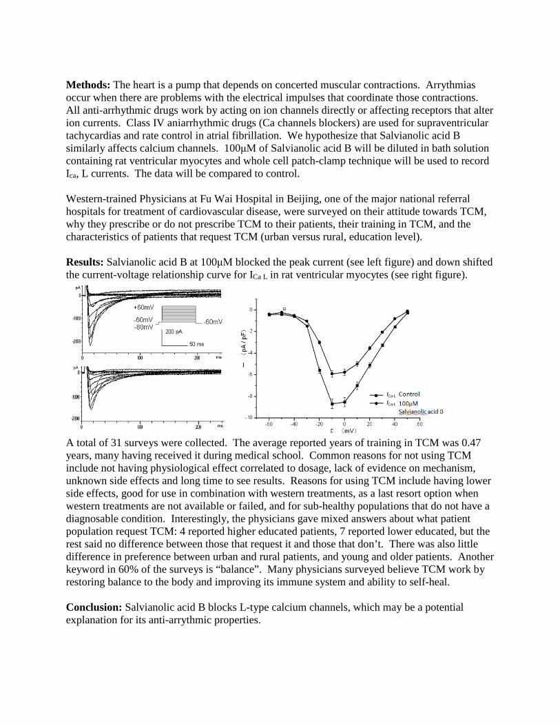
Methods: The heart is a pump that depends on concerted muscular contractions. Arrythmias occur when there are problems with the electrical impulses that coordinate those contractions. All anti-arrhythmic drugs work by acting on ion channels directly or affecting receptors that alter ion currents. Class IV aniarrhythmic drugs (Ca channels blockers) are used for supraventricular tachycardias and rate control in atrial fibrillation. We hypothesize that Salvianolic acid B similarly affects calcium channels. 100μM of Salvianolic acid B will be diluted in bath solution containing rat ventricular myocytes and whole cell patch-clamp technique will be used to record Ica, L currents. The data will be compared to control. Western-trained Physicians at Fu Wai Hospital in Beijing, one of the major national referral hospitals for treatment of cardiovascular disease, were surveyed on their attitude towards TCM, why they prescribe or do not prescribe TCM to their patients, their training in TCM, and the characteristics of patients that request TCM (urban versus rural, education level). Results: Salvianolic acid B at 100μM blocked the peak current (see left figure) and down shifted the current-voltage relationship curve for ICa L in rat ventricular myocytes (see right figure).
A total of 31 surveys were collected. The average reported years of training in TCM was 0.47 years, many having received it during medical school. Common reasons for not using TCM include not having physiological effect correlated to dosage, lack of evidence on mechanism, unknown side effects and long time to see results. Reasons for using TCM include having lower side effects, good for use in combination with western treatments, as a last resort option when western treatments are not available or failed, and for sub-healthy populations that do not have a diagnosable condition. Interestingly, the physicians gave mixed answers about what patient population request TCM: 4 reported higher educated patients, 7 reported lower educated, but the rest said no difference between those that request it and those that don’t. There was also little difference in preference between urban and rural patients, and young and older patients. Another keyword in 60% of the surveys is “balance”. Many physicians surveyed believe TCM work by restoring balance to the body and improving its immune system and ability to self-heal. Conclusion: Salvianolic acid B blocks L-type calcium channels, which may be a potential explanation for its anti-arrythmic properties.

Most physicians in Fu Wai Hospital were trained in TCM during medical school and do believe it works (most saying by holistically improving the body’s ability to restore itself). Their patients also accept and use the TCM they prescribe. However, they currently reserve TCM to chronic conditions, as a last resort therapy, or to accompany western medication.
Krishna Upadhyaya, MS2 Sam Spinowitz, MS2 Preceptors: Dr. Randeep Kashyap Assistant Professor, Department of Transplant and Hepatobiliary Surgery University of Rochester School of Medicine and Dentistry Dr. C. Ramachandra Bhat Associate Professor, General Medicine KVG Medical College and Hospital Assessing Post-Operative Pain and the Role of Psychosocial Factors on Pain Management
and Recovery at KVG Medical College and Hospital, Sullia, India Introduction: Poor management of post-operative pain has been shown to be associated with a number of adverse consequences such as a prolonged return to daily activities and increased medical costs [11, 14]. In the United States, Patient Controlled Analgesia, pain clinics and novel medicines are used to combat pain [15]. In India, the resources available for pain management, as well as cultural attitudes towards pain management may lead to a different pattern of analgesia delivery. A survey, based off the Strategic and Clinical Indicators in Postoperative Pain Management (SCQIPPM), as well as complementary questions, was used to explore the psychosocial management of post-operative pain on levels of communication, action, trust, and environment. Learning about the way that pain is managed in India may provide novel frames of reference within which to consider pain management in the United States. Objective:
1. To gain an understanding of the existing system in place for post-operative pain management at KVG Medical College and Hospital.
2. To explore the barriers and facilitators of health care providers’ response to reported level of post-operative pain.
3. To evaluate the feasibility and acceptability of routine patient pain assessment after surgery.
Background: Proper control of post-operative pain is important in preventing negative outcomes. A 2010 review paper by Nalini et. al [15] showed that post-operative pain can lead to decreased vital capacity, pneumonia, tachycardia, hypertension, MI, myocardial ischemia, poor wound healing and insomnia. Furthermore, one of the major consequences of post operative pain is the transition to chronic pain [15]. A study done by the World Health Organization (WHO) revealed that “individuals who live with chronic pain are four times more likely to suffer from depression
International Medicine Research
and anxiety [1].” In the U.S., one of the biggest concerns for patients undergoing surgery is the level of pain they will be feeling afterwards [15].
Since pain can lead to many adverse outcomes as noted above, this may prolong the return to daily activities. In the location of our study, Sullia, India, a majority of the population is agriculturists where prolonged delay to daily activities, or continued post operative pain may affect their livelihood. India, in general, has also been on the decline in use of narcotics, such as morphine, for pain treatment. In 1997, India was rated among the lowest in the world in terms of per capita morphine consumption as rated by the International Narcotics Control Board (United Nations) [8]. A recently developed questionnaire, the Strategic and Clinical Indicators in Postoperative Pain Management (SCQIPPM), has been validated to be an excellent tool for assessing post operative pain management [6]. The questionnaire covers four major categories: communication, action, trust and environment. As a result, it gives an excellent representation of the overall patient satisfaction and the various biopsychosocial factors that play a role in the relief of pain after surgery. Methods: We used a mixed method design in order to collect quantitative and qualitative data. A Yes/No survey was used for quantitative data collection. While administering the survey we asked complimentary questions to patients to gain a better understanding of their response to the survey for qualitative data collection. Our survey was based on the SCQIPPM questionnaire. Results: We received responses from a total of 89 patients. We found that 23.6% of patients didn’t know what surgery they had. 66.3% of patients didn’t know how long they would be in the hospital after surgery. Among the patients that knew when they would be discharged, the average length of stay in the hospital after surgery was 6.1 days. 41.6% of patients did not tell the staff that they were in pain though 67.6% of these patients actually had pain. It was observed that 33.7% of patients could not sleep at night. Among those patients that couldn’t sleep, 66.7% couldn’t sleep due to pain. When asked to describe level of pain on a 1-10 scale, patients did not understand what it meant. However, a visual analog scale was understood. Additionally, 98.9% of patients had family visiting them in the hospital. Of these patients 65.9% said that their family was in the hospital all the time, and couldn’t put a number on it. Besides meeting with family, some of the activities patients did in the hospital were reading, talking, praying and walking around the wards. Lastly, 100% of patients were happy with their care after surgery. Conclusion: Many preliminary conclusions can be made from this data. Illiteracy rates may play a role in patient education about their surgery, thus affecting their recovery. Long lengths of stay may be due to difficulty following up with patients travelling from far away to the hospital. Since a majority of patients did not tell staff about their pain, this demonstrates the possibility that patients may have trouble advocating for themselves in their post-operative recovery. The lack of sleep in patients may indicate that the prescribed pain medication is not strong enough, or that they don’t have enough distractions to take their mind off the pain such as television or laptops. Since the visual analog scale seemed effective, implementing it might be a cheap and efficient way to standardize pain management. 100% of patients were happy with their care, reflecting a strong standard of care and commitment to surgical patients in the recovery after surgery as well as an appreciation that patients have for the health care that is provided to them.
References:
1. Brennan, Frank. Pain Management: A Fundamental Human Right. International Anesthesia Research Society. Vol. 105, No. 1, July 2007
2. Carlsson, EVA, et al. Concerns and Quality of Life Before Surgery and During the Recovery Period in Patients with Rectal Cancer and an Ostomy. Journal of Wound, Ostomy & Continence Nursing. December 2010. 37(6): 654-661
3. Dasta JF. Patterns of prescribing and administering drugs for agitation and pain in patients in a surgical intensive care unit. Crit Care Med. 1994 Jun; 22(6): 974-980
4. Egbert, L.D. et al. Value of preoperative visit by anesthesiologists: study of doctor-patient rapport, J.A.M.A. 185: 553-555, 1963
5. Gayathri, P. Post operative Pain Services. Indian Journal of Anesthesiology. 2005; 49(1): 17-19
6. Idvall, E. et al. Patient and nurse assessment of quality of care in postoperative pain management. Quality and Safety in Health Care. 2002, December; 11(4): 327-334
7. Official KVG Medical College website. <http://www.kvgmedicalcollege.org/> Accessed January 19, 2011
8. International Narcotics Control Board. Narcotic Drugs: estimated world requirements for 2001-statistics for 1999. New York, NY: United Nations, 2001
9. McMain, Lorraine. Principles of Acute Pain Management. Journal of Perioperative Practice. 18.11 (Nov 2008): 472 477
10. Official National Institute of Health Pain Consortium website. <http://painconsortium.nih.gov/pain_scales/index.html> Accessed January 19, 2011.
11. Official Sullia Town Panchayath website <http://sulliatown.gov.in/tourism> Accessed January 19, 2011
12. Popping, DM, et al. Effectiveness and safety of postoperative pain management. British Journal of Anesthesia 101 (6): 832-840 (2008)
13. Rosen, HI. Patients’ experiences and perceived causes of persisting discomfort following day surgery. BMC Nursing. 2010 Oct 27; 9:16
14. Stepoe, Andrew. Psychosocial processes and health. Cambridge University Press, New York, NY 1994
15. Vadivelu, Nalini et al. Recent Advances in Postoperative Pain Management. Yale Journal of Biology and Medicine. 2010 March; 83(1): 11-25
16. Official World Health Organization Glossary of Terms website. <http://www.wpro.who.int/NR/rdonlyres/D62DEC22-E06E-4BC8-9D12-9BFBF15CF9FA/0/definitions05.pdf> p423-430. Accessed January 19, 2011
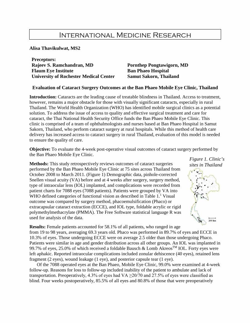
Alisa Thavikulwat, MS2 Preceptors: Rajeev S. Ramchandran, MD Flaum Eye Institute University of Rochester Medical Center
Pornthep Pongtawigorn, MD Ban Phaeo Hospital Samut Sakorn, Thailand
Evaluation of Cataract Surgery Outcomes at the Ban Phaeo Mobile Eye Clinic, Thailand
Introduction: Cataracts are the leading cause of treatable blindness in Thailand. Access to treatment, however, remains a major obstacle for those with visually significant cataracts, especially in rural Thailand. The World Health Organization (WHO) has identified mobile surgical clinics as a potential solution. To address the issue of access to quality and effective surgical treatment and care for cataract, the Thai National Health Security Office funds the Ban Phaeo Mobile Eye Clinic. This clinic is comprised of a team of ophthalmologists and nurses based at Ban Phaeo Hospital in Samut Sakorn, Thailand, who perform cataract surgery at rural hospitals. While this method of health care delivery has increased access to cataract surgery in rural Thailand, evaluation of this model is needed to ensure the quality of care. Objective: To evaluate the 4-week post-operative visual outcomes of cataract surgery performed by the Ban Phaeo Mobile Eye Clinic. Methods: This study retrospectively reviews outcomes of cataract surgeries performed by the Ban Phaeo Mobile Eye Clinic at 75 sites across Thailand from October 2008 to March 2011. (Figure 1) Demographic data, pinhole-corrected Snellen visual acuity (VA) before and at 4 weeks after surgery, surgery method, type of intraocular lens (IOL) implanted, and complications were recorded from patient charts for 7088 eyes (7088 patients). Patients were grouped by VA into WHO defined categories of functional vision as described in Table 1.1 Visual outcome was compared by surgery method, phacoemulsification (Phaco) or extracapsular cataract extraction (ECCE), and IOL type, foldable acrylic or rigid polymethylmethacrylate (PMMA). The Free Software statistical language R was used for analysis of the data. Results: Female patients accounted for 58.1% of all patients, who ranged in age from 19 to 98 years, averaging 69.3 years old. Phaco was performed in 89.7% of eyes and ECCE in 10.3% of eyes. Those undergoing ECCE were on average 2.5 older than those undergoing Phaco. Patients were similar in age and gender distribution across all other groups. An IOL was implanted in 99.7% of eyes, 25.0% of which received a foldable Bausch & Lomb AkreosTM IOL. Forty eyes were left aphakic. Reported intraocular complications included zonular dehiscence (40 eyes), retained lens fragment (2 eyes), wound leakage (1 eye), and posterior capsule tear (1 eye).
Of the 7088 operated eyes at the Ban Phaeo, Mobile Eye Clinic, 99.0% were examined at 4-week follow-up. Reasons for loss to follow-up included inability of the patient to ambulate and lack of transportation. Preoperatively, 4.3% of eyes had VA ≥20/70 and 27.5% of eyes were classified as blind. Four weeks postoperatively, 85.5% of all eyes and 80.8% of those that were preoperatively
International Medicine Research
Figure 1. Clinic’s sites in Thailand

considered blind had VA ≥20/70. Postoperative VA <20/200 was recorded for 1.8% of eyes and was often due to coexisting pathology such as macular degeneration.(Table 1)
Table 1. Pre- and post-operative VAs at the Ban Phaeo Mobile Eye Clinic WHO Definition (Category)1 Visual Acuity Pre-op Post-op Mild/No Impairment (0) 20/20-20/70 304 (4.3%) 6001 (85.5%) Visual Impairment (1,2) <20/70-20/400 4822 (68.1%) 958 (13.7%) Blindness (3) <20/400-CF* 1164 (16.4%) 51 (0.7%) Blindness (4) HM, LP, L Proj* 781 (11.0%) 5 (0.1%) Blindness (5) NLP* 5 (0.1%) 0 (0.0%)
*CF=counting fingers, HM=hand motions, LP=light perception, L Proj=light projection, NLP=no light perception
The decision to perform Phaco or ECCE was made by the ophthalmic surgeon based on cataract
grading. A larger percentage of eyes undergoing Phaco (86.7%) had postoperative VA ≥20/70, when compared to those eyes treated by ECCE (71.8%) (χ2 = 111.990, df = 2, p-value <0.001). While rigid IOLs were available for all patients for the entirety of the study, foldable IOLs became the primary IOL for Phaco patients beginning in October 2010. Slightly, but statistically significantly more eyes undergoing Phaco with foldable IOL implantation, 89.4% (1410/1578) had postoperative VA ≥20/70, compared to 86.6% (4062/4688) of those Phaco patients receiving rigid IOLs (χ2 = 8.359, df = 2, p-value = 0.0153). (Table 2)
Table 2. Postoperative VA compared by surgery method and by type of IOL implanted
*CF=counting fingers, HM=hand motions, LP=light perception, L Proj=light projection, NLP=no light perception
Conclusion: Cataract extraction by experienced ophthalmologists at the Ban Phaeo Mobile Eye Clinic in Thailand reaches the WHO’s quality target that 85% of eyes have postoperative VA ≥20/70 and are comparable to those of an urban hospital in Bangkok2. Visual outcomes are better following Phaco than ECCE and slightly better with a foldable rather than rigid IOL after Phaco. The difference between Phaco and ECCE might be attributed to the quality of the cataracts, with more mature cataracts being harder to remove using the Phaco machine and the most mature requiring ECCE, a more invasive procedure. Measurement of postoperative VA before suture removal is another limitation of this study. As more sutures are placed in ECCE than Phaco and rigid than foldable IOLs due to larger wounds, ECCE and eyes with rigid IOLs are more likely to have suture-induced astigmatism that can be corrected with suture removal. Nonetheless, these results suggest that the mobile eye clinic is an effective method for providing quality cataract surgery to rural Thailand and that Phaco with foldable IOLs may offer optimal outcomes. References: 1. World Health Organization. International Statistical Classification of Diseases and Health Related Problems 10th Revision. Geneva; 2007. 2. Dulayajinda D. et al. Outcomes of cataract surgery in senile cataract patients at Siriraj Hospital: a prospective observational study. J Med Assoc Thai. 2005;88(9):S82–8.
Surgery Method ECCE Phaco Phaco Visual Acuity IOL Rigid Rigid Foldable 20/20-20/70 497 (71.8%) 4062 (86.7%) 1410 (89.4%) <20/70-20/400 178 (25.7%) 598 (12.8%) 163 (10.3%) <20/400-CF* 16 (2.3%) 23 (0.5%) 5 (0.3%) HM, LP, L Proj* 1 (0.1%) 4 (0.1%) 0 (0.0%) NLP* 0 (0.0%) 0 (0.0%) 0 (0.0%)
Scott Walter, MS2 Nicholas Zinn, MS2 Preceptor: David Hursh, PhD Associate Professor Warner School of Education Teaching & Curriculum
Education for Health Advocacy in Kampala, Uganda
Introduction: Uganda is a rapidly growing nation located in Eastern Africa and currently has a
population of approximately 33 million people (1). However, like other developing countries, Uganda faces several health problems due to lack of modern infrastructure, access to healthcare, and clean water. The infant mortality rate remains high at 79.4 per 1000 live births, and Ugandans have an average life expectancy of 53 years (2). Despite governmental efforts to improve access to healthcare, the burden of disease remains a concern for the country. Objectives:
This project aimed to teach children and their teachers in schools about the numerous environmental health problems that exist in Uganda. We concentrated on the importance of clean water, good hygiene, and the health risks associated with indoor air pollution. Both the scientific nature of the problems as well as possible interventions were taught. Our hope was that by educating the children and their teachers on these issues, we could help them become better advocates for their own health as well as the health of their families, neighbors and communities. Background:
Unsafe water, sanitation, and hygiene have generally been associated with poor health outcomes, and are a significant cause of morbidity and mortality in low- and middle-income countries. Research has shown that Kampala, the capital city of Uganda, with its rapid population growth, reliance on protected springs, and frequent rainfall due to its proximity to Lake Victoria, is particularly at risk for illnesses related to water contamination (3). In order to assess the bacteriological quality of water in the region, a cross-sectional sanitary risk assessment was carried out from December 2001 to March 2002 on ten randomly selected springs in Katwe and Kisenyi parishes, two suburbs of Kampala (4) . Results revealed that of the 80 samples of water collected from these springs, 90% exceeded the World Health Organization (WHO) coliform count for drinking water, and 60% exceeded the WHO recommended limit for nitrate levels. It is estimated that 93% of the Uganda’s population uses biomass as a primary source of energy. Biomass is commonly used in cooking and includes such material as wood, cow dung, crop residues and charcoal. Cooking in sub-Saharan Africa primarily occurs indoors inpoorly ventilated spaces. This leads to an increased exposure to damaging pollutants and carcinogenic
International Medicine Research

compounds that burning biomass emits. A study measuring levels of particulate matter and carbon monoxide (an indicator of particulate matter levels) in nearby sub-Saharan Malawi found indoor air pollution levels to be four times greater than the WHO recommended level for outdoor air quality. Since women are in charge of cooking, they along with their children have the highest exposure to these pollutants and thus increased chance for respiratory infections other lung diseases. Studies have found that 50% of childhood pneumonia deaths in sub-Saharan Africa could be attributed to indoor air pollution. Evidence with varying degrees of certainty has also shown links between indoor air pollution and low birth weight in children, interstitial lung disease, tuberculosis, cardiovascular disease and cataracts in adults (5, 6). Methods: Through surveys containing both multiple choice and open-ended response options, we began by evaluating the knowledge of students aged 8-12 at the Circle of Peace primary school in Kampala, Uganda on environmental health problems, and what they knew about the relationship between the environment and their health. The surveys addressed topics including the negative effects of indoor air pollution and the importance of drinking clean water and practicing good hygiene. We then taught the relevant concepts regarding each environmental health topic and the science behind it. For example, to address the topic of air pollution, we taught basic lung biology, illustrated how pollution affects the lungs, and ultimately conducted a laboratory experiment using petri dishes of petroleum jelly to capture and visibly reveal the particulate matter that exists in heavily polluted locations such as kitchens and busy streets. We also explored possible solutions to these problems through our curriculum, such as rocket stoves and water purifiers, so that the students could improve their own health conditions as they grew older. At the conclusion of our time at the school we evaluated how much the students had learned and how their opinion had changed on these health issues. Results: Quantitative results from our study are still pending, however preliminary analysis of the data currently reveals no significant improvement in student knowledge and awareness of environmental health problems following exposure to the curriculum. Conclusion: Preliminary analysis of our results indicates that exposure to our curriculum yielded no significant improvement in student knowledge of environmental problems effecting their community. There are multiple reasons why this could be the case. For one, our teaching methods may have been unfamiliar to the students, rendering them ineffective. We found Uganda’s education system to be heavily based on recall rather than synthesis and application of concepts, likely making it difficult for students to grasp our teaching methods that consisted of open discussions and hands-on activities.
Our choice of using a questionnaire as a way to assess student knowledge both prior to and after exposure to our curriculum may also have been flawed. Even though the survey was tested in the United States beforehand with doctoral students, we still had problems with language and comprehension when the survey was presented to students in Africa. It is also possible that, consistent with a learning style based on recall, the students tried to answer the survey in the exact way they had the first time. If this were the case, it would imply that the
students did not understand how the survey was to be used, which consequently undermined the results.
Our primary goal was to implement a curriculum which taught children in Uganda how their environment could influence their health. We believe we were successful in giving them an introduction to this topic. However, multiple factors, including adapting teaching methods to fit cultural norms and implementing a survey that an unknown population could fully understand, ultimately prevented our intervention from having a dramatic impact on our sample. Future research in this area should be aware of these obstacles.
References:
1. https://www.cia.gov/library/publications/the-world-factbook/geos/ug.html Accessed 2/1/10.
2. Level & Trends in Child Mortality. Report 2010. Estimates Developed by the UN Inter-agency Group for Child Mortality Estimation (UNICEF, WHO, World Bank, UN DESA, UNPD).
3. 10. Lewin, S., Norman, R., Nannan, N., Tomas, E. and Bradshaw, E. Estimating the burden of disease attributable to unsafe water and lack of sanitation and hygiene in South Africa in 2000. South African Medical Journal. August 2007. 97(8):755-8.
4. Haruna R, Francis E, Kabagambe E. The quality of water from protected springs in Katwe and Kisenyi parishes, Kampala city, Uganda. African Health Sciences. 2005. 5(1): 14-20.
5. Uganda National Energy Balance 2002. Ministry of Energy and Mineral Development. 6. Norman R. et al. Estimating the Burden of Disease Attributable to Four Selected
Environmental Risk Factors in South Africa. Reviews on Environmental Health. 2010. 25(2): 87- 119.

Gene Yang, MS2 Preceptors: Cheryl Kodjo, MD, MPH Borie Park Associate Professor Secretary General Department of Pediatrics, Adolescent Medicine Social Welfare Association of Korea University of Rochester Medical Center Sexually transmitted infection awareness and motivations of sex workers in the Republic of
Korea Introduction: Prostitution in Korea is illegal, but widespread.i According to a 2007 government report, the sex industry accounted for about $13 billion dollars (about 1.6% of GDP) and involved at least 269,000 women.ii In another indicator of the significant role that prostitution plays in Korea, over 21% of males experience their initial coitus with a prostitute. iii Additionally, more than a quarter of male university students have had sex with a prostitute. iii In terms of sexual education, Korea lags very far behind. Only in 2009 did the Ministry of Education, Science, and Technology issue guidelines for teaching sexual education; prior to this, cartoons of metaphors, videos of bees pollinating flowers, and non-specific references were the norm. iv This combination of Korean young adults who a lack basic sexual education and their active use of prostitutes creates a dangerous scenario for both the sex workers and their clients which has been demonstrated by higher rates of infection in at-risk populations. v, vi, vii Objective: To better understand the level of knowledge that the sex worker population has about STIs and their motivations for working in the sex industry. Background: Korea is an ethnically homogeneous country with a common language and a social structure and system of values system based on Confucianism. Due to the Confucian history, Korean society tends to be conservative in the government, media, and in international image. Korea maintains an adultery law and has outlawed pornography. It has been observed that Korea has a similar conservative attitude, especially about anything regarding sex, which the United States had in the 1950’s. Although this conservative view is what is accepted by Koreans as their national identity, it conflicts with a very active sex industry that plays a significant role in male society. viii, ix, x, xi
The active sex trade has led to distinctions and a hierarchy of different types of sex worker, such as those based out of a brothel and free-lance prostitutes.v The primary differences between the different types are both venue and choice. For example, brothel based prostitutes (BBP) of the red-light districts or massage parlors have no freedom of choice, while free-lance prostitutes (FLP) provide services other than sex and may have complete autonomy over the decision to have sex with a client. vii, xiii
International Medicine Research
Methods: Administer surveys with the assistance of Korean government social workers. Results: 5 survey responses. Although most sex workers who did not feel comfortable filling out the survey, they were all comfortable speaking about their experiences during face to face encounters. Conclusion: The sex worker population is ill-informed regarding STIs. However, that does not indicate a large gap in knowledge with the general population, since many Koreans have a poor understanding of STIs. All sex workers surveyed indicated that STI knowledge came from sources other than school (mostly other sex workers, but also included the internet and free clinics). Knowledge regarding options for treatment was generally widespread, but sex workers who worked for less than one year were not as aware of the free testing/treatment options available from the government. The sex worker population generally has a junior/senior high school education level and their parents have a lower education level. They generally perceive their occupation as neutral/slightly negative in the eyes of the country, but all respondents would choose a different occupation if they had the means (i.e. startup money to open a shop). The majority of respondents surveyed also indicated that they do not have problems with their clients and that they were working as a sex worker by choice, but the vast majority of the sex workers who declined the survey all indicated that they were forced into the sex trade due to financial debts. According to Korean government officials, kidnapping young women to become sex workers has been replaced by targeting and lending money to at-risk young women who do not realize their subsequent debts are not legally valid. These brothel operators are generally individuals or families who run the brothels as small businesses with no criminal gang affiliations. The greatest positive effect regarding the prevention of STIs would be from the introduction of sex education during high school, since both sex workers and their clients would be better educated. Additionally, reinstituting government programs to allow health teams to enter red light districts would help in reducing the incidence and prevalence of STIs in the sex worker population. References: i 2009 Human Rights Report: South Korea. United States Department of State. 11 March 2010 ii Korean Government Report on Sex Industry and Prostitution in 2007. Republic of Korea Ministry of Gender Equality and Family. 2007. iii Kim, J; Celentana, DD; Crum, RM. Alcohol Consumption and Sexually Transmitted Disease Risk behavior: Partner Mix Among Male Korean University Students. Alcoholism: Clinical and Experimental Research. Vol. 22, No. 1, pp. 126-131. February 1998. iv Sung, So-young. Breaking the Taboo: Sex Education in the Spotlight. Korea Joonang Daily. 2 December 2009 v Sohn, Myongsei and Kinam Jin. AIDS Related Perceptions and Condom Use of Prostitutes in Korea. Yonsei Medical Journal. Vol. 40, No. 1, pp. 9-13. 1999
vi Kim, O, et al. Seroprevalence of Sexually Transmitted Viruses in Korean populations including HIV-seropositive individuals. International Journal of STD & AIDS. Vol. 14, No. 1, pp. 46-49. 2003. vii Yoo, Hyera, et al. HIV/AIDS Knowledge, Attitudes, Related Behaviors, and Sources of Information Among Korean Adolescents. Journal of School Health. Vol. 75, Issue 10, pp.l 393-399. 28 November 2005. viii Moon, Jaewon. Obscenity Laws in a Paternalistic Country: The Korean Experience. Washington University Global Studies Law Review. Vol. 2:353. 2003. ix Country Study: South Korea. United States Library of Congress Federal Research Division. May 2005. x Choi, Yeong Jin, et al. Masturbation and Its Relationship to Sexual Activities of Young Males in Korean Military Service. Yonsei Medical Journal. Vol 41, No. 2, pp 205-208. 2000. xi Lee, WH, et al. Sexual Activity on the Survey of Sexual Behavior Among military Soldiers. Korean Journal of Urology. Vol. 39, pp. 593-6. 1998. xii Byun, Youngmin, et al. Differences in Chlamydia trachomatis Cryptic Plasmid Loads in Two Types of Female Commercial Sex Workers in Korea. Japanese Journal of Infectious Disease. Vol. 61, pp. 146-147. 2008. xiii Micollier, Evelyne. Sexual Cultures in East Asia: The Social Construction of Sexuality and Sexual Risk in the Time of AIDS. RoutledgeCurzon, London. 2004.

Nathaniel Bayer, MS2 Preceptors: Olle Jane Sahler, M.D., David Korones, M.D., and Stephanie Brown Clark, M.D., Ph.D. University of Rochester School of Medicine and Dentistry Sites in Project: Golisano Children’s Hospital, NewYork-Presbyterian, The Hole in the Wall Gang Camp, Memorial Sloan-Kettering Cancer Center, Double H Ranch, The Children’s Hospital of Philadelphia, St. Jude Children’s Research Hospital, UCSF Benioff Children’s Hospital, Camp Okizu, and Lucille Packard Children’s Hospital at Stanford
Life and Positivity in Pediatric Oncology
Introduction: The field of pediatric oncology is rife with feelings, communication concerns, and multi-tiered care for the patient, the family, the community, and the healthcare providers. Its emotive depth spans tragic death, triumphant victory, disappointing remission, and all possible iterations of the above.
Objective: This project aims to explore the emotions and communication within the field of pediatric oncology. It is designed to span inpatient, outpatient, and summer camp settings for patients, their families, and the healthcare providers working in such environments. Background: Physician-patient relationship is crucial in cancer care. Pediatric oncologists identify their main concern as being the messenger of life-threatening conditions.1 Sharing a cancer diagnosis with a patient and his/her family is both rich in communication and emotional issues. Oncologists must master their own emotions while also guiding those of their patients and families. This journey is one which the patients must remain in both within the healthcare setting and beyond. As an almost supplemental therapy, summer camps have been shown to be effective in supporting a child to function at a “more normal”, healthier, and happier level.2 Methods: The project included three components: (1) Interviewing pediatric oncologists, (2) Conducting participant observation in patient encounters at 7 children’s hospitals throughout the United States, and (3) Volunteering at 3 summer camps for children with cancer and other life-threatening conditions. Evidence was analyzed through a theme-based approach. Results: Central themes explored through interviewing pediatric oncologists included: (1) The approach in sharing a cancer diagnosis with a patient and family, (2) Navigating the balance of aggressive treatment versus palliative care in treatment, (3) Deciding the level of emotional attachment for the care provider, (4) Discussing memorable patient cases, and (5) The response to the assertion that pediatric oncology “must be so sad”. The patient encounters provided strategies and approaches to manage emotions within the field. Furthermore, the summer camp experience was invaluable in exploring not only the emotions of the children, but my own as well.
Medical Humanities Research

Conclusion: Pediatric oncology is a field rich in emotional considerations, but it is not overwhelmed in sadness. As one oncologist put it, “The enemy is not death, but pain and suffering.”3 This recognition permeated my entire experience. Each child I met was filled with resilience, innocence, and beauty. Life’s value is judged not in length, but in depth of each and every moment. This is the lens through which the field should be viewed, both by physicians and patients. References: 1 Stenmarker M, Hallberg U, Palmérus, K, et al. Being a messenger of life-threatening conditions: Experiences of pediatric oncologists. Pediatric Blood & Cancer 2010; 55: 478–484. 2 Smith K, Gotlieb S, Gurwitch R, et al. Impact of a Summer Camp Experience on Daily Activity and Family Interactions Among Children with Cancer. Journal of Pediatric Psychology 1987; 12 (4): 533-542. 3Interview with Dr. OJ Sahler at Golisano Children’s Hospital
Behdad Bozorgnia, MS2 Preceptor: Stephanie Brown-Clark, M.D., Ph.D. University of Rochester School of Medicine Medical Humanities
In The Flesh: Philosophical Reflection on the Anatomy Lab Introduction: The author explores the experience of the anatomy lab from the perspective of an imaginary character named Reza. The protagonist is a ruminative and absent minded medical student who cannot help but engage in philosophical reflection about the anatomy lab. Each chapter reveals both an aspect of the Reza’s ordinary life and philosophical thoughts about a particular subject matter. Objectives: The author weaves philosophical reflection into a narrative framework in order to accomplish three distinct points. First, couching philosophical though within the context of a story allows for appeal to a broader audience than those interested in academic philosophy. Secondly, the development of a particular character who gives voice to philosophical reflection amplifies the investment of the reader in the text and thus intensifies engagement with the philosophical arguments presented. Thirdly, the contextualization of thought in within the life of a specific individual reflects a more realistic presentation of the actual process of philosophical reflection, which is bound by the life and character of the thinker. Method: The short story begins with reflection on the nature of violence, morality, medicine and their pertinent relations. The author also explores themes regarding the basic assumption of the doctor patient relationship, the phenomenology of embodiment, and the relation of physiological nature of disease to the lived experience of disease. Conclusion: No final conclusions are reached about any topic presented in the short story, the purpose being to elicit readers into thought rather than convince them of a particular philosophical point of view.
Medical Humanities Research
Sarada Abraham, MS1 Preceptor: Michael Keefer, M.D. Professor of Medicine University of Rochester Medical Center Department of Infectious Disease
Beyond Project Vogue
HIV/AIDS remains a worldwide pandemic with no known cure. Of the over one million people in the USA living with HIV, roughly 75% are men and the main route of infection remains men who have sex with men, or MSM (48%), (CDC 2008). Although Whites make up the majority of the US population, their rate of HIV infection is 225/100,000 versus 1,715/100,000 in African Americans (46% of those living with HIV) and 585/100,000 in Latin Americans (18% of those living with HIV), despite the fact that African Americans and Latinos make up only 12% and 15% of the US population respectively (CDC 2008). Though people of color are over represented in HIV acquisition, they are historically underrepresented in HIV vaccine clinical trials. This includes the House Ball community, a marginalized sub-population composed of MSM of color with high HIV and STD prevalence who traditionally do not access HIV and medical services due to institutional mistrust. A detailed, semi-qualitative survey assessing barriers and facilitators that arise when recruiting MSM of color for HIV vaccine trial research initiatives was administered with the purpose of gaining a full understanding of the environmental and social factors that influence this demographic so that an effective module for HIV education and prevention strategies, including increased participation in HIV HVTN clinical trials, can be implemented in the community. The survey was designed such that comparisons between MSM of color involved in the house ball community and those who are not can be ascertained.
Surveys were administered orally to qualified men (those indicating sexual activity with men, who personally identify as being of either African American or Latino descent, from age 18 to 50) at locations in Buffalo and Rochester, New York (AIDS Community Services {ACS} and AIDS Care in Buffalo and Rochester respectively). Recruiting was done at weekly and monthly events held at ACS, AIDS Care, and GAGV (Gay Alliance of Genesee Valley). Flyers, hand bills, and other marketing materials were also posted and distributed at locations with high visibility for the MSM population in both Rochester and Buffalo. Survey sessions lasted approximately 45 minutes each and were conducted in a private, designated room at each location.
When asked to assess personal risk of contracting HIV, 100% of the individuals who indicated ‘medium risk’ were already HIV positive. Similarly, although 54% of participants had HIV or another STD/STI (46% were HIV positive), 76% of participants identified as not having an STD or STI. 92% of those surveyed agreed both that HIV vaccine clinical trials were safe and they would participate in an HIV vaccine trial, though none have participated to date. Additionally, less than half of those surveyed stated that they knew what a clinical trial was. Of those who believed they knew what a clinical trial was, only half of them actually knew what it
Year-Out Research
was. It is important to note that to date, not enough individuals have been surveyed who identify as House Ball community members to make adequate comparisons between them and other MSM which can be expected, due to the marginalization of this subculture. Additional research must be conducted in order to make adequate and significant conclusions of this data.
References
Centers for Disease Control and Prevention (2008), HIV/AIDS Surveillance Report, 2006, 18(1). Atlanta, GA: U.S. Department of Health and Human Services.
Fields, S.D. (2005). Adaptation of an Existing HIV-Knowledge Questionnaire for use with Young Men of Color who have Sex with Men. The Journal of Multicultural Nursing and Health, 11 (1), 47-55
Murrill, C. S., Liu, K., Guilin, V., Colon, E. G., Dean, L., Buckley, L., Sanchez, T., Finlayson, T., Torian, L. (2008). HIV Prevalence and Associated Risk Behaviors in New York City’s House Ball Community. AJPH Research and Practice 98(6).
Wolitski, R. J., Valdiserri, R. O., Denning, P. H., Levine, W. C. Are We Headed for a Resurgence of the HIV Epidemic Among Men Who Have Sex With Men?. American Journal of Public Health, 2001, 91(6).

Gener Augustin, CC3 Preceptors: Dr. Frank W LoGerfo, Dr. Leena Pradhan Beth Israel Deaconess Medical Center, Harvard Medical School Vascular Surgery Research Effects of Substance P and Neuropeptide Y on migration and proliferation of Adult Dermal
Fibroblasts in normolgycemic and hyperglycemic conditions. Introduction: Chronic diabetic foot ulceration resulting from impaired wound healing in diabetes is the source of many hospitalizations and lower extremity amputations. Although there is no single theory that can explain why chronic wounds fail to heal, fibroblast may be a major contributory factor in its persistency. Fibroblasts are a major component of the wound-healing cascade and are target cells for neuropeptides, Substance P (SP) and Neuropeptide Y (NPY). In the present study, the effects of SP and NPY on Adult Dermal Fibroblasts (ADFB) migration and proliferation in normal and hyperglycemic conditions are examined. Objective: To study the effect of hyperglycemia, and neuropeptides NPY and SP on migration and proliferation of adult dermal fibroblast Background: Wound healing involves three phases: inflammatory phase, proliferative phase, and the remodeling phase. In the inflammatory phase, growth factor TGF-β promotes fibroblasts chemotaxis. Fibroblasts are present on day 2 and 3 of wound healing, becoming the dominant cell type by week 1. They are critical to the production of dermal matrix during the proliferative phase of wound healing. NPY is a 36 amino acid peptide neurotransmitter present in the central and peripheral nervous system. SP is a neurotransmitter of the afferent nervous system. Both peptides play a role in the immune and inflammatory reactions by conditioning and fine-tuning the host’s response to antigenic challenge. Fibroblasts are target cells for neuropetides SP and NPY. SP may play and important role in wound healing by promoting wound fibroblast proliferation and inhibiting apoptosis. It is thus postulated that there is direct fibroblast mediated effects of SP and NPY on migration and proliferation. Methods: ADFB are cultured in DMEM with 10% FBS and treated with different concentrations of D-glucose [5mM (normoglycemic); 10mM or 30mM (hyperglycemic)] or L-glucose [30mM, osmolarity control]. ADFBs are also co-treated with 100nM SP or NPY. Migration is assessed via scratch wound assays in 24 well plates. Area of scratch wound closure is analyzed by adherent cell cytometer, Cyntellect Celigo™ using CellTracker-Green™ at 0, 6, 12 and 24 hrs. Proliferation is assessed in 96-well plates using Hoechst stain 33342 in conjunction with CellTracker-Green™on Cyntellect Celigo™ at 0, 1, 2, 3, 4, 5, 6 and 7 days.
Year-Out Research
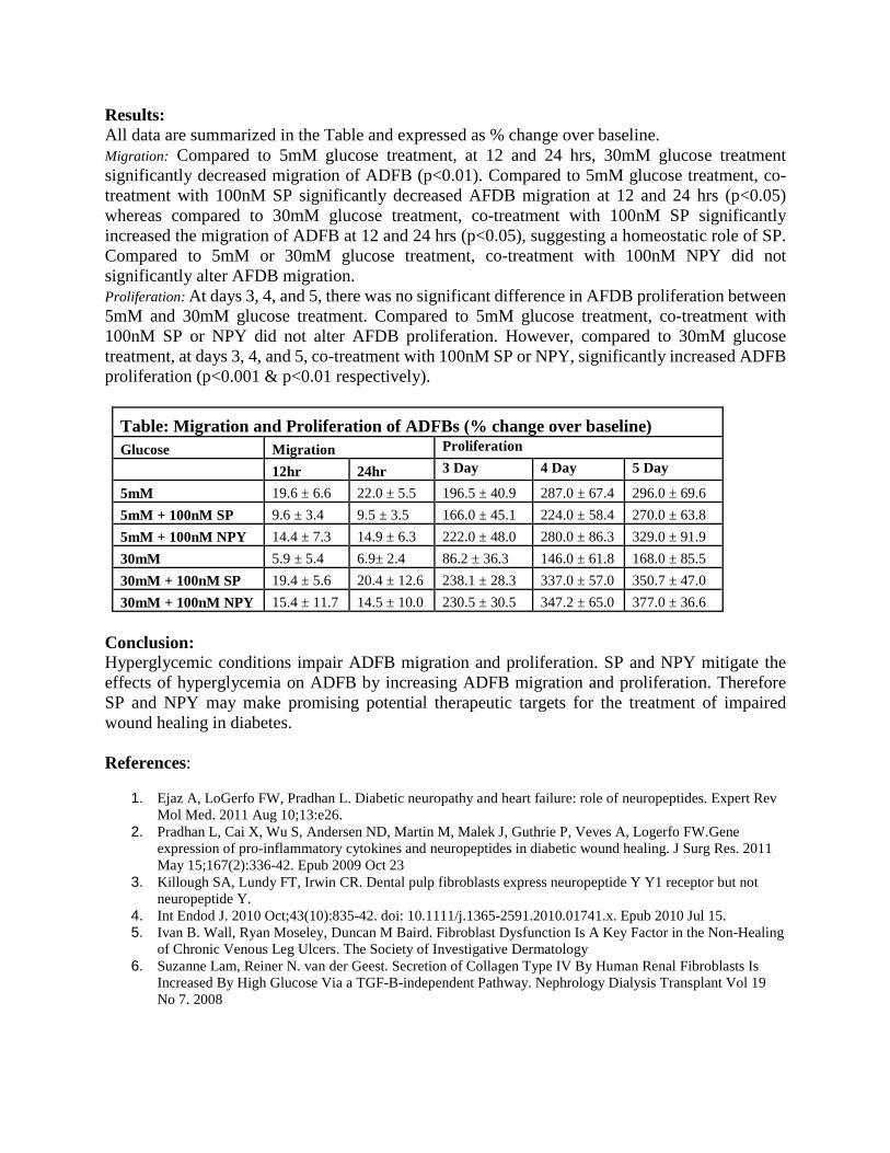
Results: All data are summarized in the Table and expressed as % change over baseline. Migration: Compared to 5mM glucose treatment, at 12 and 24 hrs, 30mM glucose treatment significantly decreased migration of ADFB (p<0.01). Compared to 5mM glucose treatment, co-treatment with 100nM SP significantly decreased AFDB migration at 12 and 24 hrs (p<0.05) whereas compared to 30mM glucose treatment, co-treatment with 100nM SP significantly increased the migration of ADFB at 12 and 24 hrs (p<0.05), suggesting a homeostatic role of SP. Compared to 5mM or 30mM glucose treatment, co-treatment with 100nM NPY did not significantly alter AFDB migration. Proliferation: At days 3, 4, and 5, there was no significant difference in AFDB proliferation between 5mM and 30mM glucose treatment. Compared to 5mM glucose treatment, co-treatment with 100nM SP or NPY did not alter AFDB proliferation. However, compared to 30mM glucose treatment, at days 3, 4, and 5, co-treatment with 100nM SP or NPY, significantly increased ADFB proliferation (p<0.001 & p<0.01 respectively).
Table: Migration and Proliferation of ADFBs (% change over baseline) Glucose Migration Proliferation
12hr 24hr 3 Day 4 Day 5 Day
5mM 19.6 ± 6.6 22.0 ± 5.5 196.5 ± 40.9 287.0 ± 67.4 296.0 ± 69.6 5mM + 100nM SP 9.6 ± 3.4 9.5 ± 3.5 166.0 ± 45.1 224.0 ± 58.4 270.0 ± 63.8 5mM + 100nM NPY 14.4 ± 7.3 14.9 ± 6.3 222.0 ± 48.0 280.0 ± 86.3 329.0 ± 91.9 30mM 5.9 ± 5.4 6.9± 2.4 86.2 ± 36.3 146.0 ± 61.8 168.0 ± 85.5 30mM + 100nM SP 19.4 ± 5.6 20.4 ± 12.6 238.1 ± 28.3 337.0 ± 57.0 350.7 ± 47.0 30mM + 100nM NPY 15.4 ± 11.7 14.5 ± 10.0 230.5 ± 30.5 347.2 ± 65.0 377.0 ± 36.6
Conclusion: Hyperglycemic conditions impair ADFB migration and proliferation. SP and NPY mitigate the effects of hyperglycemia on ADFB by increasing ADFB migration and proliferation. Therefore SP and NPY may make promising potential therapeutic targets for the treatment of impaired wound healing in diabetes. References:
1. Ejaz A, LoGerfo FW, Pradhan L. Diabetic neuropathy and heart failure: role of neuropeptides. Expert Rev Mol Med. 2011 Aug 10;13:e26.
2. Pradhan L, Cai X, Wu S, Andersen ND, Martin M, Malek J, Guthrie P, Veves A, Logerfo FW.Gene expression of pro-inflammatory cytokines and neuropeptides in diabetic wound healing. J Surg Res. 2011 May 15;167(2):336-42. Epub 2009 Oct 23
3. Killough SA, Lundy FT, Irwin CR. Dental pulp fibroblasts express neuropeptide Y Y1 receptor but not neuropeptide Y.
4. Int Endod J. 2010 Oct;43(10):835-42. doi: 10.1111/j.1365-2591.2010.01741.x. Epub 2010 Jul 15. 5. Ivan B. Wall, Ryan Moseley, Duncan M Baird. Fibroblast Dysfunction Is A Key Factor in the Non-Healing
of Chronic Venous Leg Ulcers. The Society of Investigative Dermatology 6. Suzanne Lam, Reiner N. van der Geest. Secretion of Collagen Type IV By Human Renal Fibroblasts Is
Increased By High Glucose Via a TGF-B-independent Pathway. Nephrology Dialysis Transplant Vol 19 No 7. 2008
Stanley Dunn, CC4 Preceptor: Richard Kreipe, M.D. Professor University of Rochester Medical Center Department of Pediatrics, Adolescent Medicine
Mental Health Effects of Nature and Structured Nature-based Programs on Children
Introduction There is growing evidence that interacting with nature can have a positive effect on children's physical health and mental well-being (Strife & Donwey 2009). Consistent exposure can improve young children's motor coordination, attention capacities and impulse control (Faber Taylor et al. 2001; Kuo & Faber Taylor 2004). Access to nature positively influences children's academic performance, mitigates childhood stress, and improves children's social skills by developing their language and communication (Wells & Evans 2003). Children who play in natural environments have more positive feelings about each other (Pyle 2002). Their play is more diverse, imaginative, and creative, than children's play in other settings (Faber Taylor et al. 1998). In this study we are defining “interacting with nature” as children spending time outdoors directly engaging all of their senses in active play and learning. Scientific Background Positive psychology, grounded in an ecological model, looks at child development from a multi-contextual standpoint. Peter Benson and the Search Institute in Minnesota have focused on 40 identified "developmental assets," dividing them into external assets, such as family support and caring neighborhood, and internal assets, such as honesty and self-esteem. Internal assets are further divided into four categories: commitment to learning, positive identity, positive values, and social competencies. At the core of these positive values and social competencies lies empathy. Empathy is the capacity to vicariously experience another person's pleasure or pain. It provides the child with powerful incentives for positive social interaction. As part of every child's native endowment, it is a natural strength (or virtue) upon which further pro-social growth can be built (Damon 2004). Empathy, or empathetic arousal, is at the root of morality and eventually, as a child develops, becomes an important mediator of altruism (Evans et. al 2005). Studies have shown an inverse relationship between children’s and adolescents’ degree of empathy and disruptive behavior disorders and bullying (de Wied et al. 2005; Lovett 2007 et al.) Higher levels of dispositional affective empathy are positively associated with more successful conflict management and problem solving ability and negatively linked with conflict engagement (de Wied et al. 2007). Empathy is thought to inhibit aggressive behavior through both cognitive and affective processes (de Wied et al. 2005). Hypothesis The natural world provides a unique context in which children’s empathy towards all living things can be nurtured. In the field of environmental education great emphasis is placed on
Year-Out Research
facilitating children's empathy towards other living beings, both human and non-human. I hypothesized that the exposure of children 10-14 years old to nature through participation in structured nature-based educational programs would increase measures of their empathy. Methods To this end, the study was conducted at 3 outdoor schools/nature centers: the Echo Hill Outdoor School(EHOS) in Worton, MD; the Chesapeake Bay Foundation(CBF) educational sites in Maryland and Virginia; and the YMCA Camp Erdman in Waialua, HI. All of these programs offer 3 to 5 day nature-based residential field trips for visiting school groups. There were 240 study participants, ages 10-14 and grades 5-8, from 14 elementary and middle schools from 4 states and Washington DC. 10 of the schools were public and 4 were private. These schools came from urban, rural, and suburban locations. On the first and last days of the students’ trips the Basic Empathy Scale (BES) was administered. The BES is a 20-item Liekert scale with a range of 1-5. The lowest possible total score is 20 with a possible of 100. The BES consists of Affective and Cognitive Scales which when combined yield the total score. There are 11 affective questions and 9 cognitive questions. The lowest possible affective score is 11 and with the highest being 55. The lowest possible cognitive is 9 with the highest being 45. Each of the three sites chosen performs three to five day educational nature-based field trips. They offer an environmental science curriculum using hands on, experiential learning techniques. For example a Swamp Ecology class at EHOS includes a 1 hour hike through the swamp along the board walk; and a 2 hour canoe paddle that is frequently interrupted to net species such as predacious diving beetles, leaches, and large mouth bass and discuss their different roles in the wetland ecosystem. For CBF this type of curriculum is their primary focus. EHOS and Camp Erdman have another component. This is their Adventure curriculum, which uses the programs’ extensive ropes courses to cultivate improved group dynamics, self-esteem, and goal setting abilities. This is done through elements such as the Alpine Tower, which is a 60ft, 3-sided, hourglass-shaped climbing structure. Students climb as individuals or in pairs and are anchored by two of their classmates. Thus there are both personal and group challenges within this one element. The characteristics of the nature experiences did not change based on 3,4, or 5-day duration. The longer the duration meant that the students simply had more time learning in and experiencing nature. Out of the 240, 202 participants completed the BES both pre and post the nature experience. Structured journaling exercises were done twice daily- these results will be the subject of a future paper. The BES was used to measure interpersonal empathy amongst students, while the journaling was used to assess empathy of students towards the natural world. Results The mean difference in affective scores was an increase of 1.37 +/- 4.81 S.D. with p<0.0001. The mean difference in cognitive scores was an increase of 0.06 with a +/- 4.52 S.D. and p<0.85. The mean difference in total scores was an increase of 1.43+/- 7.79 and p<0.0099. The bivariate analyses looking at difference in affective, cognitive, and total scores in relation to gender, age, and grade were statistically insignificant. Duration of nature experience with respect to increase in affective score was statistically significant with p <0.03. Public school children experienced a mean increase in affective scores of 1.97 +/- 4.93 S.D. and p <0.039. Private school children

experienced a mean increase in affective scores of 0.56 +/- 4.55 S.D. The mean pre-survey scores for public and private school children were 34.23 and 36.03 respectively, creating an insignificant difference in scores with p <0.096. There was no significant difference in the change between pre and post-survey cognitive scores with respect to public vs. private school students. Discussion There were several major findings in this study. The most important was the statistically significant increase in the participants’ affective empathy score, which then drove the increase in the total score. This finding proves that children having structured time in nature with their peers does increase their empathy. Looking at the mean increase in affective score of 1.37 +/- 4.81 S.D., the question arises: is this increase in score clinically significant as well? In future studies, it would be advantageous to use not only self-report measures, such as the BES but also behavioral measures. This would enable researchers to assign clinical significance to these values. One component of the future qualitative analysis will be to match the qualitative work for each student with his/her change in scores. This will allow me to look at the students who had above and below average changes in scores and see what light their journals will shed on those scores. Perhaps all the students who had an above average affective change had some common element to their nature immersion, which could be replicated and tested. Or conversely removing a common element found in students’ journals who had below average affective score changes. Another major finding was that there was a significant difference in the change in affective scores for public school students vs. private school students. The private schools from where the students were coming were all located in affluent suburban neighborhoods of Washington D.C., Charlottesville, VA, and Honolulu, HI. The public school students were from lower socioeconomic urban neighborhoods in Baltimore, MD, Washington, DC, and working class neighborhoods in Philadelphia, PA and Kahuku, HI. With the public school students, having less direct access to nature and less means to travel to it compared to the private school students, it is understandable how this rare experience in nature was more powerful for them, having a greater effect on their affective empathy. It was interesting to see that the mean of the pre-scores for public and private were very close, with a non-statistically significant difference. Knowing that structured nature experiences have a greater effect on the empathy of public school students would allow for more targeted interventions and research. The affective not cognitive increased significantly. The experiences the students were having were visceral and intense for many of them. They were fully immersed in swamps, bays, mountain forests, and several other ecosystems, all the while learning cooperatively with their classmates. It is not surprising that these experiences would first trigger an increase in students’ ability to empathize with their peers. I hypothesize that the intellectualization of the emotional significance of their time in nature with their peers will come some time after the end of the field trip. In future studies, I will administer the BES 2 and 4 weeks after the end of the experience. I propose that we would see a rise, albeit later than the affective rise, in the cognitive score. The longer the duration of the nature experience the greater the affective scores. It makes sense that the longer children spend learning in nature with their peers the greater they will feel its effects. In future studies it would fascinating to add a greater range of durations, instead of 3-5
days perhaps 3-7 and beyond. Also it would be interesting to see if the same results in scores could be obtained by more longitudinal, less fully immersive experiences; for example a year-long experience with children working weekly in school gardens or weekly trips to nearby parks during the school year. Another major finding was that age, gender, and grade do not have significant effect on scores. This means that the BES is generalizable to children of different ages, genders, and grades. Conclusion Empathy is an important component of the developmental assets of interpersonal competence, caring, equality and social justice, cultural competence, and peaceful conflict resolution. The heightened senses of morality, altruism, and connectedness, which greater empathy generates, are significant in the nurturing of these developmental assets. This study proves that not only do structured experiences that immerse children in nature increase their empathy, but also that the longer the duration the greater the increase. Thus immersion of children in nature with their peers does have a positive impact on their internal developmental assets. Implications The implications of structured nature experiences increasing empathy in young people are several. Given the negative relationships between empathy and disruptive behavior disorders, conflict engagement, bullying, and aggression, creating experiences such as the ones in this study for children who suffer from these behavioral conditions may prove to be effective interventions, for not only increasing empathy but also combating these conditions. Perhaps in children without these conditions, these experiences may serve as protective factors against these behaviors. Experiences in nature might also be used to cultivate skills in conflict resolution and problem solving. REFERENCES
Damon, W. (2004). What is Positive Youth Development? The Annals of the American Academy of Political and Social Science, 591, 13-24. de Wied, Minet; Goudena, Paul P; and Matthys, Walter. Empathy in Boys with Disruptive Behavior Disorders. Journal of Child Psychology and Psychiatry 46:8 (2005), pp 867–880 de Wied, Minet; Branje, Susan J.T. ;and Meeus, Wim H.J. Empathy and Conflict Resolution in Friendship Relations Among Adolescents. Aggressive Behavior. Volume 33, pages 48–55 (2007) Evans et al. (2005). Treating and Preventing Adolescent Mental Health Disorders. What We Know and Don't Know. A Research Agenda for Improving the Mental Health of our Youth. New York, NY: Oxford University Press. Faber Taylor, A., Kuo, F.E., & Sullivan, W.C. (1998). Growing up in the Inner City: Green Spaces as Places to Grow. Environment and Behavior, 30, 3-27.

Faber Taylor, A., Kuo, F.E., & Sullivan, W.C. (2001). Coping with ADD: The surprising connection to green play settings. Environment and Behavior, 33, 54-77.
Faber Taylor, A., & Kuo, F.E. (2004). Is Contact with Nature Important for Healthy Child Development? State of the Evidence. In C. Spencer & M. Blades (Eds.), Children and their Environments: Learning, Using, and Designing Spaces (pp. 124-140). Cambridge, UK: Cambridge University Press.
Lovett, Benjamin J. and Sheffield, Rebecca A. Affective Empathy Deficits in Aggressive Children and Adolescents: A Critical Review Clinical Psychology Review 27 (2007) 1–13
Pyle, R. (2002). Eden in a Vacant Lot: Special Places, Species, and Kids in Community of Life. In P.H. Kahn & S.R. Kellert (Eds.), Children and Nature: Psychological, Sociocultural, and Evolutionary Investigations. Cambridge, MA: MIT Press.
Strife, S, & Downey, L. (2009). Childhood Development and Access to Nature: A New Direction for Environmental Inequality Research. Organization and Environment, 22, 99-122.
Wells, N.M, & Evans, G.W. (2003). Nearby Nature: A Buffer of Life Stress Among Rural Children. Environment and Behavior, 35, 311-330.
Joshua Frankel, MS1 Preceptor: Susan Friedman, M.D., M.P.H. Highland Hospital Department of Internal Medicine
Dementia and Falling Among Older Adults: Risk factors INTRODUCTION, OBJECTIVES AND BACKGROUND: The population of older adults with dementia is expected to double by 2040 in developed countries and triple in developing nations.1 By itself, dementia is known to confer an increased risk of falling, though most falls appear to derive from multiple causes.2 Falls, and the fractures that commonly result, are associated with increased frailty, mortality, and financial burden.3 The aim of this study is to identify falls risk factors for older patients with dementia, as opposed to those without. METHODS: In a prospective cohort study, the authors collected baseline data on 440 adults aged 65 and over, whose first visit to a primary care practice was between 2000 and 2007, and not living in a nursing home. Data, including functional dependence, cognitive status, medications, and histories of diseases and fractures, was collected from 411 charts at random, plus an additional 29 charts chosen to enrich the sample for the outcomes of falls, hip fractures, and fractures. With the sample completed, searches for adverse outcomes, including falls, was completed using the Clinical Informations System online records. RESULTS: Among the 396 subjects without dementia and the 44 with dementia, the prevalances of those falling were comparable, at 23% and 21%, respectively. However, further analysis of these figures with traditional risk factors revealed that subjects with dementia who fell, as opposed to patients without dementia who fell, were over ten times more likely to have taken anti-depressants (OR = 10.1).
1 Ferri CP, Prince M, Brayne C, Brodaty H, Fratiglioni L, Ganguli M, Hall K, Hasegawa K, Hendrie H, Huang Y, Jorm A, Mathers C, Menezes PR, Rimmer E, Scazufca M; Alzheimer's Disease International. Global prevalence of dementia: a Delphi consensus study. Lancet. 2005 Dec 17;366(9503):2112-2113. 2 Sterke CS, Verhagen AP, van Beeck EF, van der Cammen TJ. New Insights: Dose-Response Relationship Between Psychotropic Drugs andFalls: A Study in Nursing Home Residents With Dementia. Int Psychogeriatr. 2008 Oct;20(5):890-891. 3Fried LP, Tangen CM, Walston J, Newman AB, Hirsch C, Gottdiener J, Seeman T, Tracy R, Kop WJ, Burke G, McBurnie MA; Cardiovascular Health Study Collaborative Research Group: Frailty in older adults: evidence for a phenotype. J Gerontol A Biol Sci Med Sci. 2001 Mar;56(3):146
Year-Out Research
CONCLUSION: Falls among patients with dementia are strongly associated with antidepressant use. A next step is to investigate and isolate further usage of these medications, and then replicate this more specialized method within the context of a larger sample. REFERENCES: Ferri CP, Prince M, Brayne C, Brodaty H, Fratiglioni L, Ganguli M, Hall K, Hasegawa K, Hendrie H, Huang Y, Jorm A, Mathers C, Menezes PR, Rimmer E, Scazufca M; Alzheimer's Disease International. Global prevalence of dementia: a Delphi consensus study. Lancet. 2005 Dec 17;366(9503):2112-2113. Sterke CS, Verhagen AP, van Beeck EF, van der Cammen TJ. New Insights: Dose-Response Relationship Between Psychotropic Drugs andFalls: A Study in Nursing Home Residents With Dementia. Int Psychogeriatr. 2008 Oct;20(5):890-891. Fried LP, Tangen CM, Walston J, Newman AB, Hirsch C, Gottdiener J, Seeman T, Tracy R, Kop WJ, Burke G, McBurnie MA; Cardiovascular Health Study Collaborative Research Group: Frailty in older adults: evidence for a phenotype. J Gerontol A Biol Sci Med Sci. 2001 Mar;56(3):146

Eric Hansen, CC3 Preceptors: Theodore Brown University of Rochester Department of History Stephanie Brown Clark, M.D., Ph.D. Director, Medical Humanities Programs University of Rochester Medical Center Center for Ethics, Humanities and Palliative Care
Greedy Docs? Why the AMA Opposed Universal Healthcare while the BMA supported the NHS
Objective: To determine why the AMA opposed healthcare reform for the last 90 years, while the BMA accepted the National Health Service reforms in 1948. Also, to investigate why the AMA changed its position in 2010 and supported the PPACA legislation. Background: The AMA came close to supporting national insurance in the 1910’s; once sentiment in the organization turned against state healthcare, the organization fought against it for the next 90 years, including plans from FDR, Truman, Nixon, Kennedy, and Johnson, and Clinton. The AMA determined that state healthcare would not be in doctors’ or patients’ best interest. The British Medical Association (BMA) also fought against state healthcare initially when Britain first passed the National Health Insurance Act in 1912. Yet by 1948, the BMA had decided that a National Health Service, including nationalization of the hospital system, was in doctors’ and patients’ best interests. Methods: My work is based off of information contained in Official AMA meeting notes and private correspondence from AMA leadership (Morris Fishbein Papers, UC); Official BMA meeting notes (BMA Archives); Official meeting notes RCP, RCS, and RCOB/GYN (Respective Archives); Private correspondence of BMA and Royal College leaders (National Archives); Reasons British Doctors Agreed to NHS legislation: -Personal Beliefs of Lord Moran, President of Royal College of Physicians -Division between Specialists and General Practitioners originating in the 1600’s -Division between BMA and Royal Colleges -Economic advantages provided by earlier NHI legislation -Decreased ability of patients to pay private fees during interwar years -Bad publicity BMA experienced from opposition to NHI in 1912 -Young doctors had trouble breaking into old system -Consultants sought to use it as an opportunity to consolidate their higher status over general practitioners -Royal College of Physicians sought to regain its position as adviser to the state
Year-Out Research

-Royal College of Surgeons interested in protecting its research funding Conclusion: Although the British doctors claimed they supported the NHS for idealistic reasons, most of their support was determined by economic conditions which made the legislation tolerable or even advantageous for physicians. Lord Moran did provide key leadership based on personal goals, ideological agreement with a nationalized hospital service, and pragmatism which was central to the success of the legislation; likely “Path Dependency” played the most important role in the British doctors’ decision to support the NHS.
Tanya M. Lambert, MS1
Preceptors: John P. Cullen, Ph.D and David L. Gillespie, M.D. University of Rochester Medical Center Department of Vascular Surgery
Microparticles are Increased in Multisystem Trauma Patients with Venothromboembolism
Introduction: In 2005, the U.S. Surgeon general named deep vein thrombosis (DVT) a national health problem1. Despite their high risk for venothromboembolism (VTE), trauma patients are often not anti-coagulated because of the perceived risk of hemorrhagic complications. Inferior vena cava filters are frequently placed as an alternative to prevent death from pulmonary embolism (PE). Recent data, however, suggest that some PEs in trauma patients form de novo and may not be due to embolization from the lower extremity2. Therefore, recognition of the thrombosis risk that these patients possess and support for the development of new prophylaxis are critical. Understanding the pathophysiology of the molecular markers associated with VTE is paramount.
Objective: The objective of this project is to correlate an increase in Tissue Factor-positive microparticle formation in trauma patients with the development of VTE disease.
Background: Surprisingly little is known about VTE pathophysiology in contrast to arterial thrombosis3. In arterial thrombosis, vessel inflammation or injury leads to endothelial activation, exposing sub-endothelial Tissue Factor (TF), thus initiating the clotting cascade. In venous thrombosis, however direct vessel wall injury does not appear to be a common feature4. In recent years, evidence has accumulated that active TF circulates in normal plasma, associated with cell-derived membrane microvesicles, termed microparticles (MP)s5. MPs are small (less than 1μm) vesicles released by apoptotic cells with an antigen distribution that is representative of the cell membranes from which they derive6. Circulating pro-coagulant MPs are associated with cardiovascular disease and inflammation, but their role in VTE is a relatively recent phenomenon7,8,9. Thus, in arterial thrombosis, the initiating event is thought to be vessel wall injury which directly exposes tissue factor from the vessel wall. However, in venous thrombosis, the tissue factor seems to be recruited in the form of microparticles.
Trauma patients are reported to have increased levels of circulating procoagulants10. In addition, patients with acute PE have higher levels of circulating pro-coagulant and platelet-derived MPs in comparison to control patients11. The correlation between increased pro-coagulant MPs and VTE risk is high. However, very few studies have examined circulating TF-bearing MPs in trauma.
Year-Out Research
Methods: Whole blood samples were collected from multi-system trauma patients with known VTE disease. Blood samples were also drawn from trauma patients without VTE disease. As a control, blood samples were drawn from age and sex-matched healthy volunteers. Blood samples were drawn into buffered citrate, centrifuged to purify the platelet-free plasma, ultra-centrifuged to isolate the microparticle fraction, and then used for FACS analysis. The total quantity of TF-bearing circulating microparticles was determined by incubating MPs in FITC-labeled anti-TF and annexin V450, which has an affinity for pro-coagulant phosphatidylserine at the MP surface.
Results: There was an increase in microparticle numbers in trauma patients without VTE disease, as compared to age and sex-matched controls (93% increase). Additionally, there was a significant increase in trauma patients with VTE disease as compared to controls (299% increase).
Furthermore, there was an increase in annexin V+ microparticles (those associated with pro-coagulant phosphatidylserine) in trauma patients without VTE disease as compared to controls (2073 in trauma patients without VTE disease as compared with 477 in control patients). Furthermore, there was a significant increase in Annexin V+ microparticles in trauma patients with VTE disease as compared to trauma patients without VTE disease (3644 and 2073, respectively).
Conclusion: Multisystem trauma patients have increased levels of circulating microparticles and also increased levels of annexin V+ microparticles as compared with healthy age and sex-matched controls. Further, multi-system trauma patients with VTE disease have even higher levels of circulating annexin V+ particles. Thus, circulating microparticles may play a role in the pathophysiology of VTE disease following massive soft tissue injury.
Further research into microparticle biology is needed to develop safer, more effective methods of VTE prophylaxis.
References: 1. Moll S, Mackman N. Venous thromboembolism: a need for more public awareness and research
into mechanisms. Arterioscler Thromb Vasc Biol 2008;28:367-9. 2. Velmahos GC, Spaniolas K, Tabbara M, et al. Pulmonary embolism and deep venous thrombosis
in trauma: are they related? Arch Surg 2009;144:928-32. 3. Lopez JA, Kearon C, Lee AY. Deep venous thrombosis. Hematology Am Soc Hematol Educ
Program 2004:439-56. 5. Sevitt S. The structure and growth of valve-pocket thrombi in femoral veins. J Clin Pathol
1974;27:517-28. 6. Albrecht S, Kotzsch M, Siegert G, et al. Detection of circulating tissue factor and factor VII in a
normal population. Thromb Haemost 1996;75:772-7. 7. Horstman LL, Ahn YS. Platelet microparticles: a wide-angle perspective. Crit Rev Oncol Hematol
1999;30:111-42. 8. Shantsila E, Kamphuisen PW, Lip GY. Circulating microparticles in cardiovascular disease:
implications for atherogenesis and atherothrombosis. J Thromb Haemost 2010.

9. Distler JH, Distler O. Inflammation: Microparticles and their roles in inflammatory arthritides. NatRev Rheumatol 2010;6:385-6.
10. Chirinos JA, Heresi GA, Velasquez H, et al. Elevation of endothelial microparticles, platelets, andleukocyte activation in patients with venous thromboembolism. J Am Coll Cardiol 2005;45:1467-71.
11. Chandler WL. Procoagulant activity in trauma patients. Am J Clin Pathol 2010;134:90-6.12. Bal L, Ederhy S, Di Angelantonio E, et al. Circulating procoagulant microparticles in acute
pulmonary embolism: A case-control study. Int J Cardiol 2009.
Mark Sullivan, MS2 Preceptors: Adrienne Morgan, MS Director Center for Advocacy, Community Health, Education and Diversity University of Rochester School of Medicine and Dentistry Janet McNally, ANP Clinical Coordinator St. Joseph’s Neighborhood Center
Biopsychosocial Medicine: the Case of St. Joseph’s Neighborhood Center Introduction: St. Joseph’s Neighborhood Center (SJNC) provides comprehensive health care, counseling, adult education and social work to individuals and families who are underinsured or lack access to health insurance. Established in 1993 as a ministry of the Sisters of St. Joseph, SJNC is committed to raising the health status and improving the quality of life of individuals and their families. The Center currently serves 3250 patients per year, with about 1,100 clinical encounters and 200 mental health encounters, monthly. Many of the patients who receive care are individuals who have been marginalized by the health care system. The patient base represents every ZIP code in Monroe County, as well as, residents of nine other counties. Almost 70% of patients are employed but either work in low-paying full time jobs that do not provide health insurance, or in multiple part-time jobs where insurance is not an option. Another 10-20% of patients are students, immigrants or children. The remaining people are those who live in shelters or are otherwise considered “homeless.” With the decline of America’s economy, SJNC has witnessed a severe increase in the demands for its services. The Center operates on an annual budget of $966,834. SJNC receives no federal, state or local government support, and funding is met completely through contributions, grants, and fees for service. Objectives:
1. To determine the unique challenges and barriers that exist when delivering care to underinsured and uninsured individuals in a community health care setting.
2. To use these findings to implement a tool that providers and medical students can use to address these unique challenges, and improve patient outcomes and quality of care.
Background: Delivering care to underinsured and uninsured patients presents many challenges and barriers when trying to provide optimal health care. It is not uncommon to see low-income, minority patients who present with diabetes, hypertension, hyperlipidemia, obesity, arthritis, depression, and low levels of health literacy who are overwhelmed by financial, social, and family struggles.
Year-Out Research
Working with the poor means working with patients who have significantly greater biopsycholsocial risk factors, as well as, fewer resources to cope with these issues. Thus, caring for multiple patients with complex needs can be overwhelming without proper tools and perspectives aimed and addressing these problems. The solution lies in a biopsycholsocial approach to biopsychosocial morbities. Being able to ask relevant questions at appropriate times allows the provider or medical student to diagnose the deeply rooted psychosocial issues underlying the medical problem. Timely responses to these issues ultimately improve the physician-patient relationship and the quality of care that is delivered. Methods: In 2011, I surveyed the executive director, clinical coordinator, physicians, nurses, therapists, social workers, staff, and patients from St. Joseph’s Neighborhood Center. Focus groups were held with discipline-specific volunteers and patients to determine the most pressing challenges and barriers to quality care delivery. The executive director was asked about the center’s size, organization, and policies toward the uninsured. Staff members were asked about services available at the center, the extent to which uninsured patients require care not provided at the center, the center’s ability to arrange referrals for uninsured patients, and strategies used by the center’s staff to secure additional care from specialists. The clinical director was asked a similar set of questions regarding strategies used to cope with issues in caring for the uninsured. Both qualitative and quantitative data was used to create a tool that will help providers and medical students to uncover and address the biopsychosocial morbidities manifested in the local underinsured and uninsured patient population. Results: A focused questionnaire was developed to address issues of health literacy, past medical history, compliance, insurance status, disability, unemployment, depression, and chronic pain. The majority of challenges stemmed from the lack of insurance and the resulting barriers to access of care. Conclusion: Despite the many challenges, providing quality health care in the community health center setting is feasible. Providing this care requires not only excellent clinical skills, but also the ability to effectively integrate biological, psychological, social, and spiritual factors. It also requires the availability of tools designed to uncover and promptly respond to these issues. While a focused questionnaire can be helpful in addressing these challenges, future training sessions with experienced clinical coordinators and courses in health policy can prove to be effect in helping providers and medical students deliver optimal care to those who need it most. References: Banthin, J. S., and D. M. Bernard. "Changes in Financial Burdens for Health Care: National Estimates for the Population Younger Than 65 Years, 1996 to 2003." JAMA: The Journal of the American Medical Association 296.22 (2006): 2712-719. Engel, George. "The Biopsychosocial Model in Medical Education." New England Journal of Medicine 307.12 (1982): 760-61.
Fiscella, Kevin, and David R. Williams. "Health Disparities Based on Socioeconomic Inequities: Implications for Urban Health Care." Academic Medicine 79.12 (2004): 1139-147.
Gusmano, M. K., G. Fairbrother, and H. Park. "Exploring The Limits Of The Safety Net: Community Health Centers And Care For The Uninsured." Health Affairs 21.6 (2002): 188-94.
Ross, J. S. "Use of Health Care Services by Lower-Income and Higher-Income Uninsured Adults." JAMA: The Journal of the American Medical Association 295.17 (2006): 2027-036.
Schoen, C., S. R. Collins, J. L. Kriss, and M. M. Doty. "How Many Are Underinsured? Trends Among U.S. Adults, 2003 And 2007." Health Affairs 27.4 (2008): W298-309.
St. Joseph's Neighborhood Center. Art Web Development. Web. 07 Sept. 2011. <http://www.sjncenter.org/>.
Martha Catherine Trimbur, MS4
Preceptor: Timothy Quill, M.D. Professor of Medicine Psychiatry and Medical Humanities Center for Ethics, Humanities and Palliative Care The University of Rochester School of Medicine and Dentistry
On Suffering: Palliation in Primary Care
Eric Cassell writes “too much pain is inadequately treated, and too much suffering is undiagnosed and unrelieved.” Though referencing the care of dying patients, Cassell’s words are an important call to action for providers taking care of patients at all life stages. Little research has explored how primary care providers approach patient suffering within the confines of a biomedical infrastructure.
The purpose of this study is to explore primary care providers’ experiences of patient suffering in the outpatient setting. Questions asked include: What types of suffering are seen in primary care? How do providers gain the skills to address suffering? How do providers create space for this multidimensional care within the confines of demanding outpatient practices. Twenty semi-structured interviews were conducted over a six-month period. Interviews were audiotaped, transcribed, and coded line by line for themes.
Providers identify [a] dynamic and diverse personal backgrounds which prepare them to manage suffering, including modeling, formal education in biopsychosocial programs, and experience with patients. [b] skills that were intuitively learned rather than formally taught. [c] creative office-based solutions, including scheduling patients during lunch hours and at the end of the day, providing home visits, and providing financially uncompensated appointments with families.
This study reveals a conflict between the boundaries of narrowly focused biomedical model, and the multidimensional clinical work around suffering. The providers in this study look for a unifying model of care that sees body, self, mind, and medicine in a clinical relationship. They do this in practical and philosophical ways invoking an existential sense of relationships, responsibility, and commitment to patients’ inner experiences. Many providers identify their work around suffering as the most meaningful aspect of being a physician. While much of this work is financially uncompensated, they point to the rewards of connecting with patients and their families, providing a type of care they ethically believe in, and relieving suffering as the compensation they desire. This study builds upon important literature around medical education and health care reform of primary care. The providers in this study demonstrate ways that a biopsychosocial approach provides better, more expansive care that is more fulfilling to physicians.
Year-Out Research