kuwait medical journal
96
EDITORIAL Energy Medicine (Medicine of the future) 95 Belle M Hegde REVIEW ARTICLE Cell Mediated Immunity Assays Identify Proteins of Diagnostic and Vaccine Potential from Genomic Regions of Difference of Mycobacterium Tuberculosis 98 Houda I Nashawi, Mona A Ghazal, Samuel B Kombian ORIGINAL ARTICLES Angoff Standard Setting and a Reformed Curriculum: Were the Standards Credible and Responsive? 106 Ahmed M Mohammad, Ebaa Al-Ozaire, Adel Khader Ayed, James Ware How do Medical Students Perceive Professional Attitudes? A Multi-Center Study 112 Ayse Hilal Bati, Ozlem Sarikaya, Yesim Senol, Meliksah Ertem, Deniz Caliskan, Alper Buyukakkus Treatment of Supracondylar Fracture of the Humerus in Children by Crossed Pinning 118 Firas Ahmad Suleiman, Mahmoud Mosa Odat Dialysis Requiring Pregnancy Related Acute Kidney Injury - A Single Center Experience 124 Mandal Debasmita, Munjappa Bipin, Sharma Partha Pratim, Roy Choudhary Arpita, Biswas Subhash Chandra Intestinal Helminthiasis among HIV-Related Pulmonary Tuberculosis Patients in Abeokuta, Nigeria 129 Okodua Marcellinus, Ihongbe John, Esumeh Fredrick, Tatfeng Mirabeau, Ejilude Oluwaseun Pneumococcal Vaccination Status in Adults Sixty-Five Years and Older 135 Ozge Turhan, Hasan Hüseyin Polat, Selma Öncel, Arzu Akcan, Kadriye Eravşar, Ata Nevzat Yalçın Psychological Status of Children with Type 1 Diabetes in Kuwait 139 Majedah Abdul-Rasoul, Jasim Hajiyya, Faisal Alshawaf, Yahya Abdal, Amina Al-Azmni, Maria Al-Mahdi CASE REPORTS Pyloric Stenosis as a Presenting Symptom of Crohn’s Disease Treated with Infliximab Monotherapy 146 Fahad AH AL-Najjar, Fatma AH AL-Najjar Constipation as a Complication of Pica 149 Abdallah Shamsah, Ayaz Ahmed Acute Demyelinating Encephalomyelitis with Polyneuropathy Caused by Acute Brucella Infection: A Case Report 152 Koppolu YM Prasad, Osman A Mapkar, Eisa Al Khaledy Primary Aneurysmal Bone Cyst at C5 and C6 Presenting as an Extradural Mass 155 Ekinadoese Juliana Aghahowa, Venkata Mallikarjuna Sontenam Twiddler’s Syndrome in a Patient with an Implantable Cardioverter-Defibrillator 159 Fawzia Yousef Al Kandari, Abdul Mohammed Shukkur Left Cor Triatriatum in an Elderly Patient: A Brief Review 162 Bassam Bulbanat, Marwan Aburezq, Hussam E Mostafa Cerebral Sinovenous Thrombosis in Children 165 Noor Zaman Khan, Mona Al Ajmi, Najeeb AlOthman JUNE 2010 VOLUME 42 NUMBER 2 KMJ KUWAIT MEDICAL JOURNAL The Official Journal of The Kuwait Medical Association KU ISSN 0023-5776 Continued inside
-
Upload
khangminh22 -
Category
Documents
-
view
2 -
download
0
Transcript of kuwait medical journal

EDITORIALEnergy Medicine (Medicine of the future) 95Belle M Hegde
REVIEW ARTICLECell Mediated Immunity Assays Identify Proteins of Diagnostic and Vaccine Potential from Genomic Regions of
Difference of Mycobacterium Tuberculosis 98Houda I Nashawi, Mona A Ghazal, Samuel B Kombian
ORIGINAL ARTICLES Angoff Standard Setting and a Reformed Curriculum: Were the Standards Credible and Responsive? 106Ahmed M Mohammad, Ebaa Al-Ozaire, Adel Khader Ayed, James Ware
How do Medical Students Perceive Professional Attitudes? A Multi-Center Study 112Ayse Hilal Bati, Ozlem Sarikaya, Yesim Senol, Meliksah Ertem, Deniz Caliskan, Alper Buyukakkus Treatment of Supracondylar Fracture of the Humerus in Children by Crossed Pinning 118Firas Ahmad Suleiman, Mahmoud Mosa OdatDialysis Requiring Pregnancy Related Acute Kidney Injury - A Single Center Experience 124Mandal Debasmita, Munjappa Bipin, Sharma Partha Pratim, Roy Choudhary Arpita, Biswas Subhash Chandra Intestinal Helminthiasis among HIV-Related Pulmonary Tuberculosis Patients in Abeokuta, Nigeria 129Okodua Marcellinus, Ihongbe John, Esumeh Fredrick, Tatfeng Mirabeau, Ejilude OluwaseunPneumococcal Vaccination Status in Adults Sixty-Five Years and Older 135Ozge Turhan, Hasan Hüseyin Polat, Selma Öncel, Arzu Akcan, Kadriye Eravşar, Ata Nevzat Yalçın Psychological Status of Children with Type 1 Diabetes in Kuwait 139Majedah Abdul-Rasoul, Jasim Hajiyya, Faisal Alshawaf, Yahya Abdal, Amina Al-Azmni, Maria Al-Mahdi
CASE REPORTS Pyloric Stenosis as a Presenting Symptom of Crohn’s Disease Treated with Infliximab Monotherapy 146Fahad AH AL-Najjar, Fatma AH AL-NajjarConstipation as a Complication of Pica 149Abdallah Shamsah, Ayaz AhmedAcute Demyelinating Encephalomyelitis with Polyneuropathy Caused by Acute Brucella Infection: A Case Report 152Koppolu YM Prasad, Osman A Mapkar, Eisa Al KhaledyPrimary Aneurysmal Bone Cyst at C5 and C6 Presenting as an Extradural Mass 155Ekinadoese Juliana Aghahowa, Venkata Mallikarjuna SontenamTwiddler’s Syndrome in a Patient with an Implantable Cardioverter-Defibrillator 159Fawzia Yousef Al Kandari, Abdul Mohammed ShukkurLeft Cor Triatriatum in an Elderly Patient: A Brief Review 162Bassam Bulbanat, Marwan Aburezq, Hussam E MostafaCerebral Sinovenous Thrombosis in Children 165Noor Zaman Khan, Mona Al Ajmi, Najeeb AlOthman
JUNE 2010VOLUME 42 NUMBER 2
KMJKUWAIT MEDICAL JOURNAL
The Official Journal of The Kuwait Medical Association
KU ISSN 0023-5776 Continued inside
JUNE 2010Vol. 42 No. 2
C O N T E N T S
KUWAIT MEDICAL JOURNAL
Continued from cover
THE PUBLICATION OF ADVERTISEMENTS IN THE KUWAIT MEDICAL JOURNAL DOES NOT CONSTITUTE ANY GUARANTEE OR ENDORSEMENT BY THE KUWAIT MEDICAL ASSOCIATION OR THE EDITORIAL BOARD OF THIS JOURNAL, OF THE ADVERTISED PRODUCTS, SERVICES, OR CLAIMS MADE BY THE ADVERTISERS. THE PUBLICATION OF ARTICLES AND OTHER EDITORIAL MATERIAL IN THE JOURNAL DOES NOT NECESSARILY REPRESENT POLICY RECOMMENDATIONS OR ENDORSEMENT BY THE ASSOCIATION.
PUBLISHER: The Kuwait Medical Journal (KU ISSN-0023-5776) is a quarterly publication of THE KUWAIT MEDICAL ASSOCIATION. Address: P.O. Box 1202, 13013 Safat, State of Kuwait; Telephone: 1881181 Fax: 25317972, 25333276. E-mail : kmj @kma.org.kwCOPYRIGHT: The Kuwait Medical Journal. All rights reserved. No part of this publication may be reproduced without written permission from the publisher. Printed in Kuwait.INSTRUCTIONS FOR AUTHORS: Authors may submit manuscripts prepared in accordance with the Uniform Requirements for Manuscripts Submitted to Biomedical Journals. These Requirements are published in each issue of the Kuwait Medical Journal.CHANGE OF ADDRESS: Notice should be sent to the Publisher six weeks in advance of the effective date. Include old and new addresses with mail codes.KUWAIT MEDICAL JOURNAL (previously The Journal of the Kuwait Medical Association) is added to the list of journals adhering to the “Uniform Requirements for Manuscripts Submitted to Biomedical Journals”, American College of Physicians, Independence Mall West, Sixth Street at Race, Philadelphia, PA 19106-1572, USA, and can be located at http://www.icmje.org/jrnlist.html
❈ ❈ ❈
SELECTED ABSTRACTS OF ARTICLES PUBLISHED ELSEWHERE BY AUTHORS IN KUWAIT 169
FORTHCOMING CONFERENCES AND MEETINGS 173
WHO-FACTS SHEET 180 1. Urban Planning Essential for Public Health2. Drug-Resistant Tuberculosis Now at Record Levels3. All Hands on Deck in the Battle against Dengue Mosquito4. Human Health under Threat from Ecosystem Degradation5. WHO Releases New Malaria Guidelines for Treatment and Procurement of Medicines
Open access for articles at: www.kma.org.kw/KMJ
Indexed and abstracted in:EMBASE (The Excerpta Medica Database)
Science Citation Index Expanded (also known as SciSearch®)Journal Citation Reports/Science Edition
IMEMR Current Contents (Index Medicus for the Eastern Mediterranean Region;available online at: www.emro.who.int/EMRJorList/online.aspx

Kuwait Medical Journal (KMJ)Published by the Kuwait Medical Association
Previously known as The Journal of the Kuwait Medical Association (Est. 1967)
Honorary President: Abdulaziz Al-Babtain
EDITORIAL BOARD Editor-in-Chief: Fuad Abdulla M Hasan, Kuwait Editor: Adel Khader Ayed, Kuwait International Editor: Pawan K Singal, Canada Associate Editors: Adel A Alzayed, Kuwait Ignacio Rodriguez, USA Michael Redmond, USA Mousa Khoursheed, Kuwait Mustafa M Ridha, Kuwait Nasser Behbehani, Kuwait Noura Al-Sweih, Kuwait
INTERNATIONAL ADVISORY BOARD
Ananda S Prasad, USAAnders Lindstrand, Sweden Andrew J Rees, UKBelle M Hegde, IndiaBengt Jeppsson, SwedenCharles A Dinarello, USAChristian Imielinski, PolandElizabeth Dean, CanadaFiona J Gilbert, UKFrank D Johnston, UKGeorge Russell, UKGraeme RD Catto, UK
Nirmal K Ganguli, IndiaOleg Eremin, UKPeter RF Bell, UKPhilip M Moody, USARaymond M Kirk, UKSamuel Dagogo-Jack, USAS Muralidharan, IndiaStig Bengmark, SwedenTulsi D Chugh, IndiaWilliam A Tweed, CanadaWilliam B Greenough, USAZoheir Bshouty, Canada
REGIONAL ADVISORY BOARD
Abdulla BehbehaniAbeer K Al-BahoAlexander E OmuAli Al-MukaimiAli Al-SayeghAsmahan Al-ShubailiChacko MathewEiman M MokaddasFaisal A Al-Kandari
Habib Abul John F Greally Joseph C LongeneckerKamal Al-Shoumer Kefaya AM AbdulmalekKhalid Al-JarallahMazen Al EssaMohamed AA MoussaMousa Khadadah
Mustafa Al-Mousawi Nasser J HayatNawaf Al-MutairiNebojsa RajacicSami AsfarSoad Al-BaharSukhbir Singh UppalWaleed AlazmiWaleed A Aldhahi
EDITORIAL OFFICEEditorial Manager : Babichan K ChandyScientific Researcher : Reena Alexander
Language Editor : Abhay U Patwari
EDITORIAL ADDRESSP.O. Box: 1202, 13013-Safat, Kuwait
Telephone: (00-965) 1881181 - Fax: (00-965) 25317972, 25333276E-mail: [email protected]
Website: www.kma.org.kw/KMJ
Giuseppe Botta, ItalyJames W Roach, USAJan T Christenson, SwitzerlandJasbir S Bajaj, IndiaJohn V Forester, UKJulian Little, CanadaKostadin L Karagiozov, JapanLewis D Ritchie, UKLubomir Karagiosov, BulgariaMechael M. Meguid, USAMohammed Zayer, SwedenNeva E Haites, UK
KUWAIT MEDICAL JOURNAL
i
INTRODUCTIONFormerly known as ‘The Journal of the Kuwait
Medical Association’, the Kuwait Medical Journal (KMJ) was established in the year 1967. It is the officialpublication of the Kuwait Medical Association and published quarterly and regularly in March, June, September and December.
AIMS AND SCOPEKMJ aims to publish peer-reviewed manuscripts
of international interest. Submissions on clinical, scientific or laboratory investigations of relevance tomedicine and health science come within the scope of its publication. Original articles, case reports, brief communications, book reviews, insights and letters to the editor are all considered. Review articles are solicited. Basic medical science articles are published under the section ‘Experimental Medicine’.
GENERALThe Kuwait Medical Journal is a signatory journal to
the Uniform Requirements for Manuscripts Submitted to Biomedical Journals, the fifth (1997) revision of adocument by the international Committee of Medical Journal Editors. A description of important features of this document is available on the Lancet website at http://www.thelancet.com. Alternatively, you may consult the following: N Engl J Med 1997; 336:307-315 or order the leaflet “Uniform Requirements for ManuscriptsSubmitted to Biomdical Journals” by writing to the Editor of the British Medical Journal (BMJ), BMA House, Tavistock Square, London WC1H 9JR, UK.
To present your original work for consideration, one complete set of the manuscript, written in English (only), accompanied by tables, and one set of figures(if applicable), should be submitted to the Editor. Authors should also provide the manuscript on an IBM compatible medium such as floppy, CD (in MS wordformat) or pen-drive, if not submitted through e-mail. The KMJ editorial office uses Microsoft ‘Office 2007’word processing and ‘Excel’ programs. Details of the type of computer used, the software employed and the disk system, if known, would be appreciated.
ELECTRONIC SUBMISSIONSA manuscript could be submitted through e-mail
as an attached word-document (.doc), together with a scanned copy of the signed consent letter of the author(s). The consent letter could otherwise be faxed to the journal office at (00965) 25317972 or 25333276.Figures, if any, need to be in .jpg/.jpeg/.bmp format with 300 dpi resolution and illustrations such as drawings, charts, Excel presentation, etc. shall be
KUWAIT MEDICAL JOURNAL (KMJ)Instructions for Authors
submitted as separate attachments. All the figuresincluding illustrations should be saved as Fig. 1, Fig. 2, etc only in running sequence. Incomplete/improper submissions will not be processed, and will be returned.
Following a peer review process, the corresponding author will be advised of the status; acceptance/recommendation for revision or rejection of the paper, in a formal letter sent through post and/or e-mail. A galley proof will be forwarded to the corresponding author through e-mail before publication of the accepted papers which must be returned to the journal office within 48hours with specific comments, if any. Corrections in thegalley proof, in case any, must be limited to typographical errors, or missing contents only.
ETHICAL CONSIDERATIONSWhere human investigations or animal experiments
are part of the study, the design of the work has to be approved by local ethics committee. A relevant statement of approval should be added in the ‘Subjects and Methods’ section of the manuscript.
PREPARATION OF THE MANUSCRIPT The manuscript should be typed as ‘normal text’
with no hyphenation and no hard returns within paragraphs (use automatic word wrap) on A4 size (29.7 x 21 cm) paper in single column format, preferably in font size no.12. Cell format for paragraphs, artwork and/or special effects for the text and/or table(s) are not acceptable. Italics shall be used only for foreign/Latin expressions and/or special terminologies such as names of micro organisms. Maintain a minimum of 2 cm margin on both sides of the text and a 3 cm margin at the top and bottom of each page. No part of the text other than abbreviations and/or subtitles shall be written in upper case (ALL capital). Header/foot notes, end notes, lines drawn to separate the paragraphs or pages etc. are not acceptable. Do not submit articles written/saved in ‘Track-change’ mode
THE ORDER OF THE TEXTOriginal Articles: Title page, Abstract (in structured format for original articles) of no more than 250 words, Key Words (no more than five), followed byIntroduction, Subjects (or Materials) and methods, Results, Discussion, Conclusion, Acknowledgment/s (if any), References, Legends to figures, Tables, andFigures. Each section should begin on a new page.
Review Articles (solicited): Title Page, Abstract of no more than 250 words, Key Words (no more than five), followed by Introduction, Methods/History

Instructions for Authors
ii
(if applicable), Literature Review, Conclusion, Acknowledgment/s (if any), References, Legends to figures, Tables, and Figures. Each section should beginon a new page.
Case Studies: Title page followed by Abstract (a short summary of not more than 200 words), Key Words, Introduction, Case history/report, Discussion, Conclusion, Acknowledgment/s (if any), References, Legends to figures, Tables, and Figures.
Manuscript should be paginated consecutively, commencing with the title page. Main headings, introduction, subjects and methods, etc., should be placed on separate lines.
THE TITLE PAGETitle page of the submitted manuscript should
provide a clear title of the study followed by full names of all authors, the highest academic degree and affiliations if any, the name and address of the institution/s where the work was done including the department, the name and complete address of the corresponding author to whom proofs and correspondences shall be sent, duly supported with contacts such as telephone, mobile/cell, fax and e-mail address (if available).
STRUCTURED ABSTRACTA structured abstract (no more than 250 words)
is required for studies under the section “Original Articles”. It must provide an overview of the entire paper, and should contain succinct statements on the following, where appropriate: Objective(s), Design, Setting, Subjects, Intervention(s), Main Outcome Measure(s), Result(s), and Conclusion(s). (See: Haynes RB, Mulrow CD, Huth AJ, Altman DG, Gardner MJ. More informative abstracts revisited. Annals of Internal Medicine 1990; 113:69-76). Abstract for all other category of submissions shall be a short summary followed by Key words and the report or review.
KEY WORDSKey Words should be preferably MeSH terms, and
shall not duplicate words already in the manuscript title; MesH terms can be checked at: <http://www.nlm.nih.gov/mesh/>.
TABLESTables typed on separate pages using table format
should follow the list of references. All tables must be numbered consecutively and provided with appropriate titles. Contents of the table should be simple, and information therein not duplicated, but duly referred to, in the main text. Tables recording only a few values are not appreciated, since such information can be more accurately, usefully and concisely presented as a sentence or two in the text.
DESIGN OF THE WORK
This should be stated clearly. The rationale behind the choice of sample size should be given. Those about to begin randomized controlled studies may wish to study the CONSORT statement (JAMA 1996; 276:637-639).
ILLUSTRATIONSPhotographs, Photomicrographs, line drawings,
transparencies, etc. must be of high quality and supplied in original (not photocopies or laser prints) of size 10 x 15 cm (4” x 6”). Regarding scanned image requirements, see ‘Electronic Submissions’. Photographs should fit within a print area of 164 x 235 mm. All thefigures must be numbered serially (Fig 1, Fig 2 etc.) andthe figure number written on the back side of each (incase of hard copy submission) and an arrow drawn to indicate the top edge. Figures where patient’s identity is not concealed, authors need to submit a written consent of the patient or of the patient’s guardian, in case of minors. Figure legends should be listed separately after the ‘References’ section. If any of the tables, illustrations or photomicrographs have been published elsewhere previously, a written consent for re-production is required from the copyright holder along with the manuscript. Charts and drawings must be professionally done, duly titled and submitted in Excel format as separate files. When charts are submitted, thenumerical data on which they were based should be supplied.
ABBREVIATIONSExcept for units of measurement, abbreviations
should be defined on first use and then applied consistently throughout the article. Non-standard abbreviations or those appearing fewer than three times are not accepted. Use abbreviated units of measure, only when used with numbers. Abbreviations used as legends in tables and/or figures should be duly definedbelow the respective item.
NUMBERS AND UNITSMeasurements of length, height, weight and volume
must be reported in metric units (meter, kilogram, liter etc.) or their decimal multiples. Temperature should be given in degrees Celsius. Blood pressure in mm Hg, and hematological and biochemical measurements in Système International (SI) units. For decimal values, use a point, and not a comma, e.g., 5.7. Use a comma for numbers > 10,000 (i.e., 103) and for numbers < 9999, do not use a comma (e.g., 6542).
DRUG NAMES Non-proprietary (generic) names of product should
be employed. If a brand name for a drug is used, the British or international non-proprietary (approved) name should be given in parentheses. The source of any new or experimental preparation should also be given.
REFERENCES

KUWAIT MEDICAL JOURNAL
iii
Indicate references in the text in sequence using Arabic numerals within square brackets and as superscripts (e.g.,[1, 3-5] etc). Do not quote additional data (like part of the title, year of publication etc.) from the references with citations in the text, unless very important. In the References section, list them in the same sequence as they appeared in the text. Include the names and initials of all authors if not more than six (≤ 6); when authorship exceeds six, use et al after three author names. Do not use automatic numbering, end notes or footnotes for references. References to manuscripts either in preparation or submitted for publication, personal communications, unpublished data, etc. are not acceptable.
The author’s name should be followed by the title of the article, the title of the journal abbreviated in the style of the Index Medicus, the year of publication, the volume number and the first and last page numbers. References to books should give the title of the book, followed by the place of publication, the publisher, the year and the relevant pages. Journal titles should be abbreviated according to the style in Index Medicus. References should be limited to those relating directly to the contents of the paper and should be set out in Vancouver style, as shown in the examples below.
EXAMPLESArticle
Burrows B, Lebowitz MD. The ß agonists dilemma (editorial). N Engl J Med 1992; 326:560-561.
BookRoberts NK. The cardiac conducting system and
His bundle electrogram. New York, Appleton-Century-Crofts, 1981; 49-56.
Book chapterPhilips SJ, Whisnam JP. Hypertension and stroke,
In: Laragh JH, Bremner BM, editors. Hypertension: pathophysiology, diagnosis, and management. 2nd Ed. New York: Raven Press; 1995. p 465-478.
WeblinksU.S. positions on selected issues at the third
negotiating session of the Framework Convention on Tobacco Control. Washington, D.C.: Committee on Government Reform, 2002. (Accessed June 4, 2003, at http://www.house.gov/reform/min/inves.tobacco/ index_accord.htm.)
AUTHORSHIP AND CONSENT FORM All authors must give their signed consent for
publication in a letter of submission, which should accompany the manuscript. This letter should contain the statement that “This manuscript (write the title) is an unpublished work which is not under consideration elsewhere and the results contained in this paper have not been published previously in whole or part, except in
abstract form. In consideration of the KMJ accepting my/our submission for publication, the author(s) undersigned hereby assign all copyrights ownership to the KMJ and shall have no right to withdraw its publication. It is expressly certifiedthat I/we, have done/actively participated in this study and agree to the accuracy of contents of this manuscript. It was conducted in accordance with current ethical considerations and meets with the committee’s approval. I/all of us agree to its publication in KMJ and to the authorship as expressed in this declaration and in the title page of our manuscript”. The participation of the authors must include: conception, design, analysis, interpretation, or drafting the article for critically important intellectual content. A change in authorship after initial submission of a manuscript should be duly supported with a documented request from the main author, duly endorsed by the author removed and/or-added. No chnages in authorship will be permitted after acceptance for publication of a manuscript.
More than six authors are not appreciated for an article and if listed, the authors may be asked to justify the contribution of each individual author. For case reports, not more than three authors are acceptable. Regarding contributions of authors over the limit mentioned above, read the ‘Acknowledgment’ section.
ACKNOWLEDGMENTThe objective of this section is to disclose affiliations
with or association of any organization with a direct financial interest in the study. Otherwise, it will beconsidered as having no such interests. Contributions of others who have involved in the study, such as statisticians, radiologists etc. and/or those who have assisted in the preparation of the manuscript being submitted could also be included in this section.
COPY RIGHTThe publisher reserves copyright on the Journal’s
contents. No part may be reproduced, translated or transmitted in any form by any means, electronic or mechanical, including scanning, photocopying, recording or any other information storage and retrieval system without prior permission from the publisher. The publisher shall not be held responsible for any inaccuracy of the information contained therein.
SUBMISSION OF A MANUSCRIPT
Manuscripts to be submitted to:The EditorKuwait Medical JournalP.O. Box: 1202Code-13013-SafatKuwaitTelephone (965) 1881181 Fax (965) 25317972; 25333276E-mail: [email protected] Website: www.kma.org.kw/KMJ

KUWAIT MEDICAL JOURNAL 95June 2010
Address for correspondence:Prof. B. M. Hegde, MD, FRCP, FRCPE, FRCPG, FRCPI, FACC, FAMS, “Manjunath”, Pais Hills, Bejai, Mangalore-575004. India.Tel: +91 824 245 0450; E-mail: [email protected]; web site: www.bmhegde.com
*Editor in Chief, **Vice Chancellor (Retd), #Former Visiting Professor of Cardiology, ##Affiliate Professor of Human Health.
Editorial
Energy Medicine (Medicine of the Future)
Kuwait Medical Journal 2010; 42 (2): 95-97
Belle M HegdeThe Journal of the Science of Healing Outcomes, State College, Pennsylvania, USA and Mangalore, India*
Manipal University, Manipal India**The Middlesex Medical School, University of London, UK#
Northern Colorado University, USA##
“Intellectual integrity made it quite impossible for me to accept the myths and dogmas of even very great scientists, more particularly of the belligerent and so-called advanced nations. Indeed, those intellectuals who accepted them were abdicating their functions for the joy of feeling themselves at one with the herd.”
Bertrand Russell (1872-1969)
“Knowledge advances NOT by repeating known facts but, by REFUTING false dogmas,” wrote Karl Popper years ago. With the advent of chemical molecules in treatment of diseases, starting with Nujol, in the early part of the last century, modern medicine seems to be bogged down to pharmaceuticals, radiation energy and surgery as the only modalities of disease management. Mankind, however, had existed for thousands of years before the advent of modern medicine. Diseases are also not new. The first myocardial infarction wasdiscovered in a young Chinese woman buried under the snow for more than 2500 years. Our ancients in all cultures including the Arabic medicine, which had its golden years under Ibn Siena, had effective treatment methods, most of them based on some sort of subtle energy systems. Recent scientificrevelations show that almost all modern medical reductionist pharmaceutical molecules aimed at any target would only damage the human cells, the foundation stone of the human body, while the ancient Indian and Chinese herbal drugs improve the working of the human cells, and thereby, relieve patients of their sufferings[1]. Modern medicine has not been a boon to mankind as per the recent audits in the west[2]. The Institute of Medicine in the US, an audit body created by the Academy of Science there
has now accepted in their recent meeting the new word “Whole Person Healing” as a valid method for the future instead of the reductionist linear thinking as of now.
This brings us to the science of energy as it is understood today. E = MC2 applies only in a very limited way to the world. Energy and matter are but the two faces of the same coin at the subatomic world of particles like electrons, protons and neutrons. E = M (a-duality) is the new formula devised by Huns Peter Duerr, the present Emeritus President of the Max Planck Institute of Physics in Munich[3]. Hans Peter succeeded Werner Heisenberg as the President and started life with Albert Einstein himself at the same institute. This would explain the science behind the energy medicines that our ancestors believed in. This universe is but a bundle of energy which could be converted into matter at the subatomic level. So is the human body, a bundle of jumping leptons (lepto-quarks), the last known bits of matter. Where did these protons, neutrons and electrons come from still remains an enigma for conventional physics but is being explained by great scientists in the past like Edwin Babbit, Charles Leadbeater and Annie Besant who migrated from Cambridge department of physics, to study Yoga in India.
Way back in 1920, Leadbeater and Besant wrote a book 'Occult Chemistry' where they have graphically described the atomic structure of nine elements from Hydrogen to Helium using their Yogic stance state where in they could see inside an atom and they could also see that the leptons came from another world like smoke through a key hole. The book is being republished many times over with
June 201096 Energy Medicine (Medicine of the Future)
comments from leading physicists[4]. The present model of an atom fits in more with this theory incomparison to the conventional Niels Bohr theory of an atom. This view also fits the bill correctly withthe present Superstring theory. It is now accepted that the energy known to science - electromagnetic, nuclear and gravitational - is just but 5% of the total energy of this universe. Rest of it is a combination of dark matter and dark energy. So the existence of dark energy is a scientific reality now!
Energy MedicineWith this scientific background readers will
now understand the efforts being made all over the world to use subtle energy (both known and unknown) in healing. Human body, being a bundle of energy particles that are in constant motion, (man looks solid, though) diseases are viewed as altered energy patterns, and not as organ based disorders. Russian scientists have been able to anlayse the patterns but, that has not come to main line science yet. Vladimir Voiekov, a professor at the Central Biological Institute in Moscow, is at the forefront of this new research. We were lecturing together last month in San Diego and he showed us how the water molecules in the human body could be utilized for therapeutic purposes using subtle energy. The proceedings are in the press[5]. Many other subtle energy (dark energy) methods have been investigated scientifically but that goesbeyond the purview of this editorial. I shall deal with only known subtle energy patterns in human healing. The extensively investigated method is the use of electromagnetic energy in human healing.
Every human cell has a hardware in the mitochondria and other morphologically identifiable parts, while the software is in thecytoplasm in the form of nearly 10,000 proteins which act like energy transducers converting the electromagnetic energy from the sun (which keeps all of us alive) to the hardware to work. Damaged cells, irrespective of whether it is ischemia, injury, inflammation or any other cause, cannot makeuse of the sun’s energy. Electromagnetic energy in concentrated form could be used to rejuvenate the damaged cells. We have been working with a new device, invented by late Dr. Glen Gordon of NASA fame, called Em Probe, which generates electromagnetic energy to be delivered as a pulse to avoid heating the area too much.
The device is like a cell phone which works on a nine volt battery. The energy delivered could be directed at the damaged organ, heart or brain, or any other part of the body. To date we have had 159 patients treated with excellent results using this method for diseases ranging from acute heart
attacks, brain attacks, stable angina, neurological degenerations, wound healing, opening up collaterals in near complete atherosclerotic occlusion of the abdominal aorta and even acute trans-section of the spinal cord, disc prolapse and many other conditions associated with acute inflammation. The series is separately publishedand a couple of case reports are already accepted for publication by Cases Journal in London[6].
The transducer proteins in the cytoplasm mainly are HSP70, nitric oxide synthase and VEGF 165 gene protein. All these work through this energy pattern and new vessels grow so fast that the area gets extra blood supply. The HSP 70 protein works on the same principle as our old granny’s wisdom of hot packs for pain relief. HSP (Heat Shock Protein or Stress Relief Protein) has been studied extensively in conventional medicine. So is the nitric oxide synthase that eventually increases the nitric oxide production almost 1000 times giving the much needed endothelium dependent relaxing factor to the vascular endothelium (EDRF), while VEGF 165 stimulates neo-angiogenesis.
Many other methods are in use which needs further evaluation and authentication. We are in the process of doing just that. Leading laboratories in the USA are helping, authenticate subtle energy methods. We have been able to complete the work on Praanic Healing using fMRI. Sound and light energy has also been evaluated. Subtle energy imprinted creams and water have been examined and found to be very effective, but this editorial would want to highlight only the electromagnetic energy as it can be measured by conventional physics as well. We have a new journal to publish these data to get over the conventional peer review methods that can not assess new knowledge as peers also have only read the known stuff. Recently peer review system has come under attack for good reasons! That is for another occasion. Our journal has an editorial board that reads like the who-is-who of modern science from all the continents. These people review each submission based on their specialization. Some of them are Nobel Laureates themselves.
CONCLUSIONLike the Spanish flag which had the insignia Ne
Plus Ultra (No more beyond) before Christopher Columbus discovered the New World, we, in modern medicine, have the same attitude to all other modalities of treatment. There is a lot more than what meets the eye in this world of human wellness and illness. Unless we change our attitude like the Spanish did with their flag after the discovery ofthe New World, where the present insignia reads

KUWAIT MEDICAL JOURNAL 97June 2010
simply Plus Ultra (more beyond), we, in modern medicine, have to accept that there is a lot more beyond (Plus Ultra) the tips of our noses.
“All great truths begin as blasphemies. “
George Bernard Shaw
REFERENCES
1. Wallace DC. Mitochondria as Chi. Genetics 2008; 179:727-735.
2. Starfield B. Is US medicine the best in the world?JAMA 2000; 284:483-485.
3. Duerr HP. Matter is not made out of matter.
(Accessed March 2, 2010, at www.compasnet.org/afbeeldingen/Books/EDBCD/Duerr.pdf)
4. Besant A, Leadbeater CW. “Occult Chemistry: investigations by clairvoyant magnification intothe structure of the atoms of the periodic table and some compounds.” Adayar, The Theosophical Publishing House, 1951.
5. Hegde BM, Roy R, Chopra D. Proceedings of the Scientists and Sages Conference in San Diego February 2010. Jr. Science of Healing Outcomes. March 2010 (in Press).
6. Krishnswami D. Electromagnetic Field Energy Treatment by Hegde BM 2009 (Accessed march 2, 2010, at http://www.bmj.com/cgi/eletters/338/jun04_3/b1805).

KUWAIT MEDICAL JOURNAL June 201098
Review Article
Kuwait Medical Journal 2010; 42 (2): 98-105
Cell Mediated Immunity Assays Identify Proteins of Diagnostic and Vaccine Potential from Genomic Regions
of Difference of Mycobacterium Tuberculosis
ABSTRACT
Address correspondence to: Prof. Abu Salim Mustafa, Department of Microbiology, Faculty of Medicine, Kuwait University, PO Box 24923, Safat 13110, Kuwait. Tel: 24986505, Fax: 25332719, E-mail:[email protected]
Abu Salim Mustafa Department of Microbiology, Faculty of Medicine, Kuwait University, Kuwait
INTRODUCTIONTuberculosis (TB) is among the top ten causes of
worldwide mortality, and thus is a major global health problem. In addition, among the infectious diseases, TB is the second most common cause of death in adults, after HIV / AIDS. Because of the the severity and magnitude of threat, TB has posed to global health, in 1993, the World Health Organization (WHO) declared TB a “global emergency”, the first declarationof its kind. Current estimates by WHO suggest that the causative agent of TB, Mycobacterium tuberculosis, infects approximately one third of the world’s population, leading to clinically active disease in about 9.3 million people and 1.8 million deaths per year[1]. About 10% (ca 200 million) of the currently infected people (ca 2 billion) will develop active disease in their lifetime. In spite of worldwide efforts to control TB in coordination with WHO and various governments of the world, the global burden of TB is worsening,
Tuberculosis (TB) is a health problem of global concern. To control TB, tuberculin and BCG have been used as diagnostic reagent and vaccine, respectively. However, BCG has failed to provide consistent protection in different parts of the world, and tuberculin has poor specificity due to thepresence of crossreactive antigens. Therefore, there is an urgent need to identify M. tuberculosis-specific antigens asnew vaccines and diagnostic reagents to control the global epidemic of TB. The advances in comparative genomics have identified 11 genomic regions of Mycobacterium tuberculosis, which are deleted/absent in all vaccine strains of BCG currently used to immunize against TB. By using overlapping synthetic peptides corresponding to 89 predicted proteins of these regions of difference (RDs) in cell mediated immunity (CMI) assays, attempts
have been made to identify the RDs and antigens with potential in specific diagnosis and development of newvaccines against TB. The most promising regions (antigens) identified for diagnostic applications are RD1 (ESAT-6 andCFP10), and for vaccine applications are RD1 (PPE68), RD7 (Rv2346c and Rv2347c) and RD9 (Rv3619 and Rv3620). The promise of ESAT-6 and CFP10 in the diagnosis of latent and active TB has been demonstrated in industrialized and low-burden countries, but more work is needed in poor and high-burden countries to establish the utility of these high cost tests in specific diagnosis and epidemiology ofTB. Furthermore, the vaccine application of RD antigen requires additional experiments to confirm their protectiveefficacy in animal models of TB, before they are evaluatedin humans.
KEY WORDS: CMI, diagnosis, RD antigens, tuberculosis, vaccine
particularly among the poor developing countries of Asia and Africa[1]. This deterioration is due to many reasons including wars and immigration, poverty and malnutrition, HIV-TB co-infection and the increasing problem of multi-drug resistant (MDR) and excessive drug-resistant (X-DR) TB etc[1]. The global control of TB epidemic requires cheap and effective drugs to treat all forms of TB including MDR and X-DR TB. Furthermore, an effective control and eradication strategy will also require availability of cost-effective methods / reagents for specific diagnosis and vaccine(s) that can be givensafely to all populations and in all parts of the world to protect against all forms of the disease[2,3].
TUBERCULIN AS A DIAGNOSTIC TEST FOR TB Tuberculin or purified protein derivative (PPD),first
described by Robert Koch in 1890, is a crude mixture of culture filtrate of M. tuberculosis, which contains all types of molecules (species-specificaswellcrossreactive

KUWAIT MEDICAL JOURNAL 99June 2010
with other mycobacteria) released from growing and dying cells. It is obtained after precipitation of filtratesof sterilized, concentrated cultures. A standard dose of five Tuberculin units (0.1 ml) is injected intradermallyand the test is read 48 to 72 hours later. A person who has been exposed to M. tuberculosis is expected to mount delayed type hypersensitivity (DTH) reaction to the bacterial antigens, due to pre-existing cellular immune response, at the injection site. The reaction is measured as diameter of induration in millimeters (mm). The induration is due to migration of antigen-reactive cells from blood to the site of injection and release of proinflammatory cytokines by the infiltratingcells. An induration of 10 mm or greater is generally considered positive. However, the results of tuberculin test must be interpreted carefully, because a negative tuberculin test does not rule out a diagnosis of TB but may reflect the presence of anergy or incorrecttest procedure[4]. In addition, a positive tuberculin test cannot distinguish between active disease, latent infection with M. tuberculosis, BCG vaccination, or cross-sensitization by environmental mycobacterial species[4]. Thus the tuberculin skin test has poor diagnostic value, especially in geographic areas where BCG is routinely used, the prevalence of tuberculosis is low or the environmental burden of saprophytic and non-tuberculous mycobacteria is high[3-6]. Inaccuracy of tuberculin skin test often reflects a low diagnosticspecificity due to the presence in tuberculin of antigensshared by many mycobacterial species[3-6]. Therefore, TB-specific skin test requires the development of newtuberculin(s) consisting of antigens specific for M. tuberculosis.
BACILLUS CALMETTE GUERIN (BCG) AS A VACCINE AGAINST TB
The only vaccine currently available for immunization against TB is BCG, a live attenuated strain of the bovine tubercle bacillus, Mycobacterium bovis. Although, BCG is among the world’s most widely used vaccines with more than 100 million doses administered annually and it has been used to vaccinate against TB for more than 80 years, BCG is the most controversial vaccine in current use. This vaccine has failed to provide consistent protection in different parts of the world, in people of varying ages and against various forms of clinical disease[3,7]. The variations in protection have ranged from nil (e.g., in India and Malawi) to 80% (e.g., in United Kingdom) against pulmonary disease in adults[3,8]. Although, BCG has shown some (about 50%) efficacy in childrenand against extra-pulmonary TB, it has totally failed in poor developing countries, where a protective vaccine is badly needed, and in adults against pulmonary disease, which is the major clinical manifestation of TB[3,8]. In addition, BCG is not recommended
for vaccination of immunocompromised subjects, particularly HIV/AIDS patients, because, being a live vaccine, it may cause disease by itself in such individuals[9]. Therefore, WHO has recommended that children with symptoms of HIV or AIDS should receive all the vaccines except BCG. Furthermore, because BCG vaccination induces a DTH response to PPD, which cannot be distinguished from exposure to M. tuberculosis, it becomes difficult to use PPD skintest for diagnostic or epidemiological purposes in BCG-vaccinated populations[3]. Moreover, due to the presence of antigens crossreactive with environmental mycobacteria, BCG is not ideal for the vaccination of individuals with pre-existing anti-mycobacterial reactivity due to exposure to environmental mycobacteria, which may mask or block the immunity induced by BCG[7,10]. Hence, this vaccine is not recommended for booster immunization(s)[3,7]. Therefore, the development of new and safer second generation or booster vaccine(s), to replace or supplement BCG, is urgently needed, without which the global control and eradication of TB by 2050 may not be realized, as envisaged and proposed by WHO[1,11].
Among the strategies to develop new vaccines are genetically modified and improved BCGs,attenuated M. tuberculosis, live recombinant-vector based and subunit vaccines etc.; however, the subunit vaccine approach, involving identification ofprotective antigens of M. tuberculosis, is considered the safest[12-15]. If different sets of M. tuberculosis-specific antigens are used for vaccine and diagnosticapplications, such subunit vaccines will not interfere with reactivity of antigens used for diagnosis.
COMPARATIVE GENOMICS TO IDENTIFY M. TUBERCULOSIS-SPECIFIC REGIONS DELETED IN BCG AND APPROACHES TO OBTAIN RD PROTEINS OR EQUIVALENTS FOR IMMUNOLOGICAL EVALUATION
The advances in genomics using molecular techniques have lead to the genome sequencing of various mycobacterial species including M. tuberculosis, M. bovis and BCG etc. The comparative analyses of mycobacterial genomes have identified 16genomic regions of M. tuberculosis, which are deleted/ absent in one or more strains of BCG[16, 17]. Among these regions of differences (RDs), 11 RDs (RD1, RD4 to RD7, RD9 to RD13 and RD15) covering open reading frames (ORFs) of 89 proteins of M. tuberculosis H37Rv are absent in all BCG substrains currently used as vaccines to protect against TB in different parts of the world (Table 1)[16,17]. Attempts have been made to obtain these proteins by using recombinant methods of DNA cloning and expression, followed by purification of recombinantly expressed proteins, or

June 2010100
their equivalents using overlapping synthetic peptides covering the sequence of each protein[18,19]. These recombinant antigens and synthetic peptides have been tested in cell mediated immunity (CMI) assays to identify the candidates suitable for diagnosis and vaccine development. A comprehensive description of these studies is presented below.
Purified RD proteins or peptide-equivalents forimmunological evaluation
To characterize RD proteins for immunological reactivity, it is essential that these proteins are obtained in a purified form, free from other cellularproteins. When the expression of a desired protein in M. tuberculosis is known, it may be purified fromculture filtrates or cell sonicates of M. tuberculosis using immunological and biochemical methods of purification[20,21]. However, such methods are not applicable for most of the proteins predicted in M. tuberculosis RDs, because these proteins are largely putative / hypothetical and their expression in M. tuberculosis is mostly unknown[17,22]. However, since their DNA sequences are known[16], it may be possible to express them in heterologous hosts as recombinant proteins, followed by purification using affinitycolumns[23-29]. Usually, E. coli cells have been used as a heterologous host to express and purify recombinant mycobacterial proteins in quantities sufficient forimmunological analysis[30]. However, the production of mycobacterial proteins, in general, and proteins of large size in particular, has been notoriously difficultin E. coli due to various reasons including degradation of the expressed proteins by E. coli proteases; high GC content of mycobacterial DNA; unique codon preferences, being rich in glycine, alanine, proline and arginine, all of which can cause problems in expression and purification of mycobacterial proteins from E. coli cells[23-29].
To overcome the problems associated with the recombinant protein expression and purification,pools of synthetic peptides covering the coding sequence of mycobacterial proteins have been used to faithfully replace full-length proteins in CMI assays[31-36]. One of the obvious advantages of this approach is the speed with which peptides can be synthesized and tested for CMI reactivity. Furthermore, major antigens and immunodominant peptides could be identified and exact CMI-reactiveepitopes defined by subsequent testing of peptidepools corresponding to individual ORFs, followed by single peptides included in the pools[37-39]. Thus, to test 89 ORFs of 11 RDs of M. tuberculosis in CMI assays, a total of 1,648 synthetic peptides were synthesized (Table 1)[40-42]. All of the peptides were 25-mers and overlapped with the neighboring peptides of the same ORF by 10 residues. Thus, each peptide pool covered 3 to 16 ORFs and contained 71 to 302 peptides (Table 1)[27]. The reason for 10-residue overlap was to greatly reduce the probability of missing the epitopes of individual ORFs, because mycobacterial epitopes recognized in CMI assays are usually < 10 aa in length[43].
Identification of major RD-encoded proteins reactivein CMI assays
The protective immunity against M. tuberculosis is primarily mediated by CMI involving the interaction of antigen-specific T cells and macrophages[44,45]. This interaction is often indicated by antigen-induced proliferation of T cells and is dependent on the interplay of cytokines secreted by these cells[45-47]. Although, a broad spectrum of cytokines may contribute to protection, the T-helper type 1 (Th1) cytokines, dominated by IFN-γ secretion, are considered principal mediators of protective immunity against TB[45-49]. On the other hand, the secretion of Th2 cytokines, characterized
Region deleted Predicted ORFs in each RDb No. of synthetic peptides to cover ORFs of each RD
Designationa Size (kb) No. Designation
RD1RD4RD5RD6RD7RD9RD10RD11RD12RD13RD15
9.51.92.8
12.89.05.93.04.92.0
11.012.7
1235
1187354
1615
ORF2-ORF11, ORF14, ORF15 Rv0221-Rv0223cRv3117-Rv3121Rv1506c-Rv1516cRv2346c-Rv2353cRv3617-Rv3623Rv1255c-Rv1257cRv3425-Rv3429Rv2072c-Rv2075cRv2645-Rv2660cRv1963c-Rv1977
2208072
236167108718483
225302
Table 1: Details of regions of differences (RDs) of M. tuberculosis deleted / absent in all BCG vaccine strains, i.e., Pasteur, Phipps, Frappier, Connaught, Tice, Danish, Glaxo, Prague, Birkhaug, Sweden, Japan, Moreau and Russia
a The designations of RDs are according to Behr et al[16].b ORFs designations for RD1 are according to Amoudy et al.[91] and for other RDs, according to Behr et al[16].
Cell Mediated Immunity Assays Identify Proteins of Diagnostic and Vaccine Potential from ...

KUWAIT MEDICAL JOURNAL 101June 2010
by IL-4, IL-5 and IL-10, are associated with the lack of protection[48,49]. In particular, IL-10 is associated with reduced resistance and chronic progressive TB[48,49]. Furthermore, IL-10 deactivates macrophages and down regulates the secretion of protective Th1 cells and cytokines[50], thereby it helps mycobacteria to survive intracellularly, despite the production of large quantities of IFN-γ[51]. On the contrary, the absence of IL-10 increases antimycobacterial activity and accelerates mycobacterial clearance[52]. Hence, it is imperative to avoid / minimize stimulation of IL-10 production when identifying antigens of M. tuberculosis to design a vaccine against TB. Moreover, high Th1:Th2 cytokine ratios, and in particular high IFN-γ:IL-10 ratios have been shown to correlate with protection and cure, whereas lower ratios are associated with disease severity and pathogenesis of TB[53,54]. Hence, Th1-cell reactivity, indicated by antigen-induced cell proliferation and preferential secretion of IFN-γ with high IFN-γ:IL-10 ratios in response to mycobacterial antigens, has been used to identify antigens involved in protective immunity[55-63]. Therefore, it was considered appropriate to test the proteins encoded by the RD genes of M. tuberculosis for these in vitro correlates of protective immunity to identify new candidates of RD-derived subunit vaccines against TB.
About a decade ago, both recombinant proteins and overlapping synthetic peptides, corresponding to selected antigens of M. tuberculosis-specific RD1,were tested to identify antigens reactive in protective Th1-cell assays[64-67]. However, because of the ease and feasibility of getting required peptides synthesized, recent studies have used 1,648 overlapping synthetic peptides covering all 89 ORFs predicted in 11 RDs deleted / absent in BCG (Table 1)[40-42]. In these studies, CMI reactivities in response to peptide pools of each of the 11 RDs have been determined in relation to protective Th1-type responses by evaluating antigen-induced proliferation and secretion of IFN-γ and the non-protective/pathologic Th2-type reactivity by testing for IL-10 secretion using peripheral blood mononuclear cells (PBMCs) obtained from pulmonary TB patients and healthy humans[40-42]. Furthermore, Th1-biases and Th2-biases of these responses were calculated by determining the ratios of IFN-γ:IL-10 secreted[40-42]. The results showed that highest Th1-responses were induced by RD1 peptides and the highest Th2 responses were induced by RD12 and RD13 peptides [40,41]. Moreover, strong Th1-biases were observed with peptides of RD1, RD7 and RD9, whereas strong Th2-biases were obtained with peptides of RD12 and RD13 (Table 2)[40,41]. In addition, experiments were performed by mixing peptides of RD with highest Th1-bias, i.e., RD1, with peptides of RDs having strongest Th2-bias, i.e., RD12 and RD13, to determine if IL-10 secreted by PBMCs of healthy subjects in
response to peptides of RD12 and RD13 could inhibit Th1 reactivity of RD1. The results showed that in RD1-induced cell proliferation and IFN-γ secretion assays, the peptides of RD12 and RD13 inhibited both markers of Th1-cell reactivity[40,41]. More interestingly, the extent of inhibition of RD1-induced Th1 cell responses correlated with the ability of RD12 and RD13 to induce secretion of IL-10, i.e., the inhibition was stronger with RD12 than with RD13, and quantitatively, RD12 peptides induced secretion of more IL-10 than RD13 peptides[40,41]. As it has been previously reported that IL-10 helps to maintain mycobacterial infections by acting primarily at the level of macrophages and compromises anti-mycobacterial signals delivered by the Th1 cytokine IFN-γ[50-52], it is essential that IL-10 inducing proteins of RD12 and RD13 are avoided in any future vaccine design against TB.
To identify individual proteins of Th1 cell-reactive RDs, further testing of the peptide pools corresponding to each protein of RD1, RD7 and RD9 have also been performed in Th1 cell assays. The results showed that three proteins of RD1, i.e., PPE68, ESAT6 and CFP10, and two proteins of RD7 (Rv2346c and Rv2347c) and RD9 (Rv3619 and Rv3620) were the best stimulators of Th1 cells in antigen-induced proliferation and / or IFN-γ secretion assays (Table 2)[68-71]. Among these proteins, ESAT6 and CFP10 were identified severalyears back as strong Th1-stimulating antigens using classical methods of protein purification fromculture-filtrate of M. tuberculosis[72], and have been shown to have vaccine potential in animal models of TB[73-75]. However, ESAT6 and CFP10 are also antigens recommended for the specific diagnosis ofactive and latent TB[76-80], and are widely used for this purpose[81-83]. Therefore, these antigens cannot be used as vaccines against TB. Hence other Th1-stimulating antigens of RDs, i.e., PPE68, Rv2346c, Rv2347c, Rv3619 and Rv3620 must be investigated for protective efficacy in animals. If they are foundpromising in such studies, these antigens may further be explored in clinical trials as new generation or alternative subunit vaccines against TB in humans.
Application of RD proteins in the diagnosis of active and latent TB using CMI assays
The specificity of the tuberculin skin test (TST)in the diagnosis of tuberculosis infection is seriously
Table 2: Identification of major Th1- and Th2-cell stimulatingRDs and antigens of M. tuberculosis using overlapping synthetic peptides
Th1-cell stimulating RD (antigen) Th2-cell stimulating RD
RD1 (PPE68, ESAT-6, CFP10) RD12 RD7 (Rv2346c, Rv2347c) & RD13RD9 (Rv3619, Rv3620)

June 2010102
compromised because of extensive use of the BCG vaccination and exposure to environmental mycobacteria[3]. The interferon-gamma release assays (IGRAs), a new diagnostic approach using Mycobacterium tuberculosis-specific and RD1-encodedantigens (ESAT-6 and CFP10) have been introduced in response to these needs. Two tests based on these antigens have been commercialized[84]. One of these IGRAs is known as QuantiFERON-TB Gold Test (QFT-GIT, Cellestis Ltd., Carnegie, Australia), and the other as T-SPOT.TB Test (Oxford Immunotech, UK). In general, these IGRAs have performed better than TST, both in relation to sensitivity and specificity, in thediagnosis of people with active TB and latent infection (LTBI) in industrialized and low-burden countries and regions, e.g., USA, Europe and Japan[85]. In such settings, these tests appear to be valuable public health tool because they could decrease the number of LTBI suspects to be investigated by approximately 70%[86]. Furthermore, it is suggested that such tests may identify the individuals among the latently-infected, at most risk of developing active contagious TB much more better that TST[87]. Targeted treatment of this part of the population offers the possibility of preventing TB before it becomes infectious, which would greatly contribute to the eventual control of TB[85]. When applied to patients with immunosuppression, such as aging patients, or those with HIV infection, those with immunosuppressive drug therapies, or those with renal hemodialysis, IGRAs have been shown to be more robust than the TST[88]. As regards the dynamics of responses to chemotherapy, there are reports showing a decrease in response during the treatment, which indicates the possibility that IGRAs could be used as a tool for monitoring the progress of treatment[88].
However, the utility of IGRAs in high-burden settings remains unclear and there is growing evidence that IGRA performance varies across high TB burden Vs low TB burden settings[89,90]. The lack of enthusiasm in using IGRAs in high-burden settings is due to inability of IGRAs to differentiate between active and latent TB, limited data on IGRA performance in HIV-infected patients, observed false-negative results, high costs, and logistic problems that limit the potential benefit of IGRAs[89]. Although the IGRAs, compared with TST, have greater specificity in BCG-vaccinatedindividuals, treatment of LTBI is not a priority in high-burden setting. Furthermore, in high-burden settings, the TST performs reasonably well and correlates as well, or better, with proxy measures of exposure[90]. Moreover, the current evidence does not support the implementing of IGRAs in clinical practice in settings with high endemic LTBI and high HIV prevalence[89]. As these settings are the ones that suffer the most from the TB epidemic, it is believed that the role of IGRAs in global TB control is questionable[89]. However, IGRAs
may still be useful in high-burden settings in specificsubgroups at high risk of progression, including young children, HIV-infected individuals and healthcare workers, but this requires confirmation. Although theIGRAs cannot distinguish between latent and active TB, their utility as rule-out tests, when combined with smear microscopy or the TST, requires further study. Prospective studies are required in high-burden settings to confirm whether IFN-gamma responsesare predictive of high risk of progression to active TB, particularly in HIV-infected individuals[90].
CONCLUSIONSSeveral antigens encoded by genes in M. tuberculosis-
specific RDs have been identified by using CMI assays.Two of these antigens (ESAT-6 and CFP10) have been widely tested for diagnostic applications, and have shown their promise, at least in low-burden countries, while in high-burden countries, more work is required to prove their worth. In relation to RD antigen-based vaccines, the antigens identified should be tested forprotection in animal models of TB, before they are evaluated in humans in clinical trials.
ACKNOWLEDGMENTSThis study was supported by Kuwait University
Research Administration grant MI03/05.
REFERENCES1. Global tuberculosis control: epidemiology, strategy,
financing. WHO Report 2009 WHO/HTM/TB/2009.411. (Accessed July 8, 2009, at http://www.who.int/tb/publications/global_report/2009/en/index.html).
2. Smith R. Eradication of tuberculosis by 2050 impossible without new vaccine. BMJ 2009; 338:b1291.
3. Crampin AC, Glynn JR, Fine PE. What has Karonga taught us? Tuberculosis studied over three decades. Int J Tuberc Lung Dis 2009; 13:153-164.
4. Moffitt MP, Wisinger DB. Tuberculosis.Recommendations for screening, prevention, and treatment. Postgrad Med 1996; 100:201-204.
5. Leung CC, Yew WW, Tam CM, et al. Tuberculin response in BCG vaccinated school children and the estimation of annual risk of infection in Hong Kong. Thorax 2005; 60:124-129.
6. Mahadevan B, Mahadevan S, Serane VT, Narasimhan R. Tuberculin reactivity in tuberculous meningitis. Indian J Pediatr 2005; 72:213-215.
7. Andersen P, Doherty TM. The success and failure of BCG - implications for a novel tuberculosis vaccine. Nat Rev Microbiol 2005; 3:656-662.
8. Castanon-Arreola M, Lopez-Vidal Y. A second-generation anti TB vaccine is long overdue. Ann Clin Microbiol Antimicrob 2004; 3:10.
9. Hesseling AC, Caldwell J, Cotton MF, et al. BCG vaccination in South African HIV-exposed infants - risks and benefits. S Afr Med J 2009; 99:88-91.
Cell Mediated Immunity Assays Identify Proteins of Diagnostic and Vaccine Potential from ...

KUWAIT MEDICAL JOURNAL 103June 2010
10. Dockrell HM. Real vaccines in the real world: tuberculosis vaccines move south. Expert Rev Vaccines 2008; 7:703-707.
11. Lönnroth, K, Raviglione M. Global epidemiology of tuberculosis: prospects for control. Semin Respir Crit Care Med 2008; 29:481-491.
12. Ly LH, McMurray DN. Tuberculosis: vaccines in the pipeline. Expert Rev Vaccines 2008; 7:635-650.
13. Skeiky YA, Sadoff JC. Advances in tuberculosis vaccine strategies. Nat Rev Microbiol 2006; 4:469-476.
14. Mustafa AS. Progress towards the development of new anti-tuberculosis vaccines, In: Smith LT, editor. Focus on Tuberculosis Research. New York: Nova Science Publishers Inc; 2005. p 47-76.
15. Aagaard C, Dietrich J, Doherty M, Andersen P. TB vaccines: current status and future perspectives. Immunol Cell Biol 2009; 87:279-286.
16. Behr MA, Wilson MA, Gill WP, et al. Comparative genomics of BCG vaccines by whole-genome DNA microarray. Science 1999; 284:1520-1523.
17. de Jonge MI, Brosch R, Brodin P, Demangel C, Cole ST. Tuberculosis: from genome to vaccine. Expert Rev Vaccines 2005; 44:541-451.
18. Mustafa AS. Development of new vaccines and diagnostic reagents against tuberculosis. Mol Immunol 2002; 39:113-119.
19. Mustafa AS. Mycobacterial gene cloning and expression, comparative genomics, bioinformatics and proteomics in relation to the development of new vaccines and diagnostic reagents. Med Princ Pract 2005; 14:S27-S34.
20. Mustafa AS. Biotechnology in the development of new vaccines and diagnostic reagents against tuberculosis. Curr Pharm Biotechnol 2001; 2:157-173.
21. Malen H, Berven FS, Fladmark KE, Wiker HG. Comparative analysis of exported proteins from Mycobacterium tuberculosis H37Rv. Proteomics 2007; 7:1702-1718.
22. Amoudy HA, Mustafa AS. Amplification of six putativeRD1 genes of Mycobacterium tuberculosis for cloning and expression in Escherichia coli and purification ofexpressed proteins. Med Princ Pract 2008; 17:378-384.
23. Ahmad S, Akbar PK, Wiker HG, Harboe M, Mustafa AS. Cloning, expression and immunological reactivity of two mammalian cell entry proteins encoded by the mce1 operon of Mycobacterium tuberculosis. Scand J Immunol 1999; 50:510-518.
24. Ahmad S, Ali MM, Mustafa AS. Construction of a modified vector for efficient purification of recombinantMycobacterium tuberculosis proteins expressed in Escherichia coli. Protein Expr Purif 2003; 29:167-175.
25. Ahmad S, El-Shazly S, Mustafa AS, Al-Attiyah R. Mammalian cell-entry proteins encoded by the mce3 operon of Mycobacterium tuberculosis are expressed during natural infection in humans. Scand J Immunol 2004; 60:382-391.
26. Ahmad S, El-Shazly S, Mustafa AS, Al-Attiyah R. The six mammalian cell entry proteins (Mce3A-F) encoded by the mce3 operon are expressed during in vitro growth of Mycobacterium tuberculosis. Scand J Immunol 2005; 62:16-24.
27. Mustafa AS, Skeiky YA, Al-Attiyah R, Alderson MR, Hewinson RG, Vordermeier HM. Immunogenicity of Mycobacterium tuberculosis antigens in Mycobacterium
bovis BCG-vaccinated and M. bovis-infected cattle. Infect Immun 2006; 74:4566-4572.
28. Amoudy HA, Ahmad S, Thole JE, Mustafa AS. Demonstration of in vivo expression of a hypothetical open reading frame (ORF-14) encoded by the RD1 region of Mycobacterium tuberculosis. Scand J Immunol 2007; 66:422-425.
29. Daugelat S, Kowall J, Mattow J, et al. The RD1 proteins of Mycobacterium tuberculosis: expression in Mycobacterium smegmatis and biochemical characterization. Microbes Infect 2003; 5:1082-1095.
30. Mustafa AS. Recombinant and synthetic peptides to identify Mycobacterium tuberculosis antigens and epitopes of diagnostic and vaccine relevance. Tuberculosis (Edinb) 2005; 85:367-376.
31. Oftung F, Geluk A, Lundin KE, et al. Mapping of multiple HLA class II-restricted T-cell epitopes of the mycobacterial 70-kilodalton heat shock protein. Infect Immun 1994; 62:5411-5418.
32. Ravn P, Demissie A, Eguale T, et al. Human T cell responses to the ESAT-6 antigen from Mycobacterium tuberculosis. J Infect Dis 1999; 179:637-645.
33. Mustafa AS, Lundin KE, Meloen RH, Shinnick TM, Oftung F. Identification of promiscuous epitopesfrom the Mycobacterial 65-kilodalton heat shock protein recognized by human CD4 (+) T cells of the Mycobacterium leprae memory repertoire. Infect Immun 1999; 67:5683-5689.
34. Mustafa AS, Lundin KE, Meloen RH, Oftung F. Cross-reactive epitopes and HLA-restriction elements in human T cell recognition of the Mycobacterium leprae 18-kD heat shock protein. Clin Exp Immunol 2000; 120:85-92.
35. Mustafa AS, Cockle PJ, Shaban F, Hewinson RG, Vordermeier HM. Immunogenicity of Mycobacterium tuberculosis RD1 region gene products in infected cattle. Clin Exp Immunol 2002; 130:37-42.
36. Al-Attiyah R, Mustafa AS, Abal AT, Madi NM, Andersen P. Restoration of mycobacterial antigen-induced proliferation and interferon-gamma responses in peripheral blood mononuclear cells of tuberculosis patients upon effective chemotherapy. FEMS Immunol Med Microbiol 2003; 38:249-256.
37. Mustafa AS, Oftung F, Amoudy HA, et al. Multiple epitopes from the Mycobacterium tuberculosis ESAT-6 antigen are recognized by antigen-specific human Tcell lines. Clin Infect Dis 2000; 30:S201-S205.
38. Al-Attiyah R, Shaban FA, Wiker HG, Oftung F, Mustafa AS. Synthetic peptides identify promiscuous human Th1 cell epitopes of the secreted mycobacterial antigen MPB70. Infect Immun 2003; 71:1953-1960.
39. Mustafa AS, Shaban FA, Al-Attiyah R, et al. Human Th1 cell lines recognize the Mycobacterium tuberculosis ESAT-6 antigen and its peptides in association with frequently expressed HLA class II molecules. Scand J Immunol 2003; 57:125-134.
40. Al-Attiyah R, Mustafa AS. Characterization of human cellular immune responses to novel Mycobacterium tuberculosis antigens encoded by genomic regions absent in Mycobacterium bovis BCG. Infect Immun 2008; 76:4190-4198.
41. Mustafa AS, Al-Attiyah R. Identification ofMycobacterium tuberculosis-specific genomic regions

June 2010104
encoding antigens inducing protective cellular immune responses. Indian J Exp Biol 2009; 47:498-504.
42. Al-Attiyah RJ, Mustafa AS. Mycobacterial antigen-induced T helper type 1(Th1) and Th2 reactivity of peripheral blood mononuclear cells of diabetic and non-diabetic tuberculosis patients and Mycobacterium bovis bacilli Calmette-Guerin (BCG) vaccinated healthy subjects. Clin Exp Immunol 2009; 158:64-73.
43. Mustafa AS.HLA-restricted immune response to mycobacterial antigens: relevance to vaccine design. Hum Immunol 2000; 61:166-171.
44. Mustafa AS, Al-Attiyah R. Tuberculosis: looking beyond BCG vaccines. J Postgrad Med 2003; 49:134-140.
45. Flynn JL. Immunology of tuberculosis and implications in vaccine development. Tuberculosis (Edinb) 2004; 4:93-101.
46. Al-Attiyah R, Madi N, El-Shamy AS, Wiker H, Andersen P, Mustafa A. Cytokine profiles in tuberculosis patientsand healthy subjects in response to complex and single antigens of Mycobacterium tuberculosis. FEMS Immunol Med Microbiol 2006; 47:254-261.
47. Liebeschuetz S, Bamber S, Ewer K, Deeks J, Pathan AA, Lalvani A. Diagnosis of tuberculosis in South African children with a T-cell-based assay: a prospective cohort study. Lancet 2004; 364:2196-2203.
48. Bai X, Wilson SE, Chmura K, Feldman NE, Chan ED. Morphometric analysis of Th(1) and Th(2) cytokine expression in human pulmonary tuberculosis. Tuberculosis (Edinb) 2004; 84:375-385.
49. Jo EK, Park JK, Dockrell HM. Dynamics of cytokine generation in patients with active pulmonary tuberculosis. Curr Opin Infect Dis 2003; 16:205-210.
50. Turner J, Gonzalez-Juarrero M, Ellis DL, et al. In vivo IL-10 production reactivates chronic pulmonary tuberculosis in C57BL/6 mice. J Immunol 2002; 169:6343-6351.
51. Murray PJ, Wang L, Onufryk C, Tepper RI, Young RA. T cell-derived IL-10 antagonizes macrophage function in mycobacterial infection. J Immunol 1997; 158:315-321.
52. Murray PJ, Young RA. Increased antimycobacterial immunity in interleukin-10 deficient mice. InfectImmun 1999; 67:3087-3095.
53. Jamil B, Shahid F, Hasan Z, et al. Interferon gamma/IL-10 ratio defines the disease severity in pulmonary andextra pulmonary tuberculosis. Tuberculosis (Edinb) 2007; 87:279-287.
54. Sahiratmadja E, Alisjahbana B, de Boer T, et al. Dynamic changes in pro- and anti-inflammatory cytokineprofiles and gamma interferon receptor signalingintegrity correlate with tuberculosis disease activity and response to curative treatment. Infect Immun 2007; 75:820-829.
55. Mustafa AS, Lundin KE, Meloen RH, Shinnick TM, Oftung F. Identification of promiscuous epitopesfrom the Mycobacterial 65-kilodalton heat shock protein recognized by human CD4 (+) T cells of the Mycobacterium leprae memory repertoire. Infect Immun 1999; 67:5683-5689.
56. Mustafa AS, Shaban FA, Abal AT, et al. Identificationand HLA restriction of naturally derived Th1-cell epitopes from the secreted Mycobacterium tuberculosis antigen 85B recognized by antigen-specific human CD4(+) T-cell lines. Infect Immun 2000; 68:3933-3940.
57. Mustafa AS, Lundin KE, Meloen RH, Shinnick TM, Oftung F. Identification of promiscuous epitopesfrom the Mycobacterial 65-kilodalton heat shock protein recognized by human CD4 (+) T cells of the Mycobacterium leprae memory repertoire. Infect Immun 1999; 67:5683-5689.
58. Mustafa AS, Lundin KE, Oftung F. Isolation of recombinant phage clones expressing mycobacterial T cell antigens by screening a recombinant DNA library with human CD4+ Th1 clones. FEMS Immunol Med Microbiol 1998; 22:205-216.
59. Al-Attiyah R, Mustafa AS. Computer-assisted prediction of HLA-DR binding and experimental analysis for human promiscuous Th1-cell peptides in the 24 kDa secreted lipoprotein (LppX) of Mycobacterium tuberculosis. Scand J Immunol 2004; 59:16-24.
60. Al-Attiyah R, Mustafa AS, Abal AT, El-Shamy AS, Dalemans W, Skeiky YA. In vitro cellular immune responses to complex and newly defined recombinantantigens of Mycobacterium tuberculosis. Clin Exp Immunol 2004; 138:139-144.
61. Mustafa AS, Abal AT, Shaban F, El-Shamy AM, Amoudy HA. HLA-DR binding prediction and experimental evaluation of T-cell epitopes of mycolyl transferase 85B (Ag85B), a major secreted antigen of Mycobacterium tuberculosis. Med Princ Pract 2005; 14:140-146.
62. Mustafa AS.Th1 cell reactivity and HLA-DR binding prediction for promiscuous recognition of MPT63 (Rv1926c), a major secreted protein of Mycobacterium tuberculosis. Scand J Immunol 2009; 69:213-222.
63. Mustafa AS. HLA-promiscuous Th1-cell reactivity of MPT64 (Rv1980c), a major secreted antigen of Mycobacterium tuberculosis, in healthy subjects. Med Princ Pract 2009; 18:385-392.
64. Mustafa AS, Amoudy HA, Wiker HG, Abal AT, Ravn P, Oftung F, Andersen P. Comparison of antigen-specificT-cell responses of tuberculosis patients using complex or single antigens of Mycobacterium tuberculosis. Scand J Immunol 1998; 48:535-543.
65. Weldingh K, Andersen P. Immunological evaluation of novel Mycobacterium tuberculosis culture filtrateproteins. FEMS Immunol Med Microbiol 1999; 23:159-164.
66. Mazurek GH, LoBue PA, Daley CL, et al. Comparison of a whole-blood interferon gamma assay with tuberculin skin testing for detecting latent Mycobacterium tuberculosis infection. JAMA 2001; 286:1740-1747.
67. Katti MK. Assessment of RD 1-encoded mycobacterial antigens in the immunodiagnosis of pulmonary, extrapulmonary, and latent tuberculosis infections. J Infect Dis 2001; 184:1497-1498.
68. Mustafa AS, Al-Attiyah R. Mycobacterium tuberculosis antigens and peptides as new vaccine candidates and immunodiagnostic reagents against tuberculosis. KMJ 2004; 36:171-176.
69. Hanif SNM, El-Shammy AM, Al-Attiyah R, Mustafa AS. Whole blood assays to identify Th1 cell antigens and peptides encoded by Mycobacterium tuberculosis-specific RD1 genes. Med Princ Pract 2008; 17:244-249.
70. Mustafa AS, El-Shamy AM, Madi NM, Amoudy HA, Al-Attiyah R. Cell-mediated immune responses to complex and single mycobacterial antigens in
Cell Mediated Immunity Assays Identify Proteins of Diagnostic and Vaccine Potential from ...

KUWAIT MEDICAL JOURNAL 105June 2010
tuberculosis patients with diabetes. Med Princ Pract 2008; 17:325-330.
71. Mustafa AS, Al-Attiyah R, Hanif SNM, Shaban FA. Efficient testing of large pools of Mycobacterium tuberculosis RD1 peptides and identification of majorantigens and immunodominant peptides recognized by human Th1 cells. Clin Vaccine Immunol 2008; 15:916-924.
72. Skjøt RL, Oettinger T, Rosenkrands I, et al. Comparative evaluation of low-molecular-mass proteins from Mycobacterium tuberculosis identifies members of theESAT-6 family as immunodominant T-cell antigens. Infect Immun 2000; 68:214-220.
73. Zhang H, Shi CH, Xue Y, Bai YL, Wang LM, Xu ZK. Immune response and protective efficacy induced byfusion protein ESAT6-CFP10 of M. tuberculosis in mice. Xi Bao Yu Fen Zi Mian Yi Xue Za Zhi 2006; 22:443-446.
74. Bai Y, Xue Y, Gao H, et al. Expression and purificationof Mycobacterium tuberculosis ESAT-6 and MPT64 fusion protein and its immunoprophylactic potential in mouse model. Protein Expr Purif 2008; 59:189-196.
75. Maue AC, Waters WR, Palmer MV, et al. An ESAT-6:CFP10 DNA vaccine administered in conjunction with Mycobacterium bovis BCG confers protection to cattle challenged with virulent M. bovis. Vaccine 2007; 25:4735-4746.
76. Andersen P, Munk ME, Pollock JM, Doherty TM. Specific immune-based diagnosis of tuberculosis.Lancet 2000; 356:1099-1104.
77. van Pinxteren LA, Ravn P, Agger EM, Pollock J, Andersen P. Diagnosis of tuberculosis based on the two specific antigens ESAT-6 and CFP10. Clin DiagnLab Immunol 2000; 7:155-160.
78. Arend SM, Andersen P, van Meijgaarden KE, et al. Detection of active tuberculosis infection by T cell responses to early-secreted antigenic target 6-kDa protein and culture filtrate protein 10. J Infect Dis 2000;181:1850-1854.
79. Arend SM, Geluk A, van Meijgaarden KE, et al. Antigenic equivalence of human T-cell responses to Mycobacterium tuberculosis-specific RD1-encodedprotein antigens ESAT-6 and culture filtrate protein 10and to mixtures of synthetic peptides. Infect Immun 2000; 68:3314-3321.
80. Lalvani A, Nagvenkar P, Udwadia Z, et al. Enumeration of T cells specific for RD1-encodedantigens suggests a high prevalence of latent
Mycobacterium tuberculosis infection in healthy urban Indians. J Infect Dis 2001; 183:469-477.
81. Storla DG, Kristiansen I, Oftung F, et al. Use of interferon gamma-based assay to diagnose tuberculosis infection in health care workers after short term exposure. BMC Infect Dis 2009; 9:60.
82. Goletti D, Vincenti D, Carrara S, et al. Selected RD1 peptides for active tuberculosis diagnosis: comparison of a gamma interferon whole-blood enzyme-linked immunosorbent assay and an enzyme-linked immunospot assay. Clin Diagn Lab Immunol 2005; 12:1311-1316.
83. Weldingh K, Andersen P. Replacing the tuberculin skin test with a specific blood test. Kekkaku 2005;80:581-585.
84. Menzies D, Pai M, Comstock G. Meta-analysis: new tests for the diagnosis of latent tuberculosis infection: areas of uncertainty and recommendations for research. Ann Intern Med 2007; 146:340-354.
85. Ariga H, Harada N. Evolution of IGRA researches. Kekkaku 2008; 83:641-852.
86. Diel R, Loddenkemper R, Meywald-Walter K, Gottschalk R, Nienhaus A. Comparative performance of tuberculin skin test, QuantiFERON-TB-Gold In Tube assay, and T-Spot.TB test in contact investigations for tuberculosis. Chest 2009; 135:1010-1018.
87. Diel R, Loddenkemper R, Meywald-Walter K, Niemann S, Nienhaus A. Predictive value of a whole blood IFN-gamma assay for the development of active tuberculosis disease after recent infection with Mycobacterium tuberculosis. Am J Respir Crit Care Med 2008; 177:1164-1170.
88. Mori T. Usefulness of interferon-gamma release assays for diagnosing TB infection and problems with these assays. J Infect Chemother 2009; 15:143-155.
89. Barth RE, Mudrikova T, Hoepelman AI. Interferon-gamma release assays (IGRAs) in high-endemic settings: could they play a role in optimizing global TB diagnostics? Evaluating the possibilities of using IGRAs to diagnose active TB in a rural African setting. Int J Infect Dis 2008; 12:e1-6.
90. Dheda K, Smit RZ, Badri M, Pai M. T-cell interferon-gamma release assays for the rapid immunodiagnosis of tuberculosis: clinical utility in high-burden Vs low-burden settings. Curr Opin Pulm Med 2009; 15:188-200.

KUWAIT MEDICAL JOURNAL June 2010106
INTRODUCTIONThe undergraduate medical programme at the
Kuwait University has undergone certain reforms[1, 2]. Simultaneously, the assessment system was changed to incorporate modern principles for the measurement of student achievement, and among the methods introduced was, a modified Angoff standard settingprocedure[3]. However, until the introduction of standard setting, the absolute University standard decided pass-fail grades. This report follows eight examinations delivered to the first cohort of studentsthat were standard set, after the first two examinationsin the second phase of the curriculum had not been.
There are no widely accepted criteria to compare the results of in-course standard setting. Therefore, we have arbitrarily used the stability of pass-fail rates and sought correlations between the standards set and the mean test scores (MTS) of all candidates and MTS of low testing ability candidates, among whom the cut
Original Article
Angoff Standard Setting and a Reformed Curriculum: Were the Standards Credible and Responsive?
Kuwait Medical Journal 2010; 42 (2): 106-111
Ahmed M Mohammad1, Ebaa Al-Ozaire2, Adel Khader Ayed3, James Ware1
1Centre of Medical Education, Faculty of Medicine, Health Sciences Center, Kuwait2Department of Medicine, Faculty of Medicine, Health Sciences Center, Kuwait
3Department of Surgery, and Offices of Vice Dean, Academic, Faculty of Medicine, Health Sciences Center, Kuwait
ABSTRACT
Address correspondence to: James Ware, Centre of Medical Education, Faculty of Medicine, Health Sciences Center, PO Box 24923, Safat, 13110, Kuwait. E-mail: [email protected]
KEY WORDS: assessment, multiple-choice questions and option numbers
score would fall. While Angoff judges must visualise a group of borderline candidates, it is clearly important that they should be responsive to altered examination difficulty and apparent complexity of the questions.Furthermore, the challenges of any particular course and the general level of candidate ability should be considered, if standard setting shall serve a sound educational and fair purpose. The consequences of repeat years are considerable both for candidate self esteem and the fairness and benefits of doing so.Such decisions tax many medical school examination committees[4]. The rigours of standard setting examinations of vertically and horizontally integrated organ system courses of study present some real and as yet unanswered questions.
There has also been no evidence in medicine that standard setting the same test with five and threeoptions, using item analysis to remove the least performing distractors, will result in any significant
Objectives: Determine the quality of eleven knowledge based multiple-choice examinations (A-J) and compare pass-fail rates before and after standard setting as a criterion of credibility; intra-class correlation coefficients(ICC) for inter-rater reliability and correlations between mean test scores and standards set for all individual items as a measure of validity. Determine the effect of standard setting an exam 121 days after it had been previously standard set and two options removed using item analysis.Design: Cohort studySetting: In-course examinations delivered to 85-111 medical and dental students in one medical school.Intervention: Modified method of Angoff standard settingmultiple-choice examinations
Main outcome measures: KR-20 coefficients of reliability,mean test scores, point biserial coefficients, standards setand item score correlation, intra-class correlations and pass-fail ratesResults: All the examinations were of high quality according to the criteria used. The pass-fail rates of exams A and B not standard set were 56.4% and 51.8% which then fell to a range 6% - 29% (C-J) after standard setting. The ICC was 0.69 + 0.12, indicating good inter-rater reliability while the MTS – item Diff correlations were low. The theoretical exam confirmed an acceptable standard set in terms of astatistically insignificant change in pass-fail rates.Conclusions: These results support the use of standard setting as being credible and responsive, but only a moderate level of validity was achieved.

KUWAIT MEDICAL JOURNAL 107June 2010
change of the standard set. Lord postulated by theoretical modeling that whereas able students seldom guess, low testing ability candidates frequently use guessing as an exam strategy[5]. Therefore, we used the same examination that was standard set with five and then three options to re-calculate theresults as though it would have been delivered with five and three options. We wished to see whether theAngoff judges found the items theoretically easier for borderline candidates when confronted by only three options. Finally, a further recalculation of one set of exam results with four and three options would be compared to the first delivery with five, exactly as theitems were constructed and originally delivered.
The choice of the five option multiple-choicequestion format was based on the presumed face validity[6]. There is considerable evidence that extra options for A-type multiple–choice items neither confer increased reliability or item discrimination[7]
but remain considerably more time consuming to produce with the extra distractors seldom performing adequately, seemingly because they are not plausible[8] or are flawed[9]. There also is some experimental evidence that reducing option numbers will increase mean test scores[10], however, there is contradictory evidence from the health sciences[11].
Aims of the Study1. Determine the quality of ten in-house multiple-
choice examinations, using item discrimination indices, point biserial coefficients and testreliability.
2. Investigate the significance of failure rates beforeand after modified Angoff standard setting as ameans of judging the credibility of the process and use correlations between standards set and item scores to determine process validity and intraclass correlation coefficients to determine inter-raterreliability.
3. Using a theoretical test based on the original fiveoption test, a further standard would be set for a three option version as a comparative response test after a four month delay. The responsiveness of the judges to altered complexity and difficulty ofthe test would then be determined again and the results re-computed.
MATERIAL AND METHODSTen integrated organ-system based examinations
(A-J) were available for analysis from the second phase of the curriculum. The results of 86-111 students who took the ten assessments (medical and dental students combined for 8/10, and only medical student in 6/8) with 900 items formed the basis of the data that were analysed. A large number of item writing workshops had been held and most of the item writers involved
with these examinations had attended at least one. The items used in all assessments were submitted first tothe module coordination group then a smaller panel for further review and editing before final review bythe Phase II Examination Committee to confirm thatthose items to be used in each assessment met the technical standards required and were strictly aligned with published learning objectives creating acceptably criterion referenced assessments.
Standard setting was carried out for examinations C-J by 7-15 judges. The judges had received written instructions prior to their first attendance with furtherclarifications made before starting on the modifiedAngoff procedure. Each paper presented was without the correct keyed options indicated. The results of the first ten items standard set were compared by alljudges only with explanations given for the highest and lowest scores. Angoff standards set for the eight examinations have been recorded and failure rates for all ten noted.
All students were clearly informed about the consequences of number correct (dichotomous) scoring[12]. Following an observational study of departure times from the exam hall, during tests A and B, confirming a worsening examinee score correlationwith early departure time, r = 0.94, p<0.001 (data not shown), all students were encouraged to use the benefit of what partial knowledge they had[13].
Backgounds of Standard setters Number of standard setters
Field Clinical Basic Medical Science
Education Doctorate or PhDClinical fellowshipBoth
96
933
Mean Range
Years as faculty staff Times standard setting
76.4
1.5-122-12
Table 1: Backgrounds of 15 standard setters who took part in the comparative experiment standard setting, a five option multiple-choice examination, and then the same examination with three options.
For every assessment, the Kuder Richardson coefficient of reliability (KR-20), item difficulty andpoint biserials were computed and the number of candidate selections made for every distractor recorded (n = 3920). Using the data from a separate assessment delivered to another student cohort, with 126 students taking the assessment with 100 items in the same phase of study, the standard set was first determinedwith all five options by fifteen Angoff judges, see Table1, and then again after a four month interval with

June 2010108
three options by the same judges. The least performing distractors had been removed one at a time and those candidate selections affected, randomly redistributed to the remaining options including the correct keyed one to create two theoretical tests with four and three options. The mean test scores (MTS) were re-computed (overall and for each ability group), the KR-20 for each test, mean point biserial coefficients and distractor anditem numbers with zero selections in each test.
Both student cohorts were divided into three groups by test result, but only the low testing candidate results are shown in Table 2. Separately for the three ability groups in the second cohort (top 27%, middle and bottom 27%), changes in classification ofcandidate performances in each group were computed after removal of one and then two distractors for each of the two theoretical tests and candidate numbers re-classified as pass or fail against the original and new121 day standard set.
The results are presented as means + 1SD, unless stated. Pearson’s product moment coefficients wereused to compare Angoff standards set with the mean test scores of all candidates and those of the lowest testing ability candidates and intraclass correlation coefficients used to determine inter-rater reliability.Analysis of variance was used to test group mean differences and item analysis was carried out with the IDEAL® software v4.1[14] and SPSS vers 15 was used to perform all statistical comparisons. Differences, p > 0.05, were not considered significant.
RESULTSAll ten assessments had acceptable reliability, mean
KR-20, 0.88 + 0.03, and mean item point biserial was 0.30 + 0.03, Table 2. The absolute university standard of 60% was used for assessments A and B resulting in failures rates of 56.4% and 51.8%. Thereafter, assessments C-J used Angoff standards set by 7-15 judges, within the
Basic data
No. of itemsNo. of examineesKR-20 ReliabilityMean Item point biserialsError of measurementStandard set, %Mean test score (MTS) %Standard deviation for MTSPearson r with SS vs MTSLow examinee MTSPearson r with SS vs LowExaminee fails, %
A
50110 0.80 0.32 5.95-* 55.28 13.32-* 39.26-* 56.4
B
100110 0.86 0.29 4.26-*58.5710.80-*44.84-*51.8
C
100111 0.89 0.30 4.04 51 65.18 12.10 0.12 49.62 0.09 18
D 80110 0.87 0.31 4.58 55 63.19 12.84 - 47.29 - 24.5
E
100 85** 0.88 0.28 3.91 55 67.87 11.17 0.362
53.21 0.302
13
F
130111 0.89 0.27 3.67 57 62.48 10.92 0.252
48.40 0.232
28.8
G 90106 0.91 0.36 4.31 54 65.08 14.58 0.07 45.80 0.01 18.9
H
9086** 0.91 0.33 4.235866.5613.77 0.241
48.49 0.261
22.2
I 80107 0.87 0.32 4.55 52 65.85 12.53 0.251
49.97 0.251
9.3
J
8086** 0.87 0.29 4.145674.7311.63 0.1459.10 0.04 5.6
Total/mean (1SD)
900824 0.88 (0.03) 0.30 (0.03) 4.36 (0.62) 55% (2.38) 64.5 (5.26) 12.37 (1.27)
48.60% (5.22)
24.9% (16.9)
Table 2: Basic data for ten consecutive tests (A-J) administered to the same undergraduate medical and dental student enrolment. Dental students did not take tests E and J. MTS based on results before post-hoc review. Mean test scores for the top and middle group ability students have not been shown.
* = standard setting not carried out; ** = no dental students; 1 = Correlation is significant at the 0.05 level (2-tailed); 2 = Correlation is significant at the 0.01 level (2-tailed).
range of 51%-58%, resulting in failure rates of 6%-29%. The range for mean test scores was also relative wide, 55.3% - 74.7%, mean 66.4% + 3.79%, Table 2.
The separate exam given to the second cohort of students in Phase II, n = 126, had a significantly higherMTS and correspondingly lower failure rate after standard setting, 7%, Table 3. This exam was for the same course of study as assessed by examination B, shown in Table 2. The standard set with the original five option test was 55% becoming 63% after thesame standard setters re-assessed the examination with three options 121 days later. There was a weak correlation when the exam was delivered with fiveoptions between the standards set for individual items and the item difficulty levels recorded, r = 0.13, whichrose when only the low testing ability candidates’ item scores were used, r = 0.22, Table 2.
Table 3: Performance data of 126 candidates in the original test with 5 options, re-calculated with removal of the least performing distractor to give four options MCQ items and then two distractors to give three options. Mean test scores (MTS) for top 27%, middle 46% and bottom 27% of candidates. Candidate selections on items removed and randomly reassigned to remaining options. Failure rates calculated using the standard set for the original examination, 55%. All examinations are based on 100 items.
MTS (SD)
All (1SD) %MTS Top %MTS Middle %MTS Bottom %MTS for failures %
Failures using original standard set (n)KR-20 ReliabilityMean point biserialsZero selection
OptionsItems
5 Options
74.23 (13.72)89.574.857.142.5
9 0.90 0.35
3929
4 Options
74.52 (13.52)89.875.357.643.5
9 0.90 0.34 10 7
3 Options
75.97 (12.69)90.376.960.345.0 7 0.89 0.32 3 2
Angoff Standard Setting and a Reformed Curriculum: Were the Standards Credible and Responsive?

KUWAIT MEDICAL JOURNAL 109June 2010
When one non-performing distracter was removed from the 100 items used in the comparative response test, 51 selections were re-allocated resulting in nine to the correct keyed option for 2.65% of students, Table 4. This did not change the test reliability, KR-20, 0.90, or mean point biserial coefficient,0.35 versus 0.34. The number of failing candidates was unchanged, n = 9. When the same procedure removed the two least performing distractors, there was an insignificant change of test indices, and theoverall MTS, 74.23% versus 75.97%. 73 selections were re-assigned resulting in a further increase of 11 to the correct keyed option, benefitting 5.79% students, Table3, re-classifying two more students as pass with the Angoff standard of 55%. A further re-calculation of failures using the 121 day delayed standard set of 63% for the three option test and the impact of re-assigning 73 selections resulted in an insignificant increment incandidate failures from 7 to 11, well within the range found for the eight tests of the previous cohort.
Table 5 shows the intraclass correlation coefficientsfor the seven standard set examinations C-J, the mean ICC was 0.69 + 0.12.
DISCUSSIONAs part of the educational reform process, all
examinations are criterion referenced. The modifiedAngoff method for setting an absolute standard was chosen for the assessments of the new medical curriuclum at Kuwait University because the judgments potentially still recognise differences in examination difficulty and course complexitythat will be presented to the students. Three further considerations influenced the choice of method andthe process of application: we wished to avoid (i) exam score manipulation by students and (ii) any temptation post hoc to influence the results[15], and (iii) the examinations were considered criterion referenced as they were strictly aligned with published learning objectives. There is no single method to determine the success of the choice or its application, but the data presented are considered evidence to support our choice and the application with the outcomes achieved.
Credibility has been reported to be related to an acceptable failure rate and the correlations between
Table 4: Details of same examinations as in Table 3, with reassignment of options by random numbers to create two theoretical exams with four and three options: these two examinations were then analysed to determine the change in outcomes for the examinees.
Items with re-allocated selectionsRe-allocated to correct optionStudents benefitting, %Overall mean difference %95% Confidence limits, %p-value
5 to 4 options
5192.650.500.36 – 0.63 0.001
5 to 3 options
73205.791.921.57 – 2.280.001
CDFGHIJ
Items
10080
13090908080
ICC
0.680.620.700.930.600.720.55
95% CI
0.57 – 0.760.50 – 0.730.61 – 0.770.90 – 0.950.47 – .0720.62 – 0.800.39 – 0.68
ANOVA (p-value)
< 0.001< 0.001< 0.001< 0.001< 0.001< 0.001< 0.001
Table 5: Results of the initial standard setting for seven examinations by Faculty staff. Between seven and twelve Angoff judges took part and the table shows the intra-rater correlation coefficient (ICC)obtained and the standards set.
Fig 1: Outcomes of eight examinations (C-J) with percent mean test scores (MTS), the constant absolute institutional standard, 60%, with one standard error measurement attached below. The Angoff standards set for the examination are shown (SS).

June 2010110
standards set for individual items and student item scores can be viewed as an indicator of validity[15]. The data showed evidence of meeting both these criteria. The theoretical modeling is based on the presumed low testing ability candidate’s strategy of guessing[5], although it is actually probable that a three option item is easier for these candidates to use their partial knowledge more effectively.
All the tests, A – J, met reasonable test criteria for quality with acceptable reliability and discrimination. Thus the assessment process for the first studentcohort was considered valid.
Whereas the two examinations not standard set had high failure rates and the lowest mean test scores for the whole class and the examinees with the lowest testing ability, these examinations were the first withthe stated intention to test higher levels of cognition. It is possible that by the third examination, the firstcohort of students had understood this and adjusted accordingly. Notwithstanding that, the failure rates remained fairly constant while the standard set varied within an acceptable range (exams C – J). Had the University standard been used, the failure rates would have been significantly higher, a result weconsidered quite unjustified. As these examinationscontribute to promotion decisions, it is important that those finally identified as unsuccessful are thosewho might need the benefit of repeating their courseof study.
The errors of test measurement often tax examination committees, frequently leading to the use of a pass-fail viva voce, an unsatisfactory solution for a problematic situation[4]. Although it can be seen in Fig 1 that, if the institutional standard had been used minus one standard error of measurement (SEM) failures rates would have been only slightly higher than those achieved with the Angoff standard. Richard Hayes et al[4] reported their findings when using asimilar SEM indicator to compare the outcomes for students who were offered resit examinations after failing the original test but within one standard error of measurement of the university standard. 83% of those students failing the first test passed the resit.In our examination system, there is no resit for in-course assessments. Therefore, a balanced judgment is needed, if non-productive attrition rates are to be avoided. The evidence presented by the data would seem to support Angoff judgments as having achieved this, and in this sense is further evidence that the process was credible.
Intuitively most medical faculty does not believe that medical students guess in their examinations[12], although this may not be a clear cut issue[15]. Recent information also suggests that when examinees re-consider those choices they are uncertain about, they do in fact benefit[13]. We, therefore, studied our
examinee behaviour, during examinations A and B, and as a result advised students not to leave the examination hall until they were entirely satisfiedthey had given as much thought as possible to those questions that they had difficulty in answering, evenaltering answers already made on their OMR sheets.
The choice of standard setters reflected a broadbase of knowledge, Table 1, but only the minority would be considered content experts for the organ-system modules being tested. We also chose not to reveal the correct keyed option as this leads to more stringent standards being set[16]. While believing that our standard setters have been responsive to the difficulty of the examinations set, we carried out anexperiment by creating an artificial examination withthree options. The original five option examinationhad been delivered to another cohort of students, for which the standard had been set at 55%. After 121 days, the same standard setters raised their standard to 63% for the three option edition. There is some evidence in the health sciences that when the same fiveoption items are delivered as three option variants, the MTS of the lowest testing ability candidates may rise significantly[10]. Our Angoff judges predicted an eight percentage point rise and the theoretical calculations using random selection re-assignments, a 3.2 percentage point increment for the low testing ability candidates, while there were proportionally nonsignificant changes observed for those in thehigh and middle groups. Re-calculating failure rates using the new three option standard, 63%; despite the improved scores of the low testing ability candidates, this produced an increase in failure rates only from 7% to 11%, a result that was again not significant. Thestandard setters appear to have recognised that more of the borderline students would mark more options correctly, raising the standard accordingly resulting in the pass-fail score to be slightly more stringent. Intuitively, it is probable that standard setters do recognise the potentially distorting effect of the guessing factor and have responded as much with this in mind as the better use of partial knowledge.
CONCLUSIONThe data obtained from the eight assessments that
were standard set provides evidence that the process was credible with moderately stable failure rates. Correlations between item standards and difficultyimproved when only the low testing ability candidates item scores were used, representing exactly the group within which the borderline students would lie. With the comparative test data we believe that we found evidence that our Faculty Angoff judges can set realistic standards in a credible and responsive way without being so arbitrary that educational decisions become unreasonable and without validity.
Angoff Standard Setting and a Reformed Curriculum: Were the Standards Credible and Responsive?

KUWAIT MEDICAL JOURNAL 111June 2010
REFERENCES
1. Ware J, Ayed AK, Al Hasan F, Behbehani AI. Curriculum change in Kuwait: Some insights into the goals, process and progress. Kuwait Medical Journal 2008; 40: 99-102.
2. Ware J. When is time to change? KMJ 2008; 40:1-23. Angoff WH. Scales, norms, and equivalent scores, In:
Thorndike RL, editor. Educational Measurement. 2nd Ed. Washington DC: American Council on Education; 1971:p508-600.
4. Hays R, Gupta TS, Veitch J. The practical value of the standard error of measurement in borderline pass/fail decisions. Med Educ 2008; 42:810-815.
5. Lord FM. Optimal number of choices per item – a comparison of four approaches. J Educ Meas 1977; 14:33-38.
6. Haladyna TM, Downing SM. How many options is enough for a multiple-choice test item? Educational and Psychological Measurement 1993; 53: 999-1010.
7. Cizek GJ, O’Day DM. Further investigations of nonfunctioning options in multiple-choice test items. Educational and Psychological Measurement 1994; 54: 861-872.
8. Owen SV, Froman RD. What’s wrong with three-option multiple choice items? Educational and Psychological Measurement 1987; 47: 513-522.
9. Tarrant M, Ware J. Impact of item writing flaws in
multiple-choice question on student achievement in high stakes nursing examinations. Med Educ 2008; 42:198-206.
10. Trevisan MS, Sax G, Michael WB. The effects of the number of options per item and student ability on test validity and reliability. Educational and Psychological Measurement 1991; 51: 829-837.
11. Tarrant M, Ware J Mohammed AM. An assessment of functioning and non-functioning distractors in multiple-choice questions: a descriptive analysis. BMC Med Educ 2009; 9:40.
12. Downing SM. Guessing on selected-response examinations. Med Educ 2003; 37: 670-671.
13. Fischer MR, Herrmann S, Kopp V. Answering multiple-choice questions in high stakes medical examinations. Med Educ 2005; 39: 890-894.
14. Prideaux D, Gordon J. Can global co-operation enhance quality in medical education? Some lessons from an international assessment consortium. Med Educ 2002; 38:404-405.
15. Verhoven BH, Verwijnen, GM, Muijtjens AMM, Scherpbier AJJA, van der Vleuten CPM. Panel expertise for an Angoff standard setting procedure in progress testing: item writers compared to recently graduated students. Med Educ 2002; 36:860-867.
16. Verheggen MM, Muijtjens AMM, Van Os J, Schuwirth LWT. Is an Angoff Standard an indication of minimal competence of examinees or of the judges? Advances in Health Sciences Education 2008; 13:203-211.

KUWAIT MEDICAL JOURNAL June 2010112
INTRODUCTIONMedical education aims at educating doctors having
knowledege and skills that can meet the requirements of social healthcare[1]. It is accepted that, if medical graduates are eager to serve the public and act in accordance with professional values, they may better contribute to the improvement of public health[2].
Professionalism, which is defined as the integrityof the values, attitudes and behaviour expected to be acquired by the medical student, has become one of the most intriguing and widely studied issues in the fieldof medicine lately[2-4]. The concept of professionalism became important as concepts such as the priority of the patient’s well-being, patient autonomy and social
Original Article
Kuwait Medical Journal 2010; 42 (2): 112-117
How do Medical Students Perceive Professional Attitudes? A Multi-Center Study
ABSTRACT
Address correspondence to: Ayse Hilal Bati, MD, PhD, Assistant Professor, Department of Medical Education, Ege University, School of Medicine, Bornova 35100, Izmir, Turkey. Tel: + 90 232 390 18 45, Fax: + 90 232 343 67 70, E-mail: [email protected]
Ayse Hilal Bati1, Ozlem Sarikaya2, Yesim Senol3, Meliksah Ertem4, Deniz Caliskan5, Alper Buyukakkus6
1Department of Medical Education, School of Medicine, University of Ege, Izmir, Turkey2Department of Medical Education, School of Medicine, University of Marmara, Turkey
3Department of Medical Education, School of Medicine, University of Akdeniz, Antalya, Turkey 4Department of Public Health, School of Medicine, University of Dicle, Diyarbakır, Turkey 5Department of Public Health, School of Medicine, University of Ankara, Ankara, Turkey
6Department of Medical Education, School of Medicine, University of Ondokuz Mayıs, Samsun, Turkey
Objectives: To determine the attitude of students from different medical schools towards the medical profession and evaluate the effect of gender and year of medical educationDesign: Multi-center cross-sectional studySetting: Six medical schools located in different geographical regions of TurkeySubjects: All the first and final year medical students (n = 1941) at these schools participated after the study protocol was approved in these schoolsInterventions: A Medical Profession Attitude Scale (MPAS) was administered to the students to determine the attitude of students towards the medical profession. Another questionnaire was established to see any association between attitudes and demographic characteristics. Mann-Whitney U test was used for the statistical analysis done using the SPSS for PC 13.0.
Main Outcome Measures: Attitude of students towards the medical profession, the year at the medical school and gender Results: 78.7% of the first and final year students could beaccessed. The Cronbach alpha reliability coefficient for thefreshmen and seniors was 0.94 and 0.95, respectively, and 0.95 for the whole group. First year students’ scores for all subscales were significantly higher. Similarly, the femalestudents had higher scores. Conclusions: Final year students’ lower scores may be related to concerns regarding confidence and competencearising during the clinical years where self-image as a doctor evolves. Providing professional education during the medical course becomes an increasingly important issue to prevent deterioration in professional attitudes. The individual qualifications, values, attitudes and behaviourwill improve in the following years.
KEY WORDS: attitude scales, attitude towards medical profession
justice came into prominence[2]. However, the process of implementing professionalism is highly complicated because it aims at realizing a change in attitudes, which is influenced by numerous factors included inthe social and cultural structures[5].
There have been various studies in the international literature on attitude changes of medical students during their medical education. Griffith and Wilsontried to determine how student attitudes towards different types of patients and towards the medical profession change during clinical rotations. The assessment tool administered before and after a 16-week-long training course attended by third-year medical students showed that the students became

KUWAIT MEDICAL JOURNAL 113June 2010
less idealistic towards elderly people with chronic pain and towards the profession[6]. According to the studies which show that the attitude of students changes during preclinical education the student attitudes decline during the 1st year of school, remain stable during the second and third years, and decline once again during the fourth year[7]. The medical students become less idealistic, more cynical and less benevolent when they graduate than they had been when they first enrolled in medical school. It has beenstated that this results from a defence mechanism[7-8]. All these changes necessitate that attitude changes be determined and closely observed during medical education.
In Turkey, studies on the attitudes related to medical profession are limited in number[9]. Thus, studies determining the attitude change of students during medical education are needed. Humanism represents indispensable values for the medical profession and its contemporary definition includesmany concepts such as compassion, respect, empathy, integrity, fidelity, authenticity, spirituality, and virtue.One or more comprehensive tools are required to assess the attitudes related to the different aspects of humanism[10].
The present study aimed to determine the attitudes of medical students from different schools towards the medical profession and to evaluate the effects of gender and preclinical medical education on these attitudes by using a scale developed in Turkey.
SUBJECTS AND METHODSThe present study was conducted in six medical
schools located in different geographical regions of Turkey during the 2006-2007 academic year. Approximately 25 to 63 years have passed since the medical schools included in the study were founded and began to recruit medical students. Student centered / problem based learning is used in two of these schools and the hybrid / integrated education model is used in the remaining four.
After being reviewed, the study protocol was approved by the scientific committees in each of thesix medical schools, which are responsible for both technical and ethical aspects of research done by the staff and in the schools.
This study aimed to reach the first and final yearmedical students. Accordingly, a Medical Profession Attitude Scale (MPAS) was administered to the firstand final year students. Out of the 1941 students,1528 (78.7%) could be reached for evaluation and responses received from 1470 students (75.7%) were considered valid. Eight hundred and thirty-one students (85.0%) were first year and 639 (66.4%)were final year students.
The MPAS used in the present study was designed as a 50-item scale by Bati and Bümen and was later transformed into a 36-item scale after the establishment of the content validity of the tool by academicians who are experts on this subject. This scale was administered to the weighted sample
Items of the scale Subscale
1. Medicine is an enjoyable profession 2. Medicine is the last career I can pursue 4. I cannot think of myself as a doctor6. Medicine is a boring profession 7. Even thinking that I will become a doctor gets on my nerves9. I do not think that I will be happy working as a doctor10. I love the medical profession11. I do not think that getting involved with patients is the perfect job for me 12. I wish I had an alternative instead of becoming a doctor14. I do not want to become a doctor at all15. Working as a doctor will make me happy 16. I will not work as a doctor unless compelled to do so 17. I wish I had never chosen the medical profession as a career 20. If I find an opportunity to pursue another career with a similar income, I will not work as a doctor21. Working as a doctor is no more than an obligation for me
Willingness
3. I feel delighted by finding solutions to the health problems of others 13. I love the medical profession because I like to help people22. Helping patients gives me peace of mind 23. Becoming a good doctor is my ideal24. Finding solutions to the health problems of people is a sacred duty for me
Helping
5. In my opinion, medicine is the most enjoyable profession in the world8. I always used to want to become a doctor 18. I can only be happy in life by working as a doctor 19. I do not believe that I will take equal satisfaction from working in any other profession but medicine
Dedication to profession
Table 1: Scale and Subscale

June 2010114
selected from the students from all years enrolled in the School of Medicine of Ege University during the 2005-2006 academic year. A factor analysis was conducted using the acquired data and a MPAS consisting of a total of 24 items (12 positive and 12 negative) was established. The scale in its present form includes three sub-dimensions: willingness, helping and dedication to profession. Table 1 shows which item corresponds to which subscale[11].
Responses to each item in the scale are measured using a five-point Likert-type scale (i.e., 1. Strongly disagree, 5. Strongly agree). The reliability coefficientfor the total scale was found to be 0.95 while the coefficients for the sub-dimensions of willingness,helping and dedication to the profession were found to be 0.95, 0.84 and 0.82, respectively.
Information regarding with the students’ age, gender and current year in the medical school was obtained during the administration of the MPAS and the remaining independent variables of the study were established.
Data analysis was performed using the database prepared by the SPSS 13.0 software program. The data collected from all medical schools were combined in the same database and the students who had not responded to at least one of the items were excluded from the study. After the conversion of the negative items of the scale (2nd, 4th, 6th, 7th, 9th, 11th, 12th, 14th, 16th, 17th, 20th, 21st items) had been performed during the encoding of the database, the total score received from the subscales were calculated for each student.
In order to evaluate the relations between the year of school and gender, Mann-Whitney U test was used.
RESULTSOut of the 1941 first and final year students who
were attending six medical schools included in the study, 1528 (78.7%) could be reached. Since the students who did not respond to at least one item in the scale were excluded from the study, the responses of 1470 students (75.7%) were considered valid and were included in the study. Out of the students included in the study, 831 (85.0%) were first yearand 639 (66.4%) were final year students. Responseswere received more easily from the first yearstudents than the final year students since final yearstudents in all six medical schools were continuing diverse clerkship programs in and outside of the faculties and less data could be obtained from them. Thus, while 90.2% of the first year students couldbe reached, only 67.1% of the final year studentsresponded to the questionnaire.
As a result of the study, the Cronbach alpha reliability coefficients for the 1st and 6th yearstudents were found to be 0.94 and 0.95, respectively. The distribution of the students according to gender was similar in both groups. The proportion of the female students was 43.7 and 43.4% in the 1st and 6th year respectively (Fig. 1). The relation of the year of school students are attending with each of the subscales, “willingness”, “helping” and “dedication to profession” of the MPAS was evaluated. The
Subscale Year N Mean SD U-test p-value
Willingness1 831 63.55 9.3
208255.5 < 0.0016 639 59.27 11.7
Helping1 831 21.96 2.8
184559.5 < 0.0016 639 20.25 3.5
Dedication to profession
1 831 13.36 3.8242154.5 0.0046 639 12.72 3.8
Table 2: The relation between the year of the students and the MPAS subscale scores
Subscale Gender N Mean SD U-test p-value
WillingnessFemale 604 63.99 9.4
191724 < 0.001Male 783 60.70 10.6
HelpingFemale 604 21.68 3.0
209130.5 < 0.001Male 783 20.99 3.4
Dedication to profession
Female 604 13.96 3.6190690 < 0.001Male 783 12.69 3.8
Table 3: Relation between the gender of the students and the MPAS subscale scores
How do Medical Students Perceive Professional Attitudes? A Multi-Center Study
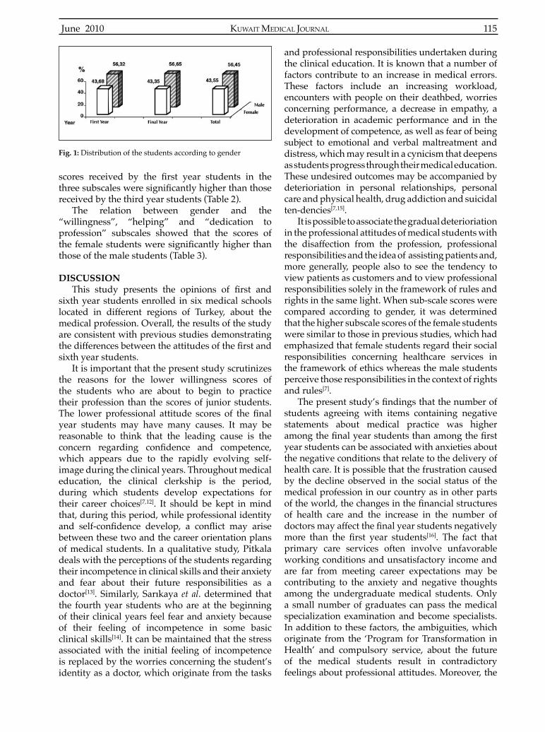
KUWAIT MEDICAL JOURNAL 115June 2010
scores received by the first year students in thethree subscales were significantly higher than thosereceived by the third year students (Table 2).
The relation between gender and the “willingness”, “helping” and “dedication to profession” subscales showed that the scores of the female students were significantly higher thanthose of the male students (Table 3).
DISCUSSION This study presents the opinions of first and
sixth year students enrolled in six medical schools located in different regions of Turkey, about the medical profession. Overall, the results of the study are consistent with previous studies demonstrating the differences between the attitudes of the first andsixth year students.
It is important that the present study scrutinizes the reasons for the lower willingness scores of the students who are about to begin to practice their profession than the scores of junior students. The lower professional attitude scores of the finalyear students may have many causes. It may be reasonable to think that the leading cause is the concern regarding confidence and competence,which appears due to the rapidly evolving self-image during the clinical years. Throughout medical education, the clinical clerkship is the period, during which students develop expectations for their career choices[7,12]. It should be kept in mind that, during this period, while professional identity and self-confidence develop, a conflict may arisebetween these two and the career orientation plans of medical students. In a qualitative study, Pitkala deals with the perceptions of the students regarding their incompetence in clinical skills and their anxiety and fear about their future responsibilities as a doctor[13]. Similarly, Sarıkaya et al. determined that the fourth year students who are at the beginning of their clinical years feel fear and anxiety because of their feeling of incompetence in some basic clinical skills[14]. It can be maintained that the stress associated with the initial feeling of incompetence is replaced by the worries concerning the student’s identity as a doctor, which originate from the tasks
and professional responsibilities undertaken during the clinical education. It is known that a number of factors contribute to an increase in medical errors. These factors include an increasing workload, encounters with people on their deathbed, worries concerning performance, a decrease in empathy, a deterioration in academic performance and in the development of competence, as well as fear of being subject to emotional and verbal maltreatment and distress, which may result in a cynicism that deepens as students progress through their medical education. These undesired outcomes may be accompanied by deterioriation in personal relationships, personal care and physical health, drug addiction and suicidal ten-dencies[7,15].
It is possible to associate the gradual deterioriation in the professional attitudes of medical students with the disaffection from the profession, professional responsibilities and the idea of assisting patients and, more generally, people also to see the tendency to view patients as customers and to view professional responsibilities solely in the framework of rules and rights in the same light. When sub-scale scores were compared according to gender, it was determined that the higher subscale scores of the female students were similar to those in previous studies, which had emphasized that female students regard their social responsibilities concerning healthcare services in the framework of ethics whereas the male students perceive those responsibilities in the context of rights and rules[7].
The present study’s findings that the number ofstudents agreeing with items containing negative statements about medical practice was higher among the final year students than among the firstyear students can be associated with anxieties about the negative conditions that relate to the delivery of health care. It is possible that the frustration caused by the decline observed in the social status of the medical profession in our country as in other parts of the world, the changes in the financial structuresof health care and the increase in the number of doctors may affect the final year students negativelymore than the first year students[16]. The fact that primary care services often involve unfavorable working conditions and unsatisfactory income and are far from meeting career expectations may be contributing to the anxiety and negative thoughts among the undergraduate medical students. Only a small number of graduates can pass the medical specialization examination and become specialists. In addition to these factors, the ambiguities, which originate from the ‘Program for Transformation in Health’ and compulsory service, about the future of the medical students result in contradictory feelings about professional attitudes. Moreover, the
Fig. 1: Distribution of the students according to gender

June 2010116
overloaded curriculum, too many examinations, stressful working conditions in the departments, long working hours and conflicts that arise duringmedical duties may lead to a feeling of burnout. These factors may explain the finding that thewillingness subscale scores of the final year studentsare lower than those of the first year students. Asa result of the uncertainty that follows a long and difficult education, problems about employmentand financial difficulties there is an erosion ofoccupational idealism. The confrontation with these problems which become more frequent with time have negative effects on professional attitudes[17].
In order to prevent the erosion of professional attitudes, it becomes more important to integrate the characteristics of the professionalism education into the overall educational program and educational practices. It has been stated that doctors maintaining a caring, altruistic, compassionate and empathic professional attitude and are aware of their own competencies can be educated by providing them with a sufficient amount of time, clinical experienceand guidance for the development of professional values. Thus, emphasis can be laid on the notion that these qualities constitute a part of medical education and medical culture[18]. The qualities, values, attitudes and behavior, which constitute the basis of healthcare service and medical practice, should develop step by step during the years of preclinical education. In this respect a number of factors are particularly important in the professional development of students. They include the career orientation of the students, the hidden curriculum aimed at developing professional values and attitudes, good role models provided by instructors, tolerance to cultural differences in medical practice and teamwork in primary and secondary healthcare services[19-21]. Each medical education program should aim at ensuring that future doctors consider these factors important for and relevant to their profession and should demonstrate these qualities significantly in practice making sure that thestudents are eager to maintain the development of their professional identities.
CONCLUSION The present study supports previous studies
demonstrating that idealism declines while cynicism increases over the course of medical education. The image of the medical profession which provides caring / healing is overshadowed by medical technical knowledege and skills. Professional attitudes in medical education cannot be viewed independently of regional and cultural characteristics, the continuing education programs and the government healthcare policies that affect
the practice of the profession. The objectives of professional and ethical values in medical education should be determined by taking into consideration the effects of healthcare practices and many factors related to the profession of medicine and should be closely observed and evaluated during the education period.
ACKNOWLEDGEMENTSWe would like to thank to Prof. R W Guillery
from Marmara University and lecturer Hulya Bati from Department of Foreign Languages, University of Ege for their contribution in editing the text.
REFERENCES
1. Misch DA. Evaluating physicians’ professionalism and humanism: the case for humanism “connoisseurs”. Acad Med 2002; 77:489-495.
2. General Medical Council. Tomorrow’s doctors: recommendations on undergraduate medical education. London, GMC, 2002.
3. ABIM Foundation, American Board of Internal Medicine; ACP- ASIM Foundation. American College of Physicians- American Society of Internal Medicine; European Federation of Internal Medicine. Medical professionalism in the new millennium: a physician charter. Ann Intern Med 2002; 136:243-246.
4. Arnold L. Assessing professional behaviour: yesterday, today and tomorrow. Acad Med 2002; 77:502-515.
5. Howe A. Professional development in undergraduate medical curricula- the key to the door of a new culture? Med Educ 2002; 36:353-359.
6. Griffith CH, Wilson JF. The loss of student idealismin the 3rd-year clinical clerkships. Evaluation and the Health Professions 2001; 24:61-71.
7. Crandall SJ, Volk RJ, Cacy D. A longitudinal investigation of medical student attitudes towards the medically indigent. Teaching and Learning in Medicine 1997; 9:254-260.
8. Kopelman L. Cynicism among medical students. JAMA 1983; 250:2006-2010.
9. Kasapoglu A. Medikal sosyoloji konu alanları. Cumhuriyet Üniversitesi Sosyal Bilimler Dergisi 1984; 3:139-147. (Article in Turkish).
10. Rogers JC, Coutts L. Do Students’ attitudes during preclinical years predict their humanism as clerkship students? Acad Med 2000; 75:S74-S77.
11. Bati AH, Bümen NT. Hekimlik mesleğine yönelik tutum ölçeğinin geliştirilmesi, Developing a Scale for Attitudes towards a Medical Profession (Tıp Eğitimi Dünyası Dergisi), 2006; 23:41-50. (Article in Turkish)
12. Kihara M, Matsuo O, Kamisako T, et al. Effect of clinical clerkship on students’ attitudes towards medical learning in Japan: a case study at Kinki University School of Medicine. Pathophysiology 2003; 9:111-113.
13. Pitkala KH, Mantyranta T. Professional socialization
How do Medical Students Perceive Professional Attitudes? A Multi-Center Study

KUWAIT MEDICAL JOURNAL 117June 2010
revised: medical students’ own conceptions related to adoption of the future physician’s role-a qualitative study. Med Teach 2003; 25:155-160.
14. Sarikaya O, Civaner M, Kalaca S. The anxieties of medical students related to clinical training. Int J Clin Pract 2006; 60:1414-1418.
15. Dyrbye LN, Thomas MR, Shanafelt TD. Medical student distress: causes, consequences, and proposed solutions. Mayo Clin Proc 2005; 80:1613-1622.
16. Bligh J. Is it time for a community based- medical school in the UK? Med Edu 1999; 33:315.
17. Woloschuk W, Harasym PH, Temple W. Attitude change during medical school: a cohort study. Med Educ 2004; 38:522-534.
18. Shapiro J, Rucker L, Robitshek D. Teaching the art of doctoring: an innovative medical student elective. Med Teach 2006; 28:30-35.
19. Stephenson A, Higgs R, Sugarman J. Teaching professional development in medical schools. Lancet 2001; 357:867-870.
20. Coulehan J. Viewpoint: Today’s professionalism: Engaging the mind but not the heart. Academic Medicine 2005; 80:892-898.
21. Batenburg V. Medical students’ attitudes: Attitude development in a medical school [S.l:s.n.], Tekst.-Proefschrift Universiteit Utrecht., 1997. (Accessed December 6, 2007, at http://igiturarchive.library.uu.nl/dissertations/01817837/inhoud.htm.)

KUWAIT MEDICAL JOURNAL June 2010118
Original Article
Treatment of Supracondylar Fracture of the Humerus in Children by Crossed Pinning
Kuwait Medical Journal 2010; 42 (2): 118-123
Firas Ahmad Suleiman, Mahmoud Mosa OdatDepartment of Orthopedics, Prince Rashid Bin Al-Hassan Hospital, Royal Medical Services, Irbid, Jordan
ABSTRACT
Address correspondence to: Firas Ahmad Suleiman, MD, Orthopedic Surgeon, Department of Orthopedic Surgery, Pediatric Orthopedic Division, Farah Royal Rehabilitation Center and Prince Rashid Bin Al-Hassan Military Hospital, Royal Medical Services, P O Box 620007, Irbid 2116, Jordan. Tel: 00962-0777785847/00962-02-7260277 Fax: 00962-2-7241641, E-mail: [email protected], [email protected]
INTRODUCTIONFractures around the elbow represent approximately
10% of all pediatric orthopedic injuries, and supracondylar fractures (SCF) of the humerus represents 75% of these fractures[1]. These fractures are frequently associated with complications, including nerve injury, arterial injury, skin slough, Volkmann’s ischemic contracture and changes in carrying angle. Several methods of treatment have been evolved in an attempt to decrease the frequency of these complications[2]. The most frequent methods of treatment are closed reduction and application of cast, traction (skeletal or skin), closed reduction and percutaneous Kirschner-wire fixation,and open reduction with internal fixation[3].
Gartland in 1959[4] classified SCF into threemain types depending on displacement. Type I or undisplaced fracture, is basically treated with collar and cuff immobilization. Type II or displaced fracture with intact posterior cortex, may require closed
Objective: To assess the result of closed reduction and percutaneous cross pinning in the management of displaced extension type supracondylar fracture of the humerus in childrenDesign: Retrospective study Setting: Prince Rashid Bin Al-Hassan Military hospital, Irbid, JordanSubjects: Seventy-eight cases of extension-type supracondylar humeral fracture (Gartland III) treated at the Prince Rashid Bin Al-Hassan Military Hospital between January 2000 and August 2006Interventions: All cases were treated by closed reduction and cross pinning under general anesthesia using an image intensifier. The children were usually discharged within24 hours after surgery. The wires were removed after 3 - 4 weeks in the office without anesthesia. Main Outcome Measures: Patients were followed up
postoperatively for an average period of 42 months (range from 15 - 72 months). They were observed for deformity of the elbow and range of motion and any neurological deficits.Results: All cases were reviewed by clinical and radiological examination. We found that according to Flynn criteria, 70 patients had excellent to good result, six patients had fair result, and two patients had poor result.Conclusion: Displaced extension-type supracondylar fracture in children can be successfully managed by emergency surgical intervention (closed reduction and percutaneous crossed pinning). We believe that the most important factor for a good outcome after the treatment of a supracondylar type III humeral fracture is adequate reduction and stable fixation. Transient neurological problems arecommon with these fractures and their incidence should not be underestimated.
KEY WORDS: closed reduction, cross pinning, elbow fracture in children, supracondylar humeral fracture
reduction. Type III or completely displaced fracture is either treated by closed or open reduction and internal fixation. Management of the displaced fracture isfraught with problems, including neurovascular compromise, difficulty in obtaining or maintainingreduction, and poor late results because of stiffness of the elbow or cubitus varus.
The preferred treatment of displaced SCF of the humerus in children is closed reduction and percutaneous pinning. Cross-wiring techniques are biomechanically superior to parallel lateral wiring techniques[1,5].
SUBJECTS AND METHODSA retrospective study was undertaken to evaluate
the results of treatment of a displaced extension-type SCF of the humerus in children who were admitted to the Prince Rashid Hospital between January 2000 and August 2006. During this period, 105 patients

KUWAIT MEDICAL JOURNAL 119June 2010
were admitted; two of them had flexion type SCF, fourhad open fractures, and one had type two Gartland fracture. They were excluded from the study. The records of 98 patients were reviewed and the parents contacted to bring the child for clinical and radiological examinations. However, only 78 children reported for follow-up and were included in the study.
Out of 78 patients, 55 were male and 23 female with a mean age of nine years (range 2.5 to 11 years). All had history of falling down while riding a bicycle except one who had a fall from height. The left elbow was injured in 61 patients. All patients had Gartland type III extension SCF of the humerus. All were closed injuries. Two patients had signs of ulnar nerve injury, two had signs of anterior interosseous nerve injury, one had wrist drop, one had signs of brachial artery injury, two patients had distal radius fractures and one had proximal humerus fracture.
Patients were splinted in the emergency department after the diagnosis had been made, and thorough neurovascular examination was performed. All patients were taken urgently (within 12 hours of their
injury) to the operating room for surgical management. After the induction of the general anesthesia, draping and surgical toweling was done and gentle traction applied to the upper extremity with counter traction of the upper arm. Using an image intensifier, translationin the medio-lateral plane was corrected and, proximal and distal fragments were aligned. The elbow was then hyperflexed with gentle pressure applied over theolecranon to reduce the fracture. The fracture was then examined under the image intensifier to confirm theadequacy of the reduction. Displacement, rotation and angulations in both coronal and sagittal planes must be corrected at the time of reduction. If the reduction was judged to be acceptable (i.e., no rotational displacement, and no medio-lateral translation but upto 10 degrees of angulation in the sagittal plane), a K-wire was placed laterally and angled 30° to the shaft of the humerus, driven across the fracture site and impacted in the cortex of the distal humeral metaphysis proximal to the fracture site. The surgeon selected the wire size to be used. A second K-wire was then placed beginning medially and angled 45° to the humeral shaft. The elbow was brought into a position of 60 - 90° flexion and palpation was carried out on themedial side of the elbow.
After the placement of the K-wires, the fracture position was confirmed using an image intensifier,and the wires were cut off outside the skin without bending. Dressing and crepe bandage were then applied without a cast. An arm sling was then applied with the elbow in 80 to 90° of flexion, leavingadequate room for swelling in the antecubital fossa. The patient was discharged when post-operative clinical examination showed no signs of compartment syndrome with normal distal pulses, and post-operative antero-posterior and lateral X-ray showed acceptable reduction. Three to four weeks later, the patient was seen in the out patient clinic. An X-ray was done to see the signs of healing, and the K-wires were removed in the office without any anesthesia. Activemotion exercises were encouraged.
The patients with preoperative nerve injury underwent the same procedures of closed reduction and crossed pinning, with the same postoperative protocol except for the physiotherapy protocol. Those with pre and postoperative nerve injury underwent more active and passive physiotherapy and nerve stimulation after nerve conduction study and EMG showed continuous nerve fibers with incomplete nerveinjury. The patients with distal radius or proximal humerus fractures were splinted after pinning for three weeks. All cases were operated by the author himself or by a senior resident under the author’s supervision. The boy with clinical evidence of brachial artery injury who presented with pink and pulseless hand, underwent closed reduction with crossed K-
Fig. 1: Displaced extension type supracondylar fracture humerus

June 2010120
wire pinning, followed by exploration of the brachial artery at another central hospital where a vascular surgeon was available. The boy underwent primary anastomosis of the transected brachial artery.
The final outcome was reviewed by measuringrange of motion, varus-valgus deformity and distal neurovascular injury. Fig. 1 and 2 show a displaced type III supracondylar humeral fracture and the postoperative X-ray after three weeks. All patients were then graded according to Flynn criteria (Table 1), and were administered a satisfaction survey questionnaire[6].
The study was approved by the hospital ethical committee.
RESULTSA closed manipulation was successfully performed
in all patients. There were no intraoperative complications. All fractures were reduced to an acceptable position. Hospitalization time averaged 1.5 days (range 1 - 2 days). Full or nearly full range of motion was gained within five months on average
(range 3 - 8 months). There were five patients withpostoperative ulnar nerve injury. Exploration of one case of postoperative ulnar nerve injury showed that the nerve was continuous, away from the medial K-wire, but contused from the manipulation during closed reduction. The rest of the patients with nerve injury underwent no corrective action till the removal of the K-wire. Cases where the nerve conduction study showed partial nerve injury were given physiotherapy with nerve stimulation until recovery which took between 4 - 7 months. All cases of nerve injury recovered. Two cases developed cubitus varus. One of them had comminuted fracture of the medial supracondylar region, whereas the second patient fell down on his elbow within the first three weeks of thepost-operative period before healing was complete. When he presented to us at the time of follow-up, the fracture was united in a deformed position. There was postoperative pin tract infection in two patients at the end of the three weeks; they required oral antibiotics and pin removal. One patient developed cellulitis in the distal arm, which required hospitalization for five
Fig. 2a, b: Three weeks after closed reduction and crossed pinning
Fig. 2a Fig. 2b
Treatment of Supracondylar Fracture of the Humerus in Children by Crossed Pinning

KUWAIT MEDICAL JOURNAL 121June 2010
days for intravenous antibiotics only. According to the Flynn criteria, there were 61 excellent, nine good, six fair and two poor results. Over all, 76 (97%) cases had a satisfactory outcome and two elbows (3%) had an unsatisfactory outcome (Table 2). No patient in this series developed Volkmann’s ischemic contracture.
There was no repeat surgery performed on any case. There was no case of failed close reduction that required open reduction. All cases were reduced by the closed method during surgery and fixed withcrossed pinning. No case had significant limitationof range of motion. All cases regained an almost full range of motion by a maximum of eight months post injury. We did not find any significant difference in therange of motion after the first eight months followingsurgery. Total limitation of range of motion was lower in our series than in other published studies[7]. This may be due to the fact that no case needed revision or open reduction and to the availability of a senior surgeon during the first attempt of closed reductionin all cases.
DISCUSSIONTreatment of displaced supracondylar humeral
fracture should result in perfect anatomic alignment and full functional elbow with no cosmetic defects[8]. There is a general agreement among orthopedic surgeons that displaced supracondylar humeral fractures in children are best treated by closed reduction and percutaneous fixation by K-wires[7]. The successful treatment of a totally displaced supracondylar humeral fracture depends on the perfect achievement of three stages of management; First, a good technique of manipulation to obtain a perfect reduction of the fracture, second, correct assessment of the quality of reduction of the fracture and the third stage, which involves maintaining the reduction until the fracture is healed enough to be intrinsically stable[9]. In rare SCF in children, where multidirectional instability results in displacement into flexion and / or extension, the keyfactors in the surgical technique are pre-placement of Kirschner wires into the distal fracture fragment and rotation of the fluoroscopic arm, not the child’s arm,for lateral imaging[10].
These fractures are usually stabilized with two K-wires inserted in a parallel or divergent
orientation through the lateral epicondyle or in a crossed configuration with insertion points throughthe medial and lateral epicondyles[11]. Fracture healing and clinical results with either of these two techniques have generally been comparable[11-
13], though some recommend lateral pinning for type 2 fractures and cross pins for type 3 fractures when increased stability is required. Divergent pins had similar stability compared with crossed pins in extension, varus and valgus testing. In axial rotation testing crossed pins were more stable[11]. In a systemic review of the literature, Brauer[5] indicates that medial / lateral pinning of pediatric SCF remains the most stable configuration and thatcare needs to be taken regardless of the technique to avoid iatrogenic ulnar nerve injury and loss of reduction. Unfortunately, one major complication associated with the use of the medial pin is iatrogenic ulnar nerve injury; temporary nerve palsies and even permanent damage have been reported in the literature[14-16,5]. The ulnar nerve and the medial epicondyle location is sometimes difficult to palpateand identify because of swelling associated with a displaced fracture. Thus, during insertion of the pin, the nerve may be inadvertently entrapped or impaled by the pin. The technique requires experience and carries risks such as neurovascular complications or incomplete success.
The most important argument from the supporters of lateral pinning is the possibility of iatrogenic ulnar nerve injury during the insertion of the medial K-wire. In this series five cases (6%)developed signs of ulnar nerve palsy, one case was explored within six hours of surgery, which showed that the nerve was away from the K-wire but was contused from the manipulation. Fortunately, all cases recovered fully within six months of the injury. In our opinion, it is not necessary to explore the ulnar nerve in every case of post-reduction ulnar nerve palsy. To prevent such an injury, it is recommended to insert the medial K-wire while the elbow is in 90 degrees flexion and to be moreanterior while inserting the K-wire. Use of a small skin incision to place the medial K-wire is the safer option[7]. Rasool blamed incorrect pin placement on surgical inexperience[14]. Skaggs et al[17] noted that
Result Rating N (78) Percentage
Satisfactory
Unsatisfactory
ExcellentGoodFair
Poor
6196
2
7812
7
3
Result RatingFunctional factor:Loss of motion (flexion& extension) (°)
Cosmetic factor: Loss of carrying angle (°)
Satisfactory
Unsatisfactory
ExcellentGoodFair
Poor
0 - 5 5 - 10 10 - 15
> 15
0 - 5 5 - 10 10 - 15
> 15
Table 2: Results in 78 elbows according to the Flynn criteriaTable 1: Flynn criteria for grading of supracondylar fractures

June 2010122
the position of the elbow during medial K-wire insertion was a significant determinant of the riskof ulnar nerve injury. Karapinar et al[18] noted that in spite of decreased elbow flexion, iatrogenic ulnarnerve injury may occur. Wind et al[19] suggested intra-operative nerve stimulation to assist in localization of the nerve prior to placement of the medial pin. Stimulation of the pin itself following placement was an alternative recommended option. Green et al[20] confirms that crossed pin fixation using a mini-open approach is an appropriate and safe treatment option in the management of displaced supracondylar humerus fractures, and this does not add additional morbidity to the procedure. The reported rate of primary nerve injury in displaced SCF is up to 20%[6,21]. The incidence of reported iatrogenic ulnar nerve injury ranges from 2 to 8%[15,22]. Lyons et al[16] reviewed their own experience and that of the published literature on the issue of management of iatrogenic ulnar nerve injuries. They concluded that majority of injuries were related to the medial K-wire placement in cross pinning, that observation was the most appropriate course of action, and that immediate exploration was not warranted. If the clinician suspects skewering of the nerve in the notch, then removal followed by re-siting in a more anterior position is indicated.
The incidence of impaired circulation of the forearm after reduction of the fracture is 0.3%. The indication for exploration is a cold hand with no palpable radial or ulnar pulses immediately after reduction. When there is a warm and pink hand but we cannot feel pulses, we frequently check the vascular status by routine physical examination and Doppler sonography before we decide to undertake more invasive procedures such as angiography and exploration. This policy is similar to that described by others[23]. Patients were followed till almost full range of motion was gained and the neurological defect had recovered (about 8 months). The cubitus varus is due to medial tilting of the distal fragment combined with rotation[24] and not to the physical injury[25]. It does not remodel with time and is not progressive[6,15]. The 90% good or excellent functional and cosmetic results are in concordance with reports in the literature[12,14]. Two patients developed a cubitus varus of more than 15° (2.5%). This result is in concordance with other studies[13].
CONCLUSIONOur study confirms that crossed pin fixation
is an appropriate and safe treatment option in the management of displaced supracondylar humerus fractures. We believe that the most important factor for a good outcome after the treatment of a supracondylar type 3 humeral fracture is adequate
reduction and stable fixation. Transient neurologicalproblems are common with these fractures and their incidence should not be underestimated. The availability of senior pediatric orthopedic surgeon at the time of surgery is very important to minimize the acute and long term complications.
REFERENCES
1. Shanon FJ, Mohan P, Chacko J and D’Souza LG. “Dorgan’s” percutaneous lateral cross-wiring of supracondylar fractures of the humerus in children. J Pediatr Orthop 2004; 24:376-379.
2. Rose RE, Phillips W. Iatrogenic ulnar nerve neuropathies post-pinning of displaced supracondylar humerus fractures in children. West Indian Med J 2002; 51:17-20.
3. Tachdjian MO. Pediatric Orthopedics. Philadelphia, WB Saunders, 1972.
4. Gartland JJ. Management of supracondylar fractures of the humerus in children. Surg Gynecol Obstet 1959; 109:145-154.
5. Brauer CA, Lee BM, Bae DS, Waters PM, Kocher MS. A systematic review of medial and lateral entry pinning versus lateral entry pinning for supracondylar fractures of the humerus. J Pediatr Orthop 2007; 27:181-186.
6. Flynn JC, Matthews JG, Benoit RL. Blind pinning of displaced supracondylar fractures of the humerus in children. Sixteen years’ experience with long term follow-up. J Bone Joint Surg Am 1974; 56:263-272.
7. Solak S, Aydin E. Comparison of two percutaneous pinning methods for the treatment of the pediatric type III supracondylar humerus fractures. J Pediatr Orthop B 2003; 12:346-349.
8. Ay S, Akinci M, Kamiloglu S, Ercetin O. Open reduction of displaced pediatric supracondylar humeral fractures through the anterior cubital approach. J Pediatr Orthop 2005; 25:149-153.
9. Kaewpornsawan K. Comparison between closed reduction with percutaneous pinning and open reduction with pinning in children with closed totally displaced supracondylar humeral fractures: a randomized controlled trial. J Pediatr Orthop B 2001; 10:131-137.
10. Leitch KK, Kay RM, Femino JD, Tolo VT, Storer SK, Skaggs DL. Treatment of multidirectionally unstable supracondylar humeral fractures in children. A modified Gartland type-IV fracture. J Bone Joint SurgAm 2006; 88:980-985.
11. Lee SS, Mahar AT, Miesen D, Newton PO. Displaced pediatric humerus fractures: biomechanical analysis of percutaneous pinning techniques. J Pediatr Orthop 2002; 22:440-443.
12. Cheng JC, Lam TP, Shen WY. Closed reduction and percutaneous pinning for type III displaced supracondylar fracture of the humerus in children. J Othop Trauma 1995; 9:511-515.
13. Pirone AM, Graham HK, Krajbich JI. Management of displaced extension-type supracondylar fracture of the humerus in children. J Bone Joint Surg Am 1988; 70:641-650.
Treatment of Supracondylar Fracture of the Humerus in Children by Crossed Pinning

KUWAIT MEDICAL JOURNAL 123June 2010
14. Rasool MN. Ulnar nerve injury after K-wire fixationof supracondylar humerus fracture in children. J Pediatr Orthop 1998; 18:686-690.
15. Brown IC, Zinar DM. Traumatic and iatrogenic neurological complications after supracondylar humerus fractures in children. J Pediatr Orthop 1995; 15:440-443.
16. Lyons JP, Ashley E, Hoffer MM. Ulnar nerve palsies after percutaneous cross-pinning of supracondylar fractures in children’s elbows. J Pediatr Orthop 1998; 18:43-45.
17. Skaggs DL, Hale JM, Bassett J, Kaminsky C, Kay RM, Tolo VT. Operative treatment of supracondylar fractures of the humerus in children. The consequences of pin placement. J Bone Joint Surg Am 2001; 83:735-740.
18. Karapinar L, Oztürk H, Altay T, Köse B. Closed reduction and percutaneous pinning with three Kirschner wires in children with type III displaced supracondylar fractures of the humerus. Acta Orthop Traumatol Turc 2005; 39:23-29.
19. Wind WM, Schwend RM, Armstrong DG. Predicting ulnar nerve location in pinning of supracondylar humerus fractures. J Pediatr Orthop 2002; 22:444-447.
20. Green DW, Widmann RF, Frank JS, Gardner MJ. Low incidence of ulnar nerve injury with crossed pin placement for pediatric supracondylar humerus fractures using a mini-open technique. J Orthop Trauma 2005; 19:158-163.
21. Gosens T, Bongers KJ. Neurovascular complications and functional outcome in displaced supracondylar fractures of the humerus in children. Injury 2003; 34:267-273.
22. Campbell CC, Waters PM, Emans JB, Kasser JR, Millis MB. Neurovascular injury and displacement in type III supracondylar humerus fractures. J Pediatr Orthop 1995; 15:47-52.
23. Sabharwal S, Tredwell SJ, Beauchamp RD, et al. Management of pulseless pink hand in pediatric supracondylar fractures of humerus. J Pediatr Orthop 1997; 17:303-310.
24. Smith L. Deformity following supracondylar fractures of the humerus in children. J Bone Joint surg Am 1960; 42:235-252.
25. France J, Strong M. Deformity and function in supracondylar fractures of the humerus in children variously treated by closed reduction and splinting, traction, and percutaneous pinning. J Pediatr Orthop 1992; 12:494-498.

KUWAIT MEDICAL JOURNAL June 2010124
INTRODUCTIONAcute kidney injury (AKI) is a clinical syndrome
characterized by rapid decrease in glomerular filtration rate and retention of nitrogenous wasteproducts[1]. Pregnancy related acute kidney injury (PR- AKI) severe enough to warrant dialysis can cause substantial feto-maternal morbidity and mortality. Five decades ago in 1958 the estimated incidence of AKI in pregnancy was as high as one in 1400[2]. Today in industrialized countries, the approximate incidence is one in 20,000[2]. In Eastern India the incidence of PR- AKI fell from 15% in 1982 -1991 to 10% in 1992 - 2002[3]. Reports from Southern and Western India indicate incidence of PR-AKI to be 4.24% and 9.06% respectively[4,5]. The recent improvement in PR-AKI has been attributed to improved antenatal care, earlier accessibility to physicians and management of obstetric complications in due time. In India sources from
Original Article
Dialysis Requiring Pregnancy Related Acute Kidney Injury - A Single Center Experience
Kuwait Medical Journal 2010; 42 (2): 124-128
Mandal Debasmita1, Munjappa Bipin2, Sharma Partha Pratim1, Roy Choudhary Arpita2, Biswas Subhash Chandra1 1Department of Obstetrics and Gynecology, Institute of Postgraduate Medical Education and Research, Kolkata, India
2Department of Nephrology, Institute of Postgraduate Medical Education and Research, Kolkata, India
ABSTRACT
Address correspondence to: Dr. Debasmita Mandal, Qr / No - C/11, SSKM Hospital Campus, 242, A J C Bose Road, Kolkata -700 020, West Bengal, India. Tel: 9831522415, 03322232615, E-mail: [email protected], [email protected]
KEY WORDS: postpartum period, pregnancy, sepsis
Eastern and Southern region had reported incidence of PR-AKI requiring dialysis to be 11.6% and 53.66% respectively[3,4 ]. PR-AKI requiring dialysis continues to be a significant problem in developingcountries because of several factors such as; poor health care facilities in peripheral centers, delayed referral of morbid patients, pre-existing anemia and malnutrition complicating the course of AKI[6,7] .
We conducted a prospective longitudinal study to define the current etiology of PR-AKI and to analyzethe maternal and fetal mortality and morbidity associated with this complication of pregnancy.
SUBJECTS AND METHODSInstitute of Postgraduate Medical Education
and Research (IPGME&R), Kolkata is one of the big referral centers of Eastern India with 1650 beds and each year more than 2000 deliveries are conducted in the Obstetric unit.
Objective: To highlight the etiology and spectrum of pregnancy related acute kidney injury (PR - AKI) requiring dialysis and to analyze the maternal and fetal outcomeDesign: Longitudinal prospective studySetting: Tertiary care hospitalSubjects: Fifty consecutive pregnant women with acute kidney injury (PR-AKI) from June 2007 were considered possible subjects. Out of these, 38 requiring dialysis were included in the study. Patients with diabetes, kidney disease, hypertension, septic abortion and cases with AKI unrelated to the pregnancy were excluded.Interventions: All patients were evaluated by relevant investigations. Cases with non-recovery and partial recovery of renal function were subjected to renal biopsy. All were managed as per the institute policy with standard dialysis and obstetric care indications. Main Outcome Measure(s): Maternal mortality and
morbidity, renal outcome and fetal outcomeResults: PR-AKI requiring dialysis constituted 11.24% (38 / 338) of total AKI requiring dialysis patients. PR-AKI developed in 25 patients (65.78%) during postpartum period, nine (23.68%) in third trimester and two each in the first and second trimester respectively. Etiologies werepostpartum sepsis, postpartum hemorrhage and toxemia of pregnancy in 12 (31.51%), 10 (26.31%) and 9 (23.68%) patients respectively. Majority (61.5%) who recovered completely required up to five sessions of dialysis (p <0.001). Subjects with surgical intervention had a delayed renal recovery. Eleven (28.94%) maternal deaths were observed and the most common cause was postpartum sepsis. Unfavorable fetal outcome was noted in 11 patients (32.35%). Conclusion: PR- AKI is associated with high maternal mortality and poor fetal outcome.

KUWAIT MEDICAL JOURNAL 125June 2010
Fifty consecutive subjects with PR- AKI admitted in the Nephrology and Obstetric unit from June 2007 were considered for the study. Those with history of hypertension, diabetes and kidney disease prior to pregnancy, those who did not require dialysis, subjects with septic abortion (considered as exogenous cause of AKI in pregnancy) and causes of kidney injury unrelated to course of pregnancy were excluded from the study. Out of these 50 patients, 12 were excluded (six did not require dialysis, four had septic abortion, one had pre existing hypertension and in one patient the cause of AKI was unrelated to pregnancy) . A total of 38 patients aged between 20 - 36 years with PR-AKI requiring dialysis were included in the study. AKI was defined by documenting oliguria (urine output < 400ml per day) and elevated serum creatinine (> 0.8 mg/dl)[8]. Clinical history was obtained from all subjects. They were investigated with hemogram, peripheral blood smear, liver function test, renal function test, coagulation profile, serum electrolyte, renal sonogram,relevant viral markers (HbsAg, Anti HCV, HIV 1 and HIV 2) for hemodialysis. Those patients with non-recovery or partial recovery of renal function by two weeks were subjected to the renal biopsy. Hemodialysis was given to all patients. Dialysis was initiated as per standard recommendations during pregnancy[9]. Patients were dialyzed through either femoral or internal jugular hemodialysis catheter. Fresenius F6 hemodialyzer was used for all patients. Precautions were taken to avoid intradialytic hypotension. Ultrafiltration volume was determined as per clinicalassessment and weight gain. Obstetrical events were defined by standards described in William’s Obstetrics,22nd edition[10]. Maternal outcomes were evaluated in terms of mortality and morbidity. Maternal morbidity was defined as morbidity originating from any causerelated to pregnancy or its management any time during the antepartum, intrapartum and postpartum period, usually up to 42 days after confinement. Fetaloutcomes were analyzed in terms of birth weight, birth asphyxia, still births and neonatal deaths. In this study low birth weight infant has been definedas any infant with birth weight of less than 2.5 kg irrespective of gestational age. All therapies were individualized and then subjects managed with antibiotics, antihypertensives and other medications along with dialysis as and when required. All cases were followed up for six months. Statistical analysis was done by Fisher’s exact test. A p-value < 0.05 was considered statistically significant.
The study was approved by the local ethical committee.
RESULTSThe demographic and clinical characteristics of our
study population are described in the Table 1. Three
hundred and thirty eight patients with AKI requiring dialysis were observed during the study period out of which 38 (11.24%) had PR-AKI requiring dialysis. Mean age of study group was 27.96 years (range: 20 - 36 years). Range of gestation during delivery was between eight and 40 weeks. PR- AKI was observed in 18 primigravidae (47. 36%) and 20 multigravidae (52.63%). Mode of delivery was both abdominal (39.47%) and vaginal (50%) as per obstetric indications. The majority of patients (18 / 38, 47.36%) had home delivery. Majority had onset of AKI in the postpartum period (65.78%). Nine cases had onset of AKI in the third trimester (n = 9, 23.68%). Two (5.1%) cases each had onset of AKI in first and second trimesterdue to ectopic pregnancy and spontaneous abortion respectively. Oliguric renal failure was noted in 26 (68.42%) patients.
Anemia (< 11 gm%), abnormal liver function tests (liver enzymes more than twice the upper limit of normal value), deranged bleeding parameters (INR > 1.5) and thrombocytopenia (total platelet count < 1.5 lakh / cumm) were seen in 34 (89.47%), 17 (44.73%), eight (21.05%) and four (10.52%) cases respectively (Table 2).
Number of sessions of dialysis among all patients is shown in the Table 3. Most patients (60.52%) required less than five sessions of dialysis. One patient required22 sessions of dialysis during her hospital stay. She was dialysis dependant and continued to do so in her follow up. Duration of dialysis indicated the time needed for recovery of renal function in the present study (p < 0.001).
Sepsis (31.57%), postpartum hemorrhage (PPH) (26.31%) and toxemia of pregnancy (23.68%) were
Table 1: Demographic characteristics of PR-AKI patients
Demographic Characteristics
Age in years 20 - 29 30 - 40 Gravidity Primigravidae MultigravidaeMode of termination Ruptured ectopic pregnancy Spontaneous abortion LSCS VDOnset of renal failure
Antepartum1st trimester 2nd trimester3rd trimester
PostpartumNature of renal failure Oliguria Nonoliguric
No of Patients(N = 38)
335
1820
221519
22925
2612
%
86.84 13.15
47.36 52.63
5.265.25
39.47 50.00
5.26 5.26
23.68 65.78
68.42 31.05
LSCS = Lower segment cesarean section, VD = Vaginal delivery

June 2010126
the commonly observed etiologies of PR-AKI in the present study (Table 4). Other causes were antepartum hemorrhage (APH) (5.26%), ruptured ectopic pregnancy (5.26%), acute fatty liver syndrome (2.63%), abruptio placentae (2.63%) and chorioamnionitis (2.63%) respectively.
Maternal outcome is described in the Table 5. Three women had persistent renal dysfunction but they were off dialysis, while one continued to be dialysis dependant in the follow up period. One patient each (2.63%) required bilateral internal iliac artery ligation and hysterectomy for PPH. Wound dehiscence was noticed in two cases (5.26%). Eleven (28.94%) maternal deaths were observed. The maternal mortality was due to puerperal sepsis (n = 5,), toxemia of pregnancy (n = 3), PPH with sepsis (n = 2) and acute fatty liver syndrome (n = 1 ).
Cases with non-recovery or partial recovery of renal function (n =10) were subjected to renal biopsy (one patient denied biopsy). A renal biopsy (Table 6) was done in nine patients. Acute tubular necrosis (ATN) was seen in three, cortical necrosis and acute interstitial nephritis in two patients each. ATN was
the main feature in toxemia of pregnancy. Cortical necrosis was observed in PPH and sepsis cases. One case each with abruptio placentae and sepsis showed presence of interstitial nephritis. During follow up six had complete recovery of renal function, three had persistent renal dysfunction not requiring dialysis support and one patient approached end stage renal disease.
Out of a total of 34 pregnancies (excluding two abortions and two ectopic pregnancies) nine (26.47%) had still birth, two (5.88%) had neonatal death and 25 (73.52%) had live births. Twenty three women (58.82%) were observed to have favorable perinatal outcome. Among 25 live births seven (28.0%) were low birth weight (LBW) and two (8.0%) had birth asphyxia but in follow up babies were seen to be in good condition.
DISCUSSIONPR-AKI is a critical condition which has grave
prognosis for both the mother and the fetus. In the present study incidence of PR-AKI was 11.24% which was comparable to other studies from India[3]. The onset of AKI was noted mainly in the postpartum period (65.78%) and was equally distributed between primigravidae (47.36%) and multigravidae (52.63%). AKI in pregnancy follows bimodal peak[2]. A peak in early pregnancy is associated with mainly septic abortions and in late trimester is related to obstetrical complication including PPH, puerperal sepsis, toxemia of pregnancy and hemorrhage due to operative intervention. We excluded patients with septic abortion in our study. Hence, most of our patients had onset of AKI in the postpartum period. Anemia was observed in 89.47% in the current study. One study from South India reported a similar incidence (87.4%) of anemia in PR-AKI[4]. Possibly anemia predisposes these patients
Investigations
Anemia (Hb < 11gm%)Deranged liver function test (liver enzymes more than twice the upper limit of normal value)Deranged bleeding parameters (INR > 1.5)Thrombocytopenia (total platelet count < 1.5 lakh/cumm)
No. of Patients(N = 38)
34
168
4
%
89.47
42.1021.05
10.52
Table 2: Deranged laboratory findings in PR-AKI patients
Sessions of Dialysis
1 - 56 - 1011 - 1516 - 2021 - 25
No. of Patients (N = 38)
238331
%
60.5221.057.897.892.63
Table 3: Number of dialysis sessions done in PR-AKI
Causes
SepsisPPHToxemia of pregnancyAPHAcute fatty liver syndromeRuptured ectopic pregnancyChorioamnionitisAbruptio placentae
No. of Patients (N = 38)
1210 9 2 1 2 1 1
%
31.5726.3123.68 5.26 2.63 5.26 2.63 2.63
Table 4: Causes of PR- AKI
Maternal Morbidity
Persistent renal dysfunctionEnd stage renal diseaseHysterectomyBilateral internal iliac artery ligationWound dehiscenceSpontaneous abortionsRuptured ectopic pregnancy
No of Patients N = 38 (%)
3 (9.89)1 (2.63)1 (2.63)1 (2.63)2 (5.26)2 (5.26)2 (5.26)
Maternal Mortality Etiologies
Puerperal sepsisToxemia of pregnancyPPH with sepsisAcute fatty liver syndrome
No of Patients N =11 (%)
5 (45.45)3 (27.27)2 (18.18)1 (9.09)
Table 5: Maternal outcome in PR-AKI patients
Dialysis Requiring Pregnancy Related Acute Kidney Injury - A Single Center Experience

KUWAIT MEDICAL JOURNAL 127June 2010
for AKI[6]. Anemia puts them at risk of hemodynamic compromise in case of blood loss and also increases the susceptibility towards ischemic ATN.
The common etiologies of PR-AKI in the present series were puerperal sepsis (31.57%), PPH (26.31%) and toxemia of pregnancy (23.07%). While a study by Kilari et al had shown sepsis as the cause of PR-AKI in 39.02%, followed by toxemia of pregnancy in 24.39% and hemorrhage of pregnancy in 17.08%[4], another study from India by Goplanil et al also observed sepsis (61.42%) to be the predominant cause for PR-AKI[5]. This study had included the septic abortion cases in their study population of PR-AKI and this may be the reason for such a high incidence of sepsis induced PR-AKI. Sepsis in pregnancy is commonly seen in developing countries. The reasons possibly are due to home deliveries, poor hygiene and failure of timely medical intervention and increased prevalence of anemia. It has been observed that presence of anemia predisposes to susceptibility for both AKI and sepsis[6,11]. In contrary to studies in India, a Western study concluded that pre-eclampsia and eclampsia were the most commonly associated causes of PR-AKI[12].
Complete recovery of renal function was seen in 23 (60.52%) patients and all of them required less (up to 5) sessions of dialysis. Patients requiring up to five sessions of dialysis had good recovery of renalfunction but more sessions of dialysis was associated with serious renal morbidities (p < 0.001).
Ten of our patients had incomplete or non-recovery of renal function at the end of second week. We subjected these patients to renal biopsy (one patient denied) and concluded varied histology findings. ATN was observed in three cases of toxemiaof pregnancy, cortical necrosis and interstitial nephritis were seen each in two cases of PPH with sepsis. Earlier studies had only demonstrated cortical necrosis in their histology results[5]. But out of these ten cases six had complete recovery of renal function in their subsequent follow up. Three had incomplete recovery of renal function leading to persistent renal dysfunction (9.89%) and one had complete non-recovery of renal function and developed end stage renal disease (2.63%). This observation was similar to the study by Kilari et al [4].
We observed a more complicated course of AKI among patients with operative interventions and these
women also needed more number of dialysis sessions. Fourteen out of 16 patients who required more than five sessions of dialysis had LSCS and subsequentlyhad various renal morbidities. One study reported a high incidence of sepsis induced PR-AKI following LSCS (19.5%) than spontaneous vaginal delivery (9.76%)[4].
Maternal mortality in the present study was 28.94%. Common causes of maternal death were sepsis (45.45%), toxemia of pregnancy (27.27%) and PPH with sepsis (18.18%). Other studies from India reported lesser (6.4%, 18.57%) maternal mortality[3,5]. Maternal mortality rate in the general population of West Bengal, the state where we had conducted the study was 14.8% from 2001-2003[13]. Higher maternal mortality in our study was probably due to the selection bias, as our institute being a referral center received more critical and morbid patients. From all these studies including the present analysis it is obvious that occurrence of PR-AKI increases the incidence of maternal mortality.
Favorable fetal outcome was observed in 67.64% (23/34) patients. We had nine still births (26.47%). Low birth weight and neonatal death were seen in 28.0% (7 / 25) and 8.0% (2 / 25) of live births respectively. Two (8.0%) babies also had birth asphyxia. In India low birth weight rate is 30% of live birth[13]. An earlier Western study analyzed 21 patients with PR-AKI and reported poor fetal outcome with 47.61% live births, 38% still birth, 19% low birth weight, and 14% abortions[12]. But this study had even included patients with various co-existing co-morbidities, which affected the fetal outcome. The favorable fetal outcome in our study may be due to the fact that majority of our patients had onset of renal failure in the post partum period, which was unlikely to have any bearing on the fetal outcome. Unfavorable fetal outcome were observed particularly in patients with raised serum creatinine (> 3 mg/dl), sepsis, abruption placentae and multiorgan failure.
Good antenatal care, immediate management of obstetrical hemorrhage, timely diagnosis and management of hypertensive disorders of pregnancy and most important, aseptic labor care can reduce the incidence as well as mortality and morbidity of AKI in
Table 6: Renal biopsy findings of PR- AKI patients
Biopsy Diagnosis
Acute tubular necrosisCortical necrosisAcute interstitial nephritisNon-specific changes
No. of Cases (N = 9)
3222
Table 7: Fetal outcome of PR-AKI patients
Fetal Outcome
Alive babies with favorable outcome Still bornNeonatal deathLow birth weightBirth Asphyxia
No. of Cases
23 9 2 7 2
%
58.8226.47 8.028.0 8.0
*Two spontaneous abortions and two ruptured ectopic pregnancies were present

June 2010128
pregnancy. CONCLUSION
PR- AKI requiring dialysis constituted 11.53% of total dialysis requiring AKI patients. It was more prevalent during post partum period. Sepsis was the most common causative factor for pregnancy related AKI. Anemia increased susceptibility of both AKI and sepsis in pregnancy. Maternal mortality and unfavorable fetal outcome was observed in 28.94% and 32.35% respectively. Occurrence of AKI in pregnancy increases the risk of maternal mortality and poor fetal outcome. Operative interventions were associated with maternal and renal morbidity. Renal histology finding other than cortical necrosis is also observedin PR-AKI. PR-AKI usually requires dialysis support. Subjects with complete recovery usually require less number of dialysis sessions. Community intervention to reduce the incidence of anemia in pregnant women, improvement in the obstetric care, active management of postnatal infections and PPH, avoidance of unnecessary operative interventions, early referral to a tertiary care institute with good nephrology and neonatal care backup and timely institution of renal replacement therapy play pivotal roles in achieving good feto-maternal outcome.
REFERENCES
1. Sagheb MM, Ghotbi SH, Moini M. Peripartum acute renal failure. Shiraz E-Medical Journal 2008; 9:45-54.
2. Hou S, Peano C. Acute renal failure in pregnancy. Saudi J Kidney Dis Transpl 1998; 9:261-266.
3. Prakash J, Kumar H, Sinha DK, et al. Acute renal failure in pregnancy in a developing country: twenty years of experience. Ren Fail 2006; 28:309-313.
4. Kilari SK, Chinta RK, Vishnubhotla SK. Pregnancy related acute renal failure. J Obstet Gynecol India 2006; 56:308-310.
5. Goplani KR, Shah PR, Gera DN, et al. Pregnancy related acute renal failure: A single center experience. Indian J Nephrol 2008; 18: 17-21.
6. Santo LD, Romano G, Corte AD, et al. Pre-operative anemia in patients undergoing coronary artery bypass grafting predicts acute kidney injury. J Thorac Cardiovasc Surg 2009; 138:965-970.
7. Ficcadori E, Lombardi M, Leonardi S, Rotelli CF , Tortorella G, Borghetti A. Prevalence and clinical outcome associated with preexisting malnutrition in acute renal failure: a preoperative cohort study. J Am Soc Nephrol 1999; 10:581-593.
8. Krane NK. Acute renal failure in pregnancy. Arch Intern Med 1988; 148:2347-2357.
9. Holley JL, Reddy SS. Pregnancy in dialysis patients: a review of outcomes, complications and management. Semin Dial 2003; 16: 384-388.
10. Cuningham FG, Leveno KJ, Bloom SL, Hauth JC, Gilstrap LC , Wenstrom KD . Williams Obstetrics. New York, Mc Graw-Hill Co., 2005; 761-880.
11. Dare FO, Bako AU, Ezechi OC. Puerperal sepsis: a preventable postpartum complication. Trop Doct 1998; 28:92-95.
12. Nzerue C, Hewan-Lowe K, Nwawka C. Acute renal failure in pregnancy: A review of clinical outcomes at an inner-city hospital from 1986-1996. J Natl Med Assoc1998; 90:486-490.
13. Park J E. Preventive Medicine in Obstetrics, Paediatrics and Geriatrics, In Park JE, editor. Park’s text book of Preventive and Social Medicine. 19th Ed.
Dialysis Requiring Pregnancy Related Acute Kidney Injury - A Single Center Experience

KUWAIT MEDICAL JOURNAL 129June 2010
Address correspondence to:Okodua Marcellinus, Department of Medical Laboratory Science, College of Medicine, Ambrose Alli University, P.M.B. 14, Ekpoma, Edo State, Nigeria. E-mail: [email protected]
INTRODUCTIONIntestinal parasitic infections continue to be a
problem worldwide and helminthes, which are the primary culprits[1] is estimated to infect more than one billion people in the world[2]. The major burden of this disease falls on the developing countries where it causes high morbidity and mortality especially among paediatrics[3,4], poor growth, reduced physical activity and impaired learning ability in children[5-9].
Infections are frequently acquired through unhygienic habit such as direct transfer of ova or cysts from anal region to mouth, eating with unwashed hands or eating and drinking of contaminated food and drink[3].
Tuberculosis (TB) remains the leading cause of morbidity and mortality due to any one infectious agent worldwide[10].
Worldwide, one person out of three is infected with Mycobacterium tuberculosis (about 2 billion people in total). It accounts for 2.5% of the total burden of disease, and currently holds the seventh place in the global ranking of causes of death[11]. It has also been estimated that approximately 1-2 billion people harbour at least one species of intestinal parasite worldwide. In fact, 50-100 million people are infected with Strongyloides stercoralis, with a high prevalence in tropical regions of Africa, Asia, Brazil and South America[12].
The association between intestinal helminthes and tuberculosis remain an area of interest. It has been suggested that helminthic infections in humans are associated with a significant hyperactive humoralimmunity and a depressed cellular immune response. Ultimately, the immunological dysregulation may facilitate concomitant infection or increase
Intestinal Helminthiasis among HIV-Related Pulmonary Tuberculosis Patients in Abeokuta, Nigeria
Okodua Marcellinus1, Ihongbe John1, Esumeh Fredrick2, Tatfeng Mirabeau3, Ejilude Oluwaseun4.1Department of Medical Laboratory Science, College of Medicine, Ambrose Alli University, Ekpoma, Edo State, Nigeria
2Microbiology Department, Ambrose Alli University, Ekpoma, Edo State, Nigeria3Department of Medical Laboratory Science, Faculty of basic Medical Sciences, Niger- Delta University, Bayelsa State, Nigeria
4Sacred Heart Hospital, Lantoro, Abeokuta, Nigeria
Objective: To determine the prevalence of intestinal helminthes among Tuberculosis patients with or without HIV infection in Abeokuta, NigeriaDesign: Prospective studySetting: Referral center for Tuberculosis patients in Abeokuta, NigeriaSubjects: Patients diagnosed for pulmonary tuberculosis (PTB), by chest X-ray, and those without PTB or HIV as controlInterventions: Three early morning sputum samples were collected in three consecutive days. Stool and blood samples were also collected from each patient. Main Outcome Measures: The sputum samples were examined microscopically for the presence of tubercle bacilli using Ziehl Neelsen’s method. The stool samples were examined for presence of intestinal helminthes using macroscopic and microscopic methods, while Stoll’s
egg counting technique was used for the helminthes’ egg counting. The blood samples were screened for HIV antibody using indirect solid phase enzyme immunoassay. Result: Overall parasitic infection rate was 31.3%. Infection among female 45 (32.6%) was higher than in male 34 (29.8%) but not statistically significant (P = 0.635). Helminthicinfection rate among PTB patients generally was 27 (28.1%), PTB-HIV patients was 4 (36.4%), PTB alone and Control were 23 (27.1%) and 34 (21.8%) respectively (P = 0.518). The prevalence of Ascaris lumbricoides among tuberculosis patients (18.8%) was statistically higher than other intestinal helminthes ( χ2 = 26.89) (P < 0.001). Intensity of the helminthes varies between light and moderate.Conclusion: Helminthic infection was relatively high in tuberculosis patients. Proper treatment and preventive measures are recommended as this could in a way reduce tuberculosis pandemic.
KEY WORDS: Abeokuta, helminthes, HIV, Nigeria, tuberculosis
Original Article
Kuwait Medical Journal 2010; 42 (2): 129-134
ABSTRACT

June 2010130
pathogenicity of other microbes; however, this is not fully understood.
Co-infections with Mycobacterium tuberculosis (MTB) is common in developing world; however, whether helminthic infection worsens immune responses to TB is unclear, as evidenced by recent studies [13,14].
During M. tuberculosis infection, the protective immunity is mediated by T-helper-1 (Th1) CD4+ cells which activate macrophages and up-regulate the immune response[15]. Active TB is characterized by a profound and prolonged suppression of MTB-specificT cell responses, as evidenced by decreased production of the T helper 1 (Th1) cytokines, interleukin (IL)-2 and interferon (IFN)-γ[16,17]. Intestinal helminth infections in both human and experimental animal models are generally associated with a strong Th2- like immune response and significant suppression of Th1-typeresponses[18]. Intestinal helminthes have also been shown to polarize the immune response to T-helper-2 (Th2)[19]. Th1 and Th2 however, cross-regulate each other and thus cytokines produced by one T-helper subset can suppress the production and/or activity of the other[20].
Considering that effective anti-MTB immunity is dependent upon an intact Th1-type immune response, it is possible that pre-existing infection with intestinal helminthes may down-regulate the required Th1-type immune response via up-regulation of Th2-type cytokine production, thus ultimately facilitating progressive mycobacterial infection[13].
Since intestinal helminthic infection affect the protective immunity required to combat mycobacteria infection, and its distribution varies from different locations, this work is conducted to determine the prevalence of intestinal helminthes among tuberculosis patients with or without HIV infection in Abeokuta, Nigeria, so as to plan for effective helminthic chemotherapy among patients, especially those with PTB.
MATERIALS AND METHODArea of Study: This study was carried out in a Missionary Hospital in Abeokuta.
Abeokuta, the capital of Ogun State, Nigeria, lies on latitude 7º 15N and longitude 3º 25E. The city, which is about 81 km South West of Ibadan and 106 km North of Lagos, is located on an altitude of about 159 m above sea level. It has a hot humid weather with annual rainfall of 963.3 mm[21]. Its population is estimated to be 3,740,843 according to the 1991 census interim report[22]. It has one Federal Medical Centre, few General Hospitals/Dental Clinics, several private hospitals/clinics, and a Missionary Hospital (Sacred Heart Hospital), which apart from providing general health care delivery services also serves as a Referral Centre for Tuberculosis cases in the region.
Ethical consideration: Approval was obtained from the ethical committee of the hospital for the study to be carried out. Informed consent was also obtained from the patients as they visited the clinic.
Sample Selection and Collection: Patients diagnosed for pulmonary tuberculosis on the basis of chest X-ray were enrolled in the study. Systematic sampling method was carried out by selecting every third patients. The selected subjects were instructed to bring early morning sputum in a universal bottle from a deep cough for three consecutive days and also to bring stool sample in another universal bottle. Two milliliters of venous blood was also collected from each subject into a dry clean test tube.
Sample Analysis: The sputum specimens were examined microscopically for the presence of Acid Fast Bacilli (AFB) using the Ziehl-Neelsen method[23]. Briefly, smear from the blood-specked/opaque partof the sputum was made on a clean glass slide and allowed to air-dry. After fixing by passing throughflame three times, the smear was covered with strongcarbol fuchsin and heat was applied underneath and allowed to stay for five minutes. The slide was rinsedand decolourised using 3% acid alcohol for 1 min. It was later counter stained using 0.3% methylene blue for 30 sec and allowed to drain and then air dried. It was then examined under the microscope using oil immersion objective. The stool samples were examined for the presence of oocysts, cysts and ova of intestinal parasites using concentration technique and the modified Ziehl-Neelsenmethod[24]. The worm burden of the stool samples containing ova of helminthes was determined using the Stoll’s techniques[25], and the intensity of eggs was determined by comparing the egg count with the WHO standard for egg load which states that: Stool specimen from a patient with light intensity of Ascaris lumbricoides infection should contain 1 - 4,999 eggs per gram of faeces (epg). Moderate intensity contains 5,000 - 49,999, epg while heavy intensity contains ≥ 50,000 epg. For hookworm (Ancylostoma duodenale or Necator americanus), light intensity is to contain 1 - 1,999 epg and moderate intensity, 2000 - 4,999 epg while heavy intensity, ≥ 5, 000. For Trichuris trichiura, light intensity should contain 1 - 999 epg, moderate intensity, 1,000 - 9,999 epg, and heavy intensity, ≥ 10,000 epg[26].
The blood samples were screened for the presence of HIV antibody using the indirect solid phase enzyme immunoassay (EIA) method (Immunocomb HIV-1 and HIV-2) as described by Orgenics. The screening was carried out according to manufacturer’s instructions.
Intestinal Helminthiasis among HIV-Related Pulmonary Tuberculosis Patients in Abeokuta, Nigeria
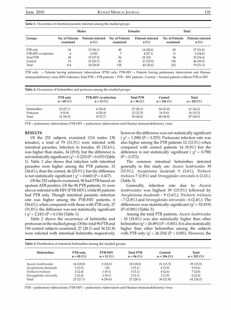
KUWAIT MEDICAL JOURNAL 131June 2010
RESULTS Of the 252 subjects examined (114 males 138
females), a total of 79 (31.3%) were infected with intestinal parasites. Infection in females, 45 (32.6%), was higher than males, 34 (29.8), but the difference is not statistically significant ( χ2 = 0.225) (P = 0.635) (Table 1). Table 1 also shows that infection with intestinal parasites were higher among the PTB patients, 33 (34.4%), than the control, 46 (29.5%), but the difference is not statistically significant ( χ2 = 0.660) (P = 0.417).
Of the 252 subjects examined, 96 had PTB based on sputum AFB positive. Of the 96 PTB patients, 11 were also co-infected with HIV (PTB-HIV), while 85 patients had PTB only. Though intestinal parasitic infection rate was higher among the PTB-HIV patients, 6 (54.6%), when compared with those with PTB only, 27 (31.8%), the difference was not statistically significant( χ2 = 2.241) (P = 0.134) (Table 1).
Table 2 shows the occurrence of helminths and protozoan in the studied group. Of the total 96 PTB and 156 control subjects examined, 27 (28.1) and 34 (21.8) were infected with intestinal helminths respectively,
however the difference was not statistically significant( χ2 = 1.298) (P = 0.255). Protozoan infection rate was also higher among the PTB patients 12 (12.5%) when compared with control patients 14 (9.0%) but the difference is not statistically significant ( χ2 = 0.798) (P = 0.372).
The common intestinal helminthes detected generally in this study are: Ascaris lumbricoides 39 (15.5%), Ancylostoma duodenale 9 (3.6%), Trichuris trichiura 7 (2.8%) and Strongyloides stercoralis 6 (2.4%). (Table 3).
Generally, infection rate due to Ascaris lumbricoides was highest 39 (15.5%) followed by Ancylostoma duodenale - 9 (3.6%), Trichuris trichiura - 7 (2.8%) and Strongyloides stercoralis - 6 (2.4%). The differences was statistically significant (χ2 = 52.819) (P<0.001) (Table 3).
Among the total PTB patients, Ascaris lumbricoides - 18 (18.8%) was also statistically higher than other helminthes (χ2 = 26.891) (P < 0.001). It is also statistically higher than other helminthes among the subjects with PTB only (χ2 = 26.254) (P < 0.001). However, the
Males Females Total
Groups
PTB onlyPTB-HIV co-infectionTotal PTBControlTotal
No. of Patients examined
36 44074
114
Patients infected n (%)
13 (36.1)2 (50)
15 (37.5)19 (25.7)34 (29.8)
No. of Patients examined
49 75682
138
Patients infected n (%)
14 (28.6) 4 (57.1)
18 (32) 27 (32.9) 45 (32.6)
No. of Patients examined
851196
156252
Patients infected n (%)
27 (31.8) 6 (54.6)33 (34.4)46 (29.5)79 (31.3)
Table 1: Occurrence of intestinal parasitic infection among the studied groups
HelminthesProtozoaTotal
PTB only n = 85 (%)
23 (27.1)8 (9.4)
31 (36.5)
PTB-HIV co-infection n = 11 (%)
4 (36.4)4 (36.4)8 (72.7)
Total PTB n = 96 (%)
27 (28.1)12 (12.5)39 (40.6)
Control n = 156 (%)
34 (21.8)14 (9.0)48 (30.8)
Total n = 252 (%)
61 (24.2)26 (10.3)87 (34.5)
Helminthes
Ascaris lumbricoidesAncylostoma duodenaleTrichuris trichiuraStrongyloides stercordisTotal
PTB onlyn = 85 (%)
16 (18.8)3 (3.5)2 (2.4)2 (2.4)
23 (27.1)
PTB-HIVn = 11 (%)
2 (18.2)- (0)
1 (9.1)1 (9.1)4 (36.4)
Total PTBn = 96 (%)
18 (18.8)3 (3.1)3 (3.1)3 (3.1)
27 (28.1)
Controln = 156 (%)
21 (13.5)6 (3.9)4 (2.6)3 (1.9)
34 (21.8)
Totaln = 252 (%)
39 (15.5)9 (3.6)7 (2.8)6 (2.4)
61 (24.2)
Table 2: Occurrence of helminthes and protozoa among the studied groups
Table 3: Distribution of intestinal helminthes among the studied groups
PTB only = Patients having pulmonary tuberculosis (PTB) only; PTB-HIV = Patients having pulmonary tuberculosis and Human immunodeficiency virus (HIV) infection; Total PTB = PTB patients + PTB – HIV patients ; Control = Normal patients without PTB or HIV
PTB = pulmonary tuberculosis; PTB-HIV = pulmonary tuberculosis and Human immunodeficiency virus
PTB = pulmonary tuberculosis; PTB-HIV = pulmonary tuberculosis and Human immunodeficiency virus

June 2010132
difference in infection rate due to Ascaris lumbricoides among those with PTB-HIV was not statistically significant ( χ2 = 2.200) (P = 0.532) (Table 3)
The intensity of the egg load varies between light to moderate in each studied group. None of these groups recorded a heavy intensity of egg load (Table 4).
DISCUSSIONThe overall prevalence of intestinal parasitic
infection (31.3%) recorded in this study is in agreement with Agi[27] and Okodua et al[28]. Agi[27] reported a prevalence rate of 33.6% in Sagbana community of the Niger-Delta, Nigeria, while Okodua et al[28] had earlier reported a rate of 28.4% from Abeokuta, Nigeria. The prevalence rate in this study is at variance to that reported by Awogun and Oyerinde et al. In their study, Awogun[29] reported a prevalence of 70.8% in Ilorin, Nigeria; while Oyerinde et al[30] also reported a prevalence rate of 89.5% in Lagos State, Nigeria. The relatively low rate observed in our study could be due to chemotherapy and public awareness on personal hygiene.
The 54.6% prevalence rate of intestinal parasites recorded in patients co-infected with pulmonary tuberculosis (PTB) and HIV (PTB-HIV) as against 31.8% among those with PTB only and 29.5% among control patients could be due to low immune status of such patients. Immunocompromise patients have been found to have a high infection rate of intestinal parasites[25].
The relatively higher intestinal helminthic infection rate recorded among the general PTB patients (28.1%), those with PTB only (27.1) and PTB-HIV patients (36.4%) as against control patients (21.8%) confirmthe finding of Tristao-Sa et al.[29] who reported 57.8% of intestinal nematode among patients with TB and 20.9% in control groups in Brazil, South America.
The common intestinal helminthes detected in this study are: Ascaris lumbricoides 39 (15.5%), Ancylostoma duodenale 9 (3.6%), Trichuris trichiura 7 (2.8%) and Strongyloides stercoralis 6 (2.4%); while the intestinal helminthes detected among the PTB patients are,
Ascaris lumbricoides 18 (18.8%), Ancylostoma duodenale 3 (3.1%), Trichuris trichiura 3 (3.1%) and Strongyloides stercoralis 3 (3.1%). The presence of most of these parasites including others had been reported from non-PTB patients in various parts of Nigeria[27,32,33]. The relatively high prevalence of Ascaris lumbricoides observed in our study may be related to the resistance of the ova to conditions such as cold and disinfectant[34], thereby increasing the opportunities for environmental contamination. Ascaris lumbricoides also produce more ova than other helminthes, which are passed out to the environment by the host[35].
The intensity of infection was generally low in all the studied groups. This may be due to chronic infection that may lead to low egg output by the helminthes. The relative higher frequency of intestinal helminthes among patients with pulmonary tuberculosis (PTB) observed in this study suggests that the immune modulation induced by worms may facilitate the progression of TB infected patients to disease state.
CONCLUSIONThe effect of intestinal helminthes infection is
multidimensional. Apart from being associated with poor-growth, impaired learning ability and reduced physical activity, especially in children[7, 8], they also impair the cellular immune responses needed to protect against HIV and tuberculosis [36]. There is accumulating evidence however, that prevention of helminthiasis could be part of the solution to the pandemics of HIV/AIDS and tuberculosis [37]. Therefore, there is a need for proper treatment and effective preventive measures to be put in place especially among the children who are more susceptible. These preventive measures need to incorporate poverty alleviation, effective sanitation, supply of clean water, appropriate education and deworming programmes. All patients with TB or HIV, who have intestinal helminthes, should be treated properly so as to improve their cell mediated immune response.
More research on the effect of Th1 and Th2 activity on patients infected with intestinal helminthes and
Infection Intensity
LightModerateHighTotal
PTB PTB-HIV Total PTB Control
Aln (%)
11(68.7)5 (31.3)
- (10)16
Adn (%)
3(100) - (0) - (0) 3
Ttn (%)
2(100) - (0) - (0) 2
Aln (%)
2 (100) - (0) - (0) 2
Adn (%)
- (0)- (0)- (0)-
Ttn (%)
1 (100) - (0) - (0) 1
Aln (%)
13 (72.2)5 (27.8)
- (0) 18
Adn (%)
3 (100) - (0) - (0) 3
Ttn (%)
3 (100) - (0) - (0) 3
Aln (%)
20 (95.2) 1 (4.8) - (0)21
Adn (%)
5 (83.3)1 (16.7)
- (0) 6
Ttn (%)
1 (25)1 (75)
- (0) 4
Table 4: Infection intensity of the helminthes among the studied groups
Intestinal Helminthiasis among HIV-Related Pulmonary Tuberculosis Patients in Abeokuta, Nigeria
PTB = pulmonary tuberculosis; PTB-HIV = pulmonary tuberculosis and Human immunodeficiency virusAl = Ascaris lumbricoides; Ad = Ancylostoma duodenale; Tt = Trichuris trichiura.

KUWAIT MEDICAL JOURNAL 133June 2010
pulmonary tuberculosis in developing countries like Nigeria is recommended.
ACKNOWLEDGMENTWe thank the Management of the Sacred Heart
Hospital, Abeokuta for providing the environment for this study. We also thank the Nurses and Laboratory staff for their co-operation especially Mrs. Peace EKPO, whose death occurred after the completion of the research. May Almighty God have mercy on her Soul and grant eternal rest.
REFERENCES
1. Palmas C, Gabriele F, Conchedda M, Bortoletti G, Ecca AR. Causality or coincidence: may the slow disappearance of helminths be responsible for the imbalance in immune control mechanisms? Journal of Helminthology 2003; 77: 147 – 153.
2. Crompton DW. How much human helminthiasis is there in the world? J. Parasitol 1999; 85: 397 – 403.
3. Okpala I. A survey of the incidence of intestinal parasites among government workers in Lagos, Nigeria. West Afr Med J 1961; 10:148 – 157.
4. World Health Organisation. Intestinal Protozoan and helminthic infections. Report of WHO scientific group,Geneva: WHO Technical Report Series 1981; 666.
5. Stephenson LS, Latham MC, Kinoti SN, Kurz KM, Brigham M. Improvement in physical fitness of Kenyanschoolboys with hookworm, Trichuris trichiura and Ascaris lumbricoides following a single dose of Albendazole. Trans R Soc Trop Med Hyg 1990; 84:277-282.
6. Nokes C, Grantham-Mc Gregor SM, Sawyer AW, Cooper ES, Bundy DA. Parasitic helminth infection and cognitive function in school children. Proc R Soc Lond 1992; 247:77-81.
7. Adams EJ, Stephenson LS, Latham MC, Kinoti SN. Physical activity and growth of Kenyan school children with hookworm, trichuris trichiura and ascaris lumbricoides infections are improved after treatment with albendazole. J Nutr 1994; 124:1199 – 1206.
8. Koroma MM, Williams AM, De La Have R, Hodges M. Effects of Albendazole on growth of primary school children and the prevalence and intensity of soil-transmitted helminthes in Sierra Leone. J Trop Ped 1996; 42: 371 – 372.
9. Stoltzfus RJ, Albonico M, Chwaya HM, et al. Hemoquant determination of hookworm-related blood loss and its role in iron deficiency in African children. Am J TropMed Hyg 1996; 55: 399 – 404.
10. World Health Organization. WHO annual report on global Tuberculosis control summary. Wkly Epidermiol Rec 2003; 78:122-128.
11. Smith I. What is the health social, and economic burden of tuberculosis?, In; Frieden, T, editor. Toman’s tuberculosis case detection, treatment and monitoring:Questions and Answers. 2nd Ed. Geneva: WHO publication; 2004. p 233-237
12. Bethony J, Brooker S, Albonico M, et al. Soil-transmitted helminth infections: ascariasis trichuriasis and
hookworm. Lancet 2006; 367:1521-1532.13. Bentwich Z, Weisman Z, Moroz C, Bar-Yehuda S,
Kalinkovich A. Immune dysregulation in Ethiopian immigrants in Israel: relevance to helminth infections? Clin Exp Immunol 1996; 103:239-243.
14. Diniz LM, Zandonade E, Dietze R, Pereira FE, Ribeiro-Rodrigues R. Short report: do intestinal nematodes increase the risk for multi bacillary leprosy? Am J Trop Med Hyg 2001; 65:852-854.
15. Kuby J. Immune response to infections, diseases, In; Janis Kuby, editor. 3rd Ed. Immunology. New York: WH Freeman and Company; 1997; p 459 – 483.
16. Toossi Z, Kleinhenz ME, Ellner JJ. Defective interleukin 2 production and responsiveness in human pulmonary tuberculosis. J Exp Med 1986; 163:1162-1172.
17. Zhang M, Gong J, Iyer DV, Jones BE, Modlin RL, Barnes PF. T cell cytokine responses in persons with tuberculosis and human immunodeficiency virusinfection. J Clin Invest 1994; 94:2435-2442.
18. Borkow G, Bentwich Z. Chronic immune activation associated with chronic helminthic and human immunodeficiency virus infections: role of hyporesponsiveness and anergy. Clin Microbiol Rev 2004; 17:1012-1030.
19. Fincham J. Helminthes, HIV/AIDS and Tuberculosis. AIDS Bulletin 2001; 10: 3.
20. Borkow G, Bentwich Z. Perspectives. Eradication of helminthic infections may be essential for successful vaccination against HIV and Tuberculosis. Bulletin of the World Health Organization 2000; 78:1368 – 1369.
21. Oyesiku OO, Kojeku GO. Abeokuta, In; Onokomaya SO, Oyesiku O, Jegede FJ, editors. Ogun State Maps. Nigeria: Rex Charles Publication ; 1992. p 153 – 155.
22. Akinrombi SOA, Ajayi S, Salawu G, et al, History of Abeokuta South Local Government. In: Akinrombi SOA, Ajayi S, Salawu G, et al, editors. Ogun State Business Directory and Yellow Pages. Ogun: Ogun state Council of Chambers of Commerce, Industry, Mines and Agriculture and Group Communications; 1999.p 101.
23. International Union against Tuberculosis and Lung Disease: IUATLD. Technical Guide for Sputum Examination for Tuberculosis by Direct Microscopy 1986. IUATLD, Paris.
24. World Health Organisation: WHO. Bench aids for the diagnosis of intestinal parasites. Geneva: WHO publication; 1994.Plate 2 and 5.
25. Chessbrough M. Parasitological tests, In: Chessbrough M, editor. District Laboratory Practice in Tropical countries. Tropical Health Technology. New York: Cambridge university press; 1998. p184 – 235.
26. World Health Organisation: WHO guidelines for the evaluation of soil transmitted helminthiasis and Schistosomiasis at community level. A guide for managers of control programmes 1998; WHO/CTB/98.1, pg. 29.
27. Agi PI. Pattern of Infection of Intestinal Parasites in Sagbama Community of the Niger Delta, Nigeria. West Afr J Med 1995; 14: 39 – 42.
28. Okodua MA, Adeyeba OA, Tatfeng YM, Okpala

June 2010134
HO. Age and Sex Distribution of Intestinal Parasitic Infection Among HIV infected subjects in Abeokuta, Nigeria. Online J Health Allied Scs 2003; 4:3.
29. Awogun IA. The prevalence of intestinal parasitic infection in children living in Ilorin Kwara state, Nigeria. Nigerian Medical Practitioner 1984; 7:176-178.
30. Oyerinde JPO, Adegbite-Hollist AF, Ogunbi O. Prevalence of intestinal parasite of man in the metropolitan Lagos. Nig J Nat Sc 1979;3: 147 – 155.
31. Tristao-Sa R, Ribeiro-Rodrigues R, Johnson LT, Pereira FE, Dietze R. Intestinal nematodes and pulmonary tuberculosis. Rev Soc Bras Med Trop 2002; 35 : 533 – 535.
32. Fashuyi SA. The pattern of human intestinal helminths infection in farming communities in different part of Ondo State, Nigeria. West Afr J Med 1992; 11 : 13 – 17.
33. Agi PI. Comparative helminthic infection in two rural
communities of the Niger Delta, Nigeria. West Afr J Med 1997; 16: 232 – 236.
34. Cook GC. Antihelminthic agents: some recent developments and their clinical application. Postgrad Med J 1991; 67: 16 – 22.
35. Mahmoud AAF. Intestinal Nematode;diseases due to trematodes. In: Mandell, Douglas, Benneth, editors. Principles and Practice of Infectious Diseases. 2nd Ed. New York: John Wiley and sons; 1990.p 2135 – 2140.
36. Ellias D, Wolday D, Akuffo H, Patros B, Bronner U, Britton S. Effect of Deworming on human T cell responses to mycobacterial antigens in helminth-exposed individuals before and after Bacille Calmette-Guerin (BCG) Vaccination. Clin Exp Immunol 2001; 123: 219 – 225.
37. Fincham J. HIV/AIDS and Tuberculosis. Science in Africa. Africa First Online Science Magazine 2003; 19:44-58.
Intestinal Helminthiasis among HIV-Related Pulmonary Tuberculosis Patients in Abeokuta, Nigeria
KUWAIT MEDICAL JOURNAL 135June 2010
INTRODUCTIONStreptococcus pneumoniae (pneumococcus) is a
bacterial pathogen that causes disseminated invasive diseases (e.g., bacteremia, meningitis, pneumonia) and other lower and upper respiratory system infections in children and adults worldwide. These diseases may be fatal for patients with underlying diseases and / or for older patients. Pneumococcal infections lead to approximately 40,000 deaths every year in the USA; more than any other disease that can be prevented by vaccines. It is estimated that nearly half of these deaths could be prevented by vaccination. Case-death rates are at their highest for meningitis and bacteremia, in patients with underlying diseases and for older patients. Despite appropriate antimicrobial treatment and intensive care, case-death rate due to pneumococcal bacteremia is 15 to 20% among adults and 30-40% among the elderly[1].
The polysaccharide vaccine contains 23 serotypes, covering 85-90% of the serotypes that cause invasive
Original Article
Pneumococcal Vaccination Status in Adults Sixty-Five Years and Older
Kuwait Medical Journal 2010; 42 (2): 135-138
Ozge Turhan1, Hasan Huseyin Polat2, Selma Oncel3, Arzu Akcan3, Kadriye Eravsar3, Ata Nevzat Yalcın1
1Department of Infectious Diseases and Clinical Microbiology, Akdeniz University Faculty of Medicine, Antalya, Turkey2Department of Public Health, Akdeniz University Faculty of Medicine, Antalya, Turkey
3Department of Public Health Nursing, Antalya School of Health, Antalya, Turkey
ABSTRACT
Address correspondence to: Ozge Turhan, MD, Department of Infectious Diseases and Clinical Microbiology, Akdeniz University Faculty of Medicine, Arapsuyu, 07070, Antalya, Turkey. Tel: +90 242 2496716, Fax: +90 242 2274490, E-mail:[email protected]
KEY WORDS: elderly, immunization, pneumococcal polysaccharide vaccine
Objective: To determine the pneumococcal vaccine immunization status of citizens 65 years and older.Design: Prospective studySetting: Akdeniz University, Faculty of Medicine, Antalya, TurkeySubjects: The study was performed by using a representative sample of the population of Antalya city center. One hundred and fifty clusters of 20 persons eachwere chosen from districts within Antalya city center.Intervention: A total of 2383 people were interviewed.Main Outcome Measures: People characteritics and vaccination statusResults: We ascertained that only 10.7% of the people involved in the study were aware of pneumococcal
vaccines and only 0.9% had been vaccinated. Of the vaccinated people, 68.2% had shots on their physicians’ advice and 31.8% were vaccinated on their own initiative or on the suggestion of their children. Out of the total vaccinations, 45.4% were performed at hospitals. The cost of vaccine was borne by the person in 45.4% vaccinations.Conclusions: The rate of pneumococcal vaccination was low among people aged 65 and older in Antalya. This result could be due to insufficient information on thisissue and the cost of vaccine not being covered by social security institutions. We recommend that physicians play a more pro-active role in encouraging people aged 65-years and above to have a pneumococcal vaccination.
pneumococcal infections. Using the polysaccharide vaccine recommended for adults, protective antibody levels are achieved for over 80% of healthy individuals in 2-3 weeks. Antibody responses may be lower in the elderly and for patients with underlying diseases such as cirrhosis, diabetes mellitus and chronic obstructive pulmonary disease (COPD). However, the efficacyof pneumococcal vaccine against invasive diseases and pneumonia has been proven in various clinical studies, case-control and serotype prevalence studies. Polysaccharide vaccine is well-tolerated by children and adults. It has also been proven that vaccination of the elderly against pneumococcal pneumonia is cost-effective[1,2].
If we consider the ever-increasing number of individuals reaching 65 years and above, then vaccination is clearly one of the most important protective health services in terms of maintainability of a healthy and functional life. Although pneumococcal vaccine takes place at adult vaccination schedules

June 2010136
worldwide and is known as an efficient, reliable andcost-effective vaccine, it does not reach the desired vaccination rates[1,3,4]. We aimed to determine the immunization status of pneumococcal vaccine in residents in the city center of Antalya for the ages of 65 and above (the vaccine being recommended for all people within this age group, if there are no contraindications).
SUBJECTS AND METHODSAccording to the records of the Antalya Health
Department, quantified by population count atHealth Centers in 2005, 704,000 people live within the 132 districts. Districts and their populations were divided into 150 sets where 704,000 / 150 = 4693 people per single set.
The research population was comprised of a total of 42,466 people over the age of 65 years who lived in the 132 neighborhoods and were registered at one of the neighborhood primary health care clinics within the city limits of Antalya Municipality. Formula consisting (n = (N . t2 . p .q) / (D2 (N-1) + t2 .p . q) was used while calculating the size of the samples, t-value was 98% ( 2.33) and frequency of vaccination (P) was decided as 10%. According to this formula, it was calculated that a sample size of 1221 individuals would be adequate. In order to decrease the possibility of originating from cluster sampling method, it was decided to reach for more samples, and in the end a total of 3000 individuals participated in the survey in 150 clusters, each consisting of 20 people. These 20 individuals from each cluster were administered the survey questionnaire. However, only 2383 individuals could complete the interview and questionnaire. The participation rate was 79.4%. Since there was no possibility of returning to the same area, those who did not agree to be interviewed and those who were not at home were not included.
Questionnaires were filled-out with the assistanceof eighty 4th year students of Akdeniz University Health Academy Nursing Program who visited houses and interviewed people face-to-face. The students had Public Health Training and before undertaking the survey, were trained in administering questionnaires. The questions were about relevant personal data and whether or not the individuals had received pneumococcal vaccine. All questionnaires were completed between March 1 and 31, 2006.
Students going to each defined district took thereeve’s house as the starting point and direction was decided by a draw. When they completed their quota for one district, they moved on to the next. Questionnaires could not be applied to people who refused the interview or those not at home, as there was no possibility of visiting the house for a second time. On completion of the questionnaire, individuals
were informed of the necessity of pneumococcal vaccination for people over 65 years of age. The data were evaluated with SPSS 12.0 software on computer.
RESULTSOut of 3000 planned visits to individuals to
complete questionnaires, 2383 individuals were interviewed (79.4%). Distribution of study group according to age and gender represents distribution of Antalya population according to age and gender (p > 0.05). Mean age of study group was 70.2 ± 5.1 years. Distribution of study group according to characteristics is shown in Table 1.
Out of all participants, 50.6% were male, 82.7% had an education level of primary school or less and 10.7% had no social security. There was no statistically
significant relationship between age distribution,gender and social security and the immunization status of the study group. Vaccination rate significantlyincreased as education level increased (χ2:14.942, p < 0.001).
Out of all participants, 10.7% knew about vaccination against pneumococci but only 0.9% of the study group had acquired the vaccination. Percentage of vaccination performed at health institutions was 90.8%. Vaccinations performed due to recommendation of physicians were 68.2% and 45.4% of the individuals paid for the vaccine themselves.
Characteristics
Distribution of Age Groups65 - 6970 - 7475 - 7980 +
GenderFemaleMale
Education LevelPrimary school and less Secondary educationHigher education
Social SecurityGovernment Retirement Fund Occupational Pension FundEmployee Pension Fund Green Card*None
Numberof people
979702419283
12051178
1969313101
532512877208254
%
41.129.417.611.9
50.649.4
82.713.14.2
22.321536.88.7
10.7
Table 1: Distribution of study group according to some characteristics (N = 2383)
*Green card: If an individual applies to government units and proves that he / she is in poor wealth and has no job, he / she is given a Green card and all his / her health care expenses are paid by the government.
Pneumococcal Vaccination Status in Adults Sixty-Five Years and Older

KUWAIT MEDICAL JOURNAL 137June 2010
DISCUSSIONInfections caused by Streptococcus pneumoniae are an
important reason of morbidity and mortality especially for the elderly with chronic diseases. The 23-valent polysaccharide pneumococcal vaccine is available in order to protect people from these infections since 1983 and it is recommended for people at risk groups and for the elderly[1,5]. Efficacy of polysaccharide vaccine isproven for protection from pneumococcal pneumonia with or without bacteremia, to decrease rates of all pneumonia cases and to decrease deaths caused by pneumonia in the elderly[1,5,6]. It has also been shown that the vaccine is cost-effective[1,2,5].
Application rates of the effective and reliable pneumococcal vaccine have not yet reached the desired levels in Turkey or worldwide. In 1985, the vaccine was recommended for an estimated 31 million people aged 65 and over. However, it was detected in 1993 that only 28% of the targeted recipients had received pneumococcal vaccination[1]. In Spain-Catalonia, a program was prepared for vaccination of individuals aged 65 and over with pneumococcal vaccine. Before this program, the prescription rate of pneuomococcal vaccine at hospitals and outpatient clinics for the elderly was 5.8% for the risk group[5]. Vila-Córcoles et al[5] reported that after the instigation of this program, vaccination rates raised to 38.6% in 2000, 44.4% in 2001, 50.9% in 2002 and 53.1% by the end of 2003. In other studies, pneumococcal vaccination rates were reported as 35 - 55%[7-9]. There is not sufficient data for Turkey, especially aboutthe elderly, but in a study by Zeybek et al[4] of 112 individuals aged 65 and over, it was observed that none had a shot of pneumococcal vaccine although they had information about the application of the vaccine. The percentage of individuals interviewed in our study who were aware of pneumococcal vaccine was 10.7%. However, only 0.9% of individuals within this age group had been vaccinated. The Center for Disease Control and Prevention (CDC) is aiming for 90% coverage of people aged 65 and over with pneumococcal vaccination in 2010[10]. The very low vaccination rates are dependant on many factors; insufficient comprehension of diseases which arepreventable with vaccination, suspicion regarding the reliability and efficacy of adult vaccines, concernsabout side effects, a lack of awareness regarding the seriousness of pneumococcal diseases and the benefits of vaccination, insufficient groundwork forvaccine distribution and the lack of cost coverage for vaccine expenses by official health institutions.Adult vaccination, especially for the elderly, is often neglected[1,4,7].
Adult vaccination has come into prominence in Turkey over recent years. The 2005 budget application instruction, which was operative at the time of our
study, stated that: “children over the age of two and adults with chronic pulmonary and cardiovascular system diseases including asthma; adults and children with any chronic metabolic disease including diabetes mellitus, renal dysfunction, hemoglobinopathy or immunosuppression; children and adolescents between the ages of 6 months - 18 years who have been receiving aspirin for a long period may have their vaccination expenses covered by their institutions every five years, if the vaccines are prescribed byphysicians and supported by board reports”. We believe that lack of coverage of vaccine expenses for adults aged 65 and over – with or without risk factors - is an important factor in low vaccination rates. With the change in budget application instruction in 2008, new risk factors were added to coverage conditions and the statement, “Vaccine expenses are covered every five years for individuals 65 years and older with noneed for reports” was added. Impact of this important change for adult vaccination, on pneumococcal vaccination should be evaluated with new studies.
Another important factor for increasing vaccination rates is community awareness. In a study by Santibanez et al[7] in which the researchers interviewed 1007 people by telephone, people reported that they were unaware of the necessity for pneumococcal vaccine and these results were similar to those of the study by Zeybek et al. In this study, it was detected that individuals completely relied on their physicians for health advice and were not vaccinated because it was not recommended by their physicians. We also found that 68.2% of the studied cases who had received vaccination had done so on their physician’s recommendation. We conclude that physicians have a frontline responsibility for increasing vaccination rates. Physicians performing adult health care should establish the vaccination status of ill and healthy individuals who consult them for any reason and encourage them to complete the missing vaccines. We believe that by giving information about the necessity of pneumococcal vaccine for people aged 65 and over, after individuals completed our study questionnaire, we contributed to that aim.
CONCLUSIONIn conclusion, invasive pneumococcal infections
and pneumococcal pneumonia are major causes of morbidity and mortality for people aged 65 and older. Although it is well-known that polysaccharide vaccine is effective in prevention of infections, desired levels of vaccination have not been achieved worldwide and in Turkey. For these reasons, vaccine application services for adults should be enhanced and appropriate and sufficient education of communities about vaccinationshould be provided. Administrators and physicians have considerable responsibility for achieving this.

June 2010138
The change to budget application instruction in Turkey is encouraging. As well as pneumococcal vaccination, influenza and diphtheria-tetanus vaccinations shouldbe considered. Countrywide data of vaccination rates should be investigated. Our study is important as it presents data for our region and establishes a precedent for further studies.
ACKNOWLEDGEMENT This study was supported by Akdeniz University
Research Foundation.
REFERENCES
1. Prevention of pneumococcal disease: recommendations of the Advisory Committee on Immunization Practices (ACIP). Center for Disease Control. MMWR Recomm Rep 1997; 46(RR-8):1-24.
2. Musher DM. Streptococcus pneumonia, In: Mandell GL, Bennett JE, Dolin R, editors. Principles and Practice of Infectious Diseases. New York: Elsevier-Churchill Livingstone; 2005. p 2392-2411.
3. Recommended adult immunization schedule - United States, 2009. Center for Disease Control (CDI). MMWR Recomm Rep 2009; 57:Q1-4.
4. Zeybek Y, Tokalak İ, Boyacıoğlu S. Vaccination in the sixty-five and over age adults. Turkish Journal ofGeriatrics 2004; 7:152-154.
5. Vila-Córcoles A, Ochoa-Gondar O, Ester F, Sará N, Ansa X, Saún N. EVAN Study Group. Evolution of vaccination rates after the implementation of a free systematic pneumococcal vaccination in Catalonian older adults: 4-year follow-up. BMC Public Health 2006; 6:231.
6. Vila-Córcoles A, Ochoa-Gondar O, Hospital I, et al. EVAN Study Group. Protective effects of the 23-valent pneumococcal polysaccharide vaccine in the elderly population: the EVAN - 65 study. Clin Infect Dis 2006; 43:860-868.
7. Santibanez TA, Nowalk MP, Zimmerman RK, et al. Knowledge and beliefs about influenza,pneumococcal disease, and immunizations among older people. J Am Geriatr Soc 2002; 50:1711-1716.
8. Zimmerman RK, Santibanez TA, Fine MJ, et al. Barriers and facilitators of pneumococcal vaccination among the elderly. Vaccine 2003; 21:1510-1517.
9. Nowalk MP, Zimmerman RK, Shen S, Jewell IK, Raymund M. Barriers to pneumococcal and influenza vaccination in older community-dwellingadults (2000 – 2001). J Am Geriatr Soc 2004; 52:25-30.
10. Influenza and pneumococcal vaccination coverageamong persons aged ≥ 65 years and persons aged 18 - 64 years with diabetes or asthma - United States, 2003. Center for Disease Control and Prevention (CDC). MMWR Morb Mortal Wkly Rep 2004; 53:1007-1012.
Pneumococcal Vaccination Status in Adults Sixty-Five Years and Older

KUWAIT MEDICAL JOURNAL 139June 2010
INTRODUCTIONThe incidence of type 1 diabetes in children,
0-14 years of age is increasing[1, 2]. A diabetic child is expected to deal with complicated daily routines of blood glucose monitoring, insulin injections, dietary regulations and exercise. Moreover, children and adolescents with diabetes are encouraged to lead a normal life as much possible, while at the same time, maintaining the demanding routines required as part of their disease management. These stresses have been regarded as risks for the development of different psychological problems[3].
Psychiatric disorders and social problems are reported to be common in children and adolescents with chronic medical disorders, including diabetes[4,5]. Although regular medical follow-up visits are usually scheduled, psychological intervention is not routinely directed at any specific time after diagnosis when
children are at risk for the development of psychological problems
The results of studies addressing whether the rate of psychiatric disorders is increased in diabetic children compared to healthy controls have been equivocal[6-9]. Kovacs et al[5,6] reported an increased rate of psychiatric disorders only immediately after the onset of diabetes, but not after 7 - 9 months. Lavigne et al[7] described an increased rate of psychiatric disorders only in boys. Other studies[10-13] reported a 2 - 3 fold increase in depression, adjustment problems and withdrawal symptoms in diabetic children compared to their peers without diabetes.
Little is known about the rates of anxiety and depression in children with diabetes in Kuwait. Moussa et al[14], using the Hopkin’s symptoms check-list to assess psychological stress, reported higher levels of stress and worse psychological adjustment
Psychological Status of Children with Type 1 Diabetes in Kuwait
Majedah Abdul-Rasoul1, Jasim Hajiyya2, Faisal Alshawaf3, Yahya Abdal4, Amina Al-Azmni2, Maria Al-Mahdi5 1Department of Pediatrics, Faculty of Medicine, Kuwait University, Kuwait
2Ministry of Social Affairs, Department of Research, Kuwait3Department of Pediatrics, Mubarak Al-Kabeer Hospital, Kuwait
4Department of Biostatistics, Kuwait University, Kuwait5Department of Pediatrics, Al-Adan Hospital, Kuwait
Address correspondence to:Majedah Abdul-Rasoul, Department of Pediatrics, Kuwait University, Faculty of Medicine, PO Box 24923, Safat, 13110, Kuwait. Tel: +965- 2543-8644; Fax: +965-2543-7977; Email: [email protected]
Objectives: Childhood diabetes puts an enormous burden on both the child and his family and may put them at increased risk for psychological problems. The purpose of the study was to assess the psychological reactions to type 1 diabetes in children and the effect of glycemic control on them. Design: A specially designed questionnaire was used to collect demographic data. The children behavioral check list and the Arabic childhood depression inventory were used to assess depression, anxiety and other behavioral disorders. Setting: Social workers and research assistants interviewed the patients in the outpatients of the 5 hospitals. Controls were interviewed at school. Subjects: A total of 215 diabetic and 221 age and sex matched healthy subjects were enrolled in the study.Main Outcome Measure: Measure depression, and other
behavioral disorders in diabetic patients.Results: Mild depression was reported in 63% of the diabetic subjects, 3-fold higher than in the control group (p < 0.05). Medium and high anxiety scores were significantlymore frequent in diabetic children (p <0.001, and p < 0.01 respectively) Attention, concentration problems, hyperactivity and aggressive behavior were significantly more frequent inthe diabetic children compared to matched controls. There was a significant association between poor glycemic controland depression; p < 0.001. hyperactivity (p = 0.001), poor attention (p = 0.007), and aggression (p = 0.03). Conclusion: Children with diabetes are at increased risk of depression and behavioral problems. Regular psychological assessment along with medical treatment must be addressed to optimize control.
KEY WORDS: ACDI, anxiety, CBCL, childhood diabetes, depression.
Original Article
Kuwait Medical Journal 2010; 42 (2): 139-145
ABSTRACT
June 2010140
compared to controls. A positive correlation between glycemic control and poor psychological adjustment was found in their study. Other studies addressing possible association between childhood diabetes and psychiatric disorders have yielded divergent results, ranging from highly significant linkage to no detectableassociation[15-19].
The aims of our study were: (1) to assess the psychological reactions to type 1 diabetes in children (8 - 18 years), including anxiety, depression, attention-hyperactivity disorder and aggressive behavior using different measurement scales, and (2) to evaluate the hypotheses that children with poor glycemic control have more psychological symptoms than those with good control.
SUBJECTS AND METHODS In the State of Kuwait, children and adolescents,
aged 0-18 years, with type 1 diabetes are primarily followed–up in diabetic out-patient clinics at fivegovernment hospitals.
Subjects included in the sample comprised 215 children and adolescents (110 males and 105 females) aged 8-18 years with type 1 diabetes for at least 12 months, and who were attending a hospital diabetic clinic. With the exception of one subject, all approached agreed to participate. The control group comprised 221 healthy children (116 male and 105 female). Each diabetic child was matched with a child without diabetes from the same class to ensure same age range, sex and social class profile. A total of 256 families were approached;239 (93.3%) responded’ and 221 (92.5%) agreed to participate. The aim and the procedure of the study were explained to the patients and/or their parents, and consents to participate were obtained before inclusion in the study. Written informed consent was obtained from the parents of each participating children. Study design and methodology was approved by the Research Ethics Committee in the Ministry of Health, Kuwait.
The Questionnaire:A questionnaire was designed to collect
demographic data including gender, nationality, age at time of study, place of residence, family dynamics (living with one, both or neither parents) and psychosocial aspects. This instrument was pre-tested on a sample of 50 children by two interviewers, and only children eight years of age and above were able to answer the questionnaire without the help of the caregivers. To eliminate the effect of parents’ response on the child, the analysis was limited to those children eight years old and above. Social workers and research assistants trained to administer such questionnaires went to the
outpatient clinics where children were interviewed either while waiting to see their physicians or after their medical assessment.
Socio-demographic measures: Family socioeconomic status (SES) was measured
at the beginning of the study using a SES score adapted for the Kuwaiti population[14]. This is based on the education level of the parents (0 = illiterate, 1 = primary/intermediate, 2 = secondary, and 3 = university and above), occupations of the parents (0 = unemployed, 1 = laborer, 2 = semiskilled, 3 = semiprofessional, 4 = skilled, and 5 = professional), and family income (1 means < 1000 Kuwaiti Dinars (KD), 2 means 1000 - 1500 KD, 3 means > 1500 KD, where KD 1 equals approximately US$ 3.3). SES score (ranges from 1 - 19) categorized families as low (scoring 1 - 7), medium (8 - 14) or high (15 - 19).
Diabetes-related data:Disease-related data including age at diagnosis,
duration of the disease and compliance with insulin injections were documented by the treating doctors. Date on frequency of home blood glucose monitoring, insulin injections, and following a meal plan (always, most of the time, infrequently) were obtained through log books, meter downloads and parents reports. Mean HbA1c value (at least 3 measurements) were used as a measure of metabolic control (less than 7% = excellent, 7 - 8.5 % = good, more than 8.5% = poor; in accord with ISPAD 2007-2008 guidelines).
School performance:Assessment was based on the children’s last report
card. The scoring system in all government schools in Kuwait is standardized by the Ministry of Education. It is divided into excellent, very good, good, average and failed.
Psychiatric evaluation and diagnosis:Children were subjected to psychiatric assessment
using two separate scales.
1. Arabic Children’s Depression Inventory (ACDI): This scores on five subscales: negative mood,interpersonal problems, ineffectiveness, anhedonia and negative self-esteem[20]. The literature on the CDI suggests that it discriminates successfully between clinical and nonclinical samples of children[20-21]. ACDI can be used across all age groups, has no cultural bias, and provides a depression score normalized to age and sex. Reliability and validity have been tested and confirmed in an Arabic cultural setting by Dr.Abdel-Khalek[20] who named it ACDI. The inventory contains 27 brief items; each item is scored on a 3-point intensity scale: rarely, sometimes and often; keyed as
Psychological Status of Children with Type 1 Diabetes in Kuwait
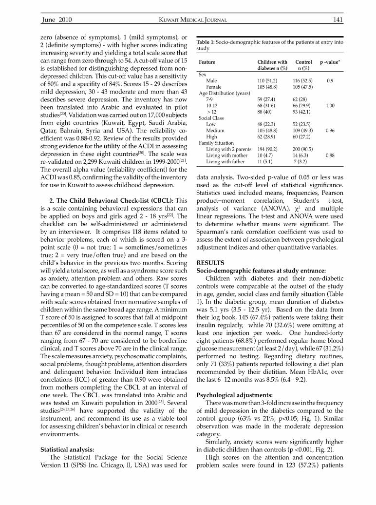
KUWAIT MEDICAL JOURNAL 141June 2010
zero (absence of symptoms), 1 (mild symptoms), or 2 (definite symptoms) - with higher scores indicatingincreasing severity and yielding a total scale score that can range from zero through to 54. A cut-off value of 15 is established for distinguishing depressed from non-depressed children. This cut-off value has a sensitivity of 80% and a specifity of 84%. Scores 15 - 29 describesmild depression, 30 - 43 moderate and more than 43 describes severe depression. The inventory has now been translated into Arabic and evaluated in pilot studies[20]. Validation was carried out on 17,000 subjects from eight countries (Kuwait, Egypt, Saudi Arabia, Qatar, Bahrain, Syria and USA). The reliability co-efficient was 0.88-0.92. Review of the results providedstrong evidence for the utility of the ACDI in assessing depression in these eight countries[20]. The scale was re-validated on 2,299 Kuwaiti children in 1999-2000[21]. The overall alpha value (reliability coefficient) for theACDI was 0.85, confirming the validity of the inventoryfor use in Kuwait to assess childhood depression.
2. The Child Behavioral Check-list (CBCL): This is a scale containing behavioral expressions that can be applied on boys and girls aged 2 - 18 yrs[22]. The checklist can be self-administered or administered by an interviewer. It comprises 118 items related to behavior problems, each of which is scored on a 3-point scale (0 = not true; 1 = sometimes/sometimes true; 2 = very true/often true) and are based on the child’s behavior in the previous two months. Scoring will yield a total score, as well as a syndrome score such as anxiety, attention problem and others. Raw scores can be converted to age-standardized scores (T scores having a mean = 50 and SD = 10) that can be compared with scale scores obtained from normative samples of children within the same broad age range. A minimum T score of 50 is assigned to scores that fall at midpoint percentiles of 50 on the competence scale. T scores less than 67 are considered in the normal range, T scores ranging from 67 - 70 are considered to be borderline clinical, and T scores above 70 are in the clinical range. The scale measures anxiety, psychosomatic complaints, social problems, thought problems, attention disorders and delinquent behavior. Individual item intraclass correlations (ICC) of greater than 0.90 were obtained from mothers completing the CBCL at an interval of one week. The CBCL was translated into Arabic and was tested on Kuwaiti population in 2000[23]. Several studies[24,25,26] have supported the validity of the instrument, and recommend its use as a viable tool for assessing children’s behavior in clinical or research environments.
Statistical analysis:The Statistical Package for the Social Science
Version 11 (SPSS Inc. Chicago, Il, USA) was used for
data analysis. Two-sided p-value of 0.05 or less was used as the cut-off level of statistical significance.Statistics used included means, frequencies, Pearson product−moment correlation, Student’s t-test, analysis of variance (ANOVA), χ2 and multiple linear regressions. The t-test and ANOVA were used to determine whether means were significant. TheSpearman’s rank correlation coefficient was used toassess the extent of association between psychological adjustment indices and other quantitative variables.
RESULTSSocio-demographic features at study entrance:
Children with diabetes and their non-diabetic controls were comparable at the outset of the study in age, gender, social class and family situation (Table 1). In the diabetic group, mean duration of diabetes was 5.1 yrs (3.5 - 12.5 yr). Based on the data from their log book, 145 (67.4%) patients were taking their insulin regularly, while 70 (32.6%) were omitting at least one injection per week. One hundred-forty eight patients (68.8%) performed regular home blood glucose measurement (at least 2/day), while 67 (31.2%) performed no testing. Regarding dietary routines, only 71 (33%) patients reported following a diet plan recommended by their dietitian. Mean HbA1c, over the last 6 -12 months was 8.5% (6.4 - 9.2).
Psychological adjustments:There was more than 3-fold increase in the frequency
of mild depression in the diabetics compared to the control group (63% vs 21%, p<0.05; Fig. 1). Similar observation was made in the moderate depression category.
Similarly, anxiety scores were significantly higherin diabetic children than controls (p <0.001, Fig. 2).
High scores on the attention and concentration problem scales were found in 123 (57.2%) patients
Feature
SexMaleFemale
Age Distribution (years)7-910-12 > 12
Social ClassLowMediumHigh
Family SituationLiving with 2 parentsLiving with motherLiving with father
Children with diabetes n (%)
110 (51.2)105 (48.8)
59 (27.4)68 (31.6)88 (40)
48 (22.3)105 (48.8)62 (28.9)
194 (90.2)10 (4.7)11 (5.1)
Controln (%)
116 (52.5)105 (47.5)
62 (28)66 (29.9)93 (42.1)
52 (23.5)109 (49.3)60 (27.2)
200 (90.5)14 (6.3) 7 (3.2)
p -value*
0.9
1.00
0.96
0.88
Table 1: Socio-demographic features of the patients at entry into study

June 2010142
in the diabetic group, compared to 24 (10.8%) in the control, p < 0.001. Similar results were found in the medium category (34% vs. 15%, p < 0.05). Male gender was associated with significantly higher scores inattention and concentration problem of any degree (p < 0.01 for all).
Higher mean scores for hyperactivity were found in the diabetic group compared to the controls (Fig 3). In both groups, scores were significantly higher inboys compared to girls (p < 0.001).
Aggressive behavior of any degree (frequent and occasional) was reported more frequently in diabetic patients compared to controls (p < 0.005 and p < 0.001 respectively). It was significantly higher in boyscompared to girls (p < 0.001).
Neither school phobia nor refusal was reported as a ‘frequent’ behavior in the diabetic group.
Glycemic control and psychological disturbance:We tested three different hypotheses addressing
possible links between psychological status and glycemic control. To study the effect of glycemic control on psychological adjustment, patients with diabetes for at least two years (179) were included in the analysis.
There was a significant association between poormetabolic control and depression in children with type 1 diabetes. Comparing different subgroups of glycemic control, depression of any severity was
Categories of depression
Mea
n ra
te (%
)
Mean HbA1c values
Fig 1: Categories of depression by ACDI score in diabetics and controls. Less than 15 is considered normal, 15-29 is mild depression, 30-43 is moderate and more than 43 is severe depression.
Fig 2: Anxiety CBCL Scores. T scores less than 67 are considered normal, 67-70 are borderline and scores above 70 (high) are in the range of clinical anxiety. p < 0.05
Fig 3: A scatter plot of ACDI total score against the mean HbA1c. Linear regression analysis revealed a significant correlation, r = 0.87,p <0.001.
Tota
l AC
DI s
core
s
Fig 4: Mean CBCL scores for hyperactivity in different glycemic control groups. Mean+/- SD score were significantly higher in thepoor glycemic control group compared with those with excellent and good control, p < 0.001.
CBC
L sc
ore
for h
yper
activ
ity(M
ean
+/- S
D)
Glycemic control groups
CBC
L sc
ore
for a
ggre
ssio
n(M
ean
+/- S
D)
Glycemic control groups
Fig 5: Mean CBCL scores for aggressive behavior: Diabetics with poor glycemic control had significantly higher scores than non-diabetics,p < 0.005. Aggressive behavior of any degree was significantlyhigher in boys compared to girls (p < 0.001).
r = 0.87p < 0.001
Psychological Status of Children with Type 1 Diabetes in Kuwait

KUWAIT MEDICAL JOURNAL 143June 2010
found significantly more frequent in the poor glycemiccontrol group (p < 0.001). Further analysis of the data showed significant positive correlation between meanHbA1c level and total ACDI depression scores (Fig. 3, r = 0.87, p < 0.001).
Both, borderline and clinical anxiety, were also significantly more frequent in the group with poorglycemic control, p < 0.001 and p < 0.01 respectively.
Furthermore, children with poor metabolic control had significantly higher scores in hyperactivity, lowerscores in attention and higher scores for aggressive behavior. When subgroups were compared according to degree of glycemic controls, there was a highly significant difference in the scores for hyperactivity(p = 0.001, Fig 4). Attention and concentration scores showed that children with poor control (HbA1c > 8.5%) had the lowest attention among the three groups, p = 0.007.
Moreover, when the three groups were compared in their performance in aggressive behavior measures by CBCL, results showed higher scores for aggressive behavior in the group with poor glycemic control compared to those with good or excellent control (p = 0.03. Fig 5).
DISCUSSIONOur study is the first to use the ACDI and CBCL to
assess depression and behavior disorders in diabetic children in Kuwait. However, accurate data are not available regarding the prevalence of depression in children and adolescents in the general population.
Few population-based studies have addressed depression in children and adolescents with diabetes. However, it has been suggested that the prevalence of depression in this population ranges between 2 to 3 fold above that of peers without diabetes[24]. In the present study, the prevalence of both mild and moderate depression was three times higher in the diabetic group than in healthy control subjects matched for age, sex and socioeconomic status. This excess is consistent with the results of the Moussa et al. study[14], which used the Hopkins System Checklist-25 (HSCL-25) in a comparable population group: higher mean scores were reported for depression, anxiety and total distress than controls.
In adolescents, the challenge of diabetes and its management responsibilities is combined with the development tasks of adapting to puberty, changing body image, peer pressure and increasing autonomy from parents. Although, Lawrence et al[15] found no increased risk of depressed mood in diabetic subjects compared with controls. Blanz et al[16] reported psychiatric disorders in 33.3% of adolescent diabetics (aged 17 - 19 yr) compared to only 9.7% in controls. Although, depression and other disorders are generally more common in older children and adolescents,
type 1 diabetes has been reported to be a risk factor for psychiatric disorder at all ages from childhood to adulthood[12,25,]. This effect was not found in our study. Hood et al[26] found that self-reports from nearly one in seven adolescents with diabetes met the clinical cutoff for depression, close to the highest estimate for the prevalence of depression in this population[27-29].
Our study failed to show a significant effect ofgender on depression in the diabetic children. In another study[15] performed on a similar population, but using different scales, depression was more common in females with diabetes than diabetic males. This finding is consistent with other studies[25-26]. On the other hand, some other studies[5,13] have reported that mean scores for depression were higher in boys (p = 0.001) . Our study also showed no correlation between depression and their duration of diabetes or age at onset of the disease. Kovacs et al[5] reported an increase in the rate immediately after diagnosis, but the increase was no longer significant 9-12 monthslater.
We found that depression failed to correlate with metabolic control measured by HbA1c. Moussa et al[14] reported that children with poor glycemic control (HbA1c > 10%) had significantly more depressionand anxiety. Blanz and others[5,9,10,14] compared three subgroups with good, moderate, or poor control. There was no significant difference in the prevalenceof depression between the three groups. Hood et al[26] found that subjects who monitored glucose less often had higher rates of depression, a possible indicator of suboptimal metabolic control. Earlier studies[30,31,32]
pointed to an association between psychiatric problems (including depression and anxiety) and good metabolic control. The authors suggested that routine monitoring of blood glucose and insulin injections is likely to be demanding and careful control of glycemia may itself be stressful. This stress could increase the risk of psychological sequale including depression in the group with good control.
In the present study, attention and concentration problems, hyperactivity and aggressive behaviors were all significantly more frequent in the diabetic children.Increased prevalence of these conditions correlated significantly with glycemic control. Moussa et al[14], found higher levels of anxiety and total distress in the diabetic population compared to the control, and these correlated positively with metabolic control. Blanz et al[16] reported that diabetic adolescents suffer from significantly more introversive symptoms than theirhealthy counterparts, especially in somatic symptoms, sleeping disturbances and compulsive behavior. In adolescents, 10 yr after first diagnosis, conduct andgeneralized anxiety disorders were reported in 47% in the study of Kovacs et al[5]. Another study[22] using the CBCL in children with diabetes found that both

June 2010144
internalized and externalized behavior problems were increased: boys with diabetes became more aggressive than boys without diabetes. Northam et al[32] reported that adolescents with type 1 diabetes are at high risk for psychiatric disorder. Though the association between diabetes, metabolic control, and psychiatric disorders suggest that diabetes predisposes to such disorders, it is also clear that poor attention and concentration, for example, are likely to impact negatively on metabolic control.
Studies addressing possible association between childhood diabetes and psychiatric disorders have yielded divergent results, ranging from highly significant linkage to no detectable association. Thisvariation has been ascribed to several factors[30-32]. First, some reports have employed study instruments of unknown reliability and validity. Second, different types and combinations of instruments (interviews, questionnaires) were used in various studies. Third, studies had addressed different age groups. Fourth, some studies included no control group.
One potential limitation of our present study is that all the data for diabetic children were obtained by self-reporting at interview in a clinical setting, while non-diabetic controls were interviewed in a school environment. Different interview contexts may underlie some of the differences observed.
However, our study has a number of strong points. The study employed scales that have been fully validated for use in Kuwaiti children. Diabetic and control children were included from across all the health areas in Kuwait and from all socioeconomic strata .The large number of children enrolled has permitted reliable assessment of statistical significance.Moreover, this work addresses an understudied population: psychological studies are most commonly performed in older children and adults, whereas our study population comprised children aged 8 - 18 years.
CONCLUSIONThis study supports the hypothesis that children
and adolescents with diabetes are at increased risk for depression and various behavior problems. The presence of psychiatric disorders in individuals with diabetes can also negatively affect metabolic control and thereby increase the risk of long-term complications. While the association between psychological problems and metabolic control is clearly bidirectional, more longitudinal research would be needed to clarify the link between glycemic control and psychological disorders, especially depression. The long-term outcome for children with diabetes is dependent on diverse physiological, cognitive and psychological factors. Therapeutic intervention should recognize that these factors are mutually inter-dependent and must
therefore, be addressed together, in order to optimize overall outcome.REFERENCES
1. Abdul-Rasoul M, Al-Qattan H, Al-Haj A, Habib H, Ismael A. Incidence and seasonal variation of type 1 diabetes in children in Farwaniya Area, Kuwait (1995-1999). Diabetes Res Clin Prac 2002; 56:153-157.
2. Karvonen M, Viik-Kajander M, Moltchanova E, Libman I, LaPorte R, Tuomilehto J. Incidence of childhood type 1 diabetes worldwide. Diabetes Mondiale (Dia Mond) Project group. Diabetes Care 2000; 23:1516-1526.
3. McCarthy AM, Lindgren S, Mengeling MA, Tsalikian E, Engvall J. Effects of diabetes on learning in children. Pediatrics 2002; 109: e9-e17.
4. Meijer SA, Sinnema G, Bijstra JO, Mellenberg GJ, Wolters WH. Coping styles and locus of control as predictors for psychological adjustment of adolescents with a chronic illness. Soc Sci Med 2002; 54:1453-1461.
5. Kovacs M, Goldston D, Obrosky DS, Bonar LK. Psychiatric disorders in youths with IDDM: rates and risk factors. Diabetes Care 1997; 20:36-44.
6. Kovacs M, Mukerji P, Iyengar S, Drash A. Psychiatric disorders and metabolic control among youths with IDDM. Diabetes Care 1996; 19:318-323.
7. Lavigne JV, Traisman HS, Marr TJ, Chasnoff IJ. Parental perceptions of the psychological adjustment of children with diabetes and their siblings. Diabetes Care 1982; 5:420-426.
8. Grey M, Whittemore R, Tamborlane W. Depression in type 1 diabetes in children: natural history and correlates. J Psychosom Res 2002; 53:907-911.
9. Kovacs M, Obrosky DS, Goldston D, Drash A. Major depressive disorder in youth with IDDM. A controlled prospective study of course and outcome. Diabetes Care 1997; 20: 45-51.
10. Finch AJ, Saylor CF, Edwards GL, McIntosh JA. Children’s depression inventory: reliability over repeated administrations. J Consult Clin Psychol 1987; 16:339-341.
11. Saylor CF, Finch AJ Jr, Spirito A, Bennett B. The children’s depression inventory: a systemic evaluation of psychometric properties. J Consult Clin Psychol 1984; 52:955-967.
12. Cameron FJ, Northam EA, Ambler GR, Daneman D. Routine psychological screening in youth with type 1 diabetes and their parents: a notion whose time has come? Diabetes Care 2007; 30: 3716-3724.
13. Gafvels C, Lithner F, Borjeson B. Living with diabetes: relationship to gender, duraion and complications. A survey in nothern Sweden. Diabet Med 1993; 10:768-773.
14. Moussa MA, Alsaeid M, Abdella N, Refai TM, Al-Sheikh N, Gomez JE. Social and psychological characteristics of Kuwaiti children and adolescents with type 1 diabetes. Soc Sci Med 2005; 60:1835-1844.
15. Lawrence JM, Standiford DA, Loots B, et al. Prevalence and correlates of depressed mood among youth with diabetes: the SEARCH for diabetes in youth study. Pediatrics 2006; 117: 1348-1358.
16. Blanz BJ, Rensch-Riemann BS, Fritz-Sigmund DJ,
Psychological Status of Children with Type 1 Diabetes in Kuwait

KUWAIT MEDICAL JOURNAL 145June 2010
Schmidt MH. IDDM is a risk factor for adolescent psychiatric disorders. Diabetes Care 1993; 16: 1579-1587.
17. Chernoff RG, Ireys HT, DeVet KA, Kim YJ. A randomised, controlled trial of a community-based support program for families of children with chronic illness: pediatric outcomes. Arch Pediatr Adolesc Med 2002; 156: 533-539.
18. Helgeson VS, Snyder PR, Escobar O, Siminerio L, Becker D. Comparison of adolescents with and without diabetes on indices of psychosocial functioning for three years. J Pediatr Psychol 2007; 32:794-806.
19. Graue M, Wentzel-Larsen T, Bru E, Hanestad BR, Sovik O. The coping styles of adolescents with type 1 diabetes are associated with degree of metabolic control. Diabetes Care 2004; 27: 1313-1317.
20. Abdel-Khalek AM. Factorial structure of the Arabic children’s depression inventory among Kuwaiti subjects. Psychol Rep 1996; 78:963-967.
21. Al-Balhan EM. The children’s depression inventory as a reliable measure for post-Iraqi invasion in Kuwaiti youth. Social Behavior Personality 2006; 34:351-366.
22. Achenbach TM, Ruffle TM. The child behaviorchecklist and related forms for assessing behavioral /emotional problems and competencies. Pediatr Rev 2000; 21: 265-271.
23. Al-Mutairi H. Psychological disturbances in Kuwaiti children of low income families. Journal of Social Science 2000; 28: 89-97.
24. Al-Eissa YA. The impact of the gulf armed conflict on
the health and behavior of Kuwaiti children. Soc Sci Med 1995; 41: 1033-1037.
25. Dantzer C, Swendsen J, Maurice-Tison S, Salamon R. Anxiety and depression in juvenile diabetes: a critical review. Clin Psychol Rev 2003; 23:787-800.
26. Hood KK, Huestis S, Maher A, Butler D, Volkening L, Laffel LM. Depressive symptoms in children and adolescents with type 1 diabetes:association with diabetes-specific characteristics. Diabetes Care 2006;29: 1389-1391.
27. Lewinsohn PM, Clarke GN, Seeley JR, Rohde P. Major depression in community adolescents: age at onset, episode duration, and time to recurrence. J Am Acad Child Adolesc Psychiatry 1994; 33:809-818.
28. Stewart SM, Rao U, Emslie GJ, Klein D, White PC. Depressive symptoms predict hospitalization for adolescents with type 1 diabetes mellitus. Pediatrics 2005; 115:1315-1319.
29. Fleming J, Boyle M, Offord D. The outcome of adolescent depression in the Ontario child health study follow-up. J Am Acad Child Adolesc Psychiatry 1993; 32:28-33.
30. Rovet JF, Ehrlich RM. Effect of temperament on metabolic control in children with diabetes mellitus. Diabetes Care 1988; 11:77-82.
31. Guthrie D. Bartsocas C, Jarcoz-Chabot P, Konstantinova M. Psychological issues for children and adolescents with diabetes: overview and recommendations. Diabetes Spectrum 2003; 1: 7-11.
32. Northam EA, Matthews LK, Anderson PJ, Cameron FJ, Werther GA. Psychiatric morbidity and health

KUWAIT MEDICAL JOURNAL June 2010146
INTRODUCTIONGastroduodenal involvement in Crohn’s disease
(CD) is relatively rare (0.5 to 13%)[1]. It can occasionally cause obstruction, for which medical therapy may be ineffective and surgery or balloon dilatation may be required. The clinical presentation is often confused with that of peptic ulcer disease. The current Crohn’s disease treatment algorithm involves a “step-up” approach in which conventional medications such as corticosteroids are given first, and anti-tumor necrosisfactor-a (TNF-a) agents are reserved for refractory cases. Early use of biologic therapy in Crohn’s disease has been associated with increased rates of response and remission[2-4]. Recent studies have shown that “top-down” therapy using anti-TNF-a agents in newly diagnosed patients improves long-term rates of mucosal healing, a therapeutic endpoint that correlates with reduced hospitalization and surgery, thereby reducing overall costs and enhancing patients’ quality of life[5]. Our case demonstrates the occurrence of pyloric stenosis in a young man as a presenting symptom of CD and the efficacy of anti-TNF-amonotherapy (infliximab) in improving symptoms.
CASE HISTORYA 19-year-old boy was admitted to the hospital
complaining of a two-month history of recurrent, post-parandial vomiting, epigastric discomfort with dyspepsia, mild constipation, right lower quadrant abdominal pain, and weight loss of about 8 kg over six
Case Report
Kuwait Medical Journal 2010; 42 (2): 146-148
Pyloric Stenosis as a Presenting Symptom of Crohn’s Disease Treated with Infliximab Monotherapy
ABSTRACT
Address correspondence to: Dr. Fahad A.H AL-Najjar,FRCP(C), Thunayan Al-Ghanim Gastroenterology Center, Amiri Hospital, Kuwait. Fax:+965-22516663,E-mail: [email protected]
Fahad AH AL-Najjar1, Fatma AH AL-Najjar2
1Thunayan Al-Ghanim Gastroenterology Center, Amiri Hospital, Kuwait 2Department of Family Medicine, Khaldiya Clinic, Kuwait
We report a rare case of pyloric stenosis as a presenting symptom of Crohn’s disease. Clinical improvement and long term relief of pyloric obstruction were obtained following a one year course of infliximab (Remicade®). In contrast
to most of the cases described previously in the literature, surgical treatment was not required and infliximab was usedas a monotherapy.
KEY WORDS: endoscopy, gastroscopy, inflammatory disease, pyloric obstruction
months. There was no history of diarrhea or bloody stool, nor a history of peptic ulcer disease. At the beginning of his symptoms, the patient had undergone a gastroscopy that showed pyloric stenosis. He was treated with a Proton Pump Inhibitor (PPI) but with no response. There was no family history of cancer or inflammatory bowel disease. Physical examinationrevealed a thin male (weight = 51 kg, height = 169 cm, BMI = 17.8). He had a body temperature of 36.7 °C, respiratory rate of 14 / minute, and a regular pulse at a rate of 75 / minute. His blood pressure was 122 / 60 mmHg. Lungs were clear at auscultation and heart sounds were normal. No murmurs or any friction rub was detected. Abdominal examination revealed a soft abdomen with no tenderness and no organomegaly. Bowel sounds were present. Neurological examination was normal.
Laboratory results, upon admission, revealed mild anemia with a hemoglobin of 10.5 g/dl (13.0 - 17.0) with an MCV of 75fl (80 - 96), hematocrit at 31% (40- 50), leukocyte count of 11 x 109/l (4.0 - 10.0 x 10⁹), and platelets at 400 x 109/l (150 - 400 x 109). Sodium and potassium were both normal. Glucose, electrolytes, kidney and liver function tests were all within the normal range: iron 8 mcg/dl (5 - 33), transferrin 2 g/l (2.0 - 4.0), Vit B12 641 pg/ml (130 - 878), and albumin 3.8 g/dl (3.5 - 4.8), erythrocyte sedimentation rate of 80 mm in two hours, and C-reactive protein 60 mg/dl (0 - 5). Serology for hepatitis B, C, and HIV were all negative. Antinuclear antibody (ANA) was negative.

KUWAIT MEDICAL JOURNAL 147June 2010
C-ANCA (antineutrophil cytoplasmic antibody) and p-ANCA were within normal limits. Tumor markers and TSH were all normal. Chest X-ray showed normal findings. Abdominal CT scan revealed a dilatationof the stomach and suspected obstruction at pyloric level. No masses or lymph nodes were observed, but a marked thickening of the ileal loops in the region of the right iliac fossa was observed (Fig. 1). A barium swallow and follow through showed a dilated stomach with markedly reduced emptying.
A repeat gastroscopy revealed a dilated stomach with pyloric stenosis and multiple superficial erosionsand aphthous ulcers. The tight stenosis precluded the passage of the gastroscope. Fig. 2 shows that the mucosa was edematous with cobble stoning. A biopsy was performed, and Helicobacter pylori was negative. Colonoscopy revealed hyperemia, ulceration, edema,
and cobble stoning of the terminal ileum and patchy colitis with superficial scattered erosions. The diagnosisof CD was confirmedbyhistopathologicexaminationofthe ileocolonic and pyloric biopsies (Fig. 3). The option of balloon dilatation was entertained, but was refused by the patient after discussing the risks and benefits ofthe procedure. The patient was treated initially with three doses of infliximab 5 mg/kg at weeks zero, two,and six, then every eight weeks for 48 weeks. The patient started showing signs of improvement two weeks after the initial dose, and it took him about six weeks to be completely asymptomatic.
A controlled gastroscopy done at six weeks to check for mucosal healing demonstrated resolution of the stenotic lesion and easy gastroscopic passage (Fig. 4). The patient continued to be free of symptoms, but unfortunately was lost to follow up after his last
Fig. 1: CT scan of the abdomen showing dilated stomach and obstruction at pyloric level
Fig. 2: Dilated stomach with pyloric stenosis and edematous mucosa with superficial erosions
Fig. 3: Intermediate power view of histological section from the pylorus showing a typical granuloma and mononuclear cells
Fig. 4: Cobble stone appearance of the first part of duodenum (6 weeks aftertreatment)

June 2010148
infliximab dose. An endoscopy was repeated (Fig. 5and 6) six months later, because the patient had non-specific dyspeptic symptoms. However, it failed toshow any abnormalities.
DISCUSSIONCD is an inflammatory disease of unknown
etiology that can affect any portion of the intestinal tract from the mouth to the anus. Approximately 80% of patients have small bowel involvement, usually in the distal ileum, with one-third of patients having exclusively ileitis. The possible causes of pyloric stenosis such as healing of a previous gastric or duodenal ulcer, bezoars, carcinoma, and extrinsic postoperative adhesion[6-8] were all excluded in our patient. Furthermore, our patient did not have the classical symptoms encountered in CD, but showed endoscopic and histopathological findings compatible with CD. Few cases have beendescribed in the literature with this association and the majority was treated surgically. In comparison, our patient was treated effectively with infliximabmonotherapy and was symptom free for 18 months.
CONCLUSIONClinicians should be aware of the possibility
that in young adults such symptoms as epigastric pain, vomiting, and weight loss may occur as initial manifestation of CD. Rapid diagnosis and initiation of infliximabmonotherapy,asdoneinourpatient, canlead to fast recovery and prevention of unnecessary
surgery. For these reasons we recommend endoscopy and biopsy in all suspected patients.
REFERENCES
1. Graadt van Roggen JF, van Krieken JH. Adult hypertrophic pyloric stenosis: case report and review. J Clin Pathol 1998; 51:479-480.
2. Hyams J, Crandall W, Kugathasan S, et al. Induction and maintenance infliximab therapy for the treatmentof moderate-to-severe Crohn’s disease in children. Gastroenterology 2007; 132:863-873.
3. Schreiber S, Reinisch W, Colombel JF, et al. Early Crohn’s disease shows high levels of remission to therapy with adalimumab: sub-analysis of CHARM. Gastroenterology 2007; 132:S2A 147.
4. Schreiber S, Colombel JF, Panes J, et al. Recent onset Crohn’s disease shows higher remission rates and durability of response to treatment with subcutaneous monthly certolizumab pegol: Results from the analysis of the PRECISE 2 Phase III Study. Gut 2006; 55:S2A 131.
5. Rutgeerts P, Diamond RH, Bala M, et al. Scheduled maintenance treatment with infliximab is superior toepisodic treatment for the healing of mucosal ulceration associated with Crohn’s disease. Gastrointest Endosc 2006; 63:433-442.
6. Lewin KJ, Riddell RH, Weistein WM. Gastrointestinal Pathology and its Clinical Implications. New York, lgaku-Shoin, 1992.
7. Simson JN, Thomas AJ, Stoker TA. Adult hypertrophic pyloric stenosis and gastric carcinoma. Br J Surg 1986; 73:379-380.
8. Ger R. Post-operative extrinsic pyloric stenosis. Br Med J 1964; 2:294.
Fig. 5: Resolution of pyloric obstruction and healing of mucosa (18 months after treatment)
Fig. 6: Normal mucosa of the first part of the duodenum (18 monthsafter treatment)
Pyloric Stenosis as a Presenting Symptom of Crohn’s Disease Treated with Infliximab ...

KUWAIT MEDICAL JOURNAL 149June 2010
INTRODUCTIONPica is a disorder of eating behavior in childhood.
It is defined as persistent intake of non-nutritivesubstances for at least one month, in an inappropriate way from an evolutionary perspective, and provided that its practice is not culturally banned. Documented from antiquity, in most cases it has been considered a symptom of another related disorder rather than as an independent condition. Its prevalence is unknown. It is mainly described in mentally disabled people, pregnant women, autism, mentally ill patients, children and others. The ingestion of earth, ice, starch, ropes, wood and other products has been observed, although some authors also include the obsessive and reiterative consumption of eatable substances[1,2].
Constipation means that bowel movements are hard and dry, difficult or painful to pass, and lessfrequent than usual. It is a common problem in children, but it is usually temporary and is not a cause for parents to be concerned[3].
Symptoms of constipation include no bowel movement for several days or daily bowel movements that are hard and dry, cramping abdominal pain, nausea, vomiting, weight loss, liquid or solid clay-like stool in the child’s underwear - a sign that stool is backed up in the rectum[4].
Although constipation is usually harmless, it can be a sign or cause of a more serious problem. A child should see a doctor, if episodes of constipation last longer than three weeks, the child is unable to participate in normal activities, small, painful tears appear in the skin around the anus, a small amount
Case Report
Kuwait Medical Journal 2010; 42 (2): 149-151
Constipation as a Complication of Pica
ABSTRACT
Address correspondence to: Dr Abdallah Shamsah, FRCP (C), FAAP, FPEM, Consultant, Department of Pediatrics and Pediatric Emergency Medicine, Dar Al Shifa Hospital, Kuwait. Tel: + 965 – 6112333, E-mail: [email protected]
Abdallah Shamsah1, Ayaz Ahmed2 1Department of Pediatrics and Pediatric Emergency Medicine, Dar Al Shifa Hospital, Kuwait
2Department of Pediatrics, Dar Al Shifa Hospital, Kuwait
Pica is a common eating disorder in children and pregnant women involving repeated or chronic ingestion of non-nutritive substances. We report a case of a three-year-old boy who was admitted in a local hospital for chronic
constipation. The child was a known case of constipation and was on laxatives. Our initial suspicion was of chronic constipation but on further investigation, the X-ray abdomen showed tiny rod shaped shiny substances.
KEY WORDS: bowel obstruction, laxatives, non-nutritive substances
of the intestinal lining is pushed out of the anus (hemorrhoids), normal pushing is not enough to expel stool, liquid or soft stool leaks out of the anus.
CASE REPORTA three-year-old boy presented in the ER with no
passage of bowel motion for the past seven days. The boy started to have constipation six months ago when the parents started toilet training for him. The mother used to give laxative syrups daily following which he passed stools. This time, in addition to laxatives, even glycerin suppositories were tried, but there was no bowel movement. There was no history of abdominal pain or vomiting.
The child was born by full term vaginal delivery to a third degree consanguineous marriage couple with birth weight of 3 kg. He was exclusively breast fed for the first six months and this was continuedtill 12 months of age. Semisolid food in the form of cereals, mashed vegetables and fruits were given from six month onwards. By the age of two years, he was on regular family diet. He had regular bowel and bladder function till the age of two and a half years and was not yet toilet trained. He had normal developmental milestones and had no history of chronic illness.
The parents were from India and from a middle class family. The father was the working member of the family and mother was a housewife. There was no history of any chronic illness in the family and they were not on any medication.
On examination, the boy looked well, alert and oriented, was not in pain or in distress. His abdomen
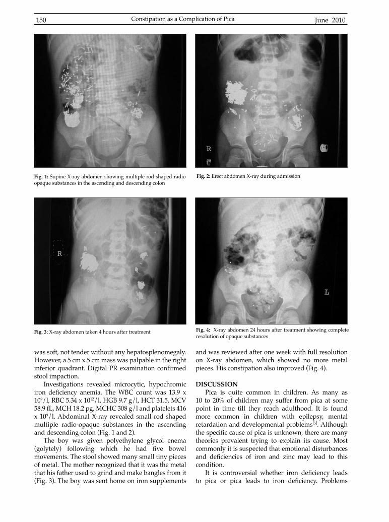
June 2010150
was soft, not tender without any hepatosplenomegaly. However, a 5 cm x 5 cm mass was palpable in the right inferior quadrant. Digital PR examination confirmedstool impaction.
Investigations revealed microcytic, hypochromic iron deficiency anemia. The WBC count was 13.9 x10⁹/l, RBC 5.34 x 10¹²/l, HGB 9.7 g/l, HCT 31.5, MCV 58.9 fL, MCH 18.2 pg, MCHC 308 g/l and platelets 416 x 109/l. Abdominal X-ray revealed small rod shaped multiple radio-opaque substances in the ascending and descending colon (Fig. 1 and 2).
The boy was given polyethylene glycol enema (golytely) following which he had five bowelmovements. The stool showed many small tiny pieces of metal. The mother recognized that it was the metal that his father used to grind and make bangles from it (Fig. 3). The boy was sent home on iron supplements
and was reviewed after one week with full resolution on X-ray abdomen, which showed no more metal pieces. His constipation also improved (Fig. 4).
DISCUSSIONPica is quite common in children. As many as
10 to 20% of children may suffer from pica at some point in time till they reach adulthood. It is found more common in children with epilepsy, mental retardation and developmental problems[5]. Although the specific cause of pica is unknown, there are manytheories prevalent trying to explain its cause. Most commonly it is suspected that emotional disturbances and deficiencies of iron and zinc may lead to thiscondition.
It is controversial whether iron deficiency leadsto pica or pica leads to iron deficiency. Problems
Fig. 1: Supine X-ray abdomen showing multiple rod shaped radio opaque substances in the ascending and descending colon
Fig. 2: Erect abdomen X-ray during admission
Fig. 3: X-ray abdomen taken 4 hours after treatment Fig. 4: X-ray abdomen 24 hours after treatment showing complete resolution of opaque substances
Constipation as a Complication of Pica
KUWAIT MEDICAL JOURNAL 151June 2010
encountered by children with pica are worm and parasitic infestation, intestinal obstruction or perforation and lead poisoning[6,7]. Treatment of pica includes behavior and development, environmental and family education approaches. Treatment should also be added on any missing nutrients like iron and zinc and other medical problems, such as lead exposure[8].
CONCLUSIONWe are the first to report a case pica involving
tiny rod shaped steel pins used to make designer bangles. We believe that pica is one of the causes of constipation. We highly recommend abdominal X-ray for chronic constipation as literature shows that 10 to 20% of children have pica problem. This approach could prevent complication and inappropriate use of medication. We also encourage family education towards health issues related to constipation and pica, and we stress highly on safety, making sure that all medicines, toxic substances, metals and other harmful substance be kept away from the reach of children. Further studies are needed to find the exactmechanism behind pica that leads to constipation.
REFRENCES
1. Viguria Padilla F, Mija’n de la Torre A. Pica: the portrait of a little known clinical entity. Nutr Hosp 2006; 21:557-566.
2. Fotoulaki M, Panagopoulou P, Efstratiou I, Nousia-Arvanitakis S. Pitfalls in the approach to pica. Eur J Pediatr 2007; 166:623-624.
3. Loening-Baucke V, Swidsinski A. Constipation as cause of acute abdominal pain in children. J Pediatr 2007; 151:666-669.
4. Lake AM. Chronic abdominal pain in childhood: diagnosis and management. Am Fam Physician1999; 59:1823-1830.
5. Boukthir S. Does your child have pica? Acta Gastroenterol Belg 2007; 70:245-246.
6. Anderson JE, Akmal M, Kittur DS. Surgical complications of pica: report of a case of intestinal obstruction and a review of the literature. Am Surg 1991; 57:663-667.
7. Numanoglu KV, Tatli D. A rare cause of partial intestinal obstruction in a child: colonic lithobezoar. Emerg Med J 2008; 25:312- 313.
8. Rose EA, Porcerelli JH, Neale AV. Pica: common but commonly missed. J Am Board Fam Pract 2000; 13:353-358.

KUWAIT MEDICAL JOURNAL June 2010152
Case Report
Kuwait Medical Journal 2010; 42 (2): 152-154
Acute Demyelinating Encephalomyelitis with Polyneuropathy Caused by Acute Brucella Infection: A Case Report
ABSTRACT
Address correspondence to: Dr. K.Y.M. Prasad, MD, MRCP (UK), Department of Medicine, Al Jahra Hospital, Kuwait. PO Box 62276, Jahra 02153, Kuwait. Tel: 00965- 4801065, E-mail: [email protected]
Koppolu YM Prasad, Osman A Mapkar, Eisa Al KhaledyDepartment of Medicine, Al Jahra Hospital, Kuwait
Brucellosis is a zoonotic infectious illness. It is the commonest zoonotic illness in the world. Our patient, a forty-year-old Saudi lady had fever, diminished level of consciousness, bilateral optic atrophy, bilateral sensorineural deafness and upper motor neurone type paraparesis with bladder involvement. This is an uncommon and rare combination of clinical presentation in acute brucellosis. Brain magnetic resonance imaging (MRI) showed features
of acute demyelinating encephalomyelitis (ADEM). Electrophysiological studies of the extremities revealed motor sensory neuropathy with axonal involvement. To the best of our knowledge, a case of acute brucellosis with such a combination involving both the central and peripheral systems simultaneously in a patient has not been reported before. After appropriate diagnosis our patient responded remarkably well to the treatment.
KEY WORDS: Brucella melitensis, neurobrucellosis, zoonosis
INTRODUCTIONBrucellosis is zoonotic infectious illness. It is the
most common zoonosis in the world with an annual occurrence of more than 500,000 cases. It is endemic in the regions where animals are raised in large numbers, in the absence of correspondingly high level of hygiene. It is transmitted to humans through consumption of raw milk or infected milk products, contact with infected animals or through the respiratory tract, abraded skin or conjuctiva[1]. It is characterized by recurrent attacks of fever, hepatosplenomegaly, lymphadenopathy and joint pain particularly affecting the sacroiliac, knee, ankle, elbow and shoulder joints. Less commonly it affects myocardium, lungs, kidney, brain meninges and gonads. Among the nations for which reliable data are available, those with the highest current incidence in annual cases per million people are: Syria (1603.4), Mongolia (605.9), Kyrgystan (362.2), Iraq (278.4), Turkey (262.2), Iran (238.6), Saudi Arabia (214.4), Tajikistan (211.9), Macedonia (148), Kazakhstan (115.8), Algeria (84.3), Albania (63.6), Azerbaijan (52.6), Turkmenistan (51.5), Lebanon (49.5), United Arab Emirates (41), Oman (35.6), Peru (34.9), Tunisia (34.5) and Kuwait (33.9)[1,2].
Nervous system involvement can be categorized into central and peripheral forms. The former is commonly acute and is seen in about five percent
cases, resulting in meningoencephalitis, while the latter may either be acute or chronic in presentation[1-3]. We present this case to highlight the unusual and rare combination of clinical features involving both the central and peripheral nervous system simultaneously in acute brucellosis.
CASE REPORTA 40-year-old Saudi lady presented with fever
and headache of 20 days duration. She had difficultyin walking, dysphasia, incontinence of bladder and bowel since two weeks and dimness of vision since 18 months. On examination she had diminished level of consciousness with a Glasgow coma score of 5, neck rigidity and a positive Kernig’s sign. She had bilateral sensorineural deafness, bilateral optic atrophy and bilateral sixth cranial nerve palsy. She had an upper motor neurone type of paraparesis with clonus and power in lower limbs of 2-3 / 5.
The routine investigations such as complete blood count, erythrocyte sedimentation rate, coagulation profile, urine routine and biochemical profile wereall normal. Chest X-ray and electrocardiogram were normal. Brucella agglutination test was negative. Cerebrospinal fluid (CSF) was clear withhypoglycorrhachia, increased proteins and no cells. CSF gram, acid fast stain and acid fast bacilli (AFB)

KUWAIT MEDICAL JOURNAL 153June 2010
culture was negative. Mantoux test was negative. Initial cultures for blood, CSF and urine were all negative.
Magnetic resonance imaging (MRI) of brain (Fig. 1 and 2) showed evidence of extensive patchy irregular confluent areas of hyperintensities on T2involving bilateral periventricular white matter with extension into subcortical fibres. Similar patchyareas were also seen involving bilateral centrum semiovale possibly due to demyelination or acute demyelinating encephalomyelitis (ADEM). MRI of the spine was normal. Electroneuromyogram (ENMG) revealed motor sensory neuropathy with axonal involvement. Repeat CSF analysis showed normal glucose with increased proteins, 170 RBCs and 260 WBCs predominantly lymphocytes. The appearance of cells in the follow up CSF analysis is due to the Jarisch Herxheimer reaction to the treatment, a well known phenomenon. The patient was being treated provisionally as tuberculous meningitis. She showed gradual improvement in her sensorium, deafness and paraparesis. About two weeks subsequently the blood culture grew Brucella melitensis. She was subsequently treated for neurobrucellosis with doxycycline 100 mg twice daily, rifampicin 900 mg daily and gentamycin 180 mg daily in divided doses for the initial two weeks with which the patient started to show gradual improvement over a period of one month. Her sensorium improved first followed by hervision and then her sixth nerve palsy. Over the next two months she recovered power in both her lower limbs completely with improvement in her bladder function. The last to recover though not completely was her deafness which took about six months. She was simultaneously followed by neurologist and the treatment (doxycycline and rifampicin) was continued for a total of 24 months as recommended to prevent any episodes of relapse.
DISCUSSIONIn the Mediterranean region and the Middle east
(ME), the species of greatest importance is B melitensis[2]. Among the highest prevalence for human B melitensis is that of the Bedouins of Kuwait[3]. Brucella are Gram negative, intracellular coccobacillary orgainsms that resemble fine grains of sand[4].
Human brucellosis is traditionally described as a disease of protean manifestations. However, fever is invariable and can be spiking and accompanied by rigors, if bacteremia is present, or may be relapsing, mild, or protracted. Malodorous perspiration is almost pathognomonic. Constitutional symptoms are generally present. Physical examination is generally nonspecific, though lymphadenopathy, hepatomegaly,or splenomegaly is often present[4].
Neurobrucellosis or nervous system affection by Brucella organisms is a rare manifestation and is seen about 5 - 7% of patients and such complications often have an ominous prognosis[4,5]. Neurological complications can occur in both acute and chronic forms. Encephalopathy is most likely to arise with melitensis (irritability, confusion, obtundation, coma)[6]. In areas of endemicity, it may be the most common cause of acquired sensorineural deafness[7]. Shakir et al have categorized the neurobrucellosis into central and peripheral forms, the former occurring commonly as acute meningoencephalitis and the latter as polyradiculoneuropathy[8]. CNS manifestations include transient ischemic attacks (TIA), hemiplegia, abscess, increased intracranial tension, intracerebral hemorrhage, subarachnoid hemorrhage, peripheral neuritis, radiculitis, papilledema, posterior fossa syndromes various cranial nerve palsies, parkinsonism, chorea, athetosis, narcolepsy or cataplexia or acute inflammatorydemyelinating polyneuropathy mimicking Guillain-Barré syndrome (GBS)[9].
Fig. 1: T2 weighted image shows periventricular hyperintensities - sagittal section
Fig. 2: T2 weighted image shows periventricular hyperintensities -coronal section

June 2010154
Why the nervous system presents itself as the primary site of focal brucellosis in some patients is not known. It may be a favored site of chronic infection because of inadequate immune surveillance, or it may be particularly vulnerable to autoimmunity engendered by Brucella organisms that have chronically infected other parts of the body or by reintroduction of infection to an already sensitized individual[5,9].
The occurrence of inflammatory central andperipheral demyelination as the pathological manifestation of some types of neurobrucellosis, associated with inflammatory perivenular infiltrationbut not with histologically demonstrable organisms, strongly suggests that autoimmune mechanisms is virtually identical to that found in ADEM, while the change that may be found in peripheral nerves or spinal roots may be identical to that found in acute inflammatory polyneuritis of the GBS variety. In thesetypes of chronic neurobrucellosis, axons tend to be spared, although in severe cases, as in severe ADEM or GBS, there may be considerable axonal loss and associated encephaloclasia or peripheral neuraxonal dissolution. The pathological changes observed also share certain aspects of their appearance with multiple sclerosis (MS) or neuroborreliosis[5,9].
Diagnosis of brucellosis can be established by blood cultures in about 15-70% of patients. Bone marrow cultures are considered the gold standard because the relatively high concentration in the reticuloendothelial system makes it easier to detect the organism[4]. Serum agglutination tests can be used for diagnosis as well especially in the recommended titers based on the endemicity of the disease. Rising titers are more informative than a single reading. In patients with neurobrucellosis, enzyme linked immunosorbent assay (ELISA) offers significant diagnostic advantages.MRI may disclose multiple plaques as in MS or ADEM. These plaques tend to be found in the frontal white matter and the centrum semiovale. In some cases, it might be symmetrical and extensive. ENMG studies show delay in F-wave latencies and mild slowing of nerve conduction (motor nerves affected more than sensory)[10].
There are various treatment regimens. Doxycycline, rifampicin and streptomycin form the basis of treatment. Doxycycline is used in dose of 100 mg BD, rifampicin in doses of 600 – 900 mg daily and streptomycin in the dose of 15 mg/kg/day. The duration of treatment
for neurobrucellosis is prolonged, varying from six months to two years based on the clinical response and analysis of the cerebrospinal fluid. Other drugsbeing used are sulphonamides, fluoroquinolones andother aminoglycosides[4].
CONCLUSIONBrucellosis is a worldwide disease with areas of
endemicity. Its clinical manifestations are protean and it is a great mimicker. In areas of endemicity it should always be kept in mind inspite of negative serology or cultures. Our case highlights that acute brucellosis may not restrict itself typically to the central or peripheral forms and there might be widespread neurological involvement which needs to be kept in mind. Our patient was diagnosed, received appropriate treatment and had an excellent response with no neurological sequelae.
REFERENCES
1. Pappas G, Papadimitriou P, Akritidis N, Christou L, Tsianos EV. The new global map of human brucellosis. Lancet Infect Dis 2006; 6:91-99.
2. Fallatah SM, Oduloju AJ, Al-Dusari SN, Fakunle YM. Human brucellosis in Northern Saudi Arabia. Saudi Med J 2005; 26:1562-1566.
3. Lulu AR, Araj GF, Khateeb MI, Mustafa MY, Yusuf AR, Fenech FF. Human brucellosis in Kuwait: a prospective study of 400 cases. Q J Med 1988; 66:39-54.
4. Pappas G, Akritidis N, Bosilkovski M, Tsianos E. Brucellosis. N Engl J Med 2005; 352:2325-2336.
5. Abramsky O. Neurological features as presenting manifestations of brucellosis. Eur Neurol 1977; 15:281-284.
6. Mousa AR, Koshy TS, Araj GF, et al. Brucella meningitis: presentation, diagnosis and treatment--a prospective study of ten cases. Q J Med 1986; 60:873-885.
7. Thomas R, Kameswaran M, Murugan V, Okafor BC. Sensorineural hearing loss in neurobrucellosis. J Laryngol Otol 1993; 107:1034-1036.
8. Shakir Ra, Al-Din AS, Araj GF, Lulu AR, Mousa AR, Saadah MA. Clinical categories of neurobrucellosis. A report of 19 cases. Brain 1987; 110:213-223.
9. Al-Deeb SM, Yaqub BA, Sharif HS, Phadke JG. Neurobrucellosis: clinical characteristics, diagnosis, and outcome. Neurology 1989; 39:498-501.
10. Akdeniz H, Irmak H, Anlar O, Demiroz AP. Central nervous system brucellosis: presentation, diagnosis and treatment. J Infect 1998; 36:297-301.
Acute Demyelinating Encephalomyelitis with Polyneuropathy Caused by Acute Brucella...

KUWAIT MEDICAL JOURNAL 155June 2010
INTRODUCTION Aneurysmal bone cysts (ABCs) are benign bone
lesions that account for approximately 1 - 6% of all bone tumors. They typically occur in the second decade of life with greater than 50% occurring in long bones and 3 - 20% in the spine. The thoracic (32%) and lumbar (34%) vertebrae are the most common sites for ABC with those in the cervical spine making up only 2% of cases[1,2]. ABCs in the spine typically involve the posterior elements, but are not uncommon in the vertebral body and may eventually extend to more than one vertebra, leaving the disc space intact. They tend to expand the vertebra with cortical thinning or destruction and may cause vertebra plana. Clinically, cervical ABCs present with neck pain, stiffness, swelling, torticollis and reduced range of movement[2] and because of their location, could result in neurological complications secondary to spinal cord or nerve root compression.
CASE REPORTA 14-year-old girl presented with a four month
history of progressive left shoulder and upper limb pain with swelling on the left side of the neck. There was no history of previous inflammatory disease orrelevant trauma. There was no neurological deficit.Conventional cervical spine radiographs showed an expansile lytic lesion of C6 spinous process, extending into the soft tissues of the neck and osteopenia of
Case Report
Kuwait Medical Journal 2010; 42 (2): 155-158
Primary Aneurysmal Bone Cyst at C5 and C6 Presenting as an Extradural Mass
ABSTRACT
Address correspondence to: Dr Eki Aghahowa, Specialist Radiologist, Department of Radiology, Al Jahra Hospital, P O Box1807, Jahra 01020, Kuwait. Fax: (965) 24577198, E-mail: [email protected]
Ekinadoese Juliana Aghahowa, Venkata Mallikarjuna SontenamDepartment of Radiology, Al Jahra Hospital, Kuwait
Aneurysmal bone cysts are rare in the spine, and when they do occur, there is the possibility of spinal cord compression with its consequent surgical risks and morbidity. We report a case of a 14-year-old girl who presented with a four month history of left-sided neck swelling. Radiological
imaging consisting of plain radiographs, CT and MRI showed cervical aneurysmal bone cyst involving C5 and C6 vertebrae with extradural cord compression and displacement. Aneurysmal bone cyst should be one of the considerations in the evaluation of a neck swelling.
KEY WORDS: bone tumors, cervical, neck swelling
C6 vertebral body (Fig. 1). CT and MRI revealed expansile lytic destruction of the left aspect of C5 vertebral body and left transverse process; expansile lytic lesion of C6 vertebral body extending into the left posterior elements and compromising C5 and C6 fossa transversarium and neural foramina which were poorly delineated. There was an associated 4.3 cm x 3.6 cm lobulated multicystic mass displacing the left posterior paraspinal muscles, with fluid-fluidlevels of different signal intensities on T1 and T2WI, representing different stages of blood by products and intense rim contrast enhancement of the cysts and septae. There was anterior displacement of the left vertebral artery and intraspinal extension of the mass from C5 to C7, causing extradural cord compression and displacement of the cord to the right. There was no signal intensity alteration within the spinal cord. There was increased signal intensity on T2WI of C6 vertebral body with associated reduced vertebral height (Figs. 2 - 6). There was also prevertebral extension with displacement of the left prevertebral muscles. There was no cortical fracture seen. A diagnosis of ABC was made. ABC is the only benign tumor which has contiguous vertebral involvement while relatively sparing the intervertebral disc.
DISCUSSIONABCs comprise approximately 1.4% of all bone
tumors and 15% of all primary spine tumors. Overall,

June 2010156
women are slightly more affected than men, with a ratio of 1.04:1. Primary ABCs (65 - 99%) are as a result of local circulatory disturbance due to trauma while secondary ABCs (1 - 3%) arise in preexisting tumors, causing venous obstruction / arterio-venous fistula.Primary ABCs are found in children and are not associated with any other tumor, whereas secondary ABCs are common in adults and develop in pre-existing fibrous dysplasia, chondroblastoma, osteoblastoma,angioma, telengiectatic osteosarcoma, solitary bone cyst, chondromyxoid fibroma, non-ossifying fibromaor metastatic carcinoma.
The mean age at presentation is 16 years. The duration of symptoms prior to presentation is usually
four to eight months. Local pain is the most common feature and is often worse at night with recumbency. This may be associated with swelling. Palpable mass occurs in 6% of ABCs of the spine. Most of spinal ABCs originate in the posterior elements of the spine. Capanna et al confirmed dorsal involvement in eachof the 22 examined spinal ABCs, with asymmetrical involvement of the vertebral body in 12 cases, symmetrical involvement in two cases and the posterior vertebral arch solely involved in eight cases[3]. While ABCs in long bones usually present as geographic and osteolytic lesions with well defined margins, spinalABCs present vesicular structural lacunae in the area around the lesion with pronounced paraosseous tumor
Fig. 1: Lateral cervical radiograph showing expansile lytic lesion of C6 spinous process with irregularity of the anterior aspect of the vertebral body
Fig. 2: Axial CT with IV contrast. Soft tissue window showing expansile lytic lesion of the left aspect of C5 vertebral body, transverse process, pedicle and lamina with resultant egg shell thinned cortex; associated fluid-fluid levels and intraspinalextension are seen.
Fig. 3a: Coronal T1WI and Fig. 3b: Coronal T2WI showing lobulated multicystic left paraspinal mass extending to the extradural space and displacing the cord to the right and surrounding left posterior paraspinal muscles laterally
Fig. 3a Fig. 3b
Primary Anuerysmal Bone Cyst at C5 and C6 Presenting as an Extradural Mass

KUWAIT MEDICAL JOURNAL 157June 2010
expansion and better defined margins adjacent to theundestroyed bone. Eggshell periosteal calcificationthat is a typical feature of spinal ABC on conventional imaging[4] was not seen in our patient. ABCs show typical imaging features on CT and MR with bony expansion, cortical thinning and destruction and either multiple double fluid level cysts or an additional solidcomponent with enhancement of the stroma. In cases where there is a solid component, it is impossible to differentiate ABC macroscopically from a giant cell tumor or hemangioblastoma and only histological analysis can determine the type of tumor.
Pathological fracture with partial collapse of the vertebral body is common, which may progress to vertebra plana. Our patient had loss of height of C6
vertebral body. CT is better for determining the extent of bone destruction, integrity of cortical bone and for detecting calcium deposits[5]. MRI provides better evaluation and delineation of the cyst contents and extent of bone marrow and soft tissue involvement. The varying signal intensities seen within the cysts on T1 and T2 weighted images are due to different oxidation levels of blood and blood breakdown products in the cyst fluid. MRI also better delineates the degreeof compression of the neural elements. Differential diagnosis of cystic lesions in the head and neck region which show fluid-fluid level on MR images are cysticschwannoma, neurofibroma, cystic hygroma, softtissue hemangioma and simple bone cyst [6].
Neurological symptoms may be the initial presentation. Early nerve root compression may cause paraesthesia and radicular pain that can be referred to the chest wall, abdomen or extremities. Extradural spinal cord compression due to the rapidly enlarging lesion or vertebral body collapse can result in myelopathy or acute paraparesis. Rapid deterioration to complete paraplegia can occur if the lesion is left untreated[2]. Spinal ABC in young patients presenting with acute cord compression symptoms has been previously reported[4-6] and therefore, early recognition of the condition is required. Our case showed a large extradural component compressing the spinal cord,
Fig. 4: Coronal T1WI with IV Gadolinium showing rim contrast enhancement of the left paraspinal and extradural mass with compression of the cord
Fig. 5: Sagittal T2WI showing loss of height of C6 vertebral body with increased marrow signal intensity and extradural compression of spinal cord posteriorly
Fig. 6: Axial T1WI showing left multiloculated paraspinal and extradural mass with fluid-fluid levels of different signal intensitiesand increased signal intensity in one of the cysts due to acute bleed

June 2010158
but surprisingly without any neurological deficit atthe time of presentation. The diagnosis of ABC in our patient was based on morphological imaging aspects of blood filled cysts with rim contrast enhancement,contrast enhancing thin smooth septae and a growth pattern sharply demarcated from soft tissue. Also, there was no involvement of the intervertebral disc even though there was contiguous vertebral involvement.
Many authors strongly recommend biopsy for tissue diagnosis[2]. Needle biopsy is the mode of choice to obtain tissue for histological evaluation, although it carries the risk of hemorrhage or failure to make a diagnosis. Due to the rarity of cervical ABC, no established therapeutic regimen exists. Traditionally, the treatment of choice for ABCs has been surgical resection, but the high recurrence rate, especially after curettage or partial resection, has led to other treatment options. These include selective arterial embolization, either as sole treatment or as an adjunct to surgery, radiation therapy, injection of methylprednisone and calcitonin or radionuclides and percutaneous ethibloc injection. Local recurrence has been also noted in patients who only had radiotherapy[3]. In our case, the patient’s guardian refused permission for the patient to have a biopsy or any form of surgery.
CONCLUSIONA high index of suspicion is necessary for early/
prompt diagnosis and treatment of cervical ABC to avoid acute cord compression.
REFERENCES
1. Ozaki T, Halm H, Hillmann A, Blasius S, Winkelmann W. Aneurysmal bone cysts of the spine. Arch Orthop Trauma Surg 1999; 119:159-162.
2. Papagelopoulos PJ, Currier BL, Shaughnessy WJ, et al. Aneurysmal bone cyst of the spine: Management and outcome. Spine 1998; 23:621-628.
3. Capanna R, Albisinni U, Picci P, Calderoni P, Campanacci M, Springfield DS. Aneurysmal bone cystof the spine. J Bone Joint Surg Am 1985; 67:527-531.
4. Pennekamp W, Peters S, Schinkel C, et al. Aneurysmal bone cyst of the cervical spine. Eur Radiol 2008; 18:2356-2360.
5. Chan MS, Wong YC, Yuen MK, Lam D. Spinal aneurysmal bone cyst causing acute cord compression without vertebral collapse: CT and MRI findings.Paediatr Radiol 2002; 32:601-604.
6. Deo SD, Fairbank JC, Wilson-Macdonald J, et al. Aneurysmal bone cyst as a rare cause of spinal cord compression in a young child. Spine 2005; 30:E80-E82.
Primary Anuerysmal Bone Cyst at C5 and C6 Presenting as an Extradural Mass

KUWAIT MEDICAL JOURNAL 159June 2010
INTRODUCTIONTwiddler’s syndrome is an uncommon complication
of defibrillator or permanent pacemaker implantation,caused by device malfunction. It occurs as a result of torsion, dislodgment and/or fracture of the defibrillatoror pacing lead when the patient either knowingly or unknowingly rotates or twiddles the implanted device in its pocket[1]. This syndrome was first described in1968 by Bayliss and his group[2]. Defibrillator Twiddler’ssyndrome is another variant of this condition in which the implanted device is an implantable cardioverter-defibrillator and was first described by Veltri et al in 1984 in a patient with abdominally implanted implantable cardiac device (ICD). Since then there are a number of reports of defibrillator Twiddler’s syndrome describedby many authors[3-7].
CASE REPORTA 76-year-old Kuwaiti gentleman with diabetes
mellitus, hypertension, chronic atrial fibrillation andhyperlipidemia sustained anterior wall myocardial infarction, was found to have triple vessel coronary artery disease, post coronary artery bypass graft (CABG in year 2000) and ischemic cardiomyopathy. Echocardiography revealed severe left ventricular systolic dysfunction with left ventricular ejection fraction of 20% and significant mitral regurgitation.Prophylactic single chamber cardioverter-defibrillator
Kuwait Medical Journal 2010; 42 (2): 159-161
Twiddler’s Syndrome in a Patient with an Implantable Cardioverter Defibrillator
ABSTRACT
Address correspondence to: Al Kandari Fawzia Yousef, FRCP (UK), Consultant Cardiologist and Cardiac Electrophysiologist,Department of Cardiology, Kuwait Heart Center, P O Box 25772, Safat 13118, Kuwait. E-mail: [email protected]
Fawzia Yousef Al Kandari, Abdul Mohammed Shukkur Department of Cardiology, Electrophysiology Division, Kuwait Heart Center, Kuwait
Twiddler’s syndrome is a rare complication in patients with permanently implanted defibrillator or pacemaker.It typically presents with device malfunction as a result of torsion, dislodgment and/or fracture of leads. This condition occurs when the patient either consciously or unconsciously manipulates the implanted device in its pocket. We report
a case of Twiddler’s syndrome presenting one year after defibrillator implantation with device malfunction as non-capture and intermittent non-sensing. Chest radiography revealed twiddling of implantable cardiac device (ICD) leads in the pocket. The treatment option was discussed and a new defibrillator was implanted without any complication.
KEY WORDS: defibrillator implantation, device malfunction, pacemaker
was implanted on January 2007 (Guidant Vitality 2 VR) with the defibrillator lead advanced to rightventricular apex. During follow up in our outpatient division at three, six and nine months, the patient did not complain of any specific symptoms apartfrom those of class 1 heart failure. Interrogation of the device did not reveal any episode of ventricular arrhythmia. At one year routine follow up ICD lead testing showed significant drop in R wave sensing,intermittent non-sensing, very high lead impedance (more than 2000 Ohms), non-capture at maximum output and one inappropriate shock after which the patient went into sinus rhythm. Chest radiography showed that the defibrillator lead had been pulled outfrom the right ventricle with loss of loop and clearly twiddled tightly many times near the device (Fig. 1A). On direct interrogation, the patient stated that he had never intentionally pulled out or manipulated the defibrillator lead. Intra-operatively an incision wasmade over the ICD pocket in the left pectoral region. ICD was explanted and lead was clearly looped repeatedly on itself in the pocket with fracture within the loop (Fig. 1B). The lead was cut, capped and a new defibrillator lead was advanced to the right ventricularapex with new atrial pacing lead in the right atrium. Both leads were connected to a new dual chamber ICD (Guidant Vitality 2 EL DR) (Fig. 2). To reduce the future risk of Twiddler’s syndrome the defibrillator
Case Report

June 2010160
was anchored to the underlying fascia with non-absorbable suture.
DISCUSSIONTwiddler’s syndrome is a rare condition and
potentially serious complication in which manipulation with the device leads to twisting, fracture or dislodgment of the pacing or defibrillator lead. Wereport a case of Twiddler’s syndrome in which the patient’s manipulation of the defibrillator lead was thecause of failure of ICD system.
Twiddler’s syndrome was first described in 1968by Bayliss et al[2]. It refers to permanent malfunction of pacemaker due to patient’s manipulation of the pulse generator. Defibrillator Twiddler’s syndromeis caused by the failure of defibrillator lead and wasfirst described by Vetri et al in 1984. Since then there have been similar cases reported in patients with ICD[1,3-7]. Our patient had many features similar to those in earlier reports of defibrillator Twiddler’ssyndrome. The patient was a 76-year-old obese, diabetic, hypertensive, post CABG with ischemic cardiomyopathy, atrial fibrillation and chronic renalfailure who had undergone ICD implantation one year earlier and who presented with device failure. Chest X-ray remains the most important diagnostic tool. In
our case the CXR showed retraction of the defibrillatorlead from the right ventricle with typical twisting and coiling of lead near the device. Multiple risk factors (as in our case) increase the subcutaneous tissue compliance and have been associated with Twiddler’s syndrome[8,9]. Various therapeutic approaches have been proposed[10,11]. In our case, a new dual chamber ICD was implanted.
Fig. 1A: Chest radiography showing retraction of the defibrillatorlead from the right ventricle, loss of loop and twisting and coiling of the lead near the device
Fig. 1B: Photograph obtained on opening the defibrillator pocketshowing loops and tight coils of leads
Fig. 2: Chest radiography showing new dual chamber implantable cardioverter-defibrillator with old lead capped
Twiddler’s Syndrome in a Patient with an Implantable Cardioverter Defibrillator

KUWAIT MEDICAL JOURNAL 161June 2010
CONCLUSIONDefibrillator Twiddler’s syndrome is an infrequent
and potentially dangerous late complication of ICD implantation. Problems specifically related to ICDs arethe failure of anti-tachycardia therapy or inappropriate shocks due to non-sensing or under-sensing, high lead impedance and non-capture. Therefore steps to avoid Twiddler’s syndrome should be taken at the time of implantation which might include anchoring the ICD in a sub pectoral position, placing the ICD in a Dacron pouch and avoiding oversizing of the ICD pocket.
REFERENCES
1. de Buitleir M, Canver CC. Twiddler’s syndrome complicating a transvenous defibrillator lead system.Chest 1996; 109:1391-1394.
2. Bayliss CE, Beannlands DS, Baird RJ. The pacemaker-twiddler’s syndrome: a new complication of implantable transvenous pacemakers. Can Med Assoc J 1968; 99:371-373.
3. Veltri EP, Mower MM, Reid PR. Twiddler’s syndrome: a new twist. Pacing Clin Electophysiol 1984; 7:1004-1009.
4. Underhill SJ, Sanders J, Davis C, Broudy D. Defibrillator“twiddler’s syndrome”. Pacing Clin Electrophysiol 1991; 14:1555-1556.
5. Mehta D, Lipsius M, Suri RS, Krol RB, Saksena S. Twiddler’s syndrome with the implantable cardioverter-defibrillator. Am Heart J 1992; 123:1079-1082.
6. Robinson LA, Windle JR. Defibrillator twiddler’ssyndrome. Ann Thorac Surg 1994; 58:247-249.
7. Ruder MA, Mead RH, Smith NA, Winkle RA. Defibrillator “twiddler’s syndrome”. Pacing ClinElectrophysiol 1990; 13:1073-1074.
8. Nicholson WJ, Tuohy KA, Tilkemeier P. Twiddler’s syndrome. N Engl J Med 2003; 348:1726-1727.
9. Castillo R, Cavusoglu E. Twiddler’s Syndrome: An interesting cause of pacemaker failure. Cardiology 2006; 105:119-121.
9. Mancini MC, Grubb BP. A technique for the prevention of automatic implantable cardioverter defibrillatorgenerator migration. Pacing Clin Electrophysiol 1990; 13:946-947.
10. Parsonnet V, Bernstein AD, Neglia D, Omar A. The usefulness of a stretch-polyester pouch to encase implanted pacemakers and defibrillators. Pacing ClinElectrophysiol 1994; 17:2274-2278.

KUWAIT MEDICAL JOURNAL June 2010162
INTRODUCTIONCor triatriatum is a rare congenital cardiac
anomaly in which a common pulmonary venous chamber (proximal chamber) is separated from the left atrium (distal chamber) by a fibromuscular septum(membrane)[1, 2]. Majority of the patients have one or more openings in the membrane. Cor triatriatum is usually an isolated congenital anomaly, but may be associated with cyanotic and acyanotic congenital heart diseases[3, 4]. The clinical manifestations depend on the size of the opening in the membrane. Approximately 75% of patients with severely restrictive cor triatriatum die in infancy unless corrected surgically[2]. In this case, we report a very rare incidental finding of left cortriatriatum in a 67-year-old gentleman.
CASE HISTORYIn April 2007, a 67-year-old man with a history
of type II diabetes mellitus (DM) complicated by retinopathy and nephropathy was admitted with bilateral flank pain and progressive renalimpairment. He had no history of fever. The baseline serum creatinine was 159 mmol/l. Moreover, he had hypercholesterolemia and long standing hypertension. Physical examination revealed a blood pressure of 130 / 80 mmHg, regular pulse and normal jugular venous pressure. His systemic examination was normal. His serum creatinine was elevated at 227 mmol/l. Complete blood count and liver profiles were normal. Electrocardiogram and chestX-ray were normal. Transthoracic echocardiography revealed normal biventricular systolic function. The aortic valve was sclerotic with mild aortic
Case Report
Kuwait Medical Journal 2010; 42 (2): 162-164
Left Cor Triatriatum in an Elderly Patient: A Brief Review
ABSTRACT
Address correspondence to: Dr. Bassam Bulbanat, Department of Medicine, Al-Amiri Hospital, Kuwait. Tel: (+965) 22526270, Fax: (+965) 22543239, E-Mail:[email protected]
Bassam Bulbanat, Marwan Aburezq, Hussam E MostafaDepartment of Medicine, Al-Amiri Hospital, Kuwait
We report a case of incidental asymptomatic cor triatriatum that was diagnosed in the 7th decade of life. The patient had progressive renal failure secondary to obstructed left kidney. He had hypertension. Routine transthoracic
echocardiography revealed a septum in the left atrium. Transesophageal echocardiography confirmed the diagnosisof cor triatriatum. We present our case, along with a review of this rare congenital cardiac anomaly.
KEY WORDS: cardiac anomaly, cor triatriatum, echocardiography
insufficiency. There was a linear fibrous strand acrossthe left atrium. Transesophageal echocardiography confirmed the diagnosis of left cor triatriatum witha discrete membrane in the left atrium (Figs. 1 & 2). No concomitant cardiac anomaly was detected. The echocardiographic images clearly show the absence of significant hemodynamic obstruction to the bloodflow across the discrete membrane due to the presenceof a large aperture (2.4 cm diameter). A Doppler flowevaluation did not show significant gradient acrossthe cor triatriatum membrane. Hence, this congenital anomaly did not cause any cardiac symptoms over the past decades and was incidentally discovered.
DISCUSSIONCor triatriatum is a rare congenital anomaly causing
a barrier to the emptying of the pulmonary veins. The incidence has been variously reported as 0.1[5] to 0.4%[3]. In this condition, the pulmonary veins empty into an accessory chamber lying superior to the true left atrial chamber communicating with it by means of a narrow opening. The narrow opening between the accessory chamber and the true left atrium constitutes a point of obstruction to the pulmonary venous flow. Functionally, the condition resembles mitral stenosis[1]. Loeffler[6] in 1949 classified cor triatriatum into threegroups on the basis of the number and size of the openings in the fibromuscular septum into the leftatrium: Group I = no opening; Group II = 1 or more small openings; and Group III = a wide opening. Patients with Group III cor triatriatum may reach late adulthood (similar to our patient). Either atrium can be subdivided, but unspecified, the term ‘cor triatriatum’

KUWAIT MEDICAL JOURNAL 163June 2010
invariably is used in the context of a subdivided left atrium – cor triatriatum dexter being used for subdivided right atrium. There are several variants of the left atrial type which have been categorized in exquisite detail by Thilenius, Bharati and Lev[4, 7]. In the most frequent form the veins drain to a proximal chamber, which is separated by an oblique muscular partition from a distal chamber. The distal chamber communicates with the left atrial appendage, contains the flap-valve of the fossa ovalis and communicateswith the left ventricle. The variants of this classical form depend upon the communication of the fossa ovalis with the proximal chamber and the association with anomalous pulmonary venous drainage. Sometimes, there is total anomalous pulmonary venous drainage to the coronary sinus, but in the absence of cor triatriatum as defined above, the sinus can be so dilated as toappear as a third atrial cavity. Cor triatriatum dexter is almost always due to persistence of the right valve of the sinus venosus. Prominent in fetal life, when it diverts the richly oxygenated inferior vena caval blood to the left atrium, it usually regresses during infancy. It may persist in tricuspid atresia when there is no egress from the right atrium other than through the fossa ovalis. Rarely, a persistent right valve may divide the right atrium in the normally structured heart. In that condition, however, this anomaly leads to an obstruction to right atrial outflow.
The embryologic basis of this anomaly remains controversial. The three main theories involve malseptation, malincorporation, and entrapment[2]. The malseptation hypothesis was put forward by Fowler[8] in 1881. He proposed that the septum sub-dividing the left atrium was an abnormal growth of septum primum. The malincorporation hypothesis suggests that cor triatriatum is the result of incomplete incorporation of the embryonic common pulmonary vein into the left atrium[9]. Van Praagh and Corsini[10] proposed the entrapment hypothesis, which holds that the left horn of the sinus venosus entraps the common
pulmonary vein and thereby prevents its incorporation into the left atrium. The clinical manifestations and longevity patterns of uncomplicated cor triatriatum depend on the degree of obstruction, which ranges from functionally insignificant ridges of tissues(similar to our patient) to complete absence of a direct communication between upper and lower atrial compartments[11,12]. Cor triatriatum usually comes to light in infants or young children, but symptoms may await adolescence or adulthood[13]. In asymptomatic adults with mild obstruction, cor triatriatum may be an incidental echocardiographic finding similar to ourpatient or, as was in a 70-year-old man, an incidental finding at necropsy[2,6,14]. The most common presenting symptoms in adults are dyspnea, hemoptysis, and orthopnea[11]. Heart murmurs are the most significantfinding during physical examination[2,15,16]. Cor triatriatum is frequently associated with other cardiac anomalies. In adults, the most common abnormalities associated with cor triatriatum[2,8,14,16] are mitral regurgitation, secundum atrial septal defect, and the presence of left superior vena cava with unroofed coronary sinus. The recent increase in the reported cases of cortriatriatum may be related to the improvement of echocardiographic and cardiac catheterization technology. Recently, 3-D echocardiographic imaging and cardiac MRI have been helpful in the diagnosis of this rare cardiac anomaly. The surgical management of patients with cor triatriatum in adult depends on the degree of obstruction to pulmonary venous flowandtheassociated intracardiac defects. An important aspect of surgical procedure in correcting cor triatriatum and its associated anomalies is to delineate (intraoperatively) the complex anatomy at the outset, before proceeding to correction[2]. In order to do that, one should approach the largest atrial chamber on the right side first. Theleft atrial chambers and their pathologic membranes must ultimately be approached through the interatrial septum and excised. Subsequently, the interatrial septum is closed with a pericardial baffle or patch, in
Fig.1 : Transesophageal echocardiographic view of discrete membrane in the left atrium (LA) with a large aperture (arrows). LA = left atrium, LV = left ventricle
Fig. 2: Transesophageal echocardiographic view shows LUPV draining into the superior chamber of the left atrium. LA = left atrium, LUPV = left upper pulmonary vein, LAA = left atrial appendage, LV = left ventricle

June 2010164
accordance with the size and nature of the defect. In a recent study, surgical correction of cor triatriatum was associated with low mortality and good long-term results[17]. This result is well reproduced, if surgical correction is undertaken earlier in the course of the disease process.
CONCLUSIONAn excellent long-term prognosis and quality of
life are to be expected in uncomplicated cases of cor triatriatum. The diagnosis of this rare cardiac anomaly is becoming more accurate with the advance of echocardiographic and radiological technology.
REFERENCES
1. Edwards JE, Dry TJ, Parker RL, Burchell HB, Wood EH, Bulbulian AH. An Atlas of Congenital Anomalies of the Heart and Great Vessels. Illinois, Charles C Thomas, 1954; 27-30.
2. Chen Q, Guhathakurta S, Vadalapali G, Nalladaru Z, Easthope RN, Sharma AK. Cor triatriatum in adults: Three new cases and a brief review. Tex Heart Inst J 1999; 26:206-210.
3. Jorgensen CR, Ferlic RM, Varco RL, Lillehei CW, Eliot RS. Cor triatriatum. Review of the surgical aspects with a follow-up report on the first patient successfullytreated with surgery. Circulation 1967; 36:101-107.
4. Anderson RH, Becker AE, Eugene Braunwald. Cardiac Pathology: an integrated text and color atlas.Edinburgh, Churchill Livingstone, 1983; 10.15 -10.16.
5. Niwayama G. Cor triatriatum. Am Heart J 1960; 59:291-317.
6. Loeffler E. Unusual malformation of the left atrium;pulmonary sinus. Arch Pathol 1949; 48:371-376.
7. Thilenius OG, Bharati S, Lev M. Subdivided left atrium: an expanded concept of cor triatriatum sinistrum. Am J Cardiol 1976; 37:743-752.
8. Fowler JK. Membranous band in the left auricle. Trans Pathol Sco London 1881; 33:77-94.
9. Parsons CG. Cor triatriatum; concerning the nature of an anomalous septum in the left auricle. Br Heart J 1950; 12: 327-338.
10. Van Praagh R, Corsini I. Cor triatriatum: pathologic anatomy and a consideration of morphogenesis based on 13 postmortem cases and a study of normal development of the pulmonary vein and atrial septum in 83 human embryos. Am Heart J 1969; 78:379-405.
11. McGuire LB, Nolan TB, Reeve R, Dammann JF Jr. Cor triatriatum as a problem of adult heart disease. Circulation 1965; 31:263-272
12. Feld H, Shani J, Rudansky HW, Rudyak E, Greengart A. Initial presentation of cor triatriatum in a 55-year-old woman. Am Heart J 1992; 124:788-791
13. Perloff JK, Child JS. Congenital heart disease in Adults. Philadelphia, WB Saunders Company, 1998:34.
14. Tanaka F, Itoh M, Esaki H, Isobe J, Inoue R. Asymptomatic cor triatriatum incidentally revealed by computed tomography. Chest 1991; 100:272-274.
15. de Belder MA, Argano V, Burrell CJ. Cor triatriatum sinister, not mitral stenosis, in an adult with previous Sydenham’s chorea: diagnosis and preoperative assessment by cross sectional echocardiography. Br Heart J 1992; 68:9-11.
16. Beller B, Childers R, Eckner F, Duchelle R, Ranniger K, Rabinowitz M. Cor triatriatum in the adult complicated by mitral insufficiency and aorticdissection. Am J Cardiol 1967; 19:749-754.
17. Lupinski RW, Shankar S, Wong KY, Chan YH, Vosloo SM, Moll JJ. Cor triatriatum: clinical presentation of 18 cases. Asian Cardiovas Thorac Ann 2001; 9:106-110.

KUWAIT MEDICAL JOURNAL 165June 2010
INTRODUCTIONCerebral venous thrombosis in children is a rare
disorder that is being increasingly diagnosed because of greater clinical awareness, sensitive neuro-imaging technique and the survival of children with previous lethal disease that confer a predisposition to sinovenous thrombosis[1]. The literature on sinovenous thrombosis in children consists mainly of case reports and analysis of small case series. We report two cases of cerebral venous thrombosis with different predisposing factors – one survived and one expired.
CASE REPORTCase I
A two-month-old Kuwaiti boy presented with fever, cough, and shortness of breath and lethargy of one week duration. He had an episode of apnea with cyanosis. On admission he was found to be drowsy, tachypneic and tachycardic with diminished peripheral perfusion. Anterior fontenela (AF) was open with normal tension and pupils were equal and reactive. The initial diagnosis was septicemia with septic shock. The child was intubated and shifted to the pediatric intensive care unit (PICU) for further management. After admission to PICU patient had a generalized convulsion which was controlled with phenobarbitone and phenytoin. Computerized tomography (CT) brain showed diffuse hypodensity involving the parieto-occipital lobes bilaterally, consistent with edema, evidence of venous sinus thrombosis involving the left transverse and straight sinus and part of left sigmoid and superior sagittal sinus and no evidence of intraventricular hemorrhage (IVH) (Fig. 1). CT
Case Report
Kuwait Medical Journal 2010; 42 (2): 165-168
Cerebral Sinovenous Thrombosis in children
ABSTRACT
Address correspondence to: Dr. Noor Zaman Khan, Pediatric Intensive Care Unit, Department of Pediiatrics, Al Sabah Hospital, PO Box 4078, Safat 13041, Kuwait. Tel: 00965 24838043, Fax:965 24835805, E-mail : [email protected]
Noor Zaman Khan, Mona Al Ajmi, Najeeb AlOthmanPediatric Intensive Care Unit, Department of Pediatrics, Al Sabah Hospital, Kuwait
Cerebral sinovenous thrombosis in childhood is a serious disease. There are many predisposing and influencing factorswhich should be unraveled to identify patients at risk and to establish treatment regimens in children. We report two cases, in which the predisposing risk factors for the cerebral
sinovenous thrombosis were antithrombin III deficiency inone case, and severe dehydration in the other. In both cases diagnosis was confirmed by standard imaging methods,computerized tomography (CT) followed by magnetic resonance imaging/venography (MRI & MRV).
KEY WORDS: antithrombin III, magnetic resonance imaging, venography
was followed by magnetic resonance imaging (MRI) and venography MRV which showed thrombosis of left transverse and sigmoid sinus with slow flow insuperior saggital sinus (Figs. 2& 3).
On admission, the Hb was 87 g/l, WBC 11.6 x 10⁹/l, platelet 433 x 109/l, PT 13.7 seconds, INR 1.1, APTT 36 seconds and ratio 1.17. Serum electrolytes and liver function tests were normal. Protein C 59% activity (NR = 50 -105%), Protein S 87% activity (NR = 61 – 145%) and Antithrombin III 43% activity, When repeated after one week, it showed 54% activity (NR = 87.8 – 132. 2%). The child was also screened for metabolic disorders and this was found to be negative. CSF study was normal. In view of the above findingscerebral venous thrombosis was considered to be due to antithrombin III deficiency. Patient was started onanticoagulation treatment. Initially with unfractionated heparin (UFH - standard heparin), with a loading dose 50 units/kg infusion over 30 minutes, followed by maintenance dose 10 units/kg infusion continuously for 10 days titrated by monitoring the APTT four hourly. On 7th day of the heparin infusion, warfarin was added at a dose of 0.2 mg/kg single dose orally monitored by INR. The heparin infusion was stopped after 10 days. Warfarin was continued as maintenance at a dose of 0.1 mg/kg orally for three months and then stopped. Repeated MRI and MRV done after 10 days of anticoagulation therapy showed normal flow in thesuperior saggital sinus with attempt at recanalization of the thrombosed sinus. Patient was extubated after seven days on mechanical ventilation. He was awake, moving all limbs and was followed by neurologist for any neurological deficit.
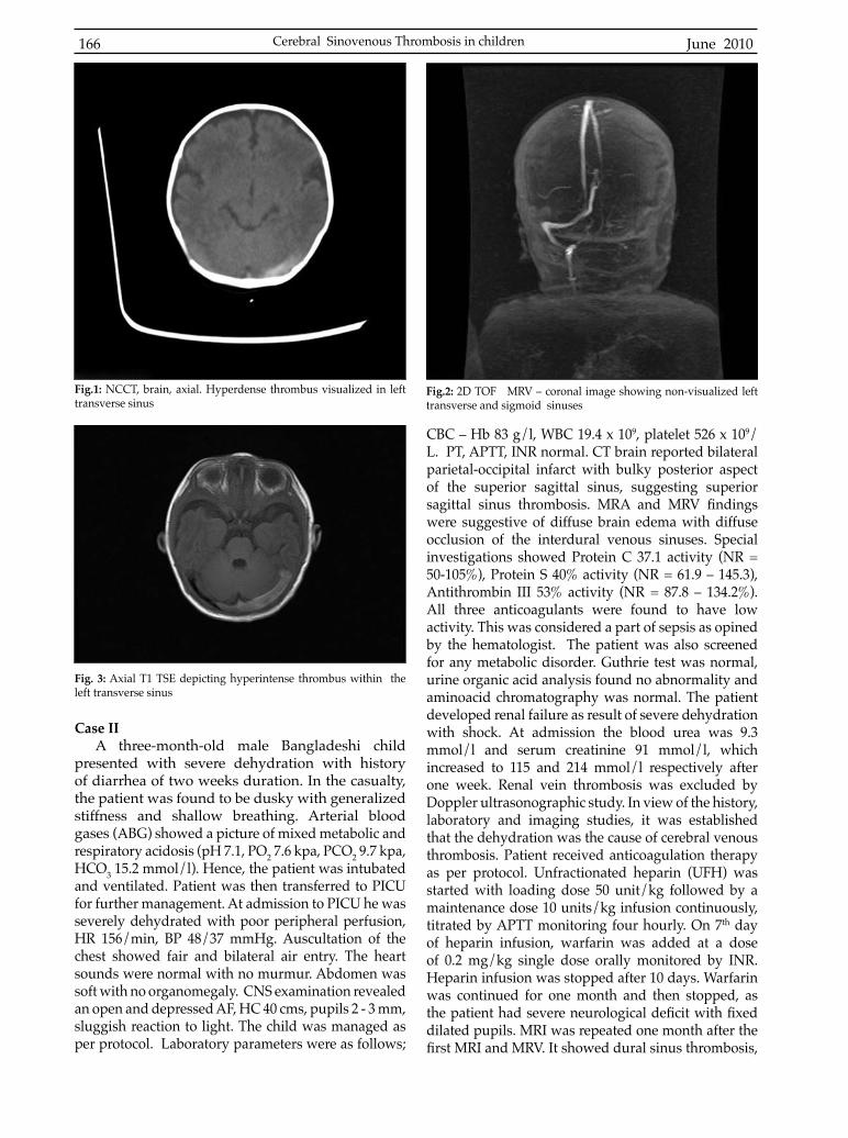
June 2010166
Case IIA three-month-old male Bangladeshi child
presented with severe dehydration with history of diarrhea of two weeks duration. In the casualty, the patient was found to be dusky with generalized stiffness and shallow breathing. Arterial blood gases (ABG) showed a picture of mixed metabolic and respiratory acidosis (pH 7.1, PO2 7.6 kpa, PCO2 9.7 kpa, HCO3 15.2 mmol/l). Hence, the patient was intubated and ventilated. Patient was then transferred to PICU for further management. At admission to PICU he was severely dehydrated with poor peripheral perfusion, HR 156/min, BP 48/37 mmHg. Auscultation of the chest showed fair and bilateral air entry. The heart sounds were normal with no murmur. Abdomen was soft with no organomegaly. CNS examination revealed an open and depressed AF, HC 40 cms, pupils 2 - 3 mm, sluggish reaction to light. The child was managed as per protocol. Laboratory parameters were as follows;
CBC – Hb 83 g/l, WBC 19.4 x 10⁹, platelet 526 x 109/L. PT, APTT, INR normal. CT brain reported bilateral parietal-occipital infarct with bulky posterior aspect of the superior sagittal sinus, suggesting superior sagittal sinus thrombosis. MRA and MRV findingswere suggestive of diffuse brain edema with diffuse occlusion of the interdural venous sinuses. Special investigations showed Protein C 37.1 activity (NR = 50-105%), Protein S 40% activity (NR = 61.9 – 145.3), Antithrombin III 53% activity (NR = 87.8 – 134.2%). All three anticoagulants were found to have low activity. This was considered a part of sepsis as opined by the hematologist. The patient was also screened for any metabolic disorder. Guthrie test was normal, urine organic acid analysis found no abnormality and aminoacid chromatography was normal. The patient developed renal failure as result of severe dehydration with shock. At admission the blood urea was 9.3 mmol/l and serum creatinine 91 mmol/l, which increased to 115 and 214 mmol/l respectively after one week. Renal vein thrombosis was excluded by Doppler ultrasonographic study. In view of the history, laboratory and imaging studies, it was established that the dehydration was the cause of cerebral venous thrombosis. Patient received anticoagulation therapy as per protocol. Unfractionated heparin (UFH) was started with loading dose 50 unit/kg followed by a maintenance dose 10 units/kg infusion continuously, titrated by APTT monitoring four hourly. On 7th day of heparin infusion, warfarin was added at a dose of 0.2 mg/kg single dose orally monitored by INR. Heparin infusion was stopped after 10 days. Warfarin was continued for one month and then stopped, as the patient had severe neurological deficit with fixeddilated pupils. MRI was repeated one month after the first MRI and MRV. It showed dural sinus thrombosis,
Fig.1: NCCT, brain, axial. Hyperdense thrombus visualized in left transverse sinus
Fig.2: 2D TOF MRV – coronal image showing non-visualized left transverse and sigmoid sinuses
Fig. 3: Axial T1 TSE depicting hyperintense thrombus within the left transverse sinus
Cerebral Sinovenous Thrombosis in children

KUWAIT MEDICAL JOURNAL 167June 2010
non-visualization of dural sinuses due to occlusion of dural sinuses associated with multiple cortical venous infarction. The child expired in the PICU after 40 days of stay on mechanical ventilation.
DISCUSSIONThe incidence of cerebral venous thrombosis
is at least 0.67 per 100,000 children per year[2]. The predisposing risk factors include local (in the head and neck) or systemic infections. Acute and chronic systemic diseases such as vascular traumas, iron deficiency anemia[4], acute lymphoblastic leukemia[5], dehydration[6], nephrotic syndrome[7], drug toxicity, lupus erythematosis, asphyxia, pregnancy and metabolic disorders[8] and prothrombotic disorders. Recently published data have suggested that multiple additional factors including prothrombotic risk factors contribute to the symptomatic onset of cerebral sinovenous thrombosis (CSVT). Assays for antithrombin III, protein C, S, lupus anticoagulant, anticardiolipin antibody and presence of factor V Leiden should be checked[2,8]. In our two cases, one had prothrombotic risk factor, antithrombin III (AT) deficiency, and the other case had dehydration as thepredisposing factor. There are two major causes of antithrombin III deficiency: (a) an inherited deficiencydue to a genetic abnormality (mutation), and (b), an acquired deficiency. The inherited AT deficiency isan uncommon genetic disorder. It is inherited in a dominant pattern. Homozygous individuals rarely survive and the fetus usually dies before birth. They are either quantitative (type I) or qualitative (type II). Type II is subdivided into the more common, but less thrombogenic, type IIb deficiency caused by adefect in the heparin-binding region of AT and the less common, but more thrombophilic, type IIa variant caused by mutations in the thrombin-binding site. The antithrombin activity level is the best initial test to obtain if one suspects that a patient may have AT deficiency. Normal ranges differ from laboratory tolaboratory, but typically, are in the order of 80 – 120%. A 50% reduction in the level of antithrombin activity is sufficient to diagnose in favor of thrombosis inhomozygous and heterozygous state. There are many different mutations in the antithrombin gene that can lead to inherited AT deficiency. Genetic testing is,therefore, not possible in routine clinical practice[13].
Five European Pediatric Neurology stroke registries, studied 42 children with CSVT, patients aged from three weeks to 13 years[10]. Symptoms on presentation were lethargy, anorexia, headache, vomiting, seizures, focal signs or coma. The same clinical findings were presentin our cases. Sinovenous thrombosis was confirmedby CT, MRI with or without MRV[8]. Transverse sinus thrombosis was more common (73%) than sagittal sinus thrombosis (35%)[9]. In case I, the CT head
reported venous sinus thrombosis involving the left transverse, straight sinus and part of left sigmoid and superior sagittal sinus. This was followed by MRI and MRV, whose findings were consistent with thrombosisof left transverse and sigmoid sinus with slow flow inthe superior sagittal sinus. In case II, CT head and MRI and MRV findings were suggestive of diffuse brainedema, with severe hypoxic changes, and occlusion of dural sinuses.
Treatment of CSVT has historically involved general supportive or symptomatic measures such as hydration, antibiotics for septic cases, control of seizure activity with anticonvulsant, and measures aimed at decreasing intracranial pressure[8]. In children with first episode of venous thromboembolism (VTE),anticoagulant therapy is recommended with either UFH or low-molecular-weight heparin (LMWH). Dosing of IV UFH should prolong the activated partial thromboplastin time (APTT) to a range that corresponds to an anti-factor Xa assay (anti-FXa) level of 0.35 to 0.7 U/ml, whereas LMWH should achieve an anti-FXa level of 0.5 to 1.0 U/ml 4 h after an injection for twice-daily dosing. In neonates with first VTE, managementis suggested with either anticoagulation or supportive care with radiologic monitoring and subsequent anticoagulation, if extension of the thrombosis occurs during supportive care[11]. In the absence of major CNS hemorrhage, anticoagulant therapy is appropriate. Anticoagulation therapy is administered for three months, if full recanalization is seen on the three month monitoring CT scan / MRV examination or for six months, if only partial recanalization is seen on the three monthly monitoring CT or MRV examination[11]. If no anticoagulants are administered due to significanthemorrhage, repeat MRV, or CT venography should be obtained at one week after diagnosis. For children with CSVT, suggested treatment is for 5 to 7 days with either UFH or LMWH followed by LMWH or VKAS (vit. K antagonists), (target INR 2.5, INR range 2.0 to 3.0) for 3 to 6 months even in the presence of localized hemorrhagic infarct[12]. In our cases, we followed the same protocol for anticoagulant therapy for both patients.
The first case showed improvement evident by bothclinical and radiological evaluation on follow up, except for mild neurological deficit which needed furtherfollow up for long term outcome determination. In the second case, the patient had dural sinus thrombosis associated with multiple cortical venous infarct with severe ischemic changes. Unfortunately, the patient expired.
CONCLUSIONThe long term neurological outcome of sinovenous
thrombosis in children is unclear. Long term follow up of affected children is very important, especially, in

June 2010168
neonates, since the onset of signs of neurological injury is delayed in this age group. Given the increasing incidence of sinovenous thrombosis in children, the variation in treatment and the adverse outcomes in half of the children with this disorder, there is a need to identify some effective immediate and secondary preventive therapies.
REFERENCES
1. Heller C, Heinecke A, Junker R, et al. Cerebral venous thrombosis in children: a multifactorial origin. Circulation 2003; 108:1362-1367.
2. de Veber G, Andrew M, Adams S, et al. Cerebral sinovenvous thrombosis in children. N Engl J Med 2001; 345:417-423.
3. Mross C, Klemm E. Acute mastoiditis in children: retrospective study 1979–1998. Otorhinolaryngol Nova 2000; 10:187-193.
4. Benedict SL, Bonkowsky JL, Thompson JA, et al. Cerebral sinovenous thrombosis in children: another reason to treat iron deficiency anemia. J Child Neurol2004; 19:526-531.
5. Lockman LA, Mastri A, Priest JR, Nesbit M. Dural venous sinus thrombosis in acute lymphoblastic leukemia. Pediatrics 1980; 66:943-947.
6. Barron TF, Gusnard DA, Zimmerman RA, Clancy RR. Cerebral venous thrombosis in neonates and children.
Pediatr Neurol 1992; 8:112-116.7. Pirogovsky A, Adi M, Dagan A, et al. Superior sagittal
sinus thrombois: a rare complication in a child with nephrotic syndrome. Pediatr Radiol 2001; 31:709-711.
8. Vielhaber H, Ehrenforth S, Koch HG, Scharrer I, van der Werf N, Nowak-Gottl U. Cerebral venous sinus thrombosis in infancy and childhood : role of genetic and acquired risk factors of thrombophilia. Eur J Pediatr 1998; 157:555-560.
9. Wasay M, Dai AI, Ansari M, Shaikh Z, Roach ES. Cerebral venous sinus thrombosis in children: a multicenter cohort from the United States. J Child Neurol 2008; 23:26-31.
10. Sebire G, Tabarki B, Saunders DE, et al. Cerebral venous sinus thrombosis in children: risk factors, presentation, diagnosis and outcome. Brain 2005; 128:477-489.
11. Monagle P, Chalmers E, Chan A, et al. Antithrombotic therapy in neonates and children: American College of Chest Physicians Evidence-Based Clinical Practice Guidelines (8th Edition). Chest 2008; 133:887S -968S.
12. Guyatt G, Schunemann HJ, Cook D, Jaeschke R, Pauker S. Applying the grades of recommendation for antithrombotic and thrombolytic therapy: the Seventh ACCP Conference on Antithrombotic and Thrombolytic Therapy. Chest 2004; 126: 179S-187S.
13. Patnaik MM, Moll S. Inherited antithrombin deficiency: a review. Haemophilia 2008; 14:1229-1239.
Cerebral Sinovenous Thrombosis in children

KUWAIT MEDICAL JOURNAL 169June 2010
Selected Abstracts of Articles Published Elsewhere by Authors in Kuwait
Kuwait Medical Journal 2010, 42 (2): 169-172
Diagnosis of Papillary Thyroid Carcinoma in Fine Needle Aspiration Smears: Factors That Affect Decision Making
Das DK, Sharma PNDepartment of Pathology, Faculty of Medicine, Kuwait University, Mubarak Al-Kabeer Hospital, Safat, Kuwait
E-mail: [email protected]
Acta Cytol 2009; 53:497-506
Objective: To investigate into the limitations in decision making in papillary thyroid carcinoma (PTC) in fine needle aspiration smears.Study design: We studied the cytology reports of 21 cases with definitive diagnoses of PTC, 17 suspicious/suggestive of (S/O) PTC, and 17 rule out (R/O) PTC, 8 R/O neoplasms and 15 nonneoplastic lesions with 1 or more cytologic features of PTC. Five important cytomorphologic features-papillary formation, intranuclear cytoplasmic inclusions, nuclear grooves, fine nuclear chromatin and psammoma bodies--were analyzed for their frequency and through a scoring system. Results: Three or more cytologic features of PTC were present in 18 (85.7%) cases with a definitivediagnosis of PTC as opposed to 7 (41.2%) cases diagnosed as S/O PTC, 2 (11.8%) of R/O PTC, 0% of R/O neoplasia and 0% of nonneoplastic lesions (p = 0.0062-<0.0001). The scores of PTC cases (5.8 +/- 1.14) were significantly higher (p <0.0001) than those of S/O PTC (3.8+/-1.47), R/O PTC (2.4+/-1.27), R/O neoplasia(1.4 +/- 0.69) and nonneoplastic lesions (1.1 +/- 0.62). Available histopathology reports in PTC, S/O PTC and R/O PTC cases confirmed the presence of PTC in 92.9%, 62.5% and 16.7%, respectively.Conclusion: During decision making, the number of cytomorphologic features as well as some specificfeatures and their extent in FNA smears give varying amounts of confidence to the cytopathologist indiagnosing the cases as PTC or otherwise.
Circulating Markers of Inflammation and Endothelial Dysfunction inArab Adolescent Subjects: Reference Ranges and Associations with
Age, Gender, Body Mass and Insulin Sensitivity
Al-Isa AN, Thalib L, Akanji AODepartment of Community Medicine, Faculty of Medicine, Kuwait University, Kuwait
Atherosclerosis 2010; 208: 543-549
Introduction: Important biochemical markers for the low-grade vascular inflammation and endothelialdysfunction that characterize the earliest events in the pathogenesis of atherosclerosis are CRP and the adhesion molecules ICAM-1 and VCAM-1. This study aims to evaluate, in healthy Arab adolescent subjects aged 10-19 yr, the reference ranges for these markers and their associations with such determinants as age, gender, body mass and insulin sensitivity.
Continued..

Selected Abstracts of Articles Published Elsewhere by Authors in Kuwait June 2010170
Methods: There were 774 Kuwaiti subjects (316 boys and 456 girls) aged mean (SD) 14.5 (2.2) yr. All had detailed clinical examination, anthropometry (to derive BMI) and assessment of fasting serum levels of CRP, ICAM-1, VCAM-1 and insulin (and HOMA-IR) to derive their reference values. Associations with putative determinants were explored by logistic regression analyses. Results and discussion: The reference ranges (mean (SD)) were: CRP 0.28 (0.49) mg/dl; ICAM-1, 286 (119) ng/ml; VCAM-1, 1148 (352) ng/ml; HOMA-IR, 4.57 (5.05). These parameters were significantlyhigher in boys than in girls, and in the overweight/obese and those aged </= 14 yr. BMI was significantlyand independently related to CRP and ICAM-1 even after adjusting for age, gender, socio-demographic factors, diet and exercise. Levels of these markers were significantly influenced by age and gender.Conclusion: BMI, age and gender are significant predictors of serum levels of circulating inflammatorybiomarkers, particularly ICAM-1, CRP and HOMA-IR. These atherogenic processes therefore probably cluster together in early life in individuals at increased later heart disease risk. Preventive public health measures should therefore commence in early childhood.
Expression of Estrogen Receptor Alpha And Estrogen Receptor Beta in Fine Needle Aspirates of Breast CarcinomaKapila K, Anim JT, Francis IM, Al-Mulla F, George SS, Behbehani AI
Cytology Unit and Cytology Laboratory, Department of Pathology, Faculty of Medicine, Kuwait University, Safat, Kuwait. E-mail: [email protected]
Acta Cytol 2010; 54:25-30
Objective: To evaluate the expression of estrogen receptor beta (ERbeta) in fine needle aspirates (FNAs)and correlate the findings with its expression in tissue sections.Study Design: In 38 cases of breast carcinoma, expression of estrogen receptor alpha (ERalpha) and ERbeta in aspirates and tissue sections was correlated with the cytologic and histologic grade of the tumor. Results: ERalpha and ERbeta were expressed as nuclear staining in 80% and 90% of the cases in tissue sections and 47% and 45% of the cases in aspirates, respectively. Tissue expression of ERalpha (grade 1, 81%; grade 2, 100%) and ERbeta (grade 1, 94%; grade 2, 100%) was greater than in grade 3 tumors (ERalpha, 50%; ERbeta, 70%). In FNAs, they were equally distributed in the different cytologic grades. In aspirates 30% of ERalpha negative tumors were positive for ERbeta, while in tissues 75% of ERalpha-negative tumors were positive for ERbeta (p = 0.007). Conclusion: Demonstration of ERbeta on FNA smears is feasible. It helps identify the specific subcohortof ERbeta-positive tumors in ERalpha-negative breast cancers; that may have therapeutic importance.
Toxic Tacrolimus Blood Levels with Rifampin Administration in a Renal Transplant Recipient
Abdel Halim M, Al-Otaibi T, Gheith O, El-Kholy O, Abdel Tawab K, Said T, Nair P, Nampoory MRHamed Alessa Ogran Transplantation Center, Ibn Sina Hospital, Safat, Kuwait.
E-mail:[email protected]
Ann Transplant 2010; 15:57-60
Background: Induction of the hepatic and intestinal cytochrome P450-3A4 system and intestinal P-glycoprotein is an unavoidable consequence of rifampin administration which requires substantial increase in tacrolimus dose when given concurrently. Chronic diarrhea is known to precipitate tacrolimus
Continued..

KUWAIT MEDICAL JOURNAL 171June 2010
toxicity irrespective of its cause in renal transplant recipients. Case Report: A 28 years old lady had a second renal transplant when she was 15 years old which is functioning normally. She was maintained on prednisolone, azathioprine, omeprazole and tacrolimus. Tacrolimus was maintained on therapeutic levels (5-7 ng/ml). She developed pyrexia of unknown origin associated with chronic diarrhea. Detailed investigations didn’t reach any definite diagnosis.Antituberculous drugs including rifampin were started emperically. During the first four months ofantituberculous treatment diarrhea worsened and unexpected high tacrolimus trough blood levels were observed requiring successive dose reduction from 7 mg/day up to 0.5 mg every other day with rise of serum creatinine from 100 to 140 umol/l. Tacrolimus was changed to low dose sirolimus and renal function gradually improved to its baseline while still on rifampin treatment. Conclusions: We conclude that chronic diarrhea may cause toxic tacrolimus blood levels even in presence of rifampin, this would be due to its significant cytochrome P450-3A4 and P-glycoprotein enzymeinhibitory effect.
Headscarf Pin Tracheobronchial Aspiration: A Distinct Clinical Entity
Al-Sarraf N, Jamal-Eddine H, Khaja F, Ayed AKDepartment of Thoracic Surgery, Chest Disease Hospital, Kuwait. E-mail: [email protected]
Interact Cardiovasc Thorac Surg 2009; 9:187-190
Foreign body (FB) aspiration is commonly seen in children and less commonly in the elderly. However, due to some social and cultural factors, a distinct group of tracheobronchial FB aspiration is increasingly recognized. We sought to assess our experience with such entity. A retrospective review of all cases with veil pin tracheobronchial FB aspiration in a single center over a 13-year period was carried out. There were 35 cases of headscarf tracheobronchial FB aspiration. All were females with mean age of 14 years. All patients experienced coughing and all had positive chest radiography findings. Commonest anatomicallocation was right main bronchus (32%) followed by left main bronchus (23%). Tracheal pins occurred in 17%. Rigid bronchoscopy was used more often than flexible bronchoscopy (83% vs. 17%, respectively).Repeat bronchoscopy was required in two cases (6%). Thoracotomy was required in one patient (3%). There were no complications or hospital deaths. Headscarf pin aspiration is seen in middle-aged women who inappropriately place the pins between their lips prior to securing their veils. Bronchoscopy is the treatment modality of choice and surgery is rarely required. Preventative educational strategies should be implemented to reduce such an avoidable risk.
Emotional Reaction to Diagnosis of Infertility in Kuwait and Successful Clients’ Perception of Nurses’ Role During Treatment
Omu FE, Omu AE
BMC Nurs 2010; 9:5 [Epub ahead of print]
Background: The unfulfilled desire of millions of infertile couples worldwide to have their own biologicalchildren results in emotional distress. This study evaluated the emotional reactions of couples attending a combined infertility clinic in Kuwait and successful clients’ perception of nurses. Methods: Quantitative and qualitative methods were used. The first phase was by structured interviewusing two standardized psychological scales: the 25-item Hopkins Symptom Checklist and ModifiedFertility Adjustment Scale. Data were collected from 268 couples attending the combined infertility clinic,
Continued..

Selected Abstracts of Articles Published Elsewhere by Authors in Kuwait June 2010172
between October 2002 and September 2007. The second phase was a semi-structured interview of 10 clients who got pregnant following treatment. The interview explored their feelings and perception of the nurses’ role. Interviews were transcribed verbatim and analyzed. Results: The average duration of infertility was 4 years; 65.7% of the women and 76.1% of men suffered from primary infertility. Emotional reactions experienced were: anxiety in women (12.7%) and men (6%), depression in women (5.2%) and men (14.9%) and reduced libido in women (6.7%) and men (29.9%). Also in men, 14.9% experienced premature ejaculation, 5.2% weak ejaculation and 7.9% had impotence although 4.9% were transient. In the semi-structured interviews, the emotions expressed were similar and in addition to anger, feelings of devastation, powerlessness, sense of failure and frustration. In the survey, 12.7% of the men were found to show more anxiety than women (6%). Although all the 10 women interviewed confirmed they were anxious; only 4 of their partners were reported to be sad or anxious.Successful clients’ perception of nurses’ roles included nurses carrying out basic nursing procedures, communicating, educating about investigative and treatment procedures, providing emotional support by listening, encouraging, reassuring and being empathetic. Conclusions: This study illuminates the emotional reactions of infertile clients. Fertility nurses in Kuwait can provide emotional support through communication. The need for additional and continuous training for nurses employed in fertility settings in Kuwait is paramount.
Anterior Cruciate Ligament Reconstruction using Autologous Hamstring Double Bundle Graft Compared with Single Bundle Procedures
Ibrahim SA, Hamido F, Al Misfer AK, Mahgoob A, Ghafar SA, Alhran H.Department of Sports Unit, Al Razi Orthopaedic Hospital, Safat, Kuwait. E-mail: [email protected]
J Bone Joint Surg Br 2009; 91:1310-1315
A total of 218 patients with unilateral anterior cruciate ligament deficiency were randomly assigned to oneof four groups. In group A, an anatomical double bundle anterior cruciate ligament reconstruction was performed; group B were treated by a single bundle using an Endobutton for femoral fixation; in groupC, by a single bundle using RigidFix cross pins for femoral fixation; and in group D, by a single bundleusing a bioabsorbable TransFix II screw for femoral fixation. For tibial fixation a bioabsorbable Intrafixinterference screw was used for all the groups and the graft was fashioned from the semitendinosus and gracilis tendons in all patients. In all, 18 patients were lost to follow-up. The remaining 200 were subjected to a clinical evaluation, with assessment of the anterior drawer, Lachman’s and the pivot-shift tests, and KT-1000 arthrometer measurement. They also completed the International Knee Documentation Committee, Lysholm knee and Tegner activity scores. At a mean of 29 months (25 to 38) follow-up there were no significant differences concerning time between injury and range of movement and Lysholmknee scores among the four groups. However, the double bundle method showed significantly betterresults for the pivot-shift test (p = 0.002). The KT 1000 measurements showed a mean difference between the reconstructed knee and the patients’ normal knee of 1.4 mm in the double bundle group and 2.4 mm in the single bundle group; which was statistically significant. The Lachman and anterior drawertests also showed superior results for the double bundle method. The International Knee Documentation Committee scale showed no significant difference among the groups (p < 0.001). On clinical evaluationthe double bundle group showed less laxity than the single bundle groups. However, regardless of the technique, all knees were improved by anterior cruciate ligament reconstruction compared with their pre-operative status.

KUWAIT MEDICAL JOURNAL 173June 2010
Forthcoming Conferences and Meetings
Kuwait Medical Journal 2010; 42 (2): 173-179
Compiled and edited by Babichan K Chandy
9th Asian-Pacific Congress of Cardiovascular and Interventional Radiology (APCCVIR 2010)Jun 01 - 04, 2010 Seoul, Republic of KoreaContact: Secretariat - APCCVIR Phone: 82-2-3471-8555; Fax: 82-2-521-8683 E-Mail: [email protected]
11th European Congress of Pediatric Surgery Jun 02 - 05, 2010 Berne, Switzerland Contact: Mrs U. Gueder Fax: 39-0-409-870-136 E-Mail: [email protected]
3rd congress of EASAPS (European Association of Societies of Aesthetic Plastic Surgery) incorporating the Spring Academy VDÄPC (Association of the German Aesthetic Plastic Surgeons) Jun 04 - 06, 2010 Aachen, Germany Contact: Francesca Rustler Phone: 0-3641-353-3273 E-Mail: [email protected]
46th American Society of Clinical Oncology (ASCO) Annual Meeting 2010 Jun 04 -08, 2010 Chicago, IL, United States Contact: Meeting Organiser: 2318 Mill Road, Suite 800, Alexandria, VA 22314 Phone: 888-282-2552 or 571-483-1300 E-Mail: [email protected]
6th World Congress of the International Society of Physical and Rehabilitation Medicine Jun 04 - 09, 2011 San Juan, Puerto Rico Contact: Werner Van Cleemputte, Managing Director Medicongress Waalpoel 28/34, B-9960 Assenede, Belgium Phone: 32-0-93-443-959; Fax: 32-0-93-444-010 E-Mail: [email protected]
The 10th Congress of The European Society of Pediatric Otorhinolaryngology (ESPO) Jun 05- 08, 2010 Pamplona, Spain Contact: Congress Secretariat Phone: 34-91-575-9393; Fax: 34-91-431-2692 E-Mail: [email protected]
29th European Academy of Allergy and Clinical Immunology Congress (EAACI) Jun 05- 09, 2010 London, England, United Kingdom Contact: Congress Secretariat E-Mail: [email protected]
57th Society of Nuclear Medicine (SNM 2010) Annual Meeting Jun 05 - 09, 2010 Salt Lake City, UT, United States Contact: Secretariat - SNM Phone: 703-708-9000 ext 1229 E-Mail: [email protected]
The International Council of Ophthalmology World Congress (WOC 2010) Jun 05 - 09, 2010 Berlin, Germany Contact: Congress Secretariat E-Mail: [email protected]
International College of Neuropsychopharmacology 2010 Congress Jun 06 - 10, 2010 Hong Kong, China Contact: Organiser Phone: 0-1-355-244-966; Fax: 0-1-355-249-959 E-Mail: [email protected]
29th World Congress of Biomedical Laboratory ScienceJun 06 - 10, 2010 Nairobi, Kenya Contact: Hila Vakrat Phone: 41-0-22-533-0948; Fax: 41-0-22-580-2953 E-Mail: [email protected]
13th International Conference on Emergency Medicine Jun 09 - 12, 2010 Singapore, Singapore Contact: Stella Chee Phone: 65-63-795-261; Fax: 65-64-752-077 E-Mail: [email protected]
The World Conference on Interventional Oncology (WCIO 2010) Jun 09 - 12, 2010 Philadelphia, PA, United States Contact: Yvonne Grunebaum Phone: 978-927-8330; Fax: 978-524-0498

Forthcoming Conferences and Meetings June 2010174
The 43rd Annual Meeting of the European Society for Paediatric Gastroenterology, Hepatology and Nutrition (ESPGHAN) Jun 09 - 12, 2010 Istanbul, Turkey Contact: Meeting Organiser Phone: 44-0-845-1800-360; Fax: 44-0-870-442-9940 E-Mail:[email protected]
World Psychiatric Association 2010 Regional Meeting Jun 10 - 12, 2010 St. Petersburg, Russian Federation Contact: Dr. Valery Krasnov E-Mail: [email protected]
Euroanaesthesia 2010: The European Anaesthesiology Congress Jun 12 - 15, 2010 Helsinki, Finland Contact: SecretariatPhone: 32-0-27-433-290; Fax: 32-0-27-433-298 E-Mail: [email protected]
2nd International Congress on Metabolic Syndrome, Obesity & Diabetes Jun 16 - 18, 2010 Zanjan, Islamic Republic of IranContact: Dr. Faranak SharifiPhone: 00-982-417-270-814; Fax: 00-982-417-270-815 E-Mail: [email protected]
Nordic Federation of Societies of Obstetrics and Gynaecology: 37th Nordic Congress of Obstetrics and Gynaecology Jun16- 18, 2010 Copenhagen, Denmark Contact: Meeting Organiser Phone: 45-70-237-823; Fax: 45-70-237-888 E-Mail: [email protected]
World Congress of Cardiology Jun 16- 19, 2010 Beijing, China Contact: Conference Secretariat: KIE Phone: 551-150-521-215; Fax: 551-150-520-286 E-Mail: [email protected]
6th Congress of the European Association of Dermatologic Oncology Jun 16 - 19, 2010 Athens, Greece Contact: Mrs. Penelope Mitroyianni Phone: 30-210-725-7693; Fax: 30-210-725-7532 E-Mail: [email protected]
Cardiostim 2010 - 17th World Congress in Cardiac Electrophysiology and Cardiac Techniques Jun 16- 19, 2010 Nice, France Contact: Jocelyne Toulouse Phone: 33-01-47-56-24-56; Fax: 33-01-47-56-24-55 E-Mail: [email protected]
20th International Federation for Psychotherapy World Congress of Psychotherapy Jun 16 - 19, 2010 Lucerne, Switzerland Contact: Dr. Ulrich Schnyder E-Mail: [email protected]
12th Danube Symposium International Otorhinolaryngological Congress Jun 17- 19, 2010 Mikulov, Czech Republic Contact: Prof. Dr. Ivo Slapak Phone: 00-420-532-234-225; Fax: 00-420-532-234-430 E-Mail: [email protected]
16th World Congress for Bronchology - WCB; 16th World Congress for Bronchoesophagology - WCBE Jun 20 - 23, 2010 Mannheim, Germany Contact: Heike Esmann Phone: 0049-0-89-307-1011; Fax: 0049-0-89-307-1021 E-Mail: [email protected]
3rd International Conference on Osteoimmunology: Interactions of the Immune and Skeletal Systems Jun 20- 25, 2010 City: Santorini , Greece Contact: Conference Secretariat Phone: 1-610-527-7630; Fax: 1-610-527-7631 E-Mail: [email protected]
International Congress of the Royal College of Psychiatrists Jun 21- 24, 2010 Edinburgh, Scotland United Kingdom, Caroline Simms Phone: 44-207-235-2351 E-Mail: [email protected]
BIT’s 3rd World Cancer Congress 2010 Jun 22 - 25, 2010 Singapore, Singapore Contact: Ms. April Wang or Nancy Jiang Phone: 0086-411-8479-5469 or 0086-411-8479-9609 ext. 803; Fax: 0086-411-8479-9629 E-Mail: [email protected] or [email protected]

KUWAIT MEDICAL JOURNAL 175June 2010
XI International Congress on Traumatic Stress and Anxiety Disorders Jun 23 - 25, 2010 Buenos Aires, Argentina Contact: Congress Secretartiat Phone: 00-5411-4903-0493; Fax: 00-5411-4903-0493E-Mail: [email protected]
ISCAS - 14th Annual Conference of the International for Computer Aided Surgery Jun 23 - 26, 2010 Geneva, Switzerland Contact: CARS Conference Office - Mrs. FranziskaSchweikert Phone: 49-7742-922-434; Fax: 49-7742-922-438 E-Mail: [email protected]
2nd Italian Conference on Thoracic Oncology Jun 24 - 26, 2010 Rome, Italy Contact: Conference Secretariat E-Mail: [email protected]
Symposium in Reconstructive Microsurgery Jun 24 - 27, 2010 Rochester, MN, United States Contact: Conference Organizer Phone: 800-323-2688 E-Mail: [email protected]
2010 Annual Meeting of the Canadian Urological Association Jun 27- 30, 2010 Charlottetown, Canada Contact: CUA Central Office, 1155 University, Suite1155, Montréal, QC, Canada H3B 3A7 Phone: 1-514-395-0376; Fax: 1-514-875-0205 E-Mail: [email protected]
12th World Congress on Gastrointestinal Cancer (WCGI & ESMO) Jun 30 - Jul 03, 2010 Barcelona, Spain Contact: Secretariat Phone: 770-751-7332; Fax: 770-751-7334 E-Mail: [email protected]
6th Asian Interventional Cardiovascular Therapeutics Congress Jul 01 - 03, 2010 Singapore, Singapore Contact: Congress Secretariat Phone: 65-6496-6845 or 65-6496-6850 Fax: 65-6496-6853 E-Mail: [email protected]
Psoriasis International Network Congress (PSO 2010)Jul 01- 04, 2010 Paris, France Contact: Congress Secretariat Phone: 33-0-153-858-259 Fax: 33-0-153-858-283 E-Mail: [email protected]
Second International Trauma Conference Jul 03- 08, 2010 Queenstown, New Zealand Contact: Bron Chesterman Phone: 61-8-8267-6660; Fax: 61-8-8267-6668 E-Mail: [email protected]
2nd Symposium of Clinical and Applied Anatomy Prague 2010 Jul 09 - 11, 2010 Prague, Czech Republic Contact: Assoc. Prof. Vaclav Baca, PhD. Phone: 420-267-102-508; Fax: 420-267-102-504 E-Mail: [email protected]
World Congress of the World Federation of Hemophilia Jul 10 - 14, 2010 Buenos Aires, Argentina Contact: Maria Milagros Salas - Congress & M Manager Phone: 1-514-394-2837 E-Mail: [email protected]
7th International Congress on Neuroendocrinology Jul 10 - 15, 2010 Rouen, France Contact: Secretary General: William Rostene, INSERM U.732 Hôpital Saint-Antoinie, 184, rue du Fauborg Saint Saint-Atoine 75012 Paris, France Phone: 33-149-284-676 E-Mail: [email protected]
14th Asia Pacific League of Associations forRheumatology (APLAR 2010) Jul 11 - 15, 2010 Hong Kong, Hong Kong Contact: APLAR 2010 Congress Secretariat Phone: 852-2559-9973; Fax: 852-2547-9528 E-Mail: [email protected]
16th World Congress of Basic & Clinical Pharmacology Jul 13 - 23, 2010 Copenhagen, Denmark Contact: Prof. Kim Brøsen / Tina Ludvig E-Mail: [email protected] / [email protected]

Forthcoming Conferences and Meetings June 2010176
XIX Biennial Meeting of the International Society for Eye Research (ISER 2010) July 18 - 23, 2010 Montreal, QC, Canada Contact: KENES International E-Mail:[email protected]
XVIII International AIDS Conference (AIDS 2010) Jul 18 - 23, 2010 Vienna, Austria Contact: International AIDS Society HQ, PO Box 20, CH - 1216 Cointrin, GENEVA, Switzerland Phone: 41-0-22-7-100-800; Fax: 41-0-22-7-100-899 E-Mail: [email protected]
British Association of Paediatric Surgeons 57th Annual Congress Jul 21 - 24, 2010 Aberdeen, Scotland, United Kingdom Contact: British Associaiton of Paediatric Surgeons, Royal College of Surgeons, 35-43 Lincolns Inn Fields, London WCA 3PN Phone: 02-0-78-696-915; Fax: 02-0-78-696-919
The 17th Scoliosis Research Society (SRS) International Meeting on Advanced Spine Techniques Jul 21 - 24, 2010 Toronto, Canada Contact: Meetings Department Phone: 1-414-289-9107; Fax: 1-414-276-3349 E-Mail: [email protected]
9th International Congress on the Future of Breast Cancer Jul 22 - 25, 2010 La Jolla, CA, United States Contact: Registration Team Phone: 888-949-0045; Fax: 214-367-3305 E-Mail: [email protected]
15th World Congress on Heart Disease, Annual Scientific Sessions 2010Jul 24 - 27, 2010 Vancouver, BC, Canada Contact: Asher Kimchi, M.D. Phone: 310-657-8777; Fax: 310-659-4781 E-Mail: [email protected]
20th Annual Mayo Clinic Hematology/Oncology Reviews Jul 29 - 31, 2010 Amelia Island, FL, United States Contact: Linda Gibson Phone: 800-462-9633; Fax: 904-953-2954 E-Mail: [email protected]
The 26th International Pediatric Association Congress of Pediatrics (IPA 2010) Aug 04 - 09, 2010 Johannesburg, South Africa Contact: Liraz Bregman Phone: 41-229-080-488; Fax: 41-227-322-850 E-Mail: [email protected]
The American Society of Emergency Radiology 2010 Annual Meeting and Postgraduate Course in Trauma and Emergency Radiology Aug 11 - 14, 2010 Seattle State, WA, United States Contact: Jessica Whalen Phone: 713-965-0566; Fax: 713-960-0488 E-Mail: [email protected]
XXIII International Congress of the Transplantation Society August 15 - 19, 2010 Vancouver, BC, Canada Contact: Diana Klingner Phone: 1-604-681-2153; Fax: 1-604-681-1049 E-Mail: [email protected]
14th International Congress of ImmunologyAug 22 - 27, 2010 Kobe, Japan Contact: Prof. Masayuki MIYASAKA Phone: 81-6-6879-3972; Fax: 81-6-6879-3979E-Mail: [email protected]
2nd European Plastic Surgery Research Council Meeting Aug 26 - 29, 2010 Hamburg, Germany Contact: Stefanie Becker Phone: 49-0-3641-3533-2708; Fax: 49-0-3641-353-321 E-Mail: [email protected]
World Congress on Gastrointestinal Cancer: Asian Perspectives Aug 27- 29, 2010 Hong Kong, China Contact: Theresa Jones Phone: 1-678-242-0906; Fax: 1-678-242-0920 E-Mail: [email protected]
Intercongress Meeting of the European Society of Pathology (ESP 2010) Aug 31- Sep 04, 2010 Krakow, Poland Contact: Congress Secretariat Phone: 49-30-300-66-90; Fax: 49-30-305-73-91 E-Mail: [email protected]

KUWAIT MEDICAL JOURNAL 177June 2010
28th World Congress of Endourology Sep 01 - 04, 2010 Chicago, IL, United States Contact: Bailey-Turner Chernise E-Mail: [email protected]
Society of Laparoendoscopic Surgeons: 19th SLS Annual Meeting and Endo Expo 2010 Sept 01- 04, 2010 New York, NY, United States Contact: SLS Secretariat Phone: 1-800-446-2659 / 1-305-665-9959; Fax: 1-305-667-4123 E-Mail: [email protected]
World Psychiatric Association 2010 Regional Meeting Sept 01 - 05, 2010 Beijing, China Contact: Dr. Yizhuang Zou E-Mail: [email protected]
33rd Annual Meeting of the European Academy of Facial Plastic Surgery Sep 01 - 05, 2010 Antalya, Turkey Contact: Prof. Fazil Apaydin Phone: 90-216-449-4945 Fax: 90-216-449-6410 E-Mail: [email protected]
45th Annual Meeting American Society of Head and Neck Radiology (ASHNR) Sep 07 - 11, 2011 San Diego, CA, United States Contact: Meeting Organiser: ASHNR, 2210 Midwest Road, Suite 207 Oak Brook, Illinois 60523-8205 Phone: 630-574-0220; Fax: 630-574-0661
2010 World Molecular Imaging Congress Sep 08 - 11, 2010 Kyoto, Japan Contact: Nichole Navar Phone: 310-215-9730; Fax: 310-215-9731 E-Mail: [email protected]
28th ESOPRS Annual Meeting - European Society of Ophthalmic Plastic and Reconstructive Surgery Sep 09 - 11, 2010 Munich, Germany Contact: Frau Claudia Schäfer Phone: 0049-0-89-307-1011; Fax: 0049-0-89-307-1021 E-Mail: [email protected]
European Society of Urogenital Radiology: 2010 Symposium of the ESUR Sep 09 - 12, 2010 City: Bruges, Belgium Contact: ESUR Head Office Phone: 43-15-334-064 Fax: 43-15-334-064 - 448 E-Mail: [email protected]
AAO-HNSF Annual Meeting and Oto Expo 2011 Sep 11 - 14, 2011 San Francisco, CA, United States Contact: Meeting Organiser: AAO-HNSF, 1650 Diagonal Road, Alexandria, VA 22314-2857, USA Phone: 1-703-836-4444 Fax: 1-703-519-1546 E-Mail: [email protected]
XVIIth International Congress of Neuropathology Sep 11 - 15, 2010 Salzburg, Austria Brigitte Millán-Ruiz Phone: 43-1-404-005-573; Fax: 43-1-404-005-511 E-Mail: [email protected]
ITC 2010: The 14th International Thyroid Congress Sep 11 - 16, 2010 Paris, France Contact: Conference Secretariat Phone: 0-153-858-280 Fax: 0-153-858-283 E-Mail: [email protected]
International Federation of Fertility Societies (IFFS): 20th World Congress on Fertility & Sterility Sept 12 - 15, 2010 Munich, Germany Contact: INTERPLAN Congress, Meeting and Event Management AG OfficePhone: 49-8-954-823-473; Fax: 49-8-954-823-442 E-Mail: [email protected]
38th Annual Meeting of the International Society for Pediatric NeurosurgerySept 13 - 16, 2010Jeju, KoreaContact: Gordon MccombE-Mail: [email protected]
11th Congress on Reproductive Biomedicine and 6th Congress on Stem Cell Biology & Technology Sep 15 - 17, 2010 Middle East, Islamic Republic of IranContact: A. Orae Phone: 098-21-2231-0406; Fax: 098-21-2231-0406 E-Mail: [email protected]

Forthcoming Conferences and Meetings June 2010178
15th Congress of the European Society of Surgical Oncology (ESSO) Sep15 - 17, 2010 Bordeaux, France Contact: Congress Secretariat Phone: 32-0-2-775-0201; Fax: 32-0-2-775-0200 E-Mail: [email protected]
2010 European Respiratory Society (ERS) Annual Congress Sep 18 - 22, 2010 Barcelona, Spain Contact: Conference Secretariat: ERS 2010 c/o K.I.T. Group GmbH Phone: 49-0-30-246-032-20 Fax: 49-0-30-246-032-00 E-Mail: [email protected]
World Congress on Refractive Error Sep 20 - 22, 2010 Durban, South Africa Contact: Conference Secretariat Phone: 27-31-201-1470; Fax: 27-31-201-1510 E-Mail: [email protected]
Central European Congress of Rheumatology (CECR2010) Sep 22 - 24, 2010 Sopron, Hungary Contact: Prof Gyula Poór Phone: 36-1-336-0464 E-Mail: [email protected]
40th Annual Meeting of the German Society of Immunology Sep 22 - 25, 2010 Leipzig, Germany Contact: Nadia Al-Hamadi Phone: 49-0-3641-3533-2236; Fax: 49-0-3641-3533-22309 E-Mail: [email protected]
ESPE 2010: 49th Annual Meeting of the European Society for Pediatric Endocrinology Sep 22 - 25, 2010 Prague, Czech Republic Contact: Congress Secretariat Phone: 46-0-8-459-66-00; Fax: 46-0-8-661-91-25 E-Mail: [email protected]
The 13th Annual International Congress of Pediatric Hepatology, Gastroenterology and Nutrition Sep 22 - 25, 2010 Sharm-El Sheikh, Egypt Contact: Mortada El-Shabrawi Phone: 201-2313-3705; Fax: 202-3761-9012 E-Mail: [email protected]
The 1st World Congress on Controversies in Gastroenterology & Liver Diseases (C-GOLD) Sep 23 - 26, 2010 Prague, Czech Republic Contact: Ilana Phone: 972-3-566-6166; Fax: 972-3-566-6177 E-Mail: [email protected]
Cardiovascular and Interventional Radiological Society of Europe (CIRSE 2010) Annual Congress Oct 02 - 06, 2010 Valencia, Spain Contact: Secretariat - CIRSE E-Mail: [email protected]
American College of Surgeons 96th Annual MeetingOct 03 - 07, 2010 Washington, DC, United StatesContact: American College of SurgeonsPhone: 312-202-5000; Fax: 312-202-5001E-Mail: [email protected]
XVIII World Congress on Psychiatric Genetics Oct 03 - 07, 2010 Athens, Greece Contact: Mrs Penelope Mitrogianni Phone: 30-210-725-7693; Fax: 30-210-725-7532 E-Mail: [email protected]
18th International Congress on Palliative Care Oct 05 - 08, 2010 City: Montreal, QC, Canada Contact: Frank Salvatori Phone: 450-292-3456 ext 224; Fax: 450-292-3453 E-Mail: [email protected]
WINFOCUS 2010: 6th World Congress on Ultrasound in Emergency & Critical Care Medicine Oct 06 - 09, 2010 Rome, Italy Contact: Winfocus Secretariat Phone: 39-051-230-385; Fax: 39-051-221-894 E-Mail: [email protected]
19th Congress of the European Academy of Dermatology and Venereology (EADV) Oct 06 - 10, 2010 Gothenburg, Sweden Contact: EADV OfficePhone: 322-650-0090; Fax: 322-650-0098 E-Mail: [email protected]
MASCON 2010 Oct 08 - 10, 2010 Mumbai, IndiaContact: Dr.Vijay Shetty or Dr.Mayuri Shetty Phone: 0-982-107-6279 or 0-982-018-5527; Fax: 91-22-2590-6346 E-Mail: [email protected]

KUWAIT MEDICAL JOURNAL 179June 2010
EMAA 2010 - European Masters in Aesthetic & Anti-Aging Medicine Oct 15 - 17, 2010 Paris, France Contact: Christophe Luino Phone: 01-56-837-800; Fax: 01-56-837-805 E-Mail: [email protected]
2010 Annual Meeting of the American Academy of Ophthalmology Oct 16 - 19, 2010 Chicago, IL, United States Contact: American Academy of Ophthalmology Phone: 415-447-0320 E-Mail: [email protected]
The 7th Combined Meeting of Orthopaedic Research Societies Oct 16 - 20, 2010 Kyoto, Japan Contact: Secretariat Fax: 81-52-950-6146 E-Mail: [email protected]
ASA 2010: American Society of Anesthesiologists Annual Meeting Oct 16 - 20, 2010 San Diego, CA, United States Contact: Meeting Organiser E-Mail: [email protected]
20th World Congress of the IASGO Oct 20 - 23, 2010 Cairo, Egypt Contact: Dr. Shahenda El Hawary Phone: 20-233-023-642; Fax: 20-233-027-672 E-Mail: [email protected]
Turkish Respiratory Society 32nd Annual Congress Oct 20 - 24, 2010 City: Antalya, Turkey Contact: ERDINC TAN Phone: 902-164-494-945; Fax: 902-164-494-945 E-Mail: [email protected]
2010 ASDS (American Society for Dermatologic Surgery) Annual Meeting Oct 21 - 24, 2010 Chicago, IL, United States Contact: American Society for Dermatologic Surgery Phone: 847-956-0900
Lymphoma & Myeloma 2010: An International Congress on Hematologic Malignancies Oct 21 - 23, 2010 New York, NY, United StatesContact: Imedex Customer Service Phone: 1-678-242-0906; Fax: 1-678-242-0920E-Mail: [email protected]
6th International Congress of the Asia-Pacific Metabolic and Bariatric Surgical Society Oct 21- 23, 2010 Singapore, Singapore Contact: Mrs Norizam Phone: 656-772-2920; Fax: 656-774-6077 E-Mail: [email protected]
48th Annual Meeting of the Infectious Diseases Society of America Oct 21 - 24, 2010 Vancouver, BC, Canada Contact: IDSA, 1300 Wilson Blvd, Suite 300, Arlington, VA 22209 Phone: 703-299-0200; Fax: 703-299-0204 E-Mail: [email protected]
American Society for Reproductive Medicine 66th Annual Meeting Oct 23 - 27, 2010 Denver, CO, United States Contact: American Society for Reproductive Medicine, 1209 Montgomery Highway, Birmingham, Alabama 35216-2809 Phone: 205-978-5000; Fax: 205-978-5005 E-Mail: [email protected]
The American Academy of Child and Adolescent Psychiatry 57th Annual Meeting Oct 26 - 31, 2010 New York, NY, United States Contact: The American Academy of Child and Adolescent Psychiatry 3615 Wisconsin Avenue, N.W, Washington, D.C.20016-3007 Phone: 202-966-7300; Fax: 202-966-2891 E-Mail: [email protected]
32nd Turkish National Congress of Otorhinolaryngology and Head & Neck Surgery Oct 27 - 31, 2010 Antalya, Turkey Contact: Prof. Atilla Tekat Phone: 90-216-449-4945; Fax: 90-216-449-6410 E-Mail: [email protected]
2010 American Society of Clincial Pathology (ASCP) Annual Meeting Oct 27 - 31, 2010 San Francisco, CA, United States Contact: Meeting Organiser Phone: 800-267-2727; Fax: 312-541-4472 E-Mail: [email protected]
Chest 2010: American College of Chest Physicians (ACCP) Annual Meeting Oct 30 - Nov 04, 2010 Vancouver, BC, Canada Contact: Meeting Organiser Phone: 847-498-1400 or 800-343-2227

KUWAIT MEDICAL JOURNAL June 2010180
WHO-Facts Sheet
1. Urban Planning Essential for Public Health2. Drug-Resistant Tuberculosis now at Record Levels
3. All Hands on Deck in the Battle against Dengue Mosquito4. Human Health under Threat from Ecosystem Degradation
5. WHO Releases New Malaria Guidelines for Treatment and Procurement of Medicines
Compiled and edited byBabichan K Chandy
Kuwait Medical Journal 2010, 42 (2): 180-184
Address correspondence to: Office of the Spokesperson, WHO, Geneva. Tel.: (+41 22) 791 2599; Fax (+41 22) 791 4858; Email: [email protected]; Web site: http://www.who.int/
1. URBAN PLANNING ESSENTIAL FOR PUBLIC HEALTH
Dramatic and rapid rise of people living in cities poses health threat and opportunity
Urban settings have a direct impact on the health of the people who live there. On World Health Day WHO is launching a campaign to highlight urban planning as a crucial link to building a healthy 21st century. In particular, the Organization calls upon municipal authorities, concerned residents, advocates for healthy living and others to take a close look at health inequities in cities and take action.
The world is rapidly urbanizing with significantchanges in our living standards, lifestyles, social behaviour and health. Thirty years ago, four out of every 10 people were living in cities, but by 2050 this number will grow to seven out of 10.
“In general, urban populations are better off than their rural counterparts. They tend to have greater access to social and health services and their life expectancy is longer. But cities can also concentrate threats to health such as inadequate sanitation and refuse collection, pollution, road traffic accidents,outbreaks of infectious diseases and also unhealthy lifestyles,” says Dr Margaret Chan, WHO Director-General.
Many cities face a triple threat: Infectious diseases which thrive when people are crowded together; chronic, noncommunicable diseases including diabetes, cancers and heart disease which are on the rise with unhealthy lifestyles including tobacco use, unhealthy diets, physical inactivity and harmful use of alcohol and urban health is often further burdened by road traffic accidents, injuries, violence and crime.
Despite these challenges, cities also bring opportunities. Five actions will significantly increasethe chance people will be able to enjoy better urban living conditions: • promote urban planning for healthy behaviours
and safety• improve urban living conditions• ensure participatory governance• build inclusive cities that are accessible and age-
friendly• make cities resilient to disasters and emergencies
“The wide range of health issues in cities and its determinants require coordinated policies and actions across multiple disciplines including environment, transport, education, parks and recreation, and urban planning,” says Dr Ala Alwan, WHO Assistant Director-General for Noncommunicable Diseases and Mental Health. “We are at a critical turning point in history where we can make a difference.”
Coordinated policies and actions are also needed to address the underlying conditions of major health issues in cities today. For instance, outdoor urban air pollution kills some 1.2 million people worldwide.
Road traffic injuries among children are ofsignificant concern in urban areas. Globally, roadtraffic injuries are the leading cause of death amongyouth aged 15–24 years, and the second leading cause of death for those in the 10–14 years old.
In many cases, rapid population growth outpaces the municipal capacity to build essential infrastructure that make life in cities safe and healthy, leading to the proliferation of informal settlements. Urbanization, both in the developed and particularly in the developing world, is accompanied by a concentration of poverty. Today, an estimated one of three urban

KUWAIT MEDICAL JOURNAL 181June 2010
dwellers, amounting to nearly one billion people, live in urban slums and informal settings -signalling the call for urgent action to address their needs.
For more information contact:Jacob Kumaresan, Director, WHO Centre for Health Development, Kobe, Japan Mobile: +81 80 2435 1861, E-mail:
2. DRUG-RESISTANT TUBERCULOSIS NOW AT RECORD LEVELS
In some areas of the world, one in four people with tuberculosis (TB) becomes ill with a form of the disease that can no longer be treated with standard drugs regimens, a World Health Organization (WHO) report says.
For example, 28% of all people newly diagnosed with TB in one region of north western Russia had the multidrug-resistant form of the disease (MDR-TB) in 2008. This is the highest level ever reported to WHO. Previously, the highest recorded level was 22% in Baku City, Azerbaijan, in 2007.
In the new WHO’s Multidrug and Extensively Drug-Resistant Tuberculosis: 2010 Global Report on Surveillance and Response, it is estimated that 440,000 people had MDR-TB worldwide in 2008 and that a third of them died. In sheer numbers, Asia bears the brunt of the epidemic. Almost 50% of MDR-TB cases worldwide are estimated to occur in China and India. In Africa, estimates show 69,000 cases emerged, the vast majority of which went undiagnosed.
Tuberculosis programmes face tremendous challenges in reducing MDR-TB rates. But there are encouraging signs that even in the presence of severe epidemics, governments and partners can turn around MDR-TB by strengthening efforts to control the disease and implementing WHO recommendations.
Two regions in the Russian Federation - Orel and Tomsk - have achieved a remarkable decline in MDR-TB in about five years. These regions join twocountries - Estonia and Latvia - which have reversed rising high rates of MDR-TB, ultimately achieving a decline. The United States of America and China, Hong Kong Special Administrative Region (SAR), have achieved sustained successes in controlling MDR-TB.
Progress remains slow in most other countries. Worldwide, of those patients receiving treatment, 60% were reported as cured. However, only an estimated 7% of all MDR-TB patients are diagnosed. This points to the urgent need for improvements in laboratory facilities, access to rapid diagnosis and treatment with more effective drugs and regimens shorter than the current two years.
WHO is engaged in a five year project to strengthenTB laboratories with rapid tests in nearly 30 countries. This will ensure more people benefit early from life-saving treatments. It is also working closely with the Global Fund to fight AIDS, Tuberculosis and Malariaand the international community on increasing access to treatment.
TB, MDR-TB and XDR-TB Multidrug-resistant TB (MDR-TB) is caused by
bacteria that are resistant to at least isoniazid and rifampicin, the most effective anti-TB drugs. MDR-TB results from either primary infection with resistant bacteria or may develop in the course of a patient’s treatment.
Extensively drug-resistant TB (XDR-TB) is a form of TB caused by bacteria that are resistant to isoniazid and rifampicin (i.e. MDR-TB) as well as any fluoroquinoloneand any of the second-line anti-TB injectable drugs (amikacin, kanamycin or capreomycin).
These forms of TB do not respond to the standard six-month treatment with first-line anti-TB drugs andcan take up to two years or more to treat with drugs that are less potent, more toxic and much more expensive, from 50 to 200 times higher. While a course of standard TB drugs cost approximately US$20, MDR-TB drugs can cost up to US$ 5000, and XDR-TB treatment is far more expensive.
In 2008, there were an estimated 9.4 million new TB cases, and 1.8 million TB deaths. 440,000 MDR-TB cases are estimated to have emerged in the same year with 150,000 MDR-TB deaths. No official estimateshave been made on the number of XDR-TB cases, but there may be around 25,000 a year with most cases fatal. Since XDR-TB was first defined in 2006, a totalof 58 countries have reported at least one case of XDR-TB.
In 27 high burden countries (i.e. countries estimated to have had at least 4000 MDR-TB cases arising annually and/or at least 10% of newly registered TB cases with MDR-TB), 1.3 million M/XDR-TB cases will need to be treated between 2010 and 2015.
Strengthening laboratories There is an urgent need to expand and accelerate
in countries access to new, rapid technologies that can diagnose MDR-TB in two days rather than traditional methods which can take up to four months. EXPAND TB is a five year project targeting 27 countries, launchedin 2008 and implemented by WHO, the Foundation for Innovative New Diagnostics (FIND), the Stop TB Partnership’s Global Drug Facility (GDF) and the Global Laboratory Initiative (GLI) with financialsupport from UNITAID. So far, it has carried out a wide range of activities in 12 countries, including upgrading of infrastructure and training of staff. Technology

WHO-Facts Sheet June 2010182
transfer has started in countries, paving the way for more patients to be diagnosed accurately and rapidly enrolled on treatment. These upgrades should lead to eventual routine surveillance of drug resistance in affected countries.
Risk factors: HIV and MDR-TBStudies show that TB patients co-infected with HIV
in three Eastern European countries (Estonia, Latvia, and the Republic of Moldova) were at a higher risk of having MDR-TB compared to TB patients without HIV infection. Similar findings have been made in studiesfrom Lithuania, Ukraine and Mozambique.
The report highlights several reasons why drug-resistant TB may be associated with HIV, particularly in some Eastern European countries. However, more research is needed to determine whether there is an overlap between the MDR-TB and HIV epidemics worldwide.
Reporting on MDR-TB globallyThis report presents drug resistance data from 114
countries and updated information from 35 of them. Despite the growing understanding of the magnitude and trends in drug-resistant TB, major gaps remain in geographical areas covered. Since 1994, only 59% of all countries globally have been able to collect high quality representative data on drug resistance. There is an urgent need to obtain information, particularly from Africa and those high MDR-TB burden countries where data have never been reported: Bangladesh, Belarus, Kyrgyzstan, Pakistan and Nigeria. Moreover, countries need to expand the scope of their surveys to cover entire populations, repeat surveys are needed to better understand trends in drug resistance and countries need to move towards adopting systematic continuous surveillance.
For more information contact: Mr Glenn Thomas, Senior Communications Adviser, Stop TB Department, WHO. Telephone:+ 41 22 791 39 83; Mobile: +41 79 509 0677,
E-mail: [email protected]
3. ALL HANDS ON DECK IN THE BATTLE AGAINST DENGUE MOSQUITO*
Dengue is a serious, potentially lethal, illness. Infections have dramatically increased in recent decades due to increased urbanization, trade and travel. No effective drug or vaccine is available yet, so the only solution is to prevent the disease-carrying mosquito from breeding and biting humans.
The battle against the mosquito that spreads dengue, a serious illness that affects around 50 million people each year, needs more than insecticides alone, according to a study published in March 2010, in the
international public health journal, the Bulletin of the World Health Organization.
This important study held in six Asian cities explored the breeding patterns of the Aedes aegypti mosquito (the species that transmits dengue) and discovered significant differences in breeding and denguetransmission between the sites, affected by such factors as household habits, local environmental conditions and the ecology and behaviour of the mosquito.
For the first time, this large-scale multi-countrystudy, supported by a research partnership between the Special Programme for Research and Training in Tropical Diseases (TDR) at the World Health Organization and the International Development Research Centre in Canada, comprehensively combed public and private buildings and open spaces in selected areas in six cities in India, Indonesia, Myanmar, the Philippines, Sri Lanka and Thailand. It identified almost 50,000containers holding water and found the highest number of mosquito larvae in unused and uncovered outdoor containers on private land.
While the study found that this mosquito prefers to breed in containers filled with rainwater, whenlarvicides were used at the study site in Thailand, the mosquitoes found alternative breeding sites indoors in covered containers filled with tap water.
“Traditionally, communities expect public health services to carry out mosquito control, normally through insecticide fogging,” says co-author Dr Johannes Sommerfeld, of TDR in Geneva. “Large-scale insecticide spraying, however, is not effective in reducing immature (larval) stages of the mosquito. These mosquitoes breed in various water containers in and around households, so individuals, families and communities have an equally important role to play in reducing these breeding sites in their own backyards. Communities need to work closely with public health and other services in this battle.”
*This was created by WHO as a forum for public health experts and the views expressed by the writers in these pages do not necessarily represent the views of WHO. Full coverage of the paper is available at: http://www.who.int/bulletin/volumes/88/3/09-067892.pdf
For more information contact: Sarah Cumberland, Bulletin of the World Health Organization, Geneva, Switzerland.
Office: +41 22 791 2570, Mobile: +41 79 206 1403, E-mail:[email protected]
4. HUMAN HEALTH UNDER THREAT FROM ECOSYSTEM DEGRADATION
Threats particularly acute in poorer countriesThe World Health Organization (WHO) is
publishing a report, ‘Ecosystems and Human Well-

KUWAIT MEDICAL JOURNAL 183June 2010
being: Health Synthesis’, which represents an attempt to describe the complex links between the preservation of healthy and biodiverse natural ecosystems and human health.
“Over the past 50 years, humans have changed natural ecosystems more rapidly and extensively than in any comparable period in human history,” said Dr LEE Jong-wook, Director-General of the World Health Organization. “This transformation of the planet has contributed to substantial net gains in health, well-being and economic development. But not all regions and groups of people have benefited equally from thisprocess.”
Approximately 60% of the benefits that the globalecosystem provides to support life on Earth (such as fresh water, clean air and a relatively stable climate) are being degraded or used unsustainably. In the report, scientists warn that harmful consequences of this degradation to human health are already being felt and could grow significantly worse over the next50 years.
“The benefits should be acknowledged,” said DrCarlos Corvalan, WHO’s lead expert on the report. “But these benefits are not enjoyed equally. And the riskswe face now from ecosystem degradation, particularly among poor populations directly dependent on natural ecosystems for many basic needs, has to be addressed.”
Ecosystem services are absolutely vital to preventing disease and sustaining good health, the Health Synthesis report underlines. Many important human diseases have originated in animals, and so changes in the habitats of animal populations that are disease vectors or reservoirs, may affect human health, sometimes positively and sometimes negatively. For example, the Nipah virus is believed to have emerged after forest clearance fires in Indonesia drove carrierbats to neighbouring Malaysia, where the virus infected intensively-farmed pigs, and then crossed to humans.
Intensive livestock production, while providing benefits to health in terms of improved nutrition, hasalso created environments favorable to the emergence of diseases, the report notes. Increased human contact with wild species and “bush meat” as a result of encroachment in forests and changes in diet also create opportunities for disease transmission.
Trends ranging from forest clearance to climate-induced habitat changes also appear to have impacted certain populations of mosquitoes, ticks and midges, altering transmission patterns for diseases like malaria and Lyme disease.
Pressures on ecosystems could have unpredictable and potentially severe future impacts on health, the report states. Regions facing the greatest present-day risks, meanwhile, include sub-Saharan Africa, Central
Asia, parts of Latin America, and certain areas in South and Southeast Asia. Some of the most serious problems include:• Nutrition: Degradation of fisheries and agro-
ecosystems are factors in the malnutrition of some 800 million people around the world. At least an additional billion people experience chronic micronutrient deficiency.
• Safe Drinking-Water: Water-associated infectious diseases claim 3.2 million lives, approximately 6% of all deaths globally. Over one billion people lack access to safe water supplies, while 2.6 billion lack adequate sanitation, and related problems of water scarcity are increasing, partly due to ecosystem depletion and contamination.
• Solid Fuel Dependency: About 3% of the global burden of disease has been attributed to indoor air pollution, a major cause of respiratory diseases. Most of the world’s population uses solid fuels to cook and heat, a factor in deforestation.“Human health is strongly linked to the health
of ecosystems, which meet many of our most critical needs,” said Maria Neira, Director of WHO’s Department for the Protection of the Human Environment. “We in the health sector need to take heed of this in our own planning, and together with other sectors, ensure that we obtain the greatest benefit from ecosystems for good health – now andin the future.”
The Health Synthesis Report is WHO’s contribution to the broader Millennium Ecosystem Assessment, a four-year series of studies and reports, involving over 1300 scientists, considering impacts on human well-being, past, present and future.
For more information contact: Dr Carlos Corvalan, Coordinator and Scientific Expert, Department for the
Protection of the Human Environment, World Health Organization, Geneva
Mobile phone: +41 79 445 1624. E-mail: [email protected]
5. WHO RELEASES NEW MALARIA GUIDELINES FOR TREATMENT AND PROCUREMENT
OF MEDICINES
The World Health Organization (WHO) is releasing new guidelines for the treatment of malaria, and the first ever guidance on procuring safe and efficaciousanti-malarial medicines.
In recent years, a new type of treatment called artemisinin-based combination therapy (ACTs) has transformed the treatment of malaria, but if not used properly the medicine could become ineffective.
The Guidelines for the Treatment of Malaria (second edition) provides evidence-based and current

WHO-Facts Sheet June 2010184
recommendations for countries on malaria diagnosis and treatment. The main changes from the first editionof the guidelines (published in 2006) are the emphasis on testing before treating and the addition of a new ACT to the list of recommended treatments.
The world now has the means to rapidly diagnose malaria and treat it effectively” said Dr Robert Newman, Director of the WHO Global Malaria Programme (GMP). “WHO now recommends diagnostic testing in all cases of suspected malaria. Treatment based on clinical symptoms alone should be reserved for settings where diagnostic tests are not available,” he added.
In 2008, just 22% of suspected malaria cases were tested in 18 of 35 African countries reporting. Until now, most clinics had to rely on microscopy, but the recent development of quality-assured Rapid Diagnostic Tests (RDTs) using a dip stick and a drop of blood means a policy change is possible. The tests can reliably demonstrate the presence or absence of malaria parasites in the blood and can be performed at all levels of the health system, including community settings.
The move towards universal diagnostic testing of malaria is a critical step forward in the fight againstmalaria, as it will allow for the targeted use of ACTs for those who actually have malaria. The aim is to reduce the emergence and spread of drug resistance and to help identify patients who have fever, but do not have malaria, so that alternative diagnoses can be made and appropriate treatment provided. Therefore, better management of malaria has a positive impact on management of other childhood illness and overall child survival.
WHO is supporting malaria endemic countries to improve the quality of their diagnostic services using both microscopy and RDTs, and urging the manufacturers of RDTs to continue improving the accuracy and quality of these critically important diagnostic tests.
WHO estimates that 80 countries have adopted ACTs for first-line treatment of uncomplicatedP. falciparum malaria. In the guidelines, WHO emphasizes the importance of treating this deadliest
form of the disease with artemisinin-based combination therapies. WHO has now added a fifthACT - dihydroartemisinin plus piperaquine - to the previous list of recommended medicines.
WHO recommends oral artemisinin-based monotherapy should be removed from the market because their use will hasten the development of parasite resistance. Countries need to ensure that patients are diagnosed properly and take the full dose of ACTs to prevent the development of drug resistance.
The first ever guidelines on “Good procurementpractices for artemisinin-based antimalarial medicines” are based on the newest stringent internationally agreed production and procurement quality standards. This manual aims to improve the capacities of national and international procurement officers intheunderstandingof key quality elements and required documentation. The content is presented as a practical and concise 16-step practical checklist to guide the selection and procurement of safe and effective medicines meeting international quality standards.
“Pharmaceutical markets in malaria endemic countries are often unregulated and national authorities need practical help to assess the quality of malaria medicines before they buy them” says Dr Andrea Bosman, Coordinator of the Medicines and Diagnostics Unit at GMP. “Procurement channels are highly fragmented and so, there are too many antimalarials of varying quality on the market.”
Poor-quality medicines affect the health and lives of patients, damage the credibility of health services and, by generating sub-therapeutic drug levels in malaria patients, help develop resistance to this important life-saving class of pharmaceuticals.
Half of the world’s population is at risk from malaria. Each year almost 250 million cases occur, causing 860,000 deaths. Approximately 85% of these deaths are among children, and most occur in Africa.
For more information contact: Samantha Bolton, Communications Advisor, Geneva.
Mobile: +41 79 239 2366; email: [email protected]