International Journal of Advanced Scientific and Technical ...
59
International Journal of Advanced Scientific and Technical Research Issue 5 volume 2, March-April 2015 Available online on http://www.rspublication.com/ijst/index.html ISSN 2249-9954 R S. Publication, [email protected] Page 451 ىنز نز ى Sudan University of Science and Technology College of Graduate Studies Detection of Extended–spectrum Beta Lactamase Genes in Urinary Tract Isolated Bacteria among Pregnant Women A Dissertation Submitted in Partial Fulfillment for the Requirements of M.Sc Degree in Medical Laboratory Science (Microbiology) By Nosaiba Mohammed Alamin Khalafalla BSc Medical Laboratory Science Sudan University of science and Technology (2011) Supervisor Dr .Yousif Fadlalla HamedElnil September 2013
-
Upload
khangminh22 -
Category
Documents
-
view
0 -
download
0
Transcript of International Journal of Advanced Scientific and Technical ...
International Journal of Advanced Scientific and Technical Research Issue 5 volume 2, March-April 2015
Available online on http://www.rspublication.com/ijst/index.html ISSN 2249-9954
R S. Publication, [email protected] Page 451
ى نز نز ى
Sudan University of Science and Technology
College of Graduate Studies
Detection of Extended–spectrum Beta Lactamase Genes in
Urinary Tract Isolated Bacteria among Pregnant Women
A Dissertation Submitted in Partial Fulfillment for the Requirements of M.Sc
Degree in Medical Laboratory Science (Microbiology)
By
Nosaiba Mohammed Alamin Khalafalla
BSc Medical Laboratory Science
Sudan University of science and Technology (2011)
Supervisor
Dr .Yousif Fadlalla HamedElnil
September 2013

International Journal of Advanced Scientific and Technical Research Issue 5 volume 2, March-April 2015
Available online on http://www.rspublication.com/ijst/index.html ISSN 2249-9954
R S. Publication, [email protected] Page 452
بسم ميحرلا نمحرلا هللا
ليك وحيه وقل رب زدن علما﴿ المل الحق ول تعجل بلقرآن من قبل آن يقض ا ﴾فتعال الله
)114 (س ر ط ة
Dedication
To my mother, soul of my honorable father, brothers, sisters, friends, colleagues
and teachers…
ACKNOWELGDEMENTS
First of all thanks to ALMIGHTY ALLAH who gives me the power to complete
this work.
With a great deal of respect I would like to express my thanks to my supervisor Dr.
Yousif FadAllah Hamed Elnil for his advice, patience, encouragement and
valuable supervision during this study. Also my thanks and gratitude extended to
Miss Suhair Ramadan for her great help and all my colleagues who helped me
throughout this study.
International Journal of Advanced Scientific and Technical Research Issue 5 volume 2, March-April 2015
Available online on http://www.rspublication.com/ijst/index.html ISSN 2249-9954
R S. Publication, [email protected] Page 453
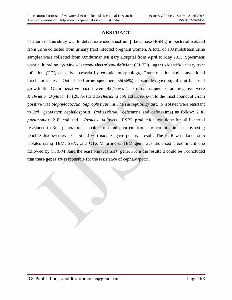
ABSTRACT
The aim of this study was to detect extended spectrum β-lactamase (ESBL) in bacterial isolated
from urine collected from urinary tract infected pregnant women. A total of 100 midstream urine
samples were collected from Omdurman Military Hospital from April to May 2013. Specimens
were cultured on cysteine – lactose- electrolyte- deficient (CLED) agar to identify urinary tract
infection (UTI) causative bacteria by colonial morphology, Gram reaction and conventional
biochemical tests. Out of 100 urine specimen; 56(56%) of samples gave significant bacterial
growth the Gram negative bacilli were 42(75%). The most frequent Gram negative were
Klebsiella Oxytoca 15 (26.8%) and Escherichia coli 10(17.9%),while the most abundant Gram
positive was Staphylococcus Saprophyticus. In The susceptibility test, 5 isolates were resistant
to 3rd generation cephalosporin (ceftazidime, ceftriaone and cefotaxime) as follow: 2 K.
pneumoniae ,2 E. coli and 1 Proteus vulgaris. ESBL production test done for all bacterial
resistance to 3rd generation cephalosporin and then confirmed by combination test by using
Double disc synergy test 5(11.9% ) isolates gave positive result. The PCR was done for 5
isolates using TEM, SHV, and CTX-M primers, TEM gene was the most predominant one
followed by CTX-M 3and the least one was SHV gene. From the results it could be Tconcluded
that these genes are responsible for the resistance of cephalosporin.
International Journal of Advanced Scientific and Technical Research Issue 5 volume 2, March-April 2015
Available online on http://www.rspublication.com/ijst/index.html ISSN 2249-9954
R S. Publication, [email protected] Page 454
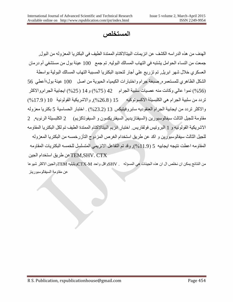
س خلص
, نذف ي ذ نذر س نكشف ع شياث نب خاالكخاو نخذة نط ف في نبكخزيا نعشن ي نبل
ع ت ل ي ي خشفي أو دريا 100حى خع . خعج ي ن اء نح يم يشخب في نخاب ن انك نبن
حى حشريع عهي أخار نخحذيذ نبكخزيا ن ببت نخاب ن انك نبن ت سطت , نع كزي خالل شز زيم
56 ع ت ل؛أعطي 100طبغت خز و خخبار ث نك اء نح يت ي طم , نشكم نظازي نه خعز
الكثز , يدا ت ندز و (%25 )14 (%75 )42 عاني كاج ي عظ اث سهب ت ندز و (56%)
%( 17.9 )10 الشزيك ت نقن ت , (%26.8 )15حزدد ي سهب ت ندز و ي نكهب هت الك حك
كخزيا يعشن 5 خخبار نح اس ت . (%23.2 )13 الكثز حزدد ي يدا ت ندز و نعقدي سا زف خ كس
2, نكهب هت نزئي2 ( ن فخزيك ن فحاكشيى, ن فخاسيذيى)يقايت نهد م نثانث س فانسبري
خخبار شيى نب خاالكخاو نخذة نط ف حى نكم نبكخزيا نقاي . نبزح س فنقاريس1 الشزيك ت نقن
نهد م نثانث س فانسبري كذ ع طزيق سخخذ و نغزص نشدج نخأسرخ ي نبكخزيا نعشن
قذ حى نخفاعم الشيي نخ ه م نهخ نبكخزياث نقاي , (%11.9 )5 نقاي عطج خ د يدا
TEM,SHV. CTXع طزيق سخخذ و ند
ي نخائح يك خهض ل ذ ند اث ي ن ؤن . SHV قم ذ CTX-Mيه ه TEM ند الكثز ش عا
ع يقايت ن فانسبريش
International Journal of Advanced Scientific and Technical Research Issue 5 volume 2, March-April 2015
Available online on http://www.rspublication.com/ijst/index.html ISSN 2249-9954
R S. Publication, [email protected] Page 455
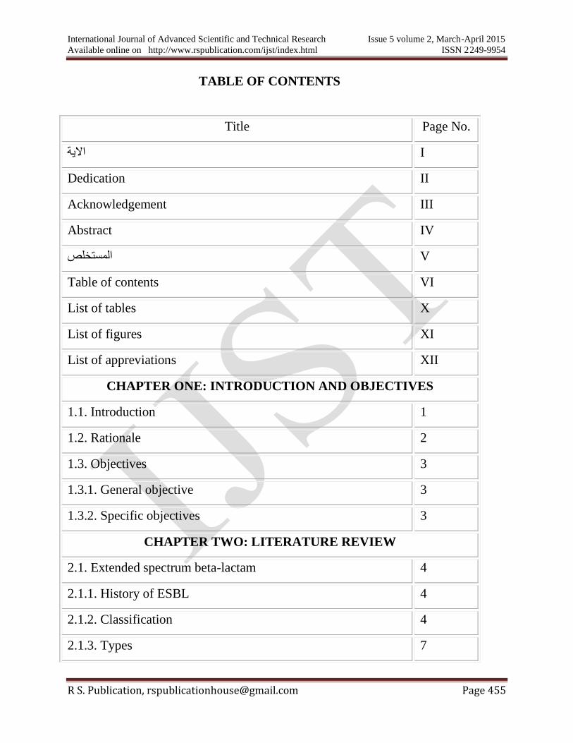
TABLE OF CONTENTS
Title Page No.
I اليت
Dedication II
Acknowledgement III
Abstract IV
V ن خخهض
Table of contents VI
List of tables X
List of figures XI
List of appreviations XII
CHAPTER ONE: INTRODUCTION AND OBJECTIVES
1.1. Introduction 1
1.2. Rationale 2
1.3. Objectives 3
1.3.1. General objective 3
1.3.2. Specific objectives 3
CHAPTER TWO: LITERATURE REVIEW
2.1. Extended spectrum beta-lactam 4
2.1.1. History of ESBL 4
2.1.2. Classification 4
2.1.3. Types 7
International Journal of Advanced Scientific and Technical Research Issue 5 volume 2, March-April 2015
Available online on http://www.rspublication.com/ijst/index.html ISSN 2249-9954
R S. Publication, [email protected] Page 456
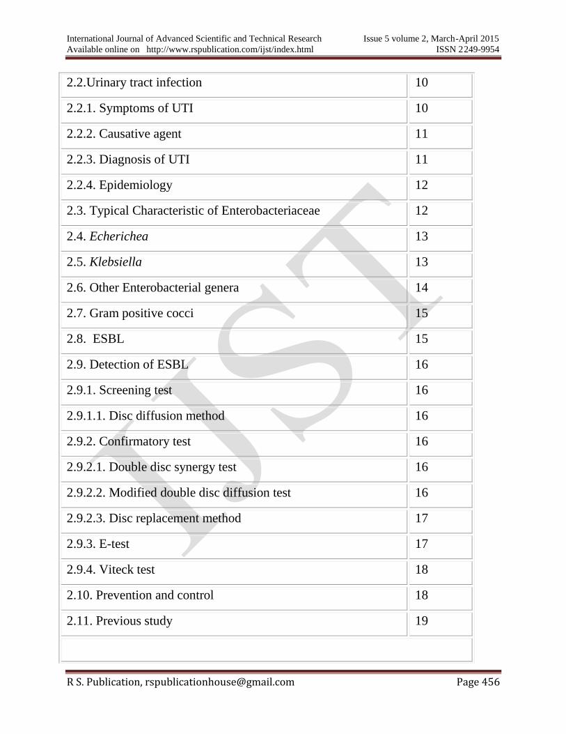
2.2.Urinary tract infection 10
2.2.1. Symptoms of UTI 10
2.2.2. Causative agent 11
2.2.3. Diagnosis of UTI 11
2.2.4. Epidemiology 12
2.3. Typical Characteristic of Enterobacteriaceae 12
2.4. Echerichea 13
2.5. Klebsiella 13
2.6. Other Enterobacterial genera 14
2.7. Gram positive cocci 15
2.8. ESBL 15
2.9. Detection of ESBL 16
2.9.1. Screening test 16
2.9.1.1. Disc diffusion method 16
2.9.2. Confirmatory test 16
2.9.2.1. Double disc synergy test 16
2.9.2.2. Modified double disc diffusion test 16
2.9.2.3. Disc replacement method 17
2.9.3. E-test 17
2.9.4. Viteck test 18
2.10. Prevention and control 18
2.11. Previous study 19
International Journal of Advanced Scientific and Technical Research Issue 5 volume 2, March-April 2015
Available online on http://www.rspublication.com/ijst/index.html ISSN 2249-9954
R S. Publication, [email protected] Page 457
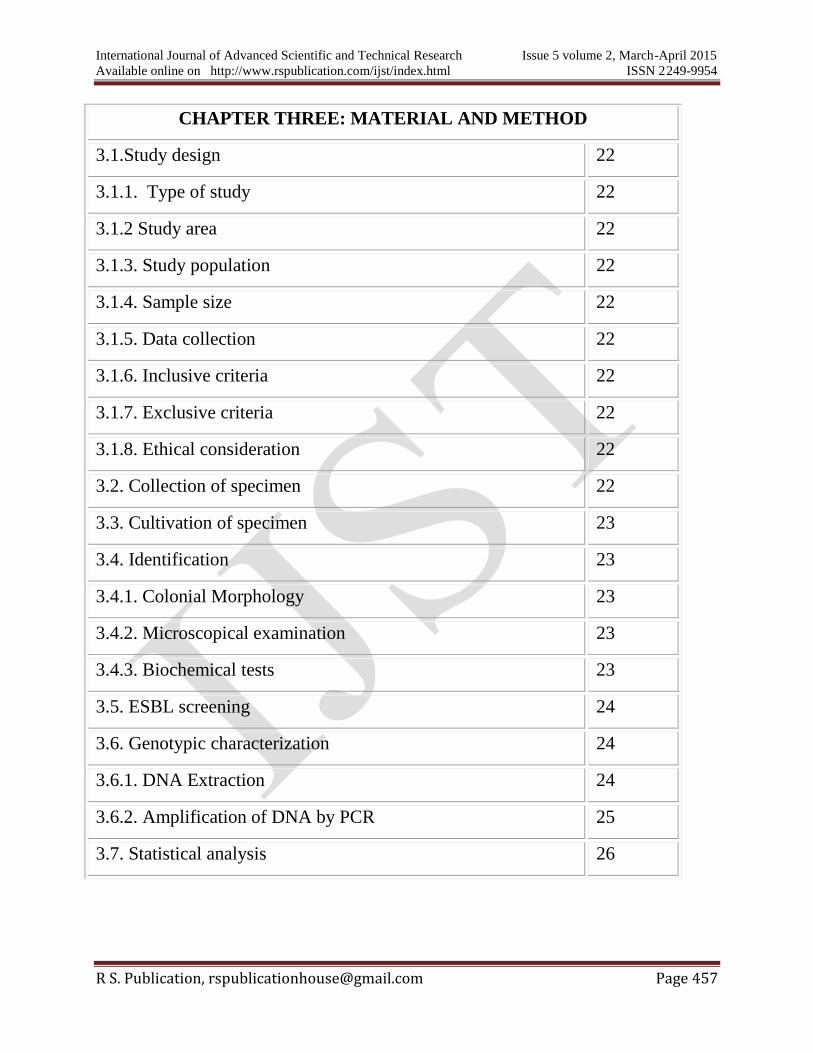
CHAPTER THREE: MATERIAL AND METHOD
3.1.Study design 22
3.1.1. Type of study 22
3.1.2 Study area 22
3.1.3. Study population 22
3.1.4. Sample size 22
3.1.5. Data collection 22
3.1.6. Inclusive criteria 22
3.1.7. Exclusive criteria 22
3.1.8. Ethical consideration 22
3.2. Collection of specimen 22
3.3. Cultivation of specimen 23
3.4. Identification 23
3.4.1. Colonial Morphology 23
3.4.2. Microscopical examination 23
3.4.3. Biochemical tests 23
3.5. ESBL screening 24
3.6. Genotypic characterization 24
3.6.1. DNA Extraction 24
3.6.2. Amplification of DNA by PCR 25
3.7. Statistical analysis 26
International Journal of Advanced Scientific and Technical Research Issue 5 volume 2, March-April 2015
Available online on http://www.rspublication.com/ijst/index.html ISSN 2249-9954
R S. Publication, [email protected] Page 458
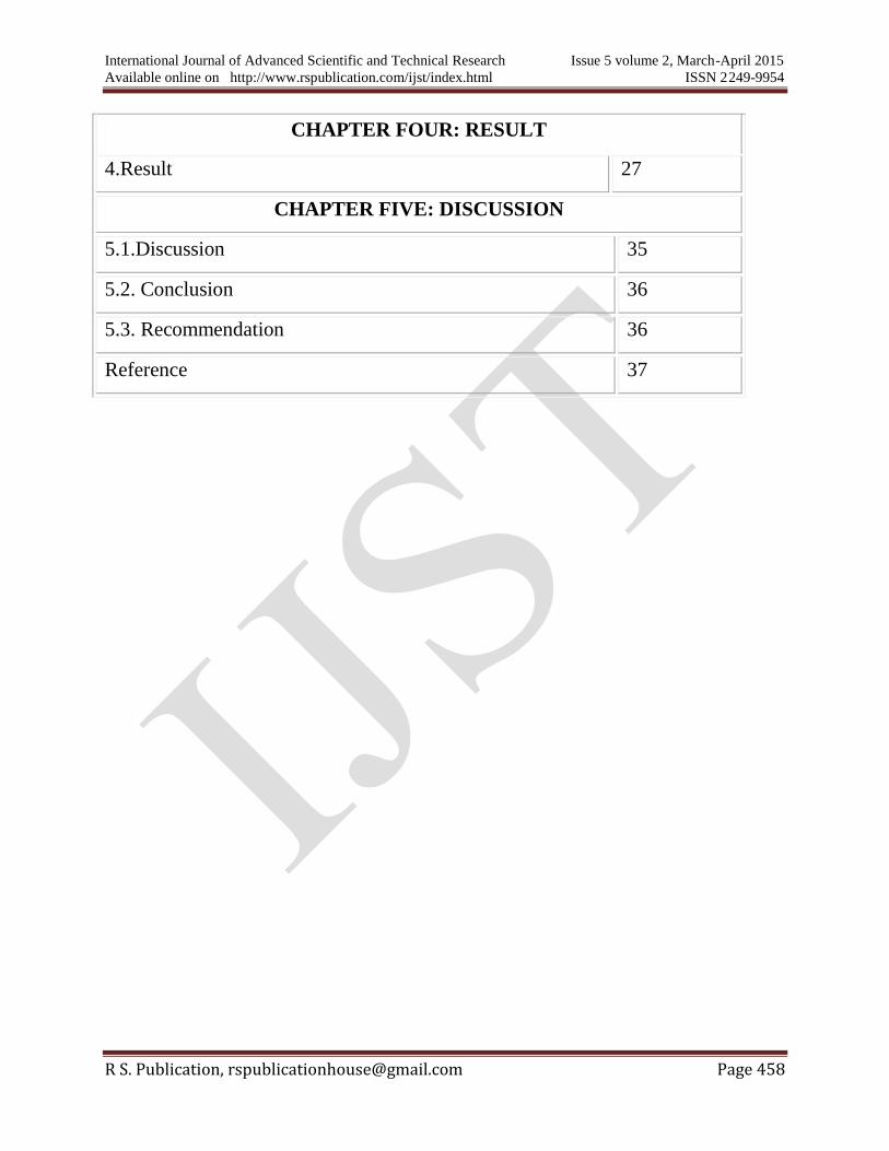
CHAPTER FOUR: RESULT
4.Result 27
CHAPTER FIVE: DISCUSSION
5.1.Discussion 35
5.2. Conclusion 36
5.3. Recommendation 36
Reference 37
International Journal of Advanced Scientific and Technical Research Issue 5 volume 2, March-April 2015
Available online on http://www.rspublication.com/ijst/index.html ISSN 2249-9954
R S. Publication, [email protected] Page 459
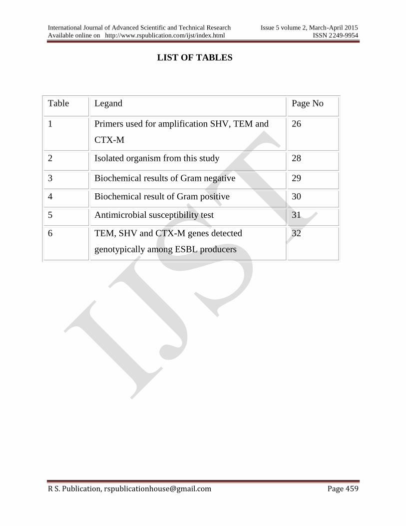
LIST OF TABLES
Table Legand Page No
1 Primers used for amplification SHV, TEM and
CTX-M
26
2 Isolated organism from this study 28
3 Biochemical results of Gram negative 29
4 Biochemical result of Gram positive 30
5 Antimicrobial susceptibility test 31
6 TEM, SHV and CTX-M genes detected
genotypically among ESBL producers
32
International Journal of Advanced Scientific and Technical Research Issue 5 volume 2, March-April 2015
Available online on http://www.rspublication.com/ijst/index.html ISSN 2249-9954
R S. Publication, [email protected] Page 460
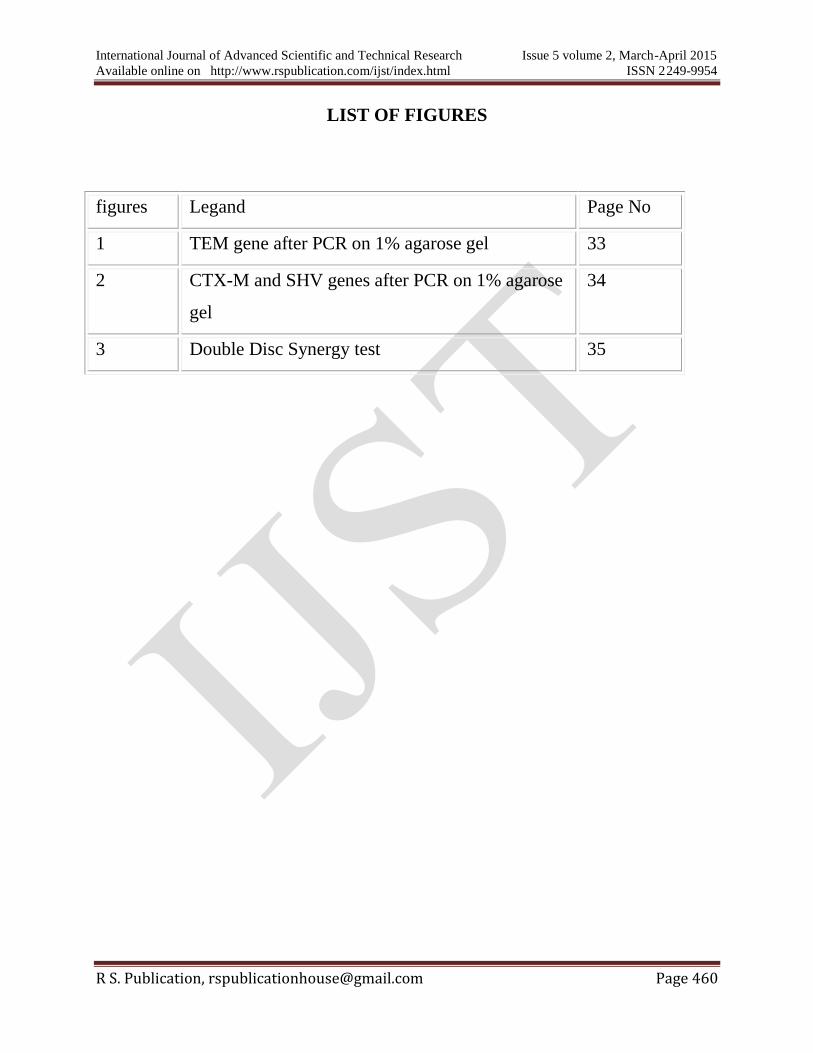
LIST OF FIGURES
figures Legand Page No
1 TEM gene after PCR on 1% agarose gel 33
2 CTX-M and SHV genes after PCR on 1% agarose
gel
34
3 Double Disc Synergy test 35
International Journal of Advanced Scientific and Technical Research Issue 5 volume 2, March-April 2015
Available online on http://www.rspublication.com/ijst/index.html ISSN 2249-9954
R S. Publication, [email protected] Page 461
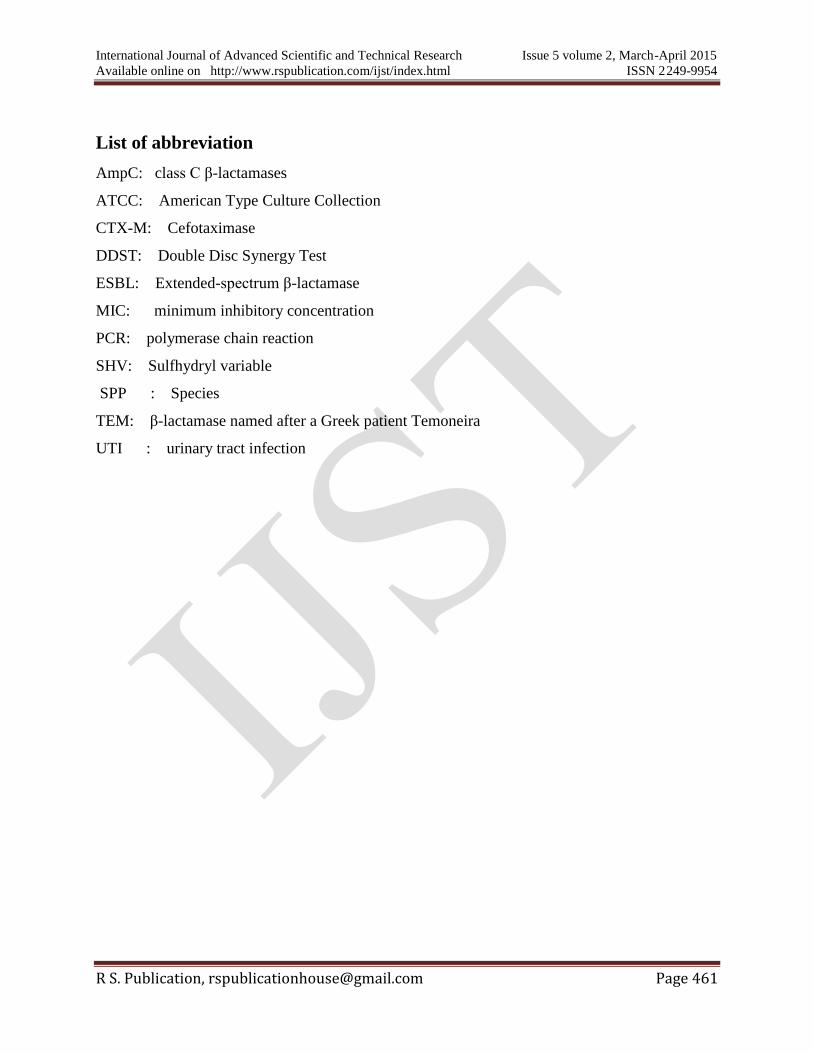
List of abbreviation
AmpC: class C β-lactamases
ATCC: American Type Culture Collection
CTX-M: Cefotaximase
DDST: Double Disc Synergy Test
ESBL: Extended-spectrum β-lactamase
MIC: minimum inhibitory concentration
PCR: polymerase chain reaction
SHV: Sulfhydryl variable
SPP : Species
TEM: β-lactamase named after a Greek patient Temoneira
UTI : urinary tract infection

International Journal of Advanced Scientific and Technical Research Issue 5 volume 2, March-April 2015
Available online on http://www.rspublication.com/ijst/index.html ISSN 2249-9954
R S. Publication, [email protected] Page 462
CHAPTER ONE
International Journal of Advanced Scientific and Technical Research Issue 5 volume 2, March-April 2015
Available online on http://www.rspublication.com/ijst/index.html ISSN 2249-9954
R S. Publication, [email protected] Page 463
1. INTRODUCTION
1.1 Introduction
Extended spectrum beta lactamases(ESBL) producing organisms are emerging pathogens with
greatest spread in general population, and their presence in clinical infection can result in
treatment failure when using 3rd generation cephalosporins (Pasteron and Bonomo, 2005). The
extended spectrum β-lactamases are enzymes elaborated by some bacteria and responsible for
their resistance to extended spectrum 3rd generation cephalosporins e.g. ceftazidim, cefotaxime
ceftriaxone and monobactam e.g. azteronem (Carlos , 2000).These enzymes catalyze the
hydrolysis of the β-lactam ring of antibiotics, ESBLs have been reported worldwide in many
different genera of Enterobactericeae, Pseudomonas aeroginosa, (Friedman et al., 2008).
Klebsiella pneumonia and E. coli (Agrawal et al., 2008). ESBL producing organisms are often
resistant to several other classes of antibiotics, as the plasmids with the gene encoding ESBLs
often carry other resistance determinants, initially ESBL producing organisms were isolated from
community (Pitout and Laupland, 2008).The ESBLs are mediated by bla TEM, bla SHV and bla
CTX-M genes in Enterobacteriaceae and other Gram- negative bacteria, numerus molecular
typing methods have been developed for their identification. The multiplex PCR assay allowed
the identification of bla TEM, bla SHV, and bla CTX-M genes in aseries of clinical isolates of
Enterobacteriaceae with previously characterized ESBL phenotype (Monstein et al., 2007).
The first plasmid β-lactamase TEM1 was described in Germany 1980, and in France 1985 (Perez
et al., 2007).The National Committee for Clinical Laboratory Standards (NCCLS) recommended
that microbiology laboratories reported ESBL –producing isolates of E.coli and Klebsiella
species are resistant to all penicillins, cephalosporins and azteronam, irrespective of their
individual in vitro test result.Urinary tract infections (UTI) that affect parts of the urinary system.
Occur more in women than men, especially in pregnancy due to the increased risk of kidney
infection. During pregnancy high progesterone level elevated the risk of decreased muscle tone
of the ureters and bladder which lead to greater likelihood of reflux where urine flows back up
the ureters and towards the kidney (Dielubanzaand Schaeffer, 2011). The bacterial spectrum
and antimicrobial resistance may vary temporarily and geographically, each institution must
undertake its own local evaluation such an evaluation may also be useful to detect emerging
trends of antimicrobial resistance (Wagenlehner et al., 2008).
The objectives of this study were to: Isolate and identify the important bacterial pathogen
causing UTI in pregnant women. Determine the drug resistance patterns of the isolated bacteria
with emphasis to third generation cephalosporins , and use molecular biology tools e.g. PCR in
the detection of ESBL encoding genes

International Journal of Advanced Scientific and Technical Research Issue 5 volume 2, March-April 2015
Available online on http://www.rspublication.com/ijst/index.html ISSN 2249-9954
R S. Publication, [email protected] Page 464
CHAPTER TWO
International Journal of Advanced Scientific and Technical Research Issue 5 volume 2, March-April 2015
Available online on http://www.rspublication.com/ijst/index.html ISSN 2249-9954
R S. Publication, [email protected] Page 465
2. LITERATURE REVIEW
2.1. Extended spectrum beta lactamase (ESBL)
2.1.1. History of ESBL:
Members of the family Enterobacteriaceae commonly express plasmid encoded β-
lactamases e.g.: (TEM1, TEM2, SHV and CTX-M). Which confers resistance to
penicillin but not to expanded spectrum cephalosporin; in the mid 1980 a new
group of extended spectrum β-lactamases (ESBL) were detected.
First detection was in GERMANY in 1983 (Knoth et al., 2003)
2.1.2. Classification:
2.2.1 Functional classification
Was described by (Bush et al., 1995) as different groups
Group 1
CEPHALOSPORINASE, Molecular Class C (not inhibited by clavulanic acid)
Group 1 are cephalosporinases not inhibited by clavulinic acid , belonging to the
molecular class C
Group 2
Group 2 are penicillinases, cephalosporinases, or both inhibited by clavulanic acid,
corresponding to the molecular classes A and D reflecting the original TEM and
SHV genes. However, because of the increasing number of TEM- and SHV-
derived β-lactamases, they were divided into two subclasses, 2a and 2b
GROUP 2a
PENICILLINASE, Molecular Class A
The 2a subgroup contains just penicillinases.
GROUP 2b
BROAD-SPECTRUM, Molecular Class A
International Journal of Advanced Scientific and Technical Research Issue 5 volume 2, March-April 2015
Available online on http://www.rspublication.com/ijst/index.html ISSN 2249-9954
R S. Publication, [email protected] Page 466
2b opposite to 2a , 2b are broad-spectrum β-lactamases, meaning that they are
capable of inactivating penicillins and cephalosporins at the same rate.
Furthermore, new subgroups were segregated from subgroup 2b:
GROUP 2be
Extended spectrum Class A
Subgroup 2be, with the letter "e" for Extended spectrum of activity, represents the
ESBLs, which are capable of inactivating third-generation cephalosporins
(ceftazidime, cefotaxime, and cefpodoxime) as well as monobactams (aztreonam).
GROUP 2br
INHIBITOR-RESISTANT, Molecular Class A (diminished inhibition by
clavulanic acid)
The 2br enzymes, with the letter "r" denoting reduced binding to clavulanic acid
and sulbactam, are also called inhibitor-resistant TEM-derivative enzymes;
nevertheless, they are commonly still susceptible to tazobactam, except where an
amino acid replacement exists at position met69.
GROUP 2c
more CARBENICILLINASE, Molecular Class A
Latersubgroup 2c was segregated from group 2 because these enzymes inactivate
carbenicillin more than benzylpenicillin, with some effect on cloxacillin.
GROUP 2d
CLOXACILANASE, Molecular Class D or A
Subgroup 2d enzymes inactivate cloxacillin than benzylpenicillin, with some
activity against carbenicillin; these enzymes are poorly inhibited by clavulanic acid,
and some of them are ESBLs
International Journal of Advanced Scientific and Technical Research Issue 5 volume 2, March-April 2015
Available online on http://www.rspublication.com/ijst/index.html ISSN 2249-9954
R S. Publication, [email protected] Page 467
the correct term is "OXACILLINASE". These enzymes are able to inactivate the
oxazolylpenicillins like oxacillin, cloxacillin, dicloxacillin. The enzymes belong to
the molecular class D not molecular class A.
GROUP 2e
CEPHALOSPORINASE, Molecular Class A
Subgroup 2e enzymes are cephalosporinases that can also hydrolyse monobactams,
and they are inhibited by clavulanic acid.
GROUP 2f
CARBAPENAMASE olecular Class A
Subgroup 2f was added because these are serine-based carbapenemases, in contrast
to the zinc-based carbapenemases included in group 3
Group 3
METALLOENZYME, Molecular Class B (not inhibited by clavulanic acid)
Group 3 are the zinc-based or metallo beta-lactamases, corresponding to the
molecular class B, which are the only enzymes acting by the metal ion zinc, as
discussed above. Metallo B-lactamases are able to hydrolyse penicillins,
cephalosporins, and carbapenems. Thus, carbapenems are inhibited by both group
2f (serine-based mechanism) and group 3 (zinc-based mechanism)
Group 4
PENICILLINASE, No Molecular Class (not inhibited by clavulanic acid)
Group 4 are penicillinases that are not inhibited by clavulanic acid, and they do not
yet have a corresponding molecular class.
Molecular classification
Renamed Beta-lactamase when the structure of the Beta-lactam ring was finally
elucidated The molecular classification of β-lactamases is based on the nucleotide
International Journal of Advanced Scientific and Technical Research Issue 5 volume 2, March-April 2015
Available online on http://www.rspublication.com/ijst/index.html ISSN 2249-9954
R S. Publication, [email protected] Page 468
and amino acid sequences in these enzymes. To date, four classes are recognised
(A-D), correlating with the functional classification. Classes A, C, and D act by a
serine-based mechanism, whereas class B or metallo-β-lactamases need zinc for
their action.
2.1.3. Types
TEM beta-lactamases (class A)
TEM-1 is the most commonly encountered beta-lactamase in Gram-negative
bacteria. Up to 90% of ampicillin resistance in E. coli is due to the production of
TEM-1. Also responsible for the ampicillin and penicillin resistance that is seen in
H. influenzae and N. gonorrhoeae in increasing numbers. Although TEM-type
beta-lactamases are most often found in E. coli and K. pneumoniae, they are also
found in other species of Gram-negative bacteria with increasing frequency. The
amino acid substitutions responsible for the ESBL phenotype cluster around the
active site of the enzyme and change its configuration, allowing access to
oxyimino-beta-lactam substrates. Opening the active site to beta-lactam substrates
also typically enhances the susceptibility of the enzyme to b-lactamase inhibitors,
such as clavulanic acid. Single amino acid substitutions at positions 104, 164, 238,
and 240 produce the ESBL phenotype, but ESBLs with the broadest spectrum
usually have more than a single amino acid substitution. Based upon different
combinations of changes, currently 140 TEM-type enzymes have been described
(Paterson et al ., 2003).
(SHV beta-lactamases class A)
SHV-1 shares 68 percent of its amino acids with TEM-1 and has a similar overall
structure for up to 20% of the plasmid-ampcillin resistance in this species ESBLs
in the SHV-1 beta-lactamase is most commonly found in
International Journal of Advanced Scientific and Technical Research Issue 5 volume 2, March-April 2015
Available online on http://www.rspublication.com/ijst/index.html ISSN 2249-9954
R S. Publication, [email protected] Page 469
K. pneumoniae and is responsible this family also have amino acid changes around
the active site, most commonly at positions 238 or 238 and 240. More than 60
SHV varieties are known. They are the predominant ESBL type in Europe and the
United States and are found worldwide. SHV-5 and SHV-12 are among the most
common (Paterson et al ., 2003).
CTX-M beta-lactamases (Class A)
These enzymes were named for their greater activity against cefotaxime than other
oxyimino-beta-lactam substrates (e.g., ceftazidime, ceftriaxone, or cefepime).
Rather than arising by mutation, they represent examples of plasmid acquisition of
beta-lactamase genes normally found on the chromosome of Kluyvera species, a
group of rarely pathogenic commensal organisms. These enzymes are not very
closely related to TEM or SHV beta-lactamases in that they show only
approximately 40% identity with these two commonly isolated beta-lactamases.
More than 80 CTX-M enzymes are currently known. Despite their name, a few are
more active on ceftazidime than cefotaxime. They have mainly been found in
strains of Salmonella enterica serovar Typhimurium and E. coli, but have also been
described in other species of Enterobacteriaceae and are the predominant ESBL
type in part of south America .They are also seen in Eastern Europe ,CTX-M-
14,CTX-M-3 and CTX-M-2 are the most widespread. CTX-M-15 is currently the
most widespread type in E. coli the UK and is widely prevalent in the community
(Woodford et al.,2006).
OXA beta-lactamases (Class D)
OXA beta-lactamases were long recognized as a less common but also plasmid-
mediated beta-lactamase variety that could hydrolyze oxacillin and related anti-
staphylococcal penicillins. These beta-lactamases differ from the TEM and SHV
enzymes in that they belong to molecular class D and functional group 2d. The
International Journal of Advanced Scientific and Technical Research Issue 5 volume 2, March-April 2015
Available online on http://www.rspublication.com/ijst/index.html ISSN 2249-9954
R S. Publication, [email protected] Page 470
OXA-type beta-lactamases confer resistance to ampicillin and cephalothin and are
characterized by their high hydrolytic activity against oxacillin and cloxacillin and
the fact that they are poorly inhibited by clavulanic acid. Amino acid substitutions
in OXA enzymes can also give the ESBL phenotype. While most ESBLs have
been found in E. coli, K. pneumoniae, and other Enterobacteriaceae, the OXA-type
ESBLs have been found mainly in P. aeruginosa. OXA-type ESBLs have been
found mainly in Pseudomonas aeruginosa isolates from Turkey and France. The
OXA beta-lactamase family was originally created as a phenotypic rather than a
genotypic group for a few beta-lactamases that had a specific hydrolysis profile.
Therefore, there is as little as 20% sequence homology among some of the
members of this family. However, recent additions to this family show some
degree of homology to one or more of the existing members of the OXA beta-
lactamase family. Some confer resistance predominantly to ceftazidime, but OXA-
17 confers greater resistance to cefotaxime and cefepime than it does resistance to
ceftazidime (Woodford et al.,2006)
Others Types of ESBL genes
Other plasmid-mediated ESBLs, such as PER, VEB, GES, and IBC beta-
lactamases, have been described but are uncommon and have been found mainly in
P. aeruginosa and at a limited number of geographic sites. PER-1 in isolates in
Turkey, France, and Italy; VEB-1 and VEB-2 in strains from South East Asia; and
GES-1, GES-2 and IBC-2 in isolates from South Africa, France and Greece. PER-1
is also common in multiresistant acinetobacter species in Korea and Turkey. Some
of these enzymes are found in Enterobacteriaceae as well, whereas other
uncommon ESBLs (such as BES-1, IBC-1, SFO-1 and TLA-1) have been found
only in Enterobacteriaceae.
International Journal of Advanced Scientific and Technical Research Issue 5 volume 2, March-April 2015
Available online on http://www.rspublication.com/ijst/index.html ISSN 2249-9954
R S. Publication, [email protected] Page 471
2.2. Urinary tract infections (UTIs):
There are two types of UTIs: lower and upper. Lower UTIs occur in urethra
(urethritis) or bladder (cystitis). Upper UTIs are infections that involve kidneys
(pyelonephritis), or ureters (ureteritis), or both. Upper UTIs can occur in both men
and women as a complication of lower UTI. Complicated UTIs are resulting from
anatomic obstructions of the urinary tract or catheterization. These abnormalities
increase the volume of residual urine and interfere with the normal clearance of
bacteria by urination. Such factors include prostate enlargement, sagging uterus
and expansion of the uterus during pregnancy, paraplegia, spinabifida, scar tissue
formation and catheterization. Uncomplicated acute UTIs refer to that seen in
patients with normal anatomic structure and function of the urinary tract (Najar et
al., 2009: Foxman, 2010).
2.2.1 Symptoms of urinary tract infection
Urethritis appears as discomfort during urination. Most of the cases of purulent
urethritis without cystitis are sexually transmitted infection is limited to the urethra.
Hemorrhagic cystitis is characterized by large quantities of visible blood in the
urine. It can be caused by an infection (bacterial) or as a result of radiation, cancer
chemotherapy or select immunosuppressive medications (Silva et al., 2010).
Pyelonephritis symptoms include fever, back pain, costovertebral angle tenderness,
nausea and vomiting and possibility of urinary urgency and frequency urine
contains white blood cell casts composed of cells that were tightly packed in the
tubules and excreted in proteinaceous matrix (lee et al., 2009).
2.2.2 Causative organisms of urinary tract infections:
The vast majority of UTIs are due to patients own fecal bacteria. Approximately
80% of acute uncomplicated UTIs are caused by E. coli, 10 to 20 % are caused by
coagulase – negative Staphylococcus saprophyticus and 5% or less are caused by
International Journal of Advanced Scientific and Technical Research Issue 5 volume 2, March-April 2015
Available online on http://www.rspublication.com/ijst/index.html ISSN 2249-9954
R S. Publication, [email protected] Page 472
other Enterobacteriaceae such as Proteus and Klebsiella or by Enterococci species
(Nicolettia et al., 2010).
The most common causes of complicated UTIs are Escherichia coli, Klebsiella
pneumoniae, Proteus mirabilis, Enterobacter Cloacae, Serratia marcescens,
Pseudomonas aeruginosa and Gram-positive organism such as Enterococci,
coagulase–negative Staphylococci and Staphylococcus aureus (Bader et al., 2010;
Pallett and Hand, 2010).
2.2.3 Diagnosis of urinary tract infection
The diagnosis based on clinical symptoms and the clinical diagnosis includes
history and physical examination.
Microscopical examination of urine
UTI can readily be diagnosed by microscopical examination of urine.
Standardized centrifuge urinary sediment investigated under a cover slip is
recommended as the routine procedure because it is cheap and the differentiation
of formed element (red, white cell and bacteria) is easier in thin fluid layers than in
traditional glass chambers. Centrifugation always leads to loss of particles and
may produce inaccurate result in quantitative terms. On the other hand, in un spun
samples a number of relevant elements can be missed. Thus the results after
centrifugation with standardized procedures are more sensitive and specific. When
compared with bright-field microscopy, the phase-contrast technique allows better
detection of most elements, especially of bacteria. The counts are usually given per
low-power field or high-power field. At the high magnification (40x) the presence
of >10 white blood cell/high-power field is indicative of pyuria (Haider et al., 2010)
Urine culture
The diagnosis of UTI is confirmed by culturing the organism from urine. Most
bacteria that cause urinary infection grow readily and the clinical diagnosis of UTI
International Journal of Advanced Scientific and Technical Research Issue 5 volume 2, March-April 2015
Available online on http://www.rspublication.com/ijst/index.html ISSN 2249-9954
R S. Publication, [email protected] Page 473
is usually confirmed within 24hours. Patients suspected of UTI are usually asked to
collect a mid-stream sample after cleaning the perineum or glans penis with soap
and water. An early-morning collection is best because the concentration of
bacteria in the urine is a greatest prior to the morning voiding. The urine is then
refrigerated or taken to the laboratory for immediate culture. Urine can be stored in
a refrigerator for up 24 hours without any loss of bacterial viability (Gupta et al.,
2011).
2.2.4 Epidemiology
From epidemiological viewpoint UTIs occur in two general setting: community-
acquired and hospital (nosocomially) acquired, E. coli caused about 80% of
community-acquired UTI so it represent the most common cause of UTI (Mims et
al ., 2004).
2.3. Typical characteristics of Enterobacteriaceae
Is the largest, most heterogeneous collection of medically important Gram-
negative bacilli. Total of 32 genera and more than 130 species have been described,
these genera have been classified based on biochemical properties, antigenic
structure, nucleic acid hybridization and sequencing; are ubiquitous organisms,
being found worldwide in soil, water, and vegetation, and are part of the normal
intestinal flora of most animals including humans (Murray et al., 2002).
The wall, cell membrane and internal structures are morphologically similar for all
enterobacteriaceae, the major cell wall antigen is the heat stable lipopolysaccharide
LPS and it consists of three components: the somatic O polysaccharide, a core
polysaccharide and lipid A. The serologic classification of the Enterobacteriaceae
is based on three major groups of antigen: somatic O polysaccharide, capsular K
antigen (either protein or polysaccharide) and flagellar H protein. Numerous
International Journal of Advanced Scientific and Technical Research Issue 5 volume 2, March-April 2015
Available online on http://www.rspublication.com/ijst/index.html ISSN 2249-9954
R S. Publication, [email protected] Page 474
virulent factors have been identified in the member of the family Enterobactericeae.
Some are common to all genera and others are unique to specific virulent strains
(Murray et al., 2002)
2.4 Escherichia
The genus Escherichia consists of five species: E.blattae, E.fergusonii, E.hermanii,
and E.vulneris. E.coli is most numerous aerobic commensally inhabitant of the
large intestine in human.
E.coli can be commonly found in animal faeces and the lower intestines of
mammals, and they possess adhesive fimbriae that promote binding to intestinal,
epithelial cells. E.coli can also be found in environments at a higher temperature,
such as on the edge of hot springs in our intestines, E.coli is probably the most
famous member of the Enterobacteriaceae group, since it is a model organism and
lots of our knowledge of biochemical processes and genetics derive from this
species (Daoud and Afif, 2011).
2.5. Klebsiella
The genus Klebsiella is among the oldest known genera in the Enterobacteriaceae
described for the first time by Trevisan in 1885. Klebsiella consist of three species:
K. oxytoca, K. pneumoniae and K. variicola.
Klebsiella species are non-motile and have a prominent mucoid polysaccharide
based capsule (K antigen). The capsule protects from phagocytosis and aids to
adherence. Due to the capsule Klebsiella form large moist colonies on solid culture
media .K. pneumoniae are widely distributed in the environment where they
contribute to biochemical and geochemical processes. Like E.coli, Klebsiella are
colonizers of the gastrointestinal tract (Nordmann et al., 2009). Klebsiella are
International Journal of Advanced Scientific and Technical Research Issue 5 volume 2, March-April 2015
Available online on http://www.rspublication.com/ijst/index.html ISSN 2249-9954
R S. Publication, [email protected] Page 475
ubiquitous and may colonize the skin, pharynx, or gastrointestinal tract in humans.
K. pneumoniae and K. oxytoca are both opportunistic pathogens found in the
environment and on mammalian mucosal surfaces; they are commonly transferred
by hands of hospital personnel (Miftode et al ., 2008).
2.6. Other enterobacterial genera
Other enterobacterial species most commonly encountered are Citrobacter .,
Hafnia alvei, Proteus mirabilis , Salmonella spp ,serratia spp and
Shigella .Citrobacter genus consists of twelve species and can be isolate from
water, sewage soils and food as well as from the faeces of man and other animals,
where they may be part of the normal microbiota of the large intestine the genus
Hafnia contains a single species, H. alvei, and it is generally motile the Yersinia
genus contains 14 species, and these are relatively slow growers among the
Enterobacteriaceae. Y.pestis, Y.pseudotuberculosis and Y.enterocolitica are very
well documented human pathogen. Yersinia displays their biochemical
characteristics most reliably at temperatures between 25 and 32 ºC.
The Proteus genus is highly motile and does not form regular colonies. Instead,
swarming colonies are formed when plated on a non-inhibitory media. P. mirabilis
is considered to be the most important member of this genus. The genus
Salmonella consists of several serotypes and most of them are motile. The Serratia
genus contains ten species and two subspecies.
However, only two species are clinically commonly isolated. They are S.
liquefaciens and S. marcescens. Often produce prodigiosin, a characteristic red
pigment, when grown at 20 ºC. Most of the species are motile. Serratia strains can
be distinguished from other enterobacterial species by their unique production of
three enzymes: DNase, lipase and gelatinase. The Shigella genus consisting of four
International Journal of Advanced Scientific and Technical Research Issue 5 volume 2, March-April 2015
Available online on http://www.rspublication.com/ijst/index.html ISSN 2249-9954
R S. Publication, [email protected] Page 476
species is closely related to Escherichia and considered to be a subspecies of E.
coli. Shigella is non-motile and anaergenic; I .e .it does not produce gas from
glucose (Larson et al., 2005; Paterson, 2006; Halstead et al., 2007; Hidron et al.,
2008) .
2.7. Gram-positive cocci
There are two medically important genera of gram-positive cocci: Streptococcus
and Staphylococcus. Two of the most important human pathogens, Staphylococcus
aureus and Streptococcus pyogenes. Staphylococci and Streptococci non motile
and do not form spores distinguished by two main criteria microscopically,
Staphylococci appear in grapelike clusters, where streptococci are in chain and
biochemical, Staphylococci produce catalase (degrade hydrogen peroxide),
whereas streptococci do not.
2.8. ESBL
Members of the family Enterobacteriaceae commonly express plasmid encoded β-
lactamases. ESBLs are beta-lactamases that hydrolyze extended spectrum
cephalosporin with oxyiminoside chain this cephalosporin include cefotaxime,
ceftriaxone, and ceftazidime as well as the oxymino, monobactam and aztreonam.
Thus ESBLs confirm resistance to these antibiotic and related oxymino beta-
lactams. Typically, they derive from gene for TEM1, TEM2, SHV1 and CTX-M
by mutation that alters the amino acid configuration around the active site of these
β-lactamases. This extended the spectrum of β-lactam antibiotic susceptible to
hydrolysis by these enzyme increasing number of ESBL not of TEM or SHV
lineage have recently been described (Emery and Weymouth, 1997).

International Journal of Advanced Scientific and Technical Research Issue 5 volume 2, March-April 2015
Available online on http://www.rspublication.com/ijst/index.html ISSN 2249-9954
R S. Publication, [email protected] Page 477
2.9. Detection of ESBL
2.9.1 Screening test for ESBL
2.9.1.1. Disc diffusion method
According to the CLSI guidelines, isolates showing inhibition zone size of <22mm
with ceftazidime (30µg), <27mm with cefotaxime (30µg), <25mm with ceftriaxone
(30µg), <27mm with aztreonam (30µg) and <22mm with cefpodoxime (10µg), was
identified as potential ESBL producers and short listed for confirmation of ESBL
production recently. Chromogenic designed specifically for screening and
identification of ESBLs producing Enterobacteriaceae, have become commercially
available (Black et al., 2005).
2.9.2 Confirmatory test
2.9.2.1. Double disc synergy test (DDST)
The disc synergy test (DDST) is oldest method for phenotypic confirmation of
ESBLs producing organisms, first proposed in 1980 (Jarlier et al., 1988)
aceftazidime 30µg disc and amoxicillin/clavulanic acid 20+10 µg disc will be
placed 25-30 mm a part, center to center. Following over night incubation in
aerobic at 37ºC, ESBL production is inferred when the zone of inhibition around
the ceftazidime disc is expanded by the clavulanate.
2.9.2.2. Modified double disc diffusion test
Muller Hinton agar media will be inoculated with standardized inoculums
(corresponding to 0.5 McFarland standard tube using sterile cotton swab.
Augmentin (20mg amoxicillin and 10mg clavulanic acid AMC) disc will be placed
in the center of the plate and test discs of 3rd
generation cephalosporin disc will be
placed at 15mm distance from the Augmentin disc. The plate will be incubated
overnight at 37 ºC. ESBL production consider positive if the zone of inhibition
International Journal of Advanced Scientific and Technical Research Issue 5 volume 2, March-April 2015
Available online on http://www.rspublication.com/ijst/index.html ISSN 2249-9954
R S. Publication, [email protected] Page 478
around the test disc increased towards the Augmentin disc or neither disc will be
inhibitory alone but bacterial growth will be inhibited were the two antibiotics
diffuse together .enhancement of the zone of inhibition around one or more of β-
lactam containing disc towards the clavulanic acid containing disc is indicative of
ESBL production. And interpretation is subjective.
2.9.2.3. Disc replacement method for ESBL confirmation
Two Amoxyclave (AMC 30mg) discs will be placed on Muller-Hinton agar
inoculated with the bacterial isolate. After 1hour at room temperature, the disc
were removed and replaced with ceftazidizm (RP-30) and ceftriaxone (AUF30).
Each cephalosporin disc will be incubated at 37Cº for 18-24hours and read for
evidence of ESBL production. Positive disc replacement method will be indicated
by increased inhibition zone of 5mm and above between the inhibition zone
formed by Augmentin. Replaced cephalosporin disc and those placed
independently. The following two procedures will be carried out in the present
study as per CLSI guideline.
2.9.3 E-Test
The E-test strip (AB Biodisk, Solna; Sweden) carries two ingredients: on the one
end, ceftazidime and on the opposite end, ceftazidime plus clavullinic acid. MIC is
interpreted as the point of intersection of the inhibition ellipse with the E-test strip
edge. A ratio of ceftazidime MIC to cetazidime-clavulinic acid MIC equal to or
greater than 8 indicates the presence of ESBL. The reported sensitivity of the
method as phenotypic confirmatory test for ESBLs is 87% to100% and the
specificity is 95-100%. The availability of cefotaxime strips, as well as ceftazidime
strips, improves the ability to detect ESBL types, which preferentially hydrolyzed
cefotaxime, such as CTX-M type enzymes (Rawat and Nair, 2010).
International Journal of Advanced Scientific and Technical Research Issue 5 volume 2, March-April 2015
Available online on http://www.rspublication.com/ijst/index.html ISSN 2249-9954
R S. Publication, [email protected] Page 479
2.9.4 .Vitek ESBL test
A specific card which includes tests for ESBL production has now been by
approved FDA.The Vitek ESBL test (bioMerieux vitek, Hazelton, Missouri)
utilizes cefotaxime and ceftazidime alone (at 0.5µg/ml) and in combination with
clavulinic acid (4µg/ml). Inoculation of the cards is identical to that performed for
regular vitek cards. Analysis of all wells is performed automatically once the
growth control well has reached a set threshold (4-15 hours of incubation). A
predetermined reduction in the growth of the cefotaxime or ceftazidime wells
containing clavulanic acid, compared with the level of growth in the well the
cephalosporin alone, indicates presence of ESBL. Sensitivity and specificity of the
method exceed 90% (Rawat and Nair, 2010)
2.10. ESBLs prevention and control
Proper infection-control practices and barriers are essential to prevent spreading
and outbreak of ESBL producing bacteria. The reservoir for these bacteria seems to
be the gastrointestinal tract of patients. The contaminated hands and stethoscopes
of healthcare providers are important factors in spreading infection between
patients. Essential infection control practices should include avoiding unnecessary
use of invasive devices such as indwelling urinary catheters, hand washing by
hospital personnel, increased barrier precaution, and isolation of patients colonized
or infected with ESBL producers. At an institutional level, practices that can
minimize the spread of such organisms include clinical and bacteriological
surveillance of patients admitted to intensive care units and antibiotic cycling; as
well as polices of restriction, especially on the empirical use of broad-spectrum
antimicrobial agent such as the third and fourth generation cephalosporin and
quinolones (Rawat and Nair, 2010).
International Journal of Advanced Scientific and Technical Research Issue 5 volume 2, March-April 2015
Available online on http://www.rspublication.com/ijst/index.html ISSN 2249-9954
R S. Publication, [email protected] Page 480
2.11. Previous studies
A Study done in Roosendaal in 2012, in total of 174 patients were included. In 24
of 174 (14%) patients, ESBL carriage could not be confirmed with the micro array.
This was verified days, with PCR and sequencing. The mean duration of isolation
was 15 adding up to a total number of isolation days of 2571. False-positive days
of results according to the micro array resulted in a total of 279 days for the
DDCT. Using unnecessary isolation for the E-test and 151 E-test to detect the
presence of ESBL results in a false-positive outcome in 14% of the cases. This
results in unnecessary isolation of patients, which can be omitted by using a
genotypic method (Wintermans et al., 2012).
Study was done in Taiwan (February 2011) by Hsueh-Hsialo to detect the genes
encoding extended-spectrum β-lactamases (ESBLs) and to determine the
epidemiological relatedness of 69 Escherichia coli and 33 Klebsiella pneumoniae
isolates collected from a regional hospital in central Taiwan, mostly from
inpatients (E. coli 87.0 %; K. pneumoniae 88.0 %). The phenotypes of these isolates
were examined according to the combination disc method recommended by the
Clinical and Laboratory Standards Institute. Most of the ESBL-producing E. coli
and K. pneumoniae isolates (98.6 % and 97 %, respectively) could be detected
using cefotaxime discs with and without clavulanate. Genotyping was performed
by PCR with type-specific primers. CTX-M-14 type (53.6 %) was the most
prevalent ESBL among E. coli isolates while SHV type (57.6 %) was the most
dominant among K. pneumoniae isolates. Six E. coli and three K. pneumoniae
isolates did not carry genes encoding ESBLs of types TEM, SHV, CTX-M-3,
CTX-M-14, CMY-2 and DHA-1. The co-existence of two or more kinds of ESBL
in a single isolate was common, occurring in 40.6 % and 72.7 % of E. coli and K.
pneumoniae isolates, respectively.
International Journal of Advanced Scientific and Technical Research Issue 5 volume 2, March-April 2015
Available online on http://www.rspublication.com/ijst/index.html ISSN 2249-9954
R S. Publication, [email protected] Page 481
Study done in West India (2013) by Dr Akpaka in Female patients (67.8%) and
urine samples (65%) yielded most ESBL isolates, with over 90% recovered from
the hospital’s medicine and surgery facilities. All ESBL isolates including all
K.pneumoniae producing ESBLs were 100% susceptible to carbapenems and
amikacin antimicrobials. Polymerase Chain Reaction detected 100% bla TEM
genes, 4.1% bla SHV and 37.5% bla CTXM genes among E. coli isolates.
Similarly, 84.3% bla TEM, 34.5% bla SHV and 58.8% bla CTXM genes were
detected in K. pneumoniae.
Study done in West India from December 2004 to April 2008 found 602 K
.pneumoniae and 1016 E. coli recovered from the clinical specimens were
identified as ESBL producers. A 15.2% ESBL rate among the K. pneumoniae
isolates and 9.3% among the E. coli isolates has previously been reported in this
Hospital (Akpaka et al., 2008)
Study done in North Eastern Italy reported that 70 ESBL-producing E. coli, 61
(87.1%) were positive for bla CTX-M, alone (30, corresponding to 42.8%) or in
combination with bla TEM (31, 44.3%). Only 8 isolates showed other resistance
genes (1 bla TEM alone, 1 bla SHV and 7 with associated bla TEM and bla SHV).
In K. pneumoniae, bla SHV was present in all 5 isolates, alone or associated with
bla TEM) or bla TEM and CTX-M. In K. oxytoca, 3 strains carried bla SHV and 1
bla TEM, while bla CTX-M was absent; only one strain resulted negative for all
the ESBL tested (Busetti et al., 2008).
International Journal of Advanced Scientific and Technical Research Issue 5 volume 2, March-April 2015
Available online on http://www.rspublication.com/ijst/index.html ISSN 2249-9954
R S. Publication, [email protected] Page 482
CHAPTER THREE
International Journal of Advanced Scientific and Technical Research Issue 5 volume 2, March-April 2015
Available online on http://www.rspublication.com/ijst/index.html ISSN 2249-9954
R S. Publication, [email protected] Page 483
MATERIALS AND METHODS
3.1. Study design
This study is hospital and laboratory-based study.
3.1.1. Type of study
The study is descriptive, cross-sectional study.
3.1.2. Study area
This work was carried out in Military Hospital in Omderman, during the period
from April to May 2013.
3.1.3. Study population
Pregnant women with urinary tract infection (UTI) were included in this study.
3.1.4. Sample size
One hundred mid –stream urine samples were collected from pregnant women
3.1.5. Data collection
Data were collected using interviewing questionnaire (Appendix 1)
3.1.6. Inclusion criteria
Pregnant women with UTI symptoms
3.1.7. Exclusion criteria
All pregnant women without UTI symptoms and pregnant women under antibiotic
treatment.
3.1.8. Ethical considerations
Approval of the National Ethics Committee, Ministry of Health (Sudan).
3.2. Collection of specimens
5 ml midstream urine samples were collected in wide mouth screw-capped and
leak-proof sterile containers containing 0.1g/10 ml boric acid as preservative.

International Journal of Advanced Scientific and Technical Research Issue 5 volume 2, March-April 2015
Available online on http://www.rspublication.com/ijst/index.html ISSN 2249-9954
R S. Publication, [email protected] Page 484
3.3. Cultivation of specimens
The specimens were inoculated under aseptic conditions on CLED agar using
sterile standard bacteriological wire loop. The inoculated culture media were
incubated aerobically at 37Cº overnight and observed for bacterial growth.
3.4. Identification
3.4.1. Colonial morphology
Used as first identification depending on size, color, edges, side views and
fermentation of lactose.
3.4.2. Microscopical examinations
Fixed and dried smear from the growth were prepared. Gram stain was performed
for each slide as follows: slides were covered with crystal violet stain for 30-60 sec,
then washed with water and covered with logol’ s iodine for 30-60 sec, washed,
decolorized rapidly (few second) with alcohol , washed immediately and covered
with safranine for 2 minutes then washed and examined microscopically using oil
immersion lens (X100). E. coli ATCC®25922 was used as control.
3.4.3. Biochemical identification
Biochemical tests were carried out according to Cheesbrough (2000) eg indole test,
urease test, citrate, motility and the tube of KIA medium will show ( slope and butt)
for Gram negative. Catalase test, coagulase, DNase test and mannitol salt agar for
Gram-positive.
3.5. ESBL screening test
All isolates were tested for their susceptibility to the 3rd
generation cephalosporins,
i.e.: ceftazidime (30µg), cefotaxime (30µg) and ceftriaxone (30µg) by the standard
disc diffusion method as recommended by the CLSI guidelines (2010). ESBL were
International Journal of Advanced Scientific and Technical Research Issue 5 volume 2, March-April 2015
Available online on http://www.rspublication.com/ijst/index.html ISSN 2249-9954
R S. Publication, [email protected] Page 485
screened by detection of reduced zones of inhibition around 3rd
generation
cephalosporins.
The bacterial isolate was considered as ESBL producer when the zone diameter
for :ceftazidime < 22mm. cefotaxime < 27mm and ceftriaxone < 25mm. Resistant
isolates to at least one of the 3rd
generation cephalosporins were checked for ESBL
production.
Double disk synergy test (DDST) was performed according to Jarlier et al., (1988).
Isolates were inoculated on Muller –Hinton agar plates. Discs containing
ceftazidime (30Mg), cefotaxime (30Mg) and ceftriaxone (30mg) were placed 20
mm (center to center) away from a disc containing 20 mg amoxicillin/10mg
clavulanic acid (AMC). Plates were incubated overnight at 37ºC. ESBL production
was considered positive if the zone of inhibition around one or more of the 3rd
antibiotic discs showed clear cut increase towards the AMCA disc
(Ananthakrishanan et al., 2000).
3.6. Genotypic characterization of ESBL genes
3.6.1. DNA extraction
1.5 ml of bacterial broth culture were transferred into eppendorff microfuge tubes
and centrifuged at 12,000-16,000 for 2 min and then 200 µl of resuspension
solution, 20 µl RNase (20 mg/ml), 25 µl lysozyme (50mg/ml), 20 µl proteinase K
(20mg/ml) vortexed and incubated at 55ºC for 20 min.Then 200 µl lysis solution,
was vortexed and incubated at70 Cº for 10 min. Prepared column by adding 500
µl of column preparation solution to the column, then centrifuged at maximum
speed (13000 rpm). To bind DNA to the column 200 µl ethanol (100%) was added
to the suspension, vortexed and transferred to the primed column, centrifuged
again at( 13000 rpm) for 2 min. The supernatant was discarded and 500 µl washing
International Journal of Advanced Scientific and Technical Research Issue 5 volume 2, March-April 2015
Available online on http://www.rspublication.com/ijst/index.html ISSN 2249-9954
R S. Publication, [email protected] Page 486
solution were then added to the column , centrifuged at (9000 rpm) for 2 min, and
then 500 µl washing solution were added to the column and centrifuge at
12000rpm for 3min ,discard and centrifuged at (13000 rpm) for 1min to dry the
column. Was transfer the column to a new microfuge tube and 200 µl elution
solution were added and centrifuged at 9000 rpm for 1 min.
3.6.2. Amplification of DNA using PCR
All isolates were screened for the resistance genes, bla TEM, bla CTX-M and bla
SHV using PCR, according to Sidjabt et al .,(2009)(table1). The thermal cycler
(CONVERGYS td peltier thermal cycle, Germany) was used throughout the study
for the amplification of the PCR mixtures. In a total volume of 25µl containing 10
pmol of each three pairs of primers (Metabion, GERMANY), 4µl FIREPOL
Master Mix (Solis BioDyne, Tartu, Estonia), 0.6 forward primer, 0.6reverse primer,
2µl plasmid DNA and 17.8 µl deionized sterile water. The PCR mixture was
subjected to initial denaturation step at 94ºC for 5 min, followed by 30 cycles of
denaturation at 94ºC for 45sec, primer annealing at 55ºC for 45sec, followed by
step of elongation at 72Cº for 60sec and the final elongation at 72ºC for 5min
(Cao et al., 2002). PCR products were analyzed by electrophoresis in a 1%
agaroses gel in TPE 1X, containing 0.5 mg/ml ethidium bromide at 120V for 40
min. Bands were visualized under U.V transilluminater (Uvite, UK).
International Journal of Advanced Scientific and Technical Research Issue 5 volume 2, March-April 2015
Available online on http://www.rspublication.com/ijst/index.html ISSN 2249-9954
R S. Publication, [email protected] Page 487
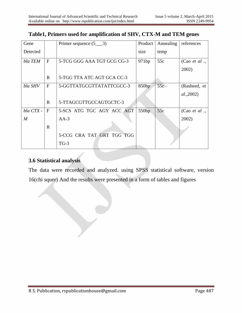
Table1, Primers used for amplification of SHV, CTX-M and TEM genes
Gene
Detected
Primer sequence (5___3) Product
size
Annealing
temp
references
bla TEM F
R
5-TCG GGG AAA TGT GCG CG-3
5-TGG TTA ATC AGT GCA CC-3
971bp 55c (Cao et al .,
2002)
bla SHV F
R
5-GGTTATGCGTTATATTCGCC-3
5-TTAGCGTTGCCAGTGCTC-3
850bp 55c (Rasheed, et
al.,2002)
bla CTX -
M
F
R
5-SCS ATG TGC AGY ACC AGT
AA-3
5-CCG CRA TAT GRT TGG TGG
TG-3
550bp 55c (Cao et al .,
2002)
3.6 Statistical analysis
The data were recorded and analyzed. using SPSS statistical software, version
16(chi squer) And the results were presented in a form of tables and figures
International Journal of Advanced Scientific and Technical Research Issue 5 volume 2, March-April 2015
Available online on http://www.rspublication.com/ijst/index.html ISSN 2249-9954
R S. Publication, [email protected] Page 488
CHAPTER FOUR
International Journal of Advanced Scientific and Technical Research Issue 5 volume 2, March-April 2015
Available online on http://www.rspublication.com/ijst/index.html ISSN 2249-9954
R S. Publication, [email protected] Page 489
4. RESULTS
4. Results
Out of 100 urine specimens collected from the study population, bacterial growth
was observed in 56 cultures. The results of Gram stain found 42(75%) Gram
negative bacilli and14 (25%) Gram positive bacteria, the most of Gram negative
was K. oxytoca 15(26.8%), E.coli 10(17.9%), P. vulgaris 8(14.3%), K. pneumoniae
5(8.9%), Citrobacter frundii 1(1.8%), Enterobacter spp 1(1.8%), Providencea spp
1(1.8%) and Serratia marcesance 1(1.8%). The most Gram positive
Staphelococcus saprophyticus 13( 23.2%) and S. aureus 1(1.8) (table2). The
results of biochemical test for Gram negatives E.coli, K, P.vulgaris, Citrobacter,
Enterobacter , Provdencea and Serratia ( table3) and for Gram positive cocci
S.aureus and S.saprophyticus (table4) The susceptibility test showed that 2(40%)
of K. pneumnae was resistant to all antibiotic used (ceftazidime, cefotaxime and
ceftriaxone) and 3(60%) K. pneumoniae was susceptible to all antibiotic used, all K.
oxytoca was susceptible to all antibiotic used, E.coli 8(80% ) were susceptible and
2(20%) were resistant, P. vulgaris 7(87.5%) were susceptible and 1(12.5%)was
resistant, all S. saprophyticus were susceptabl and Enterobacter, Providencea,
Serratia, Citrobacter and S.aureus were sensitive to 3rd
generation cephalosporine
used( table4). The ESBLs producer were screnning by resistant to 3rd
generation
cephalosporine found in 5isolates (11.9%) confirmed by using double disc synergy
test (DDST) (figer1) found in 2 K. pneumoniae, 2 E.coli and 1 P.vulgaris. the PCR
used to identifying genes resistant, The commonest prevalence of ESBL gene was
TEM gene in all 5 organisms (figer2), CTX-M was produced by 3 organisms 2 E.
coli and 1 K. pneumoniae and SHV gene in 1K. pneumoniae and 1 P. vulgaris
(table5).
International Journal of Advanced Scientific and Technical Research Issue 5 volume 2, March-April 2015
Available online on http://www.rspublication.com/ijst/index.html ISSN 2249-9954
R S. Publication, [email protected] Page 490
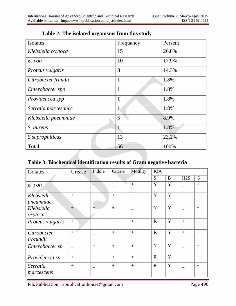
Table 2: The isolated organisms from this study
Table 3: Biochemical identification results of Gram negative bacteria
Isolates Urease Indole Citrate Motility KIA
S B H2S G
E .coli _ + _ +
Y Y _ +
Klebsiella
pneumniae
+ _ + _ Y Y _ +
Klebsiella
oxytoca
+ + + _ Y Y _ +
Proteus vulgaris + + _ + R Y + +
Citrobacter
Freundii
+ _ + + R Y + +
Enterobacter sp _ + + + Y Y _ +
Providencia sp + + + + R Y _ +
Serratia
marcescens
+ _ + + R Y _ +
Isolates Frequancy Persent
Klebsiella oxytoca 15 26.8%
E. coli 10 17.9%
Proteus vulgaris 8 14.3%
Citrobacter frundii 1 1.8%
Enterobacter spp 1 1.8%
Providencea spp 1 1.8%
Serratia marcesance 1 1.8%
Klebsiella pneumniae 5 8.9%
S. aureus 1 1.8%
S.saprophiticus 13 23.2%
Total 56 100%
International Journal of Advanced Scientific and Technical Research Issue 5 volume 2, March-April 2015
Available online on http://www.rspublication.com/ijst/index.html ISSN 2249-9954
R S. Publication, [email protected] Page 491
S: slobe
B: bute
G: gase
Y: yellow
R: red
Table 4: Biochemical identification results of Gram-positive bacteria
Isolates Dnase MSA Coagulase
S. aureus + + +
S. saprophyticus _ + _
MSA: Manetol Solte Agar
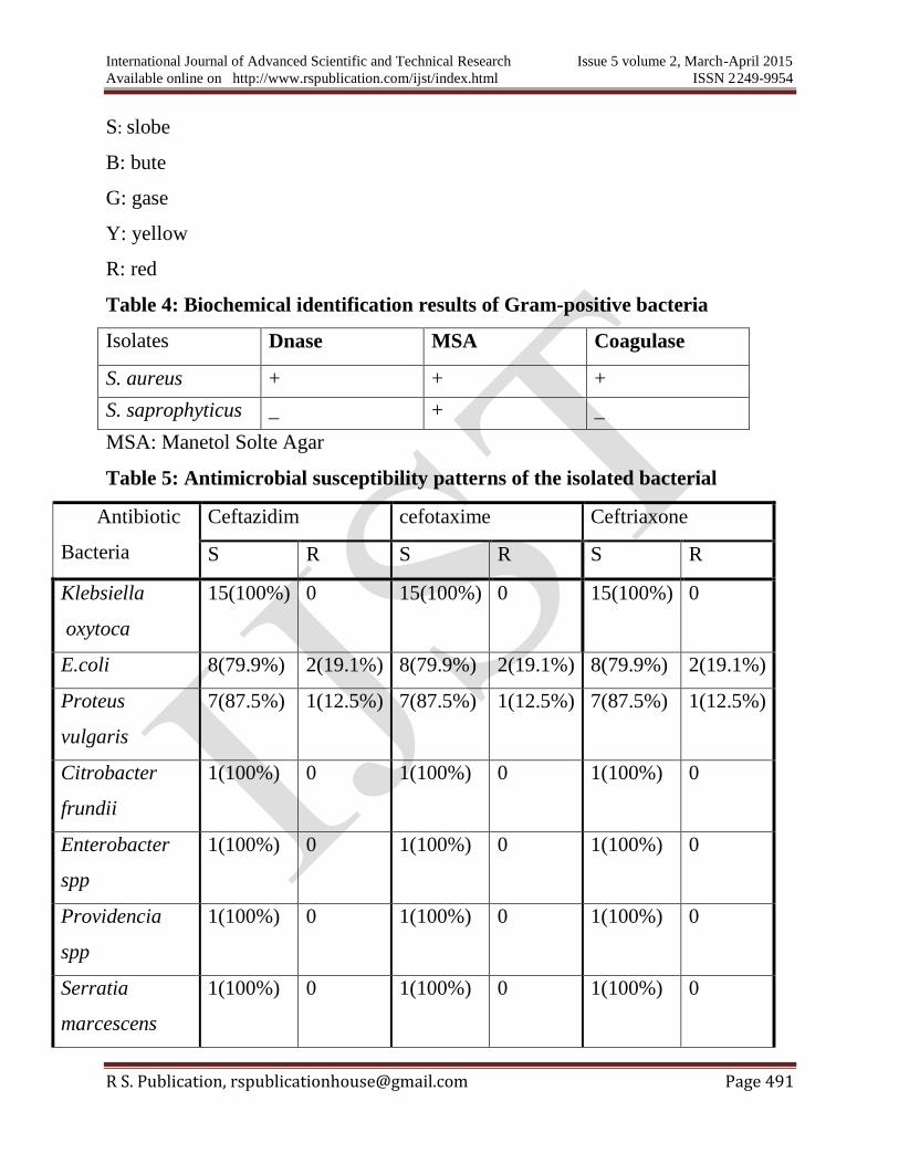
Table 5: Antimicrobial susceptibility patterns of the isolated bacterial
Antibiotic
Bacteria
Ceftazidim cefotaxime Ceftriaxone
S R S R S R
Klebsiella
oxytoca
15(100%) 0 15(100%) 0 15(100%) 0
E.coli 8(79.9%) 2(19.1%) 8(79.9%) 2(19.1%) 8(79.9%) 2(19.1%)
Proteus
vulgaris
7(87.5%) 1(12.5%) 7(87.5%) 1(12.5%) 7(87.5%) 1(12.5%)
Citrobacter
frundii
1(100%) 0 1(100%) 0 1(100%) 0
Enterobacter
spp
1(100%) 0 1(100%) 0 1(100%) 0
Providencia
spp
1(100%) 0 1(100%) 0 1(100%) 0
Serratia
marcescens
1(100%) 0 1(100%) 0 1(100%) 0
International Journal of Advanced Scientific and Technical Research Issue 5 volume 2, March-April 2015
Available online on http://www.rspublication.com/ijst/index.html ISSN 2249-9954
R S. Publication, [email protected] Page 492
Klebsiella
pneumoniae
3(60%) 2(40%) 3(60%) 2(40%) 3(60%) 2(40%)
S. aureus 1(100%) 0 1(100%) 0 1(100% 0
S.saprophyticus 13(100% 0 13(100%) 0 13(100%) 0
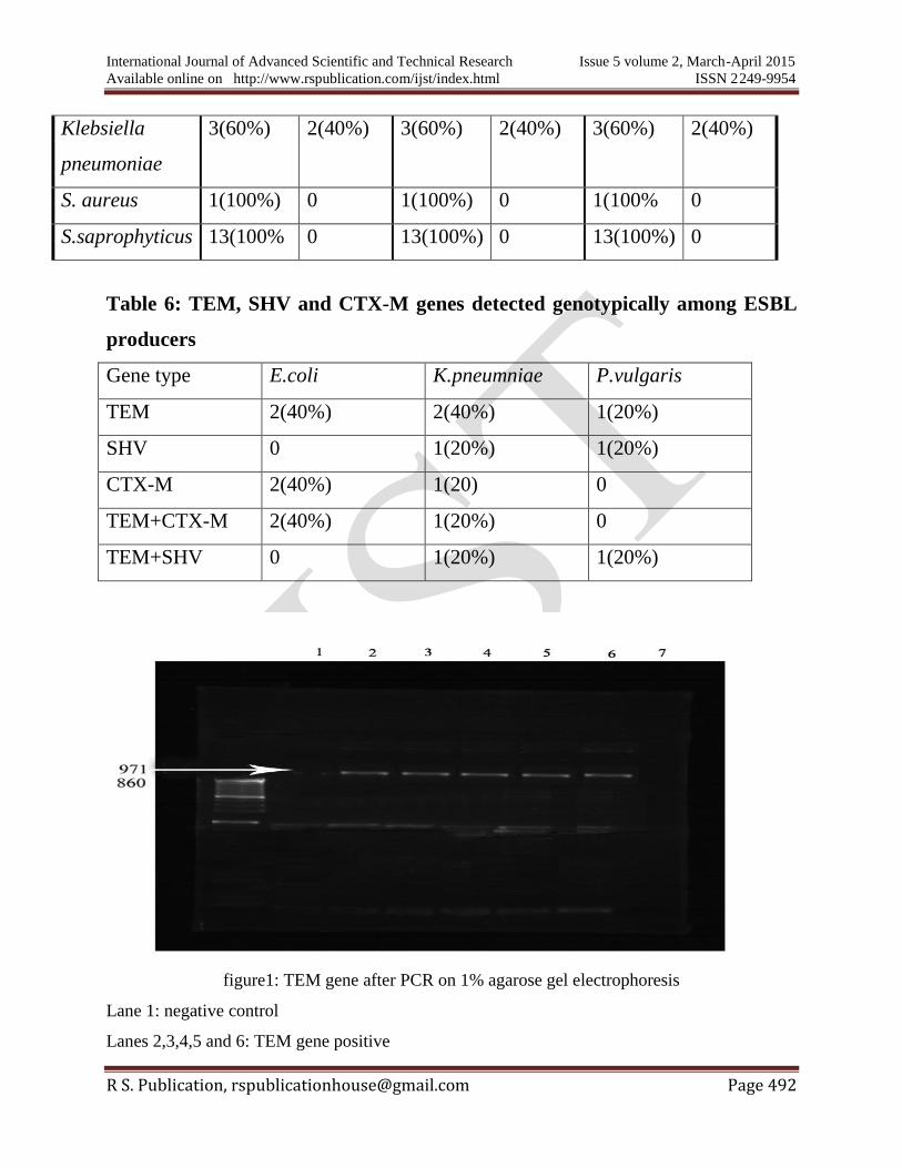
Table 6: TEM, SHV and CTX-M genes detected genotypically among ESBL
producers
Gene type E.coli K.pneumniae P.vulgaris
TEM 2(40%) 2(40%) 1(20%)
SHV 0 1(20%) 1(20%)
CTX-M 2(40%) 1(20) 0
TEM+CTX-M 2(40%) 1(20%) 0
TEM+SHV 0 1(20%) 1(20%)
figure1: TEM gene after PCR on 1% agarose gel electrophoresis
Lane 1: negative control
Lanes 2,3,4,5 and 6: TEM gene positive
International Journal of Advanced Scientific and Technical Research Issue 5 volume 2, March-April 2015
Available online on http://www.rspublication.com/ijst/index.html ISSN 2249-9954
R S. Publication, [email protected] Page 493
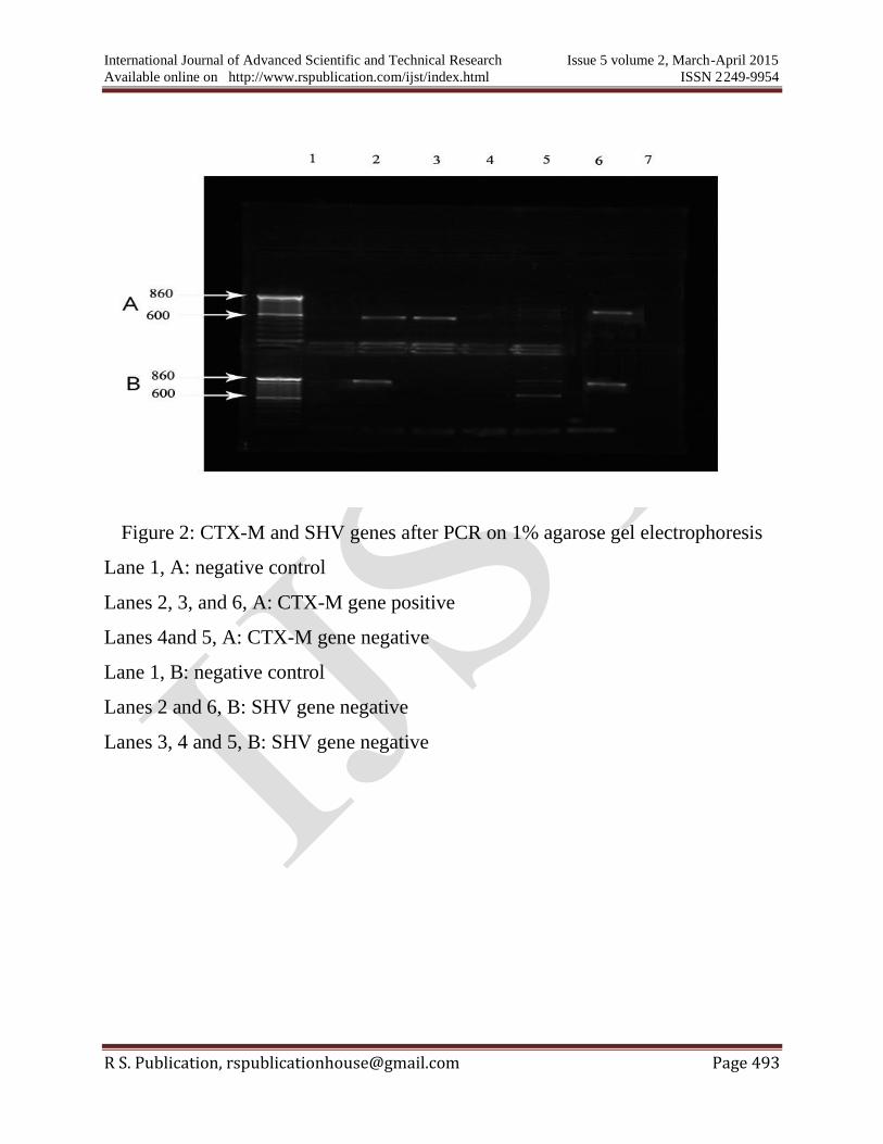
Figure 2: CTX-M and SHV genes after PCR on 1% agarose gel electrophoresis
Lane 1, A: negative control
Lanes 2, 3, and 6, A: CTX-M gene positive
Lanes 4and 5, A: CTX-M gene negative
Lane 1, B: negative control
Lanes 2 and 6, B: SHV gene negative
Lanes 3, 4 and 5, B: SHV gene negative
International Journal of Advanced Scientific and Technical Research Issue 5 volume 2, March-April 2015
Available online on http://www.rspublication.com/ijst/index.html ISSN 2249-9954
R S. Publication, [email protected] Page 494
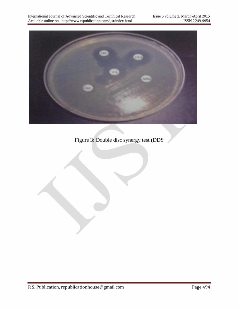
Figure 3: Double disc synergy test (DDS
International Journal of Advanced Scientific and Technical Research Issue 5 volume 2, March-April 2015
Available online on http://www.rspublication.com/ijst/index.html ISSN 2249-9954
R S. Publication, [email protected] Page 495
CHAPTER FIVE
International Journal of Advanced Scientific and Technical Research Issue 5 volume 2, March-April 2015
Available online on http://www.rspublication.com/ijst/index.html ISSN 2249-9954
R S. Publication, [email protected] Page 496
5.1. DISCUSSION
In this study out of 100 samples examined only 56 isolates identified, K. oxytoca
15(26%), K. pneumoniae 5(8.9%), E. coli 10(17.9%), P. vulgaris 8(14.3%),
Enterobacter spp 1(1.8%), Citrobacter freundii 1(1.8%), Provedancia spp 1(1.8%),
S. aureus 1(1.8%) and S. saprophyticus 13(23.2% ). Many reports showed
difference prevalence rates of ESBLs Enterobacterial uropathogens. In this study,
5(11.9) isolates producing ESBL were encountered, of which 2 (40%) K.
pneumoniae, 2 (20%) E. coli and 1(12.5%) P. vulgaris, These results were similar
to the combined disc method currently recommended by the NCCLS. For
laboratories that perform susceptibility testing by using disc diffusion, modified
DDST could be easily incorporated in to an already existing system. This finding
was similar to study done by Taslima Yasmin (2012) in Bangladesh and Omer
basher(2013) in Sudan reported that E. coli and K. pneumoniae are most ESBL
producers.
Urine was the most main source of ESBL producing isolates, which is in
agreement with that found by Akbar et al ., (2007), in Oman who reported that
(70.4%) of urine was the main source of ESBLs from all patient.
The present showed that high prevalence of TEM gene. Which was Similar to that
reported by Seker et al., (2009)? The CTX-M was the second ESBL gene detected
in this investigation, a finding that was in agreement with that reported by
Feizabadi, et al., (2010). The present this study showed that CTX-M was most
common gene among E, coli this was in agreement with that reported by Sajjad et
al .,(2006)and Lavigne et al .,(2007). The SHV gene was the less frequent gene in
this study, this result is similar with many reports around the world, such as
Thailand by Kiratisine et al (2009) and in Iran ( Dezful et al., 2011). These results
were not coniccding with those of Tasli and Bahar,(2005)and Ben-Ami et
International Journal of Advanced Scientific and Technical Research Issue 5 volume 2, March-April 2015
Available online on http://www.rspublication.com/ijst/index.html ISSN 2249-9954
R S. Publication, [email protected] Page 497
al.,(2009), who detected SHV in 74.3% of isolates. This study is also disagreed
with that reported in Sudan by Nour aldayem (2012) who found CTX-M is highly
prevalent but agreed with SHV the less gene obtained.
5.2. Conclusions
In Sudan ESBL genes were among most strains of Enterobacteriaceae, especially E.
coli and K. pneumoniae. ESBL SHV, TEM and CTX-M genes are predominant in
Sudan among Enterobacteriaceae isolates and some strains carried one or more
than of these genes and this may lead to the resistance to some cephalosporin
antibiotic, that might complicate the management of the disease problem.
5.3. Recommendation
1- Formulation of proper antibiotic policy and providing appropriate guidelines to
prescribe antibiotic, can prevent the spread of multi-drug resistant organisms in
the hospital as well as in community.
2- Laboratory detection of ESBL producing bacteria is highly recommended.
3- DNA sequencing and specific target gene primers are required to differentiate
between ESBLs and ESBLs variant of TEM and SHV genes.
International Journal of Advanced Scientific and Technical Research Issue 5 volume 2, March-April 2015
Available online on http://www.rspublication.com/ijst/index.html ISSN 2249-9954
R S. Publication, [email protected] Page 498
REFERENCES
1- Abcdef, G; Dielubanza, E.J; Schaeffer, A.J(2011). Urinary tract infection in
women. Med Clin of Worth America 95(1):27-41.
2- Agrawal, P; Cohosh, A; Kumar, S; Basu, B; Kapil, K (2008). Prevalence of
extended spectrum B-lactamases among E.coli and K. pneumoniae isolates in
tertiary care hospital, Indian Journal pathology Microbiology 5:139-42.
3- Akbar, M; Rafay, Zakariya Al-Muharrmi and Robert Toki (2007).
Prevalence of Extended spectrum beta-lactamases-producing isolates over a 1-year
period at a University Hospital in Oman. Saudi Med. J 28(1): 22-27.
4- Ananthakrishanan, A.N; Kanungo, R; Kumar,A and Badrinath,S(2000).
Detection of extended spectrum B-lactamase producer among surgical wound
infection and burns patiens in Jipmer.Indian J Med Microbiol; 18:160-5.
5- Apaka, P.E and Swanston, W.H (2008). Occurrence and phenotypic detection
of extended spectrum beta-lactamase in clinical isolates of K. pneumoniae and E.
coli at tertiary Hospital in Trinidada and Tobago. Braz. J.Infect. Dis 12:516-20.
6-Bader, M.S; Hawbold, J;Brooks, A(2010).Management of complicated urinary
tract infection. In the aria of antimicrobial resistance.postgrad med 122 (6):7-15.
7 - Ben-Ami, R; Rodrigue-Bano, J; Arslan, H; Pitout, J.D.D; Quentine, C;
Calbo, E.S; Azap, O.K; Arpin, C; Pascual, A; Livermore, D.M; Garau, J and
Carmeli, Y (2009). A multinational survey of risk factors for infection with
extended-spectrum B-lactamase producing enterobacteriaceae in non hospitalized
patients. Clin. Infec. Dis. 49:682-690.
8- Black, J.A; Thomson, K.S; Buynak, J.D and Pitout J.D (2005). Evaluation
of B-lactamase in well characterized clinical strains of Klebsiella spp, J. Clin.
Microbiol 43(4):4161-4171.
International Journal of Advanced Scientific and Technical Research Issue 5 volume 2, March-April 2015
Available online on http://www.rspublication.com/ijst/index.html ISSN 2249-9954
R S. Publication, [email protected] Page 499
9- Bush, K; Jacoby, G.A and Medeiros, AA (1995). Updated functional
classification of beta-lactamase producing organism (ESBL) Braz.J. Microbiol
31(4): 1211-1233.
10- Busetti, M; Comar, M; Fiermonta, F and Campello, C (2008). Molecular
detection of Extended spectrum beta-lactamase producing Enterobacteriaceae in
North Eastern Italy. European congress of Clinical Microbiology and Infections
Diseases Barcelona-Spain.
11- Cao V, Lambert T and Courvalin P. (2002). Colel –like plasmed pIP843 of
Klebsiella pneumoniae encoding extended-spectrum B-lactamase CTX-M-17.
Antimicrob Agents Chemother 46, 1212-1217.
12- Carlos, H; Pessoade, M and Silva (2000). Elaboration and evaluation of
anew screening medium for detection and presumptive identification of extended
spectrum B-lactamase producing organism (ESBL) Braz. J .micobiol vol 31 no 4.
13- Cheesbrough, M (2000). District laboratory practice in tropical countries, part
2, chambridge university press, UK, Ch7, p157-234.
14-Clinical and laboratory standards institute (2010). Performance standards
for antimicrobial susceptibility testing twentieth information al supplement ed.
CLSI document M100-S20 wayne PA: CLSI.
15- Daoud, Z and Afif, C (2011). Escherichia Coli isolated from urinary tract
infection of Lebanese patients between 2000-2009: Epidemiology and profiles of
resistance. Chemother Respract :1-6.
16- David, L;Paterson, D.L and Robert A, Bonomo (2005). ESBL: a clinical
update, J Clinical Microbiology Review.18 (4):657-686.
17- Dezful Ganjavian Hospital, Ahvaz Jundishapur (2011). Distribution of
TEM, SHV and CTX-M Genes among ESBL-producing enterobacteriaceae
isolates in Iran. African Journal of Microbiology Research 6 (26):5433-5439.
International Journal of Advanced Scientific and Technical Research Issue 5 volume 2, March-April 2015
Available online on http://www.rspublication.com/ijst/index.html ISSN 2249-9954
R S. Publication, [email protected] Page 500
18- Emery, C.L and Weymouth, L.A (1997). Detection and clinical significance
of extended spectrum B-lactamase in a tertiary care medical center .J .Clin.
Microbial. (35):2061-2067.
19- Feizabadi, M.M; Delfani, S; Raji, N; Majnooni, A; Aligholi, M;
Shahcheraghi, F; Parvin, M and Yadegarinia, D (2010). Distribution of bla
TEM, bla SHV and bla CTX-M genes among clinical isolates of K. pneumoniae at
labbafinejad Hospital, Tehran, Iran. Microbial drug resistance 16 (1):49-53.
20- Foxman, B (2010). The epidemiology of urinary tract infection so Nat Rev
urol .7: 653-660.
21- Friedman,C; Callery,S; Jeanes,A; Piskowski,P; Scott,L(2008). Best
infection control practices for patients extended spectrum β-lactamase
Enterobacteriaceae .Int. Infect.Control Council.
22- Goosens, H (2001). Mystic programs summary of European data from 1997 to
2000.Diagn .microbiol.infect Dis 41:183-189.
23- Gupta, K; Hooton, T.M; Naber, KG (2011). International clinical practice
guideline for the treatment of acute uncomplicated cystitis and pyelonephritis in
women: a 2010 update by the infectious disease society of American and the
European society for Microbiology and Infectious Diseases. Clin Infect Dis.
52:103-120.
24-Haider, G; Zehra, N; Munir, AA; Haider, A (2010). Risk factors of urinary
tract infection in pregnancy. J Pak Med Assoc. 60(3):213-6.
25-Halstead, D.C; Abid, J;Dowzicky, M.J(2007). Antimicrobial susceptibility
among Acinetobacter calcoaceticus- baumannii complex and enterobacteriaceae
collected as part of the Tigecycline Evaluation and surveillance Tial. J Infect.55
(1):49-57.
International Journal of Advanced Scientific and Technical Research Issue 5 volume 2, March-April 2015
Available online on http://www.rspublication.com/ijst/index.html ISSN 2249-9954
R S. Publication, [email protected] Page 501
26- Hidron, A.I; Edwards, J.R; Patel, J; Horan ,T.C; Sievert, D.M; Pollock,
D.A (2008). NHSN annual update: antimicrobial resistant pathogens associated
with healthcare. Associated infection: annual summary of data reported to
National Healthcare Safety Network at the center for Disease control and
prevention, 2006-2007. Infect Control Hosp Epidemiol .29(11):996-1011.
27- Hsueh-Hsia L.O (2013). Genotypic detection and molecular epidemiology of
Extended spectrum beta lactamase producing E. coli and K. pneumniae in
aregional Hospital central Taiwan. J. Mic. 62: 345-351.
28- Jarlier, V; Micola, M.H; Fournier,G; and Philippon, A(1988). Extended
broad-spectrum B-lactamases conferring transferable resistance to Newer B-lactam
agent in Enterobacteriaceae. Hospital prevalence and susceptibility patterns. Rev
Infect Dis 10: 867-879.
29- Kapaka, P.E; Legall, B and Padman, J (2013). Molecular detection and
epidemiology of Extended spectrum beta lactamase genes prevalent in clinical
isolates of K. pneumniae and E. coli from Tiridad and Tobago. Ind. Med. J 59 (6).
30- Kiratisin, P; Apisarnthanarak, A; Laesripa, C; Saifon, P(2008). Molecular
characterization and epidemiology of extended-spectrum B-lactamase producing
Escherichia coli and Klebsiella pneumoniae isolates causing health care –
associated infection in Thailand, where the CTX-M family is endemic. Antimicrob
Agents Chemother 52:2818-2824.
31- Knoth, H; Shah, P and Kremery ,V (2003). Transferable resistance to
cefotaxime, cefoxitine, cefamandole and cefuroxine in clinical isolates of ESBL
Infection Antimicrobe Agent Chemother 11 (6):315-7.
32- Larson, E.L; Cimiotti, J.P and Haas, J (2005). Gram-negative bacilli
associated with catheter-associated and non catheter-associated bloodstream
International Journal of Advanced Scientific and Technical Research Issue 5 volume 2, March-April 2015
Available online on http://www.rspublication.com/ijst/index.html ISSN 2249-9954
R S. Publication, [email protected] Page 502
infections and hand carriage by healthcare workers in neonatal intensive care units.
Pediatr Crit Care Med. 6(4):457-461.
33- Lee, H; Jung, D; Yeom,, J.S; Son, J.S; Jung, S.I; et al (2009). Evaluation of
ceftriaxone utilization at multicenter study. Korean J Intern Med. 24(4):374-380.
34- Mekki, A.H; Hassan, A.N and Elsayed, D.M(2010). Extended spectrum
beta-lactamases among multi drug resistant Escherichia coli Klebsiella spp causing
urinary tract infection in khartoum TBJ. Act Res, 2(3):18-21.
35- Miftode, E; Dorneanu, O; Leca, D; Teodor, A; Mihalache, D and Filip, O
(2008). Antimicrobial resistance profile of E. coli and Klebsiella spp. From urine
in the infectious Diseases Hospital Iasi. Rev Med Chir Soc Med Nat Iasi.113
(2):478-482.
36- Mims C, Dockrell, Hazel M, Goering V, Roitt.I, Wakelin. D and
Zuckerman M.(2004). Medical Microbiology, third edition, ELSEVIER MOSBY.
37- Monstein, H.J; Ostholm-Balkhed, A; Nilsson, M.V; Dornbusch and
Nilsson L.E(2007). Multiplex PCR amplification assay for rapid detection of bla
SHV, bla TEM, and bla CTX-M genes in Enterobacteriaceae. APMIS 115:1400-
1408.
38-Murray Patrick R, Rosenthal K, Kabayashi George S and Pfaaller Michael
A.(2002). Medical Microbiology 4th
edition, Mosby. P266-279.
39- National Committee for Clinical Laboratory Standards (NCCLS, 2004).
NCCLS document M100-S15. Performance for Antimicrobial Susceptibility
Testing .8th
edition .Pennsylvania.
40- Nicolettia, J;Kustera, S; Sulserb, T; Zbindenc, R; Ruefa, C; Ledergerbera,
B and Webera, R(2010). Risk factors for urinary tract infection due to
ciprophloxacin-resistant Escherichia coli in a tertiary care urology department in
Switzerland. Swiss Med Wkly.140:13059.
International Journal of Advanced Scientific and Technical Research Issue 5 volume 2, March-April 2015
Available online on http://www.rspublication.com/ijst/index.html ISSN 2249-9954
R S. Publication, [email protected] Page 503
41- Njar, M.S; Saldanha, C.L; Banday, K.A (2009). Approach to urinary tract
infections. I J N.19 (4):129-139.
42- Norbmann, P; Cuzon, G and Naas, C (2009). The real tsreat of K.
pneumoniae carbapenemase-produsing bacteria. Lancet Infection- Dis 9(4):228-
236.
43- Ozcakar, Z.B; Yalcinkaya, F; Kavaz, A; Kadioglu, G; Elhan, A.H; Aysev,
D; Guriz, H and Ekim, M (2011). Urinary tract infections owing to ESBL-
producing bacteria: microorganism change-clinical pattern does not. Acta Paediatr.
100(8):61-64.
44- Pallett, A and Hand, K(2010). Complicated urinary tract infections: practical
solutions for the treatment of multiresistant Gram-negative bacteria. J Antimicrob
Chemother.65(3)25-33.
45- Paterson, D.L (2006). Resistance in Gram-negative bacteria:
Enterobacteriaceae. Am. J. Med.119: 20-28.
46- Paterson, D.L; Hujer, K.M and Hujer, A.M (2003). Of extended spectrum
beta-lactamase in K. Pneumoniae blood stream isolated from some
countries :dominance and widespread, prevalence of SHV and CTX-M type beta-
lactamases. Antimicrob Agents Chemother 47:3554-3560.
47- Perez, F; Endimiani, A; Hujer,K.M and Bonomo, R.A (2007). The
continuing challenge of ESBLs. Curent opinion in pharmacology 7(5):459-469.
48- Pitout, JDD and Laupland, K.B(2008). Extended spectrum beta-lactamase
producing enterobacteriaceae: an emerging public health concern lancet infection
Dis 8 pp59-66.
49- Rawat, D and Nair, D (2010). Extended-spectrum beta-lactamases in Gram
negative bacteria. J Glob Infect Dis. 2(3):263-274.
International Journal of Advanced Scientific and Technical Research Issue 5 volume 2, March-April 2015
Available online on http://www.rspublication.com/ijst/index.html ISSN 2249-9954
R S. Publication, [email protected] Page 504
50- Sajjad, H; Muhammed, S; Umar, K and Amin, W (2006). CTX-M ESBL
enzyme in E. coli from urology patients in Rawalpindi. J. Pak. Med. Assoc 56 (12):
576-578.
51- Sekar, B; Shwetha, R; Arunagiri, K; Lalitha, P and Aparna ,V (2009).
Detection and characterization of bla CTX-M gene by PCR-RFLP analysis among
third generation cephalosporin resistant Gram-negative isolates. proceoding of the
XXX National congress of Indian association of medical Microbiologists.p27
52- Sidjabat, H.E; Paterson, D.L; Adams-Haduch, J.M; Ewan. L; Pasculle,
A.W; Muto, C.A; Tian, G.B and Doi, Y (2009). Molecular Epidemiology of
CTX-M producing Escherichia coli isolates at Tertiary Medical Center in western
Pennsylvania. Antimicrob Agents Chemother.53 (11):4733-4739.
53- Silva Lde, P; Patah, P.A; Saliba, R.M; Szewczyk, N.A; Gilman, L;
Neumann, J; Han, X.Y; Tarrand, J; Ribeiro, R; Gulbis, A; Shpall, E.J; Jones,
R; Popat, U; Walker, J.A; Petropoulos, D; Chiattone, A; Stewart, J; El-
Zimaity, M; Anderlini, P; Giralt, S; Champlin, R.E and de Lima, M(2010).
Hemorrhagic cystitis after allogeneic hematopoietic stem cell transplants is the
complex result of BK virus infection, preparative regimen intensity and donor type.
Haematologica .95(7):1183-1190.
54- Tasli, H and Bahar, I.H(2005). Molecular characterization of TEM and
SHV derived ESBL in hospital based enterobacteriaceae in Turkey .Jpn.
Infect.Dis.58:162-167.
55- Wagnlehnerhner, F.M; Niemetz, A.H; Weidner, W and Naber, K.G
(2008). Department of urology, hospital ST Elisabth, Straubing
Germany .International Journal of Antimicrobial Agents 3(5):525-534.
56- Wintermans, B.B; Reuland, F.A; Wintermans, R.G; Bergmans, A.M and
Kluytmans, J.A (2012 ). The cost-effectiveness of Extended spectrum beta-
International Journal of Advanced Scientific and Technical Research Issue 5 volume 2, March-April 2015
Available online on http://www.rspublication.com/ijst/index.html ISSN 2249-9954
R S. Publication, [email protected] Page 505
lactamase detection: towards molecular detection methods. Clin. Microbiol. Infect
19(7):662-5.
57- Woodford, N; Ward, E and Kaufmann, M.G (2006). Molecular
characterization of E.coli isolates producing CTX-M-15 extended spectrum β-
lactamase in United Kingdom (PDF) health protection agenc.
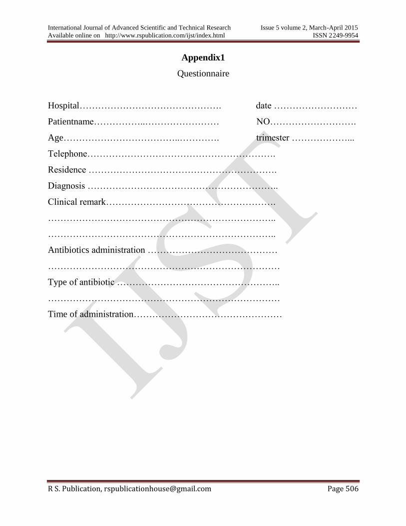
International Journal of Advanced Scientific and Technical Research Issue 5 volume 2, March-April 2015
Available online on http://www.rspublication.com/ijst/index.html ISSN 2249-9954
R S. Publication, [email protected] Page 506
Appendix1
Questionnaire
Hospital………………………………………. date ………………………
Patientname……………..…………………… NO……………………….
Age………………………………..…………. trimester ………………...
Telephone…………………………………………………….
Residence …………………………………………………….
Diagnosis ……………………………………………………..
Clinical remark……………………………………………….
………………………………………………………………..
………………………………………………………………..
Antibiotics administration ……………………………………
…………………………………………………………………
Type of antibiotic ……………………………………………..
…………………………………………………………………
Time of administration…………………………………………
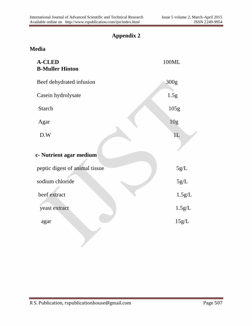
International Journal of Advanced Scientific and Technical Research Issue 5 volume 2, March-April 2015
Available online on http://www.rspublication.com/ijst/index.html ISSN 2249-9954
R S. Publication, [email protected] Page 507
Appendix 2
Media
A-CLED 100ML
B-Muller Hinton
Beef dehydrated infusion 300g
Casein hydrolysate 1.5g
Starch 105g
Agar 10g
D.W 1L
c- Nutrient agar medium
peptic digest of animal tissue 5g/L
sodium chloride 5g/L
beef extract 1.5g/L
yeast extract 1.5g/L
agar 15g/L
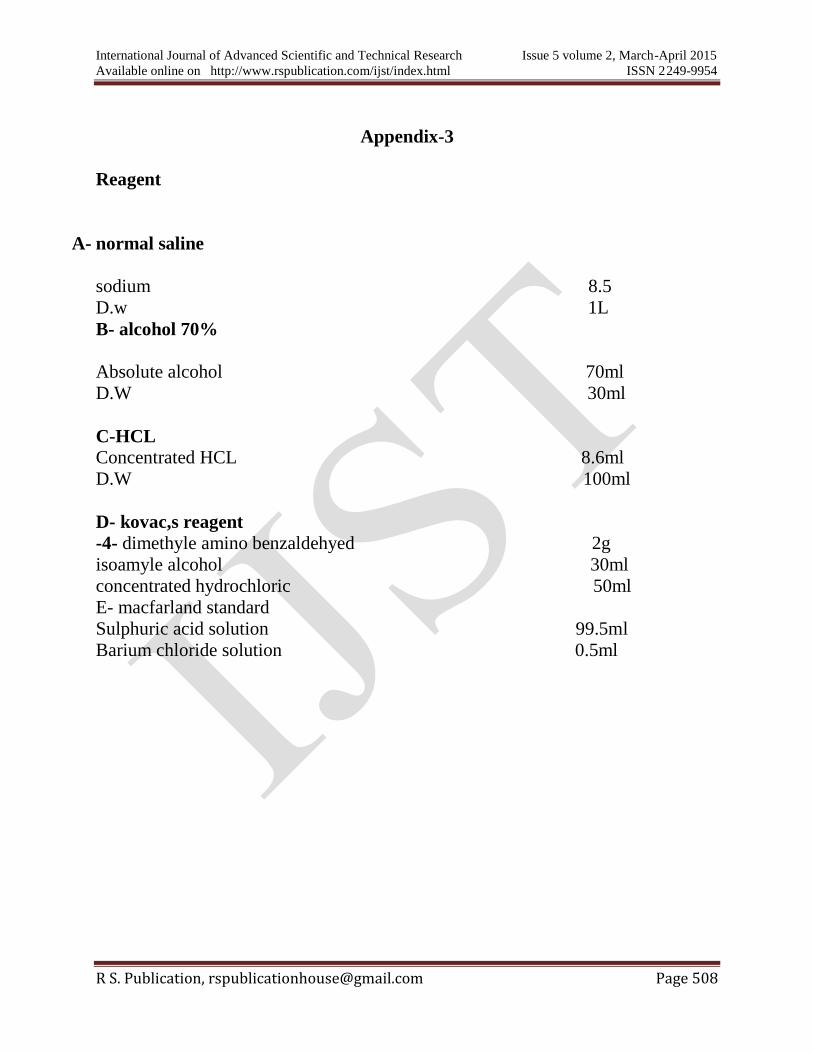
International Journal of Advanced Scientific and Technical Research Issue 5 volume 2, March-April 2015
Available online on http://www.rspublication.com/ijst/index.html ISSN 2249-9954
R S. Publication, [email protected] Page 508
Appendix-3
Reagent
A- normal saline
sodium 8.5
D.w 1L
B- alcohol 70%
Absolute alcohol 70ml
D.W 30ml
C-HCL
Concentrated HCL 8.6ml
D.W 100ml
D- kovac,s reagent
-4- dimethyle amino benzaldehyed 2g
isoamyle alcohol 30ml
concentrated hydrochloric 50ml
E- macfarland standard
Sulphuric acid solution 99.5ml
Barium chloride solution 0.5ml
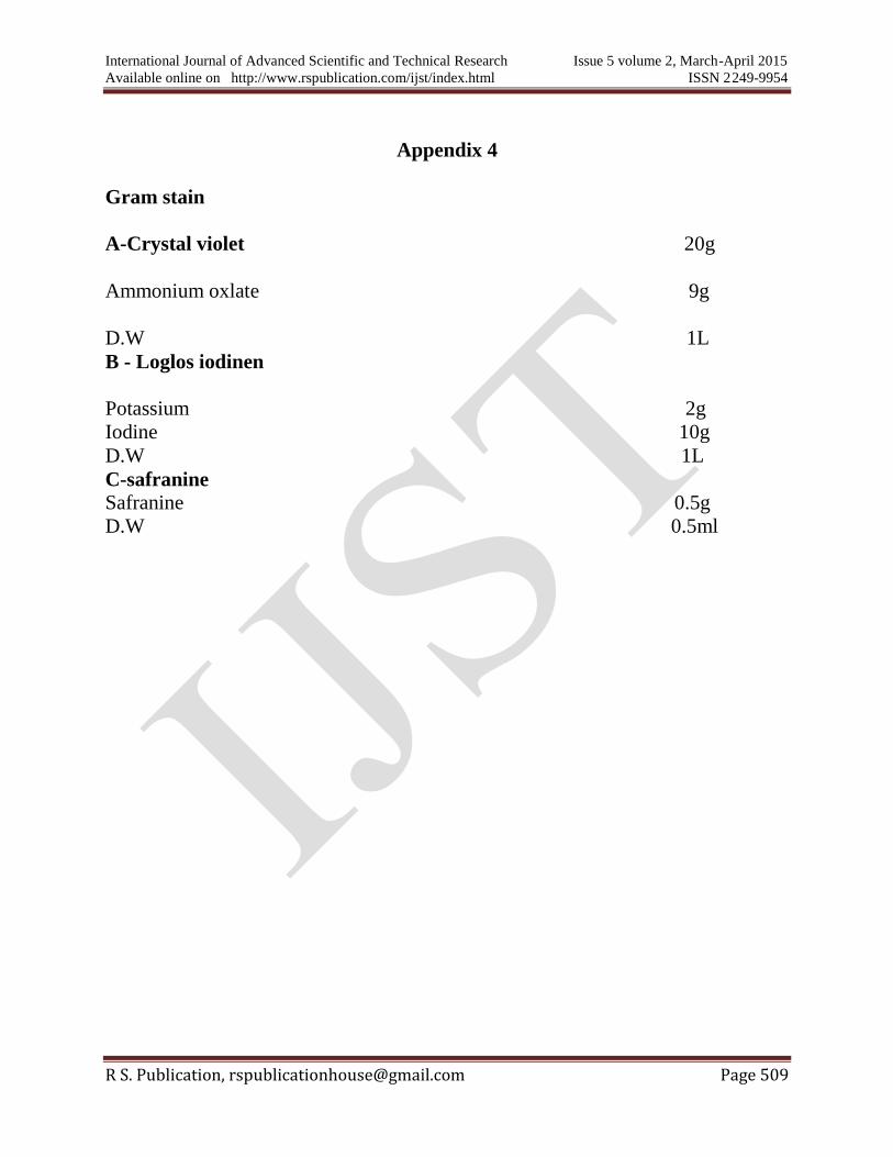
International Journal of Advanced Scientific and Technical Research Issue 5 volume 2, March-April 2015
Available online on http://www.rspublication.com/ijst/index.html ISSN 2249-9954
R S. Publication, [email protected] Page 509
Appendix 4
Gram stain
A-Crystal violet 20g
Ammonium oxlate 9g
D.W 1L
B - Loglos iodinen
Potassium 2g
Iodine 10g
D.W 1L
C-safranine Safranine 0.5g
D.W 0.5ml