Intercomparison for individual monitoring of external exposure ...
219
IAEA-TECDOC-1126 ....... XA0053402-4<<6 Intercomparison for individual monitoring of external exposure from photon radiation Results of a co-ordinated research project 199&-1998 ffl INTERNATIONAL ATOMIC ENERGY AGENCY December 1999 31-03
-
Upload
khangminh22 -
Category
Documents
-
view
1 -
download
0
Transcript of Intercomparison for individual monitoring of external exposure ...

IAEA-TECDOC-1126 ....... XA0053402-4<<6
Intercomparison for individualmonitoring of external exposure
from photon radiationResults of a co-ordinated research project
199&-1998
fflINTERNATIONAL ATOMIC ENERGY AGENCY
December 1999
3 1 - 0 3

The originating Section of this publication in the IAEA was:
Radiation Monitoring and Protection SectionInternational Atomic Energy Agency
Wagramer Strasse 5P.O. Box 100
A-1400 Vienna, Austria
The IAEA does not normally maintain stocks of reports in this series. However, electroniccopies of these reports can be obtained from:
INIS ClearinghouseInternational Atomic Energy AgencyWagramer Strasse 5P.O.Box 100A-1400 Vienna, Austria
Telephone: (43) 1 2600-22880 or 22866Fax: (43) 1 2600-29882E-mail: [email protected] site: http://www.iaea.org/programmes/inis/inis.htm
Orders should be accompanied by prepayment of 100 Austrian Schillings in the form of acheque or credit card (MasterCard, VISA).
INTERCOMPARISON FOR INDIVIDUAL MONITORING OFEXTERNAL EXPOSURE FROM PHOTON RADIATION
IAEA, VIENNA, 1999IAEA-TECDOC-1126
ISSN 1011-4289
©IAEA, 1999
Printed by the IAEA in AustriaDecember 1999

IAEA SAFETY RELATED PUBLICATIONS
IAEA SAFETY STANDARDSUnder the terms of Article III of its Statute, the IAEA is authorized to establish standardsof safety for protection against ionizing radiation and to provide for the application of thesestandards to peaceful nuclear activities.
The regulatory related publications by means of which the IAEA establishes safetystandards and measures are issued in the IAEA Safety Standards Series. This seriescovers nuclear safety, radiation safety, transport safety and waste safety, and also generalsafety (that is, of relevance in two or more of the four areas), and the categories within itare Safety Fundamentals, Safety Requirements and Safety Guides.
• Safety Fundamentals (silver lettering) present basic objectives, concepts andprinciples of safety and protection in the development and application of atomicenergy for peaceful purposes.
• Safety Requirements (red lettering) establish the requirements that must be met toensure safety. These requirements, which are expressed as 'shall' statements, aregoverned by the objectives and principles presented in the Safety Fundamentals.
• Safety Guides (green lettering) recommend actions, conditions or procedures formeeting safety requirements. Recommendations in Safety Guides are expressed as'should' statements, with the implication that it is necessary to take the measuresrecommended or equivalent alternative measures to comply with the requirements.
The IAEA's safety standards are not legally binding on Member States but may be adoptedby them, at their own discretion, for use in national regulations in respect of their ownactivities. The standards are binding on the IAEA for application in relation to its ownoperations and to operations assisted by the IAEA.
OTHER SAFETY RELATED PUBLICATIONSUnder the terms of Articles III and VIII.C of its Statute, the IAEA makes available andfosters the exchange of information relating to peaceful nuclear activities and serves as anintermediary among its members for this purpose.
Reports on safety and protection in nuclear activities are issued in other series, in particularthe IAEA Safety Reports Series, as informational publications. Safety Reports maydescribe good practices and give practical examples and detailed methods that can be usedto meet safety requirements. They do not establish requirements or makerecommendations.
Other IAEA series that include safety related sales publications are the Technical ReportsSeries, the Radiological Assessment Reports Series and the INSAG Series. The IAEAalso issues reports on radiological accidents and other special sales publications. Unpricedsafety related publications are issued in the TECDOC Series, the Provisional SafetyStandards Series, the Training Course Series, the IAEA Services Series and theComputer Manual Series, and as Practical Radiation Safety and Protection Manuals.

FOREWORD
This TECDOC presents the results of a Co-ordinated Research Project on Intercomparison forIndividual Monitoring of External Exposure from photon radiation.
The International Basic Safety Standards for Protection against Ionizing Radiation and for theSafety of Radiation Sources (BSS) have endorsed the use of the operational quantities formonitoring purposes. Specifically, personal dose equivalent, Hp(d), is to be used for individualdosimetry to demonstrate compliance with the exposure limit recommendations, while forworkplace area monitoring the ambient dose equivalent and the directional dose equivalent arerecommended.
In view of the technical difficulties associated with the introduction of these operationalquantities the IAEA decided to assist Member States in their provision of appropriatedosimetry for occupational protection. In this respect, intercomparisons have proven to be acost effective method of providing such support.
A Co-ordinated Research Project (CRP) was started in 1997 on Intercomparison for IndividualMonitoring of External Exposure from photon radiation, involving more than twentylaboratories from eastern Europe and the countries of the former Soviet Union, and focusingon personnel dosimetry services for nuclear power plants. This CRP was part of the activitiesof the IAEA Occupational Protection Programme, the objectives of which are to promote aninternationally harmonized approach for optimizing occupational radiation protection through:
• the development of guides, within the IAEA activities for establishing standards forradiation protection, for restricting radiation exposures in the workplace and for applyingcurrent occupational radiation protection techniques, and
• the promotion of the application of these guidelines.
The preparatory phase included, in May 1997, a workshop aimed at familiarizing theparticipants with the new operational quantities.
The support of the European Commission during this project has been highly appreciated andthanks are due, in particular, to K. Schnuer of the Radiation Protection Division for hisefficient co-operation.
The IAEA wishes to thank all participants for their contribution to the intercomparison.Special thanks are due to J. Bohm and P. Ambrosi (Physikalisch-Technische Bundesanstalt,Braunschweig, Germany), V.E. Aleinikov (Joint Institute for Nuclear Research, Dubna,Russian Federation), D.T. Bartlett (National Radiological Protection Board, UnitedKingdom), I. Csete (National Office of Measures, Budapest, Hungary), V. Forminykh(Mendeleyev Institute for Metrology, St. Petersburg, Russian Federation) and H. Stadtmann(Austrian Research Centre Seibersdorf, Austria) for providing excellent technical co-ordination and review of the CRP results.
M. Gustafsson, of the IAEA's Division of Radiation and Waste Safety initiated the CRP andguided the project until March 1997. R. Ouvrard of the same Division continued the work andwas responsible for the final compilation of this TECDOC.

EDITORIAL NOTE
In preparing this publication for press, staff of the IAEA have made up the pages from theoriginal manuscripts as submitted by the authors. The views expressed do not necessarily reflectthose of the IAEA, the governments of the nominating Member States or the nominatingorganizations.
Throughout the text names of Member States are retained as they were when the text wascompiled.
The use of particular designations of countries or territories does not imply any judgement bythe publisher, the IAEA, as to the legal status of such countries or territories, of their authorities andinstitutions or of the delimitation of their boundaries.
The mention of names of specific companies or products (whether or not indicated asregistered) does not imply any intention to infringe proprietary rights, nor should it be construed asan endorsement or recommendation on the part of the IAEA.
The authors are responsible for having obtained the necessary permission for the IAEA toreproduce, translate or use material from sources already protected by copyrights.

CONTENTS
INTRODUCTION
IAEA activities in the field of occupational radiation protection 3M. Gustafsson, R. V. Griffith
Outline of the 1996-1998 IAEA Co-ordinated Research Project on Intercomparisonfor Individual Monitoring of External Exposure from photon radiation 9J. Bohm, M. Gustafsson, R. Ouvrard
Dosimeter Irradiations for the 1996-1998 IAEA Co-ordinatedResearch Project on Intercomparison for Individual Monitoring ofExternal Exposure from photon radiation 13V.E. Aleinikov, P. Ambrosi, D. Bartlett, L Burmann, D.T. Bartlett,D.R. Mclure, I. Csete, V. Fominykh, A. V. Oborin, H. Stadtmann
Standards in radiation protection at the IAEA dosimetry laboratory 27L. Czap, F. Pernicka, G. Matscheko, P. Andreo
THE USE OF OPERATIONAL QUANTITIES FOR INDIVIDUALMONITORING OF EXPOSURE TO EXTERNAL RADIATION
Calibration of personal dosimeters: quantities and terminology 37V.E. Aleinikov
Quantities and units for external dose assessment 47R. V. Griffith
Workplace photon radiation fields 67P.H. Burgess, D.T. Bartlett, P. Ambrosi
Calibration of personal dosimeters for photon radiation with respect to the personaldose equivalent Hp( 10) 85J. Bohm
Calibration of a personal dosimeter in the field of a radionuclide 113C. Strachotinsky, H. Stadtmann
Dosimeter characteristics and service performance requirements 119P. Ambrosi, D. T. Bartlett
Characteristics of personal dosimeters to measure Hp(10) for photons 141D.T. Bartlett, J.D. Steele
Passive dosimeter characteristics and new developments 151J. Trousil, F. Spurn
Electronic dosimeter characteristics and new developments 167I.M.G. Thompson
RESULTS OF THE 1996-1998 IAEA CO-ORDINATEDRESEARCH PROJECT ON INTERCOMPARISON FOR INDIVIDUALMONITORING OF EXTERNAL EXPOSURE FROM PHOTON RADIATION
Results of the IAEA 1996-1998 Co-ordinated Research Project onIntercomparison for Individual Monitoring of External Exposure toPhoton Radiation 179R. Ouvrard, J. Bohm

ANNEX: GRAPHIC REPRESENTATION OF RESULTS
Phase II results 191
Phase III results 199

INTRODUCTION

IAEA ACTIVITIES IN THE FIELD OF OCCUPATIONAL XA0053403RADIATION PROTECTION
M. GUSTAFSSON, R.V. GRIFFITHInternational Atomic Energy Agency, Vienna
AbstractThe Co-ordinated Research Project on Intercomparison for Individual Monitoring of External Exposure to PhotonRadiation is placed into the context of the IAEA occupational protection programme by describing related activities such asdevelopment of standards and guidelines, technical co-operation programmes and the information system on occupationalexposure (ISOE). A brief summary of former intercomparisons is also included.
1. INTRODUCTION
The objective of the IAEA Occupational Protection Programme is to promote aninternationally harmonized approach for optimizing occupational radiation protection through:
• the development of standards for restricting radiation exposures in the workplaceand for occupational radiation protection techniques, and
• the provision for the application of these standards.
This paper is intended to place the Co-ordinated Research Project (CRP) on Intercomparisonfor Individual Monitoring of External Exposure to Photon Radiation into the context of thisprogramme, focusing on activities related to the CRP.
2. SETTING THE STANDARDS
Basic guidance for IAEA Member States is provided through the hierarchical Safety StandardsSeries - Fundamentals, Requirements (previously called Safety Standards) and Guides. Whilethe Safety Standards Series documents are directed at national Regulatory Authorities, theSafety Guides may present detailed information that is also of value for senior management inthe contractor or licensee organizations responsible for establishing and managingoccupational radiation protection programmes. Publications in this series are consensusdocuments drafted during one or more expert advisory group meetings, and refined throughsubsequent consultations with the experts before review by the Radiation Safety StandardsAdvisory Committee - RASSAC - and final publication recommendation by the AdvisoryCommission on Safety Standards - ACSS.
In the area of radiation and transport safety, the Safety Fundamentals, Safety Series 120, is thetop level document, presenting basic safety principles, concepts and objectives for radiationprotection. For radiation safety, the next level is represented by the International Basic SafetyStandards for Protection against Ionizing Radiation and for the Safety of Radiation Sources(BSS) [1]. The BSS specify requirements to ensure safety governed by the principles in theFundamentals. General requirements for occupational protection are presented in Appendix Iof the Standards, which, like the Fundamentals, have been co-sponsored jointly by the FAO,ILO, OECD/NEA, PAHO, and WHO.

Other Appendices address radiation protection for medical, public, potential, emergency, andchronic exposures.
Guidance on application of the BSS to occupational protection is elaborated upon in threeSafety Guides close to publication - Occupational Radiation Protection, Assessment ofOccupational Exposure due to External Sources of Radiation, and Assessment ofOccupational Exposure due to Intakes of Radionuclides. The Safety Guide "OccupationalRadiation Protection" outlines the elements which are needed to form the basis for aneffective worker protection programme. The companion Guides on dose assessment willprovide specific guidance required for the accurate assessment of occupational radiationexposure.
Technical Reports Series No. 133, Calibration of Radiation Protection Monitoring Instruments,is over 25 years old. A full revision of this report, which includes ICRU and ISO(International Standards Organization) principles, has been prepared and begun its waythrough the publication process. It is also expected that, by the year 2000, a Safety Report onDosimetry Services for Individual Monitoring of Occupational Exposure from ExternalRadiation Sources will be published.
3. PROVIDING ASSISTANCE
Referred to as "TC", the Technical Cooperation programme is the mechanism used to providedirect support to meet Member States national needs. There are currently about 150 national,regional and interregional projects in the general area of radiation protection. Many of theseare aimed at upgrading national radiation protection infrastructures, with emphasis on safe useof sources and developing occupational monitoring programmes. The national projects mayinclude expert missions, fellowship training for national staff, or provision of equipment.
The radiation protection priority in TC work for the end of this century will be theInterregional Model Project on Upgrading Radiation and Waste Safety Infrastructure, orsimply "the Model Project". The Agency recognizes that many Member States do not have asufficiently developed national radiation protection infrastructure to meet the requirements ofthe BSS. The Agency's Member States have supported the need for adequate infrastructure asa prerequisite for providing large radiation sources or radiation producing equipment with thepotential for serious accidents. The Model Project was established to focus on those countriesprepared to participate, and establish Action Plans outlining the actions to be taken by thecountry and the Agency to achieve strengthened infrastructures.
Originally established with five participating Member States, the Model Project has beenexpanded to 51 Member States in Africa, Eastern Europe, Latin America and Asia. The goal isto complete activities identified in the Action Plans by the year 2000. Under the ModelProject, there are ten areas that are being addressed: Laws and Regulations, RegulatoryAuthority, Regulatory Control, Occupational Exposure Control, Medical Exposure Control,Emergency Response, Environmental Public Exposure Control, Waste Management,Technical Support, and Manpower. The first priority for each country is the establishment ofnational systems to identify and locate those sources that have the potential for seriousaccident consequences through loss or mishandling. Upgrading occupational exposure control,personal dosimetry services and facilitating access to proper calibration services are also keycomponents of the Model Project.
An additional TC supported activity in occupational radiation protection is a Regional Projectlaunched in 1997 on Improving Occupational Radiation Protection in Nuclear Power Plants in

Central and Eastern Europe and in Republics of the former Soviet Union. The objective of thisproject is to improve the implementation of the optimization principle in accordance with theBSS by facilitating the exchange of feedback experience, ensuring the dissemination of anALARA culture and assisting in investigations of radiation exposure of workers and in theimplementation of proper measures to reduce this exposure.
This project is, for example, supporting meetings for Health Physicists responsible forradiation protection in WWER and RBMK Nuclear Power Plants, Training Courses onOptimization of Radiological Protection in the Design and Operation of Nuclear PowerPlants, organized in collaboration with NRPB and CEPN (France) and sponsored jointly bythe IAEA and the European Commission (EC), and scientific visits.
4. INTERCOMPARISONS
Complementary to the IAEA's Technical Co-operation Programme is the Agency's ResearchContract Programme with the primary objectives of stimulating advances in scientificknowledge, assisting the developing countries whenever possible to increase theirparticipation in nuclear research and to co-ordinate research between the Agency and nationalcentres. Several intercomparisons have been performed and are currently performed as Co-ordinated Research Projects.
International Intercomparison 1988-1992
A CRP on Intercomparison for Individual Monitoring was conducted in two phases over theperiod 1988-1992 with a total participation of twenty-nine laboratories from twenty-oneMember States and three international organizations. The first phase of the project focused onselection of a backscatter phantom for calibration, and identifying systematic differences inthe quality of dosimetry. Based on the results [2] and pending modifications of the ICRUguidance on practical use of the operational quantities, a second intercomparison wasconducted addressing issues of phantom and angular dependence as well as energydependence. This second phase of the CRP was summarized in an IAEA TECDOC [3], andreviewed in Radiation Protection Dosimetry [4].
The intercomparison demonstrated that a number of dosimetry systems were capable ofmeasuring the new ICRU quantities to an acceptable degree of accuracy. However, a numberof participants were recommended to modify their evaluation technique. It was demonstratedthat the performance of TLD systems was usually superior to film based systems. However, afew film based systems that had been carefully characterized and calibrated performed as wellas the TLD systems. Dosimeters with simple designs performed as well as the moresophisticated ones. More detailed results are given in the mentioned reports.
IAEA/RCA Personal Dosimeter Intercomparison 1990-1992
The initial programme of the IAEA Regional Co-operative Agreement project (RCA) forstrengthening the radiation protection infrastructure in the Asian and Pacific region containeda regional personal dosimetry intercomparison, which was conducted in three phases over theperiod 1990-1992. Seventeen organizations from all fourteen Member States participated inthis programme. It was concluded that this intercomparison contributed significantly to thetechnical improvement of personal dosimetry and instrument calibration in the region ofSouth East Asia. However some concerns were raised, for example, regarding dosimetryservices using film, which prompted another CRP to be organized in the same region.

Ongoing Intercomparisons
Currently intercomparisons for individual dosimetry are being conducted under the RCAprogramme and in Latin America.
The IAEA/RCA Regional Personal Dosimetry Intercomparison has been initiated with theobjectives of evaluating regional dosimetry services' abilities to conduct individual monitoringin terms of the ICRU operational quantities for photons, providing access for the participantsto photon field qualities for calibration of their systems that they might not otherwise be ableto obtain, and providing a unique opportunity for regional exchange of information regardingpersonal monitoring.
Another regional CRP has been initiated to encourage regional harmonization of individualmonitoring practices in the Latin American region.
5. ISOE
In order to facilitate the exchange of techniques and experience in occupational exposurereduction, the Nuclear Energy Agency (NEA) of the Organisation for Economic Co-operationand Development (OECD) launched the Information System on Occupational Exposure(ISOE) on 1 January 1992. In 1993 the IAEA decided to co-sponsor ISOE by inviting MemberStates which are not members of the OECD to participate through the IAEA, which acts asTechnical Centre for non-NEA countries. Since 1998 the ISOE Secretariat is a jointundertaking of NEA and IAEA.
The objective of the ISOE is to make available to the participants:
• broad and regularly updated information on methods to improve the protection ofworkers and on occupational exposure in nuclear power plants
• a mechanism for dissemination of information on these issues, includingevaluation and analysis of the data assembled, as a contribution to theoptimization of radiation protection.
A growing number of IAEA Member States are participating through the IAEA. As of 1November 1998, there are ten utilities and five authorities from nine countries participating;i.e., all utilities in Armenia, Brazil, China, Lithuania, Romania, Slovak Republic, Slovenia,South Africa and Ukraine (representing 31 reactors) and the regulatory authorities in Armenia,China, Romania, Slovak Republic and Slovenia. Three countries, which earlier participated inthe ISOE through the IAEA, have joined the OECD. Invitations to Bulgaria, India, and theRussian Federation are pending.
As Regional Technical Centre the IAEA is collecting and forwarding the annual occupationalexposure data from their participants to the central database. Thus the IAEA is responsible forthe quality control of the data provided by their participants, which prompted the Agency toinclude in the current CRP some of the dosimetry services providing these data. In additionthe Agency has, for example, organized a one week meeting for representatives from IAEAISOE participants, supported the participation of ISOE contact persons in the annual ISOEmeetings and Topical Sessions and purchased a world-wide (except in OECD countries)license to distribute the software learning program RADIOR and make it available in Russian.
6. THE CURRENT CRP
Taking into account the activities in the Agency's Occupational Protection programmedescribed above, the endorsement of the operational quantities for radiation monitoring of

workers in the recently adopted BSS [1] and the concerns about nuclear safety and radiationprotection that have developed in Eastern Europe and the Republics of the former SovietUnion, it was found to be appropriate to initiate an intercomparison that would focus on IAEAMember States in Eastern Europe and on personal dosimetry services for nuclear powerplants. The outline of the CRP is given in a separate paper.
REFERENCES
[1] IAEA, FAO, ILO, NEA/OECD, PAHO, WHO, International Basic Safety Standards forProtection against Ionizing Radiation and for the Safety of Radiation Sources. SafetySeries No. 115, IAEA, Vienna (1996).
[2] BOHM, J., GRIFFITH R., (Eds), Intercomparison for Individual Monitoring (Proc.IAEA Research Co-ord. Meet. Vienna, 1989). Physikalisch-Technische BundesanstaltRep. PTB-Dos-20, Braunschweig (1991).
[3] INTERNATIONAL ATOMIC ENERGY AGENCY, Intercomparison of radiationdosimeters for individual monitoring. IAEA TECDOC-704, Vienna (1993).
[4] AMBROSI, P., BUCHHOLZ, G., BOHM, J., GRIFFITH, R.V., HERRMANN, D.,AND STRACHOTINSKY, C. Results of the IAEA Intercomparison for IndividualMonitoring. Rad. Prot. Dos. 54(1):5-17 (1994).

XA0053404OUTLINE OF THE 1996-1998 IAEA CO-ORDINATED RESEARCHPROJECT ON INTERCOMPARISON FOR INDIVIDUAL MONITORINGOF EXTERNAL EXPOSURE FROM PHOTON RADIATION
J. BOHM,Physikalisch-Technische Bundesanstalt,Braunschweig, Germany
M. GUSTAFSSON, R. OUVRARDInternational Atomic Energy Agency, Vienna
AbstractThe outline of the IAEA Co-ordinated Research Project 1996-1998 on intercomparison for individual monitoring isdescribed. The intercomparison focused on IAEA Member States in Eastern Europe and was based on the operationalquantity personal dose equivalent, H//10). The three phases of the intercomparison -were: the preparatory phase including aworkshop, the "type-test" intercomparison, and the "simulated workplace field" intercomparison. Details of the phases are given.
1. INTRODUCTION
Radiation monitoring of workers using personal dosimeters (film, TLD, etc.) is an essentialcomponent to assess the effectiveness of any occupational radiation protection programmefocused on limiting the exposure to external radiation. The International Basic SafetyStandards for Protection against Ionizing Radiation and for the Safety of Radiation Sources[1] recently adopted by FAO, IAEA, ILO, OECD/NEA, PAHO and WHO have endorsed theuse of the operational quantities for monitoring purposes. Specifically, personal doseequivalent, /fp(10), is to be used for individual dosimetry to demonstrate compliance with theexposure limit recommendations, whereas the ambient dose equivalent and the directionaldose equivalent are recommended for workplace area monitoring [2]. Comprehensiveinternational guidance on the use of the dose related quantities for radiological protectionagainst external radiation will be provided in the near future by the publication of a report ofthe Joint Task Group of the International Commission on Radiological Protection and theInternational Commission on Radiation Units and Measurements.
In view of the technical difficulties associated with the introduction of the new radiationquantities for measurement and reporting, it is important that the Agency assists MemberStates in their provision of appropriate dosimetry for occupational protection. Dosimetryintercomparisons have proved to be a cost effective method to provide this support. Between1988 and 1992 [3], the Agency conducted an interregional intercomparison programme, at thesame time providing an intercomparison for Asia and Oceania under the RCA (Regional Co-operative Agreement) programme. Intercomparisons were, or are, also being conducted forindividual dosimetry under the RCA programme and in Latin America.
This Co-ordinated Research Project focused on IAEA Member States in Eastern Europe andon personnel dosimetry services for nuclear power plants. Its main objectives was to giveparticipants an opportunity to assess :
the recommendations of the IAEA to use the operational quantity Hp(10) inindividual monitoring,

• the energy and angular dependence of the response of their dosimeters,
• their ability to measure external photon radiation fields in terms of Hp(10).
The following outlines the programme for this Co-ordinated Research Project. It not onlyimplied co-operation, but also financial support of the EC.
2. PARTICIPATING LABORATORIES AND SERVICES
The Physikalisch-Technische Bundesanstalt (PTB) in Braunschweig, Germany, providedtechnical co-ordination and support. Several other standards laboratories provided additionaltechnical support:
• Joint Institute for Nuclear Research (JINR), Dubna, Russian Federation;
• Mendeleyev Institute for Metrology (VNIIM), St. Petersburg, Russian Federation;
• Austrian Research Centre (ARCS), Seibersdorf, Austria;
• National Radiological Protection Board (NRPB), Chilton, United Kingdom;
• National Office of Measures (OMH), Budapest, Hungary.
The following services participated with personal dosimeters :
• Department of Radiation Safety, Yerevan, Armenia;
• IAEA Radiation Monitoring & Protection Services Section, Vienna, Austria;
• National Centre of Radiobiology & Radiation Protection, Sofia, Bulgaria;
• National Personnel Dosimetry Ltd., Prague, Czech Republic;
• Nuclear Power Plant Dukovany, Czech Republic;
• Radiation Protection Centre, Tallinn, Estonia;
• "Frederic Joliot Curie" National Research Institute, Budapest, Hungary;
• National Office of Measures, Budapest, Hungary;
• Radiation Protection Centre, Vilnius, Lithuania;
• Nuclear Power Plant Ignalina, Visaginas, Lithuania;
• Central Laboratory for Radiological Protection, Warsaw, Poland;
• Nofer Institute of Occupational Medicine, Lodz, Poland;
• Institute of Hygiene and Public Health, Bucharest, Romania;
• Institute for High Energy Physics, Moscow Region, Russian Federation;
• Mendeleyev Institute for Metrology, St. Petersburg, Russian Federation;
• Joint Institute of Nuclear Research, Dubna, Russian Federation;
• Bohunice Nuclear Power Plant, Jaslovske Bohunice, Slovak Republic;
• Slovak Institute of Metrology, Bratislava, Slovak Republic;
10

Josef Stefan Institute, Ljubljana, Slovenia;
Krsko Nuclear Power Plant, Krsko, Slovenia;
AMS of Ukraine, Scientific Centre for Radiation Medicine, Kiev, Ukraine;
Research Institute of Medical Radiology, Kharkov, Ukraine;
State Enterprise Regional Environmental Monitoring & Dosimetric Control,Chernobyl, Ukraine.
3. PHASES OF THE CO-ORDINATED RESEARCH PROJECT
3.1. Survey
The project consisted of three phases which partly overlapped. The focal point of Phase I, thepreparatory phase, was a workshop in May 1997, to familiarize the participants with the newoperational dose equivalent quantities, in particular with the operational quantity personaldose equivalent. Phase n, the "type-test" intercomparison, provided the participants with dataabout the variation of the energy and angular dependence of the response of their dosimeterswith respect to this operational quantity. Finally, Phase El, the "simulated workplace field"intercomparison, enabled the participants to judge the performance of their dosimeters underrealistic conditions arising in practice. The final results of the project were discussed during aconsultants meeting in December 1998 and are published in this technical document. Thefollowing explains the three phases in more details.
3.2. Phase I: Preparatory Phase
This phase was the planning phase and provided the prerequisites for the success of theintercomparison. The first consultants meeting was held in Vienna in May 1996. An outlinefor the intercomparison and for a workshop was prepared to inform participants and observersabout the new operational dose quantities and several topics on personnel monitoring.Requests for participation together with detailed questionnaires were sent to potentialparticipants by the IAEA. During a second consultants meeting in December 1996, progress ofthe work and details of the workshop, in particular the harmonization of the different papers tobe presented, were discussed. Pill box dosimeters with TLDs were prepared by the PTB inDecember 1996 to intercompare the four irradiation laboratories involved in Phase n.Distribution of the dosimeters was performed via the IAEA. In March 1997, it turned out thatthe results of the intercomparison of the irradiation laboratories were satisfactory. During aworkshop in May 1997, all the background information required for the intercomparison wereimparted to the participants and interested observers. The participants brought along to theworkshop all dosimeters to be irradiated in Phase II. During the workshop, these dosimeterswere labelled and distributed to the representatives of the irradiation laboratories. Theworkshop was combined with the third consultants meeting. The papers presented during theworkshop are reproduced in this technical document.
3.3. Phase n: "Type-Test" Intercomparison
Phase II started with the irradiations of the dosimeters distributed during the workshop ofPhase I. The dosimeters were irradiated in June, July and August 1997 and then returned tothe participants together with all data of the irradiations. The participants forwarded their
11

results to the IAEA by 1 November 1997, where a statistical analysis was first done, and theresults were discussed during a consultants meeting in January 1998, in Luxembourg.
3.4. Phase III: "Simulated Workplace Field" Intercomparison
Final details of Phase HI were agreed upon during the Luxembourg meeting. Recentlydeveloped passive and electronic dosimeters were also included in this phase. In June 1998,the results of Phase U were discussed with all participants, at a meeting held at the PTB. Theyalso brought their dosimeters for the irradiations under Phase IH. After the irradiations in Juneand July 1998, the dosimeters were sent back to the participants for evaluation. The resultswere then transmitted to the IAEA, a statistical analysis of the results was done and they werediscussed during a consultants meeting in Vienna in December 1998. Final results were sentto each participating laboratory at the end of 1998. They are presented in this technicaldocument.
REFERENCES
[1] INTERNATIONAL ATOMIC ENERGY AGENCY, International Basic SafetyStandards for Protection against Ionizing Radiation and for the Safety of RadiationSources, Safety Series No. 115, Vienna (1996).
[2] INTERNATIONAL COMMISSION ON RADIATION UNITS ANDMEASUREMENTS, Quantities and Units in Radiation Protection, ICRU Report 51,ICRU Publications, Bethesda, MD (1993).
[3] INTERNATIONAL ATOMIC ENERGY AGENCY, Intercomparison of RadiationDosimeters for Individual Monitoring, IAEA-TECDOC-704, Vienna (1993).
12

XA0053405DOSIMETER IRRADIATIONS FOR THE 1996-1998 IAEACO-ORDINATED RESEARCH PROJECT ONINTERCOMPARISON FOR INDIVIDUAL MONITORINGOF EXTERNAL EXPOSURE FROM PHOTON RADIATION
V.E. ALEINIKOV'", P. AMBROSI<2>, L. BURMANN(2), D.T. BARTLETT(3),D.R. McCLURE(3), I. CSETE(4), V.I. FOMINYKH(5>, A.V. OBORIN(5), H. STADTMANN(6)
(1) Joint Institute for Nuclear Research, Dubna, Russian Federation<2) Physikalisch-Technische Bundesanstalt, Braunschweig, Germany(3) National Radiological Protection Board, Chilton, United Kingdom(4) National Office of Measures, Budapest, Hungary<5> Mendeleyev Institute for Metrology, St. Petersburg, Russian Federation(6) Austrian Research Centre Seibersdorf, Austria
Abstract
This paper gives information on the facilities used by the irradiating laboratories, how the irradiations in both Phase II andPhase III were performed, as well as the procedures followed to ensure that all irradiations were done so that anyuncertainty in the dose estimates of the irradiating laboratories, for the purpose of this intercomparison, can be negligible.
1. INTRODUCTION
The irradiations, details of which are given in this section, were carried out for Phases II andIII of the Co-ordinated Research Project (CRP).
In Phase II, a 'mini type test' was performed. This had two purposes, firstly to provide somecalibration data in terms of //p(10) for the dosimetry services participating in the CRP,secondly to assist in the harmonization of procedures at secondary standards laboratories inEastern European States to type test in terms of /7p(10). In addition an intercomparison wascarried out of the dosimetry of the participating irradiating laboratories including PTB, ARCSand NRPB. Further details of the Phase II programme are given in section 3 and [1].
In Phase HI, an intercomparison was carried out of the performance of dosimeters in simulatedworkplace fields. In some cases where complex algorithms are applied and where there islimited information on the workplace field, the dosimeter performance characteristicsdetermined using narrow spectral width calibration fields do not give a reliable indication ofits performance in fields of broad energy and angle distribution. This could be tested in PhaseHI. More generally, however, the purpose of the Phase III intercomparison was to examine theperformance of the dosimetry systems in radiation fields which were similar to thoseencountered in practical routine monitoring (see the paper on workplace fields [2]). Thesimulated workplace fields used in Phase in were devised to reproduce features of fieldsencountered in practice including higher energy direct components with lower energy broadangle scattered components; photons in the 4 MeV to 7 MeV energy range with and withoutsecondary electron equilibrium; and for a range of doses. The radiation fields used wereselected from the ISO recommended fields, either individually or in combination, plusindium-192.
13

2. OPERATIONAL QUANTITIES AND PHANTOMS
Effective dose [3, 4] is the radiation protection quantity assessed for control purposes inrespect of stochastic effects of ionizing radiation. Operational quantities have been defined [5]which provide, in general, conservative estimates of the protection quantity, effective dose.The operational quantities are used in the routine monitoring of occupational exposure. Forthe individual monitoring of photon external radiation, the relevant operational quantity is thepersonal dose equivalent, Hp(l0). Personal dosimeters are, in this approach, characterized androutinely calibrated in terms of Hp(l0) [6]. The dosimeter reading is then used as an estimateof Hp(\0) to be compared with dose limits or investigation levels expressed in terms ofeffective dose [3], and entered in dose records as an estimate of effective dose.
Hp(\0) is defined primarily in the human body. The definition is extended [7] to calibrationphantoms. In this case Hp(l0) is the dose equivalent at 10 mm depth in a phantom of the samesize and shape as that used for calibration but composed of ICRU 4-element tissue equivalentmaterial. The assumption is made that a personal dosimeter whose response matches theenergy and angle dependence of response of Hp(l0) in the calibration phantom will determineadequately Hp(l0) in the human body when worn, and provide an estimate of effective dose ofsufficient accuracy. A phantom is, in general, required for the calibration of personaldosimeters in terms of//p(10) (but see [6] on routine calibration) because the radiation field atthe wearing position of the dosimeter on the body comprises an incident component and abackscattered component, the characteristics of which depend on the energy and angle of theincident photons, and also on the body itself, and where on it the dosimeter is positioned. Theresponse of a dosimeter will, in general, depend on both components of the radiation field,incident and backscattered.
A solid material having the composition of the ICRU 4-element tissue equivalent material hasnot been fabricated. A number of tissue substitute materials are available, among which arepolymethyl methacrylate (PMMA), water and a various specially fabricated plastics. Theimportant property of tissue substitute materials is their ability to replicate the backscatterfrom tissue. The backscattered field, its magnitude and its energy and angle distributiondepends not only on the material but on the shape of the phantom (see [8] and referencestherein). The International Organisation for Standardization (ISO) and the IAEA recommendthe use of a calibration phantom which is a 300 mm x 300 mm x 150 mm slab made of thinPMMA walls and filled with water [9] (see also reference [6]), the backscattered field ofwhich adequately matches that calculated for a phantom of the same shape and size, but ofICRU 4-element tissue. Dosimeters are then calibrated on this phantom in terms of Hp(\0)calculated for a 300mmx300mmx 150 mm slab of ICRU 4-element tissue, HPiS\^(l0).Account may need to be taken for the variation across the face of the phantom of both theincident and backscattered components of the field [9].
3. RADIATION QUALITIES, CONVERSION COEFFICIENTS
In Phase II, for the 'mini type test', radiation qualities of the ISO narrow spectrum series [10]were selected to allow detailed information to be determined of the energy and angledependence of the response, see Table I. In addition, from the readings of the dosimetersNo. 24 to 26 the free-in-air calibration with respect to air kerma Kz can be checked.
The conversion coefficients for all radiation qualities were taken from ISO 4037-3 [9] and theirradiation protocols given therein followed.
14

In Phase HI, the performance of dosimeters in simulated workplace fields was to be deter-mined. To simulate the scattered radiation component of workplace fields, radiation qualitiesof the ISO wide spectrum series [10] were selected and the dosimeters were irradiated forangles of incidence between + 80° and - 80° about the vertical. Depending on the technicalequipment at the different irradiating laboratories this was done in one of two ways:oscillating with a constant angular velocity (NRPB, ARCS) or irradiation at discrete angleswith step increments of 5° (PTB). These "wide angle" irradiations (WA ± 80°) were done forfour different radiation qualities, see Table II.
For this kind of irradiation condition, there are no conversion coefficients /JPK(10) from airkerma Ka to personal dose equivalent i/P;Siab(10) given in the relevant standard, ISO-4037-3.Therefore, appropriate values were calculated using the following algorithm:
Interpolation of missing ftp^OO; E; a,) data between the given values (a = 0°, 10°, 20°, ...) forintermediate angles of incidence using a spline or 4-point Lagrangian (linear-linear) inter-polation [11] either for 1° steps (oscillating method) or 5° steps (discrete angle method) anddetermination of the mean value ftpx(10; E; WA ± 80°) (over the corresponding angular rangefrom - 80° to + 80°) of these calculated data points. Figure 1 gives an example for thisprocedure to determine ftp*(10; W-300; WA ± 80°) for the discrete angle method.
The resulting conversion coefficients and their estimated standard uncertainties for anoscillating phantom (phantom rotating in the angular range from -80° to +80°) are given inTable m together with those for S-Ir.
1,5
1,3
1,2
1,1
1,0
0,9
0,8 ^
:
;
Ih
7
• ISO/FDIS 4037-3- • — Fit
Mean value:/ipK( 10;W-300; WA ± 80°) = 1 ,c
\
. I>55 r
I
V
\
\
\{ I
-80 -60 -40 -20 0 20 40 60 80
FIG.I. Angular dependence of the conversion coefficient for W-300 radiation quality.
15

TABLE I. RADIATION QUALITIES, ANGLES OF INCIDENCE AND NOMINAL DOSEVALUES SELECTED FOR PHASE H OF THE CRP TOGETHER WITH SOMEADDITIONAL INFORMATION. THE ABBREVIATIONS OF THE IRRADIATINGLABORATORIES AND MORE DETAILS ARE EXPLAINED IN SECTION 4. ALLIRRADIATIONS WITH NOMINAL #P,SLAB(10) DOSE VALUES WERE DONE WITHDOSIMETERS POSITIONED ON THE FRONT SURFACE OF AN ISO WATER SLABPHANTOM, THOSE WITH NOMINAL KA VALUES FREE IN AIR.
Dosi-meterNo.
01
0203040506
10
11121314
1519202122
2324
2526
303132
ISOquality
N-40
N-40N-40N-60
N-60N-60
N-100
N-100N-100N-250N-250N-250S-Co
S-Co
S-CoS-CoS-CoS-Co
S-CoS-Co
R-FR-FR-F
Irrad.lab.
OMH
OMH
OMHOMHOMHOMH
VNIIM
VNHMVNIIMVNIIM
VNIIMVNIIMJINR
JINRJINRJINRJINR
JINRJINR
JINR
PTBPTBPTB
Meanenergy
keV
33
3333484848
83
8383
2082082081250
1250
125012501250
125012501250
661066106610
Angle ofradiationincidence
0°
30°60°0°30°60°
0°30°60°0°30°60°
0°
0°0°
30°60°
0°0°0°0°30°60°
Nominal dosevalue
//o(10) = 3.0mSv
#o(10) = 3.0mSv//o(10) = 3.0mSv//D(10) = 3.0mSv/fD(10) = 3.0mSv#p(10) = 3.0mSv
#D(10)=1.0mSv
#D(10)=1.0mSv/fD(10) = 1.0mSv//D(10) = 1.0mSvi/p(10)=1.0mSvtfD(10)=1.0mSv#p(10) = 3.3mSv
//0(10) = 3.3mSvZ/D(10) = 3.3mSv
#p(10) = 3.3mSv//D(10) = 3.3mSv
K& = 8.6 mGy
#a = 8.6mGyKti = 8.6 mGy
^tfp(10) = 3.0 mSvtfo(10) = 3.0mSv#D(10) = 3.0mSv
Conversioncoefficient
V (10)Sv/Gy
1.17
1.120.851.651.591.27
1.88
1.821.531.481.55
1.381.15
1.151.151.151.14——
—
1.121.121.12
ISO 4037-3 does not give a conversion coefficient hp/c(l0; S-Ir, 0°) from air kerma Ka topersonal dose equivalent Hp(\0) for S-Ir. Therefore this coefficient was calculated from thepublished values for mono-energetic radiation taking the emission probability for each photonenergy into account. For normal incidence a conversion coefficient /JPA:(10; S-Ir, 0°) of1.317 Sv/Gy was obtained.
For the wide angle irradiation the corresponding conversion coefficients #PA;(10; S-Ir, a,) - fordifferent directions of incidence a - were calculated as described, interpolated (by splineinterpolation) between 0°, 10°,..., 70°, 80° and the mean value used.
16

TABLE H. RADIATION QUALITIES, ANGLES OF INCIDENCE AND NOMINAL DOSEVALUES SELECTED FOR PHASE HI OF THE CRP TOGETHER WITH SOMEADDITIONAL INFORMATION.
Dosi-meterNo.
0102
030408091011
15
161718
Radiation quality
S-Ir (WA ± 80°)S-Ir (0°) + S-Ir (WA ± 80°) [50 % + 50 %]
S-Ir (0°)
S-Ir(0°)S-Co (0°) + W-80 (WA ± 80°) [50 % + 50 %]S-Co (0°) + W-80 (WA ± 80°) [80 % + 20 %]S-Co (0°) + W-80 (WA ± 80°) [80 % + 20 %]
W-80(WA±80°)R-F (0°) + W-300 (WA ± 80°) [20 % + 80 %]
R-F (0°) + W-300 (WA ± 80°) [50 % + 50 %]R-F (0°)
R-F (0°) without electronic equilibrium
Irrad.lab.
ARCSARCS
ARCS
ARCSNRPBNRPBNRPB
NRPBPTB
PTBPTBPTB
Nominali7p(10) dose
value
lOmSvlOmSv
1.0 mSv
40mSv3.0 mSv80mSv1.0 mSv0.4 mSv
7.2 mSv
1.0 mSv
1.0 mSv1.0 mSv
The //p(10) dose value for R-F (0°) irradiations without electronic equilibrium was estimatedfrom the measurement of the charge Q of a 3 cm3 cylindrical graphite ionization chamber witha wall thickness of 6 mm (corresponding with 1 g/cm2) when exposed to the field. The sameionization chamber was used for the determination of the air kerma free in air in the R-F field(see sect. 4.2.3). The absorbed dose to air £>AIR in the chamber cavity is given by DAIR = No Q,where No is the absorbed dose to air chamber factor. If the air cavity is replaced by ICRU4-element tissue equivalent material the absorbed dose ACRU can be estimated byACRU = (jIp)ICRUiAIR ^AIR where (jIp),CRU/AIR is the mean value of the stopping powerratio of ICRU 4-element tissue equivalent material and air averaged over the electron fluencespectrum in the cavity. //p,siab( 10) was then estimated from the product ACRU k where k is afactor which accounts for the difference between the absorbed dose to ICRU tissue measuredat the geometrical centre of the chamber and the absorbed dose at 10 mm depth in the ICRUslab phantom when irradiated with R-F radiation.
4. DOSIMETER IRRADIATIONS
4.1. Irradiations at the JINR, Dubna
4.1.1. Irradiation facilities60,The irradiations were carried out with a Co calibration unit. The source has sufficient
shielding and a variable size collimator. The reference 60Co source used was of the activity(on October 13, 1995) to produce an exposure rate of (1.27 ± 0.06)10"4 R/s at a distance of1 m with a collimator of 60 mm in diameter.
17

4.1.2. Irradiation conditions
The source exposure position is in a cylindrical lead shield, having a ring-collimator with adiameter of 60 mm. For all irradiations the distance between the source and the referencepoint of the dosimeter was 150 cm. The field homogeneity over the area of the dosimeters wasbetter than 0.5%.
A carriage system was used to position the dosimeters' reference points at the calibrationdistance from the source centre. The dosimeters were fixed with adhesive tape at the centre ofthe front face of the ISO water slab phantom. The variation of the angles of incidence wasperformed by turning the phantom about a vertical axis through the front surface of thephantom. To provide electronic equilibrium for all irradiations, a 4 mm thick PMMA build-uplayer covering the whole dosimeter was used. The average distance between the build-up layerand dosimeter was a few centimetres.
TABLE m. CONVERSION COEFFICIENTS FROM KA TO HP(\0) FOR THERADIATION QUALITIES SELECTED FOR PHASE HI OF THE CRP.
Radiation quality
S-Ir (0°)S-Ir (WA ± 80°)
W-80 (WA ± 80°)
S-Co (0°)R-F (0°)
W-300 (WA ± 80°)
R-F (0°) without electronic equilibrium
Conversion coefficienthpK(10)Sv/Gy
1.3171.2621.523
1.151.12
1.355
1.41
Standarduncertainty
0.030.0350.0250.02
0.0290.025
0.086
4.1.3. Calibration
Table I details the irradiation of individual dosimeters provided by the participants of the CRPat the JINR calibration facility.
The conventional true value of the personal dose equivalent Z7PjSlab(10,a) at the reference pointwas calculated for each individual irradiation time. //p(10,a) was obtained by:
p , a ) = hKX-hpK{\0;S-Co,a)kPMMAX
where
Hp(l0,cc) is the conventional true value;
hgx is the conversion coefficient from exposure to air kerma;
hpK{\0;S-Co,a) is the conversion coefficient from air kerma to personal dose equivalent;
kpMMA ' s t n e correction factor for PMMA build-up plate.
The value of Xrej- in the certificate, traceable to the primary standard VNIIM, is given for the
reference date. This value was reduced for the actual date as follows:

X=Xrercxp\-—• In 2
where
X is the actual exposure rate at the point of test;
Xrej is the exposure rate at the point of test at the reference date;
t is the time since the reference date;
Tm is the half life of the source.
4.2. Irradiations at the PTB, Braunschweig
4.2.1. Irradiation facilities
The irradiations were carried out at a 3.5 MeV Van-de-Graff accelerator and a 420 kV X rayunit. The Van-de-Graff accelerator is used to produce the R-F radiation quality and the420 kVXrgy unit to produce the W-300 radiation quality.
The R-F radiation quality is produced through the nuclear reaction I9F(p,ay)16O by bom-barding a CaF2 layer, 6 mg/cm2 to 7 mg/cm2 evaporated onto a 2 mm thick carbon substrate,with 2.7 MeV protons from the accelerator. The proton current can be varied between 50 nAand 10 uA yielding air kerma rates at 1 m of 7.5 uSv/h to 1.5 mSv/h. To avoid destruction ofthe target at high proton currents through beam heating the beam is defocused and the target isrotated and cooled with water. These precautions allow irradiations cycles at constant doserates lasting many days. Details of the method of production and field properties are describedby Buermann et al. [12]. The X ray unit has a constant potential high voltage generator(Seifert Isovolt-420 D) with a voltage divider to measure directly the high voltage. Allirradiations were performed using an automatic dosimeter changer with rotational table. Thenominal distance and the angle of radiation incidence were measured with high resolution of0.1 mm and 0.1° respectively.
4.2.2. Irradiation conditions
All irradiations were performed on an ISO water slab phantom. The R-F irradiations weredone at 0.5 m distance from the target with 4 dosimeters simultaneously. The dose rate wasabout 3 mSv h'1 and the dosimeters were fixed on the phantom with adhesive tape. Accordingto ISO 4037-3 [9] a 25 mm PMMA plate in front of the dosimeters was used for the R-Firradiations (dosimeters Nos. 15 to 17) to establish secondary electronic equilibriumconditions, for dosimeter No. 18 a PMMA plate was not used. The W-300 irradiations weredone at 2.5 m distance from the focus with a beam diameter of 43 cm with one dosimeter at atime. The rotation was done in steps 5° from - 80° to + 80° and the time for every 5° step wasconstant. The dosimeters were put in small bags made of 0.1 mm PE foil and fixed in PMMAframes to be handled by the automatic dosimeter changer.
As agreed by all irradiating laboratories for Phase III of the CRP the reference point for theirradiations was the geometric centre of the phantom surface and the axis of the rotation was aline on the phantom surface through this point and parallel to one edge.
19

ISO waterslab phantom
FIG. 2. Experimental setup for R-F irradiations of Phase II.
4.2.3. Calibration
The Xray irradiation facility uses a calibrated monitor chamber which is calibrated in terms ofair kerma Ka free-in-air using a secondary standard ionization chamber which is directlycalibrated against the PTB national standard. The standard uncertainty of the value of the airkerma Ka free in air is less than 1.3 % and those of the personal dose equivalent Hp(\0) lessthan 2.5 %. So the expanded uncertainty (k = 2) of the stated value of Hp(l0) is less than 5 %.
The R-F radiation field is calibrated in terms of air kerma Ka free-in-air using a 3 cm3 graphiteionisation chamber. Details of the calibration procedure are described by Buermann et al. [8].A Geiger-Miiller (GM) counter (Valvo, type 1100) positioned at a distance of about 50 cmfrom the target and at an angle of about 30° with respect to the beam axis served as a monitorduring the irradiations. The standard uncertainty of the value of the air kerma Ka free in air isless than 2,8 % and those of the personal dose equivalent Hp(\0) in the cases with and withoutelectronic equilibrium are respectively less than 4 % and 9 %. So the corresponding expandeduncertainties (k = 2) of the stated values of Hp(\0) with and without electronic equilibrium arerespectively less than 8 % and 18 %.
4.3. Irradiations at the NRPB, Chilton
4.3.1. Irradiation facilities
The radiation standards of the facility are based upon those recommended by the InternationalOrganization for Standardization (ISO 4037-1, [10]). The air kerma rates are determined bythe use of secondary ionization chambers which have been directly calibrated by the U.KNational Physical Laboratory. Conversion coefficients from air kerma to //p,siab(10) were taken
20

from ISO 4037-3, [9]. The 60Co source used was of activity of 250 GBq approximately. Thebeam is highly collimated, collimator angle of 20°. Filtered (transmission) X ray's areproduced by a high frequency 300 kV constant potential generator. The ISO filtration isselected from a filter wheel assembly. The stability of the X ray intensity is monitoredcontinuously by means of a transmission parallel plate ionization chamber.
4.3.2. Irradiation conditions
All irradiations were performed with dosimeters mounted singly on the front surface of theISO water slab phantom. The point of test was the geometrical centre of the front face of thephantom and the axis of rotation, the vertical line through this point. For the S-Co irradiations,a 3 mm thick plate of PMMA was put in the beam to ensure secondary electron equilibrium(as described in ISO 4037-3). For this source, the irradiation distance was 1.25 m, air kermarate 4.4 mGy h'1 and the conversion coefficient applied 1.15 Sv Gy"1. For the W-80 (58 keVmean energy) wide angle (- 80°to + 80°) irradiations the phantom was rotated at a constantspeed between the angle limits, one complete oscillation taking 20 s. The irradiation distancewas 2 m, air kerma rates from 3.3 mGy h"1 to 127 mGy h'1 with the irradiation time being keptconstant at 300 s, conversion coefficient 1.523 Sv Gy"1 (calculated as described in section I.Iabove).
4.3.3. Calibration
The air kerma rate at the point of test was determined using Exradin ionization chambers, anA6 of volume 800 ml for the S-Co beam and an A5 of volume 100 ml for the W-80 beam.The calibration of these chambers for the ISO series of reference radiation is carried outperiodically at the UK National Physical Laboratory (NPL). The electrometer used to monitorthe ionization current for the X ray generator monitor chamber is also calibrated at NPL. Thedosimetry procedures followed are those recommended in ISO 4037-2, [13].
4.4. Irradiations at the OMH, Budapest
4.4.1. Irradiation facilities
The primary standard dosimetry laboratory of Radiation Physics Section at National Office ofMeasures (OMH) was chosen to irradiate the dosimeters of participating laboratories usinglow energy X ray beams. One of the main task of the Phase II of the CRP was to investigatethe energy and angular dependence of different type of personal dosimeters below 50 keVphoton energy, because the recent investigations of the spectrum of the real working placeradiation field show large amount of scattered low energy photons. Details of the significantover response of some types of TL and film type of personal dosimeters at low energy photonradiation are shown in reference [8] for example.
The selected beam qualities for the investigation were the N-40 and N-60 from the narrowspectrum series X rays (ISO 4037 Part 1, Table IV, [10]). The radiation parameters of thesebeam qualities are given in the Table I. The collimated X ray beams were generated by aconstant potential X ray system MG 324, using Philips MCN 321 roentgen tube. The airkerma rate at the reference point of measurement without the water phantom was measuredusing a secondary standard ionization chamber, type ND 1001 No.7808.
4.4.2. Irradiation conditions
One hundred and twenty personal dosimeters provided by the participants of the CRP wereirradiated. For the irradiations, an ISO water slab water phantom was used and conversionfactors V ( 1 0 ) of tfP,siab(10) to Kaaccording to ISO 4037-3 [9].
21

Arrangement for the irradiation of personal dosimeters at angle a were done according toISO 4037-3 [9]. The reference point of each dosimeter was taken to be in the mid-plane of thedosimeter unless stated otherwise by the participant.
Three different angles (0° 30° 60°) for both N-60 (48 keV) and N-40 (33 keV) were used, thatis 6 irradiation conditions, for each of which 20 dosimeters were irradiated. The irradiationdistance was 2.0 m. The beam diameter at the phantom front surface was 25 cm. The personaldose equivalent rate was from 22 mSvh'1 to 56 mSvh"1. The delivered dose for each dosimeterwas controlled by a monitor ionization chamber. The conventional true //p,siab(10) values forall the dosimeters were equal to the nominal 3 mSv within 1 % repeatability.
4.4.3. Calibration
The secondary standard ionization chamber type ND 1001 No.7808 was calibrated again tothe primary standard of air kerma. The standard uncertainty of the calibration factor is 0.7 %.The uncertainty of the Ap^(10) conversion coefficients is less than 2 % (see e.g. section 7.2 d)of ISO 4034-3). The uncertainty calculation of the delivered personal dose equivalent(conventional true value) was done according to the ISO 4037 part 3 point 7.2 and EAL-R2(Guide to Expression of Uncertainty in Calibration). The expanded (k = 2) uncertainty of thepersonal dose equivalent values are less than 5 %.
4.5. Irradiations at the VNIIM, St. Petersburg
4.5.1. Irradiation facilities
For the irradiations of personal dosimeters at the VNIIM, an X ray machine ISOVOLT-400(from Rich. Seifert & Co) with a Z 400/3 tube (AEG, inner filtration of the tube 4 mm Al) wasused. The irradiation plan for VNIIM, see Table I, consists of one irradiation of each of thetwo radiation qualities: N-100 and N-250 (by ISO 4037-3) at angles of incidence of 0°, 30°and 60° for 20 personal dosimeters. In total 120 personal dosimeters were irradiated.
4.5.2. Irradiation conditions
The irradiations of all personal dosimeters were made in a distance of 260 cm from the X raytube. The personal equivalent doses, calculated from the measured air kerma rates free in airwere:
Hp (10)« 0,49 uSv/s for N-100 (cc=O°);
Hp (10)« 1,5 uSv/s for N-250 (a=0°).
A laser system was used to position the dosimeters at the reference point at the calibrationdistance. Each dosimeter was irradiated separately, the irradiation time being, depending onthe angle of radiation incidence, between 35 min and 43 min for N-100 and between 33 minand 38 min for N-250. The field homogeneity over the dosimeter was better then 0,3 %.
The backs of the dosimeters were fixed with adhesive tape at the centre of the front face of anISO water slab phantom.
4.5.3. Calibration
The air kerma rate at the point of test was determined by means of the Xray primary standardof the VN1TM - a free air plate parallel ionization chamber IK 70-300. The combined type Aand type B uncertainties of the measured values of air kerma Ka for the radiation qualitiesN-100 and N-250 are estimated to lie between 0,4 and 0,6 %.
22

In 1998, the Xray primary standard of the VNIIM was compared with the BIPM standard in afield of medium X ray energy. The results were in good agreement with the results of otherlaboratories in the intercomparison.
4.6. Irradiations at the ARCS, Seibersdorf
4.6.1. Irradiation facilities
A 192Ir irradiation facility was used from the multi-source facility, the selected 192Ir source israised from an underground storage container to the exposure position in a cubic lead shield,having a conical ring-collimator (ISO 4037) with an angular aperture of 15°. For the intercom-parison an 192Ir source of 1.5 TBq activity was used.
4.6.2. 4.6.2. Irradiation Conditions
The irradiations with the indium source were made at a distance of 2000 mm. This radiationquality is referred as S-Ir (note: the quality S-Ir is not given in ISO 4037-3). A carriage systemwas used to position the reference point (which was assumed to be on the front surface of theslab) at the calibration distance from the source centre. For the wide angle irradiations thevertical axis was also located on the front surface of the phantom.
Each dosimeter was irradiated separately, the irradiation time being between 75 s and 3100 s.For irradiations with the oscillating phantom a device with adjustable rotational speed wasused and the number of full oscillations during an irradiation was between 10 and 20. Thefield homogeneity over the dosimeter was better than 0.3 %.
The dosimeters were fixed with tape at the centre of the front face of the PMMA phantom. APMMA plate to ensure full secondary electronic equilibrium was not used.
4.6.3. Calibration
The air kerma at the reference point was determined by means of a 1000 cm3 air equivalentsecondary ion chamber, type LS01, operated at - 400 V chamber voltage. The energy depen-dence of this standard chamber is very well known from measurements in the accreditedARCS calibration laboratory. The calibration factor for air kerma for this chamber varies lessthan ± 0.5 % over the relevant energy range from 100 keV to 600 keV. The calibration factorfor S-Ir was determined from the energy dependence of the calibration factor considering theemission probability for each photon energy. The air kerma rate in the reference point coveredthe range of 30 mGy/h to 40 mGy/h due to the mean half live time of 73.8 d for l92Ir.
The expanded (k = 2) and combined uncertainty of the air kerma Ka at the reference point isestimated to about 1.5 %. The corresponding conversion factors APk(10; S-Ir; d) from airkerma Ka to personal dose equivalent //p(10) for S-Ir are not stated in ISO-4037-3. Thereforethese factors were calculated from the published values for mono-energetic radiation, seesection 3.
5. CHECK OF IRRADIATION FACILITIES
All irradiating laboratories in Phases II and III of the CRP were checked using a thermolumi-nescence dosimetry (TLD) system of the Physikalisch-Technische Bundesanstalt (PTB). Thecheck was carried out between November 1996 and July 1998 as a quality control measure toreduce the likelihood of serious errors in the irradiations by the irradiating laboratories. It was
23

not the aim of the check to detect differences of a few percent in //p>S]ab(10) values stated atdifferent laboratories. It was done to demonstrate that, if the dosimeter irradiations had beenperformed at the PTB, the dosimeter readings would have been the same, within the limits ofuncertainty.
The TL dosimeters used were irradiated on ISO water slab phantoms with all radiationqualities that were intended to be used during the intercomparison, but only with 0°(perpendicular) radiation incidence. Each irradiating laboratory irradiated the TL dosimeterson the front surface of the ISO water slab phantom with a dose whose value //p,Siab(10)LAB wasstated including all corrections to their best knowledge. The PTB evaluated the TLDs using acarefully calibrated system resulting in a value MPTB. From this value, the //p,siab(10)PTB valuestated by the PTB as conventional true value is calculated as follows:
The "energy correction factor" At ( £ , 0°) depends on the radiation quality (characterized bythe mean photon energy E ) and the angle of radiation incidence, which was always 0°. Thevalues of At (E , 0°) have been determined for all the irradiation conditions of the intercom-parison by at least 5 measurements.
The dosimetry system used was the same as described in [14]. The system uses TLD-700detectors (LiF with 99.93 % Li, dimensions 0 4.5 mm and 0.38 mm thick) manufactured byBicron NE Technologies. Each TL dosimeter has one TL detector that is completely containedin an aluminium casing made from two disks and one ring. The front and back disks are 1 mmthick, the wall thickness of the inner ring is 2 mm. In addition the aluminium casing issurrounded on all sides by about 3 mm PMMA. Details of the badge are shown in the sketchin Figure 1 in [14].
The check of the irradiating laboratories included all handling parameters for 'on phantomirradiations'. The TL dosimeters were irradiated in the centre of the front surface of thephantom, and 3 cm above and below the centre of the phantom. No effect of this displacementcould be determined. Under ideal conditions the quotient r, r = //PjSiab(10)LAB//fpisiab(10)PTB,should be unity for all irradiating laboratories. For the PTB this value for S-Co is unitybecause this value was used for normalization. The results of the check are shown in Figure 3.The error bars give twice the coefficient of variation of the measured mean values of the TLDsystem. Any additional uncertainties of the dose values of the irradiating laboratories and thePTB have to be added, see data given in section 4. According to ISO 4037-3 these standarduncertainties are for the measurand //PtSiab(10) at least 2 %. The PTB irradiated with W-300and R-F. For R-F no check was possible because no other irradiating laboratory could providethat quality, but for W-300 a check of the PTB irradiations was possible with the help of theNPRB. A W-300 irradiation at PTB would of course give a r-value of unity because theenergy correction factor kg for that quality is determined with the PTB facility, but the factthat the r-value of the NRPB as shown in Figure 3 was also close to unity assures, that thePTB irradiation facility is in accordance with that of the NRPB.
All r values are within 1 ± 0,03 so that the check of the irradiating facilities demonstrates theagreement of the irradiating facilities of all irradiating laboratories with those of the PTB.
24

N-40 N-60 W-80 N-100 W-300 N-250
Radiation quality
S-lr S-Co
FIG. 3: Results of the check of the irradiation facilities, Phases II and III.The quotient r is given byr = H^IO)"" /Kp(I0)FTB and the radiation quality is indicated
according to ISO 4037-3. See text for the error bars.The irradiating laboratories are indicated besides the measured values.
REFERENCES
[1] BOHM, J., GUSTAFSSON, M., GRIFFITH, R.V., Outline of the IAEA Co-ordinatedResearch Project 1996-1998 on Intercomparison for Individual Monitoring of ExternalExposure to Photon Radiation, this publication.
[2] BURGESS, P.H., BARTLETT, D.T., AMBROSI, P., Workplace Photon RadiationFields, this publication.
[3] INTERNATIONAL ATOMIC ENERGY AGENCY, International Basic SafetyStandards for Protection Against Ionizing Radiation and for the Safety of RadiationSources, Safety Series No. 115, IAEA, Vienna, (1996).
[4] INTERNATIONAL COMMISSION ON RADIOLOGICAL PROTECTION, Recom-mendations of the International Commission on Radiological Protection, PergamonPress, Oxford, (1991).
[5] GRIFFITH, R.V., Quantities and Units for External Dose Assessments, this publication.[6] BOHM, J., Calibration of Personal Dosimeters for Photon Radiation with Respect to the
Personal Dose Equivalent //p(10), this publication.[7] INTERNATIONAL COMMISSION ON RADIATION UNITS AND
MEASUREMENTS, Measurement of Dose Equivalents from External Photon andElectron Radiations. ICRU Report 47, Bethesda, (1992).
[8] BARTLETT, D.T., STEELE, J.D., Characteristics of Personal Dosemeters to Measure/fp(10) for Photons, this publication.
25

[9] INTERNATIONAL ORGANIZATION FOR STANDARDIZATION, X and GammaReference Radiations for Calibrating Dosemeters and Doserate meters and forDetermining their Response as a Function of Photon Energy. Calibration of Area andPersonal Dosemeters and the Measurement of their Response as a Function of Energyand Angle of Incidence, final draft International Standard ISO/FDIS 4037-3, Geneva,(1998).
[10] INTERNATIONAL ORGANIZATION FOR STANDARDIZATION, X and GammaReference Radiations for Calibrating Dosemeters and Doserate meters and forDetermining their Response as a Function of Photon Energy - Part 1: RadiationCharacteristics and Production Methods, International Standard ISO 4037-1, Geneva,(1996).
[11] INTERNATIONAL COMMISSION ON RADIATION UNITS ANDMEASUREMENTS, Conversion Coefficients for use in Radiological ProtectionAgainst External Radiation, ICRU Report 57, Bethesda, (1998).
[12] BUERMANN, L., GULDBAKKE, S., KRAMER, H.M., Calibration of personal andarea dosimeters in high-energy photon fields. PTB-Dos-32, Braunschweig, 1999.
[13] INTERNATIONAL ORGANIZATION FOR STANDARDIZATION, X and GammaReference Radiations for Calibrating Dosemeters and Doserate meters and forDetermining their Response as a Function of Photon Energy - Part 2: Dosimetry over theEnergy Ranges 8 keV to 1,3 MeV and 4 MeV to 9 MeV, ISO 4037-2, Geneva, (1998).
[14] AMBROSI, P., Backscattered Radiation measured by the PTB individual dosimetrysystem type TOOL Intercomparison for Individual Monitoring, PTB-Dos-20,Braunschweig, (1991).
26

IllXA0053406
STANDARDS IN RADIATION PROTECTION AT THEIAEA DOSIMETRY LABORATORY
L. CZAP, F. PERNICKA, G. MATSCHEKO, P. ANDREOInternational Atomic Energy Agency, Vienna
AbstractApproximately 90% of the Secondary Standard Dosimetry Laboratories (SSDLs) provide users with calibrations of radiationprotection instruments, and the Agency is making every necessary effort to insure that SSDLs measurements in radiationprotection are traceable to Primary Standards. The IAEA provides traceable calibrations ofionization chambers in terms ofair kerma at radiation protection levels and ambient dose equivalent calibrations. SSDLs are encouraged to use thecalibrations available from the Agency to provide traceability for their radiation protection measurements. Measurementson diagnostic Xray generators have become increasingly important in radiation protection and some SSDLs are involved insuch measurements. The IAEA has proper radiation sources available to provide traceable calibrations to the SSDLs in thisfield, including an Xray unit specifically for mammography dedicated to standardization procedures. The different photonbeam qualities and calibration procedures available in the Agency's Dosimetry Laboratory will be described.
1. INTRODUCTION
The emphasis of the IAEA Dosimetry Programme is focused into services provided todeveloping Member States through the IAEA/WHO Network of Secondary StandardDosimetry Laboratories (SSDLs) and dose quality audits. The latter are performed through theIAEA/WHO TLD postal service to SSDLs and radiotherapy centers and through theInternational Dose Assurance Service (IDAS) for radiation processing facilities, mainly forfood-irradiation and sterilization of medical products. The staff of the Dosimetry Section(NAHU) provides the programmatic responsibility, supervision and support required for themeasurements at the Agency's Dosimetry Laboratory in Seibersdorf, where all the equipmentis located. This consists of a 60Co therapy unit, brachytherapy 137Cs sources, gamma-irradiators and X ray generators for the calibration of ionization chambers and radiationdetectors for radiotherapy, radiation protection and diagnostic radiology, thermoluminescencedosimetry (TLD) systems, Electron Spin Resonance (ESR) equipment, and ancillaryequipment. Besides, the Dosimetry Laboratory has access to two 60Co irradiators (Gammacell-220) for calibration of dosimeters used for radiation processing. A detailed description of thevarious irradiators used at the Agency Laboratory is given in Appendix A. Those can beconsidered a typical equipment of an SSDL.
2. NETWORK OF THE SSDLS
The Agency's Dosimetry Laboratory is the central laboratory of the SSDLs network,establishing the link to the International Measurement System, IMS (Figure 1). The SSDLnetwork presently includes 69 laboratories and 7 SSDL national organizations in 57 MemberStates; the network also includes 19 affiliated members, mainly PSDLs, ICRU, BIPM, andother international organizations. An updated list of members of the IAEA/WHO SSDLnetwork is published regularly in the IAEA SSDL Newsletter (bi-annual).
The establishment of the network of SSDLs has the responsibility to guarantee that theservices provided by the laboratories follow internationally accepted metrological standards.
27

At present, this is achieved by providing traceable calibrations for therapy level and radiationprotection instruments by the Agency. The traceability is accomplished first with thetransmission of calibration factors for ionization chambers from the BIPM or PSDLs throughthe Agency's Dosimetry Laboratory. As a second step, follow-up programmes and dosequality audits (intercomparisons using ionization chambers and TLDs) are implemented forthe SSDLs to guarantee that the standards transmitted to users in the Member States are keptwithin the levels required by the International Measurement System. This second step ispresently implemented only for instruments at therapy level although recent recommendationshave been made by the SSDL Scientific Committee to extend the quality audits togamma rays at protection level.
137,Cs
INTERNATIONAL MEASUREMENT SYSTEM
PRIMARYSTANDARDS
SECONDARYSTANDARDS
BIPM PSDLs
IAEADosimetry Lab
traceablestandards
SSDLs
FIELDINSTRUMENTS
USERS
FIG. 1. The International Measurement System (IMS), where traceability of field instruments(users) to Primary Standards is guaranteed through the network of Secondary StandardDosimetry Laboratories and the Agency's Dosimetry Laboratory. The inner box shows
the steps where traceable calibrations are accomplished. By providing alsocalibrations and reference irradiations to the Agency's Radiation Monitoring and
Protection Services, traceability can be guaranteed down to the level of personal andarea monitoring in Member States
Figure 2 shows the number of calibrations provided during the last years by the Agency'sDosimetry Laboratory to Member States both at therapy and radiation protection level, whereit can be seen that the latter is considerably smaller than the number of calibrations forradiotherapy instruments. The large number of calibrations for therapy during 1995 is due to abiennial campaign of intercomparisons of ion chamber calibrations with a large number ofSSDLs. The SSDLs submit to the IAEA Secretariat annual reports on their activities in the
28

various fields. Data between 1985-1995 show on the average that approximately 8% of thelaboratories do radiotherapy calibrations only, 12% do radiation protection calibrations only,and nearly 80% of the laboratories do both types of calibrations.
3. IAEA STANDARDS FOR RADIATION PROTECTION
As is well known, the basic idea of radiation protection quantities defined by ICRP [1] andICRU [2, 3] is to relate the risk due to exposure with ionizing radiation to a single quantitywhich takes into account the human body as a receptor. Two types of quantities, limiting andoperational, can be related to basic physical quantities which are defined without need forconsidering specific aspect of radiation protection, e.g. air kerma for photons and fluence forneutrons. In practice, the conventional true value of a radiation protection quantity is usuallyobtained by multiplying the value of a measured physical quantity by an appropriate factorwhich relates the two quantities. The calibration of a dosimeter for measurements in radiationprotection consists then of a calibration in terms of a physical quantity together with the use ofinternationally agreed data for conversion from physical into protection quantities.
60
50
ate
d
1 40
S 30
E£ 20o
S 10z • u
0
lonization chambers calibrated at Dosimetry Laboratory
fgg& Rad. protect.
B533I Rad. therapy
-
-
iiliill ̂ Mfflro^i mm• •
91 92
pa93
••mmurniH
94Year
••mm
95
-
-
• •mm
96
FIG. 2. Calibrations of secondary standards for therapy and radiation protection levelsprovided by the Agency to SSDLs in Member States during the period 1991-1996.
Because of the low levels of measured currents, secondary standard instruments for radiationprotection levels consist of a large ionization chamber (1000 cm3 approximately), connectedto an appropriate electrometer. Their calibration is performed in specific reference conditionsat a standards laboratory. The list of all reference ionization chambers used at the Agency'sDosimetry Laboratory is given in Appendix B. The secondary standards for radiationprotection level at the Agency's Dosimetry Laboratory are based on spherical ionization
29

chambers LS-01 and HS-01 designed and manufactured at the Austrian Research Center(ARCS) at Seibersdorf. The energy response of the LS-01 chamber is optimized formeasurements of air kerma, Kair, while that of HS-01 for the measurement of ambient doseequivalent, H*. Chambers are calibrated in terms of air kerma and ambient dose equivalent atPTB and BIPM. The ionization current is measured with the programmable electrometermodel Keithley 6517 controlled via an IEEE-488.2 interface using an application for dataacquisition developed with the Lab View software. For monitoring the X ray unit output aKeithley 617 electrometer is used.
The calibration setups at the Agency's Dosimetry Laboratory are shown in Figure 3 for X raysand 60Co/137Cs gamma rays respectively. They reproduce the configurations used at BIPM andPTB. Table I gives the ISO 4037 [4] radiation qualities (narrow series) available forcalibrations at protection levels. Air kerma and ambient dose equivalent calibrations for thesequalities are provided.
4. NEW DEVELOPMENTS
During their last meeting in September 1996, the Standing Advisory Committee "SSDLScientific Committee", SSC (its members are from ICRU, BIPM and Primary Laboratories),has recommended extending the long experience of the Agency in the field of standardizationand monitoring dosimetry calibrations at radiotherapy level for the SSDL network, to thefields of protection and diagnostic X rays dosimetry. It was emphasized that
"Nearly 80% of the SSDLs provide measurements in the radiation protection range. The SSCrecommends that the Agency takes every necessary effort to insure that SSDLs measurementsin radiation protection are traceable to Primary Standards. As the Agency's DosimetryLaboratory provides traceable calibrations of ionization chambers in terms of air kerma atradiation protection levels, the SSC encourages the SSDLs to use the service available fromthe Agency to provide traceability for their radiation protection measurements. Measurementson diagnostic X ray machines have become increasingly important and some SSDLs areinvolved in such measurements. The Agency's Dosimetry Laboratory should, therefore, haveproper radiation sources available to provide traceable calibrations to the SSDLs
The annual postal comparison has been shown to be successful in assuring the coherence ofthe measurement quality of the SSDL Network in the range of therapeutic doses. Thisprogramme should be extended to assure the traceability of secondary standards also atradiation protection irradiation level and diagnostic X rays. The Committee recommends thatthe Agency's Dosimetry Laboratory should start the task for Radiation Protectionmeasurements using 137Cs gamma radiation at protection levels;... "
The above recommendations have led the Dosimetry Section to start with new activities.Because of the importance of mammographic examinations world-wide, as a first step theAgency's Dosimetry Laboratory has acquired a mammography X ray unit. It is equipped witha tube that has two targets, one of Molybdenum and the other of Rhodium. Seventeen beamqualities have recently been established for tube voltages between 23 and 40 kV that areequivalents of the NIST mammography calibration beams. Experiments have been initiatedaiming at the selection of an ionization chamber as the secondary standard for themammography calibration facility.
General guidance on the basic requirements for protection instruments, e.g. the quantities thatshould be measured, their overall accuracy, etc. has been given by different internationalbodies like ICRP [1, 5], ICRU [3, 6,] and IAEA [7]. The overall accuracy of any dosimetry
30

system is determined from the combined effects of a number of uncertainties. Calibrationprocedures are typical sources of uncertainties whose influence on the accuracy of a protectionlevel measurement is usually reduced by more important components like the energy andangular dependence of the dosimeter. Nevertheless, large errors in the calibration, if theyoccur, can have severe consequences. In addition the traceability of any measurement is theonly possible link to the IMS, that is, even if large errors can sometimes be accepted inradiation protection, it is necessary to know the reference with which that error can becompared. The extension of activities for checks of the SSDLs I37Cs calibrations thus seems anatural continuation of the Agency role in disseminating international standards to MemberStates. At present, the Agency's Dosimetry Laboratory is developing the system that will beimplemented for this task.
0 240 mm
lonization chamber(1000 cm")
4
Monitor chamber
3000
(Drawing not to scale)
"Co / ""CS source
lonization chamber(1000 cm1)
Field size0 810 mm
3000 mm
FIG. 3. Setups used at the Agency's Dosimetry Laboratory for the calibration of secondarystandards in radiation protection for X rays and ^Co/^Cs gamma rays. Beam qualities
available at the Laboratory are given in Table I.
31

TABLE I. RADIATION QUALITIES AT PROTECTION LEVEL AVAILABLE AT THEAGENCY'S DOSIMETRY LABORATORY. (FDCED FILTRATION: 2.2 mm Be + 3 mmAl).
Generating potential
[kV]
40
60
80
100
120
150
200
250
300
l37Cs
"Co
Added filtration
[mm Al]
1.00
1.00
1.00
1.00
1.00
1.00
1.00
1.00
1.00
[mmCu]
0.22
0.59
1.85
5.30
5.00
-
2.00
-
-
(mmSn]
-
-
-
-
1.00
2.50
3.00
2.50
3.00
[mmPb]
-
-
-
-
-
-
1.00
2.50
5.00
HVL
[mm Al]
2.70
[mraCu]
0.24
0.59
1.15
1.74
2.40
4.06
5.21
6.19
REFERENCES
[1] INTERNATIONAL COMMISSION ON RADIOLOGICAL PROTECTION,Recommendations of the International Commission on Radiological Protection, ICRPPublication 60, Ann. ICRP, 21(1-3), Pergamon Press, Oxford (1991).
[2] INTERNATIONAL COMMISSION ON RADIATION UNITS ANDMEASUREMENTS ICRU, Determination of Dose Equivalents Resulting from ExternalRadiation Sources, ICRU Report 39, ICRU, Bethesda, MD (1985).
[3] INTERNATIONAL COMMISSION ON RADIATION UNITS ANDMEASUREMENTS ICRU, Determination of Dose Equivalents Resulting from ExternalRadiation Sources - Part 3, ICRU Report 47, ICRU, Bethesda, MD (1992).
[4] INTERNATIONAL ORGANIZATION FOR STANDARDIZATION, X and yReference Radiations for Calibrating Dosemeters and Dose Ratemeters and forDetermining their Response as a Function of Photon Energy, ISO 4037-1979, ISO,Geneva (1979).
[5] INTERNATIONAL COMMISSION ON RADIOLOGICAL PROTECTION, Generalprinciples of Monitoring for radiation Protection of Workers, ICRP Publication 35, Ann.ICRP, 9, (4), Pergamon Press, Oxford (1985).
[6] INTERNATIONAL COMMISSION ON RADIATION UNITS ANDMEASUREMENTS ICRU, Determination of Dose Equivalents Resulting from ExternalRadiation Sources - Part 2, ICRU Report 43, ICRU, Bethesda, MD (1988).
[7] INTERNATIONAL ATOMIC ENERGY AGENCY, International Basic SafetyStandards for Protection Against Ionizing Radiation and for the Safety of RadiationSources, Safety Series No.l 15, IAEA, Vienna (1991).
32

Appendix A
CALIBRATION SOURCES AT THE IAEA DOSIMETRY LABORATORY
Picker V4M/60
Radionuclide 60Co
Maximum activity 148 TBq
The current activity of the 60Co source 11.8 TBq (on 97-10-01)
Height of the source above the floor 1.10m
Maximum radiation field-size 400 cm2 at 100 cm
Calibration field-size 100 cm2 at 100 cm
Calibration source to detector distance 100 cm
Air kerma rate at the calibration position 455 (aGy/min (on 97-10-01)
Dose equivalent rate of leakage radiation at 100 cm 15 jiSv/h
OB-85
Radionuclide 60CoCurrent activity of the 60Co source 20.5 GBq (on 97-10-01)Air kerma rate at the calibration position 22.8 |iGy/min (on 97-10-01)
Radionuclide 137CsCurrent activity of the 137Cs source 660 GBq (on 97-10-01)Air kerma rate at the calibration position 191 mGy/min (on 97-10-01)
Height of the source above the floor 1.10m
Calibration field-size 0150 cm at 300 cm
Calibration source to detector distance 300 cm
Dose equivalent rate of leakage radiation at 100 cm <1 ^Sv/h
X ray units
Philips X ray machine MG 324
Metal-ceramic tube MCN 321 with beryllium window, oil cooling
Target material Wolfram
Generating potential continuously adjustable 16 to 320 kV
Tube current continuously adjustable 0.1 to 10 mA at 320 kV
Inherent filtration 3 m Be
Added filters, changeable medium or heavy filtration
Height of X ray tube above floor 1.10m
Maximum field size area at 100 cm 576 cm^
Min. focus to target distance 50 cm
Leakage dose equivalent rate through shutterat 100 cm distance from focus < 6 \iSvfh
33

Philips X ray machine MG 164
Metal-ceramic tube MCN 165 with beryllium window, oil cooling
Target material Wolfram
Generating potential continuously adjustable up to 160 kV
Tube current continuously adjustable 0.1 to 18 mA at 160 kV
Inherent filtration 1 mm Be
Added filters, changeable medium or heavy filtration
Height of X ray tube above floor 1.10m
Maximum field size area at 50 cm 144 cm^
Min. focus to target distance 50 cm
Leakage dose equivalent rate through shutterat 50 cm distance from focus < 25 uSv/h
Senographe DMR
Dual target X ray tube GS 412-49
Target material
Target angle with respect to the reference axis
Nominal focal spot values
Generating potential
Tube current
Inherent filtration
Added filters, changeable
Maximum field size area at 50 cm
Min. focus to target distance
Molybdenum (Vanadium-doped) Rhodium
f0.1-6 o , f0 .3-20°
0.1 and 0.3
22 to 49 kV in steps of 1 kV
20 to 130 mA
0.8 mm Be
1 mm Al, 0.03 mm Mo,0.025 mm Rh
325 cm2
20 cm
Appendix B
SECONDARY STANDARD EQUIPMENT USED AT THE IAEA DOSIMETRYLABORATORY
Chamber model
NE-2561
NE-2561
LS-0I
HS-01
LS-01
PTW-23342
Ser. No.
321
265
114
102
130
1128
Measured quantity
K^andDwfor^Co
K^ for X rays (100-300 kV)
K* for X rays (40-300 kV), l37Cs, 60Co
H' for X rays (40-300 kV), 137Cs, "Co
Kair for 137Cs, 60Co
Ka,r for X rays (25-50 kV)
Traceability
BIPM
BIPM
BIPM, PTB
BIPM, PTB
PTB
PTB
34

THE USE OF OPERATIONAL QUANTITIES FOR INDIVIDUALMONITORING OF EXPOSURE TO EXTERNAL RADIATION

XA0053407CALIBRATION OF PERSONAL DOSIMETERS:QUANTITIES AND TERMINOLOGY
V.E. ALEINIKOVJoint Institute for Nuclear Research,Dubna, Russian Federation
AbstractThe numerical results obtained in the interpretation of individual monitoring of external radiation depend not only on theaccurate calibration of the radiation measurement instruments involved, but also on the definition of the quantities in termof which these instruments are calibrated. The absence of uniformity in terminology not only makes it difficult to understandproperly the scientific and technical literature but can also lead to incorrect interpretation of particular concepts andrecommendations. In this paper, brief consideration is given to definition of radiation quantities and terminology used incalibration procedures.
1. INTRODUCTION
By the end of the 1980s, a vast amount of new information had accumulated, prompting anew look at the basis of protection against exposure to ionizing radiation. Following thesedevelopments, the ICRP, in 1990, revised its standing recommendations [1]. The newrecommendations included, amongst other things a new definition of body dose quantities towhich the limits are related. Body dose quantities are not measurable as they are defined asaverage doses in organs and tissues of the human body. Moreover, their values depend on theindividual person and on the orientation of the person in the radiation field. For radiationprotection practice, special "operational" quantities are therefore important whose values canbe determined from measurements and in the units of which measuring instruments can becalibrated. As early as in 1985 the International Commission on Radiation Units andMeasurements (ICRU) presented a concept of radiation protection quantities formeasurements in area and individual monitoring of external radiation [2]. In this concept, the"operational" quantities are defined so as to be applicable to all types of ionizing radiation,and provide a reasonable and conservative approximation to the effective dose for mostphoton energies. The concept was further developed by the ICRU in the following years [3 -5]. The new ICRU quantities are increasingly accepted world-wide. Recent draft standards ofthe International Organization for Standardization (ISO) in the field of dosimetry make use ofthe new ICRU quantities. Many countries are preparing for the introduction of thesequantities.
The numerical results obtained in the interpretation of individual monitoring of externalradiations depend not only on the accurate calibration of the radiation measurementinstruments involved, but also on the definition of the quantities in term of which theseinstruments are calibrated. The absence of uniformity in terminology not only makes itdifficult to understand properly the scientific and technical literature but can also lead toincorrect interpretation of particular concepts and recommendations. It is this which has led tothe need for the compilation of a unified glossary on calibration of personal dosimeters to beused by participants of the IAEA CRP on Intercomparison for Individual Monitoring ofExternal Exposure to Photon Radiation.
37

2. OPERATIONAL QUANTITIES AND PHANTOMS
2.1. Personal Dose Equivalent, Hp(d)
To obtain an estimate of the effective dose, the operational quantity for the personal dose,personal dose equivalent, Hp(d), is used. The personal dose equivalent, Hp(d), is the doseequivalent in ICRU 4-element tissue, at an appropriate depth, d, below a specified point onthe body.
Unit: J kg1
The special name for the unit of personal dose equivalent is sievert (Sv).
Any statement of personal dose equivalent should include a specification of the referencedepth, d. In order to simplify notation, d should be expressed in mm.
For weakly penetrating radiation, a depth of 0.07 mm for the skin and 3mm for the eye areemployed. The personal dose equivalent for these depths is then denoted by Hp(0.07) and//p(3), respectively.
For strongly penetrating radiation, a depth of 10 mm is frequently employed, with analogousnotation.
2.2. Phantoms
For the calibration of personal dosimeters the definition of Hp(d) is considered to include thefollowing phantoms [6] consisting of ICRU 4-element tissue:
• slab phantom of 300 mm x 300 mm x 150 mm depth to represent the humantorso (for the calibration of whole body dosimeters);
• pillar phantom, a circular cylinder with the diameter of 73 mm and the length of300 mm, to represent a lower arm or leg (for the calibration of wrist or ankledosimeters);
• rod phantom, a circular cylinder with the diameter of 19 mm and the length of300 mm, to represent a finger (for the calibration of finger dosimeters).
Personal dosimeters should, in principle, be calibrated using standardized phantoms.Three phantoms have been selected for calibrations and type tests with photon, beta andneutron radiations:
a) ISO water slab phantom
The phantom to represent the human torso with regard to backscattering of the incidentradiation is the ISO water slab phantom of 30 cm x 30 cm x 15 cm depth. The front face ofthe water phantom consists of a 2.5 mm thick PMMA (PMMA is polymethyl methacrylatewith a density of 1.19 g cm"3 and a mass composition of 8.05% H, 59.99% C and 31,96% O)plate. The other phantom sides are 10 mm thick PMMA.
b) ISO water pillar phantom
The phantom to represent a lower arm or leg with regard to backscattering of the incidentradiation to test wrist or ankle dosimeters, is the water pillar phantom, a right circular cylinderwith a diameter of 73 mm and length of 300 mm. The walls of the phantom consist ofPMMA; the circular walls are 2.5 mm thick, the end walls have a thickness of 10 mm.
38

c) ISO PMMA rod phantom
The phantom to represent a finger with regard to backscattering of the incident radiation totest finger dosimeters, is the PMMA rod phantom, a right circular cylinder with a diameter of19 mm and a length of 300 mm. The phantom consist of PMMA.
A schematic drawing of the phantoms is given in Figure 1.
(a) (b) (c)
FIG. 1. Phantoms for the calibration of personal dosimeters: ISO water slab phantom (a)(300 mm x 300 mm x 150 mm), ISO water pillar phantom (b) (73 mm in diameter, 300 mm in
height) and ISO PMMA rod phantom (c) (19 mm in diameter, 300 mm in height).
It is obvious that these three types of phantoms are only approximate representations of therespective parts of the body. They do, however, serve the purpose because
• according to the definition of Hp(d), a personal dosimeter should be constructed insuch a way that it is sensitive to radiation backscattered from the body; thedifference in backscatter between the standardized phantom and the actual part ofthe body where the dosimeter is worn is thereby, in principle, automaticallymeasured;
• the three different shapes of phantoms cover the needs of calibrations and type-testing
1) of whole body dosimeters worn, for example, on the trunk to estimate theeffective dose, and
2) of wrist or ankle dosimeters and of finger dosimeters to estimate the partialbody doses;
• reference phantoms in which the quantity Hp(d) is defined for calibration ofpersonal dosimeters are consistently composed of ICRU 4-element tissue and arethe same shapes as the phantoms actually used; the conversion coefficients givenin the standards only relate to the reference phantoms;
39

• the use of reference phantoms enable consistent calibration conditions to beestablished at different laboratories.
When these phantoms are used, no correction factors shall be applied to correct forany differences in backscatter relative to ICRU tissue.
3. TERMINOLOGY
Primary Standard
A standard which has the highest metrological quantities in a specified field. Primarystandards are maintained at national laboratories that a) perform research for the purposes ofmetrology and b) participate in recognized international intercomparisons of primarystandards laboratories.
Secondary Standard
A standard whose value is fixed by direct comparison with a primary standard, and isaccompanied by a certificate which documents that traceabilitv.
Tertiary Standard
A standard whose value is fixed by comparisons with a secondary standard.
National Standard
A standard recognized by an official national decision as the basis for fixing the value, in acountry, of ail other standards of the given quantity.
Reference Source
A reference source shall be a secondary standard source calibrated with primary standards bya national primary laboratory or at an acknowledged reference laboratory which holdsappropriate standards.
Influence quantity
An influence quantity (parameter) is a quantity which may have a bearing on the result of ameasurement without being the objective of measurement.
Conventional True Value (of a Quantity)
The conventional true value of a quantity is the best estimate of the value of the quantity to bemeasured, determined by a primary or secondary standard or by a reference instrument thathas been calibrated against a primary or secondary standard.
Response
The response, R, of a measuring instrument is the quotient of the indication, M, of theinstrument and the conventional true value of the measured quantity. The type of responseshould be specified. For example "fluence response" R0 (response with respect to fluence,0)
40

or "dose equivalent response" RH (response with respect to dose equivalent H)
Calibration
A calibration is the set of operations that establish, under specified conditions, therelationship between the quantity indicated by a dosimeter and the corresponding valuerealised by standards.
Arrangement for the calibration of personal dosimeters at angle a is given in Figure 2.
Near monodirectional
radiation
liuild-up layerif required Reference point
Dosimeter
FIG. 2. Arrangement for the calibration of personal dosimeters at angle a.
Calibration Factor
The calibration factor, N, is the conventional true value of the quantity the instrument isintended to measure (measurand), H, divided by the indication, M, (corrected if necessary)given by the instrument under reference conditions i.e.
N =H
M
Reference conditions
The reference conditions represent the set of influence quantities for which the calibrationfactor is valid without any correction.
Note: The value for the quantity to be measured may be chosen freely in agreement with theproperties of the instrument to be calibrated. The quantity to be measured is not an influencequantity.
41

Standard test conditions
The standard test conditions represent the range of values of a set of influence quantitiesunder which a calibration or a determination of response is carried out.
Calibration conditions
The calibration conditions are those within the range of standard test conditions actuallyprevailing during the calibration.
Before any calibration is made the dosimeter shall be examined to confirm that it is in a goodserviceable condition and free of radioactive contamination. The set-up procedure and themode of operation of the dosimeter shall be in accordance with its instruction manual. Thecalibration conditions represent the range of a set of influence quantities under which acalibration actually is carried out. These conditions should lie within the range of standardtest conditions recommended in international standards. Ideally, calibrations should be carriedout under reference conditions. As this is not always achievable (e.g. for ambient air pressure)or convenient (e.g. for ambient temperature) a (small) interval around the reference valuescan be used. The deviations of the calibration factor from its value under reference conditionscaused by these deviations should in principle be corrected for. In practice the uncertaintyaimed at serves as a criterion as to which influence quantity has to be taken into account by anexplicit correction or whether its effect may be incorporated into the uncertainty. Thestandard test conditions together with the reference conditions recommended by ISO aregiven in Tables I and II.
Reference direction
The reference direction is the direction, in the co-ordinate system of a dosimeter, with respectto which the angle to the direction of radiation incidence is measured in unidirectional fields.
Reference orientation
The reference orientation of the dosimeter is that for which the direction incident radiationcoincides with the reference direction of the dosimeter.
Reference point of a measuring instrument (dosimeter)
The reference point of a measuring instrument is the point to be used in order to position theinstrument at the point of test. The reference point should be marked on the instrument by themanufacturer. If this proves impossible, the reference point should be indicated in theaccompanying documentation supplied with the instrument.
Note: When calibrating or type-testing a personal dosimeter, the dosimeter and therecommended standard test phantom should be regarded as a unit. The reference point of thisunit by convention is the reference point of the dosimeter and this should be positioned at thepoint of test.
Point of test
The point of test is the point in the radiation field at which the reference point of theinstrument is placed for purposes of calibration or type test and at which the conventional truevalue of the quantity to be measured is known.
42

TABLE I. RADIOLOGICAL PARAMETERS
Influence quantities
Photon energy
Angle of radiationincidence
Contamination byradioactive elements
Radiation background
Reference conditions
I37Cs(I)
Reference orientation
Negligible
Ambient dose equivalentrate H*(\0)0.1 \iSv h"1 or
less if practical
Standard test conditions(unless otherwise indicated)
I37Cs(.)
Reference orientation ±5°
Negligible
Ambient dose equivalent rate//*(10) less than 0.25 ^ v h'1
(1) Another radiation quantity may be used if the rated range for the photon energy does not comprise the energyemitted by 137Cs.
TABLE II. OTHER PARAMETERS
Influence quantities
Ambient temperature
Relative humidity
Atmospheric pressure
Stabilisation time
Power supply voltage
Frequency ^
A.C. power supply
Electromagnetic field ofexternal origin
Magnetic induction ofexternal origin
Assembly controls
Reference conditions
20°
65%
101.3 kPa
15 min
Nominal power supplyvoltage
Nominal frequency
Sinusoidal
Negligible
Negligible
Set up for normaloperation
Standard test conditions (unlessotherwise indicated)
18°Cto22°C1)2)
50% to 75% 1)2)
86 to 106 kPa 1)2)
>15 min
Nominal power supply voltage ± 3 %
Nominal frequency ± 1%
Sinusoidal with total wave formharmonic distortion less than 5% 3)
Less than the lowest value thatcauses interference
Less than twice the value of theinduction due to the earth's magnetic
field
Set up for normal operation
(I)
(3)
The actual values of these quantities at the time of test shall be stated.The values in the table are intended for calibrations performed in temperate climates. In other climates, the actualvalues of the quantities at the time of calibration shall be stated. Similarly, a lower limit of pressure of 70 kPamay be permitted where instruments are to be used at higher altitudes.Only for assemblies which are operated from the main voltage supply.
43

Kerma to Dose Equivalent Conversion Coefficient
The kerma - to - dose equivalent conversion coefficient, hk, is the quotient of the doseequivalent, H, and the air kerma, ATa, at a point in the radiation field:
hk = H/Ka
Any statement of these conversion coefficients requires the statement of the type of doseequivalent, e.g. ambient, directional or personal dose equivalent.
Relative intrinsic error, I(%)
The relative intrinsic error is defined as the quotient, expressed as a percentage, of the error ofthe indication, H-M, of a quantity and the conventional true value of the measurand, H, whenthe measuring instrument is subjected to a specified reference radiation under specifiedreference conditions
l(%)=\00iH-M)/H
Half value layer (air kerma), HVL
The half value layer (air kerma), HVL, is the thickness of specified material which attenuatesthe photon beam to an extent such that the air kerma rate is reduced to one half of its originalvalue. In this definition, the contribution of all scattered photon radiation other than anywhich might be present initially in the beam concerned, is deemed to be excluded.
Effective energy, Etn
The effective energy, 2seff, of radiation comprised of X rays with a range of energies, is theenergy of those monoenergetic X rays which have the same HVL.
Backscatter factor
The backscatter factor is the ratio of air kerma in front of a phantom to the air kerma at thesame position free in air. The field is considered to be unidirectional with a direction ofincident perpendicular to the phantom surface.
Traceability
Calibrations should be traceable to an appropriate national standard. This means:
• That each reference instrument used for calibration purposes has itself beencalibrated against a reference instrument of higher quality up to the level at whichthe higher quality instrument is the accepted national standard.
• That the frequency of such calibration, which is dependent on the type, quality,stability, use and environment of the lower quality standard, is such as to establishreasonable confidence that its value will not move outside the limits of itsspecification between successive calibrations.
• That the calibrations of any instrument against a reference instrument is valid inexact terms only at the time of calibration, and its performance thereafter must beinferred from a knowledge of the factors mentioned above.
The mode of operation of the reference instrument shall be in accordance with its calibrationcertificate and the instrument instruction manual, e.g. set zero control, warm up time, batterycheck, application of range or scale correction factors. The time interval between periodiccalibrations of the reference instrument shall be within the acceptable period defined by
44

national regulations. Where no such regulations exist, the time interval should not exceed 3years. Measurement shall be made regularly, using either a radioactive check source or acalibrated radiation field, to determine that the reproducibility of the reference instrument iswithin 2% of the certificated value. Corrections shall be applied for the radioactive decay ofthe source and for changes in air density from the conditions when applicable.
4. CONCLUDING REMARKS
Most of the quantities and terms discussed have been defined by the ICRU and the ISO. Therelevant definitions have been extracted from ISO standards [6] and IAEA draft Handbook onCalibration [7] to which reference should be made for further details and explanatoryinformation.
REFERENCES
[1] INTERNATIONAL COMMISSION ON RADIOLOGICAL PROTECTION.Recommendations of the International Commission on Radiological Protection. ICRPPublication 60, Annals of ICRP 21, 1-3, Pergamon Press, New York (1991).
[2] INTERNATIONAL COMMISSION ON RADIATION UNITS ANDMEASUREMENTS. Determination of Dose Equivalents Resulting from ExternalRadiation Sources, ICRU Report 39, ICRU, Bethesda, MD (1985).
[3] INTERNATIONAL COMMISSION ON RADIATION UNITS ANDMEASUREMENTS. Determination of Dose Equivalents from External RadiationSources -Part 2, ICRU Report 43, ICRU, Bethesda, MD (1988).
[4] INTERNATIONAL COMMISSION ON RADIATION UNITS ANDMEASUREMENTS. Measurement of Dose Equivalents from External Photon andElectron Radiations, ICRU Report 47, ICRU, Bethesda, MD (1992).
[5] INTERNATIONAL COMMISSION ON RADIATION UNITS ANDMEASUREMENTS. Quantities and Units in Radiation Protection Dosimetry, ICRUReport 39, ICRU, Bethesda, MD (1993).
[6] INTERNATIONAL STANDARDS ORGANIZATION. Reference Photon Radiation:Calibration of Area and Personal Dosemeters and the Determination of their Responseas a Function of Photon Energy and Angle of Incidence, ISO/DIS 4037-3.
[7] INTERNATIONAL ATOMIC ENERGY AGENCY, IAEA Calibration Handbook.
45

XA0053408QUANTITIES AND UNITS FOR EXTERNAL DOSE ASSESSMENT
R.V. GRIFFITHInternational Atomic Energy Agency, Vienna
Abstract
Three sets of quantities are relevant for occupational radiation protection purposes - physical quantities, protectionquantities and operational quantities. While the protection quantities have been defined by the International Commission onRadiological Protection and recommended by the IAEA for dose limitation purposes, they are not measurable. Measurableoperational quantities have been defined by the International Commission on Radiation Units and Measurements andrecommended by the IAEA for practical demonstration of compliance with the dose limits.
A Joint ICRP/ICRU Task Group has prepared a report, published by each Commission, which reviews the relationship ofthese quantities in detail. The report includes detailed sets of dose conversion coefficients which are recommended for use incalibration and interpretation of dosimeters and instruments. Additional dose conversion coefficients for reference radiationfields specified by the International Organization for Standardization has been calculated and are recommended for use indosimeter and instrument calibration.
The result of the work of these international organizations - IAEA, ICRP, ICRU and ISO - is a coherent, harmonized andcomprehensive set of data that is recommended for use in occupational radiation protection.
1. INTRODUCTION
Monitoring of workers potentially exposed to external radiation constitutes an integral part ofany radiation protection programme and aids in assuring acceptably safe and satisfactoryradiological conditions in the workplace. The general objective of operational monitoringprogrammes is the assessment of workplace conditions and individual exposures. The specificpurposes of individual monitoring include:
• Demonstration of good working practices which indicate the adequacy of supervision,training and engineering standards ;
• Estimation of the actual radiation exposure of workers, to demonstrate compliancewith prescribed limits; and
• Contribution to the development of safer radiation working practices by evaluating anddeveloping improved operating procedures.
The internationally accepted quantities used for radiation measurement and radiation protectionhave been defined by the International Commission on Radiological Protection (ICRP) [1] andthe International Commission on Radiation Units and Measurements (ICRU) [2]. In addition, theInternational Standards Organization (ISO) provides guidance on calibration and use ofdosimeters and instruments in terms of these quantities [3, 4, 5]. The IAEA uses therecommendations and definitions of the ICRP, ICRU and ISO as a basis for its guidance inradiation protection [6].
Three types of quantities have been defined for radiation measurement and radiation protection:
Physical quantities - Directly measurable quantities defined by the ICRU, anduniversally accepted for characterization ofradiation fields.
Protection quantities - Quantities defined by the ICRP for dose limitationpurposes, but are not directly measurable.
47

Operational quantities - Quantities defined by the ICRU in terms that can bemeasured. These quantities are used fordemonstration of compliance with dose limitsrecommended by the ICRP and the IAEA.
The relationship between these quantities is illustrated in Figure 1 [7]. A detailed review of thisrelationship has been conducted by a Joint Task Group of the ICRP and ICRU [7].
2. QUANTITY DEFINITIONS
Physical Quantities
Three physical quantities are of particular relevance for radiation protection:• Fluence• Kerma• Absorbed dose
These quantities are defined as follows:
Fluence
The quantity 0 is the quotient of dN by da, where dN is the number of particles incident on asphere of cross section da, thus
dNda
unit of fluence is m"2
Kerma
The quantity K defined as:
K =dm
where dEtr is the sum of the initial kinetic energies of all charged ionizing particlesliberated by uncharged ionizing particles in a material of mass dm. The SI unit of kermais the joule per kilogram (J.kg"1), termed gray (Gy).
Absorbed dose
The fundamental dosimetric quantity £>, defined as:
D-fdm
where ds is the mean energy imparted by ionizing radiation to matter in a volumeelement and dm is the mass of matter in the volume element. The energy can beaveraged over any defined volume, the average dose being equal to the total energyimparted in the volume divided by the mass in the volume. The SI unit of absorbed doseis the joule per kilogram (J.kg'1), termed the gray (Gy).
48

Calculated using Q(L) and simplephantoms (sphere or slab) validated Jby measurements and calculations v ^
Ooerational quantities
Ambient dose equivalent, H*(d)
Phvsical auantities
Fluence,^
Kerma, K
Absorbed dose, 0
Directional dose equivalent, W(d,ft)
Personal dose equivalent, Wp (d)
Related by calibrationand calculation
Monitored quantities:
Instrument responses
Compared by measurementand calculations (using wT,wRand anthropomorphicphantoms
Calculated using w~ , vvy antS . anthropomorphic phantoms
Protection auantities
Organ absorbed dose, OT
Organ equivalent dose, Hj
Effective dose, E
i
FIG. 1. Relationship of radiation related quantities for external dosimetry purposes [7].

Protection quantities
Primary physical quantities are not used directly for dose limitation purposes because;
• The same dose levels of different radiations (i.e. photons, electrons, neutrons, etc.)do not have the same level of biological effect. The ICRP has defined theradiation weighting factor, WR, as a measure of the biological effectiveness ofdifferent radiations and energies; and,
• Different body tissues have different biological sensitivities to the same radiationtype and dose. This has lead the ICRP to introduce the tissue weighting factor,WT, as a measure of the radiosensitivity of the various organs and tissues.
The dose limits recommended by the ICRP for occupational protection are specified in termsof the protection quantities, equivalent dose for individual organs and tissues, and effectivedose for the whole body. The definitions of equivalent dose and effective dose include WR toaccount for the relative effectiveness of different types of radiation in inducing health effects.The definition of effective dose includes WT as a weighting factor to account for thedifferences in radio-sensitivity of the various organs and tissues.
Radiation weighting factors
Multipliers (as follows) of absorbed dose used for radiation protection purposes to account forthe relative effectiveness of different types of radiation in inducing health effects.
Type and energy range of radiationPhotons, all energies
Electrons and muons, all energies2
Neutrons, energy <10keVlOkeVtolOOkeV
>100keVto2MeV> 2 MeV to 20 MeV
> 20 MeVProtons, other than recoil protons, energy > 2 MeV
Alpha particles, fission fragments, heavy nuclei
Radiation weighting factor WR11510201055
20
1 The choice of values for other radiations is discussed in Annex A of ICRP 60, and the definition of wR
presented in the Basic Safety Standards [1,6].2 Excluding Auger electrons emitted from nuclei to DNA, for which special microdosimetric
considerations apply.
Tissue weighting factors
Multipliers (as follows) of the equivalent dose to an organ or tissue used for radiationprotection purposes to account for the different sensitivities of different organs and tissues tothe induction of stochastic effects of radiation.
50

Tissue or organGonads
Bone marrow (red)
Colon3
Lung
Stomach
BladderBreast
LiverOesophagus
Thyroid
SkinBone surfaceRemainder
Tissue weighting factor wT
0.200.12
0.12
0.12
0.12
0.050.050.050.05
0.05
0.01
0.010.05
Lower large intestine.For the purposes of calculation, the remainder is composed of adrenal glands, brain, upper large intestine,small intestine, kidney, muscle, pancreas, spleen, thymus and uterus. In those exceptional cases in which asingle one of the remainder tissues or organs receives an equivalent dose in excess of the highest dose in anyof the twelve tissues or organs for which a weighting factor is specified, a weighting factor of 0.025 shall beapplied to that tissue or organ and a weighting factor of 0.025 to the average dose in the rest of the remainderas defined here.
Equivalent dose
The quantity HT, defined as:
HT = DT»WR
where Dj is the absorbed dose delivered by radiation type R averaged over a tissue or organ Tand WR is the radiation weighting factor for radiation type R.
When the radiation field is composed of different radiation types with different values of WR, theequivalent dose is:
HT = Y,RWR*DT
The unit of equivalent dose is J.kg'1, termed the sievert (Sv).
Effective dose
The quantity E, defined as a summation of the tissue equivalent doses, each multiplied by theappropriate tissue weighting factor:
E = ^TwT»HT
where Hj is the equivalent dose in tissue T and wT is the tissue weighting factor for tissue T.
From the definition of equivalent dose, it follows that:
51

where vvR is the radiation weighting factor for radiation R, and £>T,R is the average absorbeddose in the organ or tissue T. The unit of effective dose is J.kg'1, termed the sievert (Sv).
Operational quantities
The protection quantities are not measurable. This means that they have been defined in a waythat does not permit direct measurement in terms of either equivalent dose or effective dose.Although it is possible, with adequate information about the conditions of exposure (radiationintensity, energy spectrum, irradiation geometry, etc) to calculate the magnitude of exposurein terms of the protection quantities, this does not offer a practical option for operationalradiation protection.
The ICRU, with the responsibility for providing guidance on issues related to radiationmeasurement, has defined a set of measurable quantities - operational quantities - that can beused to demonstrate compliance with the dose limits recommended by the ICRP and IAEA.These quantities are defined in a way that is intended to provide a reasonably conservativeestimate of the protection quantities. They are based on the dose equivalent, H, at a specificpoint in a tissue or tissue equivalent material
H = Q*D
D is the absorbed dose at a point of tissue or tissue equivalent material, and Q is the linearenergy dependent radiation quality factor. Three quantities have been defined for 1) areamonitoring and 2) individual monitoring.
Area Monitoring
Ambient dose equivalent (for strongly penetrating radiation)
Directional dose equivalent (for weakly penetrating radiation)
Individual Monitoring
Personal dose equivalent
The quantities for area monitoring have been defined using the concepts of expansion andalignment in describing the geometrical characteristic of the radiation field. These conceptsare illustrated in Figures 2 and 3. Expansion (Figure 2) means that the radiation field is largeenough to completely and uniformly "illuminate" or irradiate the detector or instrument. Thelength of the arrows or vectors represents radiation energy, while the field direction isrepresented by the direction of the arrow. The arrows in Figure 2 b indicated that the field isisotropic, or the same from all directions. Figure 3 illustrates the expanded and aligned fieldused in the definitions below. As indicated by the direction of the arrows, the aligned field ismonodirectional. The specific definitions of ambient dose equivalent, directional doseequivalent, and personal dose equivalent follow.
52

Field at point, P
P
Expanded field
i
X]FIG. 1. Expanded field at point P.
FIG. 2. Expanded and aligned field at point P.
53

Ambient dose equivalent
The quantity H*(d) at a point in a radiation field, defined as the dose equivalent that would beproduced by the corresponding aligned and expanded field in the ICRU sphere at a depth d onthe radius opposing the direction of the aligned field. A depth d = 10 mm is recommended forstrongly penetrating radiation.
Directional dose equivalent
The quantity If(d,Q) at a point in a radiation field, defined as the dose equivalent that wouldbe produced by the corresponding expanded field in the ICRU sphere at depth d on a radius ina specified direction 9. A depth d = 0.07 mm is recommended for weakly penetratingradiation.
Personal dose equivalent
The quantity defined for both strongly and weakly penetrating radiations as Hp(d), the doseequivalent in soft tissue below a specified point on the body at an appropriate depth d. Therelevant depths for the purposes of the Basic Safety Standards are generally d = 10 mm forstrongly penetrating radiation and d = 0.07 mm for weakly penetrating radiation.
Dose Conversion Coefficients
The relationship between the three sets of quantities - physical, protection, and operational -is obviously complex. Therefore, a Joint Task Group representing the ICRP and ICRU wasestablished to established to conduct a comprehensive review of this relationship, withemphasis on the use of the operational quantities as a valid estimate of the protectionquantities for demonstration of compliance with the dose limits. The Joint Task Group wasasked to provide:
• Fluence to Effective Dose calculations for a variety of radiations and energies forreference man and 15 year old, 5 year old, and 3 month old children
• Fluence to ambient dose equivalent, directional dose equivalent, individual doseequivalent (penetrating), and individual dose equivalent (superficial) calculations
• A detailed discussion of the relationship between the two sets of calculations
The report of the Joint Task Group has been published by both Commissions [7], includingdetailed sets of dose conversion coefficients for both sets of quantities in terms on the relevantphysical quantities. The dose conversion coefficients are essential to arrive at an adequateunderstanding of the relationship between the quantities, and to serve as a basis for thecalibration and interpretation of dosimeters and instruments, particularly in terms of theoperational quantities. The Joint Task Group prepared Figure 1 for inclusion in the report as agraphical illustration of the relationship between the three sets of quantities, and the meansthat are used in practice (measurement, calculation, etc.) to quantify this relationship.
The dose conversion coefficients for both the protection and operational quantities werebased on data published in open the literature. The available data sets were compared andcombined to produce single sets of energy dependent dose conversion coefficients for eachradiation type (photons, electrons and neutrons) and each quantity. Conversion coefficients forthe protection quantities where determined using a four step process. Steps 1 and 2 had beencarried out by the authors of the data used in the Joint Task Group study in conducting theirstudies. As indicated below, the Joint Task Group then used the available data sets to obtainsingle sets of equivalent dose conversion coefficients for each of the organs and tissues that
54

contribute to the effective dose. The single set of data is presented in the Joint Task Groupreport, and was used to calculate the dose conversion coefficients for effective dose.
1. Calculate Dj for critical organs and tissues
2. Use WR to calculate Hj
3. Joint Task group determined single set of Hi values from published data.
4. Using proper tissue weighting convention, calculate E.
Conversion coefficients for the operational quantities were obtained in a similar way usingpublished data for each quantity. For the area quantities, the original calculations areperformed with radiation transport codes using a 30 cm diameter sphere of ICRU musclesubstitute ("ICRU sphere") as illustrated in Figures 2 and 3. Although personal doseequivalent is defined in the body, the value of //p(d) depends on the location of measurements.Moreover, a water filled slab is normally used as a backscatter phantom for calibration ofpersonal dosimeters. Therefore, calculation of the personal dose equivalent dose conversioncoefficients were performed using a 30 cm x 30 cm x 15 cm thick slab of ICRU musclesubstitute ("ICRU slab") to obtain an unambiguous set of dose conversion coefficients.Muscle substitute is used rather than water for consistency with the calculation of the areaquantities, and to obtain a slightly better match with human tissue than is obtained with water.The results of these calculations for photons are presented in Tables I to IV below.
The dose conversion coefficients for effective dose are shown in Figure 4. These calculationsare presented for five exposure orientations: Anterior-Posterior (AP); Posterior-Anterior (PA);Lateral (LAT); Isotropic (ISO); and Rotational (ROT). The conversion coefficients forambient dose equivalent and directional dose equivalent are presented in Figure 5. As noted,the relationship between the protection quantities and operational quantities is critical toeffective use of the operational quantities for demonstration of compliance with theappropriate dose limits. Figure 6 presents the relationship between E and #p(d) for normalphoton incidence (AP), and indicates that, in this situation, the personal dose equivalent is avalid surrogate for effective dose. Figure 7 shows the relationship between E and H*{6), withthe same conclusion. Comprehensive information for neutrons and electrons has beencompiled by the Joint Task Group [7].
ISO Reference Radiations
The information presented to this point is only for monoenergetic radiations. However, thefields found in operational situations, and most calibration fields have spectra that aredistributed over some energy range. The ISO has specified the conditions that should be usedto produce well characterized calibration fields [3]. The characteristics associated with anumber of these Reference Radiations are presented in Table V. The photon spectra associatedwith six of these fields are illustrated in Figures 8 and 9. The dose conversion coefficientsrecommended by the ICRP/ICRU Joint Task Group have been used together with thereference radiation field spectra to provide spectrum weighted dose conversion coefficients fora number of these fields (Table VI).
55

TABLE I. CONVERSION COEFFICIENTS FROM AIR KERMA TO HP(10,0°) IN ANICRU SLAB PHANTOM AND ANGULAR DEPENDENCE FACTORS {PHOTONS) [7]
Photonenergy
(MeV)
0.010
0.0125
0.015
0.0175
0.020
0.025
0.030
0.040
0.050
0.060
0.080
0.100
0.125
0.150
0.200
0.300
0.400
0.500
0.600
0.800
1
1.5
3
6
10
H? (10,0°)/Ka
(Sv.Gy1)
0.009
0.098
0.264
0.445
0.611
0.883
1.112
1.490
1.766
1.892
1.903
1.811
1.696
1.607
1.492
1.369
1.300
1.256
1.226
1.190
1.167
1.139
1.117
1.109
1.111
0°
1.000
1.000
1.000
1.000
1.000
1.000
1.000
1.000
1.000
1.000
1.000
1.000
1.000
1.000
1.000
1.000
1.000
1.000
1.000
1.000
1.000
1.000
1.000
1.000
1.000
15°
0.889
0.929
0.966
0.971
0.982
0.980
0.984
0.986
0.988
0.988
0.997
0.992
0.998
0.997
0.997
1.000
1.004
1.005
1.005
1.001
1.000
1.002
1.005
1.003
0.998
Ratio HP (10
30°
0.556
0.704
0.822
0.879
0.913
0.937
0.950
0.959
0.963
0.969
0.970
0.972
0.980
0.984
0.991
0.996
1.001
1.002
1.004
1.003
0.996
1.003
1.010
1.003
0.995
,a)/#p(10,0
45°
0.222
0.388
0.576
0.701
0.763
0.832
0.868
0.894
0.891
0.911
0.919
0.927
0.938
0.947
0.959
0.984
0.993
1.001
1.003
1.007
1.009
1.006
0.998
0.992
0.989
°)
60°
0.000
0.102
0.261
0.416
0.520
0.650
0.716
0.760
0.779
0.793
0.809
0.834
0.857
0.871
0.900
0.931
0.955
0.968
0.975
0.987
0.990
0.997
0.998
0.997
0.992
75°
0.000
0.000
0.030
0.092
0.167
0.319
0.411
0.494
0.526
0.561
0.594
0.612
0.647
0.677
0.724
0.771
0.814
0.846
0.868
0.892
0.910
0.934
0.958
0.995
0.966
56

TABLE n. CONVERSION COEFFICIENTS FROM AIR KERMA TO HP(0.07,0°) IN ANICRU SLAB PHANTOM AND ANGULAR DEPENDENCE FACTORS (PHOTONS) [7]
Photonenergy
(MeV)
0.005
0.010
0.015
0.020
0.030
0.040
0.050
0.060
0.080
0.100
0.150
0.200
0.300
0.400
0.500
0.600
0.800
1.000
//p(0.07,0°)/Ka
(SvGy)
0.750
0.947
0.981
1.045
1.230
1.444
1.632
1.716
1.732
1.669
1.518
1.432
1.336
1.280
1.244
1.220
1.189
1.173
0°
1.000
1.000
1.000
1.000
1.000
1.000
1.000
1.000
1.000
1.000
1.000
1.000
1.000
1.000
1.000
1.000
1.000
1.000
Ratio i/p (0.07,a)/#P(0.07,0°)
15°
0.991
0.996
1.000
0.996
0.990
0.994
0.994
0.995
0.994
0.993
1.001
1.001
1.002
1.002
1.002
1.003
1.001
1.002
30°
0.956
0.994
1.001
0.996
0.989
0.990
0.979
0.984
0.991
0.990
1.005
1.001
1.007
1.009
1.008
1.009
1.008
1.005
45°
0.895
0.987
0.994
0.987
0.972
0.965
0.954
0.961
0.966
0.973
0.995
1.003
1.010
1.016
1.020
1.019
1.019
1.016
60°
0.769
0.964
0.992
0.982
0.946
0.923
0.907
0.913
0.927
0.946
0.977
0.997
1.019
1.032
1.040
1.043
1.043
1.038
75°
0.457
0.904
0.954
0.948
0.897
0.857
0.828
0.837
0.855
0.887
0.950
0.981
1.013
1.035
1.054
1.057
1.062
1.060
57

TABLE m. CONVERSION COEFFICIENTS FOR THE AMBIENT DOSE EQUIVALENT,H*(10), AND DIRECTIONAL DOSE EQUIVALENT, H'(0.07,0°), FROM PHOTONKERMA FREE-IN-AIR [7]
Photon energy
(keV)
10
15
20
30
40
50
60
80
100
150
200
300
400
500
600
800
1000
1500
2000
3000
4000
5000
6000
8000
10000
H\l0, 0°)/Ka
(Sv.Gy1)
0.008
0.26
0.61
1.10
1.47
1.67
1.74
1.72
1.65
1.49
1.40
1.31
1.26
1.23
1.21
1.19
1.17
1.15
1.14
1.13
1.12
1.11
1.11
1.11
1.10
H(0.07, 0o)/Ka
(Sv.Gy1)
0.95
0.99
1.05
1.22
1.41
1.53
1.59
1.61
1.55
1.42
1.34
1.31
1.26
1.23
1.21
1.19
1.17
1.15
1.14
1.13
1.12
1.11
1.11
1.11
1.10
58

TABLE IV. PHOTON CONVERSION COEFFICIENTS FROM AIR KERMA TO H'(0.07,0°), AND ANGULAR-DEPENDENCE FACTORS UP TO 180° [7]
E
(MeV)
0.005
0.010
0.020
0.030
0.050
0.100
0.150
0.300
0.662
1.25
2
3
5
10
^(0.07, 0°)/Ka
(Sv.Gy1)
0.76
0.95
1.05
1.22
1.53
1.55
1.42
1.31
1.20
1.16
1.14
1.13
1.11
1.10
0°
1.00
1.00
1.00
1.00
1.00
1.00
1.00
1.00
1.00
1.00
1.00
1.00
1.00
1.00
Ratio
15°
0.96
0.99
1.00
0.99
0.99
0.99
0.99
1.00
1.00
1.00
1.00
1.00
1.00
1.00
//(0.07,ct
30°
0.87
0.98
0.99
0.99
0.98
0.99
0.99
1.00
1.00
1.00
1.00
1.00
1.00
1.00
°)/H(0.07,
45°
0.79
0.98
1.00
0.99
0.98
0.99
0.99
1.00
1.00
1.00
1.00
1.00
1.00
1.00
0°) for
60°
0.41
0.96
1.00
0.98
0.97
0.98
0.99
1.02
1.00
1.00
1.00
1.00
1.00
1.00
a° = 0°to
75°
0.00
0.89
0.98
0.94
0.92
0.94
0.97
1.00
0.98
0.98
0.98
0.98
0.98
0.98
180°
90°
0.00
0.19
0.54
0.62
0.69
0.77
0.87
0.89
0.89
0.90
0.90
0.90
0.91
0.94
180°
0.00
0.00
0.00
0.00
0.02
0.05
0.07
0.10
0.18
0.30
0.39
0.46
0.54
0.63
59

TABLE V. SPECIFICATION FOR ISO PHOTON REFERENCE RADIATIONS, NARROWSPECTRUM SERIES (X RAYS AND GAMMA RADIATIONS) [3]
FLUORESCENT RADIATIONS
Mean energy
(keV)
9.88
17.4
23.1
25.2
30.9
Tube high voltage
(kVp)
60
80
100
100
100
Total primaryfiltration
(g.cnr2)
Al 0.135
Al 0.27
Al 0.27
Al 0.27
Al 0.27
Radiator
Germanium
Molybdenum
Cadmium
Tin
Caesium
Secondaryfiltration
(gcnr2)
GaO 0.020
Zr 0.035
Ag 0.053
Ag 0.071
TeO2 0.132
FILTERED X RAYSMean
energy*1'
(keV)
33
48
65
83
100
118
163
205
248
Resolution
Re
(%)
30
36
31
28
27
36
32
30
34
Constant Additional filtration'3'
potential'2'
(kV)
Pb(mm)
40
60
80
100
120
150
200
250
300
-
-
-
-
-
-
1.0
3.0
5.0
Sn(mm)
-
-
-
1.0
2.5
3.0
2.0
3.0
Cu(mm)
0.21
0.6
2.0
5.0
5.0
-
2.0
-
-
1st
HVL
2^
HVL
(mm of copper)
0.09
0.24
0.59
1.11
1.73
2.4
3.9
5.2
6.2
0.12
0.29
0.64
1.2
1.74
2.58
4.29
5.2
~
Homogeneity
coefficient
0.75
0.83
0.93
0.93
0.99
0.93
0.91
1.00
-
(1> The value of the mean energy adopted with a tolerance of a 3%.(2' The constant potential is measured under load.(3) The total filtration includes, in each case, the fixed filtration adjusted to 4 mm of aluminum.
GAMMA RADIATIONS
(Mean) Energy (keV)
662
1250
Gamma source
Caesium-137
Cobalt-60
First HVL Cu (mm)
10.3
14.6
60

TABLE VI. CONVERSION COEFFICIENTS FROM AIR KERMA FOR HP(10) AND HP(0.07) IN AN ICRU SLAB FOR ISO PHOTONREFERENCE RADIATIONS [5]
Radiatior
Reference
Radiation1
F60
F80
FlOOa
FlOOb
FlOOc
N40
N60
N80
N100
N120
N120
N200
N250
N300137Cs
1 Qualities
Mean
Energy keV
9.88
17.4
23.1
25.2
30.9
33
48
65
83
100
118
161
205
248
662
1250
a = 0°
~
0.449
0.778
0.879
1.15
1.22
1.68
1.89
1.87
1.80
1.72
1.57
1.48
1.42
1.21
1.15
a = 20°
—
0.420
0.757
0.861
1.13
1.20
1.64
1.86
1.85
1.78
1.71
1.55
1.47
1.42
1.21
1.15
f7p(10, a)/tfa
a = 40°
—
0.342
0.678
0.782
1.04
1.10
1.53
1.74
1.74
1.69
1.63
1.49
1.42
1.38
1.20
1.15
a = 60°
—
0.184
0.484
0.583
0.830
0.89
1.26
1.46
1.48
1.45
1.41
1.34
1.30
1.27
1.20
1.15
//p(0.07, a)/*.
a = 0°
0.951
1.01
1.09
1.12
1.25
1.29
1.57
1.72
1.71
1.67
1.61
1.49
1.42
1.37
1.21
1.15
a = 20°
0.946
1.01
1.08
1.12
1.24
1.28
1.56
1.71
1.71
1.65
1.60
1.49
1.42
1.38
1.21
1.15
a = 40°
0.941
1.00
1.08
1.11
1.22
1.26
1.52
1.66
1.66
1.62
1.58
1.48
1.41
1.37
1.22
1.16
a = 60°
0.919
0.987
1.06
1.09
1.17
1.23
1.42
1.54
1.56
1.53
1.50
1.43
1.39
1.36
1.23
1.20
( l ) F - Fluorescent series
N - Narrow Spectrum SeriesNumber denotes Tube Potential
OS

10
Sv/Gy 10-l
10-2
10-3I • ' • • I I t I i i ,
0.01 0.1 1.Energy - MeV
10.
FIG. 3 Photon dose conversion coefficients for effective dose, E.
10
1
Sv/Gy
101
lO 2
(
; ^(0.07,0°
r /
1 . .• . 0 1
)
0.1
Energy - MeV
1. 10.
FIG. 4. Photon dose conversion coefficients for theoperational quantities for area monitoring.
62

10
Sv/Gy 10"
10k-3 1 1 I 1 1 I I 1 1 1 I I
5lat/1 0)
1 1 1 1 1 1 . _ . 1 1 1 1 1. I IJL.
0.01 0.1 1. 10.Energy - MeV
FIG. 5. Photon dose conversion coefficients for E and Hslab.
10
1
Sv/Gy I©'1
io-2
1 MM
0.01• . . i . . i
0.1
E
Energy - MeV1.
*
10.
FIG. 6. Photon dose conversion coefficients for E and H
63

Relative number
100
80
60
40
20
o(
Inn
= •
_
) 20
33keV
/ \
/ | 48 keV
J ^/ y
40 60
Photon energy - keV
A 65keV\
\\\\
80 100
FIG. 7. ISO reference photon radiations for calibration (33 keV, 48 keVand 65 keV).
Relative number
600
400
200
nu
I i 1
1i
i i
i i
i i
-
0
83keV
A A I D
jjX\50 100
Photon energy •
DkeV
" \ 118 keV\\\\150 200
•keV
FIG. 8. ISO reference photon radiation for calibration (83 keV, 100 keV and 118 keV).
64

REFERENCES
[1] INTERNATIONAL COMMISSION ON RADIOLOGICAL PROTECTION,Recommendations of the ICRP, Publication 60, Oxford, Pergamon Press (1991).
[2] INTERNATIONAL COMMISSION ON RADIATION UNITS ANDMEASUREMENTS, Quantities and Units in Radiation Protection Dosimetry, ICRUReport 51, Bethesda, MD (1993).
[3] INTERNATIONAL ORGANIZATION FOR STANDARDIZATION, Characteristics ofthe Radiations and their Methods of Production, X and Gamma Reference Radiationsfor Calibrating Dosemeters and Dose Ratemeters and for Determining Their Responseas a Function of Photon Energy, ISO 4037/Part 1. Geneva (1997).
[4] INTERNATIONAL ORGANIZATION FOR STANDARDIZATION, Dosimetry of Xand Gamma Reference Radiations for Radiation Protection over the Energy Range from8 keV to 1.3 MeV and from 4 MeV to 9 MeV, X and Gamma Reference Radiations forCalibrating Dosemeters and Dose Ratemeters and for Determining Their Response as aFunction of Photon Energy, ISO 4037/Part 2. (Approved for publication).
[5] INTERNATIONAL ORGANIZATION FOR STANDARDIZATION, Reference PhotonRadiations: Calibration of Area and Personal Dosemeters and the Determination of theirResponse as a Function of Photon Energy and Angle of Incidence X and GammaReference Radiations for Calibrating Dosemeters and Dose Ratemeters and forDetermining Their Response as a Function of Photon Energy, ISO 4037/Part 3.(Approved for publication).
[6] INTERNATIONAL ATOMIC ENERGY AGENCY International Basic SafetyStandards for Protection Against Ionizing Radiation and for the Safety of RadiationSources, IAEA Safety Series No. 115, Vienna (1996).
[7] INTERNATIONAL COMMISSION ON RADIOLOGICAL PROTECTION.INTERNATIONAL COMMISSION ON RADIATION UNITS ANDMEASUREMENTS, Conversion Coefficients for Use in Radiological ProtectionAgainst External Radiation, Report of the Joint Task Group, ICRP Report 74 (1997),ICRU Report 57 (1997).
HEXTleft BLANK
65

WORKPLACE PHOTON RADIATION FIELDS XA0053409
P.H. BURGESS, D.T. BARTLETTNational Radiological Protection Board,Chilton, United Kingdom
P. AMBROSIPhysikalisch-Technische Bundesanstalt,Braunschweig, Germany
AbstractThe knowledge of workplace radiation fields is essential for measures in radiation protection. Information about the energyand directional distribution of the incident photon radiation was obtained by several devices developed by the NationalRadiation Protection Board, United Kingdom, by the Statens Stralskyddsinstitut, Sweden, together with EURADOS and bythe Physikalisch-Technische Bundesanstalt, Germany. The devices are described and some results obtained at workplaces innuclear industry, medicine and science in the photon energy range from 20 keV to 7 MeV are given.
1. INTRODUCTION
Based to a large extent on the radiological protection principles developed by the InternationalCommission on Radiological Protection (ICRP) [1], the Basic Safety Standards Directive ofthe European Union [2] and the IAEA International Basic Safety Standards [3] expressrequirements for dose limitation and optimisation in terms of the protection quantitieseffective dose and equivalent dose. Operational quantities are recommended for monitoringpurpose, which are intended to be generally conservative but which do not overestimate theprotection quantity by too great a margin. For individual monitoring for penetrating radiationwe are concerned with the operational quantity personal dose equivalent, Hv{\§), and for thepurpose of this paper, photon radiation.
In practical situations personal dosimeters are required to estimate the quantity of interest withreasonable accuracy for the workplace photon radiation field, which, in principle, may bedistributed over all angles and for the energy range 15 keV to 7 MeV. We may generally takethis as meaning that the dosimeter indication is within a factor of 1.5 of the conventional truevalue (ICRP Publication 75) [4].
There are several reasons for making measurements of the energy and angle distributions ofphoton workplace fields. One reason is to assess the suitability of the dosimetric model toestimate the protection quantity effective dose (see ICRP Publication 75, paragraph 239). Thiswas the main reason for the two investigations of NRPB reported here. Figure 1 shows therelationships of//p(10) and effective dose. At photon energies less than 100 keV there is aprogressive overestimate by i/p(10) of effective dose (E), reaching, for the anterior-posterior(AP) direction, a factor of five at 20 keV.
A second reason is the consideration of the performance of practical designs of personaldosimeter in estimating Hp(l0). Practical dosimeters do not have ideal performancecharacteristics. Examples of the energy and angle response characteristics of personaldosimeters are shown in Figures 2 and 3 (refer also to the presentations at this meeting byTrousil and Thompson). This was the main purpose of the investigations by SSI (StatensStralskyddsinstitut) and EURADOS (European Radiation Dosimetry Group) some of whoseresults are given in Section 3.
67

10 r
10-2
H p (10 ,0° ) /E(AP)
H'(10,90°)/E(LAT)
Hp(10,ROT)/E(ROT)
H1 (10, ISO) IE (ISO)
10"1 1 MeVPhoton energy
10
FIG. 1. E/Hp - Various geometries.
Normalisedreading perHp(10)
FIG. 2. Film badge response (NRPB - R236 algorithm).
68

Modified — — —
Normalisedreading 2perHp(10)
FIG. 3. NRPB TLD response.
The third reason, and the purpose for the PTB investigations reported in section 4, is to assessthe suitability or adequacy of the dosimeter performance requirements. In the case of the PTBinvestigations the angle performance requirement in particular was under consideration.
In summary, good knowledge of workplace fields i.e. data on energy spectra, angulardistributions, dose rates, worker orientation and occupancy factors, is useful in order to assessthe dosimetric model used to calculate Hp(10); predict the performance of practical, non idealdosimeters; optimise the design of dosimeters; frame the dosimeter performance requirementssensibly; and assist the retrospective interpretation of dosimeter readings if required.
2. NRPB MEASUREMENTS
NRPB has performed a study on practical fields which was supported by the EuropeanCommission (EC) [5,6]. At that time the decision was made to use an array of filtered GeigerMiiller detectors connected to portable sealers to characterise the radiation field. This wasbecause multichannel analysers, which would be required for true spectrometry, were oflimited portability and were expensive. This meant we could not use them in difficult accessareas and we could not risk them becoming contaminated. We understood the limitations ofusing Geiger Miiller detectors, in that we would get very limited information, but it seemed tous that portability, ease of use and low cost were more important.
The unit that was constructed is shown in Figure 4. Filters were designed for each detector toproduce energy data. The measured responses are shown in Figure 5. An algorithm wasproduced to extract useful data from the measured count rates. This is illustrated in Figure 6.
69

1
Filter '
11
Lead
Detector 1
Detector 2
Detector 3
FIG. 4. Filtered Geiger Mtiller and angle spectrometer.
Air kermaresponsenormalised o 5to 137Csy radiation
0
10 102 keVPhoton energy
FIG. 5. Response of filtered Geiger Mutter spectrometer.
70

1
Air kermasubtractionresponse
0
R3
A Aa-R1.2
<
R1" 3 " "
-
R3-1.2
R1>
R3
R3
-01.
-01
.82R2y
.82R29
100 200 300Photon energy
400 keV 500
FIG. 6. Algorithm of Geiger Midler unit.
The design of the lead shielding was chosen to give, essentially, a 90° total viewing angle. Inthis way, 6 directions for each measurement position, could be chosen, 4 in the horizontalplane and, 2 in the vertical, making it possible to assess the radiation spectrum over the wholeof the 4n solid angle. Additional information was provided for the dominant exposuredirection by covering one of the detectors with an additional 6 mm thick lead filter. Thisproduced a measure of the very penetrating component as the thickness of lead corresponds tothe half value layer for 400 keV radiation.
The equipment was taken to a variety of establishments. These included:
• BNFL, Sellafield (Fuel manufacturer and reprocessing)
• Amersham International
• David Brown Gears
• Gloucester Royal Hospital
(Radiopharmaceuticals)
(Industrial radiography)
(Hospital)
The method of use was to identify where a worker was likely to spend a reasonable fraction ofthe day in one position. A typical example is at a glove box where the worker would beworking with material inside the glove box. The spectrometer head was positioned 1 m abovethe ground and the count rate was measured for each detector for each of the 6 directions. Anadditional measurement was made with the thick lead absorber in position for the dominantdirection of irradiation.
An early observation was that radiation fields could be complicated in terms of their angulardistribution, i.e. the worker was irradiated significantly from a variety of directions, some ofwhich were unexpected, and also that, on some occasions, AP irradiation was not dominant.As an example one worker position was dominated not by radiation from the material onwhich he was working but on radiation incident from directly behind him which was comingin through the window from a building 20 metres away. In another example the radiationsource, Co, was in a well shielded enclosure and the main source of exposure was lowenergy radiation (~ 100 keV) from the maze entrance, which was at 90° to the source position.
71

A large number of measurements was performed with this equipment. One set is given as anexample and is shown in Tables 1 to IV below. The following conclusions can be drawn:-
• Hp(10) normally overestimates E (i.e. in about 90% of the measurements)
• Hp(10) is normally within ± 25% of E (i.e. in about 78% of the measurements)
• The biggest influence on the ratio of Hp(10) to E is the angular distribution, notthe energy spectrum.
TABLE I. RESULTS
Position
1
2
3
4
5
6,8,10,11,13,15
9
12
FOR VARIOUS DOSE
Workerorientation
AP
AP
LAT
AP,PA
AP, PA, LAT
AP
ROT
AP, LAT, FH
QUANTITIES BNFL
Estimated values per unitSv.Gy'1
E
1.05
1.09
0.67
0.90
0.92
1.07
0.82
0.89
tf*(10)
1.21
1.25
1.25
1.27
1.26
1.21
1.21
1.21
air kerma,
tfp(10)
1.21
1.23
0.95
0.79
0.73
1.21
0.80
1.07
TABLE n. RESULTS FOR VARIOUS DOSE QUANTITIES: AMERSHAMINTERNATIONAL
Position
1
2
3
4
5
Workerorientation
AP
AP
AP
AP
AP
Estimated values per unit air kerma,Sv Gy"1
E
1.11
1.43
1.07
1.11
1.32
H*(10)
1.25
1.63
1.21
1.25
1.50
Hp(10)
1.25
1.63
1.21
1.25
1.50
72

TABLE m. RESULTS FOR VARIOUS DOSE QUANTITIES : DAVID BROWN GEARS
Position
1
2
3
4
5
6
Workerorientation
ROT
ROT
ROT
ROT
ROT
ROT
Estimated values per unit air kerma,Sv.Gy'1
E
0.55
0.84
0.86
0.90
0.89
0.92
H*(10)
1.24
1.27
1.34
1.46
1.42
1.50
Hp(10)
0.57
0.80
0.80
0.80
0.80
0.80
TABLE IV. RESULTS FOR VARIOUS DOSE QUANTITIES : GLOUCESTER HOSPITAL
Position
1
Workerorientation
AP
Estimated values per unit air kerma, Sv.Gy"1
E
1.11
H*(10)
1.26
Hp(10)
1.26
The original project created sufficient interest for the EC to sponsor a further contract startingin 1993 [7,8]. By this time the capability of laptop personal computers had increasedconsiderably and their cost had reduced. Manufacturers had introduced a range of add oncards which were capable of driving sodium iodide scintillation detectors and performingpulse height analysis. We decided to take advantage of this by building a directional sodiumiodide based spectrometer which could be used to generate more detailed spectra. The size ofthe unit was limited by the maximum acceptable weight, 15 kg, which, in turn, demanded abalance between detector size and thickness of the surrounding shielding and collimator. Thefinal design is shown in Figure 7. The crystal was 19 mm in diameter and 25 mm long.
Again this unit was designed to give an approximately 90° viewing angle and was used in thesame way i.e. 4 measurements made in the horizontal plane and 2 in the vertical. This methodcovers approximately 78 % of the 4K solid angle. Attempts were made to ensure that theobvious sources of radiation did not lie at angles of 45° to the reference orientations.
The data at each site were recorded as raw spectra which were then analysed by a spectrumstripping programme. This was based on Monte Carlo simulations of the complete detector,including the shielding, and generated a fluence spectrum incident on the detector from eachof the 6 directions. Such a process is not perfect and the detector was, of necessity, rathersmall for measurements above 1 MeV. However when the detector was tested usingcollimated beams of known radiation spectra up to 1.25 MeV (60Co) agreement was good.
Some examples of observed spectra are given in Figures 8, 9 and 10.
73

Housing Nal detector
K - V*' i
11 i,.:
PM tubeEnd plug
Lead shielding
FIG. 7. Sodium iodide spectrometer.
Facing
Right
Back
Left
Down
Up
oro In
20
-
' | i a «- - t ::""
11ifi ii 11111111111111111111n1111111uin iii111111111ii 1111 rn 111ii 1111 n iii11
480 1120 2080 2720 keV
FIG. 8. Magnox reactor spectrum.
Facing
Right
Back
Left
Down
Up
olof
1
Q | ||i II timji mi nun ill II I I MM n II in MI mi mi II I I ill mi limit ill ill ill ill
20 480 1120Pnprnv ——
2080 2720 keV
FIG. 9. "Co Source manufacture.
74

Facing
Right
Back
Left
Down
Up
n
("1 1 .aPiif»p!?T«
Ol-^fcLlfr' ..
n l HLh.
1.11,1
r* 11WWTAAM11111111j11nitMiF111111f111111niF11111111ii1111
"20 480 1120 2080iiniiiiiiiniiiii
2720 keV
FIG. 10. '"Cs contaminated waste.
These results were then combined with the appropriate conversion coefficients for fluence toE and #p(10) and a value of the ratio of Hp{\0) to E generated for each position. Someexamples are shown in Table V.
TABLE V. VALUES OF £.(10), E AND THEIR QUOTIENTS FOR 12 LOCATIONS
Location
i
ii
iii
iv
V
vi
vii
viii
ix
X
xi
xii
Dose Rate £p(10)
in jiSv.h'1
7.02
7.00
5.82
1.01
12.75
3.03
5.99
4.50
2.15
9.08
5.12
0.62
Dose Rate E
in ^Sv.h"1
6.57
5.42
5.08
0.92
10.78
2.66
5.74
4.03
2.01
7.20
4.65
0.58
gp(10)E
1.07
1.29
1.15
1.10
1.18
1.14
1.04
1.12
1.07
1.26
1.10
1.06
75

Again conclusions were similar to the previous exercise:
• Hp(10) normally overestimates E, i.e. in 93 % of cases
• Hp(10) is normally within ± 25% of E, i.e. in 90 % of cases
• Lowest ratio of Hp(10) to E was 0.68
The biggest influence on the ratio of Hp(10) to E is the angular distribution, not the energyspectrum. As an additional exercise, a much larger scintillator (127 mm x 102 mm) was takento Hinkley Point A power station. This is a twin reactor steel pressure vessel CO2 cooledMagnox reactor with heat exchangers outside the main shielding. Measurements of spectraindicated that a large fraction of the dose rate over a wide area of the site was 6 to 7 MeV yradiation from the 16O(n,p)16N reaction. Similar fields will exist where the transit time ofwater from the core of a reactor to an accessible or less shielded area is less thanapproximately one minute.
3. SSI/EURADOS MEASUREMENTS
As part of a field investigation and comparison exercise at nuclear facilities in Swedenundertaken by Statens Stralskyddsinstitut (SSI) and EURADOS [9,10,11], measurements weremade of the photon fields inside operating PWR and BWR containment vessels.Measurements were made with passive and electronic personal dosimeters mounted on allsurfaces of slab phantoms, and with spectrometers. The results indicated a large contribution,in the workplace fields sampled, from the high energy (6 - 7 MeV) photons from the decay ofthe excited state of oxygen-16 (after beta decay of nitrogen-16). A measured spectrum isshown in Figure 11 and the data are also given in Table VI. Dose is dominated by photons ofenergy greater than 2 MeV, with a large fraction from photons of 6 - 7 MeV.
6 000keV 8 000
FIG. 11. SSI/EURADOS measurements in a PWR.
76

TABLE VI. SPECTRUM IN OPERATING PWR (LOCATION E)
Fluence proportion in %
Mean energy in keV
#*(10) proportion in %
Energy Interval in keV
180-1000
33
300
6
1000-2000
24
1300
17
2000-6000
15
3800
35
6000-7000
28
7200
42
4. PTB INVESTIGATIONS
Existing German legislative requirements demand a good performance from a dosimeter onlyfor angles of incidence from 0° to 45° to the normal. A programme of measurements [12,13]was undertaken to determine whether, in the workplace, the contribution to dose of radiationincident at more than 45° was large. The PTB investigations used equipment which was wornby a person. This, in some ways, makes for a more realistic measurement, but places moresevere limitations on the weight and hence on the size of the detectors. Hemispherical CdTedetectors with an active volume of about 50 mm and an integrated charge-sensitivepreamplifier were therefore chosen as detectors. These detectors are sufficiently small, lightand robust and can be operated without any cooling and their sensitivity and energy resolutionare sufficient for this application [12]. The unit is shown being worn in Figure 12. The photonspectrometer contains of a stack of three hemispherical CdTe detectors, see Figure 13. Eachdetector is surrounded by a cylindrical shielding of lead and copper to allow the photonradiation incident on the wearer of the spectrometer to be measured from three differentcylindrical segments of the solid angle. Figure 14 shows the measured polar responsecharacteristics using 65 keV mean energy X radiation.
FIG. 12. PTB unit view.
77

Lead shielding
Copper housing
CdTe-Detectorsin Al-housing
PMMA
FIG. 13. PTB unit internal construction.
FIG. 14. PTB unit polar response.
A programme of measurements was undertaken
• at a cell used for processing 147Pm,
• at a cell used for processing 131I,
• at a cell for processing 137Cs,
• in a despatch area for sources produced in the cells above,
• in a feedwater pump area in a BWR, and
• for work with X ray tubes.
Spectra are presented in Figure 15 for the source handling activities, Figure 16 for the reactorexposures and in Figure 17 for exposures to radiation from X ray tubes. Figure 18 gives dataon the angular distribution at the position of the worker from the same sources. This clearlyindicates that the contribution to dose of radiation incident at more than 45° was large.
78

1 , 4 - T —
MSv/keV -
i
1UJ"O*"*&
•o
1,2-
1,0-
0,8-
0,6-
0 ,4-
0,2-
0 ,0-
20
HZ-JHZ-Pm
• HZ-CS\/O OilVQ-UU
Bremsstrahlung(Pm-147)
1 \ N/ \ ' \
1 \S x - -
i i i i 1
1 ' 1
[35 min][30 min][33 min][23 min]
I>JS
i i i i
100
t
p
• -S^ X
I ?
456
k
I
5.: ^>; ^•2 H
§ | |
keV
j
1 /
>
5
—
i * A2
U J "1000 2000
FIG. 15. PTB Unit Results for Source Handling.
30
nSv/keV-:
2 5 -
i 20-
Uj•o
1 5 -
O 1 0 -
735 -
-SWR-P [15 min]•-SWR-O [15 min]
2000
FIG. 16. PTB unit BWR results.
79

o,
0,00
B-160 [ 6 min]B-225 [17 min]B-300 [11 min]C-Bog [10 min]
keV 400
F/G. 77. P7B unit X ray field results.
4,5r / s r -4 , 0 -
3 , 5 -
3 , 0 -
2 , 5 -
2 , 0 -
1 ,0 -
0 , 5 -
0,0
• 1 o°Y77A 54°r~~i 67°
- 54°, Q
-67°, Q
-79°, Q.
= 0,82 re
= 0,39 TC
= 0,37 TC
Workplace
FIG. 18. PTB unit angular data.
80

One important aspect concerns the 147Pm process, where the majority of the dose is from lowenergy bremsstrahlung. This is in an energy range where dosimeters tend to be less accurateand also where the ratio of E to Hp(10) falls sharply. However it is normally possible to reduceexposure to direct radiation from such sources by simple shielding such as lead loaded glassor PMMA. Another important aspect is that in most of the circumstances most of the radiationwas incident at large angles (> 54°) referred to normal incidence.
5. SUMMARY OF RESULTS OF THE INVESTIGATIONS INTO WORKPLACEFIELDS AND POTENTIAL IMPACT ON DOSIMETER DESIGNS AND
ALGORITHMS
In a complicated situation with well shielded sources and multiple scattering it is not possibleto predict either the energy spectrum or the angular distribution of radiation incident on theworker. Hence any dosimeter used in such situations has to be capable of responding correctlyto photon radiations with energies from essentially the minimum relevant to the quantity, i.e.10 to 15 keV for Hp(10), up to the maximum possible. If a dosimeter is available which doeshave satisfactory characteristics for the measurement of Hp(10) then in the vast majority ofcircumstances it will provide an acceptable measurement of E, i.e., it will produce a resultwhich is within ± 25% of E. The wide angular distribution of observed radiation fieldsdemands that dosimeters operate acceptably out to very large angles of incidence. This is oftendifficult to achieve, especially with dosimeters using flat metal filters, and algorithms arenormally developed to balance out the performance over the front 2n.
With some designs of dosimeter it is possible to ensure that the device is wom in the referenceorientation, i.e. with one particular side always to the body. This is generally easy withdosimeters that are obviously asymmetrical. These dosimeters need not have a good lowenergy performance in the rear 2n, i.e. for radiation either backscattered from or transmittedthrough the body. The dosimeter can be designed to give the right response, i.e., to appear torespond to both incident and backscattered radiation, by over responding to the incidentradiation and under responding to the backscattered radiation. This has the advantage thatresults are less susceptible to the dosimeter being off the body surface, on coveralls forexample. The body is also a very effective attenuator of radiation incident from behind. Fordosimeters which are essentially symmetrical, however, it is less easy to ensure that thedosimeter is worn in the correct orientation. In such cases it is prudent to design the dosimeterso that it responds correctly to backscattered radiation and hence will not be sensitive to beingwom the wrong way round. This is the case in Germany. Unfortunately, practicalconsiderations may well require a dosimeter which is less than perfect in terms of its ability toreproduce the quantity. What action should be taken in such cases? Measurement of theenergy and angular distribution will give some information but the equipment is expensive,the analysis time consuming, and the results are difficult to apply to an individual worker. Themost effective procedure is often an intercomparison on phantoms of dosimeters whichreproduce the quantity and the preferred practical dosimeter. Overnight or over weekendexposures can often be employed to allow the accumulation of sufficient dose well above themeasurement threshold. Multiple dosimeters can be used on the same phantom to mimicrotation of the worker. In some countries, e.g. Germany, such a 'field calibration' is notpossible due to legal regulations. Other procedures can be used to identify areas where there isa strong low energy component which may lie below, for example, the threshold of anelectronic personal dosimeter. Copper 1 mm thick has a transmission of essentially zero below40 keV, 30 % at 60 keV and 73 % at 100 keV. A comparison of the indication of an ionchamber with and without a 1 mm thick copper cover will clearly identify situations where a
81

low energy component exists. In similar ways it is possible to search for radiation incident atunexpected angles, by using lead shielding around a Geiger Muller detector to collimate theresponse to a few tens of degrees. All these procedures must be performed in advance to anymeasurement to an individual worker and are only the second choice if no well suiteddosimeter is available.
If a dosimeter has only one detector, then the measured value of this detector simply is thedose value of the dosimeter. If such a dosimeter is tested with narrow spectra (i.e. ISO N-Series) and shows good performance in the ranges of energy and angle of radiation incidencewhich cover the ranges measured in the field analysis, then the dosimeter will be appropriateto all fields, mixed and wide energy, within the range of photon energies investigated.
The situation is more complicated if a dosimeter has more than one detector. In such cases adose calculating algorithm is required to combine the reading from each detector in order toproduce a measured dose value. The simplest is the linear combination of the detectorreadings. The latest method uses linear programming, which has been in use in economics formore than 30 years. For these two linear methods the situation is almost as simple as for adosimeter with one detector. A type test with narrow spectra covering the anticipated energyrange is sufficient to establish whether the dosimeter is appropriate. Algorithms which rely onthe ratio of readings from several of the detectors in the dosimeter are more difficult to test,particularly those that use branching programmes. Strictly, the performance of suchdosimeters can only be assumed for the radiation qualities used in the testing process [14].Performance in workplace fields may be disappointing, as the algorithm may have beendesigned, quite deliberately, to generate good results in established testing programmes ratherthan to operate well in environments with wide energy spectra and angles of incidence. Henceit is important to test such dosimeters using spectra and angular distributions typical ofworkplace fields using, for example, broad X ray spectra and multiple nuclide exposures.
The potential problems associated with the use of non-linear algorithms was clearlydemonstrated by the experience of a dosimetry service in the United States. A particular multielement TLD was supplied by the manufacturer with an algorithm which had been optimisedto meet the US DOELAP requirements. The algorithm computer program involved tests ofelement reading ratios before applying correction factors. The computer program in effectapplied tests to recognise the performance test radiation quality before applying an appropriatecorrection factor. When the dosimetry service carried out performance tests with mixtures offields used in the DOELAP test, the dosimeter estimates of dose equivalent (approximatelythe same as Hp(10)) were outside the DOELAP requirements and were not equal to thedosimeter estimates which would have been anticipated from a summation of its estimates forthe components of the mixed field. These tests at this time were only of energy dependence ofresponse. More difficulties can be anticipated with the use of non-linear algorithms/programsfor the calculation of readings in fields of wide angle distributions.
REFERENCES
[1] INTERNATIONAL COMMISSION ON RADIOLOGICAL PROTECTION. 1990Recommendations of the International Commission on Radiological Protection. ICRPPublication 60. Pergamon Press, Oxford (1991).
[2] EURATOM, Council Directive 96/29/ of 13 May 1996 Laying Down the Basic SafetyStandards for Protection of the Health of Workers and the General Public Against theDangers Arising from Ionising Radiation, Official Journal of the EuropeanCommunities 39, LI 59 (29 June 1996).
82

[3] INTERNATIONAL ATOMIC ENERGY AGENCY. International Basic SafetyStandards for Protection Against Ionising Radiation and for the Safety of RadiationSources, Safety Series N° 115, IAEA, Vienna (1996).
[4] INTERNATIONAL COMMISSION ON RADIOLOGICAL PROTECTION. GeneralPrinciples of Monitoring for Radiation Protection of Workers. ICRP Publication 75.Pergamon Press, Oxford (1997).
[5] BURGESS, P. H., BARTLETT, D.T., MCCLURE, D.R. Field Measurement of PhotonEnergy and Angle Spectral Distributions; the Determination of Ambient DoseEquivalent, Individual Dose Equivalent Penetrating and Effective Dose Equivalent andtheir Comparison. Final Report of European Commission Directorate-General XIIContract B16- A-015-UK (1989).
[6] BURGESS, P.H., BARTLETT, D.T., MCCLURE, D.R. The Measurement of PracticalRadiation Fields and Calculation of Ambient and Effective Dose Equivalent. Radiat.Prot. Dosim. 28(2), (1989) 83-87.
[7] CLARK, M.J. in European Advances in Radiological Protection: CEC-NRPBAgreement of Association Project Co-ordinator Reports for 1993-1995, EUR 16951NRPB, Chilton (1996).
[8] BRITCHER, A.R., BARTLETT, D.T., BURGESS, P.H.. The Measurement ofOccupational Exposure to External Radiation: Principles and Practice. J. Radiol. Prot.11(3), (1991) 185-190.
[9] KLEIN, H., LINDBORG, L. Determination of Neutron and Photon Dose Equivalent atWork Places in Nuclear Facilities of Sweden. An SSI-EURADOS Comparison Exercise,Part I: Measurement and Data Analysis. SSI-report 95-15, SSI, Stockholm (1995).
[10] BARTLETT, D.T., DRAKE, P., KLEIN, H., LINDBORG, L., SCHMITZ, T., TICHY,M. Determination of Neutron and Photon Dose Equivalent at Work Places in NuclearFacilities of Sweden. An SSI-EURADOS Comparison Exercise. Part II: Evaluation ofthe Results. SSI-report 95-16, SSI, Stockholm).
[11] LINDBORG, L., BARTLETT, D.T., DRAKE, P., KLEIN, H., SCHMITZ, T., TICHY,M. Determination of Neutron and Photon Dose Equivalent at Work Places in NuclearFacilities of Sweden. A Joint SSI-EURADOS Comparison Exercise. Radiat. Prot.Dosim. 61,(1995)89-100.
[12] ALT, R, AMBROSI, P , BOHM, J , HILGERS, G., JORDAN, M., RITZENHOFF, K-H. The Generation of the Response Matrix of Hemispherical CdTe Detectors. NuclearInstruments and Methods in Physics Research A353, (1994) 71-75.
[13] AMBROSI, P., BOHM, J., HILGERS, G., JORDAN, M., RJTZENHOFF, K-H. APhoton Spectrometer to Investigate the Energy Spectrum and the Angular Distributionof Photon Radiation Incident on a Personal Dosimeter. Proceedings of the 9thInternational Congress on Radiation Protection,, 4, Vienna, (1996) 263-265.
[14] KRAGH, P., RITZENHOFF, K-H., JORDAN, M., AMBROSI., BOHM, J., HILGERS,G. The Evaluation of Multi-Element Personal Dosimeters using Linear ProgrammingMethod. Proceedings of the 9th International Congress on Radiation Protection, 4,Vienna (1996) 272-274.
83

XA0053410CALIBRATION OF PERSONAL DOSIMETERS FORPHOTON RADIATION WITH RESPECT TO THEPERSONAL DOSE EQUIVALENT HP(10)
J. BOHMPhysikalisch-Technische Bundesanstalt,Braunschweig, Germany
Abstract
The main steps of the calibration of personal dosimeters in terms of the personal dose equivalent Hp(I0) are described.Special consideration is given to ISO photon reference radiations, conversion coefficients from air kerma to Up(10), variouscalibration methods including an example of a routine calibration, and positioning of dosimeters for the calibration. Inparticular, radiation qualities used for measuring the response as a function of the photon energy and of the direction of theincident radiation in an intercomparison of a Co-ordinated Research Project of the IAEA are dealt with.
1. INTRODUCTION
This paper deals with the calibration of personal dosimeters for photon radiation to be womon the trunk with respect to the measurand (quantity subject to measurement) personal doseequivalent Hp(\0). Hp(l0) is defined in the body and, for calibration purposes, also in the
calibration phantom, a slab phantom. An ideal personal dosimeter for Hp(\0) should have a
response with respect to Hp(l0) which does no vary with the energy and directional
distribution of incident radiation. When personal dosimeters are calibrated withmonodirectional radiation, the angle a between the direction of incidence of the (calibration)radiation and the reference direction of the personal dosimeter is of importance. In suchcalibrations, the quantity measured is denoted by Hp(l0;a), or by Hp(l0; E,a) if it is
stressed that the quantity is valid only for the photon energy E (see Figure 1). More generalinformation about calibration, the appertaining terminology and other dosimetric measurandscan be found elsewhere [1-10].
In the past air kerma or photon dose equivalent on the front surface of a person was frequentlyused as the measurand for the dose to be determined for individual monitoring, the personaldose. When changing to the operational quantity personal dose equivalent Hp(l0), much will
remain unaltered:
For example, radiation backscattered from the body contributed also to the values of the oldmeasurands. If one compares the value of Hp(l0) with the value of air kerma, Ka, multiplied
by the backscatter factor of the trunk at parallel incidence of the radiation from the front (seeFigure 2), one can see that the quotient of these two values deviates from unity by less than25 % above a photon energy of about 25 keV.
Moreover, the measurement point of the personal dose remains a location on the body surfacerepresentative of the radiation exposure. This is not even changed by the fact that, now asbefore, the dosimeter measures at the surface of a person's body while the measured value of/ / p (10) is related to a dose at 10 mm depth inside the person.
85

Monodirectional
radiation
y
Point of definition
Slab x
phantom
FIG. 1. Definition of Hp (10; E, Of) for the photon energy E and the angle of incidence a in the slab
phantom consisting oflCRU tissue.
t•
1.4
1.2
1.0
0.8
0.6
0.2
0.010
-
-
-
-
-
-
1 1 1 1 1 1
/
I/
r I I I 1 1 1 1 1
" * •
1 1 1
• .
1 1 1 1 1
100 keVE •
1000
FIG. 2. Quotient Hp(I0; E ,0°)/( K& • B) for the ISO water slab phantom for monoenergetic photonradiation as a function of the photon energy E. B is the backscatter factor for the
ISO water slab phantom [5J.
86

The term calibration needs some clarification. Calibration is defined in the InternationalVocabulary of Basic and General Terms in Metrology [1]. If the text is applied to a dosimeteras a measuring instrument, the definition of calibration reads: "The set of operations thatestablish, under specified conditions, the relationship between the quantity indicated by adosimeter and the corresponding value realised by standards." Three notes are added to thedefinition:
• The result of the calibration permits either the assignment of values of measurandsto the indications or the determination of corrections with respect to indications.
• A calibration may also determine other metrological properties such as the effectof influence quantities.
• The result of a calibration may be recorded in a document, sometimes called acalibration certificate or a calibration report.
Attention has to be paid also to routine calibration because of its importance in practice. Aroutine calibration can be performed, under simplified conditions, either to check thecalibration carried out by the manufacturer or to check whether the calibration factor issufficiently stable during a continued long-term use of the dosimeter. In general, the methodsof a routine calibration will be worked out on the basis of the results of a type test, or it maybe one of the objectives of a type test to establish the procedures for a routine calibration insuch a way that the result of a routine calibration approximates that of a calibration understandard test conditions as closely as possible. A routine calibration is often used to providebatch or individual calibration factors.
The calibration of personal dosimeters on a phantom, which is basically necessary, does notmean that regular routine calibrations cannot be carried out free in air when the performancecharacteristics of a personal dosimeter are known from a previous test (e.g. a patternevaluation) which covered irradiations on a phantom and when the characteristics oncedetermined are invariant. The calibration free in air must then be corrected for the influence ofthe phantom.
A calibration procedure in compliance with ISO standards [2,12] consists of the followingsteps:
1. Selection of the personal dosimeter to be calibrated. Examination of the dosimeter toconfirm that it is in a good serviceable condition and free from radioactivecontamination. The set-up procedure and the mode of operation of the dosimeter mustbe in accordance with its instruction manual.
2. Selection of the calibration conditions, including the radiation type and energy from theseries of ISO reference radiations, and of the ISO water slab phantom orientation.
3. Selection of the actual radiation field and the point of test in this radiation field at whichthe conventional true value of the personal dose equivalent is known.
4. Selection of the calibration method, i.e. calibration against a reference instrumentwithout any monitor or with a monitor; or calibration in a known radiation field.
5. If necessary, establishment of full secondary charged particle equilibrium by means ofan additional layer of appropriate material in front of the dosimeter.
87

6. Positioning of the personal dosimeter with its reference point at the point of test,together with the ISO water slab phantom, both properly oriented in the desireddirection, and irradiation of the dosimeter.
7. Computation of the dosimeter's calibration factor or response from the conventionaltrue value of the personal dose equivalent and the value measured by the dosimeter.
This paper deals with the crucial features of the calibration procedure. The ISO photonreference radiations (see step 2) are described with special attention given to the narrow-spectrum series, radiations from radionuclide sources and high-energy photon radiations.These radiations are used in the present "type test" intercomparison of the IAEA. Theconversion coefficients necessary to calculate the conventional true value of the personal doseequivalent at the point of test (step 3) are given. Three different methods of calibration (step4) are explained, and an example of a routine calibration is given. The basic expressions aregiven for calculating a dosimeter's calibration factor based on an air kerma calibration of theuser's reference instrument. Finally, a section is devoted to the positioning of the personaldosimeter at the point of test, including the establishment of secondary charged particleequilibrium (steps 5 and 6).
The term dosimeter is used as a generic term denoting any personal dose or dose rate meter.The term kerma is used to denote air kerma free in air. Detailed definitions of other dosimetricterms are given in a previous paper of the workshop [9].
2. FUNDAMENTALS OF CALIBRATION
2.1. Calibration factor (for reference conditions)
In radiation protection dosimetry the term calibration factor is frequently used in a restrictedmeaning. When assessing whether a particular dosimeter is adequate for its intended use andbefore it is used the first time, it is important to have access to reliable type test data of thatdosimeter. Each dosimeter should be calibrated before its first use and then be recalibratedperiodically. In some countries type test and periodic calibration are already prescribed by law.The calibration factor is determined under a controlled set of conditions which lie within arange of standard test conditions (e.g. photon energy, angle of radiation incidence, airpressure and temperature, see table in [3, 9]) and its value is corrected for reference conditions(frequently only for air pressure and temperature). This convention assigns unequivocally onecalibration factor to a dosimeter, a significant simplification for routine monitoring.
To be consistent with international standards on personal dosimeters, the term calibrationfactor is defined only for reference conditions.
The calibration factor (for reference conditions), N, is the conventional true value of thequantity the dosimeter is intended to measure, Hp(10), divided by the dosimeter's reading, themeasured value M (corrected if necessary), under reference conditions. The calibration factorwith respect to Hp(10) is given by:
(1)M '
88

The calibration factor refers to reference conditions and shall be determined under conditionswhich lie within the range of standard test conditions recommended in internationalstandards. The conditions under which the determination of the calibration factor was actuallycarried out shall be specified in the calibration certificate.
To obtain the measured value M, as may be prescribed in the dosimeter's instruction manual,it may be necessary, for example, to correct the indicated value M\ for the zero indication Moand other effects represented by the appropriate correction factors kci:
M = (Mx-M0) l\kci (2)i
The factors £ci are unity for reference conditions. The additional index c indicates that thiscorrection factor is specific to the calibration. It should not be confused with other correctionfactors applied in practice, for example to correct the indicated value of a dosimeter in a well-known radiation field for the energy dependence of the response to obtain a more accurateresult (in routine monitoring, however, such a correction is not necessary).
The calibration factor N is dimensionless when the instrument indicates the quantity to bemeasured. A dosimeter correctly indicating the conventional true value has a calibration factorof unity.
The value of the calibration factor may vary with the magnitude of the quantity to bemeasured. In such cases a dosimeter is said to have a non-linear response.
To supplement the definition of the calibration factor when determining other metrologicalproperties such as the effect of influence quantities (e.g. photon energy E, angle of radiationincidence a), the term response has been introduced in several international standards. Thereciprocal of the calibration factor is equal to the response under reference conditions. Incontrast to the calibration factor which refers to the reference conditions only, the responserefers to any conditions prevailing. -
In practice, response divided by the value of the response for the reference radiation quality(relative response) is of particular importance. As an example, the relative response R of aphosphate glass dosimeter with regard to Hp(\0, 0°) as a function of the mean photon energy
E is shown in Figure 3 [5]. In this case, the response is divided by the value of the responsefor 137Cs gamma radiation, the reference radiation quality. The calibration factor is unity forthis radiation quality.
The conventional true value of //p(10) is determined from air kerma (realised by standards)
using conversion coefficients (see section 4); thus, flp(10) is not normally determined
directly by means of primary standard measuring devices. The conversion coefficient refers tothe slab phantom made of ICRU tissue. Calibrations of personal dosimeters shall beperformed on the ISO water slab phantom described in the next chapter.
2.2. Phantoms
For calibrations of whole-body dosimeters, ICRU has extended the definition of//p(10) to aslab phantom made of ICRU tissue equivalent material (ICRU tissue), with the dimensions300 mm x 300 mm x 150 mm [4].
89

As the ICRU tissue cannot be realized in practice, the personal dosimeters to be calibrated areto be irradiated on the ISO water slab phantom (substitute for the trunk). The ISO water slabphantom has the dimensions 300 mm x 300 mm x 150 mm. It is water filled, the walls aremade of polymethyl-methacrylate (PMMA), the front side is 2.5 mm thick, the other sides are10 mm thick. This phantom is recommended by ISO [2] and has only the function of abackscatter body. Figure 4 shows an example of such an ISO water slab phantom togetherwith a personal dosimeter fastened in the center of the phantom's front surface by means of aholder. The holder consists of a minimum of material (PMMA) to avoid radiation beingscattered from the holder into the dosimeter.
FIG. 3. Example of the relative response R with regard to Hp(10, 0°) as a function of the mean photon
energy E [5]. The response was normalized for 137Cs gamma radiation, the reference radiationquality. The calibration factor is unity for the reference radiation quality.
In the past, dosimeter irradiations were frequently performed free in air with a typical diameterof the radiation field of about 10 cm; now the diameter of the radiation field on the ISO waterslab phantom should be approximately 40 cm to irradiate the whole phantom. As a rule, anincrease in the distance between dosimeter and radiation source will be necessary, resulting ina prolongation of the irradiation times by up to a factor 10.
The conversion coefficients for calibrations are calculated using phantoms made of ICRUtissue. For photon radiation, Figure 5 shows a comparison of the backscatter factors of the ISOwater slab phantom and of the PMMA slab phantom originally favoured by ICRU, with thebackscatter factors of the slab phantom made of ICRU tissue. The backscatter factor of theISO water slab phantom is much closer to the backscatter factor of the ICRU tissue phantomthan that of the PMMA phantom. When the ISO water slab phantom is employed as described
90

above, no correction factors shall be applied to the dosimeter reading for possible differencesbetween the backscatter properties of the phantom and those of ICRU tissue.
3. PHOTON REFERENCE RADIATIONS
All photon reference radiations shall be chosen from and produced in accordance withISO Standard 4037-1 [12]. In general, it will be useful to select an appropriate radiationquality, taking into account the specified energy and dose or dose rate range of the dosimeterto be calibrated. For reasons of brevity, short names have been introduced. For X radiation theletters F, L, N, W or H denote the radiation quality, i.e. the fluorescence, the low air-kermarate, the narrow-spectrum, the wide-spectrum, the high air-kerma rate series, respectively,followed by the chemical symbol of the radiator for the fluorescence X radiation and thegenerating potential for filtered X radiation. Reference radiations produced by radioactivesources are denoted by the letter S combined with the chemical symbol of the radionuclide;reference radiations produced by nuclear reactions are denoted by the letter R followed by thechemical symbol of the element of the target responsible for the emission of the radiation.Table 1 states all radiation qualities recommended by ISO, together with their mean energiesE averaged over the fluence spectrum.
FIG. 4. Example of an ISO water slab phantom together with a personal dosimeter fastened inthe center of the phantom's front surface.
91

Each series produces spectra of different resolutions and air-kerma rates. The spectralresolution, RE (full width at half maximum), is the ratio, expressed as a percentage
(AEfE)lOO where increment AE is the spectrum width corresponding to half themaximum ordinate of the spectrum. The low air-kerma rate series have the narrowest spectraand lowest air-kerma rates. The high air-kerma rate series produce very wide spectra and thehighest air-kerma rates. The narrowest spectra should be used for measurements of thevariation of the response of a detector with photon energy, provided that the dose equivalentrates of that series are consistent with the dose equivalent rate range of the instrument undertest. The high air-kerma rate series is suitable for determining the overload characteristics ofsome instruments.
t1of
1.10 . — . —
1.08
1 Afi
1.04
1.02
1.00
0.98
-
-
-
-
-
-
i
/
10
/
/
/
#
i
i
i i
\
\
i i i i i
\
100
ISO waterslab phantomPMMA slabphantom
-
i i i i i i t
keV 1000
FIG. 5. Quotient of the backscatter factor for a slab phantom made of the material m, B(m),and that of a slab phantom made ofICRU tissue, B(ICRU). The phantom materials m arewater with polymethyl-methacrylate (PMMA) walls (ISO water slab phantom: solid curve)
and PMMA (dashed curve) [11]. E is the photon energy.
Typical differences between these ISO series can be recognized, for example, whencomparing spectra produced at the same high voltage. Figure 6 shows spectra calculated forthe high voltage of 100 kV [13]. The calculations were performed by the semi-empiricalprogram described in [14], a program similar to the program XCOMP5R described in [16]. Asno wide spectrum has been recommended by ISO for the high voltage of 100 kV, it isassumed that the filtration for the wide spectrum W-110 produced at 110 kV is adequate forthe purpose of this comparison. Table 2 summarizes characteristics of these spectra. It canclearly be seen that dose equivalent rates (expressed by the spectral photon flux per solidangle and tube current, N£) belonging to the spectra, the resolution and the relativecontribution of the fluorescent radiation to the dose equivalent rate decrease from the high air-kerma rate spectrum to the low air-kerma rate spectrum. The maximum values of NE for thenarrow spectrum and the low air-kerma rate spectrum in the upper part of Figure 6 are sosmall that the spectra almost cannot be seen there and are, therefore, magnified in the lower
92

part of Figure 6 (both parts of the Figure have a linear scale). The decrease of the resolution ofthe spectra is accompanied by an increase of the mean energy E.
The narrow-spectrum series and the reference radiations produced by radionuclide sources andhigh-energy photon radiations are of particular interest in the "type test" intercomparison ofthe co-ordinated research project of the IAEA. Their characteristics are given in Tables 3 and4. The particular spectra used for the supplementary tests are shown in Figure 7.
Details of the operational conditions required to produce the filtered X radiations are alsospecified in the ISO Standard 4037-1. A typical calibration set-up for the calibration against areference instrument and a monitor (see section 5.3) is shown schematically in Figure 8. Howsuch a set-up may look in practice is shown in Figure 9. The wheels with the filters used forthe computer controlled selection of the filtration of the X radiation can clearly be seen.
93
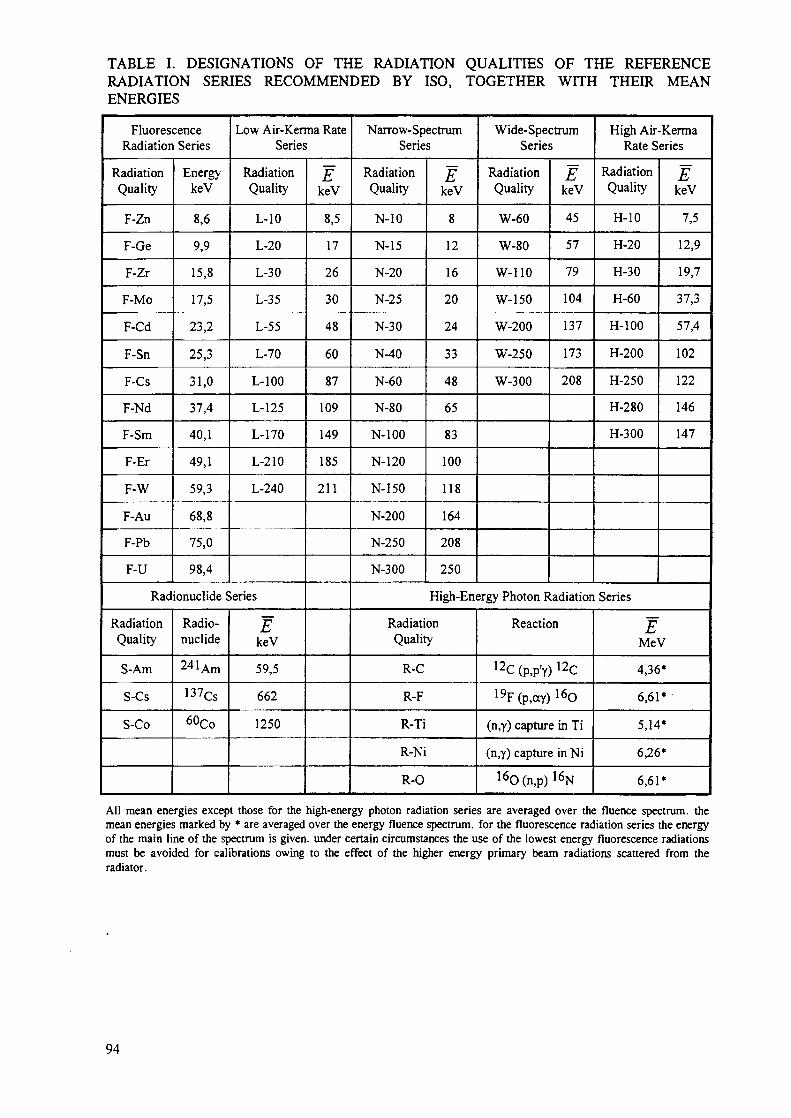
TABLE I. DESIGNATIONS OF THE RADIATION QUALITIES OF THE REFERENCERADIATION SERIES RECOMMENDED BY ISO, TOGETHER WITH THEIR MEANENERGIES
FluorescenceRadiation Series
RadiationQuality
F-Zn
F-Ge
F-Zr
F-Mo
F-Cd
F-Sn
F-Cs
F-Nd
F-Sm
F-Er
F-W
F-Au
F-Pb
F-U
EnergykeV
8,6
9,9
15,8
17,5
23,2
25,3
31,0
37,4
40,1
49,1
59,3
68,8
75,0
98,4
Low Air-Kerma RateSeries
RadiationQuality
L-10
L-20
L-30
L-35
L-55
L-70
L-100
L-125
L-170
L-210
L-240
Radionuclide Series
RadiationQuality
S-Am
S-Cs
S-Co
Radio-nuclide
24lAm
137Cs
60Co
EkeV
59,5
662
1250
EkeV
8,5
17
26
30
48
60
87
109
149
185
211
Narrow-SpectrumSeries
RadiationQuality
N-10
N-15
N-20
N-25
N-30
N-40
N-60
N-80
N-100
N-120
N-150
N-200
N-250
N-300
EkeV
8
12
16
20
24
33
48
65
83
100
118
164
208
250
Wide-SpectrumSeries
RadiationQuality
W-60
W-80
W-110
W-150
W-200
W-250
W-300
EkeV
45
57
79
104
137
173
208
High Air-KermaRate Series
RadiationQuality
H-10
H-20
H-30
H-60
H-100
H-200
H-250
H-280
H-300
EkeV
7,5
12,9
19,7
37,3
57,4
102
122
146
147
High-Energy Photon Radiation Series
RadiationQuality
R-C
R-F
R-Ti
R-Ni
R-0
Reaction
12C (p,p'y) 12C
19F(p,ay) 1 6O
(n,y) capture in Ti
(n,y) capture in Ni
1 6O(n,p) 1 6N
EMeV
4,36*
6,61*
5,14*
6,26*
6,61*
All mean energies except those for themean energies marked by * are averagedof the main line of the spectrum is givenmust be avoided for calibrations owingradiator.
high-energy photon radiation series are averaged over the fluence spectrum, theover the energy fluence spectrum, for the fluorescence radiation series the energy. under certain circumstances the use of the lowest energy fluorescence radiationsto the effect of the higher energy primary beam radiations scattered from the
94

>9
* 8
tn
*b
Ul
543210
high air kerma ratewide spectrumnarrow spectrumlow air kerma rate
HJl
H -i%=2=:
20 40 60E in keV
80 100
(0
to
Ul
6-
5-
4 -
3 -
2 -
1 -
0 -
t
IIJIII
I
A
) 201
40E
J
60inkeV
1
/ \
\
•i
80 100
F/G. 6. Comparison of the calculated spectral photon flux per solid angle and tube current, N£, of
the low air-kerma rate spectrum L-100, the narrow spectrum N-100, the wide spectrum "W-100 " andthe high air-kerma rate spectrum H-100. The spectra are calculated for a distance of 2.5 mfrom atube with a tungsten anode with an anode angle of 20°. The tube potential is 100 kV. The Figure is
split in two parts to allow the four spectra to be presented with linear scales.In the upper Figure the narrow spectrum and the low air-kerma rate spectrum cannot be seen
because of their low N E values
95

TABLE II. CHARACTERISTICS OF THE SPECTRA SHOWN IN FIGURE 5. (THE SPECTRALPHOTON FLUX PER SOLID ANGLE AND TUBE CURRENT, NE , IS CALCULATED FOR A
DISTANCE OF 2.5 M FROM THE TUBE. E IS THE MEAN PHOTON ENERGY AVERAGEDOVER THE FLUENCE SPECTRUM. THE FILTRATION OF BE IS THE INHERENTFILTRATION)
Filtration
mm
Be
Al
Cu
Sn
E in keV
Resolution in %
NE (s"1 sr1 A"1 keV'1)
Characteristic of Reference Radiation
Radiation Quality
L-100
1.0
4.0
0.5
2.0
87
22
3.6 109
N-100
1.0
4.0
5.0
—
83
28
1.2 1010
"W-100"
1.0
4.0
2.0
—
75
46
1.1 1011
H-100
1.0
3.9
0.15
-
57
75
1.3 1012
TABLE III. CHARACTERISTICS OF THE NARROW-SPECTRUM SERIES. THE TUBEPOTENTIAL IS MEASURED UNDER LOAD.
RadiationQuality
N-10
N-15
N-20
N-25
N-30
N-40
N-60
N-80
N-100
N-120
N-150
N-200
N-250
N-300
EkeV
8
12
16
20
24
33
48
65
83
100
118
164
208
250
Resolution
RE%
28
33
34
33
32
30
36
32
28
27
37
30
28
27
TubePotential
kV
10
15
20
25
30
40
60
80
100
120
150
200
250
300
Additiona Filtration(mm)
Pb Sn
1.0
3.0
5.0
1.0
2.5
3.0
2.0
3.0
Cu
0.21
0.6
2.0
5.0
5.0
2.0
Al
0.1
0.5
1.0
2.0
4.0
1st
HVLmm
0.047 Al
0.14 Al
0.32 Al
0.66 Al
1.15 Al
0.084Cu
0.24 Cu
0.58 Cu
1.11 Cu
1.71 Cu
2.36 Cu
3.99 Cu
5.19 Cu
6.12 Cu
2nd
HVLmm
0.052 Al
0.16 Al
0.37 Al
0.73 Al
1.30 Al
0.091Cu
0.26 Cu
0.62 Cu
1.17 Cu
1.77 Cu
2.47 Cu
4.05 Cu
5.23 Cu
6.15 Cu
For the five lowest energies the recommended filtration is 1 mm be but other values may be used provided thatthe mean energy is within _+ 5 % and the resolution is within _+̂ 15 % of the values given in the table, for thehigher energies (radiation qualities N-40 and above) the total filtration consists of the additional filtration plusthe inherent filtration adjusted to 4 mm of aluminium, the minimum purity of the filters should be 99.9 %. thehalf value layers (hvls) are measured at a distance of 1 m from the focal spot.
96

TABLE IV. CHARACTERISTICS OF THE REFERENCE RADIATIONS PRODUCED BYRADIONUCLIDE SOURCES AND OF THE HIGH-ENERGY PHOTON RADIATIONS
RadiationQuality
S-Co
S-Cs
S-Am
R-C
R-F
R-Ti
R-Ni
R-0
Energy of the Radiation(MeV)
1.1733 and 1.3325
0.6616
0.05954
4.36
6.13 to 7.12
5.14
6.26
6.13 to 7.12
Half-Life(days)
1925.5
11050
157788
Air-kerma Rate Constant
mGyrfW.MBq"1
0.31
0.079
0.0031
The value of the air-kerma rate constant is valid only for an unshielded point radionuclide source. It is given onlyas a guide. Air-kerma rates at the exposure positions should be measured using a secondary ionisation chamber.Instead of using sources with different activities, the air-kerma rate may also be varied by means of lead attenuatorsfor collimated beams of 137Cs and ̂ Co. The attenuators shall be placed in close vicinity to the diaphragm.
1
0.9
0,8
5 0,7
10 .6io.5•EO.4
En
•©-0,3
0,2
0,1
010
N-40 N-60 N-100 N-250 S-Co R-F
"1jI /
1I I! I
i
——
—i
i . J100 1 0 0 0 EinkeV
1 0 0 0 °
FIG. 7. Relative spectralphoton fluence Q>E of the reference photon radiations used in the "typetest" intercomparison of the co-ordinated research project of the IAEA. The radiation qualities N-40,
N-60, N-100 and N-250 belong to the narrow-spectrum series, the radiation quality S-Co to theradionuclide series and the radiation quality R-F to the high-energy photon radiation series. All
spectra except the R-F spectrum are theoretical spectra. The R-F spectrum is an unfolded spectrumgenerated at 2.7 Me Vproton energy [15]. In this spectrum the photon radiation generated by an
annihilation reaction at 0.51 MeV is omitted for reasons of clarity.
97

Y/////////////////////Y/Anode
Heated cathode
Shutter
Filament voltageY//////////////////////?/
Additional filtration
•; Diaphragms
j/J Monitor detector
A
Detector of Phantom withthe reference personal dosimeterinstrument
FIG. 8. Scheme of a typical calibration set-up with X radiation for the calibration against a referenceinstrument and a monitor.
7 .
F/G. P. Example of a calibration set-up with X radiation for the calibration against a referenceinstrument and a monitor.
98

4. CONVERSION COEFFICIENT FROM AIR KERMA TO Hp(10) IN THE ICRUSLAB PHANTOM
In general reference instruments for photon radiation do not directly indicate i7p (10; E,cc)
but the air-kerma rate. Hp(l0;E,a) is derived from air kerma ATa using appropriate
conversion coefficients, /zpK(10; E,a), for photon radiation of energy E, with an angle a
between the reference direction of the dosimeter and the direction of radiation incidence:
;E,a) = Hp(l0;E,a) I Ka (3)
Tabulated values for /ZpK(10; £ ,«) presuppose the establishment of secondary charged
particle equilibrium for the radiation field. An appropriate build-up layer may be required (seesection 6.4) resulting in a substitution of hpK(10; E,a) by ApK(10; E,a) • #PMMA i° eQ- (3)-
If a reference instrument is used for calibration (denoted by subscript R in the following), asfor the methods given in sections 5.2, 5.3 and 5.5, its calibration factor for air kerma, NR,given in the calibration certificate can be used to detennine the conventional true value ofHp(\0; E,a) by means of the conversion coefficient hpK(10;E,oc) of eq. (3) and the
measured (indicated) value MR of the reference instrument (corrected for referenceconditions):
Hp(l0;E,a) = / ^ ( I O ^ C O - A ^ M * (4)
For radiation qualities of finite spectral width, the symbol E is replaced by the relevant letteraccording to Table I denoting a particular series of reference radiation, i.e. F, L, N, W, H, S orR.
Conversion coefficients ^^(10; E,a) for monoenergetic radiation shall be treated as if they
are not affected by an uncertainty. The conversion coefficients for the narrow-spectrum series,the radionuclide sources and the high-energy photon radiations given in Tables 5 and 6 shallbe considered as being affected by a relative standard uncertainty of 2 % except those with anexclamation mark. The relative standard uncertainty of 2 % takes into account differencesbetween the spectrum used for the calculation of the conversion coefficient [2,12] and thatprevailing at the point of test.
The numerical values with an exclamation mark actually applicable to a given experimentalset-up may differ by considerably more than 2 % from the given value. Such exclamationsmarks have to be considered only for tube voltages below about 30 kV when photons of lowenergies may strongly influence the numerical value of the conversion coefficients. Smalldifferences in the energy distribution can result in significant changes in the numerical valuesof these conversion coefficients as the majority contribution to the air kerma originates fromthe low-energy part of the spectrum, while the majority contribution to Hp(\V) originates fromthe high-energy part of the spectrum. Differences between the experimental arrangements asregards the energy distribution may be due to a great number of factors, e.g. anode angle,
99

anode roughening, tungsten evaporated on the tube window, presence of a transmissionmonitor chamber in the beam, deviation of the thickness of filters from nominal values, lengthof the air path between focal spot and point of test, and atmospheric pressure at the time ofmeasurement.
In practice, calibrations are always performed in divergent beams. This is taken into accountby relating the conversion coefficients to a reference distance between radiation source andpoint of test. In cases where a reference distance is given together with an angle a of thedirection of radiation incidence, a pertains to the angle between the reference and actualorientation of the dosimeter in the field.
TABLE V. CONVERSION COEFFICIENT /*pK(10 ; N,a) FROM AIR KERMA, Ka, TO THE
DOSE EQUIVALENT Hp(10;N,a) FOR RADIATION QUALITIES GIVEN IN ISO 4037,
PART 1 [12] AND THE SLAB PHANTOM, REFERENCE DISTANCE 2 m
Radiation
Quality
N-15 !
N-20!
N-25!
N-30
N-40
N-60
N-80
N-100
N-120
N-150
N-200
N-250
N-300
Irr. Dist.
m
1,0-2,0
1,0-2,0
1,0-3,0
1,0-3,0
1,0-3,0
1,0-3,0
1,0-3,0
1,0-3,0
1,0-3,0
1,0-3,0
1,0-3,0
1,0-3,0
1,0-3,0
cm
25
25
23
20
16
11
11
11
11
11
12
13
15
/ip^10;N,a) in Sv /Gy for Angle of Incidence of
0°
0,06
0,27
0,55
0,79
1,17
1,65
1,88
1,88
1,81
1,73
1,57
1,48
1,42
10°
0,06
0,27
0,55
0,78
1,16
1,64
1,87
1,88
1,80
1,72
1,56
1,48
1,42
20°
0,06
0,26
0,53
0,77
1,15
1,62
1,86
1,86
1,79
1,71
1,56
1,48
1,42
30°
0,04
0,23
0,50
0,74
1,12
1,59
1,83
1,82
1,76
1,68
1,55
1,47
1,41
40°
0,03
0,20
0,44
0,68
1,06
1,52
1,76
1,76
1,71
1,64
1,51
1,44
1,40
45°
0,03
0,17
0,41
0,65
1,02
1,47
1,71
1,73
1,68
1,61
1,49
1,42
1,38
50°
0,02
0,15
0,37
0,60
0,98
1,42
1,66
1,68
1,64
1,58
1,46
1,40
1,36
60°
0,01
0,09
0,28
0,49
0,85
1,27
1,50
1,53
1,51
1,46
1,38
1,33
1,30
70°
0,00
0,04
0,15
0,32
0,65
1,04
1,26
1,31
1,28
1,26
1,23
1,21
1,19
The irradiation distance is measured from the focal spot of the x-ray tube to the point of test, at whichthe reference point of the dosimeter shall be located. The values of the conversion coefficients may beused without modification over the given range of irradiation distances. For radiation qualities with anexclamation mark, care needs to be taken as variations in energy distribution may have a stronginfluence on the numerical values of conversion coefficients. The meaning of the diameter dF isexplained in Section 6.
100

5. CALIBRATION METHODS
5.1. General
As stated in section 2, the calibration factor shall be determined under standard test conditionswhich implies that ISO reference radiations should be used. The first three methods describedin this section presume the existence of such reference fields in the calibration laboratory.
However, some laboratories or services have only irradiation facilities differing from thoserecommended by ISO and, for the time being, have to use this test field for calibrations.
TABLE VI. CONVERSION COEFFICIENTS ApK(10;S,a) AND /jpK(10; R,a) FROM AIR
KERMA, Ka, TO THE DOSE EQUIVALENT Hp(\0;S,a) AND J / p (10 ; R,a),
RESPECTIVELY, FOR RADIATION QUALITIES GIVEN IN ISO 4037-1 [12] AND THE SLABPHANTOM
Radiation
Quality
S-Am
S-Cs
S-Co
R-C
R-F
T-Ti
R-Ni
R-0
Irr. dist
m
2,0 - 3,0
1,5-4,0
1,5-4,0
1,0-5,0
1,0-5,0
1,0-5,0
1,0-5,0
1,0-5,0
cm
l i
15
15
15
15
15
15
15
Build-ufLayer
mm
-
1.5
4
25
25
25
25
25
kpMMA
-
1.00
1.00
0.94
0.94
0.94
0.94
0.94
hpK(l0; S, a) and hpK(10; R, a) in Sv /Gy
for Angle of Incidence of
0°
1.89
1.21
1.15
1.11
1.12
1.11
1.11
1.12
10°
1.88
1.22
1.15
1.11
1.12
1.11
1.11
1.12
20°
1.86
1.22
1.15
1.12
1.12
1.11
1.11
1.12
30°
1.83
1.22
1.15
1.12
1.11
1.11
1.11
1.11
40°
1.77
1.22
1.16
1.11
1.11
1.10
1.10
1.11
45°
1.72
1.22
1.16
1.11
1.11
1.11
1.10
1,11
50°
1.66
1.22
1.16
1.11
1.11
1.11
1.10
1.11
60°
1.50
1.19
1.14
1.11
1.12
1.11
1.11
1.12
70°
1.25
1.14
1.12
1.10
1.13
1.12
1.12
1.13
The irradiation distance is measured from the geometrical centre of the radionuclide source to thepoint of test, at which the reference point of the dosimeter shall be located. In the case of high-energyphoton radiations, the irradiation distance shall be measured from the centre of the radiator or targetsurface from which the radiation emerges to the point of test. The values of the conversioncoefficients may be used without modification over the given range of irradiation distances. Themeanings of the diameter dp, the build-up layer and the correction factor &PMMA
a r e explained inSection 6.
An example of such a test field is a photon radiation field generated by a l31Cs sourceoperated in a small room so that radiation scattered from the walls makes a remarkablecontribution to the air-kerma rate at the reference point, say 20 %. The application of eq. (3) insection 4 using a value of the air-kerma rate measured by a reference instrument and theconversion coefficient from Table 6 would produce a faulty result as :
101

• the detector of the reference instrument for the air-kerma rate probably is an ionisationchamber with an almost isotropic angular response to air kerma measuring the air kermanot only of the collimated beam but also of the radiation scattered from all directions ofthe room, i.e. even of that backscattered radiation which does not contribute to theindication of a personal dosimeter fastened on a phantom because this scatteredradiation is shielded by the phantom;
• the conversion coefficient from Table VI refers to pure I37Cs radiation only, i.e.collimated beam conditions are presumed.
Another example of a test field is a field of high-energy beta radiation emitted by a betasource (e.g. of the radionuclide Y). This radiation may not even be a type of radiation whichthe dosimeter is intended to measure.
It is essential that the correspondence of the dosimeter's reading in the test field to the readingof the dosimeter in a reference field is established, and that this correspondence, oncedetermined in a type test, is invariant. In this case, a routine calibration with, of course, lessthan the highest metrological quality can be carried out in such a test field. Considering theimportance such routine calibrations have in practice, it is shown in section 5.5 by an examplehow such a test field can be used.
5.2. Calibration with a reference instrument without any monitor
This procedure is appropriate if the value of the air-kerma rate is stable over a time periodcorresponding to the duration of the calibration so as to achieve results of the desiredaccuracy. The calibration is carried out under standard test conditions close to the referenceconditions. The calibration set-up is shown schematically in the upper half of Figure 10. Thereference points of the reference instrument and the dosimeter under calibration aresubsequently positioned at the point of test in the radiation field for calibration in terms ofHp(l0;E,a). The position of the point of test is determined by the intersection of the dashed
lines in Figure 10.
Part (1) of the Figure: For the reference instrument (subscript R) one obtains the calibrationfactor NR of the reference instrument (under reference conditions) from the measured(indicated) value MR of the reference instrument corrected for reference conditions by meansof eq. (4):
N = Pv J ( 5 )R (5)
hpK(l0;E,a) is the coefficient to convert from air kerma measured by the reference
instrument to Hp(l0;E,a). Depending on the radiation quality used, the energy E has to be
replaced in eq. (5), and consequently in the following equations, by F, L, N, W, H, S or R (seesection 3).
102

Part (2) of the Figure: The dosimeter under calibration (subscript I) has an indication directlyrelated to the dose equivalent quantity Hp(l0;E,a). The dosimeter is positioned on the ISO
water slab phantom with an angle a between the axis of the reference radiation field and thereference direction of the personal dosimeter; most frequently a = 0° will be chosen. Thedosimeter's calibration factor (under reference conditions) Nj is obtained from the measured(indicated) value, corrected for reference conditions, Mj:
HM0;E,a)Nj = P \ ;
M i
The combination of eqs. (5) and (6) results in the calibration factor Nj derived from NR :
(6)
hoK(l0;E,a)-MR
= NR. pKK ' ' - (7)M
5.3. Calibration with a reference instrument and with a monitor
Moderate variations in the course of time in the physical quantities that characterize thedosimetric properties of the radiation field (e.g. air-kerma rate) can be corrected by using amonitor and by irradiating the reference instrument and the personal dosimeter undercalibration sequentially. This technique is often employed with X-ray units in order to correctfor variations in the air-kerma rate when reference instrument and dosimeter under calibrationare alternately placed at the point of test. The calibration set-up is schematically shown in thelower part of Figure 10 in a way similar to that chosen for the calibration set-up in theprevious chapter. The reference points of the reference instrument and the dosimeter undercalibration are subsequently positioned at the point of test in the radiation field for calibrationin terms of #p(10; E,a). Its value at the point of test is related to the calibration factor of the
monitor chamber, N M, and its measured (indicated) value m by
NM= pV (8)m
Part (1) of the Figure: The calibration factor A^ of the reference instrument (under referenceconditions) is
H.(lO;E,a)NR = p — (9)
* hpK(\0;E,a) MR
where MR is the measured (indicated) value of the reference instrument corrected forreference conditions (i.e. indication multiplied by applicable correction factors, e.g. acorrection factor considering differences in air density).
103

(1)
(2)
en
(2)
ii
i.V,
* < - Pf
Calibration with a reference instrument without any monitor
I.1
II
A1',-
•V R • •
!>!,
Calibration with a reference instrument and with a monitor
FIG. 10. Calibration with a reference instrument (schematically).
104

Part (2) of the Figure: The corresponding equation for the calibration factor Nj of thedosimeter (under reference conditions) is:
„ Hv(l0;E,a)
Mj is the respective value of the dosimeter under calibration.
Hp(\0;E,a) can be eliminated in eqs. (9) and (10) by means of eq. (8) if one introduces the
measured values mR and ntj of the monitor for the irradiation of the reference instrument
and the dosimeter under calibration:
M mR
hpK(l0;E,a) MR
N
mR is the measured (indicated) value of the monitor for the irradiation of the referenceinstrument, corrected for reference conditions (i.e. indication multiplied by applicablecorrection factors, e.g. differences in air density) and mt the corresponding value of themonitor for the irradiation of the dosimeter.
By division of eqs. (11) and (12), the calibration factor N^ disappears and one obtains forthe calibration factor of the dosimeter (under reference conditions) N 7 :
In practice, if the irradiations of reference instrument and dosimeter to be calibrated areperformed in brief succession, the ambient conditions of the radiation monitor remain thesame and corrections of the indicated value of the monitor to reference conditions areunnecessary.
In cases where the monitor is of good long-term stability, it may serve as the referenceinstrument after having been calibrated against another reference instrument.
105

. 11. Calibration in a known radiation field (schematically).
5.4. Calibration in a known radiation field
For a radiation field in which the conventional true value of Hp(l0;E,a) at the point of test
is directly known, the calibration factor Nj of the dosimeter is obtained from its measured
value, corrected for reference conditions, Mj (see Figure 11):
_Hp(l0;E,cc)1 M,
(14)
5.5. Example of a routine calibration
It is assumed that a service has established a test field with a 137Cs source for routinecalibrations of dosimeters as described in section 5.1. This test field is used in connection withoccasional calibrations in a reference field of a secondary standard laboratory. Thecorrespondence must be established between the dosimeter's reading in the test field and thereading of the dosimeter in the reference field, and it must be shown that this correspondenceis invariant. Routine calibrations will be needed for new batches of dosimeters and for routinechecks of the reproducibility of the dosimeter evaluation. In this example, a three stepprocedure is followed by the service (see Figure 12).
106

(1)
Calibration of the dosimeter in the reference field: Ar, Kj- = Hp (10; )'
(2)II
Transfer of ihc calibration to the test field; Hp 1KI (10; Elext, a Uv!) «= Mf ,„, • JV t ref
(3)I
Routine calibration in the test field: h'n>M - f?^ ,&t (] 0 ; Elest, a to7 ) ••' A/,rom
FIG. 12. Example of a routine calibration with three experimental set-ups (schematically).
107

Step I. For simplicity it is assumed that one routine dosimeter is sent by the service to asecondary standard laboratory; if more dosimeters are sent in, appropriate mean values of themeasured values have to be taken into account. In the secondary standard laboratory theirradiation is performed on an ISO water slab phantom in the reference field, for which theconventional true value of Hp ref (10; Erej-,aref) is known at the point of test. According to
eq. (14), the calibration factor NIref is obtained from the measured value, corrected for
reference conditions, Mj ref :
HpTef(l0;Eref,aref)NI ™f = Tt
Ml ref
Step 2. After return to the service, the calibrated dosimeter is irradiated homogeneously at acertain point (point of test) in the test field where the measured value, corrected for referenceconditions, M7 test, is obtained. This can be formally associated with a dose equivalent
/ / test (10; Etest,alest) if one assumes the validity of the calibration factor of the reference
field, Nj ref :
Hptcst(l0;Etest,atesl) = MItest-Nlref (16)
Here the routine dosimeter plays the role of a reference instrument. It links the quantityHp test(10; Etest>atest) of the test field with Hp re f(10; Erej-,arej-) in the reference field
even if the dosimeter is not irradiated on a phantom.
Step 3. This step 3 is the routine calibration in the true sense of the word. Subject is any(routine) dosimeter of the same type as the dosimeter calibrated in the reference field (step 1).The calibration factor Nrout of the (routine) dosimeter is obtained at the point of test in thetest field from the the measured value, corrected for reference conditions, Mrout, and the doseequivalent Hp test(10; Etest,atest) determined in step 2:
_ " p test (10 S Efest > atest)Nrout = ^ (17)
mrout
It is obvious that this routine calibration is, in principle, of less metrological quality than acalibration based on one of the methods described in sections 5.2 to 5.4 because a routinedosimeter of inferior performance is used as a reference instrument in step 1. Moreover, theassumption that the calibration factor ,/V; ref is valid for the test field has to be checked
(compare eq. (16)). This assumption is questionable, for example, if the reading in thereference field is primarily generated by the backscatter of the phantom and the calibration inthe test field is performed free in air.
108

6. POSITIONING OF THE PERSONAL DOSIMETER
6.1. Reference point and point of test
For the calibration the reference point of the dosimeter has to be placed at the so-called pointof test in the radiation field at which the conventional true value of Hp(\0) is known. Thereference point and the reference direction of the dosimeter should be stated by themanufacturer. The reference point should be marked on the outside of a dosimeter. If thisproves impossible, the reference point should be indicated in the accompanying documentssupplied with the dosimeter. All distances between the radiation source and the dosimetershall be taken as the distance between the radiation source and the dosimeter's reference point.
In the absence of information on the reference point or on the reference direction of thedosimeter to be calibrated, these parameters shall be fixed by the calibrating laboratory. Theyshall be stated in the calibration certificate.
When the angular dependence of the response has to be measured, in a first step, the dosimeteris fastened on the phantom's front surface so that the dosimeter's reference direction coincideswith the normal on the phantom's front surface (Figure 13). Then the dosimeter's referencepoint and the point of test in the radiation field are brought into coincidence and, finally, thecombination of dosimeter and phantom is rotated about an axis passing through the referencepoint of the dosimeter so that the reference direction of the dosimeter and the direction ofradiation incidence of the irradiation facility form the desired angle a.
The calibration factor is determined under conditions lying within the range of standard testconditions which usually means a = 0°.
Near monodirectionaJ
radiation
Build-up layerif required Reference point
Dosimeter
FIG. 13. Arrangement for the calibration and the measurement of the response of a personaldosimeter at the angle a.
109

6.2. Simultaneous irradiations of dosimeters
When several personal dosimeters are irradiated simultaneously on the front face of the slabphantom they shall not cover any phantom surface outside a circle of diameter dF, given bythe approximate locus of the 98 % isodose contour with respect to the dose in the centre of thephantom. The values of hpK(l0 ; N,a) depend on the radiation quality and they are given for
some reference radiations in Tables V and VI. If irradiation distances smaller than those givenin the Tables are used, the diameter dF becomes smaller.
Two effects associated with this (simplified) procedure require additional attention:
• by positioning several dosimeters on the phantom surface the backscatter may bereduced due to the attenuation of the primary radiation passing through thedosimeters and
• possibly different distances of the reference points from the radiation source haveto be considered.
Before such a practice is adopted it shall be verified that it leads to results identical to within2 % to those obtained when only one dosimeter is irradiated in the centred position.
There may be certain types of dosimeters which respond very sensitively to small changes inthe properties of the backscattered photon field. This may be due to the use of strongly energy-dependent detectors or possibly, to the properties of the algorithms used to arrive at the valueof the dose equivalent from the detector signal. In such cases it may be advisable to have onlyone dosimeter mounted on the phantom surface for any calibration.
6.3. Misplacement and dosimeter supports
In the case of point sources and in the absence of scattered radiation and photon absorption,the dose rate changes with the inverse square of the distance R. A misplacement of thedosimeter's reference point in the beam by the amount of AR in the direction of the beam willlead to a relative error in the calibration factor of 2AR/R at the distance R. Misalignmentperpendicular to the beam axis by Ar causes a relative error of (Ar/R)2 . In the presence ofscattered radiation and for sources of finite dimensions, the above approximations are limitedto values of AR or Ar small in comparison with R.
The supports used for the dosimeter and the reference instrument, and the calibration sourceshould introduce as little scattered radiation as possible. The effects of such scattered radiationon the indication of the instruments should be taken into account.
6.4. Effects associated with electron ranges
In some photon fields, effects associated with electron ranges have to be considered. Fordosimeters being calibrated there is no secondary electronic equilibrium within the sensitivevolume of their detectors. In some cases, the detector window or encapsulation is notsufficiently thick for dose build-up, one prerequisite for secondary electronic equilibrium. Forthose dosimeters one would obtain different indications in photon radiation fields withdiffering electronic equilibrium. By placing a layer in front of the detector, which togetherwith the wall material and the cover of the detector gives a combined layer of a thicknesslarger than the range of the most energetic secondary electrons, one is able to obtainreproducible results. Experience has shown that one does not require any additional layers forphoton energies below 250 keV; up to 0.66 MeV, a layer of PMMA 1.5 mm thick is
110

sufficient. For energies up to 1.33 MeV, a 4 mm PMMA layer is sufficient. The cross-sectional area of the plate shall be 30 cm x 30 cm.
The modification of the radiation field by introducing the PMMA plate shall be taken intoaccount by multiplying the conversion coefficient by the correction factor ICPMMA (see Table6).
For irradiations on a phantom it may be practical to position the PMMA plate at a certaindistance away from the dosimeter or dosimeter-phantom combination so that it is notnecessary to also rotate the plate when the variation of response with the direction of radiationincidence is examined.
REFERENCES
[1] DEUTSCHES INSTITUT FUR NORMUNG E.V., International Vocabulary of Basicand General Terms in Metrology, ISBN 3-410-13086-1 (1994).
[2] INTERNATIONAL STANDARDS ORGANISATION, Reference Photon Radiationsfor Calibrating Dosemeters and Dose Ratemeters and for Determination of theirResponse as a Function of Photon Energy and Angle of Incidence, ISO/DIS 4037-3,Geneva (1996).
[3] INTERNATIONAL ELECTROTECHNICAL COMMISSION, Radiation ProtectionInstrumentation - Direct Reading Personal Dose Equivalent (Rate) Monitors - X,Gamma and High Energy Beta Radiation, International Standard IEC 1283 (1995).
[4] INTERNATIONAL COMMISSION ON RADIATION UNITS ANDMEASUREMENTS, Measurement of Dose Equivalents from External Photon andElectron Radiations. ICRU Report 47, Bethesda, (1992).
[5] ALBERTS, W.G., AMBROSI, P., BOHM, J., DffiTZE, G., HOHLFELD, K., WILL,W., New Dose Quantities in Radiation Protection. PTB Report PTB-Dos-23e,Braunschweig, (1995).
[6] BOHM, J., THOMPSON, I.M.G., GRIFFITH, R., The New IAEA Recommendations onthe Calibration of Radiation Protection Monitoring Instruments. 9th Int. Congress ofIRPA, Vienna, 14-19 April 1996. Proc. Vol. 4, (1996) 186-188.
[7] BARTLETT, D.T., ALBERTS, W.G., Type Testing and Calibration of PersonalDosemeters. Radiat. Prot. Dosim. 54, (1994) 259-266.
[8] BARTLETT, D.T., ILES, W.J., Calibration of Personal Dosemeters. NRPB ReportNRPB-M520 (1994).
[9] ALEINIKOV, V., Calibration of Personal Dosimeters: Quantities and Terminology.This publication.
[10] GRIFFITH, R., Quantities and Units for External Dose Assessment. This publication.
I l l

[11] KRAMER, H.M., BOHM, J., ILES, W.J., THOMPSON, I.M.G., On the Current Statusof an ISO Working Document on the Calibration and Type-Testing of RadiationProtection Dosimeters for Photons. Radiat.Prot.Dosim. 54, (1994) 267-272.
[12] INTERNATIONAL STANDARDS ORGANISATION, X and Gamma ReferenceRadiation for Calibrating Dosemeters and Doserate Meters and for Determining theirResponse as a Function of Photon Energy. Part 1: Radiation Characteristics andProduction Methods, ISO 4037-1, Geneva (1996).
[13] BEHRENS, R., Physikalisch-Technische Bundesanstalt, Braunschweig, Personal Com-munication (1997).
[14] BEHRENS, R., Rechnergesriitzte Optimierung von Rontgenfluoreszenzanlagen,Diploma, Braunschweig (1995).
[15] BUERMANN, L., Physikalisch-Technische Bundesanstalt, Braunschweig, PersonalCommunication (1997).
[16] NOWOTNY, R., HOFER, A., Ein Programm fur die Berechnung von diagnostischenRontgenspektren. Fortschr. Rontgenstr. 142, (1985) 685-689.
112

TilXA0053411
CALIBRATION OF A PERSONAL DOSIMETER IN THEFIELD OF A RADIONUCLIDE
C. STRACHOTINSKY, H. STADTMANNAustrian Research Centre,Seibersdorf, Austria
Abstract
An example of the practical implementation of the international standard ISO4037-3 for the calibration of an electronicdosimeter for Hp(10) in the radiation field of a l37Cs source for normal incidence of the radiation is given. The set-up, theirradiation and the calculation of the results including the assessment of the uncertainties are described in detail.
1. INTRODUCTION
This paper gives an introduction in the practical implementation of the international standardISO4037-3 [1] for the calibration of an electronic dosimeter for i7p(10) in the radiation field ofa 137Cs source for normal incidence of the radiation.
2. SOURCE AND BEAM
Radionuclide: 137Cs - Gamma source, encapsulated in steel.
Material of the holder: Aluminium
Nominal activity: Aact = 65 GBq
The beam is collimated by an ISO-Collimator with 15° opening.
The beam is measured and certified in terms of air-kerma rate.
For the calibration in a known radiation field in which the conventional true value ofHp(\0;E,a) at the point of test is directly known, the calibration factor Nt of the dosimeter
is obtained from its measured value, corrected for reference conditions, M7(see eq. (14) in
[2]):
Ni ( 1 )
M,
whereby Hp(l0;E,a) is obtained by (see eq.(3) in [2]:
Hp aO; S - Cs,0°; = hpK (10; S - Cs,0°) • kPMMA . Ka (2)
113

Hp(\O,S-Cs,O°): Personal dose equivalent
/JPK(10;S-CS,0°): Conversion coefficient from air kerma to personal dose equivalent#P(10 ,S-CS,0° )
£PMMA Correction factor for PMMA build-up plate
Number of certificate from Bundesamt fur Eich- und Vermessungswesen (BEV): T 951091/6.
The value of K^ ref in the certificate is given for the reference date. This value has to be
reduced to the actual value as follows:
(3)— In!
V 2 )
ATa: Actual air-kerma rate (unit: |j.Gy/h) at the point of test
K^xef'- Air-kerma rate at reference date (unit: jo.Gy/h)At: Time since reference date (unit: d)t\/2'. Half-life of the source (unit: d)
3. DOSIMETER
• Type: Siemens electronic personal dosimeter EPD2
• Detector: Silicon diode
• Reference point: 5 mm behind mark
• Measuring quantity: Hp(\0)
• Rated range of use 0 |iSv to 15 Sv
The following accessory and documents are necessary:
• Manual
• Type test
• Batteries
4. SET-UP
The calibration will be done on the ISO water slab phantom [1].
Note: The irradiation conditions on the phantom should be similar to the practical wearingconditions on the body. Therefore a clip (if existing) must be used on the phantom.
114

The distance source-phantom should be sufficient, to assure that the whole phantom is withinthe beam.
The reference point of the dosimeter must be brought in the test point (in source-detectordistance, SDD) and the phantom is used as a backscatter phantom. Reference point and centreof the phantom-surface are positioned in the main beam direction.
SETUP FOR CALIBRATION IN THE FIELD OF A NUCLIDE SOURCE
SOURCE - DETECTOR DISTANCE
DISTANCE SPACE PT - SURF/ ,CE
DEPHT OF REFERENCE POINT
SOURCE KOLLIMATOR
PMMA PLATE 1.5 mm
REFERENCE POINTOF DOSEMETER
SPACE POINT
DOSEMETER
ISO PHANTOM
FIG 1. Set-up for the calibration of a dosimeter in a known radiation field.
5. IRRADIATION
The applied dose should be at least 100 times larger as the last digit of reading and also in arange where the background reading can be neglected. The irradiation time should be longenough to minimise uncertainties due to shutter time. The irradiation time tt can be calculatedas:
(4)
Indicated "Opening time" of shutter
Delay of shutter time.
The delay time can be found in the certificate or has to be determined by the laboratory.
The indicated time U can be adjusted only in steps of seconds. The applied air kerma istherefore:
(5)
115

6. DETERMINATION OF THE CALIBRATION FACTOR
The calibration factor N{ is determined by combining equations (1) to (4):
N,=
hpk(l0;S-Cs,00).kPMMA.Ka^.exp(-ln2.—).(ti-Ats)ha
M,
7. NUMERICAL VALUES FOR THE GIVEN EXAMPLE
7.1. Source
TABLE I. SOURCE AND RADIATION FIELD
SDD = 2500 mm
Reference date: 1993 12 3
At = 1243d
ka,ref= 841 ^Gy/h
hpK(\0: S-Cs,0°) =1,21 Sv/Gy
Beam diameter = 655 mm
Calibration date: 1997 05 27
fi/2=11050d
ka,act= 777,9 \iGy/h
kpMMA = 1 . 0
ifp(10) = 941 (̂ Sv/h = 0,2161 uSv/s
7.2. Set-up
Beam diameter = 650 mm which is more then the diagonal of phantom = 425 mm
(SPACE POINT => SURFACE) = 2500 - 458.8 - 5 = 2036,2 mm (see Figure 1)
7.3. Irradiation
Requested dose: Hp( 10) = 100 ja.Sv
Irradiation time: Ats = 2,1 s
Applied dose: Ka = 82,74 uGy
7.4. Calibration factor
M= 100,9
5= 1.1 nSv
W = 0,992
M
s
N
=> Ka = 82,64
ti = 384,56 s tj* = 385 s (integer)
=> ^p(10)appl= 100,12 ^Sv
Meter reading as mean value of 10 readings
Experimental standard deviation
Calibration factor
116

8. UNCERTAINTIES OF THE CALIBRATION FACTOR
Uncertainties are evaluated according to the IAEA Technical Report Series No. 374 [3].
Due to the used formula relative uncertainties (w) are used.
• The coverage factor k = 2
The uncertainty of the certified Air-kerma rate is stated with 1.5 % according to[3] and a coverage factor k = 2.
The position of the reference point of the dosimeter is within s(R) = ± 1.5 mm.(Experience in Lab.)
—e. = _ 2. —- => w=0.12 % (inverse square law => sensitivity factor = 2).Ka K
Field inhomogeneity: (AK</Ka) < 0,2 %. (Measurement in laboratory). Valuesare upper and lower limits
w2 = f / 3
(rectangular probability distribution)
The uncertainty of the Conversion factor hpK(10; S-Cs,0°) is given in [1]:
w = 2%.
Meter reading M.:
J=l . l |LlSv. 52mean = S2 I ft
Smean experimental standard deviation of the meann number of measurements
Wmean = ^mean / M
Irradiation time: Uncertainties are negligible
TABLE II. SUMMARY OF EVALUATION OF UNCERTAINTIES
Input Quantity
Air-kerma rate
Air-kerma rate
Air-kerma rate
Conversion factor
Meter reading
Output Quantity
Calibration factor
certified
positioning
field inhom.
Kt
K>
K,
M
N
Uncertainty
2w=1.50%
s(R) = ± 1.5 mm
AK/Ka<0.2%
w = 2%
J = 1.1 fiSv
Reference
certificate BEV
Lab
Lab
ISO4037-3
measured value
..,m .:0,75 %
0,12%
0,12 %
2,00 %
0,35 %
2,17%
117

Uncertainty u = w (N). N u = 0,022
U = k . u U = 0,044
U Associated expanded uncertainty to N
k coverage factor (k = 2)
The calibration factor can now be written as:
7V= 0,99 ±0,04
REFERENCES
[1] INTERNATIONAL STANDARDS ORGANISATION, Reference Photon Radiations forCalibrating Dosimeters and Dose Ratemeters and for Determination of their Response as aFunction of Photon Energy and Angle of Incidence. ISO/DIS 4037-3, Geneva (1996).
[2] BOHM, J., Calibration of personal dosimeters for photon radiation with respect to thepersonal dose equivalent Hp(10). Physikalisch-Technische Bundesanstalt, Braunschweig(1997).
[3] INTERNATIONAL ATOMIC ENERGY AGENCY, Calibration of Dosimeters Used inRadiotherapy. Technical Report Series No. 374, IAEA, Vienna (1994).
118

XA0053412DOSIMETER CHARACTERISTICS AND SERVICEPERFORMANCE REQUIREMENTS
P. AMBROSIPhysikalisch Technische Bundesanstalt,Braunschweig, Germany
D.T. BARTLETTNational Radiological Protection Board,Chilton, United Kingdom
Abstract
The requirements for personal dosimeters and dosimetry services given by ICRP 26, ICRP 35, ICRP 60 and ICRP 75 aresummarised and compared with the requirements given in relevant international standards. Most standards could be mademore relevant to actual workplace conditions. In some standards, the required tests of energy and angular dependence of theresponse are not sufficient, or requirements on overall uncertainty are lacking.
1. INTRODUCTION
World-wide, there are many standards covering requirements for personal dosimeters anddosimetry services. This paper gives an overview of those standards which are either inde-pendent of the measurement quantity, or are written to be used with the new quantities Hp(l0)and //p(0,07). In the following only the new quantity Hp(l0) and photon radiation will beconsidered.
2. GENERAL RADIATION PROTECTION REQUIREMENTS
2.1. ICRP Recommendations 26, 35, 60 and 75
The basis for all requirements in the field of radiation protection is given by the InternationalCommission on Radiation Protection (ICRP).
In Publication 26 [1] in paragraph (104) the Commission recommends a limit for the annualdose-equivalent [The remarks in square brackets are given by the authors.]:
(104) For stochastic effects the Commission's recommended dose limitation is based on theprinciple that the risk should be equal whether the whole body is irradiated uniformly orwhether there is non-uniform irradiation. This condition will be met if... the recommendedannual dose-equivalent limit for uniform irradiation of the whole body... [is] 50 mSv (5 rem).
In Publication 35 [2] the concept of 'Recording Level' is introduced in paragraph (18) and the'Accuracy Required in Routine Monitoring' is given in paragraph (109).
119

(18) The recording level is a formally defined value for dose equivalent or intake above whicha result from a monitoring program is of sufficient interest to be worth keeping. TheCommission has recommended that the recording level for individual monitoring should bebased on one-tenth of that fraction of the annual limit corresponding to the period of time towhich the individual monitoring measurement refers...
(109) The uncertainties acceptable in routine monitoring for external radiation should besomewhat less than the investigation level and can best be expressed in relation to theestimates of the annual deep and shallow dose-equivalent indices [now taken to be //p(10) and//p(0.07)] that are measured. The uncertainty in the measurement of the annual value of thesequantities (or of the upper limits if a cautious interpretation is being conducted) should bereduced as far as reasonable achievable. If these quantities are of the order of the relevantannual limits, the uncertainties should not exceed a factor of 1.5 at the 95% confidence level.Where they amount to less than 10 mSv an uncertainty of a factor of 2 at the 95% confidencelevel is acceptable. This uncertainty includes errors due to variations in the dosimeterssensitivity with incident energy and direction of incidence as well as intrinsic errors in thedosimeter and its calibration. It does not include uncertainties in deriving tissue or organdose equivalents from the dosimeter results.
In Publication 60 [3] earlier concepts, relevant to matters of concern here, are mostly retained.In paragraph (257) the reference levels are explained and in paragraph (271) uncertainties aregiven. In paragraph (S25) the dose limits are summarised:
(257) It is often helpful in the management of operations to establish values of measuredquantities above which some specified action or decision should be taken. These values aregenerally called reference levels. They include recording levels, above which a result shouldbe recorded, lower values being ignored; investigation levels, above which the cause or theimplication of the result should be examined; and intervention levels, above which someremedial action should be considered. The use of these levels can avoid unnecessary orunproductive work and can help in the effective deployment of resources. If recording levelsare used, the fact that no unrecorded results exceeded the recording level should be madeclear.
(271) In practice, it is usually possible without great difficulty to achieve an accuracy ofabout 10% at the 95% confidence level for measurements of radiation fields in goodlaboratory conditions. In the workplace, where the energy and the orientation of the radiationfield are rarely known, uncertainties by a factor of 1.5 will not be unusual in the estimation ofthe annual doses from the external exposure of the individual workers. In view of the otheruncertainties this factor is acceptable...
(S25) The Commission recommends a limit on effective dose of 20 mSv per year, averagedover 5 years (100 mSv in 5 years), with the further provision that the effective dose should notexceed 50 mSv in any single year. The 5-year period would have to be defined by theregulatory agency, e.g. as discrete 5-year calendar periods. The Commission would notexpect the period to be introduced and then applied retrospectively. It is implicit in theserecommended dose limits that the dose constraint for optimisation should not exceed 20 mSvin a year.
In Publication 75 [4], the revision of Publication 35, ICRP states in paragraphs (229), (230)Table 2, (232), and (233), and in (249), (250) and (251) the following:
120

The Use of Reference Levels
(229) Reference levels are values of measured quantities above which some specified actionor decision should be taken. They include recording levels, above which a result should berecorded, lower values being ignored; investigation levels, above which the cause or theimplications of the result should be examined; intervention levels, above which some remedialaction should be considered; and, more generally, action levels above which some specifiedaction should be taken. ...
(230) Table 2: [The] Recording level [is] set by operating management or nationalauthority, [it] allows records to exclude trivial information, [it is] advisory but should beapplied consistently, [it] applies principally to occupational exposure with particularreference to monitoring of individuals and workplaces.
(232) The Commission now considers that the recording level for individual monitoringshould be derived from the duration of the monitoring period and an annual effective dose ofno lower than 1 mSv or an annual equivalent dose of about 10% of the relevant dose limit. ...
(233) In practice, little use is made of recording levels in individual monitoring for externalexposure because the measured dose is usually entered directly as a measure of the effectivedose. The minimum level of detection should then be used as the recording level with resultsbelow that level being deemed to be zero. However, the recording level is useful in definingthe low dose requirements of dosimeters; it can be used as the basis for defining performancerequirements....
Accuracy
(249) The errors in the use of monitoring to provide estimates of individual doses and intakeslie partly in the measurement and partly in the models linking the measured and the requiredquantities. The errors contributing to the overall uncertainty may be regarded as falling intoat least four broad categories:
(a) random errors in the measurement, e.g. counting statistics,
(b) systematic errors in the measurements, e.g. calibration errors,
(c) errors, mainly systematic, in dosimetric and metabolic models, ...
(d) errors... in the use of the models, ...
(250) Individuals are usually exposed over an extended period and so assessments tend to bebased on a number of measurements made over that period. The use of multiplemeasurements reduces the random errors. For most assessments, the systematic errors inmodelling result in a bias towards over-estimation of the true dose.
(251) The Commission has noted that, in practice, it is usually possible to achieve an accuracyof about 10% at the 95% confidence level for measurements of radiation fields in goodlaboratory conditions (Paragraph 271, Publication 60). In the workplace, where the energyspectrum and orientation of the radiation field are generally not well known, the uncertaintiesin a measurement made with an individual dosimeter will be significantly greater. ... The
121

overall uncertainty at the 95% confidence level in the estimation of effective dose around therelevant dose limit may well be a factor of 1.5 in either direction for photons and may besubstantially greater for neutrons of uncertain energy and for electrons. Greater uncertaintiesare also inevitable at low levels of effective dose for all qualities of radiation.
In the following these requirements are interpreted for the most important case of amonitoring period of one month. The factors given above mean that the value of the quotientHJHX of the measured dose value, Hm, and the conventionally true value, Ht, has firstly to besmaller than or equal to the factors and secondly larger than or equal to the reciprocal of thefactors. The 95% confidence level means that the given requirement must be fulfilled for 19 of20 different measurements. In terms of uncertainties, the recording level is interpreted toallow a 100% relative uncertainty for a true dose value of the recording level itself. Therecording level for individual monitoring should be derived from the duration of themonitoring period. For a monitoring period of one month the recording level is
50 mSv/(1012) = 0.42 mSv, according to ICRP 21/35,20 mSv/(1012) = 0.17 mSv, according to ICRP 35/60,not lower than 1 mSv/12 = 0.085 mSv according to ICRP 60/75.
Thus the following two requirements are drawn from ICRP 26 to ICRP 75, see also Table 1and the broad lines in Figure 1:
1. For a dose value equal to or approaching the annual dose limit, the relation1.5 > HJHx ^ 1/1-5 must be fulfilled for 19 of 20 different measurements.
2. For a dose value less than or equal to Ht, the recording level for monthly monitoring, therelation 2.0 > HJHX > 0.0 must be fulfilled for 19 of 20 different measurements.
These two requirements are represented by the solid bars in Figure 1. They must be linkedtogether in some way. This necessity is indicated by the straight dashed lines in that Figure 1.
TABLE I. REQUIREMENTS GIVEN BY ICRP 26 TO ICRP 75 AND THEIRINTERPRETATION WITH RESPECT TO UNCERTAINTY FOR MONTHLY MONITORING
Dose limit orlevel
ftn>ft,annual limit
Hm<Ht
recordinglevel
Dose value according to
ICRP 26/35 ICRP 35/60 ICRP 60/75
/fa =50mSv
ft = 0.42mSv
Ha =20mSv
ft = 0.17mSv
Ha =20mSv
ft > 0.085mSv
Requirements
1.5 > ft/ft > 1/1.5for 19 of 20 measurements
2.0 > ft/ft > 0.0for 19 of 20 measurements
122

2.2. Trumpet curves
The requirements of Table 1 can be met by so-called 'trumpet curves' [5,6]. They are used inmany countries and are also incorporated in a number of standards. If Ho is the equivalent tothe recording level for monthly monitoring, e.g. 0.017 mSv according to ICRP 35/60 or notlower than 0.085 mSv according to ICRP 60/75, then the upper limit of the ratio HJHt isgiven by
= L5 1+Upper Limit
and the lower limit by
2H0+Ht(1)
Lower Limit
_ 1 f, 2H015 I H0+Ht
(2)
These limits, the trumpet curves, are shown as dashed curves in Figure 1 for the twocombinations ICRP 35/60 and ICRP 60/75 given above. They are the basic requirements forperformance testing of dosimetry services and dosimeters. The methods for testing are givenin the Section 3.
1/1 .5-
0 . 5 -
Monthly monitoring• " • • ICRP 60 and ICRP 75
trumpet curve, Ho = 0.17 mSvtrumpet curve, HQ = 0.085 mSv
100 mSv 1000
FIG. 1. Limits for the ratio HJHt for monthly monitoring according to ICRP 60 and ICRP 75 and thetrumpet curves for Ho = 0.17 mSv and Ho = 0.0.085 mSv. (Hm is the^measwed dose value and H,, the
conventionally true value. 95% of all measurements must be within the limits).
123

3. REQUIREMENTS FROM INTERNATIONAL STANDARDS
3.1. Relevant standards
Table II gives a list of those relevant international standards for personal monitoring which areeither independent of the measuring quantity or are written to be used with the new quantitiesZ/p(10) and i/p(0,07). In the following the requirements of these standards for Hp(\0) fromphoton radiation will be described and compared in detail with respect to requirements onoverall accuracy, energy and directional dependence, linearity, coefficient of variation,environmental effects, electromagnetic fields and mechanical shock. There are of course addi-tional influence quantities such as chemical vapours, and other types of radiation such as betaor neutron radiation, but these effects are not discussed here. Finally, we give our judgementon the relevance of these standards for actual conditions of use.
The International Electrotechnical Commission (IEC) issues a series of requirements,IEC 1283, IEC 1525 and EEC 1526 (draft), which are intended to be identical for the quantity//p(10) from X, gamma and high energy beta radiation and which should differ only inadditional requirements, either in the validity for neutrons in the case of EEC 1525 or in theadditional quantity Hp(0,07) in the case of the draft of EEC 1526. There are in fact somemarginal differences in the requirements for the quantity Hp(\0) from X, gamma and highenergy beta radiation, but in the following all these requirements are referenced asEEC 1283 series. If in some cases the differences seem important then they are indicated in thetext.
3.2. Overall uncertainty
Methods for calculating the overall uncertainty from component uncertainties measured, forexample, in type tests are given in the 'ISO guide', by ISO and other international bodies [15].It is not the aim of this paper to repeat details of these methods here. Table EH gives anoverview on the requirements on overall accuracy given in the relevant standards.
Comparing the different requirements three problems arise. Firstly, the requirements by ICRP(and consequently the trumpet curves) are asymmetrical (on a linear plot) with respect to theconventionally true value whereas the percentage deviation, which is used in nearly all caseswhere the overall uncertainty is calculated from component uncertainties measured in typetests, is symmetrical. Secondly, some requirements, for example those of ICRP are directedtowards the final dose values assessed by a service which may include a calibration error ornormalisation factor, whereas some requirements concentrate on the type test (performance)characteristics of a dosimeter and do not incorporate calibration errors, for example PTB 95.Thirdly, the confidence level (or the coverage factor according to [15]) is not the same for allstandards. Some standards do not even give any requirement on the overall uncertainty. Inthese cases in Table 3 the overall uncertainty shown is calculated according to the ISO guidewith a coverage factor of two.
The requirements by EUR 73, ISO 14146 draft and IAEA 97 draft essentially follow theICRP 35/60 requirements with asymmetrical limits, the PTB 95 requirements withsymmetrical limits. The trumpet curves were devised to be in accordance with the ICRPrequirements. The requirements of EEC 1066 and EEC 1283 series permit much largeruncertainties than those of ICRP.
124

TABLE II. LIST OF RELEVANT INTERNATIONAL STANDARDS FOR PERSONALMONITORING
Abbreviation
ICRP35
ICRP60
ICRP75
EUR 73
ISO 14146 d
IEC 1066
EEC 1283 ser.
IAEA 97 d
PTB95
Standard or requirement
ICRP: General Principles of Monitoring for Radiation Protection ofWorkers. Publication 35, 1982 [2]
ICRP: 1990 Recommendations of the International Commission onRadiological Protection. Publication 60, 1991 [3]
ICRP: General Principles for the Radiation Protection of Workers.Written to update Publication 75, 1997, in press [4]
European Commission: Technical recommendations for monitoringindividuals occupationally exposed to external radiation. EUR 14852EN, 1994 [7]
ISO/DIS 14146: Criteria and performance limits for periodic testing ofexternal individual dosimetry for X and gamma radiations. ISO/TC85/SC2N518,Draftl996[8]
IEC 1066: Thermoluminescence dosimetry systems for personal andenvironmental monitoring. 1991 [9]
DEC 1283: Radiation protection instrumentation - Direct readingpersonal dose equivalent (rate) monitors - X, gamma and high energybeta radiation. 1995 [10]
DEC 1525: Radiation protection instrumentation - X, gamma, highenergy beta and neutron radiations - Direct reading personal doseequivalent and/or dose equivalent rate monitors. 1996 [11]
DEC 45B/162/CDV: Direct reading personal dose equivalent and/ordose equivalent rate monitors for the measurement of personal doseequivalent Hp( 10) andHp(0.07) for X, gamma and beta radiation. Draftof IEC 1526,1996 [12]
IAEA Safety Series: Draft Safety Guide: Assessment of occupationalexposures to external radiation. NENS-12. Revised 6 February, 1997[13]
PTB requirements: Measuring instruments for use in radiationprotection: Individual dosimeters for the measurement of personal doseequivalent Hp(10) andH/0.07), November 1995 [14]
125

TABLE III. REQUIREMENTS OF RELEVANT INTERNATIONAL STANDARDS ONOVERALL ACCURACY FOR MONTHLY MONITORING
Standard
ICRP35
ICRP60
ICRP75
EUR 73
ISO 14146 d
IEC 1066
IEC 1283 ser.
IAEA 97 d
PTB95
Requirements on overall accuracy
95% level: 1.5 > HJHt > 1/1.5 for Ht * Ha (Ha = 50 mSv [ICRP 26])
(H^ = 20mSv[ICRP60])
2.0 > HJHt > 0.5 for Ht < 10 mSv [ICRP 26]2.0>HJHt>0 forHt=Htt Ht = HJ 120
1.5 > HJHt ^ 1 /1.5 for Hx « Ht = 20 mSv
95% level: 1.5 > HJHt > 1 /1.5 for Ht« Ha (Wa = 20 mSv [ICRP 60])
2.0 > HJHt Z 0 for Ht =Ht, Ht > 0.085 mSv
95% level: trumpet curve with Ho = H\ 110 (2.0 > HJHt > 0 for Ht = Ho)(HQ is lowest dose required to be measured)Ha - 20 mSv as given by ICRP 60
• /To = 0.17 mSv for monthly monitoring period, H\ =20mSv/12• Ho - 0.08 mSv for two-weekly monitoring period, H\ =20 mSv/24
90% level: trumpet curve {HQ > 0.2 mSv is the lower limit of the doserange specified in the type test)
no requirement(95% level: 1.77 > HJH, > 0.33 Calculated according to ISO guide)
no requirement(95% level: 2.1 > HJH, >0.0 Calculated according to ISO guide)
as EUR 73
92% level: 1 + 0A-t(HJ > HJHmje{> 1 - OA-t(H0 , H0 < 0.2 mSv
Definitions: Hm := measured dose value for the period considered^m,ref '•- measured dose value under reference conditionsHx :- conventional true value of the doseHa := dose limit for the period of one yearHT := recording level for the period of one monthH) := dose limit for the period consideredt/95 := absolute uncertainty ofHm on 95% level
trumpet curve: 1(\ 2™P ) < H m < L 5 f l i H ° 1
20 hft r u m p e t function: t[Ht) - 1 + " ; „ --0- ,
9 Ho + Ht
HQ := lowest dose for which trumpet curve can be used
126

3.3. Energy and directional dependence
Table IV shows the requirements on energy and directional dependence. There are significantdifferences between these requirements.
TABLE IV. REQUIREMENTS OF RELEVANT INTERNATIONAL STANDARD ON ENERGYAND DIRECTIONAL DEPENDENCE
Standard Requirements on acceptable uncertainty due toenergy and directional dependence
ICRP35ICRP60ICRP75
No specific requirements, they are
included in total overall uncertainty of a factor of 1.5
at or near dose limits
EUR 73- 1 < 1.96 A (A » 0.16)
E : ISO narrow spectrum series is preferred, no mixturesA : calculated to fulfill overall uncertainty requirement
ISO 14146 d No specific requirements, included in overall uncertaintyE, a : all values from rated range of use, even mixtures
IEC 1066£,0u < 0.3 - / (I := confidence interval « 0.03)
E : 15.8 keV, 30 - 40 keV, 80-100 keV, 137Cs or 60Cono other energies, no mixtures
< 0.15 - / (I := confidence interval » 0.03)
a : 20°, 40°, 60°; no other energies, no mixtures
IEC 1283 ser. R n/R£.0u Cs-137,0u - 1 < 0.30 , E : ISO narrow spectrum series
50 keV to 1.5 MeV, no mixtures (IEC 1526 draft: 20 keV to 1.5 MeV)
- 1 <0.50 and - 1 <0.20
a : 15°, 30°, 45°, 60°, 75°; no other energies, no mixtures(IEC 1526 draft: without 75°)
IAEA97d as EUR 73, but all ISO series can be used
PTB95 and G(Ht) < 0.4t(Ht)
E, a : all values from rated range of use, even mixtures
Definitions: Hm : = measured dose valueHi : = conventional true dose valueR : = response, R = Hm /H,RE, a '• = response at mean energy E and incident angle aG(Hf): = overall uncertainty at the 92% level for the true dose H,
20 HTrumpet function t(Ht) = 1 + — ——°—,
9 Ho + Ht
127

The ICRP and ISO 14146 draft have no specific requirements, only the requirement on theoverall uncertainty.
In EUR 73 and IAEA 97 draft, the requirements on energy and directional dependence arecombined. For every radiation quality used for the test the directional dependence isconsidered by calculating the mean value of the response at 0°, 20°, 40° and 60°. Thedeviation of this mean value from unity is limited by a parameter A, which is calculated suchthat the overall uncertainty (for all influence quantities and other parameters like coefficient ofvariation) does not exceed the limits given by the trumpet curves. A is of the order of 0.16. Forthe tests, ISO (EUR 73: narrow only specified) spectrum series are preferred, no mixtures areallowed. Due to the use of the mean value of the response the overall uncertainty can exceedthe limits given by the trumpet curves, if the directional dependence of the response is largeand the monitored person is irradiated mainly from one direction. The limits can also beexceeded by mixed radiation fields or even broad spectra, if an evaluation procedure usingnon-linear algorithms is optimised only for the test radiation fields (see paper on 'WorkplaceFields" [15]).
In IEC 1066, in order to take into account the statistical uncertainty in a test result, theconfidence interval, /, for the test is specified (from experience I is of the order of 0.03).IEC 1066 has separate requirements for the energy dependence of the response and the di-rectional dependence of the response. For the first the deviation from unity must not exceed0.3 - 7 for 4 test energies: 15.8 keV, 30 - 40 keV, 80 - 100 keV and 137Cs or 60Co. No otherenergies and no mixtures are used. The directional dependence of the response is only testedat one energy and four angles: 60 keV and 0°, 20°, 40° and 60°. The deviation of the responsefor 20°, 40° and 60° must not deviate from that at 0° by more than 0.15-7. Due to the limitedextent of the tests the overall uncertainty can exceed the limits given by the trumpet curves.The probability to exceed the limits is larger than for the EUR 73 requirements.
The IEC 1283 series requirements have similarities with the IEC 1066 requirements. Again,separate requirements for the energy dependence of the response and the directionaldependence of the response are given. For the energy dependence the deviation from unitymust not exceed 0.3 for all 8 (EEC 1526 draft: 11) test energies of the ISO series. No mixturesare used. The directional dependence of the response is tested at two energies and six(IEC 1526 draft: five) angles: 60keV plus 137Cs and 0°, 15°, 30°, 45°, 60° and 75°(IEC 1526 draft: without 75°). The deviation of the response for oblique incidence must notdeviate from that at 0° by more than 0.5 for 60 keV and 0.20 for 137Cs. For use in the vicinityof nuclear reactor installations the response at 6 MeV must be between -50% and +100%. Alsofor the IEC 1283 series requirement the overall uncertainty can exceed the limits given by thetrumpet curves.
The PTB 95 requirements on energy and directional dependence are combined. The responsefor every energy and direction of incidence, even any mixture, must not deviate from that atreference conditions by more than about 0.35. This factor depends slightly on the otherperformance characteristics and is adjusted so that the overall uncertainty calculated accordingto the ISO guide with a coverage factor of ^3 does not exceed OAtfHJ. This procedure issimilar to that in EUR 73. The function t(HJ is given in Table 3. It produces a symmetricaltrumpet curve.
128

3.4. Linearity and coefficient of variation
Table 5 shows the requirements on linearity and Table 6 those on coefficient of variation.Most standards have no specific requirements, only the requirement on the overall uncertainty.For those standards having specific requirements the differences are not very large.
TABLE V. REQUIREMENTS OF RELEVANT INTERNATIONAL STANDARDS ONLINEARITY
Standard
ICRP35ICRP60ICRP75EUR 73ISO 14146d
EEC 1066
IEC 1283ser.
IAEA 97 d
PTB95
Requirements on Linearity
No specific requirements, included in overall uncertainty
No specific requirements, included in overall uncertaintyThe range Ho < Hx < 1 Sv should be tested
R(Ht) - 1
R(Ht) - 1
< 0.1-7 (/ := confidence interval» 0.03) for 0.1 mSv < Ht < 1 Sv
< 0.15 for the 'value of the relative intrinsic error over the
effective range of measurement'
No specific requirements, included in overall uncertainty. No rated range ofuse
^m a*~^m l n <0.15 for 0.2 mSv < Hx < 1 Sv
(For larger dose values a ratio of 0.25 is allowed)
Definitions: Hm := measured dose valueHx := conventional true dose valueR(HJ := response, R = HmIHx
jRmax := maximum value of the response in the specified rangei?mjn := minimum value of the response in the specified range
The IEC 1066 requires for linearity, that the deviation of the response from unity must notexceed 0.1-7 for the range HQ<HX< 1 Sv. The requirement for the coefficient of variation isthat it must not exceed 0.075-7 for both, a sample of n dosimeters and 10 repeatedmeasurements with the same dosimeter. Again, 7 is the confidence interval for the test.
The IEC 1283 series have a combined requirement for linearity and coefficient of variation,called 'relative intrinsic error'. The deviation of the response from unity must not exceed 0.15for the 'effective range of measurement' for any single measurement.
The PTB 95 requires that the nonlinearity, calculated according to the formula given inTable V, must not exceed 0.15 in the range 0.2 mSv < Hx < 1 Sv. For larger dose values avalue of 0.25 is allowed. For the coefficient of variation a test procedure is given to calculatethis coefficient in such a way that it incorporates the uncertainty due to differences in batchmanufacturing, in build-up or fading due to the time of exposure in the given measuringperiod and its dose dependence. The requirement itself is included in the overall uncertainty.
129

TABLE VI. REQUIREMENTS OF RELEVANT INTERNATIONAL STANDARDS FORCOEFFICIENT OF VARIATION
Standard
ICRP35ICRP60ICRP75EUR 73ISO 14146d
EEC 1066
IEC 1283ser.
IAEA 97 d
PTB95
Requirements for coefficient of variation
No specific requirements, included in overall uncertainty
v(10 mSv) < 0.075 - / (/ := confidence interval« 0.03) fora) a sample of n dosimeters orb) 10 repeated measurements with the same dosimeter
No specific requirements, included in the requirement on linearity
No specific requirements, included in overall uncertainty
v(Ht) must be measured over the whole dose range for a sample ofdosimeters stored up to the maximum wear period and fulfilloverall uncertainty requirement
Definitions: Hm : = measured dose value
Ht : = conventional true dose value
v (B) := coefficient of variation,v(Ht )=s{Hm)/ Hm
3.5. Environmental effects, electromagnetic fields and mechanical shock
The requirements on climatic effects, light exposure, electromagnetic fields and mechanicalshock dependence are shown in Tables VT to X.
Explicit requirements for these influence quantities are only given by three standards,IEC 1066, EEC 1283 series and PTB 95, all others have included the requirements in theoverall uncertainty.
In EEC 1066 three tests on climatic dependence are prescribed, one at normal temperature(20 °C) and elevated relative humidity (90%), and a second at elevated temperature (50 °C)and normal relative humidity (65%). In both cases the storage time is 30 days and therequirement is that the deviation of the response from unity must not exceed 0.2 - /. The thirdtest is at standard test conditions (18-20 degree C, 50-60% RH), for which the deviation of theresponse must not exceed 5% after 30 days storage, or 10% after 90 days. For the lightdependence one test at bright sunlight (1000 W/m2 with the spectral distribution at sea level,normal incidence and clear sky) is given. The duration is 168 h (7 days) and the requirement isthat the deviation of the response from unity must not exceed 0.1 - /.
130

The UV part especially of the spectral distribution must be carefully monitored during the test.For the shock dependence one test is given. After a drop of 1.0 m height on concrete surfacethe deviation of the response from unity must not exceed 0.1 - /. As before, / is the confidenceinterval.
TABLE VII. REQUIREMENTS OF RELEVANT INTERNATIONAL STANDARDS FOREFFECTS OF CLIMATIC CONDITIONS
StandardICRP35ICRP60ICRP75EUR 73ISO 14146 dIEC 1066
IEC 1283 ser.
IAEA 97 dPTB 95
Requirements given for effects due to climatic conditions
No specific requirements, included in overall uncertainty
Not tested, see type test results
RTr - 1 < 0.2 - / (/ := confidence interval « 0.03) for
a) 30 days storage at T= 20 °C and r = 90% rel. humidityb) 30 days storage at T= 50 °C and r = 65% rel. humidity
* r . 6 5 % / # 2 0 O c 6 5 % - l
RT65%/R o - 1
/VT-«qt / K — 1
<0.2 for - 1 0 ° C < r < 4 0 ° C
< 0.5 for - 20 °C < T< 50 °C (only IEC 1283: -25 °C)
< 0.1 for 40% < r < 90%
No specific requirements, included in overall uncertainty
- 10°C<T<
< 0.2 for 48 h storage at any combination of
: 40 °C and 10% < r < 90% (&, < 30 g.m"3)Definitions: Hm := measured dose value
Ht := conventional true dose valueR := response, R = Hm /Ht
RT, r '•- response at temperature T and relative humidity rPv, := water vapour density
In IEC 1283 series the temperature dependence and the relative humidity dependence aretested separately. For the temperature two rated ranges of use are given, one from -10°C to40°C with a maximum change of the response of 20% and a second lager one from -20°C(only IEC 1283: -25°C) to 50°C with a maximum change of the response of 50%. For relativehumidity the rated range of use is from 40% to 90% and a maximum change of the responseof 10%. For light dependence no test and no requirement is given. The requiredelectromagnetic field immunity is that for the range of values shown (Table DC), the responsemust not change by more than 10%. Similarly, the response must not change by more than10% if a mechanical shock, a 1.5 m drop on hard wood surface, is applied. Additionalvibration tests are prescribed.
The PTB 95 combines temperature dependence and the humidity dependence and requires thatthe response must not change by more the 20% for any combination out of the two ratedranges of use, for temperature from -10°C to 40°C and for relative humidity from 40% to
131

90%. The absolute humidity in these tests is limited to 30 g.m'3 (equivalent at 40°C to arelative humidity of about 60%). The light dependence is limited to a change of the responseof 10% after 48 h storage at bright sunlight of the same spectral distribution as given byIEC 1066. The electromagnetic field immunity requirements are shown in Table IX. There aretwo sets of tests in PTB 95, one following an IEC recommendation (DEC 1000-4-2/3/4/5/6/8/11) and the other set designed to simulate the fields produced by portable digitaltelephones. For both sets of tests, the requirement is that the response must not change bymore than 10%. For mechanical shock the requirement is that after a sinusoidal shock of 1 msduration and 4900 m/s2 amplitude the response must not change by more than 10%. Thisshock is equivalent to a drop of 0.5 m on a concrete surface.
4. RELEVANCE/APPLICABILITY TO WORKPLACE CONDITIONS
The aim of all the relevant standards should be to determine whether the performance of anindividual dosimeter or dosimetry service is good, or at least adequate, in normal routine use.The requirements of the standards should be relevant for actual workplace conditions. Anystandard is a compromise of the views of the originators, influenced by their professionalexperience, and the state of the art of the dosimeters to which the standard should be applied.We give in Table XI our opinion as to the relevance of these standards for workplaceconditions.
The ICRP publications serve as general guidelines and have, therefore, a large relevance, butthey do not give any detailed requirements or test procedures.
The EUR 73 recommendations give much information on the measurement quantities andgive some detailed test procedures, but the requirements for the most important influencequantities, the energy and radiation incidence direction, are not complete (see paragraph 3.3)and for many influence quantities, none are given. The relevance/applicability to workplaceconditions is only medium. However, together with IAEA 97, these recommendations includerequirements for many operational aspects of dosimetry services
The ISO 14146 draft is intended for the routine testing of dosimeters in use by dosimetryservices, and therefore the relevance/applicability to normal conditions of use is large, but atype test should also be specified.
The EEC 1066 is a good standard, in general, which addresses both detectors and readers, butspecifies only four energies for the assessment of the energy response characteristics and thedependence of response on radiation incidence direction is only required to be investigated atone energy. Two dosimeters with the same test results, may have quite different performancein realistic workplace conditions. Therefore, the relevance/applicability to such conditions isconsidered small.
The DEC 1283 series have more energy values than EEC 1066 for testing the influence quantityradiation energy, and specifies at least two energy values for testing radiation incidencedirection, but has much the same deficiencies. We conclude that IEC 1283 series havemedium relevance/applicability to realistic workplace conditions, and note that the requiredvalue for the overall uncertainty is rather large.
132

The IAEA 97 Draft has many similarities to EUR 73, and therefore similarrelevance/applicability (medium) to actual conditions of use, but note the useful requirementsgiven for operational aspects of dosimetry services
TABLE Vm. REQUIREMENTS OF RELEVANT INTERNATIONAL STANDARDS FORSENSITIVITY TO LIGHT
Standard
ICRP35
ICRP60
ICRP75
EUR 73
ISO 14146 d
IEC 1066
IEC 1283 ser.
IAEA 97 d
PTB 95
Requirements for sensitivity to light
No specific requirements, included in overall uncertainty
Not tested, see type test results
for 168 h storage
< 0 .1- 7 (/ := confidence interval « 0.03)
at bright sunlight (1000 W.m'2 at sea level)
No test, no requirement
No specific requirements, included in overall uncertainty
|^Light/-RDark~l
at sea level)
< 0.1 for 48 h storage at bright sunlight (1000 W.m'2
Definitions: Hm := measured dose valueHx := conventional true dose valueR •- response, R = Hm /Ht
-Kught '•= response after specified light exposure-̂ Dark := response after storage in darkness
The PTB 95 requirements give detailed test procedures for almost all influence quantities andplace most importance on satisfying the requirements on overall uncertainty. We consider thatthe relevance/applicability to actual workplace conditions is large.
4.1. List of methods
The best method of performance testing is one which allows the assessment of theperformance of the service for routine measurements in workplace conditions. The servicemust not do any special treatment to the dosimeters to avoid unrepresentative results. Threemethods for performance testing will be explained in the following, the 'blind' test, the'surprise' test and the 'announced' test. According to the legal and local circumstances, otherapproaches may be acceptable.
133

In a 'blind' test the service is not aware of the tests and cannot use selected dosimeters orspecial evaluation procedures for the tests. One approach is the invention of a 'dummycustomer' and controlled irradiation of the dosimeters by a control institute. Normally, anydose above a certain threshold leads to an alert to the authorities, this alert needs to be blockedfor these tests. The largest Netherlands and UK services use a dummy customer for qualityassurance purposes [17]. Another approach is to issue the same person, identified as someonewho gets a non-zero dose, with several (perhaps electronic) dosimeters. In this case themeasurement is done in a real radiation field under workplace conditions, but the value of Ht
is unknown.
TABLE IX. REQUIREMENTS OF RELEVANT INTERNATIONAL STANDARDS FORELECTROMAGNETIC FIELD IMMUNITY
Standard Requirements given for effects due to electromagnetic fields
ICRP35
ICRP60
ICRP75
EUR 73
No specific requirements (included in overall uncertainty)
ISO 14146 d Not tested, see type test results
IEC 1066 No requirements
IEC 1283 ser. Warning if influenced by electromagnetic fieldsIf manufacturer claims insensitivity:
\Rfield/Rref-]- <0.1 forlOV/m 100 kHz to 500 MHzCW-field
1 V/m 500 MHz to 1 GHz CW-E-field60 A/m 50 Hz CW-field6 kV discharge (IEC 1526 draft: 8 keV)
IAEA 97 d No requirements
PTB95 Rfield/Rref-l < 0.1 for the total of all tests,
tests according to IEC 1000-4-2/3/4/5/6/8/11 (8 kV discharge,10 V/m 80 kHz to 1 GHz fields, ±2 kV transients , ±4 kV bursts,10 V 150 kHz to 80 MHz line voltage, 30 A/m 50 Hz field)20 V/m 0.9 GHz 100% AM/200 Hz/50% and15 V/m 1.8 GHz 100% AM/200 Hz/50%(equivalent to Handy at 30 cm distance)
Definitions: Hm
RAfield
:= measured dose value:= conventional true dose value:= response, R = Hm /Ht
:= response after exposure to specified field:= response after storage without shock
134

In a 'surprise' test the service is aware of the tests but does not know the actual test date inadvance (e. g. once a year). The service may use selected dosimeters but cannot use specialevaluation procedures. The control institute receives regularly a fixed number of dosimeters.The dosimeters are irradiated. Without prior notice, an official of the verification officesubmits, in person, the irradiated dosimeters to the service. The official observes theevaluation, which should follow written quality assured procedures, and passes the resultsback to the control institute. This method avoids the high dose alert to the authorities.
In an 'announced' test the service is aware of the tests and may use selected dosimeters andspecial evaluation procedures. The control institute asks the service to send the dosimeters toit and irradiates them. Then the dosimeters are sent back to the service for evaluation. The UKregulatory body (HSE) and many international (including IAEA) intercomparisons are of thistype.
TABLE X. REQUIREMENTS OF RELEVANT INTERNATIONAL STANDARDS FOREFFECTS OF MECHANICAL SHOCK
Standard
ICRP35
ICRP60
ICRP75
EUR 73
ISO 14146 d
IEC 1066
IEC 1283 ser.
IAEA 97 d
PTB95
Definitions:
Requirements given for effects of mechanical shock
No specific requirements, included in overall uncertainty
Not tested, see type test results
after 1.0
1 ^Shock/-fW ~ 1(additional vibra
< 0 .1-1 (/ := confidence interval« 0.03)
m drop on concrete surface
< 0.1 after 1.5 m drop on hard wood surface
tion test)
No specific requirements, included in overall uncertainty
1 ^Shock/-^ef ~ ^(equivalent to 0.
< 0.1 after sinusoidal shocks, 1 ms, 4900 m/s2
5 m drop on concrete surface)
Hm := measured dose valueHx :- conventional true dose valueR := response, R = Hm /Ht
•Kshock := response after specified shock exposureRm '•= response after storage without shock
135

TABLE XI. CONDITIONS OF USE OF DOSIMETERS AND DOSIMETRY SERVICES
Standard
ICRP35ICRP60ICRP75EUR 73
ISO 14146 d
IEC 1066
IEC 1283 ser.
IAEA 97 d
PTB 95
Relevance/applicability of the standard to workplace conditions
Large: General guidance, no test procedures given
Medium: Dependent on evaluation procedure and angular dependenceof the response. Operational aspects of service included.
Large: But 10 tested dosimeters per year are only sufficient if a type testhas been performed in advance
Small: No overall uncertainty, only 4 test energies, the angular depend-ence is tested at only one energy, strongly dependent on evaluationprocedure
Medium: No overall uncertainty, no mixture of test energies, theangular dependence is tested at only 2 energies, strongly dependent onevaluation procedure. Required overall uncertainty large
Medium: Dependent on evaluation procedure and angular dependenceof the response. Operational aspects of service included.
Large: Detailed test procedures given
5. METHODS OF PERFORMANCE TESTING
5.1. Example
The German performance tests are of the surprise test type. The method of the performancetest is illustrated in Figure 2. Every service is tested once a year with all its types ofdosimeters. The Physikalisch-Technische Bundesanstalt (PTB) in Braunschweig is the controlinstitute. Therefore, the PTB irradiates every year about 200 dosimeters of the 6 Germanservices for this test. The dose varies from 0.2 mSv to 10 Sv, the radiation mean energy from20keV to 1250 keV and the angle of radiation incidence from 0° to ±45°. Mixtures ofradiation qualities and angles of radiation incidence are frequently used.
The results for the years 1990 to 1995 are shown in Figure 3. All services fulfilled therequirements. The difference in performance between film, TL and RPL dosimeters isminimal. Similar results have been obtained in other countries [6]. All these results indicatethat the ICRP requirements are practical and can be fulfilled in routine monitoring.
6. OVERVIEW OF SERVICE PERFORMANCE REQUIREMENTS
Some service performance requirements are given by the ICRP, more requirements are onlygiven in two standards, EUR 73 and IAEA 97 draft. Both give a lot of requirements andrecommendations which are similar in many aspects. In this overview it is only possible to listsome headlines of these two standards; this is done in Table XII.
136

Dosimetry service
evaluate ixjifeis evening
FIG. 2. Method of the regular surprise tests conducted in Germany. See text for details.
137

TABLE XII. SERVICE PERFORMANCE REQUIREMENTS AND RECOMMENDATIONS INEUR 73 AND IAEA 97 DRAFT
Standard
EUR 73
IAEA 97 d
Requirements given for
• Dose record data
• Recording and reporting levels
• Reporting of dose information
• Setting up a dose record and information system
• Organisational structure and personnel
• Laboratory accommodation and environment
• Scientific research
• Quality assurance
• Dose record keeping for individual monitoring
• Record keeping for workplace monitoring
• Quality assurance requirements
• Documentation of methods, procedures and test results
• Quality awareness and training of personnel
• Acceptance testing of newly supplied materials
• The laboratory accommodation and environment
• Maintenance and testing of equipment, materials and processes
• Verification of calibration facilities
• Testing the overall performance of the system
138

i l l nri 11 11II I I
1990-1996
o
RIrn 13of420outTLD 1of250outPLD 3of29Dout
0.08510
i i i i 1111
100 1000 rrS/10000.17 n
FIG. 3. Results of the regular surprise tests conducted in Germany for film, TL and RPL dosimeters ofthe years 1990 to 1995. The solid lines are the trumpet curves for Ho = 0.17 mS\, the dashed ones
those for Ho = 0.085 mSv. Hm is the measured dose value andHb the conventionally true value. 95%of all measurements must be within the limits
REFERENCES
[1] INTERNATIONAL COMMISSION ON RADIOLOGICAL PROTECTION.Recommendations of the International Commission on Radiological Protection. ICRPPublication 26, Pergamon Press, Oxford (1977).
[2] INTERNATIONAL COMMISSION ON RADIOLOGICAL PROTECTION. GeneralPrinciples of Monitoring for Radiation Protection of Workers. ICRP Publication 35,Annals of ICRP 9, Nr.4, Pergamon Press, Oxford (1982).
[3] INTERNATIONAL COMMISSION ON RADIOLOGICAL PROTECTION. 1990Recommendations of the International Commission on Radiological Protection. ICRPPublication 60, Annals of ICRP 21, Nr.1-3, Pergamon Press, Oxford (1991).
[4] INTERNATIONAL COMMISSION ON RADIOLOGICAL PROTECTION. GeneralPrinciples for the Radiation Protection of Workers. ICRP Publication 75, written toupdate and replace ICRP Publication 35 [2], (1997).
[5] BOHM, J., AMBROSI, P., Mandatory Type Tests of Solid State Dosimetry Systems asan Appropriate Aid to Quality Assurance in Individual Monitoring. Rad. Prot. Dosim.34, 1990, 123-126.
139

[6] BOHM, 1, LEBEDEV, V. N., MCDONALD, J. C , Performance testing of DosimetryServices and its Regulatory Aspects. Rad. Prot. Dosim., 54, 1994, S. 311-319.
[7] EUROPEAN COMMISSION, Technical recommendations for monitoring individualsoccupationally exposed to external radiation. EUR 14852 EN, (1994).
[8] ISO/DIS 14146, Criteria and performance limits for periodic testing of externalindividual dosimetry for X and gamma radiations. ISO/TC 85/SC 2 N 518, (1996).
[9] INTERNATIONAL ELECTROTECHNICAL COMMISSION, Thermohiminescencedosimetry systems for personal and environmental monitoring. IEC 1066,1991-12.
[10] INTERNATIONAL ELECTROTECHNICAL COMMISSION, Radiation protectioninstrumentation - Direct reading personal dose equivalent (rate) monitors - X, gammaand high energy beta radiation. IEC 1283,1995-02.
[11] INTERNATIONAL ELECTROTECHNICAL COMMISSION, Radiation protectioninstrumentation - X, gamma, high energy beta and neutron radiations - Direct readingpersonal dose equivalent and/or dose equivalent rate monitors. IEC 1525,1996-09
[12] INTERNATIONAL ELECTROTECHNICAL COMMISSION, Direct reading personaldose equivalent and/or dose equivalent rate monitors for the measurement of personaldose equivalent Hp(10) and Hp(0.07) for X, gamma and beta radiation. 45B/162/CDVDraft of IEC 1526, 1996.
[13] INTERNATIONAL ATOMIC ENERGY AGENCY, Assessment of OccupationalExposures to External Radiation. Safety Series, Draft Safety Guide, NENS-12, Revised6 February, Vienna, 1997.
[14] PHYSIKALISCH-TECHNISCHE BUNDESANSTALT, Measuring instruments for usein radiation protection, Individual dosimeters for the measurement of personal doseequivalent Hp(10) and Hp(0.07), November 1995. (an unofficial English translation isavailable from Physikalisch-Technische Bundesanstalt, Fachlabor 6.51,Bundesallee 100, D-38116 Braunschweig).
[15] INTERNATIONAL ORGANISATION FOR STANDARDISATION, Guide to theExpression of Uncertainty in Measurement, ISO, Geneva, (1993).
[16] BURGESS P., BARTLETT D., AMBROSI P., Workplace fields, this publication.
[17] VAN DIJK, T.WE., JULIUS, H. W., Dose thresholds and quality assessment bystatistical analysis of routine individual monitoring TLD data. Rad. Prot. Dosim., 66,(1994), 17-22.
140

XA0053413
CHARACTERISTICS OF PERSONAL DOSIMETERSTO MEASURE HP(10) FOR PHOTONS
D.T. BARTLETT, J.D. STEELENational Radiological Protection Board,Chilton, United Kingdom
Abstract
This paper explains how the calibration of dosimeters free-in-air in terms of air kerma (or exposure) or tissue kerma, can berelated to an on-phantom calibration in terms of HP(IO), and discusses the characteristics of some simple designs ofdosimeter.
1. INTRODUCTION
Many photon personal dosimeters in current use were designed to measure exposure or airkerma at the surface of the body. In some cases the free-in-air calibration in terms of exposureor air kerma has been related to tissue kerma, and estimates made of tissue dose at the surfaceof the body. The relationship of tissue dose at the surface of the body to dose in organs of thebody was discussed by the International Commission on Radiological Protection (ICRP) inPublication 21 [1]. Alternatively the use of filters over the sensitive volume of the detector (orpart thereof) enables the estimation of dose to tissue at a depth in the body either using a free-in-air calibration or calibration on a phantom in terms of dose at a depth in the phantom [2].Following the introduction by the International Commission on Radiation Units andMeasurements (ICRU) of the operational quantity personal dose equivalent, Hp(10) [3,4], andits adoption by the ICRP [5] and their incorporation into the IAEA Basic Safety Standards [6]and the European Union Basic Safety Standards Directive [7], personal dosimeters are, ingeneral, characterized and calibrated in terms of this quantity.
2. CALIBRATION PROCEDURES: FREE-IN-ATR VERSUS PHANTOMCALIBRATION QUANTITIES
Figure 1 shows the ratio of tissue kerma to air kerma as a function of photon energy calculatedusing the mass energy absorption coefficients (to a good approximation equal to the massenergy transfer coefficients) tabulated by Hubbell [8]. It can be seen that a dosimeter whosereading is proportional to air kerma, when calibrated in terms of air kerma will give a goodestimate of tissue kerma or tissue dose (to within about 10%).
The radiation field of the surface of the body is the sum of two components, the incident andthe backscattered radiation. The ratio of the total tissue dose at the surface of the ICRU slabphantom (30 x 30 x 15 cm of ICRU 4-element tissue) [4] to the incident component is shownin Figure 2 (data for HPjSiab(10) conversion coefficients and for Hpsiat>(10) have been takenfrom references [9,10,11,12].
141

/7G. 7. Tissue dose relative top air kerma.
Tissue Dose at Slab Phantom Surface
FIG. 2. Tissue dose at slab phantom surface.
If the reading of a dosimeter is proportional to dose to tissue more-or-less independent ofenergy and if it responds to both the incident and backscattered radiation, the dosimeterreading will be proportional to the total dose at the surface of the body, and no explicitaccount need be taken of the backscattered component, (but see discussion below).
For a dosimeter whose reading is to be taken as an estimate of Hp(10) in the body when worn,the accepted procedure is to characterize and calibrate the dosimeter in terms of Hp(10) in theICRU slab phantom. The ratio of HP]Siab(10) to tissue dose at the surface (at the point at whichit may be assumed the dosimeter is positioned) is shown in Figure 3. To take account of thedrop-off in Hp>Siab(10) at lower photon energies, if it is required to measure Hp(10) in thisenergy region, a suitable filter must be incorporated in the dosimeter. In simple terms, adosimeter with a tissue-equivalent detector would need a 10 mm thick filter of tissueequivalent material. For such a dosimeter, good response characteristics when calibrated free-in-air in terms of air kerma would translate into good response characteristics whencharacterized on the ICRU slab in terms of HPiSiab(10).
142

Hp jl»b(10) Rdalive 10 TBSIK DOK « Slab Surfeee
FIG. 3. Hp,Sjab(10) relative to tissue dose at slab surface.
3. DOSIMETER CHARACTERISTICS
In general, the detectors in personal dosimeters are not fully tissue equivalent. The ratio ofdetector material kerma to tissue kerma is shown in Figure 4 for a number of commonthermoluminescent detector materials. For lithium fluoride (LiF:Mg,Ti) for example, the ratioof response at 30 keV to that at 662 keV (137Cs) is 1.5. (Note that the choice of detectormaterial may be made for reasons other than its energy dependence of response).
FIG. 4. Kerma in different detector materials.
The overresponse of dosimeters with these detectors can be compensated for by the use offilters. The filters may also be chosen such that the required drop-off in Hp(10) relative totissue dose at the surface of the body is also obtained. This is illustrated in Figure 5 in the caseof a dosimeter with a lithium fluoride detector (LiF:Mg,Ti).
143

Dose to Lithium Fluoride Detector Relative to Tissue Dose, With and WithoutAbsorbers
S I
K[LiFJ/K!Tl
Photon Energy (keV)
FIG. 5. Dose to lithium fluoride detector relative to tissue dose, with and without absorbers.
With suitable choice of filter, an adequate (6 mm polyethylene plus 0.7 mm aluminium) orgood (6 mm polyethylene plus 0.7 mm aluminium/copper filter) approximation to theHP;S]ab(10)/Hp>S]ab(10) ratio can be obtained. As well as the energy dependence of response fornormal incidence, personal dosimeters need to match the angle dependence of Hp(10). Thedependence of HPsiab(10) on the direction of incident radiation is shown in Figure 6.
Dependence of Hp,slab(10) on Direction of Incident Radiation
0 degrees 15 degrees 30 degrees
FIG. 6. Dependence ofHpsiab(I0) on direction of incident radiation.
Where a filter is used to both produce the required drop-off in response at lowerphoton energy to match the HPjSiat,(10)/HpiSiab(0) ratio and also any over-response of thedetector material relative to tissue, it will not generally be possible to additionally match the
144

angle dependence of response. The response characteristics of some simple designs ofthermoluminescence dosimeters are shown in Figure 7.
15 -
I
Simple TLD Designs
6 m PE • 0.7 n > Al @ 60 d e f r a
( ran PE + 0 7 ran Aj @ 0 depcci
6 Dm PE • 0.7 inn (65% Al f 35% Cu)
Photon Eacrgy(keV)
. 7. Swip/e TLD design.
The principles which have been illustrated in the foregoing for thermoluminescent detectorsapply, of course, to film badge dosimeters, but here the detector over-response of lowerphoton energies relative to tissue is more pronounced, see Figure 8.
Response characteristic of combined emulsions (smoothed with 40% resolution spectra)
ra 20
iS. 10
FIG. 8. Response characteristics of combined emulsions (smoothed with 40% resolution spectra).
A reasonable Hp(10) response characteristic can be obtained with a single filter (Figure 9), andfull, acceptable characteristics with, essentially, just two filters and linear algorithm can beseen from the data shown in Figure 10 [13].
145

Response characteristic of combined emulsion beneath tin/lead filter
FIG. 9. Response characteristics of combined emulsion beneath tin/lead filter.
Him DoNBcltrAiilt Averaged Energy RcipoBK ClunctcrUtlci
2
l.B
16
I"I"j :I o.«
Of
02
0
3/X" " "L'̂ T"
ManHp(10)
•3OVI
•30%
F/G. 70. F//AK dosimeter angle averaged energy response characteristics.
4. BACKSCATTERED RADIATION FIELD
To be able to provide a good estimate of Hp(10) over the energy range from 10 or 20 keV to afew MeV, a personal dosimeter must either be able to respond fully, or partly to thebackscattered radiation, or by other means must incorporate the contribution of the backscatterto the radiation field at the position of the dosimeter (Figure 11).
The first approach has been assumed in this paper. It should be noted that the backscatter fielddecreases with increasing dosimeter - body separation (Figure 12), varies in magnitude andangle distribution across the face of a calibration phantom (Figure 13).
146

Backscatter Factor - ICRU Sphere at Surface1.6
1.5Backscatterfactor 1,4air kerma
1.2
1.1
65keV
250 keV
30Angle
60 90
FIG. 11. Backscatter factor - ICRU sphere at surface.
Backscatter Factor - ICRU Sphere Central Axis
1.6
1.5
1.4Backscatterfactorair kerma
Distance fromsurface, mm
0 S
33 48 6583100 164 250Energy, keV
662 1,250
FIG. 12. Backscatter factor - ICRU sphere central axis.
Variation of Air Kerma across Surface ofICRU Slab Phantom
1 °° r 250, point director
99Centralvalue, %
98
97 _L JL4 6 8 10Distance from centre, cm.
12 14
FIG. 13. Variation of air kerma across surface of ICRU slab phantom.
147

The energy of the backscattered field, in general, has a different spectrum to theincident field (Figures 14-15 and references 14 to 18).
Photonfluence
Backscatter from fCRU SphereISO Narrow 40 kV
SpectrumIncidentBackscatteredTotal
0
FIG. 14. Backscatter from ICRU sphere ISO narrow 40 kV.
The backscattered field is dependent on the shape and material (see Figure 16) and this has ledto the recommended ISO calibration phantom which closely matches the backscatter from aphantom of ICRU 4-element tissue [12].
Photonfluence
Backscatter from ICRU SphereISO Narrow 300 kV
SpectrumIncidentBackscatteredTotal
30 6tr"StT 12tiTi50 TSO "2fO 240 270 300
FIG. 15. Backscatter from ICRU sphere. ISO narrow 300 kV.
148

Effect of Phantom Shape and Materialon Backscatter
Backscatterfactorrelative toICRU sphere
33 48 65 83100 164 250Energy, keV
662
FIG. 16. Effect of phantom shape and material on backscatter.
REFERENCES
[1] INTERNATIONAL COMMISSION ON RADIOLOGICAL PROTECTION, Data forProtection against Ionising radiation for External Sources: Supplement to ICRPPublication 15, ICRP Publication 21, Pergamon Press, Oxford (1971).
[2] SHAW, K. B., WALL, B. F., Performance Tests on the NRPB ThermoluminescentDosemeter. National Radiological Protection Board, NRPB-R65, Chilton, UK, (1977).
[3] INTERNATIONAL COMMISSION ON RADIATION UNITS ANDMEASUREMENTS, Quantities and Units in Radiation Protection Dosimetry. ICRUReport 51, ICRU, Bethesda (1997).
[4] INTERNATIONAL COMMISSION ON RADIATION UNITS ANDMEASUREMENTS, Measurement of Dose Equivalents from External Photon andElectron Radiations. ICRU Report 47, ICRU, Bethesda (1992).
[5] INTERNATIONAL COMMISSION ON RADIOLOGICAL PROTECTION,Recommendations of the International Commission on Radiological ProtectionPublication 60, Pergamon Press, Oxford, UK (1991).
[6] INTERNATIONAL ATOMIC ENERGY AGENCY, International Basic SafetyStandards for Protection Against Ionizing Radiation and for the Safety of RadiationSources, Safety Series No. 115, IAEA, Vienna (1996).
[7] EUROPEAN COMMISSION, Council Directive 96/29/EURATOM of 13 May 1996laying down basic safety standards for the protection of the health of workers and thegeneral public against the dangers arising from ionizing radiation. Official Journal of theLI59, Vol. 39, 29 June 1996.
[8] HUBBELL, J.H., SELTZER, S.M. Tables of X-Ray Mass Attenuation Coefficients andMass Energy-Absorption Coefficients 1 keV to 20 MeV for Elements Z=l to 92 and 48Additional Substances of Dosimetric Interest, National Institute of Standards andTechnology Reports NISTIR 5632, Gaithersburg, USA (1995).
149

[9] GROSSWENDT, B. The Angular Dependence and Irradiation Geometry Factor for theDose Equivalent for Photons in PMMA Slab Phantoms and Tissue Equivalent MaterialRadiat. Prot. Dosim. 35 (1991) 221-235
[10] DMBYLOW, P.J., FRANCIS, T.M., BARTLETT, D.T. Calibration of Photon PersonalDosemeters in Terms of the ICRU Operational Quantities: Calculations of PhantomBackscatter and Depth Dose Distributions. National Radiological Protection BoardReport, NRPB-R230, Chilton, (1990).
[11] INTERNATIONAL COMMISSION ON RADIATION UNITS ANDMEASUREMENTS, Conversion Coefficients for Use in Radiological Protection againstExternal Radiation. ICRU Report 57, Bethesda, (1998).
[12] INTERNATIONAL ORGANIZATION FOR STANDARDIZATION, X and GammaReference Radiations for Calibrating Dosemeters and Dose Ratemeters and forDetermining their Response on a Function of Photon Energy - Part 3: Calibration ofArea and Personal Dosemeters, and the Measurement of their Response as a Function ofEnergy and Angle of Incidence, ISO DIS 4037-3, ISO, Geneva, (1998).
[13] DUES, W.J., MILTON, M.I.L., BARTLETT, D.T., BURGESS, P.H., HILL, C.E. TypeTesting of the NRPB/AERE Film Badge Dosemeter in Terms of the New ICRUSecondary Quantities - Part 1: Photons Over the Energy Range 10 keV to 1250 keV.National Radiological Protection Board Report, NRPB-R236, NRPB, Chilton, (1990).
[14] KRAMER, H.M., GROSSWENDT, B., HOHLFELD, K., SELBACH, H.J. AnInvestigation of Tissue Equivalent Materials of Determining Their Backscatter Factor.Proc. 6th Int. Congress IRPA Berlin, May 7-12, 1987, Ed. Kaul, A., Neider, R., Pesko,J., Stieve, F.E., and Brunner, H. (Julich ; Fachverband fur Strahlenschutz e.v.) (1984).
[15] KRAMER, H.M. Personal Communication (1997).[16] GUALDRINI, G.F. Field Parameters and Operational Quantities for the ICRU Sphere
with Reference Photon, Part 1: Monte Carlo Calculations of Angular Distribution ofBackscatter Factors. Ente per le Nuove Tecnologie, l'Energia e l'Ambiente, ENEAReport RT/AMB/92/12, ENEA, Bologna, (1992).
[17] GUALDRINI, G.F., MORELLI, B. Studies on the Homogeneity of Photon Fields forContemporary Calibration of Personal Dosimeters on Slab Phantoms. Ente per le NuoveTecnologie, l'Energia e l'Ambiente RT/AMB/96/22, ENEA, Bologna, (1996).
[18] GUALDRINI, G.F., PADOANI, F., MORELLI, B. Field Parameters and OperationalQuantities for the ICRU Sphere with Reference Photon Beams - Part 2: Monte CarloCalculations of Backscattered Spectrum and Backscattered Mean Energy AngularDistribution, Ente per le Nuove Tecnologie, l'Energia e l'Ambiente, ENEA ReportRT/AMB/92/13, ENEA, Bologna, (1992).
150
XA0053414
PASSIVE DOSIMETER CHARACTERISTICS AND NEW DEVELOPMENTS
J. TROUSILNational Personal Dosimetry Service,Prague, Czech Republic
F. SPURNDepartment of Radiation Dosimetry,Nuclear Physics Institute,Prague, Czech Republic
Abstract
The characteristics of the various personal dosimeters presently used by the national dosimetry services are reviewed.Recent developments in this field are also presented, including the Direct Ion Storage technique.
1. GENERAL REQUIREMENTS
Individual monitoring enables individual control of exposure in order to ensure that exposurelimits are not exceeded and to support reduction of exposures ever further below these limits.The dose limit for occupational exposure has been reduced to 20 mSv.y"1 averaged over 5consecutive years with a maximum of 50 mSv within one year (ICRP 60). For a workingperiod of 1800 h.y"1, 20 mSv results in a mean dose rate of about 11 ^iSv.h'1 or a mean doseper month of 1.7 mSv. If we take into account the ICRP 35/60 recommendations an individualdosimeter used for monthly recording of doses should be able to register about 10% of thisdose at least, so that the lowest measurable dose (recording level) should be about 0.17 mSv.The new recommendation ICRP 75 which is at the printers (the revision of ICRP 35)considers that the recording level for individual monitoring should be derived from theduration of the monitoring period and an annual effective dose of no lower than 1 mSv or anannual equivalent dose of about 10% of the relevant dose limit. For the monitoring period ofone month the recording level is in this case not lower than 1 mSv/12=0.085 mSv.
In personal dosimetry the maximum error in the measurement of a dose at the level of theannual dose limit should not exceed a factor of 1.5 at the 95% confidence level. This meansthat the measured dose should be within the interval from - 33% to + 50% of the conventionaltrue value of the quantity of interest which for individual monitoring is personal doseequivalent Hp(10).
The highest measurable dose for a personal dosimeter is not given by the ICRPrecommendations, as for the gamma radiation. This value should be at least several Gy, and,in some control areas the accident dosimeter is recommended.
The overall accuracy of a dosimetry system is determined from the combined effect of anumber of systematic and random errors [1-5]. The following sources are usually considered tocause systematic uncertainties:
(1) energy dependence
(2) directional dependence
151

(3) fading
(4) non linearity of the response
(5) effects from exposure to light
(6) effects from exposure to types of ionising radiation not intended to be measuredby the dosimeter
(7) effect from a mechanical shock
(8) calibration errors
(9) variation in local natural background
(10) effect of dose rate.
Typical sources of random uncertainties are inhomogeneity of detector sensitivity and zerodose for the batch of dosimeters used and fluctuations in reading parameters, including readersensitivity and background.
The combined uncertainty may be expressed in the form of a standard deviation
S=-y/8r +5j where 5r and 5S are the resultant random and systematic standard deviations. It is
convenient to differentiate between the systematic uncertainty component related to the energyand angular responses and to all other systematic errors.
2. STATE OF THE ART
From the practical point of view personal dosimeters can be divided into four categories(ICRP 60):
Basic whole body dosimeter which is worn to estimate the operational quantities Hp(10) andHp(0.07). It is not required to provide any other information. Examples of this category aretwo-element thermoluminescent dosimeters and radiophotoluminescent dosimeters.
Discriminating whole body dosimeter which provides information on the radiationconditions, for example:
• the type, energy and direction of the radiation having caused the exposure, and
• contamination of the dosimeter.
This kind of information can help to estimate effective dose following accidental exposure, or,in situations where workers may regularly receive doses approaching the dose limits incomplex radiation fields.
A typical example of a discriminating dosimeter is the film badge type which may be capableof providing a great deal of information on the circumstances of the exposure.
A multi-element thermoluminescent dosimeter with different filters and thickness can alsogive information on the type and energy of the radiation.
152

Extremity Dosimeters
An extremity dosimeter is such a one which is worn on an extremity, i.e. hand, fore-arm, footor ankle, when the extremity may become the limiting organ or tissue. These dosimeters areusually worn in addition to a whole-body dosimeter.
Thermoluminescent dosimeters are mostly used for extremity dosimetry.Radiophotoluminescent dosimeters are also used for this purpose.
3. THE CHARACTERISTICS OF PERSONAL DOSIMETERS PRESENTLYUSED ON NATIONAL SCALE
3.1. Film Dosimeter
Film dosimeters are used for determining individual exposure to photon, beta and thermalneutron radiations. For individual monitoring, the photographic films are commonly placedinside suitable holders. Such assemblies are often referred to as film badges [1,18, 20].
The emulsion of the film is made of silver bromide crystals which are suspended in agelatinous medium. A thin layer of this emulsion is coated uniformly on a thin plastic base,mostly on both sides. After exposure to ionizing radiation, the latent image is developed, and,the optical density is measured by a densitometer.
The optical density depends on the:
• film type
• developing process (temperature and time)
• type and energy of radiation
• time after exposure (fading).
The optical density does not vary linearly with the dose. The complicating factor, important inpractical photon dosimetry, is the energy dependence of the film response relative to humantissue. Compensation of this effect is achieved by using one or more filters, either with adifferent atomic number, or of various thickness.
Selection of films
Various films for individual dosimetry are on the market now. Those mostly used are Agfa orKodak and, in the Czech Republic, Foma. Two films - one of high and one of low sensitivity- are packed in a plastic cover. For film evaluation, the following criteria should be taken intoaccount:
• sensitivity and dose range
• energy response
• fading
• homogeneity
153

When selecting the sensitivity range of films which can be influenced by the type of developerand temperature and time of developing, the lower limit as well as the upper one of the dosemust be considered. Taking into account the general requirements (ICRP 60), for monthlyrecording of doses the range of doses should be at present from about 0.17 mSv (-50%+100%) to at least 100 mSv (-33% +50%). The new ICRP 75 Publication which is at presentat the printers requires the recording level for one month monitoring period 0.085 mSv.
Dosimetric Analysis and Interpretation of Results
The non-linearity of the dose-density dependence is simply overcome by the calibrationprocess. In this some films are irradiated in a relevant range of doses (-0.2 mSv ~ 1 Sv) by137Cs or 60Co sources and/or X rays at maximum sensitivity (~ 45 keV). The set of calibratedfilms is then developed together with films to be evaluated. A new type test calibration isnecessary when a new type of film is used or changes are made to the developing process.
The energy dependence is often compensated, either by using one filter with a higher atomicnumber (Pb or Cd), or by a filtration analysis. While the single filter method is suitable forphoton energies higher than about 0.1 MeV, for the lower range of energies, filtration analysisis inevitable. In this case, the energy of incident radiation can be estimated from the ratio ofresponses behind different filters, and, thus the correction factor for the response of theunscreened part of the film can be found.
For the practical application a linear combination method is often used. In this case, each filterelement as well as open window can be regarded as an individual dosimeter with its ownindividual dose measurement value. In general, the individual dose values of one dosimetervary according to the exposure conditions, i.e. the energy and angle of incidence of theradiation. The final dose value of the personal dosimeter is calculated by means of the linearcombination of individual dose values under each element, the evaluation algorithm must befound experimentaly. Recently the linear programming method was proposed [21].
Computers are generally used for dose calculation. A fading correction can be simply made byappropriate time of calibration with regard to the monitoring period.
The dependence of Hp(10) on the photon energy for film under the open window is given inFigure 1. The curve is normalized to 137Cs radiation. For the dose calculation by means offiltration analysis and filters 0.05; 0.5; 1.6 mm Cu; 0.5 mm Pb and open window the secondcurve on Figure 1 is obtained. The ratio of measured Hp(10), and irradiated Hp(10) lies within± 10% for narrow photon spectra in the range of 15 keV and 6 MeV. For mixed photonenergies and routine conditions the accuracy is worse (up to ± 25%). The linear combinationmethod gives similar results for narrow photon spectra, but for mixed photon energies theresults are generally worse.
From the practical point of view, the filtration analysis method gives additional informationabout the spectrum of photons, while the linear combination method gives additionalinformation concerning the uncertainty of the evaluated dose.
The film badge characteristics, including desirable and undesirable features, are summarizedin Table I. There has been a big discussion whether to substitute this technique with another(TLD or RPL). One view is that in countries with extreme weather conditions (hightemperature and humidity), latent image fading and higher fogging of film are the reasons forreplacing the film badge technique with TLD.
154

TABLE I. FILM BADGE CHARACTERISTICS IN INDIVIDUAL DOSIMETRY OF PHOTONS
Influence quantity
Dose - density dependence
Dose range covered
Type of radiation covered
Energy dependence
Compensation of energydependence
Fading
Fading compensation
Developing process
Light sensitivity
Dependence upon the incidence
Information on radiation conditions
Stability of background density(fog)
Permanent record of received dose
Cost of dosimeter
Effect of dose rate
Character
Non linear, calibration is unavoidable
~ 0,2 mSv - ~ 3 Sv (two films in one package)
Photons - greater than 15 keV electrons - greaterthan 200 keV thermal neutrons
Yes sensitivity ratio 45 keV -15 1J7Cs
Unavoidable, by one filter method (E>0.1 MeV) orfiltration analysis (E<0.1 MeV)
Less than about 15% in 90 days for high sensitivefilm and less than about 20% for low sensitive filmboth for temperature range up to 30 °C and humidityup to 60%
Simple by computer program or time of calibration
Time consuming, special equipment withtemperature and time held constant. Whole processmust be in a dark room
Yes, must be protected from exposure by light
Yes, can be compensated to some extent angle ofphotons
Yes, provides information about the type and energyof radiation angle of incidence, contamination etc.
Not stable, changes with time, temperature andhumidity, the higher the sensitivity - the higher is theincrease of fog with time and temperature
Yes
Not expensive
None
3.2. Thermoluminescent dosimeter
The application of the phenomenon of thermoluminescence to dosimetry is based on the factthat the amount of the light released during heating is directly related to the dose initiallyreceived by the material. The luminescent light output vs. temperature is called the "glowcurve". The glow curve and the area below it depend on the number of lattice defects, on thetype and amount of impurities present, as well as on the thermal history and treatment of thematerial. The response of most thermoluminescent materials as a function of absorbed dose islinear over a wide dose range.
155

In the higher dose range superlinearity may appear until saturation occurs. The decrease of theresponse after irradiation - fading - depends on the type of detector and environmentalconditions (temperature, humidity). Energy dependence changes with Zeff, some phosphors(LiF) have a very low energy dependence, while some others (CaSO^Dy) cannot be usedwithout compensation of energy dependence.
Compensation for energy dependence can be realized either by filtration analysis or by linearcombination method. Both these methods are described in the previous chapter. For the energydependence compensation of TL dosimeters one filter with a higher atomic number (Pb, Cd)can be also used, where only part of the detector is screened. Practical application is enabledby using a cross-shaped filter compensating also the directional dependence, especially whenthe photon energies below about 0.1 MeV are present [19].
Thermoluminescent materials
To date more than hundred phosphors have been described. Some common characteristics ofphosphors vary greatly from one to another. So far some ten different phosphors have beenused in radiation dosimetry and about four of them are suitable for personal monitoringpurposes. These phosphors are LiF, CaF2, aluminophosphate glass and CaSC>4. The otherphosphors have not been fully accepted for personnel monitoring, though few of them, whichare being studied, are showing a considerable promise. General characteristics of somecommercially available TL dosimeters are summarised in Table II.
Lithium fluoride
More commonly used phosphor because of its low energy dependence (maximum + 30%) andwide range of doses (0.2 - 10 Gy). Fading after a special annealing process is negligible.Higher doses produce radiation damage and the sensitivity of the phosphor decreases. It ispreferable that the phosphor is recalibrated after each processing. Using phosphors of differentcompositions (6LiF; 7LiF) one can discriminate gamma radiation from thermal and epithermalneutrons.
Calcium fluoride
The natural mineral and the synthetic monocrystal CaF2 activated with Mn are widely used inTLD. The dose response is strictly linear over a very wide dose range (0.02 - 103 Gy) with alittle superlinearity above about 102 Gy. Energy dependence must be compensated.
Aluminophosphate glass
Glass is a clean, inert, insoluble and non-toxic material and can be produced cheaply indifferent shapes [18], The energy dependence must be compensated, fading is about 15% in athree months period. The dose range covered is wide (0.2 ~ 102 Gy), higher doses aremeasurable using the colonization effect. Annealing after use is important, reusability withouta change in sensitivity is a valuable feature.
Calcium sulfate
This phosphor is available in two forms - one activated with Mn and the other with Tm or Dy.CaSO4:Mn is the most sensitive phosphor but its fading is very high. Activation by Dy or Tmor Sm gives high sensitivity, and fading is within tolerable limits. Energy dependence must becompensated.
156

Reading Instruments
The irradiated phosphor must be heated to release luminescence emission. This must bemeasured by a suitable detector. All TLD reading instruments consist of the following basiccomponents:
• Heating and temperature control unit
• Mechanical arrangement for positioning the phosphor on a heating pan or into theheating circuit in a dark chamber
• Luminescent detector with optical filters
• Power supplies
• Read out.
The heating unit must produce temperatures at the maxima of the glow curve as rapidly aspossible without the infrared background. If the peak height is used for dose evaluation, theheating cycle must be sufficiently reproducible.
The mechanical part of the reader must allow quick placement of the dosimeters one afteranother in a light-tight chamber and their connection with the heating circuit.
As a luminescence detector a photomultiplier tube is used. The selection of thephotomultiplier must be done with regard to the emission spectrum of the phosphor and thespectral sensitivity of the cathode.
The characteristics of TL dosimeters strongly depend on the reading instrument and method ofmeasurement. A number of reading instruments for manual or automatic processing isavailable on the market at acceptable prices. TLD is increasingly accepted for radiationprotection dosimetry for the following reasons:
• the existence of nearly tissue equivalent TL dosimeters where energy dependencecompensation is not necessary
• sufficiently high sensitivity and accuracy
• number of TL materials in different type and shape are commercially produced ona large scale
• commercial availability of reading equipment semi automatic or fully automaticproduced by Harshaw, Teledyne, Ris0-Alnor, NE Technology etc. makes routineservice very simple and economical
• suitability for extremity dosimetry
• excellent long term stability under varying environmental conditions
• reusability of detectors after annealing process.
Today TLD is a commonly used dosimetric method for routine individual dosimetry,environmental monitoring and clinical radiation dosimetry. In many countries the film badgemethod was replaced by TLD not only in nuclear power stations but also in national personaldosimetry services. Mostly both methods, film badge and TLD, are used for personaldosimetry because if the dose evaluation process is kept under appropriate control then theaccuracy of both methods fulfills the general requirements for individual monitoring. Theregular testing of the whole process is the basic requirement which must be fulfilled in everyauthorized service.
157

TABLE II. GENERAL CHARACTERISTICS OF SOME COMMERCIALLY AVAILABLE TLDOSIMETERS
TLDtype
LiF:Ti,Mg
LiF:Na,Mg
LiF:Mg,Cu,P
Li2B407:Mn
Li2B407:Cu
MgB407:Dy
BeO
CaSO4:Dy
CaSO4:Tm
CaF2:Mn
CaF2 (natural)
CaF2:Dy
AIP glass
Zeff
8.3
8.3
8.3
7.3
7.3
8.4
7.1
14.5
14.5
1
16.3
16.3
12.3
Mainpeak (°C)
200
200
210
220
205
190
190
220
220
260
260
215
280
Emissionmaximum
(nm)
400
400
400
605
368
490
200-400
480-570
452
500
380
480-570
560
Relativesensitivity
1
1
25
0.20"
2"
10"
0.20"
so-so"5"
23
15"
2
Fading (at25°C)
5%/year *
5%/year *
5%/year
4%/lm
10%/2m'
4%/lm *
8%/2m
l%/2m
l-2%/2m
16%/2w
very slight
8%/2m
7%/l m
Fading in the dark (after using a post-irradiation annealing of 15 min at 100°C) related to 1 day storageLight sensitive
3.3. Radiophotoluminescent Dosimeter
Photon individual dosimeters based on the phosphate radiophotoluminescent (RPL) glasseshave been known for many years [1]. They have been developed in Japan, France, and formany years also in Germany, where they are also approved for routine individual monitoring[2]. Their basic properties as personal dosimeters have been recently summarized and can bebriefly presented as follows:
Energy and angular dependence
As far as the energy response is concerned, for Hp(10) it is within ± 15% in the energy rangefrom 10 keV to 8 MeV when an appropriate compensating Sn filter is used. The uncertaintymentioned slightly increases (to about ± 25%) when incidence angle interval is enlarged up to90°. The typical data on energy and angular responses are presented in Figure 2.
Lowest detectable dose and linearity
Due to the pulsed UV laser excitation the pre-dose of previous types of RPL glasses has beenlargely suppressed, and can be measured individually. The typical mean value of pre-dosefound is about 30 uSv with a standard deviation of about 1 to 2 uSv. According to the patternapproval specification the lowest detectable dose is 0.03 mSv, and the coefficient of variationat 0.1 mSv is estimated to be about ± 3%. At higher doses it decreases to about ± 1 % . Theresponse is linear with dose equivalent up to about 10 Sv.
158

Long term stability
It is known that the RPL signal builds up with time immediately after the exposure, themaximum value at 20 °C is achieved about 10 days after irradiation. When preheating at35 °C for 24 hours is applied, the long term stability can be characterized by a standarddeviation about ± 0.4% over a period of nearly one year. The specific ability of repeatedreadouts should also be noted.
Sensitivity to other types of radiation
RPL glasses can be used as dosimeters also for other penetrating low LET radiation (electrons,high energy charged particles, etc.). Their response per unit absorbed decreases, as for themost of solid state detectors, with the LET of particles depositing the energy in a glass. As faras neutrons are concerned, the energy deposited in RPL glasses is much lower than the energydeposited in tissue. As a result, their relative response to neutrons is much lower than 1.0 evenin terms of kerma [4], obviously much lower in terms of dose equivalent. Typical values ofresponses in terms of tissue kerma relative to gamma radiation response at EN ~ 10 MeV areless than 0.01.
Some remarks on routine experience
The basic characteristics mentioned above have been also confirmed in some larger scaletests, both at nuclear power plants as well as during some intercomparisons. As an example,Figure 3 shows the results obtained with the RPL personal dosimeter during two year testsperformed at PTB Braunschweig. The standard deviation of the glass reading (5.6%) wasbetter than for TLD systems tested at the same time, The RPL characteristics are summarisedin Table HI. The cost of the RPL method, and the availability of reading equipment is worse incomparison with TLD.
4. NEW DEVELOPMENTS
4.1. Progress in the systems already used
Film badge methods:
• production of new dosimeter films with higher sensitivity and/or betterhomogeneity of emulsion (fog value, density - dose slope, fading)
• production of densitometers with higher density range (0-6)
• production of developing equipment with better parameters for large scaledeveloping (fixing of temperature, time and mixing of developer)
• densitometer on line with computer
• improvements in dose calculation process
a) calibration curves calculated by a computer program from density of filmswhich are sumultaneously developed with every group of films issued
b) energy correction curves introduced into the dose computation programmeregular testing of the quality of films and whole evaluation process.
159

ONO
IS
14
11
13
LI
> W
> 9
I 'I 7
6
5
4
3
2
1
1
Snargy ftaspanaa of Tlln O«t«m«t«r
[ l .,,
s111_J-—
Ii
1. .
L ...
1
|1
1 *•1
1
|
1
\
-
\
\\
\M
V\
\\\
\\
mm !~TC _——
_^^«=£
— • ••>
Hp<10lB
H^<tOI
1,0
0.5
0 IS 30 30 40 50 100 200 300 400 300 1000 2000 5000 10000
F/G 7. Energy response of film dosimeter.

Angle of incidence .
100 1000
Photon energy (keV)
10000
/7G 2. Energy and angular depends of the RPL glass (FKZ) dosimeter for themeasurements ofHp(IO).
100 1000
FIG 3. FZK results of the glass dosimeter relative to the PTB reference dose in terms of Hfor theannual PTB irradiations
161

TABLE III. RADIOPHOTOLUMINESCENT CHARACTERISTICS IN INDIVIDUALDOSIMETRY OF PHOTONS
Influence quantity
Type of detector
Dose - density dependence
Type of radiation covered
Dose range covered
Energy dependence
Compensation of energy dependence
Fading
Fading compensation
Build up compensation afterexposure
Light sensitivity
Dependence upon the incidenceangle of photons
Effect of temperature
Evaluation equipment
Evaluation process
Information on radiation conditions
Stability of background
Reusability
Repeated measurement of exposedglass
Permanent record of received dose
Effect of dose rate
Character
Phosphate glass activated with Ag commerciallyavailable
Linear up to several Gy
Photons: greater than ~ 20 keV
Electrons: greater than about 1 MeV
Thermal neutrons - over sensitivity should bereduced
0.1 mSv - ~ 1 0 Sv (with pre-dose suppressed)
Yes, sensitivity ratio 45 keV/137Cs ~ 8
Unavoidable, by one filter method from ~ 80 keVwith perforated filter from ~ 40 keV
Buildup, max 10% in about 1 day, fading less thanabout 10 % in 1 year
Not necessary
Response measurement after ~ 1 day
Must be protected from ultra-violet light
Yes, can be compensated to some extent, by theshape of perforated filter
During irradiation ± 3% in the temperature range0 - 40°C during storage - up to 40°C negligible
Commercially available but expensive
The dosimeter glass must be completely clean (theprocess is time consuming)
None
After evaluation an annealing process (400°C for20 min) is unavoidable, the pre-dose increases
after total exposure of about tens of mGy
Yes, after annealing (400°C for 20 min)
Possible with negligible decrease of response
Not possible
None
162

New thermoluminescent materials
Two very sensitive thermoluminescent materials have appeared recently: LiF:Mg,Cu,P [5] andAl203:C [6]. Their sensitivities to gamma radiation are about 30 times higher, relatively towell known TLD 700 [7]. Their properties have been extensively studied over several years aswell as the possibility of their use in different applications. One application could beindividual dosimetry. Principal properties of both mentioned TL materials and TLD 700 arepresented in Table IV. One can see there that they should mainly improve the overallsensitivity of individual monitoring. As far as other properties are concerned:
• the energy and angular dependence, as well as fading, of 7 LiF:Mg,Cu,P should bebetter than to TLD 700;
• Al203:C will overestimate photons below 100 keV up to a factor about 5 at 60keV; it should be easily compensated by proper filtration;
• the relative response of both hypersensitive TL materials decreases with LETmore rapidly than for other TL materials [8]; their relative response to neutrons istherefore also much lower [7,8].
TABLE IV. BASIC CHARACTERISTICS OF TLD700, 7LIF:MG,CU,P AND AL203:C
Property
Relative gamma sensitivity
Maximum heating temperature,[°C]
Glow curve maximum [K]
Lowest detectable dose [\xSv]
Linearity up to [Sv]
TLD 700
(7LiF:Mg,Ti)
1
400
463
30
100
7LiF:Mg,Cu,P
- 3 0
240
510
1
10
A12O3:C
- 2 5
400
440/570
1
10
4.2. Other passive detectors
The use of thermally (or optically) stimulated exoelectron emission (TSEE) has been studiedfor many years. Up to now, however, no detectors based on these effects have been introducedin routine individual dosimetry. One of the reasons could be the toxicity of the best knownTSEE material, BeO. Another could be related to more complicated reading procedure incomparison with, for example, TLD. Nevertheless, studies are still continuing. One of the lastcontribution was presented at the 11th International Conference on Solid State Dosimetry inBudapest, July 1995 [9]. It was reported that the response of thin BeO on the ISO phantom,correlates well with personal dose equivalent, independent of photon energy. The studies onthe correlation between TL and TSEE mechanisms are also continuing [10].
163

Lyoluminescence is one of other methods which has been studied for special emergencysituations. Organic materials, after irradiation, emit light when dissolved in water. Theadvantage of lyoluminescence dosimetry is in the fact that some lyoluminescence phosphorsmay closely approximate the chemical composition of tissue. The minimum detectable dose isabout 0.1 Sv, and, therefore, the applications are in radiobiological, radiotherapeutic andcriticality dosimetry.
A new technique, direct ion storage (DIS), was also presented during the same conference as apossible new alternative for individual photon dosimetry (both passive and active) [11,12]. Itsprinciple is described in few sentences [13]: "A method for detecting ionising radiation byallowing the radiation to affect the surface of the floating gate of the MOSFET transistorthrough on air or gas space. For this purpose an uncovered area is formed on the surface of thefloating gate of the MOSFET transistor forming the detector. The MOSFET transistor is usedso that a charge is formed on its floating gate, the charge changing as a result of the ionisingradiation the transistor is exposed to. The radiation dose is determined by the change whichtakes place in the charge of the gate". This new personal dosimeter has been compared with aTLD (Harshaw TLD 100) on the base of the IEC standard 1066 [4]. The results of suchcomparison are shown in Table 5. One can see there that the properties of DIS do not fulfillsome requirements, on a personal photon dosimeters (energy response, equivalent dose range).Nevertheless, some technological improvements are possible, and the reading procedurepromises to be an advantage.
TABLE V. COMPARISON OF TLD AND DIS ON THE BASE OF IEC 1066 PERFORMANCECRITERIA [13]
Test
Batch homogeneity
Reproducibility
Linearity(2)
Stability(3)
Energy response(4) to photons
Isotropy(5)
Results(1)
TLD
7%
< 1.1 %
separately
< 1.1 %
collectively
< 26 %
< 19%
< 12%
<11%
DIS
20%
<4.5%
< 20%
< 1 %
< 80%
< 12%
(1) within 95% confidence interval(2) TLD: 0.1 mSv - 1 Sv; DIS: 0.5 mSv - 50 mSv(3) 30 days at 20 °C, 90% RH(4)E p h «e<15keV;3 .0MeV>(5) Mean value of the responses at the angle of incidence of 20°, 40° and 60° from normal
164

REFERENCES
[I] BECKER K. Solid State Dosimetry, CRC Press, Cleveland, OH (1975).
[2] PIESCH E., BURGHARDT B., VILGIS M. Progress in Phosphate Glass Dosimetry:Experience and Routine Monitoring With a Modern Dosimetry System. Radiat. Prot.Dosim, 47, (1993)403-414.
[3] BURGHARDT B., UGI S., VILGIS M, PIESCH E. Experience with Phosphate GlassDosimeters in Personal and Area Monitoring. Radiat. Prot. Dosim. 66, (1996) 83 - 88
[4] SPURNY F. Methods of External Dosimetry and Their Use. DSc thesis (in Czech),Prague, (July 1984).
[5] WANG S., CHEN G., WU F., LI Y., ZHA Z., ZHU J.. Newly Developed HighlySensitive LiF:Mg,Cu,P TL Chips with High Signal-to-noise Ratio. Radiat. Prot. Dosim.14,(1986)223-229.
[6] AKSELROD M.S., KORTOV V.S. Thermoluminescent and Exoemission Properties ofNew High-Sensitivity TLD a-Al2O3:C Crystals. Radiat. Prot. Dosim. 33, (1990) 123 -126.
[7] KLEMIC G.A., AZZIZ N., MARINO S.A. The Neutron Response of A12O3:C,7LiF:Mg,CuP and 7LiF:Mg,Ti TLDs. Radiat. Prot. Dosim. 65, (1996) 221 - 226.
[8] SPUNY F, VOTOCKOVA I. Response of some TLD to Alpha Particles with Energiesfrom 2.5 to 5 MeV. Radiat. Prot. Dosim. 48, (1993) 355 - 358.
[9] BURKHARDT W., KRIEGSEIS W., PETEL M., REGULLA D., SCHAM M.,SCHARMAN A. TSEE Response of BeO thin Film Detectors Irradiated on ISO RodPhantoms to Photons and Beta Radiation. Radiat. Prot. Dosim. 66, (1996) 13-16.
[10] MILMAN LI, SJURDO A.I., KORTOV V.S., LESZ JA. TSEE and TL of NonStoichiometric BeO-TiO2 Ceramics. Radiat. Prot. Dosim. 65, (1996) 401-404.
II1] WEMII C. Dosimetric Characteristics of a Novel Personal Dosimeter Based on DirectIon Storage (DIS). Radiat. Prot. Dosimetry, 66, (1996) 23 - 28.
[12] KOSUNEN A, VARTIAINEN E., HYVONEN H., RANTANEN E., KAHILAINEN J.Testing of a TLD and a Direct Ion Storage (DIS) Dosimeter for use as a PersonalDosemeter Radiat. Prot. Dosimetry 66, (1996) 29 - 32.
[13] KAHILAINEN J. RADOS Technology Oy, Finland, International Patent PublicationNumber WO 95/12134, 4 May 1995, World Intellectual Property Organization (WIPO).
[14] INTERNATIONAL ELECTORTECHNICAL COMMISSION. International StandardThermolummescence Systems for Personal and Environmental Monitoring. CEC/TEC1066(1991).
[15] CHRISTENSEN P., JULIUS H.W., MARSHALL T.O.: Technical Recommendationsfor monitoring individuals occupationally exposed to external radiation. EUR 14852(1994).
[16] PHYSIKALISCH-TECHNICHE BUNDESANSTALT Intercomparison for individualMonitoring, PTB-Bericht Dos-20, Braunschweig (1991).
[17] TROUSIL J., STRBA J., PLICHTA J. The Personnel Computer Application forHs(0.07) and Hp(10) Measurement in the National Personnel Dosimetry Service inCzechoslovakia. Kemenergie 34, (1991) 94 - 97.
[18] TROUSIL J., ET AL., Basic Dosimetric Characteristics of the CzechoslovakThermoluminescent and Neutron Dosimeter. Kemenergie 27, (1984) 246 - 254.
165

[19] TROUSIL J., KOKTA L. Compensation for the Energy and Directional Dependence ofa Thermoluminescent Dosemeter. Rad. Prot. Dosim. Vol.2, 31, (1982) 69 - 174.
[20] INTERNATIONAL ATOMIC ENERGY AGENCY. Personnel Dosimetry Systems forExternal Radiation Exposures. Technical Reports Series No. 109. IAEA, Vienna,(1970).
[21] KRAGH P. ET AL. The Evaluation of Multi-Element Personal Dosimeters using theLinear Programming Method. Proc. of IRPA9. International Congress on RadiationProtection, Vienna (1996)
166

XA0053415
ELECTRONIC DOSIMETER CHARACTERISTICS AND NEW DEVELOPMENTS
I.M.G. THOMPSONInternational Atomic Energy Agency,Vienna
Abstract
Electronic dosimeters are very much more versatile than existing passive dosimeters such as TLDs and film badges whichhave previously been the only type of dosimeters approved by national authorities for the legal measurement of doses tooccupationally exposed workers. Requirements for the specifications and testing of electronic dosimeters are given in thestandards produced by the International Electrotechnical Commission Working Group IEC SC45B/B8. A description isgiven of these standards and the use of electronic dosimeters as legal dosimeters is discussed.
1. INTRODUCTION
Electronic personal dosimeters (EPDs) have for at least the past 25 years been used assecondary dosimetry for occupationally exposed workers and because they are easy to readand incorporate alarm capability they have almost replaced pocket ion chambers (commonlycalled pen dosimeters) for this application. As the size of EPDs have diminished and theircapabilities have increased with improvements in electronics, they are being considered forprimary dosimetry in place of the commonly used film badges and thermoluminescentdosimeters (TLDs). It has long been predicted [1] and [2] that such devices may replace filmsand TLDs for use as primary dosimeters in personal dosimetry services approved by theirnational authorities for occupationally exposed workers. However, as with all innovations theEPD brings with it some of the problems of instruments which are not seen with traditionaldosimeters. Before EPDs are accepted as primary dosimeters the radiation protectioncommunity has to reassure itself that their costs, reliability and accuracy of the data arecomparable to traditional methods.
The International Electrotechnical Commission (IEC) in 1984 set up a working group, IECSC45B/WGB8, to produce standards on electronic dosimeters.
This report describes the standards being produced by this DEC working group and discusses therole of electronic dosimeters in radiation protection monitoring.
2. IEC STANDARDS
2.1. Discussion
DEC Working Group TC45/SC45B/B8 terms of reference are to prepare draft standards onpocket active electronic direct reading and warning dose equipment rate monitors for use withphoton and neutron radiations. It is served by 24 experts from 11 different countries. It hasproduced four separate standards for equipment to measure the personal dose equivalentHp(10) from penetrating radiation. These standards are titled:
IEC 1283, radiation protection instrumentation - direct reading personal dose equivalent (rate)monitors - X, gamma and high energy beta radiations.
167

DEC 1323, radiation protection instrumentation - neutron radiation - direct reading personaldose equivalent and/or dose equivalent rate monitors.
DEC 1525, radiation protection instrumentation - X, gamma, high energy beta and neutronradiations - direct reading personal dose equivalent and/or dose equivalent rate monitors.
DEC 1344, radiation protection instrumentation, monitoring equipment - personal warningdevices for X and gamma radiations.
This last standard is for simple warning equipment, frequently known as a bleeper. It provides anaudible and/or visual alarm and does not quantify the dose received since it has no recording ordigital indication of the accumulated dose.
In addition to these four standards, a standard DEC 1526: (45B/162/CDV) has just been approvedfor registrations as a FDIS.
DEC 1526, radiation protection instrumentation - direct reading personal dose equivalent and/ordose equivalent rate dosimeter for the measurement of personal dose equivalent Hp(10) andHp(0.07) for X, gamma and beta radiations.
Since all the standards are for measurement of the same quantity, namely the personal doseequivalent, great care has been taken to ensure that the relevant specifications and test methodsof all the standards are consistent. Therefore to illustrate the specifications to be found in allthese standards those for this last standard for the measurement of Hp(10) and Hp(0.07) aredescribed.
2.2. Requirements of Standard
2 . 2 . 1 . RADIATION PERFORMANCE SPECIFICATIONS
The scope of the standard is for dosimeters worn on the trunk of the body which measure thepersonal dose equivalents (rate) Hp(10) and Hp(0.07) from X and gamma radiations ofenergies 20 keV to 1.5 MeV, and from beta radiation of mean energy >0.06 MeV. Where thedosimeters are to be worn by individuals in the location of nuclear reactor installations where6 MeV photon radiation is present then testing is required at photon energies above 4 MeV.The required specifications for the dosimeter are given in Table I. These requirements areapplicable for both the measurement of Hp(10) and of Hn(0.07). For the determination at therelative intrinsic error 137Cs is to be used for Hp(10) and 9°Sr/90Y for Hp(0.07).
For the variation of the response with radiation energy, tests are required for both beta andphoton radiations. For the beta tests the ISO radiations from l47Pm(Emax = 0.225 MeV),io4Tl(Emax = 0.78 MeV), and !06Rh(Emax = 3.5 MeV) are used to test that the Hp(0.07) responseis within ± 30% of the 90Sr/90Y response. For the photon tests the use of the ISO filtered X-radiation, low air kerma rate series, at 17,26,30, 48 60(or 241Am), 87, 109, 148 and 211 keVand 60Co shall be used to demonstrate that the response over the energy region 20 keV to 1.5MeV is within ± 30% of the Hp(10) response to 137Cs, whilst at 6 MeV the response has to bewithin -50% to +100%. If, for the design of the dosimeter, the air kerma rates from the ISOLow Air Kerma Rate Series are insufficient to perform the test conveniently then the follwingenergies of the ISO Narrow Series shall be used (20,24,33,48,65,83,100,164 and 208 keV).
168

TABLE I. TESTS PERFORMED WITH VARIATIONS OF INFLUENCE QUALITIES
Characteristic under test orinfluence quantity
Relative intrinsic error
Response timeAccuracy of alarm levels
Radiation energyBetaPhoton
Angle of incidenceBetaPhoton
Retention of readingClass I and 2 dosimetersClass I dosimeters only
Dose equivalent rate dependenceOverloadPower supply voltage
Primary batteriesSecondary batteries
Drop testsVibration testMicrophonyAmbient
Temperature 3)Temperature shock
Relative humidityElectromagnetic field ofexternal originMagnetic field ofexternal origin
Electrostatic
Range of values of influencequantity
Effective range of measurement
5sAll settings
>Emax = 0.78MeV20keVtol.5MeV6MeV
0° to ± 60°0° to ± 60°
8 hours24 hours after loss of principal
jK>wer supplyUp to 1 Sv.h'1
10 times range maxima
After 100 h continuous useAfter 10 h continuous use1.5 metres2gn over frequencies 10 to 33 Hz10 cm-10°Cto40°C- 20°C to 50°C-10°Cto50°C
40% to 90% at +35°C100 Vm"1 at 100 kHz to 600 MHzand IV m"1 at 500 MHz to 1 GHz60 A m"1 at 50 to 60 Hz
6 kV, 2mJ
Limits of variation ofindication
Dose equivalent: ± 15%Dose equivalent: ± 20%<±10%± 15%± 20%
± 30%± 30%-50% to+100%
± 30% for '"Sr/^Y+ 20% for 137Cs± 50% for 241Am
±5%±5%
< ± 20%Indication > full scale
± 15%± 15%+ 10%±15%spurious dose <1 fiSv± 20%± 50%+ 15% relative to+20°C± 10%+ 10%± 10%± 10%
± 10%
The specifications of the variation of response with angle of incidence of beta and photonradiation is made for two planes, one horizontal and one vertical through the front face of thedosimeter. For the 90Sr/90Y beta radiation the ratio of the dosimeter reading Hp(0.07) at 0°relative to the reading at a = 0° for angles at 0°, 20°, 40° and 60° shall be within ± 30% of theratios given in Table 2. For the photon radiations two energies are used and at 60 keV from 0°to 60° the ratios in Table m shall be within ± 50% and for 662 keV from 0° to 60° the ratios inTable III shall be within ± 20%.
It should be noted that all the IEC SC45B standards on electronic dosimeters specify that allthe radiation tests shall be performed with the electronic dosimeter mounted on the ISO waterphantom of 30 cm x 30 cm x 15 cm.
169

TABLE II. CONVERSION FACTORS FOR A TISSUE EQUIVALENT SLAB PHANTOM FORHP(0.07) FOR BETA RAYS (EMITTED BY STANDARD SOURCES AND EXTENDED AREASOURCES) AT ANGLES OF 0°, 20°, 40° AND 60°, NORMALISED TO 0° (SEE NOTE)
Nuclide
Data (extended area sources)
Strontium-90/Yttrium-90
Strontium-90/Yttrium-90
Thallium-204
Thallium-204
Promethium-147
Data (PTB standards)*
Strontium-90/Yttrium-90
Typel
Type 2
Thallium-204
Promethium 147
Data normalised to zero degrees
Distance (cm) 0° 20° 40° 60°
20.0
30.0
20.0
30.0
15.0
30.0
30.0
30.0
20.0
1.00
1.00
1.00
1.00
1.00
1.00
1.00
1.00
1.00
1.00
1.03
1.02
1.02
1.01
1.87
1.02
1.02
0.97
0.95
1.10
1.08
1.00
0.97
1.7
1.10
1.10
0.93
0.71
1.14
1.09
1.82
0.80
0.48
1.15
1.19
0.73
Note:
Type 1: With beam flattening filter.Type 2: Without beam flattening filterIn compliance with ISO series 1 reference radiations [11]
For beta irradiations it is only necessary to use factors to convert from normal incidence of theradiation to different angles of incidence as the calibration beams of the secondary standard beta unitsare normally calibrated in units of Hp(0.07) in tissue.
The electrical, mechanical and environmental performance requirements are summarised inTable I. Of these the most practically important are the performance of the dosimeter withvariations in power supply, to temperature shock and the ability of the dosimeter not torespond to RF fields. This latter requirement is particularly important since in an emergencysituation the wearer may also be operating a portable radio transmitter.
TABLE m. REQUIRED VARIATION OF THE RATIO OF READING AT oc° FOR PHOTONRELATIVE TO THE READING AT <x = 0° FOR MONITORS USED TO MEASUREPERSONAL DOSE EQUIVALENT (RATE), H P ( 1 0 )
Radiationsource
241Am (orfiltered X-radiation)
137Cs
Photonenergy
59.5 keV(or 60 keV)
662 keV
Ratio = reading ocTReading at 0°
oc=15°
0.99
1.0
oc = 30°
0.97
1.0
oc = 45°
0.90
0.98
°c = 60°
0.77
0.95
oc = 75°
0.51
0.80
170

The required mechanical characteristics of the dosimeter are as follows:
2.2.2. SIZE
The dimensions shall not exceed 15 cm length, 3 cm depth, 8 cm width, excluding any clip orretaining device but in addition the volume shall not exceed 250 cm3 excluding the clip orother fixing arrangement.
2.2.3. MASS
The mass shall not exceed 200 g.
2.2.4. CASE
The case should be smooth, rigid, shock resistant, dust and shower proof
The dosimeter shall be able to withstand dropping onto a hard floor from 1.5 m.
Means shall be provided for fixing the dosimeter to clothing, e.g. a strong clip or a ring or alanyard. Due regard should be given to the necessary orientation of the detector and alarmindicators.
2.2.5. SWITCH
If external switches are provided these shall be adequately protected from accidental orunauthorised operation. Operation of any switches provided shall not interfere with theintegrating function of the dosimeter. Switches should be operable through a plastic bag ifused for contamination control and with gloved hands.
In the near future there are likely to be significant advances in the design of electronicdosimeters. They are almost certain to replace some dosimetry systems based upon passivedetectors and in a few cases already have. It is therefore fortunate that the JEC standards onelectronic dosimeters are already well developed and will provide the basis for manufacturersto design and test as well as for users to evaluate the dosimeter's potential performance againstthe standard criteria.
3. DISCUSSIONS
EPDs are obviously more complex than passive dosimeters, and in particular compared toTLDs. The degree of complexity that is required can to some extent be reduced by the specificneeds of the operators and some of these are discussed in a US report [3] which deals with theadoption of EPDs as legal dosimeters.
(1) Users of EPDs have noted the tendency of workers to use the EPD as a survey meter.This will invalidate the results from the EPD and lead to recording of excessively highexposures. They therefore recommend that two steps should be taken to eliminate thetendency to use the EPD as a survey meter. First, display of dose rate should be eliminatedand replaced by a single resetting alarm. If additional dose rate information is needed, surveymeters or supplemental EPDs should be used. Second, administrative controls should be used,including worker training on the use of EPDs and worker reprimands for misuse of EPDs.
171

(2) Permanence of record is an issue with the EPD since electronic failure could lead to lossof data. Many EPDs periodically write the dose data to a nonvolatile EPROM memory.Transfer of data should occur at short intervals. Any EPD used as a primary dosimeter shouldhave a non-volatile memory with dose data written to memory at least every 15 minutes.Documented procedures for recovery of the information from the nonvolatile memory mustexist.
(3) EPDs can be susceptible to electromagnetic interference (e.g. RF emissions). Thissusceptibility, particularly in the case of EPDs designed to measure Hp(0.07), may be virtuallyimpossible to eliminate in intense fields such as pulsed radar or radio/TV transmitters.Therefore all EPDs should pass the test criteria given in the IEC standard. Manufacturersshould have a quality control program that tests each EPD for susceptibility. Users shouldeliminate use of EPDs in high RF emission areas or areas with intense magnetic fields andshould institute worker training programs.
(4) Many commercial EPDs have a poor low energy photon response. However, this maynot be a severe problem in most environments. Studies in America, [4] and [5], have shownthat significant beta or low energy photon exposures are rare in nuclear power plants. Below50 keV, the value of Hp(10) per air kerma decreases rapidly as a function of decreasingenergy, down to zero at 10 keV, while that for Hp(0.07) decreases by only 20% down to 10keV. Thus, below 50 keV, it is the personal dose equivalent Hp(0.07) that is more restricting.Also, the data produced by ICRU and ICRP show that below 100 keV, the measurement ofHp(10) significantly overestimates the effective dose equivalent as well as the dose to mostindividual organs. Hence, it can be argued that the dosimeter's Hp(10) response can fallsignificantly at low energies to compensate for this overestimation. Alternatively, there is noneed to establish a requirement for measurements below 50 keV.
(5) Many commercial EPDs do not measure Hp(0.07). Also, neutron dose is not easilymeasured by using EPDs. Users should therefore review workers' exposure data to determinethe need to measure Hp(0.07) since it is generally not a concern and Hp(0.07) and neutron dosecould be handled by supplemental dosimetry or workplace studies.
The conventional dosimeter is worn on the trunk placed on top of clothing. Usually, suchworkers wear a shirt or blouse and a coverall. With the thinnest shirt being about 30 mg.cm'2,it is obvious that the worker's body is never receiving a dose equivalent at a depth of 0.07 mm(7-mg.cm"2). It is also questionable that the shallow dose recorded should be assumed to bereceived by the wearer's extremities. The spatial dose-rate distribution from weaklypenetrating radiations is frequently very variable and usually so inhomogeneous that thevalues measured on the trunk provide only little information on the actual exposure situationof the person to be monitored [6]. It would therefore seem more prudent to monitor the dose atthe location where the dose is accumulated, namely by the issue of conventional extremity andskin dosimeters. For example Thind [7] observed that in radiation source fields near the handsof operators the dose gradient between the wrist and the tips of the fingers could vary byfactors of up to 30. Thus, requirements for neutron measurements and measurements ofHp(0.07) with EPDs may not be justifiable in many cases and are probably handled better withconventional dosimeters.
172

(6) Alarms are often difficult to hear and, in the case of multiple alarms may be difficult todistinguish particularly in noisy environments. It is therefore recommended that manufacturersare aware of the alarm problem and the use of earphones, vibrators, etc., should eliminate theaudibility problem. Reduction of dose-rate alarms and other alarms should reduce theproblems with multiple alarms. Dose, dose overload, battery failure, and dosimeter failureshould trigger audible alarms; combining the three latter alarms would be practical since theyindicate a need to leave the area and check the dosimeter for condition. Actual alarm conditioncould be displayed. Thus, one dose, one dose-rate, and one "failure" alarm would need to beavailable.
4. CONCLUSIONS
In summary the advantages of using electronic dosimeters over passive dosimeters are:
• no specialised processing laboratory is required to read out the doses
• they can be readily linked to entry and exit controls
• they can alarm to warn the wearer, or his controller, that pre-set dose or dose ratelevels have been exceeded.
Disadvantages are:
• the cost of the dosimeter is significantly higher than films or TLDs
• their size and weight is much greater
• they need batteries to operate the dosimeter. Such batteries should be standard E Cbatteries which are readily available in the country of use and also present nosafety problems. It should be noted that the most recent commercial EPD operateswithout any batteries [8].
Some manufacturers claim that electronic dosimeters are more sensitive than TLDs and sowill enable lower doses to be measured. Typically electronic dosimeters integrate in units of1 ̂ Sv. A European Commission (EC) environmental intercomparison [9] showed that even themost simple EPDs could, after correction for its inherent response and cosmic response,measure accurately environmental doses (Figure 1).
173

Field (Cosmic + Terrestrial)
20 30 40 0 70 BO » 100110nGyfll
30140150140170
"5
Cosmic, Sea
5 0 6 0 7 0 0 0 9 0 100H012Q1301401501W170
Terrestrial
40 50 60 70 80 90 100110120130140150160170oGy/h
lid State I
FIG. 1. Measurement of environmental doses with EPD.
174

These environmental measurements with TLDs and electronic dosimeters were made at astandard field station where the air kerma rate was about 75 nGy.h"1 (approximately 0.07(iSv.h"1). The electronic dosimeter with 1 jaSv sensitivity would have to be in this field for 14hours before the first digit was displayed. However, with the more sensitive TLD materialsnow available and using glow curve analysis the TLDs were accurately reading, within ± 20%,the dose after only 6 hours exposure. To achieve a response as good as these TLDs theelectronic dosimeter would have to have a 0.1 u.Sv sensitivity.
1.70E+01 4.00E+01 7.00E+01 1.00E+02 4.00E+02 7.00E+02 1.O0E+O3 4.00E+03
Photon Energy keV
Multi PIN - - Single PIN - - GM - - - Direct Ion Storage
REFERENCES
[1] THOMPSON, I. M. G., Society for Radiological Protection, Scientific Meeting onPersonal Dosimetry, London, (1972).
[2] SWINTH, K. L., Radiation Measurements, IEEE, Trans. Nuc. Sci. 35 (1988) 941.
[3] SWINTH, K. L., McDONALD, J. C, SISK, D. R., THOMPSON, I. M. G., PIPER, R.K. Performance Testing of Electronic Personal Dosimeters, NUREG/CR-634, PNL-10560, US Nuclear Regulatory Commission, Washington, DC (1995).
[4] ROBERTSON, P. L., ENDRES, G. W. R., FOX, R. A., HAGGARD, D. L.,HOLBROOK, D. L., RATHBUN, L. A. Spectral and Dosimetric Measurements ofPhoton Fields at Commercial Nuclear Sites, NUREG/CR-3569, US Nuclear RegulatoryCommission, Washington DC, (1984).
[5] SORBER, R. E., TWIGGS, J. A., NEI, N. H., Estimating Low Energy Photon Fractionsat Nuclear Power Stations, Rad. Prot. Management, 5:8.59 (1988).
175

[6] ALBERTS,W.G., AMBROSI, P., BOHM, I , DIETZ, G., HOHLFELD, K., WILL, W.New Dose Quantities in Radiation Protection, PTB-Dos23e, Physikalisch-TechnischeBundesanstalt, Braunschweig (1995).
[7] THIND, K.S. Extremity Dose: Its Definition, Standards and Regulatory Limits,Radiological Significance, Measurement and Practical Considerations, Health Physics,161, 52, No. 6(1987)695-705.
[8] KAHILAINEN, J. The Direct Storage Dosemeter, Solid State Dosimetry InternationalConference, Budapest, Rad . Prot. Dosim., 66, 1-4, (1996).
[9] BOTTER-JENSEN, 1., THOMPSON, I. M.G. An International Intercomparison ofPassive Dosemeters, Electronic Dosemeters and Dose-rate Meters used forEnvironmental Radiation Measurements, Rad. Prot. Dosim., Vol. 60, No. 3 pp. ( 1995),201-211.
176

RESULTS OF THE 1996-1998 IAEA CO-ORDINATED RESEARCHPROJECT ON INTERCOMPARISON FOR INDIVIDUAL
MONITORING OF EXTERNAL EXPOSURE TO PHOTON RADIATION
NEXTleft E-LA

XA0053416
RESULTS OF THE 1996-1998 IAEA CO-ORDINATED RESEARCH PROJECTON INTERCOMPARISON FOR INDIVIDUAL MONITORING OFEXTERNAL EXPOSURE TO PHOTON RADIATION
R. OUVRARDInternational Atomic Energy Agency, Vienna
J. BOHMPhysikalisch-Technische Bundesanstalt,Braunschweig, Germany
Abstract
This paper presents the conclusions drawn at the end of the intercomparison, the purpose of which was to examine theperformance of the dosimetry systems in radiation fields similar to those encountered in practical routine monitoring. These fieldsincluded, for a range of doses, mixed normally incident and wide angle fields of simulated direct source and room scatterradiation for some typical energy distributions and high-energy photons (6-7 MeV) with and without secondary electronequilibrium. Almost all of the participating services satisfied the evaluation criteria on overall accuracy for all fields.
1. INTRODUCTION
The results reported here stem from the Phase m of the IAEA Co-ordinated Research Project.Phase II was concluded in June 1998 with a Research Co-ordination Meeting (RCM) inBraunschweig, Germany.
The results are presented here in a form allowing conclusions to be drawn, while preserving theanonymity of the participants. The whole evaluation procedure was computer-assisted.
2. CHARACTERIZATION OF THE PARTICIPANTS' DOSIMETRY SYSTEMS
Before the irradiations, participants received a questionnaire to be filled in to provide informationon their dosimetry system. The data obtained were used to prepare a table with all relevant details.Participants were given the opportunity to amend this table during the RCM in Braunschweig. Thecorrected data are summarized in the Appendix.
Sixteen participants used only thermoluminescence (TL) detectors, four participants used onlyfilms, and three participants used a film-TL detector combination. The TL materials are LiF:Mg,Ti- LiF-N:Mg,Ti - Li2B4O7, Si - LiF7 - LiF6 - CaF2 - A12O3. The films are from differentmanufacturers.
3. EVALUATION OF THE SUBMITTED DATA
The dosimeters of each participant's dosimetry system were irradiated according to the irradiationplan described in this TECDOC.
179

For every irradiation, the participant was informed of the irradiation date, but no information wasprovided about the radiation quality or the angle of incidence.
As the dosimeters of every participant had to be divided into groups to enable dosimeterirradiation to be carried out at three different institutes, three dosimeters from each group werepreserved unirradiated for background corrections.
Each group was composed of 4 dosimeters, for irradiations at different radiation qualities and/ordifferent dose levels. In total, 12 results had to be reported by each participant.
Participants were requested to report their results on a prepared data-sheet, according to thefollowing instructions:
- all results corrected for background,
- dose values given in term of Hp(10)
- mean energy and angle of incidence to be completed, if the dosimetry system permits toprovide such data.
An example of an individual result sheet is given in Table I.
Seven participants provided mean energy and angle of incidence, one participant provided themean energy only, 15 participants did not provide any additional information besides the dosevalues.
Details of the irradiation conditions are given in Table II of [1]
TABLE I. EXAMPLE OF INDIVIDUAL DATA SHEET
Results for the Individual Dosimetry System 1 - Film
Dos
N°
1
2
3
4
8
9
10
11
15
16
17
18
Irradiation data given by the IAEA
RadiationQuality
lr-192
lr-192
lr-192
lr-192
S-Co+W-80
S-Co+W-80
S-Co+W-80
W-80
R-F+W-300
R-F+W-300
R-F
R-F C)
Hp(10)mSv
9.83
9.99
1.00
41
3.09
80.35
1.52
0.43
7.25
1.1
1.03
1.29
Irradiation date
23/7/98
28/7/98
16/7/98
6/7/98
17/7/98
20/7/98
15/7/98
16/7/98
22/06/98 + 14/07/98
22/06/98 +15/07/98
22/06/98
23/06/98
Angledeg
±80
0(60%) ±80(50%)
0
0
0(50%) ±80 (50%)
0 (20%) ±80 (80%)
0 (80%) ±80 (20%)
±80
0(50%) ±B0(50%)
0(50%) ±B0(50%)
0
0
Irradiation data given by the participant
Meanenergy keV
400
400
400
400
100
200
130
60
600
1000
6000
6000
Hp(10)mSv
9.05
9.66
1.05
39
3.08
83.3
1.55
0.4
7.71
1.15
0.99
1.27
Angle
0°
0 '
0°
0°
0°
0°
0°
0°
0°
0°
0"
0°
Remarks
ROT
ROT
AP
AP
ROT
ROT
ROT
ROT
ROT
ROT
AP
AP
Quotient
Quotient
0.92
0.97
1.05
0.95
1.00
1.04
1.02
0.94
1.06
1.05
0.96
0.98
(*) Without electronic equilibrium
180

4. GRAPHICAL REPRESENTATION OF THE RESULTS OF EVERY PARTICIPANT
The results of each participant are arranged in a set of two diagrams aiming to facilitatecomparison of the data sets of the participants. The data-sheet table is given together with thesetwo diagrams in the Appendix, for every participant.
The first diagram illustrates the values of the response in the various radiation fields. The responseis the quotient of measured value and the conventionally true value of Hp(10).
The second diagram shows the location of the response values in relation to the "trumpet".
Details of the diagrams
In the diagram of Figure 1, where the participant is identified by the laboratory number and thetype of dosimeter used is indicated, each of the 12 response values is represented by a boxproviding a visual indication as to whether the assessment by the dosimetry system is anoverestimate or an underestimate. All Q values in the range of 2.0 are indicated.
For each radiation quality, the Q value is the mean of the values reported by the participant.
Laboratory 1 - F
2.00
a 1.00
0.50
0.00
Radiation Field
FIG. 1. Response (Q) in various fields (here the Laboratory 1, with a film dosimetry system).
In the second diagram (Figure 2) also identified by the laboratory number and the type ofdosimeter, each response value in the range of 0 to 2 is indicated by a dot, together with so-calledtrumpet curves as given in [2]:
high : Hui = 1.5[1 + H<>/2Ho + H,)]
low : Hn =(l/1.5)[l-2Ho/(H0 + H,)]
with Ho taken as equal to 0.08 mSv.
Q values above 2.0 are separately indicated.
181

Laboratory 1 - F
1.5
Q
0.5
I
•
•
• - • •
- — - — •
•1 •
0.1 1 10 100
Hp(10) (mSv)
FIG. 2. Example of trumpet curve.
5. GENERAL SURVEY OF THE RESULTS FOR ALL DOSIMETRY SYSTEMS
Table 2 compiles the results obtained with all the participants' dosimetry systems, giving thefollowing data:
- mean value R of all responses Q
- standard deviation, u, belonging to R
- numbers of outliers O, i.e. the number of quotients which lie outside the ICRPinterval.
The table is sub-divided into film dosimetry systems, TL dosimetry systems and others (film +TLD, phosphate dosimetry systems), and the results in every subsection are arranged according tothe value of u for Hp(10) beginning with the smallest value. The number of outliers (O) outsidethe trumpet is given in the last column. Table II illustrates the great variety of the results reflectingthe different performances of the dosimetry systems for the measurand Hp(10).
6. FREQUENCY DISTRIBUTIONS OF THE Q VALUES AND OF THE VARIATIONCOEFFICIENT
The frequency distribution of all Q values is given in Figure 4.
182

TABLE n. GENERAL SURVEY OF THE RESULTS FOR ALL DOSIMETRY SYSTEMS
System
Type
Film
TLD
Other
R
0.99
0.97
1.23
1.30
0.99
l.ifi
1.05
0.99
0.99
1.05
0.98
0.93
1.01
1.0ft
l.lfi
0.94
1.21
1.25
1.14
1.11
1.11
1.0ft
1.14
1.20
v in %
5
11
28
40
7
8
10
15
15
16
17
18
18
20
21
23
29
29
34
15
15
20
24
43
O
0
0
0
4
n0
0
0
0
0
0
0
0
0
i
n2
2
i
i
i
0
0
4
N=122 • iman=1.18 - s*0.33
FIG. 4. Frequency distribution of all Q values.
183

N=103 - mean=1.16 - s = 0.31
FIG. 5. Frequency distribution ofQ values without dosimeter 18(R-F without electronic equilibrium).
The frequency distribution of the variation coefficient u is given in Figure 6.
ncy
Fre
qu
e
7 -
\ :
4 -
s
*
1
0-10
••ri1
10-20
1Hi
20-30
in %
3 0 -40
4 0 -
El Film s
• TLD
• Others
50
FIG. 6. Frequency distribution of all the v values.
7. FREQUENCY DISTRIBUTION OF THE OUTLIERS
An impression of the irradiation conditions under which the doses have been most difficult tomeasure can be obtained from the frequency of the outliers for the various irradiations.
184

Figure 7 shows the frequency distribution for all the dosimetry systems for the measurand Hp(10).The radiation qualities given in the abscisse are the same as in Figure 1.
From this diagram, one can see that the radiation qualities Ir-192, S-Co+W-80 and R-F can bemeasured quite well by all the participants. But for W-80, R-F+W-300, R-F at low level and R-Fwithout electronic equilibrium, there are, respectively, 3,2, 5 and 6 outliers.
FIG. 7. Frequency distribution of all the outliers for all dosimetry systems.
Another question is how many dosimetry systems in this intercomparison fulfill the ICRPrequirement, that is results for which at most one quotient lies within the trumpet curves. This isanswered in Table m.
TABLE m. RELATIVE NUMBER OF DOSIMETRY SYSTEMS FOR WHICH AT MOST ONEQUOTIENT LIES BETWEEN 1/1.5 AND 1.5.
Dosimetry system
TLD
Film
Others
Percentage
62.5 %
75%
100%
185

8. CONCLUSIONS
The overall objective of the IAEA Occupational Protection Programme is to promote aninternationally harmonized approach for optimizing occupational radiation protection through thedevelopment of standards and the provision for the application of these standards. This Co-ordinated Research Project had the aim to further both these objectives. In particular, this CRPgave participating dosimetry services from IAEA Member States in Eastern Europe, theopportunity to assess the recommendation of the IAEA to use the operational quantity personaldose equivalent, Hp(l0), and to evaluate the performance of their dosimetry systems. Theintercomparison of systems was limited to whole body photon dosimeters in this instance, andexamined the performance in simulated workplace fields. The use of such fields was to allow theassessment of the dosimetry systems under actual working conditions, at least in part.
The Workshop, attended by all the participants, gave information on the philosophyunderlying the development and adoption of the operational quantities; on calibration procedures;on photon workplace fields energy and angle distributions; and on dosimeter characteristics. Theparticipants were actively involved in the discussions and gave details of their services anddosimeters.
The "mini type test" enabled the participants to adjust, where necessary, their energy andangle response characteristic data, calibration and normalization factors to be in terms of personaldose equivalent. The discussion meeting which followed, clarified many aspects of the use of thenew quantities. The "mini type test" had the second purpose of assisting in the harmonization ofprocedures at secondary standards laboratories in Eastern European States to type test in terms of//p(10). In addition a check was carried out of the dosimetry of the participating irradiatinglaboratories including PTB, ARCS and NRPB. The check demonstrated the agreement of theirradiation facilities in providing a given magnitude of flp(10), on phantom, within about 3%.
The intercomparison had 23 participating dosimetry services. The purpose of theintercomparison was to examine the performance of the dosimetry systems in radiation fieldswhich were similar to those encountered in practical routine monitoring. These fields included, fora range of doses, mixed normally incident and wide angle fields of simulated direct source androom scatter radiation for some typical energy distributions and high-energy photons (6-7 MeV)with and without secondary electron equilibrium. Almost all of the services satisfied theevaluation criteria on overall accuracy for all fields.
The CRP was carried out successfully in every regard. Furthermore, the participatingdosimetry services demonstrated a satisfactory proficiency to assess personal dose equivalent,//p(10), the quantity recommended by the IAEA to assess the occupational whole body exposureto photons.
REFERENCES
[1] V.E. ALEINIKOV, P. AMBROSI, D. BARTLETT, L. BUERMANN, D.R. MCLURE, I.CSETE, V. FOMINYKH, H. STADTMANN. Dosimeter Irradiations for the IAEA 1996-1998 Co-ordinated Research Project on Intercomparison of Individual Monitoring (ThisTECDOC)
[2] P. AMBROSI, D. BARTLETT. Dosimeter characteristics and service performancerequirements (this TECDOC)
186

APPENDIX
List of participants' dosimetry systems
Note : The participants are responsible for the technical information given.
Parti.Number
1
2
3
4
5
Quantityroutinely
measured
Hp(10)
Hp
Exposure
Exposure
Photonenergyrange(keV)
50-1500
50-3000
10-1250
15-130030-3000
Nominal doserange
0.1 mSv-1 Sv
0.1-50 mSv
0.5 mSv - 2 Sv
0.1 mSv-1 Sv
5^R-1000R
Detector
Rim
Film
F*TLD
TLD
TLD
Filter front
Material
PlasticCuCu
Sn (437) • Pb(227)CdCuPbCuCu
ForsanPlasticPlastic
CuCuCu
Sn + PbPlasticPlastic
Al
Plastic
Thickness(mg.cm'1)
15045715G64
690
15050
30045
4461338
436.8/340.210
500214
480
Filter back
Material
PlasticCuCu
Sn (437) • Pb(227)CdCuPbCuCU
ForsanPlasticPlastic
CuCuCu
Sn + PbPlasticPlastic
PlasticPlastic
Thicknessimp..em"J)
15045715664
690
15050300454461338
436.8/340.2
600100106130
Pre-irradiationprocedure
None
None
Film: noneTLO: anneal at 400°Cfor 1 h followed by 100
"Cfor2h
80°Cfor1 h
Annealing 400 °C for5-7 min
Post-irradiationprocedure
Singledevelopment
FilmTLD: armed at 100
°Cfor20mln
Time annealing
Li2B4O7:preheating for 1.5s and heating for10.5 sat 300 "CLiF: Pre-heating
for 1.5 sandheating for 8 s at
300 "C
Read-out procedure
Den irtometer Melico
DensHometer TRD 04
Film: manual opticaldensity measurementwith Vtatoreen 07-440
TLD. Harshaw 2000 AB
Radose RF-1
RADOS

ooCO
Parti.Number
6
7
S
9
10
11
12
Quantityroutinely
measured
Hp
Exposure
Hp
Hp(10)Hp(0.07)
HE
Absorbeddose i n air
Hp(10)
Exposure
PhotonenergyrangeflieV)
2 0 -10000
30-125020-
10000
15-6000
20-1300
40-1256
50-1500
Nominal doserange
0.01 mSv-10Sv
0.1 mSv-1 Sv
0.05 mSv - 20 Sv
0.15mSv-2Sv
0.04 mGy - 6 Gy
0.1 mSv-10 Sv
0.2 mSv - 5 mSv
Detector
TLD
TLD
TLD
Film
Film +TLO
TLD
TLD
Filter front
Material
At (241)Plastic (130)
Al(241)Plastic (130)
Al(241)Plastic (130)
MylarDural (230)
Plastic (800)ABS (242) +
Cu(91)ABS + PTFE
MylarABSCuCuCuPb
Plastic
PlasticPlasticDural
Sn + Pb • CdIn
Plastic + PFTEMylar
PVC(18)ABS(242)Cu(91)
PTFE (7)PVC (18)
Teflon (1000)PTFE (7)
PVC (18)Mylar (17)PTFE (7)
PVC (18) ABS(300) PTFE (7)
OpenAI(214)
Plastic (130)
Thickness(ma.cnY2)
371
371
371
1030
333
100013
30045
4451427565150
50300
358
1025
42
345
374
Fitter back
Material
Plastic
Plastic
Plastic
PlasticPlastic
Plastic
PlasticPlasticPlasticPlastic
CuCuCuPb
PlasticPlastic
DuralSn • Pb + Cd
PlasticPlastic
PVC (18)ABS (166)PTFE (7)PVC (18)ABS (166)
PTFEPVC (18)ABS (166)
PTFEPVC (18)
ABS (166)PTFE
BPO (106)Plastic (130)BPO (106)
Plastic (130)
Thickness(mg.cm'1)
306
306
306
30630
50
5050
30045445142756515050
191
184
184
184236
236
Pre-irradiationprocedure
Annealing 300 °C for12s
Annealing
Film: noneTLD: automated
internal annealing
None
Annealing at 320 °C
Post-irradiationprocedure
24 h waiting time
None
FilmTLD heating 150
*C for 15 s
P re-heating 150 °Cfor 10 s
Time annealing for24 h
Read-out procedure
RADOS
DTU-01
DensKometer
Film: densttometerParry DR 1105
TLD: Harshaw 6600
Harshaw 6600
RADOS

Parti.Number
14
15
16
18
19
20
23
24
Quantityroutinelymeasured
HP
Hp(10)
Hp(10)
Exposure
Exposure
Doseequivalent
Hp{10)Hp(0.07)
Exposure
Photonenergyrange(keV)
50-1300100-3000
60-1300
60-1250
20-1250
50-1500
15-10000
50-2000
Nominal doserange
0.01 mSv • 1 Sv
0.1mSv-1Sw
0.1 mSv • 1 Sv
0.01 R -1000 R
2 mR - 200 R
0.1 mSv-0 .1 Sv
0.05 mSv - 3 Sv
5-200 rnrem
Detector
TLD
TLD
TLD
TLD
TLD
Film
TLD
Pen
Filter front
Material
Plastic
ABS (242) Cu(91)
ABS () PTE(1000)MylarABSABS
Plastic
SnAlAlAl
Open windowCuCuCuPb
Open windowPolypropylene
AlAl
Thickness(ma.cmJ)
75
333
1000
173001000
254
2 mm1 mm1 mm
10
0.05 mm0.5 mm1.5 mm1.0 mm
6 mm0.7 mm0.8 mm
Filter back
Material
Plastic
ABSABSABSABS
Plastic
Plastic
AlAlAlAl
Open windowCuCuCuPb
PolypropylenePolypropylene
Thickness(mg.cmJ)
75
173173173173
50
413
1 mrn1 mm1 mm1 mm
0.05 mm0.5 mm1.5 mm1.0 mm2 mm2 mm
Pre-irradiationprocedure
Annealing 30 mn at350 "CNone
Annealing 1 h at 400"C followed by 2 h at
100 "CAnnealing 15 mn at
400 °C (if dose > 15 R)
Annealing 10mlnat350 °C
None
Annealing 2 h 300°C •16 h 80°C
Post-irradiationprocedure
None
None
0.5 h at 100 SC
15sat115°C-25$at220°C-20s
at 270 "CNone
Manualdevelopment
Read-out procedure
Home built
Harshaw 8800
Harshaw6600
KDT-02M
PROTECTA
VICTOREEN 07-440
Vinten 802
CO

Annex
GRAPHIC REPRESENTATION OF RESULTS
PHASE n RESULTS
(Note: because participants may have adapted their dosimetry systems after the meeting inBraunschweig, the following graphs may not necessarily represent the status of the
dosimetry services at the time of Phase HI.)
W1XT PAGE(S)left BS,AK

Laboratory 1
1.5
0.5
Laboratory 2
1.5
0.5
I I * * I * I Ii I i i i z z
o o t
5 X
Laboratory 3
1.5
0.5
193

Laboratory 4
1.5
0.5
Laboratory 5
1.5
0.5
Laboratory 6
194

2 -
1 •
0.5
0 •
i-O
oN
-40
Uga
m
N-4
0-
1n4*9*jyiiuw
i
1
2.1
iss
1
1HBP
1
1
•
ii
1
Laboratory
1
* 1 I «
N-1
00
N-1
00 -
N-1
00-
N-2
50
7
N-2
SO-
i <
N-2
S0-
? s
S-C
o-
1 « S i
S-C
o-
R-F
R-F
-
R-F
-
Laboratory 8
0.5
« I i * I I *H j i i i l j j ! J n t
* «c x
Laboratory 9
N-4
0-O
o
N-4
0-3O
O i
N-S
O-O
o
?
N-s
a-so
o
N-1
00-0
0
N-1
00 •
30o
N-1
00 -
60o
00 - OSZ-N
I
z
^ _
i
i S-C
o-O
oE
2i1
r r r
-is i i^ 4 4K E K
1.5
0.5
195

Laboratory 10
0.5
Laboratory 11
Laboratory 12
196

Laboratory 13
1.5
0.5
* £ £ « I £S S o O O H - U . I L1 1 s s s a « s
Laboratory 14
1.5 - =
$ * H-K K K
0.5
Laboratory 15
197

Laboratory 16
s I * I I a I I * II i I 1 I 1 I | 1 |
I * I I a Iis
0.5
198

PHASE m RESULTS(per laboratory)
NEXTleft BLANK

Results for the Individual Dosimetry System 1 - Film
Dos
N°
1
2
3
4
8
9
10
11
15
16
17
18
Irradiation data given by the IAEA
RadiationQuality
lr-192
lr-192
lr-192
lr-192
S-Co+W-80
S-Co+W-80
S-Co+W-80
W-80
R-F+W-300
R-F+W-300
R-F
R-F C)
Hp(10)mSv
9.83
9.99
1.00
41
3.09
80.35
1.52
0.43
7.25
1.1
1.03
1.29
Irradiation date
23/7/98
28/7/98
16/7/98
6/7/98
17/7/98
20/7/98
15/7/98
16/7/98
22/06/98 + 14/07/98
22/06/98 +15/07/98
22/06/98
23/06/98
Angledeg
±90
0(50%) *80(50%)
0
0
0(50%) ±80 (50%)
0 (20%) ±80 (80%)
0 (80%) ±80 (20%)
±80
0(50%) ±80(50%)
0(50%) 180(50%)
0
0
Irradiation data given by the participant
Meanenergy keV
400
400
400
400
100
200
130
60
600
1000
6000
6000
H,(10)mSv
9.05
9.66
1.05
39
3.08
83.3
1.55
0.4
7.71
1.15
0.99
1.27
Angle
0°
0'
0°
0°
0"
0s
0"
0°
0°
0°
0°
0°
Remarks
ROT
ROT
AP
AP
ROT
ROT
ROT
ROT
ROT
ROT
AP
AP
Quotient
Quotient
0.92
0.97
1.05
0.95
1.00
1.04
1.02
0.94
1.06
1.05
0.96
0.98
(•) Without electronic equilibrium
2.00
1.50
0 1.00
0.50
0.00
1.5
0.5
0.1
Laboratory 1 - F
Radiation Field
Laboratory 1 - F
1 •
•
*
1 10
Dose (quivalent, Hp(10) (mSv)
100
201

Results for the Individual Dosimetry System 2 - Film
Dos
N°
1
2
3
4
8
9
10
11
15
16
17
18
Irradiation data given by the IAEA
RadiationQuality
lr-192
lr-192
lr-192
lr-192
S-Co+W-80
S-Co+W-80
S-Co+W-80
VMO
R-F
R-F+W-300
R-F
R-F (•)
Hp(10)mSv
9.84
9.99
0.996
41
3.071
80.272
0.999
0.426
7.23
1.1
1.03
1.29
Irradiation date
23/7/98
28/7/98
16/7/98
6/7/98
17/7/98
20/7/98
21/7/98
16/7/98
22/06/98 + 14/07/98
22/06/98 +15/07/98
22/06/98
23/06/98
Angledeg
±80
0(50%) ±80(50%)
0
0
0(50%)±B0(50%)
0 (20%) ±80 (80%)
0 (80%) ±80 (20%)
±80
0(50%) ±60(50%)
0(50%) ±80(50%)
0
0
Irradiation data given by the participant
Meanenergy keV
400
400
400
400
100
200
130
60
600
1000
6000
6000
Hp(10)mSv
8.84
8.75
1
37.1
3.45
82.83
0.9
0.5
7.56
1.1
0.86
1.16
Angle
0°
0°
0°
0°
0°
0°
0°
0°
0"
0°
0°
0"
Remarks
ROT
ROT
ROT
ROT
ROT
Quotient
Quotient
0.90
0.88
1.00
0.90
1.12
1.03
0.90
1.17
1.05
1.00
0.83
0.90
(*) Without electronic equilibrium
2.00
1.50
a LOO -:
0.50 —
0.00
Laboratory 2 - F
S-B £ i^~—4AJ1
s
(*) without electronic equilibrium
Radiation Field
Laboratory 2 - F
•
— — — —
•
_
•
1 10Dou •quIvaUnt. Hp(10)(mSv)
202

Results for the Individual Dosimetry System 3 - Film + TLD
Dos
N°
1
2
3
4
B
9
10
11
15
16
17
18
Irradiation data given by the IAEA
RadiationQuality
lr-192
lr-192
lr-192
lr-192
S-Co+W-80
S-Co*W-80
S-Co+W-80
W-80
R-F
R-F+W-300
R-F
R-F (•)
Hp(10)mSv
9.83
9.99
0.996
40
3.07
80.358
0.999
0.426
7.21
1.2
1.03
1.28
Irradiation date
23/7/98
28/7/98
16/7/98
10/7/98
17/7/98
20/7/98
21/7/98
16/7/98
22/06/98 + 14/07/98
22/06/98 -15/07/98
22/O6/9B
23/06/98
Angledeg
±80
0(50%) ±80(50%)
0
0
0(50%) ±80 (50%)
0 (20%) ±80 (80%)
0 (80%) ±80 (20%)
180
0(50%) ±80(50%)
0(50%) ±80(50%)
0
0
Irradiation data given by the participant
Meanenergv keV
400
400
400
400
100
200
130
60
600
1000
6000
6000
H,(10)mSv
9.7
9.7
0.9
41.6
3.3
88.1
1
0.5
7.8
1.1
1
2.2
Angle Remarks
Quotient
Quotient
0.99
0.97
0.90
1.04
1.07
1.10
1.00
1.17
1.08
0.92
0.97
1.72
C) Without electronic equilibrium
2.00
1.50
O 1.00
0.50
0.00
Laboratory 3 - F+TLD
s.S
I i|~ | | | | j | i i|?
(*) without electronic equilibrium
Radiation Field
1.5
S 0.5
0.1
Laboratory 3 - F + TLD
•
-—-
————__
r •
1
1 10Dose equivalent, Hp(10) (mSv)
100
203

Results for the Individual Dosimetry System 4 - TLD
Dos
N°
1
2
3
4
8
9
10
11
15
16
17
1B
Irradiation data given by the IAEA
RadiationQuality
Ir-192
Ir-192
Ir-192
Ir-192
S-Co+W-80
S-Co+W-80
S-Co*W-80
W-80
R-F
R-F+W-300
R-F
R-F (•)
H9(10)mSv
9.77
9 98
0.996
40.1
3.071
80.386
0.998
0.425
7.2
1.1
1.03
1.28
Irradiation date
24/7/98
28/7/98
16/7/98
13/7/98
17/7/98
20/7/98
21/7/98
16/7/98
22/06/98 + 14/07/98
22/06/98 +15/07/98
22/06/98
23/06/98
Angledeg
±80
0(50%) 180(50%)
0
0
0(50%) ±80 (50%)
0 (20%) ±80 (80%)
0 (80%) 180 (20%)
±80
0(50%) ISO (50%)
0(50%) ±80(50%)
0
0
Irradiation data given by the participant
Meanenergy keV
Hf(10)mSv
8.36
8.15
0.82
33.45
2.94
69.27
0.85
0.48
6.48
0.98
0.87
1.79
Angle Remarks
Quotient
Quotient
0.86
0.82
0.82
0.83
0.96
0.86
0.85
1.13
0.90
0.89
0.84
1.40
(•) Without electronic equilibrium
Laboratory 4 - TLD
2.00
1.50
O 1.00
0.50
0.00
' H* _. S il =: S „ a
lis sit sit—<A-s IO-B <^e—
(*) without electronic equilibrium
Radiation Field
Laboratory 4 - TLD
•
1
—
_
• •
1 10Don equivalent, Hp(10) (mSv)
204

Results for the Individual Dosimetry System 5 • TLD
Dos
N°
1
2
3
4
8
9
10
11
15
16
17
18
Irradiation data given by the IAEA
RadiationQuality
lr-192
lr-192
lr-192
lr-192
S-Co+W-80
S-Co+W-80
S-Co+W-SO
V\A80
R-F
R-F+W-300
R-F
R-FO
Hp(10)mSv
9.76
9.98
0.996
40.1
3.071
80.376
0.799
0.425
7.2
1.13
1.28
1.02
Irradiation date
24/7/98
28/7/98
16/7/58
12/7/98
17/7/98
20/7/98
21/7/98
16/7198
22/06/98 * 14/07/98
22/06/98*15/07/98
22/06/98
23/06/98
Angledeg
±80
0(50%) ±80(50%)
0
0
0(50%) ±80 (50%)
0 (20%) ±80 (80%)
0 (60%) ±80 (20%)
±80
0(50%) ±80(50%)
0(50%) ±80(50%)
0
0
Irradiation data given by the participant
Meanenergy keV
H,(10)mSv
9.13
9.37
0.9
36.89
3.37
73.51
0.99
0.527
6.31
0.93
1.76
0 86
Angle Remarks
Quotient
Quotient
0.94
0.94
0.90
0.92
1.10
0.91
1.24
1.24
0.88
0.82
1.38
0.84
O Without electronic equilibrium
2.00
1.50
20.50 + — * -
0.00
Laboratory 5 - TLD
a s s g 6 g g g g g
(*) without electronic equilibrium
Radiation Field
0.1
Laboratory 5 TLD
1 . 5 •
1 •
0.5 •
n -
• •
—
"
.
1 10Dote equivalent, Hp(10) (mSv)
100
205

Results for the Individual Dosimetry System 6 - TLD
DosN°
1
2
3
4
8
9
10
11
15
16
17
18
Irradiation data given by the IAEA
RadiationQuality
lr-192
lr-192
lr-192
lr-192
S-Co+W-80
S-Co+W-80
S-Co+W-80
W-80
R-F
R-F+W-300
R-F
R-F (•)
Hp(10)
mSv
9.76
9.98
0.996
40
3.071
80.348
0.799
0.998
7.2
1.13
1.28
1.02
Irradiation date
24/7/98
28/7/98
16/7/98
12/7/98
17/7/98
20/7/98
21/7/98
16/7/98
22/06/98 + 14/07/98
22/06/98+15/07/98
22105199
23/06/98
Angledeg
±80
0(50%) ±80(50%)
0
0
0(50%) ±80 (50%)
0 (20%) ±80 (80%)
0 (B0%) ±80 (20%)
±80
0(50%) ±80(60%)
0(50%) ±80(50%)
0
0
Irradiation data given by the participant
Meanenergy keV
Hp(10)mSv
8.67
8.95
0.84
37.32
3.2
70.61
0.9
0.47
7,13
1.04
1.81
0.9
Angle Remarks
Quotient
Quotient
0.89
0.90
0.84
0.93
1.04
0.68
1.13
0.47
0.99
0.92
1.41
0.88
(*) Without electronic equilibrium
2.00
1.50
O 1.00
0.50
0.00
Laboratory 6 - TLD
Radiation Field
1.5
0.5
Laboratory 6 - TLD
•
• —
•
•
r* " *
[
# •
1 10Dose iqulvil»nt, Hp(10) (mSv)
100
206

Results for the Individual Dosimetry System 7 - TLD
Dos
N"
1
2
3
4
8
9
10
11
15
16
17
18
Irradiation data given by the IAEA
RadiationQuality
lr-192
lr-192
lr-192
lr-192
S-Co+W-80
S-Co+W-80
S-Co+W-80
W-80
R-F
R-F+W-300
R-F
R-F (*)
Hp<10)
mSv
9.76
9.98
0.99S
40
3.072
80.33
0.998
0.425
0
0
0
0
Irradiation date
24/7/98
28/7/98
16/7/98
12/7/98
17/7/98
20/7/98
21/7/98
16/7/98
1A3/00
1/0/00
1/0/00
1/0/00
Angledeg
±80
0(50%) ±80(50%)
0
0
0(50%) ±80 (50%)
0 (20%) ±80 (80%)
0 (80%) ±80 (20%)
±80
0(50%) ±80(50%)
0(50%) ±80(50%)
0
0
Irradiation data given by the participant
Meanenergy key
HP(10)
mSv
11.23
11.44
1.12
42.23
3.8
88.8
1.1
0.57
0
0
0
0
Angle Remarks
Quotient
Quotient
1.15
1.15
1.13
1.06
1.24
1.11
1.10
1.34
(*) Without electronic equilibrium
2.00
1.50
O 1.00
0.50
0.00
Laboratory 7 - TLD
e.I S£a uS& °«s.
Dosimeters not irradiated
e. c
(*) without electronic equilibrium
Radiation Field
1.5
0.5
Laboratory 7 - TLD
•
I
— -
.
•I *
. • — - —
•
1 10
D O M «qulvil«nt, Hp(fO) {mSv)
100
207

Results for the Individual Dosimetry System 8 • TLD
Dos
N°
1
2
3
4
8
9
10
11
15
16
17
18
Irradiation data given by the IAEA
RadiationQuality
Ir-192
Ir-192
Ir-192
Ir-192
S-Co+W-80
S-Co+W-80
S-Co+W-80
W-80
R-F
R-F+W-300
R-F
R-F (*)
H,<10)mSv
9.76
9.98
0.995
40
3.072
80.311
0.99
0.425
7.21
1.12
1.02
1 28
Irradiation date
24/7/98
28/7/98
16/7/98
12/7/98
17/7/98
20/7/98
21/7/98
16/7/98
22/06/98 + 14/07/98
22/06/98 +15/07/98
22/06/98
23/06/98
Angledeg
±80
0(50%) ±80(50%)
0
0
0(50%) ±80 (50%)
0 (20%) ±80 (80%)
0 (80%) ±80 (20%)
±80
0(50%) ±80(50%)
0(50%) ±80(50%)
0
0
Irradiation data given by the participant
Meanenergy KeV
HpOO)
mSv
9.74
9.56
0.943
38.15
3.406
80.44
1.022
0.558
7.817
1.203
1.035
1.407
Angle Remarks
Quotient
Quotient
1.00
0.96
0.95
0.95
1.11
1.00
1.03
1.31
1.08
1.07
1.01
1.10
(*) Without electronic equilibrium
2.00
1.50
a LOO
0.50 *
0.00
Laboratory 8 - TLD
Radiation Field
1.5
0 5
0.1
Laboratory 8 - TLD
<i
r \
.—
- • •
1 10Doxe equivalent, Hp(10)(mSv)
100
208

Results for the Individual Doslmetry System 9 - Film
DosN"
1
2
3
4
8
9
10
11
15
16
17
18
Irradiation data given by the IAEA
RadiationQuality
lr-192
lr-192
lr-192
lr-192
S-C0+W8O
S-C0+W8O
S-C0+W8O
W-BO
R-F
R-F+W300
R-F
R-F C)
Hp(10)mSv
9.83
9.99
0.996
41
3.073
80.352
1.522
0.426
7.23
1.1
1.03
1.29
Irradiation date
23/7/98
28/7/98
16/7/98
6/7/98
17/7/98
20/7/98
21/7/98
16/7/98
2206/98 + 14/07/98
22/06/98 +15/07/98
22/06/98
23/06/98
Angledeg
ISO
0(50%) ±80(50%)
0
0
0(50%) ±80 (50%)
0 (20%) ±80 (80%)
0 (80%) ±80 (20%)
±80
0(50%) ±80(50%)
0(50%) ±80(50%)
0
0
Irradiation data given by the participant
Meanenergy keV
320
320
320
320
80
250
100
55
600
1000
6000
600
Hp(10)mSv
9.38
9.5
1.05
38.91
4.17
83
1.25
0.55
7.6
1.05
0.9
1.18
Angle
0°
0°
0°
0°
0°
0°
0°
0°
0°
0°
0°
0°
Remarks
ROT
ROT
ROT
ROT
ROT
Quotient
Quotient
0.95
0.95
1.05
0.95
1.36
1.03
0.82
1.29
1.05
0.95
0.87
0.91
(*) Without electronic equilibrium
a 1.00
0.50
0.00
Laboratory 9 - F
*) without electronic equilibrium
Radiation Field
ri....
0
<
1
Laboratory 9 - F
•
• i
. • "
i •
10 100D O H equivalent, Hp[10) (mSv)
209

Results for the Individual Dosimetry System 10 - Film + TLD
Dos
N°
1
2
3
4
8
9
10
11
15
16
17
18
Irradiation data given by the IAEA
RadiationQuality
lr-192
lr-192
lr-192
lr-192
S-Co<-W-80
S-Co+W-80
S-Co+WBO
W-80
R-F
R-F+W-300
R-F
R-F(-)
HpdO)mSv
9.83
9.99
0.996
40.9
3.071
80.372
0.998
0.426
7.21
1.1
1.03
1.28
Irradiation date
23/7/98
28/7/98
16/7/98
6/7/98
17/7/98
20/7/98
21/7/98
16/7/98
22/06/98 + 14/07/98
22/06/98 +15/07/98
22/06/98
23/06/98
Angledeg
±80
0(50%) ±80(50%)
0
0
0(50%) ±80 (50%)
0 (20%) ±80 (80%)
0 (80%) ±80 (20%)
±80
0(50%) ±80(50%)
0(50%) ±80(50%)
0
0
Irradiation data given by the participant
Meanenergy keV
Hp(10)mSv
10.51
10.52
1.02
43.1
1.76
60.93
0.66
0.47
8.81
1.69
2.3
2.65
Angle Remarks
Quotient
Quotient
1.07
1.05
1.02
1.05
0.57
0.76
0.66
1.10
1.22
1.54
2.23
2.07
(*) Without electronic equilibrium
Laboratory 10 - F+TLD
2.00
1.50
O 1.00
0.50
0.00
Radiation Field
Laboratory 10 - F + TLD•>
1 5 -
I
5
Z 0.5 -
0
t
L A 1
2.23 2 S ?
"5
•
1 1 10 100Dose equivalent, Hp(10) (mSv)
210

Results for the Individual Dosimetry System 11 -TLD
Dos
N*
1
2
3
4
8
9
10
11
15
16
17
18
Irradiation data given by the IAEA
RadiationQuality
lr-192
lr-192
lr-192
lr-192
S-Co*W-80
S-Co+W-80
S-Co+W-80
W80
R-F
R-F+W-300
R-F
R-FO
H,(10)mSv
9.77
9.99
0.996
40.1
3.07
80 381
0.999
0.425
7.2
1.1
1.03
1.28
Irradiation date
24/7/98
28/7/98
16/7/98
12/7/98
17/7/98
20/7/98
21/7/98
16/7/98
22/06/98 • 14/07/98
22/06/98* 15/07/98
22/06/98
23/06/98
Angledeg
±80
0(50%) ±80(50%)
0
0
0(50*) ±80 (50%)
0 (20%) ±80 (80%)
0 (80%) ±80 (20%)
±60
0(50%) 180(50%)
0(50%) ±80(50%)
0
0
Irradiation data given by th» participant
Meanenergy keV
Hp(10)mSv
10.072
10.233
1.05
40.932
3.539
85.919
1.046
0.601
8.325
1.183
1.05
2.353
Angle Remarks
Quotient
Quotient
1.03
1.02
1.05
1.02
1.15
1.07
1.05
1.41
1.16
1.08
1.02
1.84
(*) VWhout electronic equilibrium
2.00
1.50
O 1.00
0.50
0.00
Laboratory 11 - TLD
I o55 -i 55 i 55 ^i r > "
(*) without electronic ecfilibriurn
Radiation Field
1.5 -
0.5
Laboratory 11 - TLD
•
<
•
• •f «
_ '
•
1 10Dose equivalent, Hp(10) (mSv)
100
211

Results for the Individual Dosimetry System 12 - TLD
Dos
N°
1
2
3
4
8
9
10
11
15
16
17
18
Irradiation data given by the IAEA
RadiationQuality
lr-192
lr-192
lr-192
lr-192
S-Co+W-80
S-Co+W-80
S-Co+W-80
W-80
R-F
R-F+W-300
R-F
R-F (*)
Hp(10)
mSv
9.77
9.98
0.996
40.1
3.071
80.376
0.998
0.425
7.2
1.1
1.03
1.28
Irradiation date
24/7/98
28/7/98
16/7/98
12/7/98
17/7/98
20/7/98
21/7/98
16/7/98
22/06/98 + 14/07/98
22/06/98 +15/07/98
22/06/98
23/06/98
Angledeg
±80
0(50%) ±80(50%)
0
0
0(50%) ±80 (50%)
0 (20%) ±80 (80%)
0 (80%) ±80 (20%)
±80
0(50%) ±80(50%)
0(50%) ±80(50%)
0
0
Irradiation data given by the participant
Meanenergy ke\
H,(10)
mSv
8.71
9
0.89
35.9
3.04
71.4
0.93
0.51
6.72
1.01
0.9
1.81
Angle Remarks
Quotient
Quotient
0.89
0.90
0.89
0.90
0.99
0.89
0.93
1.20
0.93
0.92
0.87
1.41
(*) Without electronic equilibrium
2.00
1.50
I a LOO
0.50
0.00
Laboratory 12 - TLD
a
CNO>
8•H
S
lr-19
2 (
±8( J
arsia>
gg tippy ImWm
g
? • • •
LL.
rx
fr-—rj-r^rr j
h It?
Hi^ §K u.
(•) without electronic equilibrium
Radiation Field
1.5
1 —
0.5
0.1
Laboratory 12 - TLD
•
— • —
•
t • *
•
1 10Do» tqulvil.nt, Hp(10) (mSv)
100
212

Results for the Individual Dosimetry System 14 - TLD
Dos
N*
1
2
3
4
8
9
10
11
15
16
17
18
Irradiation data given by the IAEA
RadiationQuality
lr-192
lr-192
lr-192
lr-192
S-Co+W-80
S-Co+W-80
S-Co+W-80
W-80
R-F
R-F4W300
R-F
R-F (•)
HB(10)mSv
9.76
9.98
0.995
40.1
3.072
80.339
0.998
0.426
7.2
1.13
1.28
1.02
Irradiation date
24W98
28/7/98
16/7/98
13/7/98
17/7/98
20/7/98
21/7/98
16/7/98
22/06/98 * 14/07/98
22/06/98 +15/07/98
22/06/98
23/06/98
Angledeg
±80
0(50%) ±80(50%)
0
0
0(50%) ±80 (50%)
0 (20%) ±80 (80%)
0 (80%) ±80 (20%)
±80
0(50%) 180(50%)
0(50%) ±80(50%)
0
0
Irradiation data given by the participant
Meanenergy keV
HP(10)mSv
9.5
9.8
1
38.4
3
70
1
1
7.7
1.3
1.5
1.2
Angle Remarks
Quotient
Quotient
0.97
0.98
1.01
0.96
0.98
0.87
1.00
2.35
1.07
1.15
1.17
1.18
(*) Without electronic equilibrium
Laboratory 14 - TLD
2.00
1.50
O 1.00
0.50
0.00(*) without electronic equilibrium
Radiation Field
1.5
0.1
Laboratory 14 - TLD
2.35
,
1 10
Dose equivalent, Hp(10) (mSv)
100
213

Results for the Individual Dosimetry System 15 - TLD
Dos
H°
1
2
3
4
8
9
10
11
15
16
17
18
Irradiation data given by the IAEA
RadiationQuality
lr-192
lr-192
lr-192
lr-192
S-Co+W-80
S-Co+W-80
S-Co+W-80
W-80
R-F
R-F+W-300
R-F
R-F (•)
Hp(10)
mSv
9.76
9.98
0.995
40.2
3.072
80.32
0.998
0.425
7.21
1.12
1.02
1.28
Irradiation date
24/7/98
28/7/98
16/7/98
12/7/98
17/7/98
20/7/98
21/7/98
16/7/98
22/06/98 + 14/07/98
22/06/98+15/07/98
22/06/98
23/06/98
Angledeg
±80
0(50%) ±80(50%)
0
0
0(50%) ±80 (50%)
0 (20%) ±80 (80%)
0 (80%) ±80 (20%)
±80
0(50%) ±80(50%)
0(50%) ±80(50%)
0
0
Irradiation data given by the participant
Meanenergy keV
Hp(10)
mSv
9.26
9.55
0.92
38.65
3.27
76.05
0.97
0.48
6.95
1.18
0.91
1.31
Angle Remarks
Quotient
Quotient
0.95
0.96
0.92
0.96
1 06
0.95
0.97
1.13
0.96
1.05
0.89
1.02
(•) Without electronic equilibrium
Laboratory 15 - TLD
2.00
1.50
a LOO
0.50
0.00(*) without electronic equilibrium
Radiation Field
1.5
0.5
0.1
Laboratory 15 - TLD
•
. — . —
1 10Doss equivalent, Hp(10)(mSv)
100
214

Results for the Individual Dosimetry System 16 - TLD
Dos
N°
1
2
3
4
8
9
10
11
15
16
17
18
Irradiation data given by the IAEA
RadiationQuality
Ir-192
Ir-192
Ir-192
Ir-192
S-Co+W-80
S-Co+W-80
S-Co+W-80
W-80
R-F
R-F+W-300
R-F
R-F (•)
H,(10)mSv
9.76
9.98
0.996
40.1
3.071
80.367
0.799
0.425
7.2
1.13
1.28
1.02
Irradiation date
24/7/98
28/7/98
16/7/98
13/7/98
17/7/98
20/7/98
21/7/98
16/7/98
22/06/98 + 14/07/98
22/06/98 +15/07/98
22/06/98
23/06/98
Angledeg
180
0(50%) ±80(50%)
0
0
0(50%) 180(50%)
0 (20%) ±80 (80%)
0 (80%) ±80 (20%)
±80
0(50%) ±80(50%)
0(50%) ±80(50%)
0
0
Irradiation data given by the participant
Meanenergy keV
Hp(10)mSv
11.211
10.581
1.09
40.042
3.554
88.032
1.112
0.76
7.852
1.105
2.767
1.023
Angle Remarks
Quotient
Quotient
1.15
1.06
1.09
1.00
1.16
1.10
1.39
1.79
1.09
0.98
2.16
1.00
(•) Without electronic equilibrium
Laboratory 16 - TLD
2.00
1.50
O 1.00
0.50
S Si ??i !* i 1*1
0.00
o - i i •—•-<A-S—- u vs. — o c —
ih a —m £
(•) without electronic equilibrium
Radiation Field
1.5
Laboratory 16 - TLD
t— —
*m—
*——
_2_ 1 6 |
• <
• '
! *
1 10Don aqulvaltnt, Hp(10)(mSv)
215

Results for the Individual Dosimetry System 18 - TLD
DosN*
1
2
3
4
8
9
10
11
15
16
17
18
Irradiation data given by the IAEA
RadiationQuality
lr-192
lr-192
lr-192
lr-192
S-Co+WBO
S-Co+W-80
S-Co+W-80
W-80
R-F
R-F+W-300
R-F
R-F (•)
Hp(10)mSv
9.76
9.98
0.995
40.1
3.072
80.339
0.998
0.426
7.21
1.12
1.02
1.28
Irradiation date
24/7/98
28/7/98
16/7/98
13/7/98
17/7/98
20/7/98
21/7/98
16/7/98
22/06/98 + 14/07/98
22/06/98 +15/07/98
22/06/98
23/06/98
Angledeg
±80
0(50%) ±80(50%)
0
0
0(50%) ±80 (50%)
0 (20%) ±80 (80%)
0 (80%) ±30 (20%)
±80
0(50%) ±80(50%)
0(50%) ±80(50%)
0
0
Irradiation data given by the participant
Meanenergy keV
Hp(10)
mSv
9.3
10.9
1
37.6
2.9
85.8
1
0.54
6.2
1.1
1
1.9
Angle Remarks
Quotient
Quotient
0.95
1.09
1.01
0.94
0.94
1.07
1.00
1.27
0.86
0.98
0.98
1.48
(•) Without electronic equilibrium
2.00
1.50
O 1.00
0.50
0.00
Laboratory 18 - TLO
Radiation Field
1.5
0.5
0 1
Laboratory 18 - TLD
•
•
u • *
m
L
••
1 10Dot* equivalent, Hp(10) (mSv)
100
216

Results for the Individual Doslmetry System 19 - TLD
Dos
N°
1
2
3
4
8
9
10
11
15
16
17
18
Irradiation data given by the IAEA
RadiationQuality
Ir-192
Ir-192
Ir-192
Ir-192
S-Co+W-80
S-Co+W-80
S-Co+W-80
W-80
R-F
R-F+W-300
R-F
R-F (•)
HP(10)
mSv
9.77
9.99
0.996
40
3.07
80.339
0.999
0.425
7.21
1.1
1.03
1.28
Irradiation date
24/7/98
28/7/98
16/7/98
10/7/98
17/7/98
20/7/98
21/7/98
16/7/98
22/06/98 + 14/07/98
22/06/98 +15/07/98
22/06/98
23/06/98
Angledeg
±80
0(50%) *80(50%)
0
0
0(50%) ±80(50%)
0 (20%) ±80 (80%)
0 (80%) ±80 (20%)
±80
0(50%) ±80(50%)
0(50%) 180(50%)
0
0
Irradiation data given by the participant
Meanenergy keV
300
280
310
310
95
130
150
59
260
340
1250
1250
Hp(10)
mSv
9.9
10.1
1
42
3.5
85
1
0.45
7
1.5
2
2.5
Angle Remarks
ROT
AP-ROT
AP
AP
AP-ROT
AP-ROT
AP-ROT
ROT
AP-ROT
AP-ROT
ROT
AP
Quotient
Quotient
1.01
1.01
1.00
1.05
1.14
1.06
1.00
1.06
0.97
1.36
1.94
1.95
(') Without electronic equilibrium
Laboratory 18 - TLD
2.00
1.50
O 1.00
0.50
I 0.00
Radiation Field
!i
Laboratory 19 - TLD
1 •
0 •
• t
— — • '
—
1 10
D O M aquivalant, Hp(10) (mSv)
100
217

Results for the Individual Dosimetry System 20 - Film
Dos
N°
1
2
3
4
B
9
10
11
15
16
17
18
Irradiation data given by the IAEA
RadiationQuality
lr-192
lr-192
lr-192
lr-192
S-C0+W8O
S-Co+W-80
S-C0+WA8O
W-80
R-F
R-F+W-300
R-F
R-F (•)
Hp(10)
mSv
9.84
9.99
0.996
41
3.071
60.328
1.001
0 426
7.23
1.09
1.03
1.29
Irradiation date
23/7/98
28/7/98
16/7/98
6/7/98
17/7/98
20/7/98
21/7/98
16/7/98
22/06/98 + 14/07/98
2206/98 +15/07/98
22/06/98
23/06/98
Angledeg
±80
0(50%) 180(50%)
0
0
0(50%) 180 (50%)
0 (20%) 180 (80%)
0 (80%) ±80 (20%)
±80
0(50%) ±80(50%)
0(50%) 180(50%)
0
0
Irradiation data given by the participant
Meanenergy KeV
Hp(10)
mSv
12.7
12.7
1
39.5
3.7
90.4
0.7
0.7
11.9
1.7
1.7
0.9
Angle Remarks
Quotient
Quotient
1.29
1.27
1.00
0.96
1.20
1.13
0.70
1.64
1.65
1.56
1.65
0.70
(') Without electronic equilibrium
Laboratory 20 - F
2.00
1.50
Cf 1
0.50
0.00
— 3 ?
- * «-S <&•-©• w-B
(*) without electronic equilibrium
Radiation Field
0.1
Laboratory 20 - F
1 -
0 •
<
, —
V - m_\>
1 10Dan •qu!v>l«nt, Hp(10) (mSv)
100
218

Results for the Individual Dosimetry System 21 - Film
DosN°
1
2
3
4
8
9
10
11
15
16
17
18
Irradiation data given by the IAEA
RadiationQuality
lr-192
lr-192
lr-192
lr-192
S-Co+W-80
S-Co+W-80
S-Co*W-80
W-80
R-F
R-F+W-300
R-F
R-FO
H,(10)mSv
9.83
9.99
0.99
41
3.07
80.157
0.799
0.425
7.23
1.12
1.05
1.31
Irradiation date
1/0/00
1/0/00
1/OAX)
1/0/00
17/7/98
20/7/98
21/7/98
16/7/98
22/06/98 + 14/07/98
22/06/98 *15/07/98
22/06/98
23/06/98
Angledeg
±80
0(50%) ±80(50%)
0
0
0 (50%) ISO (50%)
0 (20%) ±80 (80%)
0 (80%) ±80 (20%)
±80
0(50%) ±80(50%)
0(50%) ±80(50%)
0
0
Irradiation data given by the participant
Meanenergy key
450
450
700
500
100
150
150
60
300
800
>1000
400
H,(10)mSv
9.26
9.64
1.03
35.33
3.39
70.17
1.24
0.43
8.18
1.78
2.56
2.75
Angle
+/-60
0°
0°
0°
«•/- 60
+/-60
+/-60
+/-60
+/-60
30°
0°
0°
Remarks
Quotient
Quotient
0.94
0.96
1.04
0.86
1.10
0.88
1.55
1.01
1.13
1.59
2.44
2.10
(•) Without electronic equilibrium
Laboratory 21 - F
1.5 •
1 -
OS •
0 -
"~ 2.1 & 2.4 I• • — .
4
.
•
1 10D i n <qjivaltnl, Hp(10) (m Sv)
219

Results for the Individual Dosimetry System 22 - P
Dos
N"
1
2
3
4
8
9
10
11
15
16
17
18
Irradiation data given by the IAEA
RadiationQuality
lr-192
lr-192
lr-192
lr-192
S-Co+W-80
S-Co+W-80
S-Co+W-80
W-80
R-F
R-F+W-300
R-F
R-F (•)
H,(10)mSv
9.76
9.97
0.996
40.1
3.071
80.381
0.999
0.425
7.21
1.1
1.03
1.28
Irradiation date
24/7/98
28/7/98
16/7/98
13/7/98
17/7/98
20/7/98
21/7/98
16/7/98
22/06/98 + 14/07/98
22/06/98 +15/07/98
22/06/98
23/06/98
Angledeg
±80
0(50%) 180(50%)
0
0
0(50%) ±80 (50%)
0 (20%) ±80 (80%)
0 (80%) ±80 (20%)
±80
0(50%) ±80(50%)
0(50%) ±80(50%)
0
0
Irradiation data given by the participant
Meanenergy keV
>150
>150
>150
>150
70-150
70-150
70-150
70-150
>150
>150
>150
> 150
Hp(10)
mSv
10.6
10.4
0.98
41.1
3.12
89.1
1.07
0.42
7.47
1.24
1.37
1.99
Angle Remarks
Quotient
Quotient
1.09
1.04
0.98
1.02
1.02
1.11
1.07
0.99
1.04
1.13
1.33
1.55
(*) Without electronic equilibrium
Laboratory 22 - P
2.00
1.50
CF 1.00
0.50
0.00(') without electronic equilibrium
Radiation Field
1.5
Laboratory 22 • P
<
1
. 1
.
1 1
1 10Dost equivalent, Hp(10)(mSv)
220

Results for the Individual Dosfmetry System 23 - TLD
DosN"
1
2
3
4
8
9
10
11
15
16
17
IB
Irradiation daU given by the IAEA
RadiationQuality
lr-192
lr-192
lr-192
lr-192
S-C0+W8O
S-C0+W8O
S-Co+W-80
W-80
R-F
R-F+W300
R-F
R-FO
H,(10)mSv
9.76
9.97
0.995
40.1
3.071
80.302
0.799
0.425
7.21
1.12
1.02
1.28
Irradiation date
24/7/98
28/7/98
16/7/98
12/7/98
17/7/98
20/7/98
21/7/98
16/7/98
22J06/98 + 14/07/98
22/06/98 +15/07/98
22/06/98
23/06/98
Angledeg
±80
0(50%) ±80(50%)
0
0
0(50%)i80(50%)
0 (20%) ±80 (80%)
0 (80%) ±80 (20%)
±80
0(50%) ±80(50%)
0(50%) ±80(50%)
0
0
Irradiation data given by the participant
Meanenergy keV
H,(10)mSv
11.11
9.96
0.82
36.36
3.11
86.05
0.86
0.51
7.72
1.02
0.69
1.33
Angle Remarks
Quotient
Quotient
1.14
1.00
0.82
0.91
1.01
1.07
1.08
1.20
1.07
0.91
0.68
1.04
(*) Without electronic equilibrium
2.00
1.50
: a 1.00
0.50
o.oo
Laboratory 23 - TLD
Radiation Field
Laboratory 23 - TLD
••
1
1
— •
_
• • • \•
\
1
•
•
1 10Dow aqulvaUnt, Hp(10) (mSv)
221

Results for the Individual Dosimetry System 24 - PEN
DosN°
1
2
3
4
8
9
10
11
15
16
17
16
Irradiation data given by the IAEA
RadiationQuality
lr-192
lr-192
lr-192
lr-192
S-Co+W-80
S-Co+W-80
S-Co+W-80
W-80
R-F
R-F+W-300
R-F
R-F (•)
Hp(10)mSv
2
1.99
1.99
2.264
0
0.999
0.42S
3.05
1.11
1.06
0.75
Irradiation date
30/07/98
30/07/98
30/07/98
1/0/00
17/7/98 + 22/07/98
1/0/00
21/7/98 + 15/07/98
16/7/98
22/06/98 + 16/07/98
22/06/98+15/07/98
23/06/98
23/06/98
Angledeg
180
0(50%) ±80(50%)
0
0
0(50%) ±80 (50%)
0 (20%) ±80 (80%)
0 (80%) ±80 (20%)
±80
0(50%) ±80(50%)
0(50%) ±80(50%)
0
0
Irradiation data given by the participant
Meanenergy KeV
H,(10)mSv
1.93
1.9
1.833
0
2.05
0
1.22
0.7
3.04
1.3
1.1
1.2
Angle Remarks
Quotient
Quotient
0.97
0.95
0.92
0.90
1.22
1.65
1.00
1.17
1.04
1.60
(*) Without electronic equilibrium
2.00
1.50
a 1.00
Laboratory 24 - PEN
0.50 - —
0.00
oSi
(') without electronic equilibrium
Radiation Field
Laboratory 24 - PEN
1 •
0
•
1 10Doi* •quiviltnt, Hp(10) (mSv)
222