Interactions between serotonin, thyrotropin-releasing hormone, and substance P in the CNS regulation...
19
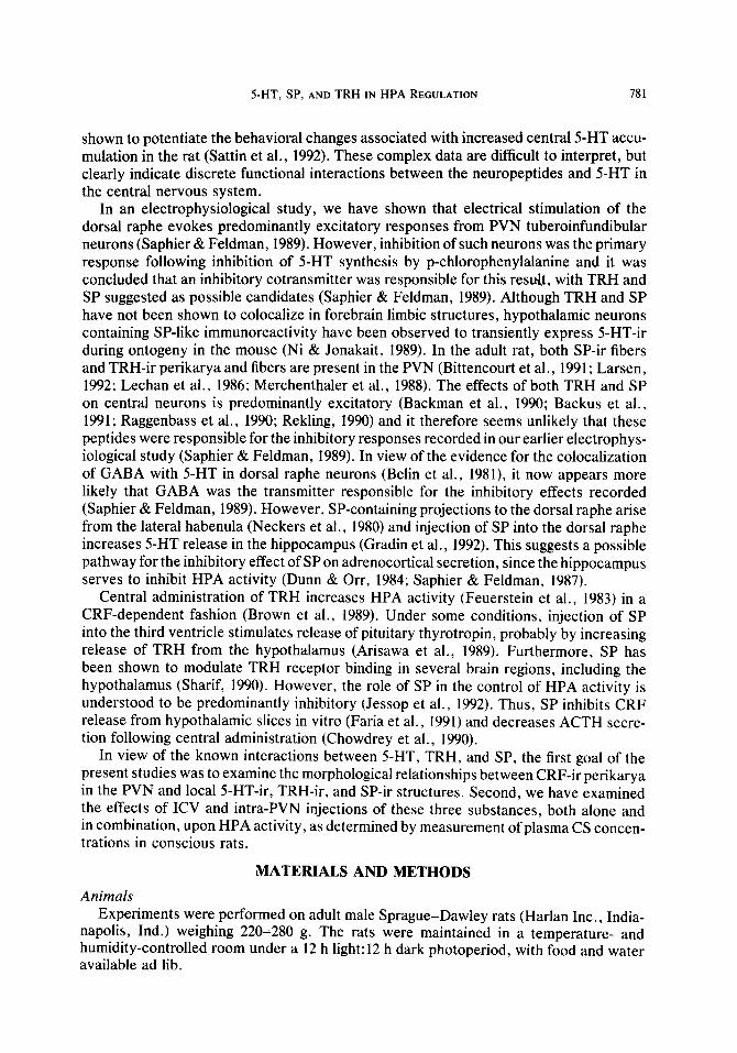
Psychoneuroendocrinology, Vol. 19, No. 8, pp. 779-797, 1994 Copyright 0 1994 Elsevier Science Ltd Printed in the USA.All rights reserved 0306-4530/94 $6.00 + .OO 0306-4530(94)EOO36-9 INTERACTIONS BETWEEN SEROTONIN, THYROTROPIN-RELEASING HORMONE, AND SUBSTANCE P IN THE CNS REGULATION OF ADRENOCORTICAL SECRETION DAVID SAPHIER,“~ JON E. WELCH,’ GLENN E. FARRAR,’ NHU Q. NGUYEN,’ FERNANDO AGUADO,~ TIMOTHY R. THALLER,’ and DAVID S. KNIGHTS Departments of ‘Pharmacology & Therapeutics, ‘Psychiatry, and 3Cellular Biology & Anatomy, Louisiana State University Medical Center, Shreveport, Louisiana 71130-3932, USA; and 4Department of Neuroanatomy, Cajal Institute, Dr. Arce #37, 28002 Madrid, Spain (Received 18 August 1993; in final form 9 February 1994j SUMMARY Double-labeling immunohistochemical studies were performed to discern the morphological rela- tionships between corticotropin-releasing factor-immunoreactive (CRF-ir) perikarya and afferent innervation in the hypothalamic paraventricular nucleus (PVN) of the rat. Attention was focussed on the local innervation by serotonin (5_hydroxytryptamine, 5-HT), thyrotropin-releasing hormone (TRH) and substance P (SP)-ir nerve terminal fibers. 5-HT-ir and SP-ir fibers were present in moderate numbers, in close apposition with CRF-ir perikarya. Sparse TRH-ir fibers were observed, but a population of TRH-ir perikarya was found in proximity with the CRF-ir cell bodies. TRH- ir perikarya in the PVN were surrounded by both S-HT- and SP-ir fibers. Neuroendocrine studies were performed to investigate the interactions between 5-HT, TRH and SP in the regulation of hypothalamo-pituitary-adrenocortical (HPA) secretion. Male rats were prepared bearing cannulae for intracerebroventricular (ICV) or intra-PVN administration of drugs. 5-HT, at all doses tested (0.1, 40, or 80 nmol, KY), caused increases in plasma corticosterone (CS) concentrations in tail- vein blood collected 20 min after injection. ICV Injections of TRH caused dose-dependent increases in plasma CS, but did not further increase HPA responses when injected together with 5-HT. SP alone had little effect, although a significant reduction in plasma CS concentrations was observed in several individual experiments. However, SP (0.1 nmol) significantly attenuated CS responses following high doses of S-HT (40 and 80 nmol, ICV), although the response to 0.1 nmol 5-HT was not affected. Combined injection of SP with TRH resulted in HPA responses not different from those following TRH alone. Similarly, SP did not reduce the HPA response observed with TRH and 40 nmol S-HT in combination. Intra-PVN injections of 5-HT (0.1 or 40 nmol) and TRH also increased plasma CS concentrations. Intra-PVN injections of SP had little effect on plasma CS concentrations although a tendency toward a decrease in plasma CS was observed, as with the ICV injections. Combined intra-PVN injection of 5-HT (0.1 nmol) with TRH (0.1 nmol) did not significantly alter the response compared with that observed following TRH alone, although plasma CS concentrations were greater than with 0.1 nmol 5-HT. Combined intra-PVN injections of SP (0.1 nmol) with 5-HT (0.1 nmol) resulted in a significant decrease in plasma CS concentration compared with that following 5-HT alone, but SP did not prevent the CS response to a higher dose Address correspondence and reprint requests to: Dr. David Saphier, Department of Pharmacology &Therapeutics, Louisiana State University Medical Center, P.O. Box 33932, 1501 Kings Highway, Shreveport, LA 71130-3932, USA; E-mail: [email protected]. 779
-
Upload
independent -
Category
Documents
-
view
2 -
download
0
Transcript of Interactions between serotonin, thyrotropin-releasing hormone, and substance P in the CNS regulation...

Psychoneuroendocrinology, Vol. 19, No. 8, pp. 779-797, 1994 Copyright 0 1994 Elsevier Science Ltd Printed in the USA. All rights reserved
0306-4530/94 $6.00 + .OO
0306-4530(94)EOO36-9
INTERACTIONS BETWEEN SEROTONIN, THYROTROPIN-RELEASING HORMONE, AND SUBSTANCE P IN THE CNS REGULATION OF
ADRENOCORTICAL SECRETION
DAVID SAPHIER,“~ JON E. WELCH,’ GLENN E. FARRAR,’ NHU Q. NGUYEN,’
FERNANDO AGUADO,~ TIMOTHY R. THALLER,’ and DAVID S. KNIGHTS
Departments of ‘Pharmacology & Therapeutics, ‘Psychiatry, and 3Cellular Biology & Anatomy, Louisiana State University Medical Center, Shreveport, Louisiana 71130-3932, USA; and
4Department of Neuroanatomy, Cajal Institute, Dr. Arce #37, 28002 Madrid, Spain
(Received 18 August 1993; in final form 9 February 1994j
SUMMARY
Double-labeling immunohistochemical studies were performed to discern the morphological rela- tionships between corticotropin-releasing factor-immunoreactive (CRF-ir) perikarya and afferent innervation in the hypothalamic paraventricular nucleus (PVN) of the rat. Attention was focussed on the local innervation by serotonin (5_hydroxytryptamine, 5-HT), thyrotropin-releasing hormone (TRH) and substance P (SP)-ir nerve terminal fibers. 5-HT-ir and SP-ir fibers were present in moderate numbers, in close apposition with CRF-ir perikarya. Sparse TRH-ir fibers were observed, but a population of TRH-ir perikarya was found in proximity with the CRF-ir cell bodies. TRH- ir perikarya in the PVN were surrounded by both S-HT- and SP-ir fibers. Neuroendocrine studies were performed to investigate the interactions between 5-HT, TRH and SP in the regulation of hypothalamo-pituitary-adrenocortical (HPA) secretion. Male rats were prepared bearing cannulae for intracerebroventricular (ICV) or intra-PVN administration of drugs. 5-HT, at all doses tested (0.1, 40, or 80 nmol, KY), caused increases in plasma corticosterone (CS) concentrations in tail- vein blood collected 20 min after injection. ICV Injections of TRH caused dose-dependent increases in plasma CS, but did not further increase HPA responses when injected together with 5-HT. SP alone had little effect, although a significant reduction in plasma CS concentrations was observed in several individual experiments. However, SP (0.1 nmol) significantly attenuated CS responses following high doses of S-HT (40 and 80 nmol, ICV), although the response to 0.1 nmol 5-HT was not affected. Combined injection of SP with TRH resulted in HPA responses not different from those following TRH alone. Similarly, SP did not reduce the HPA response observed with TRH and 40 nmol S-HT in combination. Intra-PVN injections of 5-HT (0.1 or 40 nmol) and TRH also increased plasma CS concentrations. Intra-PVN injections of SP had little effect on plasma CS concentrations although a tendency toward a decrease in plasma CS was observed, as with the ICV injections. Combined intra-PVN injection of 5-HT (0.1 nmol) with TRH (0.1 nmol) did not significantly alter the response compared with that observed following TRH alone, although plasma CS concentrations were greater than with 0.1 nmol 5-HT. Combined intra-PVN injections of SP (0.1 nmol) with 5-HT (0.1 nmol) resulted in a significant decrease in plasma CS concentration compared with that following 5-HT alone, but SP did not prevent the CS response to a higher dose
Address correspondence and reprint requests to: Dr. David Saphier, Department of Pharmacology &Therapeutics, Louisiana State University Medical Center, P.O. Box 33932, 1501 Kings Highway, Shreveport, LA 71130-3932, USA; E-mail: [email protected].
779

780 D. SAPHIER ef al.
of S-HT (40 nmol). Similar to the results observed following ICV administration, SP in combination with TRH did not affect the HPA response observed with TRH, or with S-HT and TRH in combination. The results of these studies demonstrate that 5-HT and TRH serve to stimulate adrenocortical secretion, both at the level of the PVN and at extrahypothalamic structures, but that SP exerts an inhibitory influence that is capable of overcoming some of the stimulatory effects of 5-HT.
Keywords-Serotonin; Substance P; Thyrotropin-releasing hormone; Corticotropin-releasing fac- tor; Paraventricular nucleus; Adrenocortical secretion.
INTRODUCTION
NEURONS THAT SYNTHESIZE and secrete corticotropin-releasing factor (CRF) are local- ized in the hypothalamic paraventricular nucleus (PVN) (Swanson et al., 1983). These neurons project to the median eminence (Swanson et al., 1983) to release CRF into the hypophyseal portal blood vessels, which stimulates pituitary adrenocorticotropic hormone (ACTH) secretion (Vale et al., 1983). Increased concentrations of ACTH in the peripheral circulation then stimulate corticosterone (CS) secretion from the adrenal cortex. The activity of CRF-secreting neurons is regulated by a variety of neural and humoral factors and many technical approaches have been used to determine their regula- tion in relation to hypothalamo-pituitary-adrenocortical (HPA) activity.
The PVN receives innervation from many brain structures, including the raphe nuclei (Palkovits et al., 1977; Sawchenko et al., 1983), perikarya of which are the primary source of central nervous system serotonin (5hydroxytryptamine, 5-HT). 5-HT immuno- reactive (S-HT-ir) terminals form synapses with CRF-ir perikarya in the PVN (Liposits et al., 1987) and most recent studies have concluded that 5-HT stimulates HPA activity (Bagdy et al., 1989; Feldman et al., 1991; Fuller, 1990; Saphier & Feldman, 1989; Van de Kar, 1991). However, opposing evidence suggests inhibitory effects of 5-HT on HPA activity (Kovacs et al., 1976; Saphier & Welch, 1992; Vernikos-Danellis, 1977; Welch et al., 1993). In recent studies, we have found that intracerebroventricular (ICV) adminis- tration of high doses of 5-HT (~0.1 nmol) stimulate HPA activity, while a dose lower than used in the present study (0.01 nmol) decreased plasma CS concentrations, probably through a 5-HT,.-receptor-mediated mechanism (Welch et al., 1993). Very high doses of 5-HT (220 nmol) appear to stimulate adrenocortical secretion through a central action on sympathomedullary outflow (Saphier & Welch, 1992; Welch et al., 1993). Similar results have been obtained following injections of 5-HT directly into the PVN (Welch et al., 1993).
Several studies have demonstrated the coexistence of a variety of putative cotransmit- ter substances in 5-HT neurons. Identified substances within such neurons include sub- stance P (SP) and thyrotropin-releasing hormone (TRH) (Appel et al., 1987; Johansson et al., 1981). Because of these observations, several studies have been designed to examine the interactions between 5-HT, TRH and SP in the regulation of nervous system activity (Iverfeldt et al., 1989). For example, the suppression of respiratory rate following injection of SP into the nucleus tractus solitarii was increased by 5-HT, while the increases in mean arterial pressure following 5-HT injections were not affected by combination with SP (Carter & Lightman, 1985). In contrast, in the same study, TRH injected into the nucleus of the solitary tract caused a decrease in mean arterial pressure which was reversed by coinjection of 5-HT, decreases in respiratory rate only being observed with combined injections of TRH and 5-HT (Carter & Lightman, 1985). TRH has also been
SHT,SP, AND TRH IN HPA REGULATION 781
shown to potentiate the behavioral changes associated with increased central 5-HT accu- mulation in the rat (Sattin et al., 1992). These complex data are difficult to interpret, but clearly indicate discrete functional interactions between the neuropeptides and 5-HT in the central nervous system.
In an electrophysiological study, we have shown that electrical stimulation of the dorsal raphe evokes predominantly excitatory responses from PVN tuberoinfundibular neurons (Saphier & Feldman, 1989). However, inhibition of such neurons was the primary response following inhibition of 5-HT synthesis by p-chlorophenylalanine and it was concluded that an inhibitory cotransmitter was responsible for this result, with TRH and SP suggested as possible candidates (Saphier & Feldman, 1989). Although TRH and SP have not been shown to colocalize in forebrain limbic structures, hypothalamic neurons containing SP-like immunoreactivity have been observed to transiently express 5-HT-ir during ontogeny in the mouse (Ni & Jonakait, 1989). In the adult rat, both SP-ir fibers and TRH-ir perikarya and fibers are present in the PVN (Bittencourt et al., 1991; Larsen, 1992; Lechan et al., 1986; Merchenthaler et al., 1988). The effects of both TRH and SP on central neurons is predominantly excitatory (Backman et al., 1990; Backus et al., 1991; Raggenbass et al., 1990; Rekling, 1990) and it therefore seems unlikely that these peptides were responsible for the inhibitory responses recorded in our earlier electrophys- iological study (Saphier & Feldman, 1989). In view of the evidence for the colocalization of GABA with 5-HT in dorsal raphe neurons (Belin et al., 1981), it now appears more likely that GABA was the transmitter responsible for the inhibitory effects recorded (Saphier & Feldman, 1989). However, SP-containing projections to the dorsal raphe arise from the lateral habenula (Neckers et al., 1980) and injection of SP into the dorsal raphe increases 5-HT release in the hippocampus (Gradin et al., 1992). This suggests a possible pathway for the inhibitory effect of SP on adrenocortical secretion, since the hippocampus serves to inhibit HPA activity (Dunn & Orr, 1984; Saphier & Feldman, 1987).
Central administration of TRH increases HPA activity (Feuerstein et al., 1983) in a CRF-dependent fashion (Brown et al., 1989). Under some conditions, injection of SP into the third ventricle stimulates release of pituitary thyrotropin, probably by increasing release of TRH from the hypothalamus (Arisawa et al., 1989). Furthermore, SP has been shown to modulate TRH receptor binding in several brain regions, including the hypothalamus (Sharif, 1990). However, the role of SP in the control of HPA activity is understood to be predominantly inhibitory (Jessop et al., 1992). Thus, SP inhibits CRF release from hypothalamic slices in vitro (Faria et al., 1991) and decreases ACTH secre- tion following central administration (Chowdrey et al., 1990).
In view of the known interactions between 5-HT, TRH, and SP, the first goal of the present studies was to examine the morphological relationships between CRF-ir perikarya in the PVN and local 5-HT-ir, TRH-ir, and SP-ir structures. Second, we have examined the effects of ICV and intra-PVN injections of these three substances, both alone and in combination, upon HPA activity, as determined by measurement of plasma CS concen- trations in conscious rats.
MATERIALS AND METHODS
Animals Experiments were performed on adult male Sprague-Dawley rats (Harlan Inc., India-
napolis, Ind.) weighing 220-280 g. The rats were maintained in a temperature- and humidity-controlled room under a 12 h light: 12 h dark photoperiod, with food and water available ad lib.

782 D. SAPHIER et al.
Immunohistochemical Studies Adult male rats weighing 220-240 g were used for the double-labeling immunohisto-
chemical studies. The animals were treated with colchicine (50 pug, ICV) administered under sodium pentobarbital anesthesia 48 h before sacrifice. After induction of anesthesia with sodium pentobarbital(65 mg/kg), the animals were perfused transcardially with 200 ml of Sorensen’s buffer containing 0.5% procaine + 2000 USP Units of heparin. This was followed by a 10 min perfusion with 500 ml of 2-4% paraformaldehyde and 0.2% glutaraldehyde in ice-cold Sorensen’s buffer. The brains were then carefully removed from the skull and placed in the fixative overnight in a refrigerator before transfer to buffer. Brains were then cryoprotected the following day, using buffer containing 20% and then 30% sucrose, before the diencephalic region of each brain to be labeled was frozen at -55°C and sectioned in a cryostat at 40 pm.
Peroxidase Labeling Peroxidase labeling of 5-HT, SP, CRF, and TRH was used to visualize perikarya,
dendrites and axon terminals of different nerve types. Particular attention was focussed on 5-HT-ir, SP-ir, and TRH-ir nerve processes, and TRH-ir and CRF-ir soma in the PVN. Sections were processed, free-floating, for immunolabeling using the Avidin-Bio- tin-peroxidase Complex (ABC) method (Hsu et al., 1981). Sections were placed in sodium borohydride (0.2 mg/ml) in Sorensen’s buffer for 20 min, to reduce free aldehyde groups, and rinsed three times with the buffer. The sections were presoaked in Sorensen’s buffer containing 0.3% Triton X-100, 2% bovine serum albumen (BSA) and 2% normal goat serum (NGS) for 1 h. Tissue sections were then incubated overnight at room temperature in the primary antiserum at appropriate concentrations (CRF, Incstar. 20084 at 1: 1600; TRH, Arnel 1743 at 1:lOOO; 5-HT, Incstar, 20080 at 1:4000; SP, Amersham, RAN 1572 at 1: 1200). The diluted antisera contained 0.3% Triton X-100, 1% NGS, and 0.1% sodium azide. After incubation, sections were washed three times with Sorensen’s buffer before exposure to biotinylated goat anti-rabbit IgG for 1 h. This was followed by a further three washes in Sorensen’s buffer before processing with the ABC reagent (Vectastain Kit, Vector Labs., Burlingame, CA) for 1 h. The reactive product was visualized using exposure to 0.05% diaminobenzidine (DAB) in 0.1 M Tris buffer for 10 min. Control sections were incubated for the same time in normal (nonimmune) rabbit serum.
Fluorescent Dye Labeling To visualize simultaneously two antigens in the same tissue section, the first antigen
(CRF or TRH) was labeled with specific antiserum and visualized with horseradish peroxidase-DAB chromogen by the ABC method described above. The second antigen (S-HT, SP, or TRH) was labeled with specific antiserum and visualized with fluorescein isothiocyanate (FITC) (Lechago et al., 1979). After visualization of the horseradish peroxidase (HRP)/DAB reaction product, sections were rinsed three times in Sorensen’s buffer, presoaked for 1 h, and incubated with the second primary antiserum overnight at room temperature. Biotinylated goat anti-rabbit IgG followed by FITC-labeled avidin were then applied to visualize the second antigen. After rinsing in buffer, sections were mounted with a solution of equal parts Sorensen’s buffer and glycerin and examined using a Leitz fluorescence microscope (filter block H containing KP 490 excitation filter, TK 510 dichroic beam splitting mirror, K 515 suppression filter).
Labeling with HRP-DAB chromogen in the PAP method results in a clumped reaction product that can prevent subsequent labeling of a second antigen located in the peroxi-

SHT,SP, AND TRH IN HPA REGULATION 783
dase-labeled element (Sternberger &Joseph, 1979). Previous studies (Knight et al., 1989) have indicated that peroxidase/DAB chromogen labeling of neuronal peptides prevents subsequent FITC labeling of a second antigen in the same neuron. Therefore, visualization of two neuronal antigens in the same tissue section indicates that the two occur in different neurons. Some pairs of antisera were applied in the order stated and in reverse order to identify the possible false negative results that can occur whenever the two antigens are present in the same neuron. Four possible unwanted interactions that may occur with these staining sequences are: (1) binding of the second link antibody with the first primary antibody; (2) binding of the second link antibody with bound ABC reagent; (3) binding of avidin-FITC with the first link antibody; and (4) binding of the second primary antibody with the first link antibody. To test for the first three interactions, avidin-FITC or biotinylated goat anti-rabbit IgG followed by avidin-FITC was applied to control sections in an attempt to label with FITC, CRF-ir neurons previously labeled with peroxidase. Possible binding of the second primary antibody with the first link antibody was evaluated by comparing tissue sections in which CRF-ir neurons were labeled with peroxidase and each of the other peptides was visualized with FITC.
Preparation for Neuroendocrine Studies Animals were anesthetized with sodium pentobarbital (65 mg/kg; IP) and a 22-gauge
stainless steel guide cannula was stereotaxically implanted to allow access to the lateral ventricle. Stereotaxic coordinates, taking bregma as the zero reference point, were: A -0.5; L 21.6; and D -3.6 (Pellegrino et al., 1967). Separate groups of animals were bilaterally implanted with chronic guide cannulae above the dorsal border of each PVN, using the following stereotaxic coordinates: A 0.2; L 0.3; H -7.5 (Pellegrino et al., 1967). Following surgery, animals were given prophylactic intramuscular injections of dihydrostreptomycin sesquisulfate (200 mg/kg; Sigma Chemical Co., St. Louis, MO) and potassium penicillin-G (125 mg/kg; Sigma Chemical Co., St. Louis, MO).
Experimental Protocols Following the surgery, animals were allowed to recover for 7 days before institution
of the experimental procedures. The animals were handled, with removal of the stylets, on the day preceding the first experiment. All experiments were performed between 0800-lOOOh, at the nadir in circadian HPA activity. For drug administration, the stylets were removed from the guide cannulae and replaced by 28-gauge injection needles attached to Teflon tubing and supplied by 10 ~1 (PVN) or 50 ~1 (LV) Hamilton syringes. For ICV injections, the inner cannula was designed to penetrate directly into the lateral ventricle, while for intra-PVN injections the injection needle extended to the tip of the outer guide cannulae.
The experiment consisted of injections of the test agent in a volume of 10 ~1 into the LV, or 1 ~1 into each PVN. Drugs administered were as follows: 5-HT HCl (0.1, 40, and 80 nmoles; Research Biochemicals Inc., Natick, MA); SP methyl ester (0.001, 0.01, and 0.1 nmoles; Cambridge Research Biochemicals Ltd., UK); TRH (0.001, 0.01, and 0.1 nmoles; Cambridge Research Biochemicals Ltd., UK). For intra-PVN injections, only 0. I- and 40-nmol doses of 5-HT were used. The drugs were administered alone or as isotonic admixtures. All drugs were dissolved immediately before injection in 0.9% saline, which was the vehicle was used for control animals. Animals were used for tests more than once, with individual tests being separated by at least 5 days. Twenty minutes following drug injection, 100 ~1 of tail blood was collected into heparinized tubes.

784 D. SAPHIER et al.
Corticosterone Radioimmunoassay Following sampling, blood was centrifuged and plasma was separated and stored
frozen at -20°C for subsequent assay of plasma CS concentrations. CS concentration was determined by specific radioimmunoassay (Gwosdow-Cohen et al., 1982). Cross- reactivity with other steroids is less than I%, except deoxycorticosterone and ll-/3- hydroxyprogesterone (7%), and the interassay and intraassay coefficients of variation were calculated to be less than 5%. [3H]-CS was purchased from New England Nuclear (Boston, MA), and antiCS antibody was obtained from Dr. G. Niswender (Colorado State University). Steroids were extracted from both plasma and standards using methylene chloride.
Histology and Statistical Analysis After termination of the final experiment, animals were deeply anesthetized with
sodium pentobarbital (80 mg/kg) and 1 ,ul of toluidine-blue dye was injected via the cannula. Ten minutes later, the animals were perfused through the left atrium with a 10% formalin-saline solution to ascertain the correct placement of the cannulae. This was achieved by examining sections cut through the PVN, or by observing the spread of dye through the ventricular system for those animals bearing LV cannulae. Data obtained from animals showing incorrect cannula placements, or leakage of the dye into the ventricular system from the PVN cannulae, were excluded from subsequent analyses. Student’s t-test was used for initial statistical examination of the data. Analysis of variance (ANOVA) was used, followed by post hoc testing using Dunnett’s t-test, for multiple comparisons with one control group.
RESULTS
Immunohistochemistry Most CRF-ir PVN neurons were located in the dorsal and medial parvicellular subdivi-
sions of the nucleus (Fig. l), whereas axon terminals were concentrated in the median eminence, Fewer such neurons were located in the periventricular and magnocellular parts of the PVN. Other CRF-ir neurons were either clustered or widely dispersed in the preoptic area, anterior hypothalamic nucleus, lateral hypothalamic area, and amyg- dala. CRF-ir neurons in the PVN were more densely organized and intensely labeled than those in other areas.
TRH-ir neurons were located in both the periventricular and in the dorsal and medial parvicellular subdivisions of the PVN, overlapping those areas containing CRF-ir cell bodies (Figs. lA, 2). There were groups of TRH-ir neuronal cell bodies in the preoptic area, dorsomedial hypothalamic nucleus and in ventral and lateral parts of the lateral hypothalamic area as well. Such neurons in periventricular and medial parvicellular subdivisions of the PVN were more intensely labeled than those in the other areas. FITC- labeled, TRH-ir cell bodies with several processes lie near several CRF-ir neurons labeled with the HRP/DAB reaction product (Fig. 1A). TRH-ir dendrites and nerve terminals were found in the neuropil surrounding CRF-ir and other neuronal cell bodies. There were moderate to dense TRH-ir terminal plexi in other areas containing TRH-ir cell bodies, and terminals, but no cell bodies, were seen in the ventromedial and arcuate hypothalamic nuclei.
There were no 5-HT-ir neuronal cell bodies in the hypothalamus or amygdala, but a uniformly dense plexus of varicose 5-HT-ir axons innervated most nuclei of these two

SHT,SP, AND TRH IN HPA REGULATION 785
(A)
FIG. 1: Light photomicrographs showing (A) peroxidase-labeled CRF-ir cell bodies in the PVN and associated FITC-labeled TRH-ir cell bodies, fibers and terminals; (B) peroxidase-labeled CRF- ir cell bodies in the hypothalamic PVN (dark staining) and associated FITC-labeled 5-HT-ir fibers and terminals; and (C) peroxidase-labeled CRF-ir cell bodies in the PVN and associated FITC- labeled SP-ir fibers and terminals. Note the presence of an SP-ir fiber apparently wrapped around a CRF-ir dendrite in the center of the field of view. Peroxidase-labeled structures appear in black, and FITC-labeled structures fluoresce yellow but appear in white on the photomicrographs.

786 D. SAPHIER et al.
(A)
FIG. 2: Light photomicrographs showing (A) peroxidase-labeled TRH-ir cell bodies in the hypothala- mic PVN (dark staining) and associated FITC-labeled 5-HT-ir fibers and terminals; and (B) FITC- labeled TRH-ir cell bodies in the PVN and associated peroxidase-labeled SP-ir fibers and terminals.
nuclear complexes. Terminal density was light to moderate in the PVN, media1 preoptic area, ventral retrochiasmatic area, ventromedial nucleus, arcuate nucleus, ventral part of the lateral hypothalamic area, and median eminence compared with the greater density in most other hypothalamic areas. Figure 1B shows several peroxidase-labeled, CRF-ir neuronal cell bodies surrounded by a uniform plexus of FITC-labeled 5-HT-ir nerve terminals, some of which are closely applied to CRF-ir cell bodies or dendrites. TRH- ir cell bodies were also found closely associated with 5-HT-ir terminals (Fig. 2A). Al- though an extensive S-HT-ir plexus was found in the PVN, 5-HT terminal density was greater in the hypothalamic nuclei immediately surrounding the PVN.
SP-ir nerve terminals were found in most hypothalamic nuclei, with light to moderate terminal density present in several subnuclei of the PVN. Figure 1C shows peroxidase- labeled CRF-ir neurons in the lateral portion of the media1 parvicellular subdivision surrounded by FITC-labeled, SP-ir nerve terminals. TRH-ir perikarya in the PVN were also surrounded by SP-ir nerve terminals (Fig. 2B). However, similar to the 5-HT-ir fibers, SP-ir terminal density was greater in the zone dorsal to the PVN. Indeed, terminal density was greater in areas of the hypothalamus other than PVN, including those hypothalamic nuclei immediately adjacent to the PVN. Terminal density was light in the ventromedial nucleus which, nevertheless, contained many SP-ir cell bodies. Terminal density in some nuclei of the amygdala exceeded that in any other area of the brain. In addition to those in the ventromedial nucleus, there were SP-ir neuronal cell bodies in the preoptic area, dorsomedial nucleus, ventral and lateral parts of the lateral hypothala- mic area, and amygdala.
There was no labeling of any neuronal cell bodies or axon terminals in control sections

SHT, SP, AND TRH IN HPA REGULATION 787
350
300
< 250
2 , 200
z % 150
2 100
50
0 5-HT
TRH
II -I-
32
0
0
l! 0.1
0
;
14 -
::
: 26
40
0
5
12
so
0
*
_Y-
29
0 14 - 0.1
0.1 0.1
SC *
I
9 - 40
0.1
* *
1
6
so
0.1
DRUG DOSES - NMOLES
FIG. 3: Effect of ICV injections of S-HT and TRH on plasma CS concentrations in conscious rats. The number of animals per group is shown at the base of each column. ANOVA indicated significant main effects of S-HT, F(3, 103) = 36.18, p < .OOOl, and of TRH, F(1, 82) = 172.39, p < .OOOl, and a significant interaction between treatments, F(7, 156), = 27.69, p < .OOOl. Levels of significance were determined by Student’s r-test with all changes significant by Duncan’s test: *p < .05, **p < .005, compared with saline-injected control animals.
incubated with nonimmune rabbit serum then link antiserum, and carried through the staining protocol described in the methods section. Control sections were treated with avidin-FITC or biotinylated goat anti-rabbit IgG followed by avidin-FITC in an attempt to label with FITC, neurons previously labeled with peroxidase. There was no evidence that any such unwanted binding occurred, as none of the peroxidase-labeled neurons had any bound fluorophore following these procedures. Striking differences in the distri- bution of fluorescence in CRF-labeled sections secondarily incubated with 5-HT, SP, and TRH suggests that there is little if any binding of the second primary antibody with the first link antibody.
Effects of Intracerebroventricular Injections ICV injections of saline caused an elevation of basal plasma CS concentrations to
60-75 ng/ml, a figure approximately twice that normally found in unhandled, uninjected animals; this modest elevation demonstrates the slightly stressful nature of the experimen- tal procedure. ICV injections in conscious rats of all doses of 5-HT tested caused dose- dependent increases in plasma CS concentrations in tail vein blood collected 20 min after injection (Figs. 3-5). The responses following 40 and 80 nmol 5-HT were greater than those following 0.1 nmol. ICV injections of TRH, at doses of 0.001, 0.01, and 0.1 nmol, also caused dose-dependent increases in plasma CS (Fig. 6A). TRH (0.1 nmol) did not alter the HPA response when administered with any dose of S-HT (Fig. 3). SP alone had no significant effect on HPA activity, although a significant decrease in plasma CS concentration was observed in some individual experiments. However, SP (0.1 nmol

788 D. SAPHIER et al.
350
300 m 5 250
? , 200
J_
14
* *
l 26
40
0
c 4
150
$ 100
50
0 5-HT
SUB P
-I-
37
-F
0
Ll 0.1
0
12
80 0 0.1 40 80
0 0.1 0.1 0.1 0.1
*
-L
7
# #
DRUG DOSES - NMOLES
FIG. 4: Effect of ICV injections of 5-HT and SP on plasma CS concentrations in conscious rats. The number of animals per group is shown at the base of each column. ANOVA indicated a significant main effect of 5-HT, F(3, 103) = 36.18, p < .OOOl, not of SP, F(1,77) = 0.20, p < .6529, and a significant interaction between treatments, F(7,141), = 30.88,~ < .OOOl. Levels of significance were determined by Student’s t-test with all changes significant by Duncan’s test: *p < .05, **p < .005, compared with saline-injected control animals. #p < .05, ##p < .005, compared with animals treated with 5-HT alone at the same doses.
ICV) significantly attenuated the CS responses following 40 and 80 nmol5-HT, although the response to 5-HT at 0.1 nmol was not affected (Fig. 4). Combined injections of SP (0.1 nmol) with TRH (0.1 nmol) resulted in HPA responses not different from those following TRH alone (Fig. 5). Similarly, SP (0.1 nmol) in combination with TRH (0.1 nmol) and S-HT (40 nmol) did not reduce the adrenocortical response observed with 5- HT and TRH in combination (Fig. 5).
Effects of Intra-PVN Injections The results of the studies employing intra-PVN injections of 5-HT, TRH, and SP,
were similar to those obtained following ICV injections and are summarized in Figs. 6B, 7, and 8. Plasma CS concentrations following intra-PVN injection of saline were some- what higher than those observed following ICV injection, presumably due to the some- what more stressful nature of the injection procedure. Intra-PVN injections of 0.1 nmol 5-HT or of TRH (0.1 nmol) increased plasma CS concentrations (Fig. 7). Injections of TRH, at doses of 0.001, 0.01, and 0.1 nmol caused dose-dependent increases in plasma CS, although the increase following the lowest dose was not significant (Fig. 6B). Intra- PVN injections of SP, once again, had little effect on plasma CS although a statistically nonsignificant decrease in plasma CS levels was observed (Fig. 7). Although the HPA response to combined injection of TRH (0.1 nmol) and 5-HT (0.1 nmol) was greater than that following the same dose of 5-HT alone, the response was not greater than that following TRH alone, because the CS response recorded was near-maximal (Fig. 7).

SHT, SP, AND TRH IN HPA REGULATION 789
350
300 1 < 250-
s , zoo-.
v, o 150- 9
2 loo- n
50
D 55 0
5-HT 0
SUB P 0
TRH 0
40 0
0 0.1
0 0 0.1 0
0 40 40
0 0.1 0
0.1
DRUG DOSES - NMOLES
* *
l-
6 - 0
0.1
0.1
* *
L 8
40
0.1
I 0.1
FIG. 5: Effect of ICV injections of S-HT, TRH, and SP on plasma CS concentrations in conscious rats. The number of animals per group is shown at the base of each column. ANOVA indicated significant main effects of S-HT, F(1, 79) = 68.30, p < .OOOl, and of TRH, F(1, 82) = 172.39, p < .OOOl, but not of SP, F(1, 77) = 0.20, p < .6529, with a significant interaction between S-HT and SP, F(2, 53) = 20.07, p < .OOOl, and a significant interaction between the three treatments, F(7, 155), = 30.88, p < .OOOl. Levels of significance were determined by Student’s f-test with all changes significant by Duncan’s test: *p < .05, **p < .005, compared with saline-injected control animals. #p < .05, compared with animals treated with 5-HT alone at the same dose.
Combined injections of S-HT (0.1 nmol) with SP (0.1 nmol) resulted in a significant decrease in plasma CS concentration compared with that following 5-HT alone (Fig. 7). Surprisingly, the plasma CS concentration following combined injection of SP and 5-HT was significantly less than that following saline. Similar to the results observed following ICV administration, SP (0.1 nmol) did not affect the adrenocortical response observed with TRH (0.1 nmol), or with 5-HT (0.1 nmol) and TRH (0.1 nmol) in combination (Fig. 7).
Intra-PVN injection of 40 nmol 5-HT also caused significant increases in plasma CS concentrations but no potentiation of the response was observed with coinjection of TRH (Fig. 8). In addition, SP (0.1 nmol) was not able to reduce significantly the HPA response to this dose of 5-HT (Fig. 8).
DISCUSSION
The present studies show that 5-HT-ir axon terminals are present in close apposition to the soma and dendrites of CRF-ir neurons in the dorsal and medial parvicellular subnuclei of the PVN. This result is similar to that previously reported using electron microscopy, when 5-HT terminals were shown to synapse with CRF-secreting neurons (Liposits et al., 1987). It is interesting that 5-HT terminal density appeared somewhat greater in the regions immediately surrounding the PVN. This suggests that some of the actions of 5-HT on PVN neurosecretory neurons, including those secreting CRF, may

z 250
4 , 200
D. SAPHIER et al.
OJ 4
0 0.00 1 0.01 0.1
DOSE OF TRH - NMOLES
FIG. 6: Dose-response curve showing the effect of ICV injections (A) and intra-PVN injections (B) of TRH on plasma CS concentrations in conscious rats. Each point represents the mean from seven to eight animals per group. **p < 305, compared with saline-injected control animals, determined using Dunnett’s test.
be mediated via interneurons surrounding the nucleus, or that a slow diffusion of neuro- transmitter may be responsible for altering such neurosecretory activity. Indirect evi- dence for such slow synaptic events arises from electrophysiological studies of noradren- ergic projections to the PVN (Saphier & Feldman, 1991). Such effects, termed volume transmission, are believed to exist throughout the central nervous system (Fuxe & Agnati, 1991) and are believed to be important in the temporal organization of neural events; however, they are probably of little relevance to the neuroendocrine studies reported here.

5-HT.SP. AND TRH IN HPA REGULATION 791
350
300
< 250 c3 Z , 200
0" 4 150
$ 100
50
0 5-HT
TRH
SP
,_.
- + -
*
;
11 12 6
# *
cl- 6
- +
+ - -
DRUGS GIVEN (0.1 NMdS)
* *
1
5 - +
+
-It- *
1
7 -
+
+
* *
1
7 - +
+
+
FIG. 7: Effect of intra-PVN injections of 0.1 nmol 5-HT in combinations with TRH and SP on plasma CS concentrations in conscious rats. The number of animals per group is shown at the base of each column. ANOVA indicated significant main effects of S-HT, F(1, 80) = 4.72, p < .0329, and of TRH, F(1, 75) = 29.96, p < .OOOl, but not of SP, F( 1, 99) = 0.65, p < .4220, with a significant interaction between 5-HT and TRH, F(2, 27) = 4.43, p < .0217, and a significant interaction between the three treatments, F(7, 173), = 10.94, p < .OOOl. Levels of significance were determined by Student’s r-test with all changes significant by Duncan’s test: *p < .05, **p < .005, compared with saline-injected control animals. #p < .005, compared with animals treated with 5-HT alone at the same dose.
Although our immunohistochemical studies demonstrated a close apposition of 5-HT, SP, and TRH terminals with CRF-secreting perikarya, it is important to note that the PVN also contains vasopressin-secreting neurons (Sawchenko et al., 1983), and that vasopressin acts as a potent ACTH secretagogue (Tilders et al., 1985). Thus, it is entirely possible that some of the HPA regulatory effects of the drugs injected in this study were mediated by both CRF and/or vasopressin. Thus, electrophysiological studies have shown that vasopressin-secreting neurons in the PVN are inhibited by electrical stimulation of the dorsal raphe (Saphier, 1991), whereas putative CRF-secreting neurons are excited by such stimulation (Saphier & Feldman, 1989). Chowdrey et al. (1990) have shown that SP stimulates arginine vasopressin and inhibits adrenocorticotropin release in vivo in the rat. These opposing data concerning CRF and vasopressin indicate that the results of the present study cannot be interpreted as demonstrating that CRF is responsible for the changes in adrenocortical secretion that were observed. Any effects of intra-PVN injections on vasopressin, or CRF, neurons could have been mediated by a diffusion of the injected drugs to immediately adjacent structures. However, this possibility does not detract from the physiological validity of the data obtained, since the immunohisto- chemical studies revealed the likely presence of terminal release sites around, as well as within, the nucleus.
5-HT is believed to serve a predominantly facilitatory role in the central regulation of adrenocortical secretion (Bagdy et al., 1989; Feldman et al., 1991; Fuller, 1990; Saphier

792 D. SAPHIER et al.
350 -
300 -
< 250-
$ , zoo-
* *
Is s
150
$ 100
50
kl 67 0
+ *
I 21 II 34 _ 10
*
1 21
5-HT 0 40 0 0 40
SUB P 0 0 0.1 0 0.1
TRH 0 0 0 0.1 0
*
;
21 - 40
0
0.1
19 - 0
0.1
0.1
*
;
20
40
0.1
0.1
DRUG DOSES - NMOLES
FIG. 8: Effect of intra-PVN injections of 40 nmol 5-HT in combinations with TRH and SP on plasma CS concentrations in conscious rats. The number of animals per group is shown at the base of each column. ANOVA indicated significant main effects of 5-HT, F(1, 86) = 28.58, p < .OOOl, and of TRH, F(1, 75) = 29.96, p < .OOl, but not of SP, F( 1, 99) = 0.65, p < .4220, with a significant interaction between 5-HT and SP, F(2,72) = 6.18, p < .0033, and a significant interaction between the three treatments, F(7,204), = 10.97, p < .OOl. Levels of significance were determined by Student’s f-test with all changes significant by Duncan’s test: *p < .05, **p < .005, compared with saline-injected control animals.
& Feldman, 1989; Van de Kar, 1991). However, compelling evidence also suggests an inhibitory influence of 5-HT on HPA activity (Kovacs et al., 1976; Saphier & Welch,
1992; Vernikos-Danellis, 1977; Welch et al., 1993). The inhibitory effects occur with
injections of lesser amounts of 5-HT (co.1 nmol, ICV) than used in the present study (Welch et al., 1993). Injection of the serotonergic neurotoxin, 5,7_dihydroxytryptamine (5,7-DHT) directly into the PVN depletes hypothalamic 5-HT content and reduces adreno- cortical responses following a variety of stimuli (Feldman et al., 1991). Recovery of HPA responses following 5,7-DHT treatment occurs over time and is proportional to the amount of PVN 5-HT reinnervation (Feldman et al., 1991). These effects are presumably due to the direct destruction of afferent 5-HT fibers arising from the raphe nuclei (Palkovits et al., 1977; Sawchenko et al., 1983). However, it is interesting that such 5,7-DHT treatment causes an elevation of basal plasma CS concentrations (Feldman et al., 1984), suggesting a tonic inhibitory role of 5-HT in the regulation of HPA activity. This observa- tion supports recent neuroendocrine and electrophysiological evidence from this labora- tory indicating that 5-HT can inhibit HPA activity and PVN neuronal activity via inhibi- tory 5-HT,, receptors (Saphier & Zhang, 1993; Welch et al., 1993).
In agreement with neuroendocrine observations, electrophysiological studies have shown that electrical stimulation of the dorsal raphe excites PVN tuberoinfundibular neurons antidromically identified as projecting to the median eminence (Saphier & Feld- man, 1989). Inhibition of 5-HT synthesis withp-chlorophenylalanine increased the sponta- neous activity of such PVN neurons but decreased the proportion of excitatory responses

S-HT, SP, AND TRH IN HPA REGULATION 793
recorded, suggesting both an inhibitory and a facilitatory effect of 5-HT mediated at the level of the PVN (Saphier & Feldman, 1989). 5-HT,c and 5-HT, receptors mediate excitatory neuronal responses (Aghajanian et al., 1990) and it is possible that facilitator-y PVN neuronal responses following dorsal raphe stimulation were due to activation of these receptor subtypes. In support of this suggestion, 5-HTrc and 5-HT, receptors, and 5-HT,, receptor mRNA, have been demonstrated in the PVN (Hoffman 6~ Mezey, 1989; Palacios et al., 1990) and 5-HTrc12 receptor agonists have been shown to stimulate HPA activity (Bagdy et al., 1989). In contrast, recent evidence indicates that 5-HT,, agonists inhibit PVN neuronal activity (Saphier & Zhang, 1993).
The data obtained in the present study demonstrate that 5-HT (at the doses used) and TRH were both able to stimulate HPA activity, and that combinations of TRH with the low dose of 5-HT appeared to exert an additive effect. The apparent lack of additive effects when TRH was injected with the high doses of 5-HT was probably because maximal CS secretion had been reached, although ACTH concentrations may have continued to rise. These results are consistent with our previous observations following ICV and intra-PVN injections of 5-HT (Welch et al., 1993) and those from similar studies with central injections of TRH (Brown et al., 1989). It is interesting that similar adrenocor- tical responses to 5-HT and TRH were observed following ICV injections of the same amounts as used for the intra-PVN injections. This suggests that the principal site(s) of action of these drugs, and possibly also SP, in stimulating adrenocortical secretion resides in extrahypothalamic structures. Such a suggestion indicates that a global cerebral re- sponse to altered neurotransmission is required to elicit adrenocortical responses, rather than a highly anatomically specific one.
In contrast with the responses following 5-HT and TRH, injections of SP exerted an opposing inhibitory effect on HPA activity, consistent with the observations of others (Chowdrey et al., 1990; Fariaet al., 1991; Jessop et al., 1992). Furthermore, SPprevented the adrenocortical stimulating effects of high doses of 5-HT, but was not able to prevent either the HPA stimulation by low-doses of 5-HT (ICV), or the stimulation by TRH. Thus, low doses of 5-HT may stimulate HPA activity by acting at a depolarizing receptor subtype, such as 5-HT,c/5-HT, receptors (Bagdy et al., 1989; Welch et al., 1993; Aghajan- ian et al., 1990), but higher doses of 5-HT may act, via hyperpolarizing 5-HT,, receptors (Aghajanian et al., 1990), to overcome low-dose stimulation and inhibit HPA secretory activity (Welch et al., 1993). Higher doses still may stimulate HPA activity via an indirect sympathetic stimulation (Saphier & Welch, 1992; Welch et al., 1993) or via low affinity 5-HT, receptors (Saphier & Welch, unpublished observations), which are known to rapidly desensitize (Aghajanian et al., 1990). The inhibitory effects of SP may involve an inactivation of 5-HT/5-HT, receptor interactions, thus permitting the 5-HT,,-mediated inhibitory effect to be expressed. Similar interactions between SP (receptors) and other neurotransmitter receptors have been shown, such that SP blocks the nicotinic effects of acetylcholine on Renshaw cells (Ryall & Belcher, 1977). Since SP-ir terminals, like 5-HT terminals, were found in greater density immediately surrounding the PVN, the possibility that such 5-HT/SP receptor interactions occur at the level of interneurons outside the PVN cannot be excluded, although dendrites of some PVN neurons also penetrate into the zone just outside the PVN.
The adrenocortical stimulating effects of TRH were apparently independent of 5-HT receptors, since a tendency to an additive effect on plasma CS concentrations was observed following coadministration of 0.1 nmol TRH with 0.1 nmol 5-HT into the PVN. A similar effect has been observed for 5-HT and TRH in the central regulation of

794 D. SAPHIER ef al.
respiratory rate (Carter & Lightman, 1985) and behavior (Greene & Grahame-Smith, 1974). Although most CRF-ir and TRH-ir neurons were found to lie in discrete PVN subnuclei, an intervening zone in which the two groups of cells overlapped was found. In addition, TRH-ir dendrites or axon terminals were present in the neuropil surrounding CRF-ir cell bodies in the PVN. These observations suggest that effects of TRH on CRF neurons, and HPA activity, may be physiologically relevant. A moderate density of SP- ir terminals was found in association with both CRF-ir and TRH-ir perikarya, suggesting direct actions of SP on the secretion of these peptides. Under some conditions, SP has been shown to stimulate pituitary thyrotropin secretion by increasing TRH release (Arisawa et al., 1989). It is unlikely that such an action was present in our experimental paradigm, since increased TRH release in the PVN would itself be expected to increase HPA activity (Brown et al., 1989). Furthermore, SP did not prevent the effects of exoge- nously coinjected TRH in the present study. S-HT-ir axon terminals surrounded TRH- ir neurons in the periventricular region of the PVN, as shown in other studies (Kiss & Hal&z, 1990). However, the effects of S-HT on TRH secretion are believed to be predominantly inhibitory and at least partly mediated at the level of the PVN (Toivonen et al., 1990). Thus, an indirect action of S-HT on CRF-secreting neurons mediated by TRH is unlikely, despite the 5-HT-ir fibers observed in proximity with TRH-ir perikarya. Surprisingly, the adrenocortical response to the intra-PVN high dose of 5-HT (40 nmol) was not increased by coadministration of TRH. The reason for this is not clear but it may indicate that adrenocortical responses are at least partly independent of PVN neurosecretory mechanisms, possibly requiring sympathetic activation (Saphier & Welch, 1992).
It is interesting that intra-PVN injection of SP, together with the high dose of 5-HT, did not prevent the CS response, probably due to the lack of 5-HT, receptors in the PVN (Palacios et al., 1990). This result suggests that the inhibitory effect of SP on adrenocortical responses to high-doses of 5-HT administered ICV are mediated at an extrahypothalamic site. One possible site is the cerebral cortex, where a high density of 5-HT, receptors exists (Palacios et al., 1990). In addition, the stimulatory effects of TRH were more apparent following ICV administration, as evidenced by the steeper dose-response curve, than that obtained with intra-PVN injections. The physiological significance of such interactions remains unclear at this time. However, we suggest that the three transmitter substances examined interact to permit, potentiate, or prevent HPA responses following different stressors of psychogenic, environmental, or somatic origin.
Acknowledgments: This research was supported by agrant from the U.S. National Institutes of Health (Biomedi- cal Research Support Grant #2-S07-RR05822-11, D.S.), by a Junior Investigator Award from the American Cancer Society (#00171, D.S.), by funds from the Center of Excellence in Cancer Research and Treatment, LSU Medical Center, Shreveport, and by funding from the Louisiana Board of Regents through the Louisiana Education Quality Support Fund [#LEQSF(1993-96)RD-A17, D.S. 62 D.S.K.]. We would also like to thank Dr. G. Niswender of Colorado State University for supplying the anti-CS antibody.
REFERENCES
Aghajanian GK, Sprouse JS, Sheldon P, Rasmussen K (1990) Electrophysiology of the central serotonin system: Receptor subtypes and transducer mechanisms. Ann NY Acad Sci 600:93-103.
Appel NM, WessendorfMW, Elde RP (1987) Thyrotropin-releasing hormone in spinal cord: Coexis- tence with serotonin and with substance P in fibers and terminals apposing identified pregangli- onic sympathetic neurons. Brain Res 41.5:137-143.

SHT,SP, AND TRH IN HPA REGULATION 195
Arisawa M, Snyder GD, McCann SM (1989) Effect of substance P on thyrotropin secretion from the pituitary gland in the rat. Peptides 10:763-766.
Backman SB, Sequeira-Martinho H, Henry JL (1990) Adrenal vs. nonadrenal sympathetic pregan- glionic neurones in the lower thoracic intermediolateral nucleus of the cat: Effects of serotonin, substance P, and thyrotropin-releasing hormone. Can J Physiol Pharmacol68: 1108- 1118.
Backus KH, Berger T, Kettenmann H (1991) Activation of neurokinin receptors modulates K+ and Cl- channel activity in cultured astrocytes from rat cortex. Brain Res 541:103-109.
Bagdy G, Calogero AE, Murphy DL, Szemeredi K (1989) Serotonin agonists cause parallel activa- tion of the sympathoadrenomedullary system and the hypothalamo-pituitary-adrenocortical axis in conscious rats. Endocrinology 125:2664-2669.
Belin MF, Nanopoulos D, Steinbusch HWM, Verhofstad AAJ, Maitre M, Jouvet M, Pujol JF (1981) Glutamate decarboxylase and serotonin in a single neuron in the nucleus raphe dorsalis of the rat demonstrated by combined immunocytochemical staining method. CR Acad Sci (Paris) 293:337-342.
Bittencourt JC, Benoit R, Sawchenko PE (1991) Distribution of substance P-immunoreactive projections to the paraventricular and supraoptic nuclei: Partial overlap with ascending catechol- aminergic projections. J Chem Neuroanat 4:63-78.
Brown MR, Carver-Moore K, Gray TS, Rivier C (1989) Thyrotropin-releasing factor-induced adrenocorticotropin secretion is mediated by corticotropin-releasing factor. Endocrinology 125:2558-2562.
Carter DA, Lightman SL (1985) Cardio-respiratory actions of substance P, TRH and 5-HT in the nucleus tractus solitarius of rats: Evidence for functional interactions of neuropeptides and amine neurotransmitters. Neuropeptides 6:425-436.
Chowdrey HS, Jessop DS, Lightman SL (1990) Substance P stimulates arginine vasopressin and inhibits adrenocorticotropin release in vivo in the rat. Neuroendocrinology 52:90-93.
Dunn JD, Orr SE (1984) Differential plasma corticosterone responses to hippocampal stimulation. Exp Brain Res 54: l-6.
Faria M, Navarra P, Tsagarakis S, Besser GM, Grossman AB (1991) Inhibition of CRH-41 release by substance P, but not substance K, from the rat hypothalamus in vitro. Brain Res 538:76-78.
Feldman S, Melamed E, Conforti N, Weidenfeld J (1984) Effect of central serotonin depletion on adrenocortical responses to neural stimuli. Exp Neurol 85:661-666.
Feldman S, Weidenfeld J, Conforti N, Saphier D (1991) Differential recovery of adrenocortical responses to neural stimuli following administration of 5,7_dihydroxytryptamine into the hypo- thalamus. Exp Brain Res 85: 144-148.
Feuerstein G, Hassen AH, Faden AI (1983) TRH: Cardiovascular and sympathetic modulation in brain nuclei of the rat. Peptides 4:617-620.
Fuller RW (1990) Serotonin receptors and neuroendocrine responses. Neuropsychopharmacology 56:495-502.
Fuxe K, Agnati LF (1991) Two principal modes of electrochemical communication in the brain: Volume vs. wiring transmission. In: Fuxe K, Agnati LF (Eds) Volume Transmission in the Brain: Novel Mechanisms for Neural Transmission. Raven Press, New York, pp l-9.
Gradin K, Qadri F, Nomikos GG, Hillegaart V, Svensson TH (1992) Substance-P injection into the dorsal raphe increases blood pressure and serotonin release in hippocampus of conscious rats. Eur J Pharmacol 218:2-3.
Greene AR, Grahame-Smith G (1974) TRH potentiates behavioral changes following increased brain 5-hydroxytryptamine accumulations in rats. Nature 251:524-526.
Gwosdow-Cohen A, Chen CL, Besch EL (1982) Radioimmunoassay (RIA) of serum corticosterone in rats. Proc Sot Exp Biol Med 170:29-34.
Hoffman BJ, Mezey E (1989) Distribution of serotonin 5-HT,c receptor mRNA in adult rat brain. FEBS Lett 247:453-462.
Hsu S, Raine L, Fanger H (1981) Use of avidin-biotin-peroxidase complex (ABC) in immunoperoxi- dase techniques: A comparison between ABC and unlabeled antibody (PAP) procedure. J Histochem Cytochem 29:577-580.
Iverfeldt K, Serfozo P, Diaz Arnesto L, Bartfai T (1989) Differential release of coexisting neuro- transmitters: Frequency dependence of the efflux of substance P, thyrotropin releasing hormone and 13H]-serotonin from tissue slices of rat ventral spinal cord. Acta Physiol Stand 137:63-71.
Jessop DS, Chowdrey HS, Larsen PJ, Lightman SL (1992) Substance P: Multi-functional peptide in the hypothalamo-pituitary system? Endocrinology 132:331-337.

796 D. SAPHIER et al.
Johansson 0, H&felt T, Pemow B, Jeffcoate SL, White N, Steinbusch HWM, Verhofstad AAJ, Emson PC, Spindel E (1981) Immunohistochemical support for three putative transmitters in one neurone: Coexistence of 5hydroxytryptamine-, substance P-, and thyrotropin releasing hormone-like immunoreactivity in medullary neurons projecting to the spinal cord. Neuroscience 6:1857-1881.
Kiss J, Hal&z B (1990) Ultrastructural analysis of the innervation of TRH-immunoreactive neuronal elements located in the periventricular subdivision of the paraventricular nucleus of the rat hypothalamus. Brain Res 532: 107-l 14.
Knight DS, Fabre RD, Beal JA (1989) Identification of neuropeptide Y- and vasoactive intestinal peptide-immunoreactive noradrenergic nerve terminals in the rat kidney. Am J Anat 184: 190-204.
Kovacs GL, Kishonti J, Lissak K, Telegdy G (1976) Inhibitory action of midbrain raphe stimulation on stress-induced elevation of plasma corticosterone level in rats. Neurosci Lett 3:305-310.
Larsen PJ (1992) Distribution of substance P-immunoreactive elements in the preoptic area and the hypothalamus of the rat. J Comp Neurol 316:287-313.
Lechago J, Sun NCJ, Weinstein WM (1979) Simultaneous visualization of two antigens in the same tissue section by combining immunoperoxidase with immunofluorescence techniques. J Histochem Cytochem 27:1221-1225.
Lechan RM, Wu P, Jackson IMD (1986) Immunolocalization of the thyrotropin-releasing prohor- mone in the rat central nervous system. Endocrinology 119:1210-1216.
Liposits Zs, Phelix C, Paul1 WK (1987) Synaptic interaction of serotonergic axons and corticotropin releasing factor (CRF) synthesizing neurons in the hypothalamic paraventricular nucleus of the rat. A light and electron microscopic immunocytochemical study. Histochemistry 86:541-549.
Merchenthaler I, Csernus V, Csontos C, Petrusz P, Mess B (1988) New data on the immunocyto- chemical localization of thyrotropin-releasing hormone in the rat central nervous system. Am J Anat 181:359-376.
Neckers LM, Schwartz JP, Wyatt RJ, Speciale SG (1980) Substance P afferents from the habenula innervate the dorsal raphe nucleus. Exp Brain Res 37:619-623.
Ni L, Jonakait GM (1989) Ontogeny of substance P-containing neurons in relation to serotonin- containing neurons in the central nervous system of the mouse. Neuroscience 30:257-269.
Palacios JM, Waeber C, Hoyer D, Mengod G (1990) Distribution of serotonin receptors. Ann NY Acad Sci 600:36-52.
Palkovits M, Saavedra JM, Jacobowitz DM, Kizer JS, Zaborsky L, Brownstein MJ (1977) Seroton- ergic innervation of the forebrain: Effect of lesions on serotonin and tryptophan hydroxylase levels. Brain Res 130:121-134.
Pellegrino LJ, Pellegrino AS, Cushman AJA (1967) A Stereotaxic Atlas of the Rat Brain. Plenum Press, New York.
Raggenbass M, Vozzi C, Tribollet E, Dubois-Dauphin M, Dreifuss JJ (1990) Thyrotropin-releasing hormone causes direct excitation of dorsal vagal and solitary tract neurones in rat brainstem slices. Brain Res 530:85-90.
Rekling JC (1990) Excitatory effects of thyrotropin-releasing hormone (TRH) in hypoglossal moto- neurons. Brain Res 510:175-179.
Ryall RW, Belcher G (1977) Substance P selectively blocks nicotinic receptors on Renshaw cells: A possible synaptic inhibitory mechanism. Brain Res 137:376-380.
Saphier D (1991) Paraventricular nucleus magnocellular neuronal responses following electrical stimulation of the midbrain dorsal raphe. Exp Brain Res 85:359-363.
Saphier D, Feldman S (1987) Effects of septal and hippocampal stimuli on paraventricular nucleus neurones. Neuroscience 20:749-755.
Saphier D, Feldman S (1989) Paraventricular nucleus neuronal responses following electrical stimulation of the midbrain dorsal raphe: Evidence for cotransmission. Exp Brain Res 78:407-414.
Saphier D, Feldman S (1991) Catecholaminergic projections to tuberoinfundibular neurones of the paraventricular nucleus: III. Effects of adrenoceptor agonists and antagonists. Brain Res Bull 26:863-870.
Saphier D, Welch JE (1992) Central inhibition of adrenocortical secretion by 5-HT,, receptors. International Society of Psychoneuroendocrinology XXIII Congress. Madison, Wisconsin, USA, Abstract #64.

5-HT,SP, AND TRH IN HPA REGULATION 797
Saphier D, Zhang K (1993) Inhibition by the serotoninr, g a onist, 8-hydroxy-2-(di-n-propylamino) tetralin, of antidromically identified paraventricular nucleus neurons in the rat. Brain Res 615:7-12.
Sattin A, Kubek MJ, Low WC, Staley CJ, Simon JR (1992) Some regional anatomical relationships of TRH to 5-HT in rat limbic forebrain. Neurochem Res 17:469-473.
Sawchenko PE, Swanson LW, Steinbusch HWM, Verhofstad AAJ (1983) The distribution and cells of origin of serotonergic inputs to the paraventricular nuclei of the rat. Brain Res 277:355-360.
Sharif NA (1990) A novel substance P binding site in rat brain regions modulates TRH receptor binding. Neurochem Res 15: 1045-1049.
Sternberger LA, Joseph SA (1979) The unlabeled antibody method. J Histochem Cytochem 27: 1424-1429.
Swanson LW, Sawchenko PE, Rivier J, Vale WW (1983) The organization of corticotropin releasing factor (CRF)-immunoreactive cells and fibers in the rat brain. Neuroendocrinology 36: 165-186.
Tilders FJH, Berkenbosch F, Vermes I, Linton EA, Smelik PC (1985) Role of epinephrine and vasopressin in the control of the pituitary-adrenal response to stress. Fed Proc 44: 155-160.
Toivonen M, Tuomainen P, Mannisto PT (1990) Effects of hypothalamic paraventricular nucleus lesion on the cold-stimulated TSH responses to 5-HT in male rats. Acta Physiol Stand 139:233-240.
Vale W, Vaughan J, Smith M, Yamamoto G, Rivier J, Rivier C (1983) Effects of synthetic ovine corticotropin-releasing factor, glucocorticoids, catecholamines, neurohypophysial peptides, and other substances on cultured corticotropic cells. Endocrinology 113: 112 1- 113 1.
Van de Kar LD (1991) Neuroendocrine pharmacology of serotonergic (S-HT) neurons. Annu Rev Pharmacol Toxic01 31:289-320.
Vernikos-Danellis J, Kellar KJ, Kent D, Gonzales C, Berger PA, Barchas JD (1977) Serotonin involvement in pituitary-adrenal function. Ann NY Acad Sci 297:518-526.
Welch JE, Farrar GE, Dunn AJ, Saphier D (1993) Central 5-HT,, receptors inhibit adrenocortical secretion. Neuroendocrinology 57:272-281.


![Tomographic evaluation of [131I] 6beta-iodomethyl-norcholesterol standardised uptake trend in clinically silent monolateral and bilateral adrenocortical incidentalomas](https://static.fdokumen.com/doc/165x107/6336f5011f95e36b5d086e3c/tomographic-evaluation-of-131i-6beta-iodomethyl-norcholesterol-standardised-uptake.jpg)