Interactions between a Nonionic Gemini Surfactant and Cyclodextrins Investigated by Small-Angle...
7
Journal of Colloid and Interface Science 255, 403–409 (2002) doi:10.1006/jcis.2002.8616 Interactions between a Nonionic Gemini Surfactant and Cyclodextrins Investigated by Small-Angle Neutron Scattering E. Alami, ∗,1 S. Abrahms´ en-Alami, ∗,2 J. Eastoe, ∗ I. Grillo,† and R. K. Heenan‡ ∗ School of Chemistry, University of Bristol, Bristol BS8 1TS, United Kingdom; †ILL, BP 156-X, F-38042 Grenoble Cedex, France; and ‡ISIS-CLRC, Rutherford Appleton Laboratory, Chilton, OXON OX11 0QX United Kingdom Received February 8, 2002; accepted July 16, 2002 The microstructure of complexes between hydroxypropyl– cyclodextrins (HPCDs) (α, β , and γ ) and a novel gemini surfactant has been investigated by small-angle neutron scattering (SANS). This nonionic hetero-gemini surfactant (denoted NIHG750) con- tains two hydrophobic groups and two hydrophilic groups. One is a methyl-capped polyoxyethylene chain with 16 oxyethylene units and the other is a secondary hydroxyl group. Various form factor models have been considered for fitting the SANS data. Spheri- cal aggregates (25 to 40 ˚ A) with a size slightly larger than that of NIHG750 micelles (about 23 ˚ A) appear in mixed systems. These could be micellar aggregates partly covered with a few cyclodextrin molecules. In addition, the results indicate rod formation (r ∼ 8 ˚ A, L ∼ 70 ˚ A) for the NIHG–HPCD complexes. This result is consistent with the threading of HPCDs onto NIHG750 to such an extent that the surfactant molecule takes an extended conformation at high levels of HPCD. Also, the results indicate that HPCDs may interact with the oxyethylene groups of the spherical micellar aggregates leading to an increase in micelle size and a gradual transformation to rod-shaped aggregates. The tendency to form rods increases in the order γ -CD <α-CD <β -CD. An increase in HPCD concentration results in an increased amount of rods in the system. All spheri- cal aggregates disappear at relative amounts of HPβ CD above 4 to 5 (molar ratio). However, for HPαCD and HPγ CD spherical ag- gregates coexist with rod shaped aggregates in the whole range of concentrations investigated here. C 2002 Elsevier Science (USA) Key Words: gemini surfactant; hydroxypropyl–cyclodextrin; in- clusion complexes, small-angle neutron-scattering; micellar model. INTRODUCTION Over the past two decades the field of supramolecular chem- istry has developed new “intelligent” or “smart” materials that exhibit large property changes in response to small physical or chemical stimuli. Inclusion complexes consisting of cyclic molecules and surfactants or polymers represent such a new type of molecular assembly often termed molecular necklaces (MN). Cyclodextrins (CDs) are rigid “molecular doughnuts” composed 1 To whom correspondence should be addressed. Current address: Department of Applied Surface Chemistry, Chalmers University of Technology, SE-412 96 Gothenburg, Sweden. E-mail: [email protected]. 2 Current address: AstraZeneca R&D M ¨ olndal, SE-431 83 M ¨ olndal, Sweden. of six to eight (α-1,4)-linked α-D-glucopyranose units with inter- nal cavities ranging from 5 to 8 ˚ A(α-, β -, and γ -cyclodextrin). Owing to this structure CDs readily form inclusion complexes through noncovalent interactions with molecular hosts of ap- propriate size. They are used in a variety of applications as sol- ubilizing agents, for example, in pharmaceutical formulations where they may be used in combination with nonionic surfac- tants, which are employed due to their low toxicities (1, 2). One reason for mixing two solubilizing agents is to minimize the con- centration of each component so that toxic levels are not reached. Interactions between cyclodextrin and nonionic surfactants have been studied previously using methods such as fluorescence (3, 4), nuclear magnetic resonance (4, 5), and surface tension (5–8). Native cyclodextrins often form solid crystalline complexes with polymers; for example, polyethyleneoxide (PEO) is known to form crystalline complexes with α-CD, polypropyleneoxide (PPO) forms crystalline complexes with β -CD, and the larger cavity γ -CD forms complexes with both PPO and two chains of PEO. The studies of Topchieva and Karesin imply that an interac- tion may also take place between α- or γ -CD and the hydrophilic part of surfactants containing much shorter oxyethylene groups (6). These complexes are termed pseudo-rotaxanes or, if poly- mers are the guest molecules, pseudo-polyroxatanes. Structures of crystalline CD host–guest complexes have been studied by methods such as X-ray crystallography and solid-state 1 H- and 13 C-NMR (6, 9, 10). In the literature less interest has been placed on soluble cy- clodextrin complexes formed with surfactants and polymers, al- though some studies with surfactants and Pluronic copolymers have been reported using surface tension, light scattering, and small-angle neutron scattering (6, 11, 12). In this paper a new type of nonionic gemini surfactant based on renewable resources has been investigated (13). The aggre- gation and adsorption properties of this surfactant have been thoroughly studied by our group previously (13). In addition, the aggregation behavior of complexes formed by this surfactant and cyclodextrins has been studied previously by PGSE NMR self-diffusion and surface tension (5). Studies of the interaction of this surfactant with cyclodextrin are particularly interesting due to its relatively bulky hydrophobic part. 403 0021-9797/02 $35.00 C 2002 Elsevier Science (USA) All rights reserved.
-
Upload
astrazeneca -
Category
Documents
-
view
6 -
download
0
Transcript of Interactions between a Nonionic Gemini Surfactant and Cyclodextrins Investigated by Small-Angle...

Journal of Colloid and Interface Science 255, 403–409 (2002)doi:10.1006/jcis.2002.8616
Interactions between a Nonionic Gemini Surfactant and CyclodextrinsInvestigated by Small-Angle Neutron Scattering
E. Alami,∗,1 S. Abrahmsen-Alami,∗,2 J. Eastoe,∗ I. Grillo,† and R. K. Heenan‡∗School of Chemistry, University of Bristol, Bristol BS8 1TS, United Kingdom; †ILL, BP 156-X, F-38042 Grenoble Cedex, France;
and ‡ISIS-CLRC, Rutherford Appleton Laboratory, Chilton, OXON OX11 0QX United Kingdom
Received February 8, 2002; accepted July 16, 2002
The microstructure of complexes between hydroxypropyl–cyclodextrins (HPCDs) (α, β, and γ ) and a novel gemini surfactanthas been investigated by small-angle neutron scattering (SANS).This nonionic hetero-gemini surfactant (denoted NIHG750) con-tains two hydrophobic groups and two hydrophilic groups. One isa methyl-capped polyoxyethylene chain with 16 oxyethylene unitsand the other is a secondary hydroxyl group. Various form factormodels have been considered for fitting the SANS data. Spheri-cal aggregates (25 to 40 A) with a size slightly larger than that ofNIHG750 micelles (about 23 A) appear in mixed systems. Thesecould be micellar aggregates partly covered with a few cyclodextrinmolecules. In addition, the results indicate rod formation (r ∼ 8 A,L ∼ 70 A) for the NIHG–HPCD complexes. This result is consistentwith the threading of HPCDs onto NIHG750 to such an extent thatthe surfactant molecule takes an extended conformation at highlevels of HPCD. Also, the results indicate that HPCDs may interactwith the oxyethylene groups of the spherical micellar aggregatesleading to an increase in micelle size and a gradual transformationto rod-shaped aggregates. The tendency to form rods increases in theorder γ -CD < α-CD < β-CD. An increase in HPCD concentrationresults in an increased amount of rods in the system. All spheri-cal aggregates disappear at relative amounts of HPβCD above 4 to5 (molar ratio). However, for HPαCD and HPγ CD spherical ag-gregates coexist with rod shaped aggregates in the whole range ofconcentrations investigated here. C© 2002 Elsevier Science (USA)
Key Words: gemini surfactant; hydroxypropyl–cyclodextrin; in-clusion complexes, small-angle neutron-scattering; micellar model.
INTRODUCTION
Over the past two decades the field of supramolecular chem-istry has developed new “intelligent” or “smart” materials thatexhibit large property changes in response to small physicalor chemical stimuli. Inclusion complexes consisting of cyclicmolecules and surfactants or polymers represent such a new typeof molecular assembly often termed molecular necklaces (MN).Cyclodextrins (CDs) are rigid “molecular doughnuts” composed
1 To whom correspondence should be addressed. Current address: Departmentof Applied Surface Chemistry, Chalmers University of Technology, SE-412 96Gothenburg, Sweden. E-mail: [email protected].
2 Current address: AstraZeneca R&D Molndal, SE-431 83 Molndal, Sweden.
40
of six to eight (α-1,4)-linked α-D-glucopyranose units with inter-nal cavities ranging from 5 to 8 A (α-, β-, and γ -cyclodextrin).Owing to this structure CDs readily form inclusion complexesthrough noncovalent interactions with molecular hosts of ap-propriate size. They are used in a variety of applications as sol-ubilizing agents, for example, in pharmaceutical formulationswhere they may be used in combination with nonionic surfac-tants, which are employed due to their low toxicities (1, 2). Onereason for mixing two solubilizing agents is to minimize the con-centration of each component so that toxic levels are not reached.Interactions between cyclodextrin and nonionic surfactants havebeen studied previously using methods such as fluorescence(3, 4), nuclear magnetic resonance (4, 5), and surface tension(5–8).
Native cyclodextrins often form solid crystalline complexeswith polymers; for example, polyethyleneoxide (PEO) is knownto form crystalline complexes with α-CD, polypropyleneoxide(PPO) forms crystalline complexes with β-CD, and the largercavity γ -CD forms complexes with both PPO and two chains ofPEO. The studies of Topchieva and Karesin imply that an interac-tion may also take place between α- or γ -CD and the hydrophilicpart of surfactants containing much shorter oxyethylene groups(6). These complexes are termed pseudo-rotaxanes or, if poly-mers are the guest molecules, pseudo-polyroxatanes. Structuresof crystalline CD host–guest complexes have been studied bymethods such as X-ray crystallography and solid-state 1H- and13C-NMR (6, 9, 10).
In the literature less interest has been placed on soluble cy-clodextrin complexes formed with surfactants and polymers, al-though some studies with surfactants and Pluronic copolymershave been reported using surface tension, light scattering, andsmall-angle neutron scattering (6, 11, 12).
In this paper a new type of nonionic gemini surfactant basedon renewable resources has been investigated (13). The aggre-gation and adsorption properties of this surfactant have beenthoroughly studied by our group previously (13). In addition,the aggregation behavior of complexes formed by this surfactantand cyclodextrins has been studied previously by PGSE NMRself-diffusion and surface tension (5). Studies of the interactionof this surfactant with cyclodextrin are particularly interestingdue to its relatively bulky hydrophobic part.
3 0021-9797/02 $35.00C© 2002 Elsevier Science (USA)
All rights reserved.
404 ALAMI
The aim of the present study is to further investigatethe structure of aggregates formed between NIHG750 andhydroxypropyl–cyclodextrins (α, β, γ ) using small-angle neu-tron scattering (SANS). This is a powerful technique for study-ing aggregation structures and may provide information that maycomplete our previous studies of these complexes. It providesinformation about the aggregate size and shape, as well as in-teractions between them. Owing to their high water solubilityhydroxypropyl–CD (HPCDs) derivatives α, β, and γ were usedin the present study.
MATERIALS AND METHODS
Materials
The nonionic surfactant (denoted NIHG750) was synthe-sized as described elsewhere (13). This surfactant is com-posed of two hydrophilic and two hydrophobic groups in thesame molecule (see Fig. 1). One of the hydrophilic groupsis a methyl-capped polyoxyethylene chain with 16 oxyethy-lene units, and the other is a secondary hydroxyl group.The molecular weight and critical micelle concentration areMW = 1030 g/mol and cmc = 0.04 wt%, respectively. HPαCD(Fluka), HPβCD (Janssen, Encapsin), and HPγ CD (Fluka) wereused as received. The HPCDs have a degree of modificationof about 0.6 (see Fig. 1b). The HPCDs molecular parame-ters are summarized in Table 1 according to Ref. (2). Sampleswere prepared in D2O (Fluorochem, 99.9%). All solutions wereclear, homogeneous single phases during the measurement atT = 25◦C.
Small-Angle Neutron Scattering
SANS measurements were carried out on the D22 diffrac-tometer at ILL (Grenoble, France), using a neutron wavelengthof λ = 10 A, and on the time-of-flight LOQ instrument at ISIS(United Kingdom), where incident wavelengths are 2.2 < λ <
10 A. The momentum transfer Q is given by (4π/λ)sin(θ/2) withθ the scattering angle. The Q ranges were 0.0041→ 0.362 A−1
on D22 and 0.009 → 0.22 A−1 on LOQ. Scattering intensitydata were recorded in D2O since it provides better contrast than
FIG. 1. Schematic chemical structure of (a) NIHG750 and (b) HPCDs, with6 (α), 7 (β), and 8 (γ ) sugar units and where n is the degree of substitution.
ET AL.
TABLE 1Molecular Parameters of Native α-, β-, and γ-Cyclodextrins
from Ref. (2)
CD type α-CD β-CD γ -CD
Glucose residues 6 7 8MW (g/mol) 1180a 1300a 1580a
Inner diameter (A) 5.2 6.6 8.4Outer diameter (A) 14.6 15.4 17.5Height of torus (A) 7.9 7.9 7.9Cavity volume (A3) 174 262 427
a Molecular weight of α-, β-, and γ -hydroxypropyl–cyclodextrins in thepresent study.
H2O. The data were corrected for detector background, emptycell scattering, and sample transmission. At ILL the normal-ization of the data to an absolute intensity (in units of cm−1)scale was carried out by measuring the incoherent scatteringfrom 1 mm of H2O, while at ISIS a partially deuterated polymerstandard was used (14).
SANS Data Analysis
The intensity of scattered radiation, I (Q), as a function of thewave vector, Q, is dependent on the product of two importantcontributions, the shape of the scattering species called the formfactor, P(Q, R), and the spatial arrangement of the micelles insolution (a consequence of the intermicellar interactions), thestructure factor, S(Q).
For polydisperse noninteracting (S(Q) = 1) spherical parti-cles, the general form of the scattering intensity is given by
I (Q) = npV 2p (ρp − ρsolv)2[P(Q, R)p(R)]S(Q), [1]
where np is the number of particles per unit volume, Vp is thevolume of the particle, ρp and ρsolv are the neutron scatteringlength density of the particle and the solvent, respectively, andp(R) is a normalized distribution function. For particles with aspherical shape, the form factor P(Q, R) is given by
P(Q, R) =[
3[sin(Q R) − Q R cos(Q R)]
(Q R)3
]2
, [2]
where R is the particle radius. In the absence of interactions (i.e.,dilute solutions), the scattering function may be written in thewell-known Guinier approximation
P(Q) =(
ρ
V
)2
exp(−Q2 R2
g
/3), [3]
where ρ is the difference in scattering length densities betweenthe particle and the solvent and Rg is the radius of gyration of theparticle (15). For uniform spheres, Rg is related to the particuleradius R by Rg = R(3/5)1/2. In micellar solutions near the cmc,
the Guinier plot (ln I (Q) vs Q2) is often found to be linear, and
A
GEMINI SURFACTANTit is tempting to draw the conclusion that interactions thereforeare absent or at least weak.
The aggregate structures were established by analyzing thescattering data using two models, polydisperse spheres and rod-shaped aggregates. The model for polydisperse spheres, as de-scribed above, uses a Schultz size distribution (16), Eq. [4]. Thepolydispersity is given by σ/R, where σ = R/(Z + 1)1/2 and Ris the number-average micelle radius and Z is the width param-eter of the distribution.
P(R) =(
Z + 1
R
)Z+1
RZ exp
[−
(Z + 1
R
)R
]/�(Z + 1) [4]
The model for cylinders given by Eq. [5] was also used.
I (Q) = 4πVL
Q( ρ)2φ
[J1(Q R)
Q R
]2
[5]
In the analysis of SANS data, a hard-sphere structure factor(17, 18) was used for S(Q) which is given by
S(Q) = 1
[1 + 24�F(2Q Rhs)/(2Q Rhs)], [6]
where � is the particle volume fraction, Rhs is the hard-sphereinteraction distance, and F is a function of � and Rhs.
The FISH–SANS analysis suite was used to treat the exper-imental data (19). The hard-sphere volume fraction was con-strained in the fitting routine to a volume calculated from theexperimental mass fraction. The dimensions of the micelle andthe polydispersity were adjustable parameters. The aggregationnumbers for pure surfactant micelles were calculated by the re-lation Nagg = 4π R3/3v, where R is the radius of a spherical mi-celle and v is the volume of the surfactant molecule determinedby Tanford’s formula (20).
RESULTS
Micellar Microstructure of NIHG750
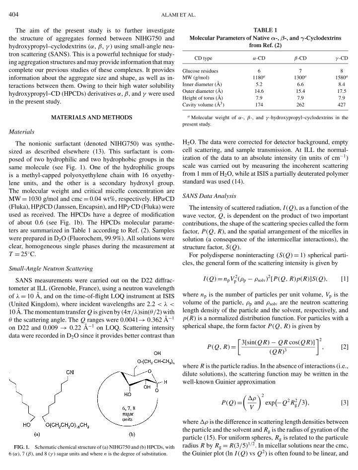
To obtain quantitative information about the structure of mi-celles formed by NIHG750, small-angle neutron scattering mea-surements were carried out at various concentrations in D2O.Model scattering curves for polydisperse spheres (see Methods)were found to give a good fit to the experimental data (Fig. 2).The structural parameters obtained from these fits are listed inTable 2. A small increase in size (21 to 24 A) is observed withconcentration and consequently an increase in the apparent ag-gregation number. The micelles show only a slight size polydis-persity that does not vary significantly with concentration. TheNIHG750 micelles remain spherical in the concentration rangeinvestigated (up to 5 wt%). (For ionic HGs, where the hydroxylis replaced by a sulfate group, the SANS data were consistentwith considering prolate-shaped aggregates (21).)
The aggregation number was estimated using Vmicelle/v,where v = 1050 A3 is the surfactant molecular volume (obtained
ND CYCLODEXTRINS 405
FIG. 2. Experimental (symbols) and model (solid line) scattering curves atvarious NIHG750 concentration. From top to bottom: 4.8, 2.8, 1.6, and 0.9 wt%.
according to Tanford’s formula (20)). A mean value of the areaper headgroup (a0 = 4π (Rmic)2/Nagg) was calculated to be about65 A2. The micelle structure and the effect of concentrationare consistent with geometric packing constraints developed byIsraelachvili et al. (22). A packing parameter (v/a0lc) of about0.31 was obtained, where v and lc were calculated according toTanford’s formula (20), i.e., both the theoretical prediction andthe experimental results (SANS) are in accord with sphericalmicelles.
Mixed Systems of NIHG750 and HPCDs
Mixtures of NIHG750 and HPCDs were investigated bySANS to determine the possible structures that may form in thesemixtures. To simulate the scattering pattern obtained for thesemixed systems, the model should take into account a coexis-tence of aggregation types, including the “bare,” noncomplexedspherical micelles.
Effect of HPCD at Constant NIHG750 Concentration
SANS measurements were carried out on systems with a con-stant NIHG750 concentration and varying amounts of HPCDs—α, β, and γ , respectively. Data were taken at the extremes of
TABLE 2Model Parameters from Structural Determination for NIHG750
NIHG750 Sphere radius Hard-sphere(wt%) (±1 A) Polydispersity radius (±1 A) Nagg ± 5
4.8 24 0.19 43 1082.8 23 0.23 43 951.6 22 0.26 30 840.9 22 0.27 30 80
Note. Nagg, Aggregation Number.

406 ALAMI
FIG. 3. Experimental and fitted scattering curves in systems containingabout 1 wt% NIHG750 and varying concentrations of HPβCD. From top tobottom: 0, 1.6, 2.4, and 8.5 wt%.
low (about 1 wt%) and high (about 5 wt%) concentrations ofNIHG750, Figs. 3 and 4, respectively. All curves clearly showthat HPCDs largely affect the structure of the aggregates presentin the system and HPβCD seems to cause the largest effect. Amodel considering a mixture of rod-shaped and spherical aggre-gates was fitted to the experimental scattering data; the resultingparameters are summarized in Tables 3–5.
HPαCD
At low NIHG750 concentration (0.8 wt%) the spherical ag-gregates were found to have a radius of around 35 A, whereasthe rod shape-aggregates also present have a radius of 9 A and a
FIG. 4. Experimental and fitted scattering curves in systems containing
about 5 wt% NIHG750 and varying concentrations of HPβCD. From top tobottom: 0, 2.1, 3.9, and 8.7 wt%.ET AL.
TABLE 3Model Parameters from Structural Determination for HPαCD
NIHG750 HPαCD Molar ratio Rsph Rcyl Lcyl
(wt%) (wt%) CD/NIHG (±1 A) Poly (±1 A) (±1 A) RS/SS
0.84 1.3 1.6 42 0.23 8 78 18.90.84 3.0 2.9 35 0.23 9 70 4.9
4.8 3.4 0.7 26 0.24 11 74 1.04.8 4.7 1.0 29 0.24 11 79 1.64.8 5.2 1.1 30 0.24 10 75 2.3
4.3 7.3 2.0 36 0.23 7 70 11.82.6 4.5 2.0 37 0.23 9 68 5.91.3 2.3 2.0 36 0.22 8 71 6.6
Note. Rsph, sphere radius; Rcyl, radius of cylinder; Lcyl, length of cylinder;Poly, polydispersity; RS/SS, scale factor for rods/scale factor for spheres.
length of about 70 A (Table 3). At low HPαCD levels the poorstatistics made it difficult to fit the data. At high NIHG750 con-centration (5 wt%) the statistics are better and a small trend isvisible. Between 3.5 and 5.5 wt% the SANS data were consis-tent with spherical aggregates with a radius of 25 to 30 A. Inaddition, there is evidence for rod-shaped aggregates coexistingwith a radius of 10 to 12 A and a length of 75 to 80 A. Thismay well correspond to a surfactant molecule with cyclodextrinthreaded onto either or both the hydrophobic groups and theoxyethylene groups of the surfactant. No systematic changes inthe rod-shaped aggregates were observed with HPαCD concen-tration. However, the proportion of rods increases upon additionof HPαCD; i.e., the ratio between the scale factors of rods andspheres increases (see Table 3).
HPβCD
The effect of several concentrations of HPβCD on the ag-gregate shape was investigated at a low (1 wt%) and a high(5 wt%) concentration of NIHG750 (Table 4). (Example data
TABLE 4Model Parameters from Structural Determination for HPβCD
NIHG750 HPβCD Molar ratio Rsph Rcyl Lcyl
(wt%) (wt%) CD/NIHG (±1 A) Poly (±1 A) (±1 A) RS/SS
0.97 1.6 1.3 33 0.24 7 70 8.50.97 2.4 2.0 35 0.23 6 77 15.10.97 8.5 6.9 35 0.24 8 76 524
4.98 2.1 0.3 25 0.20 11 64 0.74.98 3.9 0.6 26 0.23 10 74 1.44.98 8.7 1.4 38 0.22 8 67 10.4
4.4 11.0 2.0 42 0.21 7 71 22.42.6 6.6 2.0 37 0.25 8 69 16.41.7 4.3 2.0 38 0.23 7 58 15.90.9 2.5 2.0 39 0.23 8 58 14.9
Note. Rsph, sphere radius; Rcyl, radius of cylinder; Lcyl, length of cylinder;Poly, polydispersity; RS/SS, scale factor for rods/scale factor for spheres.
GEMINI SURFACTANT A
TABLE 5Model Parameters from Structural Determination for HPγCD
NIHG750 HPγ CD Molar ratio Rsph Rcyl Lcyl
(wt%) (wt%) CD/NIHG (±1 A) Poly (±1 A) (±1 A) RS/SS
0.97 3.5 2.4 27 0.24 7 76 4.10.97 7.8 5.3 34 0.24 7 71 19.80.97 11.0 7.5 38 0.23 7 76 43.8
4.98 2.3 0.3 19 0.27 — —4.98 5.8 0.8 26 0.16 12 66 0.94.98 9.7 1.3 27 0.16 11 70 1.4
4.4 12.7 2.0 28 0.16 8 58 2.82.9 8.5 2.0 27 0.17 8 71 2.81.7 5.1 2.0 27 0.20 8 67 2.80.9 2.7 2.0 23 0.27 8 70 2.4
Note. Rsph, sphere radius; Rcyl, radius of cylinder; Lcyl, length of cylinder;Poly, polydispersity; RS/SS, scale factor for rods/scale factor for spheres.
for HPβCD are shown in Fig. 3.) Similar results were obtainedat both concentrations. Rod-shaped aggregates are present in allsamples containing HPβCD and this is shown clearly by theexample data given in Fig. 3 which exhibit Q−1 scattering inthe measured Q range. Regarding the rod-shaped aggregates noclear variation of the structure with HPβCD concentration isobserved. The ratio of scale factors of rods : spheres increasessignificantly with HPβCD concentration; more and more rodsare present whereas the micellar aggregates break up. AboveHPβCD molar ratio HPβCD/NIHG750 of about 4 to 5 thereare practically no bare micelles left. Compared with HPαCD, itappears that cylindrical rod-shaped aggregates are formed moreeasily with HPβCD.
HPγ CD
A similar effect was observed with HPγ CD (Table 5). Moreand more rod-shaped aggreregates are formed as the HPγ CDconcentration increases. However, the ratio between the rod andthe sphere scale factors are much lower than those observed forHPβCD and HPαCD at the same molar ratio HPCD/NIHG750.This indicates a much weaker interaction between the two com-ponents.
HPCD/HG at Constant Ratio
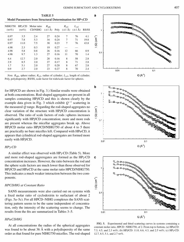
SANS measurements were also carried out on systems witha fixed molar ratio of cyclodextrin to surfactant of about 2(Figs. 5a–5c). For all HPCD–NIHG complexes the SANS scat-tering pattern seems to be the same independent of concentra-tion, only the intensity of the scattering seems to change. Theresults from the fits are summarized in Tables 3–5.
HPαCD/HG
At all concentrations the radius of the spherical aggregates˚
was found to be about 36 A with a polydispersity of the sameorder as that found for pure NIHG750 micelles. The rod-shaped
ND CYCLODEXTRINS 407
FIG. 5. Experimental and fitted scattering curves in systems containing aconstant molar ratio, HPCD : NIHG750, of 2. From top to bottom, (a) HPαCD:
7.3, 4.5, and 2.3 wt%; (b) HPβCD: 11.0, 6.6, 4.3, and 2.5 wt%; (c) HPγ CD:12.7, 8.5, 5.1, and 2.7 wt%.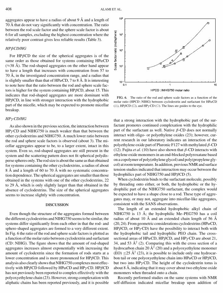
408 ALAMI
aggregates appear to have a radius of about 9 A and a length of70 A that do not vary significantly with concentration. The ratiobetween the rod scale factor and the sphere scale factor is about6 for all samples, excluding the highest concentration where therelatively poor contrast gives less reliable results (Table 3).
HPβCD/HG
For HPβCD the size of the spherical aggregates is of thesame order as those obtained for systems containing HPαCD(≈38 A). The rod-shaped aggregates on the other hand appearto have a length that increases with concentration from 57 to70 A, in the investigated concentration range, and a radius thatis slightly smaller than that of HPαCD, 7 to 8 A. It is interestingto note here that the ratio between the rod and sphere scale fac-tors is higher for the system containing HPβCD, about 15. Thisindicates that rod-shaped aggregates are more dominant withHPβCD, in line with stronger interaction with the hydrophobicpart of the micelle, which may be expected to promote micellarbreakup.
HPγ CD/HG
As also shown in the previous section, the interaction betweenHPγ CD and NIHG750 is much weaker than that between theother cyclodextrins and NIHG750. A much lower ratio betweenthe rod and sphere scale factors is observed (about 3). The mi-cellar aggregates appear to be, to a larger extent, intact in thissystem. Even so, rod-shaped aggregates are still present in thesystem and the scattering pattern does not fit spherical polydis-perse spheres only. The rod size is about the same as that obtainedfor systems containing the other cyclodextrins, a radius of about8 A and a length of 60 to 70 A with no systematic concentra-tion dependence. The spherical aggregates are smaller than thoseobtained in the systems with one of the other cyclodextrins, 22to 29 A, which is only slightly larger than that obtained in theabsence of cyclodextrins. The size of the spherical aggregatesseems to increase slightly with concentration.
DISCUSSION
Even though the structure of the aggregates formed betweenthe different cyclodextrins and NIHG750 seems to be similar, thesystems differ significantly from each other in that the rod- andsphere-shaped aggregates are formed to a very different extent.In Fig. 6 the ratio of the rod and sphere scale factors is plotted asa function of the molar ratio between cyclodextrin and surfactant(CD : NIHG). The figure shows that the amount of rod-shapedaggregates increases almost exponentially with increasing theamount of cyclodextrin since the formation of rods starts at alower concentration and is more pronounced for HPβCD. Thisanalysis also clearly shows that NIHG750 complexes most effec-tively with HPβCD followed by HPαCD and HPγ CD. HPβCDhas not previously been reported to complex effectively with the
polyoxyethylene chain (23). However, strong complexation withaliphatic chains has been reported previously, and it is possibleET AL.
FIG. 6. The ratio of the rod and sphere scale factors as a function of themolar ratio (HPCD : NIHG) between cyclodextrin and surfactant for HPαCD(�), HPβCD (�), and HPγ CD (�). The lines are guides to the eye.
that a strong interaction with the hydrophobic part of the sur-factant promotes continued complexation with the hydrophilicpart of the surfactant as well. Native β-CD does not normallyinteract with oligo- or polyethylene oxides (23); however, cur-rent research in our laboratory indicates an interaction of thepolyethylene oxide part of Pluronic F127 with methylated β-CD(12). Fujita et al. (10) have also shown that β-CD interacts withethylene oxide monomers in an end-blocked polyroxatane basedon a copolymer of poly(ethylene glycol) and poly(propylene gly-col) at room temperature. In addition, previous NMR and surfacetension studies indicated that interaction may occur between thehydrophilics part of NIHG750 and HPβCD (5).
When cyclodextrin binds to the surfactant molecule, possiblyby threading onto either, or both, the hydrophobic or the hy-drophilic part of the NIHG750 surfactant, the complex wouldbe expected to have a shape close to a rod. These type of aggre-gates may, or may not, aggregate into micellar-like aggregates,consistent with the SANS observations.
The length of an extended hydrophobic alkyl chain ofNIHG750 is 13 A; the hydrophilic Me–PEG750 has a coilradius of about 10 A and an extended chain length of 56 A(20, 24). From geometric considerations cyclodextrins (HPαCD,HPβCD, or HPγ CD) have the possibility to interact both withthe hydrophobic tail and hydrophilic PEO chain. The cross-sectional areas of HPαCD, HPβCD, and HPγ CD are about 21,34, and 53 A2 (2). Comparing this with the cross section of ahydrocarbon chain 20 A2 (20) and a polyoxyethylene monomer(EO) ≤25 A2 (23), it is possible to include only one hydrocar-bon tail or one polyoxyethylene chain into HPαCD or HPβCD,but two into HPγ CD. The height of the cyclodextrin torus isabout 8 A, indicating that it may cover about two ethylene oxidemonomers when threaded onto a chain.
Recently performed studies on the same systems with NMR
self-diffusion indicated micellar breakup upon addition of
A
GEMINI SURFACTANTcyclodextrin (HPβCD to a larger extent than HPγ CD) (5). Itwas found that the cyclodextrin interacts mainly with the hy-drophobic part of the surfactant but that an interaction withthe hydrophilic polyoxyethylene part also exists. The resultssuggest that unimeric rod-shaped NIHG750 complexes coexistwith micelle-like aggregates at intermediate concentrations ofcyclodextrin, the same as suggested by the SANS results pre-sented here.
Taking into account all the results it can be seen that thestructure of aggregates formed between HPCD and NIHG750is essentially the same, independent of the type of cyclodex-trin. The spherical aggregates have a size of about 20 to 40 A,which increases with CD concentration. One possible explana-tion for the small increase in micellar radius could be that somecyclodextrin interacts with the hydrophilic oxyethylene chain ofthe micellized surfactant molecules. The size of the aggregatesdoes not seem to vary with total concentration at a fixed molarratio (molar ratio 2 (CD : NIHG)) of surfactant to cyclodextrin;in addition rod-shaped aggregates form. These appear to have aradius of 9 ± 2 A and a length of about 70 ± 10 A that do notvary much with cyclodextrin concentration or with the total con-centration at a fixed molar ratio. A slight increase in the lengthof the rods is sometimes observed with NIHG750 concentrationat a fixed molar ratio. The NMR studies indicated formation ofmuch shorter (only 17–20 A) rod-shaped aggregates (5). Thissuggestion was based on data at low NIHG750 concentrationand a fixed molar ratio of 2. The decrease in self-diffusion coef-ficient at higher concentrations could be related to the formationof very long rod/cylindrical-shaped aggregates, as suggested bythe SANS measurements here.
It has been reported that hydrocarbon chains and oligo- orpolyethylene oxide take their extended configuration when in-cluded into a cyclodextrin cavity (23). It is therefore most proba-ble that these cylindrical aggregates are covered with cyclodex-trin (having an external radius of 7 to 9 A) to such an extentthat the chain takes its fully extended conformation (∼70 A).At a molar ratio (HPβCD : NIHG750) of about 4 to 5 the SANSdata indicates that only cylindrical aggregates are present inthe system. This shows that it is not necessary for the wholechain to be coated with cyclodextrin in order for it to take itsextented conformation. A fully covered polyoxyethylene chainof NIHG750 may contain around seven CDs and one or bothhydrophobic groups would complex with two CDs. Above thismolar ratio (i.e., 4 to 5) an increase in surface tension has beenobserved (5). This was interpreted as being a complete screeningof the hydrophobic groups, or an interaction with the hydrophilic
group resulting in lower surface activity, and a lower aggrega-tion tendency. These interpretations are also consistent with theND CYCLODEXTRINS 409
SANS data. Compared to systems composed of single-tailed sur-factants, which normally lose their ability to micellize at muchlower additions of HPβCD, NIHG750 has an advantage in sys-tems containing both surfactant and HPCD. To the best of ourknowledge, the present study is the first that reports informationabout the structure of soluble complexes between cyclodextrinand nonionic surfactants. SANS studies have proven to give newand important information, in that HPCD (α,β, andγ ) may inter-act with both the hydrophilic and the hydrophobic part of NIHGin a similar manner, however, to a different extent, dependingon the type of cyclodextrin.
ACKNOWLEDGMENTS
We acknowledge support from the competence center SNAP and from theSwedish Research Council. We thank CLRC and EPSRC for providing neutronbeam time at ILL and at ISIS.
REFERENCES
1. Stella, V. J., and Rajewski, A., Pharm. Rev. 14, 556 (1997).2. Bekers, O., Uijtendaal, E. V., Beijnen, J. H., Bult, A., and Underberg,
W. J. M., Drug Dev. Ind. Pharm. 17, 1503 (1991).3. Buschmann, H. J., Cleve, E., and Schollmeyer, E., J. Inclusion Phenom.
Macrocyclic Chem. 33, 233 (1999).4. Smith, V. K., Thilivhali, T. N., Munoz de la Pena, A., and Warner, I. M.,
J. Inclusion Phenom. Mol. Recog. Chem. 10, 471 (1991).5. Abrahmsen-Alami, S., Alami, E., Eastoe, J., and Cosgrove, T., J. Colloid
Interface Sci. 246, 191 (2002).6. Topchieva, I., and Karesin, K., J. Colloid Interface Sci. 213, 29 (1999).7. Hodul, P., Talaba, P., Srokova, I., Marcincin, A., and Peterova, M., Tenside
Surf. Deterg. 34, 169 (1997).8. Cserhati, T., and Szejtli, J., Carbohydr. Rev. 224, 165 (1992).9. Harada, A., Li, J., and Kamachi, M., Nature 370, 126 (1994).
10. Fujita, H., Ooya, T., and Yui, N., Macromolecules 32, 2534 (1999).11. Gaitano, G. G., and Brown, W., J. Phys. Chem. B. 101, 710 (1997).12. Joseph, J., Ph.D. thesis, Bristol University, Bristol, 2001.13. Alami, E., and Holmberg, K., J. Colloid Interface Sci. 239, 230 (2001).14. Wignall, G. D., and Bates, F. S., J. Appl. Crystallgr. 20, 28 (1987).15. Guinier, A., and Fournet, G., “Small-Angle Scattering of X-Rays. Wiley,
New York, 1955.16. Aragon, S. R., and Pecora, R., J. Chem. Phys. 64, 2395 (1976).17. Percus, J. K., and Yevick, G. J., Phys. Rev. 110, 1 (1958).18. Lekner, J., Phys. Rev. 145, 83 (1966).19. Heenan, R. K., FISH Data Analysis Programm. Rutherford Appleton Lab-
oratory Report RAL-89-129, Chilton OXON, United Kingdom, 1989.20. Tanford, C., J. Phys. Chem. 76, 3020 (1972).21. Alami, E., Abrahmsen-Alami, S., Eastoe, J., and Heenan, R., Langmuir. In
press.22. Israelachvili, J. N., Mitchell, D. J., and Ninham, B. W., J. Chem. Soc.
Faraday Trans. 72, 1525 (1976).23. Harada, A., Li, J., and Kamachi, M., Macromolecules 26, 5698
(1993).24. Devanand, S., and Selser, J. C., Macromolecules 24, 5943 (1991).