INTERACTION OF /31H GLOBULIN WITH CELL-BOUND C3b: Quantitative Analysis of Binding and Influence of...
14
INTERACTION OF /31H GLOBULIN WITH CELL-BOUND C3b: Quantitative Analysis of Binding and Influence of Alternative Pathway Components on Binding* BY DANIEL H. CONRAD$, JAIME R. CARLOS, ASD SHAUN RUDDY (From the Division of Immunology and Connective Tissue Diseases, Medical College of Virgznia, Health Sciences Division, Virginia Commonwealth University, Richmond, Virginia 23298 ~ Participation of C3b, the major cleavage product of C3, in both the classical and alternative pathways of complement activation is modulated by several control proteins (1-3). Two of these, C3b inactivator (C3bINA) ~ (4, 5) and flirt- globulin (B1H) (2), have been extensively purified and characterized. It is now apparent that C3bINA is a protease, and that it blocks the biologic activities of C3b by cleaving peptide bonds in this molecule (1, 4, 6). The second protein, B1H, potentiates the activity of C3bINA; indeed, recent evidence indicates an absolute requirement for/31H in the cleavage of fluid phase C3b by C3bINA (4). In addition, highly purified fllH by itself beth directly inhibits the activity of C3b (4) and accelerates the rate of decay of the alternative pathway convertases, C3bB and C3bBP (2, 7). Of great interest is the mechanism by which /31H exerts these effects. No proteolytic activity that can be directly ascribed to B1H has been found. Direct binding of/31H to C3b and subsequent steric interference with the interaction of C3b with factor B and/or C5 is the most straightforward explanation; two lines of evidence, fluid phase depletion and agglutination by antibody to /31H of EAC43 previously exposed to/]IH (8), had indicated that such binding occurs. More recently, both this laboratory (9) and another (10) have presented further information about the binding of fllH to C3b-coated particles. The studies reported here give quantitative measurement of strength and valence of this binding, examine the influence of fluid phase C3 and C3b on it, and determine the effects that factor B (B) and properdin (P), which also bind to C3b, have on the binding of ~IH to C3b-coated cells. Materials and Methods Reagents. Bio-Rad Ag-I-X-10 (chloride form), Bio-Rex 70, electrophoresis grade polyacryl- amide, bis-acrylamide, and sodium dodecyl sulfate (SDS) were obtained from Bio-Rad Laborato- * Supported by grant AI 13049 from the National Institutes of Health and an Arthritis Clinical Research Center Grant from the Arthritis Foundation, New York. This is publication no. 120 from the Charles W. Thomas Fund, Medical College of Virginia. Post doctoral trainees supported by training grant T32 AM 07079 from the National Institutes of Health. l Abbreviations used in this paper: A, rabbit antibody; B, factor B; ~IH, ~IH globulin; C3blNA, C3b inactivator; DGVB ÷÷, equal volumes of GVB +÷ and DSW++; D5W +~ , 5~ dextrose in water; E, sheep erythrocytes; FITC, fluorescein isothiocyanate; GVB ÷', 0.1~ gelatin veronal buffer~ P, properdin; SBTI, soybean trypsin inhibitor; SDS, sodium dodecyl sulfate; VBS, veronal-buffered saline. 1792 J. ExP. MED. ((:)The Rockefeller University Press . 0022-1007/78/0601-179251.00 on January 28, 2015 jem.rupress.org Downloaded from Published June 1, 1978
-
Upload
independent -
Category
Documents
-
view
1 -
download
0
Transcript of INTERACTION OF /31H GLOBULIN WITH CELL-BOUND C3b: Quantitative Analysis of Binding and Influence of...
I N T E R A C T I O N OF /31H GLOBULIN WITH CELL-BOUND C3b:
Quan t i t a t i ve Analys i s of B ind ing and Inf luence of Al t e rna t ive
P a t h w a y Componen t s on Binding*
BY DANIEL H. CONRAD$, JAIME R. CARLOS, ASD SHAUN RUDDY
(From the Division of Immunology and Connective Tissue Diseases, Medical College of Virgznia, Health Sciences Division, Virginia Commonwealth University, Richmond, Virginia 23298 ~
Participation of C3b, the major cleavage product of C3, in both the classical and alternative pathways of complement activation is modulated by several control proteins (1-3). Two of these, C3b inactivator (C3bINA) ~ (4, 5) and flirt- globulin (B1H) (2), have been extensively purified and characterized. It is now apparent that C3bINA is a protease, and that it blocks the biologic activities of C3b by cleaving peptide bonds in this molecule (1, 4, 6). The second protein, B1H, potentiates the activity of C3bINA; indeed, recent evidence indicates an absolute requirement for/31H in the cleavage of fluid phase C3b by C3bINA (4). In addition, highly purified fllH by itself beth directly inhibits the activity of C3b (4) and accelerates the rate of decay of the alternative pathway convertases, C3bB and C3bBP (2, 7).
Of great interest is the mechanism by which /31H exerts these effects. No proteolytic activity that can be directly ascribed to B1H has been found. Direct binding of/31H to C3b and subsequent steric interference with the interaction of C3b with factor B and/or C5 is the most straightforward explanation; two lines of evidence, fluid phase depletion and agglutination by antibody to /31H of EAC43 previously exposed to / ] IH (8), had indicated that such binding occurs. More recently, both this laboratory (9) and another (10) have presented further information about the binding of fllH to C3b-coated particles. The studies reported here give quantitative measurement of strength and valence of this binding, examine the influence of fluid phase C3 and C3b on it, and determine the effects that factor B (B) and properdin (P), which also bind to C3b, have on the binding of ~IH to C3b-coated cells.
Mater ia l s and Methods Reagents. Bio-Rad Ag-I-X-10 (chloride form), Bio-Rex 70, electrophoresis grade polyacryl-
amide, bis-acrylamide, and sodium dodecyl sulfate (SDS) were obtained from Bio-Rad Laborato-
* Supported by grant AI 13049 from the National Inst i tutes of Heal th and an Arthr i t i s Clinical Research Center Grant from the Arthr i t i s Foundation, New York. This is publication no. 120 from the Charles W. Thomas Fund, Medical College of Virginia.
Post doctoral t ra inees supported by t ra in ing grant T32 AM 07079 from the National Inst i tutes of Health.
l Abbreviations used in this paper: A, rabbit antibody; B, factor B; ~IH, ~IH globulin; C3blNA, C3b inactivator; DGVB ÷÷, equal volumes of GVB +÷ and DSW++; D5W +~ , 5~ dextrose in water; E, sheep erythrocytes; FITC, fluorescein isothiocyanate; GVB ÷', 0.1~ gelatin veronal buffer~ P, properdin; SBTI, soybean trypsin inhibitor; SDS, sodium dodecyl sulfate; VBS, veronal-buffered saline.
1792 J. ExP. MED. ((:) The Rockefeller University Press . 0022-1007/78/0601-179251.00
on January 28, 2015jem
.rupress.orgD
ownloaded from
Published June 1, 1978
DANIEL H. CONRAD, JAIME R. CARLO, AND SHAUN RUDDY 1793
ties, Richmond, Calif. Radioiedide, both 13~I and carrier-free '25I, was obtained from Amersham Corp., Arl ington Heights, Ill. Trypsin and soybean trypsin inhibi ter (SBTI) were obtained from Worthington Biochemical Corp., Freehold, N. J. Bovine serum albumin (Cohn Fraction V) was obtained from Calbiochem, San Diego, Calif. Las-R human complement C3 reagent kit was purchased from Hyland Diagnostics Div., Travenol Laboratories, Costa Mesa, Calif., and Sepharose 4B from Pharmacia Fine Chemicals, Inc., Piscataway, N. J.
Buffers. Isotonic veronal-buffered saline (VBS) containing 0.00015 M Ca ++, 0.0005 M Mg ÷+, and 0.1% gelat in (GVB ÷÷) and 5% dextrose in water containing the same concentrations of divalent cations (D5W ÷÷) were mixed in equal volumes; the resul t ing buffer, ionic s t rength of 0.065 and pH 7.4, is referred to as DGVB *÷ and was used in all binding studies. GVB-- was made as described above except tha t the divalent cations were not present. A stock solution of 0.086 M EDTA, pH 7.5, was diluted in GVB to prepare 0.04 M EDTA GVB-- .
Component Purification. Guinea pig C1 (11) and h u m a n C2 (12) were prepared as published elsewhere. Part ial ly purified factor B was obtained by a modification of the procedure of G6tze and Mialler-Eberhard (13). Normal human serum was adjusted to 40% Na~SO4 and the resul t ing precipitate was redissolved and subjected to chromatography on Bio-Rex 70 (13). Properdin was prepared by a modification in the procedure of Pensky et al. (14). The high salt eluate from zymosan was dialyzed against low ionic s t rength buffer, and the resul t ing P-containing precipitate was redissolved in VBS and subjected to chromatography on a Sephadex G-200 column. B (15) and P (16) were measured hemolytically as described elsewhere. Highly purified human C3 was prepared as described by Tack and Prahl (17), and B1H was prepared as described by Whaley and Ruddy (2). C3b was prepared from the purified C3 with trypsin and SBTI as described by Bokisch et al. (18).
Antisera. Antisera to C3, B, P, and 131H were induced in goats and were subsequently used in radial immunodiffusion (19) to determine the concentration of the various components. Pooled h u m a n serum, which had been previously calibrated against purified C3, B, P, and t31H, served as the standard. In radiolabeled preparations, concentrat ions of C3 and ~IH were determined nephelometrically with a Hyland Laser Nephelometer PDQ Ins t rument (Hyland Diagnostics Div.) and a Hyland LAS-R h u m a n complement C3 kit for C3 determinat ions. When fllH was measured nephelometrically, doubling dilutions of pooled h u m a n serum (1:12.5 to 1:400) were used as standards. The 1:12.5 dilution was first filtered through a 0.4-pro Nucleopore filter (Nucleopore Corp., Pleasanton, Calif.) before fur ther diluting. Rabbit ant i - f l lH was diluted with saline and subsequently an equal volume of phosphate-buffered saline (0.01 M PO4 -, 0.15 M NaC1, pH 7.4) containing 4% polyethylene glycol was added to give a final antibody dilution of 1:60.50 pl of s tandards or appropriately diluted unknowns were added to 1 ml antibody dilutions and after 1 h at room tempera ture were examined for l ight scat ter in the nephelometer; 50/~1 of the same samples added to 1 ml of saline served as b lank controls.
The purity of the '25I-labeled /31H (12'sI-f11H) (see below) was also est imated by test ing the abili ty of the preparat ion to be insolubilized with the monospecific goat ant i-f l i r t . 125I-~1H was mixed with an excess of goat anti-B1H, and after 30 rain at 37°C the /31H-antiq31H complexes were precipitated by adding a predetermined optimal amount of rabbi t anti-goat IgG (Atlantic Antibodies, Westbrook, Maine). After 1 h at 37°C and overnight at 4°C, the complexes were washed three t imes with saline and the radioactivity remain ing with the precipitate was measured. All f l lH preparat ions tested in this manne r were 80-87% precipitable by the anti-f l ir t .
Immunofluorescent Staining. The globulin fraction of goat anti-/31H was conjugated with fluorescein isothiocyanate (FITC) according to the method of Herber t et al. (20). The fluorescein to p ro te i , ratio (molar) of the final preparat ion was 2:1. Approximately 100 /~g of fl lH was covalently linked to Sepharose 4B (see below), and subsequently the specificity of the antiq31H was confirmed in blocking experiments whereby fluorescent s ta in ing of the Sepharose-beund fllH was inhibi ted by reacting the FITC-anti-/31H with highly purified fllH.
The cells were examined for fluorescence by using a Zeiss photomicroscope II (Carl Zeiss, Inc., New York) with a HBO 200 light source, FITC excitation pr imary filter, and a 530-nm secondary filter.
Radioiodination. Highly purified fl lH and C3 were radioiodinated with '25I (carrier free) or ~3'I by the use of the chloramine T procedure (21). Unbound iodide was removed by ion exchange chromatography with Bio-Rad Ag l-X-10 (chloride form) and overnight dialysis versus VBS. In the final preparat ions the radioiodide was 90-98% precipitable with 10% trichloroacetic acid. The specific activities obtained were in the range of 2-5 × 10 ~ cpm/p~g and 0.1-1.0 × 10 ~ cpm//~g for
on January 28, 2015jem
.rupress.orgD
ownloaded from
Published June 1, 1978
1794 flirt BINDING TO CELL BOUND C3b
B1H and C3, respectively. Bovine serum albumin (Cohn Fraction V) was added to the stock solutions of the radiolabeled proteins, and storage was at -70°C. The correspondences between '25I and 'a'I counts and numbers of molecules of B1H or C3 were calculated from the specific activities of the iodinated proteins, using Avogadro's number and mol wt of 185,000 and 150,000 daltons (2) for C3 and/31H, respectively.
Cellular Intermediates. Sheep erythrocytes (E) were sensitized with rabbit antibody (A), and EAC4 were prepared and stored at -70°C in a glycerol-containing medium as described elsewhere (22). EAC4 were thawed as needed. EAC14 and EAC14 °~' 2 were prepared by using guinea pig C1 and human C2 which had been oxidized with I2 (23). EAC43 were prepared with nonoxidized C2 as described previously (15); the EAC43 did not lyse when exposed to a C3-9 source (rat serum diluted 1:15 in 0.04 M EDTA GVB ). EAC14°~'23 and EAC14°x'2 ':uI-C3 were prepared by incubating EAC14 ° ' ' 2 in DGVB ++ with purified C3 (either unlabeled or '3'I-labeled) for 30 min at 37°C followed by three washes with DGVB ++.
Counting Technique. Measurements of "-'~I, t:"I, and 22Na were made in a dual channel gamma counter (model 1185, Searle Analytic, Chicago, Ill.). When all three isotopes were used simulta- neously, the samples were counted twice, first for '2'~I and 22Na and then for '3'I and 22Na. Channel settings were adjusted such that 0.1% or less spillover of the lower energy isotope (order of energy: '-'2Na > '3'I > '25I) into the higher one(s) occurred. The '2~I cpm was thus corrected for spillover of 22Na and, when necessary, '3'I; +3'I cpm was corrected for 22Na spillover. Background corrections were also made for all channels.
Radioactive Binding Assays. To avoid extensive manipulation of cells, 22Na was used as a volume marker for unbound t25I-/31H remaining with the cells. In a typical experiment, prepared cellular intermediates were incubated with '2~I-fHH for 15 min at 30°C in a vol of 1 ml. The cells were then sedimented by centrifugation and 0.1 ml of supernate removed. Approximately 90% of the remaining supernate was then removed by aspiration and the cells quantitatively transferred with DGVB ++ into a clean tube. Radioactive determinations were then made on the supernatant aliquot and the cell pellet. Based on the assumption that the ratio of free '25I-BIH and 22Na in the incubation solution was constant, the amount of/31H bound to the cells was determined by the following formula:
cpm '2'~I = /31H bound = A - (x/y)(z)
where A is the total 'zsI cpm in the cell pellet, x is the 22Na cpm in the cell pellet, y is the Z~Na cpm in the supernate, and z is the 'zsI cpm in the supernate. This method of calculation is similar to that described by Tsay and Schlamowitz (24). In situations where the 'a'I-C3 was used to quanti tate the amount of cell bound C3b, supernatant corrections with Z2Na were unnecessary since there was essentially no '3'I-C3 in the fluid phase.
In binding assays in which the objective was determination of binding parameters, EAC 14 °~ 23 or EAC14°x~2 '3q-C3 were incubated for 15 min at 30°C with various amounts (0.1-2 /~g) of '2~I- /31H in a total vol of 1 ml, and subsequently the bound 'zsI-/31H was determined as described above. The experimental data were then plotted according to the method of Scatchard (25).
r/c = nK - rK
where r represents the number of/31H molecules bound per cell (or alternatively per '~'I-C3b molecule), K is the average association constant, n the total number of binding sites per cell (or per C3b), and c is the concentration of free bindable '~5I-/31H. c was calculated as follows:
c = (131HT)(MB)- B1Hh,
where 131Hl~ is total B1H added (molecules/milliliter), MB is the maximal binding ability of the fllH preparation, and /31I-Ib is the number of molecules of 131H bound to the cell. In the final Scatchard plots, the best straight line for the experimental points was computed by the method of least squares using a Wang WCS-20 computer. K was obtained from the slope of this line and was converted to the more familiar liters per mole units by using Avogadro's number and a mol wt of 150,000 daltons (2) for 131H. The maximal number of/31H binding sites (n) was obtained from the intercept of the line with the abscissa. This intercept represents an infinite free concentration of ~IH.
Other Analytical Procedures. Immunoelectrophoresis was performed by standard technique (26). SDS-gel electrophoresis with 7.5% separation gels was performed by the method of Laemmli
on January 28, 2015jem
.rupress.orgD
ownloaded from
Published June 1, 1978
DANIEL H. CONRAD, JAIME R. CARLO, AND SHAUN RUDDY 1795
(27). Immunoadsorbents and /31H-Sepharose were prepared by linking the respective protein to Sepharose 4B by the cyanogen bromide procedure (28).
Results Congruent Immunofluorescent Staining and Agglutination by Anti-
[31H. Direct visual demonstration of both binding of ~IH to EAC14°x.'23 and agglutination of such cells by anti-B1H was obtained. For this experiment, EAC14°xY23 were prepared as described in Materials and Methods. One-half of the cell preparation (1.0 x 10⁸ cells/ml) was then incubated for 15 min at 30°C with 12 t~g B1H in a vol of 0.6 ml, and the other half with DGVB ÷+ alone. After washing three times with DGVB ÷÷, the two cell populations were remixed and subsequently allowed to interact for 30 min at 25°C with fluoresceinated anti- /~IH. After washing in DGVB ÷÷, the cell mixture was mounted on glass slides and examined by both phase and fluorescent microscopy. There was gross aggregation of some, but not all, of the cells. When examined by fluorescence microscopy, only the cells that stained for fllH by immunofluorescence were agglutinated, thus indicating that the agglutination was associated with the presence of bound ~IH on the cells.
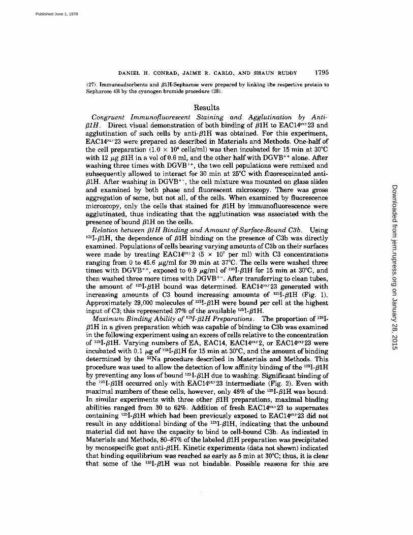
Relation between fllH Binding and Amount of Surface-Bound C3b. Using '25I-B1H, the dependence of B1H binding on the presence of C3b was directly examined. Populations of cells bearing varying amounts of C3b on their surfaces were made by treating EAC14°x.~2 (5 x 10⁷ per ml) with C3 concentrations ranging from 0 to 45.6 t~g/ml for 30 min at 37°C. The cells were washed three times with DGVB +÷, exposed to 0.9 tLg/ml of '25I-fllH for 15 min at 30°C, and then washed three more times with DGVB +÷. After transferring to clean tubes, the amount of '2~I-/31H bound was determined. EAC14°×Y23 generated with increasing amounts of C3 bound increasing amounts of 125I-fllH (Fig. 1). Approximately 29,000 molecules of ~25I-~1H were bound per cell at the highest input of C3; this represented 37% of the available 'uI-~lH.
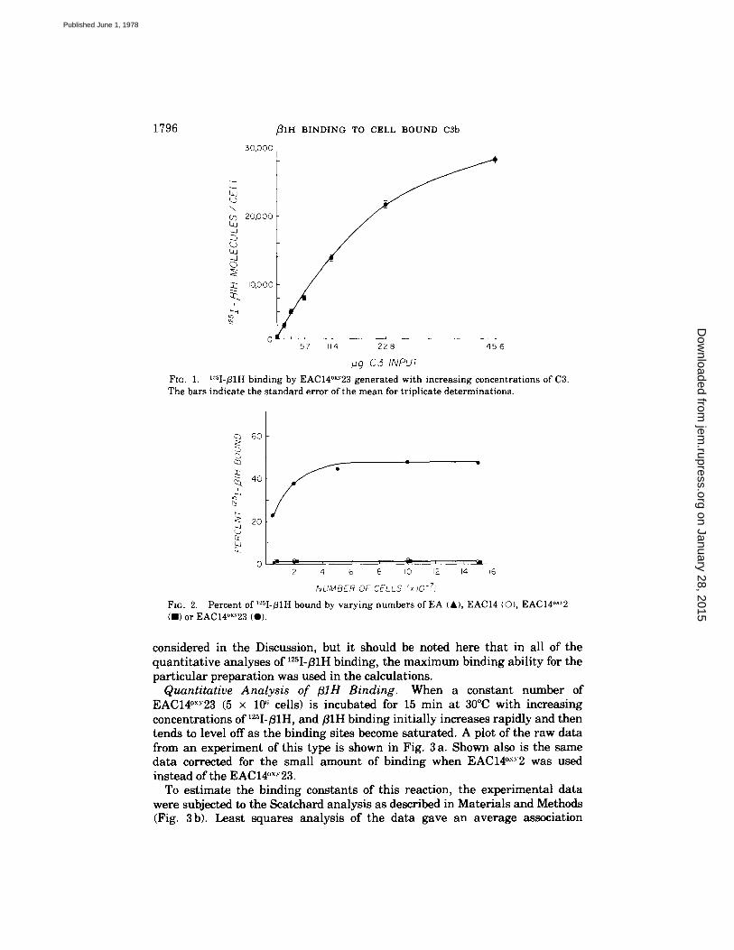
Maximum Binding Ability of '"-sI-fllH Preparations. The proportion of '~I- fllH in a given preparation which was capable of binding to C3b was examined in the following experiment using an excess of cells relative to the concentration of 12~I-~lH. Varying numbers of EA, EAC14, EAC14°x'~2, or EAC14°×Y23 were incubated with 0.1 t~g of '~I-B1H for 15 min at 30°C, and the amount of binding determined by the 22Na procedure described in Materials and Methods. This procedure was used to allow the detection of low affinity binding of the '25I-fllH by preventing any loss of bound '25I-fllH due to washing. Significant binding of the '25I-fllH occurred only with EAC14°~Y23 intermediate (Fig. 2). Even with maximal numbers of these cells, however, only 48% of the '25I-B1H was bound. In similar experiments with three other B1H preparations, maximal binding abilities ranged from 30 to 62%. Addition of fresh EAC14°×y23 to supernates containing '25I-fllH which had been previously exposed to EAC14°xy23 did not result in any additional binding of the '25I-fllH, indicating that the unbound material did not have the capacity to bind to cell-bound C3b. As indicated in Materials and Methods, 80-87% of the labeled/31H preparation was precipitated by monospecific goat anti-flirt. Kinetic experiments (data not shown) indicated that binding equilibrium was reached as early as 5 min at 30°C; thus, it is clear that some of the '25I-]31H was not bindable. Possible reasons for this are
on January 28, 2015jem
.rupress.orgD
ownloaded from
Published June 1, 1978
1796 ~ I H BINDING TO CELL BOUND c3b
30,000
...j
\ CO 20,000
US . , .J 0
10,ooo
p
0 ' ' L , . 57 114 2 2 8 45 6
,u.g C 5 INPUT
Fro. 1. ~Sl-/31H binding by EAC14°×Y23 generated w i t h increas ing concentrat ions of C3. The bars indicate the s tandard error of the m e a n for tr ipl icate de terminat ions .
c~ 6o
c~
~ 4o &
~ 2o cc t~j
f 2 4 6 8 IO 12 14 16
fvUMBER OF CELLS ;~x I0 -7)
FIG. 2. Percent of ~sI-fllH bound by varying numbers of EA {A), EACI4 IO), EACI4°X~2 (B) or EAC14"xY23 (O).
considered in the Discussion, but it should be noted here that in all of the quantitative analyses of ~2'~I-/31H binding, the maximum binding ability for the particular preparation was used in the calculations.
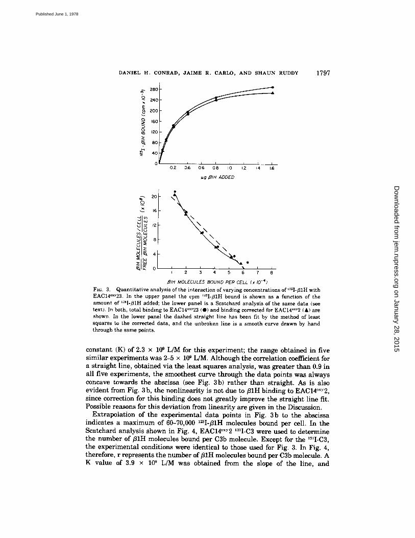
Quantitative Analysis of fllH Binding. When a constant number of EAC14°x~23 (5 x 10 ~ cells) is incubated for 15 min at 30°C with increasing concentrations of 12~I-fllH, and fllH binding initially increases rapidly and then tends to level off as the binding sites become saturated. A plot of the raw data from an experiment of this type is shown in Fig. 3 a. Shown also is the same data corrected for the small amount of binding when EAC14°x-v2 was used instead of the EAC14°x-' 23.
To estimate the binding constants of this reaction, the experimental data were subjected to the Scatchard analysis as described in Materials and Methods (Fig. 3b). Least squares analysis of the data gave an average association
on January 28, 2015jem
.rupress.orgD
ownloaded from
Published June 1, 1978
DANIEL H. CONRAD, JAIME R. CARLO, AND SHAUN RUDDY 1797
40
0 J I I 01.2 016 0i6 0~8 I0 1.12 [4 i,6
I~g 191H ADDED
&-. 28o
240
E 2oo
160
120
~. 8o
----- 2O
16
~2 \ 1 ~
L I I I L ~L i I 2 3 4 5 6 7
BIH MOLECULES BOUND PER CELL (x 10 -4)
FIG. 3. Quantitative analysis of the interaction of varying concentrations of ~sI-fllH with EAC14°xY23. In the upper panel the cpm I~sI-B1H bound is shown as a function of the amount of I~sI-B1H added; the lower panel is a Scatchard analysis of the same data (see text). In both, total binding to EAC14°xY23 (@) and binding corrected for EAC14°xy2 (A) are shown. In the lower panel the dashed straight line has been fit by the method of least squares to the corrected data, and the unbroken line is a smooth curve drawn by hand through the same points.
constant (K) of 2.3 × 10 ~ L/M for this experiment; the range obtained in five similar experiments was 2-5 × 10 ~ L/M. Although the correlation coefficient for a straight line, obtained via the least squares analysis, was greater than 0.9 in all five experiments, the smoothest curve through the data points was always concave towards the abscissa (see Fig. 3 b) rather than straight. As is also evident from Fig. 3 b, the nonlinearity is not due to fllH binding to EAC14°x.v2, since correction for this binding does not greatly improve the straight line fit. Possible reasons for this deviation from linearity are given in the Discussion.
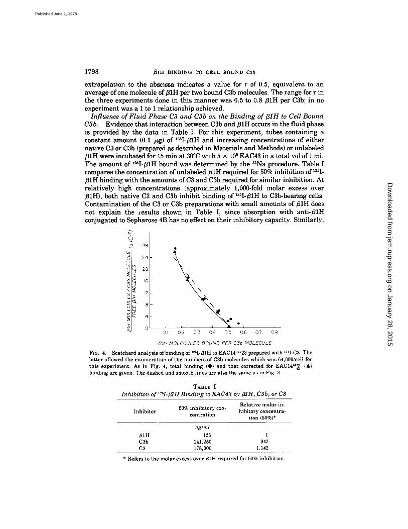
Extrapolation of the experimental data points in Fig. 3b to the abscissa indicates a maximum of 60-70,000 I~I-/~IH molecules bound per cell. In the Scatchard analysis shown in Fig. 4, EAC14°xY2 131I-C3 were used to determine the number of fllH molecules bound per C3b molecule. Except for the 13~I-C3, the experimental conditions were identical to those used for Fig. 3. In Fig. 4, therefore, r represents the number of/31H molecules bound per C3b molecule. A K value of 3.9 × 10 s L/M was obtained from the slope of the line, and
on January 28, 2015jem
.rupress.orgD
ownloaded from
Published June 1, 1978
1798 ~IH BINDING TO CELL BOUND C3b
extrapolation to the abscissa indicates a value for r of 0.5, equivalent to an average of one molecule of B1H per two bound C3b molecules. The range for r in the three experiments done in this manner was 0.5 to 0.8 B1H per C3b; in no experiment was a 1 to 1 relationship achieved.
Influence of Fluid Phase C3 and C3b on the Binding of fllH to Cell Bound C3b. Evidence that interaction between C3b and ~IH occurs in the fluid phase is provided by the data in Table I. For this experiment, tubes containing a constant amount (0.1 gg) of I~I-B1H and increasing concentrations of either native C3 or C3b (prepared as described in Materials and Methods) or unlabeled ~IH were incubated for 15 min at 30°C with 5 × 10⁶ EAC43 in a total vol of 1 ml. The amount of I=I-B1H bound was determined by the 22Na procedure. Table I compares the concentration of unlabeled B1H required for 50% inhibition of 125I'- fllH binding with the amounts of C3 and C3b required for similar inhibition. At relatively high concentrations (approximately 1,000-fold molar excess over ]31H), both native C3 and C3b inhibit binding of lZSI-fllH to C3b-bearing cells. Contamination of the C3 or C3b preparations with small amounts of B1H does not explain the results shown in Table I, since absorption with anti-fllH conjugated to Sepharose 4B has no effect on their inhibitory capacity. Similarly,
28
24
C.,.o ~ 20 :::b
~ d \ ~ 12
2:: o I I I I I
0.1 0.2 0.3 0.4 0.5 0.6 0.7 0.8
,BIH MOLECULES BCUND PER C3b MOLECULE
FIG. 4. Scatchard analysis of binding of l~sI-fllH to EAC14°xY23 prepared with "I-C3. The latter allowed the enumeration of the numbers of C3b molecules which was 64,000/cell for this experiment. As in Fig. 4, total binding (O) and that corrected for EAC14°x~ (A) binding are given. The dashed and smooth lines are also the same as in Fig. 3.
TABLE I Inhibition of r"sI-BIH Binding to EAC43 by fllH, C3b, or C3
Relative molar in- Inhibitor 50% inhibitory con- hibitory concentra-
centration tion (50%)*
ng/ml B1H 125 1 C3b 141,250 942 C3 178,000 1,142
* Refers to the molar excess over fllH required for 50% inhibition.
on January 28, 2015jem
.rupress.orgD
ownloaded from
Published June 1, 1978
DANIEL H. CONRAD, JAIME R. CARLO, AND SI-IAUN RUDDY 1799
contamination of the native C3 with C3b does not explain their approximately equivalent inhibitory capacity. No alteration in the electrophoretic mobility of the native C3 was seen by immunoelectrophoresis, and a single sharp band corresponding to the C3 a-chain was found when the preparation was examined by SDS-gel electrophoresis under reducing conditions.
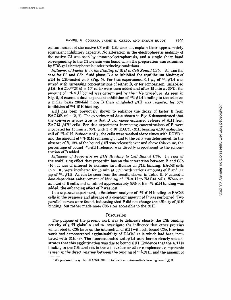
Influence of Factor B on the Binding of ;31H to Cell Bound C3b. As was the case for C3 and C3b, fluid phase B also inhibited the equilibrium binding of fllH to C3b-coated cells (Fig. 5). For this experiment, 0.1 ~g of 125I-f11H was mixed with increasing concentrations of either B, or for comparison, unlabeled fllH. EAC14°×.'23 (5. × 106 cells) were then added and after 15 rain at 30°C, the amount of ~25I-f11H bound was determined by the 22Na procedure. As seen in Fig. 5, B c a u s e d a dose-dependent inhibition of '25I-fllH binding to the cells; on a molar basis 280-fold more B than unlabeled fllH was required for 50% inhibition of '2~I-BIH binding.
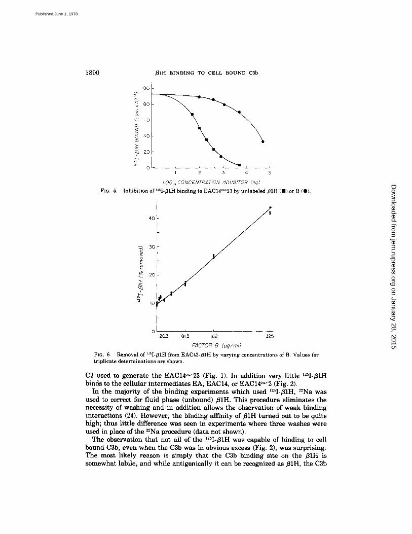
f l lH has been previously shown to enhance the decay of factor B from EAC43B cells (2, 7). The experimental data shown in Fig. 6 demonstrated that the converse is also true in that B can cause enhanced release of fllH from EAC43. fllH 2 cells. For this experiment increasing concentrations of B were incubated for 15 min at 30°C with 5 × 10⁷ EAC43. ~IH bearing 4,100 molecules/ cell of '25I-f11H. Subsequently, the cells were washed three times with DGVB ÷+ and the amount of '2~I-fllH remaining bound to the cells was determined. In the absence of B, 12% of the bound fllH was released; over and above this value, the percentage of bound 125I-~1H released was directly proportional to the concen- tration of B added.
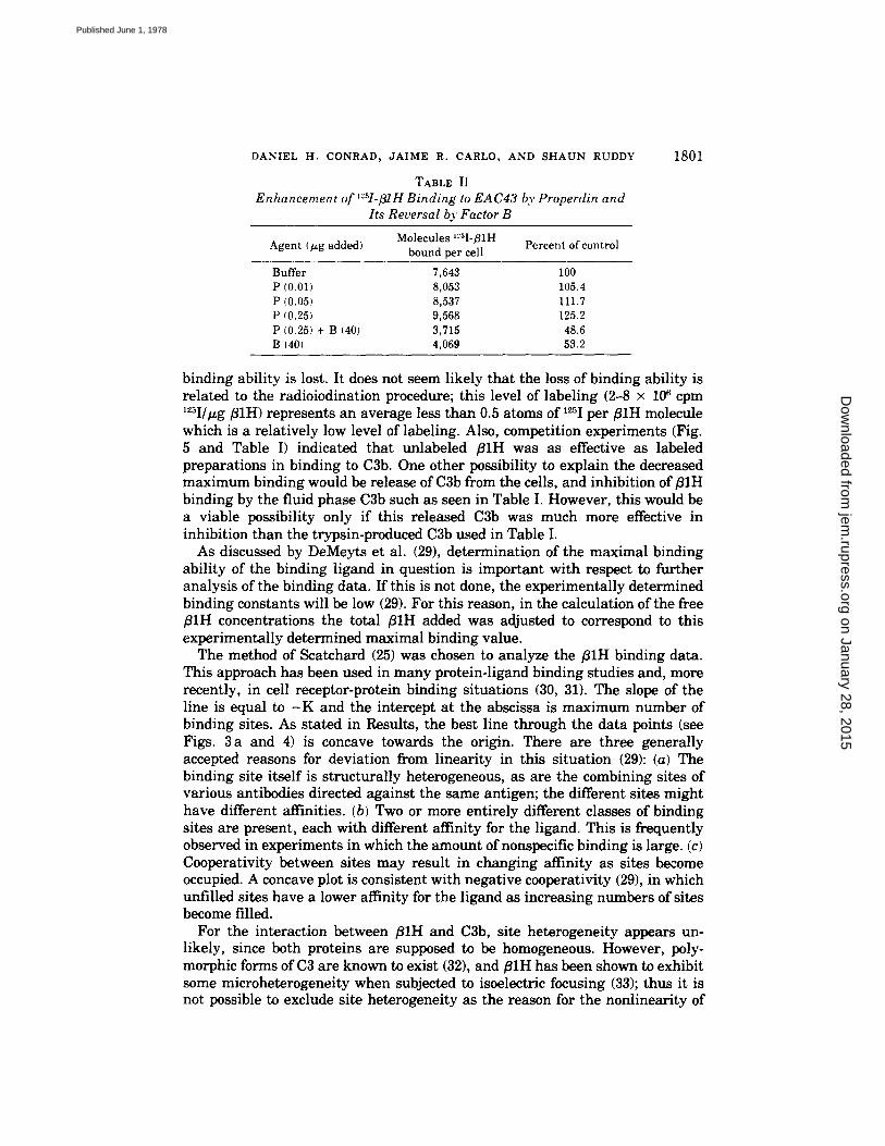
Influence of Properdin on BIH Binding to Cell Bound C3b. In view of the stabilizing effect that properdin has on the interaction between B and C3b (16), it was of interest to examine its influence on BIH binding. EAC43 cells (5 × 10 ~) were incubated for 15 min at 30°C with various amounts of P and 0.1 ~g of 12~I-f11H. As can be seen from the results shown in Table II, P caused a dose-dependent enhancement of binding of ~25I-f11H to EAC43 cells. When an amount of B sufficient to inhibit approximately 50% of the ~25I-f11H binding was added, the enhancing effect of P was lost.
In a separate experiment, a Scatchard analysis of '25I-BIH binding to EAC43 cells in the presence and absence of a constant amount of P was performed. Two parallel curves were found, indicating that P did not change the affinity of BIH binding, but rather made more C3b sites accessible to the fllH.
Discuss ion The purpose of the present work was to delineate clearly the C3b binding
activity of ~IH globulin and to investigate the influence that other proteins which bind to C3b have on the interaction of fllH with cell-bound C3b. Previous work had demonstrated agglutinability of EAC43 cells which had been incu- bated with fllH (8). The fluoresceinated ant i -~lH used herein clearly demon- strates that this agglutination was due to bound fllH. Evidence that the fllH is binding to the C3b and not to the cell surface or other complement components is seen in the direct relation between the binding of '2~I-~IH, and the amount of
We propose this symbol, EAC43. fllH to indicate an intermediate bearing bound fllH.
on January 28, 2015jem
.rupress.orgD
ownloaded from
Published June 1, 1978
1800 ~IH BINDING TO CELL BOUND C3b
FIG. 5.
I00
× 8 0
E
4O Q3
% 2o
o I 2 3 4
LOG,o CONCENTRATION INHIBITOR (rig)
Inhibition of t'-'sI-BiH binding to EAC14°xY23 by unlabeled ~ I H ( , ) or B {@).
40
5O
O E
Io
0 I L I I 1
2O3 s~.~ ~62 3Z5
FACTOR B (~q/mI)
FIG. 6. Removal of'2sI-fllH from EAC43-BIH by varying concentrations of B. Values for triplicate determinations are shown.
C3 used to generate the EAC14°×~23 (Fig. 1). In addition very little '25I-B1H binds to the cellular intermediates EA, EAC14, or EAC14°x."2 (Fig. 2).
In the majority of the binding experiments which used '25I-~1H, 22Na was used to correct for fluid phase (unbound) fllH. This procedure eliminates the necessity of washing and in addition allows the observation of weak binding interactions (24). However, the binding affinity of fl lH turned out to be quite high; thus little difference was seen in experiments where three washes were used in place of the Z2Na procedure (data not shown).
The observation that not all of the 125I-B1H was capable of binding to cell bound C3b, even when the C3b was in obvious excess (Fig. 2), was surprising. The most likely reason is simply that the C3b binding site on the ;81H is somewhat labile, and while antigenically it can be recognized as B1H, the C3b
on January 28, 2015jem
.rupress.orgD
ownloaded from
Published June 1, 1978
DANIEL H. CONRAD, JAIME R. CARLO~ AND SHAUN RUDDY
TABLE II
Enhancement of ~sI-fllH Binding to EAC43 by Properdin and Its Reversal by Factor B
Molecules ~:~I-131H Percent of control Agent (~g added) bound per cell
Buffer 7,643 100 P (0.01} 8,053 105.4 P (0.05) 8,537 111.7 P (0.25} 9,568 125.2 P (0.25) + B (40) 3,715 48.6 B (40) 4,069 53.2
1801
binding ability is lost. It does not seem likely that the loss of binding ability is related to the radioiodination procedure; this level of labeling (2-8 × 10 ~ cpm l~sI/~g fllH) represents an average less than 0.5 atoms of 1~I per fllH molecule which is a relatively low level of labeling. Also, competition experiments (Fig. 5 and Table I) indicated that unlabeled fllH was as effective as labeled preparations in binding to C3b. One other possibility to explain the decreased maximum binding would be release of C3b from the cells, and inhibition of 131H binding by the fluid phase C3b such as seen in Table I. However, this would be a viable possibility only if this released C3b was much more effective in inhibition than the trypsin-produced C3b used in Table I.
As discussed by DeMeyts et al. (29), determination of the maximal binding ability of the binding ligand in question is important with respect to further analysis of the binding data. If this is not done, the experimentally determined binding constants will be low (29). For this reason, in the calculation of the free fllH concentrations the total fllH added was adjusted to correspond to this experimentally determined maximal binding value.
The method of Scatchard (25) was chosen to analyze the ~IH binding data. This approach has been used in many protein-ligand binding studies and, more recently, in cell receptor-protein binding situations (30, 31). The slope of the line is equal to - K and the intercept at the abscissa is maximum number of binding sites. As stated in Results, the best line through the data points (see Figs. 3a and 4) is concave towards the origin. There are three generally accepted reasons for deviation from linearity in this situation (29): (a) The binding site itself is structurally heterogeneous, as are the combining sites of various antibodies directed against the same antigen; the different sites might have different affinities. (b) Two or more entirely different classes of binding sites are present, each with different affinity for the ligand. This is frequently observed in experiments in which the amount of nonspecific binding is large. (c) Cooperativity between sites may result in changing affinity as sites become occupied. A concave plot is consistent with negative cooperativity (29), in which unfilled sites have a lower affinity for the ligand as increasing numbers of sites become filled.
For the interaction between fllH and C3b, site heterogeneity appears un- likely, since both proteins are supposed to be homogeneous. However, poly- morphic forms of C3 are known to exist (32), and/~IH has been shown to exhibit some microheterogeneity when subjected to isoelectric focusing (33); thus it is not possible to exclude site heterogeneity as the reason for the nonlinearity of
on January 28, 2015jem
.rupress.orgD
ownloaded from
Published June 1, 1978
1 8 0 2 BIH BINDING TO CELL BOUND C3b
the Scatchard plots in the present study. The second reason, independent classes of binding sites, has been effectively ruled out, since the second class of sites would be those present on EAC14 T M 2, and subtraction of the small amount of binding to this intermediate still does not linearize the plots. The final reason, negative cooperativity, appears the most likely: C3b is known to be deposited on the cell surface in a nonrandom manner (34); as the B1H binding sites on the C3b become occupied, the remaining C3b molecules, due to steric reasons, may be less accessible to additional/31H.
The maximum number of binding sites, n, may be underestimated when a concave Scatchard plot such as observed for the interaction of C3b and/31H is extrapolated in a linear fashion. Thus it is possible that the ratio of fllH to C3b might approach 1:1 if sufficiently high fllH concentrations were examined.
The functional consequences of the interaction between C3b and/31H in the fluid phase have been demonstrated by Fearon and Austen (35). Only a small amount of turnover of C3 and factor B was observed in mixtures of C3, B, I), C3bINA, and/31H at concentrations similar to those found in serum. However, if fllH was left out of the reaction mixture both C3 and B were rapidly converted to hemolytically inactive components. Pangburn et al. (4), suggested that ~IH was absolutely essential for C3bINA activity on C3b in fluid phase reactions. The inhibitory activity of native C3 and C3b on/31H binding seen in Table I is further evidence of the fluid phase interaction of fllH with C3 and C3b. In spite of the above, direct demonstration of complex formation between C3b and/31H in the fluid phase has not yet been achieved.
When examined for their effect of fllH binding to cell bound C3b, the two other C3b binding proteins, B and P, have essentially opposite effects. B both displaces bound/31H (Fig. 6) from the cell and inhibits the equilibrium binding of B1H to C3b (Fig. 5). This suggests that factor B and B1H interact with the same or closely adjacent sites on the C3b molecule. Others have attributed the ability of substances to activate the alternative pathway to their furnishing a protective "microenvironment," in which C3b bound to their surfaces is less accessible to inhibition by/31H (10, 35, 36). Since the interaction of B with C3b on these same surfaces is supposedly undiminished, these data suggest that B and fllH interact with different sites on C3b. There is no obvious explanation for this apparent paradox.
In the absence of B, P appears to increase the availability of C3b sites for/31H binding. It is evident from the work of Fearon and Austen (16) that P increases the hemolytic activity of factor B. This increase has been attributed to stabilization of the alternative pathway convertase (16), but properdin may cause increased equilibrium binding of B as well. The ability of properdin to extend the half-life of bound fllH is currently being investigated.
Summary
Purified B1H globulin (f~lH) was shown to bind to C3b coated cells by both immunofluorescent and radioactive tracer techniques. With EAC43, the amount of fllH bound was directly proportional to the amount of C3 used to prepare the cells; EA, EAC14 and EAC14°×.'2 bound very small amounts of B1H. The C3b binding site on B1H was labile in that not all of the purified '25I-/31H was
on January 28, 2015jem
.rupress.orgD
ownloaded from
Published June 1, 1978
DANIEL H. CONRAD, JAIME R. CARLO, AND SHAUN RUDDY 1803
capable of binding to C3b, even when an excess of cell-bound C3b was present. Scatchard analysis of binding of fllH to C3b-coated cells indicated an equilib- rium constant of 109 L/M. Deviations from linearity were regularly found on Scatchard analyses. This was consistent with the hypothesis that the ~IH binding sites exhibit negative cooperativity in that as more sites become occupied, it becomes more difficult to fill the remaining sites. The stoichiometry of the reaction between C3b and fllH was examined using EAC14°×~23 prepared with '3'I-C3 and fllH labeled with '25I. Between 0.5-0.8 ~IH molecules were bound per C3b molecule.
Other alternative pathway components influenced the binding of I~I-~IH to cell bound C3b. Both C3b and native C3 inhibited binding of labeled ]~IH at an efficiency approximately 1/1,000 that of unlabeled fllH. Factor B inhibited binding with 1/280 the efficiency of unlabeled fllH. Properdin caused a dose- dependent increase in the binding of ~IH; this enhancement was abrogated if B was also present in the reaction mixture. Scatchard analysis indicated that the enhancement of fl lH binding by P resulted in an increased number of available binding sites rather than an increase in the affinity of binding.
The authors would like to express their appreciation to Mr. Don Purkall for excellent technical assistance and to Mr. Peter Evans for assistance with the nephelometry.
Received for publication 26 January 1978.
References 1. Ruddy, S., and K. F. Austen. 1971. C3b inactivator in man. II. Fragments produced
by C3b inactivator cleavage of cell-bound or fluid phase C3b. J. Immunol. 107:742. 2. Whaley, K., and S. Ruddy. 1977. Modulation of the alternative complement pathway
by ~IH globulin. J. Exp. Med. 144:1147. 3. Nagasawa, S., and R. M. Stroud. 1978. C3bINA and its macromolecular weight
cofactor; purification and characterization. J. [mmunol. In press (Abstr.) 4. Pangburn, M. K., R. D. Schreiber, and H. J. Mfiller-Eberhard. 1977. Human
complement C3b inactivator: isolation, characterization, and demonstration of an absolute requirement for the serum protein fllH for cleavage of C3b and C4b in solution. J. Exp. Med. 146:257.
5. Fearon, D. T. 1977. Purification of C3b inactivator and demonstration of its two polypeptide chain structure. J. Immunol. 119:1248.
"6. Lachmann, P. J., and H. J. Miiller-Eberhard. 1968. The demonstration in human serum of "conglutinogen-activating factor" and its effect on the third component of complement. J. Immunol. 100:691.
7. Weiler, J. M., M. D. Daha, K. F. Austen, and D. T. Fearon. 1976. Control of the amplification convertase of complement by the plasma protein fllH. Proc. Natl. Acad. Sci. U. S. A. 73:3268.
8. Whaley, K., and S. Ruddy. 1976. Modulation of C3b hemolytic activity by a plasma protein distinct from C3b inactivator. Science (Wash. D.C.). 193:1011.
9. Ruddy, S., J. Carlo, and D. Conrad. 1978. Mechanism of action of fllH globulin on C3b. J. Immunol. In press (Abstr.)
10. Pangburn, M. K., and H. J. Miiller-Eberhard. 1978. Molecular interactions of the control proteins C3b inactivator and fllH with various complement intermediates. J. Immunol. In press. (Abstr.)
11. Nelson, R. A., Jr., J. Jensen, I. Gigli, and R. Tamura. 1966. Methods for the separation, purification, and measurements of the nine components of hemolytic
on January 28, 2015jem
.rupress.orgD
ownloaded from
Published June 1, 1978
1804 /~IH BINDING TO CELL BOUND c3b
complement in guinea pig serum. Immunochemistry. 3:111. 12. Ruddy, S., and K. F. Austen. 1967. A stoichiometric assay for the fourth component
of complement in whole human serum using EAC'Ia gp and functionally pure human second component. J. Immunol. 99:1162.
13. GStze, O., and H. J. Mtiller-Eberhard. 1971. The C3-activator system: an alternate pathway of complement activation. J. Exp. Med. 134:90s.
14. Pensky, J., C. F. Hinz, Jr., E. W. Todd, R. J. Wedgwood, J. T. Boyer, and I. Lepow. 1968. Properties of highly purified human properdin. J. Immunol. 100:142.
15. Fearon, D. T., K. F. Austen, and S. Ruddy. 1973. Formation of a hemolytically active cellular intermediate by the interaction between properdin factors B and D and the activated third component of complement. J. Exp. Med. 138:1305.
16. Fearon, D. T., and K. F. Austen. 1975. Properdin: binding to C3b and stabilization of the C3b-dependent C3 convertase. J. Exp. Med. 142:856.
17. Tack, B. F., and J. W. Prahl. 1976. Third component of human complement: Pt, rification from plasma and physiochemical characterization. Biochemistry. 15:4513.
18. Bokisch, V. A., H. J. Mtiller-Eberhard, and C. G, Cochrane. 1969. Isolation of e~ fragment (C3a) of the third component of human complement containing anaphyla- toxin and chemotactic activity and description of an anaphylatoxin inactivator of human serum. J. Exp. IVied. 129:1109.
19. Rynes, R. I., S. Ruddy, P. H. Schur, J. Spragg, and K. F. Austen. 1974. Levels of complement components, properdin factors, and kininogen in patients with inflam- matory arthritides. J. Rheumatol. 1:413.
20. Herbert, B. A., B. Pittman, R. M. McKinney, and W. B. Cherry. 1972. The preparation and physicochemical characterization of fluorescent antibody reagents. HEW, CDC, Atlanta.
21. McConahey, P. J., and F. J. Dixon. 1966. A method for trace iodination of proteins for immunologic studies. Int. Arch. Allergy Appl. Immunol. 29:185.
22. Hunsicker, L. G., and E. R. Mayes. Preservation of antibody and complement bearing sheep erythrocytes (EA, EAC4, EAC14, and EAC43) by glycerol freezing. J. Immunol. Methods. In press.
23. Polley, M. J., and H. J. Mtiller-Eberhard. 1967. Enhancement of the hemolytic activity of the second component of human complement by oxidation. J. Exp. Med. 126:1013.
24. Tsay, D. D., and M. Schlamowitz. 1975. Comparison of the binding affinities of rabbit IgG fractions to the rabbit fetal yolk sac membrane: Use of 22Na to facilitate quantitation of 12~I-IgG binding. J. Immunol. 115:939.
25. Scatchard, G. 1949. The attractions of proteins for small molecules and ions. Ann. N .Y . Acad. Sci. 51:660.
26. Wieme, R. J. 1959. An improved technique of agar-gel electrophoresis on microscope slides. Clin. Chim. Acta. 4:317.
27. Laemmli, U. K. 1970. Cleavage of structural proteins during the assembly of the head of bacteriophage T4. Nature (Lond.). 227:680.
28. Cuatrecasas, P., and C. B. Anfinsen. 1971. Affinity chromatography. In Methods in Enzymology. S. P. Colowick and N. O. Kaplan, editors. Academic Press, Inc., New York. 22:345.
29. DeMeyts, P. 1976. Insulin and growth hormone receptors in human cultured lymphocytes and peripheral blood monocytes. In Methods in Receptor Research. M. Blecher, editor. Marcel Dekker, Inc., New York. Part I. p. 301.
30. Arend, W. P., and M. Mannik. 1973. The macrophage receptor for IgG: Number and affinity of binding sites. J. Immunol. 110:1455.
31. Conrad, D. H., H. Bazin, A. H. Sehon, and A. Froese. 1975. Binding parameters of
on January 28, 2015jem
.rupress.orgD
ownloaded from
Published June 1, 1978
DANIEL H. CONRAD, JAIME R. CARLO, AND SHAUN RUDDY 1805
the interaction between rat IgE and rat mast cell receptors. J. Immunol. 114:1688. 32. Alper, C. A., and R. P. Propp. 1968. Genetic polymorphism of the third component of
human complement (C~3). J. Clin. Invest. 47:2181. 33. Nagaki, K., K. Iida, M. Okube, and S. Inai. 1978. The reaction and mechanisms of
~IH globulin. Int. Arch. Allergy Appl. Immunol. In press. 34. Mardiney, M. R., Jr., H. J. Miiller-Eberhard, and J. D. Feldman. 1968. Ultrastruc-
rural localization of the third and fourth components of complement on complement- cell complexes. Am. J. Pathol. 53:253.
35. Fearon, D. T., and K. F. Austen. 1977. Activation of the alternative complement pathway due to resistance of zymosan-beund amplification convertase to endogenous regulatory mechanisms. Proc. Natl. Acad. Sci. U. S. A. 74:1683.
36. Fearon, D. T., and K. F. Austen. 1977. Activation of the alternative complement pathway with rabbit erythrocytes by circumvention of the regulatory action of endogenous control proteins. J. Exp. Med. 146:22.
on January 28, 2015jem
.rupress.orgD
ownloaded from
Published June 1, 1978