Interaction between Oligomers of Stefin B and Amyloid in Vitro and in Cells
22
1 INTERACTION BETWEEN OLIGOMERS OF STEFIN B AND AMYLOID-BETA IN VITRO AND IN CELLS Katja Škerget 1# , Ajda Taler-Veri 1# , Andrej Bavdek 2 , Vesna Hodnik 2 , Slavko eru 1 , Magda Tušek Žnidari 3 , Tiina Kumm 4 , Didier Pitsi 5 , Maruša Pompe Novak 3 , Peep Palumaa 4 , Salvador Soriano 5 , Nataša Kopitar Jerala 1 , Vito Turk 1 , Gregor Anderluh 2 and Eva Žerovnik 1* From 1 Department of Biochemistry, Molecular and Structural Biology, Jožef Stefan Institute, Jamova 39, 1000 Ljubljana, Slovenia, 2 Department of Biology, Biotechnical Faculty, University of Ljubljana, Vena pot 111, 1000 Ljubljana, Slovenia, 3 Department of Plant Physiology and Biotechnology, National Institute of Biology, Vena pot 111, 1000 Ljubljana, Slovenia, 4 Department of Gene Technology, Tallinn University of Technology, Akadeemia tee 15, 12618 Tallinn, Estonia, 5 Department of Neuroscience, MRC Centre for Neurodegeneration Research, Institute of Psychiatry, King’s College London, De Crespigny Park, London SE5 8AF, U.K. Running head: Stefin B oligomers inhibit A fibril formation Address correspondence to: Dr. Eva Žerovnik, Assist.prof., Department of Biochemistry, Molecular and Structural Biology, Jožef Stefan Institute, Jamova 39, 1000 Ljubljana, Slovenia tel ++ 386 1 477 3753, fax ++ 386 1 477 3984; E-mail: [email protected] # these two authors contributed equally In order to contribute to the question of putative role of cystatins in Alzheimer's disease and in neuroprotection in general, we studied the interaction between human stefin B (cystatin B) and amyloid- (1-40) peptide (A). Using surface plasmon resonance (SPR) and electrospray mass spectrometry (ESI MS) we were able to show a direct interaction between the two proteins. As an interesting new fact, we show that stefin B binding to A is oligomer specific. The dimers and tetramers of stefin B, which bind A, are domain-swapped as judged from structural studies. Consistent with the binding results, the same oligomers of stefin B inhibit A fibril formation. When expressed in cultured cells, stefin B co-localises with A intracellular inclusions. It also co-immunoprecipitates with APP fragment containing the A epitope. Thus, stefin B is another APP / A binding protein in vitro and likely in cells. Neurodegenerative diseases present a huge burden in developped world' s aging population. They are all in one way or another connected to aberrant protein folding and aggregation of the proteins involved (1). Various protein conformational disorders of the central and peripheral nervous system are known, which often appear sporadically but also run in families. These are among others: Parkinson' s and Alzheimer' s, disease, dementia with Lewy bodies, vascular and fronto-temporal dementia and alotrophic lateral sclerosis. Aβ peptide implicated in Alzheimer’s disease pathology is a cleavage product of the membrane Aβ precursor protein (APP). It is the main constituent of extracellular amyloid plaques, however, together with its oligomers, it also resides intracellularly (2). It has been shown that Aβ oligomers prepared in vitro and those extracted from living cells exert cytotoxicity and cause symptoms of reversible memory loss in animal models (3). Amyloid proteins oligomers have special structural properties, which are reflected in a common anti- oligomer antibody (4). This antibody does not only bind the oligomers against which it was raised but it binds also chaperones and some other proteins involved in dis-aggregating protein aggregates in cells (5). Aβ-binding proteins, the so called “amateur chaperones”, were suggested to have a potential in Alzheimer’s disease (AD) therapy (6,7). It has been shown before that human cystatin C is an Aβ-binding protein (8). Cystatins are single chain proteins that inhibit cysteine cathepsins (9). Human stefin B (also known as cystatin B) is a member of sub-family A of cystatins, classified as family I25 in the MEROPS scheme (10). Stefin B, a protein of 98 amino acid residues and 1 Cys residue, is predominantly intracellular, whereas cystatin C, a protein of 120 residues and 2 disulphide bonds, is a http://www.jbc.org/cgi/doi/10.1074/jbc.M109.024620 The latest version is at JBC Papers in Press. Published on December 2, 2009 as Manuscript M109.024620 Copyright 2009 by The American Society for Biochemistry and Molecular Biology, Inc. at JOZEF STEFAN INST s/b 57461, on January 11, 2010 www.jbc.org Downloaded from
Transcript of Interaction between Oligomers of Stefin B and Amyloid in Vitro and in Cells

1
INTERACTION BETWEEN OLIGOMERS OF STEFIN B AND AMYLOID-BETA
IN VITRO AND IN CELLS
Katja Škerget1#, Ajda Taler-Ver�i�1#, Andrej Bavdek2, Vesna Hodnik2, Slavko �eru1, Magda Tušek Žnidari�3, Tiina Kumm4, Didier Pitsi5, Maruša Pompe Novak3, Peep Palumaa4,
Salvador Soriano5, Nataša Kopitar Jerala1, Vito Turk1, Gregor Anderluh2 and Eva Žerovnik1*
From 1Department of Biochemistry, Molecular and Structural Biology, Jožef Stefan Institute, Jamova 39, 1000 Ljubljana, Slovenia, 2 Department of Biology, Biotechnical Faculty, University of Ljubljana, Ve�na pot
111, 1000 Ljubljana, Slovenia, 3Department of Plant Physiology and Biotechnology, National Institute of Biology, Ve�na pot 111, 1000 Ljubljana, Slovenia, 4 Department of Gene Technology, Tallinn University of
Technology, Akadeemia tee 15, 12618 Tallinn, Estonia, 5 Department of Neuroscience, MRC Centre for Neurodegeneration Research, Institute of Psychiatry, King’s College London, De Crespigny Park, London
SE5 8AF, U.K. Running head: Stefin B oligomers inhibit A� fibril formation
Address correspondence to: Dr. Eva Žerovnik, Assist.prof., Department of Biochemistry, Molecular and Structural Biology, Jožef Stefan Institute, Jamova 39, 1000 Ljubljana, Slovenia tel ++ 386 1 477 3753, fax ++ 386 1 477 3984; E-mail: [email protected] # these two authors contributed equally
In order to contribute to the question of putative role of cystatins in Alzheimer's disease and in neuroprotection in general, we studied the interaction between human stefin B (cystatin B) and amyloid-� (1-40) peptide (A�). Using surface plasmon resonance (SPR) and electrospray mass spectrometry (ESI MS) we were able to show a direct interaction between the two proteins. As an interesting new fact, we show that stefin B binding to A� is oligomer specific. The dimers and tetramers of stefin B, which bind A�, are domain-swapped as judged from structural studies. Consistent with the binding results, the same oligomers of stefin B inhibit A� fibril formation. When expressed in cultured cells, stefin B co-localises with A� intracellular inclusions. It also co-immunoprecipitates with APP fragment containing the A� epitope. Thus, stefin B is another APP / A� binding protein in vitro and likely in cells. Neurodegenerative diseases present a huge burden in developped world's aging population. They are all in one way or another connected to aberrant protein folding and aggregation of the proteins involved (1). Various protein conformational disorders of the central and peripheral nervous system are known, which often appear sporadically but also run in families. These are among others: Parkinson's and Alzheimer's, disease, dementia with Lewy bodies,
vascular and fronto-temporal dementia and alotrophic lateral sclerosis. Aβ peptide implicated in Alzheimer’s disease pathology is a cleavage product of the membrane Aβ precursor protein (APP). It is the main constituent of extracellular amyloid plaques, however, together with its oligomers, it also resides intracellularly (2). It has been shown that Aβ oligomers prepared in vitro and those extracted from living cells exert cytotoxicity and cause symptoms of reversible memory loss in animal models (3). Amyloid proteins oligomers have special structural properties, which are reflected in a common anti-oligomer antibody (4). This antibody does not only bind the oligomers against which it was raised but it binds also chaperones and some other proteins involved in dis-aggregating protein aggregates in cells (5). Aβ-binding proteins, the so called “amateur chaperones”, were suggested to have a potential in Alzheimer’s disease (AD) therapy (6,7).
It has been shown before that human cystatin C is an Aβ-binding protein (8). Cystatins are single chain proteins that inhibit cysteine cathepsins (9). Human stefin B (also known as cystatin B) is a member of sub-family A of cystatins, classified as family I25 in the MEROPS scheme (10). Stefin B, a protein of 98 amino acid residues and 1 Cys residue, is predominantly intracellular, whereas cystatin C, a protein of 120 residues and 2 disulphide bonds, is a
http://www.jbc.org/cgi/doi/10.1074/jbc.M109.024620The latest version is at JBC Papers in Press. Published on December 2, 2009 as Manuscript M109.024620
Copyright 2009 by The American Society for Biochemistry and Molecular Biology, Inc.
at JOZ
EF
ST
EF
AN
INS
T s/b 57461, on January 11, 2010
ww
w.jbc.org
Dow
nloaded from

2
secretory protein. 3D structures of stefins and cystatin C have been determined, among others, the solution structure of stefin A (11) and cystatin C (12,13).
Human cystatin C has been found as constituent of senile plaques of AD patients (14) and stefins A and B have also been reported to localise to amyloid plaques of various origin (15, 16). It has been suggested that cystatins play a role in Alzheimer’s disease (17,18).
Stefin B has been used as a model protein to study amyloid fibril formation, often in comparison to the more stable stefin A (19-21). Stefin B has a much higher tendency than stefin A to form dimers, higher oligomers and amyloid fibrils, nevertheless, stefin A domain-swapped dimer could be prepared under more extreme solution condition and its structure was determined by NMR (22, 23). The model for the mechanism of stefin B fibrillization involves off-pathway lower oligomer formation (probably involving domain-swapping, due to a high energetic barrier) and a larger nucleus in the order of 30 dimers, explaining both unusual behaviour at higher protein concentrations and a relatively long lag phase (21).
Similarly to some other amyloid proteins, it has been demonstrated that stefin B interacts predominantly with acidic phospholipids and that higher oligomers, distinct from monomers, dimers and tetramers, are toxic to cells (24, 25). Moreover, studies on the pore forming characteristics of this protein have confirmed the suggestion that the toxicity of the oligomers is related to membrane perforation (26).
As already said, Aβ peptide interacts with quite a number of amyloid proteins (6,7). To mention just a few: gelsolin, α2- macroglobulin, crystalin-αB. In this work we probed the interaction of Aβ with human stefin B. We have chosen conditions under which Aβ fibrillises and stefin B does not. The interaction of Aβ with stefin B has been confirmed directly by using surface plasmon resonance (SPR) where Aβ was bound to the sensor chip and samples of separated monomers, dimers, tetramers and higher oligomers of the wild type E31 stefin B or the dimeric Y31 stefin B were injected across. Interaction of the Y31 stefin B dimers with Aβ was additionally confirmed by electrospray ionization
mass spectrometry (ESI MS). As a further proof of the interaction, the fibrillization of Aβ was completely inhibited by exactly those oligomers, which showed binding by SPR. Co-localization and co-immunoprecipitation cellular experiments affirm the possibility that the two proteins interact in the cell.
EXPERIMENTAL PROCEDURES
Preparation and isolation of the recombinant human stefin B
Recombinant human stefin B was expressed in E. coli and purified by affinity and gel chromatography (27). On the size exclusion chromatography using Superdex 75 column, the wild-type, E31 stefin B, eluted as a set of well defined oligomers (28), allowing isolation of monomers, dimers, tetramers and higher oligomers. Y31 stefin B eluted from the Superdex 75 column predominantly as a dimer. The recombinant proteins have Ser at position 3 instead of Cys to prevent covalent disulfide bond formation.
Thioflavin T (ThT) fluorescence measurements
A stock solution of A� peptide in distilled water was prepared at 476 �M concentration and kept on ice. An aliquot was added to the reaction mixture of stefin B in PBS, pH 7.3. The order of adding the proteins is very important, as A� starts to fibrillise immediately at pH 7.3 (PBS) and at 40 oC, with only a short lag phase. The fibrillization reaction took place at 40 ºC and at different molar ratios of the two proteins, stefin B to A� (1:1 to 1:8). The starting concentration of stefin B (of any oligomeric form) was 17 µM, to which A� was added to final concentrations of 17 µM, 34 µM, 68 µM and 136 µM. During the reaction, at several time points, an aliquot was taken from the fibrillization mixture and added to a 0.025 M phosphate buffer pH 7.5 with ThT dye dissolved to A416=0.66. ThT fluorescence spectra were measured from 455 to 550 nm (�ex = 440 nm) with a luminescence spectrometer LS 50B, Perkin Elmer and the intensity at at 482 nm was read. To probe the status of A� before the CM5 chip preparation, continous time course of ThT
at JOZ
EF
ST
EF
AN
INS
T s/b 57461, on January 11, 2010
ww
w.jbc.org
Dow
nloaded from
3
fluorescence was measured at 482 nm (�ex = 440 nm) for 7 minutes, after A� was incubated for a minute in the buffer of pH 4.0 (as for coupling) with ThT dye dissolved.
Transmission electron microscopy – TEM
Protein samples were taken at certain time points of A� fibrillization and at the plateau of the reaction. 15 µl of a 17 - 34 µM solution, diluted 10 to 50 times as appropriate, were applied on a Formvar and carbon coated grid. If not indicated otherwise, after 5 minutes the sample was soaked away and the grid was stained with 1 % uranyl acetate. To check the initial status of A� on the sensor chip, a sample of A� (after 1 min of incubation at pH 4.0) was put on the grid (with 5 more min left on the grid surface) and TEM was recorded.
Philips CM 100 transmission electron microscope was used and at the applied voltage of 80 kV, magnifications were from 10000 x to 130000 x. Images were recorded by a Bioscan CCD camera Gatan, using Digital Micrograph software.
Surface plasmon resonance – SPR
SPR experiments were performed using a Biacore X biosensor instrument. A� peptide was covalently bound to the sensor chip CM5. A� peptide was coupled to the surface of the sensor flow cell activated with 0.4 M 1-ethyl-3-(3-dimethylaminopropyl) carbodiimide and 0.1 M N-hydroxysuccinimide. The buffer used for coupling was 10 mM acetate buffer pH 4.0. The excess of reactive groups was blocked with ethanolamine. Monomers, dimers, tetramers and higher oligomers of the wild-type E31 stefin B and dimeric iso-form Y31 stefin B were used as analyte. Kinetic measurements were performed at 25 ºC at a flow rate of 5 µl/min in PBS buffer of pH 7.3. To obtain the concentration range where stefin B protein interacts with A� peptide bound on the chip, concentrations of Y31-variant (0.5 – 200 µM) and E31-variant (20 – 200 µM) were applied. The SPR response was compared with a control sensor flow cell on the same chip, which was activated and blocked in the same way as flow cell with ligand, omiting the injection of A� peptide. Regeneration of the sensor surface was performed with 3 µl of 10
mM glycine pH 3.0 in the case of Y31-variant or 5 µl of 2M NaCl in the case of E31-variant.
Preparation of samples for ESI MS
Stefin B (Y31-variant, which is predominantly dimeric) was purified by SEC on SuperdexTM 75 GL column (10x300 mm) (GE Healthcare, Giles, United Kingdom ) connected to an ÄKTA Purifier system (GE Healthcare, Giles, United Kingdom ). Protein was eluted with 20 mM ammonium acetate (pH 7.5) at a flow rate of 1 ml/min. 0.2 ml fractions were collected. UV detection at 230, 280 and 360 nm was used to monitor elution.
ESI MS studies of stefin B – A� interaction
SEC purified sample of stefin B (the dimer of the Y31 variant) was diluted with 20 mM ammonium acetate (pH 7.5) to 2 �M and injected at the flow rate of 6 µl/min into the ESI-Q-TOF mass spectrometer (QSTAR Elite, AB Sciex instruments). MS spectra were recorded in the m/z region from 500 – 5000 Da using the following instrument parameters: ion spray voltage 4500 V; source gas 25 l/min; curtain gas 20 l/min; declustering potential 60V; focusing potential 320 V; detector voltage 2450V. Similar conditions were used for ESI-TOF MS analysis of 2 �M A-� (1-40) and the mixture of 2 �M stefin B with 2 �M A� (1-40) in 20 mM ammonium acetate.
Cell lines and transfection experiments
Chinese hamster ovary (CHO-K1) cells expressing wild type APP (APP-wt) or APP with the codon 670/671 “Swedish” mutation (APP-Sw), as described (29), were grown in Dulbecco’s modified Eagle’s medium containing 10% fetal calf serum and selected in 200 mg/ml G418. All reagents were from Invitrogen, UK. Life Technologies, Inc.
To induce expression of stefin B, cells stably expressing APP were seeded in 6-well plates and incubated overnight to reach 70-80% confluence and 10 µg of plasmid (the multiple cloning site (MCS) pcDNA3 vector (Invitrogen)) were transfected with LipofectamineTM 2000 according to the manufacturer’s instructions.
at JOZ
EF
ST
EF
AN
INS
T s/b 57461, on January 11, 2010
ww
w.jbc.org
Dow
nloaded from

4
Assessment of levels of full length APP, APP metabolites in CHO cells
Cells were lysed in lysis buffer (1% TX-100, 150 mM NaCl, Tris pH 7.4) containing protease and phosphatase inhibitor cocktails (Roche Diagnostics). For Western blotting, equal amounts of protein were loaded onto 4-12% Tris-glycine gradient gells, transferred onto nitrocellulose membranes, probed with the antibodies described below and analyzed using the Odyssey Infrared Imaging System (LI-COR Biotechnology, Lincoln, NB, USA)
For APP and its metabolites detection, polyclonal antiserum CT15, which recognizes the C terminus of APP, was used (30).
A� quantitation by ELISA
A� sandwich ELISAs were carried out using a human A�(1-40) kit (Invitrogen) following manufacturer’s instructions. Experiments were performed in triplicate and repeated three times. Statistical analysis was carried out in SPSS v16.0. Data are expressed as the mean +/- SEM and p<0.05 was considered statistically significant
Immunostaining
CHO-K1 cell line expressing APP-Sw, grown on 12 mm coverslips, was transiently transfected with wild type stefin B (pcDNA3-stBwt) using Lipofectamine 2000 (Invitrogen), according to the manufacturer’s instructions. Cells were then washed with PBS and fixed in 4% paraformaldehyde. Paraformaldehyde was quenched with 150 mM glycine. Cells were permeabilized with 0.2 % (v/v) Triton X-100 in PBS. After quenching and permeabilization, coverslips were washed with PBS. Coverslips were then blocked in Ab buffer (PBS, with BSA 1% w/v and fish skin gelatin 1% w/v). Rabbit polyclonal anti-stefin B (31), and mouse monoclonal anti-A� [DE2B4] from Abcam were used for co-localisation studies. The concentrations of the primary antibodies were 1 mg/ml. They were diluted in Ab buffer as follows: DE2B4 mouse monoclonal anti-A� 1:500 and rabbit polyclonal anti-stefin B 1:800. All secondary antibodies were diluted 1:500 in Ab buffer. Incubation was either overnight at 4°C for primary antibodies or at room temperature for 2 hours for secondary antibodies. After incubation
coverslips were once again washed as described above and mounted on slides in Prolong antifade reagent. All AlexaFluor–conjugated secondary antibodies and Prolong Antifade reagent were from Molecular Probes (Invitrogen).
Immunoprecipitation and Western blotting
Cells co-expressing APP-wt or APP-Sw and stefin B were lysed in RIPA buffer. The lysates were applied to the immuno-affinity Sepharose-protein A resin prepared with polyclonal anti-rabbit stefin B antibodies, after which 15% SDS gels and Western blotting with anti-A� antibodies [DE2B4] were performed. To detect bound proteins the same antibodies were used as described above, only, dilution was 1:400 for the primary anti-A� antibodies.
Confocal Microscopy
Confocal images were taken with a Carl Zeiss LSM510 Axio observer microscope equipped with Polychrome V monochromator using an Imago Type QE CCD camera at the Reference Center for Confocal Microscopy (LN-MCP, Institute of Pathophysiology, School of Medicine, Ljubljana, Slovenia). Sequential acquisition was used to minimize cross-talk between red and green channels. The hardware was configured with two control samples, one with single labelled/expressing green cells and the other with single labelled /expressing red cells. The background of collected images was corrected by ImageJ rolling ball algorithm plug in and quantitative co-localisation analysis was performed by JACoP (32), an ImageJ co-localization plug in.
RESULTS
Initial conformation and oligomeric state of stefin B variants
Two variants (iso-forms) of wild-type human stefin B were reported thus far. Y31 stefin B, as derived from protein sequencing (33) was later cloned (27) and its 3D structure in complex with papain was
at JOZ
EF
ST
EF
AN
INS
T s/b 57461, on January 11, 2010
ww
w.jbc.org
Dow
nloaded from

5
determined (34). Y31 variant is predominantly dimeric in solution while the more common wild-type as reported in the gene bank, E31 stefin B, exists as a mixture of oligomers (28,25).
The two variants differ in stability; the Y31 variant proved as a more labile protein (by 11 kJ/mol) as shown by urea denaturation (35) and, it transformed into a molten globule conformation, either by reducing pH (36) or by mutation of P74S (37).
The P79S mutant of the Y31-variant forms a tetramer. The crystal structure and the NMR structure in solution show that two domain-swapped dimers “intercalate” by exchanging loops to form the tetramer (37). The peptide bond preceding Pro 74 is cis in the tetramer, while apparently not in the monomeric stefin B in complex with papain (34).
Separate oligomers of the wild type E31 stefin B were isolated (Fig. 1) by the size exclusion chromatogryphy (SEC). The elution diagrams of monomers, dimers and higher oligomers are shown in Fig. 1A. After the SPR experiment, after 14 days at 4oC, we checked again if there was some equilibration of the oligomeric species (Fig. 1B). As elutions did not change much, one can judge that oligomers are rather stable. From the initial samples as applied to SEC and SPR, one can say that in monomer’s sample there is < 10% dimers; in dimer’s sample there are < 5% monomers and < 10% tetramers; in the tetramer’s sample there are < 5% dimers and < 20% higher oligomers; in the higher oligomers there are < 10 % of other species (> 5% tetramers). Oligomers, which are surprisingly stable, were additionally checked by ESI MS (not shown), which confirmed their molecular weight.
Stefin B inhibits Aββββ fibril growth in an oligomer specific manner
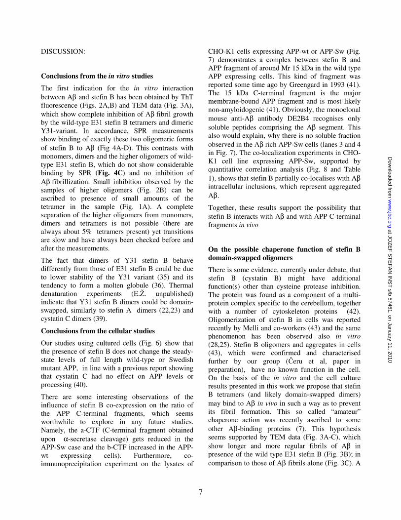
Conditions were established under which Aβ fibrillises regularly and stefin B does not. The mixture undergoing fibrillization was left unperturbed between measurements of ThT fluorescence. Amyloid-fibril formation by Aβ, alone and with stefin B in a 1:1 molar ratio, are shown in Figure 2. Dimeric Y31 stefin B completely inhibits Aβ fibril growth (Fig. 2A), which was also confirmed by transmission electron microscopy (TEM) (Fig. 3A). Wild-type E31 stefin B, which is composed from monomers and different oligomers,
shows no comparable inhibitory effect (Fig. 2A, Fig. 3B).
To see the effect of individual oligomers of the wild-type E31 stefin B on fibril growth of Aβ, monomers, dimers, tetramers and higher oligomers of stefin B were isolated by SEC (Fig. 1A, B). Each was then separately mixed with Aβ. It was observed that only the tetramers inhibited the Aβ fibril growth completely (Fig. 2B).
Stefin B interacts with Aββββ depending on the oligomeric state of the former
SPR measurements
Oligomer specific binding between stefin B and Aβ was demonstrated by using surface plasmon resonance (SPR). Aβ peptide was immobilised on the surface of a sensor chip CM5 and samples of the two stefin B variants and isolated oligomers were injected across. By several control experiments we made sure that Aβ bound to the chip was not considerably aggregated or fibrillar. We judge so from low ThT fluorescence of the Aβ sample in the buffer used for coupling before application to the chip and the TEM taken at the same time (Suppl, Fig.1). Even though some fibrils have formed in the first 5 minutes of incubation at pH 4 buffer for coupling, the majority of the peptide (70 %) does not bind ThT and is thus not in a fibrillar form. Furthermore, after immobilization of Aβ, the first injection of 25 mM NaOH desorb a significant amount of the peptide, which possibly represented noncovalently associated Aβ into oligomers or fibrils. We cannot exclude that minor amount of Aβ is in the aggregated or fibrillar form and thus the exact determination of rate constants or equilibrium affinity constant of the interaction was not feasible. The sensorgrams, however, showed clear interaction as depicted below and were reproducible.
Dimeric Y31 stefin B reacted with Aβ in a concentration dependent manner (Fig. 4A). To verify the interaction, we also immobilised Y31 stefin B and were able to observe the binding of Aβ (Fig. 4B). Tetramers of the wild-type E31 stefin B bound strongly to Aβ, but not so monomers, dimers and higher oligomers (Fig. 4C), thus, explaining the specific inhibitory effect of the tetramers on Aβ fibril growth (Fig. 2B). This interaction of the wild-
at JOZ
EF
ST
EF
AN
INS
T s/b 57461, on January 11, 2010
ww
w.jbc.org
Dow
nloaded from

6
type tetramers with A� was also concentration dependent as shown in Fig. 4D.
ESI MS measurements
Formation of the complex between stefin B and Aβ was monitored directly in an electrospray ionization mass spectrometry (ESI MS) experiment. The ESI MS spectrum of Aβ (1−40) displayed 3 major peaks (with charges +5, +4 and +3), which correspond to a molecular mass species of 4329.91 Da (theoretical mass 4329.89 Da). The isolated Y31 stefin B dimer displayed 2 major peaks corresponding to a molecular mass of 22,314.2 Da (theoretical Mw 22,301.4 Da). In the spectrum of stefin B and Aβ (1−40) mixture, 3 new peaks with molecular mass of 26,644.4 (charges +11, +10 and +9, respectively) were observed, which corresponds to the theoretical mass of the complex formed between one Y31 stefin B dimer and one molecule of Aβ (1−40).
The ESI MS results (Fig.5) clearly show the formation of a complex between Y31 stefin B dimer and A� (1-40). It coexists together with uncomplexed components and therefore it can be considered as weak. A similar complex between wild-type E31 stefin B tetramer and A� could not be observed, due to the lower intensity of MS peaks corresponding to higher oligomers.
Effect of stefin B on APP metabolism and A� generation in cells
In order to explore a possible functional link between stefin B and Aβ in vivo, we studied the effect of co-expressing stefin B in cell culture expressing APP.
Transfection of stefin B into CHO cells expressing either wild-type or Swedish mutant APP had no impact on cell toxicity, as determined by LDH release levels (not shown). Further it was shown (Fig. 6) that the presence of stefin B did not modify levels of wild-type or Swedish full length APP. Similarly, stefin B did not affect Aβ(1-40) secretion levels. In the absence of stefin B we observed a ~6-fold increase in A� levels in cells bearing the Swedish mutation (29), a difference that did not change in the presence of stefin B (Fig. 6).
Nevertheless, the presence of stefin B did have an impact on APP metabolism. Specifically, in cells expressing wild type APP, it led to an increase in the levels of C-terminal fragments of APP cleaved by �-secretase (bCTF) but not those derived from �-secretase (aCTF). By contrast, in cells expressing APP–Sw, stefin B reduced the level of aCTF without affecting bCTF (Fig. 6).
Co-immunoprecipitation experiment
In order to examine if APP / A� and stefin B associate in cells, we have transfected cDNA for stefin B into CHO cells expressing either wild-type or Swedish mutant APP. 24 hours after transfection the cells were lysed and immunoprecipitated with polyclonal antibodies against stefin B. Fraction bound to the anti - stefin B antibodies and the lysate were then run on 15% SDS gels followed by Western blotting with antibodies against A� [DE2B4] (Fig. 7) and stefin B (not shown).
As Figure 7 shows, a complex between stefin B and approximately 15 kDa C-terminal fragment of APP, comprising A� sequence, was detected in the APP-wt cells but not so in the cells expressing APP-Sw.
Co-localization of stefin B and A� aggregates in cells
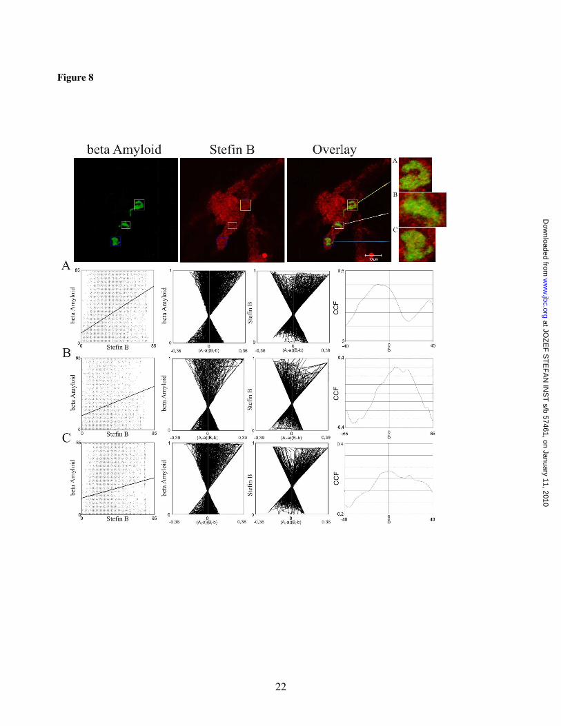
The possibility that A� aggregates and stefin B co-localise intracellularly was explored next. CHO-K1 cell line expressing APP-Sw, with the level of A� is ~6-fold increased (29), was transiently transfected with stefin B. Staining with mouse anti-A� antibodies and rabbit polyclonal anti-stefin B antibodies showed that stefin B and A� peptide associate with A� inclusions (Fig. 8). Visual inspection of the staining pairs suggested co-localization of the two proteins in A� inclusions, possibly A� aggresomes (38). The regions of interest (ROIs) were defined by a quality co-localization analysis. We used public domain tool JACoP for quantitative co-localization (Table 1). Dispersed scatter plots, the ICA plots, (Fig.8, graphs A,B,C) where most of the pixels are found on the right side of the plot, indicate partial co-localization of A� and stefin B.
at JOZ
EF
ST
EF
AN
INS
T s/b 57461, on January 11, 2010
ww
w.jbc.org
Dow
nloaded from
7
DISCUSSION:
Conclusions from the in vitro studies
The first indication for the in vitro interaction between Aβ and stefin B has been obtained by ThT fluorescence (Figs. 2A,B) and TEM data (Fig. 3A), which show complete inhibition of Aβ fibril growth by the wild-type E31 stefin B tetramers and dimeric Y31-variant. In accordance, SPR measurements show binding of exactly these two oligomeric forms of stefin B to Aβ (Fig 4A-D). This contrasts with monomers, dimers and the higher oligomers of wild-type E31 stefin B, which do not show considerable binding by SPR (Fig. 4C) and no inhibition of Aβ fibrillization. Small inhibition observed by the samples of higher oligomers (Fig. 2B) can be ascribed to presence of small amounts of the tetramer in the sample (Fig. 1A). A complete separation of the higher oligomers from monomers, dimers and tetramers is not possible (there are always about 5% tetramers present) yet transitions are slow and have always been checked before and after the measurements.
The fact that dimers of Y31 stefin B behave differently from those of E31 stefin B could be due to lower stability of the Y31 variant (35) and its tendency to form a molten globule (36). Thermal denaturation experiments (E.Ž. unpublished) indicate that Y31 stefin B dimers could be domain-swapped, similarly to stefin A dimers (22,23) and cystatin C dimers (39).
Conclusions from the cellular studies
Our studies using cultured cells (Fig. 6) show that the presence of stefin B does not change the steady-state levels of full length wild-type or Swedish mutant APP, in line with a previous report showing that cystatin C had no effect on APP levels or processing (40).
There are some interesting observations of the influence of stefin B co-expression on the ratio of the APP C-terminal fragments, which seems worthwhile to explore in any future studies. Namely, the a-CTF (C-terminal fragment obtained upon α-secretase cleavage) gets reduced in the APP-Sw case and the b-CTF increased in the APP-wt expressing cells). Furthermore, co-immunoprecipitation experiment on the lysates of
CHO-K1 cells expressing APP-wt or APP-Sw (Fig. 7) demonstrates a complex between stefin B and APP fragment of around Mr 15 kDa in the wild type APP expressing cells. This kind of fragment was reported some time ago by Greengard in 1993 (41). The 15 kDa C-terminal fragment is the major membrane-bound APP fragment and is most likely non-amyloidogenic (41). Obviously, the monoclonal mouse anti-Aβ antibody DE2B4 recognises only soluble peptides comprising the Aβ segment. This also would explain, why there is no soluble fraction observed in the Aβ rich APP-Sw cells (lanes 3 and 4 in Fig. 7). The co-localization experiments in CHO-K1 cell line expressing APP-Sw, supported by quantitative correlation analysis (Fig. 8 and Table 1), shows that stefin B partially co-localises with Aβ intracellular inclusions, which represent aggregated Aβ.
Together, these results support the possibility that stefin B interacts with Aβ and with APP C-terminal fragments in vivo
On the possible chaperone function of stefin B domain-swapped oligomers
There is some evidence, currently under debate, that stefin B (cystatin B) might have additional function(s) other than cysteine protease inhibition. The protein was found as a component of a multi-protein complex specific to the cerebellum, together with a number of cytoskeleton proteins (42). Oligomerization of stefin B in cells was reported recently by Melli and co-workers (43) and the same phenomenon has been observed also in vitro (28,25). Stefin B oligomers and aggregates in cells (43), which were confirmed and characterised further by our group (�eru et al, paper in preparation), have no known function in the cell. On the basis of the in vitro and the cell culture results presented in this work we propose that stefin B tetramers (and likely domain-swapped dimers) may bind to Aβ in vivo in such a way as to prevent its fibril formation. This so called “amateur” chaperone action was recently ascribed to some other Aβ-binding proteins (7). This hypothesis seems supported by TEM data (Fig. 3A-C), which show longer and more regular fibrils of Aβ in presence of the wild type E31 stefin B (Fig. 3B); in comparison to those of Aβ fibrils alone (Fig. 3C). A
at JOZ
EF
ST
EF
AN
INS
T s/b 57461, on January 11, 2010
ww
w.jbc.org
Dow
nloaded from

8
complete inhibition of fibril growth at the stage of granular aggregates happens in presence of the dimeric Y31-variant (Fig. 3A). The tetramers of E31 stefin B, which also completely inhibit ThT fluorescence (Fig. 2B) and bind strongly to Aβ (Fig. 4C,D) are expected to block fibrillization at the same stage.
From the 3D structures of stefin B monomer (34), tetramer (37) and protection pattern of the fibrils (44) it should be possible to derive possible Aβ binding site. The binding surface is not present in the higher oligomers, which showed no binding by SPR. Their 3D structure has not been solved as yet. In the future, as crystallization of the complex between stefin B and Aβ is a difficult task, maybe interaction could be observed by NMR.
Binding of Aβ to the � sheet edge was proposed for other Aβ binding proteins (5). Such a surface in stefin B could represent unpaired strand 5 or, alternatively, exposed surface formed by strands 2 and 3 by domain-swapping.
The feature common to chaperones is their transient interaction with non-native conformations of other proteins, which is followed either by preventing aggregation, correct folding or unfolding and, subsequent targeting to proteasomal or lysosomal degradation (45). Small heat shock proteins (Hsps) are made of typically 12-24 repeats of �-crystallin domains, which form dimers. The small heat shock proteins, �B-crystallins, bind to and inhibit amyloid fibril formation of Aβ, �2-microglobulin (46) and of �-synuclein (47). In all these cases the
interaction between the interacting proteins was weak.
In conclusion:
Mutations in stefin B (cystatin B) gene cause EPM1 (48). Mostly they lead to reduced protein expression, however, some are functional and lead to changed proteins, with different stability and aggregation properties (28). Lack of stefin B causes knock-out mice to undergo extensive loss of neurons in the granule layer of the cerebellum (49). They also show signs of ataxia and myoclonus. Stefin B gets overexpressed in many conditions of neurodegeneration, such as ALS and AD but as shown recently also in conditions of oxidative stress (50). The loss of inhibitory function might be related to mis-regulation of stefin B – cathepsin B axis of proteolysis and cell death. However, as the results presented here seem to suggest, abberant protein folding of stefin B or, aggregation of other proteins – stefin B acting as a chaperone, might also be an explanation for increased oxidative stress, when stefin B is missing.
at JOZ
EF
ST
EF
AN
INS
T s/b 57461, on January 11, 2010
ww
w.jbc.org
Dow
nloaded from

9
REFERENCES:
1. Ross, C.A., and Poirier, M.A. (2004) Nat.Med. 10, S10-S17
2. Walsh, D.M., and Selkoe, D.J. (2004) Protein Pept. Lett. 11, 213-228 Review
3. Cleary, J.P., Walsh, D.M., Hofmeister, J.J., Shankar, G.M., Kuskowski, M.A., Selkoe, D.J., and Ashe, K.H. (2005) Nat Neurosci 8, 79-84
4. Kayed, R. (2003). Science 300, 486. DOI: 10.1126/science.1079469
5. Yoshlike, Y., Minal, R., Matsuo, Y., Chen, Y.R., Kimura, T., Takashima, A.(2008) PLos ONE 3, e3235
6. Wilhelmus, M.M.M., Boelens WC, Otte-Holler I, Kamps B, de Waal RM, Verbeek MM (2006) Brain Res 1089: 67 – 78.
7. Wilhelmus MMM, de Wall, R.M.W., and Verneek, M.M. (2007) Mol.Neurobiol. 35, 203 – 216
8. Sastre, M., Calero, M., Pawlik, M., Mathews, P.M., Kumar, A., Danilov, V., Schmidt, S.D., Nixon, R.A., Frangione, B., and Levy, E. (2004). Neurobiol Aging 25, 1033-1043
9. Turk, V., Stoka, V., and Turk, D. (2008) Frontiers Biosci. 13, 5406-5420
10. Rawlings, N.D., Tolle, D.P., and Barrett, A.J. (2004) Nucleic Acids Res. 32, D160-D164
11. Martin, J.R., Craven, C.J., Jerala, R., Kroon-Zitko, L., Zerovnik, E., Turk, V., and Waltho, J.P. (1995) J Mol Biol. 246, 331-343.
12. Engh, R.A., Dieckmann, T., Bode, W., Auerswald, E.A., Turk, V., Huber, R., and Oschkinat, H. (1993) J Mol Biol. 234, 1060-1069.
13. Ekiel, I., Abrahamson, M., Fulton, D.B., Lindahl, P., Storer, A.C., Levadoux, W., Lafrance, M., Labelle, S., Pomerleau, Y., Groleau, D., LeSauteur, L., and Gehring, K. (1997) J Mol Biol. 271, 266-277.
14. Maruyama, K., Kametani, F., Ikeda, S., Ishihara, T., and Yanagisawa, N. (1992) Neurosci.Lett. 144, 38-42
15. Ii, K., Ito, H., Kominami, E., and Hirano, A. (1993) Virchows Archiv A Pathol Anat 423, 185 -194.
16. Bernstein, H. G., Rinne, R., Kirschke, H., Järvinen, M., Knöfel, B., et al. (1994). Brain Res Bull 33, 477–481.
17. Bernstein, H. G., Kirschke, H.,Wiederanders, B., Pollak, K. H., Zipress, A., and Rinne A. (1996) Mol Chem Neuropathol 27, 225–247.
18. Benussi, L., Binetti, G., and Ghidoni, R. (2006) In “Human Stefins and Cystatins” , Zerovnik E and Kopitar-Jerala N eds., Molecular Anatomy and Physiology of Proteins series, Uversky VN ed., Nova Sci Publ New York, pp.115-125
19. Žerovnik, E., Pompe-Novak, M., Škarabot, M., Ravnikar, M., Muševi�, I., and Turk, V. (2002) Biochim Biophys Acta 1594, 1-5
20. Jenko, S., Škarabot, M., Kenig, M., Gun�ar, G., Muševi�, I., Turk, D., and Žerovnik, E. (2004) Proteins: Struct Func Bioinf 55, 417-425
21. Škerget, K., Vilfan, A., Pompe-Novak, M., Turk, V., Waltho, J.P., Turk, D., and Žerovnik, E. (2009) Proteins: Struct FuncBioinf 74, 425-436
22. Jerala, R., and Žerovnik, E. (1999) J Mol Biol. 291, 1079-1089.
23. Staniforth, R.A., Giannini, S., Higgins, L.D., Conroy, M.J., Hounslow, A.M., Jerala, R., Craven, C.J., and Waltho, J.P. (2001) EMBO J. 20, 4774-4781.
24. Anderluh, G., Gutierrez-Aguirre, I., Rabzelj, S., �eru, S., Kopitar-Jerala, N., Ma�ek, P., Turk, V., and Žerovnik, E. (2005) FEBS J 272, 3042-3051
at JOZ
EF
ST
EF
AN
INS
T s/b 57461, on January 11, 2010
ww
w.jbc.org
Dow
nloaded from

10
25. �eru, S., Jenko-Kokalj, S., Rabzelj, S., Škarabot, M., Gutierrez-Aguirre, I., Kopitar-Jerala, N., Anderluh, G., Turk, D., Turk, V., and Žerovnik, E. (2008) Amyloid 15, 147-159
26. Rabzelj, S., Viero, S., Gutierrez-Aguirre, I., Turk, V., Dalla Serra, M., Anderluh, G., and Žerovnik, E. (2008) FEBS J 275, 2455 – 2466
27. Jerala, R., Trstenjak, M., Lenar�i�, B., and Turk, V. (1988) FEBS Lett 239, 41-44
28. Rabzelj, S., Turk, V., and Žerovnik, E. (2005) Protein Sci 14, 2713-2722
29. Perez, R.G., Squazzo, S.L., and Koo, E.H. (1996) J.Biol.Chem. 271, 9100-9107.
30. Yoon, I.S., Chen, E., Busse, T., Repetto, E., Lakshmana, M.K., Koo, E.H., and Kang, D.E. (2007). FASEB J. 21, 2742-2752.
31. Kopitar-Jerala, N., �urin-Šerbec, V., Jerala, R., Križaj, I., Gubenšek, F., and Turk, V. (1993) Biochim. Biophys. Acta 1164, 75-80.
32. Bolte, S., and Cordelières, P. (2006). J Microsc. 224, 213-232.
33. Ritonja, A, Machleidt, W. and Barrett, A.J. (1985). Biochem Biophys Res Commun. 131, 1187-1192.
34. Stubbs, M.T., Laber, B., Bode, W., Huber, R., Jerala, R., Lenar�i�, B., and Turk, V. (1990) EMBO J 9, 1939-1947
35. Žerovnik, E., Rabzelj, S., Kenig, M., and Turk, V. (2005) in Amyloid and Amyloidosis, eds. G. Grateau, RA Kyle, M Skinner, CRC Press, 2005, Boca Raton, Florida.
36. Žerovnik, E., Jerala, R., Kroon-Žitko, L., Turk, V., and Lohner, K. (1997) Eur J Biochem 245, 364-372
37. Jenko-Kokalj, S., Gun�ar, G., Štern, I., Morgan, G., Rabzelj, S., Kenig, M., Staniforth, R.A., Waltho, J.P., Žerovnik, E., and Turk, D. (2007) J Mol Biol 366, 1569-1579
38. Bückig, A., Tikkanen, R., Herzog, V., and Schmitz, A. (2002) Histochem Cell Biol. 118, 353-360.
39. Janowski, R., Kozak, M., Janowska, Grzonka, Z., Grubb, A., Abrahamson, M., and Jaskolski, M. (2001) Nat.Struct.Biol. 8, 316-320.
40. Mi, W., Pawlik, M., Sastre, M., Jung, S.S., Radvinsky, D.S., Klein, A.M., Sommer, J., Schmidt, S.D., Nixon, R.A., Mathews, P.M., and Levy, E. (2007) Nat Genet 39, 1440-1442.
41. Iverfeldt, K., Walaas, S.I., and Greengard, P. (1993) Proc Natl Acad Sci U S A. 90, 4146-4150.
42. Di Giamo, R., Riccio, M., Santi, S., Galeotti, C., Ambrosetti, D.C., and Melli, M. (2002) Hum Mol Genet 11, 2941 – 2950
43. Cipollini, E., Riccio, M., Di Giaimo, R., Dal Piaz, F., Pulice, G., Catania, S., Caldarelli, I., Dembic, M., Santi, S., and Melli, M. (2008) Biochim Biophys Acta 1783, 312-322.
44. Morgan, G.J., Giannini, S., Hounslow, A.M., Craven, C.J., Žerovnik, E., Turk, V., Waltho, J.P., and Staniforth, R.A. (2007). J Mol Biol. 375, 487-498. Epub 2007 Oct 22.
45. Saibil, H.R. (2008) Curr Opin Struct Biol 18, 35-42
46. Raman, B., Ban, T., Sakai, M., Pasta, S.Y., Ramakrishna, T., Naiki, H., Goto, Y., and Rao, Ch.M. (2005) Biochem J 392, 573-581
47. Rekas, A., Adda, C.G., Andrew Aquilina, J., Barnham, K.J., Sunde, M., Galatis, D., Williamson, N.A., Masters, C.L., Anders, R.F., Robinson, C.V., Cappai, R., and Carver, J.A. (2004) J Mol Biol 340, 1167-1183
48. Joensuu, T., Lehesjoki, A.E., Kopra, O. (2008). Epilepsia 49, 557 – 563.
at JOZ
EF
ST
EF
AN
INS
T s/b 57461, on January 11, 2010
ww
w.jbc.org
Dow
nloaded from

11
49. Pennacchio, L.A., Lehesjoki, A.E., Stone, N.E., Willour, V.L., Virtaneva, K., Miao, J., D'Amato, E., Ramirez, L., Faham, M., Koskiniemi, M., Warrington, J.A, Norio R, de la Chapelle A, Cox DR, and Myers RM. (1996) Science 271, 1731 – 1734.
50. Lehtinen, M.K., Tegelberg, S., Schipper, H., Su, H., Zukor, H., Manninen, O., Kopra, O., Joensuu, T., Hakala, P., Bonni, A., and Lehesjoki, A.E. (2009) J Neurosci. 29, 5910-5915.
Acknowledgements:
We are grateful to ing. Louise Kroon Žitko (Jožef Stefan Institute, Ljubljana) for help with the isolation of the recombinant stefin B protein. This work was funded by programs P1-0140 (proteolysis and its regulation) via the Slovenian Research Agency (ARRS) and Estonian Science Foundation grant 9171 (P.P.).
at JOZ
EF
ST
EF
AN
INS
T s/b 57461, on January 11, 2010
ww
w.jbc.org
Dow
nloaded from

12
FIGURE LEGENDS:
Figure 1 Oligomeric state of the wild-type E31 stefin B samples before (A) and after (B) SPR measurements. A Superdex 75 column (Pharmacia) was used to perform the size-exclusion chromatography. The column was equilibrated in phosphate buffer pH 7.0 containing 0.15 M NaCl. The flow rate was 0.5 ml/min at room temperature. 50 �l of the E31 stefin B monomers, dimers, tetramers and higher oligomers were injected.
Figure 2 Inhibition of A� fibril formation by stefin B measured by ThT fluorescence. A� peptide concentration was 17 �M throughout, pH 7.3, 40oC A) A� alone, 1:1 molar ratio of A� to Y31 stefin B (complete inhibition) and 1:1 molar ratio of A� to E31 stefin B. B) A� alone, 1:1 molar ratio to E31 stefin B monomers, dimers, tetramers and higher oligomers. Protein concentration of stefin B samples was 17 �M.
Figure 3 Morhology of the aggregates and fibrils as observed by TEM. After A� fibril formation reached steady state (cca. 100 hours at pH 7.3, 40oC) a sample of 15 �l was spread on the carbon coated grid, 1 % of uranyl acetate was added for a contrast and TEM images were recorded. At the same time point samples were taken of mixtures of A� with the following proteins A) Y31 stefin B B) E31 stefin B C) lysozyme and D,E) A� alone.
Figure 4 Binding experiments by SPR. The interaction of A� with the two stefin B iso-forms (Y31 and E31 stefin B) and with isolated oligomers of the wild-type E31 stefin B was assessed at 5 µl/ml in PBS buffer, pH 7.3. A) Binding of Y31 stefin B (analyte) to chip-immobilized A�. The concentration of Y31 stefin B was 0.5, 1, 2, 4 and 20 �M concentrations (from bottom to top). B) binding of 10 �M A� to chip-immobilized Y31 stefin B. C) Binding of monomers (curve a), dimers (b), tetramers (c) and higher oligomers of the wild-type E31 stefin B (d). to chip-immobilized A�. All forms were assessed at 100 µM concentration. D) Binding of the isolated tetramers of the wild-type E31 stefin B at 10, 25, 50 and 100 µM concentration (from bottom to top).
Figure 5 Complex detected by ESI MS. ESI MS spectra of Aβ (1−40), stefin B dimer (Y31-variant) and their mixture were recorded: A) - 2 µM Aβ (1−40), B) - the mixture of 2 µM Aβ and 2 µM stefin B and C) - 2 µM stefin B. Peaks corresponding to the Aβ − stefin B complex are denoted with an asterisk and numbers above the peaks denote charge state of the ions.
Figure 6 Western blots and ELISA of APP and its metabolites upon stefin B co-expression. Westen blots showing APP, the C-terminal fragments aCTF, bCF and stefin B in CHO-K1 cells expressing APP wild type A) and APP Swedish mutant B) and stefin B, respectively, in comparison to mock vector expression. Antibodies to stefin B, aCTF, bCF and APP were used (see, under Materials). In C) are shown the levels of A�(1-40) measured by ELISA in cells expressing APP wild type, and the same in cells co-expressing stefin B. Also shown are the levels of A�(1-40) in cells expressing APP-Sw or APP-Sw together with stefin B, which in both cases is 6-fold higher – as expected (26). Figure 7 Stefin B : A� /APP co-immunoprecipitation. CHO-K1 cells, expressing either wild-type or Swedish mutant APP, were transfected with the vector encoding stefin B and lysed after 24 hours. The lysates
at JOZ
EF
ST
EF
AN
INS
T s/b 57461, on January 11, 2010
ww
w.jbc.org
Dow
nloaded from

13
were immuno-precipitated with polyclonal anti-rabbit stefin B antibodies, after which 15% SDS gels and Western blotting with anti-A� antibodies [DE2B4] were performed (see Materials for more details). Lanes 1 or 2 show the results obtained on the whole cell lysates of the APP-wt and APP-Sw, respectively, and lanes 3 and 4 the corresponding samples after co-immunoprecipitation on the anti-stefin B antibodies column. The migration of molecular weight standards is indicated on the left. The anti-A� antibodies [DE2B4] clearly detect a fragment of Mr 15 kDa in the APP-wt cells.
Figure 8 Aββββ peptide co-localization with over-expressed stefin B in Aββββ inclusions. Upper line represents confocal images of CHO-K1 cells immunostained with an antibody against Aβ (green) and stefin B (red). Overlay represents the overlay of the two corresponding images. Scale bar is indicated on the pictures. Inclusion bodies were marked with different coloured squares (blue, white and yellow). A zoomed view of the selected regions of interest (ROIs) on the side of the colour panels are marked as A, B and C. Only the selected ROIs were used for quantitative co-localization analysis, which was performed by JACoP an ImageJ plug in. The three lines in boxes bellow the colour images marked as A, B, C, represent the results of the JACoPs analysis (see, Table 1).
at JOZ
EF
ST
EF
AN
INS
T s/b 57461, on January 11, 2010
ww
w.jbc.org
Dow
nloaded from

14
Table 1
ROI Rr Rr(random) P-value R=kstBxka� M(threshold) CCF ICQ
ka� 0,923 Ma� 0,46 A 0,37 0,0±0,166 100% kstB 0,591
0,739 MstB 0,563
0,402 0,171
ka� 0,857 Ma� 0,318 B 0,222 0,001±0,185 100% kstB 0,495
0,651 MstB 0,463
0,301 0,096
ka� 0,762 Ma� 0,416 C 0,164 0,001±0,155 100% kstB 0,565
0,656 MstB 0,43
0,261 0,072
Table 1: Quantitative co-localization analysis performed with different intensity correlation coefficient based tools, which are grouped in public domain tool JACoP. A, B and C represent selected regions of interest used in quantitative co-localization analysis. Rr - Pearson’s correlation coefficient, Rr(random) - Pearson’s correlation coefficient determined with Costes randomization based co-localization between random images of the green channel and original image of the red channel with 200 randomization rounds, P-value expressed as a percentage, R – overlap coefficient, ka� – overlap coefficient determined for Aβ (green channel) kstB – overlap coefficient determined for stefin B (red channel), M – Manders coefficient, Ma� – Manders coefficient determined for Aβ (green channel), MstB – Manders coefficient determined for stefin B (red channel) CCF – Van Steensel’s cross-correlation function, ICQ – Li’s intensity correction quotient.
at JOZ
EF
ST
EF
AN
INS
T s/b 57461, on January 11, 2010
ww
w.jbc.org
Dow
nloaded from

15
Figure 1
stefin B wt
monomer
monomer
A [m
AU
]
after SPR measurements
higher oligomershigher oligomers
tetramertetramer
dimer
dimer
before SPR measurements
2 4 6 8 10 12 14 16
V [ml]
2 4 6 8 10 12 14 16 18
A
B
at JOZ
EF
ST
EF
AN
INS
T s/b 57461, on January 11, 2010
ww
w.jbc.org
Dow
nloaded from

16
Figure 2
A
0
50
100
150
200
250
300
350
0 100 200 300 400 500 600
time (min)
ThT
fluor
esce
nce
/ 482
nm
Ab
Ab + stB(E31)
Ab + stB(Y31)
B
0
50
100
150
200
250
300
350
0 50 100 150 200 250 300 350 400
time (min)
ThT
fluor
esce
nce
/ 482
nm
Ab
Ab + mon(E31)
Ab + dim(E31)
Ab + tet(E31)
Ab + h.oli(E31)
at JOZ
EF
ST
EF
AN
INS
T s/b 57461, on January 11, 2010
ww
w.jbc.org
Dow
nloaded from
17
Figure 3
at JOZ
EF
ST
EF
AN
INS
T s/b 57461, on January 11, 2010
ww
w.jbc.org
Dow
nloaded from

18
Figure 4
at JOZ
EF
ST
EF
AN
INS
T s/b 57461, on January 11, 2010
ww
w.jbc.org
Dow
nloaded from

19
Figure 5
at JOZ
EF
ST
EF
AN
INS
T s/b 57461, on January 11, 2010
ww
w.jbc.org
Dow
nloaded from

20
Figure 6
at JOZ
EF
ST
EF
AN
INS
T s/b 57461, on January 11, 2010
ww
w.jbc.org
Dow
nloaded from

21
Figure 7:
at JOZ
EF
ST
EF
AN
INS
T s/b 57461, on January 11, 2010
ww
w.jbc.org
Dow
nloaded from
22
Figure 8
at JOZ
EF
ST
EF
AN
INS
T s/b 57461, on January 11, 2010
ww
w.jbc.org
Dow
nloaded from