Connexin 43 Reverses Malignant Phenotypes of Glioma Stem Cells by Modulating E-Cadherin
Upload
independentCategory
view
1download
0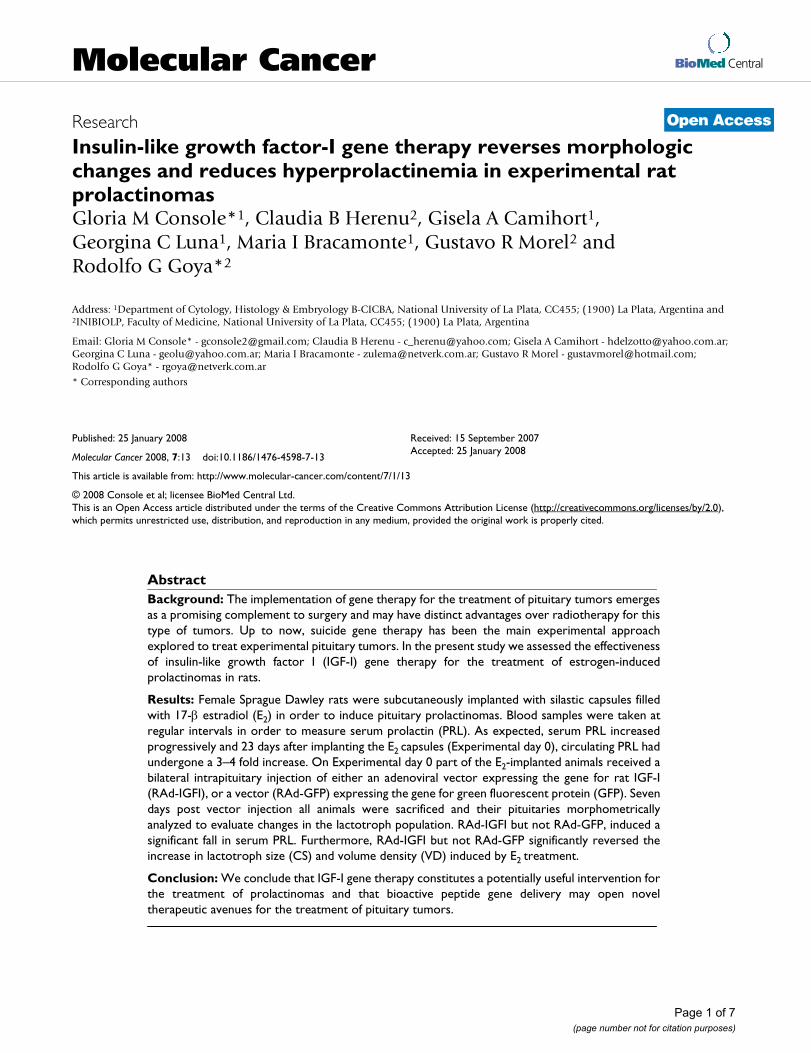
BioMed CentralMolecular Cancer
ss
Open AcceResearchInsulin-like growth factor-I gene therapy reverses morphologic changes and reduces hyperprolactinemia in experimental rat prolactinomasGloria M Console*1, Claudia B Herenu2, Gisela A Camihort1, Georgina C Luna1, Maria I Bracamonte1, Gustavo R Morel2 and Rodolfo G Goya*2Address: 1Department of Cytology, Histology & Embryology B-CICBA, National University of La Plata, CC455; (1900) La Plata, Argentina and 2INIBIOLP, Faculty of Medicine, National University of La Plata, CC455; (1900) La Plata, Argentina
Email: Gloria M Console* - [email protected]; Claudia B Herenu - [email protected]; Gisela A Camihort - [email protected]; Georgina C Luna - [email protected]; Maria I Bracamonte - [email protected]; Gustavo R Morel - [email protected]; Rodolfo G Goya* - [email protected]
* Corresponding authors
AbstractBackground: The implementation of gene therapy for the treatment of pituitary tumors emergesas a promising complement to surgery and may have distinct advantages over radiotherapy for thistype of tumors. Up to now, suicide gene therapy has been the main experimental approachexplored to treat experimental pituitary tumors. In the present study we assessed the effectivenessof insulin-like growth factor I (IGF-I) gene therapy for the treatment of estrogen-inducedprolactinomas in rats.
Results: Female Sprague Dawley rats were subcutaneously implanted with silastic capsules filledwith 17-β estradiol (E2) in order to induce pituitary prolactinomas. Blood samples were taken atregular intervals in order to measure serum prolactin (PRL). As expected, serum PRL increasedprogressively and 23 days after implanting the E2 capsules (Experimental day 0), circulating PRL hadundergone a 3–4 fold increase. On Experimental day 0 part of the E2-implanted animals received abilateral intrapituitary injection of either an adenoviral vector expressing the gene for rat IGF-I(RAd-IGFI), or a vector (RAd-GFP) expressing the gene for green fluorescent protein (GFP). Sevendays post vector injection all animals were sacrificed and their pituitaries morphometricallyanalyzed to evaluate changes in the lactotroph population. RAd-IGFI but not RAd-GFP, induced asignificant fall in serum PRL. Furthermore, RAd-IGFI but not RAd-GFP significantly reversed theincrease in lactotroph size (CS) and volume density (VD) induced by E2 treatment.
Conclusion: We conclude that IGF-I gene therapy constitutes a potentially useful intervention forthe treatment of prolactinomas and that bioactive peptide gene delivery may open noveltherapeutic avenues for the treatment of pituitary tumors.
Published: 25 January 2008
Molecular Cancer 2008, 7:13 doi:10.1186/1476-4598-7-13
Received: 15 September 2007Accepted: 25 January 2008
This article is available from: http://www.molecular-cancer.com/content/7/1/13
© 2008 Console et al; licensee BioMed Central Ltd. This is an Open Access article distributed under the terms of the Creative Commons Attribution License (http://creativecommons.org/licenses/by/2.0), which permits unrestricted use, distribution, and reproduction in any medium, provided the original work is properly cited.
Page 1 of 7(page number not for citation purposes)

Molecular Cancer 2008, 7:13 http://www.molecular-cancer.com/content/7/1/13
BackgroundPituitary adenomas constitute the most frequent neuroen-docrine pathology in humans, comprising up to 15% ofprimary intracranial tumors [1] and also are the mostprevalent pathology in old female rats [2]. In both species,prolactinomas are the most frequent type of pituitary ade-noma.
Estrogen exposure has been linked to the formation ofprolactinomas in both humans and animals [3-5]. Estro-gens, particularly estradiol and diethylstilbestrol, havebeen shown to induce lactotropic cell tumors within 2–4weeks in female rats. Women taking oral contraceptivesoften display increased prolactin (PRL) levels and haveincreased incidence of prolactinomas, although not allwomen appear to be equally susceptible to the mitogeniceffect of estradiol [6,7].
There are a number of growth factors that are estrogen-dependent and function in lactotropic proliferation, dif-ferentiation, and/or transformation. The relatedness ofthese factors and the significance of each alone are notwell-understood. Some of these estrogen-regulated factorsare epidermal growth factor (EGF), platelet-derivedgrowth factor (PDGF), transforming growth factor alpha(TGF-α), basic fibroblast growth factor (bFGF), inter-leukin-2 (IL-2), IL-6, fibroblast growth factor-4 (FGF-4),transforming growth factor-β (TGF-β), insulin-like growthfactor-I (IGF-I) and IGFI-II [8,9]. Although estrogen isknown to up-regulate IGF-I mRNA in normal rat pituitar-ies [10], large estrogen-induced pituitary adenomas havebeen reported to possess decreased levels of IGF-I mRNA.Furthermore, partial remission induced by the anti-estro-gen tamoxifen was associated with an increase in the pitu-itary content of IGF-I mRNA of these adenomas [11].Also, it has been shown that in primary cultures of rat lac-totrophs, estrogen has an antiproliferative action in thepresence of insulin or IGF-I [12,13]. In view of this evi-dence, it was of interest to determine whether in vivo over-expression of the gene for rat IGF-I in estrogen-induced ratprolactinomas could be able to restore lacotropic cellmorphology and reverse hyperprolactinemia. The presentreport describes our findings.
ResultsEffect of IGF-I gene therapy on pituitary lactotroph morphologyStereotaxic injection of RAd-GFP (an adenoviral vectorexpressing a chimeric variant of green fluorescent protein;see Methods) into pituitary adenomas induced a signifi-cant expression of GFP around the track of the needle (Fig.1) without significantly damaging the gland (Fig. 1, inset).As expected, three weeks of estrogen treatment induced asignificant increase in pituitary size (data not shown) aswell as clear changes in the lactotropic cell population,
already evident when pituitary sections were qualitativelyassessed (Fig 2). Estrogen-induced pituitary adenomasshowed a significant (p < 0.01) increase in lactotropic cellsize as compared with their intact counterparts (Fig. 3;also see Fig. 2). Seven days of IGF-I gene therapy signifi-cantly reversed this change (p < 0.01) although lactotrophcell surface (CS) was still higher than in intact animals(Fig. 3; also see Fig. 2). Lactotroph volume density (VD)was higher in estrogen-induced pituitary adenomas whencompared with the pituitaries of intact animals (Fig. 4).Again, IGF-I gene therapy significantly (p < 0.05) reversedthis alteration although lactotroph VD in RAd-IGF-Iinjected adenomas remained higher than in normal intactglands (Fig. 4).
Effect of intrapituitary tumor IGF-I gene therapy on serum PRL levelsAs expected, estrogen administration induced a markedhyperprolactinemia in the animals. Seven days after stere-otaxic intrapituitary injection of RAd-IGF-I but not RAd-GFP, a significant fall in serum PRL occurred in the estro-gen-treated rats (Fig. 5). Nevertheless, serum PRL of theRAd-IGF-I-treated rats remained higher than in the intactrats.
DiscussionCurrent therapies for pituitary tumors include surgery andradiotherapy, as well as pharmacological approaches forsome types and, although important advances have beenmade in the treatment of pituitary tumors with these strat-egies, a fully satisfactory therapy is not yet available [14].In this context, gene therapy appears as a potentially use-ful alternative for the treatment of pituitary tumors.
Early studies showed that a herpes simplex virus type-1(HSV1)-derived vector was highly effective in vivo for genetransfer in rat pituitary prolactinomas [15]. An adenoviralvector, RadTK, harboring the HSV-1 thymidine kinase(TK) suicide gene under the control of the human cytome-galovirus (hCMV) promoter, was used to transfer the TKgene to GH3 and AtT20 rodent pituitary tumor cells. Incu-bation of RadTK-treated GH3 and AtT20 cells with the pro-drug ganciclovir (which after phosphorylation by viral TKbecomes toxic) caused ample destruction of the cultures[16]. In the same study, estrogen/sulpiride-induced ratprolactinomas were stereotaxically injected with the sameRadTK. Subsequent injection of the host animals with twodaily intrapituitary doses of 25 mg ganciclovir/kg for 7days succeeded in partially reducing tumor size and serumPRL levels. Another type of gene therapy strategy for thetreatment of pituitary cancer is that based on the transferof a gene(s) with the ability to rescue the normal pheno-type of tumor cells. This approach has been implementedin mice heterozygous for the retinoblastoma (RB) tumorsuppressor gene (Rb+/- mice) which develop and succumb
Page 2 of 7(page number not for citation purposes)

Molecular Cancer 2008, 7:13 http://www.molecular-cancer.com/content/7/1/13
to characteristic pituitary intermediate lobe melanotrophtumors [17]. Intracranial delivery of an adenoviral vectorharboring the human RB cDNA to mice carrying activelygrowing melanotrophic tumors significantly reducedtumor growth and prolonged animal survival [18].
The present study is the first to implement effective pitui-tary tumor gene therapy using the gene for a bioactivepeptide. Within the time frame used, the treatment seemsto act mainly on PRL cell size and secretory activity ratherthan on PRL cell density (CD, data not shown). Ourresults are consistent with the above mentioned reportsdocumenting that large estrogen-induced pituitary adeno-mas possess decreased pituitary levels of IGF-I mRNA andthat the anti-estrogen tamoxifen increased the IGF-ImRNA content in the involuting adenomas [11]. Ourresults are also consistent with the evidence that in rat lac-totrophs, estrogen has an antiproliferative action in thepresence of insulin or IGF-I [12,13]. Furthermore, ourdata showing that IGF-I gene therapy reduces PRL secre-tion in estrogen-induced prolactinomas are in line withan early study in rat pituitary cells reporting that IGF-Iinhibited the estrogen induced rise in PRL and lactotroph
pituitary transcription factor-1 (Pit-1) mRNA levels infemales. This study also documented that the inhibitoryeffect of IGF-I on estrogen-induced PRL and lactotrophPit-1 mRNA levels did not occur in male pituitary cells[19].
The mechanism by which pituitary overexpression of IGF-I reduces lactotropic cell size and inhibits PRL secretion isnot clear. Although estrogen is known to increase pituitaryexpression of IGF-I mRNA, it also increases the expressionof IGF binding protein 2 (IGF-BP2) mRNA [10]. Since ourapproach involves pituitary overexpression of transgenicIGF-I, probably not paralleled by a concomitant increasein IGF-BP2, the possibility exists that high levels of freeIGF-I exert an inhibitory action on the lactotrophs. It iswell-established that IGF-I exerts a potent negative feed-back on somatotropic cells [20-22]. Since lactotrophs andsomatotrophs have a common origin and share a numberof physiologic features, it seems conceivable that highpituitary concentrations of free IGF-I could have an inhib-itory action on lactotropic cells. Clearly, further work isnecessary to clarify this matter.
Expression of transgenic GFP/TK in a pituitary adenomaFigure 1Expression of transgenic GFP/TK in a pituitary adenoma. The main panel shows the green fluorescence of transduced cells around the entry point (arrow) of the needle used to stereotaxically deliver RAd-GFP/TK into the tumor Obj. ×20. The inset shows a low magnification view of the same pituitary adenoma where an entry point of the needle can be seen (arrow). No structural damage is evident as a consequence of the injection. Obj. ×4.
Page 3 of 7(page number not for citation purposes)

Molecular Cancer 2008, 7:13 http://www.molecular-cancer.com/content/7/1/13
We conclude that IGF-I gene therapy constitutes a poten-tially useful intervention for the treatment of prolactino-mas and that bioactive peptide gene delivery may opennovel therapeutic avenues for the treatment of pituitarytumors.
MethodsAdenoviral vectorsRAd-IGFIA recombinant adenoviral (RAd) vector harboring the ratIGF-I gene (kindly donated by Dr. Peter Rotwein, OregonHealth Sciences University) was constructed in our labo-ratory by a variant of the two plasmid method employingthe AdMax® plasmid kit (Microbix, Ontario, Canada) [23].Briefly, the cDNA coding for rat IGF-I gene (obtained from
the mRNA for the IGF Ib precursor form [24]) was excisedfrom plasmid pBluescript KS, subcloned in pCA14 andinserted in the multiple cloning site (MCS) of shuttlepDC515 which contains an expression cassette consistingof the mouse cytomegalovirus promoter (mCMV) and thesimian virus 40 (SV40) polyadenylation signal, immedi-ately upstream and downstream to the MCS, respectively.The second plasmid of the kit, the genomic plasmid pBH-Gfrt(del)E1,3 FLP, consists of the entire genome of aden-ovirus 5 (Ad5), containing deletions in the regions E1 andE3. In cotransfected HEK293 cells, FLP recombinase isreadily expressed and efficiently catalyzes the site-directedrecombination of the expression cassette of pDC515 intopBHGfrt(del)E1,3 FLP, thus generating the genome of thedesired recombinant adenoviral vector, RAd-IGF-I (Fig.
Effect of IGF-I gene therapy on lactotropic cells of pituitary adenomasFigure 2Effect of IGF-I gene therapy on lactotropic cells of pituitary adenomas. Representative fields of specifically immunos-tained PRL-cells in the pituitary gland of an intact (Intact) rat, an animal treated with estrogen for 4 weeks (E only), and animal receiving estrogen for 4 weeks and RAd-GFP during the last experimental week (E + RAdGFP) and an animal receiving estro-gen for 4 weeks and RAd-IGF-I during the last experimental week (E + RAd-IGF-I). Insets. Higher magnification views of the specimens. EnVision system peroxidase. Bar: 25 µm. Inset: 12.5 µm.
Page 4 of 7(page number not for citation purposes)

Molecular Cancer 2008, 7:13 http://www.molecular-cancer.com/content/7/1/13
6). The newly generated RAd was rescued from HEK293cell lysates and plaque purified. It was further purified byultracentrifugation in CsCl gradient. Final virus stockswere titrated by a serial dilution plaque assay.
RAd-(GFP/TK)fusAn adenoviral vector termed RAd-(GFP/TK)fus, or RAd-GFP for short, was constructed in our laboratory followingthe general procedures outlined above and was used as a
control vector in the gene therapy studies. The vector har-bors a hybrid gene encoding the Aequorea victoriaenhanced green fluorescent protein fused to herpes sim-plex virus type 1 thymidine kinase (GFP/TK)fus (a kindgift from Dr. Jacques Galipeau, McGill University, Mon-treal, Canada). This hybrid gene is driven by the mouseCMV promoter. The vector was expanded in 293 cells andpurified and titrated as indicated above.
AnimalsYoung female Sprague-Dawley rats were housed in a tem-perature-controlled room (22 ± 2°C) on a 12:12 h light/dark cycle. Food and water were available ad libitum. Allexperiments with animals were done following the Ani-mal Welfare Guidelines of NIH (INIBIOLP's Animal Wel-fare Assurance No A5647-01).
Experimental design for in vivo IGF-I gene therapyAt experimental day -23 all animals but those of the intactgroup, were subcutaneously implanted a silastic capsulefilled with 17-β estradiol. Small blood samples (0.4 ml)were taken from the tail veins of all rats on experimentalday -16, -9, 0, day +2 and day +7 for PRL assay. On exper-imental day 0, some of the rats received bilateral 1.5 µlintrapituitary injections containing 3 × 10⁹ plaque form-ing units (pfu) of either RAd-GFP or RAd-IGF-I, respec-
Effect of IGF-I gene therapy on serum PRL levelsFigure 5Effect of IGF-I gene therapy on serum PRL levels. Serum PRL was measured in blood samples serially taken from Experimental day -23 to Experimental day 7. The number of rats used was 5 for all groups except for group E2 + RAd-IGF-I where 7 animals were assessed Asterisks indi-cate significant differences from serum PRL on experimental day 0 (vector injection); *: P < 0.05; **: P < 0.01. Other details are as in Fig. 3
Effect of IGF-I gene therapy on lactotropic cell sizeFigure 3Effect of IGF-I gene therapy on lactotropic cell size. Lactotroph cell surface (CS) in the pituitaries of animals sub-mitted to no treatment (intact), control treatments (E2 only and E2 + RAdGFP) or IGF-I gene therapy (E2 + RAd-IGF-I). Columns represent mean values whereas bars over columns represent SEM values. Five pituitaries per group were assessed. Asterisks indicate a highly significant difference from the intact group.
Effect of IGF-I gene therapy on lactotropic volume density (VD)Figure 4Effect of IGF-I gene therapy on lactotropic volume density (VD). Data correspond to the pituitaries of the same animals shown in Fig 3. Details are as in Fig. 3.
Page 5 of 7(page number not for citation purposes)
Molecular Cancer 2008, 7:13 http://www.molecular-cancer.com/content/7/1/13
tively. For this purpose, rats were anesthetized withinjection of ketamine hydrochloride (40 mg/kg, i.p.) andxylazine (8 mg/kg, i.m.), and placed in a stereotaxic frame.In order to access the pituitary region, the tip of a 26 gaugeneedle fitted to a 10 µl syringe was brought to the follow-ing coordinates relative to the bregma: 5.5 mm posterior,9.6 mm ventral and 0.7 mm right and left [25]. One weekafter intrapituitary injection rats were killed by decapita-tion. Pituitaries were immediately dissected, fixed andprocessed for routine histological studies.
ImmunohistochemistryStated in brief, pituitary tissues from 5 animals of eachgroup were fixed in Bouin's fluid and embedded in paraf-fin. Serial sections of 4 µm were obtained at different lev-els of the blocks following a ventral-to-dorsal sequence.The sections were immunostained, and then incubatedfor 1 h at room temperature with the anti-PRL primaryantiserum (murine, Dako, CA, USA), diluted 1:100. Thor-oughly washed sections were then treated for 30 min witha ready-to-use EnVision reaction system (Dako, CA, USA).The peroxide-sensitive chromogen was diaminobenzi-dine. In all instances, the specificity of the primary antise-
rum was monitored either by observing its ability to blockthe immunocytochemical reaction after its preabsorptionwith an excess of the related antigen or by its replacementwith normal rabbit serum or phosphate-buffered saline[26].
Image analysisMorphometry was performed as reported in detail previ-ously [27]. Measurements of pituitary cells were made bymeans of an image-analysis system (Imaging Technology,Optimas 5.2). The immunostained lactotrope cells andthe reference area (RA) were analyzed in each field on anaverage of ten micrographs taken from two levels (e.g. aand b) in the groups studied. These measurements wererecorded and processed automatically and the followingparameters subsequently calculated: volume density (VD= Σ cell area/RA) and cell size (CS) (the mean of individ-ual cell area, expressed in µm2). Reference area (RA) rep-resents the adenohypophyseal (pars distalis) area scanned,in which pituitary PRL cells were scored. Then, with thesum (Σ) of the individual areas (A), referred to as RA, weobtained volume density (VD), which indicates cell massaccording to a generally accepted concept.
Hormone assaysSerum levels of PRL were measured by a specific radioim-munoassay using the rat materials provided by Dr. A. F.Parlow, Pituitary Hormones and Antisera Center, UCLAMed. Center, U.S.A. Iodination grade hormones wereradiolabeled by the Iodo-Gen® method and purified onPD-10 Sephadex® G-25 M columns (Pharmacia, Uppsala,Sweden) equilibrated with 0.01 M phosphosaline, pH7.6. A 1/10 goat anti-rabbit IgG in 0.9% NaCl was used toseparate bound from free hormone. Serum concentra-tions of PRL were expressed in terms of NHPP rPRL RP-3.
Statistical analysisThe one way analysis of variance (ANOVA) were used toevaluate group differences. Tukey's method was chosen asa post hoc test. In Figs. 3 and 4 pair comparisons weremade between intact and each of the other experimentalgroups. Also, RAd-GFP vs. RAd-IGF1 pairs were comparedin both figures. In Fig. 5 one-way ANOVA was applied toeach set of data (intact, E2, etc) and pair comparisonswere made between Exptl. Day 0 and each of the othertime points.
Authors' contributionsGMC supervised the morphometric analysis of the pitui-taries and made a major contribution to the preparationof the manuscript; CBH constructed the viral vectors usedand performed the surgical procedures; GAC and GMLexecuted the different aspects of the morphometric analy-sis of pituitaries; MIB carried out the histological proce-dures involved in this study; GRM performed the studies
Diagrammatic representation of the procedure used to con-struct RAd-IGF-IFigure 6Diagrammatic representation of the procedure used to construct RAd-IGF-I. An expression cassette contain-ing the rat IGF-I cDNA (form B) flanked by the mCMV pro-moter (PmCMV) and the SV40 polyadenylation signal (SV40pA), upstream and downstream, respectively, was inserted in the shuttle vector pDC515. Subsequently, pDC515 and the genomic plasmid pBHGfrt(del)E1,3 FLP (which consists of the entire genome of adenovirus 5 (Ad 5), containing deletions in the regions E1 and E3) were cotrans-fected in permissive HEK 293 cells. Enzyme-directed recom-bination occurred in cotransfected cells, giving rise to RAd-IGFI. frt, recognition element for the yeast FLP recombinase; ITR, inverted terminal repeats; ∆1 and ∆3: deletions in the Ad 5 genome.
Page 6 of 7(page number not for citation purposes)

Molecular Cancer 2008, 7:13 http://www.molecular-cancer.com/content/7/1/13
Publish with BioMed Central and every scientist can read your work free of charge
"BioMed Central will be the most significant development for disseminating the results of biomedical research in our lifetime."
Sir Paul Nurse, Cancer Research UK
Your research papers will be:
available free of charge to the entire biomedical community
peer reviewed and published immediately upon acceptance
cited in PubMed and archived on PubMed Central
yours — you keep the copyright
Submit your manuscript here:http://www.biomedcentral.com/info/publishing_adv.asp
BioMedcentral
involving immunofluorescence analysis; RGG conceivedof the study and participated in its design and coordina-tion as well as in writing the manuscript. All authors readand approved the final manuscript.
AcknowledgementsThe authors are indebted to Mrs. C. Ferese for the technical assistance. This study was supported in part by grant 11/M105 from the National Uni-versity of La Plata and CIC-PBA to GMC and # PICT13588 from the National Agency for the Promotion of Science and Technology to RGG. RGG and GMC are CONICET and CIC-PBA career researchers, respec-tively.
References1. Daniels GH, Martin JB: Neuroendocrine regulation and diseases
of the anterior pituitary and hypothalamus. In Harrison's Princi-ples of Internal Medicine Edited by: Isselbacher KJ, Braunwald E, WilsonJD, Martin JB, Fauci AS, Kasper DL. New York: McGraw-Hill;1995:1891-1918.
2. Burek JD: Pathology of aging rats Boca Raton: CRC Press; 1978. 3. Lloyd RV: Estrogen-induced hyperplasia and neoplasia in the
rat anterior pituitary gland. Am J Pathol 1983, 113:198-206.4. Sarkar DK, Gottschall PE, Meites J: Damage to hypothalamic
dopaminergic neurons is associated with development ofprolactin-secreting tumors. Science 1982, 218:684-686.
5. Shy KK, McTiernan AM, Daling JR, Weiss NS: Oral contraceptiveuse and the occurrence of pituitary prolactinoma. J Am MedAssoc 1983, 249:2204-2207.
6. Carol N, Lauterbach H, Klinger G, Unger A, Michels W: Prolactinstimulation using the metoclopramide test in females takingoral contraceptives. Zentralbl Gynakol 1988, 110:1515-1521.
7. Fahy UM, Foster PA, Torodo HW, Hartog M, Hull MG: The effectof combined estrogen/progesterone treatment in womenwith hyperprolactinemic amenorrhea. Gynecol Endocrinol 1992,6:183-188.
8. Ray D, Melmed S: Pituitary cytokine and growth factor expres-sion and action. Endocr Rev 1997, 18:206-228.
9. Sarkar DK, Hentges ST, De A, Reddy RHR: Hormonal control ofpituitary prolactin secreting tumors. Frontiers in Bioscience 1998,3:934-943.
10. Michels KM, Lee WH, Seltzer A, Saavedra JM, Bondy CA: Up-regu-lation of pituitary [125I]insulin-like growth factor-I (IGF-I)binding and IGF binding protein-2 and IGF-I gene expressionby estrogen. Endocrinology 1993, 132:23-29.
11. Hána V, Haluzik M, Schreiber V: Independence of estrogen-induced pituitary proliferation on local IGF-I mRNA andEGF mRNA expression. Modifying effects of tamoxifen andterguride. Physiol Res 1998, 47:125-31.
12. Kawashima K, Yamakawa K, Takahashi W, Takizawa S, Yin P, Sugi-yama N, Kanba S, Arita J: The estrogen-occupied estrogenreceptor functions as a negative regulator to inhibit cell pro-liferation induced by insulin/IGF-1: a cell context-specificantimitogenic action of estradiol on rat lactotrophs in cul-ture. Endocrinology 2002, 143:2750-8.
13. Ishida M, Takahashi W, Itoh S, Shimodaira S, Maeda S, Arita J: Estro-gen actions on lactotroph proliferation are independent of aparacrine interaction with other pituitary cell types: a studyusing lactotroph-enriched cells. Endocrinology 2007,148:3131-3139.
14. Shimon I, Melmed S: Management of pituitary tumors. Annals IntMed 1998, 129:472-483.
15. Bolognani F, Albariño C, Romanowski V, Carri NG, Goya RG: In vitroand in vivo herpetic vector-mediated gene transfer in thepituitary gland: impact on hormone secretion. Eur J Endocrinol2001, 145:497-503.
16. Windeatt S, Southgate TD, Dewey RA, Bolognani F, Perone MJ, Lar-regina AT, Maleniak TC, Morris ID, Goya RG, Klatzmann D, Lowen-stein PR, Castro MG: Adenovirus-mediated herpes simplexvirus type-1 thymidine kinase gene therapy suppresses oes-trogen-induced pituitary prolactinomas. J Clin Endocr Metab2000, 85:1296-1305.
17. Hu N, Gutsmann A, Herbert DC, Bradley A, Lee WH, Lee EY: Het-erozygous Rb_1 delta 20/+mice are predisposed to tumors ofthe pituitary gland with a nearly complete penetrance. Onco-gene 1994, 9:1021-1029.
18. Riley DJ, Yu A, Lee WH: Adenovirus-mediated retinoblastomagene therapy supresses spontaneous pituitary melanotrophtumors in Rb+/- mice. Nature Med 1996, 2:1316-1321.
19. Chowen JA, Gonzalez-Parra S, Garcia-Segura LM, Argente J: Sexu-ally dimorphic interaction of insulin-like growth factor (IGF)-I and sex steroids in lactotrophs. J Neuroendocrinol 1998,10:493-502.
20. Morita S, Yamashita S, Melmed S: IGF-I action on rat anteriorpituitary cells: effects of intracellular messengers on GHsecretion and mRNA levels. Endocrinology 1987, 121:2000-2006.
21. Yamashita S, Melmed S: Insulin-like growth factor-I regulation ofgrowth hormone gene transcription in primary rat pituitarycells. J Clin Invest 1987, 79:449-452.
22. Yamashita S, Ong J, Melmed S: Regulation of human GH geneexpression by IGF-I in transfected cells. J Biol Chem 1987,262:13254-13257.
23. Hereñú CB, Cristina C, Rimoldi OJ, Becú-Villalobos D, Cambiaggi V,Portiansky EL, Goya RG: Restorative effect of insulin-likegrowth factor-i gene therapy in the hypothalamus of senilerats with dopaminergic dysfunction. Gene Therapy 2007, 14:237.
24. Daughaday WH, Rotwein P: Insulin-like growth factors I and II.Peptide, messenger ribonucleic acid and gene structures,serum, and tissue concentrations. Endocr Rev 1988, 10:68-91.
25. Paxinos G, Watson C: The rat brain in stereotaxic coordinates San Diego:Academic Press; 1998.
26. Cónsole GM, Jurado SB, Rulli S, Calandra RS, Gómez Dumm CLA:Ultrastructural and quantitative immunohistochemicalchanges induced by non steroid antiandrogens on pituitarygonadotroph population of prepuberal male rats. Cells Tissues& Organs 2001, 169:64-72.
27. Cónsole GM, Jurado SB, Petruccelli M, Carino M, Calandra RS,Gómez Dumm CLA: Influence of photoinhibition on the mor-phology and function of pituitary lactotropes in male goldenhamster. Neuroendocrinology 2002, 75:316-325.
Page 7 of 7(page number not for citation purposes)
Copyright © 2022 FDOKUMEN