Misfolded Proteins Recognition Strategies of E3 Ubiquitin Ligases and Neurodegenerative Diseases
Upload
independentCategory
view
1download
0
Insights into ubiquitin-conjugating enzyme/co-activator interactions from the structureof the Pex4p:Pex22p complex
Chris Williams1,*, Marlene van den Berg2,Santosh Panjikar1,4, Will A Stanley3,Ben Distel2 and Matthias Wilmanns1
1Structural Biology Unit, European Molecular Biology Laboratory,Hamburg, Germany, 2Department of Medical Biochemistry, AcademicMedical Center, Amsterdam, The Netherlands and 3ARC CoE in PlantEnergy Biology, University of Western Australia, Crawley, WesternAustralia, Australia
Ubiquitin-conjugating enzymes (E2s) coordinate distinct
types of ubiquitination via specific E3 ligases, to a large
number of protein substrates. While many E2 enzymes
need only the presence of an E3 ligase for substrate
ubiquitination, a number of E2s require additional, non-
canonical binding partners to specify their function. Here,
we have determined the crystal structure and function of
an E2/co-activator assembly, the Pex4p:Pex22p complex.
The peroxisome-associated E2 enzyme Pex4p binds the
peroxisomal membrane protein Pex22p through a binding
site that does not overlap with any other known interac-
tion interface in E2 enzymes. Pex22p association enhances
Pex4p’s ability to transfer ubiquitin to a substrate in vitro,
and Pex22p binding-deficient forms of Pex4p are unable to
ubiquitinate the peroxisomal import receptor Pex5p
in vivo. Our data demonstrate that the Pex4p:Pex22p
assembly, and not Pex4p alone, functions as the E2 en-
zyme required for Pex5p ubiquitination, establishing a
novel mechanism of E2 enzyme regulation.
The EMBO Journal (2012) 31, 391–402. doi:10.1038/
emboj.2011.411; Published online 15 November 2011
Subject Categories: membranes & transport; proteins
Keywords: E2 co-activator complex; peroxisomal receptor
recycling; Pex22p; Pex4p; ubiquitin-conjugating enzyme
structure
Introduction
The ubiquitin (Ub) cascade sequentially requires the activity
of three different enzymes to attach Ub to a substrate. First,
Ub is activated in an ATP-dependent manner by a ubiquitin-
activating enzyme (E1), then transferred to the active site
cysteine residue of a ubiquitin-conjugating enzyme (E2) and
finally, with the aid of a ubiquitin ligase (E3), attached to the
substrate. E2s play a coordinating role in the cascade, through
highly dynamic interactions with many E3 ligases, ultimately
determining the fate of hundreds of substrates for functional
regulation, or alternatively for degradation by the proteasome
(Capili and Lima, 2007; Dye and Schulman, 2007; Kleiger
et al, 2009; Ye and Rape, 2009). Since E2 enzymes are largely
conserved in structure and sequence, a number of common
principles are shared by all members, including conserved
binding sites for the E1 and E3 (Wenzel et al, 2011). However,
many E2 enzymes are required to perform specific ubiquiti-
nation events in the cell, such as the attachment of ubiquitin
to a certain side chain within a given substrate. While our
understanding of how E2s coordinate these events is still
incomplete, recent data may suggest an important role for
non-canonical binding partners. For example, the ERAD-
associated E3 ligase gp78 binds the ‘backside’ of Ube2g2
(an E2) through a region distinct from its ligase domain, and
this binding event enhances Ube2g2’s ubiquitin-conjugating
activity (Das et al, 2009). Two additional examples include
the non-catalytic ubiquitin E2 variant (UEV) protein Mms2,
which binds to the E2 Ube2n, allowing it to specifically build
lysine 63-linked polyubiquitin chains (VanDemark et al,
2001), and the non-covalent binding of ubiquitin itself to a
number of E2s, which may play a role in processive Ub chain
formation (Brzovic et al, 2006; Knipscheer et al, 2007).
While the above mentioned examples demonstrate regula-
tion of an E2 by a co-factor already implicated in the Ub
cascade (Ub, UEV, E3), the peroxisome-associated E2 enzyme
Pex4p (Ubc10p) requires the presence of an additional co-
factor, the trans-membrane (TM) domain-containing protein
Pex22p (Koller et al, 1999; Collins et al, 2000; Zolman et al,
2005). Pex4p is an unusual E2 enzyme in that it facilitates
site-specific cysteine ubiquitination of its substrate, the cy-
cling receptor Pex5p (Wiebel and Kunau, 1992; Platta et al,
2007; Williams et al, 2007). Pex5p, required for import of
proteins bearing a Peroxisomal Targeting Signal type-1 (PTS1)
into peroxisomes, is modified on a conserved cysteine residue
at its N-terminus by the addition of two Ub moieties (Kragt
et al, 2005; Williams et al, 2007). Pex4p-dependent ubiquiti-
nation of Pex5p (sometimes referred to as Pex5p monoubi-
quitination), which occurs on the peroxisomal membrane
and at a point after the receptor has delivered its cargo into
the peroxisomal lumen, regulates the dissociation of Pex5p
from the peroxisomal membrane (Williams et al, 2007; Platta
et al, 2008). However, this is not the only form of Pex5p
ubiquitination. Under certain conditions, for example in the
absence of Pex4p or in other mutants disturbed in the
membrane dissociation step, Pex5p is modified by another
E2 enzyme, Ubc4p (Platta et al, 2004; Kragt et al, 2005).
Ubc4p attaches between 1 and 4 Ub moieties to conserved
lysine residues in Pex5p (sometimes referred to as Pex5p
polyubiquitination), which targets the receptor for
proteasome-mediated degradation (Williams et al, 2007;Received: 6 May 2011; accepted: 19 October 2011; published online:15 November 2011
*Corresponding author. Structural Biology Unit, European MolecularBiology Laboratory, Notkestrasse 85, Hamburg 22603, Germany.Tel.: þ 49 40 8990 2144; Fax: þ 49 40 8990 2149;E-mail: [email protected] address: Australian Synchrotron, 800 Blackburn Road, Clayton,Victoria 3168, Australia
The EMBO Journal (2012) 31, 391–402 | & 2012 European Molecular Biology Organization | All Rights Reserved 0261-4189/12
www.embojournal.org
&2012 European Molecular Biology Organization The EMBO Journal VOL 31 | NO 2 | 2012
EMBO
THE
EMBOJOURNAL
THE
EMBOJOURNAL
391
Platta et al, 2008). For clarity, we will refer to the two forms of
Pex5p modification as Pex4p- and Ubc4p-dependent ubiqui-
tination.
Both the Pex4p- and Ubc4p-dependent ubiquitination of
Pex5p require Pex2p, Pex10p and Pex12p, three RING do-
main-containing proteins that form a complex at the peroxiso-
mal membrane (Kragt et al, 2005). Each RING domain exhibits
E3 ligase activity in vitro (Williams et al, 2008; Platta et al,
2009) yet the situation in vivo is complicated by the require-
ment of each RING domain for complex formation (Eckert and
Johnsson, 2003) and consequently, for Pex5p modification
(Kragt et al, 2005). While Pex10p’s E3 ligase activity is required
for Ubc4p-dependent ubiquitination of Pex5p in vivo, loss of
this activity does not affect the Pex4p-dependent modification
(Williams et al, 2008). Consequently, the identity of the E3
ligase functioning with Pex4p remains elusive.
Despite recent advances in the field, a number of key
issues remain concerning Pex4p-dependent ubiquitination
of Pex5p including crucially, the role of Pex4p’s binding
partner Pex22p in this process. Here, we report that Pex22p
not only provides a docking platform for Pex4p on the
peroxisomal membrane but also functions as an E2 co-
activator, required for Pex4p’s ability to ubiquitinate its
substrate, the PTS1 receptor Pex5p. The crystal structure of
the Pex4p:Pex22p complex from Saccharomyces cerevisiae
reveals a previously unobserved mode of binding for an E2
enzyme and, together with our functional data, provides
important insights into how a non-canonical binding partner
regulates the activity of an E2 enzyme.
Results
Pex22p specifically stimulates Pex4p’s
ubiquitin-conjugating activity in vitro
Pex22p contains an N-terminal TM segment and a large
cytosol-exposed region of unknown fold. Using secondary
structure and domain predictions, a number of truncated
versions of Pex22p were constructed and their ability to
interact with Pex4p was tested using the yeast two-hybrid
system (Supplementary Table S1). The minimal region re-
quired for Pex4p binding consists of residues 54–180, corre-
sponding to the soluble region of Pex22p (referred to as
Pex22S). Likewise, we observed that the first 14 amino
acids of Pex4p were not required for Pex22S binding and
consequently, we have used this truncated version, termed
Pex415–183, in subsequent experiments. We quantified the
Pex415–183:Pex22S interaction using isothermal titration mi-
crocalorimetry (ITC), confirming it to be direct, with a 1:1
stoichiometry and to be of high affinity, with a measured
dissociation constant (Kd) of 2 nM (Table I; Supplementary
Figure S1). In contrast, we were unable to detect an interac-
tion between Pex22S and the other ubiquitin-conjugating
enzyme involved in Pex5p ubiquitination, Ubc4p (Table I).
To test the effect such a tight binding partner would have
on Pex4p’s activity in vitro Pex415–183, together with E1 and
Ub was incubated in the presence or absence of ATP and
Pex22S and ubiquitin-conjugating activity was followed using
an E2 self-ubiquitination assay (Figure 1; Supplementary
Figure S2A). In line with our previous results (Williams
et al, 2008), purified Pex415–183 exhibited ubiquitin-conjugat-
ing activity, as seen by the formation of an anti-Ub and anti-
Pex4p reactive species in reactions containing ATP (Figure 1A
and B). The estimated molecular mass of this species
(26 kDa) correlates well with that of Pex415–183 attached to
Ub (19.5þ 8 kDa). Interestingly, the addition of Pex22S to the
reaction resulted not only in an enhanced production of
Ub–Pex415–183, but also in the formation of additional, higher
molecular mass species, corresponding to Pex415–183 attached
to 2 (35.5 kDa), 3 (43.5 kDa) or 4 (51.5 kDa) Ub moieties
(Figure 1A and B). All these species are lysine linked, as they
are resistant to the reducing agent b-mercaptoethanol. Pex22S
alone exhibited no activity (Figure 1B, last lane). Next, we
analysed Pex415–183’s ubiquitin-conjugating activity in the
presence of a mutant form of Ub where the lysine at position
48 was mutated to an arginine (K48R). Interestingly, inclu-
sion of K48R Ub effectively blocked the formation of the
additional, higher molecular mass Ub species, indicating that
Pex22S binding allows Pex415–183 to make lysine 48 linked Ub
chains (Figure 1C and D). The ubiquitin-conjugating activity
of Ubc4p remained unaffected upon addition of Pex22S
(Figure 1E), confirming Pex22S’s specificity for Pex415–183.
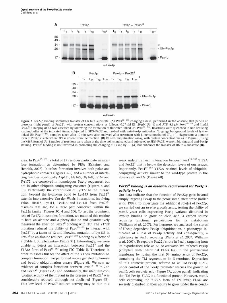
To gain further insight into the role of Pex22S binding, we
examined the rate of Pex415–183 charging by E1 in the pre-
sence and absence of Pex22S. Samples of the reactions were
taken soon after initiation (1, 3, 5 and 10 min) and were
subjected to non-reducing SDS–PAGE, western blotting and
anti-Pex4p immunoblotting (Figure 2A; Supplementary
Figure S2B). As the presence of Pex22S is not required for
the production of thioester-linked Ub–Pex415–183, nor does its
presence enhance production of this species (Figure 2A), we
conclude that Pex22S does not stimulate Pex4p’s activity by
enhancing Ub transfer from E1 to Pex4p.
Consequently, our results led us to believe that the role of
Pex22S involves the Ub-to-substrate transfer step. To test this
hypothesis, we examined the rate of Pex415–183 self-ubiquiti-
nation using the K48R form of Ub (Figure 2B; Supplementary
Figure S2C), which blocks the formation of Ub chains,
allowing us to gauge the rate of formation of a single Ub–
Pex4p species. We observed that the presence of Pex22S
enhances production of Ub–Pex415–183 by around 2–3-fold
(Figure 2B), confirming the theory that Pex22p binding
influences the transfer of Ub to a substrate.
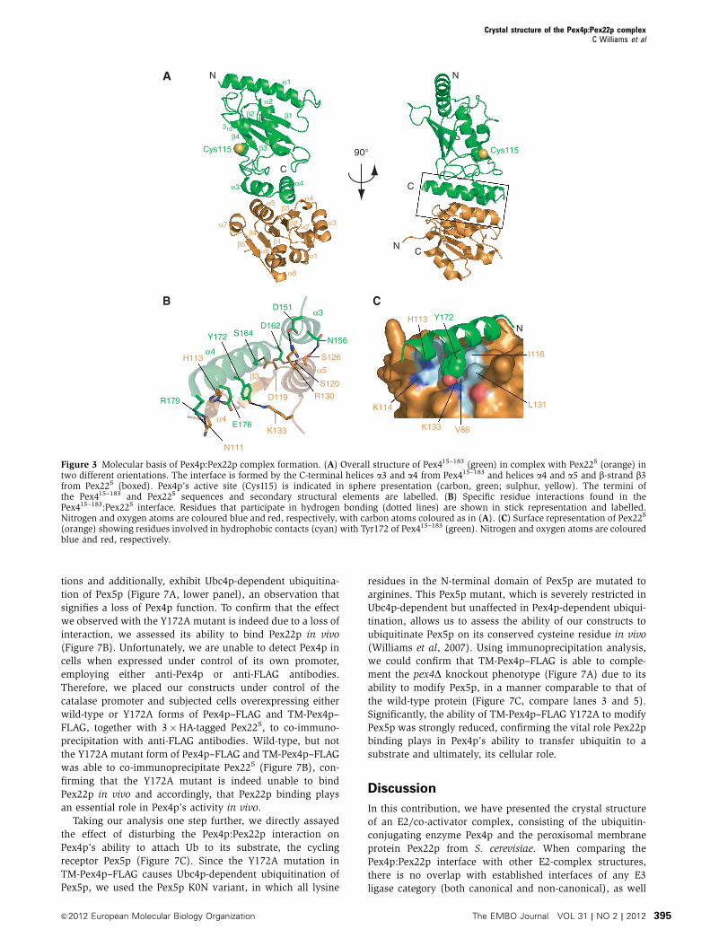
Overall structure of the Pex415–183:Pex22S complex
To unravel the molecular basis of Pex4p binding to Pex22p,
we determined the crystal structure of Pex415–183 in complex
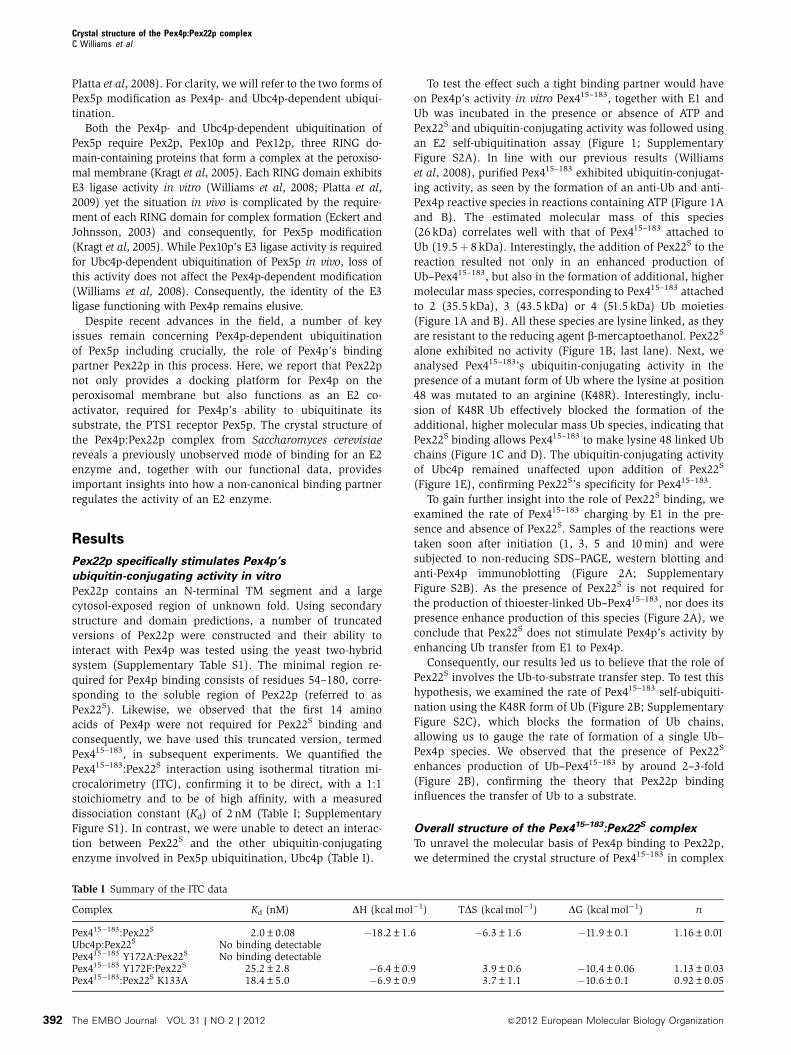
Table I Summary of the ITC data
Complex Kd (nM) DH (kcal mol�1) TDS (kcal mol�1) DG (kcal mol�1) n
Pex415�183:Pex22S 2.0±0.08 �18.2±1.6 �6.3±1.6 �11.9±0.1 1.16±0.01Ubc4p:Pex22S No binding detectablePex415�183 Y172A:Pex22S No binding detectablePex415�183 Y172F:Pex22S 25.2±2.8 �6.4±0.9 3.9±0.6 �10.4±0.06 1.13±0.03Pex415�183:Pex22S K133A 18.4±5.0 �6.9±0.9 3.7±1.1 �10.6±0.1 0.92±0.05
Crystal structure of the Pex4p:Pex22p complexC Williams et al
The EMBO Journal VOL 31 | NO 2 | 2012 &2012 European Molecular Biology Organization392

with Pex22S at 2.6 A (Figure 3; Table II). The structure is
complete, with the exception of the termini of Pex415–183 and
the three N-terminal residues and one loop (residues 145–
151) in Pex22S. Pex415–183 adopts a typical UBC fold (Cook
et al, 1997; Worthylake et al, 1998; Merkley and Shaw, 2004),
containing a core domain of four anti-parallel b-strands (b1–
b4) with one a-helix (a2), a small 310 helix (310), an N-
terminal helix (a1) and a helix-turn-helix (a3–a4) at the C-
terminus (Figures 3A and 4). As in other E2 structures, the
active site cysteine (Cys115, Figure 3A) is situated in a cleft
formed by the 310-helix and the loop region between a2
and a3.
Pex22S also comprises a mixed b-sheet/a-helical fold,
consisting of five parallel b-strands in the order b3–b2–b1–
b4–b5, which form a central b-sheet that is tightly sand-
wiched on both sides by eight a-helices (Figures 3A and 4;
Supplementary Figure S3). At first glance, the secondary
structural topology of Pex22S is suggestive of a Rossmann-
like fold, particularly as each b-strand is linked (N- and C-
terminally) to two a-helices (Supplementary Figure S3), a
topology typical for Rossmann and Rossmann-like folds (Rao
and Rossmann, 1973). However, although a similarity search
using PDBeFOLD (Krissinel and Henrick, 2004) did identify a
number of potential homologous Rossmann-like fold struc-
tures, the observation that homology was, for the most part,
confined to the central b-sheet, coupled with the low Q-scores
(highest Q-score: 0.193), leads us to the conclusion that the
overall fold of Pex22S is novel.
Examination of the residue-average B-factors in the
co-crystal structure, an evaluation of the fluctuation of
atoms in a protein structure relative to their average positions
(Supplementary Figure S4) suggests the presence of some
highly flexible ‘hotspots’ in Pex415–183—the a1–b1 loop, 310–
a2 loop and a2–a3 loop. It could be suggested that these
flexible loops effectively segment Pex415–183 into three,
loosely associated subdomains, a ‘cap’ comprising a1, a
‘core’ domain consisting of b1–b2–b3–b4–310–a2 and an
interaction motif formed by a-helices a3 and a4. These
subdomains could form the basis of the series of folding/
unfolding intermediates seen using CD (see Supplementary
Data).
Molecular details of the Pex4p–Pex22p interface
The Pex415–183:Pex22S high affinity binding site is established
by the insertion of helix a5 from Pex22S into the V-shaped
arrangement of the C-terminal helices a3 and a4 of Pex415–183
(Figures 3 and 5). Additional interactions are formed by the
exposed surface of helix a4 from Pex415–183 and strand b3,
helix a4 and the joining loop region from Pex22S, effectively
burying around 750 A2 of the solvent-accessible interface
B
α-Ub
ATP +– +– +
Pex4p
Pex4p
+ P
ex22
S
Pex22
S
- Ub–Pex4p- Ub2–Pex4p- Ub3–Pex4p- Ub4–Pex4p
- Ub–E1
- Ub
34 -26 -
17 -
43 -55 -
11 -
95 -72 -
130 -
A
34 -26 -
17 -
ATP +– +–
Pex4p
Pex4p
+ P
ex22
S
α-Pex4p
- Pex4p
- Ub–Pex4p- Ub2–Pex4p
43 -55 -
11 -
D E
ATP +–
α-Ub
+
Ubc4p
+ Pex
22S
34 -26 -
17 -
43 -55 -
11 -
95 -72 -
130 -
Ubn–Ubc4p
- Ub–E1
- Ub
*
++
WT U
b
K48R U
b
α-Pex4p
- Pex4p
- Ub–Pex4p- Ub2–Pex4p
ATP
34 -26 -
17 -
43 -55 -
11 -
α-Ub
++
WT U
b
K48R U
b
ATP
34 -26 -
17 -
43 -55 -
11 -
95 -72 -
130 -
- Ub–Pex4p- Ub2–Pex4p- Ub3–Pex4p- Ub4–Pex4p
- Ub
C
Figure 1 Pex22p acts as a specific co-activator for Pex4p in vitro. (A–E) E2 self-ubiquitination assays containing some of the following: 0.2mME1 enzyme, 11mM wild type (WT) or a lysine 48 to arginine mutant form (K48R) of Ub (Sigma), 5 mM ATP, 1.2 mM Pex415–183, 1.2 mM Ubc4pand 1.7mM Pex22S, probed with either anti-Pex4p (A, C) or anti-Ub antibodies (B, D and E). *Defines an anti-Ub crossreactive species, likely tobe Ubc4p. The presence of Pex22S stimulates the ubiquitin-conjugating activity of Pex415–183 and additionally allows Pex415–183 to produce Ubchains (A, B), linked through lysine 48 in Ub (C, D) but does not affect Ubc4p’s activity (E).
Crystal structure of the Pex4p:Pex22p complexC Williams et al
&2012 European Molecular Biology Organization The EMBO Journal VOL 31 | NO 2 | 2012 393
area. In Pex415–183, a total of 19 residues participate in inter-
face formation, as determined by PISA (Krissinel and
Henrick, 2007). Interface formation involves both polar and
hydrophobic contacts (Figures 3–5) and a number of interfa-
cing residues, specifically Asp151, Ala165, Gly168, Ile169 and
Tyr172, are conserved in homologous Pex4p sequences, but
not in other ubiquitin-conjugating enzymes (Figures 4 and
5B). Particularly, the contribution of Tyr172 to the interac-
tion, beyond the hydrogen bond to Lys133 from Pex22S,
extends into extensive Van-der-Waals interactions, involving
Val86, His113, Lys114, Leu116 and Leu131 from Pex22S,
residues that are, for a large part conserved within the
Pex22p family (Figures 3C, 4 and 5D). To test the prominent
role of Tyr172 in complex formation, we mutated this residue
to both an alanine and a phenylalanine and quantitatively
measured the effect on Pex22S binding with ITC. The Y172F
mutation reduced the ability of Pex415–183 to interact with
Pex22S by a factor of 12 and likewise, mutation of Lys133 in
Pex22S to an alanine reduced Pex415–183 binding by a factor of
9 (Table I; Supplementary Figure S1). Interestingly, we were
unable to detect an interaction between Pex22S and the
Y172A form of Pex415–183 using ITC (Table I). Therefore, in
order to assess further the affect of the Y172A mutation on
complex formation, we performed native gel electrophoresis
and in-vitro ubiquitination assays (Figure 6). We saw no
evidence of complex formation between Pex415–183 Y172A
and Pex22S (Figure 6A) and additionally, the ubiquitin-con-
jugating activity of the mutant in the presence of Pex22S was
considerably reduced, although not abolished (Figure 6B).
This low level of Pex22S-induced activity may be due to a
weak and/or transient interaction between Pex415–183 Y172A
and Pex22S that is below the detection levels of our assays.
Importantly, Pex415–183 Y172A retained levels of ubiquitin-
conjugating activity similar to the wild-type protein in the
absence of Pex22s (Figure 6B).
Pex22S binding is an essential requirement for Pex4p’s
activity in vivo
Our data indicate that the function of Pex22p goes beyond
simply targeting Pex4p to the peroxisomal membrane (Koller
et al, 1999). To investigate the additional role(s) of Pex22p,
we carried out an in-vivo growth assay, testing the ability of
pex4D yeast cells expressing Pex4p variants disturbed in
Pex22p binding to grow on oleic acid, a carbon source
requiring functional peroxisomes for its metabolism
(Williams et al, 2007). Furthermore, we analysed the status
of Ubc4p-dependent Pex5p ubiquitination, a phenotype in-
dicative of a loss of Pex4p activity and consequently, a
deficiency in Pex5p recycling (Platta et al, 2007; Williams
et al, 2007). To separate Pex22p’s role in Pex4p targeting from
its hypothesised role as E2 co-activator, we tethered Pex4p
(complete with C-terminal FLAG tag) to the peroxisomal
membrane by fusing the first 54 amino acids of Pex22p,
containing the TM segment, to its N-terminus. Expression
of this chimeric protein, referred to as TM-Pex4p–FLAG,
under control of the Pex4p promoter rescues the growth of
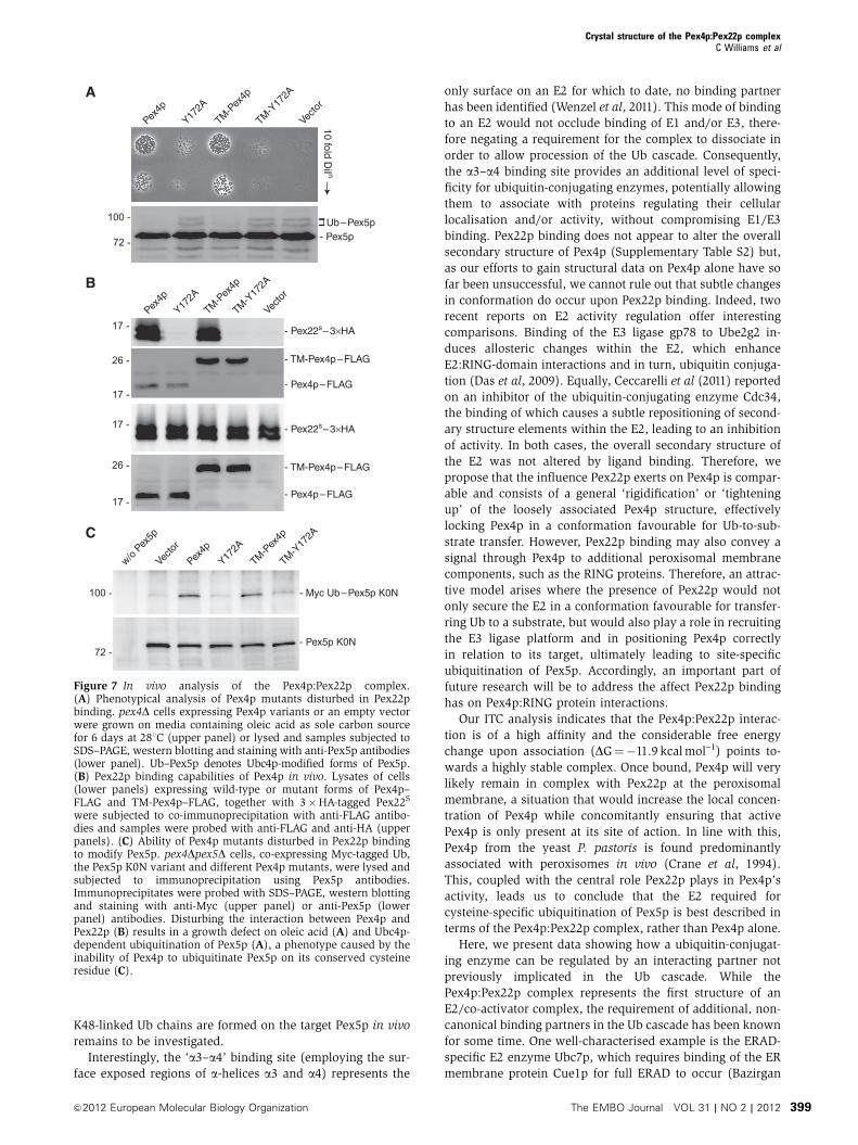
pex4D cells on oleic acid (Figure 7A, upper panel), indicating
that TM-Pex4p–FLAG is a functional protein. However, pex4Dcells expressing the Y172A form of TM-Pex4p–FLAG are
severely disturbed in their ability to grow under these condi-
A Pex4p
T5 T10T3T0 T1
Pex4p + Pex22S
T10+ T5 T10T3T0 T1 T10+
- Ub–Pex4p
- Pex4p
34 -26 -
17 -
55 -43 -
α-Pex4p
B
34 -26 -
17 -
55 -43 -
T30 T60 T120T0 T30 T60 T120T0
Pex4p Pex4p + Pex22S
- Ub–Pex4p
- Pex4p
α-Pex4p
*
Figure 2 Pex22p binding stimulates transfer of Ub to a substrate. (A) Pex415–183 charging assays, performed in the absence (left panel) orpresence (right panel) of Pex22S, with protein concentrations as follows: 0.27 mM E1, 29mM Ub, 10 mM ATP, 8.3mM Pex415–183 and 11mMPex22S. Charging of E2 was assessed by following the formation of thioester-linked Ub–Pex415–183. Reactions were quenched in non-reducingloading buffer at the indicated times, subjected to SDS–PAGE and probed with anti-Pex4p antibodies. To gauge background levels of lysine-linked Ub–Pex415–183, samples taken after 10 min were also analysed after treatment with b-mercaptoethanol (T10þ ). *Represents a dimericform of Pex4p visible when DTT is absent from the reaction. (B) E2 self-ubiquitination assay, with protein concentrations as in Figure 1, usingthe K48R form of Ub. Samples of reactions were taken at the time points indicated and subjected to SDS–PAGE, western blotting and anti-Pex4pstaining. Pex22S binding is not involved in promoting the charging of Pex4p by E1 (A) but enhances the transfer of Ub to a substrate (B).
Crystal structure of the Pex4p:Pex22p complexC Williams et al
The EMBO Journal VOL 31 | NO 2 | 2012 &2012 European Molecular Biology Organization394
tions and additionally, exhibit Ubc4p-dependent ubiquitina-
tion of Pex5p (Figure 7A, lower panel), an observation that
signifies a loss of Pex4p function. To confirm that the effect
we observed with the Y172A mutant is indeed due to a loss of
interaction, we assessed its ability to bind Pex22p in vivo
(Figure 7B). Unfortunately, we are unable to detect Pex4p in
cells when expressed under control of its own promoter,
employing either anti-Pex4p or anti-FLAG antibodies.
Therefore, we placed our constructs under control of the
catalase promoter and subjected cells overexpressing either
wild-type or Y172A forms of Pex4p–FLAG and TM-Pex4p–
FLAG, together with 3�HA-tagged Pex22S, to co-immuno-
precipitation with anti-FLAG antibodies. Wild-type, but not
the Y172A mutant form of Pex4p–FLAG and TM-Pex4p–FLAG
was able to co-immunoprecipitate Pex22S (Figure 7B), con-
firming that the Y172A mutant is indeed unable to bind
Pex22p in vivo and accordingly, that Pex22p binding plays
an essential role in Pex4p’s activity in vivo.
Taking our analysis one step further, we directly assayed
the effect of disturbing the Pex4p:Pex22p interaction on
Pex4p’s ability to attach Ub to its substrate, the cycling
receptor Pex5p (Figure 7C). Since the Y172A mutation in
TM-Pex4p–FLAG causes Ubc4p-dependent ubiquitination of
Pex5p, we used the Pex5p K0N variant, in which all lysine
residues in the N-terminal domain of Pex5p are mutated to
arginines. This Pex5p mutant, which is severely restricted in
Ubc4p-dependent but unaffected in Pex4p-dependent ubiqui-
tination, allows us to assess the ability of our constructs to
ubiquitinate Pex5p on its conserved cysteine residue in vivo
(Williams et al, 2007). Using immunoprecipitation analysis,
we could confirm that TM-Pex4p–FLAG is able to comple-
ment the pex4D knockout phenotype (Figure 7A) due to its
ability to modify Pex5p, in a manner comparable to that of
the wild-type protein (Figure 7C, compare lanes 3 and 5).
Significantly, the ability of TM-Pex4p–FLAG Y172A to modify
Pex5p was strongly reduced, confirming the vital role Pex22p
binding plays in Pex4p’s ability to transfer ubiquitin to a
substrate and ultimately, its cellular role.
Discussion
In this contribution, we have presented the crystal structure
of an E2/co-activator complex, consisting of the ubiquitin-
conjugating enzyme Pex4p and the peroxisomal membrane
protein Pex22p from S. cerevisiae. When comparing the
Pex4p:Pex22p interface with other E2-complex structures,
there is no overlap with established interfaces of any E3
ligase category (both canonical and non-canonical), as well
N111
90°
N
C
Cys115
NC
N
C
Cys115
α3 α4
A
α5α4
α1
α2
β1β2
β4
β3
310
α1
α2α3
α6
α7
α8
β2
β3
β4
β5 β1
α5
α4
α3
α4
D151
N156
D162S164Y172
E176
R179
S126H113
D119
S120
R130
K133
BY172
K133
H113
K114
I116
L131
V86
N
C
β3
Figure 3 Molecular basis of Pex4p:Pex22p complex formation. (A) Overall structure of Pex415–183 (green) in complex with Pex22S (orange) intwo different orientations. The interface is formed by the C-terminal helices a3 and a4 from Pex415–183 and helices a4 and a5 and b-strand b3from Pex22S (boxed). Pex4p’s active site (Cys115) is indicated in sphere presentation (carbon, green; sulphur, yellow). The termini ofthe Pex415–183 and Pex22S sequences and secondary structural elements are labelled. (B) Specific residue interactions found in thePex415–183:Pex22S interface. Residues that participate in hydrogen bonding (dotted lines) are shown in stick representation and labelled.Nitrogen and oxygen atoms are coloured blue and red, respectively, with carbon atoms coloured as in (A). (C) Surface representation of Pex22S
(orange) showing residues involved in hydrophobic contacts (cyan) with Tyr172 of Pex415–183 (green). Nitrogen and oxygen atoms are colouredblue and red, respectively.
Crystal structure of the Pex4p:Pex22p complexC Williams et al
&2012 European Molecular Biology Organization The EMBO Journal VOL 31 | NO 2 | 2012 395

as known E1, ubiquitin and UEV binding sites (Dye and
Schulman, 2007; Ye and Rape, 2009; van Wijk and
Timmers, 2010). This observation even holds for complexes
of more distantly related SUMO- and NEDD8-conjugating
enzymes. However, a partial overlap is seen with one of the
interfaces used by the SUMO conjugating enzyme Ube2L
when binding to Importin13 (Grunwald and Bono, 2010), as
well as when Ube2L binds to its substrate, RanGAP1
(Reverter and Lima, 2005). In both cases, Ube2L utilises
elements of a-helix 3 in the interaction. While the
Ube2L:Importin13 interaction plays a role in the nuclear
targeting, rather than the SUMO conjugating activity of
Ube2L (Grunwald and Bono, 2010), contacts made by a-
helix 3 allow the target lysine in RanGAP1 to come into
close proximity of Ube2L’s active site cysteine (Reverter and
Lima, 2005). Taking into account that many functional fea-
tures of E2 enzymes with different substrate specificities can
be conserved (Dye and Schulman, 2007; Ye and Rape, 2009;
van Wijk and Timmers, 2010), this observation may suggest
that Pex22p binding is involved in positioning Pex4p close to
its target, the conserved cysteine residue in the N-terminal
region of Pex5p. Cadwell and Coscoy (2008) reported that
attachment of Ub to a substrate can be dependent upon the
position of the target residue in the substrate, rather than the
type of residue, since both cysteine and lysine residues could
be utilised when present at equivalent positions in a sub-
strate. Recently, Grou et al (2009) showed that replacement of
the conserved cysteine in human Pex5p with a more conven-
tional lysine residue does not affect ubiquitination or function
of the protein, suggesting that positioning is more important
than the type of target residue for Pex5p ubiquitination. The
presence of Pex22p may therefore allow for site-specific
ubiquitination of the conserved cysteine, by correct position-
ing of Pex4p in relation to the exposed N-terminus of Pex5p.
As Pex22p stimulates the intrinsic Ub-to-substrate transfer
activity of Pex4p, but does not contain a RING/U-box or
HECT domain, it would be possible to describe the protein as
non-canonical E3 ligase, similar to the SUMO E3 RanBP2 or
the Replication Transcriptional Activator (RTA) Ub E3 ligase
(Pichler et al, 2004; Yu et al, 2005). These proteins lack
recognisable E3 ligase domains but can catalyse the transfer
of Ub/SUMO from E2 to substrates. In the case of RanBP2, the
comparison is particularly relevant since RanBP2 binding to
Ube2L, as with Pex22p binding to Pex4p, causes changes in
intrinsic E2 activity, probably through an allosteric mechan-
ism, rather than by providing a bridge between E2 and
substrate (Pichler et al, 2004). However, in vivo, Pex4p-
dependent ubiquitination of Pex5p still requires the presence
of three RING domain-containing proteins, all of which
exhibit E3 ligase activity in vitro (Williams et al, 2008;
Platta et al, 2009), suggesting that the role of Pex22p is
more fundamental to Pex4p’s activity and that additionally,
Pex22p’s function also precedes the step requiring an E3
ligase.
In vitro, binding of Pex22p allows Pex4p to produce lysine
48 linked Ub chains and in vivo, Pex22p binding is crucial for
Pex4p’s ability to attach two Ub moieties to the cycling
receptor Pex5p, a modification required for Pex5p recycling
(Platta et al, 2007; Williams et al, 2007). Although currently,
the type of linkage used to attach the second Ub to Pex5p has
Table II Data collection and refinement statistics
Data collection Native 1 Native 2 4 wavelength MAD
Space group P21 C2 C2Cell dimensions
a, b, c (A) 35.7, 81.3, 112.8 139.3, 43.1, 60.4 138.8, 43.1, 60.2a, b, g (deg) 90, 98.9, 90 90, 100.6, 90 90, 100.7, 90
Peak Inflection High energyremote
Low energyremote
Wavelength (A) 0.872 0.933 0.9776 0.9782 0.9747 0.960Resolution (A) 20.0–2.9
(3.0–2.9)a34.7–2.6
(2.74–2.6)20.0–2.7(2.9–2.7)
20.0–2.7(2.9–2.7)
20.0–2.7(2.8–2.7)
20.0–2.7(2.85–2.7)
Rmerge (%) 12.0 (62.0) 6.0 (47.0) 7.2 (26.8) 7.1 (27.5) 6.8 (27.5) 7.2 (29.6)I/sI 9.9 (2.6) 26.7 (4.8) 20.8 (5.9) 20.7 (5.7) 21.8 (5.8) 21.3 (5.6)Completeness (%) 97.4 (96.2) 99.4 (99.1) 99.6 (98.8) 99.4 (97.7) 99.0 (95.0) 99.4 (97.7)Redundancy 2.7 (2.6) 7.5 (7.6) 7.3 (6.7) 7.3 (6.8) 7.3 (6.8) 7.3 (6.9)
RefinementResolution (A) 20.0–2.6Unique reflections 10450Rwork/Rfree 20.8/25.9No. of atoms
Protein 2247Ligandb 16Water 9
R.m.s. deviationsBond lengths (A) 0.012Bond angles (deg) 1.33
Ramachandran plot (%)Favoured 94.2Allowed 5.8Disallowed 0
Each data set was collected from a single crystal.aValues in parentheses are for the highest resolution shell.bEthylene glycol.
Crystal structure of the Pex4p:Pex22p complexC Williams et al
The EMBO Journal VOL 31 | NO 2 | 2012 &2012 European Molecular Biology Organization396
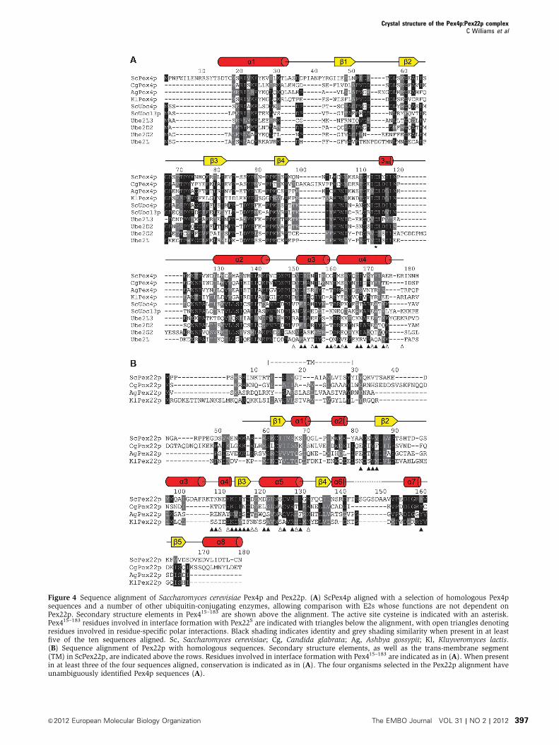
Figure 4 Sequence alignment of Saccharomyces cerevisiae Pex4p and Pex22p. (A) ScPex4p aligned with a selection of homologous Pex4psequences and a number of other ubiquitin-conjugating enzymes, allowing comparison with E2s whose functions are not dependent onPex22p. Secondary structure elements in Pex415–183 are shown above the alignment. The active site cysteine is indicated with an asterisk.Pex415–183 residues involved in interface formation with Pex22S are indicated with triangles below the alignment, with open triangles denotingresidues involved in residue-specific polar interactions. Black shading indicates identity and grey shading similarity when present in at leastfive of the ten sequences aligned. Sc, Saccharomyces cerevisiae; Cg, Candida glabrata; Ag, Ashbya gossypii; Kl, Kluyveromyces lactis.(B) Sequence alignment of Pex22p with homologous sequences. Secondary structure elements, as well as the trans-membrane segment(TM) in ScPex22p, are indicated above the rows. Residues involved in interface formation with Pex415–183 are indicated as in (A). When presentin at least three of the four sequences aligned, conservation is indicated as in (A). The four organisms selected in the Pex22p alignment haveunambiguously identified Pex4p sequences (A).
Crystal structure of the Pex4p:Pex22p complexC Williams et al
&2012 European Molecular Biology Organization The EMBO Journal VOL 31 | NO 2 | 2012 397

not been established in vivo, observations from the related
yeast H. polymorpha are worthy of note (Kiel et al, 2005).
Expression of the K48R mutant form of Ub in this organism
blocks peroxisomal matrix protein import, but only in the
presence of Pex4p, providing a link between the synthesis of
K48-linked Ub chains and the function of Pex4p. Whether
A
N C
α5
α4
β3
α3
α4
C115N
C
C
BN156
D151
D162
S164
Y172
E176
R179
I157
I169
A165
D
D119
S126
H113
F161
I116
C127
S120K133
V86
L131R130
Figure 5 Interfacing residues are conserved within homologous Pex4p and Pex22p sequences. (A) View of the Pex415–183 (surfacerepresentation) Pex22S complex (cartoon), indicating the region of Pex22S (shown in red and labelled) that forms the interface with Pex415–183.(B) Surface representation of the Pex22S binding site in Pex415–183, showing residues involved in interface formation which are invariant (blue),partially conserved (cyan) or not-conserved (grey) within homologous Pex4p sequences. (C) Mode of binding of Pex415–183 (cartoon) to Pex22S
(surface representation) indicating the interface forming helices a3 and a4 in Pex415–183 (blue). (D) Surface conservation within thePex415–183 binding site in homologous Pex22p sequences, indicating residues which are invariant (blue), partially conserved (cyan) ornot-conserved (grey).
- Pex4p
- Pex22s
- Complex
Pex4p
+ P
ex22
s
Y172A
+ P
ex22
s
Pex4p
Y172A
Pex22
sA B
α Ub
- Ub–Pex4p
- Ub2–Pex4p- Ub3–Pex4p- Ub4–Pex4p
- Ub–E1
- Ub
34 -
26 -
17 -
43 -55 -
11 -
95 -72 -
130 -
T30 T60 T120T0 T30 T60 T120T0
Pex4p + Pex22s Y172A + Pex22s
Pex4p
Y172A
T120 T120
Figure 6 In-vitro characteristics of the Y172A mutant form of Pex415–183. (A) Native gel electrophoresis analysis of the ability of wild-type andY172A forms of Pex415–183 to bind Pex22S. (B) E2 self-ubiquitination assay performed as in Figure 2B and containing E1, ATP, wild-type orY172A forms of Pex415–183 in the presence or absence of Pex22S. Samples of the reactions were taken at the time points indicated and probedwith anti-Ub antibodies. The Y172A mutation, which disturbs Pex22S binding (A), does not affect the intrinsic activity of Pex415–183 (B, leftpanel) but inhibits the Pex22S-dependent enhancement of Pex4p activity (B, right panel).
Crystal structure of the Pex4p:Pex22p complexC Williams et al
The EMBO Journal VOL 31 | NO 2 | 2012 &2012 European Molecular Biology Organization398
K48-linked Ub chains are formed on the target Pex5p in vivo
remains to be investigated.
Interestingly, the ‘a3–a4’ binding site (employing the sur-
face exposed regions of a-helices a3 and a4) represents the
only surface on an E2 for which to date, no binding partner
has been identified (Wenzel et al, 2011). This mode of binding
to an E2 would not occlude binding of E1 and/or E3, there-
fore negating a requirement for the complex to dissociate in
order to allow procession of the Ub cascade. Consequently,
the a3–a4 binding site provides an additional level of speci-
ficity for ubiquitin-conjugating enzymes, potentially allowing
them to associate with proteins regulating their cellular
localisation and/or activity, without compromising E1/E3
binding. Pex22p binding does not appear to alter the overall
secondary structure of Pex4p (Supplementary Table S2) but,
as our efforts to gain structural data on Pex4p alone have so
far been unsuccessful, we cannot rule out that subtle changes
in conformation do occur upon Pex22p binding. Indeed, two
recent reports on E2 activity regulation offer interesting
comparisons. Binding of the E3 ligase gp78 to Ube2g2 in-
duces allosteric changes within the E2, which enhance
E2:RING-domain interactions and in turn, ubiquitin conjuga-
tion (Das et al, 2009). Equally, Ceccarelli et al (2011) reported
on an inhibitor of the ubiquitin-conjugating enzyme Cdc34,
the binding of which causes a subtle repositioning of second-
ary structure elements within the E2, leading to an inhibition
of activity. In both cases, the overall secondary structure of
the E2 was not altered by ligand binding. Therefore, we
propose that the influence Pex22p exerts on Pex4p is compar-
able and consists of a general ‘rigidification’ or ‘tightening
up’ of the loosely associated Pex4p structure, effectively
locking Pex4p in a conformation favourable for Ub-to-sub-
strate transfer. However, Pex22p binding may also convey a
signal through Pex4p to additional peroxisomal membrane
components, such as the RING proteins. Therefore, an attrac-
tive model arises where the presence of Pex22p would not
only secure the E2 in a conformation favourable for transfer-
ring Ub to a substrate, but would also play a role in recruiting
the E3 ligase platform and in positioning Pex4p correctly
in relation to its target, ultimately leading to site-specific
ubiquitination of Pex5p. Accordingly, an important part of
future research will be to address the affect Pex22p binding
has on Pex4p:RING protein interactions.
Our ITC analysis indicates that the Pex4p:Pex22p interac-
tion is of a high affinity and the considerable free energy
change upon association (DG¼�11.9 kcal mol–1) points to-
wards a highly stable complex. Once bound, Pex4p will very
likely remain in complex with Pex22p at the peroxisomal
membrane, a situation that would increase the local concen-
tration of Pex4p while concomitantly ensuring that active
Pex4p is only present at its site of action. In line with this,
Pex4p from the yeast P. pastoris is found predominantly
associated with peroxisomes in vivo (Crane et al, 1994).
This, coupled with the central role Pex22p plays in Pex4p’s
activity, leads us to conclude that the E2 required for
cysteine-specific ubiquitination of Pex5p is best described in
terms of the Pex4p:Pex22p complex, rather than Pex4p alone.
Here, we present data showing how a ubiquitin-conjugat-
ing enzyme can be regulated by an interacting partner not
previously implicated in the Ub cascade. While the
Pex4p:Pex22p complex represents the first structure of an
E2/co-activator complex, the requirement of additional, non-
canonical binding partners in the Ub cascade has been known
for some time. One well-characterised example is the ERAD-
specific E2 enzyme Ubc7p, which requires binding of the ER
membrane protein Cue1p for full ERAD to occur (Bazirgan
A
- Pex5pUb–Pex5p
Vecto
r
Pex4p
Y172A
TM-P
ex4p
TM-Y
172A
10 fold DiI n
100 -
72 -
C
- Pex5p K0N
- Myc Ub–Pex5p K0N
w/o P
ex5p
Vecto
r
Pex4p
Y172A
TM-P
ex4p
TM-Y
172A
100 -
72 -
Vecto
r
Pex4p
Y172A
TM-P
ex4p
TM-Y
172A
- TM-Pex4p–FLAG
- Pex4p–FLAG
26 -
17 -
- Pex22s–3×HA17 -
- TM-Pex4p–FLAG
- Pex4p–FLAG
26 -
17 -
- Pex22s–3×HA17 -
B
Figure 7 In vivo analysis of the Pex4p:Pex22p complex.(A) Phenotypical analysis of Pex4p mutants disturbed in Pex22pbinding. pex4D cells expressing Pex4p variants or an empty vectorwere grown on media containing oleic acid as sole carbon sourcefor 6 days at 281C (upper panel) or lysed and samples subjected toSDS–PAGE, western blotting and staining with anti-Pex5p antibodies(lower panel). Ub–Pex5p denotes Ubc4p-modified forms of Pex5p.(B) Pex22p binding capabilities of Pex4p in vivo. Lysates of cells(lower panels) expressing wild-type or mutant forms of Pex4p–FLAG and TM-Pex4p–FLAG, together with 3�HA-tagged Pex22S
were subjected to co-immunoprecipitation with anti-FLAG antibo-dies and samples were probed with anti-FLAG and anti-HA (upperpanels). (C) Ability of Pex4p mutants disturbed in Pex22p bindingto modify Pex5p. pex4Dpex5D cells, co-expressing Myc-tagged Ub,the Pex5p K0N variant and different Pex4p mutants, were lysed andsubjected to immunoprecipitation using Pex5p antibodies.Immunoprecipitates were probed with SDS–PAGE, western blottingand staining with anti-Myc (upper panel) or anti-Pex5p (lowerpanel) antibodies. Disturbing the interaction between Pex4p andPex22p (B) results in a growth defect on oleic acid (A) and Ubc4p-dependent ubiquitination of Pex5p (A), a phenotype caused by theinability of Pex4p to ubiquitinate Pex5p on its conserved cysteineresidue (C).
Crystal structure of the Pex4p:Pex22p complexC Williams et al
&2012 European Molecular Biology Organization The EMBO Journal VOL 31 | NO 2 | 2012 399

and Hampton, 2008; Kostova et al, 2009). Interestingly, as
with Pex22p, Cue1p does not affect the activity of other E2
enzymes (Bazirgan and Hampton, 2008; Kostova et al, 2009).
Another protein in this class is RSUME (Carbia-Nagashima
et al, 2007). This RWD domain-containing protein binds to
Ube2L and stimulates the SUMOylation of a number of
substrates through an unknown mechanism. Again,
RSUME’s action is specific, as it does not influence the
activity of other E2s (Carbia-Nagashima et al, 2007).
Currently, our understanding of how such a small number
of E2 enzymes can control substrate-specific ubiquitination in
the presence of the huge number of E3 ligases present in the
cell is incomplete. We propose, based on our own results and
those previously reported that, for at least a subset of E2
enzymes, non-canonical binding partners not previously
linked to the Ub cascade play a crucial role in substrate-
specific ubiquitination, by regulating E2 activity and/or loca-
lisation. A number of large-scale two-hybrid and proteomics
approaches have identified numerous E2 binding partners
(Ewing et al, 2007; van Wijk et al, 2009), many of which are
not E3 ligases. Therefore, a challenge of future research will
be to define the role these interacting partners play in site-
specific and target-specific ubiquitination and the mechan-
istic details thereof.
Materials and methods
Strains, growth conditions, plasmids and protein preparationPlasmids are described in Supplementary Table S3. Yeast strainsused in this study are S. cerevisiae BJ1991 pex4D (Hettema et al,2000) and BJ1991 pex4Dpex5D (MATa, pex5HKanMX4, pex4HloxP,leu2, ura3-251, trp1, prb1-1122, pep4-3, gal2). For (co-) immuno-precipitations and trichloroacetic acid (TCA) lysates, yeast cellswere grown on 0.67% YNB containing 0.3% glucose for 24 h andthen shifted to 0.1% oleate medium containing 0.5% potassiumphosphate buffer pH 6.0, 0.3% yeast extract, 0.5% peptone, 0.2%Tween-40 and grown for between 7 and 16 h. Mutant plasmids wereconstructed using the QuikchangeTM site directed mutagenesis kit(Stratagene). Ubc4p and wild-type and mutant forms of Pex415–183
and Pex22S were expressed and purified as described in Williamset al (2008). Selenomethionine (SeMet) incorporation into Pex22S
was achieved using the B834 pRARE E. coli strain (Novagen) andconfirmed using mass spectrometry. Protein concentration wasdetermined by specific absorbance at 280 nm using a NanoDropTM
(http://www.nanodrop.com).
CrystallisationProteins were mixed and gel filtrated into GF buffer (25 mM Tris pH7.5, 100 mM NaCl, 1% glycerol, 3 mM b-mercaptoethanol) using aSuperdex 75 (10/300) column (GE Healthcare). Crystals of theresulting complex (at 10 mg ml–1) were obtained using hanging dropvapour diffusion at 201C: native Pex4:Pex22S complex 1 (Native 1)1.5M sodium malonate pH 7; native Pex4:Pex22S complex 2 (Native 2);SeMet-labelled Pex4:Pex22S complex, Native 2 condition plus 10 mM5-Amino-2,4,6-triiodoisophthalic acid (I3C, Sigma). Cryoprotectantwas 6 M sodium formate or 12% (v/v) ethylene glycol.
X-ray data collection and structure determinationSingle wavelength X-ray data sets were collected on ID23eh2(Native 1) and ID14eh1 (Native 2) at the ESRF, Grenoble. Themultiple wavelength, anomalous diffraction (MAD) X-ray data setwas collected on X12 at EMBL/DESY, Hamburg. All data wereprocessed using XDS (Kabsch, 1988) and COMBATand scaled usingSCALA (Evans, 1993). Using the MAD data set, three selenium siteswere located with autoSHARP (Vonrhein et al, 2007) and AUTO-RICKSHAW (Panjikar et al, 2005) and the phases were improvedusing RESOLVE (Terwilliger, 2000). Further improvements weremade using DMMULTI (Collaborative Computational ProjectNumber 4, 1994), carrying out multi-crystal averaging with theMAD and Native 1 data set phases. The resulting model was
enhanced manually using COOT (Emsley and Cowtan, 2004) andused as molecular replacement template for the Native 2 data set,from which the final model is derived. Refinement was carried outusing CNS (Brunger, 2007) and REFMAC (Murshudov et al, 1997),applying TLS parameters (Winn et al, 2001). Model quality wasassessed with Molprobity (Chen et al, 2010). The interface betweenPex415–183 and Pex22S was analysed using PISA (Krissinel andHenrick, 2007). All structure representations were made withPyMOL (http://www.pymol.org).
Isothermal titration microcalorimetryPrior to measurement, proteins were dialysed against GF bufferwithout glycerol. Measurements with Ubc4p or Pex415–183 at 10 mMas a sample and Pex22S at 100 mM as the titration ligand werecarried out at 251C on a MicroCal VP-ITC and data were fitted usingMicroCal Origin 7.0.
In-vitro ubiquitination assaysIn-vitro ubiquitination reactions, performed as described in Wil-liams et al (2008), contained 25 mM Tris pH 7.5, 75 mM NaCl, 5 mMMgCl2, 1 mM DTT, 5 mM ATP and proteins at the concentrationsindicated. E2 charging experiments (Figure 2A) were performed in25 mM Tris pH 7.5, 75 mM NaCl, 10 mM MgCl2, 10 mM ATP.Reactions were initiated by the addition of ATP and incubated at281C with gentle shaking. Samples were taken after 60 min, unlessotherwise stated, quenched with SDS–PAGE loading buffer contain-ing 5% b-mercaptoethanol (except in E2 charging experiments) andsubjected to SDS–PAGE. Western blots were probed with anti-Pex4p(Rabbit, polyclonal) and anti-Ub (FK2 clone, Biotrend).
Co-immunoprecipitation assaysOleate grown cells (20 A600 units) were lysed with glass beads in300ml lysis buffer (PBS pH 7.4, containing 0.5% NP-40, 1 mMPMSF, 0.5 mM DTT and yeast Protease Inhibitor Cocktail; Sigma) at41C. Debris was removed by centrifugation and lysates wereincubated end over end with 1ml of mouse monoclonal FLAGantiserum (M2 monoclonal F3165, Sigma) at 41C. After 2 h, 100 ml ofProtein G-Sepharose beads (GE Healthcare) was added and lysateswere incubated for 1 h at 41C. Precipitates were washed two timeswith lysis buffer and once with PBS and elution was carried out byheating the beads in 50 ml of IP-elution buffer (125 mM Tris, pH 6.8,1.5% SDS, 6 M urea and 20% glycerol) for 10 min at 371C. Sampleswere analysed by SDS–PAGE and immunoblotting, using antibodiesraised against the FLAG or HA (Mouse monoclonal 12CA5) tags.
Native gel electrophoresisPrior to loading, purified proteins (1:1 molar ratio for complexanalysis) were incubated end over end for 5 min at 211C in 25 mMTris pH 7.5, 75 mM NaCl. Samples were subjected to nativeelectrophoresis using 4–20% TBE gels (Invitrogen) according tothe manufacturer’s instructions and proteins were visualised withCoomassie brilliant blue staining.
MiscellaneousProtocols used for yeast two-hybrid analysis (Williams et al, 2005),TCA lysates, immunoprecipitation and growth analysis experiments(Williams et al, 2007) are described elsewhere.
Supplementary dataSupplementary data are available at The EMBO Journal Online(http://www.embojournal.org).
Acknowledgements
We thank Stan Fields for the Ubc4p plasmid, Jorg Hohfeld for the E1enzyme and Krisztian Fodor for helpful suggestions. We acknowl-edge the European Synchrotron Facility (ESRF) for provision ofsynchrotron radiation facilities and for assistance in using beam-lines ID14eh1 and ID23eh2. The project has been supported by aRubicon Fellowship from the Netherlands Organisation for ScientificResearch to CW (825.08.023), and by the Framework VI project ‘3DRepertoire’ from the European Commission to MW (LSHG-CT-2005-512028). The coordinates and structure factors of the Pex4p–Pex22pcomplex have been deposited in the protein data bank under theaccession code 2y9m.
Crystal structure of the Pex4p:Pex22p complexC Williams et al
The EMBO Journal VOL 31 | NO 2 | 2012 &2012 European Molecular Biology Organization400
Author contributions: CW, BD and MW designed the study;CW, BD and MB made the clones; CW expressed and purifiedthe proteins, performed ITC, crystallised the complex, collectedthe X-ray data and performed CD measurements; CW and SPsolved and refined the structure; WAS analysed and processedthe CD measurements; MB performed in-vitro ubiquitinationassays and in-vivo experiments; MW and BD supervised the
project; CW wrote the paper, with contributions from allauthors.
Conflict of interest
The authors declare that they have no conflict of interest.
References
Bazirgan OA, Hampton RY (2008) Cue1p is an activator of Ubc7p E2activity in vitro and in vivo. J Biol Chem 283: 12797–12810
Brunger AT (2007) Version 1.2 of the crystallography and NMRsystem. Nat Protoc 2: 2728–2733
Brzovic PS, Lissounov A, Christensen DE, Hoyt DW, Klevit RE(2006) A UbcH5/ubiquitin noncovalent complex is requiredfor processive BRCA1-directed ubiquitination. Mol Cell 21:873–880
Cadwell K, Coscoy L (2008) The specificities of Kaposi’s sarcoma-associated herpesvirus-encoded E3 ubiquitin ligases are deter-mined by the positions of lysine or cysteine residues within theintracytoplasmic domains of their targets. J Virol 82: 4184–4189
Capili AD, Lima CD (2007) Taking it step by step: mechanisticinsights from structural studies of ubiquitin/ubiquitin-like proteinmodification pathways. Curr Opin Struct Biol 17: 726–735
Carbia-Nagashima A, Gerez J, Perez-Castro C, Paez-Pereda M,Silberstein S, Stalla GK, Holsboer F, Arzt E (2007) RSUME, asmall RWD-containing protein, enhances SUMO conjugation andstabilizes HIF-1alpha during hypoxia. Cell 131: 309–323
Ceccarelli DF, Tang X, Pelletier B, Orlicky S, Xie W, Plantevin V,Neculai D, Chou YC, Ogunjimi A, Al-Hakim A, Varelas X, KoszelaJ, Wasney GA, Vedadi M, Dhe-Paganon S, Cox S, Xu S, Lopez-Girona A, Mercurio F, Wrana J et al (2011) An allosteric inhibitorof the human cdc34 ubiquitin-conjugating enzyme. Cell 145:1075–1087
Chen V, Arendall W, Headd J, Keedy D, Immormino R, Kapral G,Murray L, Richardson J, Richardson D (2010) MolProbity: all-atom structure validation for macromolecular crystallography.Acta Crystallogr D Biol Crystallogr 66: 12–21
Collaborative Computational Project, Number 4 (1994) The CCP4suite: programs for protein crystallography. Acta Crystallogr DBiol Crystallogr 50: 760–763
Collins CS, Kalish JE, Morrell JC, McCaffery JM, Gould SJ (2000)The peroxisome biogenesis factors pex4p, pex22p, pex1p, andpex6p act in the terminal steps of peroxisomal matrix proteinimport. Mol Cell Biol 20: 7516–7526
Cook WJ, Martin PD, Edwards BF, Yamazaki RK, Chau V (1997)Crystal structure of a class I ubiquitin-conjugating enzyme (Ubc7)from Saccharomyces cerevisiae at 2.9 angstroms resolution.Biochemistry 36: 1621–1627
Crane DI, Kalish JE, Gould SJ (1994) The Pichia pastoris PAS4 geneencodes a ubiquitin-conjugating enzyme required for peroxisomeassembly. J Biol Chem 269: 21835–21844
Das R, Mariano J, Tsai YC, Kalathur RC, Kostova Z, Li J, Tarasov SG,McFeeters RL, Altieri AS, Ji X, Byrd RA, Weissman AM (2009)Allosteric activation of E2-RING finger-mediated ubiquitylationby a structurally defined specific E2-binding region of gp78. MolCell 34: 674–685
Dye BT, Schulman BA (2007) Structural mechanisms underlyingposttranslational modification by ubiquitin-like proteins. AnnuRev Biophys Biomol Struct 36: 131–150
Eckert JH, Johnsson N (2003) Pex10p links the ubiquitin-conjugat-ing enzyme Pex4p to the protein import machinery of theperoxisome. J Cell Sci 116: 3623–3634
Emsley P, Cowtan K (2004) Coot: model-building tools for molecu-lar graphics. Acta Crystallogr D Biol Crystallogr 60: 2126–2132
Evans PR (1993) In Proceedings of the CCP4 Study Weekend, SawyerL, Isaacs N, Bailey S (eds) pp 114–122. Warrington: DaresburyLaboratory
Ewing RM, Chu P, Elisma F, Li H, Taylor P, Climie S, McBroom-Cerajewski L, Robinson MD, O’Connor L, Li M, Taylor R, DharseeM, Ho Y, Heilbut A, Moore L, Zhang S, Ornatsky O, Bukhman YV,Ethier M, Sheng Y et al (2007) Large-scale mapping of humanprotein-protein interactions by mass spectrometry. Mol Syst Biol3: 89
Grou CP, Carvalho AF, Pinto MP, Huybrechts SJ, Sa-Miranda C,Fransen M, Azevedo JE (2009) Properties of the ubiquitin-pex5pthiol ester conjugate. J Biol Chem 284: 10504–10513
Grunwald M, Bono F (2010) Structure of Importin13–Ubc9 complex:nuclear import and release of a key regulator of sumoylation.EMBO J 30: 427–438
Hettema EH, Girzalsky W, van Den Berg M, Erdmann R, Distel B(2000) Saccharomyces cerevisiae pex3p and pex19p are requiredfor proper localization and stability of peroxisomal membraneproteins. EMBO J 19: 223–233
Kabsch W (1988) Automatic processing of rotation diffraction datafrom crystals of initially unknown symmetry and cell constants.J Appl Cryst 21: 916–924
Kiel JA, Otzen M, Veenhuis M, van der Klei IJ (2005) Obstruction ofpolyubiquitination affects PTS1 peroxisomal matrix protein im-port. Biochim Biophys Acta 1745: 176–186
Kleiger G, Saha A, Lewis S, Kuhlman B, Deshaies RJ (2009) RapidE2-E3 assembly and disassembly enable processive ubiquityla-tion of cullin-RING ubiquitin ligase substrates. Cell 139: 957–968
Knipscheer P, van Dijk WJ, Olsen JV, Mann M, Sixma TK (2007)Noncovalent interaction between Ubc9 and SUMO promotesSUMO chain formation. EMBO J 26: 2797–2807
Koller A, Snyder WB, Faber KN, Wenzel TJ, Rangell L, Keller GA,Subramani S (1999) Pex22p of Pichia pastoris, essential forperoxisomal matrix protein import, anchors the ubiquitin-con-jugating enzyme, Pex4p, on the peroxisomal membrane. J CellBiol 146: 99–112
Kostova Z, Mariano J, Scholz S, Koenig C, Weissman AM (2009) AUbc7p-binding domain in Cue1p activates ER-associated proteindegradation. J Cell Sci 122: 1374–1381
Kragt A, Voorn-Brouwer T, van den Berg M, Distel B (2005) TheSaccharomyces cerevisiae peroxisomal import receptor Pex5pis monoubiquitinated in wild type cells. J Biol Chem 280:7867–7874
Krissinel E, Henrick K (2004) Secondary-structure matching(PDBeFold), a new tool for fast protein structure alignment inthree dimensions. Acta Crystallogr D Biol Crystallogr 60: 2256–2268
Krissinel E, Henrick K (2007) Inference of macromolecular assem-blies from crystalline state. J Mol Biol 372: 774–797
Merkley N, Shaw GS (2004) Solution structure of the flexible class IIubiquitin-conjugating enzyme Ubc1 provides insights for poly-ubiquitin chain assembly. J Biol Chem 279: 47139–47147
Murshudov GN, Vagin AA, Dodson EJ (1997) Refinement of macro-molecular structures by the maximum-likelihood method. ActaCrystallogr D Biol Crystallogr 53: 240–255
Panjikar S, Parthasarathy V, Lamzin VS, Weiss MS, Tucker PA (2005)Auto-Rickshaw: an automated crystal structure determinationplatform as an efficient tool for the validation of an X-raydiffraction experiment. Acta Crystallogr D Biol Crystallogr 61:449–457
Pichler A, Knipscheer P, Saitoh H, Sixma TK, Melchior F (2004) TheRanBP2 SUMO E3 ligase is neither HECT- nor RING-type. NatStruct Mol Biol 11: 984–991
Platta HW, Debelyy MO, El Magraoui F, Erdmann R (2008) The AAAperoxins Pex1p and Pex6p function as dislocases for the ubiqui-tinated peroxisomal import receptor Pex5p. Biochem Soc Trans36: 99–104
Platta HW, El Magraoui F, Baumer BE, Schlee D, Girzalsky W,Erdmann R (2009) Pex2 and pex12 function as protein-ubiquitinligases in peroxisomal protein import. Mol Cell Biol 29: 5505–5516
Platta HW, El Magraoui F, Schlee D, Grunau S, Girzalsky W,Erdmann R (2007) Ubiquitination of the peroxisomal importreceptor Pex5p is required for its recycling. J Cell Biol 177:197–204
Crystal structure of the Pex4p:Pex22p complexC Williams et al
&2012 European Molecular Biology Organization The EMBO Journal VOL 31 | NO 2 | 2012 401

Platta HW, Girzalsky W, Erdmann R (2004) Ubiquitination of theperoxisomal import receptor Pex5p. Biochem J 177: 37–45
Rao ST, Rossmann MG (1973) Comparison of super-secondarystructures in proteins. J Mol Biol 76: 241–256
Reverter D, Lima CD (2005) Insights into E3 ligase activity revealedby a SUMO-RanGAP1-Ubc9-Nup358 complex. Nature 435:687–692
Terwilliger TC (2000) Maximum-likelihood density modification.Acta Crystallogr D Biol Crystallogr 56: 965–972
van Wijk SJ, de Vries SJ, Kemmeren P, Huang A, Boelens R, BonvinAM, Timmers HT (2009) A comprehensive framework of E2-RING E3 interactions of the human ubiquitin-proteasome system.Mol Syst Biol 5: 295
van Wijk SJ, Timmers HT (2010) The family of ubiquitin-conjugat-ing enzymes (E2s): deciding between life and death of proteins.FASEB J 24: 981–993
VanDemark AP, Hofmann RM, Tsui C, Pickart CM, Wolberger C(2001) Molecular insights into polyubiquitin chain assembly:crystal structure of the Mms2/Ubc13 heterodimer. Cell 105:711–720
Vonrhein C, Blanc E, Roversi P, Bricogne G (2007) Automatedstructure solution with autoSHARP. Methods Mol Biol 364:215–230
Wenzel DM, Stoll KE, Klevit RE (2011) E2s: structurally economicaland functionally replete. Biochem J 433: 31–42
Wiebel FF, Kunau WH (1992) The Pas2 protein essential forperoxisome biogenesis is related to ubiquitin-conjugatingenzymes. Nature 359: 73–76
Williams C, van den Berg M, Distel B (2005) Saccharomycescerevisiae Pex14p contains two independent Pex5p bindingsites, which are both essential for PTS1 protein import. FEBSLett 579: 3416–3420
Williams C, van den Berg M, Geers E, Distel B (2008) Pex10pfunctions as an E3 ligase for the Ubc4p-dependent ubiquitinationof Pex5p. Biochem Biophys Res Commun 374: 620–624
Williams C, van den Berg M, Sprenger RR, Distel B (2007) Aconserved cysteine is essential for Pex4p-dependent ubiquitina-tion of the peroxisomal import receptor Pex5p. J Biol Chem 282:22534–22543
Winn MD, Isupov MN, Murshudov GN (2001) Use of TLSparameters to model anisotropic displacements in macromolecu-lar refinement. Acta Crystallogr D Biol Crystallogr 57:122–133
Worthylake DK, Prakash S, Prakash L, Hill CP (1998) Crystalstructure of the Saccharomyces cerevisiae ubiquitin-conjugatingenzyme Rad6 at 2.6 A resolution. J Biol Chem 273: 6271–6276
Ye Y, Rape M (2009) Building ubiquitin chains: E2 enzymes at work.Nat Rev Mol Cell Biol 10: 755–764
Yu Y, Wang SE, Hayward GS (2005) The KSHV immediate-earlytranscription factor RTA encodes ubiquitin E3 ligase activity thattargets IRF7 for proteosome-mediated degradation. Immunity 22:59–70
Zolman BK, Monroe-Augustus M, Silva ID, Bartel B (2005)Identification and functional characterization of ArabidopsisPEROXIN4 and the interacting protein PEROXIN22. Plant Cell17: 3422–3435
Crystal structure of the Pex4p:Pex22p complexC Williams et al
The EMBO Journal VOL 31 | NO 2 | 2012 &2012 European Molecular Biology Organization402
Copyright © 2022 FDOKUMEN