Insights into the Mechanisms of Protective Immunity against Cryptococcus neoformans Infection Using...
12
Insights into the Mechanisms of Protective Immunity against Cryptococcus neoformans Infection Using a Mouse Model of Pulmonary Cryptococcosis Karen L. Wozniak 1,2 , Sailatha Ravi 1,2 , Sandra Macias 1,2 , Mattie L. Young 1,2 , Michal A. Olszewski 3,4 , Chad Steele 5 , Floyd L. Wormley, Jr. 1,2 * 1 Department of Biology, The University of Texas at San Antonio, San Antonio, Texas, United States of America, 2 The South Texas Center for Emerging Infectious Diseases, The University of Texas at San Antonio, San Antonio, Texas, United States of America, 3 VA Ann Arbor Health System, University of Michigan Health System, Ann Arbor, Michigan, United States of America, 4 Division of Pulmonary & Critical Care Medicine, Department of Internal Medicine, University of Michigan Health System, Ann Arbor, Michigan, United States of America, 5 Department of Medicine, University of Alabama at Birmingham, Birmingham, Alabama, United States of America Abstract Cryptococcus neoformans is an opportunistic fungal pathogen that causes life-threatening pneumonia and meningoen- cephalitis in immune compromised individuals. Previous studies have shown that immunization of BALB/c mice with an IFN- c-producing C. neoformans strain, H99c, results in complete protection against a second pulmonary challenge with an otherwise lethal cryptococcal strain. The current study evaluated local anamnestic cell-mediated immune responses against pulmonary cryptococcosis in mice immunized with C. neoformans strain H99c compared to mice immunized with heat- killed C. neoformans (HKC.n.). Mice immunized with C. neoformans strain H99c had significantly reduced pulmonary fungal burden post-secondary challenge compared to mice immunized with HKC.n. Protection against pulmonary cryptococcosis was associated with increased pulmonary granulomatous formation and leukocyte infiltration followed by a rapid resolution of pulmonary inflammation, which protected the lungs from severe allergic bronchopulmonary mycosis (ABPM)-pathology that developed in the lungs of mice immunized with HKC.n. Pulmonary challenge of interleukin (IL)-4 receptor, IL-12p40, IL- 12p35, IFN-c, T cell and B cell deficient mice with C. neoformans strain H99c demonstrated a requirement for Th1-type T cell- mediated immunity, but not B cell-mediated immunity, for the induction of H99c-mediated protective immune responses against pulmonary C. neoformans infection. CD4 + T cells, CD11c + cells, and Gr-1 + cells were increased in both proportion and absolute number in protected mice. In addition, significantly increased production of Th1-type/pro-inflammatory cytokines and chemokines, and conversely, reduced Th2-type cytokine production was observed in the lungs of protected mice. Interestingly, protection was not associated with increased production of cytokines IFN-c or TNF-a in lungs of protected mice. In conclusion, immunization with C. neoformans strain H99c results in the development of protective anti-cryptococcal immune responses that may be measured and subsequently used in the development of immune-based therapies to combat pulmonary cryptococcosis. Citation: Wozniak KL, Ravi S, Macias S, Young ML, Olszewski MA, et al. (2009) Insights into the Mechanisms of Protective Immunity against Cryptococcus neoformans Infection Using a Mouse Model of Pulmonary Cryptococcosis. PLoS ONE 4(9): e6854. doi:10.1371/journal.pone.0006854 Editor: Ping Wang, Research Institute for Children and the Louisiana State University Health Sciences Center, United States of America Received July 16, 2009; Accepted August 3, 2009; Published September 3, 2009 Copyright: ß 2009 Wozniak et al. This is an open-access article distributed under the terms of the Creative Commons Attribution License, which permits unrestricted use, distribution, and reproduction in any medium, provided the original author and source are credited. Funding: This work was supported by grants RO1 AI071752-03 from the National Institute of Allergy and Infectious Diseases (NIAID) of the National Institutes of Health (NIH) (F.L.W.Jr.), American Lung Association (C.S.) and PHS grant HL080317 (C.S.), and a Merit Review grant from the Department of Veterans Affairs (M.A.O.). The content is solely the responsibility of the authors and does not necessarily represent the official views of NIAID, the NIH. The funders had no role in study design, data collection and analysis, decision to publish, or preparation of the manuscript. Competing Interests: The authors have declared that no competing interests exist. * E-mail: [email protected] Introduction Cryptococcus neoformans, the etiological agent of cryptococcosis, is an opportunistic fungal pathogen that typically affects individuals with impaired T cell function (i.e., individuals with AIDS, lymphoid malignancies, and recipients of immunosuppressive therapies) [1–5]. Clinical and experimental studies have suggested that protection against cryptococcosis is mediated by T helper (Th) 1–type CD4 + T cell-mediated immunity (CMI) [3,5–13]. Protec- tion against cryptococcosis is induced following the production of Th1-type cytokines including IL-12 and IFN-c, as well as the pro- inflammatory cytokine TNF-a [14–23]. Neutralization of IFN-c, IL-12, and/or TNF-a in mice results in increased susceptibility to cryptococcal infection [14–16,21]. On the other hand, production of Th2-type cytokines, such as IL-4, IL-5, IL-10, and IL-13, are associated with exacerbation of disease in animal models [15,23– 25]. To date, there is no vaccine or immunotherapy approved for the prevention of cryptococcosis. Vaccination of mice with various protein preparations has been shown to induce partial protection and delayed-type hypersensitivity (DTH) responses against subse- quent challenge [26–31]. Experimental studies in mice have also shown that passive administration of anti-cryptococcal mAbs prolongs survival and reduces fungal burden compared to control animals [32,33]. In addition, recombinant Th1-type cytokines have been investigated for their potential as adjunctive antifungal chemotherapy and to enhance anti-cryptococcal host immune responses [18,34–37]. Specifically, studies with IFN-c have yielded PLoS ONE | www.plosone.org 1 September 2009 | Volume 4 | Issue 9 | e6854
Transcript of Insights into the Mechanisms of Protective Immunity against Cryptococcus neoformans Infection Using...

Insights into the Mechanisms of Protective Immunityagainst Cryptococcus neoformans Infection Using aMouse Model of Pulmonary CryptococcosisKaren L. Wozniak1,2, Sailatha Ravi1,2, Sandra Macias1,2, Mattie L. Young1,2, Michal A. Olszewski3,4, Chad
Steele5, Floyd L. Wormley, Jr.1,2*
1 Department of Biology, The University of Texas at San Antonio, San Antonio, Texas, United States of America, 2 The South Texas Center for Emerging Infectious Diseases,
The University of Texas at San Antonio, San Antonio, Texas, United States of America, 3 VA Ann Arbor Health System, University of Michigan Health System, Ann Arbor,
Michigan, United States of America, 4 Division of Pulmonary & Critical Care Medicine, Department of Internal Medicine, University of Michigan Health System, Ann Arbor,
Michigan, United States of America, 5 Department of Medicine, University of Alabama at Birmingham, Birmingham, Alabama, United States of America
Abstract
Cryptococcus neoformans is an opportunistic fungal pathogen that causes life-threatening pneumonia and meningoen-cephalitis in immune compromised individuals. Previous studies have shown that immunization of BALB/c mice with an IFN-c-producing C. neoformans strain, H99c, results in complete protection against a second pulmonary challenge with anotherwise lethal cryptococcal strain. The current study evaluated local anamnestic cell-mediated immune responses againstpulmonary cryptococcosis in mice immunized with C. neoformans strain H99c compared to mice immunized with heat-killed C. neoformans (HKC.n.). Mice immunized with C. neoformans strain H99c had significantly reduced pulmonary fungalburden post-secondary challenge compared to mice immunized with HKC.n. Protection against pulmonary cryptococcosiswas associated with increased pulmonary granulomatous formation and leukocyte infiltration followed by a rapid resolutionof pulmonary inflammation, which protected the lungs from severe allergic bronchopulmonary mycosis (ABPM)-pathologythat developed in the lungs of mice immunized with HKC.n. Pulmonary challenge of interleukin (IL)-4 receptor, IL-12p40, IL-12p35, IFN-c, T cell and B cell deficient mice with C. neoformans strain H99c demonstrated a requirement for Th1-type T cell-mediated immunity, but not B cell-mediated immunity, for the induction of H99c-mediated protective immune responsesagainst pulmonary C. neoformans infection. CD4+ T cells, CD11c+ cells, and Gr-1+ cells were increased in both proportion andabsolute number in protected mice. In addition, significantly increased production of Th1-type/pro-inflammatory cytokinesand chemokines, and conversely, reduced Th2-type cytokine production was observed in the lungs of protected mice.Interestingly, protection was not associated with increased production of cytokines IFN-c or TNF-a in lungs of protectedmice. In conclusion, immunization with C. neoformans strain H99c results in the development of protective anti-cryptococcalimmune responses that may be measured and subsequently used in the development of immune-based therapies tocombat pulmonary cryptococcosis.
Citation: Wozniak KL, Ravi S, Macias S, Young ML, Olszewski MA, et al. (2009) Insights into the Mechanisms of Protective Immunity against Cryptococcusneoformans Infection Using a Mouse Model of Pulmonary Cryptococcosis. PLoS ONE 4(9): e6854. doi:10.1371/journal.pone.0006854
Editor: Ping Wang, Research Institute for Children and the Louisiana State University Health Sciences Center, United States of America
Received July 16, 2009; Accepted August 3, 2009; Published September 3, 2009
Copyright: � 2009 Wozniak et al. This is an open-access article distributed under the terms of the Creative Commons Attribution License, which permitsunrestricted use, distribution, and reproduction in any medium, provided the original author and source are credited.
Funding: This work was supported by grants RO1 AI071752-03 from the National Institute of Allergy and Infectious Diseases (NIAID) of the National Institutes ofHealth (NIH) (F.L.W.Jr.), American Lung Association (C.S.) and PHS grant HL080317 (C.S.), and a Merit Review grant from the Department of Veterans Affairs(M.A.O.). The content is solely the responsibility of the authors and does not necessarily represent the official views of NIAID, the NIH. The funders had no role instudy design, data collection and analysis, decision to publish, or preparation of the manuscript.
Competing Interests: The authors have declared that no competing interests exist.
* E-mail: [email protected]
Introduction
Cryptococcus neoformans, the etiological agent of cryptococcosis, is
an opportunistic fungal pathogen that typically affects individuals
with impaired T cell function (i.e., individuals with AIDS,
lymphoid malignancies, and recipients of immunosuppressive
therapies) [1–5]. Clinical and experimental studies have suggested
that protection against cryptococcosis is mediated by T helper (Th)
1–type CD4+ T cell-mediated immunity (CMI) [3,5–13]. Protec-
tion against cryptococcosis is induced following the production of
Th1-type cytokines including IL-12 and IFN-c, as well as the pro-
inflammatory cytokine TNF-a [14–23]. Neutralization of IFN-c,
IL-12, and/or TNF-a in mice results in increased susceptibility to
cryptococcal infection [14–16,21]. On the other hand, production
of Th2-type cytokines, such as IL-4, IL-5, IL-10, and IL-13, are
associated with exacerbation of disease in animal models [15,23–
25].
To date, there is no vaccine or immunotherapy approved for
the prevention of cryptococcosis. Vaccination of mice with various
protein preparations has been shown to induce partial protection
and delayed-type hypersensitivity (DTH) responses against subse-
quent challenge [26–31]. Experimental studies in mice have also
shown that passive administration of anti-cryptococcal mAbs
prolongs survival and reduces fungal burden compared to control
animals [32,33]. In addition, recombinant Th1-type cytokines
have been investigated for their potential as adjunctive antifungal
chemotherapy and to enhance anti-cryptococcal host immune
responses [18,34–37]. Specifically, studies with IFN-c have yielded
PLoS ONE | www.plosone.org 1 September 2009 | Volume 4 | Issue 9 | e6854

some promising results as both clinical and experimental studies
show that adjunctive therapy in combination with antifungal
agents enhances clearance of the organism [17,36,38,39].
Unfortunately, these studies have been unable to demonstrate
complete protection against subsequent C. neoformans challenge.
Recent studies in our laboratory have shown that an acute
infection with a C. neoformans strain H99 engineered to express IFN-
c, designated C. neoformans strain H99c, results in higher Th1-type
cytokine and chemokine expression, lower pulmonary fungal
burden, and increased pulmonary leukocyte recruitment compared
to mice infected with wild-type C. neoformans [40]. Moreover, prior
immunization with C. neoformans strain H99c, but not heat-killed C.
neoformans (HKC.n.), results in the induction of sterilizing immunity
against a subsequent lethal pulmonary challenge with wild-type
cryptococci in mice [40,41]. Although C. neoformans strain H99c is
able to induce protection in the mouse model of cryptococccal
infection, it has no attenuation in growth and expresses the full
complement of cryptococcal virulence factors [40]. The purpose of
the current study was to use this model system to determine the
immune parameters associated with protection in mice immunized
with C. neoformans strain H99c and given a secondary pulmonary
challenge with wild-type C. neoformans.
Results
Protection against secondary challenge is afforded byimmunization with C. neoformans strain H99c
Our previous studies demonstrated that prior challenge of
BALB/c mice with an IFN-c producing C. neoformans strain, H99c,
but not HKC.n., results in the development of full protection
against a second otherwise lethal challenge with the fully virulent
C. neoformans strain H99 [40,41]. To further characterize the
development of anamnestic immune responses following these
immunization regimens, BALB/c mice were inoculated with
either C. neoformans strain H99c or HKC.n. intranasally and
allowed 100 days to mount an immune response and to resolve the
infection. Both groups of animals were subsequently challenged
with wild-type C. neoformans strain H99 that does not produce IFN-
c, and the fungal burden was quantified on days 3, 7, and 14 post-
inoculation. Pulmonary infection using the same or a 10-fold lower
inoculum of wild-type C. neoformans strain H99 in mice results in
fatal infection (unpublished observations) thereby necessitating
that we use HKC.n. for the vaccination control as in previous
studies [40,41]. Mice immunized with HKC.n. showed progressive
growth of C. neoformans strain H99 in the lungs (Figure 1). In
contrast, mice immunized with C. neoformans strain H99c showed
significantly reduced pulmonary fungal burden on days 7 and 14
post-secondary inoculation (p,0.0001) compared to mice immu-
nized with HKC.n. (Figure 1). These data corroborate our previous
reports and demonstrate the reproducibility of this model [40,41].
For clarity, mice immunized with C. neoformans strain H99c or
HKC.n. yeast will hereafter be referred to as protected and non-
protected mice, respectively.
Mice immunized with C. neoformans strain H99c but notHKC.n. are protected from allergic bronchopulmonarymycosis (ABPM) pathology during secondary C.neoformans infection
Our data show significant differences in microbial clearance
between non-protected versus protected mice following challenge
with C. neoformans strain H99. Our next goal was to compare the
effects of these immunization regimens on: 1) the development of
inflammatory infiltrates in different micro-anatomical lung com-
partments; and 2) the development of lung pathology during
secondary infection with wild-type cryptococci. Lung sections were
collected on days 3, 7, and 14 post-secondary infection with C.
neoformans strain H99, stained with H&E, and analyzed by light
microscopy. Comparison of the histological samples demonstrated
that the development of the inflammatory response in non-
protected mice (Fig. 2A–F) was delayed in comparison with that
of protected mice (Fig. 2G–L), consistent with the cytology data.
However, the proportion of lungs involved in the inflammatory
response continued to increase throughout the analyzed time course
in non-protected mice (Fig. 2A, C, E). In contrast, the extent of the
inflammatory response peaked on day 7 in the protected mice
(Fig. 2G, I, K), with the considerable resolution on day 14.
We next analyzed the micro-anatomical distribution and
character of these inflammatory infiltrates in both groups of mice.
Leukocyte recruitment in the non-protected mice was diffuse and
initially concentrated in the alveolar septa/parenchyma, while
growing C. neoformans organisms remained unaccompanied by the
inflammatory cells within the alveolar air space (Fig. 2D). In
contrast, protected mice demonstrated rapid recruitment of cells
into the bronchovascular bundles and rapid formation of tight
mononuclear infiltrates (Fig. 2H&L ), consistent with mixed
lymphocyte/monocytes/DC infiltrates observed during protective
Th1/DTH responses in C. neoformans infected lungs [42,43]. By
day 7 post- challenge, cryptococci were deeply buried within
regions of granulomatous inflammation, and are virtually
unrecognizable. At this time numerous macrophages are present
within the alveoli, many showing small ingested particles, most
likely, the destroyed cryptococci (Fig. 2J, green circle).
In terms of the development of lung pathology, at day 14 post-
challenge we also observed major differences between non-
protected versus protected mice. Lungs from non-protected mice
showed a large area of diffuse inflammation with widespread
cryptococcal growth (Fig. 2E). The cellular infiltrates were
composed of eosinophils, and large/extended macrophages, of
which many harbor cryptococcal organisms with large capsules
and budding yeasts (Fig. 2F). These pathologies are consistent with
ABPM-type pathologies described during Th2-driven responses in
C. neoformans infected lungs [44,45].
Figure 1. Protection against experimental pulmonary crypto-coccosis following secondary challenge. BALB/c mice wereimmunized with either heat-killed C. neoformans (HKC.n.) (white bars)or C. neoformans strain H99c (gray bars), allowed 100 days to resolvethe infection, and subsequently given a second challenge with C.neoformans strain H99. Lungs were excised at days 3, 7, and 14 post-secondary inoculation, and the cryptococcal burden was quantified.Results are expressed as mean log CFU per milliliter 6 standard errorsof the means. Asterisks (*) indicate where significant decreases (P,0.01)in CFU were observed compared to mice immunized with HKC.n.Pulmonary fungal burden data are cumulative of three experimentsusing 5 mice per time point. Separate mice were used for each timepoint.doi:10.1371/journal.pone.0006854.g001
Anti-Cryptococcal Protection
PLoS ONE | www.plosone.org 2 September 2009 | Volume 4 | Issue 9 | e6854

In contrast with the severe pathology observed in non-protected
animals, the protected mice showed rapid resolution of inflamma-
tion from the alveolar space (Fig. 2K). The mononuclear infiltrates
were still present in bronchovascular bundles, but these infiltrates
showed clear demarcation from the majority of the alveolar area, in
which few C. neoformans organisms and inflammatory cells were
present (Fig. 2K–L). The lungs of protected mice showing resolving
inflammation demonstrates that immunization with C. neoformans
strain H99c protected lungs from the severe ABPM-pathology that
developed in the lungs of HKC.n.-immunized animals challenged
with wild-type cryptococci.
Immunization with C. neoformans H99c results in rapidpulmonary recruitment of leukocytes during secondaryresponse to challenge with wild-type C. neoformans
We next compared pulmonary leukocyte recruitment in non-
protected versus protected mice on days 3, 7, and 14 post-
secondary inoculation with C. neoformans strain H99. We quantified
numbers of CD45+ leukocytes, MHC class II+ antigen-presenting
cells (APCs), Gr-1+ cells, F4/80+ cells, and CD11c+ cells in
enzymatically dispersed lungs at these time points. Our specific
gating strategy for Gr-1+ cells concentrated on cells that expressed
high levels of Gr-1 and were also CD11b+ and CD11c-, which are
suggestive of neutrophils [46–49] (data not shown). We also gated
specifically on cells expressing high levels of F4/80+ that were also
CD11b+, Gr-12, and CD11c2 which are suggestive of macro-
phages [46,49] (data not shown).
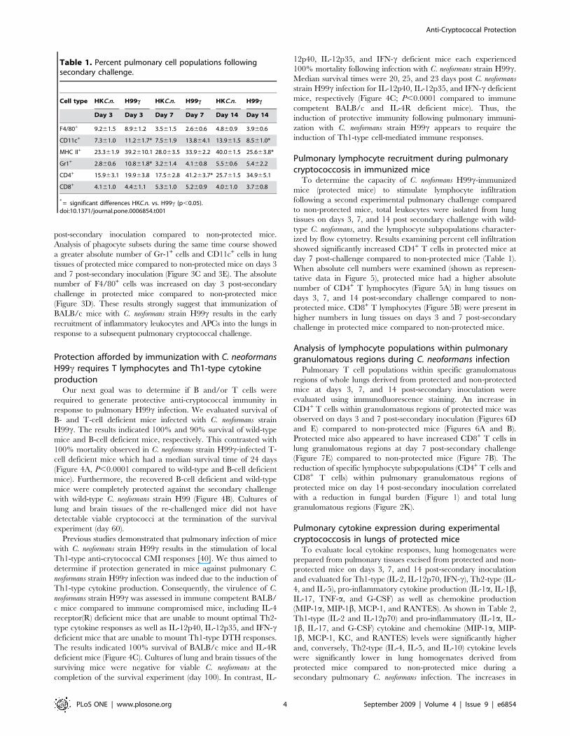
The percentages of Gr-1+ and CD11c+ cells were significantly
increased at day 3 post-secondary challenge in protected mice
compared to non-protected mice (Table 1). Concurrent with
clearance of the organism, the percentage of CD11c+ cells and
MHC class II+ APCs was significantly decreased in protected mice at
day 14 post-secondary challenge compared to non-protected mice.
However, when we examined the absolute total number of
pulmonary leukocytes (shown as representative data in Figure 3),
we found increased CD45+ cells at days 3 and 7 post-secondary
challenge in protected mice compared to non-protected mice
(Figure 3A). In addition, we observed an increase in the total number
of MHC class II+ APCs (Figure 3B) in protected mice on days 3 and 7
Figure 2. Progression of pathological changes in the lungs of HKC.n.- and C. neoformans strain H99c-immunized mice followingsecondary challenge with wild-type cryptococci. Histological samples were prepared as described in Methods. Digital photographs of H&Estained slides were taken under the light microscope at 10X and 40X power objectives. Note the progressive growth and spread of C. neoformans (redarrows), concurrent development of ‘‘loose’’ inflammatory infiltrates (A–F) in the lungs of non-protected mice, and the hallmarks of allergic broncho-pulmonary mycosis (ABPM) pathology [eosinophilia (yellow arrows) and extended macrophages harboring live organisms (E–F)]. In protected mice,note rapid formation of ‘‘tight’’ mononuclear infiltrates and the absence of C. neoformans growth in the alveolar space (G–J) and a rapid resolution ofthe inflammatory response that subsides to the well marginalized ‘‘cuffs’’ of neolymphatic tissue within the bronchovascular bundle (black arrows)with the concurrent clearance of inflammation from the alveolar space (K, L). Data are representative of 4 experiments using 3 mice per experiment.doi:10.1371/journal.pone.0006854.g002
Anti-Cryptococcal Protection
PLoS ONE | www.plosone.org 3 September 2009 | Volume 4 | Issue 9 | e6854
post-secondary inoculation compared to non-protected mice.
Analysis of phagocyte subsets during the same time course showed
a greater absolute number of Gr-1+ cells and CD11c+ cells in lung
tissues of protected mice compared to non-protected mice on days 3
and 7 post-secondary inoculation (Figure 3C and 3E). The absolute
number of F4/80+ cells was increased on day 3 post-secondary
challenge in protected mice compared to non-protected mice
(Figure 3D). These results strongly suggest that immunization of
BALB/c mice with C. neoformans strain H99c results in the early
recruitment of inflammatory leukocytes and APCs into the lungs in
response to a subsequent pulmonary cryptococcal challenge.
Protection afforded by immunization with C. neoformansH99c requires T lymphocytes and Th1-type cytokineproduction
Our next goal was to determine if B and/or T cells were
required to generate protective anti-cryptococcal immunity in
response to pulmonary H99c infection. We evaluated survival of
B- and T-cell deficient mice infected with C. neoformans strain
H99c. The results indicated 100% and 90% survival of wild-type
mice and B-cell deficient mice, respectively. This contrasted with
100% mortality observed in C. neoformans strain H99c-infected T-
cell deficient mice which had a median survival time of 24 days
(Figure 4A, P,0.0001 compared to wild-type and B-cell deficient
mice). Furthermore, the recovered B-cell deficient and wild-type
mice were completely protected against the secondary challenge
with wild-type C. neoformans strain H99 (Figure 4B). Cultures of
lung and brain tissues of the re-challenged mice did not have
detectable viable cryptococci at the termination of the survival
experiment (day 60).
Previous studies demonstrated that pulmonary infection of mice
with C. neoformans strain H99c results in the stimulation of local
Th1-type anti-crytococcal CMI responses [40]. We thus aimed to
determine if protection generated in mice against pulmonary C.
neoformans strain H99c infection was indeed due to the induction of
Th1-type cytokine production. Consequently, the virulence of C.
neoformans strain H99c was assessed in immune competent BALB/
c mice compared to immune compromised mice, including IL-4
receptor(R) deficient mice that are unable to mount optimal Th2-
type cytokine responses as well as IL-12p40, IL-12p35, and IFN-cdeficient mice that are unable to mount Th1-type DTH responses.
The results indicated 100% survival of BALB/c mice and IL-4R
deficient mice (Figure 4C). Cultures of lung and brain tissues of the
surviving mice were negative for viable C. neoformans at the
completion of the survival experiment (day 100). In contrast, IL-
12p40, IL-12p35, and IFN-c deficient mice each experienced
100% mortality following infection with C. neoformans strain H99c.
Median survival times were 20, 25, and 23 days post C. neoformans
strain H99c infection for IL-12p40, IL-12p35, and IFN-c deficient
mice, respectively (Figure 4C; P,0.0001 compared to immune
competent BALB/c and IL-4R deficient mice). Thus, the
induction of protective immunity following pulmonary immuni-
zation with C. neoformans strain H99c appears to require the
induction of Th1-type cell-mediated immune responses.
Pulmonary lymphocyte recruitment during pulmonarycryptococcosis in immunized mice
To determine the capacity of C. neoformans H99c-immunized
mice (protected mice) to stimulate lymphocyte infiltration
following a second experimental pulmonary challenge compared
to non-protected mice, total leukocytes were isolated from lung
tissues on days 3, 7, and 14 post secondary challenge with wild-
type C. neoformans, and the lymphocyte subpopulations character-
ized by flow cytometry. Results examining percent cell infiltration
showed significantly increased CD4+ T cells in protected mice at
day 7 post-challenge compared to non-protected mice (Table 1).
When absolute cell numbers were examined (shown as represen-
tative data in Figure 5), protected mice had a higher absolute
number of CD4+ T lymphocytes (Figure 5A) in lung tissues on
days 3, 7, and 14 post-secondary challenge compared to non-
protected mice. CD8+ T lymphocytes (Figure 5B) were present in
higher numbers in lung tissues on days 3 and 7 post-secondary
challenge in protected mice compared to non-protected mice.
Analysis of lymphocyte populations within pulmonarygranulomatous regions during C. neoformans infection
Pulmonary T cell populations within specific granulomatous
regions of whole lungs derived from protected and non-protected
mice at days 3, 7, and 14 post-secondary inoculation were
evaluated using immunofluorescence staining. An increase in
CD4+ T cells within granulomatous regions of protected mice was
observed on days 3 and 7 post-secondary inoculation (Figures 6D
and E) compared to non-protected mice (Figures 6A and B).
Protected mice also appeared to have increased CD8+ T cells in
lung granulomatous regions at day 7 post-secondary challenge
(Figure 7E) compared to non-protected mice (Figure 7B). The
reduction of specific lymphocyte subpopulations (CD4+ T cells and
CD8+ T cells) within pulmonary granulomatous regions of
protected mice on day 14 post-secondary inoculation correlated
with a reduction in fungal burden (Figure 1) and total lung
granulomatous regions (Figure 2K).
Pulmonary cytokine expression during experimentalcryptococcosis in lungs of protected mice
To evaluate local cytokine responses, lung homogenates were
prepared from pulmonary tissues excised from protected and non-
protected mice on days 3, 7, and 14 post-secondary inoculation
and evaluated for Th1-type (IL-2, IL-12p70, IFN-c), Th2-type (IL-
4, and IL-5), pro-inflammatory cytokine production (IL-1a, IL-1b,
IL-17, TNF-a, and G-CSF) as well as chemokine production
(MIP-1a, MIP-1b, MCP-1, and RANTES). As shown in Table 2,
Th1-type (IL-2 and IL-12p70) and pro-inflammatory (IL-1a, IL-
1b, IL-17, and G-CSF) cytokine and chemokine (MIP-1a, MIP-
1b, MCP-1, KC, and RANTES) levels were significantly higher
and, conversely, Th2-type (IL-4, IL-5, and IL-10) cytokine levels
were significantly lower in lung homogenates derived from
protected mice compared to non-protected mice during a
secondary pulmonary C. neoformans infection. The increases in
Table 1. Percent pulmonary cell populations followingsecondary challenge.
Cell type HKC.n. H99c HKC.n. H99c HKC.n. H99c
Day 3 Day 3 Day 7 Day 7 Day 14 Day 14
F4/80+ 9.261.5 8.961.2 3.561.5 2.660.6 4.860.9 3.960.6
CD11c+ 7.361.0 11.261.7* 7.561.9 13.864.1 13.961.5 8.561.0*
MHC II+ 23.361.9 39.2610.1 28.063.5 33.962.2 40.061.5 25.663.8*
Gr1+ 2.860.6 10.861.8* 3.261.4 4.160.8 5.560.6 5.462.2
CD4+ 15.963.1 19.963.8 17.562.8 41.263.7* 25.761.5 34.965.1
CD8+ 4.161.0 4.461.1 5.361.0 5.260.9 4.061.0 3.760.8
* = significant differences HKC.n. vs. H99c (p,0.05).doi:10.1371/journal.pone.0006854.t001
Anti-Cryptococcal Protection
PLoS ONE | www.plosone.org 4 September 2009 | Volume 4 | Issue 9 | e6854

Th1-type cytokine and chemokine expression in protected mice
closely mirrored the increases in pulmonary leukocyte infiltration
(Figures 2, 3, 5–7) and subsequent reductions in pulmonary fungal
burden (Figure 1) observed during secondary challenge. Addition-
ally, the protected mice showed no signs of an exacerbated Th1-
type cytokine response at day 14 post-challenge, concurrent with
pulmonary organism clearance (Figure 1). Interestingly, the
production of IFN-c and TNF-a, two cytokines previously shown
to be critical for the induction of protection against acute
pulmonary C. neoformans infection, were not significantly increased
in protected compared to non-protected mice.
Discussion
Studies to date have consistently suggested that Th1-type CMI
is predominantly responsible for protection against pulmonary C.
neoformans infection [6–10]. These studies suggested that protection
against pulmonary cryptococcosis is mediated by Th1-type
Figure 3. Pulmonary inflammatory leukocyte populations following secondary challenge. BALB/c mice received an immunization witheither HKC.n. (white bars) or C. neoformans strain H99c (gray bars), allowed 100 days to resolve the infection, and subsequently given a secondchallenge with C. neoformans strain H99. Leukocytes were labeled with anti-CD45 antibodies (A) or dual labeled with anti-IA/IE (MHC class II) and anti-CD45 (B), anti-Gr-1 and anti-CD45 (C), anti-F4/80 and anti-CD45 (D), or anti-CD11c (E) and analyzed by flow cytometry. Data shown are absolute cellnumbers from one representative experiment of 3 experiments performed using 5 mice per group per experiment.doi:10.1371/journal.pone.0006854.g003
Anti-Cryptococcal Protection
PLoS ONE | www.plosone.org 5 September 2009 | Volume 4 | Issue 9 | e6854

cytokines such as IFN-c, TNF-a, and IL-12 [14,16,17,22,23,50].
Additional work supported a protective role for antibody mediated
immunity (AMI) against pulmonary cryptococcosis [51,52]. The
studies described herein utilize a model system in which an
experimental pulmonary infection with C. neoformans strain H99cin mice results in complete protection against an otherwise lethal
challenge with wild-type C. neoformans [40,41]. Importantly,
previous studies and those presented herein show that C. neoformans
strain H99c is not an attenuated strain but instead induces
protective host immune responses resulting in its eradication and
the induction of protective anti-cryptococcal immunity. We
demonstrate that all leukocyte populations appeared to be
increased in protected mice, with Gr-1+ cells and CD11c+ cells
comprising a significantly greater proportion of total leukocytes to
respond during early infection. Recent studies by Davis and
Ramakrishnan suggest that more virulent pathogens induce
apoptosis of macrophages within primary granulomas resulting
in the recruitment of uninfected macrophages that then phago-
cytose the remnants of the dead macrophages and their microbial
contents before departing to seed other tissues [53]. While the
‘‘Trojan Horse’’ theory explaining dissemination of C. neoformans
has been examined [54,55], our studies show that protected mice
formed well-organized granulomatous regions in which the
phagocytic populations contained the infection. Pulmonary tissues
derived from non-protected mice appeared to have a more
dispersed leukocytic infiltrate with cryptococci replicating within
the macrophages and lung alveoli. In contrast, the inflammatory
response generated in protected mice subsided in correlation with
the reduction of fungal burden, and no evidence of tissue
destruction was observed. The zebrafish model used by Davis
and Ramakrishnan does not include adaptive immune cells that
may impact effector cell activity such as alternative or classical
macrophage activation [53]. The pulmonary Th1-type cytokine
environment observed in protected mice herein would putatively
favor the induction of classically activated macrophages which
have increased fungicidal activity against C. neoformans [56].
We evaluated the absolute role of T and B cells in the
generation of protection against pulmonary C. neoformans infection,
and found that B-cell deficient mice were able to resolve the acute
infection with C. neoformans strain H99c and were protected against
a second pulmonary challenge with wild-type cryptococci. In
contrast, T-cell deficient mice succumbed to pulmonary infection
with C. neoformans strain H99c, supporting a definitive role for T
cells in mediating protection against acute infection. Furthermore,
the inability of IL-12p40, IL-12p35, and IFN-c deficient mice to
survive pulmonary infection with C. neoformans strain H99c, in
contrast to the survival of IL-4R deficient mice, suggests that
protective anti-cryptococcal immune responses are promoted by
the induction of Th1-type cytokines. The pathogenesis of C.
neoformans strain H99c in immune deficient mice is comparable to
that observed in mice given a similar pulmonary inocula of wild-
type C. neoformans (unpublished observations). Also, previous
studies demonstrated no differences in several phenotypes
associated with cryptococcal pathogenesis in C. neoformans strain
H99c compared to wild-type C. neoformans [40] further suggesting
that clearance of the acute infection with the transgenic strain was
not due to attenuation. Nevertheless, the protective anamnestic
immune responses were generated against the virulent C. neofor-
mans strain H99 and appeared to be T cell-mediated. However,
simply because B cells are not absolutely required for protection in
our model does not entirely exclude any role for AMI participation
in protective anti-cryptococcal immune responses. T cells more
likely induce protective antibody responses that, in turn, assist to
shorten the duration of the infection [51,57]. In fact, we have
previously demonstrated that C. neoformans-specific antibodies
generated in protected mice were predominantly of a protective
phenotype [41]. These antibodies could potentially limit C.
Figure 4. Pathogenesis of C. neoformans strain H99c in T celland B cell deficient mice. A. Wild-type (circles), T cell deficient(squares), and B cell deficient (triangles) mice were given an intranasalinoculation with C. neoformans strain H99c. Survival data shown arefrom one experiment using 10 mice per group. B. Wild-type (circles) andB cell deficient (squares) mice that survived an initial inoculation with C.neoformans strain H99c received a second challenge with the parentalC. neoformans strain H99. Survival data shown are from one experimentusing 9–10 mice per experimental group. C. Wild-type (circles), IFN-cdeficient (squares), IL-12p40 deficient (upward triangles), IL-12p35deficient (downward triangles), and IL-4R deficient (diamonds) micewere given an intranasal inoculation with C. neoformans strain H99c.Survival data shown are from one experiment each, using 10–15 miceper group.doi:10.1371/journal.pone.0006854.g004
Anti-Cryptococcal Protection
PLoS ONE | www.plosone.org 6 September 2009 | Volume 4 | Issue 9 | e6854
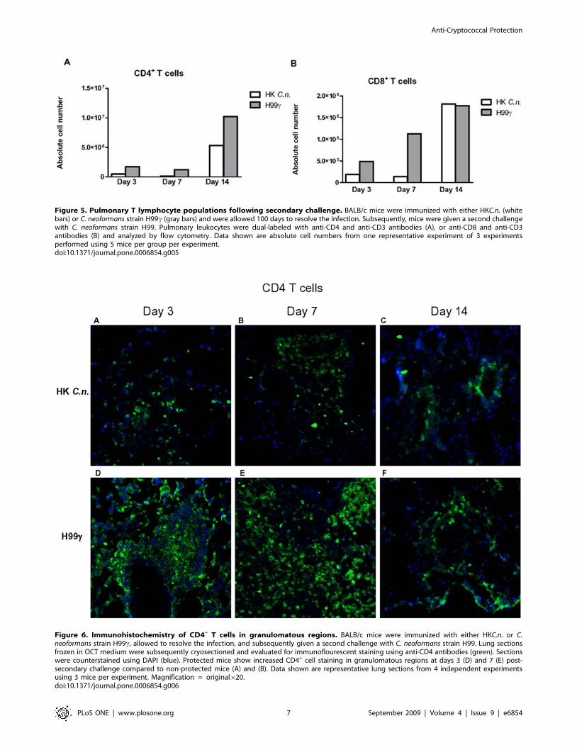
Figure 6. Immunohistochemistry of CD4+ T cells in granulomatous regions. BALB/c mice were immunized with either HKC.n. or C.neoformans strain H99c, allowed to resolve the infection, and subsequently given a second challenge with C. neoformans strain H99. Lung sectionsfrozen in OCT medium were subsequently cryosectioned and evaluated for immunoflourescent staining using anti-CD4 antibodies (green). Sectionswere counterstained using DAPI (blue). Protected mice show increased CD4+ cell staining in granulomatous regions at days 3 (D) and 7 (E) post-secondary challenge compared to non-protected mice (A) and (B). Data shown are representative lung sections from 4 independent experimentsusing 3 mice per experiment. Magnification = original620.doi:10.1371/journal.pone.0006854.g006
Figure 5. Pulmonary T lymphocyte populations following secondary challenge. BALB/c mice were immunized with either HKC.n. (whitebars) or C. neoformans strain H99c (gray bars) and were allowed 100 days to resolve the infection. Subsequently, mice were given a second challengewith C. neoformans strain H99. Pulmonary leukocytes were dual-labeled with anti-CD4 and anti-CD3 antibodies (A), or anti-CD8 and anti-CD3antibodies (B) and analyzed by flow cytometry. Data shown are absolute cell numbers from one representative experiment of 3 experimentsperformed using 5 mice per group per experiment.doi:10.1371/journal.pone.0006854.g005
Anti-Cryptococcal Protection
PLoS ONE | www.plosone.org 7 September 2009 | Volume 4 | Issue 9 | e6854

neoformans infection in patients with reduced CMI responses if
present in the correct isotype and proportions.
Nevertheless, we elected to concentrate on evaluating protective
anamnestic T cell-mediated immune responses to pulmonary C.
neoformans infection. Our results indicated that protection against a
second pulmonary C. neoformans challenge in mice is associated
with increased recruitment of APCs and leukocytes consisting of
granulocytes, macrophages, CD4+ T lymphocytes, and CD8+ T
lymphocytes into the lungs at days 3 and 7 post-secondary
inoculation compared to non-protected mice. Current studies are
underway to determine the mechanisms by which specific T cell
subsets mediate protection in this model.
Upon examination of the cytokine/chemokine profile of
pulmonary homogenates from protected mice, we observed a
predominantly Th1/pro-inflammatory-type response. Conversely,
pulmonary homogenates of non-protected mice expressed a
predominantly Th-2 type cytokine profile. Th1-type and pro-
inflammatory cytokines have long been associated with protection
against pulmonary cryptococcosis [14,16,17,22,23,50]. Most
profound were the high levels of pro-inflammatory cytokines
observed in protected mice on days 3 and 7 post-secondary
inoculation compared to non-protected mice. Although we cannot
assume that higher levels of pro-inflammatory cytokines directly
correlates to increased biological function, it may correlate with
enhanced phagocyte killing activity in protected mice. Again, the
levels of Th1/pro-inflammatory cytokines subsided as the
pulmonary fungal burden decreased in protected mice. Addition-
ally, significantly increased levels of IL-17, G-CSF and KC, all
known neutrophil chemoattaractants, were observed in lung
homogenates from protected mice compared to non-protected
mice on days 3 and 7, concurrent with increased Gr-1+ cells
infiltrating into the lungs. Our results contrast with previous
studies suggesting that increased neutrophilia during early
infection results in greater inflammation and exacerbation of
pulmonary cryptococcosis [58]. Further studies will determine the
significance of this infiltration and the putative mechanism of Gr-
1+ effector cell function in protected mice.
Interestingly, we did not observe significant increases in either
IFN-c or TNF-a, cytokines previously shown to be important for
protection against acute pulmonary C. neoformans infections
[14,16,17,59]. However, we should note that in the previous
studies, these cytokines were increased in response to primary
infections with different strains of C. neoformans – YC-11 (a
serotype A strain) [16,17] and 52D (a serotype D strain) [14,59].
Figure 7. Immunohistochemistry of CD8+ T cells in granulomatous regions. BALB/c mice were immunized with either HKC.n. or C.neoformans strain H99c, allowed to resolve the infection, and subsequently given a second challenge with C. neoformans strain H99. Lung sectionsfrozen in OCT medium were subsequently cryosectioned and analyzed for immunoflourescent staining using anti-CD8 antibodies (green). Sectionswere counterstained using DAPI (blue). Protected mice show increased CD8+ cell staining in granulomatous regions at day 7 (E) post-secondaryinoculation compared to non-proteced mice (B). Data shown are representative lung sections from 4 independent experiments using 3 mice perexperiment. Magnification = original620.doi:10.1371/journal.pone.0006854.g007
Anti-Cryptococcal Protection
PLoS ONE | www.plosone.org 8 September 2009 | Volume 4 | Issue 9 | e6854

These strains are not typically as pathogenic as C. neoformans strain
H99 (used herein) suggesting that strain-specific cytokine
responses occur. Interestingly, the lack of protection against
pulmonary C. neoformans strain H99c infection in IFN-c deficient
mice suggests that IFN-c production by the transgenic strain
alone is insufficient to promote protection. The IFN-c secreted by
the transgenic strain perhaps induces a cascade of events resulting
in a Th1-type polarized response and protection in immune
competent mice. However, IFN-c deficient mice have other
immune abnormalities (i.e., reduced macrophage activity and
production of reactive oxygen species) that mitigate forming this
conclusion. IFN-c and TNF-a have been shown to be important
mediators of protection against other pulmonary fungal infec-
tions. Specifically, TNF-a appears to be the chief mediator of
protection in both primary and secondary pulmonary Histoplasma
capsulatum infection in mice [60–64]. Vaccine induced immunity
against pulmonary Blastomyces dermatididis infection in immune-
competent animals appears to be mediated by TNF-a and IFN-c[65]. However, protection still occurs in immune-deficient mice,
suggesting that alternate and/or compensatory mechanisms of
protection are present in immune-deficient hosts. We have
previously observed significant increases in IFN-c and TNF-aduring primary pulmonary infection of mice with C. neoformans
strain H99c [40] suggesting that these cytokines may be of
greater importance in protection against acute infections. Thus,
while our results may not completely correlate with those
observed in C. neoformans or other pulmonary fungal infection
models, our results suggest a greater role for other cytokines in
the induction of protective anamnestic responses against C.
neoformans infections.
Until now, studies have been unable to demonstrate complete
clearance of C. neoformans from infected tissues or 100% protection from
subsequent infections with pathogenic C. neoformans strains. Thus, the
mechanisms responsible for mediating protective anamnestic immune
responses have largely been extrapolated using model systems that
provide varying degrees of protection against pulmonary cryptococ-
cosis. Our studies show that protective anti-cryptococcal immune
responses are predominantly dependent on the stimulation of an early
and sustained Th1-type CMI response that decreases as the infection is
brought under control thereby limiting lung damage. However, the
induction of protection via various pro-inflammatory cytokines and
chemokines and those effector cells responsible for mediating this
protection still require further study and clarification. Nonetheless,
these studies clearly demonstrate the advantage of using a pathogenic
fungus engineered to secrete host cytokines in order to achieve a
biological ‘‘print out’’ of those factors that mediate protective host
immunity. Such information will prove critical for the design of
immune-based therapies to combat human mycoses.
Materials and Methods
MiceFemale BALB/c (H-2d) (National Cancer Institute/Charles
River Laboratories and The Jackson Laboratory), CD192/2 (B
cell deficient), Foxn1nu (T cell deficient), IL12atm1Jm/J (IL-12p35
deficient), IL12btm1Jm/J (IL-12p40 deficient) (The Jackson Labo-
ratory), and IFNgtm1Ts/J (IFN-c deficient; a kind gift of Dr.
Bernard Arulanandam, The University of Texas at San Antonio,
San Antonio, TX) mice, all on the BALB/c background with an
average weight of 20–25 grams, were used throughout these
Table 2. Pulmonary Th1/Th2, inflammatory cytokine and chemokine levelsa.
Cytokine HKC.n. H99c HKC.n. H99c HKC.n. H99c
Day 3 Day 3 Day 7 Day 7 Day 14 Day 14
Th1-type
IL-2 6.061.3 74.0618.4* 17.661.7 29.763.1* 9.460.9 5.860.5*
IL-12p70 11.863.5 56.0613.0* 20.961.5 104.8618.3* 51.469.8 11.461.0*
IFN-c 1.860.6 2.360.6 6.061.3 5.060.6 6.961.3 0.660.3
Th2-type
IL-4 2.360.1 35.368.9* 175.3641.4 8.662.3* 573.4674.1 2.760.2*
IL-5 2.561.1 2.960.5* 12.762.7 1.960.4* 23.664.2 0.860.1*
IL-10 1.360.5 2.960.5* 3.460.5 4.560.6 11.161.5 0.660.1*
Pro-inflammatory
IL-1a 14.761.4 84.6618.3* 34.062.3 198.9646.6* 115.9617.2 18.061.4*
IL-1b 21.063.8 717.56322.7* 166.8635.2 2056.06492.9* 799.46133.3 84.568.6*
IL-17 1.661.1 606.56167.1* 16.163.7 1510.06382.0* 32.465.1 24.162.5
G-CSF 11.561.1 302.2654.7* 74.9611.2 374.0682.9* 87.9614.7 13.461.4*
TNF-a 4.561.2 5.661.4 10.562.3 16.863.1 5.561.9 2.360.8
Chemokines
MIP-1a 100.0656.9 307.7676.7* 399.6690.6 270.6668.4 1522.06290.6 97.2620.5*
MIP-1b 2.460.4 15.762.6* 9.461.3 13.562.7 14.961.2 1.560.2*
MCP-1 233.1632.7 1509.06247.9* 1444.06197.2 2274.06262.4* 2212.06247.9 297.1615.7*
KC 33.562.1 661.76100.7* 287.5636.0 912.16141.2* 321.0635.8 76.167.4*
RANTES 103.1614.9 411.4676.5* 215.8642.6 416.0668.6* 191.4633.0 148.3618.4
aProtein levels given in picograms per milliliter.*P,0.05 compared to infected counterpart on same day post inoculation.doi:10.1371/journal.pone.0006854.t002
Anti-Cryptococcal Protection
PLoS ONE | www.plosone.org 9 September 2009 | Volume 4 | Issue 9 | e6854

studies. Mice were housed at The University of Texas at San
Antonio Small Animal Laboratory Vivarium and handled
according to guidelines approved by The University of Texas at
San Antonio Institutional Animal Care and Use Committee.
Strains and mediaC. neoformans strains H99 (serotype A, Mat a) and H99c (an
interferon-gamma producing C. neoformans strain derived from H99
[40]) were recovered from 15% glycerol stocks stored at –80uCprior to use in the experiments described herein. The strains were
maintained on yeast-extract-peptone-dextrose (YPD) media (1%
yeast extract, 2% peptone, 2% dextrose, and 2% Bacto agar).
Yeast cells were grown for 18–20 h at 30uC with shaking in YPD
broth (Becton Dickinson and Company, Sparks, MD), collected by
centrifugation, washed three times with sterile phosphate-buffered
saline (PBS), and viable yeast quantified using trypan blue dye
exclusion in a hemacytometer.
Pulmonary infectionsPulmonary C. neoformans infections were initiated by nasal
inhalation as previously described [66,67]. Briefly, anesthetized
BALB/c mice received a yeast inocula of 16104 colony-forming
units (CFU) of either C. neoformans strain H99c or heat-killed C.
neoformans strain H99 (HKC.n.) yeasts in 50 ml of sterile PBS and
were allowed 100 days to resolve the infection. Subsequently, the
immunized mice received a second experimental pulmonary
inoculation with 16104 CFU of wild-type C. neoformans strain H99
in 50 ml of sterile PBS. The inocula used for immunizations and
challenge were verified by quantitative culture on YPD agar. The
mice were fed ad libitum and were monitored by inspection twice
daily. Mice were euthanized on days 3, 7 or 14 post-secondary
inoculation, and lung tissues were excised using aseptic technique.
Tissues were homogenized in 1 ml of sterile PBS, followed by
culture of 10-fold dilutions of each tissue on YPD agar
supplemented with chloramphenicol (Mediatech, Inc., Herndon,
VA). CFU were enumerated following incubation at 30uC for 48 h.
Alternatively, mice intended for survival analysis were monitored by
inspection twice daily and euthanized if they appeared to be in pain
or moribund. Mice were euthanized using CO2 inhalation.
Pulmonary leukocyte isolationLungs were excised on days 3, 7, and 14 post inoculation and
digested enzymatically at 37uC for 30 minutes in 10 ml of digestion
buffer (RPMI 1640 and 1 mg/ml of collagenase type IV [Sigma
Chemical Co., St. Louis, MO.]) with intermittent (every 10 min)
stomacher homogenizations. The enzymatically-digested tissues were
then successively filtered through sterile nylon filters of various pore
sizes (70 and 40 mm) (BD Biosciences) and washed with sterile HBSS to
enrich for leukocytes. Erythrocytes were lysed by incubation in NH4Cl
buffer (0.859% NH4Cl, 0.1% KHCO3, 0.0372% Na2EDTA
[pH 7.4]; Sigma) for 3 minutes on ice followed by the addition of a
10-fold excess of PBS. The resulting leukocyte population was then
collected by centrifugation (800Xg) for 5 minutes, washed twice with
sterile PBS, resuspended in sterile PBS+ 2% heat-inactivated fetal
bovine serum (FACS buffer) and enumerated in a hemacytometer
using trypan blue dye exclusion. Flow cytometric analysis was used to
determine the percentage of each leukocyte population as well as the
absolute number of total leukocytes (CD45+) within the lung cell
suspension for standardization of hemacytometer counts.
AntibodiesFor flow cytometry experiments, cells were incubated with
CD16/CD32 (Fc BlockTM) (BD Pharmingen Corp., San Diego,
CA) and the following antibodies conjugated to phycoerythrin
(PE), allophycocyanin (APC), or PECy7 were added: a cocktail of
CD3, CD4, and CD8a; CD45, MHC class II, Gr-1, and CD11b,
(BD Pharmingen Corp.), CD11c (eBioscience Inc., San Diego,
CA), and F4/80 (Caltag Laoratories, Burlingame, CA). For
immunohistochemistry experiments, the following antibodies were
used: CD4 rat anti-mouse (R&D Systems) and CD8 rat anti-mouse
(BD Pharmingen). Primary antibodies were detected using FITC
conjugated goat anti-rat IgG secondary antibody (Jackson
ImunoResearch laboratories, Inc., West Grove, PA).
Flow cytometryStandard methodology was employed for the direct immuno-
fluorescence of pulmonary leukocytes. Briefly, in 96-well U-bottom
plates, 100 ml containing16106 leukocyte-enriched lung cells in
FACS buffer were incubated with 50 ml of Fc BlockTM (BD
Pharmingen) diluted in FACS buffer for 5 minutes to block non-
specific binding of antibodies to cellular Fc receptors. Subsequent-
ly, an optimal concentration of fluorochrome-conjugated antibod-
ies (between 0.06–0.5 mg/16106 cells in 50 ml of FACS buffer)
were added in various combinations to allow for dual or triple
staining experiments and plates were incubated for 30 minutes on
ice. Following incubation, the cells were washed three times with
FACS buffer and cells were fixed in 200 ml of 2% ultrapure
formaldehyde (Polysciences, Inc., Warrington, PA). Cells incubat-
ed with either FACS buffer alone or single fluorochrome-
conjugated antibodies were used to determine positive staining
and spillover/compensation calculations, and the flow cytometer
determined background fluorescence. The samples were analyzed
using BD FACSArray softwareTM on a BD FACSArray flow
cytometer (BD Pharmingen). Dead cells were excluded on the
basis of forward angle and 90u light scatter. For data analyses,
30,000 events (cells) were evaluated from a predominantly
leukocytic population identified by backgating from CD45+-
stained cells. The absolute number of total leukocytes was
quantified by multiplying the total number of cells observed by
hemacytometer counting by the percentage of CD45+ cells
determined by flow cytometry. The absolute number of each
leukocyte subset (Gr-1+, F4/80+, CD11c+, MHC class II+, CD4+/
CD3+ and CD8+/CD3+ lymphocytes) was determined by
multiplying the percentage of each gated population by the total
number of CD45+ cells.
ImmunohistochemistryMice were euthanized at days 3, 7, and 14 post-secondary
inoculation in order to excise the lungs. The pericardium and
trachea were exposed by dissection and an incision was made in
the trachea for the insertion of a sterile flexible cannula attached to
a 3 ml syringe to slowly inflate the lungs with 0.5 to 0.7 ml of
Tissue-Tek optimal tissue cutting (OCT) compound (Sakura
Finetek, Torrance, CA) plus 2 M sucrose solution (1:1, vol/vol).
The lungs were then excised and immediately placed in cryomolds
containing OCT medium on dry ice and then stored at 280uCuntil use.
Serial frozen tissue sections were cut to a thickness of 10 mm
and fixed at 220uC in acetone for 10 minutes. Tissue sections
were then stained using hematoxylin and eosin (The University of
Texas Health Sciences Center at San Antonio Histology &
Immunohistochemistry Laboratory, San Antonio, TX) or further
processed for immunofluorescence analysis. Sections stained with
hematoxylin and eosin were examined under a light microscope
(Eclipse E400, Nikon Co, Tokyo, JAP) at high and low power
magnification and microphotographs taken using Digital Micro-
photography system DFX1200 with ACT-1 software (Nikon).
Anti-Cryptococcal Protection
PLoS ONE | www.plosone.org 10 September 2009 | Volume 4 | Issue 9 | e6854

Sections destined for immunofluorescence analysis were imme-
diately placed in 70% ethanol for 5 minutes and washed twice in
PBS for 3 minutes each. Nonspecific binding was inhibited by
blocking for 30 minutes at room temperature with species-specific
serum (10% in PBS) (matched with the species of the secondary
antibody). Tissue sections were incubated overnight at 4uC with
primary antibodies diluted in species-specific serum (3% in PBS) at
pre-optimized concentrations. Subsequently, the sections were
washed seven times in TRIS-NaCl-Tween 20 (TNT) buffer
solution for 3 minutes per wash followed by incubation with the
secondary antibodies for 30 minutes at room temperature. Slides
were then washed seven times in TNT buffer for 3 minutes per
wash, once in PBS containing 1% Triton X to minimize
background fluorescence (3 minutes) and a final wash in TNT
buffer (3 minutes). Sections were then mounted with Fluorsave
reagent (Calbiochem, La Jolla, CA) containing 0.3 mM 49,69-
diamidino-2-phenylindole dilactate (DAPI) (Molecular Probes,
Eugene, OR). Fluorescence was visualized with a Leica DMR
epifluorescence microscope (Leica Microsystems, Wetzlar, Ger-
many). Images were acquired using a cooled SPOT RT charge-
coupled device camera (Diagnostic Instruments Inc., Sterling
Heights, MI), and they were processed and analyzed using Adobe
Photoshop 7.0 (Adobe, Mountain View, CA).
Cytokine analysisCytokine levels in lung tissues were analyzed using the Bio-Plex
Protein Array System (Luminex-based technology) (Bio-Rad
Laboratories, Hercules, CA). Briefly, lung tissue was excised and
homogenized in ice-cold sterile PBS (1 ml). An aliquot (50 ml) was
taken to quantify the pulmonary fungal burden and an anti-
protease buffer solution (1 ml) containing PBS, protease inhibitors
(inhibiting cysteine, serine, and other metalloproteinases) and
0.05% Triton X-100 was added to the homogenate. Samples were
then clarified by centrifugation (8006g) for 5 minutes. Superna-
tants from pulmonary homogenates were assayed using the Bio-
Plex Protein Array System (Bio-Rad Laboratories) for the presence
of interferon (IFN)-c, interleukin (IL)-1a, IL-1b, IL-2, IL-4, IL-5,
IL-10, IL-12 p70, IL-17, tumor necrosis factor (TNF)-a, and
granulocyte-colony stimulating factor [G-CSF] expression as well
as chemokines (macrophage inflammatory protein [MIP]-1a,
MIP-1b, macrophage chemoattractant protein [MCP]-1, kerati-
nocyte-derived chemokine (KC), and regulated upon activation,
normal T cell expressed and secreted [RANTES]).
Statistical analysisThe unpaired Student’s t test (two-tailed) was used to analyze
fungal burden, pulmonary cell populations, and cytokine/
chemokine data using GraphPad Prism version 5.00 for Windows
(GraphPad Software, San Diego California USA). Survival data
was analyzed using the log-rank test (GraphPad Software).
Significant differences were defined as P,0.05.
Acknowledgments
We would like to thank Jose Lopez-Ribot, Pharm.D., Ph.D., Loles Esteve-
Gassant, Ph.D., Sarah Bubeck, Ph.D., and Judy Teale, Ph.D. for critical
reading of the manuscript.
Author Contributions
Conceived and designed the experiments: FLWJ. Performed the
experiments: KLW SR SM MLY MAO CS. Analyzed the data: KLW
SR SM MLY MAO FLWJ. Contributed reagents/materials/analysis tools:
MAO CS. Wrote the paper: KLW FLWJ.
References
1. Levitz SM (1991) The ecology of Cryptococcus neoformans and the epidemiology of
cryptococcosis. Rev Infect Dis 13: 1163–1169.
2. Mitchell TG, Perfect JR (1995) Cryptococcosis in the Era of AIDS - 100 years
after the discovery of Cryptococcus neoformans. Clin Microbiol Rev 8: 515–548.
3. Shoham S, Levitz SM (2005) The immune response to fungal infections.
Br J Haematol 129: 569–582.
4. Singh N, Gayowski T, Wagener MM, Marino IR (1997) Clinical spectrum of
invasive cryptococcosis in liver transplant recipients receiving tacrolimus. Clin
Transplant 11: 66–70.
5. Singh N, Dromer F, Perfect JR, Lortholary O (2008) Cryptococcosis in solid
organ transplant recipients: current state of the science. Clin Infect Dis 47:
1321–1327.
6. Hill JO, Harmsen AG (1991) Intrapulmonary growth and dissemination of an
avirulent strain of Cryptococcus neoformans in mice depleted of CD4+ or CD8+ T-
Cells. J Exp Med 173: 755–758.
7. Huffnagle GB, Lipscomb MF, Lovchik JA, Hoag KA, Street NE (1994) The role
of CD4(+) and CD8(+) T-Cells in the protective inflammatory response to a
pulmonary cryptococcal infection. J Leukoc Biol 55: 35–42.
8. Huffnagle GB, Yates JL, Lipscomb MF (1991) Immunity to a pulmonary
Cryptococcus neoformans infection requires both CD4+ and CD8+ T-Cells. J Exp
Med 173: 793–800.
9. Huffnagle GB, Yates JL, Lipscomb MF (1991) T-cell-mediated immunity in the
lung - a Cryptococcus neoformans pulmonary infection model using SCID and
athymic nude-mice. Infect Immun 59: 1423–1433.
10. Mody CH, Lipscomb MF, Street NE, Toews GB (1990) Depletion of CD4+(L3T4+) lymphocytes in vivo impairs murine host defense to Cryptococcus
neoformans. J Immunol 144: 1472–1477.
11. Chuck SL, Sande MA (1989) Infections with Cryptococcus neoformans in the
Acquired Immunodeficiency Syndrome. New Engl J Med 321: 794–799.
12. Blasi E, Mazzolla R, Barluzzi R, Mosci P, Bistoni F (1994) Anticryptococcal
resistance in the mouse brain: beneficial effects of local administration of heat-
inactivated yeast cells. Infect Immun 62: 3189–3196.
13. Buchanan KL, Doyle HA (2000) Requirement for CD4+ T Lymphocytes in Host
Resistance against Cryptococcus neoformans in the Central Nervous System of
Immunized Mice. Infect Immun 68: 456–462.
14. Herring AC, Lee J, McDonald RA, Toews GB, Huffnagle GB (2002) Induction
of interleukin-12 and gamma interferon requires tumor necrosis factor alpha for
protective T1-cell-mediated immunity to pulmonary Cryptococcus neoformans
infection. Infect Immun 70: 2959–2964.
15. Decken K, Kohler G, Palmer-Lehmann K, Wunderlin A, Mattner F, et al.
(1998) Interleukin-12 Is Essential for a Protective Th1 Response in Mice Infected
with Cryptococcus neoformans. Infect Immun 66: 4994–5000.
16. Kawakami K, Qifeng X, Tohyama M, Qureshi MH, Saito A (1996)
Contribution of tumour necrosis factor-alpha (TNF-alpha) in host defence
mechanism against Cryptococcus neoformans. Clin Exp Immunol 106: 468–474.
17. Kawakami K, Tohyama M, Teruya K, Kudeken N, Xie QF, et al. (1996)
Contribution of interferon-gamma in protecting mice during pulmonary and
disseminated infection with Cryptococcus neoformans. FEMS Immunol Med
Microbiol 13: 123–130.
18. Kawakami K, Tohyama M, Xie Q, Saito A (1996) IL-12 protects mice against
pulmonary and disseminated infection caused by Cryptococcus neoformans. Clin Exp
Immunol 104: 208–214.
19. Zhou Q, Gault RA, Kozel TR, Murphy WJ (2007) Protection from Direct
Cerebral Cryptococcus Infection by Interferon-{gamma}-Dependent Activation of
Microglial Cells. J Immunol 178: 5753–5761.
20. Bauman SK, Huffnagle GB, Murphy JW (2003) Effects of tumor necrosis factor
alpha on dendritic cell accumulation in lymph nodes draining the immunization
site and the impact on the anticryptococcal cell-mediated immune response.
Infect Immun 71: 68–74.
21. Huffnagle GB, Toews GB, Burdick MD, Boyd MB, McAllister KS, et al. (1996)
Afferent phase production of TNF-alpha is required for the development of
protective T cell immunity to Cryptococcus neoformans. J Immunol 157: 4529–4536.
22. Kawakami K, Tohyama M, Qifeng X, Saito A (1997) Expression of cytokines
and inducible nitric oxide synthase mRNA in the lungs of mice infected with
Cryptococcus neoformans: effects of interleukin-12. Infect Immun 65: 1307–1312.
23. Koguchi Y, Kawakami K (2002) Cryptococcal infection and Th1-Th2 cytokine
balance. Int Rev Immunol 21: 423–438.
24. Blackstock R, Buchanan KL, Adekunle M, Adesina, Murphy JW (1999)
Differential Regulation of Immune Responses by Highly and Weakly Virulent
Cryptococcus neoformans Isolates. Infect Immun 67: 3601–3609.
25. Muller U, Stenzel W, Kohler G, Werner C, Polte T, et al. (2007) IL-13 Induces
Disease-Promoting Type 2 Cytokines, Alternatively Activated Macrophages and
Allergic Inflammation during Pulmonary Infection of Mice with Cryptococcus
neoformans. J Immunol 179: 5367–5377.
Anti-Cryptococcal Protection
PLoS ONE | www.plosone.org 11 September 2009 | Volume 4 | Issue 9 | e6854

26. Murphy JW, Schafer F, Casadevall A, Adesina A (1998) Antigen-induced
protective and nonprotective cell-mediated immune components against
Cryptococcus neoformans. Infect Immun 66: 2632–2639.
27. Yauch LE, Lam JS, Levitz SM (2006) Direct Inhibition of T-Cell Responses by
the Cryptococcus Capsular Polysaccharide Glucuronoxylomannan. PLoS Patho-gens 2: e120.
28. Levitz SM, Nong S, Mansour MK, Huang C, Specht CA (2001) Molecular
characterization of a mannoprotein with homology to chitin deacetylases that
stimulates T cell responses to Cryptococcus neoformans. Proc Natl Acad Sci U S A
98: 10422–10427.
29. Huang C, Nong SH, Mansour MK, Specht CA, Levitz SM (2002) Purification
and characterization of a second immunoreactive mannoprotein from
Cryptococcus neoformans that stimulates T-cell responses. Infect Immun 70:
5485–5493.
30. Mandel MA, Grace GG, Orsborn KI, Schafer F, Murphy JW, et al. (2000) The
Cryptococcus neoformans gene DHA1 encodes an antigen that elicits a delayed-typehypersensitivity reaction in immune mice. Infect Immun 68: 6196–6201.
31. Biondo C, Beninati C, Delfino D, Oggioni M, Mancuso G, et al. (2002)
Identification and cloning of a cryptococcal deacetylase that produces protective
immune responses. Infect Immun 70: 2383–2391.
32. Dromer F, Charreire J, Contrepois A, Carbon C, Yeni P (1987) Protection of
mice against experimental cryptococcosis by anti-Cryptococcus neoformans mono-
clonal antibody. Infect Immun 55: 749–752.
33. Mukherjee J, Pirofski LA, Scharff MD, Casadevall A (1993) Antibody-mediated
protection in mice with lethal intracerebral Cryptococcus neoformans infection. Proc
Natl Acad Sci U S A 90: 3636–3640.
34. Graybill JR, Bocanegra R, Lambros C, Luther MF (1997) Granulocyte colony
stimulating factor therapy of experimental cryptococcal meningitis. J Med Vet
Mycol 35: 243–247.
35. Clemons KV, Brummer E, Stevens DA (1994) Cytokine treatment of central
nervous system infection: efficacy of interleukin-12 alone and synergy withconventional antifungal therapy in experimental cryptococcosis. Antimicrob
Agents Chemother 38: 460–464.
36. Joly V, Saint-Julien L, Carbon C, Yeni P (1994) In vivo activity of interferon-
gamma in combination with amphotericin B in the treatment of experimental
cryptococcosis. J Infect Dis 170: 1331–1334.
37. Kawakami K, Qureshi MH, Zhang T, Okamura H, Kurimoto M, et al. (1997)
IL-18 protects mice against pulmonary and disseminated infection with
Cryptococcus neoformans by inducing IFN-gamma production. J Immunol 159:
5528–5534.
38. Lutz JE, Clemons KV, Stevens DA (2000) Enhancement of antifungal
chemotherapy by interferon-gamma in experimental systemic cryptococcosis.J Antimicrob Chemother 46: 437–442.
39. Pappas PG, Bustamante B, Ticona E, Hamill RJ, Johnson PC, et al. (2004)
Recombinant interferon- gamma 1b as adjunctive therapy for AIDS-related
acute cryptococcal meningitis. J Infect Dis 189: 2185–2191.
40. Wormley FL Jr, Perfect JR, Steele C, Cox GM (2007) Protection Against
Cryptococcosis using a Murine Interferon-gamma Producing Cryptococcus
neoformans Strain. Infect Immun 75: 1453–1462.
41. Young M, Macias S, Thomas D, Wormley FL Jr (2009) A proteomic-based
approach for the identification of immunodominant Cryptococcus neoformans
proteins. Proteomics 9: 2578–2588.
42. Chen GH, McNamara DA, Hernandez Y, Huffnagle GB, Toews GB, et al.
(2008) Inheritance of Immune Polarization Patterns Is Linked to Resistance
versus Susceptibility to Cryptococcus neoformans in a Mouse Model. Infect Immun
76: 2379–2391.
43. Osterholzer JJ, Curtis JL, Polak T, Ames T, Chen G-H, et al. (2008) CCR2
Mediates Conventional Dendritic Cell Recruitment and the Formation ofBronchovascular Mononuclear Cell Infiltrates in the Lungs of Mice Infected
with Cryptococcus neoformans. J Immunol 181: 610–620.
44. Arora S, Hernandez Y, Erb-Downward JR, McDonald RA, Toews GB, et al.
(2005) Role of IFN-gamma in regulating T2 immunity and the development of
alternatively activated macrophages during allergic bronchopulmonary mycosis.
J Immunol 174: 6346–6356.
45. Chen GH, Olszewski MA, McDonald RA, Wells JC, Paine R 3rd, et al. (2007)
Role of granulocyte macrophage colony-stimulating factor in host defenseagainst pulmonary Cryptococcus neoformans infection during murine allergic
bronchopulmonary mycosis. Am J Pathol 170: 1028–1040.
46. Daley JM, Thomay AA, Connolly MD, Reichner JS, Albina JE (2008) Use ofLy6G-specific monoclonal antibody to deplete neutrophils in mice. J Leukoc Biol
83: 64–70.47. Dalod M, Hamilton T, Salomon R, Salazar-Mather TP, Henry SC, et al. (2003)
Dendritic cell responses to early murine cytomegalovirus infection: subset
functional specialization and differential regulation by interferon alpha/beta.J Exp Med 197: 885–898.
48. Nakano H, Yanagita M, Gunn MD (2001) CD11c(+)B220(+)Gr-1(+) cells inmouse lymph nodes and spleen display characteristics of plasmacytoid dendritic
cells. J Exp Med 194: 1171–1178.49. Mordue DG, Sibley LD (2003) A novel population of Gr-1+-activated
macrophages induced during acute toxoplasmosis. J Leukoc Biol 74: 1015–1025.
50. Huffnagle GB, Lipscomb MF (1998) Cells and cytokines in pulmonarycryptococcosis. Research in Immunology 149: 387–396.
51. Casadevall A (1995) Antibody immunity and invasive fungal infections. InfectImmun 63: 4211–4218.
52. Casadevall A (1998) Antibody-mediated protection against intracellular
pathogens. Trends Microbiol 6: 102–107.53. Davis JM, Ramakrishnan L (2009) The Role of the Granuloma in Expansion
and Dissemination of Early Tuberculous Infection. Cell 136: 37–49.54. Luberto C, Martinez-Marino B, Taraskiewicz D, Bolanos B, Chitano P, et al.
(2003) Identification of App1 as a regulator of phagocytosis and virulence ofCryptococcus neoformans. J Clin Invest 112: 1080–1094.
55. Santangelo R, Zoellner H, Sorrell T, Wilson C, Donald C, et al. (2004) Role of
Extracellular Phospholipases and Mononuclear Phagocytes in Dissemination ofCryptococcosis in a Murine Model. Infect Immun 72: 2229–2239.
56. Voelz K, Lammas DA, May RC (2009) Cytokine signaling regulates theoutcome of intracellular macrophage parasitism by Cryptococcus neoformans. Infect
Immun.
57. Casadevall A, Pirofski L (2005) Insights into Mechanisms of Antibody-MediatedImmunity from Studies with Cryptococcus neoformans. Current Molecular Medicine
5: 421–433.58. Mednick AJ, Feldmesser M, Rivera J, Casadevall A (2003) Neutropenia alters
lung cytokine production in mice and reduces their susceptibility to pulmonarycryptococcosis. Eur J Immunol 33: 1744–1753.
59. Herring AC, Falkowski NR, Chen GH, McDonald RA, Toews GB, et al. (2005)
Transient neutralization of tumor necrosis factor alpha can produce a chronicfungal infection in an immunocompetent host: Potential role of immature
dendritic cells. Infection and Immunity 73: 39–49.60. Allendoerfer R, Deepe GS Jr (1998) Infection with Histoplasma capsulatum: Host-
fungus interface. Rev Iberoam Micol 15: 256–260.
61. Allendoerfer R, Deepe GS Jr (1998) Blockade of endogenous TNF-alphaexacerbates primary and secondary pulmonary histoplasmosis by differential
mechanisms. J Immunol 160: 6072–6082.62. Zhou P, Miller G, Seder RA (1998) Factors involved in regulating primary and
secondary immunity to infection with Histoplasma capsulatum: TNF-alpha plays acritical role in maintaining secondary immunity in the absence of IFN-gamma.
J Immunol 160: 1359–1368.
63. Deepe GS Jr, Gibbons RS (2006) T cells require tumor necrosis factor-alpha toprovide protective immunity in mice infected with Histoplasma capsulatum. J Infect
Dis 193: 322–330.64. Cain JA, Deepe GS Jr (1998) Evolution of the primary immune response to
Histoplasma capsulatum in murine lung. Infect Immun 66: 1473–1481.
65. Wuthrich M, Filutowicz HI, Warner T, Klein BS (2002) Requisite elements invaccine immunity to Blastomyces dermatitidis: plasticity uncovers vaccine potential
in immune-deficient hosts. J Immunol 169: 6969–6976.66. Cox GM, McDade HC, Chen SC, Tucker SC, Gottfredsson M, et al. (2001)
Extracellular phospholipase activity is a virulence factor for Cryptococcus
neoformans. Mol Microbiol 39: 166–175.67. Cox GM, Mukherjee J, Cole GT, Casadevall A, Perfect JR (2000) Urease as a
virulence factor in experimental cryptococcosis. Infect Immun 68: 443–448.
Anti-Cryptococcal Protection
PLoS ONE | www.plosone.org 12 September 2009 | Volume 4 | Issue 9 | e6854