Insights into Modern Therapeutic Approaches in Pediatric ...
42
Citation: Panuciak, K.; Margas, M.; Makowska, K.; Lejman, M. Insights into Modern Therapeutic Approaches in Pediatric Acute Leukemias. Cells 2022, 11, 139. https://doi.org/10.3390/cells11010139 Academic Editor: John M. Perry Received: 14 October 2021 Accepted: 15 December 2021 Published: 2 January 2022 Publisher’s Note: MDPI stays neutral with regard to jurisdictional claims in published maps and institutional affil- iations. Copyright: © 2022 by the authors. Licensee MDPI, Basel, Switzerland. This article is an open access article distributed under the terms and conditions of the Creative Commons Attribution (CC BY) license (https:// creativecommons.org/licenses/by/ 4.0/). cells Review Insights into Modern Therapeutic Approaches in Pediatric Acute Leukemias Kinga Panuciak 1 , Mikolaj Margas 1 , Karolina Makowska 1 and Monika Lejman 2, * 1 Student Scientific Society, Laboratory of Genetic Diagnostics, Medical University of Lublin, 20-093 Lublin, Poland; [email protected] (K.P.); [email protected] (M.M.); [email protected] (K.M.) 2 Laboratory of Genetic Diagnostics, Medical University of Lublin, 20-093 Lublin, Poland * Correspondence: [email protected] Abstract: Pediatric cancers predominantly constitute lymphomas and leukemias. Recently, our knowledge and awareness about genetic diversities, and their consequences in these diseases, have greatly expanded. Modern solutions are focused on mobilizing and impacting a patient’s immune system. Strategies to stimulate the immune system, to prime an antitumor response, are of intense interest. Amid those types of therapies are chimeric antigen receptor T (CAR-T) cells, bispecific antibodies, and antibody–drug conjugates (ADC), which have already been approved in the treat- ment of acute lymphoblastic leukemia (ALL)/acute myeloid leukemia (AML). In addition, immune checkpoint inhibitors (ICIs), the pattern recognition receptors (PRRs), i.e., NOD-like receptors (NLRs), Toll-like receptors (TLRs), and several kinds of therapy antibodies are well on their way to showing significant benefits for patients with these diseases. This review summarizes the current knowledge of modern methods used in selected pediatric malignancies and presents therapies that may hold promise for the future. Keywords: immunotherapy; CAR-T; antibodies; immune checkpoint inhibitors; PRR; pediatric leukemias 1. Introduction Acute leukemias are the most frequent cancer in children. The most common is acute lymphoblastic leukemia (ALL), which accounts for over 80% of all cases of acute leukemia. ALL cases are classified as B-ALL or T-ALL, based on immunophenotyping, with B-ALL encompassing approximately 85% of cases. In the 1960s, the five-year survival rate for pediatric patients with ALL was under 10%, while now, the survival of children treated in high-income countries exceeds 90% [1–3]. Nowadays, improvement in outcomes with conventional chemotherapy is challenging due to adverse effects increasing during further intensification of chemotherapy. Based on genome-wide analyses, B-ALL can be classified into more than 30 B-lineage subtypes with prognostic and therapeutic implications. The most important of them are presented in Table 1. Accurate identification of the genetic abnormalities in ALL is important to risk-stratify the relapse and to guide the incorporation of molecular targeted therapeutic approaches to reduce the risk of relapse. Cells 2022, 11, 139. https://doi.org/10.3390/cells11010139 https://www.mdpi.com/journal/cells
-
Upload
khangminh22 -
Category
Documents
-
view
0 -
download
0
Transcript of Insights into Modern Therapeutic Approaches in Pediatric ...
�����������������
Citation: Panuciak, K.; Margas, M.;
Makowska, K.; Lejman, M. Insights
into Modern Therapeutic
Approaches in Pediatric Acute
Leukemias. Cells 2022, 11, 139.
https://doi.org/10.3390/cells11010139
Academic Editor: John M. Perry
Received: 14 October 2021
Accepted: 15 December 2021
Published: 2 January 2022
Publisher’s Note: MDPI stays neutral
with regard to jurisdictional claims in
published maps and institutional affil-
iations.
Copyright: © 2022 by the authors.
Licensee MDPI, Basel, Switzerland.
This article is an open access article
distributed under the terms and
conditions of the Creative Commons
Attribution (CC BY) license (https://
creativecommons.org/licenses/by/
4.0/).
cells
Review
Insights into Modern Therapeutic Approaches in PediatricAcute Leukemias
Kinga Panuciak 1 , Mikołaj Margas 1 , Karolina Makowska 1 and Monika Lejman 2,*
1 Student Scientific Society, Laboratory of Genetic Diagnostics, Medical University of Lublin,20-093 Lublin, Poland; [email protected] (K.P.); [email protected] (M.M.);[email protected] (K.M.)
2 Laboratory of Genetic Diagnostics, Medical University of Lublin, 20-093 Lublin, Poland* Correspondence: [email protected]
Abstract: Pediatric cancers predominantly constitute lymphomas and leukemias. Recently, ourknowledge and awareness about genetic diversities, and their consequences in these diseases, havegreatly expanded. Modern solutions are focused on mobilizing and impacting a patient’s immunesystem. Strategies to stimulate the immune system, to prime an antitumor response, are of intenseinterest. Amid those types of therapies are chimeric antigen receptor T (CAR-T) cells, bispecificantibodies, and antibody–drug conjugates (ADC), which have already been approved in the treat-ment of acute lymphoblastic leukemia (ALL)/acute myeloid leukemia (AML). In addition, immunecheckpoint inhibitors (ICIs), the pattern recognition receptors (PRRs), i.e., NOD-like receptors (NLRs),Toll-like receptors (TLRs), and several kinds of therapy antibodies are well on their way to showingsignificant benefits for patients with these diseases. This review summarizes the current knowledgeof modern methods used in selected pediatric malignancies and presents therapies that may holdpromise for the future.
Keywords: immunotherapy; CAR-T; antibodies; immune checkpoint inhibitors; PRR; pediatric leukemias
1. Introduction
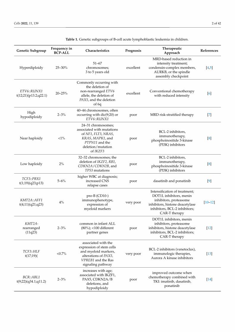
Acute leukemias are the most frequent cancer in children. The most common is acutelymphoblastic leukemia (ALL), which accounts for over 80% of all cases of acute leukemia.ALL cases are classified as B-ALL or T-ALL, based on immunophenotyping, with B-ALLencompassing approximately 85% of cases. In the 1960s, the five-year survival rate forpediatric patients with ALL was under 10%, while now, the survival of children treatedin high-income countries exceeds 90% [1–3]. Nowadays, improvement in outcomes withconventional chemotherapy is challenging due to adverse effects increasing during furtherintensification of chemotherapy. Based on genome-wide analyses, B-ALL can be classifiedinto more than 30 B-lineage subtypes with prognostic and therapeutic implications. Themost important of them are presented in Table 1. Accurate identification of the geneticabnormalities in ALL is important to risk-stratify the relapse and to guide the incorporationof molecular targeted therapeutic approaches to reduce the risk of relapse.
Cells 2022, 11, 139. https://doi.org/10.3390/cells11010139 https://www.mdpi.com/journal/cells
Cells 2022, 11, 139 2 of 42
Table 1. Genetic subgroups of B-cell acute lymphoblastic leukemia in children.
Genetic Subgroup Frequency inBCP-ALL Characteristics Prognosis Therapeutic
Approach References
Hyperdiploidy 25–30%51–67
chromosomes;3 to 5 years old
excellent
MRD-based reduction inintensity treatment;
condensin-complex members,AURKB, or the spindleassembly checkpoint
[4,5]
ETV6::RUNX1t(12;21)(p13.2;q22.1) 20–25%
Commonly occurring withthe deletion of
non-rearranged ETV6allele, the deletion of
PAX5, and the deletionof 6q
excellent Conventional chemotherapywith reduced intensity [6]
Highhypodiploidy 2–3%
40–44 chromosomes, oftenoccurring with dic(9;20) or
ETV6::RUNX1poor MRD risk-stratified therapy [7]
Near haploidy <1%
24–31 chromosomes;associated with mutations
of NF1, FLT3, NRAS,KRAS, MAPK1, and
PTPN11 and thedeletion/mutation
of IKZF3
poor
BCL-2 inhibitors,immunotherapy,
phosphoinositide 3-kinase(PI3K) inhibitors
[8]
Low haploidy 2%
32–32 chromosomes; thedeletion of IKZF2, RB1,
CDKN2A/CDKN2B, andTP53 mutations
poor
BCL-2 inhibitors,immunotherapy,
phosphoinositide 3-kinase(PI3K) inhibitors
[8]
TCF3::PBX1t(1;19)(q23;p13) 5–6%
higher WBC at diagnosis;increased CNSrelapse cases
poor dasatinib and ponatinib [9]
KMT2A::AFF1t(4;11)(q21;q23) 4%
pro-B (CD10-)immunophenotype,
expression ofmyeloid markers
very poor
Intensification of treatment;DOT1L inhibitors, menin
inhibitors, proteasomeinhibitors, histone deacetylase
inhibitors, BCL-2 inhibitors;CAR-T therapy
[10–12]
KMT2A-rearranged
(11q23)2–3%
common in infant ALL(80%); <100 different
partner genespoor
DOT1L inhibitors, menininhibitors, proteasome
inhibitors, histone deacetylaseinhibitors, BCL-2 inhibitors;
CAR-T therapy
[12]
TCF3::HLFt(17;19)( <0.7%
associated with theexpression of stem cellsand myeloid markers,
alterations of PAX5,VPREB1 and the Rassignaling pathway
very poorBCL-2 inhibitors (venetoclax),
immunologic therapies,Aurora A kinase inhibitors
[13]
BCR::ABL1t(9;22)(q34.1;q11.2) 2–3%
increases with age;associated with IKZF1,
PAX5, CDKN2A/Bdeletions, andhypodiploidy
poor
improved outcome whenchemotherapy combined with
TKI: imatinib, dasatinib,ponatinib
[14]
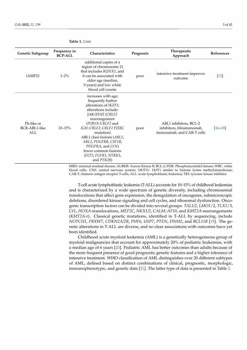
Cells 2022, 11, 139 3 of 42
Table 1. Cont.
Genetic Subgroup Frequency inBCP-ALL Characteristics Prognosis Therapeutic
Approach References
iAMP21 1–2%
additional copies of aregion of chromosome 21that includes RUNX1, andit can be associated with
older age (median,9 years) and low white
blood cell counts
poor intensive treatment improvesoutcome [15]
Ph-like orBCR-ABL1-like
ALL10–15%
increases with age;frequently harbor
alterations of IKZF1;alterations include:JAK-STAT (CRLF2
rearrangement(P2RY8::CRLF2 and
IGH::CRLF2; CRLF2 F232Cmutation)
ABL1 class fusions (ABL1,ABL2, PDGFRB, CSF1R,
PDGFRA, and LYN)fewer common fusions(FLT3, FGFR1, NTRK3,
and PTK2B)
poorABL1 inhibitors, BCL-2
inhibitors, blinatumomab,inotuzumab, and CAR-T cells
[16–18]
MRD: minimal residual disease; AURKB: Aurora Kinase B; BCL-2; PI3K: Phosphoinositide3-kinase; WBC: whiteblood cells; CNS: central nervous system; DOT1L: DOT1 similar to histone lysine methylotransferase;CAR-T: chimeric antigen receptor T-cells; ALL: acute lymphoblastic leukemia; TKI: tyrosine kinase inhibitor.
T-cell acute lymphoblastic leukemia (T-ALL) accounts for 10–15% of childhood leukemiasand is characterized by a wide spectrum of genetic diversity, including chromosomaltranslocations that affect gene expression, the deregulation of oncogenes, submicroscopicdeletions, disordered kinase signaling and cell cycles, and ribosomal dysfunction. Onco-genic transcription factors can be divided into several groups: TAL1/2, LMO1/2, TLX1/3,LYL, HOXA-translocations, MEF2C, NKX1/2, CALM::AF10, and KMT2A-rearrangements(KMT2A-r). Classical genetic mutations, identified in T-ALL by sequencing, includeNOTCH1, FBXW7, CDKN2A/2B, PHF6, USP7, PTEN, DNM2, and BCL11B [19]. The ge-netic alterations in T-ALL are diverse, and no clear associations with outcomes have yetbeen identified.
Childhood acute myeloid leukemia (AML) is a genetically heterogeneous group ofmyeloid malignancies that account for approximately 20% of pediatric leukemias, witha median age of 6 years [20]. Pediatric AML has better outcomes than adults because ofthe more frequent presence of good prognostic genetic features and a higher tolerance ofintensive treatment. WHO classification of AML distinguishes over 20 different subtypesof AML, defined based on distinct combinations of clinical, prognostic, morphologic,immunophenotypic, and genetic data [21]. The latter type of data is presented in Table 2.

Cells 2022, 11, 139 4 of 42
Table 2. Genetic subgroups of acute myeloid leukemia in children.
Genetic Subgroup Frequency Characteristics Prognosis References
RUNX1::RUNX1T1t(8;21)(q22;q22) 10–12%
FAB M2, blasts with single and thin Auerrods, median age 8 years; CBF AML;
standard risk group; almost 90% of patientsachieve complete remission with
chemotherapy alone; dasatinib (targetingKIT kinase); GO for relapsed patients
very good [20–25]
CBFB::MYH11inv(16)(p13.1q22) ort(16;16)(p13.1;q22)
8–10%
FAB M4eo, median age 9 years; corebinding factor (CBF) AML; standard risk
group; almost 90% of patients achievecomplete remission with chemotherapyalone; dasatinib (targeting KIT kinase);
gemtuzumab ozogamicin (GO) forrelapsed patients
very good [20–25]
PML::RARAt(15;17)(q24.1;q21.2) 5–10%
FAB M3, median age 7 years (1–18 years);acute promyelocytic leukemia (APL);
standard risk group; ATRA, ATO treatmentvery good [20–24,26,27]
KMT2A- rearranged(11q23) 16–21%
FAB M4 and M5, infant, median age 7 years(1–18 years) KMT2A with multiple partners;hypomethylating agents, DOT1L inhibitors,Menin-KMT2A protein–protein interaction
inhibitors, protein interaction inhibitors
poor orintermediate
[10,11,20–25,28–30]
KMT2A::MLLT3t(9;11)(p22;q23) 6–9% identified in 40% of KMT2Ar AML cases intermediate
KMT2A::MLLT1t(11;19)(q23;p13.3) 1% identified in 7% of KMT2Ar AML cases intermediate
KMT2A::ELLt(11;19)(q23;p13.1) 1–2% identified in 7% of KMT2Ar AML cases poor
KMT2A::MLLT10t(10;11)(p12;q23) 2–3% identified in 6% of KMT2Ar AML cases poor
KMT2A::MLLT4t(6;11)(q27;q23) 1–2% identified in 8% of KMT2Ar AML cases poor
NUP98::NSD1t(5;11)(q35;p15) 3–4%
FAB M4 and M5; median age 10.4 years;10%
strong association with FLT3-ITDpoor
[20–25,31–33]NUP98::KMD5At(11;12)(p15;p13) 1–2% 30% of FAB M7 (AMKL); median age
3.2 years
MNX1::ETV6t(7;12)(q36;p13) 1%
Only infants (4% of infants); 3-year EFSbelow 24%; KAT inhibitors, C646, I-CBP112,
CCS1477poor [20–25]
DEK::NUP214t(6;9)(p22;q34) 1–4%
FAB M2 and M4; median age 12 years, noinfant; association with FLT3-ITD; benefit
from HSCT in first CRpoor [20–25]
CBFA2T3::GLIS2inv(16)(p13.3;q24.3) 2–3%
FAB M7 (AMKL); infants; median age1.5 years; 20% of non-DS-AMKL; high rates
of relapse, and dismal survival; GLIinhibitors (GANT61);
the AURKA inhibitor alisertib (MLN8237)
very poor [20–25,31,32]
BCR::ABL1t(9;22)(q34;q11) 1% sensitivity to TKI poor [20–25]
KAT6A::CREBBPt(8;16)(p11;p13) <1% FAB M4 and M5; infants; spontaneous
remission has been observed intermediate [20–25,34]
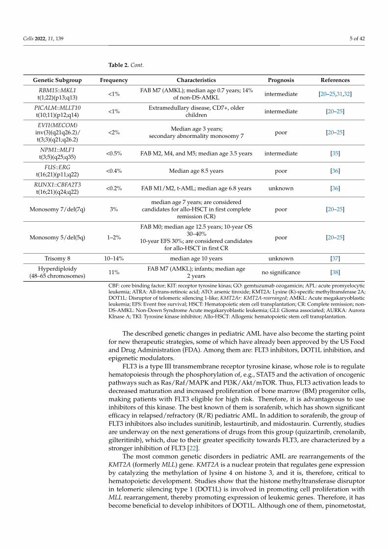
Cells 2022, 11, 139 5 of 42
Table 2. Cont.
Genetic Subgroup Frequency Characteristics Prognosis References
RBM15::MKL1t(1;22)(p13;q13) <1% FAB M7 (AMKL); median age 0.7 years; 14%
of non-DS-AMKL intermediate [20–25,31,32]
PICALM::MLLT10t(10;11)(p12;q14) <1% Extramedullary disease, CD7+, older
children intermediate [20–25]
EVI1(MECOM)inv(3)(q21q26.2)/t(3;3)(q21;q26.2)
<2% Median age 3 years;secondary abnormality monosomy 7 poor [20–25]
NPM1::MLF1t(3;5)(q25;q35) <0.5% FAB M2, M4, and M5; median age 3.5 years intermediate [35]
FUS::ERGt(16;21)(p11;q22) <0.4% Median age 8.5 years poor [36]
RUNX1::CBFA2T3t(16;21)(q24;q22) <0.2% FAB M1/M2, t-AML; median age 6.8 years unknown [36]
Monosomy 7/del(7q) 3%median age 7 years; are considered
candidates for allo-HSCT in first completeremission (CR)
poor [20–25]
Monosomy 5/del(5q) 1–2%
FAB M0; median age 12.5 years; 10-year OS30–40%
10-year EFS 30%; are considered candidatesfor allo-HSCT in first CR
poor [20–25]
Trisomy 8 10–14% median age 10 years unknown [37]
Hyperdiploidy(48–65 chromosomes) 11% FAB M7 (AMKL); infants; median age
2 years no significance [38]
CBF: core binding factor; KIT: receptor tyrosine kinas; GO: gemtuzumab ozogamicin; APL: acute promyelocyticleukemia; ATRA: All-trans-retinoic acid; ATO: arsenic tiroxide; KMT2A: Lysine (K)-specific methyltransferase 2A;DOT1L: Disruptor of telomeric silencing 1-like; KMT2Ar: KMT2A-rearranged; AMKL: Acute megakaryoblasticleukemia; EFS: Event free survival; HSCT: Hematopoietic stem cell transplantation; CR: Complete remission; non-DS-AMKL: Non-Down Syndrome Acute megakaryoblastic leukemia; GLI: Glioma associated; AURKA: AuroraKInase A; TKI: Tyrosine kinase inhibitor; Allo-HSCT: Allogenic hematopoietic stem cell transplantation.
The described genetic changes in pediatric AML have also become the starting pointfor new therapeutic strategies, some of which have already been approved by the US Foodand Drug Administration (FDA). Among them are: FLT3 inhibitors, DOT1L inhibition, andepigenetic modulators.
FLT3 is a type III transmembrane receptor tyrosine kinase, whose role is to regulatehematopoiesis through the phosphorylation of, e.g., STAT5 and the activation of oncogenicpathways such as Ras/Raf/MAPK and PI3K/Akt/mTOR. Thus, FLT3 activation leads todecreased maturation and increased proliferation of bone marrow (BM) progenitor cells,making patients with FLT3 eligible for high risk. Therefore, it is advantageous to useinhibitors of this kinase. The best known of them is sorafenib, which has shown significantefficacy in relapsed/refractory (R/R) pediatric AML. In addition to sorafenib, the group ofFLT3 inhibitors also includes sunitinib, lestaurtinib, and midostaurin. Currently, studiesare underway on the next generations of drugs from this group (quizartinib, crenolanib,gilteritinib), which, due to their greater specificity towards FLT3, are characterized by astronger inhibition of FLT3 [22].
The most common genetic disorders in pediatric AML are rearrangements of theKMT2A (formerly MLL) gene. KMT2A is a nuclear protein that regulates gene expressionby catalyzing the methylation of lysine 4 on histone 3, and it is, therefore, critical tohematopoietic development. Studies show that the histone methyltransferase disruptorin telomeric silencing type 1 (DOT1L) is involved in promoting cell proliferation withMLL rearrangement, thereby promoting expression of leukemic genes. Therefore, it hasbecome beneficial to develop inhibitors of DOT1L. Although one of them, pinometostat,
Cells 2022, 11, 139 6 of 42
as a standalone therapy, brought limited benefit in clinical trials, it has been proven thatits initial treatment sensitizes AML cell lines to further sorafenib treatment. Moreover,recent studies also show a role for DOT1L in leukemia without KMT2A rearrangement.By comparing the consequences of DOT1L inhibition in both AML cells with and withoutMLL rearrangement, Lonetti et al. showed that pinometostat-mediated cytotoxicity is notclosely related to KMT2A fusions [29].
As the common feature of AML is an altered epigenetic pattern, the Bromodomainfamily of proteins and the extracorporeal domain (including the proteins BRD2, BRD3,BRD4, and BRDT) have recently been identified. The role of these proteins is to regulategene transcription by interacting with acetylated histones, thereby facilitating the activationof transcription. Therefore, they have become an important epigenetic target, and theirinhibitors have shown anti-leukemic activity in preclinical models. Currently, they arebeing tested in the context of adult AML patients, which suggests the possibility of theiruse in pediatric AML [22].
This review will summarize the latest advances in the treatment of leukemia, focusedon mobilizing and impacting a patient’s immune system. Below, we describe the latestdevelopments in individual areas of leukemia immunotherapy.
2. FDA-Approved Drugs to Treat ALL or AML2.1. CAR-T Immunotherapy—Genetically Modified T Lymphocytes
Chimeric antigen receptor (CAR) is an unnatural form that, after activation in T-cells,imitates their receptor and leads to their effective function towards a precise antigen.
2.1.1. Chimeric Antigen Receptor
CAR contains four separate modules: the antigen recognition domain, the extracellularhinge region, the transmembrane domain, and one or more intracellular T-cell signalingdomains. An antigen recognition domain of the single-chain variable fragment (scFv)is a chimeric protein. It is composed of both light and heavy immunoglobulin chainsthat are linked together by a peptide linker [39]. The variant of the selected heavy andlight immunoglobulin chains depends on their ability to bind to a target antigen, suchas CD19. The linker is made of hydrophilic residues containing sections of serine andglycine (affecting flexibility) and sections of glutamate and lysine (providing additionalsolubility) [40]. In addition to scFv, the following have also been successfully used totarget the specificity of CAR: cytokines, growth factors, innate immunity receptors, andcompounds from the tumor necrosis factor (TNF) receptor superfamily [41]. The hingeregion, also known as a spacer, is a small structural domain. It is located between theantigen-recognition domain and the outer membrane of the cell. Its role is to increase theflexibility of the scFv receptor head, leading to a decrease in the distance between CAR andits target antigen. Spacer building sequences are usually based on the proximal regions ofmembrane molecules such as IgG, CD8, and CD28 [41,42]. The transmembrane domain ismade up of a hydrophobic alpha helix that encompasses the cell membrane. Its functionis to anchor CAR to the plasma membrane, thereby connecting the extracellular hingeand antigen recognition domains with the intracellular signaling region. It ensures thestability of the entire receptor. The intracellular signaling domain of T cells is located inthe endodomain of the receptor [41]. When an antigen is bound to an external antigen-recognition domain, CAR receptors aggregate and transmit an activation signal. Dueto the inner cytoplasmic end of the receptor, it is then fixed inside the cell [39]. Naturalactivation of the T lymphocyte is based on the phosphorylation of immunoreceptor tyrosine-based activation motifs (ITAMs), which are present in the cytoplasmic domain CD3-ζ. Toimitate this process, this domain is also commonly used in CAR as a main endodomaincomponent [43]. To persist after activation, apart from signaling CD3, T lymphocytesalso need costimulatory molecules. For this reason, CAR endodomains contain chimericdomains from costimulatory proteins. So far, the CD28, CD27, CD134 (OX40), and CD137(4-1BB) have performed well in this role [41]. Their use significantly improves T cell

Cells 2022, 11, 139 7 of 42
proliferation, cytokine secretion, resistance to apoptosis, and in vivo stability [44]. There isa complicated relationship between the domains that make up CAR, and there is, therefore,no single optimal configuration. The exact composition of the used CAR remains largelyempirical and depends on an analysis of tumor recognition in vitro [45,46].
2.1.2. Vectors
The genetically engineered CAR fusion protein is transduced into the autologous T-cellby means of a retrovirus or lentivirus [45]. By partially deleting the U3 region of the 3′ longterminal repeat (LTR), the transcriptional activity of the virus is decreased significantly [47].Although they have become safer, there is still a risk of uncontrolled integration leading tothe overexpression of neighboring genes or the disruption of genes at the site of integration.To prevent this, non-viral plasmids (pEPI series) containing a scaffold/matrix attachmentregion (S/MAR) element have been developed for the episomal long-term expression oftransgenes [48]. S/MAR is a segment of genomic DNA whose role is to anchor chromatin innuclear matrix proteins and mediate the structural organization of chromatin in the nucleus.It binds to the protein A of the scaffold attachment factor (SAF-A) and thus ensures mitoticstability of the plasmids. Jin et al. have confirmed that non-integrating lentiviral vector(NILV) containing S/MAR may become the optimal vector with a low risk of insertionalmutagenesis with long-term expression of the transgene [49].
Apart from the effectiveness and safety of the used vectors, the possibilities of theirproduction are also important. Until now, their production involved a relatively longperiod of time and many stages, which significantly increased the costs of the entire process.As a solution, non-viral allogeneic engineering of the T-cell population, according to acytokine-induced killer cell (CIK) differentiation protocol may be used here [50]. They arecharacterized by enrichment in CD3+ and CD56+ cytotoxic cells, which ensures high safetyand minimal risk of graft versus host disease (GvHD) after allogeneic CIK. Additionally,by using Sleeping Beauty (SB), which is an integrating vector belonging to the familyof Tc1/mariner DNA transposons, it is possible to obtain genetic modification ensuringprolonged expression in T-cells [51].
Magnani et al. conducted a study with CARCIK-CD19 (derived from a SB transposon-produced donor and differentiated CIK) in patients with B-ALL relapse after hematopoieticstem cell transplantation (HSCT). They found that, among the group of 13 evaluablepatients (including 4 children), the toxicity profile was low, with no GvHD occurring. Sixof the seven patients subjected to the highest doses achieved CR or CR with incompleteblood count recovery (CRi), and most reached a potent expansion of CAR-T-cells. Inaddition, production costs were up to 10 times lower than they were for viral processeswith comparable transduction efficiency and final cell viability [52].
2.1.3. Treatment by CAR-T
The patient should fulfill criteria to qualify for CAR-T-cell therapy and then undergolymphodepletion chemotherapy to reduce the number of T lymphocytes [44,53,54]. This isadvantageous because competition between the patient’s lymphocytes and newly intro-duced CAR-T is decreased and thus, CAR-T expression increases. CAR-T-cells recognizesurface antigens independently from MHC restriction. When targeted to tumor surfaceantigens, CAR-T-cells proliferate and kill tumor cells upon antigen contact. Their activity istriggered by mechanisms such as continuous intense proliferation, leading to an increase incytotoxicity, and by a boost in the secretion of interleukins, growth factors, and cytokines.
Tisagenlecleucel is a product of anti-CD19 CAR-T-cells. It was approved as the firstCAR-T gene therapy by the FDA, on 30 August 2017, for patients up to 25 years of agewith refractory B-cell precursor acute lymphoblastic leukemia (BCP-ALL) or in second orlater relapse [55]. This product also showed an indication of relapsed or refractory largeB-lymphoma on 1 May 2018. A single-center phase 1–2a study by Maude et al. showed aCR rate of 93% in children and young adults with the disease, after CAR-T therapy [56].Based on this, a phase 2 multicentre study was conducted, and the effectiveness turned
Cells 2022, 11, 139 8 of 42
out to be similar. In the initial phase, 92 people were enrolled. Of these, 75 received aninfusion of tisagenlecleucel (CTL019). The age range of the respondents was between 3and 25, and the median age was 11 years. The main goal of the study was to assess thesafety of the tested drug and to achieve a CR on the level of 20% among patients. It wasobserved that 81% of these patients attained remissions within 3 months of follow-up.Forty-five patients achieved a CR, and 16 patients had a CR without a full hematologicalrecovery. This study proved to be pivotal and led to the approval of tisagenlecleucel by theFDA (ClinicalTrials.gov number, NCT02435849) [57]. Despite the achieved remission, thepresented treatment method still needs to be improved. It is associated with the question-able achievement of the prolonged survival of patients without relapse; several reasons canbe found for the short effectiveness of CAR-T. Some of them are the progressive impairmentof function, a decline in CAR-T-cells, and an inhibition of the BM microenvironment [58,59].In this case, one solution may be to add allogenic hematopoietic stem cell transplantation(allo-HSCT) therapy. In a study by Davila et al., seven patients in remission receivedallo-HSCT, and they did not relapse within 24 months [60]. Similar results were obtainedin a clinical trial by Daniel W. Lee, where patients, after such treatment, did not report arelapse within a year [61]. Another solution may be to use cells with the CD22 antigen,which persist in the blood longer than CD19 and are highly expressed in most B-ALL, andare poorly expressed in regular B cells. A study by Jing Pan noted that the CAR CD22 iscapable of inducing remission even after the failure of treatment with CAR CD19; however,even after such treatment, a high relapse rate was observed. In this case, the introduction offollow-up treatment using allo-HSCT after CAR-T therapy proved to be beneficial, reducingthe frequency of relapses [62].
Another significant problem occurs when leukemic cells stop expressing CD19 and aretherefore not recognized by CD19-CAR-T-cells (the escape of the antigen from the tumorcells) [63]. Mutations in the genes could destroy the cognate epitope recognized by theanti-CD19 scFv, making the tumor cells no longer visible to CD19 CAR-T. Originally, thiswas thought to be due to splicing deregulation, leading to a deletion of exon 2 in the CD19gene. However, the existence of such CD19 isoforms was only investigated followingCAR-T therapy [64,65]. Subsequent studies showed that some modified CD19 isoforms,causing CAR-T escape, were not produced during therapy but existed at the time ofdiagnosis. During treatment, they can evolve and become a major clone of cancer cells. Suchconclusions were reached by Sotillo et al. They analyzed the expression of CD19 isoformsin a cohort of 14 children with CD19+ B-ALL. By taking BM and peripheral blood samples,using semiquantitative CD19 cDNA amplification by RT-PCR, and visualizing them byagarose gel electrophoresis, three different bands were obtained. These corresponded insize to full-length CD19 (800 bp) and to exon 2 deficient isoforms detected after CAR-Ttreatment [64]. This makes it possible to define a set of genes encoding hitherto unknownextracellular epitopes (which are alternatively spliced in leukemia compared to normal Blymphocytes), which would allow the development of new CAR-T targeting alternativeCD19 ectodomains. As a result, applying combination therapy to several epitopes mayimprove survival in patients with R/R B-ALL.
CAR T-cell therapy is associated with unique and potentially severe toxicities, mostparticularly, cytokine release syndrome (CRS) and neurotoxicity (or “CAR-T-cell-relatedencephalopathy syndrome”—CRES) [44]. There are many factors that influence the intensityof CRS. It depends on the type of therapy or even the characteristics of the patients [60]. Ina study conducted by Maude et al., patients experienced toxicities, and 27% of them had asevere course of CRS [61]. Teachey et al. reported that the neurotoxicity rate in children,after mild CRS, was 20% and 73% after severe CRS [66]. The fundamental mechanism ofCRES is still poorly understood. The main clinical symptoms are encephalopathy followedby focal deficits and seizures. Types of focal deficits include aphasia and, less commonly,vision change or facial droop. In addition, delirium, confusion, and hallucinations mayoccur [61,67,68]. Most of the side effects are completely reversible, but in some cases, theycan lead to death [69].
Cells 2022, 11, 139 9 of 42
Therefore, a search for ways to reduce side effects has begun. Sterner et al. presentedevidence that the abolition of neurotoxicity and CRS, following CAR-T use, can be achievedby neutralizing granulocyte macrophage colony stimulating factor (GM-CSF), which playsan important role in CRS mediation. The use of lenzilumab (a GM-CSF inhibitor) in thisstudy resulted in a reduction in CRS and inflammation of the nervous system. Additionally,it intensified the proliferation of CAR-T-cells and, thus, contributed to the improvementof their therapeutic activity. The GM-CSF-reduced CAR-T-cells had, not only, a preservedtherapeutic function, but they also had better survival and stronger activity against neo-plastic cells. This study made it possible to reduce the main side effects of CAR-T; becauseof that, phase II trials with lenzilumab in this therapy are currently planned [70].
To date, the use of CARs in T-ALL has been severely limited. Although CAR-T hasbeen shown to be effective against T-cell tumors, there are several limitations that slowthe development of this therapy in T-ALL. A major obstacle is the expression of the sametarget antigens in both healthy and neoplastic T-cells, causing CAR-T-cells to mutuallydestroy each other. Another challenge is isolating normal T-cells from cancerous ones. Thismakes it impossible to use autologous donor T lymphocytes as a substrate for creatingCAR-T based on them. Allogeneic donors can be used here, but this is associated with thelikelihood of GvHD, which may be fatal. Despite this, there is still promising research onthe use of CAR-T in T-ALL. CD7, the T-cell antigen, appears to be a good target that canbe used in this therapy, as it is strongly expressed in T-ALL [71]. Li et al. have conductedtwo clinical trials to evaluate both the safety and efficacy of GC027, the allogeneic CAR-Tproduct targeting CD7. Although only two patients with R/R T-ALL were tested, bothachieved CR, and it took over a year in one of them. CRS was noticed in both patients, butGvHD was not observed [72]. As it turns out, not only can CD7 be used in the developmentof a new CAR-T therapy against T-ALL but so can CD1a. Such conclusions were reached bySánchez-Martínez et al., who used it for cortical T-ALL (coT-ALL) therapy. In this subtypeof leukemia, CD1a shows superficial expression, associated with developmental arrest, inthe cortical stage. In this study, CD1a was found mainly in developing cortical thymocytes.It was absent in progenitor cells and T lymphocytes during ontogenesis, which reducesthe risk of toxicity outside the tumor. It was also proven that CD1a-CAR-T persists for along period of time in vivo. This indicates their safe and possible use in the case of R/RcoT-ALL [73].
The first study on the use of CAR-T in AML appeared in 2010. Peinert et al. presentedthe results of the first phase of the CAR-T study in recurrent AML with Lewis (Le) -Yantigen expression. Although they did not observe severe toxicity, all patients relapsedwithin two years [74]. In 2013, another examination by Ritchie et al. was related to theuse of second generation CD28-ζ CAR against the LeY antigen. Although only partiallyeffective, it gave hope for the biological activity of CAR-T in the fight against AML [75].Currently, research mainly concerns CD33 and CD123 antigens, which are largely presentin AML blasts. They seem to be attractive therapeutic targets, but their presence has alsobeen identified on healthy hematopoietic stem/progenitor cells [76]. As a result, the usedCAR-T cannot distinguish between healthy and neoplastic cells, leading to myeloablationmanifested by severe neutropenia, infections, and hemorrhages, leading to death. Effortswere made to create a gene that protects against myeloablation; if necessary, this wouldenable the elimination of T-cells within the body. Herpes simplex thymidine kinase (HSV-tk) has been successfully used, and it was able to convert a prodrug into a toxic compoundthat stopped the replication of genetic material, leading to cell death [77]. The use of HSV-tkhas, however, been limited by its immunogenicity [78]. A better compound than HSV-tkis the co-expression of inducible caspase 9 (iCasp9) in T-cells. It connects two domains:intracellular caspase 9 (which is a pro-apoptotic protein) with human binding proteinFK506 (FKBP). This combination allows conditional dimerization using a low molecularweight compound. The most important advantage of iCasp9 is low immunogenicity, as itis made up of human gene products. In addition, it retains its function even in cells withan overexpression of anti-apoptotic molecules, and the only effect in the body this causes
Cells 2022, 11, 139 10 of 42
is the elimination of transduced T lymphocytes [79]. Although iCasp9 has been tested inpreclinical studies, conducted by Hoyos et al., and included in other clinical trials, there arestill no conclusive data on its efficacy in CAR-T therapy [78,80].
A good solution seems to be to develop an antigen specific to AML cells that wouldnot be present in any healthy cells. Although AML genomes have a low mutational loadand contain few neoantigens, several have been described [81]. One of them are mutationsin the metabolic enzymes IDH1 and IDH2 [82]. However, the proteins encoded by thesegenes are expressed inside cells, making them inaccessible to CAR-T. Therefore, it seemsbeneficial to develop a new CAR-T that recognizes intracellular antigens. This task wasundertaken by Rafiq et al.; although they have shown that it is possible, more research isstill needed on this topic [83].
2.2. Bispecific Antibodies
An alternative approach to involving T-cells in cancer therapy is the use of antibodies,which are bispecific for CD3 in T-cells and for a surface target antigen in cancer cells. Thismechanism acts independently from T-cell receptor specificity, costimulation, and antigenpresentation. This new class of a bispecific T-cell-engaging (BiTE) antibody consists oftwo single-chain antibodies: an α-CD3 monoclonal antibody and an α-target monoclonalantibody. This construct can engage T-cells to the target neoplastic cell, subsequentlyactivating the T-cells and causing the perforin-mediated death of the malignant cell [84].
The first registered BiTE in the treatment of adult and pediatric patients with BCP-ALLwas blinatumomab, as the FDA granted its approval in 2018. Blinatumomab consists ofan anti-CD3 arm that engages CD3-expressing T-cells and an anti-CD19 arm that binds tolymphoblasts expressing the CD19 marker [85]. In the first clinical trial, blinatumomabrevealed efficacy in non-Hodgkin lymphoma (NHL), but the most important trials havebeen conducted in R/R ALL and in ALL with MRD. In this trial, 80% of patients becameMRD-negative after the first cycle of blinatumomab, with 60% of patients remaining in CRat a median follow-up of 33 months [86,87]. Blinatumomab was approved by the FDA on3 December 2014, after an accelerated review process in R/R Philadelphia chromosome-negative BCP-ALL [88].
The first results of a phase I/II study of blinatumomab in pediatric patients with R/RBCP-ALL included 49 patients in phase I and 44 patients in phase II (NCT01471782). Twoof three Ph+ patients achieved CR. The primary analysis of the results indicated that, ofthe 70 subjects, only ±27 (39%) reached CR status, of which 14 (20%) were MRD-negative.Twenty-four patients (34%) received allo-HSCT, and 11 (16%) received consecutive treat-ment. The median OS was 7.5 months, with a median follow-up of 23.8 months [89]. ARIALTO study (NCT02187354) was conducted to estimate the effects of blinatumomab.Overall, 58 patients (59%) achieved CR within the first two blinatumomab cycles. Amongthe CR group, 39 (67%) achieved full hematologic recovery, and 46 (79%) achieved MRDresponse. These results demonstrate a high response rate, with blinatumomab, in pediatricpatients with R/R BCP-ALL [90].
Queudeville et al. conducted retrospective analysis, of a single-center experiencewith blinatumomab, in 38 pediatric patients over a period of 10 years. Seventy-one per-cent of patients had undergone at least one HSCT prior to treatment with blinatumomab.They observed a response to blinatumomab in 13/38 patients (34%), with a median OS of11.1 months and a relapse-free survival of 6.17 month [91]. The latest clinical trials com-paring blinatumomab with standard chemotherapy in high-risk pediatric B-ALL showedpromising results. An investigation conducted by Locatelli et al. included 108 patients inrandomized trials. The blinatumomab group achieved event-free survival (EFS) better thanthe chemotherapy group. The number of deaths was in favor of the blinatumomab group,with 8 and 16 deaths in the blinatumomab and chemotherapy groups, respectively [92].Studies conducted by Brown et al. were aimed at comparing the use of blinatumomab andchemotherapy in post-induction therapy in the first relapse of B-ALL. Studies included208 patients (children, adolescents, and young adults; 1–27 years old); however, the ran-

Cells 2022, 11, 139 11 of 42
domization of risk-related subgroups had to be terminated. The results showed two-yeardisease-free survival (DFS) of 54.4% and 39% in the blinatumomab and chemotherapygroups, respectively (with results being not statistically significant, one-sided p = 0.03).The blinatumomab group achieved a 71.3% two-year OS rate, and the chemotherapygroup achieved 58.4%. The authors declared that early termination probably reduced DFSrates [93]. The main conclusion in both of these studies is that the experimental arms withblinatumomab resulted in an overall superior outcome. Blinatumomab-based therapy wasalso associated with a lower toxicity profile in comparison with standard chemotherapygroups [94,95].
Currently, numerous clinical trials are advancing, with results yet to be published. Thevalidation of treatment protocols in children and adults with BCP-ALL is the subject of theMoscow-Berlin 2019 Pilot (NCT04723342), which includes combined treatment with theapplication of blinatumomab and chemotherapy in children 1–18 years old [94]. Moreover,AIEOP-BFM ALL 2017 (NCT03643276) involved new approaches in several risk-relatedALL subtypes, especially in the chemotherapy-resistant or those with a high BCP-ALLrelapse risk among patients up to 17 years old. Blinatumomab in this patient’s group wasestimated to be a less toxic substitute for standard chemotherapy [95].
The Interfant-06 Protocol is investigating whether the addition of blinatumomab canimprove the outcome of mixed lineage leukemia (MLL)-rearranged ALL in infants (EUClinical Trials register 2016-004674-17) [96]. Blinatumomab is also the candidate in thebridging therapy in children and young adults (up to 25 years old), with high-risk B-ALLpreceding HSCT (NCT04556084), which is assumed to improve post-HCT outcomes [97].Furthermore, in children and young adults (1–10 years old without Down syndrome or1–31 years old with Down syndrome) newly diagnosed with B-ALL, blinatumomab isconnected with standard chemotherapy drugs (NCT03914625) to assess DFS [98].
Because of the rapid development of immunotherapy in hematologic malignancies,the bispecific anti-CD19/CD3 T-cell engager is tested with various immune checkpointinhibitors (ICIs). Blinatumomab alone, or with nivolumab (anti PD-1 antibody), is beingtested in children and young adults (1–31 years old) with a first relapse of CD19+ B-ALL(NCT04546399). Moreover, blinatumomab and nivolumab, with or without ipilimumab,(anti CTLA-4 antibody) have been validated for safety and dosing measures in poor-riskR/R CD19+ BCP-ALL in patients 16 years old or older (NCT02879695) [99]. BiTE targetingCD3 × CD33 are currently undergoing clinical trials as a solution to AML. Nevertheless,more studies are needed to fully evaluate these therapies [100].
In myeloid malignancies, a subtype of bispecific antibodies—dual affinity retargeting(DART) proteins—is used. The mechanism of action of DART is similar to BiTE; however,in their construction, an additional disulfide linker is applied. With this solution and otherconstruction features, DART achieved improvement in the molecule stability (exceedingblinatumomab in half-life circulation time), increased the affinity to cancer cells, andlowered the affinity to T-cells (preventing non-specific T-cell activation) in comparison toa single-chain anti-CD3 antibody [101–103]. However, currently, no DART treatment isapproved as a treatment for ALL or AML.
The remaining principles of the design and mechanism of action for DART and BiTEare similar. DART is capable of connecting two particular cells with particular moleculeson their surface. In leukemic malignancies, CD3+ cells (T-cells) are activated by DARTand directed to tumor cells, formatting the cytolytic synapses. Recently, CD123 has beenacknowledged as a potential target in the treatment and diagnosis of AML patients. Re-search studies testing the MGD006 drug, currently known as flotetuzumab, have continuedwith preclinical and clinical trials (NCT02152956) [76,104–106]. Results of the first studyof CD3 × CD123 DART (flotetuzumab) usage in refractory AML were recently publishedby Uy et al. Eighty-eight adult patients with primary induction failure or early relapsewere treated with flotetuzumab, and 27% achieved CR or CR with partial hematologicrecovery, where the median OS was 10.2 months. These results are correlated with standardchemotherapy protocols applied in patients in this particular stage of disease and are signif-
Cells 2022, 11, 139 12 of 42
icantly improved with an OS threefold greater for flotetuzumab treatment [107]. Currently,clinical trials evaluate whether flotetuzumab can be used with benefits in pediatric R/RAML (NCT04158739) and in patients (>12 years old) with CD123+ hematologic malignan-cies (NCT04681105) and in a general expansion program (NCT04678466) [108–110]. DARTis also tested in other leukemic malignancies, such as chronic lymphocytic leukemia (CLL),Burkitt’s lymphoma (BL), and acute monocytic leukemia (AML-M5), but these studiesare limited to human cell lines [111,112]. The development of bispecific antibodies is stillongoing. Among BiTE molecules, enhancements are made in the half-life of the molecule,which results in the reduction in drug infusion quantity [113]. AMG 300 (NCT02520427),another CD3 × CD33-targeting bispecific antibody, was tested in R/R AML in adults,bringing results of anti-leukemic activity and safety in heavily pretreated patients [114].AMG673, a CD3 × CD33 HLE bispecific antibody, also has input clinical trials in R/R AML.AMG673 (NCT03224819) demonstrated a reduction in the blast burden, but half of thepatients had varying grades of CRS [115].
2.3. Antibody–Drug Conjugates (ADCs)2.3.1. Inotuzumab Ozogamicin
Another new type of antibody-based immunotherapy is antibody–drug conjugates(ADCs), in which a targeted antibody has anti-cancer drugs. Inotuzumab ozogamicin(InO) consists of a humanized immunoglobulin class G subtype 4 (IgG4) monoclonalantibody that allows for the delivery of cytotoxic agent N-acetyl-γ-calicheamicin dimethyl-hydrazide (Calich-DMH) to CD22-expressing B-cells [116,117]. It was approved by theFDA, on 17 August 2017, for the treatment of adult patients with relapsed or refractoryBCP-ALL [118].
In the trial to investigate the tolerability and efficacy of InO (INO-VATE), InO monother-apy was estimated against the standard of care (SoC; intensive chemotherapy) as a first orsecond salvage therapy in adults with R/R BCP-ALL (NCT01564784). In the InO arm, a sig-nificantly higher proportion achieved CR/CRi (80.7% vs. 29.4%, p < 0.0001); among thosewith CR/CRi, the rate of MRD negativity was also higher (78.4% vs. 28.1%, p < 0.0001),and more patients proceeded directly to HSCT (41% vs. 11%) [119]. After these primaryanalyses, the study was continued for 2 years, and the safety and efficacy outcomes ofpatients were reported in 2019. They showed that, in patients with R/R BCP-ALL, InOwas associated with a greater probability of CR/Cri, and it served as a bridge to HSCT.Potential veno-occlusive disease (VOD)/sinusoidal obstruction syndrome (SOS) risk factorsmust be considered when InO treatment decisions are being made [120]. The use of InO in51 patients < 21 years old with R/R ALL was reported by Bhojwani et al. In this intensivepre-treated cohort, CR was achieved in 67% of patients with overt marrow disease. How-ever, modulation of the surface CD22 was detected as a possible escape mechanism in threepatients who developed a subsequent relapse after InO therapy. InO was well tolerated,with the most common side effects being cytopenia and febrile neutropenia [121]. A Frenchmulticentre pediatric retrospective study reported 12 patients ≤18 years old who had beenintensively pre-treated: 5/12 with HSCT (and 8/12 with immunotherapy), blinatumomab(n = 6), or CAR-T (n = 2). Four patients were refractory to treatment. CR/CRi was observedin 8/12 patients, and two achieved MRD negativity after their first cycle of InO and arestill in remission [122]. Recently, in pediatric patients (n = 25) with multiple R/R CD22+ALL, functional properties of InO were confirmed in a phase 1 clinical trial (ITCC-059 studyor EUDRA-CT 2016-000227-71). The overall response rate (ORR) after Course 1 was 80%of the responders, with an 84% MRD-negative CR, and the 12-month OS was 40%. Ninepatients received HSCT or CAR-T-cells after InO. The authors suggested that InO waswell tolerated, demonstrating antileukemic activity in intensively pretreated children withCD22+ R/R [123].
Cells 2022, 11, 139 13 of 42
2.3.2. Gemtuzumab Ozogamicin (Approved in 2017 for Pediatric >2 y.o. and Adult Casesof AML)
Gemtuzumab ozogamicin (GO) is a humanized recombinant kappa IG4 monoclonalantibody directed against N-acetyl-gamma-calicheamicin conjugated to the cytotoxinCD33 [124]. N-acetyl-gamma-calicheamicin is covalently attached to the antibody viaa calicheamicin derivative linker. After binding of the ADC conjugate to tumor cells ex-pressing the CD33 antigen, internalization of the ADC-CD33 complex follows, leading tothe formed and intracellular release of N-Calich-DMH by the hydrolytic degradation of thelinker. The active Calich-DMH binds to DNA, causing it to break, leading to cell cycle arrestand apoptosis [125,126]. In 2000, GO was approved by the FDA as the first monoclonalantibody conjugate for the treatment of CD33+ AML at a dose of 9 mg/m2, administeredas an intravenous infusion, every 14 days for 28-day cycles [127]. In the study conductedby Larson et al., the authors analyzed the efficacy and safety of GO, an antibody-targetedchemotherapy for CD33-positive aAML. They reported that, when GO was administeredto patients with CD33-positive AML in the first recurrence, single-agent GO induced a 26%remission rate (71/277 adult patients) with a generally acceptable safety profile [128]. Anopen-label dose-escalation study estimated the safety and efficacy of single-agent GO forpediatric patients with multiple relapsed or primary refractory AML. Eight of 29 (28%)patients achieved overall remission. Remissions were comparable in patients with refrac-tory (30%) and relapsed (26%) disease. GO was relatively well tolerated at 6 mg/m2 for2 doses and was equally effective in patients with R/R disease. The authors suggested thatfurther studies, in combination with standard induction therapy for pediatric AML, arewarranted [129]. Randomized phase 3 clinical trial (NCT00085709) evaluated the benefitof the addition of GO to standard induction and postconsolidation therapy in patientswith AML. Among those who achieved CR, the five-year relapse-free survival rate was43% in the DA + GO (daunorubicin + gemtuzumab ozogamicin) group and 42% in theDA group (p = 0.40). The five-year OS rate was 46% in the DA + GO group and 50% inthe DA group (p = 0.85). DFS was not improved with postconsolidation GO (HR, 1.48;p = 0.97). In this study, the addition of GO to induction or postconsolidation therapy failedto show improvement in the CR rate, DFS, or OS [130]. Gamis et al. have presented dataof Children’s Oncology Group (COG) AAML0531, showing a reduced relapse risk whenGO was given in upfront therapy (32.8% vs. 41.3%), but treatment-related mortality hasincreased (8.6% vs. 5.9%). GO significantly improved EFS (3 years: 53.1% vs. 46.9%) but notOS (3 years: 69.4% vs. 65.4%). Remission was not improved (88% vs. 85%; p = 0.15) [131].A study conducted by Niktoreh et al. indicates an urgent need for uniform prospectivestudies on patients with R/R AML. The probability of 4-year OS was 18± 5% in all patients,27 ± 7% in patients with HSCT, and 0% in patients without it (p < 0.0001). The administra-tion of GO on a patient-specific, use basis was frequently considered in their study groupand proved to be effective for bridging very advanced AML to HSCT in children [132].Recently, Pollard et al. investigated the impact of GO on survival in pediatric patients withKMT2A-rearranged (KMT2A-r) AML enrolled in the COG trial AAML0531 (NCT01407757).Of the 1,022 patients enrolled, 21% had KMT2A-r AML. Five-year EFS and OS from studyentry were 38% and 58%, respectively. EFS was superior with GO treatment (48% withGO vs. 29% without, p = 0.003), although OS was comparable (63% vs. 53%, p = 0.054).The GO benefit was observed in both high-risk and non-high-risk KMT2A-r subsets. Forpatients who underwent HSCT, prior GO exposure was associated with decreased relapse.In multivariable analysis, GO was independently associated with improved EFS, improvedDFS, and reduced RR. The authors recommended that future clinical trials should studyCD33-targeted agents, in combination with HSCT, for pediatric KMT2A-r AML [133].
Cells 2022, 11, 139 14 of 42
3. Future Perspectives in the Treatment of ALL or AML3.1. Monoclonal Antibodies3.1.1. Daratumumab
Daratumumab is a human immunoglobulin G1 kappa (IgG1κ) monoclonal anti-body [134]. Its role is to bind to a specific epitope on CD38-expressing cells, thus leadingto their apoptosis. This process can occur through several mechanisms, such as antibody-dependent cell-mediated cytotoxicity, complement-dependent cytotoxicity, or antibody-dependent cellular phagocytosis [135–137]. In turn, CD38 is a type II transmembraneglycoprotein, whose role is to regulate the cytoplasmic flow of Ca2+ ions and mediate signaltransduction in myeloid and lymphoid cells [138,139]. Moreover, it has been shown thatCD38 is highly expressed in myeloma cells, compared to its low expression in normallymphoid and myeloid cells [140].
This discovery led to the FDA approval of daratumumab in 2015 as the first monoclonalantibody for the treatment of patients with multiple myeloma, who had received at leastthree prior therapies [141]. Currently, there are promising studies that may lead to the useof this monoclonal antibody in pediatric T-ALL as well. This assumption is dictated bythe fact that the presence of CD38 on blasts in children and young adults with T-ALL hasbeen proven. In addition, it was shown that the expression of CD38 was maintained in theexamined patients at a constant level, even after multivariate chemotherapy [142]. Thisresult of the study by Bride et al. led to further research in this direction. Vogiatzi et al.’strial showed that the use of daratumumab contributed to the elimination of MRD in apreclinical model of T-ALL in children. Moreover, the prolongation of survival in thedaratumumab group turned out to be statistically significant, compared to the untreatedcontrol group [143]. Such evidence supports the conclusion that daratumumab may proveto be a powerful therapeutic option in patients with T-ALL, for which further research isneeded. We look forward to the results of a currently ongoing study, which is a phase IItrial of daratumumab, in addition to standard chemotherapy, for use in children and youngadults (≤30 years old) with R/R ALL from both B and T-cells (ClinicalTrials.gov identifier:NCT03384654) [144].
3.1.2. Alemtuzumab
Alemtuzumab, as with daratumumab, is a human IgG1κmonoclonal antibody [145].The mechanism of action of this antibody is based on binding to CD52, which leads tocell death by antibody-dependent cellular cytotoxicity (ADCC) and through complement-dependent cell lysis (CDC) [146]. The presence of CD52 has been demonstrated in manycells, such as B and T lymphocytes, where it is strongly expressed, and monocytes andmacrophages, which include the lower levels of CD52 [147]. Moreover, a high expressionof CD52 (also known as the CAMPATH-1 antigen) was demonstrated on most malignantlymphoid cells, which resulted in the first (accelerated) registration of alemtuzumab,on 7 May 2001, by the FDA for the treatment of chronic B-cell lymphocytic leukemia(B-CLL) [148]. Conversion from accelerated to regular approval of alemtuzumab to B-CLL, by the FDA, took place on 19 September 2007 [149]. In addition, the effectiveness ofalemtuzumab has also been proven in relapsing multiple sclerosis, which led to its approvalby the FDA in 2014 [145,150,151].
As CD52 was expressed in acute leukemia blasts, it was suspected that alemtuzumabmight prove beneficial in the treatment of ALL or AML [152]. One of the studies conductedin this direction was the trial by Angiolillo et al., which concerned the use of alemtuzumabin children and young adults with R/R CD52+ ALL. Out of 13 people participating in thestudy, only one achieved CR, which did not encourage similar research [153]. In additionto trying to treat ALL patients with the direct involvement of alemtuzumab, the antibodyhas also been attempted to improve other therapies. Due to stem cell transplantationfrom HLA-compliant donor siblings, more than 60% of pediatric ALL patients can becured. However, less than 30% of them will have compatible siblings. In this case, analternative donor transplantation can be used, but it is associated with a higher risk of
Cells 2022, 11, 139 15 of 42
GvHD. Since adding alemtuzumab to an alternative donor transplant causes a reduction inT-cell counts, there is a theory that this may ultimately reduce the risk of GvHD. To verifythis, Kennedy-Nasser et al. decided to conduct a study on a group of 83 children with ALL.They compared two groups of patients: HLA-matching stem cell transplantation and donoralternative (AD) transplantation with alemtuzumab. In the presented results, they showedthat, in both groups, the DFS and the number of relapses were at a similar level. Moreover,the incidence of GvHD was also similar. The only great difference between the two groupswas treatment-related mortality, where it was higher in the AD group. However, this maybe a consequence of the prolonged immunosuppression associated with alemtuzumab.In the AD group, a significantly higher rate of infection reactivation was observed, andsix patients died from viral infection [154]. In another study, the results were similar.Rao et al. also concluded that the use of alemtuzumab, as part of a conditioning regimen,prior to allo-HSCT (unrelated donor) in pediatric patients contributed to the reduction insevere GvHD. Contrary to the study presented earlier, no increased risk of life-threateninginfections was noted here. This supports the view that strict infection surveillance and theappropriate use of prophylactic measures can play a significant role in preventing severepost-transplant infections [155]. In addition, an increased incidence of infection-relateddeaths after allo-HSCT in children was also noted in a study by Lindsay et al., where theuse of alemtuzumab was also reported as one of the reasons [156]. The presented resultsprove that it is possible to reduce the risk of GvHD by using alemtuzumab in alternativetransplant recipients. However, it is imperative to develop better antiviral therapies toreduce patient mortality.
3.1.3. Rituximab
Rituximab is a humanized murine monoclonal antibody targeting the CD20 antigen,and the combination of these components induces cell death [157]. Its effect is based oncomplement-dependent cytotoxicity and antibody-dependent cell-mediated cytotoxicity.Moreover, it participates in the induction of apoptosis and the sensitization of cancer cellsto chemotherapy [158].
The first approval of rituximab by the FDA was in 1997 to treat R/R CD20-positiveB-cell NHL [159]. Since then, many studies have led to its use in other diseases, such asCLL, rheumatoid arthritis, granulomatosis with polyangiitis, microscopic polyangiitis, andpemphigus vulgaris [160–164]. As CD20 expression is associated with poor prognosis inadult ALL, Thomas et al. decided to check the impact of the incorporation of rituximab inthe treatment for adults with BCP-ALL and showed that the inclusion of this monoclonalantibody in hyper-CVAD (cyclophosphamide, vincristine, adriamycin, and dexamethasone)therapy in CD20+ patients contributed to a one-year DFS rate of 100%. For comparison,this rate, in the group treated without rituximab, was 49% [165]. Jeha et al. have conducteda related study on a group of 353 children with BCP-ALL, 169 of whom showed CD20expression. In contrast to the adult study, it was shown here that the current expression ofCD20 was associated with better patient outcomes. However, there was still a question ofwhether the inclusion of monoclonal therapy in the treatment could lead to better patientoutcomes [166]. Another study was performed to investigate the response rate and toxicityto rituximab in combination with chemotherapy. It was conducted by Griffin et al. andincluded pediatric patients with R/R B-cell NHL and mature B-ALL. Toxicity (althoughcommon after rituximab infusion) was manageable, and the CR/partial response ratio forthe entire group of subjects was 12/20, which was quite encouraging for further research.Only two of the examined patients suffered from B-ALL, which proves that, in the contextof this disease, the trial result was quite unreliable [167]. In turn, in a study by Rigaudet al., the survival of children and adolescents with mature lymphoma/B-cell leukemiaremained low after relapse, and no significant improvement was seen with rituximab [168].Although the presented studies do not prove the effectiveness of rituximab in childhoodALL, they do not deny it either. It has been shown that the expression of CD20 in BCP-ALLblasts may be increased after the use of steroids [169]. This suggests that an attempt to use
Cells 2022, 11, 139 16 of 42
a rituximab + steroid combination therapy in BCP-ALL, by increasing CD20 expression,could increase the effectiveness of anti-CD20 therapy and thus improve patient outcomes.However, in order to confirm this theory, appropriate studies should be carried out. Despitethe lack of evidence to support the efficacy of rituximab in pediatric ALL, its role in theother diseases mentioned above is unquestionable. For this reason, it was decided todevelop new anti-CD20 monoclonal antibodies, such as ofatumumab and obinutuzumab,on which research is still ongoing [170].
3.1.4. Ofatumumab
Ofatumumab is an Ig1 fully human monoclonal anti-CD20 antibody that is linked tothe membrane-proximal epitope in CD20+ cells [171]. Ofatumumab is characterized bylow dissociation rates and strong complement-dependent cytotoxicity, leading to tumorcell lysis [172]. In 2009, ofatumumab was approved by the FDA in the treatment ofpatients with refractory CLL [173]. A phase II clinical trial (NCT01363128), combiningchemotherapy with ofatumumab in patients with ALL or lymphoblastic lymphoma at allages, is awaiting results [174]. Similar studies have been conducted on patients with R/RALL (NCT03136146) [175]. Moreover, combinations of ofatumumab and other therapies(blinatumomab, inotuzumab ozogamicin, and chemotherapy) are under investigation. Thenew study (NCT02877303) involves 14-year-old patients (and older), and it will determinethe efficiency of the presented therapeutic solutions in patients with B-ALL [176].
3.1.5. Moxetumomab Pasudotox
Moxetumomab pasudotox is a recombinant immunotoxin targeting CD22. It con-sists of the Fv fragment of the murine anti-CD22 monoclonal antibody RFB4, linked toa truncated form of the pseudomonas exotoxin A, PE38 [177,178]. Other names that thisimmunotoxin may appear under are CAT-8015, HA22, or moxetumomab [179]. It was firstapproved in 2018, by the USA FDA, for the treatment of adults with R/R hairy cell leukemia(HCL), who had received at least two prior systemic therapies, including treatment with apurine nucleoside analogue [180]. Despite the discontinuation of the use of moxetumomabpasudotox in precursor leukemia/lymphoblastic lymphoma, NHL, and CLL, studies withthis immunotoxin in other diseases are still ongoing [177]. One of the studies on the useof moxetumomab in BCP-ALL was conducted by Mussai et al. Analyzing the therapeuticeffectiveness of HA22 against B-ALL blasts in pediatric patients suggested its cytotoxicimpact and provided additional support for another ongoing clinical trial [181]. The phaseI trial by Wayne et al. (NCT00659425) included patients aged 6 months to 25 years, withmultiple relapse or chemotherapy-refractory ALL, who had received ≥1 standard and1 salvage regimen of allo-HSCT. Of the 47 patients assessed, objective response rates wereobserved in 32%. Additionally, 5 of the 11 people who achieved composite CRs becameMRD-negative [182,183]. This study provided evidence that moxetumomab pasudotoxis active against CD22+ ALL in children and can overcome resistance to chemotherapy.This result prompted the researchers to conduct a phase II study to further evaluate theefficacy of moxetumomab. Thirty-two patients, with a median age of 10, were enrolledin the study. Among the patients who received the study drug and were assessed forits effectiveness, an objective response rate was reported in 28.6%, and 10.7% achievedMRD-positive morphological CR. Though 21.4% of patients achieved disease stabilization,progression occurred in 39.3% [184]. The results obtained did not support the transition tophase III, but moxetumomab showed some activity in patients with B-ALL. Further studieson this topic may be considered.
3.1.6. Combotox
Combotox is a combination of two murine IgG1 monoclonal antibodies, anti-CD19(HD37) and anti-CD22 (RFB4), linked together by a deglycosylated ricin A chain (dgRTA)to produce an immunotoxin [185]. HD37-dgRTA and RFB4-dgRTA have been shown to actseparately, but their ability to kill tumor cells has also been shown to be additive, indicating
Cells 2022, 11, 139 17 of 42
their greater efficacy in this combination [186–188]. In order to evaluate the safety andefficacy of combotox, Herrera et al. decided to conduct an appropriate study. It was a phaseI study involving a group of pediatric patients with R/R ALL. Of the 17 patients, aged1–16 years, three patients experienced CR, six experienced a >95% blast count decrease inperipheral blood, and one patient experienced a 75% blast count decrease. The obtainedresults showed the effectiveness of combotox. However, the reduction in the number ofperipheral blasts was generally short-lived [185].
As there is evidence that efficacy can be improved by adding cytotoxic agents to im-munotoxins, a study was conducted using this combination [189,190]. Barta et al. conducteda study to evaluate the efficacy of combotox + cytarabine (a cytotoxic agent commonlyused in the treatment of ALL) in a murine advanced BCP-ALL cell xenograft model [187].In their results, they showed that the combination of both low and high doses of combotoxand cytotoxic agent cytarabine (Ara-C) contributes to the prolongation of the median sur-vival. Moreover, they showed that the sequential administration of Ara-C and combotox ispreferable to simultaneous administration. Based on this, a phase I trial was conducted,evaluating this combination in adults with R/R B-ALL (NCT01408160) [191].
3.1.7. Denintuzumab Mafodotin (SGN-CD19A)
Denintuzumab mafodotin is the drug conjugate that combines a humanized anti-CD19antibody with the anti-mitotic agent monomethyl auristatin F (MMAF). MMAF is an anti-tubulin agent inhibiting the cell division process [192]. Results of a phase I clinical trialwith this drug showed safety and clinical activity in adult and young adult patients withR/R B-ALL, B-cell lymphoma, and Burkitt leukemia/lymphoma (NCT01786096). In theweekly dosing scheme, 19% (of 32 patients) achieved CR; once every three weeks, 35%(of 23 patients) achieved CR [193,194]. Another safe study, covering patients with B-celllymphoma (including BCP-ALL) 12 years old or older, was completed (NCT01786135);however, the results are yet to come [195]. Jones et al. proved that SGN-CD19A is an activetherapy against pediatric ALL-patient-derived xenografts. However, the denintuzumabmafodotin preclinical activity levels did not outperform those achieved with vincristine asa single agent [196].
3.1.8. Loncastuximab Tesirine (ADCT-402)
Loncastuximab tesirine is another antibody drug that combines monoclonal antibodyagainst CD19+ cells conjugated to a pyrrolobenzodiazepine (PBD) dimer—SG3199 [197].The mechanism of action of SG3199 is to create cytotoxic interstrand DNA cross-links.These cross-links are known for their non-distorting nature, which enables SG3199 to eraseslowly proliferating cells [198]. Phase 1 studies of ADCT-402 included patients 12 years oldor older with previously pretreated R/R B-ALL. Thirty-five patients were enrolled, andthree of them achieved CR. Summary results were weaker in response rate than other noveltherapies (including blinatumomab, inotuzumab ozogamicin, and tisagenlecleucel). Thestudy was terminated by the sponsor, and the authors could not match the criteria, so arecommended dose of ADCT-402 was not estimated [199]. Further studies are required todetermine which patients can benefit from this therapy.
3.1.9. Camidanlumab Tesirine (Cami-T or ADCT-301)
Camidanlumab tesirine is an ADC delivering the SG3199 dimer, and it is conjugatedwith a human monoclonal antibody against interleukin-2 receptor alpha chain (IL2RA). It isalso called CD25. The IL2RA is highly expressed in the surface of leukemic cells (ALL) witha prognostic value for those patients [200]. Studies conducted by Goldberg et al. on 35 adultpatients (34 with AML and 1 with ALL) tested the limits of Camidanlumab tesirine dosageand tolerability. Despite this trial being terminated, they observed CR in two patients andgood toleration of this drug [201]. Currently, ADCT-301 is tested in children and adultswith Hodgkins lymphoma (NCT04052997) and young adults with AML, myelodysplasticsyndrome (MDS), and myeloproliferative neoplasm (MPN) (NCT04639024) [202,203].
Cells 2022, 11, 139 18 of 42
3.1.10. Coltuximab Ravtansine (SAR3419)
Coltuximab ravtansine combines huB4, being the humanized anti-CD-19 (anti-B4)antibody, with a derived maytansine (DM4), which is a microtubule-affecting cytotoxicelement [204,205]. This drug was tested in patients with diagnosed B-ALL older than16 years old (NCT01440179); however, the study was cancelled due to the moderate ac-tivity among these patients, compared to other available therapies [206]. Preclinical trialsconducted on xenografts from BCP-ALL and infant MLL showed SAR3419 activity in theinduction of remission [207]. These results were confirmed with an insignificant clinicalresponse in patients with R/R ALL [208]. However, these studies were the origin of thenew ADC invention, the huB4-DGN462. HuB4-DGN462 consists of DGN462, which isan indolinobenzodiazepine pseudodimer with anti-tumor activity, and the HuB4 part isknown to be from SAR3419. This new ADC presents enhanced anti-tumor activity, evenwith low levels of CD19 biomarkers, which will be examined in further studies [209].
3.1.11. Epratuzumab
Epratuzumab was originally designed for NHL and leukemia, but it is also beingused in Sjögren’s disease and in systemic lupus erythematosus (SLE) [210]. The PediatricOncology Group has decided to conduct a series of studies evaluating the effectivenessof epratuzumab in the context of relapsed BCP-ALL in children. Raetz et al. focused ondetermining tolerability, serum epratuzumab levels, and its efficacy, administered alone andin combination with reinduction chemotherapy in children with relapsed CD22+ BCP-ALL.Fifteen patients, ranging in age from 2 to 21 years old, were assessed at the end of 1 block(during which two patients died of infection, and one was dropped from the protocol). Ninepatients achieved CR, and seven of them had no detectable MRD. In addition, epratuzumabhas been shown to be properly tolerated. The most common side effects, such as feverand rigor, occurred during the first infusion and responded favorably to steroids andmeperidine [211]. Such an encouraging result of the study contributed to the second part ofthe trial. This time, an attempt was made to determine whether the addition of epratuzumabto chemotherapy may contribute to the improvement of the results of the second CR inBCP-ALL with early BM relapse [212]. Initially, epratuzumab was administered once aweek (Cohort B1); however, due to its shorter half-life in children with ALL, the dosagewas increased to twice a week (Cohort B2). It was shown that 65% of patients with B1and 66% with B2 achieved second remission (CR2). However, compared to the historicalcontrol group, treated with chemotherapy alone (COG AALL01P2), the CR2 ratio did notimprove. However, this was different with the rate of MRD. Of the B1 and B2 groups, 42% ofpatients were MRD-negative, compared to 25% with chemotherapy alone [212,213]. It wasshown that, in the next phase of the study, epratuzumab, in combination with reinductionchemotherapy, is well tolerated in children and adolescents with early relapses of CD22+B-ALL. Therefore, randomized phase III trials are currently underway to provide betterinsight into the action of this antibody (www.clinicaltrials.gov; identifier: NCT01802814).The same applies for other therapeutic approaches summarized in our review (Table 3).
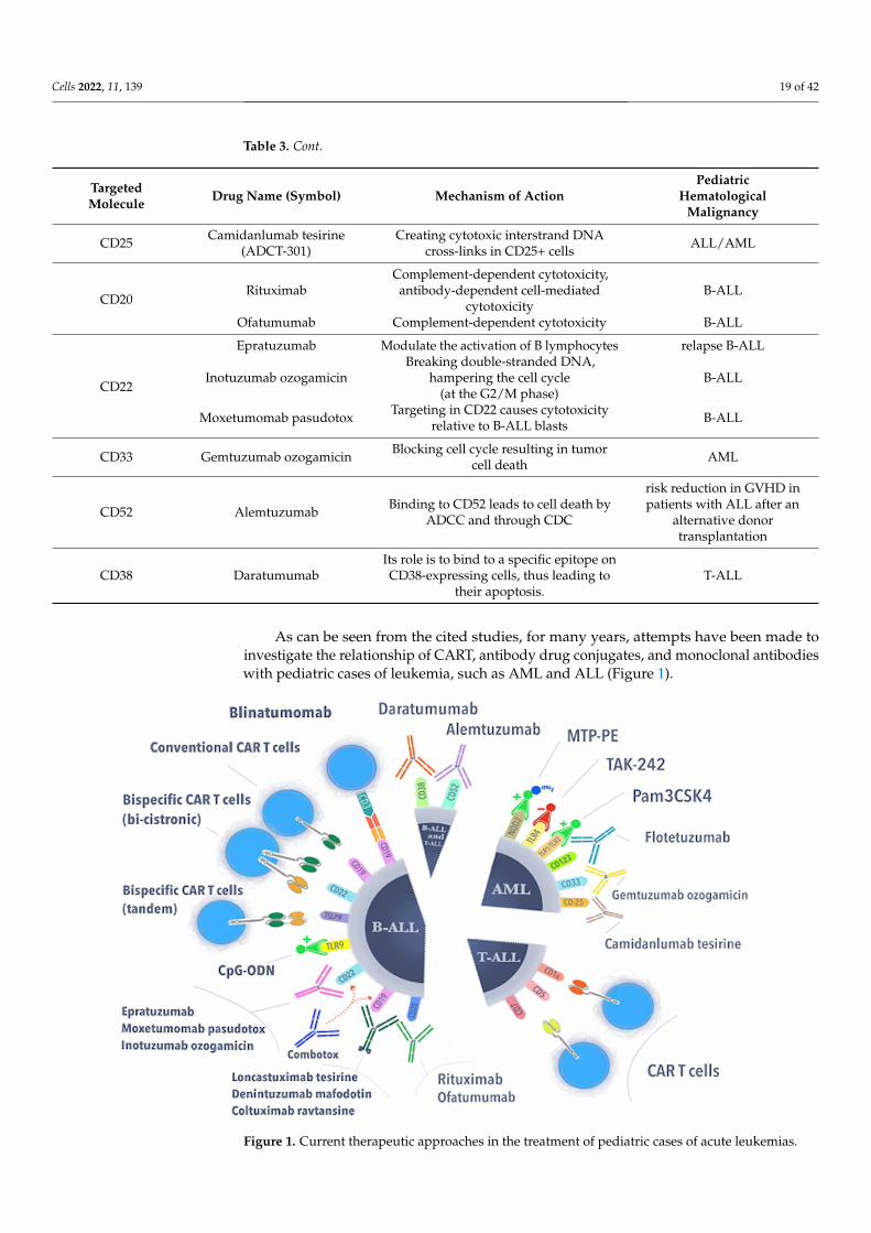
Table 3. Antibody–drug conjugates and monoclonal antibodies used in the treatment of pediatriccases of acute lymphoblastic leukemia and acute myeloid leukemia.
TargetedMolecule Drug Name (Symbol) Mechanism of Action
PediatricHematological
Malignancy
CD19
Denintuzumab mafodotin(SGN-CD19A) Inhibition of cell division in CD19+ cells ALL/B-ALL
Coltuximab Ravtansine Disruption of microtubules,impeding cell cycle B-ALL
Loncastuximab tesirine(ADCT-402)
Creating cytotoxic interstrand DNAcross-links in CD19+ cells B-ALL
Cells 2022, 11, 139 19 of 42
Table 3. Cont.
TargetedMolecule Drug Name (Symbol) Mechanism of Action
PediatricHematological
Malignancy
CD25 Camidanlumab tesirine(ADCT-301)
Creating cytotoxic interstrand DNAcross-links in CD25+ cells ALL/AML
CD20Rituximab
Complement-dependent cytotoxicity,antibody-dependent cell-mediated
cytotoxicityB-ALL
Ofatumumab Complement-dependent cytotoxicity B-ALL
CD22
Epratuzumab Modulate the activation of B lymphocytes relapse B-ALL
Inotuzumab ozogamicinBreaking double-stranded DNA,
hampering the cell cycle(at the G2/M phase)
B-ALL
Moxetumomab pasudotox Targeting in CD22 causes cytotoxicityrelative to B-ALL blasts B-ALL
CD33 Gemtuzumab ozogamicin Blocking cell cycle resulting in tumorcell death AML
CD52 Alemtuzumab Binding to CD52 leads to cell death byADCC and through CDC
risk reduction in GVHD inpatients with ALL after an
alternative donortransplantation
CD38 DaratumumabIts role is to bind to a specific epitope onCD38-expressing cells, thus leading to
their apoptosis.T-ALL
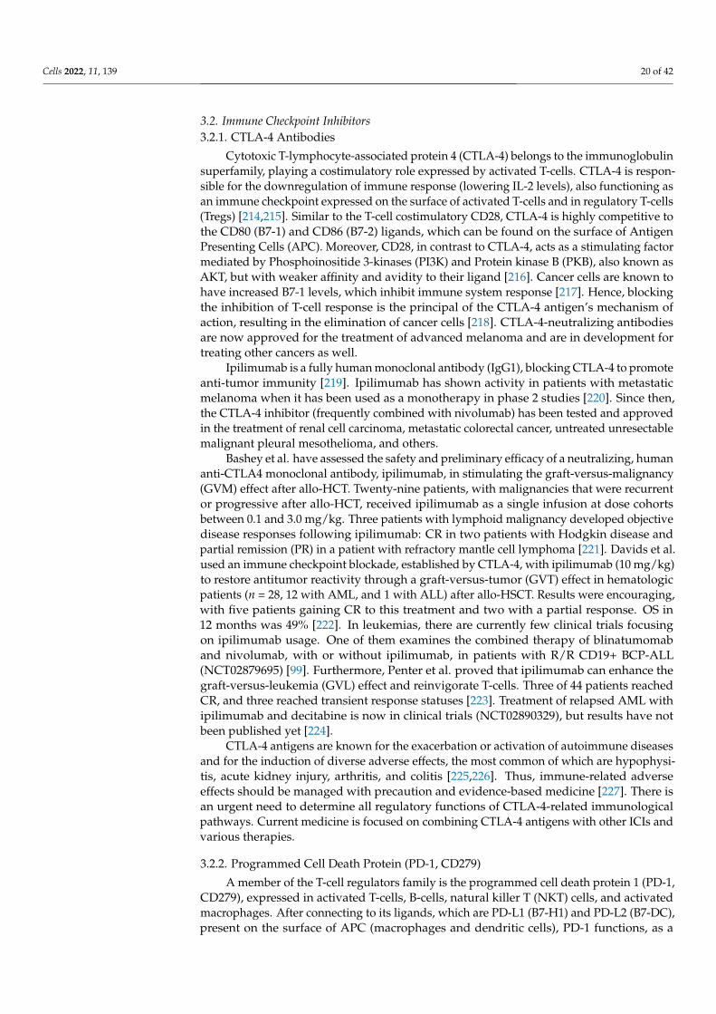
As can be seen from the cited studies, for many years, attempts have been made toinvestigate the relationship of CART, antibody drug conjugates, and monoclonal antibodieswith pediatric cases of leukemia, such as AML and ALL (Figure 1).
Cells 2021, 10, x FOR PEER REVIEW 19 of 41
Figure 1. Current therapeutic approaches in the treatment of pediatric cases of acute leukemias.
3.2. Immune Checkpoint Inhibitors
3.2.1. CTLA-4 Antibodies
Cytotoxic T-lymphocyte-associated protein 4 (CTLA-4) belongs to the
immunoglobulin superfamily, playing a costimulatory role expressed by activated T-cells.
CTLA-4 is responsible for the downregulation of immune response (lowering IL-2 levels),
also functioning as an immune checkpoint expressed on the surface of activated T-cells
and in regulatory T-cells (Tregs) [214,215]. Similar to the T-cell costimulatory CD28,
CTLA-4 is highly competitive to the CD80 (B7-1) and CD86 (B7-2) ligands, which can be
found on the surface of Antigen Presenting Cells (APC). Moreover, CD28, in contrast to
CTLA-4, acts as a stimulating factor mediated by Phosphoinositide 3-kinases (PI3K) and
Protein kinase B (PKB), also known as AKT, but with weaker affinity and avidity to their
ligand [216]. Cancer cells are known to have increased B7-1 levels, which inhibit immune
system response [217]. Hence, blocking the inhibition of T-cell response is the principal of
the CTLA-4 antigen’s mechanism of action, resulting in the elimination of cancer cells
[218]. CTLA-4-neutralizing antibodies are now approved for the treatment of advanced
melanoma and are in development for treating other cancers as well.
Ipilimumab is a fully human monoclonal antibody (IgG1), blocking CTLA-4 to
promote anti-tumor immunity [219]. Ipilimumab has shown activity in patients with
metastatic melanoma when it has been used as a monotherapy in phase 2 studies [220].
Since then, the CTLA-4 inhibitor (frequently combined with nivolumab) has been tested
and approved in the treatment of renal cell carcinoma, metastatic colorectal cancer,
untreated unresectable malignant pleural mesothelioma, and others.
Bashey et al. have assessed the safety and preliminary efficacy of a neutralizing,
human anti-CTLA4 monoclonal antibody, ipilimumab, in stimulating the graft-versus-
malignancy (GVM) effect after allo-HCT. Twenty-nine patients, with malignancies that
were recurrent or progressive after allo-HCT, received ipilimumab as a single infusion at
dose cohorts between 0.1 and 3.0 mg/kg. Three patients with lymphoid malignancy
Figure 1. Current therapeutic approaches in the treatment of pediatric cases of acute leukemias.
Cells 2022, 11, 139 20 of 42
3.2. Immune Checkpoint Inhibitors3.2.1. CTLA-4 Antibodies
Cytotoxic T-lymphocyte-associated protein 4 (CTLA-4) belongs to the immunoglobulinsuperfamily, playing a costimulatory role expressed by activated T-cells. CTLA-4 is respon-sible for the downregulation of immune response (lowering IL-2 levels), also functioning asan immune checkpoint expressed on the surface of activated T-cells and in regulatory T-cells(Tregs) [214,215]. Similar to the T-cell costimulatory CD28, CTLA-4 is highly competitive tothe CD80 (B7-1) and CD86 (B7-2) ligands, which can be found on the surface of AntigenPresenting Cells (APC). Moreover, CD28, in contrast to CTLA-4, acts as a stimulating factormediated by Phosphoinositide 3-kinases (PI3K) and Protein kinase B (PKB), also known asAKT, but with weaker affinity and avidity to their ligand [216]. Cancer cells are known tohave increased B7-1 levels, which inhibit immune system response [217]. Hence, blockingthe inhibition of T-cell response is the principal of the CTLA-4 antigen’s mechanism ofaction, resulting in the elimination of cancer cells [218]. CTLA-4-neutralizing antibodiesare now approved for the treatment of advanced melanoma and are in development fortreating other cancers as well.
Ipilimumab is a fully human monoclonal antibody (IgG1), blocking CTLA-4 to promoteanti-tumor immunity [219]. Ipilimumab has shown activity in patients with metastaticmelanoma when it has been used as a monotherapy in phase 2 studies [220]. Since then,the CTLA-4 inhibitor (frequently combined with nivolumab) has been tested and approvedin the treatment of renal cell carcinoma, metastatic colorectal cancer, untreated unresectablemalignant pleural mesothelioma, and others.
Bashey et al. have assessed the safety and preliminary efficacy of a neutralizing, humananti-CTLA4 monoclonal antibody, ipilimumab, in stimulating the graft-versus-malignancy(GVM) effect after allo-HCT. Twenty-nine patients, with malignancies that were recurrentor progressive after allo-HCT, received ipilimumab as a single infusion at dose cohortsbetween 0.1 and 3.0 mg/kg. Three patients with lymphoid malignancy developed objectivedisease responses following ipilimumab: CR in two patients with Hodgkin disease andpartial remission (PR) in a patient with refractory mantle cell lymphoma [221]. Davids et al.used an immune checkpoint blockade, established by CTLA-4, with ipilimumab (10 mg/kg)to restore antitumor reactivity through a graft-versus-tumor (GVT) effect in hematologicpatients (n = 28, 12 with AML, and 1 with ALL) after allo-HSCT. Results were encouraging,with five patients gaining CR to this treatment and two with a partial response. OS in12 months was 49% [222]. In leukemias, there are currently few clinical trials focusingon ipilimumab usage. One of them examines the combined therapy of blinatumomaband nivolumab, with or without ipilimumab, in patients with R/R CD19+ BCP-ALL(NCT02879695) [99]. Furthermore, Penter et al. proved that ipilimumab can enhance thegraft-versus-leukemia (GVL) effect and reinvigorate T-cells. Three of 44 patients reachedCR, and three reached transient response statuses [223]. Treatment of relapsed AML withipilimumab and decitabine is now in clinical trials (NCT02890329), but results have notbeen published yet [224].
CTLA-4 antigens are known for the exacerbation or activation of autoimmune diseasesand for the induction of diverse adverse effects, the most common of which are hypophysi-tis, acute kidney injury, arthritis, and colitis [225,226]. Thus, immune-related adverseeffects should be managed with precaution and evidence-based medicine [227]. There isan urgent need to determine all regulatory functions of CTLA-4-related immunologicalpathways. Current medicine is focused on combining CTLA-4 antigens with other ICIs andvarious therapies.
3.2.2. Programmed Cell Death Protein (PD-1, CD279)
A member of the T-cell regulators family is the programmed cell death protein 1 (PD-1,CD279), expressed in activated T-cells, B-cells, natural killer T (NKT) cells, and activatedmacrophages. After connecting to its ligands, which are PD-L1 (B7-H1) and PD-L2 (B7-DC),present on the surface of APC (macrophages and dendritic cells), PD-1 functions, as a
Cells 2022, 11, 139 21 of 42
promotor of apoptosis of T-cells and as an inhibitor to T-cell response [228,229]. In themechanism of evacuation from T-cell toxicity, tumor cells often express PD-L1 on theirsurface, which hampers the immune system [230,231]. Furthermore, PD-L1 expression isresponsible for Treg expansion, which results in immune system suppression. Tregs areexpected to promote AML blast growth by IL-10 and IL-35 secretion [232]. Hence, currently,the approach to the PD-1/PD-L1 complex is managed in both ways: by blocking the PD-1or by blocking the PD-L1, with PD-1 antibodies being superior over PD-L1 in OS ratesamong cancer patients [227].
3.2.3. PD-1 Inhibitors
The first registered PD-1 targeting drug was pembrolizumab (MK-3475, lambrolizumab),which is a humanized monoclonal IgG4 anti-PD-1 antibody. In the beginning, it wasregistered in advanced melanoma, after achieving auspicious results in a group of patients,after unsuccessful treatment with ipilimumab [233,234]. First trials of pembrolizumab(NCT02767934) failed in improving outcomes of patients with T-ALL and B-ALL withmeasurable MRD. A study conducted by Cassaday et al. achieved a median OS value ofonly 12.7 months, so the trial was terminated [235].
Another PD-1 immune checkpoint blocker, nivolumab (BMS-936558, ONO-4538), isalso a fully human IgG4 antibody. At first, it was tested in refractory solid tumors, followedby clinical trials on patients with advanced squamous non-small cell lung cancer [236,237].In 2014, nivolumab was approved by the FDA for the treatment of patients with advancedmelanoma [238]. Currently, it is being tested in adult patients with a high risk of AMLrelapse (NCT02532231). First results have showed that the blockade of the PD-1 checkpointdoes not lower the estimated time of CR in these patients, so more optimistic data arenot expected [239]. Another phase II clinical trial (NCT04546399) included patients witha first relapse of CD19+ B-ALL (1–31 years old), with treatment options of nivolumabcombined with blinatumomab or blinatumomab alone [240]. A trial with R/R CD19+BCP-ALL in children and young adult patients, using a combination of pembrolizumaband blinatumomab, was withdrawn (NCT03605589) [241]. The safety and tolerabilityof a combination of blinatumomab, ipilimumab, and nivolumab therapy will mainly beevaluated as a treatment of patients (16 years and older) diagnosed with poor-risk R/RCD19+ BCP-ALL or CD19+ mixed phenotype acute leukemia (MPAL) (NCT02879695) [99].Studies, conducted by Kamphorst et al. on mice models, showed that a blockade ofPD-1 may not be sufficient for the improvement in response from CD8+ T-cells. The co-stimulation of CD28 is required in PD-1 inhibitor therapy to achieve exceptional results.Therefore, the usage of CD28/CD80 and the CD86 pathway may increase clinical outcomesin various cancer regimens [242]. Moreover, it was found that PD-1 levels are at theirhighest levels in relapsed B-ALL, which encourages a benefit from the PD-1 blockade insuch patients [243]. In the BM of AML patients, PD-1 and TIM-3 levels were elevated,presumably hampering T-cell response [244]. PD-1 inhibitor-related adverse effects (irAEs)of nivolumab include skin irritation (rash) and pulmonary and hepatic symptoms, andthose of pembrolizumab include endocrinopathies, rash, and diarrhea [245].
3.2.4. PD-L1 Inhibitors
Atezolizumab (MPDL3280A) is the first PD-L1 inhibitor approved by the FDA in thetreatment of urothelial carcinoma, as a result of studies conducted by Rosenberg et al. [246].It is a human monoclonal IgG1 antibody targeting this immune checkpoint [247]. In AML,atezolizumab was tested with the combination of immunomodulatory agents (Guadecitabine)in adults with R/R, treatment-naive, and chemotherapy-resistant patients (NCT02892318) [248].However, its results have still not been published. There are no clinical trials with PD-L1antibodies as a therapeutic approach in childhood leukemias. Durvalumab (MEDI4736), an-other human PD-L1 antibody, is registered as a treatment of patients with locally advancedor metastatic urothelial carcinoma [249]. It was first tested in patients with solid tumors,achieving clinical activity [250]. Later, durvalumab improved response rates in patients
Cells 2022, 11, 139 22 of 42
with advanced urothelial bladder cancer, resulting in FDA approval in this group of pa-tients [251]. This drug was tested, in combination with azacitidine, as a front-line treatmentfor chemotherapy-unfit AML patients, bringing the expected results of an absence of anyincrease in PD-L1 surface expression. Remembering that azacitidine is known for increasingPD-L1 levels, a blockade of PD-L1 is a bright regimen solution [252]. Atezolizumab-treatedpatients developed pulmonary symptoms and diarrhea, while adverse effects of durval-umab included endocrinopathies and pulmonary symptoms [245].
3.3. Other Targets for Immune Checkpoint Inhibitors3.3.1. Anti-CD47 Antibodies
CD-47 acts as a signaling receptor for thrombospondin 1 and the counter-receptor forthe signal-regulatory protein alpha (SIRPα). All functions of this protein and its ligandsare yet to be discovered [253]. In general, CD47 acts as a phagocytosis inhibitor signal,commonly referred to as the “do not eat me” message. A crucial role is also played bythe SIRPα present in macrophages and dendritic cells. A high expression of CD47 proteinin cancer cells correlates with a poor prognosis for patients, due to the gained ability ofevasion from macrophages and dendritic cells. Magrolimab (Hu5F9-G4) is a humanizedmonoclonal antibody targeting CD47 present on macrophages and dendritic cells, enhanc-ing CD47-dependent phagocytosis [254,255]. After achieving promising preclinical resultswith in vivo and in vitro studies in AML cells, Hu5F9-G4 was forwarded into clinical tri-als [256]. Due to the fact that CD47 is expressed in red blood cells, the impact of anti-47monoclonal antibody Hu5F9-G4 on blood parameters was examined (NCT02678338). Theobtained results state that haemoglobin levels declined during this treatment [257]. Anotherphase 1b study covered magrolimab with azacytidine in adult patients with R/R AML(NCT03248479) and showed an objective response, particularly in seven of eight patientswith TP53 mutation [258]. Magrolimab-related adverse effects mainly include dermatitisacneiform, dry skin, fatigue, and infusion reactions [259].
3.3.2. Anti T-Cell Immunoglobulin and a Mucin-Domain Containing-3 (Tim-3) Antibody
Tim-3 regulates the secretion of TNF and interferon γ (IFN-γ), which results in theinhibition of Th1 cell response. TIM-3 is also expressed in monocytes, macrophages,dendritic cells, tumor infiltrating lymphocytes, and leukemic stem cells and blasts (withouthealthy hematopoietic stem cells (HSCs)) [260–262]. There are several ligands for the TIM-3 molecule: galectin 9 (Gal9), high mobility group protein B1, phosphatidylserine, andCEACAM1 [263]. Dama et al. discovered that, in AML patients, TIM-3 and Gal9 levels weresignificantly higher in patients with treatment failure, which means that, in those patients,blocking this pathway may improve therapeutic results, with a clinical outcome of CR [264].Sabatolimab (MBG453) is a humanized IgG4 antibody that targets TIM-3. It was tested in35 patients with newly diagnosed AML and resulted in an ORR of 41.2%, with an estimated12 months of progression-free survival at a rate of 44% [262]. Currently, sabatolimab isbeing tested in AML patients after allo-HSCT with present MRD. This study (NCT04623216)covers patients from 12 to 99 years old, and results are yet to come [265]. Moreover, thecombination of sabatolimab with blocked PD-1 (PDR001) and decatibine is an active phase Istudy (NCT03066648), connecting various ICI therapy targets [266]. A frequently occurringadverse effect was fatigue, which was present in 9% of 133 patients [267]. ICIs, which arein use in pediatric cases of hematological malignancies, are shown below (Figure 2).
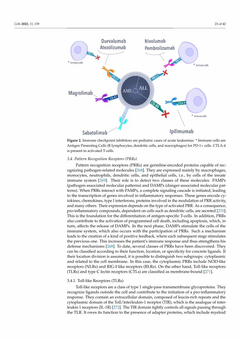
Cells 2022, 11, 139 23 of 42
Cells 2021, 10, x FOR PEER REVIEW 22 of 41
treatment [257]. Another phase 1b study covered magrolimab with azacytidine in adult
patients with R/R AML (NCT03248479) and showed an objective response, particularly in
seven of eight patients with TP53 mutation [258]. Magrolimab-related adverse effects
mainly include dermatitis acneiform, dry skin, fatigue, and infusion reactions [259].
3.3.2. Anti T-Cell Immunoglobulin and a Mucin-Domain Containing-3 (Tim-3) Antibody
Tim-3 regulates the secretion of TNF and interferon γ (IFN-γ), which results in the
inhibition of Th1 cell response. TIM-3 is also expressed in monocytes, macrophages,
dendritic cells, tumor infiltrating lymphocytes, and leukemic stem cells and blasts
(without healthy hematopoietic stem cells (HSCs)) [260–262]. There are several ligands for
the TIM-3 molecule: galectin 9 (Gal9), high mobility group protein B1, phosphatidylserine,
and CEACAM1 [263]. Dama et al. discovered that, in AML patients, TIM-3 and Gal9 levels
were significantly higher in patients with treatment failure, which means that, in those
patients, blocking this pathway may improve therapeutic results, with a clinical outcome
of CR [264]. Sabatolimab (MBG453) is a humanized IgG4 antibody that targets TIM-3. It
was tested in 35 patients with newly diagnosed AML and resulted in an ORR of 41.2%,
with an estimated 12 months of progression-free survival at a rate of 44% [262]. Currently,
sabatolimab is being tested in AML patients after allo-HSCT with present MRD. This
study (NCT04623216) covers patients from 12 to 99 years old, and results are yet to come
[265]. Moreover, the combination of sabatolimab with blocked PD-1 (PDR001) and
decatibine is an active phase I study (NCT03066648), connecting various ICI therapy
targets [266]. A frequently occurring adverse effect was fatigue, which was present in 9%
of 133 patients [267]. ICIs, which are in use in pediatric cases of hematological
malignancies, are shown below (Figure 2).
Figure 2. Immune checkpoint inhibitors are pediatric cases of acute leukemias. * Immune cells are
Antigen Presenting Cells (B lymphocytes, dendritic cells, and macrophages) for PD-1+ cells. CTLA-
4 is present in activated T-cells.
Figure 2. Immune checkpoint inhibitors are pediatric cases of acute leukemias. * Immune cells areAntigen Presenting Cells (B lymphocytes, dendritic cells, and macrophages) for PD-1+ cells. CTLA-4is present in activated T-cells.
3.4. Pattern Recognition Receptors (PRRs)
Pattern recognition receptors (PRRs) are germline-encoded proteins capable of rec-ognizing pathogen-related molecules [268]. They are expressed mainly by macrophages,monocytes, neutrophils, dendritic cells, and epithelial cells, i.e., by cells of the innateimmune system [269]. Their role is to detect two classes of these molecules: PAMPs(pathogen-associated molecular patterns) and DAMPs (danger-associated molecular pat-terns). When PRRs interact with PAMPs, a complete signaling cascade is initiated, leadingto the transcription of genes involved in inflammatory responses. These genes encode cy-tokines, chemokines, type I interferons, proteins involved in the modulation of PRR activity,and many others. Their expression depends on the type of activated PRR. As a consequence,pro-inflammatory compounds, dependent on cells such as dendritic cells, are secreted [270].This is the foundation for the differentiation of antigen-specific T-cells. In addition, PRRsalso contribute to the activation of programmed cell death, including apoptosis, which, inturn, affects the release of DAMPs. In the next phase, DAMPs stimulate the cells of theimmune system, which also occurs with the participation of PRRs. Such a mechanismleads to the creation of a kind of positive feedback, where each subsequent stage stimulatesthe previous one. This increases the patient’s immune response and thus strengthens hisdefense mechanisms [268]. To date, several classes of PRRs have been discovered. Theycan be classified according to their function, location, or specificity for concrete ligands. Iftheir location division is assumed, it is possible to distinguish two subgroups: cytoplasmicand related to the cell membrane. In this case, the cytoplasmic PRRs include NOD-likereceptors (NLRs) and RIG-I-like receptors (RLRs). On the other hand, Toll-like receptors(TLRs) and type C lectin receptors (CTLs) are classified as membrane-bound [271].
3.4.1. Toll-like Receptors (TLRs)
Toll-like receptors are a class of type 1 single-pass transmembrane glycoproteins. Theyrecognize ligands outside the cell and contribute to the initiation of a pro-inflammatoryresponse. They contain an extracellular domain, composed of leucin-rich repeats and thecytoplasmic domain of the Toll/interleukin-1 receptor (TIR), which is the analogue of inter-leukin 1 receptors (IL-1R) [272]. The TIR domain tightly controls all signals passing throughthe TLR. It owes its function to the presence of adapter proteins, which include myeloid
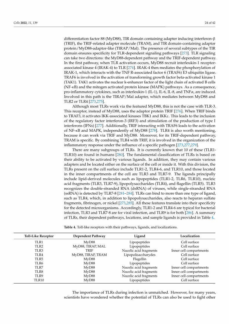
Cells 2022, 11, 139 24 of 42
differentiation factor 88 (MyD88), TIR domain containing adapter inducing interferon-β(TRIF), the TRIF-related adapter molecule (TRAM), and TIR domain-containing adaptorprotein/MyD88-adaptor-like (TIRAP/Mal). The presence of several subtypes of the TIRdomain ensures specificity for TLR-dependent signaling pathways [273]. TLR signalingcan take two directions: the MyD88-dependent pathway and the TRIF-dependent pathway.In the first pathway, when TLR activation occurs, MyD88 recruit interleukin-1 receptor-associated kinase 4 (IRAK-4) to TLR [274]. IRAK-4 then mediates the phosphorylation ofIRAK-1, which interacts with the TNF R-associated factor 6 (TRAF6) E3 ubiquitin ligase.TRAF6 is involved in the activation of transforming growth factor beta-activated kinase 1(TAK1). TAK1 activates the nuclear k-enhancer factor of the light chain of activated B cells(NF-κB) and the mitogen activated protein kinase (MAPK) pathways. As a consequence,pro-inflammatory cytokines, such as interleukin-1 (IL-1), IL-6, IL-8, and TNFα, are induced.Involved in this path is the TIRAP/Mal adapter, which mediates between MyD88 andTLR2 or TLR4 [273,275].
Although most TLRs work via the featured MyD88, this is not the case with TLR-3.This receptor, instead of MyD88, uses the adaptor protein TRIF [276]. When TRIF bindsto TRAF3, it activates IKK-associated kinases TBK1 and IKKε. This leads to the inclusionof the regulatory factor interferon-3 (IRF3) and stimulation of the production of type Iinterferons (IFNs) [277]. Additionally, TRIF interacting with TRAF6 leads to the activationof NF-κB and MAPK, independently of MyD88 [278]. TLR4 is also worth mentioning,because it can work via TRIF and MyD88. Moreover, for its TRIF-dependent pathway,TRAM is specific. By combining TLR4 with TRIF, it is involved in the organization of theinflammatory response under the influence of a specific pathogen [273,277,279].
There are many subgroups of TLRs. It is currently known that 10 of these (TLR1-TLR10) are found in humans [280]. The fundamental classification of TLRs is based ontheir ability to be activated by various ligands. In addition, they may contain variousadapters and be located either on the surface of the cell or inside it. With this division, theTLRs present on the cell surface include TLR1-2, TLR4-6, and TLR10, and those locatedin the inner compartments of the cell are TLR3 and TLR7-9. The ligands principallyinclude lipid-derived molecules such as lipopeptides (TLR1-2, TLR6, TLR10), nucleicacid fragments (TLR3, TLR7-9), lipopolysaccharides (TLR4), and flagellin (TLR5). TLR3recognizes the double-stranded RNA (dsRNA) of viruses, while single-stranded RNA(ssRNA) is detected by TLR7-8 [281–284]. TLRs can bind to more than one type of ligand,such as TLR4, which, in addition to lipopolysaccharides, also reacts to heparan sulfatefragments, fibrinogen, or nickel [273,285]. All these features translate into their specificityfor the detected microorganisms. Accordingly, TLR1-2 and TLR4-6 are typical for bacterialinfection, TLR3 and TLR7-8 are for viral infection, and TLR9 is for both [286]. A summaryof TLRs, their dependent pathways, locations, and sample ligands is provided in Table 4.
Table 4. Toll-like receptors with their pathways, ligands, and localizations.
Toll-Like Receptor Dependent Pathway Ligand Localization
TLR1 MyD88 Lipopeptides Cell surfaceTLR2 MyD88, TIRAP, MAL Lipopeptides Cell surfaceTLR3 TRIF Nucelic acid fragments Inner cell compartmentsTLR4 MyD88, TIRAP, TRAM Lipopolisaccharydes Cell surfaceTLR5 MyD88 Flagellin Cell surfaceTLR6 MyD88 Lipopeptides Cell surfaceTLR7 MyD88 Nucelic acid fragments Inner cell compartmentsTLR8 MyD88 Nucelic acid fragments Inner cell compartmentsTLR9 MyD88 Nucelic acid fragments Inner cell compartmentsTLR10 MyD88 Lipopeptides Cell surface
The importance of TLRs during infection is unmatched. However, for many years,scientists have wondered whether the potential of TLRs can also be used to fight other
Cells 2022, 11, 139 25 of 42
diseases. At the same time, more data on inflammation and recurrent infections, in patientswith hematological malignancies, have begun to appear, showing an association withTLRs. More than 10 years ago, it was suggested that TLRs may condition hematopoiesisby acting from the level of HSCs [287]. A study by Sioud et al. confirmed that TLR4,TLR7, and TLR8 can be expressed by human hematopoietic BM CD34+ progenitor cells.Additionally, it was noted that TLR7/8 signaling influenced further differentiation of CD34+cells [288]. It is known that a hallmark of acute leukemias is the uncontrolled production ofthe hematopoietic precursor cells of the myeloid or lymphoid series in the BM [289]. Thissuggests the possibility that primitive TLR-expressing cells are the beginning of unstablelineages. Moreover, a study by Eriksson et al. shows that TLR1 expression is much morepotent in the immature CD34+ and CD38- cells of AML patients, as compared to normalcells [290]. This was in line with the elevated levels of TLR1 mRNA in patients withMDS [291].
Additionally, Eriksson evaluated the effect that both the agonist and antagonist ofTLR1 would have on leukemia stem cells. After applying an inhibitory effect on TLR1, astrong decrease in the number of leukemic cells was observed, which confirmed that thisreceptor is important for AML activity. On the other hand, after the use of Pam3CSK4,which is a specific TLR1-2 agonist, an increase in the survival of leukemic cells was observed.However, further forced TLR1/TLR2 signaling led to the differentiation of leukemic cellsand thus, a reduction in the leukemia burden [290]. This proves that further studies basedon the inhibition or forced activation of TLR1 in AML could prove beneficial in achievingsatisfactory clinical effects.
Apart from TLR1-2, TLR4 may also play a significant role in the disease activity ofAML. As in the case of the previous receptors, it was assumed that the use of a TLR4inhibitor may slow the activity of AML. To do this, a study by Baakhlagh et al. wasconducted. TAK-242 was used there as a TLR4 inhibitor. The obtained results showedthat the applied inhibitor not only stopped the proliferation of all cell lines of leukemia,but also changed the distribution of their cell cycle. Additionally, it was noticed that, inAML patients with poor prognosis, there is a much higher expression of TLR4, MyD88, andNF-кB mRNA [292]. In light of this evidence, it seems advantageous to continue researchon TLR4 and its inhibitor, as it may be a good strategy for the treatment of TLR4-expressingAML in the future. Aref et al. investigated the impact of TLR2 and TLR4 polymorphismson the survival of patients with AML and their susceptibility to severe infections [293].They demonstrated that, in AML patients with TLR2 Arg753Gln with an AG genotypeand an A allele, TLR4 Asp299Gly with a CT genotype and a C allele, and TLR4 Thr399Ilewith an AG genotype and an A allele, pneumonia and sepsis were more frequent than inpatients with other genotypes and alleles. These results are in line with an earlier study bySchnetzke [294]. At the same time, it was observed that patients with the TLR2 (Arg753Gln)GG genotype had the shortest OS, which differed from the Schnetzke results. This studyshowed that, in these patients, TLR polymorphism translates into both their risk of infectionand survival. However, more research is needed on this point [293].
Sánchez-Cuaxosp et al. decided to conduct the first study on pediatric ALL patients.It showed that, in children with this leukemia, there is a reduced expression of TLR1, TLR3,TLR4, TLR7, and TLR9 compared to the control group. Moreover, they considered patientswith different phenotypes, such as Pro-B, Pre-B, and B- and T-ALL. They noticed thatthe lowest expression of TLR4 and TLR7 was found in patients with Pro-B and B-ALLsubtypes. As emphasized, this could have been influenced by many factors, such as thetype of cells or the type of stimulus. In addition, it is believed that the age of the patientmay also be an influence. Thus, there is still no relevant data to unequivocally point to thecause of this variability in TLR levels. At the same time, the presented study suggests thatthe decreased expression of TLRs, in ALL patients, may partially explain the decreasedanti-tumor response in this group of children [295].
Other strategies to prevent relapse in ALL employ synthetic single-stranded oligonu-cleotides, containing unmethylated cytosine and guanine (CpG-ODN) motifs, acting as
Cells 2022, 11, 139 26 of 42
TLR9 agonists [296]. Many years ago, it was noticed that CpG-ODN (GNKG168) stimu-lates the formation of cytokines produced by pediatric ALL cells in vitro. As a result, itincreased the allogeneic response of Th1 cells against this leukemia [297]. The TherapeuticAdvances in Childhood Leukemia and Lymphoma decided to conduct their own study.Their goal was to investigate the effect of GNKG168 on immunomodulatory molecules inMDR-positive children with ALL and those in remission. They compared the expressionlevels of 608 genes before and eight days after using GNKG168 in patients. In their results,they focused on the eight markers that showed the greatest difference before and after thestudy. They observed an increase in the level of H-RAS mRNA and promyelocytic leukemiaprotein, but a decrease in single Ig and TIR domain containing (SIGIRR, IL1R8) interleukin1 receptor 1 (IL1RL1, ST2), CC motif chemokine receptor 8 (CCR8), interleukin 7 R (IL7R),cluster of differentiation 8B (CD8B), and cluster of differentiation 3 (CD3D). Summarizing,it can be noted that the described increase in expression concerned genes involved inincreasing B and T lymphocyte responses. On the other hand, the decrease in expressionwas related to genes and involved enhancing the immune response to tumorigenesis. Itis possible that the tested GNKG168 act as checkpoint inhibitors, as it caused a decreasein SIGIRR, IL1RL1, CCR8, and IL7R, which are checkpoint signals. In order to make clearconclusions, more studies are needed on larger groups of patients [298]. Recently, the useof a properly constructed vaccine has started to be considered as well. As vaccines induceand potentiate the patient’s T-cells, this one could target leukemia-associated antigens. Thepast year has led to a human trial using a vaccine based on TLRs. It was assumed that thisvaccine could induce an anti-leukemic immune response. To test this, patients in AMLremission, with a high risk of relapse, were inoculated with mature TLR7/8 RNA-loadeddendritic cells encoding two antigens associated with this leukemia: WT1 and PRAME.The obtained results showed that the administered vaccine elicited a favorable local in-flammatory response and led to the proliferation of T CD8+ and T CD4+ in peripheralblood, which was clinically advantageous. In addition, its administration was found to besafe for patients, and the response was better suited to their younger age [299]. Althoughthis is a promising strategy for the future therapy of AML, much more research, both onchildren and adults, is still needed on the subject. Additionally, it might be beneficial tocombine mature 7/8 TLR cells with, e.g., checkpoint inhibitors, which would contribute toan additional enhancement of the immune response of patients.
3.4.2. NOD-Like Receptors (NLRs)
Nucleotide-binding oligomerization domain (NOD), as with receptors (NLRs), belongto the group of cytosolic PRRs. As receptors located inside the cell, they are involved inthe detection of intracellular pathogens and endogenous products generated during tissuedamage [300]. So far, 22 types of NLRs in humans have been described [271,301]. Theyare characterized by a common organization of domains, centrally containing NATCH,which is necessary for the activation of the NLR by ATP. In addition, there is an N-terminaleffector domain to mediate signal transduction and a C-terminal region, having a vary-ing number of leucine-rich repeat (LRR) motifs domains, involved in molecular patternrecognition [301,302]. Depending on their N-terminal region, four subgroups can be dis-tinguished among NLRs: NLRA (the acid transactivation domain), NLRC (the domain ofcaspase activation and recruitment), NLRB (the inhibitory repeat-like baculovirus domain),and NLRP (the N-terminal pyrin effector domain) [301].
NLRC includes nucleotide-binding oligomerization domain 1 (NOD1), nucleotide-binding oligomerization domain 2 (NOD2), and domains classified in this group due to theirhomology and phylogenetic relationships (NLRC3, NLRC4, NLRC5, and NLRX1) [303].The role of NOD1 and NOD2 is to detect bacterial particles produced during the synthesisor degradation of peptidoglycan. More specifically, NOD1 detects the γ-D-glutamyl-meso-diaminopimelinic acid dipeptide (iE-DAP), a component of the peptidoglycan found inmost Gram-negative bacteria and specific Gram-positive bacteria. In contrast, NOD2recognizes muramyl dipeptide (MDP), which is present in virtually all types of peptidogly-
Cells 2022, 11, 139 27 of 42
cans [304,305]. Ligand recognition, by the LRR of NOD1 and NOD2, leads to activationof the serine-threonine kinase (RICK). This kinase activates the TAK1 kinase, leading inturn to the degradation of the IκBα (nuclear factor kappa B inhibitor). As a result, NF-κBtranslocation to the nucleus takes place, where appropriate NF-κB-dependent genes canbe transcribed. NOD1 and NOD2, in addition to NF-κB, also activate MAP kinase. As aresult, the NF-κB and MAPK work together to increase the expression of pro-inflammatoryfactors [302].
NOD1 and NOD2 have been minimally tested for acute leukemias, but this doesnot mean that these receptors cannot be successfully used in the future in the treatmentof patients with acute leukemia. Similar to the TLRs, it has been shown that NOD2is expressed by human BM CD34+ cells [306]. This suggests that the activation of thisreceptor and the subsequent inflammatory response may play a role in AML. To verifythis, Buteyn et al. evaluated the effect of NOD2 receptors on AML. They decided to usephosphatidylethanolamine muramyl ripeptide (MTP-PE), which is a synthetic and lesspyrogenic derivative of MDP, a NOD2 ligand. However, they found that MTP-PE alone wasunable to produce the expected anti-tumor effects. As a result, they decided to combine itwith IFN-γ. This combination led to satisfactory results, as it led to a significant apoptosisof AML blasts in a caspase-1-dependent manner. It was reported that MTP-PE, in tandemwith IFN-γ, caused the release of pro-inflammatory cytokines, such as TNF-α and IL-1β,and supported the maturation of NK cells, both in vitro and in a murine adoptive transfermodel. In addition, it was shown that this combination reduced the severity of the diseaseand increased the survival rate of the tested mice [307]. Thus, this study showed that NOD2agonists may have a beneficial therapeutic effect on AML.
Apart from the role that NODs can play in leukemia, their polymorphism also seemsinteresting. Undoubtedly, many AML patients have a reduced number of neutrophilsat the time of diagnosis [308]. Additionally, the use of induction chemotherapy leadsto the prolongation of the neutropenia phase. As some of the sites of NOD2 expressionare neutrophils [309], Yomade et al. decided to check whether the NOD2 gene polymor-phism would translate into the frequency of infections in AML patients after intensiveinduction chemotherapy. They focused on an analysis of the three most common polymor-phisms: two missense mutations (Arg702Trp and Gly908Arg) and the frameshift mutation(Leu100fsinsC). After analyzing the results of 131 patients with AML, they concludedthat patients without any of the listed polymorphisms have a much lower likelihood ofdeveloping enteritis or mucositis. Conversely, patients with these polymorphisms weremore likely to have Streptococcus spp. detected in the bloodstream. However, it is worthadding that the presence of these mutations did not affect the incidence of complications,such as pneumonia or sepsis [310]. The presented research shows that there is a possibilityof linking NOD2 polymorphisms to the frequency of infections in AML patients, but morestudies are needed to confirm this.
4. Conclusions
In recent years, tremendous advances have been made in the treatment of childhoodacute leukemias. Currently, functioning therapies have reached such a high level that theirfurther intensification does not make sense, as they are associated with increased toxicity.Therefore, methods using the immune system have been developed to treat patients inwhom standard therapies have failed. Based on advances in genetics, it has become possibleto understand the molecular mechanisms of leukemia pathogenesis. Comprehensivegenome-wide sequencing and integration analyses identified new leukemia subtypeswith different prognostics and therapeutic factors. Moreover, it is now possible to detectantibodies present in leukemia cells, such as CD52, CD38, CD33, CD20, CD25, CD19, andCD22, and target them with new antibody–drug conjugates. The development of newtechnologies makes immunotherapy more common, and cheaper, for patients and can alsobe used to treat other diseases.
Cells 2022, 11, 139 28 of 42
In the last few years, there has been a breakthrough in the treatment of ALL withCAR-Ts. This is evidenced not only by the drug approved for this purpose by the FDA,but also by the amount of research conducted on this subject. As shown, attempts havebeen made for years to develop a satisfactory use of CAR-Ts in AML therapy, and althoughmany barriers have been encountered along this path, further clinical trials are still ongoing.In the case of ICIs, our knowledge of their function, expression, and targeted therapies hasexpanded significantly and may become a milestone in modern medicine and a hope forpatients. The known checkpoints from other diseases are very likely to be adapted to thetreatment of leukemia in adults and then in children, resulting in even better treatmentoutcomes. Investigations about the relationship between TLRs and leukemia such as AMLand ALL are still ongoing. However, we still do not have any conclusive data on their exactfunction in the diseases under consideration. It is possible that we will finally be able tofully interpret their role in cancers and thus, improve patient survival. Although the linesof research on NOD in acute leukemia seem similar to those of TLRs, they are much smallerand at an earlier stage. However, this does not exclude the possibility of their future use inAML or ALL therapy. For that to happen, a substantial amount of research has to be done.
In our work, we focused on showing the latest therapeutic possibilities of curing pa-tients with selected acute leukemias. Starting with presenting therapies whose effectivenesshas already been proven, we proceeded to summarize the future prospects for the treatmentof AML and ALL. We have chosen children as the main group because they are patientsthat show great potential for the use of the above-mentioned methods of treatment.
In addition, we believe that treatment based on targeted therapies, in which a drug isindividually selected for a patient, represents the future of medicine.
With this in mind, we can obtain an advantage in the fight against hematologicalneoplasms, contributing to the improvement of the conditions of patients.
Author Contributions: Conceptualization, M.L.; writing—original draft preparation, K.P., M.L., M.M.and K.M.; writing—review and editing, M.L.; visualization, M.M.; supervision, M.L. All authors haveread and agreed to the published version of the manuscript.
Funding: This work was funded by the Medical Research Agency, Poland (project number: 2019/ABM/01/00069-00; approval date 1 June 2020). This work was supported by the Medical Universityof Lublin grant DS 407/2021.
Institutional Review Board Statement: Not applicable.
Informed Consent Statement: Not applicable.
Data Availability Statement: No new data were created or analyzed in this study. Data sharing isnot applicable to this article.
Acknowledgments: We acknowledge support from the Medical University of Lublin for Open AccessPublishing. We acknowledge Magdalena Chmiel for help with preparing the figures.
Conflicts of Interest: The authors declare that there is no conflict of interest.
References1. Cancer Facts & Figures. 2020. Available online: https://www.cancer.org/research/cancer-facts-statistics/all-cancer-facts-
figures/cancer-facts-figures-2020.html (accessed on 12 October 2021).2. Malard, F.; Mohty, M. Acute lymphoblastic leukaemia. Lancet 2020, 395, 1146–1162. [CrossRef]3. Inaba, H.; Pui, C.-H. Advances in the Diagnosis and Treatment of Pediatric Acute Lymphoblastic Leukemia. J. Clin. Med. 2021,
10, 1926. [CrossRef] [PubMed]4. Paulsson, K.; Lilljebjörn, H.; Biloglav, A.; Olsson, L.; Rissler, M.; Castor, A.; Barbany, G.; Fogelstrand, L.; Nordgren, A.;
Sjögren, H.; et al. The genomic landscape of high hyperdiploid childhood acute lymphoblastic leukemia. Nat. Genet. 2015,47, 672–676. [CrossRef]
5. Paulsson, K.; Johansson, B. High hyperdiploid childhood acute lymphoblastic leukemia. Genes Chromosomes Cancer 2009, 48,637–660. [CrossRef]
6. Pui, C.-H. Precision medicine in acute lymphoblastic leukemia. Front. Med. 2020, 14, 689–700. [CrossRef]
Cells 2022, 11, 139 29 of 42
7. Safavi, S.; Paulsson, K. Near-haploid and low-hypodiploid acute lymphoblastic leukemia: Two distinct subtypes with consistentlypoor prognosis. Blood 2017, 129, 420–423. [CrossRef] [PubMed]
8. Pui, C.-H.; Rebora, P.; Schrappe, M.; Attarbaschi, A.; Baruchel, A.; Basso, G.; Cavé, H.; Elitzur, S.; Koh, K.; Liu, H.-C.; et al.Outcome of Children With Hypodiploid Acute Lymphoblastic Leukemia: A Retrospective Multinational Study. J. Clin. Oncol.2019, 37, 770–779. [CrossRef] [PubMed]
9. Jeha, S.; Pei, D.; Raimondi, S.C.; Onciu, M.; Campana, D.; Cheng, C.; Sandlund, J.T.; Ribeiro, R.C.; Rubnitz, J.E.; Howard, S.C.; et al.Increased risk for CNS relapse in pre-B cell leukemia with the t(1;19)/TCF3-PBX1. Leukemia 2009, 23, 1406–1409. [CrossRef]
10. Meyer, C.; Burmeister, T.; Gröger, D.; Tsaur, G.; Fechina, L.; Renneville, A.; Sutton, R.; Venn, N.C.; Emerenciano, M.; Pombo-De-Oliveira, M.S.; et al. The MLL recombinome of acute leukemias in 2017. Leukemia 2018, 32, 273–284. [CrossRef]
11. Chaer, F.E.; Keng, M.; Ballen, K.K. MLL-Rearranged Acute Lymphoblastic Leukemia. Curr. Hematol. Malig. Rep. 2020,15, 83–89. [CrossRef]
12. Wang, H.; Han, P.; Qi, X.; Li, F.; Li, M.; Fan, L.; Zhang, H.; Zhang, X.; Yang, X. Bcl-2 Enhances Chimeric Antigen Receptor T CellPersistence by Reducing Activation-Induced Apoptosis. Cancers 2021, 13, 197. [CrossRef] [PubMed]
13. Fischer, U.; Forster, M.; Rinaldi, A.; Risch, T.; Sungalee, S.; Warnatz, H.-J.; Bornhauser, B.; Gombert, M.; Kratsch, C.;Stütz, A.M.; et al. Genomics and drug profiling of fatal TCF3-HLF-positive acute lymphoblastic leukemia identifies recurrentmutation patterns and therapeutic options. Nat. Genet. 2015, 47, 1020–1029. [CrossRef] [PubMed]
14. Slayton, W.B.; Schultz, K.R.; Kairalla, J.A.; Devidas, M.; Mi, X.; Pulsipher, M.A.; Chang, B.H.; Mullighan, C.; Iacobucci, I.;Silverman, L.B.; et al. Dasatinib Plus Intensive Chemotherapy in Children, Adolescents, and Young Adults With PhiladelphiaChromosome–Positive Acute Lymphoblastic Leukemia: Results of Children’s Oncology Group Trial AALL0622. J. Clin. Oncol.2018, 36, 2306–2314. [CrossRef] [PubMed]
15. Harrison, C.J.; Moorman, A.V.; Schwab, C.; Carroll, A.J.; Raetz, E.A.; Devidas, M.; Strehl, S.; Nebral, K.; Harbott, J. An internationalstudy of intrachromosomal amplification of chromosome 21 (iAMP21): Cytogenetic characterization and outcome. Leukemia 2013,28, 1015–1021. [CrossRef] [PubMed]
16. Tasian, S.K.; Loh, M.L.; Hunger, S.P. Philadelphia chromosome–like acute lymphoblastic leukemia. Blood 2017, 130, 2064–2072. [CrossRef]17. Jain, S.; Abraham, A. BCR-ABL1–like B-Acute Lymphoblastic Leukemia/Lymphoma: A Comprehensive Review. Arch. Pathol.
Lab. Med. 2019, 144, 150–155. [CrossRef] [PubMed]18. Boer, M.L.D.; Cario, G.; Moorman, A.V.; Boer, J.M.; Groot-Kruseman, H.A.D.; Fiocco, M.; Escherich, G.; Imamura, T.; Yeoh,
A.; Sutton, R.; et al. Outcomes of paediatric patients with B-cell acute lymphocytic leukaemia with ABL-class fusion in thepre-tyrosine-kinase inhibitor era: A multicentre, retrospective, cohort study. Lancet Haematol. 2021, 8, e55–e66. [CrossRef]
19. Liu, Y.; Easton, J.; Shao, Y.; Maciaszek, J.; Wang, Z.; Wilkinson, M.R.; Mccastlain, K.; Edmonson, M.; Pounds, S.B.; Shi, L.; et al.The genomic landscape of pediatric and young adult T-lineage acute lymphoblastic leukemia. Nat. Genet. 2017, 49, 1211–1218.[CrossRef] [PubMed]
20. Rooij, J.D.; Zwaan, C.; Heuvel-Eibrink, M.V.D. Pediatric AML: From Biology to Clinical Management. J. Clin. Med. 2015, 4,127–149. [CrossRef]
21. Arber, D.A.; Orazi, A.; Hasserjian, R.; Thiele, J.; Borowitz, M.J.; Beau, M.M.L.; Bloomfield, C.D.; Cazzola, M.; Vardiman, J.W.The 2016 revision to the World Health Organization classification of myeloid neoplasms and acute leukemia. Blood 2016, 127,2391–2405. [CrossRef]
22. Lonetti, A.; Pession, A.; Masetti, R. Targeted Therapies for Pediatric AML: Gaps and Perspective. Front. Pediatrics 2019,7, 463. [CrossRef]
23. Creutzig, U.; Heuvel-Eibrink, M.M.V.D.; Gibson, B.; Dworzak, M.N.; Adachi, S.; Bont, E.D.; Harbott, J.; Hasle, H.; Johnston, D.;Kinoshita, A.; et al. Diagnosis and management of acute myeloid leukemia in children and adolescents: Recommendations froman international expert panel. Blood 2012, 120, 3187–3205. [CrossRef] [PubMed]
24. Quessada, J.; Cuccuini, W.; Saultier, P.; Loosveld, M.; Harrison, C.J.; Lafage-Pochitaloff, M. Cytogenetics of Pediatric AcuteMyeloid Leukemia: A Review of the Current Knowledge. Genes 2021, 12, 924. [CrossRef] [PubMed]
25. Balgobind, B.V.; Hollink, I.H.I.M.; Arentsen-Peters, S.T.C.J.M.; Zimmermann, M.; Harbott, J.; Beverloo, H.B.; Bergh, A.R.M.V.;Cloos, J.; Kaspers, G.J.L.; Haas, V.D.; et al. Integrative analysis of type-I and type-II aberrations underscores the geneticheterogeneity of pediatric acute myeloid leukemia. Haematologica 2011, 96, 1478–1487. [CrossRef] [PubMed]
26. Testi, A.M.; Pession, A.; Diverio, D.; Grimwade, D.; Gibson, B.; Azevedo, A.C.D.; Moran, L.; Leverger, G.; Elitzur, S.;Hasle, H.; et al. Risk-adapted treatment of acute promyelocytic leukemia: Results from the International Consortium for Child-hood APL. Blood 2018, 132, 405–412. [CrossRef]
27. Kutny, M.A.; Geyer, S.; Laumann, K.M.; Gregory, J.; Willman, C.L.; Stock, W.; Larson, R.A.; Powell, B.L.; Feusner, J.H. Outcome forpediatric acute promyelocytic leukemia patients at Childrens Oncology Group sites on the Leukemia Intergroup Study CALGB9710 (Alliance). Pediatric Blood Cancer 2018, 66, e27542. [CrossRef]
28. Balgobind, B.V.; Raimondi, S.C.; Harbott, J.; Zimmermann, M.; Alonzo, T.A.; Auvrignon, A.; Beverloo, H.B.; Chang, M.; Creutzig,U.; Dworzak, M.N.; et al. Novel prognostic subgroups in childhood 11q23/MLL-rearranged acute myeloid leukemia: Results ofan international retrospective study. Blood 2009, 114, 2489–2496. [CrossRef]
29. Lonetti, A.; Indio, V.; Laginestra, M.A.; Tarantino, G.; Chiarini, F.; Astolfi, A.; Bertuccio, S.N.; Martelli, A.M.; Locatelli, F.; Pession,A.; et al. Inhibition of Methyltransferase DOT1L Sensitizes to Sorafenib Treatment AML Cells Irrespective of MLL-Rearrangements:A Novel Therapeutic Strategy for Pediatric AML. Cancers 2020, 12, 1972. [CrossRef]
Cells 2022, 11, 139 30 of 42
30. Casillas, J.N.; Woods, W.G.; Hunger, S.P.; Mcgavran, L.; Alonzo, T.A.; Feig, S.A. Prognostic Implications of t(10;11) Translocationsin Childhood Acute Myelogenous Leukemia: A Report From the Childrens Cancer Group. J. Pediatric Hematol. Oncol. 2003, 25,594–600. [CrossRef]
31. Lopez, C.K.; Malinge, S.; Gaudry, M.; Bernard, O.A.; Mercher, T. Pediatric Acute Megakaryoblastic Leukemia: MultitaskingFusion Proteins and Oncogenic Cooperations. Trends Cancer 2017, 3, 631–642. [CrossRef] [PubMed]
32. Rooij, J.D.E.D.; Branstetter, C.; Ma, J.; Li, Y.; Walsh, M.P.; Cheng, J.; Obulkasim, A.; Dang, J.; Easton, J.; Verboon, L.J.; et al. Pediatricnon–Down syndrome acute megakaryoblastic leukemia is characterized by distinct genomic subsets with varying outcomes. Nat.Genet. 2017, 49, 451–456. [CrossRef]
33. Hollink, I.H.I.M.; Heuvel-Eibrink, M.M.V.D.; Arentsen-Peters, S.T.; Pratcorona, M.; Abbas, S.; Kuipers, J.E.; Galen, J.F.V.; Beverloo,H.B.; Sonneveld, E.; Kaspers, G.-J.J.L.; et al. NUP98/NSD1 characterizes a novel poor prognostic group in acute myeloid leukemiawith a distinct HOX gene expression pattern. Blood 2011, 118, 3645–3656. [CrossRef]
34. Haferlach, T.; Kohlmann, A.; Klein, H.-U.; Ruckert, C.; Dugas, M.; Williams, P.M.; Kern, W.; Schnittger, S.; Bacher, U.;Löffler, H.; et al. AML with translocation t(8;16)(p11;p13) demonstrates unique cytomorphological, cytogenetic, molecular andprognostic features. Leukemia 2009, 23, 934–943. [CrossRef] [PubMed]
35. Falini, B.; Nicoletti, I.; Bolli, N.; Martelli, M.P.; Liso, A.; Gorello, P.; Mandelli, F.; Mecucci, C.; Martelli, M.F. Translocationsand mutations involving the nucleophosmin (NPM1) gene in lymphomas and leukemias. Haematologica 2007, 92, 519–532.[CrossRef] [PubMed]
36. Noort, S.; Zimmermann, M.; Reinhardt, D.; Cuccuini, W.; Pigazzi, M.; Smith, J.; Ries, R.E.; Alonzo, T.A.; Hirsch, B.;Tomizawa, D.; et al. Prognostic impact of t(16;21)(p11;q22) and t(16;21)(q24;q22) in pediatric AML: A retrospective study by theI-BFM Study Group. Blood 2018, 132, 1584–1592. [CrossRef] [PubMed]
37. Wolman, S.R.; Gundacker, H.; Appelbaum, F.R.; Slovak, M.L. Impact of trisomy 8 (8) on clinical presentation, treatment response,and survival in acute myeloid leukemia: A Southwest Oncology Group study. Blood 2002, 100, 29–35. [CrossRef]
38. Sandahl, J.D.; Kjeldsen, E.; Abrahamsson, J.; Ha, S.-Y.; Heldrup, J.; Jahnukainen, K.; Jónsson, Ó.G.; Lausen, B.; Palle, J.;Zeller, B.; et al. Ploidy and clinical characteristics of childhood acute myeloid leukemia: A NOPHO-AML study. Genes Chromo-somes Cancer 2014, 53, 667–675. [CrossRef]
39. Zhang, C.; Liu, J.; Zhong, J.F.; Zhang, X. Engineering CAR-T cells. Biomark. Res. 2017, 5, 22. [CrossRef] [PubMed]40. Baldo, B.A. Chimeric Fusion Proteins Used for Therapy: Indications, Mechanisms, and Safety. Drug Saf. 2015, 38, 455–479.
[CrossRef] [PubMed]41. Chandran, S.S.; Klebanoff, C.A. T cell receptor-based cancer immunotherapy: Emerging efficacy and pathways of resistance.
Immunol. Rev. 2019, 290, 127–147. [CrossRef]42. Qin, L.; Lai, Y.; Zhao, R.; Wei, X.; Weng, J.; Lai, P.; Li, B.; Lin, S.; Wang, S.; Wu, Q.; et al. Incorporation of a hinge domain improves
the expansion of chimeric antigen receptor T cells. J. Hematol. Oncol. 2017, 10, 68. [CrossRef]43. Dotti, G.; Gottschalk, S.; Savoldo, B.; Brenner, M.K. Design and development of therapies using chimeric antigen receptor-
expressing T cells. Immunol. Rev. 2013, 257, 107–126. [CrossRef]44. Hartmann, J.; Schüßler-Lenz, M.; Bondanza, A.; Buchholz, C.J. Clinical development of CAR T cells—challenges and opportunities
in translating innovative treatment concepts. EMBO Mol. Med. 2017, 9, 1183–1197. [CrossRef]45. Sermer, D.; Brentjens, R. CAR T-cell therapy: Full speed ahead. Hematol. Oncol. 2019, 37, 95–100. [CrossRef]46. Srivastava, S.; Riddell, S.R. Chimeric Antigen Receptor T Cell Therapy: Challenges to Bench-to-Bedside Efficacy. J. Immunol. 2018,
200, 459–468. [CrossRef] [PubMed]47. Iwakuma, T.; Cui, Y.; Chang, L.J. Self-Inactivating Lentiviral Vectors with U3 and U5 Modifications. Virology 1999, 261, 120–132.
[CrossRef] [PubMed]48. Wong, S.-P.; Harbottle, R.P. Genetic modification of dividing cells using episomally maintained S/MAR DNA vectors. Mol. Ther.
Nucleic Acids 2013, 2, e115. [CrossRef] [PubMed]49. Jin, C.; Fotaki, G.; Ramachandran, M.; Nilsson, B.; Essand, M.; Yu, D. Safe engineering of CAR T cells for adoptive cell therapy of
cancer using long-term episomal gene transfer. EMBO Mol. Med. 2016, 8, 702–711. [CrossRef]50. Schmidt-Wolf, I.G.; Negrin, R.S.; Kiem, H.P.; Blume, K.G.; Weissman, I.L. Use of a SCID mouse/human lymphoma model to
evaluate cytokine-induced killer cells with potent antitumor cell activity. J. Exp. Med. 1991, 174, 139–149. [CrossRef]51. Ivics, Z.; Hackett, P.B.; Plasterk, R.H.; Izsvák, Z. Molecular Reconstruction of Sleeping Beauty, a Tc1-like Transposon from Fish,
and Its Transposition in Human Cells. Cell 1997, 91, 501–510. [CrossRef]52. Magnani, C.F.; Gaipa, G.; Lussana, F.; Belotti, D.; Gritti, G.; Napolitano, S.; Matera, G.; Cabiati, B.; Buracchi, C.; Borleri, G.; et al.
Sleeping Beauty–engineered CAR T cells achieve antileukemic activity without severe toxicities. J. Clin. Investig. 2020, 130,6021–6033. [CrossRef]
53. Yakoub-Agha, I.; Chabannon, C.; Bader, P.; Basak, G.W.; Bonig, H.; Ciceri, F.; Corbacioglu, S.; Duarte, R.F.; Einsele, H.; Hudecek,M.; et al. Management of adults and children undergoing chimeric antigen receptor T-cell therapy: Best practice recommendationsof the European Society for Blood and Marrow Transplantation (EBMT) and the Joint Accreditation Committee of ISCT andEBMT (JACIE). Haematologica 2019, 105, 297–316. [CrossRef] [PubMed]
54. Turtle, C.J.; Hanafi, L.-A.; Berger, C.; Gooley, T.A.; Cherian, S.; Hudecek, M.; Sommermeyer, D.; Melville, K.; Pender, B.;Budiarto, T.M.; et al. CD19 CAR–T cells of defined CD4: CD8 composition in adult B cell ALL patients. J. Clin. Investig. 2016,126, 2123–2138. [CrossRef]
Cells 2022, 11, 139 31 of 42
55. Marks, P. The FDA’s Regulatory Framework for Chimeric Antigen Receptor-T Cell Therapies. Clin. Transl. Sci. 2019, 12, 428–430.[CrossRef] [PubMed]
56. Maude, S.L.; Teachey, D.T.; Rheingold, S.R.; Shaw, P.A.; Aplenc, R.; Barrett, D.M.; Barker, C.S.; Callahan, C.; Frey, N.V.;Nazimuddin, F.; et al. Sustained remissions with CD19-specific chimeric antigen receptor (CAR)-modified T cells in children withrelapsed/refractory ALL. J. Clin. Oncol. 2016, 34, 3011. [CrossRef]
57. Maude, S.L.; Laetsch, T.W.; Buechner, J.; Rives, S.; Boyer, M.; Bittencourt, H.; Bader, P.; Verneris, M.R.; Stefanski, H.E.;Myers, G.D.; et al. Tisagenlecleucel in Children and Young Adults with B-Cell Lymphoblastic Leukemia. N. Engl. J. Med.2018, 378, 439–448. [CrossRef]
58. Wang, J.; Hu, Y.; Huang, H. Acute lymphoblastic leukemia relapse after CD19-targeted chimeric antigen receptor T cell therapy.J. Leukoc. Biol. 2017, 102, 1347–1356. [CrossRef]
59. Zhang, L.-N.; Song, Y.; Liu, D. CD19 CAR-T cell therapy for relapsed/refractory acute lymphoblastic leukemia: Factors affectingtoxicities and long-term efficacies. J. Hematol. Oncol. 2018, 11, 41. [CrossRef]
60. Davila, M.L.; Riviere, I.; Wang, X.; Bartido, S.; Park, J.; Curran, K.; Chung, S.S.; Stefanski, J.; Borquez-Ojeda, O.;Olszewska, M.; et al. Efficacy and Toxicity Management of 19-28z CAR T Cell Therapy in B Cell Acute LymphoblasticLeukemia. Sci. Transl. Med. 2014, 6, 224ra25. [CrossRef]
61. Lee, D.W.; Kochenderfer, J.N.; Stetler-Stevenson, M.; Cui, Y.K.; Delbrook, C.; Feldman, S.A.; Fry, T.J.; Orentas, R.; Sabatino, M.;Shah, N.N.; et al. T cells expressing CD19 chimeric antigen receptors for acute lymphoblastic leukaemia in children and youngadults: A phase 1 dose-escalation trial. Lancet 2015, 385, 517–528. [CrossRef]
62. Pan, J.; Niu, Q.; Deng, B.; Liu, S.; Wu, T.; Gao, Z.; Liu, Z.; Zhang, Y.; Qu, X.; Zhang, Y.; et al. CD22 CAR T-cell therapy in refractoryor relapsed B acute lymphoblastic leukemia. Leukemia 2019, 33, 2854–2866. [CrossRef]
63. Schultz, L.; Mackall, C. Driving CAR T cell translation forward. Sci. Transl. Med. 2019, 11, eaaw2127. [CrossRef]64. Sotillo, E.; Barrett, D.M.; Black, K.L.; Bagashev, A.; Oldridge, D.; Wu, G.; Sussman, R.; Lanauze, C.; Ruella, M.; Gazzara, M.R.; et al.
Convergence of Acquired Mutations and Alternative Splicing of CD19 Enables Resistance to CART-19 Immunotherapy. CancerDiscov. 2015, 5, 1282–1295. [CrossRef]
65. Klesmith, J.R.; Wu, L.; Lobb, R.R.; Rennert, P.D.; Hackel, B.J. Fine Epitope Mapping of the CD19 Extracellular Domain Promotes.Design. Biochem. 2019, 58, 4869–4881. [CrossRef] [PubMed]
66. Teachey, D.T.; Lacey, S.F.; Shaw, P.A.; Melenhorst, J.J.; Maude, S.L.; Frey, N.; Pequignot, E.; Gonzalez, V.E.; Chen, F.;Finklestein, J.; et al. Identification of Predictive Biomarkers for Cytokine Release Syndrome after Chimeric Antigen ReceptorT-cell Therapy for Acute Lymphoblastic Leukemia. Cancer Discov. 2016, 6, 664–679. [CrossRef]
67. Bonifant, C.L.; Jackson, H.J.; Brentjens, R.J.; Curran, K.J. Toxicity and management in CAR T-cell therapy. Mol. Ther. Oncolytics2016, 3, 16011. [CrossRef]
68. Gofshteyn, J.S.; Shaw, P.A.; Teachey, D.T.; Grupp, S.A.; Maude, S.; Banwell, B.; Chen, F.; Lacey, S.F.; Melenhorst, J.J.; Edmonson,M.J.; et al. Neurotoxicity after CTL019 in a pediatric and young adult cohort. Ann. Neurol. 2018, 84, 537–546. [CrossRef]
69. Trede, N.; Juno, T. Study Evaluating the Efficacy and Safety of JCAR015 in Adult B-Cell Acute Lymphoblastic Leukemia (B-ALL)(ROCKET). Available online: https://clinicaltrials.gov/ct2/show/NCT02535364 (accessed on 12 October 2021).
70. Sterner, R.M.; Sakemura, R.; Cox, M.J.; Yang, N.; Khadka, R.H.; Forsman, C.L.; Hansen, M.J.; Jin, F.; Ayasoufi, K.; Hefazi, M.; et al.GM-CSF inhibition reduces cytokine release syndrome and neuroinflammation but enhances CAR-T cell function in xenografts.Blood 2019, 133, 697–709. [CrossRef]
71. Cooper, M.L.; Dipersio, J.F. Chimeric antigen receptor T cells (CAR-T) for the treatment of T-cell malignancies. Best Pract. Res.Clin. Haematol. 2019, 32, 101097. [CrossRef]
72. Li, S.; Wang, X.; Yuan, Z.; Liu, L.; Luo, L.; Li, Y.; Wu, K.; Liu, J.; Yang, C.; Li, Z.; et al. Eradication of T-ALL Cells by CD7-targetedUniversal CAR-T Cells and Initial Test of Ruxolitinib-based CRS Management. Clin. Cancer Res. 2020, 27, 1242–1246. [CrossRef]
73. Sánchez-Martínez, D.; Baroni, M.L.; Gutierrez-Agüera, F.; Roca-Ho, H.; Blanch-Lombarte, O.; González-García, S.; Torrebadell, M.;Junca, J.; Ramírez-Orellana, M.; Velasco-Hernández, T.; et al. Fratricide-resistant CD1a-specific CAR T cells for the treatment ofcortical T-cell acute lymphoblastic leukemia. Blood 2019, 133, 2291–2304. [CrossRef]
74. Neeson, P.; Shin, A.; Tainton, K.M.; Guru, P.; Prince, H.M.; Harrison, S.J.; Peinert, S.; Smyth, M.J.; Trapani, J.A.;Kershaw, M.H.; et al. Ex vivo culture of chimeric antigen receptor T cells generates functional CD8 T cells with effectorand central memory-like phenotype. Gene Ther. 2010, 17, 1105–1116. [CrossRef]
75. Ritchie, D.S.; Neeson, P.J.; Khot, A.; Peinert, S.; Tai, T.; Tainton, K.; Chen, K.; Shin, M.; Wall, D.M.; Hönemann, D.; et al. Persistenceand Efficacy of Second-Generation CAR T Cell Against the LeY Antigen in Acute Myeloid Leukemia. Mol. Ther. 2013, 21,2122–2129. [CrossRef]
76. Ehninger, A.; Kramer, M.; Röllig, C.; Thiede, C.; Bornhäuser, M.; Bonin, M.V.; Wermke, M.; Feldmann, A.; Bachmann, M.; Ehninger,G.; et al. Distribution and levels of cell surface expression of CD33 and CD123 in acute myeloid leukemia. Blood Cancer J. 2014,4, e218. [CrossRef]
77. Bordignon, C.; Bonini, C.; Verzeletti, S.; Nobili, N.; Maggioni, D.; Traversari, C.; Giavazzi, R.; Servida, P.; Zappone, E.; Benazzi,E.; et al. Transfer of the HSV-tk Gene into Donor Peripheral Blood Lymphocytes for In Vivo Modulation of Donor Anti-TumorImmunity after Allogeneic Bone Marrow Transplantation. The San Raffaele Hospital, Milan, Italy. Hum. Gene Ther. 1995, 6,813–819. [CrossRef] [PubMed]
Cells 2022, 11, 139 32 of 42
78. Gargett, T.; Brown, M.P. The inducible caspase-9 suicide gene system as a “safety switch” to limit on-target, off-tumor toxicities ofchimeric antigen receptor T cells. Front. Pharmacol. 2014, 5, 235. [CrossRef]
79. Straathof, K.C.; Pulè Martin, A.; Yotnda, P.; Dotti, G.; Vanin, E.F.; Brenner, M.K.; Heslop, H.E.; Spencer, D.M.; Rooney, C.M. Aninducible caspase 9 safety switch for T-cell therapy. Blood 2005, 105, 4247–4254. [CrossRef]
80. Hoyos, V.; Savoldo, B.; Quintarelli, C.; Mahendravada, A.; Zhang, M.; Vera, J.; Heslop, H.E.; Rooney, C.M.; Brenner, M.K.; Dotti, G.Engineering CD19-specific T lymphocytes with interleukin-15 and a suicide gene to enhance their anti-lymphoma/leukemiaeffects and safety. Leukemia 2010, 24, 1160–1170. [CrossRef] [PubMed]
81. Alexandrov, L.B.; Nik-Zainal, S.; Wedge, D.C.; Aparicio, S.A.; Behjati, S.; Biankin, A.V.; Bignell, G.R.; Bolli, N.; Borg, A.;Børresen-Dale, A.L.; et al. Signatures of mutational processes in human cancer. Nature 2013, 500, 415–421. [CrossRef]
82. Goswami, M.; Hourigan, C. Novel Antigen Targets for Immunotherapy of Acute Myeloid Leukemia. Curr. Drug Targets 2017, 18,296–303. [CrossRef] [PubMed]
83. Rafiq, S.; Purdon, T.J.; Daniyan, A.F.; Koneru, M.; Dao, T.; Liu, C.; Scheinberg, D.A.; Brentjens, R.J. Optimized T-cellreceptor-mimic chimeric antigen receptor T cells directed toward the intracellular Wilms Tumor 1 antigen. Leukemia 2016,31, 1788–1797. [CrossRef]
84. Hoffmann, P.; Hofmeister, R.; Brischwein, K.; Brandl, C.; Crommer, S.; Bargou, R.; Itin, C.; Prang, N.; Baeuerle, P.A. Serial killingof tumor cells by cytotoxic T cells redirected with a CD19-/CD3-bispecific single-chain antibody construct. Int. J. Cancer 2005, 115,98–104. [CrossRef] [PubMed]
85. Brinkmann, U.; Kontermann, R.E. The making of bispecific antibodies. Mabs 2017, 9, 182–212. [CrossRef] [PubMed]86. Viardot, A.; Goebeler, M.; Scheele, J.S.; Zugmaier, G.; Noppeney, R.; Knop, S.; Topp, M.S.; Nagorsen, D.; Klinger, M.; Schmidt,
M.; et al. Treatment of Patients with Non-Hodgkin Lymphoma (NHL) with CD19/CD3 Bispecific Antibody Blinatumomab(MT103): Double-Step Dose Increase to Continuous Infusion of 60 µg/m2/d Is Tolerable and Highly Effective. Blood 2010,116, 2880. [CrossRef]
87. Topp, M.S.; Kufer, P.; Gökbuget, N.; Goebeler, M.; Klinger, M.; Neumann, S.; Horst, H.-A.; Raff, T.; Viardot, A.; Schmid, M.; et al.Targeted Therapy With the T-Cell–Engaging Antibody Blinatumomab of Chemotherapy-Refractory Minimal Residual Diseasein B-Lineage Acute Lymphoblastic Leukemia Patients Results in High Response Rate and Prolonged Leukemia-Free Survival.J. Clin. Oncol. 2011, 29, 2493–2498. [CrossRef]
88. Przepiorka, D.; Ko, C.-W.; Deisseroth, A.; Yancey, C.L.; Candau-Chacon, R.; Chiu, H.-J.; Gehrke, B.J.; Gomez-Broughton, C.; Kane,R.C.; Kirshner, S.; et al. FDA Approval: Blinatumomab. Clin. Cancer Res. 2015, 21, 4035–4039. [CrossRef]
89. Stackelberg, A.V.; Locatelli, F.; Zugmaier, G.; Handgretinger, R.; Trippett, T.M.; Rizzari, C.; Bader, P.; O’Brien, M.M.; Brethon, B.;Bhojwani, D.; et al. Phase I/Phase II Study of Blinatumomab in Pediatric Patients With Relapsed/Refractory Acute LymphoblasticLeukemia. J. Clin. Oncol. 2016, 34, 4381–4389. [CrossRef]
90. Locatelli, F.; Zugmaier, G.; Mergen, N.; Bader, P.; Jeha, S.; Schlegel, P.-G.; Bourquin, J.-P.; Handgretinger, R.; Brethon, B.;Rossig, C.; et al. Blinatumomab in pediatric patients with relapsed/refractory acute lymphoblastic leukemia: Results of theRIALTO trial, an expanded access study. Blood Cancer J. 2020, 10, 77. [CrossRef] [PubMed]
91. Queudeville, M.; Schlegel, P.; Heinz, A.T.; Lenz, T.; Döring, M.; Holzer, U.; Hartmann, U.; Kreyenberg, H.; Stackelberg, A.;Schrappe, M.; et al. Blinatumomab in pediatric patients with relapsed/refractory B-cell precursor acute lymphoblastic leukemia.Eur. J. Haematol. 2021, 106, 473–483. [CrossRef] [PubMed]
92. Locatelli, F.; Zugmaier, G.; Rizzari, C.; Morris, J.D.; Gruhn, B.; Klingebiel, T.; Parasole, R.; Linderkamp, C.; Flotho, C.; Petit, A.;et al. Effect of Blinatumomab vs Chemotherapy on Event-Free Survival Among Children With High-risk First-Relapse B-CellAcute Lymphoblastic Leukemia. JAMA 2021, 325, 843. [CrossRef]
93. Brown, P.A.; Ji, L.; Xu, X.; Devidas, M.; Hogan, L.E.; Borowitz, M.J.; Raetz, E.A.; Zugmaier, G.; Sharon, E.; Bernhardt, M.B.; et al.Effect of Postreinduction Therapy Consolidation With Blinatumomab vs Chemotherapy on Disease-Free Survival in Children,Adolescents, and Young Adults With First Relapse of B-Cell Acute Lymphoblastic Leukemia. JAMA 2021, 325, 833. [CrossRef]
94. Karachunskiy, A.I. Federal Research Institute of Pediatric Hematology, Oncology and Immunology. Acute LymphoblasticLeukemia Treatment Protocol Moscow-Berlin 2019 Pilot. Available online: https://clinicaltrials.gov/ct2/show/NCT04723342(accessed on 12 October 2021).
95. Schrappe, M.; University Hospital of Schleswig-Holstein. Treatment Protocol for Children and Adolescents With Acute Lym-phoblastic Leukemia AIEOP-BFM ALL 2017. Available online: https://clinicaltrials.gov/ct2/show/NCT03643276 (accessed on12 October 2021).
96. A Pilot Study to Test the Feasibility, Safety and Efficacy of the Addition of the BiTE Antibody Blinatumomab to the Interfant-06Backbone in Infants with MLL-Rearranged Acute Lymphoblastic Leukemia. A Collaborative Study of the Interfant Network.Available online: https://www.clinicaltrialsregister.eu/ctr-search/trial/2016-004674-17/AT (accessed on 12 October 2021).
97. Burke, M. Medical College of Wisconsin. Blinatumomab Bridging Therapy. Available online: https://clinicaltrials.gov/ct2/show/NCT04556084 (accessed on 12 October 2021).
98. Gupta, S.; Children’s Oncology Group. A Study to Investigate Blinatumomab in Combination with Chemotherapy in Patients withNewly Diagnosed B-Lymphoblastic Leukemia. Available online: https://clinicaltrials.gov/ct2/show/NCT03914625 (accessed on12 October 2021).
Cells 2022, 11, 139 33 of 42
99. Gojo, I.; JHU Sidney Kimmel Comprehensive Cancer Center LAO. Blinatumomab and Nivolumab with or Without Ipilimumabin Treating Patients with Poor-Risk Relapsed or Refractory CD19+ Precursor B-Lymphoblastic Leukemia. Available online:https://clinicaltrials.gov/ct2/show/NCT02879695 (accessed on 12 October 2021).
100. Clark, M.C.; Stein, A. CD33 directed bispecific antibodies in acute myeloid leukemia. Best Pract. Res. Clin. Haematol. 2020,33, 101224. [CrossRef] [PubMed]
101. Kuo, S.-R.; Wong, L.; Liu, J.-S. Engineering a CD123xCD3 bispecific scFv immunofusion for the treatment of leukemia andelimination of leukemia stem cells. Protein Eng. Des. Sel. 2012, 25, 561–570. [CrossRef]
102. Liu, L.; Lam, C.-Y.K.; Long, V.; Widjaja, L.; Yang, Y.; Li, H.; Jin, L.; Burke, S.; Gorlatov, S.; Brown, J.; et al. MGD011, A CD19 xCD3 Dual-Affinity Retargeting Bi-specific Molecule Incorporating Extended Circulating Half-life for the Treatment of B-CellMalignancies. Clin. Cancer Res. 2016, 23, 1506–1518. [CrossRef]
103. Rader, C. DARTs take aim at BiTEs. Blood 2011, 117, 4403–4404. [CrossRef]104. Shi, M.; Su, R.J.; Parmar, K.-P.; Chaudhry, R.; Sun, K.; Rao, J.; Chen, M. CD123: A Novel Biomarker for Diagnosis and Treatment
of Leukemia. Cardiovasc. Hematol. Disord. Drug Targets 2019, 19, 195–204. [CrossRef] [PubMed]105. Muñoz, L.; Nomdedéu, J.F.; López, O.; Carnicer, M.J.; Bellido, M.; Aventin, A.; Brunet, B.; Sierra, J. Interleukin-3 receptor alpha
chain (CD123) is widely expressed in hematologic malignancies. Haematologica 2001, 86, 1261–1269.106. Liu, K.; Zhu, M.; Huang, Y.; Wei, S.; Xie, J.; Xiao, Y. CD123 and its potential clinical application in leukemias. Life Sci. 2015, 122,
59–64. [CrossRef]107. Uy, G.L.; Aldoss, I.; Foster, M.C.; Sayre, P.H.; Wieduwilt, M.J.; Advani, A.S.; Godwin, J.E.; Arellano, M.L.; Sweet, K.L.;
Emadi, A.; et al. Flotetuzumab as salvage immunotherapy for refractory acute myeloid leukemia. Blood 2021, 137, 751–762.[CrossRef] [PubMed]
108. Lamble, A.L. Pediatric Early Phase Clinical Trial Network. Flotetuzumab for the Treatment of Pediatric Recurrent or RefractoryAcute Myeloid Leukemia. Available online: https://clinicaltrials.gov/ct2/show/NCT04158739 (accessed on 12 October 2021).
109. Aldoss, I.T.; City of Hope Medical Center. Flotetuzumab for the Treatment of Relapsed or Refractory Advanced CD123-PositiveHematological Malignancies. Available online: https://clinicaltrials.gov/ct2/show/NCT04681105 (accessed on 12 October 2021).
110. Ward, A. MacroGenics. Flotetuzumab Expanded Access Program. Available online: https://clinicaltrials.gov/ct2/show/NCT04678466 (accessed on 12 October 2021).
111. Martens, A.W.J.; Janssen, S.R.; Derks, I.A.M.; III, H.C.A.; Izhak, L.; Kampen, R.V.; Tonino, S.H.; Eldering, E.; van der Windt, G.J.W.;Kater, A.P. CD3xCD19 DART molecule treatment induces non-apoptotic killing and is efficient against high-risk chemotherapyand venetoclax-resistant chronic lymphocytic leukemia cells. J. Immunother. Cancer 2020, 8, e000218. [CrossRef]
112. Circosta, P.; Elia, A.R.; Landra, I.; Machiorlatti, R.; Todaro, M.; Aliberti, S.; Brusa, D.; Deaglio, S.; Chiaretti, S.; Bruna, R.; et al.Tailoring CD19xCD3-DART exposure enhances T-cells to eradication of B-cell neoplasms. OncoImmunology 2017, 7, e1341032.[CrossRef] [PubMed]
113. Lorenczewski, G.; Friedrich, M.; Kischel, R.; Dahlhoff, C.; Anlahr, J.; Balazs, M.; Rock, D.; Boyle, M.C.; Goldstein, R.; Coxon,A.; et al. Generation of a Half-Life Extended Anti-CD19 BiTE®Antibody Construct Compatible with Once-Weekly Dosing forTreatment of CD19-Positive Malignancies. Blood 2017, 130, 2815.
114. Ravandi, F.; Walter, R.B.; Subklewe, M.; Buecklein, V.; Jongen-Lavrencic, M.; Paschka, P.; Ossenkoppele, G.J.; Kantarjian, H.M.;Hindoyan, A.; Agarwal, S.K.; et al. Updated results from phase I dose-escalation study of AMG 330, a bispecific T-cell engagermolecule, in patients with relapsed/refractory acute myeloid leukemia (R/R AML). J. Clin. Oncol. 2020, 38, 7508. [CrossRef]
115. Subklewe, M.; Stein, A.; Walter, R.B.; Bhatia, R.; Wei, A.H.; Ritchie, D.; Bücklein, V.; Vachhani, P.; Dai, T.; Hindoyan, A.; et al.Preliminary Results from a Phase 1 First-in-Human Study of AMG 673, a Novel Half-Life Extended (HLE) Anti-CD33/CD3BiTE®(Bispecific T-Cell Engager) in Patients with Relapsed/Refractory (R/R) Acute Myeloid Leukemia (AML). Blood 2019,134, 833. [CrossRef]
116. Yurkiewicz, I.R.; Muffly, L.; Liedtke, M. Inotuzumab ozogamicin: A CD22 mAb–drug conjugate for adult relapsed or refractoryB-cell precursor acute lymphoblastic leukemia. Drug Des. Dev. Ther. 2018, 12, 2293–2300. [CrossRef]
117. Hafeez, U.; Parakh, S.; Gan, H.K.; Scott, A.M. Antibody–Drug Conjugates for Cancer Therapy. Molecules 2020, 25, 4764. [CrossRef]118. Inaba, H.; Pui, C.-H. Immunotherapy in pediatric acute lymphoblastic leukemia. Cancer Metastasis Rev. 2019, 38, 595–610. [CrossRef]119. Kantarjian, H.M.; Deangelo, D.J.; Stelljes, M.; Martinelli, G.; Liedtke, M.; Stock, W.; Gökbuget, N.; O’Brien, S.; Wang, K.;
Wang, T.; et al. Inotuzumab Ozogamicin versus Standard Therapy for Acute Lymphoblastic Leukemia. N. Engl. J. Med. 2016, 375,740–753. [CrossRef] [PubMed]
120. Kantarjian, H.M.; Deangelo, D.J.; Stelljes, M.; Liedtke, M.; Stock, W.; Gökbuget, N.; O’Brien, S.M.; Jabbour, E.; Wang, T.;White, J.L.; et al. Inotuzumab ozogamicin versus standard of care in relapsed or refractory acute lymphoblastic leukemia:Final report and long-term survival follow-up from the randomized, phase 3 INO-VATE study. Cancer 2019, 125, 2474–2487.[CrossRef] [PubMed]
121. Bhojwani, D.; Sposto, R.; Shah, N.N.; Rodriguez, V.; Yuan, C.; Stetler-Stevenson, M.; O’Brien, M.M.; Mcneer, J.L.; Quereshi,A.; Cabannes, A.; et al. Inotuzumab ozogamicin in pediatric patients with relapsed/refractory acute lymphoblastic leukemia.Leukemia 2018, 33, 884–892. [CrossRef]
122. Calvo, C.; Cabannes-Hamy, A.; Adjaoud, D.; Bruno, B.; Blanc, L.; Boissel, N.; Tabone, M.D.; Willson-Plat, G.; Villemonteix,J.; Baruchel, A.; et al. Inotuzumab ozogamicin compassionate use for French paediatric patients with relapsed or refractoryCD22-positive B-cell acute lymphoblastic leukaemia. Br. J. Haematol. 2020, 190, e53–e56. [CrossRef]

Cells 2022, 11, 139 34 of 42
123. Brivio, E.; Locatelli, F.; Lopez-Yurda, M.; Malone, A.; Díaz-De-Heredia, C.; Bielorai, B.; Rossig, C.; van der Velden, V.H.J.;Ammerlaan, A.C.J.; Thano, A. A phase 1 study of inotuzumab ozogamicin in pediatric relapsed/refractory acute lymphoblasticleukemia (ITCC-059 study). Blood 2021, 137, 1582–1590. [CrossRef] [PubMed]
124. Duong, H.; Sekeres, M. Targeted treatment of acute myeloid leukemia in older adults: Role of gemtuzumab ozogamicin. Clin.Interv. Aging 2009, 4, 197–205. [CrossRef] [PubMed]
125. Hammood, M.; Craig, A.; Leyton, J.V. Impact of Endocytosis Mechanisms for the Receptors Targeted by the Currently Ap-proved Antibody-Drug Conjugates (ADCs)—A Necessity for Future ADC Research and Development. Pharmaceuticals 2021,14, 674. [CrossRef]
126. Zein, N.; Poncin, M.; Nilakantan, R.; Ellestad, G.A. Calicheamicin gamma 1I and DNA: Molecular recognition process responsiblefor site-specificity. Science 1989, 244, 697–699. [CrossRef] [PubMed]
127. Bross, P.F.; Beitz, J.; Chen, G.; Chen, X.H.; Duffy, E.; Kieffer, L.; Roy, S.; Sridhara, R.; Rahman, A.; Williams, G.; et al. Approvalsummary: Gemtuzumab ozogamicin in relapsed acute myeloid leukemia? Clin. Cancer Res. 2001, 7, 1490–1496.
128. Larson, R.A.; Sievers, E.L.; Stadtmauer, E.A.; Löwenberg, B.; Estey, E.H.; Dombret, H.; Theobald, M.; Voliotis, D.; Bennett, J.M.;Richie, M.; et al. Final report of the efficacy and safety of gemtuzumab ozogamicin (Mylotarg) in patients with CD33-positiveacute myeloid leukemia in first recurrence. Cancer 2005, 104, 1442–1452. [CrossRef] [PubMed]
129. Arceci, R.J. Safety and efficacy of gemtuzumab ozogamicin in pediatric patients with advanced CD33 acute myeloid leukemia.Blood 2005, 106, 1183–1188. [CrossRef]
130. Petersdorf, S.H.; Kopecky, K.J.; Slovak, M.; Willman, C.; Nevill, T.; Brandwein, J.; Larson, R.A.; Erba, H.P.; Stiff, P.J.;Stuart, R.K.; et al. A phase 3 study of gemtuzumab ozogamicin during induction and postconsolidation therapy in youngerpatients with acute myeloid leukemia. Blood 2013, 121, 4854–4860. [CrossRef] [PubMed]
131. Gamis, A.S.; Alonzo, T.A.; Meshinchi, S.; Sung, L.; Gerbing, R.B.; Raimondi, S.C.; Hirsch, B.A.; Kahwash, S.B.; Heerema-Mckenney,A.; Winter, L.; et al. Gemtuzumab Ozogamicin in Children and Adolescents With De Novo Acute Myeloid Leukemia ImprovesEvent-Free Survival by Reducing Relapse Risk: Results From the Randomized Phase III Childrens Oncology Group TrialAAML0531. J. Clin. Oncol. 2014, 32, 3021–3032. [CrossRef]
132. Niktoreh, N.; Lerius, B.; Zimmermann, M.; Gruhn, B.; Escherich, G.; Bourquin, J.-P.; Dworzak, M.; Sramkova, L.; Rossig, C.;Creutzig, U.; et al. Gemtuzumab ozogamicin in children with relapsed or refractory acute myeloid leukemia: A report byBerlin-Frankfurt-Münster study group. Haematologica 2018, 104, 120–127. [CrossRef] [PubMed]
133. Pollard, J.A.; Guest, E.; Alonzo, T.A.; Gerbing, R.B.; Loken, M.R.; Brodersen, L.E.; Kolb, E.A.; Aplenc, R.; Meshinchi, S.; Raimondi,S.C.; et al. Gemtuzumab Ozogamicin Improves Event-Free Survival and Reduces Relapse in Pediatric KMT2A-Rearranged AML:Results From the Phase III Childrens Oncology Group Trial AAML0531. J. Clin. Oncol. 2021, 39, 3149–3160. [CrossRef] [PubMed]
134. Raedler, L.A. Darzalex (Daratumumab): First Anti-CD38 Monoclonal Antibody Approved for Patients with Relapsed MultipleMyeloma. Am. Health Drug Benefits 2016, 9, 70–73. [PubMed]
135. Overdijk, M.B.; Verploegen, S.; Bögels, M.; Egmond, M.V.; Bueren, J.J.L.V.; Mutis, T.; Groen, R.W.; Breij, E.; Martens, A.C.Antibody-mediated phagocytosis contributes to the anti-tumor activity of the therapeutic antibody daratumumab in lymphomaand multiple myeloma. MAbs 2015, 7, 311–320. [CrossRef] [PubMed]
136. Morandi, F.; Horenstein, A.L.; Costa, F.; Giuliani, N.; Pistoia, V.; Malavasi, F. CD38: A Target for Immunotherapeutic Approachesin Multiple Myeloma. Front. Immunol. 2018, 9, 2722. [CrossRef] [PubMed]
137. Roccatello, D.; Fenoglio, R.; Sciascia, S.; Naretto, C.; Rossi, D.; Ferro, M.; Barreca, A.; Malavasi, F.; Baldovino, S. CD38 and Anti-CD38 Monoclonal Antibodies in AL Amyloidosis: Targeting Plasma Cells and beyond. Int. J. Mol. Sci. 2020, 21, 4129. [CrossRef]
138. Deaglio, S.; Vaisitti, T.; Billington, R.; Bergui, L.; Omede, P.; Genazzani, A.A.; Malavasi, F. CD38/CD19: A lipid raft–dependentsignaling complex in human B cells. Blood 2007, 109, 5390–5398. [CrossRef]
139. Konopleva, M.; Estrov, Z.; Zhao, S.; Andreeff, M.; Mehta, K. Ligation of Cell Surface CD38 Protein with Agonistic MonoclonalAntibody Induces a Cell Growth Signal in Myeloid Leukemia Cells. J. Immunol. 1998, 161, 4702–4708.
140. Lokhorst, H.M.; Plesner, T.; Laubach, J.P.; Nahi, H.; Gimsing, P.; Hansson, M.; Minnema, M.C.; Lassen, U.; Krejcik,J.; Palumbo, A.; et al. Targeting CD38 with Daratumumab Monotherapy in Multiple Myeloma. N. Engl. J. Med. 2015,373, 1207–1219. [CrossRef]
141. Darzalex Full Prescribing Information. Available online: https://www.accessdata.fda.gov/drugsatfda_docs/label/2015/761036s000lbl.pdf (accessed on 12 October 2021).
142. Bride, K.L.; Vincent, T.L.; Im, S.-Y.; Aplenc, R.; Barrett, D.M.; Carroll, W.L.; Carson, R.; Dai, Y.; Devidas, M.; Dunsmore, K.P.; et al.Preclinical efficacy of daratumumab in T-cell acute lymphoblastic leukemia. Blood 2018, 131, 995–999. [CrossRef]
143. Vogiatzi, F.; Winterberg, D.; Lenk, L.; Buchmann, S.; Cario, G.; Schrappe, M.; Peipp, M.; Richter-Pechanska, P.; Kulozik, A.E.;Lentes, J.; et al. Daratumumab eradicates minimal residual disease in a preclinical model of pediatric T-cell acute lymphoblasticleukemia. Blood 2019, 134, 713–716. [CrossRef]
144. Janssen Research & Development. Study to Evaluate the Efficacy and Safety of Daratumumab in Pediatric and Young AdultParticipants Greater than or Equal to (>=)1 and Less than or Equal to (<=)30 Years of Age with Relapsed/Refractory PrecursorB-Cell or T-Cell Acute Lymphoblastic Leukemia or Lymphoblastic Lymphoma. Available online: https://clinicaltrials.gov/ct2/show/NCT03384654 (accessed on 12 October 2021).
145. Li, Z.; Richards, S.; Surks, H.K.; Jacobs, A.; Panzara, M.A. Clinical pharmacology of alemtuzumab, an anti-CD52 immunomodula-tor, in multiple sclerosis. Clin. Exp. Immunol. 2018, 194, 295–314. [CrossRef] [PubMed]
Cells 2022, 11, 139 35 of 42
146. Frota, N.F.; Rebouças, A.D.S.; Fuzo, C.A.; Lourenzoni, M.R. Alemtuzumab scFv fragments and CD52 interaction study throughmolecular dynamics simulation and binding free energy. J. Mol. Graph. Model. 2021, 107, 107949. [CrossRef]
147. Möhn, N.; Pfeuffer, S.; Ruck, T.; Gross, C.C.; Skripuletz, T.; Klotz, L.; Wiendl, H.; Stangel, M.; Meuth, S.G. Alemtuzumabtherapy changes immunoglobulin levels in peripheral blood and CSF. Neurol. Neuroimmunol. Neuroinflamm. 2019, 7, e654.[CrossRef] [PubMed]
148. Dumont, F.J. CAMPATH (alemtuzumab) for the treatment of chronic lymphocytic leukemia and beyond. Expert Rev. AnticancerTher. 2002, 2, 23–35. [CrossRef] [PubMed]
149. Demko, S.; Summers, J.; Keegan, P.; Pazdur, R. FDA Drug Approval Summary: Alemtuzumab as Single-Agent Treatment forB-Cell Chronic Lymphocytic Leukemia. Oncologist 2008, 13, 167–174. [CrossRef]
150. Lemtrada (Alemtuzumab) a New Treatment Option Approved by the FDA for the Treatment of Relapsing Forms of MultipleSclerosis. Available online: https://www.ahdbonline.com/web-exclusives/1943-lemtrada-alemtuzumab-a-new-treatment-option-approved-by-the-fda-for-the-treatment-of-relapsing-forms-of-multiple-sclerosis (accessed on 12 October 2021).
151. FDA Approves Lemtrada™ (Alemtuzumab) for Relapsing MS-UPDATE. Available online: https://www.nationalmssociety.org/About-the-Society/News/FDA-Approves-Lemtrada%E2%84%A2-(alemtuzumab)-for-Relapsing (accessed on 12 October 2021).
152. Tibes, R.; Keating, M.J.; Ferrajoli, A.; Wierda, W.; Ravandi, F.; Garcia-Manero, G.; Obrien, S.; Cortes, J.; Verstovsek, S.; Browning,M.L.; et al. Activity of alemtuzumab in patients with CD52-positive acute leukemia. Cancer 2006, 106, 2645–2651. [CrossRef]
153. Angiolillo, A.L.; Yu, A.L.; Reaman, G.; Ingle, A.M.; Secola, R.; Adamson, P.C. A phase II study of Campath-1H in childrenwith relapsed or refractory acute lymphoblastic leukemia: A Childrens Oncology Group report. Pediatric Blood Cancer 2009,53, 978–983. [CrossRef]
154. Kennedy-Nasser, A.A.; Bollard, C.M.; Myers, G.D.; Leung, K.S.; Gottschalk, S.; Zhang, Y.; Liu, H.; Heslop, H.E.; Brenner, M.K.;Krance, R.A. Comparable Outcome of Alternative Donor and Matched Sibling Donor Hematopoietic Stem Cell Transplant forChildren with Acute Lymphoblastic Leukemia in First or Second Remission Using Alemtuzumab in a Myeloablative ConditioningRegimen. Biol. Blood Marrow Transplant. 2008, 14, 1245–1252. [CrossRef] [PubMed]
155. Rao, A.A.N.; Kumar, R.; Altaf, S.; Gourde, J.A.; Rodriguez, V.; Khan, S.P. Pretransplant Conditioning With Campath-1H(Alemtuzumab) in Pediatric Matched Unrelated Hematopoietic Stem Cell Transplants. J. Pediatric Hematol. Oncol. 2012,34, 96–100.
156. Lindsay, J.; Kerridge, I.; Wilcox, L.; Tran, S.; Obrien, T.A.; Greenwood, M.; Chen, S.C.-A.; Kong, D.C.; Pergam, S.A.; Liu, C.; et al.Infection-Related Mortality in Adults and Children Undergoing Allogeneic Hematopoietic Cell Transplantation: An AustralianRegistry Report. Transplant. Cell. Ther. 2021, 27, 798.e1–798.e10. [CrossRef]
157. Sikaria, S.; Aldoss, I.; Akhtari, M. Monoclonal antibodies and immune therapies for adult precursor B-acute lymphoblasticleukemia. Immunol. Lett. 2016, 172, 113–123. [CrossRef] [PubMed]
158. Cerny, T.; Borisch, B.; Introna, M.; Johnson, P.; Rose, A.L. Mechanism of action of rituximab. Anti Cancer Drugs 2002, 13, S3–S10.[CrossRef] [PubMed]
159. James, J.S.; Dubs, G. FDA approves new kind of lymphoma treatment. Food and Drug Administration. AIDS Treat. News 1997,284, 2–3.
160. Jaglowski, S.M.; Byrd, J.C. Rituximab in Chronic Lymphocytic Leukemia. Semin. Hematol. 2010, 47, 156–169. [CrossRef]161. Mok, C.C. Rituximab for the treatment of rheumatoid arthritis: An update. Drug Des. Dev. Ther. 2013, 8, 87–100. [CrossRef]162. Ho, C.; Adcock, L. Rituximab Maintenance Therapy for the Management of Granulomatosis with Polyangiitis or Microscopic Polyangiitis:
A Review of Clinical Effectiveness, Cost-Effectiveness, and Guidelines; Canadian Agency for Drugs and Technologies in Health: Ottawa,ON, Canada, 2018; pp. 1–21.
163. Merkel, P.A.; Niles, J.L.; Mertz, L.E.; Lehane, P.B.; Pordeli, P.; Erblang, F.; Allen, N.; Block, J.A.; Cartin-Ceba, R.; Koening, C.; et al.Long-Term Safety of Rituximab in Granulomatosis With Polyangiitis and in Microscopic Polyangiitis. Arthritis Care Res. 2021, 73,1372–1378. [CrossRef] [PubMed]
164. Chen, D.; Odueyungbo, A.; Csinady, E.; Gearhart, L.; Lehane, P.; Cheu, M.; Maho-Vaillant, M.; Prost-Squarcioni, C.; Hebert, V.;Houivet, E.; et al. Rituximab is an effective treatment in patients with pemphigus vulgaris and demonstrates a steroid-sparingeffect. Br. J. Dermatol. 2019, 182, 1111–1119. [CrossRef] [PubMed]
165. Thomas, D.A. Treatment of Philadelphia chromosome-positive acute lymphocytic leukemia with hyper-CVAD and imatinibmesylate. Blood 2004, 103, 4396–4407. [CrossRef]
166. Jeha, S.; Behm, F.; Pei, D.; Sandlund, J.T.; Ribeiro, R.C.; Razzouk, B.I.; Rubnitz, J.E.; Hijiya, N.; Howard, S.C.; Cheng, C.; et al.Prognostic significance of CD20 expression in childhood B-cell precursor acute lymphoblastic leukemia. Blood 2006, 108, 3302–3304.[CrossRef] [PubMed]
167. Griffin, T.C.; Weitzman, S.; Weinstein, H.; Chang, M.; Cairo, M.; Hutchison, R.; Shiramizu, B.; Wiley, J.; Woods, D.;Barnich, M.; et al. A study of rituximab and ifosfamide, carboplatin, and etoposide chemotherapy in children with recur-rent/refractory B-cell (CD20 ) non-Hodgkin lymphoma and mature B-cell acute lymphoblastic leukemia: A report from theChildrens Oncology Group. Pediatric Blood Cancer 2009, 52, 177–181. [CrossRef]
168. Rigaud, C.; Auperin, A.; Jourdain, A.; Haouy, S.; Couec, M.L.; Aladjidi, N.; Gandemer, V.; Lambliotte, A.; Plat, G.; Landman-Parker, J.; et al. Outcome of relapse in children and adolescents with B-cell non-Hodgkin lymphoma and mature acute leukemia:A report from the French LMB study. Pediatric Blood Cancer 2019, 66, e27873. [CrossRef]
Cells 2022, 11, 139 36 of 42
169. Dworzak, M.N.; Schumich, A.; Printz, D.; Pötschger, U.; Husak, Z.; Attarbaschi, A.; Basso, G.; Gaipa, G.; Ratei, R.; Mann, G.; et al.CD20 up-regulation in pediatric B-cell precursor acute lymphoblastic leukemia during induction treatment: Setting the stage foranti-CD20 directed immunotherapy. Blood 2008, 112, 3982–3988. [CrossRef] [PubMed]
170. Pavlasova, G.; Mraz, M. The regulation and function of CD20: An “enigma” of B-cell biology and targeted therapy. Haematologica2020, 105, 1494–1506. [CrossRef]
171. Teeling, J.L.; Mackus, W.J.M.; Wiegman, L.J.J.M.; van den Brakel, J.H.; Beers, S.A.; French, R.R.; Meerten, T.V.; Ebeling, S.; Vink, T.;Slootstra, J.W.; et al. The Biological Activity of Human CD20 Monoclonal Antibodies Is Linked to Unique Epitopes on CD20.J. Immunol. 2006, 177, 362–371. [CrossRef]
172. Teeling, J.L. Characterization of new human CD20 monoclonal antibodies with potent cytolytic activity against non-Hodgkinlymphomas. Blood 2004, 104, 1793–1800. [CrossRef]
173. Lemery, S.J.; Zhang, J.; Rothmann, M.D.; Yang, J.; Earp, J.; Zhao, H.; Mcdougal, A.; Pilaro, A.; Chiang, R.; Gootenberg, J.E.U.S.Food and Drug Administration Approval: Ofatumumab for the Treatment of Patients with Chronic Lymphocytic LeukemiaRefractory to Fludarabine and Alemtuzumab. Clin. Cancer Res. 2010, 16, 4331–4338. [CrossRef]
174. Jabbour, E.; Anderson Cancer Center. Combination Chemotherapy and Ofatumumab in Treating Patients With Acute Lymphoblas-tic Leukemia or Lymphoblastic Lymphoma. Available online: https://clinicaltrials.gov/ct2/show/NCT01363128 (accessed on12 October 2021).
175. Ohanian, M.; Anderson Cancer Center. Combination Chemotherapy in Treating Patients With Relapsed or Refractory AcuteLymphoblastic Leukemia, Lymphoblastic Lymphoma, Burkitt Lymphoma/Leukemia, or Double-Hit Lymphoma/Leukemia.Available online: https://clinicaltrials.gov/ct2/show/NCT03136146 (accessed on 12 October 2021).
176. Jabbour, E.; Anderson Cancer Center. Blinatumomab, Inotuzumab Ozogamicin, and Combination Chemotherapy as FrontlineTherapy in Treating Patients With B Acute Lymphoblastic Leukemia. Available online: https://clinicaltrials.gov/ct2/show/NCT02877303 (accessed on 12 October 2021).
177. Dhillon, S. Moxetumomab Pasudotox: First Global Approval. Drugs 2018, 78, 1763–1767. [CrossRef] [PubMed]178. Alderson, R.F.; Kreitman, R.J.; Chen, T.; Yeung, P.; Herbst, R.; Fox, J.A.; Pastan, I. CAT-8015: A Second-Generation Pseu-
domonas Exotoxin A–Based Immunotherapy Targeting CD22-Expressing Hematologic Malignancies. Clin. Cancer Res. 2009,15, 832–839. [CrossRef]
179. Bokori-Brown, M.; Metz, J.; Petrov, P.G.; Mussai, F.; Santo, C.D.; Smart, N.J.; Saunders, S.; Knight, B.; Pastan, I.; Titball, R.W.; et al.Interactions Between Pseudomonas Immunotoxins and the Plasma Membrane: Implications for CAT-8015 Immunotoxin Therapy.Front. Oncol. 2018, 8, 8. [CrossRef] [PubMed]
180. FDA Approves Moxetumomab Pasudotox-Tdfk for Hairy Cell Leukemia. Available online: https://www.fda.gov/drugs/resources-information-approved-drugs/fda-approves-moxetumomab-pasudotox-tdfk-hairy-cell-leukemia (accessed on12 October 2021).
181. Mussai, F.; Campana, D.; Bhojwani, D.; Stetler-Stevenson, M.; Steinberg, S.M.; Wayne, A.S.; Pastan, I. Cytotoxicity of the anti-CD22 immunotoxin HA22 (CAT-8015) against paediatric acute lymphoblastic leukaemia. Br. J. Haematol. 2010, 150, 352–358.[CrossRef] [PubMed]
182. Wayne, A.S.; Shah, N.N.; Bhojwani, D.; Silverman, L.B.; Whitlock, J.A.; Stetler-Stevenson, M.; Sun, W.; Liang, M.; Yang, J.;Kreitman, R.J.; et al. Phase 1 study of the anti-CD22 immunotoxin moxetumomab pasudotox for childhood acute lymphoblasticleukemia. Blood 2017, 130, 1620–1627. [CrossRef]
183. MedImmune, L.L.C. CAT-8015 in Children, Adolescents and Young Adults With Acute Lymphoblastic Leukemia or Non-Hodgkin’s Lymphoma. Available online: https://clinicaltrials.gov/ct2/show/NCT00659425 (accessed on 12 October 2021).
184. Shah, N.N.; Bhojwani, D.; August, K.; Baruchel, A.; Bertrand, Y.; Boklan, J.; Dalla-Pozza, L.; Dennis, R.; Hijiya, N.; Locatelli,F.; et al. Results from an international phase 2 study of the anti-CD22 immunotoxin moxetumomab pasudotox in relapsed orrefractory childhood B-lineage acute lymphoblastic leukemia. Pediatric Blood Cancer 2020, 67, e2811. [CrossRef] [PubMed]
185. Herrera, L.; Bostrom, B.; Gore, L.; Sandler, E.; Lew, G.; Schlegel, P.G.; Aquino, V.; Ghetie, V.; Vitetta, E.S.; Schindler, J.A.; et al.Phase 1 Study of Combotox in Pediatric Patients With Refractory B-lineage Acute Lymphoblastic Leukemia. J. Pediatric Hematol.Oncol. 2009, 31, 936–941. [CrossRef]
186. Jegalian, A.G.; Wayne, A.S.; Kreitman, R.J.; Mussai, F.J.; Pastan, I.; Yuan, C.M.; Stetler-Stevenson, M. CD22 Expression in PediatricB-Lineage Acute Lymphoblastic Leukemia. Blood 2009, 114, 4119. [CrossRef]
187. Barta, S.K.; Zou, Y.; Schindler, J.; Shenoy, N.; Bhagat, T.D.; Steidl, U.; Verma, A. Synergy of sequential administration of adeglycosylated ricin A chain-containing combined anti-CD19 and anti-CD22 immunotoxin (Combotox) and cytarabine in amurine model of advanced acute lymphoblastic leukemia. Leuk. Lymphoma 2012, 53, 1999–2003. [CrossRef] [PubMed]
188. Herrera, L.; Farah, R.; Pellegrini, V.; Aquino, D.; Sandler, E.; Buchanan, G.; Vitetta, E. Immunotoxins against CD19 and CD22 areeffective in killing precursor-B acute lymphoblastic leukemia cells in vitro. Leukemia 2000, 14, 853–858. [CrossRef] [PubMed]
189. Ghetie, M.-A.; Podar, E.M.; Gordon, B.E.; Pantazis, P.; Uhr, J.W.; Vitetta, E.S. Combination immunotoxin treatment and chemother-apy in SCID mice with advanced, disseminated Daudi lymphoma. Int. J. Cancer 1996, 68, 93–96. [CrossRef]
190. Herrera, L.; Stanciu-Herrera, C.; Morgan, C.; Ghetie, V.; Vitetta, E.S. Anti-CD19 immunotoxin enhances the activity of chemother-apy in severe combined immunodeficient mice with human pre-B acute lymphoblastic leukemia. Leuk. Lymphoma 2006, 47,2380–2387. [CrossRef]
Cells 2022, 11, 139 37 of 42
191. Immunotoxin Therapy and Cytarabine in Treating Patients with Relapsed or Refractory B-Cell Acute Lymphoblastic Leukemia.Available online: https://clinicaltrials.gov/ct2/show/study/NCT01408160 (accessed on 12 October 2021).
192. Doronina, S.O.; Mendelsohn, B.A.; Bovee, T.D.; Cerveny, C.G.; Alley, S.C.; Meyer, D.L.; Oflazoglu, E.; Toki, B.E.; Sanderson, R.J.;Zabinski, R.F.; et al. Enhanced Activity of Monomethylauristatin F through Monoclonal Antibody Delivery: Effects of LinkerTechnology on Efficacy and Toxicity. Bioconjug. Chem. 2005, 17, 114–124. [CrossRef]
193. Fathi, A.T.; Borate, U.; Deangelo, D.J.; Obrien, M.M.; Trippett, T.; Shah, B.D.; Hale, G.A.; Foran, J.M.; Silverman, L.B.; Tibes,R.; et al. A Phase 1 Study of Denintuzumab Mafodotin (SGN-CD19A) in Adults with Relapsed or Refractory B-Lineage AcuteLeukemia (B-ALL) and Highly Aggressive Lymphoma. Blood 2015, 126, 1328. [CrossRef]
194. Feldman, E.; Seagen Inc. A Safety Study of SGN-CD19A for Leukemia and Lymphoma. Available online: https://clinicaltrials.gov/ct2/show/NCT01786096 (accessed on 12 October 2021).
195. Kostic, A.; Seagen Inc. A Safety Study of SGN-CD19A for B-Cell Lymphoma. Available online: https://clinicaltrials.gov/ct2/show/NCT01786135 (accessed on 12 October 2021).
196. Jones, L.; Mccalmont, H.; Evans, K.; Mayoh, C.; Kurmasheva, R.T.; Billups, C.A.; Houghton, P.J.; Smith, M.A.; Lock, R.B. Preclinicalactivity of the antibody-drug conjugate denintuzumab mafodotin (SGN-CD19A) against pediatric acute lymphoblastic leukemiaxenografts. Pediatric Blood Cancer 2019, 66, e27765. [CrossRef]
197. Kahl, B.S.; Hamadani, M.; Radford, J.; Carlo-Stella, C.; Caimi, P.; Reid, E.; Feingold, J.M.; Ardeshna, K.M.; Solh,M.; Heffner, L.T.A.; et al. Phase I Study of ADCT-402 (Loncastuximab Tesirine), a Novel Pyrrolobenzodiazepine-BasedAntibody–Drug Conjugate, in Relapsed/Refractory B-Cell Non-Hodgkin Lymphoma. Clin. Cancer Res. 2019, 25, 6986–6994.[CrossRef] [PubMed]
198. Hartley, J.A.; Flynn, M.J.; Bingham, J.P.; Corbett, S.; Reinert, H.; Tiberghien, A.; Masterson, L.A.; Antonow, D.; Adams, L.;Chowdhury, S.; et al. Pre-clinical pharmacology and mechanism of action of SG3199, the pyrrolobenzodiazepine (PBD) dimerwarhead component of antibody-drug conjugate (ADC) payload tesirine. Sci. Rep. 2018, 8, 10479. [CrossRef]
199. Jain, N.; Stock, W.; Zeidan, A.; Atallah, E.; Mccloskey, J.; Heffner, L.; Tomlinson, B.; Bhatnagar, B.; Feingold, J.; Ungar, D.; et al.Loncastuximab tesirine, an anti-CD19 antibody-drug conjugate, in relapsed/refractory B-cell acute lymphoblastic leukemia. BloodAdv. 2020, 4, 449–457. [CrossRef]
200. Nakase, K.; Kita, K.; Miwa, H.; Nishii, K.; Shikami, M.; Tanaka, I.; Tsutani, H.; Ueda, T.; Nasu, K.; Kyo, T.; et al. Clinical andprognostic significance of cytokine receptor expression in adult acute lymphoblastic leukemia: Interleukin-2 receptor α-chainpredicts a poor prognosis. Leukemia 2007, 21, 326–332. [CrossRef] [PubMed]
201. Goldberg, A.D.; Atallah, E.; Rizzieri, D.; Walter, R.B.; Chung, K.-Y.; Spira, A.; Stock, W.; Tallman, M.S.; Cruz, H.G.; Boni, J.; et al.Camidanlumab tesirine, an antibody-drug conjugate, in relapsed/refractory CD25-positive acute myeloid leukemia or acutelymphoblastic leukemia: A phase I study. Leuk. Res. 2020, 95, 106385. [CrossRef]
202. Study to Evaluate the Efficacy and Safety of Camidanlumab Tesirine (ADCT-301) in Patients with Relapsed or Refractory HodgkinLymphoma. Available online: https://clinicaltrials.gov/ct2/show/NCT04052997 (accessed on 12 October 2021).
203. Rizzieri, D. ADCT-301 in Patients with R/R AML, MDS, or MDS/MPN. Available online: https://clinicaltrials.gov/ct2/show/NCT04639024 (accessed on 12 October 2021).
204. Hong, E.E.; Erickson, H.; Lutz, R.J.; Whiteman, K.R.; Jones, G.; Kovtun, Y.; Blanc, V.; Lambert, J.M. Design of ColtuximabRavtansine, a CD19-Targeting Antibody–Drug Conjugate (ADC) for the Treatment of B-Cell Malignancies: Structure–ActivityRelationships and Preclinical Evaluation. Mol. Pharm. 2015, 12, 1703–1716. [CrossRef] [PubMed]
205. Widdison, W.C.; Wilhelm, S.D.; Cavanagh, E.E.; Whiteman, K.R.; Leece, B.A.; Kovtun, Y.; Goldmacher, V.S.; Xie, H.; Steeves,R.M.; Lutz, R.J.; et al. Semisynthetic Maytansine Analogues for the Targeted Treatment of Cancer. J. Med. Chem. 2006,49, 4392–4408. [CrossRef]
206. SAR3419 in Acute Lymphoblastic Leukemia (MYRALL). Available online: https://clinicaltrials.gov/ct2/show/NCT01440179(accessed on 12 October 2021).
207. Carol, H.; Szymanska, B.; Evans, K.; Boehm, I.; Houghton, P.J.; Smith, M.A.; Lock, R.B. The Anti-CD19 Antibody–Drug ConjugateSAR3419 Prevents Hematolymphoid Relapse Postinduction Therapy in Preclinical Models of Pediatric Acute LymphoblasticLeukemia. Clin. Cancer Res. 2013, 19, 1795–1805. [CrossRef]
208. Kantarjian, H.M.; Lioure, B.; Kim, S.K.; Atallah, E.; Leguay, T.; Kelly, K.; Marolleau, J.-P.; Escoffre-Barbe, M.; Thomas, X.G.; Cortes,J.; et al. A Phase II Study of Coltuximab Ravtansine (SAR3419) Monotherapy in Patients With Relapsed or Refractory AcuteLymphoblastic Leukemia. Clin. Lymphoma Myeloma Leuk. 2016, 16, 139–145. [CrossRef]
209. Hicks, S.W.; Tarantelli, C.; Wilhem, A.; Gaudio, E.; Li, M.; Arribas, A.J.; Spriano, F.; Bordone, R.; Cascione, L.; Lai, K.C.; et al. Thenovel CD19-targeting antibody-drug conjugate huB4-DGN462 shows improved anti-tumor activity compared to SAR3419 inCD19-positive lymphoma and leukemia models. Haematologica 2019, 104, 1633–1639. [CrossRef]
210. Touma, Z.; Rayes, H.A. Profile of epratuzumab and its potential in the treatment of systemic lupus erythematosus. Drug Des. Dev.Ther. 2014, 8, 2303. [CrossRef]
211. Raetz, E.A.; Cairo, M.S.; Borowitz, M.J.; Blaney, S.M.; Krailo, M.D.; Leil, T.A.; Reid, J.M.; Goldenberg, D.M.; Wegener, W.A.;Carroll, W.L.; et al. Chemoimmunotherapy Reinduction with Epratuzumab in Children With Acute Lymphoblastic Leukemia inMarrow Relapse: A Childrens Oncology Group Pilot Study. J. Clin. Oncol. 2008, 26, 3756–3762. [CrossRef] [PubMed]
Cells 2022, 11, 139 38 of 42
212. Raetz, E.A.; Cairo, M.S.; Borowitz, M.J.; Lu, X.; Devidas, M.; Reid, J.M.; Goldenberg, D.M.; Wegener, W.A.; Whitlock, J.A.;Adamson, P.C.; et al. Reinduction Chemoimmunotherapy with Epratuzumab in Relapsed Acute Lymphoblastic Leukemia (ALL)in Children, Adolescents and Young Adults: Results From Childrens Oncology Group (COG) Study ADVL04P2. Blood 2011,118, 573. [CrossRef]
213. Raetz, E.A.; Borowitz, M.J.; Devidas, M.; Hunger, S.P.; Winick, N.; Camitta, B.; Gaynon, P.S.; Carroll, W.L. Outcomes of Childrenwith First Marrow Relapse: Results from Children’s Oncology Group (COG) Study AALL01P2. Blood 2006, 108, 1871. [CrossRef]
214. Brunet, J.-F.; Denizot, F.; Luciani, M.-F.; Roux-Dosseto, M.; Suzan, M.; Mattei, M.-G.; Golstein, P. A new member of theimmunoglobulin superfamily—CTLA-4. Nature 1987, 328, 267–270. [CrossRef]
215. Schwartz, J.-C.D.; Zhang, X.; Fedorov, A.A.; Nathenson, S.G.; Almo, S.C. Structural basis for co-stimulation by the humanCTLA-4/B7-2 complex. Nature 2001, 410, 604–608. [CrossRef]
216. Wei, S.C.; Duffy, C.R.; Allison, J.P. Fundamental Mechanisms of Immune Checkpoint Blockade Therapy. Cancer Discov. 2018, 8,1069–1086. [CrossRef]
217. Saudemont, A.; Quesnel, B. In a model of tumor dormancy, long-term persistent leukemic cells have increased B7-H1 and B7.1expression and resist CTL-mediated lysis. Blood 2004, 104, 2124–2133. [CrossRef]
218. Allison, J.P. Immune Checkpoint Blockade in Cancer Therapy. JAMA 2015, 314, 1113. [CrossRef]219. Oday, S.J.; Hamid, O.; Urba, W.J. Targeting cytotoxic T-lymphocyte antigen-4 (CTLA-4). Cancer 2007, 110, 2614–2627.
[CrossRef] [PubMed]220. Wolchok, J.D.; Neyns, B.; Linette, G.; Negrier, S.; Lutzky, J.; Thomas, L.; Waterfield, W.; Schadendorf, D.; Smylie, M.;
Guthrie, T.; et al. Ipilimumab monotherapy in patients with pretreated advanced melanoma: A randomised, double-blind,multicentre, phase 2, dose-ranging study. Lancet Oncol. 2010, 11, 155–164. [CrossRef]
221. Bashey, A.; Medina, B.; Corringham, S.; Pasek, M.; Carrier, E.; Vrooman, L.; Lowy, I.; Solomon, S.R.; Morris, L.E.; Holland, H.K.CTLA4 blockade with ipilimumab to treat relapse of malignancy after allogeneic hematopoietic cell transplantation. Blood 2009,113, 1581–1588. [CrossRef]
222. Davids, M.S.; Kim, H.T.; Bachireddy, P.; Costello, C.; Liguori, R.; Savell, A.; Lukez, A.P.; Avigan, D.; Chen, Y.-B.; McSweeney, P.; et al.Ipilimumab for Patients with Relapse after Allogeneic Transplantation. N. Engl. J. Med. 2016, 375, 143–153. [CrossRef] [PubMed]
223. Penter, L.; Zhang, Y.; Savell, A.; Huang, T.; Cieri, N.; Thrash, E.M.; Kim-Schulze, S.; Jhaveri, A.; Fu, J.; Ranasinghe, S.; et al.Molecular and cellular features of CTLA-4 blockade for relapsed myeloid malignancies after transplantation. Blood 2021, 137,3212–3217. [CrossRef] [PubMed]
224. Garcia, J.S.; Farber, D.; Harvard Cancer Center LAO. Ipilimumab and Decitabine in Treating Patients with Relapsed or RefractoryMyelodysplastic Syndrome or Acute Myeloid Leukemia. Available online: https://clinicaltrials.gov/ct2/show/NCT02890329(accessed on 12 October 2021).
225. Ramos-Casals, M.; Brahmer, J.R.; Callahan, M.K.; Flores-Chávez, A.; Keegan, N.; Khamashta, M.A.; Lambotte, O.; Mariette, X.;Prat, A.; Suárez-Almazor, M.E.; et al. Immune-related adverse events of checkpoint inhibitors. Nat. Rev. Dis. Primers 2020,6, 38. [CrossRef]
226. Coutzac, C.; Adam, J.; Soularue, E.; Collins, M.; Racine, A.; Mussini, C.; Boselli, L.; Kamsukom, N.; Mateus, C.; Charrier, M.; et al.Colon Immune-Related Adverse Events: Anti-CTLA-4 and Anti-PD-1 Blockade Induce Distinct Immunopathological Entities.J. Crohns Colitis 2017, 11, 1238–1246. [CrossRef]
227. Duan, J.; Cui, L.; Zhao, X.; Bai, H.; Cai, S.; Wang, G.; Zhao, Z.; Zhao, J.; Chen, S.; Song, J.; et al. Use of ImmunotherapyWith Programmed Cell Death 1 vs Programmed Cell Death Ligand 1 Inhibitors in Patients With Cancer. JAMA Oncol. 2020,6, 375. [CrossRef]
228. Ishida, Y.; Agata, Y.; Shibahara, K.; Honjo, T. Induced expression of PD-1, a novel member of the immunoglobulin genesuperfamily, upon programmed cell death. EMBO J. 1992, 11, 3887–3895. [CrossRef]
229. Zhang, J.; Medeiros, L.J.; Young, K.H. Cancer Immunotherapy in Diffuse Large B-Cell Lymphoma. Front. Oncol. 2018, 8, 351.[CrossRef] [PubMed]
230. Dong, H.; Strome, S.E.; Salomao, D.R.; Tamura, H.; Hirano, F.; Flies, D.B.; Roche, P.C.; Lu, J.; Zhu, G.; Tamada, K.; et al.Erratum: Tumor-associated B7-H1 promotes T-cell apoptosis: A potential mechanism of immune evasion. Nat. Med. 2002,8, 1039. [CrossRef]
231. Chen, X.; Liu, S.; Wang, L.; Zhang, W.-G.; Ji, Y.; Ma, X. Clinical significance of B7-H1(PD-L1) expression in human acute leukemia.Cancer Biol. Ther. 2008, 7, 622–627. [CrossRef]
232. Dong, Y.; Han, Y.; Huang, Y.; Jiang, S.; Huang, Z.; Chen, R.; Yu, Z.; Yu, K.; Zhang, S. PD-L1 Is Expressed and Promotes theExpansion of Regulatory T Cells in Acute Myeloid Leukemia. Front. Immunol. 2020, 11, 1710. [CrossRef] [PubMed]
233. Hamid, O.; Robert, C.; Daud, A.; Hodi, F.S.; Hwu, W.-J.; Kefford, R.; Wolchok, J.D.; Hersey, P.; Joseph, R.W.; Weber, J.S.; et al.Safety and Tumor Responses with Lambrolizumab (Anti–PD-1) in Melanoma. N. Engl. J. Med. 2013, 369, 134–144. [CrossRef]
234. Robert, C.; Ribas, A.; Wolchok, J.D.; Hodi, F.S.; Hamid, O.; Kefford, R.; Weber, J.S.; Joshua, A.M.; Hwu, W.-J.; Gangadhar, T.C.; et al.Anti-programmed-death-receptor-1 treatment with pembrolizumab in ipilimumab-refractory advanced melanoma: A randomiseddose-comparison cohort of a phase 1 trial. Lancet 2014, 384, 1109–1117. [CrossRef]
235. Cassaday, R.D.; Garcia, K.-L.A.; Fromm, J.R.; Percival, M.-E.M.; Turtle, C.J.; Nghiem, P.T.; Stevenson, P.A.; Estey, E.H. Phase2 study of pembrolizumab for measurable residual disease in adults with acute lymphoblastic leukemia. Blood Adv. 2020, 4,3239–3245. [CrossRef] [PubMed]
Cells 2022, 11, 139 39 of 42
236. Brahmer, J.R.; Drake, C.G.; Wollner, I.; Powderly, J.D.; Picus, J.; Sharfman, W.H.; Stankevich, E.; Pons, A.; Salay, T.M.; Mcmiller,T.L.; et al. Phase I Study of Single-Agent Anti–Programmed Death-1 (MDX-1106) in Refractory Solid Tumors: Safety, ClinicalActivity, Pharmacodynamics, and Immunologic Correlates. J. Clin. Oncol. 2010, 28, 3167–3175. [CrossRef]
237. Felip, E.; Ardizzoni, A.; Ciuleanu, T.; Cobo, M.; Laktionov, K.; Szilasi, M.; Califano, R.; Carcereny, E.; Griffiths, R.;Paz-Ares, L.; et al. CheckMate 171: A phase 2 trial of nivolumab in patients with previously treated advanced squamousnon-small cell lung cancer, including ECOG PS 2 and elderly populations. Eur. J. Cancer 2020, 127, 160–172. [CrossRef] [PubMed]
238. Weber, J.S.; Dangelo, S.P.; Minor, D.; Hodi, F.S.; Gutzmer, R.; Neyns, B.; Hoeller, C.; Khushalani, N.I.; Miller, W.H.; Lao, C.D.; et al.Nivolumab versus chemotherapy in patients with advanced melanoma who progressed after anti-CTLA-4 treatment (CheckMate037): A randomised, controlled, open-label, phase 3 trial. Lancet Oncol. 2015, 16, 375–384. [CrossRef]
239. Kadia, T.M.; Cortes, J.E.; Ghorab, A.; Ravandi, F.; Jabbour, E.; Daver, N.G.; Alvarado, Y.; Ohanian, M.; Konopleva, M.; Kantarjian,H.M. Nivolumab (Nivo) maintenance (maint) in high-risk (HR) acute myeloid leukemia (AML) patients. J. Clin. Oncol. 2018,36, 7014. [CrossRef]
240. Cooper, S.L.; Children’s Oncology Group. A Study to Compare Blinatumomab Alone to Blinatumomab with Nivolumab inPatients Diagnosed with First Relapse B-Cell Acute Lymphoblastic Leukemia (B-ALL). Available online: https://clinicaltrials.gov/ct2/show/NCT04546399 (accessed on 12 October 2021).
241. Breese, E.; Children’s Hospital Medical Center: Cincinnati. Pembro + Blina Combination in Pediatric and Young Adult Patientswith Relapsed/Refractory Acute Leukemia or Lymphoma. Available online: https://clinicaltrials.gov/ct2/show/NCT03605589(accessed on 12 October 2021).
242. Kamphorst, A.O.; Wieland, A.; Nasti, T.; Yang, S.; Zhang, R.; Barber, D.L.; Konieczny, B.T.; Daugherty, C.Z.; Koenig, L.; Yu, K.; et al.Rescue of exhausted CD8 T cells by PD-1–targeted therapies is CD28-dependent. Science 2017, 355, 1423–1427. [CrossRef]
243. Park, S.H.; You, E.; Park, C.J.; Cho, Y.U.; Jang, S.; Im, H.J.; Seo, J.J.; Park, H.S.; Lee, J.H. Increased expression of immune checkpointprogrammed cell death protein-1 (PD-1) on T cell subsets of bone marrow aspirates in patients with B-Lymphoblastic leukemia,especially in relapse and at diagnosis. Cytom. Part B Clin. Cytom. 2020, 98, 336–347. [CrossRef]
244. Tan, J.; Yu, Z.; Huang, J.; Chen, Y.; Huang, S.; Yao, D.; Xu, L.; Lu, Y.; Chen, S.; Li, Y. Increased PD-1 Tim-3 exhausted T cells in bonemarrow may influence the clinical outcome of patients with AML. Biomark. Res. 2020, 8, 6. [CrossRef] [PubMed]
245. Martins, F.; Sofiya, L.; Sykiotis, G.P.; Lamine, F.; Maillard, M.; Fraga, M.; Shabafrouz, K.; Ribi, C.; Cairoli, A.; Guex-Crosier, Y.; et al.Adverse effects of immune-checkpoint inhibitors: Epidemiology, management and surveillance. Nat. Rev. Clin. Oncol. 2019, 16,563–580. [CrossRef] [PubMed]
246. Rosenberg, J.E.; Hoffman-Censits, J.; Powles, T.; Heijden, M.S.V.D.; Balar, A.V.; Necchi, A.; Dawson, N.; Odonnell, P.H.;Balmanoukian, A.; Loriot, Y.; et al. Atezolizumab in patients with locally advanced and metastatic urothelial carcinoma who haveprogressed following treatment with platinum-based chemotherapy: A single-arm, multicentre, phase 2 trial. Lancet 2016, 387,1909–1920. [CrossRef]
247. Herbst, R.S.; Soria, J.-C.; Kowanetz, M.; Fine, G.D.; Hamid, O.; Gordon, M.S.; Sosman, J.A.; Mcdermott, D.F.; Powderly, J.D.;Gettinger, S.N.; et al. Predictive correlates of response to the anti-PD-L1 antibody MPDL3280A in cancer patients. Nature 2014,515, 563–567. [CrossRef]
248. Hoffmann-La Roche. A Study Evaluating the Safety and Pharmacology of Atezolizumab Administered in Combination withImmunomodulatory Agents in Participants with Acute Myeloid Leukemia (AML). Available online: https://clinicaltrials.gov/ct2/show/NCT02892318 (accessed on 12 October 2021).
249. Powles, T.; Odonnell, P.H.; Massard, C.; Arkenau, H.-T.; Friedlander, T.W.; Hoimes, C.J.; Lee, J.L.; Ong, M.; Sridhar, S.S.; Vogelzang,N.J.; et al. Efficacy and Safety of Durvalumab in Locally Advanced or Metastatic Urothelial Carcinoma. JAMA Oncol. 2017, 3,e172411. [CrossRef]
250. Lutzky, J.; Antonia, S.J.; Blake-Haskins, A.; Li, X.; Robbins, P.B.; Shalabi, A.M.; Vasselli, J.; Ibrahim, R.A.; Khleif, S.; Segal,N.H. A phase 1 study of MEDI4736, an anti–PD-L1 antibody, in patients with advanced solid tumors. J. Clin. Oncol. 2014,32, 3001. [CrossRef]
251. Massard, C.; Gordon, M.S.; Sharma, S.; Rafii, S.; Wainberg, Z.A.; Luke, J.; Curiel, T.J.; Colon-Otero, G.; Hamid, O.;Sanborn, R.E.; et al. Safety and Efficacy of Durvalumab (MEDI4736), an Anti–Programmed Cell Death Ligand-1 Immune Check-point Inhibitor, in Patients With Advanced Urothelial Bladder Cancer. J. Clin. Oncol. 2016, 34, 3119–3125. [CrossRef] [PubMed]
252. Zeidan, A.M.; Cavenagh, J.; Voso, M.T.; Taussig, D.; Tormo, M.; Boss, I.; Copeland, W.B.; Gray, V.E.; Previtali, A.; Oconnor, T.; et al.Efficacy and Safety of Azacitidine (AZA) in Combination with the Anti-PD-L1 Durvalumab (durva) for the Front-Line Treatmentof Older Patients (pts) with Acute Myeloid Leukemia (AML) Who Are Unfit for Intensive Chemotherapy (IC) and Pts withHigher-Risk Myelodysplastic Syndromes (HR-MDS): Results from a Large, International, Randomized Phase 2 Study. Blood 2019,134, 829.
253. Soto-Pantoja, D.R.; Kaur, S.; Roberts, D.D. CD47 signaling pathways controlling cellular differentiation and responses to stress.Crit. Rev. Biochem. Mol. Biol. 2015, 50, 212–230. [CrossRef] [PubMed]
254. Willingham, S.B.; Volkmer, J.-P.; Gentles, A.J.; Sahoo, D.; Dalerba, P.; Mitra, S.S.; Wang, J.; Contreras-Trujillo, H.; Martin, R.; Cohen,J.D.; et al. The CD47-signal regulatory protein alpha (SIRPa) interaction is a therapeutic target for human solid tumors. Proc. Natl.Acad. Sci. USA 2012, 109, 6662–6667. [CrossRef] [PubMed]
Cells 2022, 11, 139 40 of 42
255. Majeti, R.; Chao, M.P.; Alizadeh, A.A.; Pang, W.W.; Jaiswal, S.; Gibbs, K.D.; Rooijen, N.V.; Weissman, I.L. CD47 Is an AdversePrognostic Factor and Therapeutic Antibody Target on Human Acute Myeloid Leukemia Stem Cells. Cell 2009, 138, 286–299.[CrossRef] [PubMed]
256. Liu, J.; Wang, L.; Zhao, F.; Tseng, S.; Narayanan, C.; Shura, L.; Willingham, S.; Howard, M.; Prohaska, S.; Volkmer, J.; et al.Pre-Clinical Development of a Humanized Anti-CD47 Antibody with Anti-Cancer Therapeutic Potential. PLoS ONE 2015,10, e0137345.
257. Brierley, C.; Staves, J.; Roberts, C.; Johnson, H.; Vyas, P.; Goodnough, L.; Murphy, M. The effects of monoclonal anti-CD47on RBCs, compatibility testing, and transfusion requirements in refractory acute myeloid leukemia. Transfusion 2019,59, 2248–2254. [CrossRef]
258. Sallman, D.A.; Asch, A.S.; Malki, M.M.A.; Lee, D.J.; Donnellan, W.B.; Marcucci, G.; Kambhampati, S.; Daver, N.G.; Garcia-Manero,G.; Komrokji, R.S.; et al. The First-in-Class Anti-CD47 Antibody Magrolimab (5F9) in Combination with Azacitidine Is Effectivein MDS and AML Patients: Ongoing Phase 1b Results. Blood 2019, 134, 569. [CrossRef]
259. Fisher, G.A.; Lakhani, N.J.; Eng, C.; Hecht, J.R.; Bendell, J.C.; Philip, P.A.; Odwyer, P.J.; Johnson, B.; Kardosh, A.;Ippolito, T.M.; et al. A phase Ib/II study of the anti-CD47 antibody magrolimab with cetuximab in solid tumor and col-orectal cancer patients. J. Clin. Oncol. 2020, 38, 114. [CrossRef]
260. Chiba, S.; Baghdadi, M.; Akiba, H.; Yoshiyama, H.; Kinoshita, I.; Dosaka-Akita, H.; Fujioka, Y.; Ohba, Y.; Gorman, J.V.; Colgan,J.D.; et al. Tumor-infiltrating DCs suppress nucleic acid–mediated innate immune responses through interactions between thereceptor TIM-3 and the alarmin HMGB1. Nat. Immunol. 2012, 13, 832–842. [CrossRef] [PubMed]
261. Sánchez-Fueyo, A.; Tian, J.; Picarella, D.; Domenig, C.; Zheng, X.X.; Sabatos, C.A.; Manlongat, N.; Bender, O.; Kamradt, T.;Kuchroo, V.K.; et al. Tim-3 inhibits T helper type 1–mediated auto- and alloimmune responses and promotes immunologicaltolerance. Nat. Immunol. 2003, 4, 1093–1101. [CrossRef]
262. Brunner, A.M.; Esteve, J.; Porkka, K.; Knapper, S.; Vey, N.; Scholl, S.; Garcia-Manero, G.; Wermke, M.; Janssen, J.; Traer, E.; et al.Efficacy and Safety of Sabatolimab (MBG453) in Combination with Hypomethylating Agents (HMAs) in Patients with AcuteMyeloid Leukemia (AML) and High-Risk Myelodysplastic Syndrome (HR-MDS): Updated Results from a Phase 1b Study. Blood2020, 136, 1–2. [CrossRef]
263. Wolf, Y.; Anderson, A.C.; Kuchroo, V.K. TIM3 comes of age as an inhibitory receptor. Nat. Rev. Immunol. 2019,20, 173–185. [CrossRef]
264. Dama, P.; Tang, M.; Fulton, N.; Kline, J.; Liu, H. Gal9/Tim-3 expression level is higher in AML patients who fail chemotherapy.J. Immunother. Cancer 2019, 7, 175. [CrossRef] [PubMed]
265. Novartis Pharmaceuticals. Sabatolimab as a Treatment for Patients with Acute Myeloid Leukemia and Presence of MeasurableResidual Disease after Allogeneic Stem Cell Transplantation. Available online: https://clinicaltrials.gov/ct2/show/NCT04623216(accessed on 12 October 2021).
266. Novartis Pharmaceuticals. Study of PDR001 and/or MBG453 in Combination with Decitabine in Patients with AML or High RiskMDS. Available online: https://clinicaltrials.gov/ct2/show/NCT03066648 (accessed on 12 October 2021).
267. Curigliano, G.; Gelderblom, H.; Mach, N.; Doi, T.; Tai, D.; Forde, P.M.; Sarantopoulos, J.; Bedard, P.L.; Lin, C.-C.; Hodi, F.S.; et al.Phase I/Ib Clinical Trial of Sabatolimab, an Anti–TIM-3 Antibody, Alone and in Combination with Spartalizumab, an Anti–PD-1Antibody, in Advanced Solid Tumors. Clin. Cancer Res. 2021, 27, 3620–3629. [CrossRef] [PubMed]
268. Amarante-Mendes, G.P.; Adjemian, S.; Branco, L.M.; Zanetti, L.C.; Weinlich, R.; Bortoluci, K.R. Pattern Recognition Receptors andthe Host Cell Death Molecular Machinery. Front. Immunol. 2018, 9, 2379. [CrossRef]
269. Schroder, K.; Tschopp, J. The Inflammasomes. Cell 2010, 140, 821–832. [CrossRef]270. Takeuchi, O.; Akira, S. Pattern Recognition Receptors and Inflammation. Cell 2010, 140, 805–820. [CrossRef] [PubMed]271. Kim, Y.K.; Shin, J.-S.; Nahm, M.H. NOD-Like Receptors in Infection, Immunity, and Diseases. Yonsei Med. J. 2016, 57, 5.
[CrossRef] [PubMed]272. Akira, S.; Yamamoto, M.; Takeda, K. Role of adapters in Toll-like receptor signalling. Biochem. Soc. Trans. 2003,
31, 637–642. [CrossRef]273. Takeda, K.; Akira, S. Toll-like receptors in innate immunity. Int. Immunol. 2004, 17, 1–14. [CrossRef]274. Burns, K.; Janssens, S.; Brissoni, B.; Olivos, N.; Beyaert, R.; Tschopp, J. Inhibition of Interleukin 1 Receptor/Toll-like Receptor
Signaling through the Alternatively Spliced, Short Form of MyD88 Is Due to Its Failure to Recruit IRAK-4. J. Exp. Med. 2003, 197,263–268. [CrossRef] [PubMed]
275. Landström, M. The TAK1–TRAF6 signalling pathway. Int. J. Biochem. Cell Biol. 2010, 42, 585–589. [CrossRef]276. Oshiumi, H.; Matsumoto, M.; Funami, K.; Akazawa, T.; Seya, T. TICAM-1, an adaptor molecule that participates in Toll-like
receptor 3–mediated interferon-β induction. Nat. Immunol. 2003, 4, 161–167. [CrossRef] [PubMed]277. Monlish, D.A.; Bhatt, S.T.; Schuettpelz, L.G. The Role of Toll-Like Receptors in Hematopoietic Malignancies. Front. Immunol. 2016,
7, 390. [CrossRef]278. Sato, S.; Sugiyama, M.; Yamamoto, M.; Watanabe, Y.; Kawai, T.; Takeda, K.; Akira, S. Toll/IL-1 Receptor Domain-Containing
Adaptor Inducing IFN-β (TRIF) Associates with TNF Receptor-Associated Factor 6 and TANK-Binding Kinase 1, and ActivatesTwo Distinct Transcription Factors, NF-κB and IFN-Regulatory Factor-3, in the Toll-Like Receptor Signaling. J. Immunol. 2003,171, 4304–4310. [PubMed]
Cells 2022, 11, 139 41 of 42
279. Hoebe, K.; Du, X.; Georgel, P.; Janssen, E.; Tabeta, K.; Kim, S.O.; Goode, J.; Lin, P.; Mann, N.; Mudd, S.; et al. Identification of Lps2as a key transducer of MyD88-independent TIR signalling. Nature 2003, 424, 743–748. [CrossRef]
280. Okamoto, M.; Hirai, H.; Taniguchi, K.; Shimura, K.; Inaba, T.; Shimazaki, C.; Taniwaki, M.; Imanishi, J. Toll-like Receptors (TLRs)are expressed by myeloid leukaemia cell lines, but fail to trigger differentiation in response to the respective TLR ligands. Br. J.Haematol. 2009, 147, 585–587. [CrossRef] [PubMed]
281. Pandey, S.; Singh, S.; Anang, V.; Bhatt, A.N.; Natarajan, K.; Dwarakanath, B.S. Pattern Recognition Receptors in Cancer Progressionand Metastasis. Cancer Growth Metastasis 2015, 8, 25–34. [CrossRef]
282. Akira, S.; Uematsu, S.; Takechi, O. Pathogen Recognition and Innate Immunity. Cell 2006, 124, 783–801. [CrossRef]283. Guiducci, C.; Gong, M.; Cepika, A.-M.; Xu, Z.; Tripodo, C.; Bennett, L.; Crain, C.; Quartier, P.; Cush, J.J.; Pascual, V.; et al. RNA
recognition by human TLR8 can lead to autoimmune inflammation. J. Exp. Med. 2013, 210, 2903–2919. [CrossRef] [PubMed]284. Lee, S.M.Y.; Kok, K.-H.; Jaume, M.; Cheung, T.K.W.; Yip, T.-F.; Lai, J.C.C.; Guan, Y.; Webster, R.G.; Jin, D.-Y.; Peiris, J.S.M. Toll-like
receptor 10 is involved in induction of innate immune responses to influenza virus infection. Proc. Natl. Acad. Sci. USA 2014, 111,3793–3798. [CrossRef]
285. Peana, M.; Zdyb, K.; Medici, S.; Pelucelli, A.; Simula, G.; Gumienna-Kontecka, E.; Zoroddu, M.A. Ni(II) interaction with a peptidemodel of the human TLR4 ectodomain. J. Trace Elem. Med. Biol. 2017, 44, 151–160. [CrossRef]
286. Tsujimoto, H.; Ono, S.; Efron, P.A.; Scumpia, P.O.; Moldawer, L.L.; Mochizuki, H. Role Of Toll-Like Receptors In The DevelopmentOf Sepsis. Shock 2008, 29, 315–321. [CrossRef]
287. Nagai, Y.; Garrett, K.P.; Ohta, S.; Bahrun, U.; Kouro, T.; Akira, S.; Takatsu, K.; Kincade, P.W. Toll-like Receptors on HematopoieticProgenitor Cells Stimulate Innate Immune System Replenishment. Immunity 2006, 24, 801–812. [CrossRef] [PubMed]
288. Sioud, M.; Fløisand, Y.; Forfang, L.; Lund-Johansen, F. Signaling through Toll-like Receptor 7/8 Induces the Differentiation ofHuman Bone Marrow CD34 Progenitor Cells along the Myeloid Lineage. J. Mol. Biol. 2006, 364, 945–954. [CrossRef] [PubMed]
289. Dorantes-Acosta, E.; Pelayo, R. Lineage Switching in Acute Leukemias: A Consequence of Stem Cell Plasticity? Bone Marrow Res.2012, 2012, 406796. [CrossRef] [PubMed]
290. Eriksson, M.; Peña, P.; Chapellier, M.; Högberg, C.; Fioretos, T.; Ebert, B.L.; Järås, M. Toll-like Receptor 1 Is a Candidate TherapeuticTarget in Acute Myeloid Leukemia. Blood 2014, 124, 5782. [CrossRef]
291. Wei, Y.; Dimicoli, S.; Bueso-Ramos, C.; Chen, R.; Yang, H.; Neuberg, D.; Pierce, S.; Jia, Y.; Zheng, H.; Wang, H.; et al. Toll-likereceptor alterations in myelodysplastic syndrome. Leukemia 2013, 27, 1832–1840. [CrossRef]
292. Baakhlagh, S.; Kashani, B.; Zandi, Z.; Bashash, D.; Moradkhani, M.; Nasrollahzadeh, A.; Yaghmaei, M.; Mousavi, S.A.; Ghaffari,S.H. Toll-like receptor 4 signaling pathway is correlated with pathophysiological characteristics of AML patients and its inhibitionusing TAK-242 suppresses AML cell proliferation. Int. Immunopharmacol. 2021, 90, 107202. [CrossRef]
293. Aref, S.; Elmaksoud, A.S.A.; Elaziz, S.A.; Mabed, M.; Ayed, M. Clinical Implication of Toll-Like Receptors (TLR2 and TLR4) inAcute Myeloid Leukemia Patients. Asian Pac. J. Cancer Prev. 2020, 21, 3177–3183. [CrossRef]
294. Schnetzke, U.; Spies-Weisshart, B.; Yomade, O.; Fischer, M.; Rachow, T.; Schrenk, K.; Glaser, A.; Lilienfeld-Toal, M.V.; Hochhaus,A.; Scholl, S. Polymorphisms of Toll-like receptors (TLR2 and TLR4) are associated with the risk of infectious complications inacute myeloid leukemia. Genes Immun. 2014, 16, 83–88. [CrossRef] [PubMed]
295. Sánchez-Cuaxospa, M.; Contreras-Ramos, A.; Pérez-Figueroa, E.; Medina-Sansón, A.; Jiménez-Hernández, E.; Torres-Nava, J.R.;Rojas-Castillo, E.; Maldonado-Bernal, C. Low expression of Toll-like receptors in peripheral blood mononuclear cells of pediatricpatients with acute lymphoblastic leukemia. Int. J. Oncol. 2016, 49, 675–681. [CrossRef] [PubMed]
296. Krieg, A.M. Therapeutic potential of Toll-like receptor 9 activation. Nat. Rev. Drug Discov. 2006, 5, 471–484. [CrossRef]297. Reid, G.S.D.; She, K.; Terrett, L.; Food, M.R.; Trudeau, J.D.; Schultz, K.R. CpG stimulation of precursor B-lineage acute lym-
phoblastic leukemia induces a distinct change in costimulatory molecule expression and shifts allogeneic T cells toward a Th1response. Blood 2005, 105, 3641–3647. [CrossRef]
298. Ronsley, R.; Kariminia, A.; Ng, B.; Mostafavi, S.; Reid, G.; Subrt, P.; Hijiya, N.; Schultz, K.R. The TLR9 agonist (GNKG168) inducesa unique immune activation pattern in vivo in children with minimal residual disease positive acute leukemia: Results of theTACL T2009-008 phase I study. Pediatric Hematol. Oncol. 2019, 36, 468–481. [CrossRef] [PubMed]
299. Lichtenegger, F.S.; Schnorfeil, F.M.; Rothe, M.; Deiser, K.; Altmann, T.; Bücklein, V.L.; Köhnke, T.; Augsberger, C.; Konstandin,N.P.; Spiekermann, K.; et al. Toll-like receptor 7/8-matured RNA-transduced dendritic cells as post-remission therapy in acutemyeloid leukaemia: Results of a phase I trial. Clin. Transl. Immunol. 2020, 9, e1117. [CrossRef] [PubMed]
300. Creagh, E.M.; O’Neill, L.A. TLRs, NLRs and RLRs: A trinity of pathogen sensors that co-operate in innate immunity. TrendsImmunol. 2006, 27, 352–357. [CrossRef] [PubMed]
301. Velloso, F.J.; Trombetta-Lima, M.; Anschau, V.; Sogayar, M.C.; Correa, R.G. NOD-like receptors: Major players (and targets) in theinterface between innate immunity and cancer. Biosci. Rep. 2019, 39, BSR20181709. [CrossRef]
302. Kanneganti, T.-D.; Lamkanfi, M.; Núñez, G. Intracellular NOD-like Receptors in Host Defense and Disease. Immunity 2007, 27,549–559. [CrossRef] [PubMed]
303. Motta, V.; Soares, F.; Sun, T.; Philpott, D.J. NOD-Like Receptors: Versatile Cytosolic Sentinels. Physiol. Rev. 2015,95, 149–178. [CrossRef]
304. Stutz, A.; Golenbock, D.T.; Latz, E. Inflammasomes: Too big to miss. J. Clin. Investig. 2009, 119, 3502–3511. [CrossRef]

Cells 2022, 11, 139 42 of 42
305. Hasegawa, M.; Yang, K.; Hashimoto, M.; Park, J.-H.; Kim, Y.-G.; Fujimoto, Y.; Nuñez, G.; Fukase, K.; Inohara, N. DifferentialRelease and Distribution of Nod1 and Nod2 Immunostimulatory Molecules among Bacterial Species and Environments. J. Biol.Chem. 2006, 281, 29054–29063. [CrossRef] [PubMed]
306. Sioud, M.; Fløisand, Y. NOD2/CARD15 on bone marrow CD34 hematopoietic cells mediates induction of cytokines and celldifferentiation. J. Leukoc. Biol. 2009, 85, 939–946. [CrossRef]
307. Buteyn, N.J.; Santhanam, R.; Merchand-Reyes, G.; Murugesan, R.A.; Dettorre, G.M.; Byrd, J.C.; Sarkar, A.; Vasu, S.; Mundy-Bosse,B.L.; Butchar, J.P.; et al. Activation of the Intracellular Pattern Recognition Receptor NOD2 Promotes Acute Myeloid Leukemia(AML) Cell Apoptosis and Provides a Survival Advantage in an Animal Model of AML. J. Immunol. 2020, 204, 1988–1997.[CrossRef] [PubMed]
308. Damon, L.E.; Rugo, H.S.; Ries, C.A.; Linker, C.A. Post-remission cytopenias following intense induction chemotherapy for acutemyeloid leukemia. Leukemia 1994, 8, 535–541.
309. Ekman, A.-K.; Cardell, L.O. The expression and function of Nod-like receptors in neutrophils. Immunology 2010, 130, 55–63.[CrossRef] [PubMed]
310. Yomade, O.; Spies-Weisshart, B.; Glaser, A.; Schnetzke, U.; Hochhaus, A.; Scholl, S. Impact of NOD2 polymorphisms oninfectious complications following chemotherapy in patients with acute myeloid leukaemia. Ann. Hematol. 2013, 92, 1071–1077.[CrossRef] [PubMed]