Insight into the active-site structure and function of cytochrome oxidase by analysis of...
16
Journal of Bioenergetics and Biomembranes, Vo!. 25, No. 2, 1993 Insight into the Active-Site Structure and Function of Cytochrome Oxidase by Analysis of Site-Directed Mutants of Bacterial Cytochrome aa3 and Cytochrome bo Jonathan P. Hosler, 1 Shelagh Ferguson-Miller, 1 Melissa W. Calhoun, 2 Jeffrey W. Thomas, 2 John Hill, 2 Laura Lemienx, 2 Jixiang Ma, 2 Christos Georgiou, 2 John Fetter, 1 James Shapleigh, 2'3 Mary M. J. Tecklenburg, ~'4 Gerald T. Babcock, ~ and Robert B. Gennis 2 Received November 12, 1992; accepted November 19, 1992 Cytochrome aa3 of Rhodobacter sphaeroides and cytochrome bo of E. coli are useful models of the more complex cytochrome c oxidase of eukaryotes, as demonstrated by the genetic, spectroscopic, and functional studies reviewed here. A summary of site-directed mutants of conserved residues in these two enzymes is presented and discussed in terms of a current model of the structure of the metal centers and evidence for regions of the protein likely to be involved in proton transfer. The model of ligation of the heme a 3 (or o)-Cu 8 center, in which both hemes are bound to helix X of subunit I, has important implications for the pathways and control of electron transfer. KEY WORDS: Heme ligands; Cu ligands; proton pumping; mitochondrial cytochrome c oxidase; CO- FTIR; oxidase superfamily. INTRODUCTION Mitochondrial cytochrome c oxidase has been studied intensively by a variety of spectral and bio- chemical techniques, but has not been amenable to analysis by molecular genetic methods because of the complexity of its structure and gene organization. It is thus a welcome discovery that a number of genetically accessible and structurally simpler bacterial oxidases show strong homology to the mitochondrial enzyme and provide useful model systems for examining its catalytic mechanism. The application of site-directed mutagenesis, along with highly developed spectral Departments of Biochemistry and Chemistry, Michigan State Uni- versity, East Lansing, Michigan 48824. 2School of Chemical Sciences, University of Illinois, Urbana, Illinois 61801. 3Current address: Section of Microbiology, Cornell University, Wing Hall, Ithaca, New York, 14853. 4Current address: Department of Chemistry, Central Michigan University, Mt. Pleasant, Michigan 48859. 121 methodologies, has led to new insight into the struc- tural features of the active site of cytochrome oxidase (Hosler et al., 1992b; Lemieux et aI., 1992; Minagawa et al., 1992; Shapleigh et al., 1992b). The aa3-type cytochrome c oxidases are members of a large superfamily of proton-pumping respiratory oxidases (Saraste, 1990; Gennis, 1991). This group of enzymes has in common a heine-copper binuclear center, the catalytic site where oxygen is reduced to water. The oxidase superfamily was first revealed by the extraordinary similarity of the deduced amino acid sequences of subunit I of bovine cytochrome c oxidase and subunit I of the bo-type ubiquinol oxidase of E. coli (Chepuri et al., 1990b). Approximately 40% of the residues of the bovine subunit are identical to those in the subunit from the E. coli oxidase. Considerable biophysical characterization over the last few years has firmly established the close relationship of these enzymes (Salerno et al., 1989; Chepuri et al., 1990a; Salerno et al., 1990; Puustinen et al., 1991; Hill et al., 1992; Minghetti et al., 1992). It is now evident that 0145-479X[93/0400-0121507.00[0 © 1993 PlenumPublishing Corporation
-
Upload
independent -
Category
Documents
-
view
0 -
download
0
Transcript of Insight into the active-site structure and function of cytochrome oxidase by analysis of...

Journal of Bioenergetics and Biomembranes, Vo!. 25, No. 2, 1993
Insight into the Active-Site Structure and Function of Cytochrome Oxidase by Analysis of Site-Directed Mutants of Bacterial Cytochrome aa3 and Cytochrome bo
Jonathan P. Hosler, 1 Shelagh Ferguson-Miller, 1 Melissa W. Calhoun, 2 Jeffrey W. Thomas, 2 John Hill , 2 Laura Lemienx, 2 Jixiang Ma, 2 Christos Georgiou, 2 John Fetter, 1 James Shapleigh, 2'3 Mary M. J. Tecklenburg, ~'4 Gerald T. Babcock, ~ and Robert B. Gennis 2
Received November 12, 1992; accepted November 19, 1992
Cytochrome aa3 of Rhodobacter sphaeroides and cytochrome bo of E. coli are useful models of the more complex cytochrome c oxidase of eukaryotes, as demonstrated by the genetic, spectroscopic, and functional studies reviewed here. A summary of site-directed mutants of conserved residues in these two enzymes is presented and discussed in terms of a current model of the structure of the metal centers and evidence for regions of the protein likely to be involved in proton transfer. The model of ligation of the heme a 3 (or o)-Cu 8 center, in which both hemes are bound to helix X of subunit I, has important implications for the pathways and control of electron transfer.
KEY WORDS: Heme ligands; Cu ligands; proton pumping; mitochondrial cytochrome c oxidase; CO- FTIR; oxidase superfamily.
INTRODUCTION
Mitochondrial cytochrome c oxidase has been studied intensively by a variety of spectral and bio- chemical techniques, but has not been amenable to analysis by molecular genetic methods because of the complexity of its structure and gene organization. It is thus a welcome discovery that a number of genetically accessible and structurally simpler bacterial oxidases show strong homology to the mitochondrial enzyme and provide useful model systems for examining its catalytic mechanism. The application of site-directed mutagenesis, along with highly developed spectral
Departments of Biochemistry and Chemistry, Michigan State Uni- versity, East Lansing, Michigan 48824.
2School of Chemical Sciences, University of Illinois, Urbana, Illinois 61801.
3Current address: Section of Microbiology, Cornell University, Wing Hall, Ithaca, New York, 14853.
4Current address: Department of Chemistry, Central Michigan University, Mt. Pleasant, Michigan 48859.
121
methodologies, has led to new insight into the struc- tural features of the active site of cytochrome oxidase (Hosler et al., 1992b; Lemieux et aI., 1992; Minagawa et al., 1992; Shapleigh et al., 1992b).
The aa3- type cytochrome c oxidases are members of a large superfamily of proton-pumping respiratory oxidases (Saraste, 1990; Gennis, 1991). This group of enzymes has in common a heine-copper binuclear center, the catalytic site where oxygen is reduced to water. The oxidase superfamily was first revealed by the extraordinary similarity of the deduced amino acid sequences of subunit I of bovine cytochrome c oxidase and subunit I of the bo-type ubiquinol oxidase of E. coli (Chepuri et al., 1990b). Approximately 40% of the residues of the bovine subunit are identical to those in the subunit from the E. coli oxidase. Considerable biophysical characterization over the last few years has firmly established the close relationship of these enzymes (Salerno et al., 1989; Chepuri et al., 1990a; Salerno et al., 1990; Puustinen et al., 1991; Hill et al., 1992; Minghetti et al., 1992). It is now evident that
0145-479X[93/0400-0121507.00[0 © 1993 Plenum Publishing Corporation
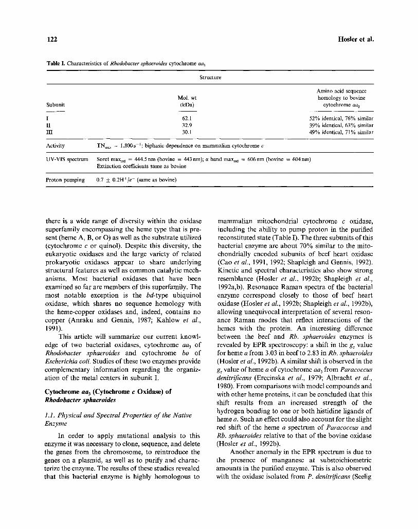
122 Hosler et al.
Table I. Characteristics of R h o d o b a c t e r sphaero ides cytochrome aa 3
Structure
Mol. wt Subunit (kDa)
I 62.1 II 32.9 III 30.1
Amino acid sequence homology to bovine
cytochrome aa 3
52% identical, 76% similar 39% identical, 63% similar 49% identical, 71% similar
Activity TNm~ = 1,800 s - t ; biphasic dependence on mammal ian cytochrome c
UV-VIS spectrum Soret max~d = 444.5 n m (bovine = 443 nm); c~ band max~d = 606 nm (bovine = 604 nm) Extinction coefficients same as bovine
Proton pumping 0.7 _.+ 0.2H+/e - (same as bovine)
there is a wide range of diversity within the oxidase superfamily encompassing the heme type that is pre- sent (heine A, B, or O) as well as the substrate utilized (cytochrome e or quinol). Despite this diversity, the eukaryotic oxidases and the large variety of related prokaryotic oxidases appear to share underlying structural features as well as common catalytic mech- anisms. Most bacterial oxidases that have been examined so far are members of this superfamily. The most notable exception is the bd-type ubiquinol oxidase, which shares no sequence homology with the heme-copper oxidases and, indeed, contains no copper (Anraku and Gennis, 1987; Kahlow et al., 1991).
This article will summarize our current knowl- edge of two bacterial oxidases, cytochrome aa3 of Rhodobaeter sphaeroides and cytochrome bo of Eseheriehia eoli. Studies of these two enzymes provide complementary information regarding the organiz- ation of the metal centers in subunit I.
Cytochrome aa 3 (Cytochrome c Oxidase) of Rhodobacter sphaeroides
1.l. Physical and Spectral Properties o f the Native Enzyme
In order to apply mutational analysis to this enzyme it was necessary to clone, sequence, and delete the genes from the chromosome, to reintroduce the genes on a plasmid, as well as to purify and charac- terize the enzyme. The results of these studies revealed that this bacterial enzyme is highly homologous to
mammalian mitochondrial cytochrome c oxidase, including the ability to pump proton in the purified reconstituted state (Table I). The three subunits of this bacterial enzyme are about 70% similar to the mito- chondrially encoded subunits of beef heart oxidase (Cao et al., 1991, 1992; Shapleigh and Gennis, 1992). Kinetic and spectral characteristics also show strong resemblance (Hosler et al., 1992b; Shapleigh et al., 1992a,b). Resonance Raman spectra of the bacterial enzyme correspond closely to those of beef heart oxidase (Hosler et al., 1992b; Shapleigh et al., 1992b), allowing unequivocal interpretation of several reson- ance Raman modes that reflect interactions of the hemes with the protein. An interesting difference between the beef and Rb. sphaeroides enzymes is revealed by EPR spectroscopy: a shift in the gz value for heme a from 3.03 in beef to 2.83 in Rb. sphaeroides (Hosler et al., 1992b). A similar shift is observed in the gz value of heme a of cytochrome aa3 from Paracoccus denitrifieans (Erecinska et al., 1979; Albracht et al., 1980). From comparisons with model compounds and with other heme proteins, it can be concluded that this shift results from an increased strength of the hydrogen bonding to one or both histidine ligands of heme a. Such an effect could also account for the slight red shift of the heme a spectrum of Paracoccus and Rb. sphaeroides relative to that of the bovine oxidase (Hosler et al., 1992b).
Another anomaly in the EPR spectrum is due to the presence of manganese at substoichiometric amounts in the purified enzyme. This is also observed with the oxidase isolated from P. denitrificans (Seelig

Analysis of Site-Directed Mutants of Cytochromes aa3 and bo 123
OUT
I II III IV V Vl VII VIII IX X XI XII
Fig. 1. The amino acid sequence of subunit I of the Rb. sphaeroides oxidase showing 12 predicted t ransmembrane helices, The six conserved histidines labelled in the t ransmembrane helices serve as metal ligands. Other completely conserved residues are shown as filled circles; highly conserved residues are in bold open circles.
et al., 1981; Haltia, 1992). Treating the Rb. sphaeroides oxidase with high concentrations of chelating agents does not remove the manganese. However, three site- directed mutants of the enzyme are found to no longer bind manganese: His411Asn, His411Ala, and Asp 412Asn. Therefore, manganese binding appears to involve residues in the highly conserved hydrophilic loop between helices IX and X (Fig. 1), which is likely to be close to the location of both hemes a and a3 (Hosler et al., 1992a). Proximity of this site to CUA, located within subunit II, is suggested by studies of Haltia (1992), who showed changes in the EPR signal of manganese upon reduction of CUA. The low occupancy (~ 5%) of this binding site by Mn may be an indication that another metal is present, such as magnesium or zinc.
Fourier transform infrared (FTIR) analysis of the Rb. sphaeroides enzyme in the bacterial membrane provides another powerful spectroscopic tool for examining the metal centers. Caughey (Caughey et al. 1970; Einarsd6ttir et al., 1988) and Alben (Alben
et al., 1981; Fiamingo et al., 1982, 1986, 1990), and colleagues have identified stretching frequencies associated with CO binding to heme a3 and CuB in beef heart oxidase, and essentially identical frequencies are found for the Rhodobacter enzyme (Shapleigh et al., 1992a). An FTIR difference spectrum of CO bound to the oxidase from Rb. sphaeroides is shown in Fig. 2. Disruption of the binuclear (heme a3-CuB) center causes broadening and major shifts in the CO stretch- ing frequencies. Subtle shifts are also observed with some mutant forms. These signals can be measured in situ using isolated membranes, without purifying the oxidase, and give important complementary informa- tion to that obtained by EPR and resonance Raman spectroscopy.
Although the purified bacterial oxidase is extremely active (Vmax = 1800sec I with soluble cytochrome c), measurements of its activity in the cytoplasmic membrane are complicated by the presence of another cytochrome c oxidase in Rb. sphaeroides, a cb-type oxidase. Efforts to prepare a

124 Hosler et al.
Fe-C~--O
~ u-C~O
E.coli bo-type
ox idase
I A Abs. = 6,5 x 10 -3
F~C~O
Rb. sohaeroides
aa~type oxidase
J- AAbs.=2.0xl0 3
I I I I I I 1900 1950 2000 2050 21 O0 2150
Wavenumber (cm-1)
Fig. 2. Low-temperature Fourier transform infrared (FTIR) dif- ference spectra of CO bound to the Rb. shaeroides or E. eoli oxidases in their respective membranes. Each spectrum was obtained by subtracting a spectrum obtained during continuous photolysis, where CO is bound to Cu B , from the spectrum obtained in the dark, where CO is bound to the heine iron. Hence, the negative peak is due to the CO stretching of Fe-CO. There are two distinct confor- mational states of the Rb. sphaeroides binuclear center (c~ and/3) reported by this technique, just as found with the bovine oxidase. (Fiamingo et al., 1986). This is not observed with the E. coli oxidase. Contributions of the eb-type oxidase to the aa 3-type oxidase spec- trum are discussed in Shapleigh et al. 1992a.
bacterial strain deleted in this enzyme are currently under way. In spite of this complication, it is clear that cytochrome aa3 of Rb. sphaeroides in the native mem- brane is strongly associated with a membrane-bound cytochrome e that permits high activity with TMPD alone (Hosler et al., 1992b). This cytochrome c is removed during purification and likely resembles cytochrome c552 of P. denitrifieans (Berry and Trumpower, 1985; Steinriicke et al., 1991a). The physiological role of the bound cytochrome e is not clear, since kinetic analyses show that the membrane- bound oxidase has the additional ability to react with
soluble cytochrome c with relatively high affinity, in contrast to the caa3 enzymes of Thermus thermophilus (Yoshida and Fee, 1984) and PS3 (Nicholls and Sone, 1984).
1.2 The COXII /COXIII Operon Contains ORFs Resembling coxl0 and c o x / / o f Yeast
The coxI (etaD) gene encoding subunit I of the Rb. sphaeroides oxidase is not adjacent to the genes encoding subunits II or III (Shapleigh and Gennis, 1992). The latter are together on a separate COXII/ COXIII operon in which two open reading frames, coding for proteins homologous to COX10 and COX11 in yeast (Nobrega et al., 1990; Tzagoloffet al., 1990), are present between coxII (ctaC) and eoxIII (ctaE) (Cao et al., 1992). These proteins apparently function to assemble the aa3-type cytochrome e oxidase in both yeast and Rb. sphaeroides, perhaps assisting protein folding or through a role in heine A biosynthesis or metal insertion. Homologous proteins are found in P. denitrificans (Steinrficke et al., 1991b), which has an operon structure similar to that of Rb. sphaeroides (Raitio et al., 1987). Homologues of COX10 have also been identified in E. coli (see Section 2.1) and in B. subtilis (Saraste et al., 1991).
1.3. The Metal Center Ligands in COXI
The basis for our initial efforts in mutational analysis was evidence that heine a, heme a3, and CuB are ligated by residues in COXI and that the ligands involved are predominantly histidines (see Wikstrom et al. 1985; Saraste, 1990); analysis of the amino acid sequences of over 30 different cytochrome oxidases reveals six totally conserved histidines in COXI (Fig. 1), these histidines are the obvious candidates to fulfil the metal liganding function.
Measurement of the activity of the purified mutant forms of the Rb. sphaeroides oxidase supports the idea that the totally conserved histidines of COXI play a critical role, since alteration of any one of them creates an inactive enzyme. Resonance Raman and visible spectra provide strong evidence for the assign- ment of Hisl02 and His421 as the heme a ligands, His333 and His334 as CuB ligands, and His284 or His419 as the ligand of heme a3 (Table II) (Shapleigh et al., 1992b). Further evidence regarding the heme a3 ligand has been obtained from resonance Raman studies of mutant forms of the enzyme at different

Analysis of Site-Directed Mutants of Cytochromes aa3 and bo 125
Table II. Mutational Analysis of Subunit I of Cytochrome c Oxidase from Rhodobacter sphaeorides
Subunit I Helix Mutation(s) Phenotype of mutant oxidasC Proposed role of residue residue d
*Hisl02 II N Unstable protein; loss of e-band absorbance Heme a ligand
His277 VI L b Severe disruption of a3-Cn B Unknown
*His284 VI A Severe disruption of a 3-CUB, but iron-his stretch of a 3 Possible CuB ligand detectable; altered CO binding
Glu286 VI Q, D b Low CO binding to heme a3, but normal FTIR Possible resting state bridging ligand
*Tyr288 VI F Severe disruption of a 3-CUB, but iron-his stretch of a 3 Possible CuB ligand detectable; altered CO binding
*His333 VII N Largely intact a3-Cu B center by RR, EPR, but low CO CuB ligand binding and altered Cu B by FTIR
Y, D b, C b Severe disruption of a3-Cu B
*His334 VII N, yb Severe disruption of a 3-Cu B Cu B ligand
*Thr352 c VIII A, I b a 3 environment disturbed, but iron-his stretch of a 3 Close to a 3-Cu B center, possibly evident involved in H + transfer or
stabilization of oxygenated intermediates
Thr359 VIII A 25% active; near wild-type optical and RR spectra, but H+/H20 pathway to the a3-CuB minor alteration of the a 3-Cu B environment by FTIR center
Lys362 VIII M a3 iron-his stretch altered (His419 deprotonated?) H+/H20 pathway to the a3-Cu B
center R b Enzyme unstable or poorly expressed
His411 IX-X loop A 80% active; metal centers normal; no Mn binding Non-redox active metal ligand near active site
N a 3 environment severely disturbed; no Mn binding
Asp412 I X - X loop N, E b 30% active; a 3 iron-his stretch altered; no Mn binding Non-redox active metal ligand near active site
Thr413 I X - X loop N 80% active; metal centers normal
Tyr414 I X - X loop F 50% active; a3-Cu B environment normal; heme a Possible member of H-bond optically red-shifted network to heme a propionates
*His419 X N, yb a3-Cu B environment disrupted; a 3 iron-his stretch lost a 3 ligand
*Phe420 X V b Partially active; minor alteration of a3-Cu B Unknown environment
*His421 X N, yb, i b Unstable protein; loss of a-band absorbance Heme a ligand
aAll mutants are inactive in electron transfer except where noted. bAssayed in the membrane-bound form only; all other mutant oxidases have been purified. cCompletely conserved as Thr or Ser. dAn asterisk before the residue denotes a completely conserved residue among 33 oxidases.

126 Hosler et ai.
334 heme~ 411 413 b414
"~ I
,Cu B ~ _ ,
'284
Fig. 3. A helical wheel model showing the ligation of heine a, heme a3, and CuB by the six conserved histidines in subunit I of the Rb. sphaeroides oxidase. Conserved residues are in bold circles. Also included is the predicted extra-membrane sequence connecting helix IX and helix X.
h e m e a_
i o2°°
stages of purification (Hosler, Tecklenburg, Shap- leigh, Gennis, Babcock, and Ferguson-Miller, unpub- lished results). The His284Ala mutant shows some evidence of a native iron-histidine stretch in mem- brane-bound and partially purified states, whereas the His419Asn mutant does not. Apparently His284Ala can still adopt a native-like conformation in the appropriate lipid environment, even though other spectral signals show that the mutation disorders the binuclear center pocket. Since alanine cannot function as a metal ligand, the presence of an apparently native heine a3 iron-histidine bond in His284Ala strongly favors the alternative model of His419 as the heme a3 ligand. F TIR analysis on similar mutants of cyto- chrome bo (see Section 2.8) also supports the tentative assignment of His419 as the heme a3 ligand.
A model that is consistent with these assignments depicts an arrangement of several of the transmem- brane helices of COXI (Fig. 3 and see cover). It must be emphasized that this is a working model or pic- torial aid. Furthermore, the assignment of ligands is predicated on the assumption that all of the ligands
are histidines. Although this assumption is strongly supported by spectroscopic evidence in the case of hemes a and a3, the number and nature of the ligands of CuB are less certain (Powers et al., 1981; Cline et al., 1983; Scott et al., 1986; Surerus et al., 1992). Inter- pretation of the data must also take into account the possibility that the residue that is introduced functions as a metal ligand, or that the metal adopts an existing residue to serve as an alternative ligand in the mutant oxidase.
1.4. The Roles of Other Conserved Residues in COXI
Figure 1 shows a hypothetical arrangement of COXI in the membrane predicted from hydropathy plot analysis and supported by gene fusion exper- iments on the bo-type oxidase (see Section 2.6). Totally and highly conserved residues are also shown in this representation. Besides residues in the immediate vicinity of the designated metal ligands, there are several other regions of the polypeptide that suggest functional importance because of high levels of conservation. These include a hydrophilic face of
Analysis of Site-Directed Mutants of Cytochromes aa3 and bo 127
helix VIII, the extra-membranous loop between helix II and helix III, predicted to be cytoplasmic, and the loop between helix IX and helix X, predicted to be periplasmic. Mutational analysis of these and other regions is currently under way. Table II summarizes some of the findings obtained so far.
Mutants of several consecutive residues (His411, Asp412, Thr413, and Tyr414) in the IX-X loop affect the spectroscopic characteristics of heme a and heine a3, supporting the model showing proximity of these centers (Hosler et al., 1992a). Two residues in this loop, His411 and Asp412, are also apparently involved in binding manganese (see Section 1.1). A further role for this region in proton pumping has been investi- gated, with particular interest in Asp412, since a car- boxylate residue, capable of rapid deprotonation, may be a necessary component of the proton pathway to the external phase. However, mutants in residues 411, 412, 413, and 414 show both electron transfer and proton pumping activity (Fetter, Hosler, Thomas, Gennis, and Ferguson-Miller, unpublished results). A proposed role for Tyr414 as a hydrogen bond donor to the formyl group of heme a (Saraste, 1990) is dis- proved by the finding that Tyr414Phe does not alter the intensity or position of the resonance Raman frequency attributed to that formyl stretch (Hosler, Shapleigh, Tecklenburg, Ferguson-Miller, and Gennis, unpublished results). The proximity of Tyr414 to heine a is, however, suggested by the absorption spectrum of heme a which is altered in the Tyr414Phe mutant.
The hydrophilic face of helix VIII is one of the most highly conserved regions of subunit I (Fig. 1) and may form part of the lining of a channel that conducts water and protons within the protein. Our current results favor a close interaction of this region with a heme a 3 - C u B center that is located toward the outer side of the membrane (Fig. 3). Mutation of Thr352, with a predicted location close to the periplasmic or outer side of the membrane, to alanine, disrupts the heme a 3 environment and eliminates catalytic activity. Substitution of alanine for Thr359, located near the center of the membrane, creates a partially active oxidase with heme a3 and the other metal centers apparently native. Interestingly, substitution of Lys362, located near the inner surface, with methio- nine has a specific effect (Table II) on the axial ligation of heine a3, associated with loss of catalytic activity and altered CO binding. This supports the idea that this residue may play some role in proton or water access to the metal centers:
The proposed region providing the amino acid ligands of the heme a 3-Cu B binuclear center (helices VI, VII, and X) has been subject to further mutational analysis, including Tyr288Phe and Glu286Gln. Both of these mutants have little or no activity. The Tyr288Phe mutant shows significant disruption of the binuclear center and shares many characteristics with His284Ala. Since Tyr288 is four residues (or one helical turn) below His284, it is well positioned to serve as a CuB ligand, perhaps transiently, as the geometry of CUB ligation changes during the redox cycle. The pres- ence of a totally conserved proline between His284 and Glu286 suggests a disruption or distortion of the helix at this position that could result in a different relationship of Glu286 and Tyr288 with respect to the binuclear center than that shown in the helical model of Fig. 3. Whether sufficient distortion could occur to allow interaction between Glu286 and either CuB or heme a3 (Brown et al., 1992) is an interesting question (see Conclusions). Glu286 is the only conserved carboxylate residue predicted to be in a membrane- spanning region of subunit I. Such acidic residues have been shown to be important in facilitating pro- ton transport in other membrane proteins (Otto et al., 1989; Henderson et al., 1990; Takahashi and Wraight, 1991).
Cytochrome bo (Ubiquinol Oxidase) of Escherichia coli
2.1. Cytochrome bo Is a Five-Subunit Oxidase
Most, if not all, members of the heme-copper oxidase superfamily appear to have in common three core subunits that correspond to the three mitochon- drially encoded subunits of the eukaryotic oxidases. In E. coli, these three subunits are encoded by the first three genes of the cyo operon. The entire operon contains five genes, corresponding to the five subunits of the purified oxidase: cyoA (subunit II), cyoB (sub- unit I), eyoC (subunit III), cyoD (subunit IV), and cyoE (subunit V) (Chepuri et al., 1990b). Subunits IV and V are not related to nuclear-encoded subunits of the eukaryotic enzymes, but they are homologous to subunits or open reading frames associated with some prokaryotic aa3-type cytochrome c oxidases (see Section 1.2) (Chepuri et al., 1990b; Saraste et al., 1991).
Previous studies have shown E. coli cytochrome bo to be a four-subunit enzyme (Matsishita et al., 1984; Georgiou et al., 1988; Puustiunen et al., 1991;

128 Hosler et al.
I a~a3-type Cytochrome c Oxidase I
2 heine A ~~.,--~--"-""~g 2 - 3 c o p p e r
......... l-_y.. (Cu B a~ is I - . . . . . . . . . His--~--His
I II
J b.__qo-type Ubiquinol Oxidase ]
• , C u . .
I I I
heme B heme O copper
Fig. 4. Schematic showing subunits I and II of the aa3-type cytochrome e oxidase and the bo-type ubiquinol oxidase.
Wu et al., 1992). Alteration of the preparation pro- cedure demonstrates a fifth subunit that has the mol- ecular weight expected for the eyoE gene product (Mogi and Anraku, 1990; Minghetti et al., 1992). The catalytic and spectroscopic properties of the four- and five-subunit forms of the enzyme appear to be virtually identical. Although subunit V is not an essential com- ponent for oxidase turnover, the eyoE gene product is essential for the assembly of the functional oxidase (see Section 2.6 and Mingnetti et al., 1992).
Work on the aa3-type cytochrome e from P. denitrificans has demonstrated that subunits I and II alone are sufficient for both electron transfer activity and proton pumping (Haltia et al., 1991; Hendler et al., 1991). The corresponding subunits of cyto- chrome bo are shown schematically in Fig. 4. Subunit I, which binds all three of the metals associated with the ubiquinol oxidases and three of the four metals associated with the cytochrome e oxidases, has been the focus of most of the mutagenesis studies to date
(Lemieux et al., 1992; Minagawa et al., 1992). The results are fully consistent with the conclusions based on the studies with the Rb. sphaeroides cytochrome e oxidase, presented in Section 1.
2.2. Cytochrome bo Is a Proton Pump
The bo-type oxidase catalyzes both scalar and vectorial proton translocation (Matsushita et al., 1984; Puustinen et al., 1989, 1991). The two-electron oxidation of quinol is associated with the release o f two protons on the periplasmic side of the membrane. Concomitantly, the four-electron reduction of mol- ecular oxygen of water is associated with the uptake of four protons, which presumably come from the cyto- plasmic side of the membrane. Hence, chemical pro- cesses alone predict that during enzyme turnover one proton per electron will disappear from the cytoplasm and appear in the periplasm. This "scalar" mechanism is sufficient to account for the ratio of one H+/e -. However, studies of the bo-type oxidase in sphero- plasts yield a ratio of 2 H+/e - (Puustinen et al., 1989, 1991). This is most reasonably explained by an additional vectorial mechanism similar to that observed in the aa3-type cytochrome e oxidases (Wikstr6m, 1989; Larsen et al., 1992), involving the coupling of the redox process to movement of a second proton across the membrane.
It is noted that there is no report of the recon- stituted purified bo-type oxidase functioning as a pro- ton pump. Efforts to do so have yielded only the ratio of 1 H+/e - expected for the simple scalar mechanism (Matsushita et al., 1984). This may be the result of a scrambled orientation of the purified quinol oxidase in reconstituted phospholipid vesicles (Puustinen et al., 1991) or loss of activity during purification. It is possible, though not probable, that the failure of this reconstituted, four-subunit preparation to function as a vectorial proton pump is due to the loss of subunit V.
2.3. Cytochrome bo Has Only One Copper
Metal analysis indicates that the purified oxidase contains two equivalents of iron and one equivalent of copper per mole of enzyme (Puustinen et al., 1991; Minghetti et al., 1992). The two irons are accounted for by the two heme components and the copper is associated with the binuclear center. The bo-type quinol oxidase does not contain CuA, the EPR-detect- able component of the cytochrome c oxidases which is putatively ligated to residues within the hydrophilic

Analysis of Site-Directed Mutants of Cytochromes aa3 and bo 129
domain of subunit II (Fig. 4) (Blair et al., 1983; Minghetti et al., 1992). This is consistent with the fact that the amino acid residues that are probable ligands of CUA in the cytochrome e oxidases are not conserved in the sequence of subunit II of the quinol oxidase (Chepuri et al., 1990b; Saraste, 1990; van der Oost et al., 1992). One of the putative functions of CUA is to oxidize cytochrome c; since the bo-type oxidase exhibits no cytochrome c oxidase activity, the lack of CUA is not surprising. The fact, however, that the quinol oxidase functions as a vectorial proton pump makes it unlikely that CUA plays a critical role in the proton pumping mechanism, as has been proposed for the cytochrome e oxidases (Gelles et al., 1987).
2.4. Cytochrome bo Has a Heme-Copper Binuclear Center
Electron spin resonance studies of the heme com- ponents of the bo-type oxidase show the presence of a heme-copper binuclear center, a diagnostic feature of this entire superfamily of oxidases (Salerno et al., 1989, 1990). A redox titration shows an EPR signal typical of a high-spin berne, but at high potential (+ 400 mV) this signal is quenched. This is explained by the presence of a nearby copper (CUB) which, upon oxidation, is electronically coupled to the heme iron in such a way that the EPR signal is eliminated. This behaviour is also observed with cytochrome c oxidise (van Gelder and Beinert, 1969).
A second technique that demonstrates the pres- ence of a heme-copper binuclear center is Fourier transform infrared (FTIR) spectroscopy, in which CO bound to the oxidase is used as a probe (Fiamingo et al., 1982, 1986). The stretching frequency of the CO bond is sensitive to the environment of the CO, includ- ing its ligation state. When bound to the heme o component of the bo-type oxidase, the CO stretching frequency is 1959cm -1 (Chepuri el al., 1990a; Hill et al., 1992). After photolysis, the bound CO shifts from heme o to CuB, and the FTIR band is centered at 2063 cm 1. The difference spectrum for cytochrome bo is shown in Fig. 2 and compared with that from the Rb. sphaeroides oxidase. These data demonstrate definitively the presence of a heine-copper binuclear center in cytochrome bo.
2.5. Cytochrome bo Contains Two Heme Components
Extraction of heme from the purified bo-type
oxidase reveals two equivalents of heme, correspond- ing to the two iron equivalents. Earlier work identified the two hemes as being protoheme IX (heine B), based on simple spectroscopic critieria (Kita el al., 1984; Matsushita el al., 1984). Closer examination, how- ever, indicated that one of the two heme components is chemically distinct from heme B, and this new heme has been named heme O (Puustinen and Wikstr6m, 1991; Wu et al., 1992). The structures of heine B, heme O, and heme A are shown in Fig. 5.. Heme O is a modification of heme B, with a hydroxylfarnesyl side- chain addition (Wu et al., 1992). This same modifi- cation is found in heine A, which differs from heme O by the addition of a formyl group. The formyl sub- stituent is responsible for shifting the peak of the alpha band from about 560 nm in cytochrome bo to 603-606 nm in cytochrome aa 3.
The heme B prosthetic group of cytochrome bo is normally present as the low-spin six-coordinate cytochrome b562 component. This component is responsible for virtually all of the visible absorption of the oxidase, and has a split alpha band with peaks at 555 and 563.5nm (Puustinen et al., 1991; Minghetti el al., 1992). In some strains of E. coli that over- produce the bo-type oxidase due to the presence of the cyo operon on a multicopy plasmid, heme O can substitute for heme B at this locus in the enzyme (Puustinen el al., 1992). Depending on growth con- ditions, the enzyme can contain up to two equivalents of heme O, although the substitution of heme O for heme B has very little effect on the catalytic function of the oxidase. Hence, this is a promiscuous heme binding locus, although under normal physiological conditions in a wild-type strain heme B is always found at this site (Puustinen et al., 1992).
The proper assembly of the oxidase requires the cyoE gene product (homologous to COX10 of yeast; see Section 1.2), as demonstrated by truncating the cyo operon so that cyoE is not expressed. The result is the assembly of a non-functional oxidase in the E. coli membrane. FTIR spectroscopy shows that the binuclear center is abnormal, and HPLC analysis indicates that heme O is absent from the membranes (Hill et al., 1992). Whereas heme O can substitute for heme B to yield an active enzyme, the reverse sub- stitution yields an inactive enzyme, indicating that cytochrome bo does not tolerate heme B in the binuclear center. Although the presence of subunit V (cyoE gene product) is not essential for activity of the purified enzyme, it is essential for correct assembly. This gene product may be required for the insertion of

130 Hosler et al.
COOH \ f OOH
CH 2 /
COOH \
~M
COOH 1
CH 2
1 mw
COOH~ ~.uurl
C~2 CH2 /
Fig. 5. The structures of heme B, heme O, and heine A.
heme O into the binuclear center, or for the biosynthesis of heine O from heme B.
2.6. Subunit topology and Two-Dimensional Modelling
The hydropathy profiles of the five subunits of the bo-type oxidase indicate that each contains multiple transmembrane spans (Chepuri et al., 1990b). Based on the known structures of bacteriorhodopsin (Hen- derson et al., 1990) and the bacterial photosynthetic reaction centers (Deisenhofer and Michel, 1989; Feher et al., 1989), it is reasonable to assume that the trans- membrane spans are helices with a minimum length of about 20 residues. The number and approximate boundaries of each putative transmembrane alpha helix was determined experimentally using the gene fusion approach (Chepuri and Gennis, 1990). This technique can identify regions of each polypeptide that are on or near the periplasmic side of the mem- brane, and those portions of each polypeptide that are on the cytoplasmic surface. The results are internally
consistent and are also consistent with the relatively limited data available from other approaches.
Subunit I: The data indicate that subunit I of the E. coli oxidase has 15 transmembrane spans. Twelve of these putative helical spans correspond to those shown in Fig. 1. The E. coli subunit, however, has extentions on both the N- and C-termini compared to the homologous subunit from Rb. sphaeroides or beef. On the N-terminus, there is one additional transmem- brane span, which places the amino terminal residue on the periplasmic side; on the C-terminus, there are two additional transmembrane spans. Similar exten- sions are found in several of the bacterial cytochrome C oxidases (Ishizuka et al., 1990; Saraste et al., 1991). It is interesting to note that in those cases where subunit I has the two additional transmembrane spans on the C-terminus, subunit III has two fewer on the N-terminus. Since the gene encoding subunit III in E. coli immediately follows that encoding subunit I, it is easy to rationalize how a transfer might have occurred at the genetic level (see also Fee et al., this volume). However, there is little amino acid sequence homology

Analysis of Site-Directed Mutants of Cytochromes aa3 and bo 131
to confidently conclude that the last two spans of E. coli subunit I correspond to the first two spans normally found in subunit III.
There are no data apart from those from the gene fusion experiments that define which portions of sub- unit I are on the inner or outer side of the membrane or which define the number of membrane-spanning domains.
Subunit II: The gene fusions with the E. coli subunit II show that this subunit has both the N- and C-termini on the periplasmic side of the membrane, with two membrane-spanning helices in between (Chepuri and Gennis, 1990). There is a large hydro- philic domain on the C-terminus which is the likely site for the CuA ligation and cytochrome c interaction in aa3-type oxidases. The topology suggested by the gene fusions with the E. coli subunit is identical to that determined by more classical biochemical techniques for the equivalent subunit from beef and P. denitrificans (Bisson et al., 1982; Finel, 1988; Saraste, 1990).
Subunit III: The E. coli subunit is predicted to have five transmembrane spans with the N-terminus on the cytoplasmic side of the membrane. As indicated above, in most other organisms including Rb. sphaeroides (Cao et al., 1992), subunit III is larger and contains two additional predicted spans on the N- terminal side.
Subunits IV and V: Subunits IV and V have three and seven transmembrane spans, respectively. In each case the N-terminus is predicted to be on the inner side of the membrane (Chepuri and Gennis, 1990).
The results of the gene fusion experiments indicate that the C-terminus of subunit II and the N-terminus of E. coli subunit I are both on the peri- plasmic side of the membrane. Similarly, the C- terminus of subunit I and the N-terminus of subunit III are expected to be on the cytoplasmic side of the E. coli membrane. The gene order, cyoA-eyoB-cyoC (subunit II-I-III), facilitates the construction of in- frame fusions to link subunits I, II, and III to form a single polypeptide. In principle, this is possible because the topology places the N- and C-termini to be fused on the same side of the membrane for each pair of polypeptides. This experiment was performed successfully: the resulting product is an active oxidase with subunits I, II, and III fused together (Ma and Gennis, unpublished results). Minimally, this experi- ment confirms the two-dimensional folding pattern already described.
The two-dimensional models of the subunits form the backdrop that is essential for the structural
interpretation of the results from site-directed muta- genesis. The latter, as indicated below, provides information concerning the three-dimensional relationship of portions of the oxidase.
2.7. Mutagenesis of Subunit H
The bulk of the mutagenesis experiments to date have involved residues in subunit I. However, an important set of experiments has been initiated inves- tigating the structure and function of subunit II. A set of residues which are thought to be the ligands to CuA in the cytochrome c oxidases (Stevens et al., 1982; Blair et al., 1983; Li et al., 1987) was placed in the hydrophilic domain of subunit II of the E. coli oxi- dase. EPR spectra of the resulting subunit demon- strate the restoration of a CuA-like center (van der Oost et al., 1992). These data strongly suggest that the three-dimensional folding pattern of the E. coli subunit is similar to that of cytochrome c oxidase, although the function of this subunit is not conserved.
2.8. Site-Directed Mutagenesis and Three- Dimensional Modelling of Subunit I
Many mutations have been made in subunit I of the E. coli oxidase. The results are completely com- patible with the data obtained with the mutations in the equivalent residues of Rb. sphaeroides cytochrome e oxidase (Section 1). The results obtained with the E. coli oxidase independently support the model shown in Fig. 3. Helices II, VI, VII, and X are clearly involved directly in ligating the metals within subunit I (Lemieux et al., 1992; Minagawa et al., 1992), and residues on the polar face of Helix VIII are important for catalytic function (Thomas and Gennis, unpub- lished results). Some of these data are summarized in Table III.
Metal Binding. All substitutions for any of the six totally conserved histidine residues in subunit I are catalytically inactive (Lemieux et al., 1992; Minagawa et al., 1992). In most cases, the mutant enzyme is assembled and overproduced in the E. coli membrane. Spectroscopic techniques, including optical absorp- tion, EPR, and FTIR spectroscopies, have been par- ticularly valuable in deciphering the nature of each lesion. Hisl06 (Helix II) and His421 (Helix X) are the ligands of the low-spin heme b562 component of the oxidase, as indicated by loss of this heme when either of these histidines is replaced (Lemieux et al., 1992; Minagawa et al., 1992). The equivalent histidines have

132 Hosler et al.
Table IlL Some mutants in Subunit I of Cytochrome bo Oxidase from E. coli
Subunit I Helix Mutation(s) Phenotype of mutants" Proposed role of residue residue d
*His 106 II G, L Loss of or-band absorbance; no low-spin EPR Heme b ligand
*Metl I0 II I Complements; wild-type FTIR spectrum Nonessential
Asn277 VI D, Y Complement; have wild-type spectra Nonessential
*His284 VI G, L FTIR Fe-CO similar to wild-type; low CO binding Possible Cu B ligand C, N, Q, Low CO binding
W, Y
Glu286 VI A Not active Possible resting state bridging ligand Q Partially active with near wild-type FTIR
*Tyr288 VI A, F, S Loss of Cu B by FTIR Possible Cu B ligand
*His333 VII C, L, M, Loss of Cu B by FTIR and EPR Cu B ligand N , Q , R
*His334 VII C, L, M, Loss of Cu B by FTIR and EPR Cu B ligand N , Q , R
*Thr352 b VIII A Disrupts binuclear center Role in H + transfer and/or stabilization of oxygenated intermediates
S Complements
Pro358 VIII A Complements Nonessential
Thr359 VIII A Oxidase inactive, but all metal centers present H+/H20 pathway S Complements
Lys362 VIII A, M, Q All metal centers present but inactive H+/H20 pathway
His411 IX-X loop G Complements Nonessential L Does not complement
*His419 X N Binuclear center with shifted Fe-CO and Cu-CO Heine o ligand frequencies
C, D, G, L, Binuclear center disrupted M , Q , Y
*His421 X G, L Loss of a-band absorbance; no low spin EPR Heme b ligand
aUnless noted by the term "complements," all mutations are inactive by the specific criterion of being unable to support aerobic growth in the eyo eyd host strain RG 129 (An and Gennis, 1987). This strain cannot grow aerobically without an active terminal oxidase that is plasmid- encoded (see Lemieux et al., 1992). Inactivity by this criterion does not necessarily mean a complete loss of biochemical function. bConserved as Thr or Ser. CResidue numbers are identical to Rb. sphaeroides except for Hisl06 and Met! 10, which are Hisl02 and Metl06 in Rb. sphaeroides. dAsterisks denote completely conserved residues.
been s h o w n to be the l igands fo r the a n a l o g o u s low-
spin h e m e ( h e m e a) in Rb. sphaeroides c y t o c h r o m e c
ox idase (Sec t ion 1.3; S h a p l e i g h et al., 1992b). Sub-
s t i tu t ions fo r e i the r o f the two a d j a c e n t h i s t id ines in
He l ix V I I (His333 a n d His334) resul ts in the e l imina -
t ion o f CuB, as i n d i c a t e d by F T I R , E P R , a n d a t o m i c
a b s o r p t i o n m e a s u r e m e n t s ( C a l h o u n , L e m i e u x , A l b e n , a n d Genn i s , u n p u b l i s h e d da ta ; M i n a g a w a et al.,
Analysis of Site-Directed Mutants of Cytochromes aa3 and bo 133
1992). The assignment of either His284 or His419 as the ligand to the high-spin heme o has, until recently, been more problematic (Lemieux et al., 1992; Minagawa et al., 1992). However, it has been observed by FTIR spectroscopy that His284 can be replaced by a glycine or leucine without perturbing the band width and only slightly perturbing the center frequency of CO bound to the heine, whereas the signal expected for CO bound to CuB is not observed (Calhoun, Thomas, Lemieux, Alben, and Gennis, unpublished results). This is easier to rationalize if His284 is assigned as a CuB ligand and is not the axial ligand to the CO-binding heine. Hence, the model shown in Fig. 3 is strongly favored, in which His419 is shown as the high-spin heine (o or a3) ligand, and His284 is a third potential CuB ligand. Indirect support for this model comes from the observation that substitution of Tyr288, located four residues (or one helical turn) below His284 in Helix VI, by phenylalanine also results in the loss of the CuB FTIR signal (Thomas and Gennis, unpublished results). Thus, Tyr288 must be considered as a possible ligand to CuB. Similar results have been obtained with the Rb. sphaeroides oxidase (Section 1.3). Possible sulfur ligation to CuB has been previously suggested (Powers et al., 1981). However, the only sulfur-containing residue that is totally con- served in subunit I is MetllO (E. coli notation) in Helix II. Due to the assignment of Hisl06 of Helix II as a ligand for heine b562 (heine a), Met110 is unlikely to ligand Cu B. In fact, mutagenesis data show that Met110 is not an essential residue in the E. coli oxidase (Calhoun, Lemieux, Thomas, and Gennis, unpub- lished results).
The Polar Face of Helix VIII. Substitution of Thr352 in Helix VIII by alanine results in complete loss of catalytic activity (Thomas and Gennis, unpub- lished results). This is also ass6ciated with severe per- turbation of the binuclear center, suggesting that Thr352 may be located at or very close to the heme o-CuB binuclear center. This is easily accommodated by the model in Fig. 3 and is consistent with the results obtained with mutations in the equivalent position in the cytochrome c oxidase of Rb. sphaeroides (Section 1.4). Substitution of Thr359 by alanine or of Lys362 by methionine causes a severe loss of catalytic activity, but in neither of these cases is there any perturbation of the FTIR spectra to indicate that the metal centers are directly affected by the mutation (Thomas, Alben, and Gennis, unpublished results). The equivalent Lys362Met mutation in Rb. sphaeroides, however, does perturb the heme a3 resonance Raman spectrum
OUT
IN VII V ~
"419
421
X
i
I
- .~ , J -
e. m
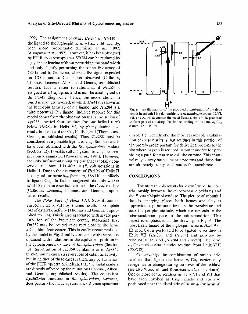
H -I- Fig. 6. An illustration of the proposed organization of the three metals in subunit I in relationship to transmembrane helices, II, VI, VII, and X, which contain the metal ligands. Helix VIII, proposed to form part of a hydrophilic channel leading to the heme a 3-Cu B center, is not shown.
(Table II). Tentatively, the most reasonable explana- tion of these results is that residues in this portion of the protein are important for delivering protons to the site where oxygen is reduced to water and/or for pro- viding a path for water to exit the enzyme. This chan- nel may convey both substrate protons and those that are ultimately transported across the membrane.
CONCLUSIONS
The mutagenesis results have confirmed the close relationship between the cytochrome c oxidases and the E. coli ubiquinol oxidase. The picture of subunit l that is emerging places both heroes and CUB at approximately the same level in the membrane and near the periplasmic side, which corresponds to the intermembrane space in the mitochondrion. This aspect is emphasized in the drawing in Fig. 6. The most likely ligand of the high-spin heine is His419 of Helix X. CuB is postulated to be ligated by residues in Helix VII (His333 and His334) and possibly by residues in Helix VI (His284 and Tyr288). The heine a3-CuB pocket also includes residues from Helix VIII (Thr352).
Conceivably, the combination of amino acid residues that ligate the heine a3-CuB center may reorganize or change during turnover of the oxidase (see also Woodruff and Rousseau et al., this volume). One or more of the residues in Helix VI and VII that have been invoked as CuB ligands and are also positioned near the distal side of heine a3 (or heme o)

134 Hosler et al.
may interact directly with the heme iron under certain conditions. For example, His284 and Tyr288 could participate in a ligand switching mechanism between the heine iron and CuB; both could be involved in proton pumping. His284 might also serve as the bridg- ing ligand postulated to couple heme a3 (or o) and CuB, particularly since the strength of the coupling between the two metals is suggestive of a histidine bridge (Palmer et al., 1976; Tweedle et al., 1978). Alternatively the so-called resting state of the oxidase might result from an interaction of Glu286 (Helix VI) with the metals of the binuclear center (Brown et al., 1992). Glu286 follows the sequence Gly283-His284-
Pro285, which is highly conserved in the sequences of subunit I. A search for the sequence G l y - H i s - P r o in soluble proteins of known structure shows that it is found in turns. Hence, this portion of the polypeptide may not be simply helical, as we show in the models (Figs. 1 and 6). The possibility of different conforma- tional states, for example "fast" vs. "slow" enzyme (Baker et al., 1987; Moody et al., 1991), being manifes- tations of the interactions of His284 and Glu286 with the metals in the binuclear center is intriguing. Future studies may clarify some of these issues.
An interesting feature of the developing model is that heme a3 (or o) and heine a (or b562) are both postulated to be ligated by residues within Helix X at essentially the same level in the membrane, close to the periplasmic (outer) side. Hence, electron flow from heme a to the binuclear center can occur through sigma bonds (Wuttke et al., 1992) across Helix X, essentially parallel to the surface of the membrane. The protons needed to form water from oxygen must be transported from the opposite side of the mem- brane via some structure within the enzyme, and the resulting product, water, must similarly be transported away from the binuclear center. The highly conserved polar residues found in Helix VIII are strong candidates for forming part of such a hydrophilic channel.
Our proposal of His419 as the ligand of heme a3, rather than His284 (Shapleigh et al., 1992b), leads to the current model (Fig. 3) in which CuB is no longer located between the two hemes. This would appear to conflict with the electron transfer pathway of heme a to CuB to heme a3 (Babcock and Wikstrom, 1992). One can envision, however, ligand rearrangements associated with CUB reduction and oxidation (Larsen et al., 1992) that affect the redox potential of heme a3 (or CuB) and thus control the pathway. For example, Tyr288 may ligand reduced but not oxidized CuB;
unliganded Tyr288 could interact with the porphyrin ring of heme a3 to alter the redox potential of the heme (see the front cover of this volume). A similar mechan- ism could be envisioned for His284. Ligand switching at the heme a3 iron associated with oxygen binding is also proposed as a mechanism for control of electron flow, in this case by directly changing the pathway (see articles by Woodruff and Rousseau et al., this volume).
The combination of mutagenesis techniques and biophysical methods is particularly powerful, especi- ally with the rich array of techniques available for studying metalloproteins such as these oxidases. Even in the absence of the long sought, high-resolution X-ray structure, much can be learned and the future promises to be interesting.
ACKNOWLEDGMENT
This work was supported by grants from the National Institutes of Health (GM26916 to S.F-M., GM25480 to G.T.B., and HL16101 to R.B.G.), from the Human Frontier Science Program (to R.B.G.), from the Department of Energy (DEFG 02- 87ER13716 to R.B.G.), and the Research Excellence Fund, State of Michigan (to S.F-M. and J.P.H.).
REFERENCES
Alben, J. O., Moh, P. P., Fiamingo, F. G., and Altschuld, R. A. (1981). Proc. Natl. Acad. Sei. USA 78, 234-237.
Albracht, S. P. J., van Verseveld, H. W., Hagen, W. R., and Kalk- man, M. L. (1980). Bioehim. Biophys. Aeta 593, 173-186.
Anraku, Y., and Gennis, R. B. (1987). Trend. Bioehem. Sei. 12, 262-266.
Au, D. C.-T., and Gennis, R. B., (1987). J. Bacteriol. 169, 3237- 3242.
Babcock, G. T., and Wikstrom, M. (1992). Nature (London), 356, 301-309.
Baker, G. M., Noguchi, M., and Palmer, G. (1987). J. Biol. Chem. 262, 595-604.
Berry, E. A., and Trumpower, B. L. (1985). J. Biol. Chem. 260, 2458-2467.
Bisson, R., Steffens, G. C. M., and Buse, G. (1982). J. Biol. Chem. 257, 6716-6720.
Blair, D. F., Martin, C. T., Gelles, J., Wang, H., Brudvig, G. W., Stevens, T. H., and Chan, S. I. (1983). Chem. Scri. 21, 43-53.
Brown, S., Moody, A. J., Jeal, A. E., Bourne, R. M., Mitchell, J. R., and Rich, P. R. (1992). EBEC 7, 39.
Cao, Shapleigh, J., Gennis, R., Revzin, A,, and Ferguson-Miller, S. (1991). Gene 101, 133-137.
Cao, J., Hosler, J., Shapleigh, J., Revzin, A., and Ferguson-Miller, S. (1992). J. Biol. Chem. 267, 24273-24278.
Caughey, W. S., Bayne, R. A. and McCoy, S. (1970) J. Chem. Soe. 950-951.
Chepuri, V., and Gennis, R. B. (1990). J. Biol. Chem. 265, 12978- 12986.
Analysis of Site-Directed Mutants of Cytochromes aa3 and bo 135
Chepuri, V., Lemieux, L., Hill, J., Alben, J. O., and Gennis, R. B. (1990a). Biochim. Biophys. Acta 1018, 124-127.
Chepuri, V., Lemieux, L. J., Au, D. C.-T., and Gennis, R. B. (1990b). J. Biol. Chem. 265, 11185-11192.
Cline, J., Reinhammar, B., Jensen, P., Venters, R., and Hoffman, B. M. (1983). J. Biol. Chem. 258, 5124-5128.
Deisenhofer, J., and Michel, H. (1989). Science 245, 1463-1473. Einarsd6ttir, O., Choc, M. G., Weldon, S., and Caughey, W. S.
(1988). J. Biol. Chem. 263, 13641-13654. Erecinska, M., Wilson, D. F., and Blasie, J. K. (1979). Biochim.
Biophys. Aeta 545, 352-364. Feher, G., Allen, J. P., Okamura, M. Y., and Rees, D. C. (1989).
Nature (London) 339, 111-116. Fiamingo, F. G., Altschuld, R. A., Moh, P. P., and Alben, J. O.
(1982). J. Biol. Chem. 257, 1639-1650. Fiamingo, F. G., Altschuld, R. A., and Alben, J. O. (1986). J. Biol.
Chem. 261, 12976-12987. Fiamingo, F. G., Jung, D. W., and Alben, J. O. (1990). Biochemistry
29, 4627-4633. Finel, M. (1988). FEBS Lett. 236, 415-419. Gelles, J., Blair, D. F., and Chart, S. I. (1987). Biochim. Biophys.
Acta 853, 205-236. Gennis, R. B. (1991). Biochim. Biophys. Acta 1058, 21-24. Georgiou, C., Cokic, P., Carter, K., Webster, D. A., and Gennis,
R. B. (1988). Biochim. Biophys. Acta 933, 179-183. Haltia, T. (1992). Bioehim. Biophys. Acta 1098, 343-350. Haltia, T., Saraste, M., and Wikstr6m, M. (1991). EMBO J. 10,
2015-2021. Henderson, R., Baldwin, J. M., Ceska, T. A., Zemlin, F., Beck-
mann, E., and Downing, K. H. (1990). J. Mol. Biol. 213, 899-929.
Hendler, R. W., Pardhasaradhi, K., Reynafarje, B., and Ludwig, B. (1991). Biophys. J. 60, 415-423.
Hill, J., Goswitz, V. C., Calhoun, M., Garcia-Horsman, J. A., Lemieux, L., Albert, J. O., and Gennis, R. B. (1992). Biochemis- try, in press.
Hosler, J., Fetter, J., Shapleigh, J., Espe, M., Thomas, J., Kim, Y., Gennis, R., Babcock, G., and Ferguson-Miller, S. (1992a). EBEC 7, 38.
Hosler, J. P., Fetter, J., Tecklenberg, M. M. J., Espe, M., Lerma, C., and Ferguson-Miller, S. (1992b). J. Biol. Chem. 267, 24264- 24272.
Ishizuka, M., Machida, K., Shimada, S., Mogi, A., Tsuchiya, T., Ohmori, T., Souma, Y., Gonda, M. A., and Sone, N. (1990). J. Biochem. 108, 866-873.
Kahlow, M. A., Zuberi, T. M., Gennis, R. B., and Loehr, T. M. (1991). Biochemistry 30, 11485-11489.
Kita, K., Konishi, K., and Anraku, Y. (1984). J. Biol. Chem. 259, 3368-3374.
Larsen, R. W., Pan, L.-P., Musser, S. M., Li, Z., and Chan, S. I. (1992). Proc. Natl. Acad. Sci. USA 89, 723-727.
Lemieux, L. J., Calhoun, M. W., Thomas, J. W. Ingledew, W. J., and Gennis, R. B. (1992). J. Biol. Chem. 267, 2105-2113.
Li, P. M., Gelles, J., Chan, S. I., Sullivan, R. J., and Scott, R. A. (1987). Biochemistry 26, 2091-2095.
Matsushita, K., Patel, L., and Kaback, H. R. (1984). Biochemistry 23, 4703-4714.
Minagawa, J., Mogi, T., Gennis, R. B., and Anraku, Y. (1992). J. Biol. Chem. 267, 2096-2104.
Minghetti, K. C., Goswitz, V. C., Gabriel, N. E., Hill, J. J., Barassi, C., Georgiou, C. D., Chan, S. I., and Gennis, R. B. (1992). Biochemistry 31, 6917-6924.
Mogi, T. and Anraku, Y. (1990). In International Symposium on Bioenergetics of Proton Pumps: Biochemistry, Cell Biology, and Molecular Pathology (Fuku, T., Futai, M., Maeda, M., Mor- iyama, Y., and Tanizawa, K., eds.), Osaka University Press, Osaka, Japan, pp. 96-99.
Moody, A. J., Cooper, C. E., and Rich, P. E. (1991). Biochim. Biophys. Acta 1059, 189-207.
Nicholls, P., and Sone, N. (1984). Biochim. Biophys. Acta 767, 240-247.
Nobrega, M. P., Nobrega, F. G., and Tzagoloff, A. (1990). J. Biol. Chem. 265, 14220-14226.
Otto, H., Marti, T., Holz, M., Mogi, T., Lindau, M., Khorana, H. G., and Heyn, M. P. (1989). Proc. Natl. Acad. Sci. USA 86, 9228-9232.
Palmer, G., Babcock, G. T., and Viekery, L. E. (1976). Proc. Natl. Acad. Sci. USA 73, 2206-2210.
Powers, L., Chance, B., Ching, Y., and Angiolillo, P. (1981). Biophys. J. 34, 465-498.
Puustinen, A., and Wikstr6m, M. (1991). Proc. Natl. Acad. Sci. USA 88, 6122-6126.
Puustinen, A., Finel, M., Virkki, M., and Wikstr6m, M. (1989). FEBS Lett. 249, 163-167.
Puustinen, A., Finel, M., Haltia, T., Gennis, R. B., and Wikstr6m, M. (1991). Biochemistry 30, 3936-3942.
Puustinen, A., Morgan, J. E., Verkhovsky, M., Thomas, J. W., Gennis, R. B., and Wikstr6m, M. (1992). Biochemistry 31, 10363-10369.
Raitio, M., Jalli, T., and Saraste, M. (1987). EMBO J. 6, 2825-2833. Salerno, J. C., Bolgiano, B., and Ingledew, W. J. (1989). FEBS Lett.
247, 101-105. Salerno, J. C., Bolgiano, B., Poole, R. K., Gennis, R. B., and
Ingledew, W. J. (1990). J. Biol. Chem. 265, 4364-4368. Saraste, M. (1990). Q. Rev. Biophys. 23, 331-366. Saraste, M., Metso, T., Nakari, T., Jalli, T., Lauraeus, M., and Van
der Oost, J. (1991). Eur. J. Biochem. 195, 517-525. Scott, R. A., Schwartz, J. W., and Cramer, S. (1986). Biochemistry
25, 5546-5555. Seelig, A., Ludwig, B., Seelig, J., and Schatz, G. (1981). Biochim.
Biophys. Acta 636, 162-167. Shapleigh, J. P., and Gennis, R. B. (1992). Mol. Microbiol. 6,
635-642. Shapleigh, J. P., Hill, J. J., Alben, J. 0., and Gennis, R. B. (1992a).
J. Bacteriol. 174, 2338-2343. Shapleigh, J. P., Hosler, J. P., Tecklenburg, M. M. J., Kim, Y.,
Babcock, G. T., Gennis, R. B., and Ferguson-Miller, S. (1992b). Proc. Natl. Acad. Sci. USA 89, 4786-4790.
Steinriicke, P., Gerhus, E., Jetzek, M., Turba, A., and Ludwig, B. (1991a). J. Bioenerg. Biomembr. 23, 227-239.
Steinriicke, P., Gerhus, E., and Ludwig, B. (1991b). J. Biol. Chem. 266, 7676-7681.
Stevens, T. H.0 Martin, C. T., Wang, H., Brudvig, G. W., Scholes, C. P., and Chan, S. I. (1982). J. Biol. Chem. 257, 12106-12113.
Surerus, K. K., Oertling, W. A., Fan, C., Gurbiel, R. J., Einars6ttir, O., Antholine, W. E., Dyer, R. B., Hoffman, B. M., Woodruff, W. H., and Fee, J. A. (1992). Proc. Natl. Acad. Sci. USA 89, 3195-3199.
Takahashi, E., and Wraight, C. A. (1991). Biochemistry 31, 855- 866.
Tweedle, M F., Wilson, L. J., Garcia-Iniguez, L., Babcock, G.T., and Palmer, G. (1978). J. Biol. Chem. 253, 8065-8071.
Tzagoloff, A., Capitganio, N., Nobrega, M. P., and Gatti, D. (1990). EMBO 3". 9, 2759-2764.
van der Oost, J., Pappalainen, P., Musacchio, A., Warne, A., Lemieux, L., Rumbley, J., Gennis, R. B., Aasa. R., Pascher, T., Malmstr6m, B. G., and Saraste, M. (1992). EMBO 3". 11, 3209-3217.
van Gelder, B. F., and Beinert, H. (1969). Bioehim. Biophys. Acta 189, 1-24.
Wikstr6m, M., Saraste, M. and Penttitfi, T. (1985) in The Enzymes of Biological Membranes (Martonosi, A., ed.) Vol. 4, Plenum Press, NY, pp. 111-148.
Wikstr6m, M. (1989). Nature (London) 338, 776-778.

136 Hosler et al.
Wu, W., Chang, C. K., Varotsis, C., Babcock, G. T., Puustinen, A., and Wikstr6m, M. (1992). J. Am. Chem. Soc. 114, 1182-1187.
Wuttke, D. S., Bjerrum, M. J., Winkler, J. R., and Gray, H. (1992).
Science 256, 1007-1009. Yoshida, T., and Fee, J. A. (1984). J. BioL Chem. 259, 1031-1036.