Inscribing the Perimeter of the PagP Hydrocarbon Ruler by ...
12
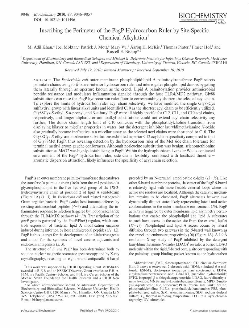
pubs.acs.org/Biochemistry Published on Web 09/20/2010 r 2010 American Chemical Society 9046 Biochemistry 2010, 49, 9046–9057 DOI: 10.1021/bi1011496 Inscribing the Perimeter of the PagP Hydrocarbon Ruler by Site-Specific Chemical Alkylation † M. Adil Khan, ‡ Joel Moktar, ‡ Patrick J. Mott, ‡ Mary Vu, ‡ Aaron H. McKie, § Thomas Pinter, § Fraser Hof, § and Russell E. Bishop* ,‡ ‡ Department of Biochemistry and Biomedical Sciences and Michael G. DeGroote Institute for Infectious Disease Research, McMaster University, Hamilton, ON, Canada L8N 3Z5, and § Department of Chemistry, University of Victoria, Victoria, BC, Canada V8W 3 V6 Received July 19, 2010; Revised Manuscript Received September 16, 2010 ABSTRACT: The Escherichia coli outer membrane phospholipid:lipid A palmitoyltransferase PagP selects palmitate chains using its β-barrel-interior hydrocarbon ruler and interrogates phospholipid donors by gating them laterally through an aperture known as the crenel. Lipid A palmitoylation provides antimicrobial peptide resistance and modulates inflammation signaled through the host TLR4/MD2 pathway. Gly88 substitutions can raise the PagP hydrocarbon ruler floor to correspondingly shorten the selected acyl chain. To explore the limits of hydrocarbon ruler acyl chain selectivity, we have modified the single Gly88Cys sulfhydryl group with linear alkyl units and identified C10 as the shortest acyl chain to be efficiently utilized. Gly88Cys-S-ethyl, S-n-propyl, and S-n-butyl PagP were all highly specific for C12, C11, and C10 acyl chains, respectively, and longer aliphatic or aminoalkyl substitutions could not extend acyl chain selectivity any further. The donor chain length limit of C10 coincides with the phosphatidylcholine transition from displaying bilayer to micellar properties in water, but the detergent inhibitor lauryldimethylamine N-oxide also gradually became ineffective in a micellar assay as the selected acyl chains were shortened to C10. The Gly88Cys-S-ethyl and norleucine substitutions exhibited superior C12 acyl chain specificity compared to that of Gly88Met PagP, thus revealing detection by the hydrocarbon ruler of the Met side chain tolerance for terminal methyl group gauche conformers. Although norleucine substitution was benign, selenomethionine substitution at Met72 was highly destabilizing to PagP. Within the hydrophobic and van der Waals-contacted environment of the PagP hydrocarbon ruler, side chain flexibility, combined with localized thioether- aromatic dispersion attraction, likely influences the specificity of acyl chain selection. PagP is an outer membrane palmitoyltransferase that catalyzes the transfer of a palmitate chain (16:0) from the sn-1 position of a glycerophospholipid to the free hydroxyl group of the (R)-3- hydroxymyristate chain at position 2 of lipid A (endotoxin) (Figure 1A) (1-3). In Escherichia coli and related pathogenic Gram-negative bacteria, PagP evades host immune defenses by resisting antimicrobial peptides (4-7) and attenuating the in- flammatory response to infection triggered by lipopolysaccharide through the TLR4/MD2 pathway (8-10). Transcription of the pagP gene is governed by the PhoP/PhoQ regulon, which con- trols expression of bacterial lipid A modification enzymes induced during infection by host antimicrobial peptides (11, 12). PagP is thus a target for the development of anti-infective agents and a tool for the synthesis of novel vaccine adjuvants and endotoxin antagonists (2, 3). The structure of E. coli PagP has been determined both by solution nuclear magnetic resonance spectroscopy and by X-ray crystallography, revealing an eight-strand antiparallel β-barrel preceded by an N-terminal amphipathic R-helix (13-15). Like other β-barrel membrane proteins, the center of the PagP β-barrel is relatively rigid with more flexible external loops where the active site residues are localized. Although the catalytic mechan- ism remains to be elucidated, PagP alternates between two dynamically distinct states likely representing latent and active conformations in the outer membrane environment (16). PagP activity is triggered by outer membrane lipid asymmetry pertur- bations that enable the phospholipid and lipid A substrates to each have access to the active site from the external leaflet (17-19). Phospholipid and lipid A access occurs by lateral diffusion through two gateways in the β-barrel wall known as the crenel and embrasure, respectively (20) (Figure 1A). A 1.9 A ˚ resolution X-ray study of PagP inhibited by the detergent lauryldimethylamine N-oxide (LDAO) 1 revealed a buried LDAO molecule within the rigid β-barrel core, a site corresponding with the palmitoyl group binding pocket known as the hydrocarbon † This work was supported by CIHR Operating Grant MOP-84329 awarded to R.E.B. and an NSERC Discovery Grant awarded to F.H. A. H.M. is a Pacific Century Scholar, and F.H. is a Career Scholar of the Michael Smith Foundation for Health Research and a CIHR New Investigator. *To whom correspondence should be addressed: Department of Biochemistry and Biomedical Sciences, McMaster University, Health Sciences Centre 4H19, 1200 Main St. W., Hamilton, ON, Canada L8N 3Z5. Telephone: (905) 525-9140, ext. 28810. Fax: (905) 522-9033. E-mail: [email protected]. 1 Abbreviations: βME, β-mercaptoethanol; CD, circular dichroism; Kdo, 3-deoxy-D-manno-oct-2-ulosonic acid; DDM, n-dodecyl β-D-mal- toside; ESI-MS, electrospray ionization mass spectrometry; EDTA, ethylenediaminetetraacetic acid; Gdn-HCl, guanidine hydrochloride; IPTG, isopropyl β-D-thiogalactopyranoside; LDAO, lauryldimethyla- mine N-oxide; MNBS, methyl p-nitrobenzenesulfonate; MPD, 2-meth- yl-2,4-pentanediol; Nle, norleucine; PDB, Protein Data Bank; PtdCho, phosphatidylcholine; PtdEtn, phosphatidylethanolamine; PBS, phos- phate-buffered saline; SeM, selenomethionine; SDS, sodium dodecyl sulfate; T u , thermal unfolding temperature; TLC, thin layer chroma- tography; UV, ultraviolet.
-
Upload
khangminh22 -
Category
Documents
-
view
0 -
download
0
Transcript of Inscribing the Perimeter of the PagP Hydrocarbon Ruler by ...
pubs.acs.org/Biochemistry Published on Web 09/20/2010 r 2010 American Chemical Society
9046 Biochemistry 2010, 49, 9046–9057
DOI: 10.1021/bi1011496
Inscribing the Perimeter of the PagP Hydrocarbon Ruler by Site-SpecificChemical Alkylation†
M. Adil Khan,‡ Joel Moktar,‡ Patrick J. Mott,‡ Mary Vu,‡ Aaron H. McKie,§ Thomas Pinter,§ Fraser Hof,§ andRussell E. Bishop*,‡
‡Department of Biochemistry and Biomedical Sciences andMichael G. DeGroote Institute for Infectious Disease Research, McMasterUniversity, Hamilton, ON, Canada L8N 3Z5, and §Department of Chemistry, University of Victoria, Victoria, BC, Canada V8W3V6
Received July 19, 2010; Revised Manuscript Received September 16, 2010
ABSTRACT: The Escherichia coli outer membrane phospholipid:lipid A palmitoyltransferase PagP selectspalmitate chains using its β-barrel-interior hydrocarbon ruler and interrogates phospholipid donors by gatingthem laterally through an aperture known as the crenel. Lipid A palmitoylation provides antimicrobialpeptide resistance and modulates inflammation signaled through the host TLR4/MD2 pathway. Gly88substitutions can raise the PagP hydrocarbon ruler floor to correspondingly shorten the selected acyl chain.To explore the limits of hydrocarbon ruler acyl chain selectivity, we have modified the single Gly88Cyssulfhydryl group with linear alkyl units and identified C10 as the shortest acyl chain to be efficiently utilized.Gly88Cys-S-ethyl, S-n-propyl, and S-n-butyl PagP were all highly specific for C12, C11, and C10 acyl chains,respectively, and longer aliphatic or aminoalkyl substitutions could not extend acyl chain selectivity anyfurther. The donor chain length limit of C10 coincides with the phosphatidylcholine transition fromdisplaying bilayer to micellar properties in water, but the detergent inhibitor lauryldimethylamine N-oxidealso gradually became ineffective in a micellar assay as the selected acyl chains were shortened to C10. TheGly88Cys-S-ethyl and norleucine substitutions exhibited superior C12 acyl chain specificity compared to thatof Gly88Met PagP, thus revealing detection by the hydrocarbon ruler of the Met side chain tolerance forterminal methyl group gauche conformers. Although norleucine substitution was benign, selenomethioninesubstitution atMet72 was highly destabilizing to PagP.Within the hydrophobic and van derWaals-contactedenvironment of the PagP hydrocarbon ruler, side chain flexibility, combined with localized thioether-aromatic dispersion attraction, likely influences the specificity of acyl chain selection.
PagP is an outermembrane palmitoyltransferase that catalyzesthe transfer of a palmitate chain (16:0) from the sn-1 position of aglycerophospholipid to the free hydroxyl group of the (R)-3-hydroxymyristate chain at position 2 of lipid A (endotoxin)(Figure 1A) (1-3). In Escherichia coli and related pathogenicGram-negative bacteria, PagP evades host immune defenses byresisting antimicrobial peptides (4-7) and attenuating the in-flammatory response to infection triggered by lipopolysaccharidethrough the TLR4/MD2 pathway (8-10). Transcription of thepagP gene is governed by the PhoP/PhoQ regulon, which con-trols expression of bacterial lipid A modification enzymesinduced during infection by host antimicrobial peptides (11, 12).PagP is thus a target for the development of anti-infective agentsand a tool for the synthesis of novel vaccine adjuvants andendotoxin antagonists (2, 3).
The structure of E. coli PagP has been determined both bysolution nuclear magnetic resonance spectroscopy and by X-raycrystallography, revealing an eight-strand antiparallel β-barrel
preceded by an N-terminal amphipathic R-helix (13-15). Likeotherβ-barrelmembrane proteins, the center of the PagPβ-barrelis relatively rigid with more flexible external loops where theactive site residues are localized. Although the catalytic mechan-ism remains to be elucidated, PagP alternates between twodynamically distinct states likely representing latent and activeconformations in the outer membrane environment (16). PagPactivity is triggered by outer membrane lipid asymmetry pertur-bations that enable the phospholipid and lipid A substratesto each have access to the active site from the external leaflet(17-19). Phospholipid and lipid A access occurs by lateraldiffusion through two gateways in the β-barrel wall known asthe crenel and embrasure, respectively (20) (Figure 1A). A 1.9 Aresolution X-ray study of PagP inhibited by the detergentlauryldimethylamineN-oxide (LDAO)1 revealed a buried LDAOmolecule within the rigid β-barrel core, a site corresponding withthe palmitoyl group binding pocket known as the hydrocarbon
†This work was supported by CIHR Operating Grant MOP-84329awarded toR.E.B. and anNSERCDiscoveryGrant awarded to F.H.A.H.M. is a Pacific Century Scholar, and F.H. is a Career Scholar of theMichael Smith Foundation for Health Research and a CIHR NewInvestigator.*To whom correspondence should be addressed: Department of
Biochemistry and Biomedical Sciences, McMaster University, HealthSciences Centre 4H19, 1200 Main St. W., Hamilton, ON, Canada L8N3Z5. Telephone: (905) 525-9140, ext. 28810. Fax: (905) 522-9033.E-mail: [email protected].
1Abbreviations: βME, β-mercaptoethanol; CD, circular dichroism;Kdo, 3-deoxy-D-manno-oct-2-ulosonic acid; DDM, n-dodecyl β-D-mal-toside; ESI-MS, electrospray ionization mass spectrometry; EDTA,ethylenediaminetetraacetic acid; Gdn-HCl, guanidine hydrochloride;IPTG, isopropyl β-D-thiogalactopyranoside; LDAO, lauryldimethyla-mine N-oxide; MNBS, methyl p-nitrobenzenesulfonate; MPD, 2-meth-yl-2,4-pentanediol; Nle, norleucine; PDB, Protein Data Bank; PtdCho,phosphatidylcholine; PtdEtn, phosphatidylethanolamine; PBS, phos-phate-buffered saline; SeM, selenomethionine; SDS, sodium dodecylsulfate; Tu, thermal unfolding temperature; TLC, thin layer chroma-tography; UV, ultraviolet.

Article Biochemistry, Vol. 49, No. 42, 2010 9047
ruler (Figure 1A,B) (15). Recently, a 1.4 A resolution X-raystructure of PagP crystallized from a mixture of sodium dodecylsulfate (SDS) and 2-methyl-2,4-pentanediol (MPD) has revealeda crenel gating mechanism for the transit of the phospholipidbetween the hydrocarbon ruler and the outer membrane externalleaflet (13).
PagP is exquisitely selective for a 16-carbon palmitate chainbecause its hydrocarbon ruler excludes lipid acyl chains differing
in length by a solitarymethylene unit (15, 21).Mutation ofGly88lining the hydrocarbon ruler floor can modulate lipid acyl chainselection (21). Appropriate amino acid substitutions shorten theselected acyl chain by a degree predictable from the expected risein the hydrocarbon ruler floor. In prior investigations of PagPhydrocarbon rulermutants, whereGly88Ala,Gly88Cys-S-methyl,and Gly88Met were constructed, acyl chain selection waspredictably shifted toward C15, C13, and C12 acyl chains,
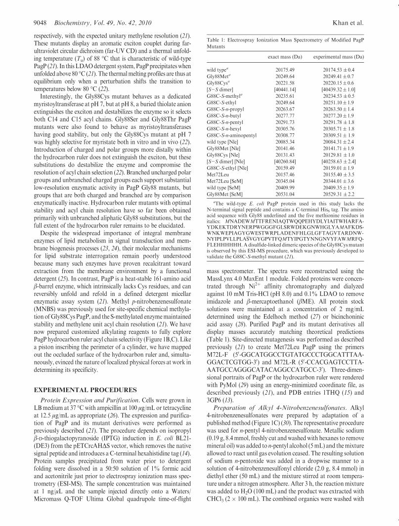
FIGURE 1: Structural relationships among PagP, the hydrocarbon ruler, andE. coli outer membrane lipids. (A)E. coli PagP transfers a palmitoylgroup (red) from the sn-1 position of a phospholipid, such as phosphatidylethanolamine (PtdEtn), to the free hydroxyl group of the N-linked(R)-3-hydroxymyristate chain at position2on theproximal glucosamineunit of lipidA.Oneof the simplest lipopolysaccharide acceptors forPagPin the outer membrane is known as Kdo2-lipid A or Re endotoxin. PagP is an eight-strand antiparallel β-barrel preceded by an N-terminalamphipathic R-helix (colored green). The amino (N) and carboxyl (C) termini are labeled with black letters, and the β-strands are labeled withwhite letters (A-H). Aromatic belt residues (shown in wireframe format) demarcate the membrane interfaces between the extracellular andperiplasmic spaces.Aboundmolecule of the detergent (coloredmagenta) delineates the interiorhydrocarbon ruler. Points for lateral phospholipidaccess via the crenel (flanked by Pro127 and Pro144) and for lateral lipid A access via the embrasure (flanked by Pro28 and Pro50) are indicated.Although the catalytic mechanism is unknown, several key residues (shown in stick format) have been mapped to the extracellular surface loops(L1-L4). Loop L4 is disordered in the crystal structure (Protein Data Bank entry 1THQ) but has been introduced and energy minimized in themodel shown. (B)A sagittal section reveals the boundLDAOmolecule and residues flanking thewalls of the hydrocarbon ruler.A nondegenerateexciton interaction between Tyr26 and Trp66 manifests a couplet during far-UV CD spectroscopy. PagP far-UV CD exhibits positive excitonellipticity at 232 nm, and the negative ellipticity at 218 nm that arisesmore strongly from the β-barrel is also enhanced by the exciton. The adjacentGly88 can be substituted to modulate lipid acyl chain selection. Gly88Cys functions as a dedicated myristoyltransferase at pH 7, but at pH 8, itintroduces a destabilizing thiolate anion that extinguishes the exciton and broadens selection to include C15 acyl chains. Cys methylation withmethyl p-nitrobenzenesulfonate (MNBS) restores the exciton at pH 8 and renders PagP selective for C13 acyl chains. (C) Synthetic scheme for asuite of alkyl p-nitrobenzenesulfonates used to inscribe the perimeter of the PagP hydrocarbon ruler by site-specific chemical alkylation.
9048 Biochemistry, Vol. 49, No. 42, 2010 Khan et al.
respectively, with the expected unitary methylene resolution (21).These mutants display an aromatic exciton couplet during far-ultraviolet circular dichroism (far-UV CD) and a thermal unfold-ing temperature (Tu) of 88 �C that is characteristic of wild-typePagP (21). In this LDAOdetergent system, PagPprecipitateswhenunfolded above 80 �C (21). The thermalmelting profiles are thus atequilibrium only when a perturbation shifts the transition totemperatures below 80 �C (22).
Interestingly, the Gly88Cys mutant behaves as a dedicatedmyristoyltransferase at pH 7, but at pH 8, a buried thiolate anionextinguishes the exciton and destabilizes the enzyme so it selectsboth C14 and C15 acyl chains. Gly88Ser and Gly88Thr PagPmutants were also found to behave as myristoyltransferaseshaving good stability, but only the Gly88Cys mutant at pH 7was highly selective for myristate both in vitro and in vivo (22).Introduction of charged and polar groups more distally withinthe hydrocarbon ruler does not extinguish the exciton, but thesesubstitutions do destabilize the enzyme and compromise theresolution of acyl chain selection (22). Branched uncharged polargroups and unbranched charged groups each support substantiallow-resolution enzymatic activity in PagP Gly88 mutants, butgroups that are both charged and branched are by comparisonenzymatically inactive. Hydrocarbon ruler mutants with optimalstability and acyl chain resolution have so far been obtainedprimarily with unbranched aliphatic Gly88 substitutions, but thefull extent of the hydrocarbon ruler remains to be elucidated.
Despite the widespread importance of integral membraneenzymes of lipid metabolism in signal transduction and mem-brane biogenesis processes (23, 24), their molecular mechanismsfor lipid substrate interrogation remain poorly understoodbecause many such enzymes have proven recalcitrant towardextraction from the membrane environment by a functionaldetergent (25). In contrast, PagP is a heat-stable 161-amino acidβ-barrel enzyme, which intrinsically lacks Cys residues, and canreversibly unfold and refold in a defined detergent micellarenzymatic assay system (21). Methyl p-nitrobenzenesulfonate(MNBS) was previously used for site-specific chemical methyla-tion ofGly88Cys PagP, and the S-methylated enzymemaintainedstability and methylene unit acyl chain resolution (21). We havenow prepared customized alkylating reagents to fully explorePagP hydrocarbon ruler acyl chain selectivity (Figure 1B,C). Likea piston inscribing the perimeter of a cylinder, we have mappedout the occluded surface of the hydrocarbon ruler and, simulta-neously, evinced the nature of localized physical forces at work indetermining its specificity.
EXPERIMENTAL PROCEDURES
Protein Expression and Purification. Cells were grown inLBmedium at 37 �Cwith ampicillin at 100 μg/mL or tetracyclineat 12.5 μg/mL as appropriate (26). The expression and purifica-tion of PagP and its mutant derivatives were performed aspreviously described (21). The procedure depends on isopropylβ-D-thiogalactopyranoside (IPTG) induction in E. coli BL21-(DE3) from the pETCrcAHΔS vector, which removes the nativesignal peptide and introduces a C-terminal hexahistidine tag (14).Protein samples precipitated from water prior to detergentfolding were dissolved in a 50:50 solution of 1% formic acidand acetonitrile just prior to electrospray ionization mass spec-trometry (ESI-MS). The sample concentration was maintainedat 1 ng/μL and the sample injected directly onto a Waters/Micromass Q-TOF Ultima Global quadrupole time-of-flight
mass spectrometer. The spectra were reconstructed using theMassLynx 4.0 MaxEnt 1 module. Folded proteins were concen-trated through Ni2þ affinity chromatography and dialyzedagainst 10 mM Tris-HCl (pH 8.0) and 0.1% LDAO to removeimidazole and β-mercaptoethanol (βME). All protein stocksolutions were maintained at a concentration of 2 mg/mLdetermined using the Edelhoch method (27) or bicinchoninicacid assay (28). Purified PagP and its mutant derivatives alldisplay masses accurately matching theoretical predictions(Table 1). Site-directed mutagenesis was performed as describedpreviously (21) to create Met72Leu PagP using the primersM72L-F (50-GGCATGGCCTGTATGCCCTGGCATTTAA-GGACTCGTGG-30) and M72L-R (50-CCACGAGTCCTTA-AATGCCAGGGCATACAGGCCATGCC-30). Three-dimen-sional portraits of PagP or the hydrocarbon ruler were renderedwith PyMol (29) using an energy-minimized coordinate file, asdescribed previously (21), and PDB entries 1THQ (15) and3GP6 (13).Preparation of Alkyl 4-Nitrobenzenesulfonates. Alkyl
4-nitrobenzenesulfonates were prepared by adaptation of apublishedmethod (Figure 1C) (30). The representative procedurewas used for n-pentyl 4-nitrobenzenesulfonate. Metallic sodium(0.19 g, 8.4mmol, freshly cut andwashedwith hexanes to removemineral oil) was added to n-pentyl alcohol (5mL) and themixtureallowed to react until gas evolution ceased. The resulting solutionof sodium n-pentoxide was added in a dropwise manner to asolution of 4-nitrobenzenesulfonyl chloride (2.0 g, 8.4 mmol) indiethyl ether (50 mL) and the mixture stirred at room tempera-ture under a nitrogen atmosphere. After 3 h, the reactionmixturewas added to H2O (100 mL) and the product was extracted withCHCl3 (2 � 100 mL). The combined organics were washed with
Table 1: Electrospray Ionization Mass Spectrometry of Modified PagP
Mutants
exact mass (Da) experimental mass (Da)
wild typea 20175.49 20174.53 ( 0.4
Gly88Meta 20249.64 20249.41( 0.7
Gly88Cysa 20221.58 20220.15( 0.6
[S-S dimer] [40441.14] [40439.32( 1.0]
G88C-S-methyla 20235.61 20234.53 ( 0.5
G88C-S-ethyl 20249.64 20251.10( 1.9
G88C-S-n-propyl 20263.67 20263.50( 1.4
G88C-S-n-butyl 20277.77 20277.20( 1.9
G88C-S-n-pentyl 20291.73 20291.78 ( 1.8
G88C-S-n-hexyl 20305.76 20305.71( 1.8
G88C-S-n-aminopentyl 20308.77 20309.51( 1.9
wild type [Nle] 20085.34 20084.31( 2.4
Gly88Met [Nle] 20141.46 20141.71( 1.9
Gly88Cys [Nle] 20131.43 20129.81 ( 1.0
[S-S dimer] [Nle] [40260.84] [40258.63 ( 2.4]
G88C-S-ethyl [Nle] 20159.49 20159.01( 1.9
Met72Leu 20157.46 20155.40 ( 3.5
Met72Leu [SeM] 20345.04 20344.01( 3.6
wild type [SeM] 20409.99 20409.35( 1.9
Gly88Met [SeM] 20531.04 20529.31 ( 2.2
aThe wild-type E. coli PagP protein used in this study lacks theN-terminal signal peptide and contains a C-terminal His6 tag. The aminoacid sequence with Gly88 underlined and the five methionine residues initalics: MNADEWMTTFRENIAQTWQQPEHYDLYIAITWHARFA-YDKEKTDRYNERPWGGGFGLSRWDEKGNWHGLYAMAFKDS-WNKWEPIAGYGWESTWRPLADENFHLGLGFTAGVTARDNW-NYIPLPVLLPLASVGYGPVTFQMTYIPGTYNNGNVYFAWMRFQ-FLEHHHHHH.Adisulfide-linked dimeric species of theGly88Cysmutantis observed by this ESI-MS procedure, which was previously developed tovalidate the G88C-S-methyl mutant (21).
Article Biochemistry, Vol. 49, No. 42, 2010 9049
water (50mL), saturated aqueous NaHCO3 (100 mL), and water(50mL) again prior to being dried over Na2SO4. The solvent wasremoved on a rotary evaporator and the crude product purifiedby column chromatography (SiO2, 20% EtOAc in hexanes) toyield the pure product as a yellow solid (1.65 g, 72% yield): mp55-56 �C; 1H NMR (300 MHz, CDCl3) δ 8.41 (d, 2H, J =8.8 Hz), 8.12 (d, 2H, J = 8.8 Hz), 4.14 (t, 2H, J= 6.5 Hz), 1.69(m, 2H), 1.30 (m, 4H), 0.87 (t, 3H, J = 6.5 Hz); 13C NMR (75MHz,CDCl3) δ 140.5, 127.5, 122.8, 113.0, 70.4, 26.9, 25.7, 20.3, 12.1.
The more elaborate n-aminopentyl 4-nitrobenzenesulfonate(Figure 3A) was prepared in two steps. A mixture of Boc-protected 5-amino-n-pentan-1-ol (0.25 g, 1.2 mmol) and dryCH2Cl2 (3 mL) was placed under a nitrogen atmosphere at0 �C, and a solution of 4-nitrobenzenesulfonyl chloride (0.32 g,1.2 mmol) dissolved in dry CH2Cl2 (3 mL) was added dropwisevia syringe over the course of 15 min. The mixture was stirred at0 �C for 2 h and allowed to warm to ambient temperature for24 h. The reaction was quenched and the mixture subsequentlywashed three times with 1MHCl (5mL) and once with saturatedaqueous NaCl (5 mL). The organic layer was dried over MgSO4,filtered, and concentrated in vacuo. The crude yellow solid wasdissolved in minimal EtOAc and purified by flash chromatogra-phy (SiO2, 20 to 40% EtOAc in hexanes) to afford the corre-sponding Boc-protected 4-nitrobenzenesulfonate derivative as apale yellow solid (0.28 g, 60% yield): mp 82-88 �C; 1H NMR(300 MHz, CDCl3) δ 8.38 (d, 2H, J= 9.1 Hz), 8.08 (d, 2H, J=9.1 Hz), 4.49 (s, 1H), 4.11 (t, 2H, J = 6.5 Hz), 3.05 (q, 2H, J =6.6 Hz), 1.65 (quintet, 2H, J=6.6 Hz), 1.48-1.30 (m, 13H); 13CNMR (75MHz, CDCl3) δ 156.1, 150.9, 142.1, 129.3, 124.6, 79.4,71.7, 40.3, 29.6, 28.7, 28.5, 22.1; HRMS m/z 411.1201 [(M þNa)þ calcd 411.1194]. A solution of this Boc-protected inter-mediate (0.05 g, 0.12 mmol) in MeOH (10 mL) was treated withHCl (1 mL of a 4M solution in dioxane). Themixture was stirredat ambient temperature for 1 h, and all solvents were evaporatedin vacuo. The crude yellow product was triturated in CH2Cl2,filtered, and washed with CH2Cl2 to afford the n-aminopentyl4-nitrobenzenesulfonate product (Figure 3A) as its HCl salt (paleyellow solid, 0.024 g, 60% yield): mp 104-108 �C; 1H NMR(500MHz, CD3OD) δ 8.48 (d, 2H, J=8.8Hz), 8.17 (d, 2H, J=8.8 Hz), 4.19 (t, 2H, J = 6.2 Hz), 2.90 (t, 2H, J = 7.6 Hz), 1.75(quintet, 2H, J = 7.6 Hz), 1.65 (quintet, 2H, J = 7.6 Hz), 1.46(quintet, 2H, J = 7.5 Hz); 13C NMR (125 MHz, CD3OD) δ151.2, 141.7, 129.3, 124.5, 71.1, 39.3, 28.3, 26.7, 25.3; HRMSm/z289.0858 [(M þ H - Cl)þ calcd 289.0863].Site-Specific Chemical Alkylation of Gly88Cys PagP.
MNBS is commercially available, and we have previouslyadapted the S-methylation procedure developed by Heinrick-son (31) for the covalent modification of Gly88Cys PagP (21). Inthe work presented here, the customized alkylating reagentsdescribed above were utilized to incorporate ethyl, n-propyl,n-butyl, n-pentyl, n-hexyl, and n-aminopentyl groups into theGly88Cys PagPmutant. The reactions were conducted in cappedglass tubes. To achieve buffer concentrations of 6 M guanidinehydrochloride (Gdn-HCl), 0.25 M Tris-HCl (pH 8.6), 3.3 mMdisodium ethylenediaminetetraacetic acid (EDTA), and 25%acetonitrile (v/v), 25 mg of Gly88Cys PagP was dissolved in avolume of 3.75 mL of 8 M Gdn-HCl, 0.34 M Tris-HCl (pH 8.6),and 4.4 mM EDTA and the volume adjusted to 5 mL with 1.25mL of 100% acetonitrile. The solutions were flushed with N2 for1min to create an anoxic barrier, and 50 μL of 260mM βMEwasadded (10-50-fold molar excess of the protein). The tubes weretightly sealed and placed in a 50 �C bath for 1.5 h and then
gradually cooled to 37 �C.Under a N2 barrier, 0.5 mL of 5.2 mMethyl, n-propyl, n-butyl, n-pentyl, n-hexyl, or n-aminopentyl4-nitrobenzenesulfonate (2-fold molar excess of the βME) dis-solved in acetonitrile was added to the reaction mixture. Thereactions were conducted at 37 �C for 2 h with ethyl, 4 h withn-propyl, 8 h with n-butyl, and overnight with n-pentyl, n-hexyl,and n-aminopentyl 4-nitrobenzenesulfonate. Reactions werequenched by addition of 5 μL of 14 M βME. The reactionmixture was dialyzed exhaustively against distilled water, and theprecipitated proteins were collected. A small amount of theprecipitate was stored for the purposes of ESI-MS as describedabove. The remainder was dissolved in 10mMTris-HCl (pH 8.0)and 6 M Gdn-HCl for protein refolding as described pre-viously (21).Norleucine and Selenomethionine Substitution. To incor-
porate norleucine (Nle) or selenomethionine (SeM) into PagPand its mutants, the appropriate expression plasmids weretransformed into methionine auxotrophic cell line E. coli B834-(DE3). Nle incorporation was based on the procedure describedby Budisa (32), except that we adapted media designed for SeMlabeling. Four stock solutions were prepared. First, a stocksolution of 50� amino acid mix was prepared by adding to500 mL of autoclaved water 1 g each of the standard L-aminoacids, excluding cystine, trans-proline, tyrosine, glutamine, andmethionine. The solution was passed through a 0.22 μm filter,and 40mL aliquots were stored at-80 �C. Second, a 1000� tracemetal stock solution was prepared to yield a final concentrationin culture media of 10 μM for each metal: 12.4 g of(NH4)6(Mo7)24 3 4H2O, 2.4 g of CoCl2 3 6H2O, 2.5 g of CuSO4 35H2O, 0.62 g ofH3BO3, 2.0 g ofMnCl2 3 4H2O, and 1.4 g of ZnCl2were added to 0.1% HCl in a final volume of 1.0 L andautoclaved. Third, a buffer solution was prepared by dissolving4.0 g of (NH4)2SO4, 18.0 g of KH2PO4, 42.0 g of K2HPO4, and2.0 g of Na3-citrate in 800 mL of water and autoclaved. Finally,we prepared the TyB “Tyrosine and nucleotide bases” mix bydissolving 0.16 g of tyrosine and 2.0 g each of adenine, guanosine,thymine, and uracil in 2.0 L of autoclaved water by heating thesample to 50 �C with stirring and then autoclaving. After thesamples had been cooled to room temperature, the followingwere added to the TyBmix in order: 80mLof the 50� amino acidstock, 8 mL of 20 mg/mL glutamine (filter-sterilized), 80 mL of50% glycerol (autoclaved), 5 mL of the 1000� trace metal mix,4 mL of 0.5 M CaCl2 (filter-sterilized), 4 mL of MgSO4 (filter-sterilized), 1.6 mL of 5 mg/mL thiamine (filter-sterilized),1000 mL of autoclaved water, and 800 mL of the buffer solution.The TyB mix was stored at 4 �C until it was used. A solution ofNle was also prepared via addition of 0.33 g to 10 mL of waterfor a final medium concentration of 5 mM at pH 2.31 and filter-sterilized.
For Nle substitution, wild-type, Gly88Met, and Gly88CysPagP strains were grown in 5 mL of LB medium at 37 �Covernight, and 1 mL of each overnight culture was inoculatedinto 1 L of prewarmed minimal medium described above towhich a limited amount ofMet (0.05 mM) had been added. Afterthe OD reached 0.6, the Met-exhausted culture was supplemen-ted with 5 mM Nle and incubated for 10 min before inductionwith 1mMIPTG.Cells were induced for 4 h at 37 �Cbefore beingharvested. As a control, we also grew these mutants in the samemedium supplemented with 5 mMMet. The protein was purifiedas described previously (21).
For SeM substitution, 10 mL overnight cultures of wild-typeandGly88Met PagP strains were prepared inminimal medium as

9050 Biochemistry, Vol. 49, No. 42, 2010 Khan et al.
described above, but supplemented with 0.3 mMMet. After 16 h,the cultures were harvested at 7000 rpm for 10min. The pelletwaswashed twice with 10 mL of minimal medium without Met andthen resuspended in 20 mL of minimal medium supplementedwith 0.05 mMMet. The suspension was then added to 980mL ofminimal medium supplemented with 0.05 mMMet. After an ODof 0.7 had been reached, 10 mL of a 300 mM filter-sterilized SeMsolution was added to the Met-depleted culture to give a finalSeM concentration of 3 mM. After incubation for 10 min, theculture was induced with 1 mM filter-sterilized IPTG and grownfor 4 h before the cells were harvested. The proteinwas purified asdescribed previously (21), except that solutions were maintainedin a reduced state by addition of 20 mM βME. No oxidationof SeM to SeM selenoxide in PagP was evident by ESI-MS(Table 1).CD Spectroscopy. Samples to be analyzed by far-UV CD
weremaintained at a concentration of 15 μM in 10mMTris-HCl(pH 8.0) and 0.1% LDAO using a cuvette with a path length of1 mm. An Aviv 215 spectrophotometer was linked to a MerlinSeries M25 Peltier device for temperature control. For eachsample, three accumulations were averaged at a data pitch of1 nm and a scanning speed of 10 nm/min. The temperature wasmaintained at 25 �C, and data sets were obtained from 200 to260 nm for far-UV CD. Thermal denaturation profiles wereobtained by heating the samples from 20 to 100 �Cat 218 nmwitha temperature slope of 2 �C/min and a response time of 16 s.Preparation of [32P]Kdo2-Lipid A.To prepare radiolabeled
lipidAbearing two units of 3-deoxy-D-manno-oct-2-ulosonic acid(Kdo2-lipid A), a 10 mL culture of E. coli WBB06 was grownovernight as described previously (33). Briefly, a 1:100 dilutionwas made in 5 mL of LB containing 25 μCi of [32P]ortho-phosphate (Perkin-Elmer) and grown at 37 �C for 4-5 h. Afterbeing harvested, the pellet was washed with phosphate-bufferedsaline (PBS) (pH 7.4) (26) and harvested again. The cells weresuspended in a CHCl3/MeOH/PBS (pH 7.4) mixture (1:2:0.8,v/v) and allowed to sit at room temperature for 1 h with slightagitation. The tube was centrifuged at 7000 rpm and roomtemperature for 10 min, and the supernatant was collected.Appropriate amounts of CHCl3 and H2O were added to forma two-phase Bligh-Dyer solution [CHCl3/MeOH/H2O mixture(2:2:1.8, v/v)] (34), and the lower phase was extracted. The lowerphase was dried under N2, and the dried lipids were resuspendedin a CHCl3/MeOH/H2O mixture (2:3:1, v/v) (solvent A) andapplied to a DE-52 cellulose column. We prepared the resin bywashing dryDE-52 resin with 2.4MNH4-acetate three times andthen washing the resin with distilled water. The resin was thenwashed twice with aMeOH/2.4MNH4-acetatemixture (8:2, v/v)and then twice with a CHCl3/MeOH/2.4MNH4-acetate mixture(2:3:1, v/v). The resin was washed several times with solvent A toremove all the salt. This resin was packed in a glass column towhich the sample was applied. The column was washed with2 column volumes of solvent A and then eluted with serial 5-8column volumes of solvent A containing 60, 120, 240, 360, and480mMNH4-acetate as the aqueous component. Tenmicrolitersof each fraction was visualized by thin layer chromatography(TLC) in the CHCl3/pyridine/88% formic acid/H2O solventsystem (50:50:16:5, v/v) to identify the fractions containingKdo2-lipid A with a monophosphate moiety at position 1. TheTLC plates were visualized using a PhosphorImager (MolecularDynamics).Acyltransferase Assays. The hydrocarbon ruler assays
were performed using a TLC-based approach as previously
described (21). Synthetic sn-1,2-diacylphosphatidylcholine(PtdCho) with unbranched saturated acyl chains of definedcomposition, and Kdo2-lipid A, were obtained from AvantiPolar Lipids (Alabaster, AL). The assays were conducted in0.5 mL microcentrifuge tubes, in which sufficient Kdo2-lipid Awas added to achieve a concentration of 10 μM in a final assayvolume of 25 μL. To this sample was added a trace amount of[32P]Kdo2-lipid A, yielding a density of 200 cpm/μL. SufficientPtdChowas added to attain a final concentration of 1mM.Theseconstituents were dried under a gentle N2 stream and subse-quently dissolved in 22.5 μL of a reaction cocktail containing0.1 M Tris-HCl (pH 8.0), 10 mM EDTA, and 0.25% dodecylmaltoside (DDM). The reaction was initiated via addition ofsufficient PagP, serially diluted in DDM to remove inhibitoryLDAO, to achieve a linear reaction profile. All reactions wereconducted at 30 �C andwere stopped by directly spotting 4 μL ofthe reaction mixture to the origin of a Silica Gel 60 TLC plate.The TLC plate was resolved in a CHCl3/pyridine/88% formicacid/H2O system (50:50:16:5, v/v) within a tightly sealed glasstank. The constituents of the tank were allowed to equilibrate fora period of 3 h prior to being exposed to the TLC plate. After theplate was dried, it was exposed to a Molecular DynamicsPhosphorImager screen overnight to visualize the reaction. Theproduct was quantified using ImageQuant. Detergent exchangeacyltransferase assays for wild-type and Gly88Cys-S-n-butylPagP were conducted by dilution of the protein samples inreaction buffers containing either 0.25% DDM, 0.1% Cyclofos7, or 0.1% LDAO [all detergents were obtained from Anatrace(Maumee, OH)]. Wild-type PagP was assayed using di16:0PtdCho, and Gly88Cys-S-n-butyl PagP was assayed usingdi10:0 PtdCho. The inhibitory effects of LDAO upon PagP weredetermined via addition of increasing concentrations of LDAOto the assay.
RESULTS AND DISCUSSION
PagP and its mutants were produced in E. coli BL21(DE3)using IPTG-inducible PagP expression plasmid pETCrcAHΔS(14). The expressed proteins possess a C-terminal hexahistidinetag and lack the N-terminal signal peptide to target PagP forsecretion to the outer membrane. The proteins are expressed asinsoluble aggregates, which can be dissolved in Gdn-HCl, puri-fied byNi2þ-ion affinity chromatography, and folded by dilutioninto the detergent LDAO (21). Site-specific chemical alkylationof Gly88Cys PagP was performed by an established procedure(Figure 1B) (21, 31) and confirmed by ESI-MS of the purifiedproteins (Table 1). Alkyl p-nitrobenzenesulfonates were preparedfrom the corresponding sodium alkoxides and 4-nitrobenzene-sulfonyl chloride as described in Experimental Procedures(Figure 1C). PagP phospholipid:lipid A palmitoyltransferaseactivity is monitored in vitro using a defined detergent micellarenzymatic assay with TLC separation of radioactive lipid pro-ducts (21). The inhibitory LDAO detergent used during PagPfolding is exchanged by dilution into DDM to support enzymaticactivity.Wild-type PagP is highly selective for a palmitoyl group atthe sn-1 position in a glycerophospholipid but largely unspecificfor the polar headgroup (1). We employ the palmitoyl donordi-16:0-PtdCho and the palmitoyl acceptor [32P]Kdo2-lipid A.When challenged with a spectrum of donor acyl chain lengths,wild-type PagP functions as a dedicated palmitoyltransferasewhereas Gly88 PagP mutants can utilize shorter acyl chains.Analysis of Gly88Cys-S-alkyl Mutants. We examined
the hydrocarbon ruler profiles for the Gly88Cys-S-alkyl PagP

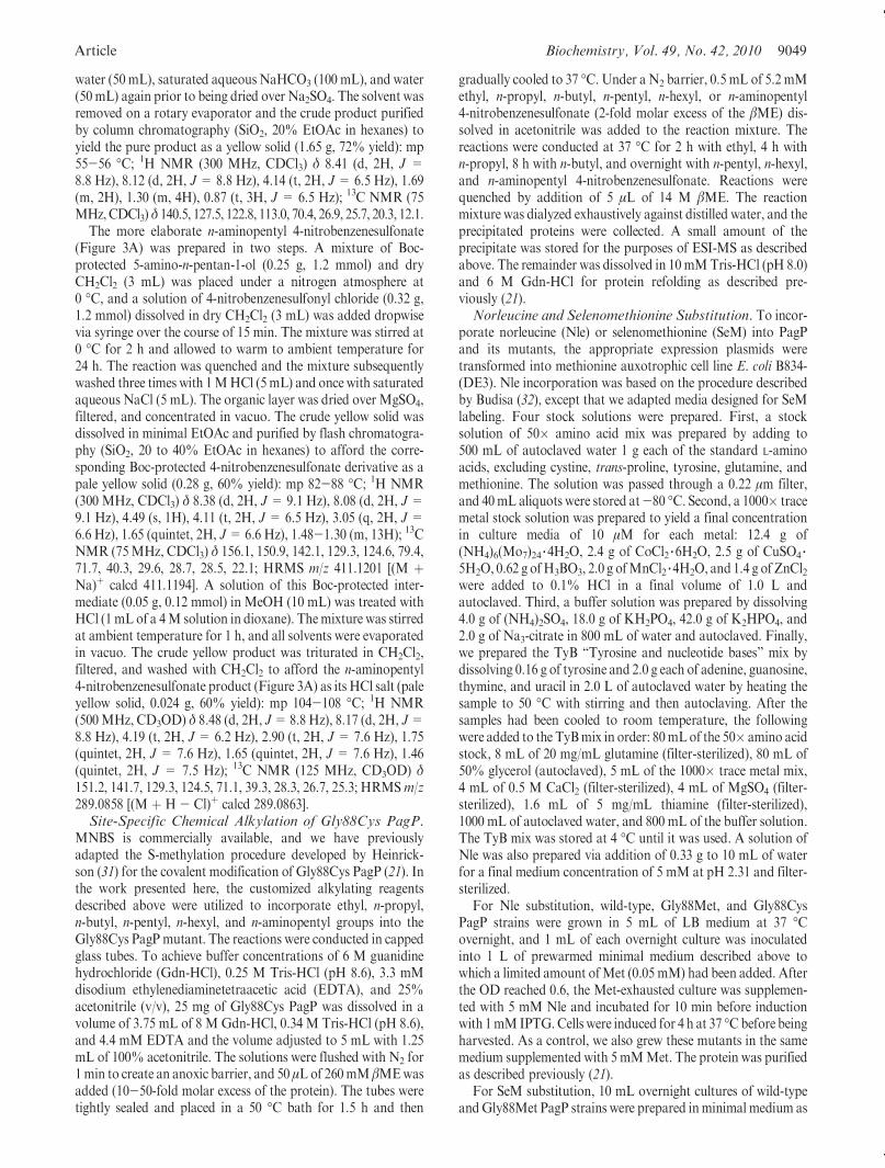
Article Biochemistry, Vol. 49, No. 42, 2010 9051
derivatives using a suite of diacyl PtdCho’s varying in saturatedacyl chain length fromC14 toC8 (Figure 2A). Gly88Cys-S-ethyl,S-n-propyl, and S-n-butyl PagP were all highly specific for C12,C11, andC10 acyl chains, respectively. As expected,Gly88Cys-S-n-pentyl PagP utilized a C9 acyl chain optimally, but with onlyhalf the specific activity compared to that of the wild type and theshorter alkyl substitutions when using their optimal substrates.Gly88Cys-S-n-hexyl PagPwas by comparison inactive (Figure 2A).Far-UV CD analysis of the folded S-alkylated proteins clearly
identified the positive 232 nm ellipticity derived from the intactaromatic exciton (Figure 2B) (21). The Tu of 88 �C for wild-typePagP β-barrel unfolding is elevated by ∼2-4 �C among thespectrum of Gly88Cys-S-alkyl derivatives (Figure 2C). Addition-ally, the 218 nm component of the exciton is observed by its lossabove 40 �C, consistent with the known thermal sensitivity of theexciton (Figure 2C) (21). We had previously observed an ∼2 �Cincrease in thermal stability for Gly88Cys-S-methyl PagP(21), and that finding is recapitulated here (Figure 2C).
FIGURE 2: Modulating the PagP hydrocarbon ruler by site-specific chemical alkylation. (A) PagP specific activity was monitored with a suite ofPtdCho’s having defined saturated acyl chain compositions varying fromC14 to C8. Panels from top to bottom represent the activity profiles forGly88Cys PagPmodified with S-ethyl, S-n-propyl, S-n-butyl, S-n-pentyl, and S-n-hexyl alkyl groups, respectively. (B) Far-UVCD identifies theβ-barrel and exciton signatures characteristic of wild-type PagP. (C) Thermal melts at 218 nm reveal exciton loss near 40 �C followed by β-barrelunfolding above 80 �C. (D) LDAO inhibition profiles for wild-type PagP and the Gly88Cys-S-alkyl substitutions using their preferred acyl chaindonors. (E) PagP specific activities forwild-type andGly88Cys-S-n-butyl PagPwere assayed usingC16 andC10 acyl chain donors, respectively, inthree different detergent systems. ND means not detected.
9052 Biochemistry, Vol. 49, No. 42, 2010 Khan et al.
Although S-alkylation appears to stabilize PagP by occludingthe hydrocarbon ruler cavity (Figure 2C), such stabilization wasnot observed in Gly88Met PagP (21).
The observed donor chain length limit of C10 (Figure 2A)happens to coincide with the PtdCho transition from displayingbilayer to micellar properties in water (35). If shorter acyl chainshad been supported, a substrate that also functioned as adetergent might have replaced DDM in the in vitro PagP assaysystem. DDM thus remains an intrinsic component of the assay,but its detergent properties disrupt bilayer phospholipid phasebehavior (25). The C10 limit might better reflect the minimaloccluded surface area between the donor acyl chain and hydro-carbon ruler required for PagP to selectively engage its substrate.We thus tested inhibition by the detergent LDAO, which alsogradually became ineffective in the DDM micellar assay as theselected acyl chains were shortened to C10 (Figure 2D). Inhibi-tion at 10 mM LDAO corresponds to a mole fraction of twoLDAO molecules to one DDM molecule. Although LDAOinhibition in the mixed micelles was gradually lost, we couldnot replace DDM to assay the Gly88Cys-S-n-butyl PagP deri-vative in LDAO alone (Figure 2E). Either the LDAO at a highmole fraction can still inhibit competitively, or else the physicalproperties of the LDAO micelles themselves are somehowincompatible withGly88Cys-S-n-butyl PagP activity. The hydro-carbon ruler floor was shown previously to be solvent accessiblewhen DDM supported the palmitoyltransferase reaction, butsolvent exchange and enzymatic activity were both blocked whenthe enzyme was folded in LDAO micelles (22). DDM and
Cyclofos-7 remain the only detergents known to support PagPactivity without interfering as unspecific substrates or as compe-titive inhibitors (15, 16).Effect of Incorporation of Ammonium Ion within the
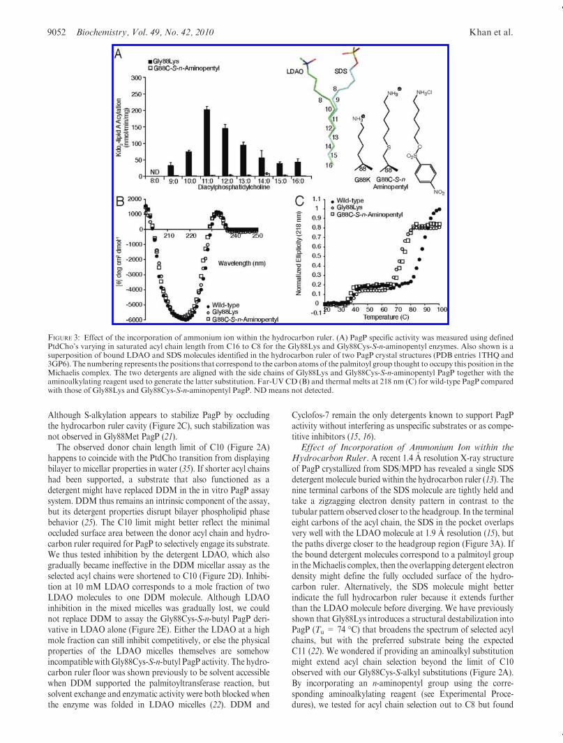
Hydrocarbon Ruler. A recent 1.4 A resolution X-ray structureof PagP crystallized from SDS/MPD has revealed a single SDSdetergent molecule buried within the hydrocarbon ruler (13). Thenine terminal carbons of the SDS molecule are tightly held andtake a zigzagging electron density pattern in contrast to thetubular pattern observed closer to the headgroup. In the terminaleight carbons of the acyl chain, the SDS in the pocket overlapsvery well with the LDAO molecule at 1.9 A resolution (15), butthe paths diverge closer to the headgroup region (Figure 3A). Ifthe bound detergent molecules correspond to a palmitoyl groupin theMichaelis complex, then the overlapping detergent electrondensity might define the fully occluded surface of the hydro-carbon ruler. Alternatively, the SDS molecule might betterindicate the full hydrocarbon ruler because it extends furtherthan the LDAO molecule before diverging. We have previouslyshown that Gly88Lys introduces a structural destabilization intoPagP (Tu = 74 �C) that broadens the spectrum of selected acylchains, but with the preferred substrate being the expectedC11 (22). We wondered if providing an aminoalkyl substitutionmight extend acyl chain selection beyond the limit of C10observed with our Gly88Cys-S-alkyl substitutions (Figure 2A).By incorporating an n-aminopentyl group using the corre-sponding aminoalkylating reagent (see Experimental Proce-dures), we tested for acyl chain selection out to C8 but found
FIGURE 3: Effect of the incorporation of ammonium ion within the hydrocarbon ruler. (A) PagP specific activity was measured using definedPtdCho’s varying in saturated acyl chain length from C16 to C8 for the Gly88Lys and Gly88Cys-S-n-aminopentyl enzymes. Also shown is asuperposition of bound LDAO and SDS molecules identified in the hydrocarbon ruler of two PagP crystal structures (PDB entries 1THQ and3GP6). The numbering represents the positions that correspond to the carbon atoms of the palmitoyl group thought to occupy this position in theMichaelis complex. The two detergents are aligned with the side chains of Gly88Lys and Gly88Cys-S-n-aminopentyl PagP together with theaminoalkylating reagent used to generate the latter substitution. Far-UV CD (B) and thermal melts at 218 nm (C) for wild-type PagP comparedwith those of Gly88Lys and Gly88Cys-S-n-aminopentyl PagP. ND means not detected.

Article Biochemistry, Vol. 49, No. 42, 2010 9053
theGly88Cys-S-n-aminopentyl PagP to be enzymatically inactiveby comparison withGly88Lys PagP (Figure 3A). The incorpora-tion of ammonium groups is most easily discerned by compar-isons between two different pairs of nearly isosteric partners:Gly88Nle with Gly88Lys and Gly88Cys-S-n-hexyl withGly88Cys-S-n-aminopentyl. Far-UV CD (Figure 3B) showed nomajor structural perturbation, while thermal melts (Figure 3C)showed that both NH3þ-containing partners were destabilized bythe same amount relative to their CH3-terminated counterparts.Unlike the significant differences observed between S-ethylcysteineandMet (see below), these data showno dependence on the locationof the ammonium ion in the folded state. This suggests that the largedesolvation energy penalties that must be paid to bury an ammo-nium ion in the core of a protein are the dominant effects for this setof compounds.Role of Side Chain Flexibility and Localized Thioether-
Aromatic Dispersion Attraction in Engineering a Dedi-catedPagPLauroyltransferase.TheC10 acyl chain transition
most likely specifies diminishing binding forces between van derWaals-contacted groups localized within a hydrophobic micro-environment. In the absence of water or electrostatic perturba-tions, London dispersion forces appear to be a dominant sourceof intermolecular attraction, especially when nonpolar surfacecontacts are maximized without steric repulsion (36). Localizeddispersive attractions should be paramount for the highlypolarizable thioether sulfur atom, which is more electron-densethan typical hydrocarbon moieties (37). If a thioether-containingside chain fits snugly within the hydrocarbon ruler, van derWaals-contacted dispersive attractions with neighboring aro-matic side chains should be more pertinent compared to inter-actions with aliphatic or hydroxylated side chains that lackconjugated resonance-stabilized π-electron systems (38, 39).
Interestingly, Gly88Cys-S-ethyl PagP behaves as a dedicatedlauroyltransferase (Figure 2A), but acyl chain selection by itsregioisomer Gly88Met PagP is imperfect by comparison (21)(Figure 4A). The main observed differences are the partial
FIGURE 4: Role of side chain flexibility and localized thioether-aromatic dispersion attraction in engineering a dedicated PagP lauroyltransfer-ase. (A) PagP specific activitywasmeasured using defined PtdCho’s varying in saturated acyl chain length fromC16 toC10. The unnatural aminoacid Nle was substituted for Met by misaminoacylation of tRNAMet. Panels from top to bottom represent the activity profiles for Gly88Met,Gly88Met [Nle], and Gly88Cys-S-ethyl [Nle], respectively. For comparison, the behavior of Gly88Cys-S-ethyl PagP is shown in Figure 2. (B)Far-UVCDand (C) thermalmelts at 218 nmreveal that substituting the fivemethionines inwild-type andGly88CysPagP (six inGly88MetPagP)withNle does not compromise structure or stability. (D) Schematic representation of side chain conformational preferences forNle (left) andMet(right). Viewed in Newman projection down the central χ3 torsion axis, the penultimate atom is rotated to emphasize the enthalpic preference ofNle formaintaining the terminalmethyl group in the extendedanti conformation,whereasMet is slightly favored toadopt one of the twoavailablegauche conformers.
9054 Biochemistry, Vol. 49, No. 42, 2010 Khan et al.
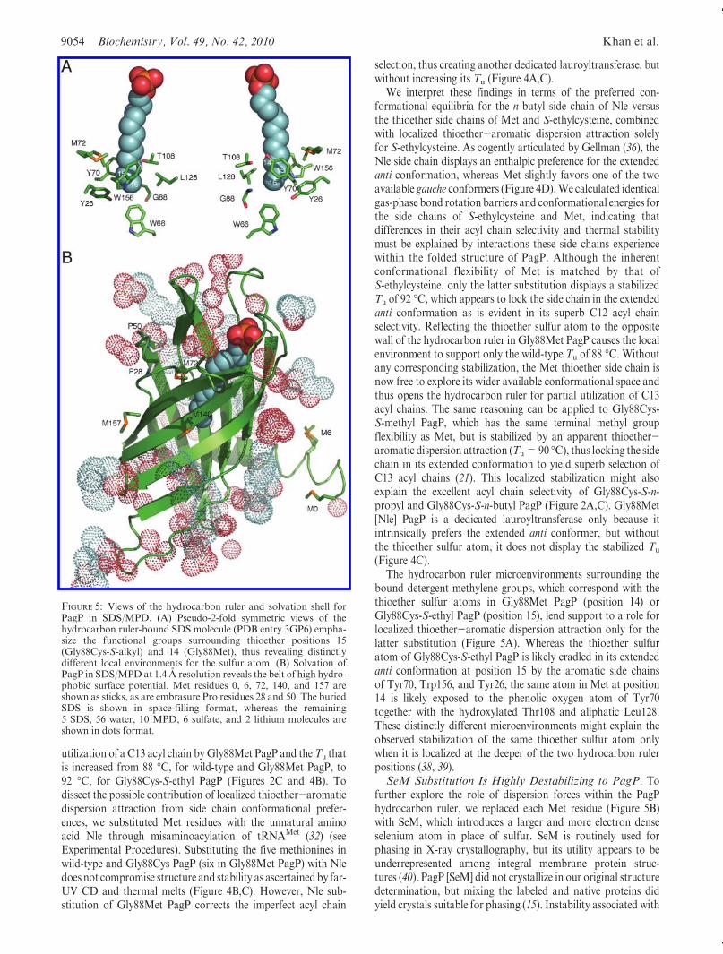
utilization of a C13 acyl chain byGly88Met PagP and theTu thatis increased from 88 �C, for wild-type and Gly88Met PagP, to92 �C, for Gly88Cys-S-ethyl PagP (Figures 2C and 4B). Todissect the possible contribution of localized thioether-aromaticdispersion attraction from side chain conformational prefer-ences, we substituted Met residues with the unnatural aminoacid Nle through misaminoacylation of tRNAMet (32) (seeExperimental Procedures). Substituting the five methionines inwild-type and Gly88Cys PagP (six in Gly88Met PagP) with Nledoes not compromise structure and stability as ascertainedby far-UV CD and thermal melts (Figure 4B,C). However, Nle sub-stitution of Gly88Met PagP corrects the imperfect acyl chain
selection, thus creating another dedicated lauroyltransferase, butwithout increasing its Tu (Figure 4A,C).
We interpret these findings in terms of the preferred con-formational equilibria for the n-butyl side chain of Nle versusthe thioether side chains of Met and S-ethylcysteine, combinedwith localized thioether-aromatic dispersion attraction solelyfor S-ethylcysteine. As cogently articulated by Gellman (36), theNle side chain displays an enthalpic preference for the extendedanti conformation, whereas Met slightly favors one of the twoavailable gauche conformers (Figure 4D).We calculated identicalgas-phase bond rotation barriers and conformational energies forthe side chains of S-ethylcysteine and Met, indicating thatdifferences in their acyl chain selectivity and thermal stabilitymust be explained by interactions these side chains experiencewithin the folded structure of PagP. Although the inherentconformational flexibility of Met is matched by that ofS-ethylcysteine, only the latter substitution displays a stabilizedTu of 92 �C, which appears to lock the side chain in the extendedanti conformation as is evident in its superb C12 acyl chainselectivity. Reflecting the thioether sulfur atom to the oppositewall of the hydrocarbon ruler in Gly88Met PagP causes the localenvironment to support only the wild-type Tu of 88 �C. Withoutany corresponding stabilization, the Met thioether side chain isnow free to explore its wider available conformational space andthus opens the hydrocarbon ruler for partial utilization of C13acyl chains. The same reasoning can be applied to Gly88Cys-S-methyl PagP, which has the same terminal methyl groupflexibility as Met, but is stabilized by an apparent thioether-aromatic dispersion attraction (Tu=90 �C), thus locking the sidechain in its extended conformation to yield superb selection ofC13 acyl chains (21). This localized stabilization might alsoexplain the excellent acyl chain selectivity of Gly88Cys-S-n-propyl and Gly88Cys-S-n-butyl PagP (Figure 2A,C). Gly88Met[Nle] PagP is a dedicated lauroyltransferase only because itintrinsically prefers the extended anti conformer, but withoutthe thioether sulfur atom, it does not display the stabilized Tu
(Figure 4C).The hydrocarbon ruler microenvironments surrounding the
bound detergent methylene groups, which correspond with thethioether sulfur atoms in Gly88Met PagP (position 14) orGly88Cys-S-ethyl PagP (position 15), lend support to a role forlocalized thioether-aromatic dispersion attraction only for thelatter substitution (Figure 5A). Whereas the thioether sulfuratom of Gly88Cys-S-ethyl PagP is likely cradled in its extendedanti conformation at position 15 by the aromatic side chainsof Tyr70, Trp156, and Tyr26, the same atom in Met at position14 is likely exposed to the phenolic oxygen atom of Tyr70together with the hydroxylated Thr108 and aliphatic Leu128.These distinctly different microenvironments might explain theobserved stabilization of the same thioether sulfur atom onlywhen it is localized at the deeper of the two hydrocarbon rulerpositions (38, 39).SeM Substitution Is Highly Destabilizing to PagP. To
further explore the role of dispersion forces within the PagPhydrocarbon ruler, we replaced each Met residue (Figure 5B)with SeM, which introduces a larger and more electron denseselenium atom in place of sulfur. SeM is routinely used forphasing in X-ray crystallography, but its utility appears to beunderrepresented among integral membrane protein struc-tures (40). PagP [SeM] did not crystallize in our original structuredetermination, but mixing the labeled and native proteins didyield crystals suitable for phasing (15). Instability associated with
FIGURE 5: Views of the hydrocarbon ruler and solvation shell forPagP in SDS/MPD. (A) Pseudo-2-fold symmetric views of thehydrocarbon ruler-bound SDS molecule (PDB entry 3GP6) empha-size the functional groups surrounding thioether positions 15(Gly88Cys-S-alkyl) and 14 (Gly88Met), thus revealing distinctlydifferent local environments for the sulfur atom. (B) Solvation ofPagP in SDS/MPD at 1.4 A resolution reveals the belt of high hydro-phobic surface potential. Met residues 0, 6, 72, 140, and 157 areshown as sticks, as are embrasure Pro residues 28 and 50. The buriedSDS is shown in space-filling format, whereas the remaining5 SDS, 56 water, 10 MPD, 6 sulfate, and 2 lithium molecules areshown in dots format.

Article Biochemistry, Vol. 49, No. 42, 2010 9055
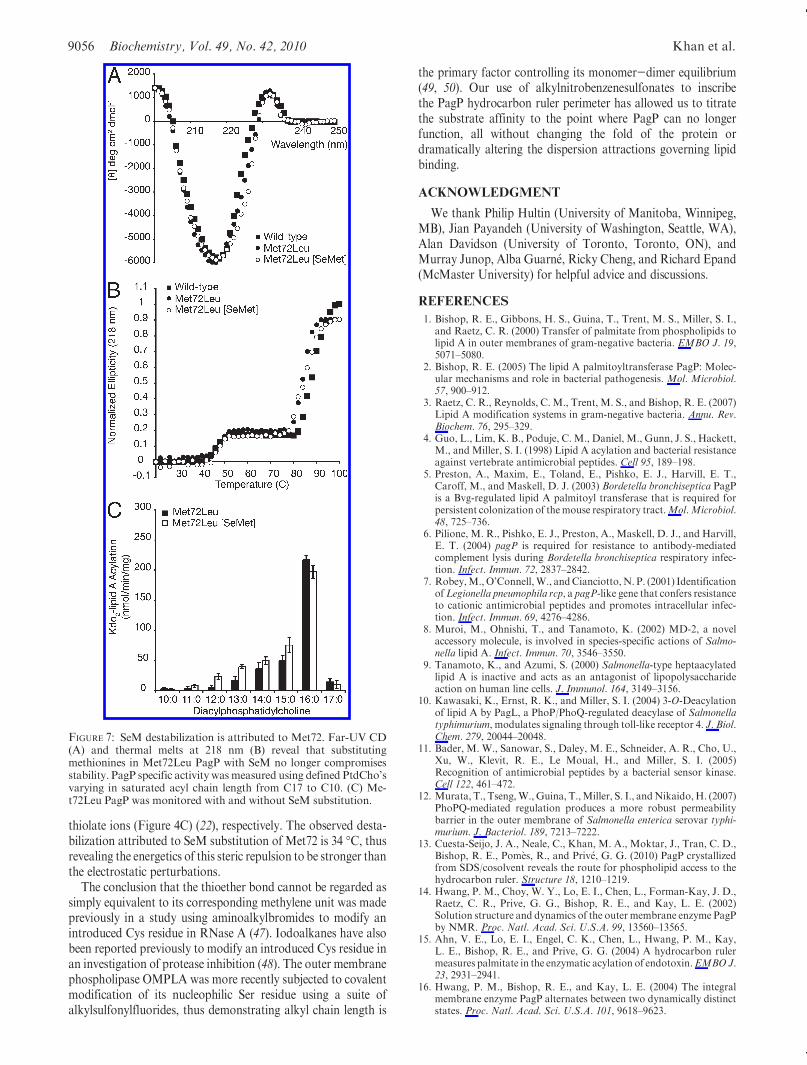
SeM labeling of the β-barrel membrane protein FadL has alsobeen reported (41). SeM-labeled wild-type and Gly88Met PagPwere prepared in the presence of βME, and no oxidation of SeMto SeM selenoxide was evident by ESI-MS (Table 1). Althoughthe exciton and β-barrel structure of PagP are evident in far-UVCD, we were surprised by the extent of the destabilizationassociated with SeM substitution in the corresponding thermalmelts (Figure 6A,B). The greatest instability previously asso-ciated with an active PagP mutant is that of Gly88Cys PagP atpH 8, which loses its exciton and has its Tu reduced by 22 �Cbecause of a buried thiolate anion (21) (see also Figure 4B,C). Bycomparison, SeM-substituted wild-type and Gly88Met PagPeach display the exciton, but their Tu values are reduced by34 �C (Figure 6B). PagP with Nle and PagP without Nle werevirtually indistinguishable in terms of stability (Figure 4C), andwild-type acyl chain selection (Figure 6C) was not distinctlydifferent for the Nle-substituted protein (not shown). However,SeM substitution of the five intrinsic Met residues drasticallybroadened acyl chain selection and reduced the specific activity(Figure 6C). The introduction of a sixth SeM residue at position88 did not further destabilize the protein (Figure 6B), but theresidual enzymatic activity was extinguished (Figure 6D). Weconclude that substituting Met88 with SeM compromises lipidacyl chain selection without any apparent PagP destabilization.
Of the five Met residues in wild-type PagP, only Met72 isburied and lines the hydrocarbon ruler wall (Figures 1B and 5).Two conformers of this thioether side chain are apparent in theSDS/MPD structure (13), and the corresponding selenide mightbe envisaged to influence lipid acyl chain selection from thisposition through disruption of neighboring van der Waalspacking interactions (Figure 6C). We purified Met72Leu PagP,both with and without SeM substitution (Table 1), and foundMet72 to be entirely responsible for the perturbation observed in
the wild-type enzyme upon SeM substitution (Figure 7). Nodestabilization was associated with SeM substitution of the foursurface-exposed SeM residues.Met6 and the non-nativeMet0 areeach located in the N-terminal helix, whereas the C-terminalMet140 and Met157 are both exposed on the β-barrel surface(Figure 5B). These four surface residues all map onto the belt ofhigh hydrophobic surface potential marking the transmembranedomain in the 1.4 A resolution SDS/MPDstructure (13, 42). SeMresidues buried within the hydrophobic core or exposed tosolvent on the surface of soluble globular proteins are usuallyreasonably well tolerated (32, 40, 43). Our findings point to astrongly destabilizing influence of SeM at position 72 in PagP.The Lennard-Jones potential describes a narrow optimal distancerange between neutral molecules for the ideal interaction energyto balance weak van der Waals attraction with stronger stericrepulsion (44). We thus suspect that the larger van der Waalsradius and/or bond lengths associated with the selenide com-pared to the sulfide at residue 72 are somehow incompatiblewith local packing arrangements necessary for optimal PagPstabilization (45).Concluding Remarks. The thioether-aromatic dispersion
attraction described here can also be regarded specifically as amethionine-π attraction (38), or more generally as a sulfide-π attraction (39). In an aqueous environment, the energetic stabi-lization of the methionine-π attraction is on the order of 0.3-0.5 kcal/mol (38). Similarly, conformational energies at equilib-rium in the gas phase indicate that theNle side chain enthalpicallyfavors the anti conformer (Figure 4D) by 0.8 kcal/mol (36, 46). Inour LDAO detergent system, these energetics translate into a Tu
increase of 2-4 �C for the S-alkylated Gly88Cys PagP deriva-tives. Coulombic repulsions are typically stronger than Londondispersion attractions (44), hence the 14 and 22 �Cdecreases inTu
observed with the buried ammonium (Figure 3C) (22) and
FIGURE 6: SeM substitution is highly destabilizing to PagP. Far-UVCD (A) and thermalmelts at 218 nm (B) reveal that substitutingmethioninesin wild-type and Gly88Met PagP with SeM compromises stability. PagP specific activity was measured using defined PtdCho’s varying insaturated acyl chain length from C17 to C10. Wild-type (C) and Gly88Met (D) PagP were monitored with and without SeM substitution. NDmeans not detected.
9056 Biochemistry, Vol. 49, No. 42, 2010 Khan et al.
thiolate ions (Figure 4C) (22), respectively. The observed desta-bilization attributed to SeM substitution of Met72 is 34 �C, thusrevealing the energetics of this steric repulsion to be stronger thanthe electrostatic perturbations.
The conclusion that the thioether bond cannot be regarded assimply equivalent to its corresponding methylene unit was madepreviously in a study using aminoalkylbromides to modify anintroduced Cys residue in RNase A (47). Iodoalkanes have alsobeen reported previously to modify an introduced Cys residue inan investigation of protease inhibition (48). The outer membranephospholipase OMPLA was more recently subjected to covalentmodification of its nucleophilic Ser residue using a suite ofalkylsulfonylfluorides, thus demonstrating alkyl chain length is
the primary factor controlling its monomer-dimer equilibrium(49, 50). Our use of alkylnitrobenzenesulfonates to inscribethe PagP hydrocarbon ruler perimeter has allowed us to titratethe substrate affinity to the point where PagP can no longerfunction, all without changing the fold of the protein ordramatically altering the dispersion attractions governing lipidbinding.
ACKNOWLEDGMENT
We thank Philip Hultin (University of Manitoba, Winnipeg,MB), Jian Payandeh (University of Washington, Seattle, WA),Alan Davidson (University of Toronto, Toronto, ON), andMurray Junop, Alba Guarn�e, Ricky Cheng, and Richard Epand(McMaster University) for helpful advice and discussions.
REFERENCES
1. Bishop, R. E., Gibbons, H. S., Guina, T., Trent, M. S., Miller, S. I.,and Raetz, C. R. (2000) Transfer of palmitate from phospholipids tolipid A in outer membranes of gram-negative bacteria. EMBO J. 19,5071–5080.
2. Bishop, R. E. (2005) The lipid A palmitoyltransferase PagP: Molec-ular mechanisms and role in bacterial pathogenesis. Mol. Microbiol.57, 900–912.
3. Raetz, C. R., Reynolds, C. M., Trent, M. S., and Bishop, R. E. (2007)Lipid A modification systems in gram-negative bacteria. Annu. Rev.Biochem. 76, 295–329.
4. Guo, L., Lim, K. B., Poduje, C. M., Daniel, M., Gunn, J. S., Hackett,M., and Miller, S. I. (1998) Lipid A acylation and bacterial resistanceagainst vertebrate antimicrobial peptides. Cell 95, 189–198.
5. Preston, A., Maxim, E., Toland, E., Pishko, E. J., Harvill, E. T.,Caroff, M., and Maskell, D. J. (2003) Bordetella bronchiseptica PagPis a Bvg-regulated lipid A palmitoyl transferase that is required forpersistent colonization of themouse respiratory tract.Mol.Microbiol.48, 725–736.
6. Pilione, M. R., Pishko, E. J., Preston, A., Maskell, D. J., and Harvill,E. T. (2004) pagP is required for resistance to antibody-mediatedcomplement lysis during Bordetella bronchiseptica respiratory infec-tion. Infect. Immun. 72, 2837–2842.
7. Robey,M.,O’Connell,W., andCianciotto,N. P. (2001) IdentificationofLegionella pneumophila rcp, a pagP-like gene that confers resistanceto cationic antimicrobial peptides and promotes intracellular infec-tion. Infect. Immun. 69, 4276–4286.
8. Muroi, M., Ohnishi, T., and Tanamoto, K. (2002) MD-2, a novelaccessory molecule, is involved in species-specific actions of Salmo-nella lipid A. Infect. Immun. 70, 3546–3550.
9. Tanamoto, K., and Azumi, S. (2000) Salmonella-type heptaacylatedlipid A is inactive and acts as an antagonist of lipopolysaccharideaction on human line cells. J. Immunol. 164, 3149–3156.
10. Kawasaki, K., Ernst, R. K., and Miller, S. I. (2004) 3-O-Deacylationof lipid A by PagL, a PhoP/PhoQ-regulated deacylase of Salmonellatyphimurium, modulates signaling through toll-like receptor 4. J. Biol.Chem. 279, 20044–20048.
11. Bader, M. W., Sanowar, S., Daley, M. E., Schneider, A. R., Cho, U.,Xu, W., Klevit, R. E., Le Moual, H., and Miller, S. I. (2005)Recognition of antimicrobial peptides by a bacterial sensor kinase.Cell 122, 461–472.
12. Murata, T., Tseng,W.,Guina, T.,Miller, S. I., andNikaido,H. (2007)PhoPQ-mediated regulation produces a more robust permeabilitybarrier in the outer membrane of Salmonella enterica serovar typhi-murium. J. Bacteriol. 189, 7213–7222.
13. Cuesta-Seijo, J. A., Neale, C., Khan, M. A., Moktar, J., Tran, C. D.,Bishop, R. E., Pom�es, R., and Priv�e, G. G. (2010) PagP crystallizedfrom SDS/cosolvent reveals the route for phospholipid access to thehydrocarbon ruler. Structure 18, 1210–1219.
14. Hwang, P. M., Choy, W. Y., Lo, E. I., Chen, L., Forman-Kay, J. D.,Raetz, C. R., Prive, G. G., Bishop, R. E., and Kay, L. E. (2002)Solution structure and dynamics of the outer membrane enzyme PagPby NMR. Proc. Natl. Acad. Sci. U.S.A. 99, 13560–13565.
15. Ahn, V. E., Lo, E. I., Engel, C. K., Chen, L., Hwang, P. M., Kay,L. E., Bishop, R. E., and Prive, G. G. (2004) A hydrocarbon rulermeasures palmitate in the enzymatic acylation of endotoxin.EMBOJ.23, 2931–2941.
16. Hwang, P. M., Bishop, R. E., and Kay, L. E. (2004) The integralmembrane enzyme PagP alternates between two dynamically distinctstates. Proc. Natl. Acad. Sci. U.S.A. 101, 9618–9623.
FIGURE 7: SeM destabilization is attributed to Met72. Far-UV CD(A) and thermal melts at 218 nm (B) reveal that substitutingmethionines in Met72Leu PagP with SeM no longer compromisesstability. PagP specific activity wasmeasured using defined PtdCho’svarying in saturated acyl chain length from C17 to C10. (C) Me-t72Leu PagP was monitored with and without SeM substitution.

Article Biochemistry, Vol. 49, No. 42, 2010 9057
17. Bishop, R. E. (2008) Structural biology of membrane-intrinsicβ-barrel enzymes: Sentinels of the bacterial outer membrane.Biochim.Biophys. Acta 1778, 1881–1896.
18. Jia, W., Zoeiby, A. E., Petruzziello, T. N., Jayabalasingham, B.,Seyedirashti, S., and Bishop, R. E. (2004) Lipid trafficking controlsendotoxin acylation in outer membranes of Escherichia coli. J. Biol.Chem. 279, 44966–44975.
19. Smith, A. E., Kim, S. H., Liu, F., Jia, W., Vinogradov, E., Gyles,C. L., andBishop,R. E. (2008) PagP activation in the outermembranetriggers R3 core oligosaccharide truncation in the cytoplasm ofEscherichia coli O157:H7. J. Biol. Chem. 283, 4332–4343.
20. Khan, M. A., and Bishop, R. E. (2009) Molecular mechanism forlateral lipid diffusion between the outermembrane external leaflet anda β-barrel hydrocarbon ruler. Biochemistry 48, 9745–9756.
21. Khan, M. A., Neale, C., Michaux, C., Pomes, R., Prive, G. G.,Woody, R. W., and Bishop, R. E. (2007) Gauging a hydrocarbonruler by an intrinsic exciton probe. Biochemistry 46, 4565–4579.
22. Khan, M. A., Moktar, J., Mott, P. J., and Bishop, R. E. (2010) Athiolate anion buried within the hydrocarbon ruler perturbs PagPlipid acyl chain selection. Biochemistry 49, 2368–2379.
23. van Meer, G. (2005) Cellular lipidomics. EMBO J. 24, 3159–3165.24. Forneris, F., and Mattevi, A. (2008) Enzymes without borders:
Mobilizing substrates, delivering products. Science 321, 213–216.25. Popot, J. L. (2010) Amphipols, nanodiscs, and fluorinated surfac-
tants: Three nonconventional approaches to studying membraneproteins in aqueous solutions. Annu. Rev. Biochem. 79, 737–775.
26. Sambrook, J., Fritsch, E. F., and Maniatis, T. (1989) Molecularcloning: A laboratory manual, 2nd ed., Cold Spring Harbor LaboratoryPress, Plainview, NY.
27. Edelhoch, H. (1967) Spectroscopic determination of tryptophan andtyrosine in proteins. Biochemistry 6, 1948–1954.
28. Smith, P. K., Krohn, R. I., Hermanson, G. T.,Mallia, A. K., Gartner,F. H., Provenzano, M. D., Fujimoto, E. K., Goeke, N. M., Olson,B. J., and Klenk, D. C. (1985) Measurement of protein usingbicinchoninic acid. Anal. Biochem. 150, 76–85.
29. DeLano, W. L. (2002) The PyMOL molecular graphics system,DeLano Scientific, San Carlos, CA.
30. Morgan, M. S., and Cretcher, L. H. (1948) A kinetic study ofalkylation by ethyl arylsulfonates. J. Am. Chem. Soc. 70, 375–378.
31. Heinrikson, R. L. (1971) The selective S-methylation of sulfhydrylgroups in proteins and peptides withmethyl-p-nitrobenzenesulfonate.J. Biol. Chem. 246, 4090–4096.
32. Budisa, N., Steipe, B., Demange, P., Eckerskorn, C., Kellermann, J.,and Huber, R. (1995) High-level biosynthetic substitution of methio-nine in proteins by its analogs 2-aminohexanoic acid, selenomethio-nine, telluromethionine and ethionine in Escherichia coli. Eur. J.Biochem. 230, 788–796.
33. Kanipes, M. I., Lin, S., Cotter, R. J., and Raetz, C. R. (2001) Ca2þ-induced phosphoethanolamine transfer to the outer 3-deoxy-D-manno-octulosonic acid moiety of Escherichia coli lipopolysaccharide.
A novel membrane enzyme dependent upon phosphatidylethanola-mine. J. Biol. Chem. 276, 1156–1163.
34. Bligh, E. G., and Dyer, W. J. (1959) A rapid method of total lipidextraction and purification. Can. J. Biochem. 37, 911–917.
35. Hauser, H. (2000) Short-chain phospholipids as detergents. Biochim.Biophys. Acta 1508, 164–181.
36. Gellman, S. H. (1991) On the role of methionine residues in thesequence-independent recognition of nonpolar protein surfaces. Bio-chemistry 30, 6633–6636.
37. Fersht, A. (1998) Structure andmechanism in protein science: A guideto enzyme catalysis and protein folding, 3rd ed., W. H. Freeman & Co.,New York.
38. Tatko, C. D., and Waters, M. L. (2004) Investigation of the nature ofthe methionine-π interaction in β-hairpin peptide model systems.Protein Sci. 13, 2515–2522.
39. Meyer, E. A., Castellano, R. K., andDiederich, F. (2003) Interactionswith aromatic rings in chemical and biological recognition. Angew.Chem., Int. Ed. 42, 1210–1250.
40. Morth, J. P., Sorensen, T. L., and Nissen, P. (2006) Membrane’sEleven: Heavy-atom derivatives of membrane-protein crystals. ActaCrystallogr. D62, 877–882.
41. van den Berg, B., Black, P. N., Clemons, W. M., Jr., and Rapoport,T. A. (2004) Crystal structure of the long-chain fatty acid transporterFadL. Science 304, 1506–1509.
42. Evanics, F., Hwang, P. M., Cheng, Y., Kay, L. E., and Prosser, R. S.(2006) Topology of an outer-membrane enzyme: Measuring oxygenand water contacts in solution NMR studies of PagP. J. Am. Chem.Soc. 128, 8256–8264.
43. Yamniuk, A. P., Ishida, H., Lippert, D., and Vogel, H. J. (2009)Thermodynamic effects of noncoded and coded methionine substitu-tions in calmodulin. Biophys. J. 96, 1495–1507.
44. Chang, R. (2000) Physical chemistry for the physical and biologicalsciences, University Science Books, Sausalito, CA.
45. Bondi, A. (1964) van der Waals volumes and radii. J. Phys. Chem. 68,441–452.
46. Allinger, N. L., Yuh, Y. H., and Lii, J. H. (1989) MolecularMechanics. TheMM3Force Field forHydrocarbons. 1. J. Am.Chem.Soc. 111, 8551–8566.
47. Messmore, J. M., Fuchs, D. N., and Raines, R. T. (1995) Ribonu-clease A: Revealing structure-function relationships with semisynth-esis. J. Am. Chem. Soc. 117, 8057–8060.
48. Hasan, Z., and Leatherbarrow, R. J. (1998) A study of the specificityof barley chymotrypsin inhibitor 2 by cysteine engineering of the P1residue. Biochim. Biophys. Acta 1384, 325–334.
49. Stanley, A. M., Chuawong, P., Hendrickson, T. L., and Fleming,K. G. (2006) Energetics of outer membrane phospholipase A(OMPLA) dimerization. J. Mol. Biol. 358, 120–131.
50. Stanley, A.M., Treubrodt, A.M., Chuawong, P., Hendrickson, T. L.,and Fleming, K. G. (2007) Lipid chain selectivity by outer membranephospholipase A. J. Mol. Biol. 366, 461–468.


![Inscribing Interaction: Middle Woodland Monumentality in the Appalachian Summit, 100 B.C. - A.D. 400 [Table of Contents]](https://static.fdokumen.com/doc/165x107/631838244de2f6960b03f7c9/inscribing-interaction-middle-woodland-monumentality-in-the-appalachian-summit.jpg)


![King Wladislaus II: Weak Ruler or Victim of Circumstances? [Unpublished, the written version of a lecture held in Prague, 2011]](https://static.fdokumen.com/doc/165x107/6315669385333559270d1c2b/king-wladislaus-ii-weak-ruler-or-victim-of-circumstances-unpublished-the-written.jpg)