Inhibition of bacterial adhesion on PVC endotracheal tubes by RF-oxygen glow discharge, sodium...
13
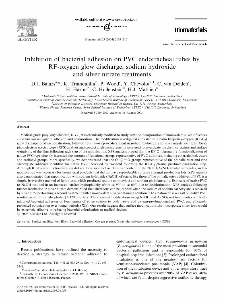
Biomaterials 25 (2004) 2139–2151 Inhibition of bacterial adhesion on PVC endotracheal tubes by RF-oxygen glow discharge, sodium hydroxide and silver nitrate treatments D.J. Balazs a, *, K. Triandafillu b , P. Wood c , Y. Chevolot a,1 , C. van Delden c , H. Harms b , C. Hollenstein d , H.J. Mathieu a a Materials Science Institute, Swiss Federal Institute of Technology (EPFL), CH-1015 Lausanne, Switzerland b Institute of Environmental Science and Technology, Swiss Federal Institute of Technology (EPFL), CH-1015 Lausanne, Switzerland c Division of Infectious Diseases, University Hospital of Geneva, CH-1211 Geneva, Switzerland d Plasma Physics Research Center, Swiss Federal Institute of Technology (EPFL), CH-1015 Lausanne, Switzerland Received 8 July 2003; accepted 15 August 2003 Abstract Medical-grade poly(vinyl chloride) (PVC) was chemically modified to study how the incorporation of monovalent silver influences Pseudomonas aeruginosa adhesion and colonization. The modification investigated consisted of a radio frequency-oxygen (RF-O 2 ) glow discharge pre-functionalization, followed by a two-step wet-treatment in sodium hydroxide and silver nitrate solutions. X-ray photoelectron spectroscopy (XPS) analysis and contact angle measurements were used to investigate the chemical nature and surface wettability of the films following each step of the modification. XPS analysis proved that the RF-O 2 plasma pre-functionalization of native PVC reproducibly increased the amount of functional groups representative of PVC additives, including ether/alcohol, esters and carboxyl groups. More specifically, we demonstrated that the O2 % CQO groups representative of the phthalic ester and zinc carboxylate additives identified for native PVC increased by two-fold following the RF-O 2 plasma pre-functionalization step. Although RF-O 2 pre-functionalization did not have an effect on the silver content of the NaOH/AgNO 3 treated substrates, such a modification was necessary for biomaterial products that did not have reproducible surfaces amongst production lots. XPS analysis also demonstrated that saponification with sodium hydroxide (NaOH) of esters, like those of the phthalic ester additives of PVC is a simple, irreversible method of hydrolysis, which produced sodium carboxylate and sodium phthalate salts. Exposure of native PVC to NaOH resulted in an increased surface hydrophilicity (from ca 90 to ca 60 ) due to dechlorination. XPS analysis following further incubation in silver nitrate demonstrated that silver ions can be trapped when the sodium of sodium carboxylate is replaced by silver after performing a second treatment with a monovalent silver-containing solution. The creation of silver salt on native PVC resulted in an ultra-hydrophobic (>120 ) surface. The chemical modifications using NaOH and AgNO 3 wet treatments completely inhibited bacterial adhesion of four strains of P. aeruginosa to both native and oxygen-pre-functionalized PVC, and efficiently prevented colonization over longer periods (72 h). Our results suggest that surface modifications that incorporate silver ions would be extremely effective at reducing bacterial colonization to medical devices. r 2003 Elsevier Ltd. All rights reserved. Keywords: Surface modification; Silver; Bacterial adhesion; Oxygen plasma; X-ray photoelectron spectroscopy (XPS) 1. Introduction Recent publications have outlined the necessity to develop a strategy to reduce bacterial adhesion to endotracheal devices [1,2]. Pseudomonas aeruginosa (P. aeruginosa) is one of the most prevalent nosocomial bacterial pathogens and is responsible for 30% of hospital-acquired infections [3]. Prolonged endotracheal intubation is one of the greatest risk factors for ventilator-associated pneumonia (VAP) [4]. Coloniza- tion of the intubation device and upper respiratory tract by P. aeruginosa precedes over 90% of VAP cases, 40% of which are fatal, despite aggressive antibiotic therapy ARTICLE IN PRESS *Corresponding author. Tel.:+41-21-693-2948; fax: +41-21-693- 3946. E-mail address: dawn.balazs@epfl.ch (D.J. Balazs). 1 Presently at Laboratoires Go. emar, UMR 1931 CNRS/Labora- toires Go. emar, F-29660 Roscoff, France. 0142-9612/$ - see front matter r 2003 Elsevier Ltd. All rights reserved. doi:10.1016/j.biomaterials.2003.08.053
Transcript of Inhibition of bacterial adhesion on PVC endotracheal tubes by RF-oxygen glow discharge, sodium...
Biomaterials 25 (2004) 2139–2151
ARTICLE IN PRESS
*Correspondin
3946.
E-mail addres1Presently at
toires Go.emar, F
0142-9612/$ - see
doi:10.1016/j.bio
Inhibition of bacterial adhesion on PVC endotracheal tubes byRF-oxygen glow discharge, sodium hydroxide
and silver nitrate treatments
D.J. Balazsa,*, K. Triandafillub, P. Woodc, Y. Chevolota,1, C. van Deldenc,H. Harmsb, C. Hollensteind, H.J. Mathieua
aMaterials Science Institute, Swiss Federal Institute of Technology (EPFL), CH-1015 Lausanne, Switzerlandb Institute of Environmental Science and Technology, Swiss Federal Institute of Technology (EPFL), CH-1015 Lausanne, Switzerland
cDivision of Infectious Diseases, University Hospital of Geneva, CH-1211 Geneva, SwitzerlanddPlasma Physics Research Center, Swiss Federal Institute of Technology (EPFL), CH-1015 Lausanne, Switzerland
Received 8 July 2003; accepted 15 August 2003
Abstract
Medical-grade poly(vinyl chloride) (PVC) was chemically modified to study how the incorporation of monovalent silver influences
Pseudomonas aeruginosa adhesion and colonization. The modification investigated consisted of a radio frequency-oxygen (RF-O2)
glow discharge pre-functionalization, followed by a two-step wet-treatment in sodium hydroxide and silver nitrate solutions. X-ray
photoelectron spectroscopy (XPS) analysis and contact angle measurements were used to investigate the chemical nature and surface
wettability of the films following each step of the modification. XPS analysis proved that the RF-O2 plasma pre-functionalization of
native PVC reproducibly increased the amount of functional groups representative of PVC additives, including ether/alcohol, esters
and carboxyl groups. More specifically, we demonstrated that the O2%CQO groups representative of the phthalic ester and zinc
carboxylate additives identified for native PVC increased by two-fold following the RF-O2 plasma pre-functionalization step.
Although RF-O2 pre-functionalization did not have an effect on the silver content of the NaOH/AgNO3 treated substrates, such a
modification was necessary for biomaterial products that did not have reproducible surfaces amongst production lots. XPS analysis
also demonstrated that saponification with sodium hydroxide (NaOH) of esters, like those of the phthalic ester additives of PVC is a
simple, irreversible method of hydrolysis, which produced sodium carboxylate and sodium phthalate salts. Exposure of native PVC
to NaOH resulted in an increased surface hydrophilicity (from ca 90� to ca 60�) due to dechlorination. XPS analysis following
further incubation in silver nitrate demonstrated that silver ions can be trapped when the sodium of sodium carboxylate is replaced
by silver after performing a second treatment with a monovalent silver-containing solution. The creation of silver salt on native PVC
resulted in an ultra-hydrophobic (>120�) surface. The chemical modifications using NaOH and AgNO3 wet treatments completely
inhibited bacterial adhesion of four strains of P. aeruginosa to both native and oxygen-pre-functionalized PVC, and efficiently
prevented colonization over longer periods (72 h). Our results suggest that surface modifications that incorporate silver ions would
be extremely effective at reducing bacterial colonization to medical devices.
r 2003 Elsevier Ltd. All rights reserved.
Keywords: Surface modification; Silver; Bacterial adhesion; Oxygen plasma; X-ray photoelectron spectroscopy (XPS)
1. Introduction
Recent publications have outlined the necessity todevelop a strategy to reduce bacterial adhesion to
g author. Tel.:+41-21-693-2948; fax: +41-21-693-
s: [email protected] (D.J. Balazs).
Laboratoires Go.emar, UMR 1931 CNRS/Labora-
-29660 Roscoff, France.
front matter r 2003 Elsevier Ltd. All rights reserved.
materials.2003.08.053
endotracheal devices [1,2]. Pseudomonas aeruginosa
(P. aeruginosa) is one of the most prevalent nosocomialbacterial pathogens and is responsible for 30% ofhospital-acquired infections [3]. Prolonged endotrachealintubation is one of the greatest risk factors forventilator-associated pneumonia (VAP) [4]. Coloniza-tion of the intubation device and upper respiratory tractby P. aeruginosa precedes over 90% of VAP cases, 40%of which are fatal, despite aggressive antibiotic therapy
ARTICLE IN PRESS
COO-, Na+
COO-, Na+
OC8H17
OC8H17
O
O
+ Na+OH- 2C8H17OH+
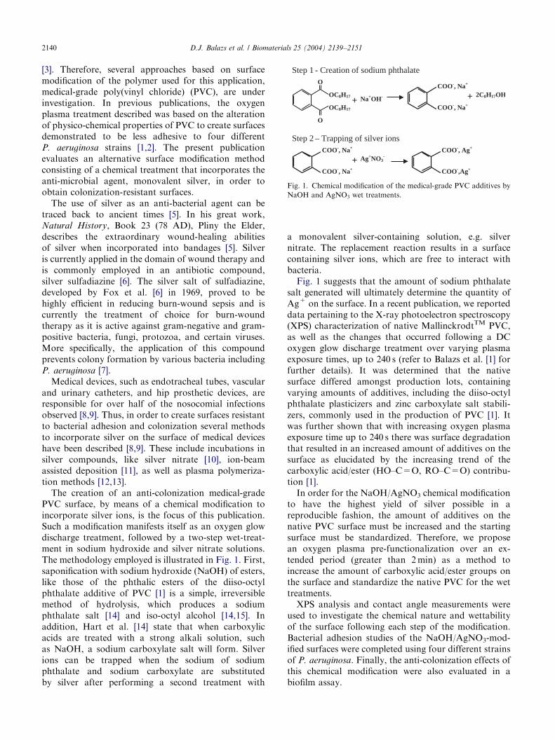
Step 1 - Creation of sodium phthalate
COO-, Na+
COO-, Na+
+ Ag+NO3-
COO-, Ag+
COO-,Ag+
Step 2 – Trapping of silver ions
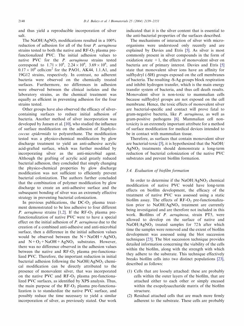
Fig. 1. Chemical modification of the medical-grade PVC additives by
NaOH and AgNO3 wet treatments.
D.J. Balazs et al. / Biomaterials 25 (2004) 2139–21512140
[3]. Therefore, several approaches based on surfacemodification of the polymer used for this application,medical-grade poly(vinyl chloride) (PVC), are underinvestigation. In previous publications, the oxygenplasma treatment described was based on the alterationof physico-chemical properties of PVC to create surfacesdemonstrated to be less adhesive to four differentP. aeruginosa strains [1,2]. The present publicationevaluates an alternative surface modification methodconsisting of a chemical treatment that incorporates theanti-microbial agent, monovalent silver, in order toobtain colonization-resistant surfaces.
The use of silver as an anti-bacterial agent can betraced back to ancient times [5]. In his great work,Natural History, Book 23 (78 AD), Pliny the Elder,describes the extraordinary wound-healing abilitiesof silver when incorporated into bandages [5]. Silveris currently applied in the domain of wound therapy andis commonly employed in an antibiotic compound,silver sulfadiazine [6]. The silver salt of sulfadiazine,developed by Fox et al. [6] in 1969, proved to behighly efficient in reducing burn-wound sepsis and iscurrently the treatment of choice for burn-woundtherapy as it is active against gram-negative and gram-positive bacteria, fungi, protozoa, and certain viruses.More specifically, the application of this compoundprevents colony formation by various bacteria includingP. aeruginosa [7].
Medical devices, such as endotracheal tubes, vascularand urinary catheters, and hip prosthetic devices, areresponsible for over half of the nosocomial infectionsobserved [8,9]. Thus, in order to create surfaces resistantto bacterial adhesion and colonization several methodsto incorporate silver on the surface of medical deviceshave been described [8,9]. These include incubations insilver compounds, like silver nitrate [10], ion-beamassisted deposition [11], as well as plasma polymeriza-tion methods [12,13].
The creation of an anti-colonization medical-gradePVC surface, by means of a chemical modification toincorporate silver ions, is the focus of this publication.Such a modification manifests itself as an oxygen glowdischarge treatment, followed by a two-step wet-treat-ment in sodium hydroxide and silver nitrate solutions.The methodology employed is illustrated in Fig. 1. First,saponification with sodium hydroxide (NaOH) of esters,like those of the phthalic esters of the diiso-octylphthalate additive of PVC [1] is a simple, irreversiblemethod of hydrolysis, which produces a sodiumphthalate salt [14] and iso-octyl alcohol [14,15]. Inaddition, Hart et al. [14] state that when carboxylicacids are treated with a strong alkali solution, suchas NaOH, a sodium carboxylate salt will form. Silverions can be trapped when the sodium of sodiumphthalate and sodium carboxylate are substitutedby silver after performing a second treatment with
a monovalent silver-containing solution, e.g. silvernitrate. The replacement reaction results in a surfacecontaining silver ions, which are free to interact withbacteria.
Fig. 1 suggests that the amount of sodium phthalatesalt generated will ultimately determine the quantity ofAg+ on the surface. In a recent publication, we reporteddata pertaining to the X-ray photoelectron spectroscopy(XPS) characterization of native MallinckrodtTM PVC,as well as the changes that occurred following a DCoxygen glow discharge treatment over varying plasmaexposure times, up to 240 s (refer to Balazs et al. [1] forfurther details). It was determined that the nativesurface differed amongst production lots, containingvarying amounts of additives, including the diiso-octylphthalate plasticizers and zinc carboxylate salt stabili-zers, commonly used in the production of PVC [1]. Itwas further shown that with increasing oxygen plasmaexposure time up to 240 s there was surface degradationthat resulted in an increased amount of additives on thesurface as elucidated by the increasing trend of thecarboxylic acid/ester (HO–C=O, RO–C=O) contribu-tion [1].
In order for the NaOH/AgNO3 chemical modificationto have the highest yield of silver possible in areproducible fashion, the amount of additives on thenative PVC surface must be increased and the startingsurface must be standardized. Therefore, we proposean oxygen plasma pre-functionalization over an ex-tended period (greater than 2min) as a method toincrease the amount of carboxylic acid/ester groups onthe surface and standardize the native PVC for the wettreatments.
XPS analysis and contact angle measurements wereused to investigate the chemical nature and wettabilityof the surface following each step of the modification.Bacterial adhesion studies of the NaOH/AgNO3-mod-ified surfaces were completed using four different strainsof P. aeruginosa. Finally, the anti-colonization effects ofthis chemical modification were also evaluated in abiofilm assay.
ARTICLE IN PRESSD.J. Balazs et al. / Biomaterials 25 (2004) 2139–2151 2141
2. Materials and methods
2.1. Substrate preparation
The substrate used for the investigation of the NaOH/AgNO3 modifications was PVC, obtained from Mal-linckrodt Hi-LoTM endotracheal tubes. The tubes wereflattened to allow microscopic counting of bacteriaas previously described [1,2]. Briefly, the PVC tubewas split and cut into pieces of 2� 1 cm2, the pieceswere then heated, below the melting point of PVC,between two clean, glass microscope slides for 90 s at180�C. All experiments were performed on flattenedMallinckrodtTM PVC substrates, referred to hereafter asnative PVC.
2.2. RF-oxygen plasma pre-functionalization
A custom-made plasma system, described elsewhere[16], was used for the radio frequency-oxygen (RF-O2)glow discharge pre-functionalization. Briefly, the systemis composed of a capacitively coupled RF reactorconsisting of two cylindrical electrodes (13 cm indiameter) separated by a distance of 4 cm, gas bottles,flow meters to control gas flow, a system of vacuumpumps, and an RF generator operating at 13.56MHzcombined with a matching network.
The flat PVC samples were placed on the sampleholder and mounted on the ground electrode. Thesystem was then evacuated to 5� 10�3mbar (1mbar=102 Pa). O2 was introduced into the chamber at a flowrate of 50 sccm; the chamber pressure was maintained ata pressure of 0.4mbar. The plasma was generated at aninput power of 60W for a time period of 10min. Afterplasma treatment the chamber was re-evacuated to5� 10�3mbar to remove any by-products of the plasmaprocessing. The samples were subsequently removed andthose reserved for NaOH/AgNO3 modification weredirectly placed in the NaOH solutions (describedbelow). Those samples reserved for analysis by thevarious surface characterization techniques were storedin ultrapure (18MO cm) deionized water before anyfurther use to prevent aging effects. Immediatelybefore analysis the samples were dried with purenitrogen flow.
2.3. NaOH/AgNO3 incubation technique
Sodium hydroxide and silver nitrate were purchasedfrom Sigma. First, the native and RF-oxygen-modifiedsubstrates were incubated for 14 days in a solution of 5mNaOH at room temperature. After incubation, thesamples were rinsed three times with deionized waterand analyzed by the various surface characterizationtechniques, or submitted to the second treatment.NaOH-treated substrates were then incubated for
14 days at room temperature in a 0.2m solution ofAgNO3, protected from light. After the second, 2-weekperiod expired the samples were rinsed with deionizedwater, subsequently analyzed by the various surfacecharacterization techniques, or submitted to bacterialadhesion and biofilm assays. Immediately before surfaceanalysis the samples were dried with pure nitrogenflow.
2.4. X-ray photoelectron spectroscopy analysis
XPS analysis was performed using an imaging KratosAxis Ultra (UK) X-ray photoelectron spectrometerequipped with a conventional hemispherical analyzer.The X-ray source employed was a monochromatized AlKa (1486.6 eV) operated at 150W, to avoid X-raydegradation. Spectral acquisition is performed underultra-high vacuum (UHV; 10�7 Pa) conditions. Analysiswas performed on a 0.21mm2 (300 mm� 700 mm) samplearea using a take-off angle of 90� relative to thesubstrate surface. The pass energies were 80 and 20 eVfor wide-scan and high-resolution elemental scans,respectively. These pass energies correspond to energyresolutions of 1.6 and 0.4 eV, respectively. Chargecompensation was performed with a self-compensatingdevice using field emitted low energy electrons (0.1 eV)to adjust the main hydrocarbon (C–C, C–H) componentto 285 eV. The data reduction (atomic concentration,shift, curve fitting, etc.) was performed with CasaXPSVersion 2.0.69 software. The operating software, Visionv2, corrects for the transmission function. The sensitiv-ity factors were 0.328, 0.891, 0.278, 5.987, 0.78, 2.957,and 1.685 for Si 2p, Cl 2p, O 1s, Ag 3d, O 1s, Fe 2p, andNa 1s, respectively. Spectra were fitted after linearbackground subtraction assuming a Gaussian–Lorent-zian (70/30) peak shape. Prior to XPS analysis, allsamples were outgassed overnight, under UHV condi-tions, to maintain an appropriate pressure in theanalysis chamber.
2.5. Contact angle measurements
Wettability of the surfaces was determined bymeasuring deionized water contact angle angles. Themeasurements were performed by a sessile drop methodusing a microscope equipped with a horizontal lightpath and a goniometric eyepiece (Kr .uss GmbH,Germany). All cited values are the average of threemeasurements per sample.
2.6. Bacterial adhesion and biofilm assays
Initial adhesion assays were performed using fourdifferent P. aeruginosa strains, including the wild-typestrain PAO1 [17] (obtained from the Laboratoryof Microbial Biology, University of Lausanne,
ARTICLE IN PRESSD.J. Balazs et al. / Biomaterials 25 (2004) 2139–21512142
Switzerland), the mutant AK44 [18], and two genotypi-cally different, pulmonary clinical isolates 1.1.A1 and19G12 obtained from colonized intubated patients(obtained from the University Hospital of Geneva).
The native PVC substrates modified by oxygenplasma pre-functionalization and the subsequentNaOH/AgNO3 treatment were evaluated for bacterialadhesion efficiency, using native PVC samples ascontrols. Batch adhesion experiments were carried outin glass vials containing a PVC patch and filled to thetop with degassed phosphate-buffered saline (PBS),according to an adapted method described by Rijnaartset al. [19]. After the bacterial suspension was added, thevials were placed on a slantwise rotating wheel to avoidsedimentation of bacteria and incubated at roomtemperature. After 2 h of incubation, the vials wereopened and rinsed with cell-free PBS. The PVC sampleswere carefully removed from the vials to preventdewetting as much as possible, placed on a microscopeslide and covered immediately with a cover slip. Theslides were observed under a light microscope (magni-fication � 1250; BX-60, Olympus Optical Co. Ltd.,Tokyo, Japan) equipped with a digital camera (SenSys,Photometrics Ltd., Tucson, USA). The number ofadherent cells was counted for six randomly chosenlocations per sample. Each experiment was done intriplicate. Levels of adhesion were given as numbers ofcells per square centimeter and calculated by averagingthe values of the 18 adhesion locations obtained for thethree samples.
2.7. Biofilm assays
The various substrates were also submitted to biofilmassays.
2.7.1. Cultivation of biofilms on PVC surfaces
Biofilms of P. aeruginosa were grown on the surface ofcontrol and modified PVC samples in a static system.Overnight cultures of P. aeruginosa grown in Luria–Bertani (LB) broth [20] were diluted to an opticaldensity (OD660) of 0.5 in sterile LB broth and furtherincubated for 6 h at 37�C in an orbital shaker until thecells had reached the early stationary phase. At thispoint, the samples were immersed in the culture andincubated statically for 1 h in order to allow initialadhesion to occur. Following initial adhesion, thesamples were rinsed in sterile saline (NaCl, 0.9% w/v)to remove any non-adherent cells and then transferredto the wells of six-well cell culture plates containing 9mlof sterile AP medium [21] (monosodium glutamate,100mm; sodium gluconate, 100mm; NaH2PO4, 7.5mm;K2HPO4, 16.3mm; MgSO4.7H2O, 10mm; NaCl, 300mm
at pH 7.0). The samples were incubated statically at37�C for 24, 48 or 72 h prior to removal for thequantification of the biofilm population [22].
2.7.2. Quantification of biofilms on the surface of native
PVC by serial blotting
We employed a quantitative method to assess theextent of biofilm formation on the surface of the nativeand modified PVC samples. The technique involvesserial blotting of colonized surfaces onto a succession ofpre-dried agar plates, as described by Eginton et al. [23].It provides a quantitative measurement, not only of thetotal biofilm population but also of the strength ofattachment of the sessile bacteria to the substratum onwhich the biofilm has developed.
Colonized native and treated PVC samples wereremoved from the cell culture plates following incuba-tion for 24, 48 or 72 h, rinsed in sterile saline to removeany non-biofilm population cells that may have depos-ited on the samples, then transferred to 2ml saline(suspension A) and vortexed vigorously for 30 s. Thesamples were then transferred to 2ml sterile saline(suspension B) and sonicated in a Branson 2200sonicator for 3min. Samples were transferred oncemore to 2ml sterile saline (suspension C) and vortexedvigorously for 30 s. Suspensions A, B and C werepooled, serially diluted and plated onto Luria agar (LA)for viable counting. The cells removed during these threephases represent the loosely attached biofilm popula-tion. Following the final vortex step, the samples wereblotted onto the surface of a pre-dried LA plate for1min. The process was repeated through a succession of25 pre-dried plates and colony counts were performed,following an overnight incubation at 37�C. The numberof colonies recovered per plate decreases exponentiallyaccording to the following relationship:
CFU ¼ A � 10�kN ; ð1Þ
where CFU is the number of colonies transferred to aplate; k is the removal exponent, expressed as the slopeof the best-fit line through the data; A is the ordinateaxis intercept; and N is the plate succession number. Inorder to quantify the residual attached population, thearea under the curve was calculated using the followingequation:
R ¼XN
1
A � 10�kN ; ð2Þ
where R is the proportion of the biofilm population thathas resisted removal by vortexing and sonication. Thus,according to the above equation, if Log(CFU)=0, thenthe following relation applies:
N ¼�LogðAÞ
k: ð3Þ
Once the value of N is known, then a simple iterativealgorithm can be used to compute the value of R:
ARTICLE IN PRESS
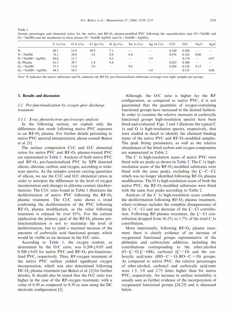
Table 1
Atomic percentages and elemental ratios for the native and RF-O2 plasma-modified PVC following the saponification step (N+NaOH and
O2+NaOH) and the incubation in silver nitrate (N+NaOH AgNO3 and O2+NaOH+AgNO3)
C 1s (%) O 1s (%) Cl 2p (%) Si 2p (%) Na 1s (%) Ag 3d (%) Cl/C O/C Na/C Ag/C
N 68.1 13.9 10.9 7.1 — — 0.160 0.204 — —
N+NaOH 76.1 10.8 5.8 0.9 6.4 — 0.076 0.142 0.08 —
N+NaOH+AgNO3 80.6 13.7 — 0.2 — 5.9 — 0.170 — 0.07
O2 Plasma 61.5 30.7 1.4 6.4 — — 0.023 0.500 — —
O2+NaOH 75.5 11.8 2.0 — 9.6 0.026 0.156 0.13 —
O2+NaOH+AgNO3 84.3 10.5 — — — 5.0 — 0.125 — 0.06
Note: N indicates the native substrates and O2 indicates the RF-O2 pre-functionalized substrates (average over eight samples per group).
D.J. Balazs et al. / Biomaterials 25 (2004) 2139–2151 2143
3. Results and discussion
3.1. Pre-functionalization by oxygen glow discharge
treatment
3.1.1. X-ray photoelectron spectroscopy analysis
In the following section, we explain only thedifferences that result following native PVC exposureto an RF-O2 plasma. For further details pertaining tonative PVC spectral interpretation, please consult Balazset al. [1].
The surface composition Cl/C and O/C elementalratios for native PVC and RF-O2 plasma-treated PVCare represented in Table 1. Analysis of both native PVCand RF-O2 pre-functionalized PVC by XPS detectedsilicon, chlorine, carbon, and oxygen, according to wide-scan spectra. As the samples contain varying quantitiesof silicon, we use the Cl/C and O/C elemental ratios inorder to interpret the differences in the level of oxygenincorporation and changes in chlorine content (dechlor-ination). The Cl/C ratio found in Table 1 illustrates thedechlorination of native PVC following the RF-O2
plasma treatment. The Cl/C ratio shows a trendconfirming the dechlorination of the PVC followingRF-O2 plasma modification, as the value followingtreatment is reduced by over 85%. For the currentapplication the primary goal of the RF-O2 plasma pre-functionalization is not to maximize the level ofdechlorination, but to yield a maximal increase of theamounts of carboxylic acid functional groups, whichwould be visible as an increase in the O/C ratio.
According to Table 1, the oxygen content, asdetermined by the O/C ratio, was 0.20470.05 and0.50070.03 for native PVC and RF-O2 pre-functiona-lized PVC, respectively. Thus, RF-oxygen treatment ofthe native PVC surface yielded significant oxygenincorporation, which was also determined followingDC-O2 plasma treatment (see Balazs et al. [1] for furtherdetails). It should also be stated that the O/C ratio washigher in the case of the RF-oxygen treatment, with avalue of 0.50 as compared to 0.39 as seen using the DCelectrode configuration [1].
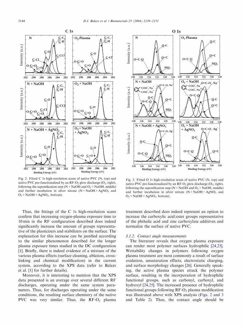
Although, the O/C ratio is higher for the RFconfiguration, as compared to native PVC, it is notguaranteed that the quantities of oxygen-containingfunctional groups have increased in the desired fashion.In order to examine the relative increases in carboxylicfunctional groups high-resolution spectra have beenfitted and evaluated. Figs. 2 and 3 illustrate the typical C1s and O 1s high-resolution spectra, respectively, thatwere studied in detail to identify the chemical bindingstates of the native PVC and RF-O2-modified samples.The peak fitting parameters, as well as the relativeabundances of the fitted carbon and oxygen componentsare summarized in Table 2.
The C 1s high-resolution scans of native PVC werefitted with six peaks as shown in Table 2. The C 1s high-resolution scans of the RF-O2-modified substrates werefitted with the same peaks, excluding the
%C2C2Cl;
which was no longer identified following RF-O2 plasmamodification. The O 1s high-resolution scans of both thenative PVC, the RF-O2-modified substrates were fittedwith the same four peaks according to Table 2.
Analysis of the C 1s high-resolution scans confirmsthe dechlorination following RF-O2 plasma treatment,where evidence includes the complete disappearance ofthe
%C2C2Cl and net decrease of the
%C2Cl contribu-
tion. Following RF-plasma treatment, the%C2Cl con-
tribution dropped from 16.2% to 1.7% of the total C 1scontribution.
More importantly, following RF-O2 plasma treat-ment there is clearly evidence of an increase ofoxygenated functional groups representative of thephthalate and carboxylate additives, including thecontributions corresponding to the ether/alcohol(O2
%C2O;
%C2OH), carbonyl (
%CQO) and the car-
boxylic acid/ester (HO2CQO;RO2CQO) groups.As compared to native PVC, the relative percentagesof ether/alcohol, carbonyl and carboxylic acid/esterwere 1.5, 5.0 and 2.75 times higher than for nativePVC, respectively. An increase in surface wettability isusually seen as further evidence of the incorporation ofoxygenated functional groups [24,25] and is discussedbelow.
ARTICLE IN PRESS
O 1sO2 Plasma
528530532534536538
O=C-O
O-C
O=C
SiO2
528530532534536538
O=C-OO-C
O=C
COO-, Na
+
COO-, Na
+
O2 + NaOH
528530532534536538
Binding Energy (eV)
O2 + NaOH+ AgNO3
O-C
O=CO=C-O
O=C-O-, Na+
O=C-O-, Ag+
O=C-O, Ag+
O=C-O, Na+
528530532534536538
O-C
O=CO=C-O
N
SiO2
528530532534536538
SiO2
O=C-OO-C
O=C
COO-, Na
+
COO-, Na
+
N + NaOH
528530532534536538Binding Energy (eV)
N + NaOH + AgNO3
SiO2
O-C O=C
O=C-OInte
nsity
(a.
u.)
Inte
nsity
(a.
u.)
Inte
nsity
(a.
u.)
Fig. 3. Fitted O 1s high-resolution scans of native PVC (N, top) and
native PVC pre-functionalized by an RF-O2 glow discharge (O2, right),
following the saponification step (N+NaOH and O2+NaOH, middle)
and further incubation in silver nitrate (N+NaOH+AgNO3 and
O2+NaOH+AgNO3, bottom).
C 1s
Inte
nsity
(a.
u.)
282284286 288 290 292
N C-C, C-H
C-O
C-Cl
C=O O-C=O
C-CCl
282284286 288 290 292
Inte
nsity
(a.
u.)
O-C=O C=O
C-Cl
C-O
C-CCl
C-C, C-H
N + NaOH
282284286 288 290 292 Binding Energy (eV)
Inte
nsity
(a.
u.)
O-C=O
C-O
C-C, C-H
N + NaOH + AgNO3
C=O
O - C =O C =O
C - O
C - C, C - H
O 2 + NaOH
282284 286 288290292
282284 286 288290292Binding Energy (eV)
O - C =O
C - O
C - C, C - H
O 2 + NaOH + AgNO 3
C =O
O 2 Plasma
O - C =O
C - C, C - H
C - O
C =O
C - Cl
282284 286 288290292
Fig. 2. Fitted C 1s high-resolution scans of native PVC (N, top) and
native PVC pre-functionalized by an RF-O2 glow discharge (O2, right),
following the saponification step (N+NaOH and O2+NaOH, middle)
and further incubation in silver nitrate (N+NaOH+AgNO3 and
O2+NaOH+AgNO3, bottom).
D.J. Balazs et al. / Biomaterials 25 (2004) 2139–21512144
Thus, the fittings of the C 1s high-resolution scansconfirm that increasing oxygen-plasma exposure time to10min in the RF configuration described does indeedsignificantly increase the amount of groups representa-tive of the plasticizers and stabilizers on the surface. Theexplanation for this increase can be justified accordingto the similar phenomenon described for the longerplasma exposure times studied in the DC configuration[1]. Briefly, there is indeed evidence of a mixture of thevarious plasma effects (surface cleaning, ablation, cross-linking and chemical modification) in the currentsystem, according to the XPS data (refer to Balazset al. [1] for further details).
Moreover, it is interesting to mention that the XPSdata presented is an average over several different RFdischarges, operating under the same system para-meters. Thus, for discharges operating under the sameconditions, the resulting surface chemistry of the nativePVC was very similar. Thus, the RF-O2 plasma
treatment described does indeed represent an option toincrease the carboxylic acid/ester groups representativeof the phthalic acid and zinc carboxylate additives andnormalize the surface of native PVC.
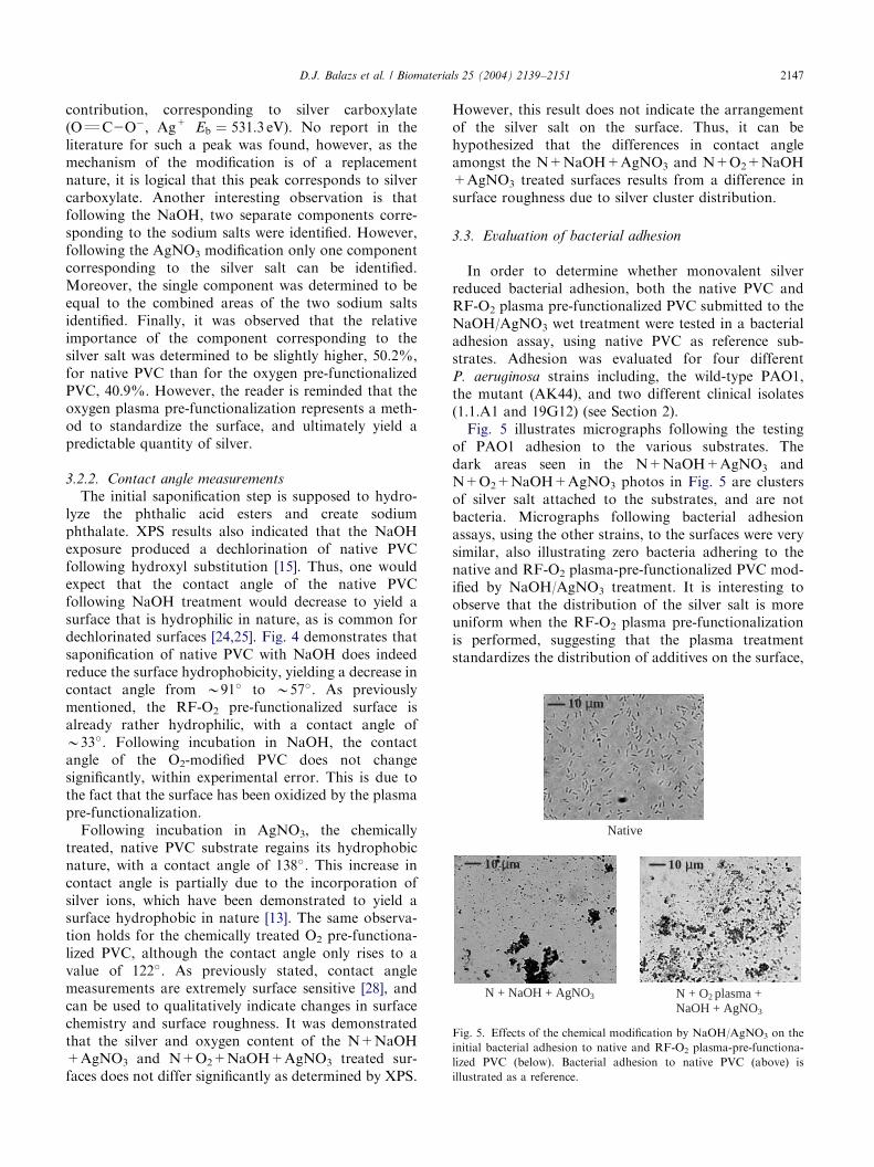
3.1.2. Contact angle measurements
The literature reveals that oxygen plasma exposurecan render most polymer surfaces hydrophilic [24,25].Wettability changes in polymers following oxygenplasma treatment are most commonly a result of surfaceoxidation, unsaturation effects, electrostatic charging,and surface morphology changes [26]. Generally speak-ing, the active plasma species attack the polymersurface, resulting in the incorporation of hydrophilicfunctional groups, such as carbonyl, carboxyl, andhydroxyl [24,25]. The increased presence of hydrophilicfunctional groups following RF-O2 plasma modificationwas illustrated above with XPS analysis (Figs. 2 and 3and Table 2). Thus, the contact angle should be
ARTICLE IN PRESSTable
2
PeakfittingparametersandpercentpeakareasfortheC
1sandO
1shigh-resolutionscansofthenativeandRF-O
2plasm
a-m
odified
PVC
followingthesaponificationstep
(N+
NaOH
and
O2+
NaOH)andtheincubationin
silver
nitrate
(N+
NaOH
AgNO
3andO
2+
NaOH+
AgNO
3)
Peak
Bindingenergy
(eV)
FWHM
(eV)
N %area
N+
NaOH
%area
N+
NaOH
+
+AgNO
3
%area
O2
%area
O2+
NaOH
%area
O2+
NaOH
+
+AgNO
3
%area
C1s %C2C=%C2H
285.0
1.1
51.4
81.3
81.0
66.6
88.7
84.4
%C2C2Cl
285.5
(70.05)
15.8
4.2
——
——
%C2O
286.4
13.2
5.6
13.2
20.6
5.5
11.2
%C2Cl
287.0
16.2
4.2
—1.7
1.2
—
%CQ
O288.0
0.8
1.4
1.6
3.9
1.1
0.8
O2%CQ
O289.2
2.6
3.3
4.1
7.2
3.5
3.7
O1s OQ
C2O
�;N
aþ
531.3
1.35
—31.0
——
34.9
—
OQ
C2O
�;A
gþ
531.3
(70.05)
——
50.2
——
40.9
Si2
%O2
531.8
22.2
4.0
1.1
9.9
——
%OQ
C532.0
3.9
12.3
10.7
20.4
7.3
6.8
%O2C
532.6
60.7
21.4
32.0
54.8
18.9
44.8
%O2CQ
O533.6
13.3
8.9
6.0
15.0
13.7
7.5
C6H
2COO
�,Na+
535.0
—22.5
——
25.2
—
0
20
40
60
80
100
120
140
160
N N +NaOH
N +NaOH
+ AgNO3
O2 O2 +
NaOH
O2 +
NaOH+ AgNO3
91˚
33˚
57˚
138˚
40˚
122˚
Con
tact
Ang
le (
˚)Fig. 4. Evolution of water contact angle following the incubation of
native and RF-O2-modified PVC in the NaOH and AgNO3 solutions,
respectively. N corresponds to the native PVC and O2 corresponds to
native PVC modified by an RF-O2 plasma pre-functionalization.
D.J. Balazs et al. / Biomaterials 25 (2004) 2139–2151 2145
significantly lower than the value of native PVC,following the RF-O2 pre-functionalization.
Fig. 4 illustrates the evolution of the wettability ofnative PVC following RF-O2 plasma treatment. Watercontact angle measurements of the surfaces indicatedthat the RF-O2 plasma pre-functionalization of nativePVC rendered the surface very hydrophilic, B33�, asexpected. This corresponds to a B60� change in contactangle from the value of native PVC.
3.2. Evaluation of the NaOH/AgNO3 treatment of native
PVC and RF-O2 Pre-functionalized surfaces
The amount of phthalic and carboxylic acids of thenative or RF-O2-modified substrates, represented by theO2
%CQO functionalities, will directly influence the
amount of sodium phthalate and sodium carboxylatesalts that are created following the NaOH treatment,and consequently the silver salt content following theAgNO3 incubation will be affected, as well. In the nextsections, we explore the individual effects that thesaponification step and AgNO3 incubation have on thenative and RF-O2-modified surfaces.
3.2.1. X-ray photoelectron spectroscopy analysis
The surface composition data for the native andRF-O2 plasma-modified PVC following the saponifica-tion step (N+NaOH and O2+NaOH) and the incuba-tion in silver nitrate (N+NaOH AgNO3 andO2+NaOH+AgNO3) are illustrated in Table 1. The
ARTICLE IN PRESSD.J. Balazs et al. / Biomaterials 25 (2004) 2139–21512146
interpretation of the data in this table will focus on thesaponification step first, followed by the silver nitratetreatment.
Following NaOH treatment, XPS wide-scan spectraof the N+NaOH detected signals corresponding tocarbon, oxygen, chlorine, silicon, and sodium, asexpected according to Fig. 1 and Hart et al. [14]. XPSwide-scan spectra of the O2+NaOH samples detectedthe same elements, with the exception of the siliconsignal. Thus, as the samples contain varying quantitiesof silicon, we refer to the Cl/C, Na/C and Ag/Celemental ratios in order to interpret the differences inthe level of dechlorination, and the incorporation ofsodium and silver, respectively. The sodium content issignificantly higher for the RF-O2 plasma pre-functio-nalized PVC treated with NaOH (0.13), as compared tothe native PVC samples treated with NaOH (0.08), asdemonstrated by the Na/C ratio found in Table 1.
Another interesting observation lies in the chlorinecontent, Cl/C ratio, of the N+NaOH and O2+NaOHsamples. Due to the dechlorination that occurs withoxygen plasma pre-functionalization, the chlorine con-tent of the O2+NaOH samples is much lower (0.026)than that of the N+NaOH samples (0.076). However, adechlorination of the native PVC following NaOHexposure did occur, as demonstrated by a drop in theCl/C ratio from 0.160 before the saponification step, to0.076 after NaOH incubation. The formation of HClthat results from the substitution of hydroxyl groups isthe cause of the dechlorination [15].
According to Fig. 1, the amount of sodium phthalatecreated following NaOH treatment will ultimatelydetermine the amount of monovalent silver incorpo-rated on the surface. More specifically, followingtreatment with AgNO3, the silver should simply replacethe sodium due to differences in electro-negativity,where silver has a value of 1.9 and sodium a value of0.9 [14]. Thus, according to XPS analysis, it is expectedthat the sodium signal should no longer be detected anda silver signal should appear according to wide scanspectra. This phenomenon was indeed observed. Fol-lowing further incubation of the N+NaOH substratesin the AgNO3 solution, the sodium disappears and silveris detected at an atomic percentage of 5.970.5%.
Furthermore, the quantities of Na and Ag created,according to the Na/C (0.08) and Ag/C (0.07) ratios,respectively, were very similar for the NaOH andAgNO3 treated native PVC samples. Similar findingswere observed for the O2+NaOH samples treated withAgNO3, as the Na 1s signal was not detected and Agwas identified at an atomic concentration of 5.070.5%.However, it is interesting to notice that the silvercontent, Ag/C ratio, of the O2+NaOH+AgNO3 didnot differ greatly from that of the N+NaOH AgNO3
substrates, having values of 0.07 and 0.06, respectively.Such a finding is indeed surprising as it was expected
that the silver content would be higher for the RF-O2
pre-functionalized substrates, as the sodium content wasover 1.5� greater than that of the N+NaOH sub-strates. It is interesting to notice that the silver contentfollowing the AgNO3 incubation of the RF-O2 pre-functionalized substrates is 50% lower than the sodiumcontent as illustrated by the Na/C and Ag/C ratios. Thismay signify that some of the silver salt is lost during therinsing procedure.
In order to further evaluate the changes following theNaOH and AgNO3 treatments, the C 1s and O 1s high-resolution scans were fitted and evaluated. Figs. 2 and 3,respectively, illustrate the typical C 1s and O 1s high-resolution spectra that were studied in detail to identifythe chemical binding states of the native and RF-O2
plasma-modified PVC, following the treatments byNaOH and AgNO3. The peak fitting parameters, aswell as the relative abundances of the fitted carbon andoxygen components are summarized in Table 2. Themain differences identified following the NaOH andAgNO3 treatment of the various surfaces was identifiedin the O 1s high-resolution scans. Therefore, theinterpretation will focus on the binding states identifiedfor these spectra.
Following incubation of both of the native and RF-O2-modified PVC in NaOH, Figs. 2 and 3 and Table 2illustrate that two new binding states were identified,corresponding to sodium carboxylate (O=C–O�, Na+
Eb ¼ 531:3 eV) and sodium phthalate (C6H2COO�, Na+
Eb ¼ 535:0 eV). The creation of these groups is notsurprising as Hart et al. [14] state that when carboxylicacids are treated with a strong base, such as 5m NaOH,sodium carboxylate is the result. Phthalic acid(C6H2(COOH)) has been previously identified by Barthet al. [27] at a binding energy of 534.5 eV. As thesaponification of esters, like those of the phthalic acidesters result in the creation of the salt of that acid,sodium phthalate, it is logical that a new component wasidentified very near to the binding energy of phthalicacid. For both the native and RF-O2-modified PVC, itwas determined that the sodium carboxylate contribu-tion was 40% higher than the sodium phthalatecontribution. Finally, it is interesting to notice thatboth the sodium carboxylate and sodium phthalatecontributions were present in greater quantities (>12%increase) following the RF-O2 pre-functionalization.This finding further emphasizes that the RF-O2 pre-functionalization increased the amount of O2
%CQO
additives on the native PVC surface available for thecreation of sodium phthalate and sodium carboxylatesalts by saponification methods.
Following the second incubation of both the nativeand RF-O2-modified PVC in AgNO3, Fig. 2. Fig. 3and Table 2 show that the sodium carboxylate andsodium phthalate are no longer identified in the O 1shigh-resolution scans. There is evidence of a new
ARTICLE IN PRESS
10 ∝ m
10 ∝ m
10 ∝ m
N + O2 plasma + NaOH + AgNO3
N + NaOH + AgNO3
10 µm 10 µm
10 µm
Native
Fig. 5. Effects of the chemical modification by NaOH/AgNO3 on the
initial bacterial adhesion to native and RF-O2 plasma-pre-functiona-
lized PVC (below). Bacterial adhesion to native PVC (above) is
illustrated as a reference.
D.J. Balazs et al. / Biomaterials 25 (2004) 2139–2151 2147
contribution, corresponding to silver carboxylate(OQC2O�; Ag+ Eb ¼ 531:3 eV). No report in theliterature for such a peak was found, however, as themechanism of the modification is of a replacementnature, it is logical that this peak corresponds to silvercarboxylate. Another interesting observation is thatfollowing the NaOH, two separate components corre-sponding to the sodium salts were identified. However,following the AgNO3 modification only one componentcorresponding to the silver salt can be identified.Moreover, the single component was determined to beequal to the combined areas of the two sodium saltsidentified. Finally, it was observed that the relativeimportance of the component corresponding to thesilver salt was determined to be slightly higher, 50.2%,for native PVC than for the oxygen pre-functionalizedPVC, 40.9%. However, the reader is reminded that theoxygen plasma pre-functionalization represents a meth-od to standardize the surface, and ultimately yield apredictable quantity of silver.
3.2.2. Contact angle measurements
The initial saponification step is supposed to hydro-lyze the phthalic acid esters and create sodiumphthalate. XPS results also indicated that the NaOHexposure produced a dechlorination of native PVCfollowing hydroxyl substitution [15]. Thus, one wouldexpect that the contact angle of the native PVCfollowing NaOH treatment would decrease to yield asurface that is hydrophilic in nature, as is common fordechlorinated surfaces [24,25]. Fig. 4 demonstrates thatsaponification of native PVC with NaOH does indeedreduce the surface hydrophobicity, yielding a decrease incontact angle from B91� to B57�. As previouslymentioned, the RF-O2 pre-functionalized surface isalready rather hydrophilic, with a contact angle ofB33�. Following incubation in NaOH, the contactangle of the O2-modified PVC does not changesignificantly, within experimental error. This is due tothe fact that the surface has been oxidized by the plasmapre-functionalization.
Following incubation in AgNO3, the chemicallytreated, native PVC substrate regains its hydrophobicnature, with a contact angle of 138�. This increase incontact angle is partially due to the incorporation ofsilver ions, which have been demonstrated to yield asurface hydrophobic in nature [13]. The same observa-tion holds for the chemically treated O2 pre-functiona-lized PVC, although the contact angle only rises to avalue of 122�. As previously stated, contact anglemeasurements are extremely surface sensitive [28], andcan be used to qualitatively indicate changes in surfacechemistry and surface roughness. It was demonstratedthat the silver and oxygen content of the N+NaOH+AgNO3 and N+O2+NaOH+AgNO3 treated sur-faces does not differ significantly as determined by XPS.
However, this result does not indicate the arrangementof the silver salt on the surface. Thus, it can behypothesized that the differences in contact angleamongst the N+NaOH+AgNO3 and N+O2+NaOH+AgNO3 treated surfaces results from a difference insurface roughness due to silver cluster distribution.
3.3. Evaluation of bacterial adhesion
In order to determine whether monovalent silverreduced bacterial adhesion, both the native PVC andRF-O2 plasma pre-functionalized PVC submitted to theNaOH/AgNO3 wet treatment were tested in a bacterialadhesion assay, using native PVC as reference sub-strates. Adhesion was evaluated for four differentP. aeruginosa strains including, the wild-type PAO1,the mutant (AK44), and two different clinical isolates(1.1.A1 and 19G12) (see Section 2).
Fig. 5 illustrates micrographs following the testingof PAO1 adhesion to the various substrates. Thedark areas seen in the N+NaOH+AgNO3 andN+O2+NaOH+AgNO3 photos in Fig. 5 are clustersof silver salt attached to the substrates, and are notbacteria. Micrographs following bacterial adhesionassays, using the other strains, to the surfaces were verysimilar, also illustrating zero bacteria adhering to thenative and RF-O2 plasma-pre-functionalized PVC mod-ified by NaOH/AgNO3 treatment. It is interesting toobserve that the distribution of the silver salt is moreuniform when the RF-O2 plasma pre-functionalizationis performed, suggesting that the plasma treatmentstandardizes the distribution of additives on the surface,
ARTICLE IN PRESSD.J. Balazs et al. / Biomaterials 25 (2004) 2139–21512148
and thus yield a reproducible incorporation of silversalt.
The NaOH/AgNO3 modifications resulted in a 100%reduction of adhesion for all of the four P. aeruginosa
strains tested to both the native and RF-O2 plasma pre-functionalized PVC. The initial adhesion values tonative PVC for the P. aeruginosa strains testedcorrespond to 1.71� 106, 2.24� 106, 3.69� 106, and0.17� 106 cells/cm2 for the PAO1, AK44, 1.1.A1, and19G12 strains, respectively. In contrast, no adherentbacteria were observed on the chemically treatedsurfaces. Furthermore, no differences in adhesionwere observed between the clinical isolates and thelaboratory strains, as the chemical treatment wasequally as efficient in preventing adhesion for the fourstrains tested.
Other groups have also observed the efficacy of silver-containing surfaces to reduce initial adhesion ofbacteria. Another method of silver incorporation wasdeveloped by Jansen et al. [10], who studied the influenceof surface modification on the adhesion of Staphylo-
coccus epidermidis to polyurethane. The modificationtested was a physico-chemical modification by glowdischarge treatment to yield an anti-adhesive acrylicacid-grafted surface, which was further modified byincorporating silver as the anti-microbial agent.Although the grafting of acrylic acid greatly reducedbacterial adhesion, they concluded that simply changingthe physico-chemical properties by glow dischargemodification was not sufficient to efficiently preventbacterial colonization. The authors further concludedthat the combination of polymer modification by glowdischarge to create an anti-adhesive surface and thesubsequent bonding of silver was an extremely effectivestrategy in preventing bacterial colonization.
In previous publications, the DC-O2 plasma treat-ment demonstrated to be less adhesive to four differentP. aeruginosa strains [1,2]. If the RF-O2 plasma pre-functionalization of native PVC were to have a specialeffect on the initial adhesion of P. aeruginosa due to thecreation of a combined anti-adhesive and anti-microbialsurface, then a difference in the initial adhesion valueswould be observed between the N+NaOH+AgNO3
and N+O2+NaOH+AgNO3 substrates. However,there was no difference observed in the adhesion valuesbetween the native and RF-O2 plasma pre-functiona-lized PVC. Therefore, the important reduction in initialbacterial adhesion following the NaOH/AgNO3 chemi-cal modification can be directly attributed to thepresence of monovalent silver, that was incorporatedon the native PVC and RF-O2 plasma pre-functiona-lized PVC surfaces, as identified by XPS analysis. Thus,the main purpose of the RF-O2 plasma pre-functiona-lization is to standardize the native PVC surface, andpossibly reduce the time necessary to yield a similarincorporation of silver, as previously stated. Our work
indicated that it is the silver content that is essential tothe anti-bacterial properties of the surfaces described.
The mechanisms of interaction of silver with micro-organisms were understood only recently and areexplained by Davies and Etris [5]. As silver is mostcommonly present in silver compounds in the form ofoxidation state +1, the effects of monovalent silver onbacteria are of primary interest. Davies and Etris [5]state that monovalent silver ions have an affinity forsulfhydryl (-SH) groups exposed on the cell membranesof bacteria. The resulting -S-Ag groups block respirationand inhibit hydrogen transfer, which is the main energytransfer system of bacteria, and thus cell death results.Monovalent silver is non-toxic to mammalian cellsbecause sulfhydryl groups are not exposed on the cellmembrane. Hence, the toxic effects of monovalent silverare bacterial-specific and contact will prove fatal togram-negative bacteria, like P. aeruginosa, as well asgram-positive pathogens [6]. Mammalian cell non-toxicity is an extremely important attribute for a methodof surface modification for medical devices intended tobe in contact with mammalian tissue.
Therefore, as surfaces that contain monovalent silverare bacterial-toxic [5], it is hypothesized that the NaOH/AgNO3 treatments should demonstrate a long-termreduction of bacterial colonization of the native PVCsubstrates and prevent biofilm formation.
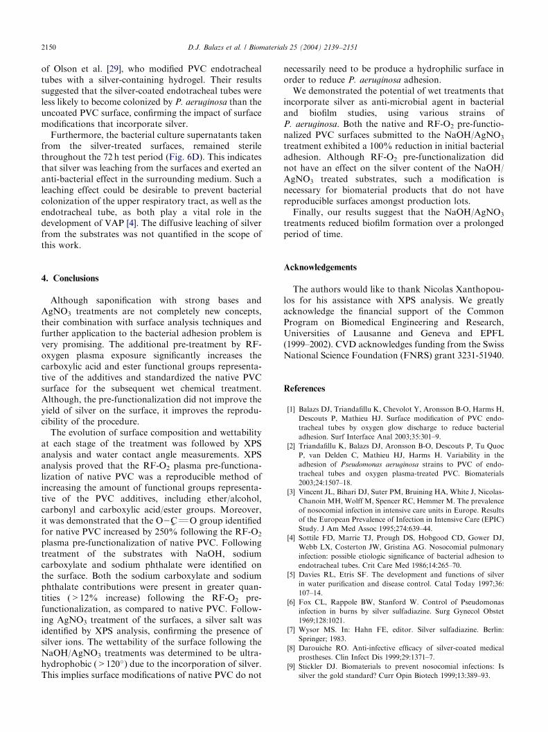
3.4. Evaluation of biofilm formation
In order to determine if the NaOH/AgNO3 chemicalmodification of native PVC would have long-termeffects on biofilm development, the efficacy of thetreatment of native PVC was assessed using a staticbiofilm assay. The effects of RF-O2 pre-functionaliza-tion prior to NaOH/AgNO3 treatment are currentlybeing investigated and are therefore not included in thiswork. Biofilms of P. aeruginosa, strain PT5, wereallowed to develop on the surface of native andNaOH/AgNO3 treated samples for 72 h after whichtime the samples were removed and the extent of biofilmdevelopment was assessed using the blot successiontechniques [23]. The blot succession technique providesdetailed information concerning the viability of the cellswithin the biofilm, along with the strength with whichthey adhere to the substrate. This technique effectivelybreaks biofilm cells into two distinct populations [23],described as follows:
(1)
Cells that are loosely attached: these are probablycells within the outer layers of the biofilm, that areattached either to each other or simply encasedwithin the exopolysaccharide matrix of the biofilmstructure.(2)
Residual attached cells that are much more firmlyadherent to the substrate. These cells are probably
ARTICLE IN PRESS
Fig. 6
nativ
PVC
the n
D.J. Balazs et al. / Biomaterials 25 (2004) 2139–2151 2149
directly in contact with the native PVC andprotected from the shear stresses exerted byvortexing and sonication.
Biofilms, established on the surface of native andN+NaOH+AgNO3, were processed as describedabove and blotted over a succession of 25 pre-driedLA plates. Calculations of total biofilm populationsdemonstrated the extremely high efficiency of this formof surface treatment in controlling bacterial biofilmdevelopment. Indeed, total biofilm populations onnative PVC were of 3� 109 cfu/g (CFU colony formingunit) of PVC at 24 h and 2� 109 cfu/g of PVC at 72 h,whereas those on NaOH/AgNO3 treated PVC wereof the order 4.5� 102 cfu/g of PVC at 24 h and2.2� 101 cfu/g of PVC at 72 h. This represents a seven-logarithmic drop in biofilm population for silver-treatedsurfaces at 24 h and an eight-logarithmic reduction at72 h (Fig. 6A).
As illustrated in Fig. 6B, the size of residual attachedpopulations remaining adherent to native PVC was4� 104 cfu/g of PVC at 24 h, dropping to 4� 103 cfu/g ofPVC at 72 h. No data points are illustrated for theNaOH/AgNO3 treated PVC, as no calculation could bemade for the substrates since there were no bacterialcells recovered from the blot successions. This further
20 30 40 50 60 70 801e+0
1e+1
1e+2
1e+3
1e+4
1e+5
1e+6
1e+7
1e+8
1e+9
1e+10
NativeAgNO3
Tot
al b
iofi
lm p
opul
atio
n
20 30 40 50 60 70 8020 30 40 50 60 70 801e+0
1e+1
1e+2
1e+3
1e+4
1e+5
1e+6
1e+7
1e+8
1e+9
1e+10
1e+0
1e+1
1e+2
1e+3
1e+4
1e+5
1e+6
1e+7
1e+8
1e+9
1e+10
NativeNativeAgNO3AgNO3
20 30 40 50 60 70 800.14
0.16
0.18
0.20
0.22
0.24
K V
alue
Biofilm age (hours)
Biofilm age (hours)
(A)
(C)
(B
. Effect of the NaOH/AgNO3 wet treatment of native PVC on surviva
e and NaOH+AgNO3 treated PVC; (B) residual attached biofilm cells o
; and (D) sterility of the supernatant following the NaOH/AgNO3 modifi
ative PVC surface is illustrated on the left.
underlines the efficacy of the NaOH/AgNO3 treatmentto prevent biofilm formation.
In a similar manner to residual population sizes,strength of attachment could only be calculated forbiofilms growing on native PVC substrates. These dataare shown in Fig. 6C. The ‘k’ value represents the best-fit line of the exponential decrease in colonies trans-ferred from the PVC substrates to the plate surface witheach successive plate. The best-fit line was estimatedusing the Marquardt–Levenberg algorithm (see Section2) [23]. As cells are more easily removed from a surface(less firmly attached), the slope of this best-fit lineincreases. The k value is therefore inversely proportionalto the strength of bacterial cell attachment. Fig. 6Cshows a decrease in k value over the 72 h test period,indicating that as the biofilm matures, those cells at thebiofilm–substratum interface become more firmly at-tached. The fact that no bacterial cells were recoveredfrom the NaOH/AgNO3 treated surfaces during the blotsuccession experiments indicates that cell death occurredat the biofilm–substratum interface. Destruction of these‘‘anchoring’’ layers of cells should destabilize even thethickest of biofilm layers, thus rendering them moresusceptible to removal.
The impressive effects that we observed onP. aeruginosa colonization are validated by the work
NNoonn--sstteerr iillee SStteerr iillee
20 30 40 50 60 70 801e+3
1e+4
1e+5
Biofilm age (hours)
(D) Supernatants
)
Res
idua
l Atta
ched
Pop
ulat
ion
l of biofilm cells: (A) evolution of the total biofilm population for both
n native PVC; (C) strength of attachment of the biofilm cells on native
cation (right), where the supernatant following biofilm development on
ARTICLE IN PRESSD.J. Balazs et al. / Biomaterials 25 (2004) 2139–21512150
of Olson et al. [29], who modified PVC endotrachealtubes with a silver-containing hydrogel. Their resultssuggested that the silver-coated endotracheal tubes wereless likely to become colonized by P. aeruginosa than theuncoated PVC surface, confirming the impact of surfacemodifications that incorporate silver.
Furthermore, the bacterial culture supernatants takenfrom the silver-treated surfaces, remained sterilethroughout the 72 h test period (Fig. 6D). This indicatesthat silver was leaching from the surfaces and exerted ananti-bacterial effect in the surrounding medium. Such aleaching effect could be desirable to prevent bacterialcolonization of the upper respiratory tract, as well as theendotracheal tube, as both play a vital role in thedevelopment of VAP [4]. The diffusive leaching of silverfrom the substrates was not quantified in the scope ofthis work.
4. Conclusions
Although saponification with strong bases andAgNO3 treatments are not completely new concepts,their combination with surface analysis techniques andfurther application to the bacterial adhesion problem isvery promising. The additional pre-treatment by RF-oxygen plasma exposure significantly increases thecarboxylic acid and ester functional groups representa-tive of the additives and standardized the native PVCsurface for the subsequent wet chemical treatment.Although, the pre-functionalization did not improve theyield of silver on the surface, it improves the reprodu-cibility of the procedure.
The evolution of surface composition and wettabilityat each stage of the treatment was followed by XPSanalysis and water contact angle measurements. XPSanalysis proved that the RF-O2 plasma pre-functiona-lization of native PVC was a reproducible method ofincreasing the amount of functional groups representa-tive of the PVC additives, including ether/alcohol,carbonyl and carboxylic acid/ester groups. Moreover,it was demonstrated that the O2
%CQO group identified
for native PVC increased by 250% following the RF-O2
plasma pre-functionalization of native PVC. Followingtreatment of the substrates with NaOH, sodiumcarboxylate and sodium phthalate were identified onthe surface. Both the sodium carboxylate and sodiumphthalate contributions were present in greater quan-tities (>12% increase) following the RF-O2 pre-functionalization, as compared to native PVC. Follow-ing AgNO3 treatment of the surfaces, a silver salt wasidentified by XPS analysis, confirming the presence ofsilver ions. The wettability of the surface following theNaOH/AgNO3 treatments was determined to be ultra-hydrophobic (>120�) due to the incorporation of silver.This implies surface modifications of native PVC do not
necessarily need to be produce a hydrophilic surface inorder to reduce P. aeruginosa adhesion.
We demonstrated the potential of wet treatments thatincorporate silver as anti-microbial agent in bacterialand biofilm studies, using various strains ofP. aeruginosa. Both the native and RF-O2 pre-functio-nalized PVC surfaces submitted to the NaOH/AgNO3
treatment exhibited a 100% reduction in initial bacterialadhesion. Although RF-O2 pre-functionalization didnot have an effect on the silver content of the NaOH/AgNO3 treated substrates, such a modification isnecessary for biomaterial products that do not havereproducible surfaces amongst production lots.
Finally, our results suggest that the NaOH/AgNO3
treatments reduced biofilm formation over a prolongedperiod of time.
Acknowledgements
The authors would like to thank Nicolas Xanthopou-los for his assistance with XPS analysis. We greatlyacknowledge the financial support of the CommonProgram on Biomedical Engineering and Research,Universities of Lausanne and Geneva and EPFL(1999–2002). CVD acknowledges funding from the SwissNational Science Foundation (FNRS) grant 3231-51940.
References
[1] Balazs DJ, Triandafillu K, Chevolot Y, Aronsson B-O, Harms H,
Descouts P, Mathieu HJ. Surface modification of PVC endo-
tracheal tubes by oxygen glow discharge to reduce bacterial
adhesion. Surf Interface Anal 2003;35:301–9.
[2] Triandafillu K, Balazs DJ, Aronsson B-O, Descouts P, Tu Quoc
P, van Delden C, Mathieu HJ, Harms H. Variability in the
adhesion of Pseudomonas aeruginosa strains to PVC of endo-
tracheal tubes and oxygen plasma-treated PVC. Biomaterials
2003;24:1507–18.
[3] Vincent JL, Bihari DJ, Suter PM, Bruining HA, White J, Nicolas-
Chanoin MH, Wolff M, Spencer RC, Hemmer M. The prevalence
of nosocomial infection in intensive care units in Europe. Results
of the European Prevalence of Infection in Intensive Care (EPIC)
Study. J Am Med Assoc 1995;274:639–44.
[4] Sottile FD, Marrie TJ, Prough DS, Hobgood CD, Gower DJ,
Webb LX, Costerton JW, Gristina AG. Nosocomial pulmonary
infection: possible etiologic significance of bacterial adhesion to
endotracheal tubes. Crit Care Med 1986;14:265–70.
[5] Davies RL, Etris SF. The development and functions of silver
in water purification and disease control. Catal Today 1997;36:
107–14.
[6] Fox CL, Rappole BW, Stanford W. Control of Pseudomonas
infection in burns by silver sulfadiazine. Surg Gynecol Obstet
1969;128:1021.
[7] Wysor MS. In: Hahn FE, editor. Silver sulfadiazine. Berlin:
Springer; 1983.
[8] Darouiche RO. Anti-infective efficacy of silver-coated medical
prostheses. Clin Infect Dis 1999;29:1371–7.
[9] Stickler DJ. Biomaterials to prevent nosocomial infections: Is
silver the gold standard? Curr Opin Biotech 1999;13:389–93.
ARTICLE IN PRESSD.J. Balazs et al. / Biomaterials 25 (2004) 2139–2151 2151
[10] Jansen B, Kohnen W. Prevention of biofilm formation by polymer
modification. J Ind Microbiol 1995;15:391–6.
[11] Klueh U, Wagner V, Kelly S, Johnson A, Bryers JD. Efficacy of
silver-coated fabric to prevent bacterial colonization and sub-
sequent device-based biofilm formation. J Biomed Mater Res
2000;53:621–31.
[12] Favia P, Creatore M, Palumbo F, Colaprico V, d’Agostino R.
Process control for plasma processing of polymers. Surf Coat
Technol 2001;142–144:1–6.
[13] Favia P, Vulpio M, Marino R, d’ Agostino R, Mota RP,
Catalano M. Plasma-deposition of Ag-containing polyethylene-
oxide-like coatings. Plasmas Polym 2000;5:1–14.
[14] Hart H, Craine LE, Hart D. Organic chemistry: a short course,
10th ed. New York: Houghton Mifflin; 1998.
[15] Shin SM, Jeon HS, Kim YH, Yoshioka T, Okuwaki A. Plasticizer
leaching from flexible PVC in low temperature caustic solution.
Polym Degradat Stability 2002;78:511–7.
[16] Gupta B, Hilborn J, Hollenstein C, Plummer CJG, Houriet R,
Xanthopoulos N. Surface modification of polyester films by RF
plasma. J Appl Polym Sci 2000;78:1083–91.
[17] Holloway BW. Genetic recombination in Pseudomonas aerugino-
sa. J Gen Microbiol 1955;13:572–81.
[18] Lightfoot J, Lam JS. Molecular cloning of genes involved with
expression of A-band lipopolysaccharide, an antigenically conserved
form, in Pseudomonas aeruginosa. J Bacteriol 1991;173:5624–30.
[19] Rijnaarts HHM, Norde W, Bouwer EJ, Lyklema J, Zehnder AJB.
Bacterial adhesion under static and dynamic conditions. Appl
Environ Microbiol 1993;59:3255–65.
[20] Sambrook J. Molecular cloning: a laboratory manual. New York:
Cold Spring Harbor Laboratory Press; 1989.
[21] Terry JM, Pina SE, Mattingly SJ. Role of energy metabolism in
conversion of nonmucoid Pseudomonas aeruginosa to the mucoid
phenotype. Infect Immun 1992;60:1329–35.
[22] Takeoka K, Ichimiya T, Yamasaki T, Nasu M. The in vitro effect
of macrolides on the interaction of human polymorphonuclear
leukocytes with Pseudomonas aeruginosa in biofilm. Chemother-
apy 1998;44:190–7.
[23] Eginton PJ, Gibson H, Holah J, Handley PS, Gilbert P.
Quantification of the ease of removal of bacteria from surfaces.
J Ind Microbiol 1995;15:305–10.
[24] Inagaki N. Hydrophilic surfaces (plasma treatment and plasma
polymerization). Boca Raton, FL: CRC Press; 1996.
[25] Asfardjani K, Segui Y, Aurelle Y, Abidine N. Effect of plasma
treatments on wettability of polysulfone and polyetherimide.
J Appl Polym Sci 1991;43:271–81.
[26] Hudis M. In: Hollahan JR, Bell AT, editors. Plasma treatment of
solid materials. New York: Wiley; 1974. p. 113–47.
[27] Barth G, Linder R, Bryson C. Advances in charge in neutraliza-
tion for XPS measurements of nonconducting materials. Surf
Interface Anal 1988;11:307–11.
[28] Good RJ. In: Mittal KL, editor. Contact angle, wetting, and
adhesion: a critical review. Utrecht, The Netherlands: VSP; 1993.
p. 3–36.
[29] Olson ME, Harmon BG, Kollef MH. Silver-coated endotracheal
tubes associated with reduced bacterial burden in the lungs of
mechanically ventilated dogs. Chest 2002;121:863–70.