Influence of base stacking on excited-state behavior of polyadenine in water, based on...
6
Influence of base stacking on excited-state behavior of polyadenine in water, based on time-dependent density functional calculations F. Santoro † , V. Barone ‡ , and R. Improta ‡§¶ † Istituto per i Processi Chimico–Fisici, Consiglio Nazionale delle Ricerche (CNR), Area della Ricerca del CNR, Via G. Moruzzi 1, I-56124 Pisa, Italy; ‡ Dipartimento di Chimica and Interuniversitario Nazionale per la Scienza e la Tecnologia dei Materiali, Universita ` Federico II, Complesso Monte S. Angelo, Via Cintia, I-80126 Napoli, Italy; and § Istituto di Biostrutture e Bioimmagini, CNR, Via Mezzocannone 16, I-80134 Napoli, Italy Communicated by George C. Schatz, Northwestern University, Evanston, IL, April 11, 2007 (received for review January 16, 2007) A thorough study of the excited-state properties of the stacked dimers and trimers of 9-methyladenine in B-DNA conformation has been performed in aqueous solution by using time-dependent density functional calculations and the solvent polarizable contin- uum model, and results were compared with experimental results on polyadenine oligomers. The effect of base stacking on the absorption and emission spectra is fully reproduced by our calcu- lations. Although light absorption leads to a state (SB ) delocalized over several nucleobases, excited-state geometry optimization indicates that SB subsequently evolves into a state in which the excitation is localized on a single base. Analysis of the excited-state potential energy surfaces shows that SB can easily decay into the lowest energy excited state, SCT , which is a dark excimer produced by intermonomer charge transfer between two stacked bases. The subpicosecond features of the time-resolved experiments are in- terpreted in terms of ultrafast decay from SB . After localization, two easy, radiationless decay channels are indeed open for SB :(i) ground-state recovery, according to the same mechanisms pro- posed for isolated adenine and/or (ii) decay to SCT . Our calculations suggest that the slowest part of the excited-state dynamics de- tected experimentally involves the SCT state. nucleic acid quantum mechanics spectroscopy time-dependent phenomena T he absorption of UV light by nucleic acids is a process of primary biophysical interest because it can start a cascade of photochemical reactions leading to base damage and genetic modifications. The photostability of nucleic acids is thus funda- mental for the development of life, and its microscopic origin has been the object of several experimental and computational studies (1). Even if a number of important aspects have not yet been convincingly assessed, there is general consensus regarding the mechanisms that explain the photostability of isolated nucleobases. Indeed, time-resolved spectroscopic studies (1) and quantum mechanical calculations (1–11) agree in suggesting the existence of an ultrafast nonradiative pathway between the bright excited state and the ground electronic state for both pyrimidine and purine nucleobases, which can explain their extremely short (1-ps) excited-state lifetimes. However, the excited-state behavior of DNA and nucleobase multimers (the systems of actual interest in vivo) appears much more complex, showing multiexponential decays with very different time con- stants (12–18). Recent experimental studies on single-stranded polyadenine (polyA) oligomers [(dA) n ] and double-stranded thymine adenine oligonucleotides [(dT) n /(dA) n ] provide deeper insight into the behavior of the multimer excited states and the factors tuning their decay (12–18). In (dA) 18 single-strand and (dA) 18 (dT) 18 double-strand, a fast initial decay, in the subpico- second time range, is indeed followed by slower relaxations (lifetimes of 0.8 and 126 ps, respectively) (12, 13). Additional time-resolved studies on (dA) 20 provide evidence for temporal evolution from three distinct states, the first characterized by ultrafast decay (lifetime 0.39 ps) and the other two by significantly longer lifetimes (4.3 and 182 ps, respec- tively) (15). It has been suggested (12, 13, 15) that the slower excited-state decay is due to the presence of ‘‘excimers’’ related to the formation of interbase stacking. The existence of excimers has already been noted in dinucleotides (19), and also in several different oligo- and polynucleotides (12–15, 20, 21), and its signature is an emission maximum red-shifted with respect to that of the parent monomers. It is thus clear that base stacking tunes energy delocalization after photoexcitation and inf luences the excited-state deactivation pathway. However, a number of fundamental questions remain. First, the mechanism of excita- tion must be more firmly established, especially in regard to its delocalization. Two scenarios appear plausible: either the exci- tation is delocalized over different nucleobases (22, 23) or it is localized on individual nucleobases and the base stacking merely modulates the temporal evolution of the system on the excited- state potential energy surface (PES), providing additional decay pathways through excimer states. A detailed molecular descrip- tion of the excimers and their time evolution would also allow a clearcut analysis of the possible role of charge transfer (CT) states (as suggested by refs. 12–14 and 16) and, eventually, an explication of the puzzling difference between the behavior of excimers in various stacked nucleobases. Indeed, experiments suggest that excimers influence the dynamics of polyA, whereas this is not the case for polythymine (12–14). The objective of the present article is to analyze the above questions on the basis of refined quantum mechanical compu- tations that include solvent effects, in order to attain a deeper understanding of the mechanisms underlying the influence of stacking on the excited-state dynamics in nucleic acids. We study, in particular, the ground and lowest excited electronic states of adenosine (Ado), of the dimer of 2-deoxyadenosine (dA) 2 , of 9-methyladenine (9-MA), and of its stacked dimer (9-MA) 2 and trimer (9-MA) 3 (see Fig. 1). To the best of our knowledge, this is the first study on the behavior of excited electronic states performed for stacked multimers of nucleobases by using accu- rate quantum mechanical methods that include solvent effects (24–26). Results First, we checked the reliability of polarizable continuum model (PCM)/time-dependent density functional theory (TD-DFT) Author contributions: F.S., V.B., and R.I. designed research; F.S. and R.I. performed research; and F.S., V.B., and R.I. wrote the paper. The authors declare no conflict of interest. Abbreviations: 9-MA, 9-methyladenine; CT, charge transfer; FC, Franck–Condon (region); TD-DFT, time-dependent density functional theory; PCM, polarizable continuum model; PES, potential energy surface(s); VEE, vertical excitation energy. ¶ To whom correspondence should be addressed. E-mail: [email protected]. This article contains supporting information online at www.pnas.org/cgi/content/full/ 0703298104/DC1. © 2007 by The National Academy of Sciences of the USA www.pnas.orgcgidoi10.1073pnas.0703298104 PNAS June 12, 2007 vol. 104 no. 24 9931–9936 BIOPHYSICS CHEMISTRY
Transcript of Influence of base stacking on excited-state behavior of polyadenine in water, based on...
Influence of base stacking on excited-state behaviorof polyadenine in water, based on time-dependentdensity functional calculationsF. Santoro†, V. Barone‡, and R. Improta‡§¶
†Istituto per i Processi Chimico–Fisici, Consiglio Nazionale delle Ricerche (CNR), Area della Ricerca del CNR, Via G. Moruzzi 1, I-56124 Pisa, Italy;‡Dipartimento di Chimica and Interuniversitario Nazionale per la Scienza e la Tecnologia dei Materiali, Universita Federico II, ComplessoMonte S. Angelo, Via Cintia, I-80126 Napoli, Italy; and §Istituto di Biostrutture e Bioimmagini, CNR, Via Mezzocannone 16, I-80134 Napoli, Italy
Communicated by George C. Schatz, Northwestern University, Evanston, IL, April 11, 2007 (received for review January 16, 2007)
A thorough study of the excited-state properties of the stackeddimers and trimers of 9-methyladenine in B-DNA conformation hasbeen performed in aqueous solution by using time-dependentdensity functional calculations and the solvent polarizable contin-uum model, and results were compared with experimental resultson polyadenine oligomers. The effect of base stacking on theabsorption and emission spectra is fully reproduced by our calcu-lations. Although light absorption leads to a state (SB) delocalizedover several nucleobases, excited-state geometry optimizationindicates that SB subsequently evolves into a state in which theexcitation is localized on a single base. Analysis of the excited-statepotential energy surfaces shows that SB can easily decay into thelowest energy excited state, SCT, which is a dark excimer producedby intermonomer charge transfer between two stacked bases. Thesubpicosecond features of the time-resolved experiments are in-terpreted in terms of ultrafast decay from SB. After localization,two easy, radiationless decay channels are indeed open for SB: (i)ground-state recovery, according to the same mechanisms pro-posed for isolated adenine and/or (ii) decay to SCT. Our calculationssuggest that the slowest part of the excited-state dynamics de-tected experimentally involves the SCT state.
nucleic acid � quantum mechanics � spectroscopy �time-dependent phenomena
The absorption of UV light by nucleic acids is a process ofprimary biophysical interest because it can start a cascade of
photochemical reactions leading to base damage and geneticmodifications. The photostability of nucleic acids is thus funda-mental for the development of life, and its microscopic origin hasbeen the object of several experimental and computationalstudies (1). Even if a number of important aspects have not yetbeen convincingly assessed, there is general consensus regardingthe mechanisms that explain the photostability of isolatednucleobases. Indeed, time-resolved spectroscopic studies (1) andquantum mechanical calculations (1–11) agree in suggesting theexistence of an ultrafast nonradiative pathway between thebright excited state and the ground electronic state for bothpyrimidine and purine nucleobases, which can explain theirextremely short (�1-ps) excited-state lifetimes. However, theexcited-state behavior of DNA and nucleobase multimers (thesystems of actual interest in vivo) appears much more complex,showing multiexponential decays with very different time con-stants (12–18). Recent experimental studies on single-strandedpolyadenine (polyA) oligomers [(dA)n] and double-strandedthymine adenine oligonucleotides [(dT)n/(dA)n] provide deeperinsight into the behavior of the multimer excited states and thefactors tuning their decay (12–18). In (dA)18 single-strand and(dA)18�(dT)18 double-strand, a fast initial decay, in the subpico-second time range, is indeed followed by slower relaxations(lifetimes of �0.8 and �126 ps, respectively) (12, 13).
Additional time-resolved studies on (dA)20 provide evidencefor temporal evolution from three distinct states, the first
characterized by ultrafast decay (lifetime �0.39 ps) and the othertwo by significantly longer lifetimes (�4.3 and �182 ps, respec-tively) (15). It has been suggested (12, 13, 15) that the slowerexcited-state decay is due to the presence of ‘‘excimers’’ relatedto the formation of interbase stacking. The existence of excimershas already been noted in dinucleotides (19), and also in severaldifferent oligo- and polynucleotides (12–15, 20, 21), and itssignature is an emission maximum red-shifted with respect tothat of the parent monomers. It is thus clear that base stackingtunes energy delocalization after photoexcitation and influencesthe excited-state deactivation pathway. However, a number offundamental questions remain. First, the mechanism of excita-tion must be more firmly established, especially in regard to itsdelocalization. Two scenarios appear plausible: either the exci-tation is delocalized over different nucleobases (22, 23) or it islocalized on individual nucleobases and the base stacking merelymodulates the temporal evolution of the system on the excited-state potential energy surface (PES), providing additional decaypathways through excimer states. A detailed molecular descrip-tion of the excimers and their time evolution would also allow aclearcut analysis of the possible role of charge transfer (CT)states (as suggested by refs. 12–14 and 16) and, eventually, anexplication of the puzzling difference between the behavior ofexcimers in various stacked nucleobases. Indeed, experimentssuggest that excimers influence the dynamics of polyA, whereasthis is not the case for polythymine (12–14).
The objective of the present article is to analyze the abovequestions on the basis of refined quantum mechanical compu-tations that include solvent effects, in order to attain a deeperunderstanding of the mechanisms underlying the influence ofstacking on the excited-state dynamics in nucleic acids. We study,in particular, the ground and lowest excited electronic states ofadenosine (Ado), of the dimer of 2�-deoxyadenosine (dA)2, of9-methyladenine (9-MA), and of its stacked dimer (9-MA)2 andtrimer (9-MA)3 (see Fig. 1). To the best of our knowledge, thisis the first study on the behavior of excited electronic statesperformed for stacked multimers of nucleobases by using accu-rate quantum mechanical methods that include solvent effects(24–26).
ResultsFirst, we checked the reliability of polarizable continuum model(PCM)/time-dependent density functional theory (TD-DFT)
Author contributions: F.S., V.B., and R.I. designed research; F.S. and R.I. performed research;and F.S., V.B., and R.I. wrote the paper.
The authors declare no conflict of interest.
Abbreviations: 9-MA, 9-methyladenine; CT, charge transfer; FC, Franck–Condon (region);TD-DFT, time-dependent density functional theory; PCM, polarizable continuum model;PES, potential energy surface(s); VEE, vertical excitation energy.
¶To whom correspondence should be addressed. E-mail: [email protected].
This article contains supporting information online at www.pnas.org/cgi/content/full/0703298104/DC1.
© 2007 by The National Academy of Sciences of the USA
www.pnas.org�cgi�doi�10.1073�pnas.0703298104 PNAS � June 12, 2007 � vol. 104 � no. 24 � 9931–9936
BIO
PHYS
ICS
CHEM
ISTR
Y

calculations for describing the excited states of 9-MA itself. Adetailed analysis is given in supporting information (SI) Text.Briefly, our computations predict the presence of two strongabsorption peaks at �5 and �6 eV, which is in good agreementwith the experimental spectra (15, 27) except for a smalloverestimation of the energy of the red-side peak (see Fig. 2 andSI Table 2). In agreement with previous CASPT2 studies onadenine (7–11), three electronic transitions contribute to thelowest energy absorption band: two with �/�* character (usuallylabeled �*La and �*Lb, respectively) and one with n/�* char-acter. Most of the absorption intensity is carried by the spec-troscopic �*La state, which corresponds essentially to aHOMO3LUMO transition (we refer to the HOMO andLUMO orbitals as H and L, respectively), as shown in SI Fig. 6.In contrast, the �*Lb state is mainly an H3L � 1 transition.Replacement of the 9-methyl substituent by the deoxyribose ringand inclusion of four water molecules from the first solvationshell do not significantly change the computed vertical excitationenergies (VEEs) (see SI Figs. 7 and 8 and SI Table 2).
The above considerations, together with our previous work onpyrimidine nucleobases (4, 5), support the reliability of theproposed computational approach and provide a basis for thesubsequent analysis of 9-MA oligomers.
(9-MA)2 and (dA)2 Absorption Spectra. We have performed partialgeometry optimizations (see Materials and Methods) in aqueoussolution for the ground electronic state (S0) of a supermoleculeformed by two 9-MA stacked as in B-DNA (see Fig. 1d and SIFig. 9). Stacking interaction induces small but not negligibleintramolecular distortions with respect to the geometry of theisolated monomer in aqueous solution. Furthermore, in B-DNAconformation the mutual arrangement of the two monomers isnot symmetric, and thus their minimum energy geometries arenot perfectly equivalent. The dimer VEEs computed at thePCM/TD-PBE0 level in water are reported in Table 1, and theabsorption spectrum is shown in Fig. 2. The picture issuing froma comparison between the spectra computed for the monomerand those for the dimer is very similar to its experimentalcounterpart. Experiments and computations agree, indicatingthat passage from the monomer to the multimer induces (i) aslight blue-shift of the maximum of the absorption, (ii) a slightred-shift of the low-energy side, and (iii) a significant decreasein oscillator strength.
The most intense transition in the dimer spectrum arises fromthe symmetric combination of H � 13L and H3L � 1transitions. The orbitals involved in this transition (see SI Fig.10) derive from the symmetric and antisymmetric combinationsof the monomers H and L. The combination of two Hs and twoLs gives rises to the four transitions collected in Table 1. Theinterbase distance of �3.6 Å leads to a small but not vanishingoverlap (and coupling) between the orbitals of the two 9-MAmoieties. As a consequence, the energy of the orbitals involvedin the bright electronic transition changes only slightly, account-ing for the small blue-shift of the most intense transition [S4 atthe PMC/TD-PBE0/6-31G(d) level] with respect to the mono-mer spectra. Three additional weak transitions (responsible forthe red-shift of the low-energy side) explain the hypsochromiceffect caused by the base stacking. Excited states S5 and S6 arereminiscent of the n3�* electronic transitions of the monomer.The S7 and S8 transitions receive contributions from excitationsinvolving several molecular orbitals, with a predominant weightof excitations from H and H � 1 toward L � 2 and L � 3. Thesetransitions are thus the counterparts of the �*Lb electronictransition of the monomer.
To verify the influence of the deoxyribose/phosphate back-bone on the excited-state properties of the nucleobases, we haveoptimized the ground-state geometry in aqueous solution for the(dA)2 system (see Fig. 1c) and computed its absorption spectrum(see SI Fig. 7). The results on (dA)2 fully reproduce the effect ofbase stacking on nucleobase absorption. Furthermore, both theVEE and the character of the excited states are very similar tothose of (9-MA)2 (with the single exception discussed below),thus indicating that 9-MA multimers should be suitable modelsfor the study of polyA behavior. The only significant differencebetween the excited states of (9-MA)2 and (dA)2 is the clear CTcharacter of the dark S1 state in (dA)2. This is the result of a morepronounced asymmetry in (dA)2, which localizes the H and Lorbitals mainly on two different nucleobases. As soon as theFranck–Condon (FC) region is exited, S1 acquires a clear CTcharacter in (9-MA)2 as well (see below).
The effect of asymmetry has been further analyzed by partialgeometry optimizations in aqueous solution on a pentamer of9-MA molecules in the B-DNA conformation (see Fig. 1d),extracting the four possible proximal dimers. The results of thesubsequent TD/PBE0 excited-state calculations are reported inSI Fig. 11. It is noteworthy that the overall absorption spectra arevery similar to those shown in Fig. 2. The most striking difference
Fig. 1. Schematic drawing and atom labeling of the systems investigated inthis study. (a) 9-MA. (b) Adenine nucleoside (Ado). (c) (dA)2. (d) 9-MA pen-tamer (9-MA)5. (The dimer is shown enclosed in a solid-rule box, and the trimeris indicated by the added dotted box).
Fig. 2. Computed absorption spectra of 9-MA monomer and dimer. Eachtransition is convoluted by a Gaussian with a FWMH of 0.30 eV.
9932 � www.pnas.org�cgi�doi�10.1073�pnas.0703298104 Santoro et al.
with respect to the optimized dimer is the clear CT character ofthe weak transitions on the red side of the absorption band. Inthe pentamer, the presence of multiple base stacking interac-tions leads indeed to small variations in the equilibrium geom-etry of each 9-MA molecule. For example, the amino groupsexhibit different degrees of pyramidalization because of theformation of weak NH2���NH2 hydrogen bonds. These smallstructural modifications increase the energy difference betweenthe frontier orbitals of the two monomers, accounting for the CTcharacter of some of the lowest energy transitions.
(9-MA)2 Excited-State Geometry Optimizations. Full geometry opti-mizations of the three lowest energy excited states of (9-MA)2with a fully equilibrated solvent were performed next (see SIText: Computational Details). Note that, in this limit at the FCpoint, the bright state is the third excited state (S3).
PCM/TD-PBE0 geometry optimization of the S1 state leads toa global minimum, minSCT, with a nuclear structure typical of a9-MA��9-MA� stacked pair (see SI Fig. 9). In fact, the geom-etries of two monomers are strongly different and closelyresemble those of the 9-MA radical cation and anion, respec-tively (see SI Fig. 12). Not surprisingly, the H3L transitioninduces the intermolecular transfer of a net electronic charge, sothat in this region S1 has a clear CT character and can be labeledSCT. The minSCT energy is 0.51 eV below the FC point, and thevertical emission energy is 3.82 eV. The above results areconfirmed by PCM/TD-PBE0/6-31�G(d,p) optimizations thatindividuate a minimum structure with an emission energy of 3.86eV, whose stabilization with respect to the FC point is 0.43 eV.
We also located the global minimum of the S2 second excitedadiabatic surface (hereafter SDEL, described in SI Text). Unlikewith S1, S2 keeps in its minimum an electronic character verysimilar to that exhibited in the FC region, with the electronicexcitation always nearly equally delocalized over the twomonomers.
Geometry optimization starting at the FC point on S3 (thebright state) and following the adiabatic state that has the largestoscillator strength with S0 leads to another excited-state mini-mum (see SI Fig. 9). In this region of the coordinate space, the
bright-state minimum corresponds to the lowest energy excitedstate, S1. Two different minima (corresponding to two differentdiabatic states) thus exist on the S1 adiabatic PES: minSCT andminSB (the minimum of the bright state, SB).
At minSB, the S03S1 electronic transition is a ‘‘localizedexcitation’’ because both H and L reside on the same monomer.Indeed, although the geometry of one monomer is very close tothat of S0, the other monomer shows a structure similar to thatobtained by optimizing the �*La state of an isolated 9-MAmolecule. The emission energy is 4.37 eV at the 6-31G(d) leveland 4.22 eV at the 6-31�G(d,p) level, whereas the energy ofminSB is �0.25 eV above minSCT [�0.15 eV at the 6-31�G(d,p)level]. Geometry optimization of SB is rather troublesomebecause a second state with CT character always remains closeto the SB state, so that optimization can easily collapse intominSCT where S1 has a CT character (vide infra).
Because TD-DFT calculations may poorly describe long-rangeCT transitions (28), it is important to check the reliability of thepresent TD-DFT calculations in predicting that the lowestenergy transition in the stacked dimers and trimers (vide infra)has a CT character. In SI Text, we support in detail the reliabilityof our results. Here, we note that our results are in goodagreement with the experimental absorption and emission spec-tra and that test calculations at the CASSCF(8,8)/PCM levelconfirm the conclusions of TD-DFT calculations. Furthermore,the values of ionization potential and electron affinity of 9-MAin aqueous solution indicate that the estimate of the SCTtransition energy is correct (see SI Text). Finally, when appliedto the study of stacked cytosine dimer, our computationalapproach provides a picture extremely similar to that of veryrecent CASPT2 calculations (23), confirming that TD-PBE0can accurately describe the excited-state behavior of stackednucleobases.
9-MA Trimer. We have optimized in aqueous solution the geom-etry of (9-MA)3, a trimer of 9-MA molecules in the B-DNAstacking geometry. The VEEs for the lowest energy excitedstates are collected in Table 1 and the computed absorptionspectra in SI Fig. 13. As already found for (9-MA)2, the
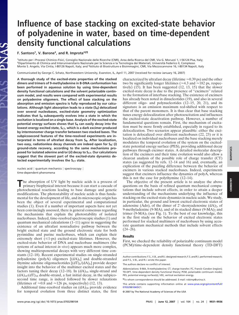
Table 1. VEEs and oscillator strengths issuing from PCM/TD-PBE0/6-31G8(d)��PCM/PBE0/6-31G(d) optimizationsin aqueous solution of 9-MA dimer and trimer
Transition VEE, eV Oscillator strength Assignment Monomer charge Dipole moment, D
(9-MA)2†
S1 4.96 0.008 H3L 0.05; �0.05 5.74S2 4.99 0.002 H � 13L (�) H3L � 1 �0.06; 0.06 6.35S3 5.16 0.03 H � 13L � 1 0.03; �0.03 5.76S4 5.21 0.32 H � 13L (�) H3L � 1 0.06; �0.06 5.88S5 5.31 0.03 H � 23L (�) H � 33L � 1 0.003; �0.003 4.73S6 5.32 0.023 H � 33L (�) H � 23L � 1 0.002; �0.002 4.91S7 5.39 0.007 MIxed �/�* �0.01; 0.01 6.70S8 5.43 0.10 Mixed �/�* �0.008; 0.008 6.41
(9-MA)3‡
S1 4.77 0.004 H3L �0.91; 0.89; 0.02 15.85S2 4.80 0.000 H3L � 1 0.02; 0.89; �0.91 19.33S3 5.04 0.002 H3L � 2 0.06; �0.30; 0.24 7.48S4 5.11 0.045 Mixed 0.23; �0.55; 0.32 7.33S5 5.17 0.031 Mixed 0.02; �0.35; 0.33 8.47S6 5.19 0.067 Mixed 0.43; �0.39; �0.04 12.1S7 5.21 0.336 Mixed �0.001; 0.001; 0.000 7.69S8 5.31 0.024 Mixed 0.02; 0.00; �0.02 7.41S9 5.34 0.005 Mixed �0.22; 0.00; 0.22 9.10
D, debye (unit of dipole moment): 10�18 esu�cm [0.358 � 10�30 m�C (meter coulomb)].†Monomer charge in the ground state: 0.0009; �0.0009. Dipole moment � 6.15 D.‡Monomer charge in the ground state: �0.003; 0.005; �0.002. Dipole moment � 8.34 D.
Santoro et al. PNAS � June 12, 2007 � vol. 104 � no. 24 � 9933
BIO
PHYS
ICS
CHEM
ISTR
Y
absorption maximum of the spectra is slightly blue-shifted andless intense than that of the monomer, thus confirming that basestacking causes significant hypochromic effects. The brightelectronic transition SB (corresponding to the seventh excitedstate in the solvent nonequilibrium regime) is delocalized overthe three monomers and does not exhibit any CT character, asshown in Table 1 by the Mulliken population analysis. On theother hand, the two lowest energy excitations, S1 and S2, have asignificant CT character and are very similar to the SCT state of(9-MA)2. They correspond indeed to the transfer of a netelectronic charge from the ‘‘central’’ monomer toward a lateralmonomer. Interestingly, the CT transition is always localized ona dimer, i.e., there is one monomer that is not affected by theelectronic transition. Indeed, geometry optimization of thelowest energy excited state leads to a structure analogous tothe SCT minimum of (9-MA)2. The central, and one of the lateral,monomers exhibit a geometry similar to that of 9-MA in itscationic and anionic form, respectively. The other lateral mol-ecule does not show any significant deviation from the S0geometry. The emission energy of this transition (3.87 eV) is veryclose to that of SCT in (9-MA)2 (3.82 eV).
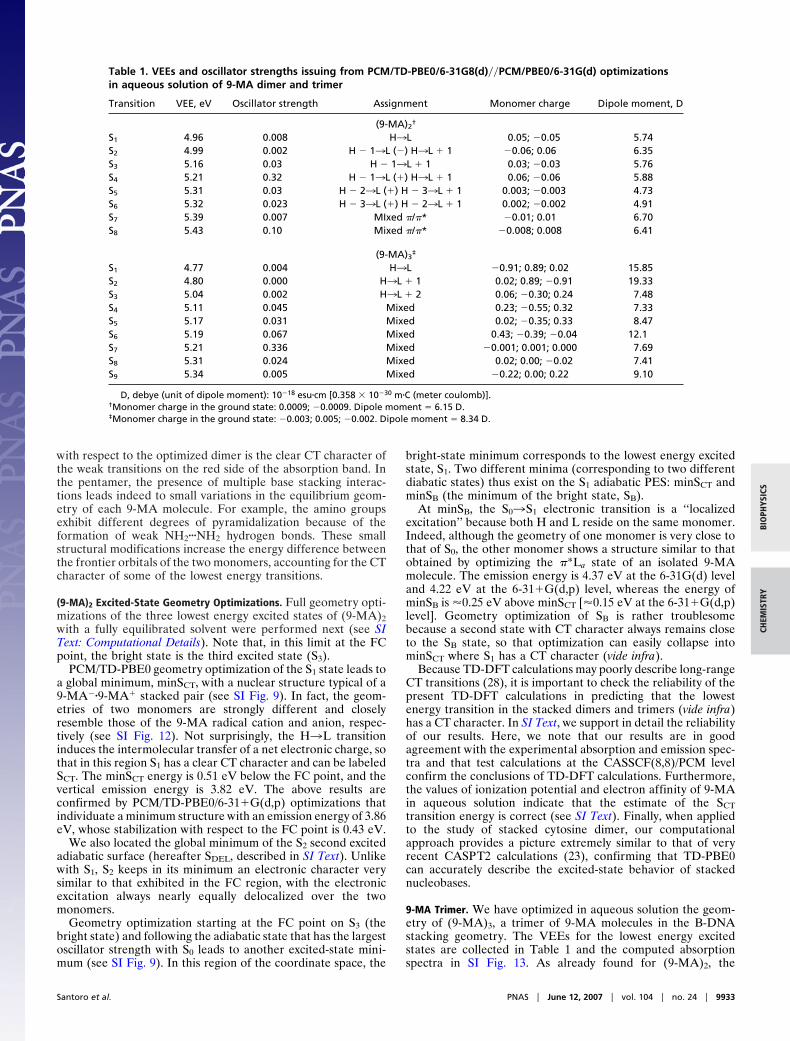
Scans of the (9-MA)2 Excited-State PES. Preliminary insights into thedynamical behavior of (9-MA)2 can be gained by examiningselected energy scans of the lowest energy excited states in theregion connecting the FC point to the excited-state minima. Fig.3a reports the energy of the three lowest energy states in the pathbetween the FC point and minSB. The S1 and S3 adiabatic statesalong this path (see Fig. 3b) drastically change their electroniccharacter (interchanging their oscillator strengths with S0, here-after Fn0). Thus, it has been convenient to define at each pointof the path the two diabatic states SB and SCT (bright and dark,respectively) introduced in the previous section (see Fig. 3c andSI Text for technical details). At the FC point, the opticalexcitation drives the system essentially on the S3 state that, asshown by Fig. 3b, brings most of the Fn0. In contrast, S1 is nearlydark. At this point, therefore, the two adiabatic states S3 and S1correspond to the diabatic states SB and SCT. Subsequently, theelectronic character of S1 changes strongly along the path andacquires oscillator strength at the expense of S3, transformingprogressively into the bright SB state, even if some residualdifference among the diabatic and adiabatic states persists at theminSB point because S3 also retains a little oscillator strength(see SI Text for further details).
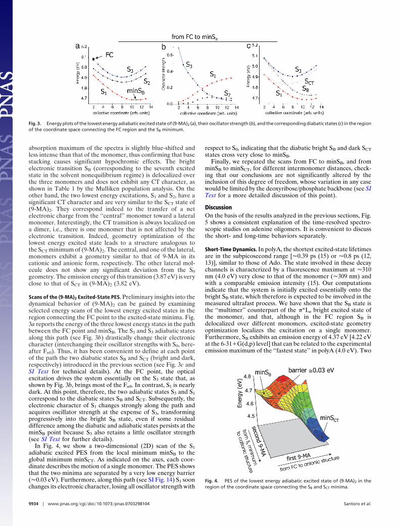
In Fig. 4, we show a two-dimensional (2D) scan of the S1adiabatic excited PES from the local minimum minSB to theglobal minimum minSCT. As indicated on the axes, each coor-dinate describes the motion of a single monomer. The PES showsthat the two minima are separated by a very low energy barrier(�0.03 eV). Furthermore, along this path (see SI Fig. 14) S1 soonchanges its electronic character, losing all oscillator strength with
respect to S0, indicating that the diabatic bright SB and dark SCTstates cross very close to minSB.
Finally, we repeated the scans from FC to minSB, and fromminSB to minSCT, for different intermonomer distances, check-ing that our conclusions are not significantly altered by theinclusion of this degree of freedom, whose variation in any casewould be limited by the deoxyribose/phosphate backbone (see SIText for a more detailed discussion of this point).
DiscussionOn the basis of the results analyzed in the previous sections, Fig.5 shows a consistent explanation of the time-resolved spectro-scopic studies on adenine oligomers. It is convenient to discussthe short- and long-time behaviors separately.
Short-Time Dynamics. In polyA, the shortest excited-state lifetimesare in the subpicosecond range [�0.39 ps (15) or �0.8 ps (12,13)], similar to those of Ado. The state involved in these decaychannels is characterized by a fluorescence maximum at �310nm (4.0 eV) very close to that of the monomer (�309 nm) andwith a comparable emission intensity (15). Our computationsindicate that the system is initially excited essentially onto thebright SB state, which therefore is expected to be involved in themeasured ultrafast process. We have shown that the SB state isthe ‘‘multimer’’ counterpart of the �*La bright excited state ofthe monomer, and that, although in the FC region SB isdelocalized over different monomers, excited-state geometryoptimization localizes the excitation on a single monomer.Furthermore, SB exhibits an emission energy of 4.37 eV [4.22 eVat the 6-31�G(d,p) level] that can be related to the experimentalemission maximum of the ‘‘fastest state’’ in polyA (4.0 eV). Two
Fig. 3. Energy plots of the lowest energy adiabatic excited state of (9-MA)2 (a), their oscillator strength (b), and the corresponding diabatic states (c) in the regionof the coordinate space connecting the FC region and the SB minimum.
Fig. 4. PES of the lowest energy adiabatic excited state of (9-MA)2 in theregion of the coordinate space connecting the SB and SCT minima.
9934 � www.pnas.org�cgi�doi�10.1073�pnas.0703298104 Santoro et al.

considerations can account for the residual discrepancy betweencomputed and experimental values. First, the computed VEE ofthe bright SB state is also overestimated with respect to theabsorption maximum by �0.25 eV. Second, the mutual arrange-ment of the dimer has been frozen to that adopted in B-DNAground state, so that fully relaxed geometry optimizations of theintermolecular degrees of freedom would favor the excited stateand decrease the computed/experimental difference.
Always bearing in mind that the details of the ground-staterecovery depend on the dynamics of the stacking interactionsand on the presence of nonstacked bases, our calculationssuggest two possible radiationless ultrafast deactivation path-ways for SB.Monomer-like decay pathway. After localization on a single mono-mer, SB state can follow the same decay pathways highlighted for�*La in the monomer. Indeed, theoretical studies on adenine(7–11) indicate that two paths characterized by small or vanish-ing energy barriers lead from the bright excited �*La state toconical intersections with the ground state, S0. These two pathscould be responsible for the two different excited-state lifetimes(�0.13 and �0.45 ps) shown by Ado (15). In this scenario, onemust conclude that the fastest excited-state decay pathway of themonomer is absent, or slowed, in the polymer. Several effectscould, in principle, explain this. First, base stacking could makemore difficult the out-of-plane deformation of the ring neededto reach the conical intersection with S0 (7–11). Alternatively,one of the two paths (probably the one involving the �*Lb state)may not be accessible in the polymer. Finally, the nuclearrearrangement leading from an excitation delocalized on two ormore bases to one localized on a single base could require anonvanishing time.Additional decay pathway due to stacking. The scans of the excited-state PES for (9-MA)2 suggest an additional decay mechanismfor SB in the multimers, leading the system into the SCT state. Infact, SB interacts with SCT along the path from the FC point tothe SB minimum and, furthermore, a very small barrier separatesthe minima of the SB and SCT states. These features suggest that,in the polymer, the decay to SCT is competitive with themonomer-like decay to S0.
A final remark concerns the lively debate about the delocal-ized (17, 18, 22, 23) or localized (12–15, 19) nature of the brightstate in nucleic acids. We have shown that an excitation delo-calized over multiple bases in the FC region can lead to a ‘‘local’’excited state. In other words, the fact that a state exhibits decayand emission properties typical of a localized excitation does not
necessarily imply that the excitation is also localized during theabsorption process. On the other hand, our results suggest thatthe possible delocalization/localization of the bright state in theFC region is ruled by the degree of asymmetry of the variousmonomers in the multimer.
It is noteworthy that, according to our calculations, n3�*transitions should not play any relevant role in the polyAexcited-state dynamics, at least in aqueous solution (see SI Text).
Long-Time Dynamics. In regard to the long-lifetime (�4 ps)component of the excited-state population, experiments onpolyA indicate the existence of two ‘‘excimer-like’’ states: onewith a lifetime of �4.3 ps (15) and another with a lifetime � 100ps (�182 ps according to ref. 15 and �126 ps according to refs.12 and 13). In (dA)20, the former state is characterized by a veryweak fluorescence maximum at 348 nm (3.56 eV) (15). Accord-ing to our analysis, the SCT excited state resulting from the CTbetween two stacked monomers is the best candidate for thisstate. Indeed, the computed emission maximum of SCT has a verysmall oscillator strength and its energy (3.86 eV) correlates wellwith that of the excimer (3.56 eV), bearing in mind that, asreported above, our calculations overestimate the emissionenergy of the bright state as well (by 0.20 eV). Furthermore,besides its small f luorescence quantum yield, several experimen-tal results suggest that this excimer has a significant CT character(12, 13, 15). Finally, several features of SCT are fully consistentwith the experimental findings. First, according to our compu-tations, the formation of SCT does not require any substantialconformational rearrangement or modification of the stackinggeometry and is thus compatible with a fast conversion of thebright state. Second, the excited-state PES indicates that thebright SB state has a significant tendency to decay to SCT. The twodiabatic states cross along the path between the FC and minSBstructures and, although a reliable evaluation of the transitionprobability is beyond the scope of our study, additional 1D scansreported in SI Text suggest that if the system passes on the darkSCT state it is irreversibly accelerated toward its minimumminSCT, reaching a region where it is no longer coupled to SB andcannot therefore jump back onto SB. Furthermore, after reach-ing minSB, the system can easily decay to SCT by movingadiabatically on S1. The above finding fully agrees with theexperimental indications that the large majority of the excitedpopulation in polyA decays to excimer-like states (12, 13, 15).
The minimum energy structure of SCT proves the existence ofan anion-like 9-MA monomer stacked with a cation-like 9-MAmonomer. On a longer time scale, this would likely lead to asignificant geometry rearrangement within the single strand,which would lose its regular and symmetric stacking geometry.Our calculations do not provide any definite indication regardingthe nature of this geometry rearrangement because, on this timescale, structural f luctuations of the strand should be taken intoaccount. However, the existence of a fully relaxed CT minimumcould be related to the excimer-like state characterized by theslowest decay (�100 ps); in (dA)20 this state is characterized bya fluorescence maximum of 390 nm (3.18 eV) (15). Both theslowness of the decay and the large Stokes shift are indeedcompatible with a significant conformational rearrangementwithin the DNA strand.
The CT character of the excimer suggests that another possibledecay route can involve an excited-state proton transfer betweenadjacent bases or, more likely, with solvent molecules. Theinvolvement of a proton transfer in the excited-state deactivationhas already been proposed in double-stranded DNA (29). In thepresence of anionic and cationic species, interstrand protontransfer could be more favored and its role in the deactivationpathway more important.
In summary, the present study reports a detailed moleculardescription of the most relevant excited states of polyA oligomers,
Fig. 5. Schematic description of the possible time-evolution of the excitedstates of the adenine oligomer.
Santoro et al. PNAS � June 12, 2007 � vol. 104 � no. 24 � 9935
BIO
PHYS
ICS
CHEM
ISTR
Y

providing strong evidence in support of a fast transfer from thebright spectroscopic state to an underlying excimer-like state witha clear CT character in a stacked dimer. On this basis, it is possibleto suggest a consistent explanation for the most significant exper-imental results and to identify the states responsible for thedifferent lifetime components. Obviously, the situation in single-and double-strand DNA multimers can be much more complexthan that modeled in our calculations. The experimental absorptionand emission spectra can receive contributions from multiple states,involving a varying number of stacked pairs and with differentstacked geometry, strongly modulated by structural fluctuations ofthe multimer. In double-strand DNA, hydrogen-bonded pairs couldplay a relevant role, from both static and dynamic viewpoints. Forexample, simple thermodynamic considerations suggest that inter-strand CT states can be present, because the electron affinity ofthymine is larger than that of adenine. All of the above effects areconsistent with the multiexponential excited-state decay shown bythe latest time-resolved experiments (17). Additional theoreticaland experimental efforts are thus necessary to fully assess thebehavior of the excited states in nucleic acids. On the other hand,it is encouraging that our calculations are able to reproduce theeffect of base stacking on the absorption and emission spectra. Thismay represent a significant step toward a deeper understanding ofthe influence of base stacking on the excited-state decay in DNA.
Materials and MethodsThe geometry of adenine stacked oligomers in a single-strandedpolynucleotide has been extracted from the experimental struc-
ture of polydeoxyadenosine�polydeoxythymine (1PLY.pdb), adouble-stranded DNA. In the ground- and excited-state geom-etry optimizations of the 9-MA oligomers, the intramoleculardegrees of freedom were fully unconstrained, whereas theinterbase distance and mutual orientation were kept frozen totheir experimental value. Whenever not explicitly stated, geom-etry optimizations have been performed in aqueous solution atthe PBE0/6-31G(d) (30) and TD-PBE0/6-31G(d) (31) levels,with bulk solvent effects included by using the PCM (32, 33). Theeffect of basis set extension on the VEEs has been estimated bytest computations with more extended 6-31�G(d,p) and6-311�G(2d,2p) basis sets. We checked, by test calculationsusing the counterpoise method (see SI Text), that inclusion ofbasis set superposition errors does not significantly affect ourconclusions. Ionization potential and electron affinity valueshave been computed as the difference of energies of the neutraland charged species. Possible pathways available to the systemupon excitation have been characterized by 1D and 2D scans ofthe excited-state PES. More details regarding the computationalprocedures, and definition of the relevant diabatic states can befound in SI Text. All of the quantum mechanical calculationswere performed by using a development version of the Gaussian03 software (34).
We thank Dr. A. Lami for very useful discussions and Village-Na(http://village.unina.it) for computational resources.
1. Crespo-Hernandez CE, Cohen B, Hare PM, Kohler B (2004) Chem Rev104:1977–2020.
2. Merchan M, Serrano-Andres L (2003) J Am Chem Soc 125:8108–8109.3. Ismail N, Blancafort L, Olivucci M, Kohler B, Robb MA (2002) J Am Chem Soc
124:6818–6819.4. Santoro F, Barone V, Gustavsson T, Improta R (2006) J Am Chem Soc
128:16312–16322.5. Gustavsson T, Banyasz A, Lazzarotto E, Markovitsi D, Scalmani G, Frisch MJ,
Barone V, Improta R (2006) J Am Chem Soc 128:607–619.6. Gustavsson T, Sarkar N, Lazzarotto E, Markovitsi D, Barone V, Improta R
(2006) J Phys Chem B 110:12843–12847.7. Blancafort L (2006) J Am Chem Soc 128:210–219.8. Perun S, Sobolewski AL, Domcke W (2005) J Am Chem Soc 127:6257–6265.9. Chen H, Li S (2005) J Phys Chem A 109:8443–8446.
10. Serrano-Andres L, Merchan M, Borin AC (2006) Proc Natl Acad Sci USA103:8691–8696.
11. Serrano-Andres L, Merchan M, Borin AC (2006) Chem Eur J 12:6559–6571.12. Crespo-Hernandez CE, Cohen B, Kohler B (2005) Nature 436:1141–1144.13. Crespo-Hernandez CE, Cohen B, Kohler B (2006) Nature 441:E8.14. Crespo-Hernandez CE, Kohler B (2004) J Phys Chem B 108:11182–11188.15. Kwok W-M, Ma C, Phillips DL (2006) J Am Chem Soc 128:11894–11905.16. Cohen B, Hare PM, Kohler B (2003) J Am Chem Soc 125:13594–13601.17. Markovitsi D, Talbot F, Gustavsson T, Onidas D, Lazzarotto E, Marguet S
(2006) Nature 441:E7.18. Markovitsi D, Onidas D, Gustavsson T, Talbot F, Lazzarotto E (2005) J Am
Chem Soc 127:17130–17131.
19. Eisinger J, Gueron M, Shulman RG, Yamane T (1966) Proc Natl Acad Sci USA55:1015–1020.
20. Plessow R, Brockhinke A, Eimer W, Kohse-Hoinghaus K (2000) J Phys ChemB 104:3695–3704.
21. Ballini J-P, Daniels M, Vigny P (1991) Biophys Chem 39:253–265.22. Emanuele E, Zakrzewska K, Markovitsi D, Lavery R, Millie P (2005) J Phys
Chem 109:16109–16118.23. Holmen A, Broo A, Albinsson B, Norden B (1997) J Am Chem Soc 119:12240–
12250.24. Jean JM, Hall KB (2001) Proc Natl Acad Sci USA 98:37–41.25. Olaso-Gonzalez G, Roca-Sanjuan D, Serrano-Andres L, Merchan M (2006)
J Chem Phys 125:231102.26. Varsano D, Di Felice R, Marques MAL, Rubio A (2006) J Phys Chem B
110:7129–7138.27. Cohen B, Hare PM, Kohler B (2003) J Am Chem Soc 125:13594–13601.28. Dreuw A, Head-Gordon M (2005) Chem Rev 105:4009–4037.29. Schultz T, Samoylova E, Radlo W, Hertel IV, Sobolewski AL, Domcke W
(2004) Science 306:1765–1768.30. Adamo C, Barone V (1999) J Chem Phys 110:6158–6170.31. Adamo C, Scuseria GE, Barone V (1999) J Chem Phys 111:2889–2899.32. Tomasi J, Mennucci B, Cammi R (2005) Chem Rev 105:2999–3093.33. Cossi M, Barone V (2001) J Chem Phys 115:4708.34. Frisch MJ, Trucks GW, Schlegel HB, Scuseria GE, Robb MA, Cheeseman JR,
Montgomery JA, Jr, Vreven T, Kudin KN, Burant JC, et al. (2006) Gaussian03, Development Version (Gaussian, Inc., Wallingford CT), Revision E.05.
9936 � www.pnas.org�cgi�doi�10.1073�pnas.0703298104 Santoro et al.