Influence of Aluminum-Based Adjuvant on the Immune Response to Multiantigenic Formulation
10
712 VIRAL IMMUNOLOGY Volume 19, Number 4, 2006 © Mary Ann Liebert, Inc. Pp. 712–721 DOI: 10.1089/vim.2006.0038 Influence of Aluminum-Based Adjuvant on the Immune Response to Multiantigenic Formulation ENRIQUE IGLESIAS,* ORISLEY FRANCH, YAMILKA CARRAZANA, YADIRA LOBAINA, DAYMIR GARCÍA, JORGE SANCHEZ, JOSÉ GARCÍA, DIOSLAIDA URQUIZA, VERENA MUZIO, GERARDO GUILLÉN, and JULIO C. AGUILAR* ABSTRACT Several adjuvants have been described and tested in humans. However, the aluminum-based adju- vants remain the most widely used component in vaccines today. Emerging data suggest that alu- minum phosphate and aluminum hydroxide adjuvants do not promote a strong commitment to the helper T cell type 2 (Th2) pathway when they are coadministered with some Th1 adjuvants. In this regard, subtle differences between both aluminum-based adjuvants have been demonstrated. We have previously shown that subcutaneous immunization, in aluminum phosphate, of a mixture com- prising the surface and core antigens of hepatitis B virus (HBV) and the multiepitopic protein CR3 of human immunodeficiency virus type 1 elicits a CR3-specific Th1 immune response. In these ex- periments, the antigens were adjuvated at the same time. As the final selection of the best adjuvant should be based on experimental evidence, we asked whether aluminum hydroxide allows a better Th1 immune deviation than aluminum phosphate. We also studied several ways to mix the antigens and the impact on CR3-specific interferon (IFN)- secretion. Our findings indicate that aluminum hydroxide allows better Th1 immunodeviation than aluminum phosphate adjuvant for the mixture of HBV antigens and CR3. In addition, CR3-specific IFN- secretion of the various formulations tested was the same irrespective of the order in which the antigens were combined. Centro de Ingeniería Genética y Biotecnología, Havana, Cuba. *E.I. and J.C.A. contributed equally to this work. INTRODUCTION S ELECTION OF THE RIGHT ADJUVANT is a key element in any vaccine formulation. Aluminum-containing adju- vants were first described by Glenny and coworkers in the early years of the last century (13). Further improve- ments of the former work arrived soon with the substi- tution of the so-called protein aluminates by the use of preformed aluminum hydroxide (AlOOH) (28) and alu- minum phosphate (AlPO 4 ) hydrated gels (11). Although many experimental adjuvants have been described and tested in animal models and humans, the aluminum-based adjuvants remain the most widely used component of pro- phylactic vaccines in use today (6). It is known that antigen adsorption efficiency on aluminum adjuvants is dependent on ligand exchange and electrostatic interactions (24,25). Because of the chemical nature of the adjuvant, it is known that proteins with an acid isoelectric point adsorb better to aluminum hydroxide than those with a basic iso- electric point; in the case of aluminum phosphate the opposite is true (36). Thus, only with these two alu- minum-based adjuvants is the adsorption of most pro- teins possible.
-
Upload
independent -
Category
Documents
-
view
4 -
download
0
Transcript of Influence of Aluminum-Based Adjuvant on the Immune Response to Multiantigenic Formulation
712
VIRAL IMMUNOLOGYVolume 19, Number 4, 2006© Mary Ann Liebert, Inc.Pp. 712–721DOI: 10.1089/vim.2006.0038
Influence of Aluminum-Based Adjuvant on the ImmuneResponse to Multiantigenic Formulation
ENRIQUE IGLESIAS,* ORISLEY FRANCH, YAMILKA CARRAZANA, YADIRA LOBAINA,DAYMIR GARCÍA, JORGE SANCHEZ, JOSÉ GARCÍA, DIOSLAIDA URQUIZA,
VERENA MUZIO, GERARDO GUILLÉN, and JULIO C. AGUILAR*
ABSTRACT
Several adjuvants have been described and tested in humans. However, the aluminum-based adju-vants remain the most widely used component in vaccines today. Emerging data suggest that alu-minum phosphate and aluminum hydroxide adjuvants do not promote a strong commitment to thehelper T cell type 2 (Th2) pathway when they are coadministered with some Th1 adjuvants. In thisregard, subtle differences between both aluminum-based adjuvants have been demonstrated. Wehave previously shown that subcutaneous immunization, in aluminum phosphate, of a mixture com-prising the surface and core antigens of hepatitis B virus (HBV) and the multiepitopic protein CR3of human immunodeficiency virus type 1 elicits a CR3-specific Th1 immune response. In these ex-periments, the antigens were adjuvated at the same time. As the final selection of the best adjuvantshould be based on experimental evidence, we asked whether aluminum hydroxide allows a betterTh1 immune deviation than aluminum phosphate. We also studied several ways to mix the antigensand the impact on CR3-specific interferon (IFN)-� secretion. Our findings indicate that aluminumhydroxide allows better Th1 immunodeviation than aluminum phosphate adjuvant for the mixtureof HBV antigens and CR3. In addition, CR3-specific IFN-� secretion of the various formulationstested was the same irrespective of the order in which the antigens were combined.
Centro de Ingeniería Genética y Biotecnología, Havana, Cuba.*E.I. and J.C.A. contributed equally to this work.
INTRODUCTION
SELECTION OF THE RIGHT ADJUVANT is a key element inany vaccine formulation. Aluminum-containing adju-
vants were first described by Glenny and coworkers inthe early years of the last century (13). Further improve-ments of the former work arrived soon with the substi-tution of the so-called protein aluminates by the use ofpreformed aluminum hydroxide (AlOOH) (28) and alu-minum phosphate (AlPO4) hydrated gels (11). Althoughmany experimental adjuvants have been described andtested in animal models and humans, the aluminum-based
adjuvants remain the most widely used component of pro-phylactic vaccines in use today (6).
It is known that antigen adsorption efficiency on aluminum adjuvants is dependent on ligand exchangeand electrostatic interactions (24,25). Because of thechemical nature of the adjuvant, it is known that proteins with an acid isoelectric point adsorb better to aluminum hydroxide than those with a basic iso-electric point; in the case of aluminum phosphate theopposite is true (36). Thus, only with these two alu-minum-based adjuvants is the adsorption of most pro-teins possible.
The work of researchers has long focused on the ac-tion mechanisms of adjuvants. In this regard, currentthinking indicates that adjuvants may exert their immune-enhancing effect according to five immune-functional ac-tivities (34): (1) enhanced translocation of antigens to thelymph nodes; (2) physical protection and prolonged de-livery of antigens; (3) interaction with PRRs (pathogenrecognition receptors) present on accessory cells; (4) ca-pacity to cause local reactions at the injection site, in-ducing danger signals; and (5) induction of inflammatorycytokines at the injection site. For aluminum adjuvants,their depot effect and immunostimulatory capacity are themechanisms generally cited to explain their adjuvant ef-fect. Although the depot effect has been a matter of de-bate (17) it has been demonstrated that adsorption allowsefficient antigen uptake by dendritic cells (29). The re-sulting immunomodulation induced by aluminum adju-vants is characterized by interleukin (IL)-4 production,leading to a helper T cell type 2 (Th2) antibody responsedominated by murine IgG1 antibodies. Because of theirpreferential Th2 immune response, aluminum adjuvantsare appropriate for vaccines against exotoxins and extra-cellular infectious agents. On the other hand, they areconsidered ineffective in the case of vaccines for intra-cellular microorganism such as viruses, for which a Th1immune response is desirable. The Th2 immunomodula-tory effect of aluminum-based adjuvants is a direct con-sequence of immune mechanisms elicited by themselves(17,25). Nevertheless, experimental evidence suggeststhat they do not promote a strong commitment of the im-mune system to the Th2 pathway. For instance, adjuvantformulations including CpG and cytokines are able to in-duce a shift to a Th1 response (8,42). Moreover, it isknown that Algammulin, prepared by mixing aluminumhydroxide with �-inulin, promotes an immune deviationto a Th1 immune response (7). Consequently, there areno reasons, in principle, to exclude aluminum-based ad-juvants in vaccine formulations for eliciting Th1 immuneresponses against intracellular pathogens such as humanimmunodeficiency virus (HIV)-1 and hepatitis B virus(HBV).
The core antigen (HBcAg) of HBV promotes Th1 im-munomodulation of the immune response to coadminis-tered antigens, including the surface antigen (HBsAg) ofHBV (27,31). Riedl and coworkers have related this ad-juvant activity to the nucleic acid content in the particle,which interacts with Toll-like receptor-3 (TLR3) (31,32).However, there is evidence of a synergistic effect in theenhancement of the immunogenicity for both antigens ofHBV, the surface and core antigens, in the combined for-mulation after nasal administration, indicating thatHBcAg increases the anti-HBsAg response and, con-versely, HBsAg increases the specific HBcAg response(1,27). On the basis of that evidence we hypothesized that
HBV AND HIV PROTEINS IN ALUMINUM-BASED ADJUVANTS
a mixture of HBsAg and HBcAg would act as a Th1 ad-juvant formulation. We have shown that a simple mix-ture containing the recombinant protein from HIV-1,CR3, and the surface and core antigens from hepatitis Bvirus adjuvated in aluminum phosphate elicited a Th1 re-sponse against the recombinant antigen after subcuta-neous immunization far better than the same formulationwithout adjuvant and the combination of CR3 with eachHBV antigen separately (19). In this sense, the recombi-nant antigens derived from HBV had a significant role inthe resulting Th1 immune modulation. In addition, fur-ther immuno-enhancing activity provided by the alu-minum phosphate adjuvant was evident when the samemultiantigenic formulation was inoculated in saline so-lution by the subcutaneous route.
In the literature, it is generally assumed that all alu-minum-based adjuvants behave the same way in everyexperimental situation. However, unexpected differencesbetween the aluminum hydroxide and the aluminumphosphate might arise in particular experimental settingsas demonstrated by Wang and coworkers, who used IL-12 with the surface antigen of HBV (42). In addition, inour previous work (19), the antigens CR3, HBsAg, andHBcAg were mixed and adjuvated at the same time; how-ever, a comparison of different ways to formulate thethree antigens in the mixture is an important issue. Insome cases, the antigens compete for adsorption to theadjuvant (39); also, when working with several antigensit is possible that interactions among them influence theadsorption of the resulting aggregates to the adjuvantand/or their presentation to the immune system.
In the present work, we studied the influence of alu-minum-based adjuvants on the resulting immune re-sponse to a mixture of CR3, HBsAg, and HBcAg pro-teins as well as the effect of various ways of mixing theantigens on the cellular anti-CR3 immune response. Al-though our investigation was focused mainly on the de-velopment of a vaccine candidate against HIV-1, we alsomeasured specific HBcAg and HBsAg responses to gainsome insight concerning antigen interactions in the mul-tiantigenic formulation. In particular, the anti-HBsAg re-sponse elicited by the former formulation was furtherevaluated and is discussed because of its protective ef-fect against hepatitis B.
MATERIALS AND METHODS
Antigens
The entire recombinant (r)HBcAg particle of 183amino acids was expressed in Escherichia coli and puri-fied for immunization experiments as already described(26). Recombinant HBsAg was taken from the produc-
713
tion process of the Cuban hepatitis B vaccine Heberbio-vac HB (Centro de Ingeniería Genética y Biotecnología[CIGB], Havana, Cuba). This protein was expressed inthe yeast Pichia pastoris and its purification procedurehas been published elsewhere (14).
The HIV-1 antigen CR3 is a multiepitopic protein com-posed of cytotoxic T lymphocyte (CTL) and helper T cell(Th) epitope-rich regions comprising T1 and T2 fromgp120, an epitope from gp41, another from Vpr, a frag-ment of the p66/p51 (reverse transcriptase [RT]) protein(positions 2663–3109, HIV-1 SF2 provirus), a part of Nef(positions 8516–8818, HIV-1 LAI isolate), and a part ofGag (positions 1451–1696, HIV-1 SF2) (18). It was pu-rified from E. coli BL21-CodonPlus(DE3)-RIL (Strata-gene, La Jolla, CA [Cat. No. 230245]) and the purificationprocess was as described previously (19). A pyrogen-freeproduct with more than 95% purity was achieved.
Determination of antigen binding to aluminum adjuvants
To estimate the degree of adsorption the protocol re-ported by Berthold and coworkers was followed with fewvariations (3). Several aliquots were tested: (1) antigenswithout adjuvants (positive control), (2) AlPO4 withoutantigens (negative control), (3) AlOOH without antigens(negative control), (4) antigens adjuvated with AlPO4,and (5) antigens adjuvated with AlOOH. After overnightincubation at 4°C under agitation, aliquots were cen-trifuged (centrifuge 5415C; Eppendorf, Hamburg, Ger-many) at 1500 rpm (200 � g) for 3 min to pellet the alu-minum gel. The supernatants were carefully removed andconcentrated at least twice by centrifugation, using Cen-tricon-10 concentrators (Amicon, Beverly, MA). Proteinin the supernatants was measured with the bicinchoninicacid (BCA) protein assay (Pierce Biotechnology, Rock-ford, IL). The low-protein protocol (5–250 �g) for themicrotiter plate assay was followed. The degree of ad-sorption was estimated on the basis of the calculated dif-ference between the amount of protein in aliquot 1 andthe fraction recovered in the supernatants of aliquots 4and 5. All aliquots were treated as for the immunizations.
Immunizations
Female BALB/c mice, 6–8 wk old, were purchasedfrom CENPALAB (Havana, Cuba). Antigens were ad-ministered subcutaneously in a 100-�L volume, adju-vated in AlPO4 or AlOOH (1 mg/mL) (Brenntag Bio-sector, Frederikssund, Denmark). In all cases mixtures ofantigens were prepared 1 d ahead and stored at 4°C un-til inoculation. Animals were anesthetized by intraperi-toneal administration of 30 �L of ketamine (10 mg/mL).
In the first immunization schedule three groups of ninemice were inoculated with the following: (1) HBsAg
IGLESIAS ET AL.
(AlOOH) (current hepatitis B vaccine), (2) HBcAg �HBsAg � CR3 (AlPO4), and (3) HBcAg � HBsAg �CR3 (AlOOH). Antigen administrations were carried outon days 0, 14, and 35, and sera were collected 10 d af-ter the last immunization. Doses of HBsAg, HBcAg, andCR3 were 7, 9, and 20 �g/mouse, respectively.
A second immunization schedule was carried out with6 groups of 12 mice each. They were inoculated with thefollowing: (1) phosphate-buffered saline (PBS; placebo),(2) HBsAg � HBcAg, (3) HBcAg � HBsAg � CR3, (4)(HBcAg � HBsAg) � CR3, (5) HBcAg � (HBsAg �CR3), and (6) HBsAg � (HBcAg � CR3). As high-lighted by parentheses for groups 4–6, two antigens weremixed and incubated for 16 h, in PBS at 4°C, before ad-dition of the last antigen and the adjuvant. In this sched-ule, we administered three doses on days 0, 14, and 42and sera were collected 10 d after the last immunization.The dose for all antigens was 5 �g/mouse in AlOOH ad-juvant. All experiments were conducted in accordancewith institutional guidelines.
Biological fluids
Blood was collected through the retrorbital plexus 10d after the last immunization. It was centrifuged at 10,000rpm for 10 min (centrifuge 5415C; Eppendorf) and theserum was stored at –20°C until evaluation.
Serology
The specific IgG antibody responses in serum weremeasured by enzyme-linked immunosorbent assay(ELISA). High binding capacity 96-well plates (CorningLife Sciences, Acton, MA) were coated with the antigenat 5 �g/mL in 100 �L of coating buffer (11 mM Na2CO3
and 35 mM NaHCO3, pH 9.6) and incubated overnightat 4°C. Plates were blocked with 2% skim milk in PBSfor 1 h at 37°C. Subsequently, they were incubated withserum samples diluted in 1% skim milk, 5% calf serum,and 0.5% Tween 20 in PBS for 2 h at 37°C. Rabbit anti-mouse total IgG, IgG1, and IgG2a peroxidase conjugates(MP Biomedicals, Aurora, OH) were incubated for 1 hat 37°C at appropriate dilutions. The reactions were thendeveloped with substrate solution (52 mM Na2HPO4, 25mM sodium citrate, o-phenylenediamine [OPD, 1mg/mL] [Sigma-Aldrich, Milwaukee, WI], and 0.1%H2O2) for 10 min at room temperature. The reaction wasstopped with 50 �L of 3 M H2SO4. Last, the plates wereread at 492 nm in a microplate reader (Sensident Scan352, Labsystems, Finland). Washes (at least five) with0.05% Tween 20 in distilled water were carried out be-tween each step. Results were expressed as the log10 titercalculated by interpolation of absorbance values at a fixedserum dilution into a linear regression analysis plottingdilution versus A492 of the standard curve of a known titer
714
serum. The titer was defined as the log10 highest dilutionthat gave twice the absorbance of negative control seradiluted 1:100. Titers are given as the geometric mean �SD from the individual sera.
Measurement of interferon-� and IL-4 cytokinesin culture supernatants
Mice were immunized as described above. Ten dayslater, four animals per group were randomly selected andkilled and duplicate aliquots (2 � 105 cells) of spleno-cytes were placed into 96-well microplates in completemedium and stimulated separately with CR3 and HBsAg(2.5 �g/mL). Cells were also cultured in medium aloneand with concanavalin A (5 �g/mL) as negative and pos-itive controls, respectively. Aliquots of supernatant wereremoved 50 h later and stored at �20°C. Interferon(IFN)-� (Th1-type) and IL-4 (Th2-type) were assessedby a sandwich ELISA according to the manufacturer’srecommendations (Invitrogen Pharmingen, San Diego,CA). Standard curves were obtained with recombinantmouse IFN-� and IL-4 (Invitrogen Pharmingen).
Quantification of IFN-�-secreting cells
The number of IFN-�-secreting cells was detected inan enzyme-linked immunospot (ELISPOT) assay. Tendays after the last immunization, the spleens of four miceper group, drawn randomly, were aseptically removedand cell suspensions were prepared. Erythrocytes werelysed after a 5-min incubation in 0.83% NH4Cl. The cellswere extensively washed with medium, diluted in com-plete medium (RPMI 1640 [Invitrogen GIBCO, Paisley,UK] supplemented with 10% fetal calf serum [FCS], 2mM glutamine, 2 mM sodium pyruvate, 50 �M 2-mer-captoethanol, and antibiotics), and counted. Nitrocellu-lose-backed 96-well plates (MultiScreen-IP filter plates[cat. no. MAIPN4550]; Millipore, Bedford, MA) werecoated with 100 �L of murine IFN-�-specific mAb R4-6A2 (5 �g/mL; Invitrogen Pharmingen) overnight at 4°C,washed three times with PBS, and blocked with completemedium at 37°C for 1 h. Three dilutions (2 � 105, 1 �105, and 0.5 � 105) of freshly isolated splenocytes wereincubated for 50 h at 37°C in 5% CO2 in completemedium with sterile and nonpyrogenic CR3 and HBsAgproteins at 2.5 �g/mL. Wells with splenocytes withoutprotein were incubated as negative controls and with con-canavalin A (2.5 �g/mL) as positive controls. The plateswere washed three times with PBS and five times with0.05% Tween 20 in PBS. Secondary biotin-conjugatedantibody XMG1.2 (0.5 �g/mL; Invitrogen Pharmingen)was then added and incubated for 2 h at room tempera-ture. The wells were washed five times with 0.05%Tween 20 in PBS, and peroxidase-labeled streptavidin(Sigma, St. Louis, MO) was added at a 1:1000 dilution
HBV AND HIV PROTEINS IN ALUMINUM-BASED ADJUVANTS
for 1 h. The plates were then washed again with 0.05%Tween 20 in PBS, and afterward with PBS, and the spotswere developed by adding 100 �L of 3-amino-9-ethyl-carbazole (AEC; Sigma) solution (15). After 15 min, thereaction was stopped with tap water. Plates were dried,and spots were counted under a dissection microscope.Results were expressed as the number of spot-forming-cells (SFC) per 106 splenocytes after subtracting the spotsof negative wells. Values above the mean number of spotsin the placebo or negative group plus 3 standard devia-tions were considered positive.
Statistical procedures
GraphPad Prism version 4.00 statistical software(GraphPad Software, San Diego, CA) was used to carryout analysis of variance (ANOVA), t tests, andMann–Whitney and Wilcoxon signed rank tests to deter-mine the significance of differences between the meansof groups. The Tukey test was chosen for post-ANOVAcomparisons. All titers were transformed to log10 to ob-tain a normal distribution of the values. For the nonse-roconverting sera, an arbitrary titer of 1:50 was assignedfor statistical processing. p � 0.05 was considered sta-tistically significant.
RESULTS
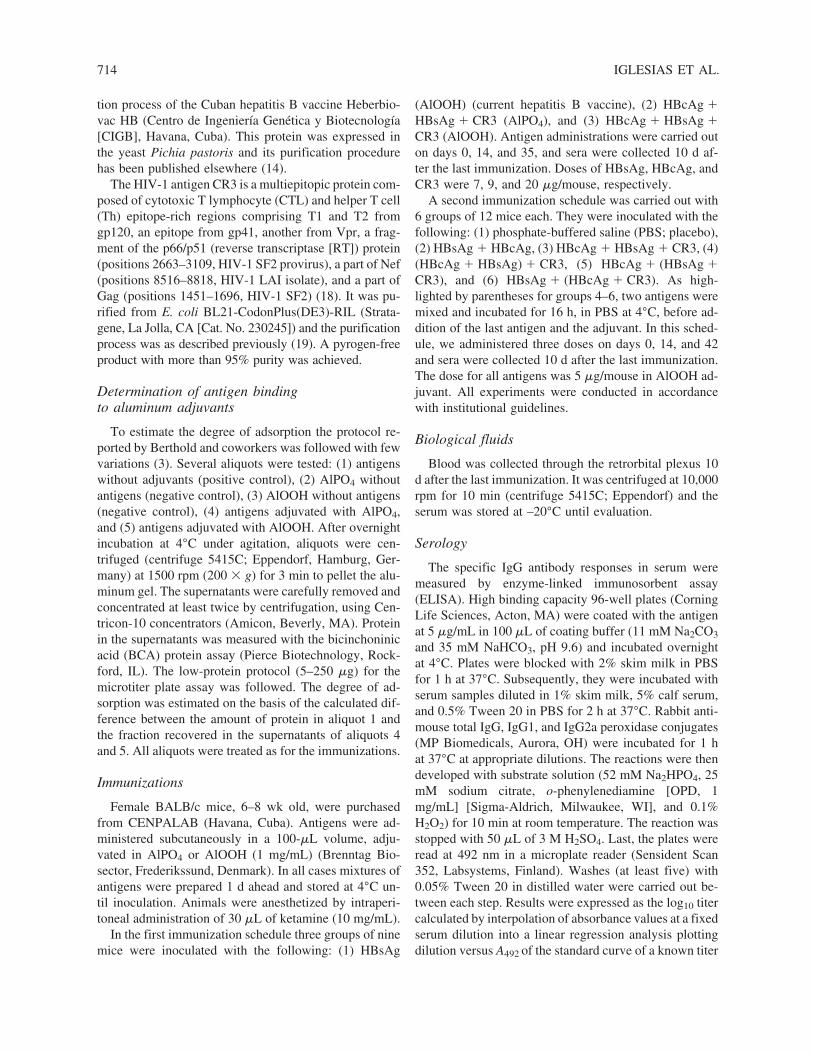
Adjuvant effect of AlPO4 versus AlOOH
First, we studied the adsorption of the mixture HB-sAg � HBcAg � CR3 to both aluminum-based adju-vants and no differences were observed. To study in vivothe role of the adjuvants, mice were immunized with themixture HBsAg � HBcAg � CR3 in aluminum phos-phate and aluminum hydroxide by the subcutaneousroute. In addition, a group of animals was immunizedwith the current vaccine formulation based on HBsAgabsorbed in aluminum hydroxide for comparison. TheIgG1:IgG2a ratio, considered a useful parameter to dis-criminate Th1 and Th2 profiles (30,37,38,41), was de-termined against CR3 and HBsAg after the third dose inanimal sera. In this regard, considering the CR3-specificresponse, better Th1 immunomodulation was obtainedwith the aluminum hydroxide adjuvant (p � 0.0105; two-tailed unpaired t test) (Fig. 1A). However, no differencewas observed when the HBsAg-specific response wasevaluated. In analyzing further, we noted that HBsAg-specific IgG1:IgG2a ratios induced by the multiantigenicformulation with aluminum hydroxide and aluminumphosphate were significantly lower than with HBsAgalone in aluminum hydroxide (the vaccine formulation)(p � 0.0147; one-way ANOVA and Tukey multiple com-parison test). Whole IgG responses specific for CR3, HB-
715
sAg, and HBcAg antigens were not different betweenboth adjuvants (Fig. 1B).
On the other hand, four animals per group were ran-domly selected and killed to evaluate IFN-� and IL-4 se-
IGLESIAS ET AL.
cretion in the supernatant of splenocytes stimulated withCR3 and HBsAg antigens, and the frequency of IFN-�-secreting cells in an ex vivo ELISPOT IFN-� assay. Af-ter an overall analysis of the data, no differences were
716
FIG. 1. Humoral immune response anti-HBsAg and anti-CR3. BALB/c mice were immunized three times with the follow-ing: (1) HBsAg (S) (subcutaneous, AlOOH), a similar formulation to the current commercial vaccine; (2) HBcAg (C) � HB-sAg � CR3 (subcutaneous, AlPO4); (3) HBcAg � HBsAg � CR3 (subcutaneous, AlOOH). Sera were collected 10 d after thelast dose and titers were calculated by ELISA. (A) IgG1:IgG2a ratios and (B) total IgG titers. Data represent means � SD ofnine mice per group. The dotted line in (A) represents the IgG1:IgG2a � 1 ratio.

found between groups of mice immunized with alu-minum phosphate and aluminum hydroxide on consider-ing the level of total IFN-� and IL-4 secreted in the su-pernatants. However, intragroup analysis showed thatIFN-� levels were significantly higher than IL-4 levelsonly in the group inoculated with aluminum hydroxideadjuvant (p � 0.0391; Wilcoxon signed rank test) (Fig.2). The median level of total IFN-� secretion increasedmore than 4000 times compared with IL-4 secretion withAlOOH (median, 158,219; [range, 0 to 430,396] vs. 37.73[range, 0.36–60.57], respectively). In the AlPO4 groupthe same coefficient increased only about 10 times (273.1[range, 0–497,258] vs. 27.75 [range, 7.225–138.9], re-spectively). To characterize further the Th1 response thefrequency of IFN-�-secreting cells was measured. In an-alyzing the specific anti-HBsAg response, we noted a sig-nificant increase in the median frequency of IFN-�-se-creting cells of 313 SFC/106 cells (range, 295 to 1085SFC/106 cells) in the aluminum hydroxide group com-pared with 170 SFC/106 cells (range, 140 to 218 SFC/106
cells) in the aluminum phosphate group (p � 0.0286;two-tailed Mann–Whitney test). A similar evaluation forthe anti-CR3-specific response was carried out. Unfortu-nately, for this antigen an unexpectedly high frequency
HBV AND HIV PROTEINS IN ALUMINUM-BASED ADJUVANTS
of IFN-�-secreting cells was scored in all animals (datanot shown). Because of that it was impossible to deter-mine any difference between groups.
Immune responses elicited by several mixtures of HBsAg, HBcAg, and CR3
Considering the preceding results, aluminum hydrox-ide allowed better Th1 immunomodulation for the mix-ture of the recombinant protein CR3 from HIV-1 withHBV antigens. Because of that, we decided to continuethe experiments with aluminum hydroxide instead of alu-minum phosphate as adjuvant.
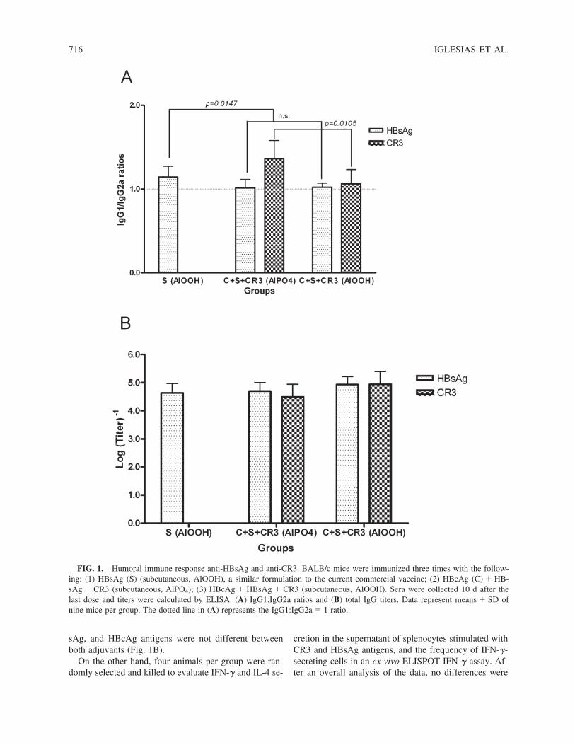
Three additional formulations, variations of the simplemixture of antigens, were prepared for comparison. First,the recombinant core and surface antigens of HBV wereincubated together for several hours before the additionof CR3 and the adjuvant ([HBsAg � HBcAg] � CR3);second and third, the CR3 protein was incubated withHBsAg or HBcAg before addition of the other antigenof HBV ([HBsAg � CR3] � HBcAg and [HBcAg �CR3] � HBsAg, respectively). In addition, groups ofmice immunized with placebo and with HBsAg �HBcAg were included for comparison. After three dosesthe humoral response against the antigens as well as thecellular immune response against CR3 were measured.
Whole IgG anti-HBsAg titers did not differ signifi-cantly among the four formulations of CR3, HBsAg, andHBcAg antigens tested (groups 3 to 6) or with the HB-sAg � HBcAg group (Fig. 3). Regarding the anti-HBcAgresponse, mice immunized with the simple mixture HB-sAg � HBcAg � CR3 elicited the highest titers com-pared with those inoculated with other mixtures of thesame antigens (groups 4 to 6) (p � 0.0001; one-wayanalysis of variance and Tukey multiple comparison test).
As shown in Fig. 3, the highest anti-CR3 titers wereelicited with the formulations HBsAg � HBcAg � CR3and (HBsAg � HBcAg) � CR3 and the lowest titerswith HBsAg � (HBcAg � CR3) (p � 0.0212; one-wayanalysis of variance and Tukey multiple comparison test).In mice immunized with HBcAg � (HBsAg � CR3) thegeometric mean of titers was between those of the othergroups and no statistically significant differences wereobserved.
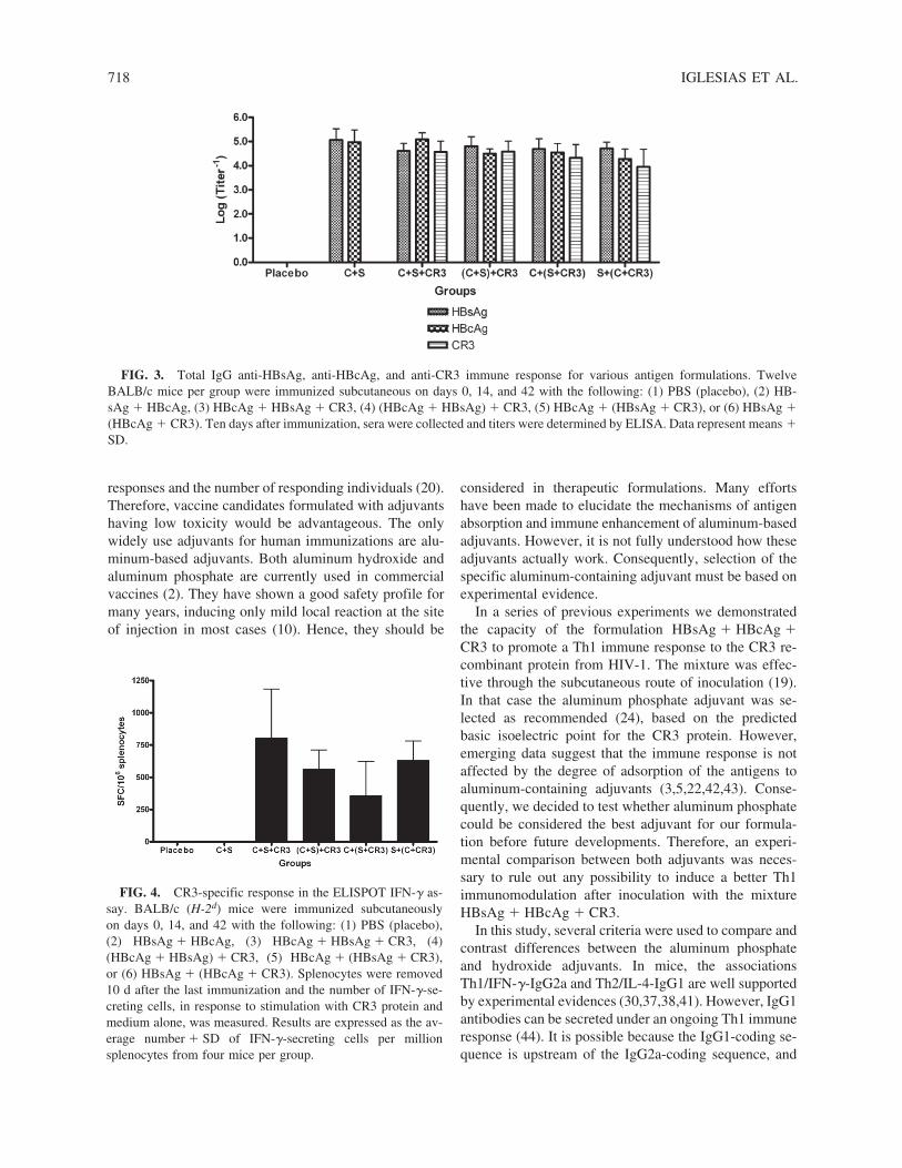
Finally, although the simple mixture of antigens pro-moted the highest anti-CR3 frequency of IFN-�-secret-ing cells in mouse spleen, significant differences fromanimals immunized with the rest of the formulations un-der study were not found (Fig. 4).
DISCUSSION
Data emerging from various therapeutic trials in hu-mans suggest that multiple doses are required to increase
717
FIG. 2. Splenic IFN-� and IL-4 responses after immu-nization with aluminum phosphate and aluminum hydroxideadjuvants. Mice were immunized on days 0, 14, and 35 withthe following: HBcAg � HBsAg � CR3 (subcutaneous,AlPO4) or HBcAg � HBsAg � CR3 (subcutaneous, AlOOH).Ten days after the end of the schedule animals were killed andthe splenocytes were purified, followed by HBsAg and CR3stimulation in culture for 50 h. Culture supernatants were thenharvested and assayed by ELISA. Results were calculated bysubtracting the background of unstimulated control culturesfrom the net values of antigen-pulsed cultures. Data representaverages � SD of nine mice per group.
responses and the number of responding individuals (20).Therefore, vaccine candidates formulated with adjuvantshaving low toxicity would be advantageous. The onlywidely use adjuvants for human immunizations are alu-minum-based adjuvants. Both aluminum hydroxide andaluminum phosphate are currently used in commercialvaccines (2). They have shown a good safety profile formany years, inducing only mild local reaction at the siteof injection in most cases (10). Hence, they should be
IGLESIAS ET AL.
considered in therapeutic formulations. Many effortshave been made to elucidate the mechanisms of antigenabsorption and immune enhancement of aluminum-basedadjuvants. However, it is not fully understood how theseadjuvants actually work. Consequently, selection of thespecific aluminum-containing adjuvant must be based onexperimental evidence.
In a series of previous experiments we demonstratedthe capacity of the formulation HBsAg � HBcAg �CR3 to promote a Th1 immune response to the CR3 re-combinant protein from HIV-1. The mixture was effec-tive through the subcutaneous route of inoculation (19).In that case the aluminum phosphate adjuvant was se-lected as recommended (24), based on the predicted basic isoelectric point for the CR3 protein. However,emerging data suggest that the immune response is notaffected by the degree of adsorption of the antigens toaluminum-containing adjuvants (3,5,22,42,43). Conse-quently, we decided to test whether aluminum phosphatecould be considered the best adjuvant for our formula-tion before future developments. Therefore, an experi-mental comparison between both adjuvants was neces-sary to rule out any possibility to induce a better Th1immunomodulation after inoculation with the mixtureHBsAg � HBcAg � CR3.
In this study, several criteria were used to compare andcontrast differences between the aluminum phosphateand hydroxide adjuvants. In mice, the associationsTh1/IFN-�-IgG2a and Th2/IL-4-IgG1 are well supportedby experimental evidences (30,37,38,41). However, IgG1antibodies can be secreted under an ongoing Th1 immuneresponse (44). It is possible because the IgG1-coding se-quence is upstream of the IgG2a-coding sequence, and
718
FIG. 3. Total IgG anti-HBsAg, anti-HBcAg, and anti-CR3 immune response for various antigen formulations. TwelveBALB/c mice per group were immunized subcutaneous on days 0, 14, and 42 with the following: (1) PBS (placebo), (2) HB-sAg � HBcAg, (3) HBcAg � HBsAg � CR3, (4) (HBcAg � HBsAg) � CR3, (5) HBcAg � (HBsAg � CR3), or (6) HBsAg �(HBcAg � CR3). Ten days after immunization, sera were collected and titers were determined by ELISA. Data represent means �SD.
FIG. 4. CR3-specific response in the ELISPOT IFN-� as-say. BALB/c (H-2d) mice were immunized subcutaneously on days 0, 14, and 42 with the following: (1) PBS (placebo),(2) HBsAg � HBcAg, (3) HBcAg � HBsAg � CR3, (4)(HBcAg � HBsAg) � CR3, (5) HBcAg � (HBsAg � CR3),or (6) HBsAg � (HBcAg � CR3). Splenocytes were removed10 d after the last immunization and the number of IFN-�-se-creting cells, in response to stimulation with CR3 protein andmedium alone, was measured. Results are expressed as the av-erage number � SD of IFN-�-secreting cells per millionsplenocytes from four mice per group.

switching to IgG1 may also be induced by IL-2, whichis a Th1 interleukin (9,12). Consequently, sensu stric-tisimo, only the presence or deficiency of IgG2a anti-bodies may be considered a surrogate marker of helperT cell immune status. In this regard, a high or lowIgG1:IgG2a ratio results in a Th2 or Th1 immune re-sponse, respectively.
Our results indicate that BALB/c mice immunized witha mixture of HBsAg � HBcAg � CR3 with aluminumhydroxide adjuvant developed a better Th1-biased re-sponse than with the aluminum phosphate, as indicatedby the increased level of CR3-specific IgG2a antibodies(lower IgG1:IgG2a ratio). However, the HBsAg-specificIgG1:IgG2a ratios were not different between the adju-vants. These observations support the notion that a dif-ferential Th1–Th2 commitment of the immune systemmight arise to antigens in combined formulations (21). Itis important to note that whole IgG titers against CR3,HBsAg, and HBcAg were of similar magnitude betweenboth groups of animals and it suggests the absence of adifferential immunoenhancing effect on the specific IgGresponses. In addition, the lower IgG1:IgG2a ratio ob-served with both adjuvant formulations compared withthe vaccine formulation suggests the possible anti-HBVtherapeutic application of the mixture of antigens.
When considering the potential role of the aluminumadjuvants (AlPO4 and AlOOH) on anti-CR3 and anti-HB-sAg IFN-� and IL-4 secretions, as a whole, we did notfind significant differences. However, we did find betterTh1 polarization (IFN-� vs. IL-4 secretion) in the CR3 �HBsAg � HBcAg group adjuvated with aluminum hy-droxide. Consistent with this finding, a higher frequencyof specific anti-HBsAg IFN-�-secreting cells was also in-duced. These results provide an explanation and confirmthe previous differences in the IgG1:IgG2a ratios.
Previous studies have shown differential induction ofhelper T cell patterns between the aluminum phosphateand hydroxide adjuvants. Wang et al. demonstrated thatIL-12 combined with aluminum phosphate induced a farbetter Th1 response against HBsAg than did aluminumhydroxide (42). Interestingly, the adjuvant effect of IL-12 bound to aluminum phosphate was not related todifferential adsorption to either adjuvant. In our investi-gation, we demonstrated that the simple mixture of theCR3, HBsAg, and HBcAg antigens is equally adsorbedto aluminum phosphate and aluminum hydroxidewhereas the last adjuvant allowed a stronger commitmentof the immune system to the Th1 response. The fact thatthe immunoenhancing activity of aluminum-based adju-vants does not involve any TLR pathway in antigen-pre-senting cells (35) might explain why Th1 immunomod-ulating agents dominate in combining formulations.Taken together, these observations suggest that there aresubtle differences in the effect of both aluminum-based
HBV AND HIV PROTEINS IN ALUMINUM-BASED ADJUVANTS
adjuvants that modulate the resulting helper T cell pat-tern, depending also on the nature and influence of theantigens. Systematic comparisons of the adjuvant effectaddressing quantitative and/or quality aspects are needed.
In multiantigenic formulations the resulting immunitywill be a consequence of the individual characteristics ofthe antigens as well as of new features that emerge as aresult of reciprocal influences among them. HBcAg in-duces a strong Th1 response, allowing deviation of theimmune response elicited to coadministered antigens(1,27,32). On the other hand, animals immunized withCR3 and HBsAg elicit a Th2 response after inoculationwith alum-based adjuvants (19). Thus, we consider theinfluence of HBcAg as the main cause of the specificanti-CR3 and anti-HBsAg IFN-� secretion and IgG2a an-tibody production in the mixture of CR3 � HBsAg �HBcAg. However, it is worth noting the immunoen-hancing effect contributed by HBsAg to the ongoing anti-HBcAg Th1 response in combining formulations withHBcAg (1,27) and to the ongoing anti-CR3 Th1 responsein combining formulations with the former antigen andCR3 protein (19). In that sense, the proteoliposomal na-ture of the surface antigen (33) might be related to thiseffect. In addition, there is a spontaneous tendency of thesurface and core antigens to aggregate to form supramol-ecular structures up to 200 nm in size (1). In our multi-antigenic formulation, assuming the association withCR3 protein (19), the size might increase further than 200nm. Such structures will increase the phagocytic inter-nalization of the antigens (4) and consequently their im-munogenicity. Finally, bystander stimulation due to TLRstimulation (23,40) and antigen-specific ongoing immuneresponses elicited in the vicinity might also contribute tothe individual immunogenicity of the antigens.
A preliminary experiment was also carried out withvarious formulations of the antigens. Previous studiesshowed that CR3, HBsAg, and HBcAg aggregate in so-lution (19). Thus, it could be argued that preferential in-teractions between particular pairs of such antigens mightimpact on the specific immune response elicited againstsome of them. This hypothesis corresponds well with ourdata showing that the antibody levels among groups ofanimals differed with the anti-HBcAg and anti-CR3 hu-moral responses. Nevertheless, anti-HBsAg levels did notdiffer among the groups. These results are in line withprevious findings showing a stronger interaction betweenCR3 and HBcAg than with CR3-HBsAg and it might ex-plain the former results. Because the anti-HBcAg andanti-CR3 antibodies might be of limited practical value,we emphasize the protective value of the anti-HBsAg hu-moral response (16). In this regard, the strong interactionbetween CR3 and HBcAg could benefit the anti-HBsAghumoral response. In addition, because we did not findany difference in the frequency of anti-CR3 IFN-�-se-
719

creting cells among the antigen formulations it could beargued that no other formulation elicited a better immuneresponse than the simple mixture of HBsAg, HBcAg, andCR3.
In summary, the results thus far revealed evidence thataluminum hydroxide is a better choice than aluminumphosphate for Th1 induction after immunization with themixture CR3 � HBcAg � HBsAg. In addition, in the ab-sence of evidence against the former formulation of anti-gens this work suggests the use of aluminum hydroxideas a potential adjuvant in antigen formulations elicitingcellular anti-HBV and anti-HIV-1 immunity in humans.
ACKNOWLEDGMENTS
The authors thank Ismarisley Revé for technical assis-tance in the culture laboratory. This work was financedby the Center of Genetic Engineering and Biotechnology(Havana, Cuba).
REFERENCES
1. Aguilar JC, Lobaina Y, Muzio V, Garcia D, Penton E, Igle-sias E, Pichardo D, Urquiza D, Rodriguez D, Silva D, Petro-vsky N, and Guillen G: Development of a nasal vaccinefor chronic hepatitis B infection that uses the ability of he-patitis B core antigen to stimulate a strong Th1 responseagainst hepatitis B surface antigen. Immunol Cell Biol2004;82:539–546.
2. Baylor NW, Egan W, and Richman P: Aluminum salts invaccines: US perspective. Vaccine 2002;20:S18–S23.
3. Berthold I, Pombo ML, Wagner L, and Arciniega JL: Im-munogenicity in mice of anthrax recombinant protectiveantigen in the presence of aluminum adjuvants. Vaccine2005;23:1993–1999.
4. Brewer JM, Pollock KG, Tetley L, and Russell DG: Vesi-cle size influences the trafficking, processing, and presen-tation of antigens in lipid vesicles. J Immunol 2004;173:6143–6150.
5. Chang M, Shi Y, Nail SL, HogenEsch H, Adams SB, WhiteJL, and Hem SL: Degree of antigen adsorption in the vac-cine or interstitial fluid and its effect on the antibody re-sponse in rabbits. Vaccine 2001;19:2884–2889.
6. Clements CJ, and Griffiths E: The global impact of vac-cines containing aluminium adjuvants. Vaccine 2002;20:S24–S33.
7. Cooper PD: Vaccine adjuvants based on � inulin. PharmBiotechnol 1995;6:559–580.
8. Davis HL, Weeratna R, Waldschmidt TJ, Tygrett L, SchorrJ, and Krieg AM: CpG DNA is a potent enhancer of spe-cific immunity in mice immunized with recombinant he-patitis B surface antigen. J Immunol 1998;160:870–876.
IGLESIAS ET AL.
9. DeKruyff RH, Mosmann RR, and Umetsu DT: Inductionof antibody synthesis by CD4� T cells: IL-5 is essentialfor induction of antigen-specific antibody responses by TH2but not TH1 clones. Eur J Immunol 1990;20:2219–2227.
10. Duclos P: Safety of immunisation and adverse events fol-lowing vaccination against hepatitis B. Expert Opin DrugSaf 2003;2:225–231.
11. Ericsson H: Purification and adsorption of diphtheria tox-oid. Nature 1946;158:350–351.
12. Finkelman FD, Katona IM, Urban JF Jr, Snapper CM,Ohara J, and Paul WE: Suppression of in vivo polyclonalIgE responses by monoclonal antibody to the lymphokineB-cell stimulatory factor 1. Proc Natl Acad Sci USA1986;83:9675–9678.
13. Glenny A, Pope C, Waddington H, and Wallace U: Theantigenic value of toxoid precipitated by potassium alum.J Pathol Bacteriol 1926;29:38–45.
14. Hardy E, Martinez E, Diago D, Diaz R, Gonzalez D, andHerrera L: Large-scale production of recombinant hepati-tis B surface antigen from Pichia pastoris. J Biotechnol2000;77:157–167.
15. Helms T, Boehm BO, Asaad RJ, Trezza RP, Lehmann PV,and Tary-Lehmann M: Direct visualization of cytokine-producing recall antigen-specific CD4 memory T cells in healthy individuals and HIV patients. J Immunol2000;164:3723–3732.
16. Hilleman MR: Overview of the pathogenesis, prophylaxisand therapeusis of viral hepatitis B, with focus on reduc-tion to practical applications. Vaccine 2001;19:1837–1848.
17. HogenEsch H: Mechanisms of stimulation of the immuneresponse by aluminum adjuvants. Vaccine 2002. 20:S34–S39.
18. Iglesias E, Ruiz M, Carrazana Y, Cruz LJ, Aguilar A,Jimenez V, Carpio E, Martinez M, Perez M, Martinez C,Cruz O, Martin A, and Duarte C: Chimeric proteins con-taining HIV-1 T cell epitopes: Expression in E. coli, pu-rification and induction of antibodies in mice. J BiochemMol Biol Biophys 2001;5:109–120.
19. Iglesias E, Thompson R, Carrazana Y, Lobaina Y, GarciaD, Sanchez J, Garcia J, Cruz O, Brown E, Martin A, MuzioVL, and Aguilar JC: Coinoculation with hepatitis B sur-face and core antigen promotes a Th1 immune response toa multiepitopic protein of HIV-1. Immunol Cell Biol2006;84:174–183.
20. Im EJ, and Hanke T: MVA as a vector for vaccines againstHIV-1. Expert Rev Vaccines 2004;3:S89–S97.
21. Ismail N, and Bretscher PA: The Th1/Th2 nature of con-current immune responses to unrelated antigens can be in-dependent. J Immunol 1999;163:4842–4850.
22. Iyer S, HogenEsch H, and Hem SL: Effect of the degreeof phosphate substitution in aluminum hydroxide adjuvanton the adsorption of phosphorylated proteins. Pharm DevTechnol 2003;8:81–86.
720

23. Kamath AT, Sheasby CE, and Tough DF: Dendritic cellsand NK cells stimulate bystander T cell activation in re-sponse to TLR agonists through secretion of IFN-�� andIFN-�. J Immunol 2005;174:767–776.
24. Lindblad EB: Aluminium adjuvants: In retrospect andprospect. Vaccine 2004;22:3658–3668.
25. Lindblad EB: Aluminium compounds for use in vaccines.Immunol Cell Biol 2004;82:497–505.
26. Lobaina Y, Garcia D, Abreu N, Muzio V, and Aguilar JC:Mucosal immunogenicity of the hepatitis B core antigen.Biochem Biophys Res Commun 2003;300:745–750.
27. Lobaina Y, Palenzuela D, Pichardo D, Muzio V, GuillenG, and Aguilar JC: Immunological characterization of twohepatitis B core antigen variants and their immunoenhanc-ing effect on co-delivered hepatitis B surface antigen. MolImmunol 2005;42:289–294.
28. Maschmann E, Küster E, and Fischer W: Über die Fähigkeitdes Tonerde-Präparates B, Diphtherie-Toxin zu adsor-bieren. Ber Dtsch Chem Ges 1931;64:2174–2178.
29. Morefield GL, Sokolovska A, Jiang D, HogenEsch H,Robinson JP, and Hem SL: Role of aluminum-containingadjuvants in antigen internalization by dendritic cells invitro. Vaccine 2005;23:1588–1595.
30. Mosmann TR, Cherwinski H, Bond MW, Giedlin MA, andCoffman RL: Two types of murine helper T cell clone. I.Definition according to profiles of lymphokine activitiesand secreted proteins. J Immunol 1986;136:2348–2357.
31. Riedl P, Buschle M, Reimann J, and Schirmbeck R: Bind-ing immune-stimulating oligonucleotides to cationic pep-tides from viral core antigen enhances their potency as ad-juvants. Eur J Immunol 2002;32:1709–1716.
32. Riedl P, Stober D, Oehninger C, Melber K, Reimann J, andSchirmbeck R: Priming Th1 immunity to viral core parti-cles is facilitated by trace amounts of RNA bound to itsarginine-rich domain. J Immunol 2002;168:4951–4959.
33. Satoh O, Imai H, Yoneyama T, Miyamura T, Utsumi H,Inoue K, and Umeda M: Membrane structure of the he-patitis B virus surface antigen particle. J Biochem (Tokyo)2000;127:543–550.
34. Schijns VE: Immunological concepts of vaccine adjuvantactivity. Curr Opin Immunol 2000;12:456–463.
35. Schnare M, Barton GM, Holt AC, Takeda K, Akira S, andMedzhitov R: Toll-like receptors control activation of adap-tive immune responses. Nat Immunol 2001;2:947–950.
HBV AND HIV PROTEINS IN ALUMINUM-BASED ADJUVANTS
36. Seeber SJ, White JL, and Hem SL: Predicting the adsorp-tion of proteins by aluminium-containing adjuvants. Vac-cine 1991;9:201–203.
37. Sideras P, Bergstedt-Lindqvist S, and Severinson E: Par-tial biochemical characterization of IgG1-inducing factor.Eur J Immunol 1985;15:593–598.
38. Snapper CM, and Paul WE: Interferon-� and B cell stim-ulatory factor-1 reciprocally regulate Ig isotype production.Science 1987;236:944–947.
39. Timofeev AV: Unexpected problem of the traditional ap-proach to the addition of aluminium hydroxide to vaccinepreparations. Vaccine 2005;23:2953.
40. Tough DF, Borrow P, and Sprent J: Induction of bystanderT cell proliferation by viruses and type I interferon in vivo.Science 1996;272:1947–1950.
41. Vitetta ES, Ohara J, Myers CD, Layton JE, Krammer PH,and Paul WE: Serological, biochemical, and functionalidentity of B cell-stimulatory factor 1 and B cell differen-tiation factor for IgG1. J Exp Med 1985;162:1726–1731.
42. Wang S, Liu X, and Caulfield MJ: Adjuvant synergy in theresponse to hepatitis B vaccines. Vaccine 2003;21:4297–4306.
43. Weissburg RP, Berman PW, Cleland JL, Eastman D, Fa-rina F, Frie S, Lim A, Mordenti J, Nguyen TT, and Peter-son MR: Characterization of the MN gp120 HIV-1 vac-cine: Antigen binding to alum. Pharm. Res. 1995;12:1439–1446.
44. Yip HC, Karulin AY, Tary-Lehmann M, Hesse MD,Radeke H, Heeger PS, Trezza RP, Heinzel FP, Forsthu-ber T, and Lehmann PV: Adjuvant-guided type-1 andtype-2 immunity: Infectious/noninfectious dichotomydefines the class of response. J Immunol 1999;162:3942–3949.
Address reprint requests to:Dr. Enrique Iglesias
Centro de Ingeniería Genética y BiotecnologíaAvenida 31 e/ 158 y 190, Cubanacán, Playa
P.O. Box 6162Havana 10600, Cuba
E-mail: [email protected]
Received May 12, 2006; accepted June 10, 2006.
721