Infliximab Dose Rounding Pratices in Pediatric Inflammatory ...
Upload
healthsciences-uclaCategory
view
0download
0
Infliximab Attenuates Early Postresuscitation MyocardialDysfunction in a Swine Cardiac Arrest Model
James T. Niemann, MD1,4, Scott Youngquist, MD, MSc5, John P. Rosborough, PhD1, AtmanP. Shah, MD2,4, Quynh T. Phan, BS3, and Scott G. Filler, MD3,41 Department of Emergency Medicine, Harbor-UCLA Medical Center, Torrance, CA2 Department of Medicine, Divisions of Cardiology, Harbor-UCLA Medical Center, Torrance, CA3 Department of Infectious Disease, Harbor-UCLA Medical Center, Torrance, CA4 David Geffen School of Medicine at UCLA, Los Angeles, CA5 University of Utah School of Medicine, the Department of Surgery, Division of EmergencyMedicine, Salt Lake City, UT
AbstractObjective—Left ventricular (LV) dysfunction after successful cardiopulmonary resuscitationcontributes to early death following resuscitation. Proinflammatory cytokines are known to depressmyocardial function and TNF-alpha has been shown to increase after successful resuscitation. Wehypothesized that blocking the effects of TNF-alpha with infliximab would prevent or minimizepostresuscitation cardiac dysfunction.
Design—Randomized, placebo-controlled comparative study.
Setting—Large animal research laboratory.
Subjects—Twenty-eight anesthetized and instrumented domestic male swine (Yorkshire andYorkshire/Hampshire mix, weight 35-45 kg).
Interventions—Infusion of infliximab (5 mg/kg) or normal saline after resuscitation from VFcardiac arrest.
Measurements and Main Results—Hemodynamic variables, indices of LV function, and TNF-alpha were measured before and after 8 min of cardiac arrest during the early post-resuscitation period(3 hrs). Within 5 min of restoration of spontaneous circulation, 14 animals received infliximab, 5mg/kg, infused over 30 min. Fourteen animals received an infusion of normal saline. Inotropes andvasopressors were not administered to either group following resuscitation. TNF-alpha increasedfollowing restoration of circulation and remained elevated throughout the observation period.Differences between groups were not significant. IL-1β concentration did not change significantlyduring the observation period in either study group. Mean arterial pressure and stroke work weresignificantly greater in the infliximab group within 30 mins of resuscitation and these differenceswere sustained throughout the 3 hr postresuscitation period. The effect of TNF-α blockade wasevident only in animals with a significant increase (doubling) in plasma TNF-α at 30 minutes post-arrest.
Conclusion—TNF-α plays a role in post-arrest cardiac dysfunction and Infliximab may attenuateor prevent postresuscitation myocardial dysfunction when given immediately after resuscitation.
Address for correspondence: James T. Niemann, MD, Harbor-UCLA Medical Center, Department of Emergency Medicine, 1000 WestCarson Street, Box 21, Torrance, CA 90509, [email protected], Phone: 310-222-6742.
NIH Public AccessAuthor ManuscriptCrit Care Med. Author manuscript; available in PMC 2011 April 1.
Published in final edited form as:Crit Care Med. 2010 April ; 38(4): 1162–1167. doi:10.1097/CCM.0b013e3181d44324.
NIH
-PA Author Manuscript
NIH
-PA Author Manuscript
NIH
-PA Author Manuscript
Keywordsheart arrest; ventricular fibrillation; cardiopulmonary resuscitation; tumor-necrosis factor;interleukins; hemodynamics
IntroductionApproximately 60% of all cardiac deaths are due to out-of-hospital sudden cardiac death.Although resuscitation efforts are effective in restoring a pulse in 30-40% of victims of cardiacarrest, the eventual hospital discharge rate is <5%.(1,2) In-hospital death is usually the resultof ischemic brain injury, multi-organ failure, or cardiac dysfunction.(3) Post-resuscitationmyocardial dysfunction has been reported in experimental cardiac arrest models and the clinicalpopulation and potential mechanisms defined.(4) Despite the prevalence of post-resuscitationmyocardial dysfunction in successfully resuscitated patients and its potential contribution toearly mortality, few studies have addressed its etiology or management.(5-8)
Ischemia/reperfusion injury has been characterized as a multifactorial antigen-independentinflammatory condition wherein accelerated proinflammatory cytokine synthesis and releaseare commonly observed.(9,10) TNF-α and IL-1β are proinflammatory cytokines known todepress myocardial contractility.(11) Observational clinical studies in small series of patientshave demonstrated elevated TNF-α and IL-1β levels in patients resuscitated from cardiac arrest.(12,13) TNF-α is elevated at the time of initial sampling after resuscitation and hospitaladmission and remains elevated in those patients who eventually die in-hospital or who requirecatecholamines for hemodynamic support. A recent laboratory study in a porcine cardiac arrestand resuscitation model demonstrated an inverse relationship between plasma TNF-αconcentrations and myocardial contractility, measured as LV dp/dt.(14)
The purpose of this study was to determine if blocking TNF-α following resuscitation fromprolonged cardiac arrest would prevent post-resuscitation myocardial dysfunction.
MethodsThis investigation was approved by the Animal Care and Utilization Review Committee of ourinstitution and conformed to the National Institutes of Health policy on humane care and useof laboratory animals.
Twenty-eight domestic male swine (Yorkshire and Yorkshire/Hampshire mix, weight 35-45kg) were premedicated with ketamine (20 mg/kg) and xylazine (2 mg/kg). General anesthesiawas induced with isoflurane via nose cone and, following endotracheal intubation, maintainedwith inhaled isoflurane and nitrous oxide in a 1:1 mixture with oxygen. Minute ventilation wasadjusted to maintain an end-tidal CO2 of 35-45 mm Hg. Standard lead II of the surface ECGwas monitored during instrumentation and throughout the study protocol.
Both external jugular veins and a femoral artery were surgically exposed and micromanometer-tipped catheters (Millar Instruments, Houston, TX) were inserted and positioned in the RA,LV, and Ao arch. The tip of a bipolar pacing catheter was positioned in contact with the rightventricular endocardium for induction of VF. A multilumen catheter thermistor (EdwardsLifesciences, Irvine, CA) was positioned in a branch of the pulmonary artery for thermodilutionCO determinations. Standard adhesive defibrillation electrode patches were applied to the leftand right lateral aspects of the shaved thorax (Quick-Combo, Medtronic Emergency ResponseSystems, Redmond, WA). Transthoracic impedance was measured using a tetrapolar constantcurrent impedance measuring system (THRIM®, Morro Bay, CA). A small value non-
Niemann et al. Page 2
Crit Care Med. Author manuscript; available in PMC 2011 April 1.
NIH
-PA Author Manuscript
NIH
-PA Author Manuscript
NIH
-PA Author Manuscript
inductive resistor (30 Ω) was then placed in series with the truncated exponential biphasicdefibrillation waveform defibrillator (LifePak 12, Medtronic Emergency Response Systems,Redmond, WA).
Following instrumentation, heart rate, core temperature (measured with thermistor catheter),systolic and diastolic Ao pressure, LVEDP, and CO (measured in triplicate) were recorded andarterial blood was analyzed (I-Stat CG8+, I-Stat Corp, Princeton, NJ). Mean arterial pressure(MAP) and stroke volume (SV) were derived using standard formulae. The time constant ofisovolumic LV relaxation (tau) was determined using commercially available software (BloodPressure Module, v 1.0.3, ADInstruments, Colorado Springs, CO). Hemodynamic data wererecorded and stored on a lap-top computer using PowerLab Chart v. 5.2 (ADInstruments,Colorado Springs, CO). VF was then induced with a brief 60 Hz AC current pulse deliveredto the right ventricular endocardium via the pacing catheter. After 7 min of untreated VF,mechanical closed-chest compressions (Thumper®, Michigan Instruments, Grand Rapids, MI)were begun with the animal in the supine position and were administered at a rate ofapproximately 100/min with force sufficient to depress the sternum 1.5 to 2.0 inches.Ventilation was not performed during the one minute interval preceding the first countershock.After one minute, electrical defibrillation was attempted with a biphasic waveform defibrillator(Medtronic Physio-Control Corporation, LifePak 12). Three shocks were administered in anescalating energy sequence (200-300-360 J), if necessary. Chest compressions were performedbetween countershocks. If one of the first three countershocks terminated VF but resulted in anonperfusing spontaneous cardiac rhythm, i.e., asystole or PEA or if VF persisted after the firstthree shocks, chest compressions were restarted and continued for two minutes or ROSC.Animals not resuscitated within two minutes were given intravenous epinephrine (0.01 mg/kg) every 5-8 min and CPR continued until spontaneous perfusing rhythm was established.
Following resuscitation, animals were randomized into two groups using permuted blockdesign. Control animals (n = 14) were given 250 cc of normal saline over 30 min, beginning5 min after ROSC. Animals in the treatment group (n = 14) were given infliximab, a monoclonalanti-TNF-α antibody, at a dose of 5 mg/kg in 250 ml of normal saline infused over 30 minbeginning 5 min after ROSC. No other drugs were given or interventions performed duringthe ensuing 3 hr post-ROSC observation period. Hemodynamic measurements and coretemperature were made at 30, 60, 120, and 180 min following ROSC.
Prior to VF induction and at 30, 60, 120, and 180 min following restoration of circulation,arterial blood was sampled, placed in sterile, chilled (0° C), heparinized tubes, and centrifugedat 5000 rpm for 10 min. Plasma was immediately separated and stored at -80° C until analysis.TNF-α and IL1-β concentrations were determined by a quantitative sandwich ELISA usingcommercially available kits specific for these porcine cytokines (R&D Systems, Inc.,Minneapolis, MN).
Data AnalysesStatistical analyses were conducted using SAS Version 9.1.3 (SAS Institute, Cary, NC).Summary measures are reported as means and standard deviations for normal distributions ormedians and inter-quartile ranges for non-normal distributions. Student's t-tests or Mann-Whitney Rank Sum tests were used for comparing fixed time-point variables between salineand infliximab treated animals.
Generalized linear mixed models were employed to evaluate associations between variableswhile accounting for the autocorrelation inherent in repeated measure designs. The SASprocedure, Proc Mixed, was employed for this purpose, with time included as a random effectand other variables included as fixed effects. Variable selection was based on a prioriassumptions and iterative fitting of potential models. Model fit and correlation structure
Niemann et al. Page 3
Crit Care Med. Author manuscript; available in PMC 2011 April 1.
NIH
-PA Author Manuscript
NIH
-PA Author Manuscript
NIH
-PA Author Manuscript
specification was assessed by Akaike Information Criteria. When making multiple post-resuscitation comparisons with prearrest values, p values were adjusted according to themethod of Dunnett-Hsu.(15) We considered an alpha level of 0.05 to be significant.
ResultsDifferences in prearrest hemodynamic variables were not observed between treatment groups(table 1). Similarly, variables associated with resuscitation outcome and post-resuscitationventricular function were likewise not significantly different (table 2).
Following resuscitation, TNF-α concentration 30 and 60 min after ROSC were significantlygreater than pre-arrest values for both study groups. However, IL-1β levels did not varysignificantly from pre-arrest values in either group. There was no statistically significantdifference in the response of either TNF-α or IL-1β between treatment groups over time (figures1 and 2). At 3 hrs post-resuscitation, core temperature was not significantly different betweengroups (control group, 35.5 ± 1.3 °C, infliximab group, 35.6 ± 3 °C).
In a generalized linear mixed model, infliximab treated animals had a MAP that was, onaverage, 8.7 mmHg (95% confidence interval [CI] 4.5-13.0 mmHg, p=0.0001) higher thancontrol animals. Treated animals also had a mean SW that was, on average, 9.1 gm-m (95%CI 5.4-12.8 gm-m, p<0.0001) higher than control animals. Postresuscitation hemodynamicvariables are shown in table 3. Differences in mean values for MAP and SW are illustrated infigure 3. Systolic dp/dt was significantly greater in the infliximab group at all postresuscitationtime points and stroke volume was greater from 30 to 120 min. TNF-α levels were inverselycorrelated with MAP (r= −0.41, p<0.0001) and with SW (r= −0.37, p<0.0001). IL-1β, however,was not associated with either response (p>0.11 for both associations).
When animals were classified into TNF-α producers (defined as at least a doubling of TNF-α at 30 minutes when compared to prearrest values, n = 15) and non-producers (n = 13) (figure4), an association between infliximab treatment and higher MAP and SW was seen only withinthe TNF-α producer group. In this group, infliximab treated animals had, on average, 10.8mmHg higher MAP (95% CI 5.5-16.1 mmHg, p=0.0002) than controls. There was also aninteraction between time and treatment assignment. This interaction resulted in a lessening ofthe treatment effect on MAP over time until there was no statistical difference in MAP betweenthe two groups at the 180 minute measurement. Infliximab treatment was also associated withhigher SW in producers, but not non-producers. SW among infliximab treated animals was,on average, 11.2 gm-m (95% CI 5.7-16.7 gm-m, p=0.0002) higher than controls. This effectwas not time-varying in the model with infliximab-treated animals having a consistently higherSW across all time points.
DiscussionThis study in a conventional porcine cardiac arrest model demonstrates that infliximab, amonoclonal anti-TNF-α antibody, administered after resuscitation, is effective in preservinghemodynamic indices of left ventricular function. Left ventricular stroke work, a globalmeasure of cardiac function, was significantly greater in the treatment group within 30 min ofsuccessful resuscitation and hemodynamic differences persisted throughout the observationperiod. When animals were subsequently classified based upon the post-arrest TNF-α response,the beneficial effect of infliximab was most prominent in those animals with a significantincrease in TNF following restoration of spontaneous circulation. These findings suggest thatTNF-α plays a contributory role in post-arrest myocardial dysfunction, one of the hallmarksof the post-arrest syndrome.
Niemann et al. Page 4
Crit Care Med. Author manuscript; available in PMC 2011 April 1.
NIH
-PA Author Manuscript
NIH
-PA Author Manuscript
NIH
-PA Author Manuscript

TNF-α is a known myocardial depressant, presumably by disrupting calcium homeostasis orcalcium sensitivity and the normal myocardial contraction-relaxation cycle.(16,17) TNF-α mayalso induce a state of relative catecholamines insensitivity or refractoriness.(18-20) Thehemodynamic effects of TNF-α include depressed myocardial contractility, decreased ejectionfraction, and ventricular dilatation. The use of monoclonal antibodies to TNF-α in low doses(1 mg/kg) has been shown to minimize cardiac dysfunction in in vivo and isolated heart modelsof acute global and regional myocardial ischemia.(21).
Infliximab is produced by a recombinant cell line and is a chimeric IgG1κ monoclonal antibodycomposed of human constant and murine variable regions of TNF-α. Porcine TNF shares asimilar structure with that of human and murine TNF and exhibits cytotoxicity to targetindicator cells (PK(15) and L929) at similar concentrations.(22) Porcine TNF-α cytotoxicactivity can be totally neutralized with anti-human TNF monoclonal antibody.(23) PorcineTNF-α receptors likewise share a structure similar to that of humans and mice and humansoluble TNF-α receptors bind porcine TNF-α.(24) Considering these characteristics, bindingto and neutralization of porcine TNF-α by infliximab would be expected.
We observed considerable variability in the TNF-α response to ischemia and reperfusion withinthe control and treatment groups. We postulate that this variable response is likely due togenetic polymorphism in the promoter region of the TNF-α gene. TNF-α single nucleotidepolymorphism has been extensively studied in humans and is postulated to play a role insusceptibility and response to infection as well as outcome.(25-27) Polymorphism would beexpected in the typical outbred, mixed breed swine used in research laboratories.
This study has several limitations. Although the anatomy of the porcine coronary circulationis similar to that of humans, the absence of significant underlying atherosclerotic coronaryartery disease and prior myocardial injury, both of which are usually present in resuscitatedpatients, is likely to have affected the extent of cardiac dysfunction following resuscitation.We used only male swine in this study due to prior observations suggesting that there are genderdifferences in the proinflammatory response following resuscitation and that the response ismore dramatic and more predictable in males than females.(28) We evaluated one cardiac arrestduration (7 min) prior initiation of resuscitation efforts. A long duration of ischemia may haveresulted in a more pronounced inflammatory response. The anesthetic agent necessary toconduct the study may also have impacted coronary vascular tone as well as baseline andpostresuscitation LV function. Although statistically significant differences in energy requiredfor defibrillation, time to restoration of spontaneous circulation, and epinephrine dose werenot demonstrated, small differences might be physiologically significant and may have affectedpostresuscitation ventricular function. We evaluated only one dose of infliximab in this study.We selected a dose of 5 mg/kg because prior work in clinical trials indicated that 5 mg/kg didnot produce immediate significant side effects when administered intravenously andapproximates the dose used in large clinical trials of TNF-α blockade in sepsis.(29,30) We didnot evaluate the potential role of other cytokine inhibitors, e.g., an anti-IL-1β inhibitor, on post-arrest cardiac dysfunction. However, IL-1β concentrations did not significantly vary duringthe 3 hour post-arrest study period, were not associated with hemodynamic variables, and thusis unlikely to have contributed substantially to the observed hemodynamic depression. We didnot attempt to control body temperature during the observation period and observed statisticallyinsignificant differences in temperature between groups. Prior work in whole animal modelssubjected to hypothermia following cardiac arrest and hemorrhagic shock has not demonstrateda significant effect of hypothermia on the inflammatory response.(31,32)
Anti-cytokine therapy has not been shown to be beneficial in some disease states characterizedby increased cytokine production, specifically TNF-α. Notably, infliximab and etanercept havenot been shown to be consistently effective in the setting of chronic congestive heart failure or
Niemann et al. Page 5
Crit Care Med. Author manuscript; available in PMC 2011 April 1.
NIH
-PA Author Manuscript
NIH
-PA Author Manuscript
NIH
-PA Author Manuscript
the sepsis syndrome and reasons for the lack of a positive effect have been enumerated.(33,34) However, it is likely that the organism resuscitated from an acute, short-lived, global (totalbody) ischemic insult, i.e., cardiac arrest, represents a unique physiologic state characterizedby an early and sustained maximal stress response and a unique proinflammatory cytokineresponse when compared to other more indolent insults.(35) The findings of this investigationin a commonly used porcine cardiac arrest model suggest that TNF-α plays a role inpostresuscitation myocardial contractile dysfunction and that therapeutic interventions thatinhibit its effects may attenuate to some degree, but not prevent its myocardial depressantproperties. Other factors contribute to the postresuscitation myocardial dysfunction.(4)Hemodynamic benefit appears to be dependent upon the TNF-α response after resuscitationand reperfusion and are likely genotypically determined. TNF-α blockers, either alone or incombination with drugs that block other cytokines or their receptors, may be useful adjunctsin the management of the post-arrest syndrome, particularly if the TNF-α or other cytokineblood levels are known early after resuscitation.
AcknowledgmentsSupported, in part, by a grant from the National Institutes of Health, NHLBI R01 HL076671.
References1. Nichol G, Steen P, Herlitz J, et al. International resuscitation network registry: design, rationale and
preliminary results. Resuscitation 2005;65:265–277. [PubMed: 15919562]2. Nichol G, Thomas E, Callaway CW, et al. Regional variation in out-of-hospital cardiac arrest incidence
and outcome. JAMA 2008;300:1423–31. [PubMed: 18812533]3. Neumar RW, Nolan JP, Adrie C, et al. Post-cardiac arrest syndrome. Epidemiology, pathophysiology,
treatment, and prognostication. Circulation 2008;118:2452–83. [PubMed: 18948368]4. Gazmuri, RJ.; Weil, MH.; Kern, KB., et al. Prevention and therapy of postresuscitation myocardial
dysfunction. In: Paradis, NA.; Halperin, HR.; Kern, KB.; Wenzel, V.; Chamberlain, DA., editors.Cardiac Arrest. The Science and Practice of Resuscitation Medicine. Second. New York: CambridgeUniversity Press; 2007. p. 829-847.
5. Gazmuri RJ, Ayoub IM, Kolarova J. Myocardial protection during resuscitation from cardiac arrest.Curr Opin Crit Care 2003;9:199–204. [PubMed: 12771670]
6. Kern KB, Hilwig RW, Berg RA, et al. Postresuscitation left ventricular systolic and diastolicdysfunction. Treatment with dobutamine. Circulation 1997;95:2610–13. [PubMed: 9193427]
7. Niemann JT, Garner D, Khaleeli E, Lewis RJ. Milrinone facilitates resuscitation from cardiac arrestand attenuates postresuscitation myocardial dysfunction. Circulation 2003;108:1032–36.
8. Vasquez A, Kern KB, Hilwig RW, Heidenreich J, Berg RA, Ewy GA. Optimal dosing of dobutaminefor treating post-resuscitation left ventricular dysfunction. Resuscitation 2004;61:199–207. [PubMed:15135197]
9. Boros P, Bromber JS. New cellular and molecular immune pathways in ischemia/reperfusion injury.Am J Tranplant 2006;6:652–58.
10. Stangl V, Baumann G, Stangl K, Felix SB. Negative inotropic mediators from heart after myocardialischaemia-reperfusion. Cardiovasc Res 2002;53:12–30. [PubMed: 11744010]
11. Kan H, Finkel MS. Inflammatory mediators and reversible myocardial dysfunction. J Cell Physiol2003;195:1–11. [PubMed: 12599203]
12. Ito T, Saitoh D, Fukuzuka K, et al. Significance of elevated serum interleukin-8 in patients resuscitatedafter cardiac arrest. Resuscitation 2001;51:47–53. [PubMed: 11719173]
13. Adrie C, Adib-Conquy M, Laurent I, et al. Successful cardiopulmonary resuscitation after cardiacarrest as a “sepsis-like” syndrome. Circulation 2002;106:562–68. [PubMed: 12147537]
14. Niemann JT, Garner D, Lewis RJ. Tumor necrosis factor-α is associated with early postresuscitationmyocardial dysfunction. Crit Care Med 2004;32:1753–58. [PubMed: 15286554]
Niemann et al. Page 6
Crit Care Med. Author manuscript; available in PMC 2011 April 1.
NIH
-PA Author Manuscript
NIH
-PA Author Manuscript
NIH
-PA Author Manuscript

15. Dunnett CW. A multiple comparison procedure for comparing several treatments with a control. JAmer Statist Assoc 1955;50:1096–1121.
16. Meldrum DR. Tumor necrosis factor in the heart. Am J Physiol 1998;174:R577–95. [PubMed:9530222]
17. Yokoyama T, Vaca L, Rossen RD, Durante W, Hazarika P, Mann DL. Cellular basis for the negativeinotropic effects of tumor necrosis factor-alpha in the adult mammalian heart. J Clin Invest1993;92:2303–12. [PubMed: 8227345]
18. Gulick T, Chung M, Pieper SJ, Lange LG, Schreiner GF. Interleukin 1 and tumor necrosis factorinhibit cardiac myocytes beta-adrenergic responsiveness. Proc Natl Acad Sci 1989;86:6753–57.[PubMed: 2549546]
19. Kumar A, Kosuri R, Kandula P, Dimou C, Allen J, Parrillo JE. Effects of epinephrine and amrinoneon contractility and cyclic adenosine monophosphate generation of tumor necrosis factor alpha-exposed cardiac myocytes. Crit Care Med 1999;27:286–92. [PubMed: 10075051]
20. Bucher M, Kees F, Taeger K, Kurtz A. Cytokines down-regulate α-1 adrenergic receptor expressionduring endotoxemia. Crit Care Med 2003;31:566–71. [PubMed: 12576967]
21. Li D, Zhao L, Liu M, et al. Kinetics of tumor necrosis factor α and the cardioprotective effect of amonoclonal antibody to tumor necrosis factor α in acute myocardial infarction. Am Heart J1999;137:2245–52.
22. Pauli U. Porcine TNF: a review. Vet Immunol Immunopathol 1995;47:187–201. [PubMed: 8571540]23. Pauli U, Beutler B, Peterhans E. Porcine tumor necrosis factor alpha: cloning with the polymerase
chain reaction and determination of the nucleotide sequence. Gene 1989;81:185–91. [PubMed:2478420]
24. Pauli U, Bertoni G, Duerr M, Peterhans E. A bioassay for the detection of tumor necrosis factor fromeight different species: evaluation of neutralization rates of monoclonal antibody against human TNF-α. J Immunol Meth 1994;171:263–65.
25. Wilson AG, Symons JA, McDowell TL, McDevitt HO, Duff GW. Effects of a polymorphism in thehuman tumor necrosis factor α promoter on transcriptional activation. Proc Natl Acad Sci1997;94:3195–99. [PubMed: 9096369]
26. Holmes CL, Russell JA, Walley KR. Genetic polymorphisms in sepsis and septic shock. Role inprognosis and potential for therapy. Chest 2003;124:1103–15. [PubMed: 12970043]
27. Imahara SD, O'Keefe GE. Genetic determinants of the inflammatory response. Curr Opin Crit Care2004;10:318–24. [PubMed: 15385745]
28. Niemann JT, Rosborough JP, Youngquist S. Is the tumor necrosis factor-alpha response followingresuscitation gender dependent in the swine model? Resuscitation 2008;77:258–63. [PubMed:18304717]
29. Chung ES, Packer M, Lo KH, Fasanmade AA, Willerson JT, ATTACH Investigators. Randomizeddouble-blind, placebo-controlled, pilot trial of infliximab, a chimeric monoclonal antibody to tumornecrosis factor-α, in patients with moderate –to-severe heart failure. Results of the anti-TNF TherapyAgainst Congestive Heart Failure (ATTACH) Trial. Circulation 2003;107:3133–40. [PubMed:12796126]
30. Abraham E, Anzueto A, Guitierrez G, et al. Double-blind randomised controlled trial of monclonalantibody to human tumor necrosis factor in treatment of septic shock. NORASEPT II Study Group.Lancet 1998;351:929–33. [PubMed: 9734938]
31. Wu X, Stezoski J, Safar P, et al. Mild hypothermia during hemorrhagic shock in rats improves survivalwithout significant effects on inflammatory responses. Crit Care Med 2003;31:195–202. [PubMed:12545015]
32. Callaway CW, Rittenberger JC, Logue ES, McMichael MJ. Hypothermia after cardiac arrest doesnot alter serum inflammatory markers. Crit Care Med 2008;36:2607–2612. [PubMed: 18679114]
33. Anker SD, Coates AJS. How to RECOVER from RENAISSANCE? The significance of the resultsof RECOVER, RENAISSANCE, RENEWAL, and ATTACH. Int J Cardiol 2002;86:123–30.[PubMed: 12419548]
34. Abraham E. Why immunomodulatory therapies have not worked in sepsis. Intens Care Med1999;25:556–66.
Niemann et al. Page 7
Crit Care Med. Author manuscript; available in PMC 2011 April 1.
NIH
-PA Author Manuscript
NIH
-PA Author Manuscript
NIH
-PA Author Manuscript
35. Kellum JA, Kong L, Fink MP, et al. Understanding the inflammatory cytokine response in pneumoniaand sepsis. Results of the genetic and inflammatory markers of sepsis (GemIMS) study. Arch InternMed 2007;167:1655–63. [PubMed: 17698689]
Niemann et al. Page 8
Crit Care Med. Author manuscript; available in PMC 2011 April 1.
NIH
-PA Author Manuscript
NIH
-PA Author Manuscript
NIH
-PA Author Manuscript
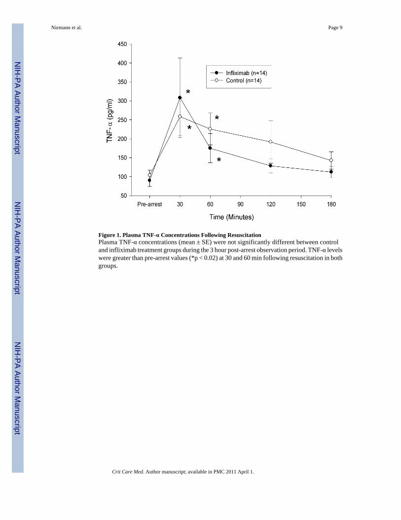
Figure 1. Plasma TNF-α Concentrations Following ResuscitationPlasma TNF-α concentrations (mean ± SE) were not significantly different between controland infliximab treatment groups during the 3 hour post-arrest observation period. TNF-α levelswere greater than pre-arrest values (*p < 0.02) at 30 and 60 min following resuscitation in bothgroups.
Niemann et al. Page 9
Crit Care Med. Author manuscript; available in PMC 2011 April 1.
NIH
-PA Author Manuscript
NIH
-PA Author Manuscript
NIH
-PA Author Manuscript

Figure 2. Plasma IL-1β Concentrations Following ResuscitationPlasma IL-1β concentrations (mean ± SE) did not increase significantly from pre-arrest valuesfollowing resuscitation and differences were not observed between groups.
Niemann et al. Page 10
Crit Care Med. Author manuscript; available in PMC 2011 April 1.
NIH
-PA Author Manuscript
NIH
-PA Author Manuscript
NIH
-PA Author Manuscript

Figure 3. Mean Arterial Pressure (MAP) and Left Ventricular Stroke Work (LV SW) FollowingResuscitationMAP and SW (mean ± SE) were significantly decreased from pre-arrest values in both groupswithin 30 min of reperfusion. Values were significantly higher at all time points in theinfliximab group. (*p < 0.002, Dunnet-Hsu adjusted for differences between groups).
Niemann et al. Page 11
Crit Care Med. Author manuscript; available in PMC 2011 April 1.
NIH
-PA Author Manuscript
NIH
-PA Author Manuscript
NIH
-PA Author Manuscript

Figure 4. Plasma TNF-α Concentrations in TNF Producers and NonproducersA dichotomy in the TNF-α response to reperfusion was observed in animals of both treatmentgroups. Fifty four percent of animals (n=15) demonstrated at least a doubling of TNF-α at 30min post-arrest. *p < 0.002 compared to pre-arrest value. Data are mean ± SE.
Niemann et al. Page 12
Crit Care Med. Author manuscript; available in PMC 2011 April 1.
NIH
-PA Author Manuscript
NIH
-PA Author Manuscript
NIH
-PA Author Manuscript

NIH
-PA Author Manuscript
NIH
-PA Author Manuscript
NIH
-PA Author Manuscript
Niemann et al. Page 13
Tabl
e 1
Prea
rres
t var
iabl
es fo
r th
e tr
eatm
ent g
roup
s
Con
trol
(n =
14)
Infli
xim
ab (n
= 1
4)P
valu
e
HR
(bea
ts p
er m
inut
e)86
(17)
96(1
0)0.
06
MA
P (m
mH
g)87
(8)
92(1
0)0.
12
LVED
P (m
mH
g)6.
29(1
.90)
5.43
(2.1
0)0.
27
Stro
ke w
ork
(gm
-m)
47.1
(6.9
2)50
.3(7
.38)
0.25
Tem
pera
ture
(°C
)37
.3(1
.1)
37.0
(0.7
)0.
38
TNF-α
leve
l (pg
/ml)
135
(82-
210)
113
(65-
159)
0.45
IL-1β
leve
l (pg
/ml)
0(0
-15)
0(0
-16)
0.65
Val
ues a
re th
e m
ean
± SD
or m
edia
n an
d in
terq
uarti
le ra
nge.
Crit Care Med. Author manuscript; available in PMC 2011 April 1.

NIH
-PA Author Manuscript
NIH
-PA Author Manuscript
NIH
-PA Author Manuscript
Niemann et al. Page 14
Tabl
e 2
Res
usci
tatio
n va
riab
les f
or th
e tr
eatm
ent g
roup
s
Con
trol
(n =
14)
Infli
xim
ab (n
= 1
4)P
valu
e
Tran
stho
raci
c im
peda
nce
(Ohm
s)67
.5(2
.87)
69(4
.14)
0.20
Cor
onar
y Pe
rfus
ion
Pres
sure
(mm
Hg)
12.9
(3.5
5)13
.2(5
.00)
0.86
Cou
nter
shoc
ks to
def
ibril
late
1.5
(1-2
)1
(1-2
)0.
28
Tota
l cou
nter
shoc
ks d
eliv
ered
2(1
-3)
1(1
-2)
0.28
Joul
es d
eliv
ered
500
(200
-800
)20
0(2
00-5
00)
0.22
Epin
ephr
ine
adm
inis
tere
d (m
g)0.
5(0
.5-0
.5)
0.5
(0.5
-0.5
)0.
21
Tim
e to
RO
SC*
(sec
onds
)11
4(8
4-16
3)99
(84-
134)
0.44
* Ret
urn
of S
pont
aneo
us C
ircul
atio
n
Val
ues a
re th
e m
ean
± st
anda
rd d
evia
tion
or m
edia
n an
d in
terq
uarti
le ra
nge.
Crit Care Med. Author manuscript; available in PMC 2011 April 1.

NIH
-PA Author Manuscript
NIH
-PA Author Manuscript
NIH
-PA Author Manuscript
Niemann et al. Page 15
Tabl
e 3
Hem
odyn
amic
Var
iabl
es F
ollo
win
g R
esus
cita
tion
Prea
rres
t30
min
60 m
in12
0 m
in18
0 m
in
Con
trol
HR
(bea
t/min
)86
± 1
794
± 1
685
± 1
580
± 1
377
± 1
3
LVED
P (m
m H
g)6
± 2
11 ±
512
± 2
14 ±
313
± 3
SV (m
l)41
± 5
16 (5
-18)
25 (2
1-28
)35
± 9
43 ±
9
S dp
/dt (
mm
Hg/
s)10
99 ±
156
420
± 15
572
8 ±
104
954
± 17
910
00 (9
52-1
180)
tau
(mse
c)30
± 4
66 ±
18
45 ±
645
(41-
49)
44 ±
13
Inflx
imab
HR
(bea
ts/m
in)
96 ±
10
99 ±
990
± 1
083
± 9
79 ±
11
LVED
P (m
mH
g)5
± 2
11 ±
410
± 3
12 ±
411
± 5
SV (m
l)42
± 5
21 (1
8-24
)†31
(528
-33)
*41
± 6
*46
± 7
S dp
/dt (
mm
Hg/
s)12
30 ±
216
603
± 20
4**
883
± 19
4**
1130
± 2
55*
1174
(104
2-12
50)*
tau
(mse
c)33
± 4
58 ±
14
45 ±
544
(42-
50)
46 ±
7
Val
ues a
re th
e m
ean
± st
anda
rd d
evia
tion
or m
edia
n an
d in
terq
uarti
le ra
nge.
HR
= h
eart
rate
; LV
EDP
= Le
ft ve
ntric
ular
end
-dia
stol
ic p
ress
ure;
S d
p/dt
= sy
stol
ic d
p/dt
; SV
= st
roke
vol
ume
* p <
0.05
,
**p
< 0.
025,
† p <
0.00
5 ve
rsus
con
trol g
roup
Crit Care Med. Author manuscript; available in PMC 2011 April 1.
Copyright © 2022 FDOKUMEN