Induction of Protective Immune Responses by Immunization with Linear Multiepitope Peptides Based on...
11
1998, 66(7):3232. Infect. Immun. and V. S. Chauhan Ashima Bharadwaj, Pawan Sharma, Sunil K. Joshi, Balwan Singh Antigens Plasmodium falciparum Based on Conserved Sequences from Peptides Immunization with Linear Multiepitope Induction of Protective Immune Responses by http://iai.asm.org/content/66/7/3232 Updated information and services can be found at: These include: REFERENCES http://iai.asm.org/content/66/7/3232#ref-list-1 This article cites 41 articles, 18 of which can be accessed free at: CONTENT ALERTS more» cite this article), Receive: RSS Feeds, eTOCs, free email alerts (when new articles http://journals.asm.org/site/misc/reprints.xhtml Information about commercial reprint orders: http://journals.asm.org/site/subscriptions/ To subscribe to to another ASM Journal go to: on September 13, 2014 by guest http://iai.asm.org/ Downloaded from on September 13, 2014 by guest http://iai.asm.org/ Downloaded from
-
Upload
independent -
Category
Documents
-
view
2 -
download
0
Transcript of Induction of Protective Immune Responses by Immunization with Linear Multiepitope Peptides Based on...
1998, 66(7):3232. Infect. Immun.
and V. S. ChauhanAshima Bharadwaj, Pawan Sharma, Sunil K. Joshi, Balwan Singh
AntigensPlasmodium falciparumBased on Conserved Sequences from
PeptidesImmunization with Linear Multiepitope Induction of Protective Immune Responses by
http://iai.asm.org/content/66/7/3232Updated information and services can be found at:
These include:
REFERENCEShttp://iai.asm.org/content/66/7/3232#ref-list-1This article cites 41 articles, 18 of which can be accessed free at:
CONTENT ALERTS more»cite this article),
Receive: RSS Feeds, eTOCs, free email alerts (when new articles
http://journals.asm.org/site/misc/reprints.xhtmlInformation about commercial reprint orders: http://journals.asm.org/site/subscriptions/To subscribe to to another ASM Journal go to:
on Septem
ber 13, 2014 by guesthttp://iai.asm
.org/D
ownloaded from
on S
eptember 13, 2014 by guest
http://iai.asm.org/
Dow
nloaded from
INFECTION AND IMMUNITY,0019-9567/98/$04.0010
July 1998, p. 3232–3241 Vol. 66, No. 7
Copyright © 1998, American Society for Microbiology. All Rights Reserved.
Induction of Protective Immune Responses by Immunizationwith Linear Multiepitope Peptides Based on Conserved
Sequences from Plasmodium falciparum AntigensASHIMA BHARADWAJ, PAWAN SHARMA, SUNIL K. JOSHI,
BALWAN SINGH, AND V. S. CHAUHAN*
International Centre for Genetic Engineering and Biotechnology, Aruna Asaf Ali Marg,New Delhi 110067, India
Received 17 November 1997/Returned for modification 15 December 1997/Accepted 14 April 1998
A cysteine-containing peptide motif, EWSPCSVTCG, is found highly conserved in the circumsporozoiteprotein (CSP) and the thrombospondin-related anonymous protein (TRAP) of all the Plasmodium speciesanalyzed so far and has been shown to be crucially involved in the sporozoite invasion of hepatocytes. We haverecently shown that peptide sequences containing this motif, and also the antibodies raised against the motif,inhibit the merozoite invasion of erythrocytes. However, during natural infection, and upon immunization withrecombinant CSP, this motif represents a cryptic epitope. Here we present the results of immunization studieswith two linear multiepitopic constructs, a 60-residue (P60) and a 32-residue (P32) peptide, containing theconserved motif sequence. Both the peptides per se generated high levels of specific antibodies in BALB/c mice.P32 was found to be genetically restricted to H-2d and H-2b haplotypes of mice, whereas P60 was found to beimmunogenic in five different strains of mice. The antibody response was predominantly targeted to theotherwise cryptic, conserved motif sequence in P60. Anti-P60 antibodies specifically stained the asexual bloodstages of Plasmodium falciparum and Plasmodium yoelii in an immunofluorescence assay, recognized a 60- to65-kDa parasite protein in an immunoblot assay, and blocked P. falciparum merozoite invasion of erythrocytesin a dose-dependent manner. Immunization with P60 also induced significant levels of the cytokines interleu-kin-2 (IL-2), IL-4, and gamma interferon in BALB/c mice. Moreover, >60% of mice immunized with P60survived a heterologous challenge infection with a lethal strain of P. yoelii. These results indicate thatappropriate medium-sized synthetic peptides might prove useful in generating specific immune responses to anotherwise cryptic but critical and putatively protective epitope in an antigen and could form part of amulticomponent malaria vaccine.
Several antigens from different stages of the life cycle of themalaria parasite Plasmodium falciparum have been character-ized, and some of these, produced by recombinant DNA tech-niques or by chemical synthesis, are being tested as vaccinecandidates (21, 22, 35). The circumsporozoite protein of P.falciparum (PfCSP) is the best-characterized antigen of theparasite because of its role in protective immunity againstpreerythrocytic stages of malaria (29, 30). This protein con-tains a stretch of highly conserved, immunodominant tetrapep-tide repeats in the middle of its structure (13). However, clin-ical trials with PfCSP peptides or recombinant CSP and itsfragments, aimed at developing specific antibody (Ab) re-sponses to the repeats, have proved disappointing (2, 20). Thishas led to the suggestion that there might be other antigenicsites on the CSP; in fact, several B and T epitopes from thenonrepeat region of CSP have already been characterized (17,37). The CSPs of all Plasmodium species contain a nonrepeatconserved portion termed region II. Further, a nonapeptidemotif (W-S-P-C-S-V-T-C-G) within region II has been foundhighly conserved in all CSP sequences analyzed so far (32).This conserved motif is also found in a variety of biologicallyimportant proteins, such as thrombospondin, properdin, andcomponents of the complement pathways (19, 32). Interest-ingly, this nonapeptide motif is also found in the throm-
bospondin-related anonymous protein (TRAP), first describedfrom erythrocytic stages of P. falciparum. Subsequently, TRAPhas also been shown on the surface of P. falciparum sporozo-ites, and a homolog of TRAP, termed sporozoite surface pro-tein 2 (SSP-2), was found on the surface of sporozoites ofPlasmodium yoelii (33, 34). Both CSP and TRAP (SSP-2) arethought to have crucial roles in recognition and entry of sporo-zoites into the liver cell, and in both, the conserved-motifsequence acts as a specific sporozoite ligand for putative he-patocyte receptors (7, 9, 10, 28). Recently a recombinant con-struct, RTS, S, containing a truncated version of CSP, inclusiveof the region II sequence, attached to hepatitis core protein,has been synthesized. The construct has been found to beprotective against sporozoite challenge in humans, raisinghopes of a single-antigen-based malaria vaccine (41).
The role of TRAP and, indeed, its expression and location atthe blood stages, is not yet known, although TRAP-specificmRNA has been detected in infected erythrocytes (32). Wehave recently shown that synthetic peptides representing theconserved motif sequences and the antisera raised againstthese peptides inhibited the merozoite invasion of erythrocytes(38). Further, the anti-peptide Abs recognized a TRAP-likemolecule in the blood stage lysate of P. falciparum (38). Abetter understanding of the structure of the region II peptidesequences and immune responses against them may providethe basis for their inclusion in a subunit malaria vaccine.
Vaccine constructs based on generating only Ab responseagainst well-characterized B epitopes from malaria antigenshave not met with the expected success, for several reasons (2,
* Corresponding author. Mailing address: International Centre forGenetic Engineering and Biotechnology, Aruna Asaf Ali Marg, P.O.Box 10504, New Delhi 110067, India. Phone: 00 91 11 6195007. Fax: 0091 11 6162316. E-mail: [email protected].
3232
on Septem
ber 13, 2014 by guesthttp://iai.asm
.org/D
ownloaded from
14, 20). There is now evidence to show that both Ab-mediatedand Ab-independent T-cell-mediated protection mechanismsare operative at different stages of the parasite life cycle (4, 11,45). Also, a successful malaria vaccine will be partly dependenton natural boosting to maintain high levels of protective Absbecause of the impracticality of repeatedly administering avaccine, particularly in the third-world countries where such avaccine is most needed. To facilitate natural boosting, a vac-cine would require T epitopes of parasite origin, and prefera-bly the T and B epitopes should come from the same antigenin the parasite. With this perspective, we have investigated theimmunogenicity of synthetic peptides containing B and T celldeterminants, based on CSP and TRAP sequences of P. falci-parum, in mice (13, 17). We have found that a linear 60-residue-long peptide (P60) containing the conserved region IIsequence (amino acids [aa] 331 to 390 of PfCSP) is highlyimmunogenic in mice without the use of a carrier protein. Wedescribe here in detail the immunological characteristics ofP60. We also describe the synthesis and immunological prop-erties of a chimeric peptide (P32) containing a T-cell epitopeoverlapping with the conserved motif sequence and show evi-dence that mice immunized with these two P. falciparum-basedpeptides are partially protected against lethal challenge withheterologous murine malaria blood stage parasites.
MATERIALS AND METHODS
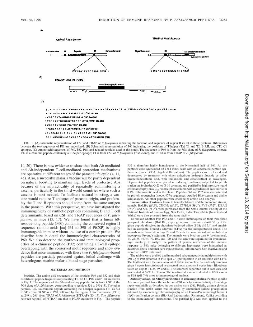
Peptides. The amino acid sequences of the peptides P60 and P32 and theirconstituent peptide fragments representing T1, T2, C1, P18, and PT18 are shownin Fig. 1. The sequence of the 60-residue-long peptide P60 is from CSP of the7G8 clone of P. falciparum, corresponding to residues 331 to 390 (13). The otherpeptide, P32, is a chimeric peptide containing the T-helper sequence (T1; aa 331to 347) from PfCSP as in P60, followed by the region II motif sequence (PT18;aa 249 to 266) from TRAP of P. falciparum (PfTRAP) (13, 17). The differencesbetween region II of PfTRAP and that of PfCSP are shown in Fig. 1. The peptide
P32 is therefore highly homologous to the N-terminal half of P60. All thepeptides were synthesized on a 0.1-mmol scale with an automated peptide syn-thesizer (model 430A; Applied Biosystems). The peptides were cleaved anddeprotected by treatment with either anhydrous hydrogen fluoride or triflu-oromethanesulfonic acid, with thioanisole and ethanedithiol as scavengers.Deprotected peptides were placed in reducing conditions, subjected to gel fil-tration on Sephadex G-25 or G-10 columns, and purified by high-pressure liquidchromatography on a C18 reverse-phase column with a gradient of acetonitrile in0.1% trifluoroacetic acid as the eluant. Peptides P60 and P32 were characterizedby protein sequencing (model 477A sequencer; Applied Biosystems) and aminoacid analysis. All other peptides were checked by amino acid analysis.
Immunization of animals. Four- to 6-week-old mice of different inbred strains,namely, BALB/c (H-2d), C3H/He (H-2k), C57BL/6 (H-2b), FVB (H-2b), DBA/2(H-2d), and SJL (H-2S), were purchased from the Small Animal Facility of theNational Institute of Immunology, New Delhi, India. Two rabbits (New ZealandWhite) were also procured from the same facility.
To find out whether P60, P32, and P18 were immunogenic on their own, threegroups of inbred mice (BALB/c; six per group) were immunized with 50 mg of thegiven peptide dissolved in phosphate-buffered saline (PBS; pH 7.4) and emulsi-fied in complete Freund’s adjuvant (CFA) via the intraperitoneal route. Theanimals were boosted on days 28 and 35 with the same inoculum emulsified inincomplete Freund’s adjuvant. The animals were bled on days 0 (preimmune),14, 28, 35, 49, 64, 70, 100, and 120, and the sera were separated for immunoas-says. Similarly, to analyze the pattern of genetic restriction of the immuneresponse to P60, mice belonging to different haplotypes were immunized asdescribed above and their sera were collected. All sera were heat inactivated andstored at 220°C until used.
The rabbits were prebled and immunized subcutaneously at multiple sites with250 mg of P60 dissolved in PBS (pH 7.4) per injection in an emulsion with CFA.The first boost with the same amount of P60 in incomplete Freund’s adjuvant wasgiven 4 weeks later, followed by a second boost another 4 weeks later. Blood wastaken on days 0, 14, 28, 49, and 63. The sera were separated out in each case andinactivated at 56°C for 30 min. The inactivated sera were diluted in 0.5% caseinin PBS (pH 7.4) prior to immunological analysis.
Antibody assays. (i) Affinity purification of immunoglobulins. Peptide-specificAbs were purified from the rabbit anti-P60 sera by immunoaffinity chromatog-raphy essentially as described in our earlier work (38). Briefly, gamma globulinfraction from rabbit serum was obtained by ammonium sulfate precipitationfollowed by ion-exchange chromatography on an Econo-Pac immunoglobulin G(IgG) purification column (Bio-Rad Laboratories, Richmond, Calif.) accordingto the manufacturer’s instructions. The purified IgG was then applied to the
FIG. 1. (A) Schematic representation of CSP and TRAP of P. falciparum indicating the location and sequence of region II (RII) in these proteins. Differencesbetween the two sequences of RII are underlined. (B) Schematic representation of P60 indicating the positions of T-helper (Th) T1 and T2, B RII, and CTL C1epitopes. (C) Amino acid sequences of P60, P32, P18, and related peptides used in this study. The sequence of P60 is from the 7G8 clone of P. falciparum, whereasP32 is a chimeric peptide containing a T-helper epitope; T1 is from CSP of P. falciparum (7G8 clone), and PT18 is from TRAP of P. falciparum.
VOL. 66, 1998 INDUCTION OF IMMUNE RESPONSE BY P. FALCIPARUM PEPTIDES 3233
on Septem
ber 13, 2014 by guesthttp://iai.asm
.org/D
ownloaded from
immunoadsorbent column prepared by the coupling of P60 to a cyanogen bro-mide-activated Sepharose 4B column. The peptide-specific IgG Abs were elutedwith glycine-HCl buffer (0.1 M; pH 2.5), and the fractions were neutralized by theaddition of Tris base (2.0 M). The fractions were pooled and characterized asdescribed previously (38).
(ii) ELISA. Antibody levels in the sera from the mice immunized with thepeptides were assayed by enzyme-linked immunosorbent assay (ELISA), usingappropriate synthetic peptides as capture antigens. Briefly, the wells of flat-bottom 96-well microtiter plates (Greiner, Nurtingen, Germany) were coatedwith the relevant antigen. Uncoated reactive sites in the wells were blocked byincubation with a 5% solution of a nonfat dried milk powder in PBS, pH 7.2, for1 h. The plates were washed three times with washing buffer (0.15 M NaClsolution containing 0.05% Tween 20). All serum samples were serially diluted inPBS, pH 7.2, containing 0.5% milk powder and incubated in antigen-coated wellsfor 90 min at room temperature in a humid chamber. The wells were washedthoroughly with the washing buffer, and the plates were incubated with 50 ml ofoptimally diluted horseradish peroxidase-conjugated goat anti-mouse IgG orgoat anti-human IgG (Sigma) for 90 min in the respective assays. The enzymereaction was developed with 100 ml of substrate solution (o-phenylenediaminedihydrochloride [2 mg/ml] and H2O2 in citrate buffer, pH 5.0). The reaction wasstopped with 8 N H2SO4 (50 ml/well), and the optical density (OD) of thereaction product was obtained with a microplate reader (Molecular Devices) at490 nm. The last dilution of a test serum giving an OD value greater than twicethe OD value obtained with the respective preimmune serum diluted 1/100 wastaken as the endpoint titer. Sera obtained from the control mice receiving onlyCFA were also screened for Abs to the relevant peptides.
(iii) Inhibition ELISA. Different concentrations of the relevant peptides werepreincubated with optimally diluted polyclonal anti-P60 Abs for 1 h at 4°C. Theplates, coated with P60 (1 mg/well) and blocked as described above, were thenincubated with 50 ml of the Ab-peptide solution for 30 min at room temperaturealong with the polyclonal anti-P60 Abs diluted in 0.5% casein in PBS (pH 7.2)without peptide. The plates were washed extensively with the washing buffer andincubated with 50 ml of optimally diluted goat anti-mouse IgG for 90 min.Following washings with washing buffer, the reaction was developed with 100 mlof substrate solution (o-phenylenediamine dihydrochloride [2 mg/ml] and H2O2in citrate buffer, pH 5.0). The reaction was stopped with 8 N H2SO4 (50 ml/well),and the OD of the reaction product was obtained with a microplate reader(Molecular Devices) at 490 nm. An unrelated linear 23-residue-long peptide,VH-1, based on a plant protein (FLTTYAQAANTHLFLLKDAQIYG) wasused as a negative control in this assay.
(iv) Subtyping of IgG. The ELISA plate was coated with P60 and washed threetimes with the washing buffer, followed by incubation of 50 ml of serially dilutedmouse anti-P60 serum samples in duplicate for 90 min. The plate was washed andincubated with different goat anti-mouse IgG subtypes, namely, IgG, IgG1,IgG2a, IgG2b, and IgG3 (diluted 1/1,000 in PBS [pH 7.4] containing 0.5% nonfatdried milk) for 90 min. The plate was washed another three times with thewashing buffer and incubated with horseradish peroxidase-conjugated rabbitanti-goat immunoglobulin (1/500) for 90 min, and the assay was completed asdescribed above.
IFA. Indirect immunofluorescence assays (IFAs) were performed with seraobtained from BALB/c mice and rabbits immunized with P60 or P32. Briefly, thewells of slides were coated with P. yoelii- or P. falciparum-infected erythrocytes.The cells were fixed on slides by immersing them in cold acetone at 220°C for2 h. The slides were incubated with different dilutions of sera in individual wellsfor 1 h. After extensive washing with PBS, the slides were incubated with a 1:40dilution of goat anti-mouse IgG conjugated to fluorescein isothiocyanate for 1 hin the dark in a humid chamber. Following washings, the slides were observedunder a fluorescence microscope (Nikon) by visible and UV light alternately tosee specific binding of the antibody to the infected erythrocytes. Serum samplesobtained from rabbits immunized with adjuvant alone were also tested in thisassay and served as a negative control.
Western blot analysis. P. falciparum proteins were fractionated on sodiumdodecyl sulfate (SDS)–10% polyacrylamide gels under reducing conditions. Re-combinant PfTRAP (a truncated version lacking the signal and transmembranesequences; residues 26 to 503) expressed in the pQE vector and recombinantPfCSP (a kind gift from P. Sinnis) were also included in the gel. The fractionatedproteins were then electroblotted onto nitrocellulose paper. The parasite pro-teins were probed with the polyclonal Ab raised against P60 in rabbit serum(preadsorbed on human erythrocytes), followed by incubation with horseradishperoxidase labelled anti-rabbit IgG. The reaction was developed with 3,39-dia-minobenzidine as a substrate. In each case rabbit anti-parasite antibodies andpreimmune rabbit IgG or serum were used as a positive and negative control,respectively. Monoclonal Ab (MAb) 2A10 (a kind gift from P. Sinnis) directedagainst PfCSP and polyclonal serum raised against recombinant PfTRAP ininbred female BALB/c mice served as positive controls for the respective recom-binant proteins. The serum obtained from the adjuvant-immunized rabbit wasalso screened for generation of the parasite-specific Abs.
Merozoite invasion inhibition and parasite growth inhibition assays. TheFID-3 isolate of P. falciparum was used for the merozoite invasion inhibitionassays. The parasite was cultured following methods described by Trager andJenson (42). For the merozoite invasion inhibition assay, cultures of the FID-3isolate of P. falciparum were synchronized by two treatments with 5% sorbitol
(25) and incubated for about 30 h so that at the time of setting up the assay, morethan 95% of the parasites were late trophozoites. For the merozoite invasioninhibition assay, the cultures were incubated for about 20 h with various con-centrations of the immunoglobulin IgG, obtained from the rabbits immunizedwith P60, as well as with the sera obtained from P32-immunized BALB/c mice.Only the ring-stage-infected erythrocytes were counted as parasitized cells forcalculating percent parasitemia. In each case the sera obtained from rabbitsimmunized with the synthetic peptide, P8 (LDNIKGNVGKMEDYIKKNNKC),from merozoite surface protein 1 (MSP1) of P. falciparum, was used as thenegative control (39). Each serum or immunoglobulin concentration was testedin triplicate. Percent invasion inhibition was calculated as follows: 100 2 (percentparasitemia in test serum [immune immunoglobulin]/percent parasitemia in pre-immune serum [or immunoglobulin] 3 100).
Cellular immune responses. (i) Lymphocyte proliferation assays. Two groupsof four mice each were primed with 50 mg of P60 or P32 in PBS emulsified withequal volumes of CFA via tail base inoculations, while the group of control micereceived emulsified PBS alone. Twelve days later, the inguinal lymph nodes (LN)were extracted and crushed to release the cells. The cells were washed twice withRPMI 1640 medium (Sigma) and plated at 4 3 105/well in 96-well tissue cultureplates (Costar) in RPMI 1640 medium supplemented with 15 mM HEPES, 0.2%sodium bicarbonate, 50 mM b-mercaptoethanol (Bio-Rad), 2 mM glutamine, 50U of penicillin/ml, 50 mg of streptomycin/ml, and 10% fetal calf serum (Sigma).Appropriate peptides were incubated with the seeded lymphocytes at differentconcentrations. All cultures were set up in quadruplicate. The plates were incu-bated at 37°C in 5% CO2 (Forma Scientific). Tritiated thymidine (0.5 mCi;Amersham, Buckinghamshire, United Kingdom) was added to each well in thelast 15 h of the 5 days of culture. Cells were harvested, and the tritiated thymidineincorporation was determined with a liquid scintillation counter (Betaplate;Pharmacia, Uppsala, Sweden). Counts were derived from the averages of fourseparate experiments and expressed as the stimulation index (SI) (SI 5 countsper minute of stimulated culture/counts per minute of control culture). TheT-cell mitogen concanavalin A (Sigma) was used as a positive control.
(ii) Cytokine analysis. Supernatants were collected from in vitro lymphopro-liferative cultures, growing in the presence or absence of the peptide, after 72 h.Cytokine levels were estimated with the appropriate commercially availablemurine ELISA kits (Endogen) according to the manufacturer’s instructions. Thecytokines interleukin-2 (IL-2), IL-4, and gamma interferon (IFN-g) were mea-sured with 50 ml of supernatant diluted four times. The plates were read at awavelength of 450 nm. The concentration of each cytokine was calculated fromstandard curves obtained with known concentrations of the positive controlprovided with the respective kits.
Protection in mice. A group of 15 inbred mice (BALB/c) were immunizedintraperitoneally with 50 mg of P60 emulsified in CFA. Control mice receivedonly the adjuvant in PBS. All the mice received boosts on days 28 and 42. On day49, the mice were bled and sera were collected. A week later, the immunized andcontrol mice were challenged with an inoculum of 104 Plasmodium yoelii nigerien-sis (lethal strain)-infected erythrocytes. From the third day after the challenge,thin blood smears obtained from each mouse were stained with Giemsa stain andpercent parasitemia was determined by microscopy. To assess the protectivepotential of P32, a separate group of 10 BALB/c mice were immunized with thepeptide and challenged with P. yoelii parasites as described above. The protectionexperiments were repeated twice to confirm the observations.
RESULTS
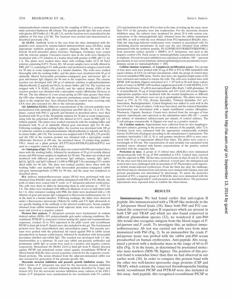
Immunoassays. We had found earlier that anti-region IIpeptide Abs immunoreacted with a TRAP-like molecule in theP. falciparum blood lysate (38). Since both P60 and P32 con-tained the conserved region II sequences which are present inboth CSP and TRAP and which are also found conserved indifferent plasmodium species (32), we wondered if anti-P60Abs would also recognize antigens from the blood stages of P.falciparum and P. yoelii. To investigate this, an indirect immu-nofluorescence Ab test was carried out with sera from miceimmunized with P60 (Fig. 2). In an immunoblot the crude P.falciparum lysate was probed with the rabbit anti-P60 serumpreadsorbed on human erythrocytes. Anti-peptide Abs recog-nized a protein with a molecular mass in the range of 60 to 65kDa (Fig. 3) in the lysate, as determined by prestained molec-ular mass markers (SDS-7B; Sigma). The position of this pro-tein band is somewhat lower than that we had observed in ourearlier work (38). In order to compare this protein band withthe other two well-known sporozoite stage proteins (CSP andTRAP), which contain the conserved thrombospondin bindingmotif, recombinant PfCSP and PfTRAP were also included inthis assay. Anti-peptide Abs recognized recombinant PfCSP as
3234 BHARADWAJ ET AL. INFECT. IMMUN.
on Septem
ber 13, 2014 by guesthttp://iai.asm
.org/D
ownloaded from
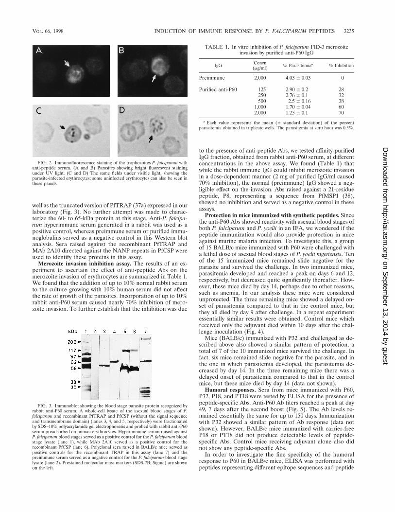
well as the truncated version of PfTRAP (37a) expressed in ourlaboratory (Fig. 3). No further attempt was made to charac-terize the 60- to 65-kDa protein at this stage. Anti-P. falcipa-rum hyperimmune serum generated in a rabbit was used as apositive control, whereas preimmune serum or purified immu-noglobulins served as a negative control in this Western blotanalysis. Sera raised against the recombinant PfTRAP andMAb 2A10 directed against the NANP repeats in PfCSP wereused to identify these proteins in this assay.
Merozoite invasion inhibition assay. The results of an ex-periment to ascertain the effect of anti-peptide Abs on themerozoite invasion of erythrocytes are summarized in Table 1.We found that the addition of up to 10% normal rabbit serumto the culture growing with 10% human serum did not affectthe rate of growth of the parasites. Incorporation of up to 10%rabbit anti-P60 serum caused nearly 70% inhibition of mero-zoite invasion. To further establish that the inhibition was due
to the presence of anti-peptide Abs, we tested affinity-purifiedIgG fraction, obtained from rabbit anti-P60 serum, at differentconcentrations in the above assay. We found (Table 1) thatwhile the rabbit immune IgG could inhibit merozoite invasionin a dose-dependent manner (2 mg of purified IgG/ml caused70% inhibition), the normal (preimmune) IgG showed a neg-ligible effect on the invasion. Abs raised against a 21-residuepeptide, P8, representing a sequence from PfMSP1 (38),showed no inhibition and served as a negative control in theseassays.
Protection in mice immunized with synthetic peptides. Sincethe anti-P60 Abs showed reactivity with asexual blood stages ofboth P. falciparum and P. yoelii in an IFA, we wondered if thepeptide immunization would also provide protection in miceagainst murine malaria infection. To investigate this, a groupof 15 BALB/c mice immunized with P60 were challenged witha lethal dose of asexual blood stages of P. yoelii nigeriensis. Tenof the 15 immunized mice remained slide negative for theparasite and survived the challenge. In two immunized mice,parasitemia developed and reached a peak on days 6 and 12,respectively, but decreased quite significantly thereafter. How-ever, these mice died by day 14, perhaps due to other reasons,such as anemia. In our analysis these mice were consideredunprotected. The three remaining mice showed a delayed on-set of parasitemia compared to that in the control mice, butthey all died by day 9 after challenge. In a repeat experimentessentially similar results were obtained. Control mice whichreceived only the adjuvant died within 10 days after the chal-lenge inoculation (Fig. 4).
Mice (BALB/c) immunized with P32 and challenged as de-scribed above also showed a similar pattern of protection; atotal of 7 of the 10 immunized mice survived the challenge. Infact, six mice remained slide negative for the parasite, and inthe one in which parasitemia developed, the parasitemia de-creased by day 14. In the three remaining mice there was adelayed onset of parasitemia compared to that in the controlmice, but these mice died by day 14 (data not shown).
Humoral responses. Sera from mice immunized with P60,P32, P18, and PT18 were tested by ELISA for the presence ofpeptide-specific Abs. Anti-P60 Ab titers reached a peak at day49, 7 days after the second boost (Fig. 5). The Ab levels re-mained essentially the same for up to 150 days. Immunizationwith P32 showed a similar pattern of Ab response (data notshown). However, BALB/c mice immunized with carrier-freeP18 or PT18 did not produce detectable levels of peptide-specific Abs. Control mice receiving adjuvant alone also didnot show any peptide-specific Abs.
In order to investigate the fine specificity of the humoralresponse to P60 in BALB/c mice, ELISA was performed withpeptides representing different epitope sequences and peptide
FIG. 2. Immunofluorescence staining of the trophozoites P. falciparum withanti-peptide serum. (A and B) Parasites showing bright fluorescent stainingunder UV light. (C and D) The same fields under visible light, showing theparasite-infected erythrocytes; some uninfected erythrocytes can also be seen inthese panels.
FIG. 3. Immunoblot showing the blood stage parasite protein recognized byrabbit anti-P60 serum. A whole-cell lysate of the asexual blood stages of P.falciparum and recombinant PfTRAP and PfCSP (without the signal sequenceand transmembrane domain) (lanes 3, 4, and 5, respectively) were fractionatedby SDS–10% polyacrylamide gel electrophoresis and probed with rabbit anti-P60serum preadsorbed on human erythrocytes. Hyperimmune serum raised againstP. falciparum blood stages served as a positive control for the P. falciparum bloodstage lysate (lane 1), while MAb 2A10 served as a positive control for therecombinant PfCSP (lane 6). Polyclonal sera raised in BALB/c mice served aspositive controls for the recombinant TRAP in this assay (lane 7) and thepreimmune serum served as a negative control for the P. falciparum blood stagelysate (lane 2). Prestained molecular mass markers (SDS-7B; Sigma) are shownon the left.
TABLE 1. In vitro inhibition of P. falciparum FID-3 merozoiteinvasion by purified anti-P60 IgG
IgG Concn(mg/ml) % Parasitemiaa % Inhibition
Preimmune 2,000 4.03 6 0.03 0
Purified anti-P60 125 2.90 6 0.2 28250 2.76 6 0.1 32500 2.5 6 0.16 38
1,000 1.70 6 0.04 602,000 1.25 6 0.1 70
a Each value represents the mean (6 standard deviation) of the percentparasitemia obtained in triplicate wells. The parasitemia at zero hour was 0.5%.
VOL. 66, 1998 INDUCTION OF IMMUNE RESPONSE BY P. FALCIPARUM PEPTIDES 3235
on Septem
ber 13, 2014 by guesthttp://iai.asm
.org/D
ownloaded from
fragments spanning more than one epitope (Fig. 1). The high-est Ab response was seen against the peptides represented byP18 and T1: the endpoint ELISA titers were as high as 1/10,000for these peptides (Fig. 6). Peptide PT18 showed a reactivitysimilar to that of P18 in this assay. In the case of P32 also theresponse was focused on the sequences represented by PT18 orP18 (data not shown). We also performed ELISA in whichspecific Abs could bind competitively to a given peptide insolution or to P60 coating the wells. Results of the competitiveELISA experiments further supported the above observations,
and a dose-dependent inhibition was observed with the con-stituent peptides, except the unrelated control peptide (Fig. 7).Of all the constituent peptides of P60, P18 was the most ef-fective, causing an inhibition of 77.1% at a concentration of6.25 nmol.
To determine whether humoral response to the peptides P60and P32 was genetically restricted, mice of different inbredstrains, namely, FVB, BALB/c, SJL, DBA/2j, and C3H/Hewere primed and boosted twice with both peptides. All the
FIG. 4. Time course of P. yoelii nigeriensis infection in 15 BALB/c miceimmunized with P60 and challenged 10 days after the last boost. Parasitemia isexpressed as the percentage of infected erythrocytes. Ten immunized mice whichdid not develop parasitemia and remained slide negative are not included in thefigure. F, profiles of parasitemia in two control mice which received adjuvantonly; }, course of parasitemia in five immunized mice which developed para-sitemia; 1, death of the animal.
FIG. 5. Kinetics of peptide-specific IgG response in BALB/c mice immu-nized with P60 as monitored by ELISA. Immunization of control mice with CFAin PBS did not induce detectable level of peptide-specific Abs.
FIG. 6. Fine specificities of humoral responses generated in BALB/c miceimmunized with P60. The animals were primed on day 0 and boosted on day 28with P60. Sera collected on day 35 were tested in an ELISA with different peptideconstructs as capture antigens.
FIG. 7. Inhibition of binding of anti-P60 mouse Ab to P60 in an ELISA.Mouse serum diluted to 1/2,000 was preincubated with the indicated amounts(final concentrations) of different peptide fragments before being added to theELISA plate coated with P60. VH-1 is an unrelated peptide used as a negativecontrol in this assay.
3236 BHARADWAJ ET AL. INFECT. IMMUN.
on Septem
ber 13, 2014 by guesthttp://iai.asm
.org/D
ownloaded from
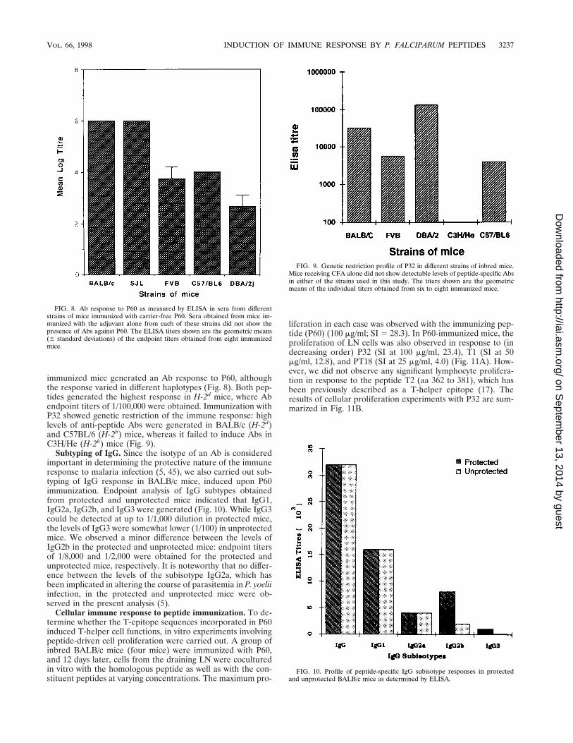
immunized mice generated an Ab response to P60, althoughthe response varied in different haplotypes (Fig. 8). Both pep-tides generated the highest response in H-2d mice, where Abendpoint titers of 1/100,000 were obtained. Immunization withP32 showed genetic restriction of the immune response: highlevels of anti-peptide Abs were generated in BALB/c (H-2d)and C57BL/6 (H-2b) mice, whereas it failed to induce Abs inC3H/He (H-2k) mice (Fig. 9).
Subtyping of IgG. Since the isotype of an Ab is consideredimportant in determining the protective nature of the immuneresponse to malaria infection (5, 45), we also carried out sub-typing of IgG response in BALB/c mice, induced upon P60immunization. Endpoint analysis of IgG subtypes obtainedfrom protected and unprotected mice indicated that IgG1,IgG2a, IgG2b, and IgG3 were generated (Fig. 10). While IgG3could be detected at up to 1/1,000 dilution in protected mice,the levels of IgG3 were somewhat lower (1/100) in unprotectedmice. We observed a minor difference between the levels ofIgG2b in the protected and unprotected mice: endpoint titersof 1/8,000 and 1/2,000 were obtained for the protected andunprotected mice, respectively. It is noteworthy that no differ-ence between the levels of the subisotype IgG2a, which hasbeen implicated in altering the course of parasitemia in P. yoeliiinfection, in the protected and unprotected mice were ob-served in the present analysis (5).
Cellular immune response to peptide immunization. To de-termine whether the T-epitope sequences incorporated in P60induced T-helper cell functions, in vitro experiments involvingpeptide-driven cell proliferation were carried out. A group ofinbred BALB/c mice (four mice) were immunized with P60,and 12 days later, cells from the draining LN were coculturedin vitro with the homologous peptide as well as with the con-stituent peptides at varying concentrations. The maximum pro-
liferation in each case was observed with the immunizing pep-tide (P60) (100 mg/ml; SI 5 28.3). In P60-immunized mice, theproliferation of LN cells was also observed in response to (indecreasing order) P32 (SI at 100 mg/ml, 23.4), T1 (SI at 50mg/ml, 12.8), and PT18 (SI at 25 mg/ml, 4.0) (Fig. 11A). How-ever, we did not observe any significant lymphocyte prolifera-tion in response to the peptide T2 (aa 362 to 381), which hasbeen previously described as a T-helper epitope (17). Theresults of cellular proliferation experiments with P32 are sum-marized in Fig. 11B.
FIG. 8. Ab response to P60 as measured by ELISA in sera from differentstrains of mice immunized with carrier-free P60. Sera obtained from mice im-munized with the adjuvant alone from each of these strains did not show thepresence of Abs against P60. The ELISA titers shown are the geometric means(6 standard deviations) of the endpoint titers obtained from eight immunizedmice.
FIG. 9. Genetic restriction profile of P32 in different strains of inbred mice.Mice receiving CFA alone did not show detectable levels of peptide-specific Absin either of the strains used in this study. The titers shown are the geometricmeans of the individual titers obtained from six to eight immunized mice.
FIG. 10. Profile of peptide-specific IgG subisotype responses in protectedand unprotected BALB/c mice as determined by ELISA.
VOL. 66, 1998 INDUCTION OF IMMUNE RESPONSE BY P. FALCIPARUM PEPTIDES 3237
on Septem
ber 13, 2014 by guesthttp://iai.asm
.org/D
ownloaded from
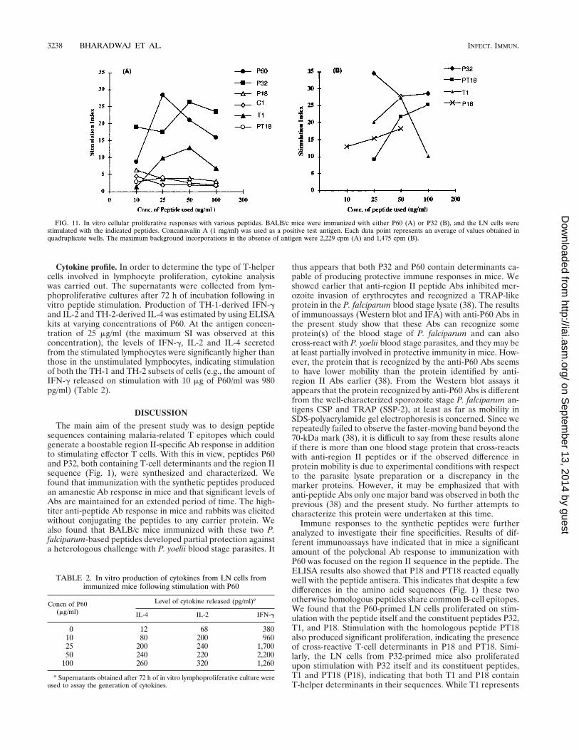
Cytokine profile. In order to determine the type of T-helpercells involved in lymphocyte proliferation, cytokine analysiswas carried out. The supernatants were collected from lym-phoproliferative cultures after 72 h of incubation following invitro peptide stimulation. Production of TH-1-derived IFN-gand IL-2 and TH-2-derived IL-4 was estimated by using ELISAkits at varying concentrations of P60. At the antigen concen-tration of 25 mg/ml (the maximum SI was observed at thisconcentration), the levels of IFN-g, IL-2 and IL-4 secretedfrom the stimulated lymphocytes were significantly higher thanthose in the unstimulated lymphocytes, indicating stimulationof both the TH-1 and TH-2 subsets of cells (e.g., the amount ofIFN-g released on stimulation with 10 mg of P60/ml was 980pg/ml) (Table 2).
DISCUSSION
The main aim of the present study was to design peptidesequences containing malaria-related T epitopes which couldgenerate a boostable region II-specific Ab response in additionto stimulating effector T cells. With this in view, peptides P60and P32, both containing T-cell determinants and the region IIsequence (Fig. 1), were synthesized and characterized. Wefound that immunization with the synthetic peptides producedan amanestic Ab response in mice and that significant levels ofAbs are maintained for an extended period of time. The high-titer anti-peptide Ab response in mice and rabbits was elicitedwithout conjugating the peptides to any carrier protein. Wealso found that BALB/c mice immunized with these two P.falciparum-based peptides developed partial protection againsta heterologous challenge with P. yoelii blood stage parasites. It
thus appears that both P32 and P60 contain determinants ca-pable of producing protective immune responses in mice. Weshowed earlier that anti-region II peptide Abs inhibited mer-ozoite invasion of erythrocytes and recognized a TRAP-likeprotein in the P. falciparum blood stage lysate (38). The resultsof immunoassays (Western blot and IFA) with anti-P60 Abs inthe present study show that these Abs can recognize someprotein(s) of the blood stage of P. falciparum and can alsocross-react with P. yoelii blood stage parasites, and they may beat least partially involved in protective immunity in mice. How-ever, the protein that is recognized by the anti-P60 Abs seemsto have lower mobility than the protein identified by anti-region II Abs earlier (38). From the Western blot assays itappears that the protein recognized by anti-P60 Abs is differentfrom the well-characterized sporozoite stage P. falciparum an-tigens CSP and TRAP (SSP-2), at least as far as mobility inSDS-polyacrylamide gel electrophoresis is concerned. Since werepeatedly failed to observe the faster-moving band beyond the70-kDa mark (38), it is difficult to say from these results aloneif there is more than one blood stage protein that cross-reactswith anti-region II peptides or if the observed difference inprotein mobility is due to experimental conditions with respectto the parasite lysate preparation or a discrepancy in themarker proteins. However, it may be emphasized that withanti-peptide Abs only one major band was observed in both theprevious (38) and the present study. No further attempts tocharacterize this protein were undertaken at this time.
Immune responses to the synthetic peptides were furtheranalyzed to investigate their fine specificities. Results of dif-ferent immunoassays have indicated that in mice a significantamount of the polyclonal Ab response to immunization withP60 was focused on the region II sequence in the peptide. TheELISA results also showed that P18 and PT18 reacted equallywell with the peptide antisera. This indicates that despite a fewdifferences in the amino acid sequences (Fig. 1) these twootherwise homologous peptides share common B-cell epitopes.We found that the P60-primed LN cells proliferated on stim-ulation with the peptide itself and the constituent peptides P32,T1, and P18. Stimulation with the homologous peptide PT18also produced significant proliferation, indicating the presenceof cross-reactive T-cell determinants in P18 and PT18. Simi-larly, the LN cells from P32-primed mice also proliferatedupon stimulation with P32 itself and its constituent peptides,T1 and PT18 (P18), indicating that both T1 and P18 containT-helper determinants in their sequences. While T1 represents
FIG. 11. In vitro cellular proliferative responses with various peptides. BALB/c mice were immunized with either P60 (A) or P32 (B), and the LN cells werestimulated with the indicated peptides. Concanavalin A (1 mg/ml) was used as a positive test antigen. Each data point represents an average of values obtained inquadruplicate wells. The maximum background incorporations in the absence of antigen were 2,229 cpm (A) and 1,475 cpm (B).
TABLE 2. In vitro production of cytokines from LN cells fromimmunized mice following stimulation with P60
Concn of P60(mg/ml)
Level of cytokine released (pg/ml)a
IL-4 IL-2 IFN-g
0 12 68 38010 80 200 96025 200 240 1,70050 240 220 2,200
100 260 320 1,260
a Supernatants obtained after 72 h of in vitro lymphoproliferative culture wereused to assay the generation of cytokines.
3238 BHARADWAJ ET AL. INFECT. IMMUN.
on Septem
ber 13, 2014 by guesthttp://iai.asm
.org/D
ownloaded from
a well-known T-helper epitope (18) and a cytotoxic-T-lympho-cyte epitope within its sequence (24, 37), we were somewhatsurprised to find that the region II peptide sequences, P18 andPT18, also contain T-helper epitopes. The fact that we alsoobserved a strong Ab response focused on this epitope uponP32 and P60 immunization indicates that these peptide se-quences contain overlapping B- and T-cell epitopes. Clearly,P32 and P60, lying downstream of the immunodominant re-peats, represent high-epitopic-density regions of CSP. The oc-currence of overlapping B- and T-cell epitopes in the above-mentioned peptides may not be surprising; in several antigenicproteins the B- and T-cell epitopes are often located close toeach other (15, 27). On the other hand, immunization witheither P18 or PT18, which apparently contain both B- andT-cell determinants, did not induce any detectable Ab re-sponse in rabbits or in BALB/c mice. Keeping in mind thatshort peptides containing B- and T-cell epitopes can producehigh levels of specific Ab responses, the reasons for this ob-servation are not clear. But it has been shown that in shortpeptides, T cells may not always induce an Ab response in Bcells when their determinants overlap (36). It is also suggestedthat pathogens causing the dominant T-cell determinants tooverlap with the critical B-cell determinant may interfere withAb responses detrimental to the pathogen. This may well beone of the several possible reasons why the B-cell determinantsin region II sequences remain largely cryptic during the courseof natural infection (3).
The fine specificities of the Ab humoral immune responsesin P. falciparum-infected individuals are generally dominatedby the repeat peptide structures, and the region II conservedsequence seems to be a cryptic B epitope, at least during thecourse of natural infection (3). One of the reasons for this maybe that the CSP repeats dominate the structural features of theprotein in such a manner that the region II sequences, whichlie downstream of the repeats, are not easily accessible to theimmune system in the intact protein. On the other hand, it hasalso been reported that B cells may respond best against rigidand highly repetitive surface antigens of pathogens and maynot even require T-helper cells for this (1). The repeats aremost likely to represent highly structured B epitopes (6), theimmune response to which could easily dominate the re-sponses to other regions of CSP even if they are exposed.However, these explanations will remain speculative untilstructural details of malaria proteins containing repeats, suchas CSP, become available. In another study, immunization witha repeatless CSP construct of P. falciparum in mice showedthat the region II sequences still remained poorly immuno-genic when Ab specificity was determined by using overlappingoctapeptides (44).
Whatever the reason may be, it is quite clear that region IIis not as immunodominant as some other malaria epitopes, andit is likely that the immune response to such cryptic epitopes israised only very slowly. It is well known that natural immunityto malaria in individuals living in areas where it is endemic isnot fully acquired before adolescence, even following repeatedinfections (11, 12). Masking of the crucial protective epitopesin an antigen during the course of natural infection has alsobeen reported in the case of tryptomastigote surface antigen 1(TSA-1) of Trypanosoma cruzi (46). When mice were immu-nized with the intact recombinant protein, the Ab response wasfound to be mainly focused on the carboxy-terminal region ofthe protein, which did not provide any protection against achallenge infection. On the other hand, immunization with arecombinant N-terminal fragment provided protection, leadingto the conclusion that in TSA-1, the protective epitopes of theN-terminal region remain cryptic in the intact protein and that
the removal of the immunodominant carboxy-terminal regionfrom the protein allows the immune response to be focused onthese cryptic, but crucial, epitopes (46). The results of thepresent study also suggest that through the use of syntheticpeptides it may be possible to focus Ab response on theepitopes which tend to remain cryptic during immunizationwith the intact protein. But it should also be emphasized thatit may not always be possible to predict the nature of immuneresponses from multiepitopic peptides. We and others haveshown that such immunogens may be polar, and there are norules yet to design these molecules for specific immune re-sponses (10, 16, 39).
In general, immune response to short synthetic peptides isgenetically restricted. Inclusion of appropriate T-cell determi-nants may help to overcome this problem. In fact, in the caseof the shorter peptide, P32, the immune response was re-stricted to H-2d and H-2b haplotypes of mice. On the otherhand, immunization with P60, which contains several T-celldeterminants, produced significant response in all the haplo-types tested. These results suggest that in a synthetic peptideimmunogen, inclusion of more than one T epitope may attimes be a reasonable way to circumvent the problem of ge-netic restriction of the immune response. In a multiple-antigenpeptide vaccine construct designed to produce high levels ofAbs against P. yoelii CSP repeats, two T-helper epitopes wereused (43).
Differential activation of T-cell subsets, TH-1 and TH-2,seems to play a crucial role in parasitic diseases (26, 30, 31). Inmalaria also, TH subsets have been implicated in modulatingthe course of infection during different stages of the parasitelife cycle (31). The results of our cytokine analyses of thesupernatants obtained from in vitro cellular proliferation ex-periments with P60 indicated that both the TH-1 and TH-2subsets of T cells were activated upon peptide immunization.Since mature erythrocytes do not bear or express major histo-compatibility complex class I or II antigens, it is difficult toenvisage a direct role for T cells in protective immunity againstblood stages of the parasite. But at the same time, activatedlymphocytes release a battery of cytokines, which could medi-ate the functions of phagocytic cells and possibly promotephagocytosis of the intraerythrocytic parasite. For example,IFN-g, which is known to play an important role in modulatinginfection (40), is released by both TH-1 and CD81 cells. Spe-cific activation of T cells could have a role in inducing protec-tive immunity against malaria (29, 31).
The relative roles of different immunoglobulin subtypeshave also been assessed, and there is evidence that in bothhuman and rodent malaria, the distribution of Ab subisotypescan modulate the course of infection (5, 45). We found notice-ably higher levels of IgG3 and IgG2b in protected mice com-pared to those in unprotected mice. However, no differenceswere seen between the levels of IgG2a in protected and un-protected mice. In an earlier study, the IgG2a subisotype alonewas found to alter the course of parasitemia in mice infectedwith P. yoelii (45). The qualitative and quantitative roles of Absin malaria are not well understood and need to be evaluatedfor the development of malaria vaccine (4).
Can a functional conserved malaria protein sequence whichis also a part of self molecules like thrombospondin and pro-perdin be considered for inclusion in a peptide malaria vaccineconstruct? It can be validly argued that the induction of animmune response to the conserved motif could give rise toautoimmune responses, as shown for the heat shock protein 70(hsp-70) cognate parasite protein. At the same time, several P.falciparum proteins, viz., Pf25, PfMSP-1, and PfP41, containsequences homologous to those of host proteins, such as epi-
VOL. 66, 1998 INDUCTION OF IMMUNE RESPONSE BY P. FALCIPARUM PEPTIDES 3239
on Septem
ber 13, 2014 by guesthttp://iai.asm
.org/D
ownloaded from

dermal growth factor (23), the intermediate filament protein,and the human aldolase enzyme, respectively (8); and, signif-icantly, none of these sequences have been shown to induce orbe a target of any autoimmune response. In conclusion, thepresent study indicates that (i) through appropriate syntheticpeptides it may be possible to focus immune response on theepitopes that remain cryptic when the whole antigen is pre-sented to the immune system, (ii) that linear, nonpolymericpeptide can be a potent immunogen, (iii) that inclusion ofmore than one T epitope may be necessary to circumvent theproblem of genetic restriction in peptide immunization, and(iv) that highly conserved motifs in malaria surface antigensmay be useful targets for inclusion in synthetic peptide malariavaccine constructs.
ACKNOWLEDGMENTS
We thank V. N. Sailaja, J. Ananya, Sachhidanand, and MridulMukherjee for their help in peptide synthesis and immunological as-says. We also thank V. S. Dattu, P. Sejwali, and Photini Sinnis forproviding us samples of recombinant P. falciparum TRAP and CSP.We are grateful to Photini Sinnis for also providing us a sample ofMAb 2A10 specific to CSP.
This work was partly supported by EC grant TS CT 9302.
REFERENCES
1. Bachmann, M. F., H. Hengartner, and R. M. Zinkernegel. 1995. T helper cellindependent neutralising B cell response against vesicular stomatitis virus:role of antigen patterns in B cell induction? Eur. J. Immunol. 25:3445–3451.
2. Ballou, W. R., S. L. Hoffman, J. A. Sherwood, M. R. Hollingdale, F. A. Neva,W. T. Hockmeyer, D. M. Gordon, I. Schneider, R. A. Wirtz, J. F. Young, J. F.Waserman, P. Reeve, C. L. Diggs, and J. D. Chulay. 1987. Safety and efficacyof recombinant Plasmodium falciparum sporozoite DNA vaccine. Lanceti:1277–1281.
3. Ballou, W. R., J. Rothbard, R. A. Wirtz, D. M. Gordon, J. S. Williams, R. W.Gore, I. Schneider, M. R. Hollingdale, R. L. Beaudoin, W. L. Maloy, L. H.Miller, and W. T. Hockmeyer. 1985. Immunogenicity of synthetic peptidesfrom circumsporozoite protein of Plasmodium falciparum. Science 228:996–999.
4. Bouharoun-Tayoun, H., P. Altanals, A. Sabchareon, T. Changsuphajaisid-dhi, and P. Druilhe. 1990. Antibodies that protect humans against Plasmo-dium falciparum blood stages do not on their own inhibit parasite growth invitro, but act in cooperation with monocytes. J. Exp. Med. 172:1633–1641.
5. Bouharoun-Tayoun, H. D., and P. Druilhe. 1992. Plasmodium falciparummalaria: evidence for an isotype imbalance which may be responsible fordelayed acquisition of protective immunity. Infect. Immun. 60:1473–1481.
6. Brooks, B. R., R. W. Pastor, and F. W. Carson. 1987. Theoretically deter-mined three dimensional structure for the repeating tetrapeptide unit of thecircumsporozoite coat protein of the malaria parasite Plasmodium falcipa-rum. Proc. Natl. Acad. Sci. USA 84:4470–4474.
7. Cerami, C., U. Frevert, P. Sinnis, B. Tackacs, P. Clavejo, M. J. Santos, andV. Nussenzweig. 1992. The basolateral domain of hepatocyte plasma mem-brane bears the receptor for CSP of Plasmodium falciparum sporozoites. Cell70:1021–1023.
8. Certa, U., P. Ghersa, H. Dobeli, H. Matile, H. P. Kochar, I. K. Srivastava,A. R. Shaw, and L. H. Perrin. 1988. Aldolase activity of Plasmodium falci-parum protein with protective properties. Science 240:1036–1038.
9. Chatterjee, S., M. Wery, P. Sharma, and V. S. Chauhan. 1995. A conservedpeptide sequence of the Plasmodium falciparum circumsporozoite proteinand antipeptide antibodies inhibit Plasmodium berghei sporozoite invasion ofHep-G2 cells and protect immunized mice against P. berghei sporozoitechallenge. Infect. Immun. 63:4375–4381.
10. Chatterjee, S., P. Sharma, S. Kumar, and V. S. Chauhan. 1994. Fine spec-ificity of immune responses to epitopic sequences in synthetic peptides con-taining B and T epitopes from conserved P. falciparum blood stage antigens.Vaccine 13:1474–1481.
11. Cohen, S., G. A. Butcher, G. H. Mitchell, J. A. Deans, and J. Langhorn. 1977.Acquired immunity and vaccination in malaria. Am. J. Trop. Med. Hyg.26:223–227.
12. Cohen, S., I. A. McGregor, and S. C. Carrington. 1961. Gamma globulin andacquired immunity to malaria. Nature (London) 192:733–737.
13. Dame, J. B., J. L. Williams, T. F. McCutchan, J. L. Weber, R. A. Wirtz, W. T.Rockmeyer, W. L. Maloy, J. D. Haynes, I. Schneider, D. Roberts, G. S.Sanders, E. P. Reddy, C. L. Diggs, and L. H. Miller. 1984. Structure of thegene encoding the immunodominant surface antigen in the sporozoite of thehuman malaria parasite Plasmodium falciparum. Science 225:593–599.
14. Dolan, S. A., L. H. Miller, and T. E. Wellems. 1990. Evidence for a switching
mechanism in the invasion of erythrocytes by Plasmodium falciparum. J. Clin.Invest. 86:618–624.
15. Francis, M. J., C. M. Fry, D. J. Rowlands, J. L. Bittle, R. A. Houghton, R. A.Lerner, and F. Brown. 1987. Immune response to uncoupled peptides of footand mouth disease virus. Immunology 61:1–6.
16. Golvano, J., J. L. Lasarte, P. Sarobe, A. Gullan, J. Prieto, and F. B. Cuesta.1990. Polarity of immunogen: implications for vaccine design. Eur. J. Immu-nol. 20:2363–2366.
17. Good, M. F., D. Pombo, I. A. Quakyi, E. M. Riley, R. A. Houghton, A. Menon,D. W. Alling, J. A. Berzfosky, and L. H. Miller. 1988. Human T-cell recog-nition of the circumsporozoite protein of Plasmodium falciparum: immuno-dominant T-cell domains map to the polymorphic regions of the molecule.Proc. Natl. Acad. Sci. USA 85:1199–1203.
18. Good, M. F., W. L. Moloy, M. N. Lunde, H. Margalit, J. L. Cornetto, G. L.Smith, B. Moss, L. H. Muller, and J. A. Berzofsky. 1987. Construction of asynthetic immunogen: use of a new T-helper epitope on malaria circum-sporozoite protein. Science 235:1059–1062.
19. Goundis, D., and B. M. Reid. 1988. Properdin, the terminal complementcomponents, thrombospondin and CSP of malaria parasites contain similarsequence motifs. Nature (London) 335:82–85.
20. Herrington, D. A., D. F. Clyde, G. Losonsky, M. Cortesia, J. R. Murphy, J.Dais, S. Baqar, A. M. Felix, E. P. Heighmer, G. Gillesen, E. Nardin, R. S.Nussenzweig, V. Nussenzweig, M. R. Hollingdale, and M. M. Levine. 1987.Safety and immunogenicity in man of synthetic peptide malaria vaccineagainst Plasmodium falciparum sporozoites. Nature (London) 328:257–259.
21. Hoffman, S. L., and T. R. Jones. 1994. Malaria vaccine development. Clin.Microbiol. Rev. 7:303–310.
22. Howard, R. J., and B. L. Paloske. 1993. Target antigen for asexual malariavaccine development. Parasitol. Today 9:369–372.
23. Kaslow, D. C., I. A. Quakyi, C. Syin, M. G. Raum, D. B. Keister, J. E.Coligan, T. F. McCutchan, and L. H. Miller. 1988. A vaccine candidate fromsexual stage of human malaria that contains EGF like domains. Nature(London) 333:74–76.
24. Kumar, S., L. H. Miller, I. A. Quakyi, D. B. Keister, R. A. Houghten, W. L.Maloy, B. Moss, J. A. Berzfosky, and M. F. Good. 1988. Cytotoxic T cellsspecific for the circumsporozoite protein of Plasmodium falciparum. Nature(London) 334:258–260.
25. Lambrose, C., and J. Vanderberg. 1979. Synchronization of P. falciparumerythrocytic stages in culture. J. Parasitol. 65:418–420.
26. Locksley, R. M., and P. Scott. 1991. Helper T-cell subsets in mouse leish-maniasis: induction, expansion and effector function. Immunol. Today 12:58–60.
27. Milich, D. R., A. McLachlan, G. B. Thornton, and J. L. Hughes. 1987.Antibody production to the nucleocapsid and envelope of hepatitis B virusprimed by single synthetic T-cell site. Nature (London) 329:547–549.
28. Muller, H. M., I. Reckman, M. R. Hollingdale, H. Bujard, K. J. H. Robson,and A. Crisanti. 1993. Thrombospondin related anonymous protein (TRAP)of Plasmodium falciparum binds specifically to sulfated glyco conjugates andto HepG2 hepatoma cells suggesting a role for this molecule in sporozoiteinvasion of hepatocytes. EMBO J. 12:2881–2889.
29. Nardin, E. H., and R. S. Nussenzweig. 1993. T-cell responses to pre-eryth-rocytic stages of malaria: role in protection and vaccine development. Annu.Rev. Immunol. 11:687–727.
30. Nussenzweig, R. S., and V. Nussenzweig. 1981. Development of sporozoitevaccines. Philos. Trans. R. Soc. Lond. 307:117–128.
31. Robinson-Taylor, A. W. 1995. Regulation of immunity to malaria: valuablelessons learned from murine model. Parasitol. Today 11:334–341.
32. Robson, K. J. H., J. R. S. Hall, M. W. Jennings, T. J. R. Harris, K. Marsh,C. I. Newbold, W. E. Tate, and D. J. Weatherall. 1988. A highly conservedamino acid sequence in thombospondin, properdin, and sequence fromsporozoites and blood stages of human malaria parasites. Nature (London)335:79–82.
33. Rogers, W. O., M. D. Rogers, R. C. Hedstrom, and S. L. Hoffman. 1992.Characterisation of the gene encoding sporozoite surface protein, a protec-tive Plasmodium yoelii sporozoite antigen. Mol. Biochem. Parasitol. 53:45–52.
34. Rogers, W. O., A. Malik, S. Mellouck, K. Nakamura, M. D. Rogers, A.Szarfman, D. M. Gordon, A. K. Nussler, M. Aikawa, and S. L. Hoffman.1992. Characterization of Plasmodium falciparum sporozoite surface pro-tein-2. Proc. Natl. Acad. Sci. USA 89:9176–9180.
35. Romero, P. 1992. Malaria vaccines. Curr. Opin. Immunol. 4:432–441.36. Sakurai, T., A. Ametani, Y. Nakamura, N. Shimizu, T. Idota, and S. Kami-
nogawa. 1995. Cryptic B cell determinant: in a short peptide T-cells do notinduce antibody response of B-cells when their determinants entirely overlapeach other. Int. Immunol. 5:793–800.
37. Sedegah, M., B. K. L. Sim, C. Mason, T. Nutman, A. Malik, C. Roberts, A.Johnson, J. Ochola, D. Koech, B. Were, and S. L. Hoffman. 1992. Naturallyacquired CD81 cytotoxic T-lymphocytes against the Plasmodium falciparumcircumsporozoite protein. J. Immunol. 149:966–971.
37a.Sejwali, P., et al. Unpublished data.38. Sharma, P., A. Bharadwaj, V. K. Bhasin, V. N. Sailaja, and V. S. Chauhan.
1996. Antibodies to a conserved-motif peptide sequence of the Plasmodium
3240 BHARADWAJ ET AL. INFECT. IMMUN.
on Septem
ber 13, 2014 by guesthttp://iai.asm
.org/D
ownloaded from

falciparum thrombospondin-related anonymous protein and circumsporozo-ite protein recognize a 78-kilodalton protein in the asexual blood stages ofthe parasite and inhibit merozoite invasion in vitro. Infect. Immun. 64:2172–2179.
39. Sharma, P., A. Kumar, S. Batni, and V. S. Chauhan. 1993. Codominant andreciprocal T-helper cell activity of epitopic sequences and formation ofjunctional B-cell determinants in synthetic T:B chimeric immunogens. Vac-cine 11:1321–1326.
40. Shear, L. H., R. Srinivasan, T. Nolan, and C. Ng. 1989. Role of IFN-g inlethal and non-lethal malaria in susceptible and resistant murine hosts.J. Immunol. 143:2038–2044.
41. Stoute, J. A., M. Saloui, D. G. Happner, et al. 1997. A preliminary evaluationof a recombinant circumsporozoite protein vaccine against Plasmodium fal-ciparum malaria. N. Engl. J. Med. 336:86–91.
42. Trager, W., and J. B. Jensen. 1975. Human malaria parasite in continuousculture. Science 143:673–675.
43. Wang, R., Y. Charoenvit, G. Corradin, R. Porozzi, R. L. Hunter, G. Glenn,C. R. Alving, P. Church, and S. L. Hoffman. 1995. Induction of polyclonalantibodies by immunisation with Plasmodium yoelii circumsporozoite proteinmultiple antigen peptide vaccine. J. Immunol. 154:2784–2793.
44. White, K., U. Krzych, D. M. Gordon, T. G. Porter, R. L. Richards, C. R.Alving, C. D. Deal, M. Hollingdale, C. Silverman, D. R. Sylvester, W. R.Ballou, and M. Gross. 1993. Induction of cytolytic and antibody responseusing Plasmodium falciparum repeatless circumsporozoite protein encapsu-lated in liposomes. Vaccine 11:1341–1346.
45. White, W. I., C. B. Evans, and D. W. Taylor. 1991. Antimalarial antibodies ofthe immunoglobulin G2a isotype modulate parasitemias in mice infectedwith Plasmodium yoelii. Infect. Immun. 59:3547–3554.
46. Wrightsman, R. A., B. D. Dawson, D. L. Fouts, and J. E. Manning. 1994.Identification of immunodominant epitopes in Trypanosoma cruzi trypomas-tigote surface antigen-1 protein that mask protective epitopes. J. Immunol.153:3148–3154.
Editor: J. M. Mansfield
VOL. 66, 1998 INDUCTION OF IMMUNE RESPONSE BY P. FALCIPARUM PEPTIDES 3241
on Septem
ber 13, 2014 by guesthttp://iai.asm
.org/D
ownloaded from