Induction of Profound Hypothermia for Emergency Preservation and Resuscitation Allows Intact...
11
Stezoski, Kristin Cochran, Robert Garman and Samuel A. Tisherman Xianren Wu, Tomas Drabek, Patrick M. Kochanek, Jeremy Henchir, S. William Stezoski, Jason Hemorrhage and Trauma in Dogs Allows Intact Survival After Cardiac Arrest Resulting From Prolonged Lethal Induction of Profound Hypothermia for Emergency Preservation and Resuscitation Print ISSN: 0009-7322. Online ISSN: 1524-4539 Copyright © 2006 American Heart Association, Inc. All rights reserved. is published by the American Heart Association, 7272 Greenville Avenue, Dallas, TX 75231 Circulation doi: 10.1161/CIRCULATIONAHA.105.587204 2006;113:1974-1982; originally published online April 17, 2006; Circulation. http://circ.ahajournals.org/content/113/16/1974 World Wide Web at: The online version of this article, along with updated information and services, is located on the http://circ.ahajournals.org/content/suppl/2006/04/17/CIRCULATIONAHA.105.587204.DC1.html Data Supplement (unedited) at: http://circ.ahajournals.org//subscriptions/ is online at: Circulation Information about subscribing to Subscriptions: http://www.lww.com/reprints Information about reprints can be found online at: Reprints: document. Permissions and Rights Question and Answer this process is available in the click Request Permissions in the middle column of the Web page under Services. Further information about Office. Once the online version of the published article for which permission is being requested is located, can be obtained via RightsLink, a service of the Copyright Clearance Center, not the Editorial Circulation in Requests for permissions to reproduce figures, tables, or portions of articles originally published Permissions: by guest on April 27, 2014 http://circ.ahajournals.org/ Downloaded from by guest on April 27, 2014 http://circ.ahajournals.org/ Downloaded from by guest on April 27, 2014 http://circ.ahajournals.org/ Downloaded from
-
Upload
independent -
Category
Documents
-
view
0 -
download
0
Transcript of Induction of Profound Hypothermia for Emergency Preservation and Resuscitation Allows Intact...
Stezoski, Kristin Cochran, Robert Garman and Samuel A. TishermanXianren Wu, Tomas Drabek, Patrick M. Kochanek, Jeremy Henchir, S. William Stezoski, Jason
Hemorrhage and Trauma in DogsAllows Intact Survival After Cardiac Arrest Resulting From Prolonged Lethal
Induction of Profound Hypothermia for Emergency Preservation and Resuscitation
Print ISSN: 0009-7322. Online ISSN: 1524-4539 Copyright © 2006 American Heart Association, Inc. All rights reserved.
is published by the American Heart Association, 7272 Greenville Avenue, Dallas, TX 75231Circulation doi: 10.1161/CIRCULATIONAHA.105.587204
2006;113:1974-1982; originally published online April 17, 2006;Circulation.
http://circ.ahajournals.org/content/113/16/1974World Wide Web at:
The online version of this article, along with updated information and services, is located on the
http://circ.ahajournals.org/content/suppl/2006/04/17/CIRCULATIONAHA.105.587204.DC1.htmlData Supplement (unedited) at:
http://circ.ahajournals.org//subscriptions/
is online at: Circulation Information about subscribing to Subscriptions:
http://www.lww.com/reprints Information about reprints can be found online at: Reprints:
document. Permissions and Rights Question and Answer this process is available in the
click Request Permissions in the middle column of the Web page under Services. Further information aboutOffice. Once the online version of the published article for which permission is being requested is located,
can be obtained via RightsLink, a service of the Copyright Clearance Center, not the EditorialCirculationin Requests for permissions to reproduce figures, tables, or portions of articles originally publishedPermissions:
by guest on April 27, 2014http://circ.ahajournals.org/Downloaded from by guest on April 27, 2014http://circ.ahajournals.org/Downloaded from by guest on April 27, 2014http://circ.ahajournals.org/Downloaded from
Induction of Profound Hypothermia for EmergencyPreservation and Resuscitation Allows Intact Survival After
Cardiac Arrest Resulting From Prolonged LethalHemorrhage and Trauma in Dogs
Xianren Wu, MD; Tomas Drabek, MD; Patrick M. Kochanek, MD; Jeremy Henchir, BS;S. William Stezoski; Jason Stezoski; Kristin Cochran, BS; Robert Garman, DVM; Samuel A. Tisherman, MD
Background—Induction of profound hypothermia for emergency preservation and resuscitation (EPR) of trauma victimswho experience exsanguination cardiac arrest may allow survival from otherwise-lethal injuries. Previously, weachieved intact survival of dogs from 2 hours of EPR after rapid hemorrhage. We tested the hypothesis that EPR wouldachieve good outcome if prolonged hemorrhage preceded cardiac arrest.
Methods and Results—Two minutes after cardiac arrest from prolonged hemorrhage and splenic transection, dogs wererandomized into 3 groups (n�7 each): (1) the cardiopulmonary resuscitation (CPR) group, resuscitated withconventional CPR, and the (2) EPR-I and (3) EPR-II groups, both of which received 20 L of a 2°C saline aortic flushto achieve a brain temperature of 10°C to 15°C. CPR or EPR lasted 60 minutes and was followed in all groups by a2-hour resuscitation by cardiopulmonary bypass. Splenectomy was then performed. The CPR dogs were maintained at38.0°C. In the EPR groups, mild hypothermia (34°C) was maintained for either 12 (EPR-I) or 36 (EPR-II) hours.Function and brain histology were evaluated 60 hours after rewarming in all dogs. Cardiac arrest occurred after 124�16minutes of hemorrhage. In the CPR group, spontaneous circulation could not be restored without cardiopulmonarybypass; none survived. Twelve of 14 EPR dogs survived. Compared with the EPR-I group, the EPR-II group had betteroverall performance, final neurological deficit scores, and histological damage scores.
Conclusions—EPR is superior to conventional CPR in facilitating normal recovery after cardiac arrest from trauma andprolonged hemorrhage. Prolonged mild hypothermia after EPR was critical for achieving intact neurological outcomes.(Circulation. 2006;113:1974-1982.)
Key Words: cardiopulmonary bypass � cardiopulmonary resuscitation � heart arrest � hemorrhage � hypothermia
Conventional resuscitation, including open cardiac mas-sage, is often unsuccessful after exsanguination cardiac
arrest in trauma victims,1–3 particularly when it results fromprolonged hemorrhagic shock. A novel approach is needed.Previously, we reported the success of inducing emergencypreservation and resuscitation (EPR) with profound hypother-mia in animal models.4 The goal of EPR is to “buy time” fortransport and resuscitative surgery during pulselessness, fol-lowed by delayed resuscitation. EPR of up to 2 hours wasinduced with a rapid aortic flush with ice-cold (2°C) saline toinduce profound hypothermia, followed by delayed resusci-tation with cardiopulmonary bypass (CPB).5,6 We used EPRto achieve intact survival of dogs after rapid hemorrhage(over 5 minutes) and cardiac arrest.5–9
The success of EPR relies on the timely initiation ofpreservation during cardiac arrest. When 30-minute EPR was
Clinical Perspective p 1982delayed for 2 or 5 minutes after cardiac arrest, all dogssurvived with good neurological function. However, whenEPR was delayed by 8 minutes, none had good outcomes.10
We speculated that prolonged hemorrhagic shock beforecardiac arrest may decrease the efficacy of EPR. Longerdurations of hemorrhagic shock may cause severe tissueacidosis and exhaust reserves. Although the central nervoussystem may be damaged only minimally during hemorrhagicshock,11 superimposing transient, normothermic cardiac ar-rest and a period of EPR on prolonged hemorrhagic shockmay substantially complicate efforts to save trauma victims.
In this study, we designed a model relevant to military andcivilian trauma, characterized by rate-controlled bleeding,12,13
trauma (laparotomy and spleen transection), limited (hypoten-
Received September 15, 2005; revision received February 14, 2006; accepted February 21, 2006.From the Safar Center for Resuscitation Research (X.W., T.D., P.M.K., J.H., S.W.S., J.S., K.C., R.G., S.A.T.) and the Departments of Anesthesiology
(X.W., T.D.), Critical Care Medicine (P.M.K., S.A.T.), and Surgery (S.A.T.), University of Pittsburgh, Pittsburgh, Pa, and Consultants in VeterinaryPathology, Inc (R.G.), Murrysville, Pa.
The online-only Data Supplement can be found at http://circ.ahajournals.org/cgi/content/full/CIRCULATIONAHA.105.587204/DC1.Correspondence to Samuel A. Tisherman, MD, Safar Center for Resuscitation Research, 3434 Fifth Ave, Pittsburgh, PA 15260. E-mail
[email protected]© 2006 American Heart Association, Inc.
Circulation is available at http://www.circulationaha.org DOI: 10.1161/CIRCULATIONAHA.105.587204
1974 by guest on April 27, 2014http://circ.ahajournals.org/Downloaded from
sive) fluid resuscitation during hemorrhagic shock,14 and cardiacarrest. Allowing volume depletion and circulatory decompensa-tion to cause cardiac arrest was intended to create an insultnonsalvageable with conventional cardiopulmonary resuscita-tion (CPR). We hypothesized that EPR could allow survivalwith good neurological outcomes in this setting.
MethodsThe experimental protocol was approved by the Institutional AnimalCare and Use Committee of the University of Pittsburgh.
Experimental DesignThe animal model (Figure 1) included 3 phases: (1) the hemorrhagicshock and cardiac arrest phase: bleeding was continued until cardiacarrest; (2) the CPR/EPR phase: 2 minutes after cardiac arrest, dogswere treated with conventional CPR or EPR; and (3) the delayedresuscitation phase, including 2-hour CPB, and 72- to 96-hourintensive care. Trauma included spleen transection during hemor-rhagic shock. To simulate life-saving surgery, a splenectomy wasperformed during resuscitation.
Animals were assigned to 1 of 3 groups at the end of thehemorrhagic shock and cardiac arrest phase: (1) The CPR group wasresuscitated with conventional CPR as per Advanced Cardiac LifeSupport guidelines plus aggressive fluid resuscitation; (2) the EPR-Igroup was resuscitated with an arterial flush with 20 L of ice-coldsaline, followed by delayed resuscitation with CPB, 12 hours ofpost-EPR mild systemic hypothermia (34°C), and euthanization 60hours after initiation of rewarming (resuscitation time [RT], 72hours); and (3) the EPR-II group was identical to the EPR-I group,except that the post-EPR mild hypothermia lasted for 36 hours andwas reversed by slower rewarming, and euthanizing occurred 60hours after rewarming (RT, 96 hours). The latter group was added tothe study after 5 experiments each had been performed in the othergroups. For the first time in our extensive experience with dogcardiac arrest experiments, several animals in the EPR-I group hadneurological deterioration with seizures. We hypothesized that thiswas due to delayed neuronal injury, which might be ameliorated byprolonging the postresuscitation mild hypothermia.
Anesthesia and PreparationCustom-bred, male hunting dogs (n�21; 21.1 to 26.6 kg) (RotzKennel, Shippensburg, Pa) were housed for at least 3 days before theexperiment. Dogs were fasted with free access to water for 12 hours.Ketamine 10 mg/kg and atropine 0.4 mg were administered intra-
muscularly. After anesthesia induction with 4% halothane by facemask, endotracheal intubation (internal diameter, 8 to 9 mm) wasperformed. Continuous anesthesia was provided with �1% halo-thane, titrated during preparation with O2:N2O, 50%:50%. Controlledventilation (Piston Ventilator Model 613, Harvard Apparatus, SouthNatick, Mass) was initiated with a tidal volume of 12 to 15 mL/kg,a positive end-expiratory pressure of 2 cm H2O, and a frequency of20 to 25/min, titrated to maintain a PaCO2 of 35 to 45 torr. ECG leadII was continually monitored. A cannula (18 gauge) was inserted intoa peripheral vein, and fluid infusion (D5W/0.45% NaCl at 4 mL ·kg�1 · h�1) was started. A Foley catheter was placed. Sterilecutdowns were performed in both groins and the right side of theneck. Temperature probes were inserted for measuring rectal, esoph-ageal, and both tympanic membrane temperatures (Tty). A PE-90catheter was inserted into the left femoral artery for blood pressuremonitoring and blood sampling. A pulmonary artery catheter (7.5F)was inserted via the left femoral vein to monitor pressure, cardiacoutput, and core temperature (Tpa). A CPB arterial cannula (7 or 9gauge) was inserted into the right femoral artery. A multiple-holecannula (16F) was inserted into the inferior vena cava via the rightfemoral vein for blood withdrawal. Another multiple-hole cannula(19F) was inserted 10 cm into the right external jugular vein. Thiscannula was advanced into the right atrium when mean arterialpressure (MAP) was 30 mm Hg during hemorrhagic shock. Thecannulas were flushed intermittently with dilute, heparinized saline.The CPB system, including an oxygenator (Medtronic, GrandRapids, Mich) and centrifugal pump (Biomedicus, Eden Prairie,Minn), was primed with shed blood (30 mL/kg) and Plasma-Lyte A(Baxter, Deerfield, Ill). In the CPR group, 500 U of heparin wasadded to the solution.
After baseline measurements were taken, a midline laparotomy(15 cm) was performed and the spleen mobilized medially. Theabdominal wound was temporarily closed. Halothane was transientlydecreased, and return of spontaneous respiration was achieved. FiO2
was set at 0.25 with O2:N2O at 25%:75%. Halothane was titrated toensure anesthesia with spontaneous breathing.
Hemorrhagic Shock and Cardiac Arrest PhaseAll heating sources were stopped. At hemorrhagic shock time 0minutes, continuous venous blood withdrawal via the right femoralvein catheter was set at 1 mL · kg�1 · min�1 over 40 minutes.Withdrawn blood was anticoagulated with 0.125 mL · kg�1 · min�1
citrate delivered through a PE-60 catheter inside the femoral veincatheter lumen. The tip of the citrate catheter was 3 cm from the tipof the femoral vein catheter. At hemorrhagic shock time 40 minutes,the spleen was transected, and the blood withdrawal rate wasdecreased to 0.5 mL · kg�1 · min�1. Halothane was decreased to 0.5%when MAP was �50 mm Hg. When MAP reached �30 mm Hg,limited fluid resuscitation (simulating field resuscitation) was startedwith bolus infusions of lactated Ringer’s solution (100 mL over 2minutes), with a maximum volume of 500 mL. Cardiac arrest wasdefined as either an MAP�10 mm Hg and severe bradycardia (�20bpm) or asystole or ventricular fibrillation.
CPR/EPR PhaseTwo minutes after cardiac arrest, dogs were randomized into theCPR or EPR groups. In the CPR group, conventional AdvancedCardiac Life Support protocols were initiated. In brief, chest com-pressions with a mechanical thumper (Michigan Instruments, GrandRapids, Mich) were started at 60/min; the compressing distance wasadjusted to generate a systolic blood pressure of 100 mm Hg.Ventilation with 100% O2 was provided at 12 breaths/min, with thepeak airway pressure set at 40 cm H2O. Epinephrine (0.01 mg/kg IV)was administered every 5 minutes as needed for a maximum of 5doses. After epinephrine administration, defibrillation with 150 J wasattempted if ventricular fibrillation was present and then increased inincrements of 50 J after 2 unsuccessful shocks. Sodium bicarbonateand CaCl2 were administered for base deficit �6 mmol/L andionized calcium �1 mmol/L, respectively. At CPR 0 minutes,lactated Ringer’s solution (1 L) was infused over 10 minutes,followed by infusion of shed blood (30 mL/kg) over 5 minutes. Up
Figure 1. Experimental protocol including prolonged hemor-rhagic shock with limited fluid resuscitation (LFR) to cardiacarrest with resuscitation by CPR or preservation by EPR fol-lowed by resuscitation with CPB. Splenic transection (spleen-X)was performed during hemorrhagic shock and splenectomy,during resuscitation. Temperature and MAP values are shown.
Wu et al Profound Hypothermia for Traumatic Cardiac Arrest 1975
by guest on April 27, 2014http://circ.ahajournals.org/Downloaded from

to 3 additional boluses of lactated Ringer’s solution (250 mL over 15minutes) were administered per Advanced Trauma Life Supportrecommendations.
In the EPR groups, the lungs were inflated with air to maintain anairway pressure of �10 cm H2O during EPR. An aortic flush of 20L of 2°C saline via the right femoral arterial cannula was initiated at1.6 L/min with use of a roller pump (Ardiem, Indiana, Pa). The flushsolution was drained through the external jugular catheter. The dogwas then covered with ice.
Delayed Resuscitation Phase (RT 0 to 2 Hours)Sixty minutes after the onset of aortic flush or CPR, CPB wasstarted.5 Just before CPB, additional heparin (1500 U) and sodiumbicarbonate (2 mEq/kg) were injected into the circuit. Dogs wereparalyzed with pancuronium. CPB was started at 100 mL · kg�1 ·min�1. Reinfusion of shed blood in the EPR groups was titrated toachieve a central venous pressure of 10 to 15 mm Hg. Repetitivedoses of epinephrine (0.01 mg/kg) were given when necessary toincrease MAP to 100 mm Hg. O2 flow through the CPB oxygenatorwas adjusted to keep the PaCO2 at 30 to 35 mm Hg. Ventilation at arate of 8 to 10/min was resumed to prevent atelectasis. IV fluids wererestarted at 100 mL/h. A base deficit of �6.0 mEq/L was correctedwith sodium bicarbonate. CPB flow was reduced to 75 mL · kg�1 ·min�1 at 60 minutes and to 50 mL · kg�1 · min�1 at 90 minutes.During CPB, activated clotting times were maintained at �300seconds with heparin.
At RT 0 minutes, a splenectomy was performed, and the abdomenwas packed with gauze to simulate the clinical management of atrauma victim with a ruptured spleen. An abdominal drainagecatheter was placed through the abdominal wall. The abdominalwound was closed. Tpa in the CPR group was maintained at38.0�0.5°C. Dogs in the EPR groups were rewarmed to 34°C over1 hour. Defibrillation was again attempted, when necessary, whenthe splenectomy was completed in the CPR group or when the Tpareached 32°C in the EPR groups. CPB was stopped at 2 hours.
Intensive Care Management (RT 2 to 24 Hours inthe CPR and EPR-I Group or 48 Hours in theEPR-II Group)Neuromuscular blockade was maintained with intermittent doses ofpancuronium (0.1 mg/kg). Sedation and analgesia were provided withN2O/O2 (50%:50%) plus IV boluses of morphine (0.1 to 0.3 mg/kg) anddiazepam (0.1 to 0.2 mg/kg) to prevent signs of wakefulness, eg,mydriasis. Severe hypertension (MAP�150 mm Hg) despite adequateanalgesia was controlled with IV boluses of labetalol (0.25 to 0.5 mg/kg)or hydralazine (0.1 to 0.2 mg/kg). Hypotension (MAP�70 mm Hg) wastreated by normalization of filling pressures by administration oflactated Ringer’s solution and titrated norepinephrine. The dogs re-ceived cefazolin (250 mg IV) every 8 hours for infection prophylaxis.
Intensive care unit (ICU) care, including mechanical ventilation,was provided for at least 24 hours in the CPR and EPR-I groups andfor 48 hours in the EPR-II group to ensure an equivalent period ofpostrewarming intensive care. At 20 hours, the abdominal packingwas removed and the abdominal wall closed. In the CPR group, bodytemperature was maintained at 37.5°C to 38.5°C throughout theexperiment. In the EPR-I group, body temperature was maintained at34°C until RT 12 hours, which was followed by self-rewarming and,when needed, external heating with blankets and a heater (targetrewarming rate, 1°C/h) to 37.5°C. In the EPR-II group, rewarmingwas delayed to 36 hours and was deliberately slower (0.3°C/h).
Outcome EvaluationFunctional outcomes5,15 were evaluated after discontinuing sedationaccording to overall performance categories (1�normal [able to eatand walk]; 2�moderate disability [able to eat and sit but not stand];3�severe disability [responds to pain but unaware of the environ-ment]; 4�coma [minimal response to pain; positive pupillary lightreflex; running movements and opisthotonus common]; and5�death) and neurological deficit scores. The neurological deficitscore is based on assessment of 5 facets of neurological function
(level of consciousness, breathing pattern, cranial nerve function,sensory and motor function, and behavior), each with a maximumvalue of 20% (neurologic deficit score 0% to 10%�normal;100%�brain death). Evaluations were agreed on by at least 2 teammembers. Because of the number of team members needed toconduct these experiments and the differences in observation timebetween groups, there was no practical way for the evaluations to beblinded. In previous experiments, interobserver agreement has beenexcellent. Results at 60 hours after initiation of rewarming weretaken as the final measurements in each group. Blood samples wereobtained at baseline and every 24 hours for cardiac (troponin I,creatine phosphokinase MB fraction), and liver (transaminases andbilirubin) enzymes. At 72 hours (EPR-I) or 96 hours (EPR-II),animals were reanesthetized with ketamine and halothane. A leftthoracotomy was performed. Perfusion-fixation of the brain wasaccomplished with aortic infusion of 4% paraformaldehyde. A grossnecropsy was performed. The brain was removed �1 to 2 hours afterperfusion-fixation and retained in 10% neutral buffered formalinuntil dissection.
NeuropathologyWhole perfusion-fixed brains were divided into multiple coronalslices. Six coronal brain slices plus 3 transverse sections of themedulla oblongata and upper cervical cord were selected for micro-scopic evaluation. These slices were taken at the following levels: (1)optic chiasm; (2) anterior thalamus; (3) posterior thalamus; (4)midbrain; (5) posterior portions of the occipital lobes; (6) middle ofthe cerebellum and underlying brain stem; and (7) medulla oblongataand upper cervical cord. Brain slices were processed for paraffinembedding, resulting in 20 tissue blocks per brain. Blocks weresectioned at 5 �m, and the sections were stained with hematoxylin/eosin and Fluoro-Jade B.16 The examining neuropathologist (R.G.)was blinded to treatment. A total of 25 neuroanatomic regions wereexamined. Each region with damage on microscopic examinationreceived a pathological grade ranging from 1� (minimal) to 5�(severe). Each affected region on each side of the brain receivedseparate scores in hematoxylin/eosin-stained and Fluoro-JadeB–stained sections. In each region, scores for edema were multipliedby 1, and scores for neuronal degeneration were multiplied by 2.Edema was not scored on the Fluoro-Jade B sections. Thus, the totalpossible scores for each region were 5�1 plus 5�2 (total, 15) for thehematoxylin/eosin stain and 5�2 (total, 10) for the Fluoro-Jade Bstain. Total histological damage scores were determined by totalingthese individual scores (ie, for each region with each stain). Themaximum score was 1250 ([15�10 maximum per region]�25regions�2 sides of the brain).
Statistical AnalysisData are presented as mean�SD unless otherwise stated. A repeated-measures ANOVA was performed, followed by Bonferroni post hoctests to identify differences in hemodynamic parameters, tempera-ture, and neurological deficit and histological damage scores (withranked data). ANOVA was performed for other physiological vari-ables. The Mann-Whitney U test was used for the final neurologicaldeficit score and the total histological damage score. The Fisherexact test was used to assess differences in overall performancecategory proportions (ie, normal outcome [overall performancecategory 1] versus abnormal outcome) among groups. A probabilityvalue �0.05 was considered significant.
The authors had full access to the data and take responsibility forits integrity. All authors have read and agree to the manuscript aswritten.
Results
BaselineBaseline hemodynamics, hematological and biochemical pa-rameters, and acid-base status were similar among groups.
1976 Circulation April 25, 2006
by guest on April 27, 2014http://circ.ahajournals.org/Downloaded from
Hemorrhagic Shock and Cardiac ArrestThe hemorrhage time before cardiac arrest was 124�16minutes and did not differ among groups (Table 1). Samplesof arterial blood gases and chemistries taken 1 minute aftercardiac arrest were markedly abnormal but did not differamong groups (Table 1).
Resuscitation of the CPR GroupDuring chest compressions, MAP was maintained at �50 to60 mm Hg (Figure 2). However, return of spontaneouscirculation (ROSC) was not achieved by CPR in any dog.
During CPB, ROSC was achieved with defibrillation (meantotal defibrillation energy, 157�181 J) in all dogs 15�16minutes after initiation of CPB. However, substantial fluid lossesfrom the rectum, orogastric tube, and intraperitoneal drainoccurred during the resuscitation phase (Table 2; all P�0.01versus the EPR groups). Progressive hypotension developed
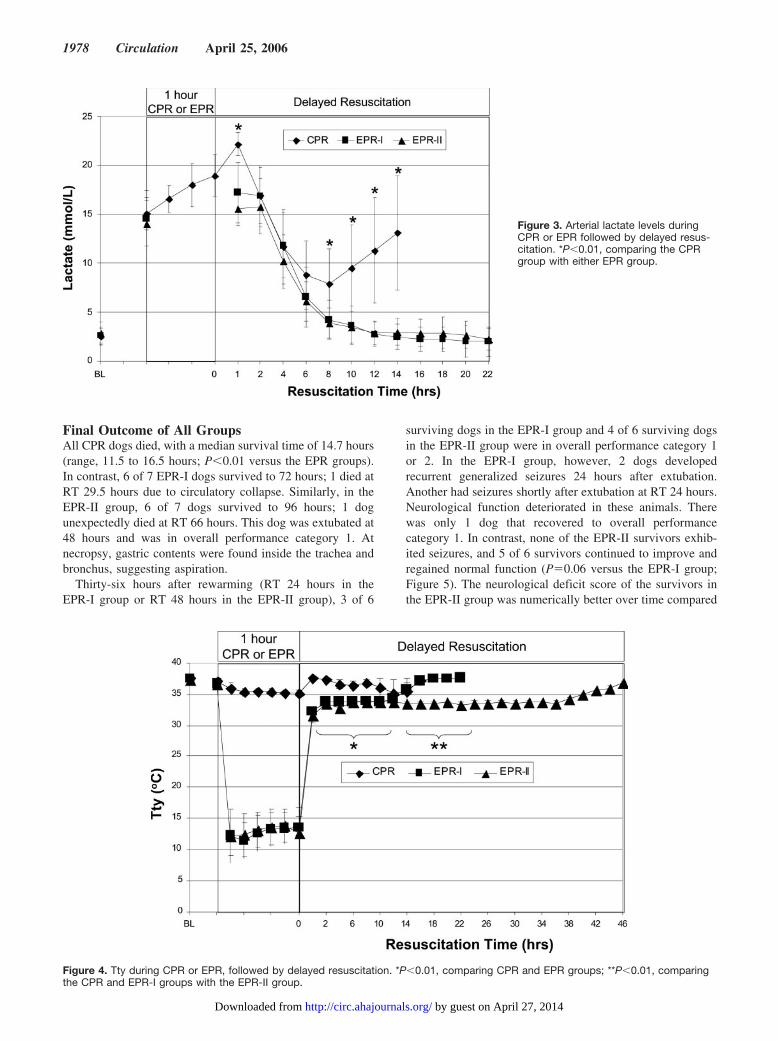
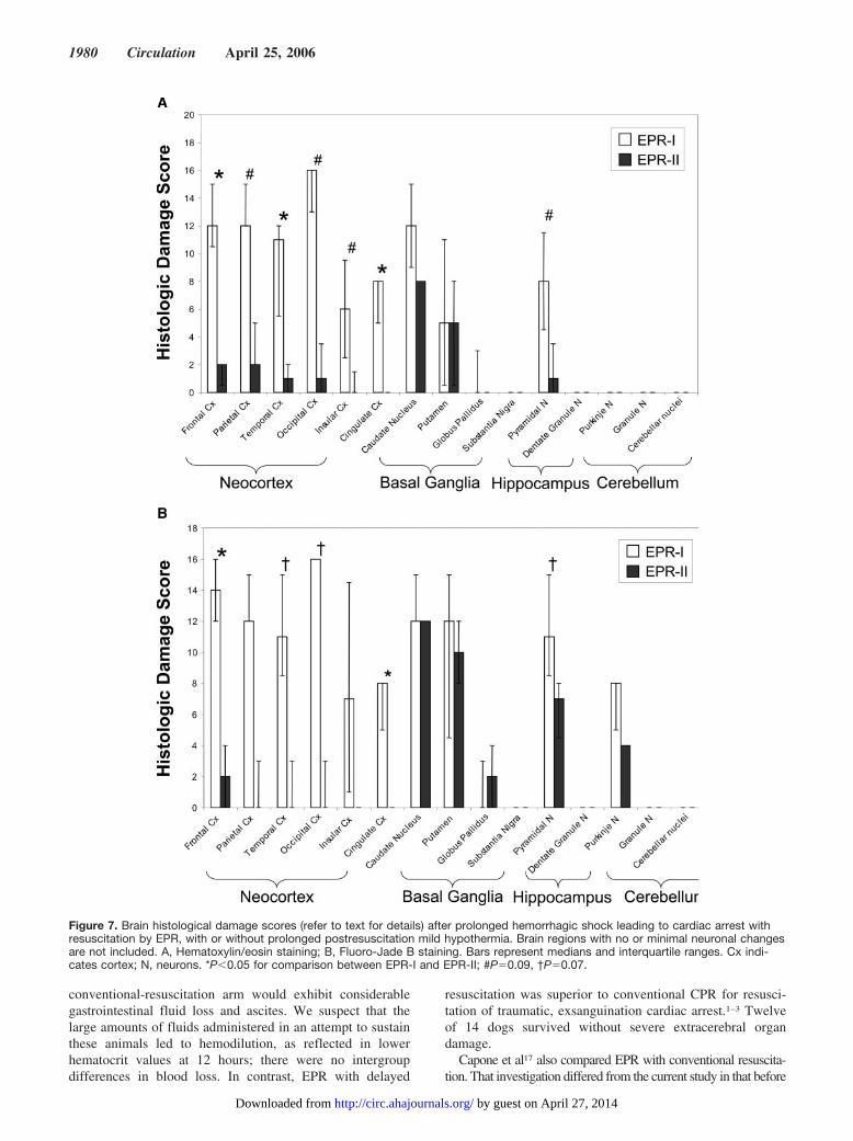
despite massive fluid resuscitation and vasoactive support. Lac-tate levels decreased transiently but increased sharply again untildeath (Figure 3; P�0.01 versus the EPR groups). Tty decreasedslightly during CPR to �36°C and then increased to 38°C withCPB (Figure 4).
Resuscitation of EPR GroupsThe perfusion pressure during aortic flush was �20 mm Hg,with no difference between the EPR groups (Figure 2). At theend of the aortic flush, Tty had decreased to similar levels,with little change during circulatory arrest (Figure 4).
EPR dogs were rewarmed to 34°C within 1 hour by CPB.When Tpa reached 32°C, defibrillation yielded ROSC in alldogs at 30�12 minutes in the EPR-I group and at 32�23minutes in the EPR-II group (P�NS). The total defibrillationenergy required was 229�225 J in the EPR-I group and264�326 J in the EPR-II group (P�NS).
At RT 12 hours, hematocrit was lower in the CPR group(19�9%) compared with the EPR-I (33�7%, P�0.006) andEPR-II (28�9%, P�0.16) groups. Final hematocrit values were29�3% at RT 24 hours in the EPR-I group and 30�6% at RT48 hours in the EPR-II group (P�NS versus the EPR-I group).
TABLE 1. Physiological Parameters During Cardiac ArrestAfter Prolonged Hemorrhage
CPR Group EPR-I Group EPR-II Group
Hemorrhage time, min 124.4�10.5 118.4�19.7 126.4�19.8
pH 6.88�0.24 6.99�0.16 6.93�0.12
PCO2, mm Hg 86�39 58�30 73�28
PO2, mm Hg 58�8 85�31 79�24
Base deficit, mmol/L 16.6�2 16.4�1.3 15.5�1.8
Potassium, mmol/L 7.6�1.2 6.8�1.3 7.3�0.9
Glucose, mg/dL 449�150 563�110 522�207
Lactate, mmol/L 15.1�1.6 14.6�2.8 14.1�2.3
Blood urea nitrogen, mg/dL 23.4�6.1 27.6�6.5 25.6�8.1
Hematocrit, % 16.2�2.2 18.1�2.3 19.6�1.8
Figure 2. MAP during CPR or EPR, followed by delayed resuscitation. *P�0.01, comparing the CPR group with either EPR group.
TABLE 2. Fluid Balance During the First 16 Hoursof Resuscitation
CPR Group EPR-I Group EPR-II Group
Fluid requirement, L 15.3�2.6* 4.1�0.6 4.2�0.9
Gastrointestinal fluid loss, L† 5.1�1.7* 0.7�0.4 1.5�0.4
Urine output, L 0.03�0.08* 2.0�1.2 2.7�0.9
Abdominal drainage, L 2.4�1.2* 0.3�0.2 0.5�0.4
*P�0.01, compared with EPR groups.†Includes orogastric and rectal fluid losses.
Wu et al Profound Hypothermia for Traumatic Cardiac Arrest 1977
by guest on April 27, 2014http://circ.ahajournals.org/Downloaded from
Final Outcome of All GroupsAll CPR dogs died, with a median survival time of 14.7 hours(range, 11.5 to 16.5 hours; P�0.01 versus the EPR groups).In contrast, 6 of 7 EPR-I dogs survived to 72 hours; 1 died atRT 29.5 hours due to circulatory collapse. Similarly, in theEPR-II group, 6 of 7 dogs survived to 96 hours; 1 dogunexpectedly died at RT 66 hours. This dog was extubated at48 hours and was in overall performance category 1. Atnecropsy, gastric contents were found inside the trachea andbronchus, suggesting aspiration.
Thirty-six hours after rewarming (RT 24 hours in theEPR-I group or RT 48 hours in the EPR-II group), 3 of 6
surviving dogs in the EPR-I group and 4 of 6 surviving dogsin the EPR-II group were in overall performance category 1or 2. In the EPR-I group, however, 2 dogs developedrecurrent generalized seizures 24 hours after extubation.Another had seizures shortly after extubation at RT 24 hours.Neurological function deteriorated in these animals. Therewas only 1 dog that recovered to overall performancecategory 1. In contrast, none of the EPR-II survivors exhib-ited seizures, and 5 of 6 survivors continued to improve andregained normal function (P�0.06 versus the EPR-I group;Figure 5). The neurological deficit score of the survivors inthe EPR-II group was numerically better over time compared
Figure 3. Arterial lactate levels duringCPR or EPR followed by delayed resus-citation. *P�0.01, comparing the CPRgroup with either EPR group.
Figure 4. Tty during CPR or EPR, followed by delayed resuscitation. *P�0.01, comparing CPR and EPR groups; **P�0.01, comparingthe CPR and EPR-I groups with the EPR-II group.
1978 Circulation April 25, 2006
by guest on April 27, 2014http://circ.ahajournals.org/Downloaded from

with the EPR-I group (P�0.09), although the final neurolog-ical deficit scores were better (P�0.04; Figure 6).
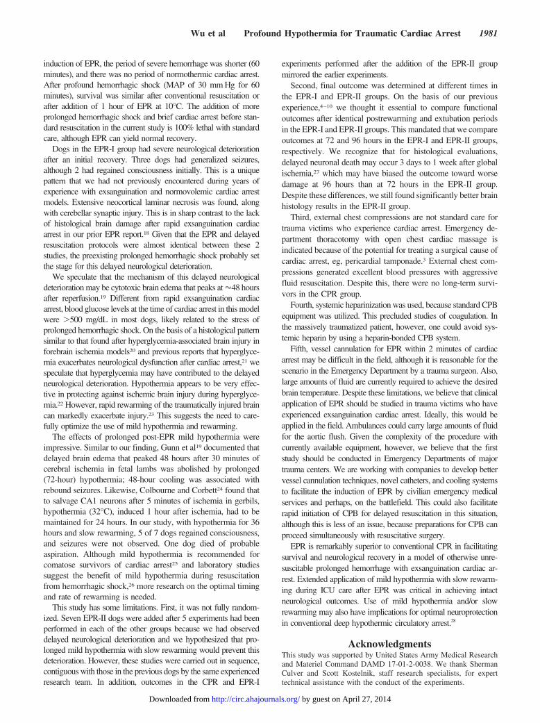
Brain HistologyIn the EPR-I group, the brains exhibited prominent, acuteeosinophilic neuronal degeneration within the frontal, parietal,temporal, and occipital cortices (Figure 7). In the most severelyaffected regions, a laminar band of necrosis with associatedneuropil spongiosis was evident. Marked to severe neuronaldegeneration was present in the caudate region, with slightly lessdegeneration in the putamen. Within the caudate, degenerationwas primarily present in the medium spiny neurons. The largerinterneurons were relatively unscathed. In the hippocampus,moderate to marked neuronal degeneration was present primar-ily within the CA1 region, although degeneration often extendedto the CA3 and CA4 regions. In the cerebellum of the EPR-Igroup, Fluoro-Jade B staining revealed small numbers of degen-erating Purkinje neuron cell bodies and greater numbers ofdendrites. Degrees of neuronal degeneration were scored slightlyhigher in Fluoro-Jade B–stained sections than in hematoxylin/eosin-stained sections.
In contrast to the EPR-I group, the EPR-II group had onlymild damage in the neocortex (Figure 7A; P�0.05 versus EPR-I
in the frontal and temporal lobes). In the hippocampus of theEPR-II group, damage severity as assessed by Fluoro-Jade Bstaining was also less (Figure 7B; P�0.065). Furthermore,Fluoro-Jade B staining in the hippocampus of EPR-II groupdogs was primarily restricted to neuronal processes within CA3and CA4. Degenerative changes in the caudate and putamen ofEPR-I and EPR-II groups were similar (Figure 7). Within thecerebellum of the EPR-II group, Fluoro-Jade B staining revealedscattered Purkinje neurons with degenerative dendrites.
Figure 7 includes only those brain regions that had greaterthan minimal degrees of degenerative changes. All other brainregions, ie, the piriform cortex, entorhinal cortex, septal region,basal forebrain, anterior thalamus, posterior thalamus, amygdala,midbrain, pons, and medulla oblongata, were characterized byno or only minimal degrees of damage, with no differencesbetween groups. By repeated-measures ANOVA for brain re-gions, the histological damage scores were higher in the EPR-Igroup than in the EPR-II group (P�0.006). The total histologicaldamage scores in the EPR-I group (226�111) were numericallyhigher than those in EPR-II (102�87; P�0.15).
Extracerebral Organ InjuryThe heart and liver enzyme levels in the EPR groups weremarkedly increased after resuscitation. They decreased grad-ually after 24 hours but remained increased at 72 or 96 hours,with no difference between groups (data available in DataSupplement Table I).
At necropsy, in the CPR group, all dogs had generalizededema, severe endocardial hemorrhage, lung edema, and bloodyascites. Although the serosal side of the intestine had mildhemorrhage, the intestinal mucosa was sloughed over the entirelength of the intestine. In the EPR groups, the most consistentfindings were mild to moderate endocardial hemorrhage andextensive hemorrhage in the gallbladder wall. Occasionally,hemorrhagic spots were found on the serosal surface of theintestine, but there was no necrosis. Lung edema was found onlyin 1 EPR-I dog.
DiscussionWe established an exsanguination cardiac arrest model that isunsalvageable by contemporary conventional resuscitation.At the time of cardiac arrest, �60% to 90% of the estimatedblood volume was removed. Arterial blood gases taken at thebeginning of cardiac arrest revealed severe acidemia(pH�7.0), hyperkalemia, and hyperlactemia. As expected,none of the dogs could be resuscitated with CPR, despitevigorous blood and fluid replacement, standard drug therapy,and chest compressions with an MAP at 50 to 60 mm Hg.Such favorable hemodynamic responses would be difficult toachieve in traumatic, exsanguination cardiac arrest victims.Although all of the dogs could be resuscitated with CPB, allsubsequently died of severe multiple-organ failure, includingcardiovascular dysfunction, renal failure, and extensive gas-trointestinal mucosal necrosis. This pattern is anticipated afterprolonged hemorrhagic shock and cardiac arrest.2 Suchtrauma victims similarly develop irreversible shock, includ-ing vasodilatation unresponsive to vasopressors and massivecapillary leak, presumably secondary to the systemic inflam-matory response. Thus, it was anticipated that animals in the
Figure 5. Overall performance categories (OPC; refer to text fordetails) after prolonged hemorrhagic shock leading to cardiacarrest with resuscitation by CPR or EPR. *P�0.01, survival inthe CPR group vs the EPR-I and EPR-II groups; **P�0.06, nor-mal (OPC 1) vs abnormal (OPC 2 to 5) in the EPR-II vs theEPR-I group.
Figure 6. Neurological deficit scores (refer to text for details)after prolonged hemorrhagic shock leading to cardiac arrestwith resuscitation by EPR, with or without prolonged resuscita-tive mild hypothermia. Median and interquartile ranges areshown. *P�0.09 for repeated-measures ANOVA; P�0.04 forfinal neurological deficit scores by the Mann-Whitney U test.
Wu et al Profound Hypothermia for Traumatic Cardiac Arrest 1979
by guest on April 27, 2014http://circ.ahajournals.org/Downloaded from
conventional-resuscitation arm would exhibit considerablegastrointestinal fluid loss and ascites. We suspect that thelarge amounts of fluids administered in an attempt to sustainthese animals led to hemodilution, as reflected in lowerhematocrit values at 12 hours; there were no intergroupdifferences in blood loss. In contrast, EPR with delayed
resuscitation was superior to conventional CPR for resusci-tation of traumatic, exsanguination cardiac arrest.1–3 Twelveof 14 dogs survived without severe extracerebral organdamage.
Capone et al17 also compared EPR with conventional resuscita-tion. That investigation differed from the current study in that before
Figure 7. Brain histological damage scores (refer to text for details) after prolonged hemorrhagic shock leading to cardiac arrest withresuscitation by EPR, with or without prolonged postresuscitation mild hypothermia. Brain regions with no or minimal neuronal changesare not included. A, Hematoxylin/eosin staining; B, Fluoro-Jade B staining. Bars represent medians and interquartile ranges. Cx indi-cates cortex; N, neurons. *P�0.05 for comparison between EPR-I and EPR-II; #P�0.09, †P�0.07.
1980 Circulation April 25, 2006
by guest on April 27, 2014http://circ.ahajournals.org/Downloaded from
induction of EPR, the period of severe hemorrhage was shorter (60minutes), and there was no period of normothermic cardiac arrest.After profound hemorrhagic shock (MAP of 30 mm Hg for 60minutes), survival was similar after conventional resuscitation orafter addition of 1 hour of EPR at 10°C. The addition of moreprolonged hemorrhagic shock and brief cardiac arrest before stan-dard resuscitation in the current study is 100% lethal with standardcare, although EPR can yield normal recovery.
Dogs in the EPR-I group had severe neurological deteriorationafter an initial recovery. Three dogs had generalized seizures,although 2 had regained consciousness initially. This is a uniquepattern that we had not previously encountered during years ofexperience with exsanguination and normovolemic cardiac arrestmodels. Extensive neocortical laminar necrosis was found, alongwith cerebellar synaptic injury. This is in sharp contrast to the lackof histological brain damage after rapid exsanguination cardiacarrest in our prior EPR report.18 Given that the EPR and delayedresuscitation protocols were almost identical between these 2studies, the preexisting prolonged hemorrhagic shock probably setthe stage for this delayed neurological deterioration.
We speculate that the mechanism of this delayed neurologicaldeterioration may be cytotoxic brain edema that peaks at �48 hoursafter reperfusion.19 Different from rapid exsanguination cardiacarrest, blood glucose levels at the time of cardiac arrest in this modelwere �500 mg/dL in most dogs, likely related to the stress ofprolonged hemorrhagic shock. On the basis of a histological patternsimilar to that found after hyperglycemia-associated brain injury inforebrain ischemia models20 and previous reports that hyperglyce-mia exacerbates neurological dysfunction after cardiac arrest,21 wespeculate that hyperglycemia may have contributed to the delayedneurological deterioration. Hypothermia appears to be very effec-tive in protecting against ischemic brain injury during hyperglyce-mia.22 However, rapid rewarming of the traumatically injured braincan markedly exacerbate injury.23 This suggests the need to care-fully optimize the use of mild hypothermia and rewarming.
The effects of prolonged post-EPR mild hypothermia wereimpressive. Similar to our finding, Gunn et al19 documented thatdelayed brain edema that peaked 48 hours after 30 minutes ofcerebral ischemia in fetal lambs was abolished by prolonged(72-hour) hypothermia; 48-hour cooling was associated withrebound seizures. Likewise, Colbourne and Corbett24 found thatto salvage CA1 neurons after 5 minutes of ischemia in gerbils,hypothermia (32°C), induced 1 hour after ischemia, had to bemaintained for 24 hours. In our study, with hypothermia for 36hours and slow rewarming, 5 of 7 dogs regained consciousness,and seizures were not observed. One dog died of probableaspiration. Although mild hypothermia is recommended forcomatose survivors of cardiac arrest25 and laboratory studiessuggest the benefit of mild hypothermia during resuscitationfrom hemorrhagic shock,26 more research on the optimal timingand rate of rewarming is needed.
This study has some limitations. First, it was not fully random-ized. Seven EPR-II dogs were added after 5 experiments had beenperformed in each of the other groups because we had observeddelayed neurological deterioration and we hypothesized that pro-longed mild hypothermia with slow rewarming would prevent thisdeterioration. However, these studies were carried out in sequence,contiguous with those in the previous dogs by the same experiencedresearch team. In addition, outcomes in the CPR and EPR-I
experiments performed after the addition of the EPR-II groupmirrored the earlier experiments.
Second, final outcome was determined at different times inthe EPR-I and EPR-II groups. On the basis of our previousexperience,4–10 we thought it essential to compare functionaloutcomes after identical postrewarming and extubation periodsin the EPR-I and EPR-II groups. This mandated that we compareoutcomes at 72 and 96 hours in the EPR-I and EPR-II groups,respectively. We recognize that for histological evaluations,delayed neuronal death may occur 3 days to 1 week after globalischemia,27 which may have biased the outcome toward worsedamage at 96 hours than at 72 hours in the EPR-II group.Despite these differences, we still found significantly better brainhistology results in the EPR-II group.
Third, external chest compressions are not standard care fortrauma victims who experience cardiac arrest. Emergency de-partment thoracotomy with open chest cardiac massage isindicated because of the potential for treating a surgical cause ofcardiac arrest, eg, pericardial tamponade.3 External chest com-pressions generated excellent blood pressures with aggressivefluid resuscitation. Despite this, there were no long-term survi-vors in the CPR group.
Fourth, systemic heparinization was used, because standard CPBequipment was utilized. This precluded studies of coagulation. Inthe massively traumatized patient, however, one could avoid sys-temic heparin by using a heparin-bonded CPB system.
Fifth, vessel cannulation for EPR within 2 minutes of cardiacarrest may be difficult in the field, although it is reasonable for thescenario in the Emergency Department by a trauma surgeon. Also,large amounts of fluid are currently required to achieve the desiredbrain temperature. Despite these limitations, we believe that clinicalapplication of EPR should be studied in trauma victims who haveexperienced exsanguination cardiac arrest. Ideally, this would beapplied in the field. Ambulances could carry large amounts of fluidfor the aortic flush. Given the complexity of the procedure withcurrently available equipment, however, we believe that the firststudy should be conducted in Emergency Departments of majortrauma centers. We are working with companies to develop bettervessel cannulation techniques, novel catheters, and cooling systemsto facilitate the induction of EPR by civilian emergency medicalservices and perhaps, on the battlefield. This could also facilitaterapid initiation of CPB for delayed resuscitation in this situation,although this is less of an issue, because preparations for CPB canproceed simultaneously with resuscitative surgery.
EPR is remarkably superior to conventional CPR in facilitatingsurvival and neurological recovery in a model of otherwise unre-suscitable prolonged hemorrhage with exsanguination cardiac ar-rest. Extended application of mild hypothermia with slow rewarm-ing during ICU care after EPR was critical in achieving intactneurological outcomes. Use of mild hypothermia and/or slowrewarming may also have implications for optimal neuroprotectionin conventional deep hypothermic circulatory arrest.28
AcknowledgmentsThis study was supported by United States Army Medical Researchand Materiel Command DAMD 17-01-2-0038. We thank ShermanCulver and Scott Kostelnik, staff research specialists, for experttechnical assistance with the conduct of the experiments.
Wu et al Profound Hypothermia for Traumatic Cardiac Arrest 1981
by guest on April 27, 2014http://circ.ahajournals.org/Downloaded from

DisclosuresDrs Wu, Kochanek, Tisherman and S.W. Stezoski have submitted aprovisional patent entitled “Method of Inducing Suspended AnimationFollowing Cardiopulmonary Arrest.” The other authors report noconflicts.
References1. Bellamy R, Safar P, Tisherman SA, Basford R, Bruttig SP, Capone A,
Dubick MA, Ernster L, Hattler BGJ, Hochachka P, Klain M, KochanekPM, Kofke WA, Lancaster JR, McGowan FXJ, Oeltgen PR, SeveringhausJW, Taylor MJ, Zar H. Suspended animation for delayed resuscitation.Crit Care Med. 1996;24:S24–S47.
2. Hopson LR, Hirsh E, Delgado J, Domeier RM, Krohmer J, McSwain NEJ,Weldon C, Friel M, Hoyt DB. Guidelines for withholding or termination ofresuscitation in prehospital traumatic cardiopulmonary arrest. J Am CollSurg. 2003;196:475–481.
3. Rhee PM, Acosta J, Bridgeman A, Wang D, Jordan M, Rich N. Survival afteremergency department thoracotomy: review of published data from the past25 years. J Am Coll Surg. 2000;190:288–298.
4. Kochanek PM, Tisherman SA, Stezoski SM, Nozari A, Wu X, Safar P: Novelpotentials for emergency hypothermia: suspended animation with delayedresuscitation from exsanguinations cardiac arrest. Hypothermia for AcuteBrain Damage. New York, NY: Springer-Verlag Publishers; 2004:271–277.
5. Behringer W, Safar P, Wu X, Kentner R, Radovsky A, Kochanek PM, DixonCE, Tisherman SA. Survival without brain damage after clinical death of60–120 mins in dogs using suspended animation by profound hypothermia.Crit Care Med. 2003;31:1523–1531.
6. Nozari A, Safar P, Tisherman S, Stezoski W, Kochanek PM, Wu X,Kostelnic S, Carcillo J. Suspended animation and plasma exchange enablesfull neurologic recovery from lethal traumatic exsanguination, even after 2 hperiod of no-flow. Crit Care Med. 2004;31(suppl):A9. Abstract.
7. Behringer W, Prueckner S, Kentner R, Tisherman SA, Radovsky A, Clark R,Stezoski SW, Henchir J, Klein E, Safar P. Rapid hypothermic aortic flush canachieve survival without brain damage after 30 minutes cardiac arrest in dogs.Anesthesiology. 2000;93:1491–1499.
8. Behringer W, Prueckner S, Safar P, Radovsky A, Kentner R, Stezoski SW,Henchir J, Tisherman SA. Rapid induction of mild cerebral hypothermia by coldaortic flush achieves normal recovery in a dog outcome model with 20-minuteexsanguination cardiac arrest. Acad Emerg Med. 2000;7:1341–1348.
9. Behringer W, Safar P, Kentner R, Wu X, Kagan VE, Radovsky A, Clark RS,Kochanek PM, Subramanian M, Tyurin VA, Tyurina YY, Tisherman SA.Antioxidant Tempol enhances hypothermic cerebral preservation during pro-longed cardiac arrest in dogs. J Cereb Blood Flow Metab. 2002;22:105–117.
10. Behringer W, Safar P, Wu X, Kentner R, Radovsky A, Tisherman SA.Delayed intra-ischemic aortic cold flush for preservation during prolongedcardiac arrest in dogs. Crit Care Med. 2001;29(suppl):A17. Abstract.
11. Carrillo P, Takasu A, Safar P, Tisherman S, Stezoski SW, Stolz G, Dixon CE,Radovsky A. Prolonged severe hemorrhagic shock and resuscitation in ratsdoes not cause subtle brain damage. J Trauma. 1998;45:239–248.
12. Alam HB, Austin B, Koustova E, Rhee P. Resuscitation-induced pulmonaryapoptosis and intracellular adhesion molecule-1 expression in rats are attenuatedby the use of ketone Ringer’s solution. J Am Coll Surg. 2001;193:255–263.
13. Healey MA, Samphire J, Hoyt DB, Liu F, Davis R, Loomis WH. Irreversibleshock is not irreversible: a new model of massive hemorrhage and resusci-tation. J Trauma. 2001;50:826–834.
14. Holcomb JB. Fluid resuscitation in modern combat casualty care: lessonslearned from Somalia. J Trauma. 2003;54:S46–S51.
15. Bleyaert AL, Nemoto EM, Safar P, Stezoski SM, Mickell JJ, Moossy J, RaoGR. Thiopental amelioration of brain damage after global ischemia inmonkeys. Anesthesiology. 1978;49:390–398.
16. Wu X, Drabek T, Kochanek PM. A novel approach to cerebral resuscitation:suspended animation with delayed resuscitation: studies in dog and rat models. In:Vincent JL, Pickett K, eds: Yearbook of Intensive Care and Emergency Medicine.New York, NY: Springer-Verlag Publishers; 2005;298–314.
17. Capone A, Safar P, Radovsky A, Wang YF, Peitzman A, Tisherman SA.Complete recovery after normothermic hemorrhagic shock and profound hypo-thermic circulatory arrest of 60 minutes in dogs. J Trauma. 1996;40:388–395.
18. Nozari A, Safar P, Wu X, Stezoski SW, Henchir J, Kochanek PM, Klain M,Radovsky A, Tisherman SA. Suspended animation can allow survivalwithout brain damage after traumatic exsanguination cardiac arrest of 60 minin dogs. J Trauma. 2004;56:1266–1275.
19. Gunn AJ, Gunn TR, de Haan HH, Williams CE, Gluckman PD. Dramaticneuronal rescue with prolonged selective head cooling after ischemia in fetallambs. J Clin Invest. 1997;99:248–256.
20. Longstreth WTJ, Inui TS. High blood glucose level on hospital admission andpoor neurological recovery after cardiac arrest. Ann Neurol. 1984;15:59–63.
21. Li PA, Siesjo BK. Role of hyperglycaemia-related acidosis in ischaemic braindamage. Acta Physiol Scand. 1997;161:567–580.
22. Lundgren J, Smith ML, Siesjo BK. Influence of moderate hypothermia onischemic brain damage incurred under hyperglycemic conditions. Exp BrainRes. 1991;84:91–101.
23. Suehiro E, Povlishock JT. Exacerbation of traumatically induced axonalinjury by rapid posthypothermic rewarming and attenuation of axonal changeby cyclosporin A. J Neurosurg. 2001;94:493–498.
24. Colbourne F, Corbett D. Delayed and prolonged post-ischemic hypothermiais neuroprotective in the gerbil. Brain Res. 1994;654:265–272.
25. Nolan JP, Morley PT, Vanden Hoek TL, Hickey RW, Kloeck WG, Billi J,Bottiger BW, Morley PT, Nolan JP, Okada K, Reyes C, Shuster M, Steen PA,Weil MH, Wenzel V, Hickey RW, Carli P, Vanden Hoek TL, Atkins D;International Liaison Committee on Resuscitation. Therapeutic hypothermiaafter cardiac arrest: an advisory statement by the advanced life support taskforce of the International Liaison Committee on Resuscitation. Circulation.2003;108:118–121.
26. Wu X, Kochanek PM, Cochran K, Nozari A, Henchir J, Stezoski SW,Wagner R, Wisniewski S, Tisherman SA. Mild hypothermia improvessurvival after prolonged, traumatic hemorrhagic shock in pigs. J Trauma.2005;59:291–301.
27. Back T, Hemmen T, Schuler OG. Lesion evolution in cerebral ischemia.J Neurol. 2004;251:388–397.
28. Kurth CD, Priestly M, Golden J, McCann J, Raghupathi R. Regionalpatterns of neuronal death after deep hypothermic circulatory arrest innewborn pigs. J Thorac Cardiovasc Surg. 1999;118:1068–1077.
CLINICAL PERSPECTIVEThe success of emergency preservation and resuscitation (EPR) may have important implications. First, the potential forits use may not be limited by the duration of preexisting hemorrhagic shock. This is important clinically, because the exactduration of hemorrhage is usually unknown, and prolonged hemorrhagic shock before cardiac arrest is common in the ruralor military trauma situation. Second, EPR is much more reliable in preserving tissue viability during cardiac arrest than isoptimized cardiopulmonary resuscitation and may allow intact survival. Third, the impressive benefits of prolongedpostischemic mild hypothermia and slow rewarming may have implications for neuroprotection after deep hypothermiccirculatory arrest used in cardiac or neurological surgery. Brain damage found in deep hypothermic circulatory arrest issimilar to that seen in our study. The development of EPR has focused on the exsanguinating trauma patient. Although mosttrauma patients are young and have normal coronary arteries, similar to the animals in this study, exsanguination can occurin older patients with coronary artery disease. The effects of inducing EPR in such patients are difficult to predict, althoughone should keep in mind that the results of current management strategies for trauma patients who experience cardiac arrestare dismal for all victims. Rapid cooling of the heart might help preserve cardiac function, just as brain cooling protectsneurological function.
1982 Circulation April 25, 2006
by guest on April 27, 2014http://circ.ahajournals.org/Downloaded from
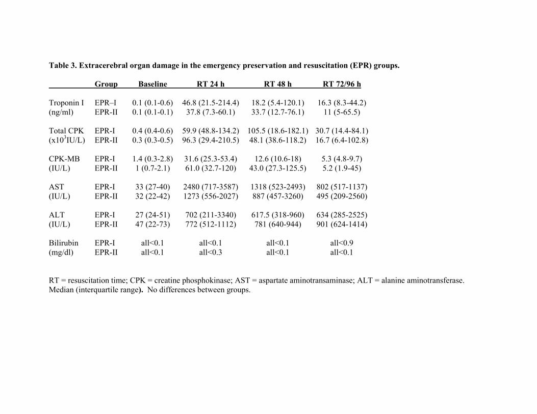
Table 3. Extracerebral organ damage in the emergency preservation and resuscitation (EPR) groups.
Group Baseline RT 24 h RT 48 h RT 72/96 h
Troponin I EPR–I 0.1 (0.1-0.6) 46.8 (21.5-214.4) 18.2 (5.4-120.1) 16.3 (8.3-44.2) (ng/ml) EPR-II 0.1 (0.1-0.1) 37.8 (7.3-60.1) 33.7 (12.7-76.1) 11 (5-65.5)
Total CPK EPR-I 0.4 (0.4-0.6) 59.9 (48.8-134.2) 105.5 (18.6-182.1) 30.7 (14.4-84.1) (x103IU/L) EPR-II 0.3 (0.3-0.5) 96.3 (29.4-210.5) 48.1 (38.6-118.2) 16.7 (6.4-102.8)
CPK-MB EPR-I 1.4 (0.3-2.8) 31.6 (25.3-53.4) 12.6 (10.6-18) 5.3 (4.8-9.7) (IU/L) EPR-II 1 (0.7-2.1) 61.0 (32.7-120) 43.0 (27.3-125.5) 5.2 (1.9-45)
AST EPR-I 33 (27-40) 2480 (717-3587) 1318 (523-2493) 802 (517-1137) (IU/L) EPR-II 32 (22-42) 1273 (556-2027) 887 (457-3260) 495 (209-2560)
ALT EPR-I 27 (24-51) 702 (211-3340) 617.5 (318-960) 634 (285-2525) (IU/L) EPR-II 47 (22-73) 772 (512-1112) 781 (640-944) 901 (624-1414)
Bilirubin EPR-I all<0.1 all<0.1 all<0.1 all<0.9 (mg/dl) EPR-II all<0.1 all<0.3 all<0.1 all<0.1
RT = resuscitation time; CPK = creatine phosphokinase; AST = aspartate aminotransaminase; ALT = alanine aminotransferase. Median (interquartile range). No differences between groups.