Indonesia. - Revista.Ccba.Uady.Mx
12
Tropical and Subtropical Agroecosystems 25 (2022): #021 Aisyah et al., 2022 1 THE EFFECT OF PH AND SUCROSE ON THE EMBRYOGENIC CELLS GROWTH OF SUGAR CANE (Saccharum officinarum) IN LIQUID CULTURE † [EL EFECTO DEL PH Y LA SACAROSA EN EL CRECIMIENTO DE CÉLULAS EMBRIOGÉNICAS DE CAÑA DE AZÚCAR (Saccharum officinarum) EN CULTIVO LÍQUIDO] Novi Nur Aisyah 1 , Laily Ilman Widuri 1 , Firdha Narulita Alfian 2 and Parawita Dewanti 1,3* 1 Faculty of Agriculture, University of Jember, Kalimantan st.. 37, Jember 68121, Indonesia. 2 Master Program of Biotechnology, University of Jember, Kalimantan st. 37, Jember 68121, Indonesia 3 Center for Development of Advanced Science and Technology (CDAST), University of Jember, Kalimantan st. 37, Jember 68121, Indonesia. Email. *[email protected] *Corresponding author SUMMARY Background. Sugarcane (Saccharum officinarum L.) is one of the important commodities in Indonesia as raw material for sugar production. The constraints for sugarcane cultivation are limited availability and poor quality of seedling. The utilization of liquid culture in tissue culture enables to produce good quality seedlings, free of viruses and diseases, and can generate a large quantity in a short time. Liquid culture can produce embryogenic cells faster since embryogenic callus can develop easily in liquid media. Objective. This study determined the appropriate pH and concentration of sucrose in the liquid culture of embryogenic cells. Methodology. This study was conducted in 3 stages of in vitro culture, which were callus induction, solid media proliferation, and liquid media proliferation that using pH and sucrose treatments. Result. The results showed that pH 6.5 + 3% sucrose (A3B1) produced the highest number of callus with the average callus on the first week 29 callus, the second week 8,33 callus and the third week 4,33 callus and the color did not easily turn to brown with color 5Y 8/2. Implications. The protocol was developed that allows embryogenic callus of sugarcane plants to be obtained in liquid media that can be used for embryogenic callus proliferation. Conclusions. pH 6.5 + sucrose 3% (A3B1) was the best combination treatment of pH and sucrose for sugarcane callus proliferation in liquid culture media, which produced the highest number of calluses and color of callus that not easily turned brown. Keywords: somatic embryogenesis; callus; proliferation; globular; scutellar. RESUMEN Antecedentes. La caña de azúcar (Saccharum officinarum L.) es uno de los productos básicos más importantes en Indonesia como materia prima para la producción de azúcar. Las limitaciones para el cultivo de la caña de azúcar son la disponibilidad limitada y la mala calidad de las plántulas. La utilización del cultivo líquido en el cultivo de tejidos permite producir plántulas de buena calidad, libres de virus y enfermedades, y puede generar una gran cantidad en poco tiempo. El cultivo líquido puede producir células embriogénicas más rápidamente ya que el callo embriogénico puede desarrollarse fácilmente en medios líquidos. Objetivo. Este estudio determinó el pH y la concentración de sacarosa adecuados en el cultivo líquido de células embriogénicas. Metodología. Este estudio se llevó a cabo en 3 etapas de cultivo in vitro, que fueron la inducción de callo, la proliferación en medio sólido y la proliferación en medio líquido que utilizando tratamientos de pH y sacarosa. Resultados. Los resultados mostraron que el pH 6,5 + 3% de sacarosa (A3B1) produjo el mayor número de callos con el promedio de callos en la primera semana 29 callos, la segunda semana 8,33 callos y la tercera semana 4,33 callos y el color no se convirtió fácilmente en marrón con el color 5Y 8/2. Implicaciones. Se desarrolló un protocolo que permite obtener callos embriogénicos de plantas de caña de azúcar en medios líquidos que pueden ser utilizados para la proliferación de callos embriogénicos. Conclusiones. pH 6,5 + sacarosa 3% (A3B1) fue el mejor tratamiento de combinación de pH y sacarosa para la proliferación de callos de caña de azúcar en medios de cultivo líquidos, que produjo el mayor número de callos y el color del callo que no se volvió marrón fácilmente. Palabras Clave: embriogénesis somática; callo; proliferación; globular; escutelar. † Submitted July 21, 2021 – Accepted October 7, 2021. This work is licensed under a CC-BY 4.0 International License. ISSN: 1870-0462.
-
Upload
khangminh22 -
Category
Documents
-
view
2 -
download
0
Transcript of Indonesia. - Revista.Ccba.Uady.Mx
Tropical and Subtropical Agroecosystems 25 (2022): #021 Aisyah et al., 2022
1
THE EFFECT OF PH AND SUCROSE ON THE EMBRYOGENIC
CELLS GROWTH OF SUGAR CANE (Saccharum officinarum) IN
LIQUID CULTURE †
[EL EFECTO DEL PH Y LA SACAROSA EN EL CRECIMIENTO DE
CÉLULAS EMBRIOGÉNICAS DE CAÑA DE AZÚCAR (Saccharum
officinarum) EN CULTIVO LÍQUIDO]
Novi Nur Aisyah1, Laily Ilman Widuri1, Firdha Narulita Alfian2
and Parawita Dewanti1,3*
1Faculty of Agriculture, University of Jember, Kalimantan st.. 37, Jember 68121,
Indonesia. 2Master Program of Biotechnology, University of Jember, Kalimantan st. 37,
Jember 68121, Indonesia 3Center for Development of Advanced Science and Technology (CDAST),
University of Jember, Kalimantan st. 37, Jember 68121, Indonesia. Email.
*Corresponding author
SUMMARY
Background. Sugarcane (Saccharum officinarum L.) is one of the important commodities in Indonesia as raw
material for sugar production. The constraints for sugarcane cultivation are limited availability and poor quality
of seedling. The utilization of liquid culture in tissue culture enables to produce good quality seedlings, free of
viruses and diseases, and can generate a large quantity in a short time. Liquid culture can produce embryogenic
cells faster since embryogenic callus can develop easily in liquid media. Objective. This study determined the
appropriate pH and concentration of sucrose in the liquid culture of embryogenic cells. Methodology. This study
was conducted in 3 stages of in vitro culture, which were callus induction, solid media proliferation, and liquid
media proliferation that using pH and sucrose treatments. Result. The results showed that pH 6.5 + 3% sucrose
(A3B1) produced the highest number of callus with the average callus on the first week 29 callus, the second
week 8,33 callus and the third week 4,33 callus and the color did not easily turn to brown with color 5Y 8/2.
Implications. The protocol was developed that allows embryogenic callus of sugarcane plants to be obtained in
liquid media that can be used for embryogenic callus proliferation. Conclusions. pH 6.5 + sucrose 3% (A3B1)
was the best combination treatment of pH and sucrose for sugarcane callus proliferation in liquid culture media,
which produced the highest number of calluses and color of callus that not easily turned brown.
Keywords: somatic embryogenesis; callus; proliferation; globular; scutellar.
RESUMEN
Antecedentes. La caña de azúcar (Saccharum officinarum L.) es uno de los productos básicos más importantes
en Indonesia como materia prima para la producción de azúcar. Las limitaciones para el cultivo de la caña de
azúcar son la disponibilidad limitada y la mala calidad de las plántulas. La utilización del cultivo líquido en el
cultivo de tejidos permite producir plántulas de buena calidad, libres de virus y enfermedades, y puede generar
una gran cantidad en poco tiempo. El cultivo líquido puede producir células embriogénicas más rápidamente ya
que el callo embriogénico puede desarrollarse fácilmente en medios líquidos. Objetivo. Este estudio determinó
el pH y la concentración de sacarosa adecuados en el cultivo líquido de células embriogénicas. Metodología.
Este estudio se llevó a cabo en 3 etapas de cultivo in vitro, que fueron la inducción de callo, la proliferación en
medio sólido y la proliferación en medio líquido que utilizando tratamientos de pH y sacarosa. Resultados. Los
resultados mostraron que el pH 6,5 + 3% de sacarosa (A3B1) produjo el mayor número de callos con el
promedio de callos en la primera semana 29 callos, la segunda semana 8,33 callos y la tercera semana 4,33 callos
y el color no se convirtió fácilmente en marrón con el color 5Y 8/2. Implicaciones. Se desarrolló un protocolo
que permite obtener callos embriogénicos de plantas de caña de azúcar en medios líquidos que pueden ser
utilizados para la proliferación de callos embriogénicos. Conclusiones. pH 6,5 + sacarosa 3% (A3B1) fue el
mejor tratamiento de combinación de pH y sacarosa para la proliferación de callos de caña de azúcar en medios
de cultivo líquidos, que produjo el mayor número de callos y el color del callo que no se volvió marrón
fácilmente.
Palabras Clave: embriogénesis somática; callo; proliferación; globular; escutelar.
† Submitted July 21, 2021 – Accepted October 7, 2021. This work is licensed under a CC-BY 4.0 International License.
ISSN: 1870-0462.
Tropical and Subtropical Agroecosystems 25 (2022): #021 Aisyah et al., 2022
2
INTRODUCTION
Sugarcane (Saccharum officinarum L.), a raw
material for sugar production is one of the
important commodities in Indonesia. According to
The Directorate General of Plantation (2019),
sugarcane production in Indonesia tends to decrease
from 2011 until 2019. According to Sulaiman et al
(2019), the amount of sugar consumption increases
by 4.3% every year which made Indonesia the
largest sugar importer in 2017-2018. This problem
occurred because sugarcane production was unable
to meet the needs of raw materials for sugar
production due to low availability and poor quality
of seedlings. Provision of seedlings can be
conducted conventionally or unconventionally by
tissue culture.
Tissue culture is a method for isolating part of a
plant and maintaining cells or pieces of plant tissue
grown on suitable artificial media under aseptic
conditions (Nofrianinda et al., 2017). Tissue culture
technique is classified into organogenesis and
somatic embryogenesis. Organogenesis is the
process of direct organ formation from explants
(Hinchee et al., 1988). While somatic
embryogenesis is a regeneration process by forming
an embryo-like structure derived from somatic cells
with plumule and radicle (Pardal, 2003). The
advantages of somatic embryogenesis are shorter
propagation times, bipolar embryo production,
higher transformation ability, and can be utilized
for germplasm storage (Sapsuha et al., 2011).
Somatic embryogenesis can be developed using
solid or liquid media. An embryogenic callus is
easier to develop using liquid media because it
accelerates the growth of cells (Taryono, 2016).
According to Minarsih et al (2013), temporary
immersion system method in liquid culture can
produce higher number and uniformity shoots and
plantlets compare to solid media.
Liquid culture is the culture of free cells or small
cell aggregates in a liquid medium by shaking
method (Gunawan, 1992). The success of liquid
culture is influenced by explants (cultured plant
parts), hormones, media composition, the physical
environment of tissue culture, and growth
regulators. The pH and sucrose contents during
proliferation are media composition factors
affecting the growth of callus. The pH of medium
indicates the value of acidity and alkalinity of a
solution in water. It indicates the presence of H+
ions in the solution and affects the solubility of
compounds in form of salt (Mastuti, 2017).
According to Mishra et al (2019), pH media of 5.82
produced high biomass and alkaloids. Sucrose is a
source of carbon in tissue culture. Disaccharides
application in the form of sucrose will provide an
optimum growth response in tissue culture (Azmi et
al, 2017). Furthermore, the application of 5%
sucrose can increase the production of cell biomass
and alkaloid content in Eurycoma longifolia cell
(Siregar, 2010). This finding also conveyed by
Hanifa (2013) that higher sucrose concentration
increased the dry weight of Croton tiglium.
Meanwhile, according to Yaacob et al (2014), the
highest callus production obtained in sucrose
concentration by 5%, but the fastest callus growth
(25-30 days) exhibited in 3% sucrose application.
Tissue culture media with pH 5.8 and 6.8 was
proven to induce callus growth within 30 days in
lime. Therefore, it is necessary to evaluate the
effectiveness of pH and sucrose application in
liquid culture media for enhancing the growth of
embryogenic cells of sugarcane.
MATERIALS AND METHODS
The study was conducted at the Center for
Development of Advanced Sciences (CDAST)
Laboratory in University of Jember from April
2020 to May 2021. The plant material used was
obtained from the spindle leaf of Sugarcane
“Bululawang” (BL) variety. Plant material was
taken from 4-6 months old healthy plants in the
field. The spindle leaf was prepared and sterilized
with 70% alcohol and placed in Laminar Air Flow
(LAF) cabinet. The plant tip dipped in 96% alcohol,
and then the whole plant material was heated over a
bunsen burner to minimize contamination. The
sterilized spindle leaf peeled to a 5 mm diameter
and sliced into ± 2 mm width on filter paper then
cut into 7 pieces.
Callus Induction
These entire experiments using Murashige and
Skoog (MS) media as the based media with
addition of plant growth regulators for each stage of
culture. Callus induction was carried out by
planting explants in MS + 4 mg L-1 2,4-
Dichlorophenoxyacetic acid (2,4-D) + 300 mg L-1
Casein Hydrolysate + 30 g L-1 sucrose + 5 g L-1
agar with a pH of 6.2. The explants were planted in
LAF and the culture bottles were stored on a culture
rack for 6 weeks at a temperature of 23°-25°C in a
dark room. The explants were sub-cultured every 3
weeks. The induced callus was selected and the
embryogenic callus sub-cultured into solid
proliferation media. Morphological observations
using a Leica EZ4HD microscope.
Solid Media Callus Proliferation
The 6 weeks old callus transferred to solid
proliferation media for 3 weeks. The selected callus
was embryogenic callus characterized with crumb
texture and white or yellowish white color.
Proliferation was carried out in dark conditions.
Callus proliferation used MS + 2,4-D 2 mg L-1 +
500 mg L-1 Casein Hydrolysate + 560 mg L-1 L-
Prolin + 40 mg L-1 L-Glutamine + 30 g L-1 Sucrose
+ 5 g L-1 agar. Morphological observations using a
Leica EZ4HD microscope
Tropical and Subtropical Agroecosystems 25 (2022): #021 Aisyah et al., 2022
3
Liquid Media Callus Proliferation
The 9 weeks old callus was transplanted to the
combination of pH (5.5; 6.0; 6.5) and sucrose (3%,
4%, 5%) liquid media (Table 1). The media
contained MS + 2 mg L-1 2,4-D + 500 mg L-1
Casein Hydrolysate + 560 mg L-1 L-Proline + 40
mg L-1 L-Glutamine + 50 mg L-1 Arginine + 300
mg L-1 Polyvinylpyrrolidone (PVP). The media was
placed into erlenmeyer or bottles then sterilized in
an autoclave and incubated for 24 hours. Selected
callus from proliferation in solid media were
pressed with a scalpel. The selected callus was
weighed to determine weight. As much as 0.25 g of
callus was put into 25 mL liquid media in a 100 mL
Erlenmeyer. The culture media was then shaken
using a shaker at 100 rpm in the dark condition and
25°C room temperature. Shaking was carried out
for 2 hours, and then left for 6 hours. Shaking was
repeated 3 times within 24 hours.
Table 1. The combination of pH and sucrose
treatments.
Code Combination of treatments
A1B1 pH 5.5 + sucrose 3%
A1B2 pH 5.5 + sucrose 4%
A1B3 pH 5.5 + sucrose 5%
A2B1 pH 6.0 + sucrose 3%
A2B2 pH 6.0 + sucrose 4%
A2B3 pH 6.0 + sucrose 5%
A3B1 pH 6.5 + sucrose 3%
A3B2 pH 6.5 + sucrose 4%
A3B3 pH 6.5 + sucrose 5%
Callus proliferation was carried out for 3 weeks and
selection of callus was conducted every week
during the culture period. The callus selection was
carried out 3 times. The liquid callus proliferation
was filtered with 1 mm diameter filter to collect the
number of selected callus data. The bigger clumps
of callus which filtered were counted visually on
the sterile petridish. While smaller callus (< 1 mm
diameter size) which passed the filter were continue
to be shaken for a week before filtered again in the
next week.
Callus morphological and color of callus
observations were carried out microscopically with
a Leica EZ4HD microscope and observation of
callus cells using Olympus CX31 microscope.
Color observation was conducted using Munsell
Color Charts for Plant Tissues (Munsell Color
1977) every week microscopically. The selected
callus that filtered was chosen as sample for color
of callus data. The selected callus to be observed
were callus that had an average color in one callus
group and has represented one group of these
calluses.
Data Analysis
Two types of data were collected, they were
qualitative and quantitative data. Qualitative data
which include morphology of callus and color of
callus. Meanwhile quantitative data which include
number of selected callus. Complete random design
(CRD) with 2 factors was used for the experimental
design for this research. The quantitative data were
analyzed statistically by analysis of variance
(ANOVA) using the least significant different
(LSD) test (P<0.05) in Ms Excel.
RESULTS
Callus Induction
Morphology of callus
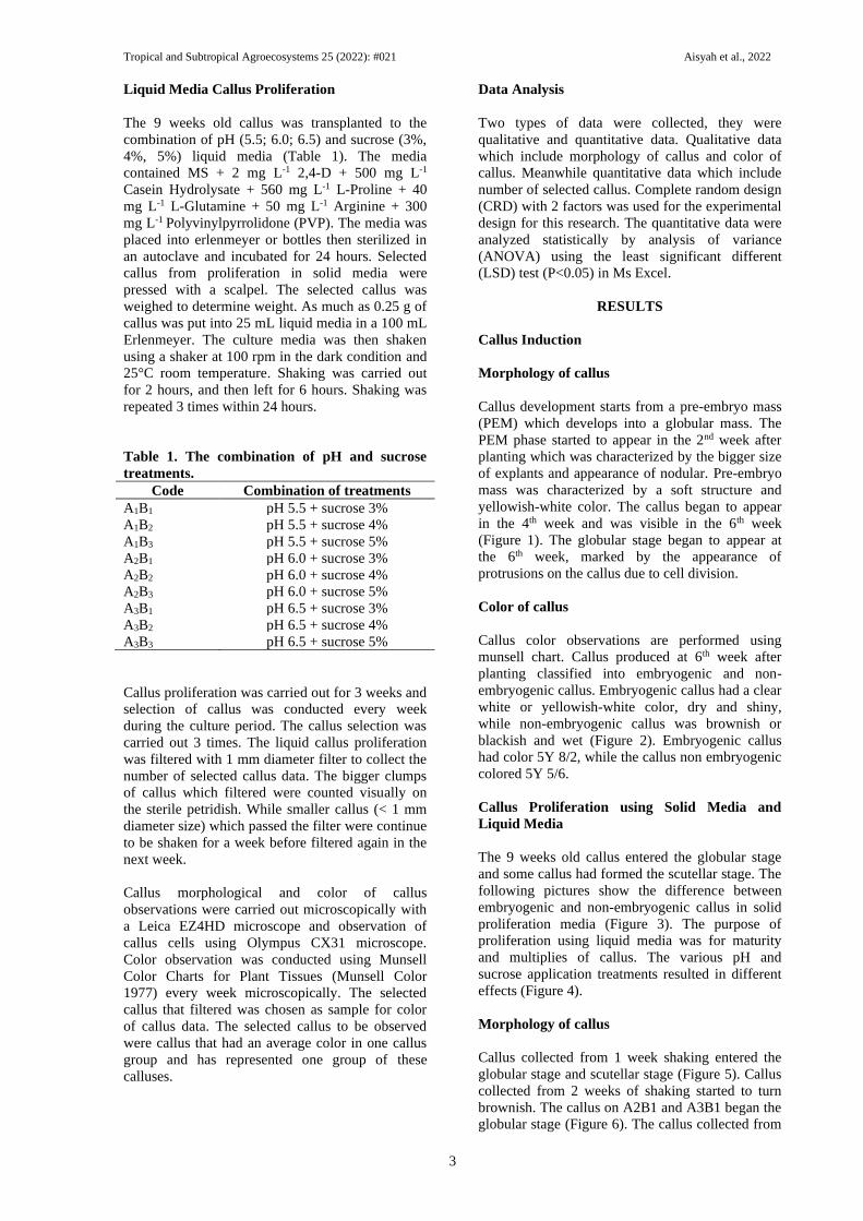
Callus development starts from a pre-embryo mass
(PEM) which develops into a globular mass. The
PEM phase started to appear in the 2nd week after
planting which was characterized by the bigger size
of explants and appearance of nodular. Pre-embryo
mass was characterized by a soft structure and
yellowish-white color. The callus began to appear
in the 4th week and was visible in the 6th week
(Figure 1). The globular stage began to appear at
the 6th week, marked by the appearance of
protrusions on the callus due to cell division.
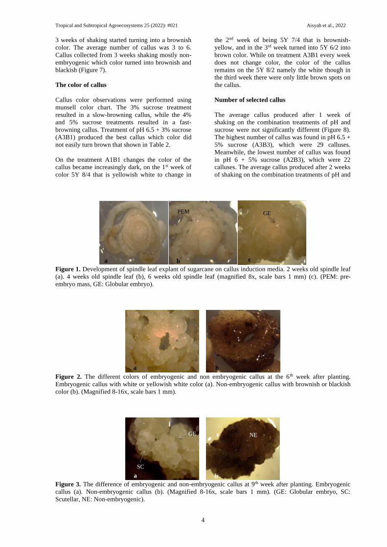
Color of callus
Callus color observations are performed using
munsell chart. Callus produced at 6th week after
planting classified into embryogenic and non-
embryogenic callus. Embryogenic callus had a clear
white or yellowish-white color, dry and shiny,
while non-embryogenic callus was brownish or
blackish and wet (Figure 2). Embryogenic callus
had color 5Y 8/2, while the callus non embryogenic
colored 5Y 5/6.
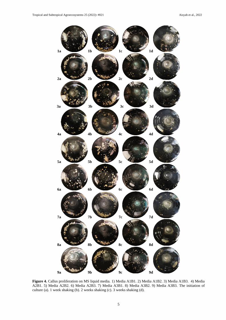
Callus Proliferation using Solid Media and
Liquid Media
The 9 weeks old callus entered the globular stage
and some callus had formed the scutellar stage. The
following pictures show the difference between
embryogenic and non-embryogenic callus in solid
proliferation media (Figure 3). The purpose of
proliferation using liquid media was for maturity
and multiplies of callus. The various pH and
sucrose application treatments resulted in different
effects (Figure 4).
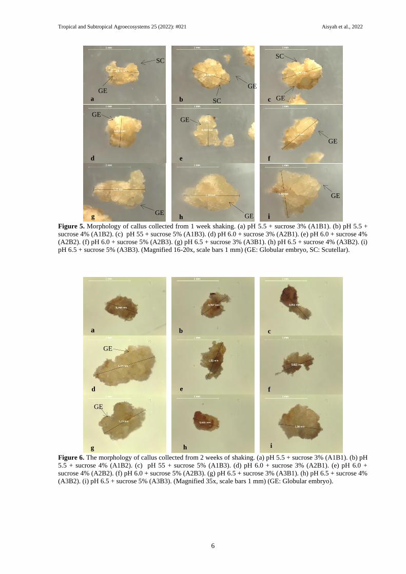
Morphology of callus
Callus collected from 1 week shaking entered the
globular stage and scutellar stage (Figure 5). Callus
collected from 2 weeks of shaking started to turn
brownish. The callus on A2B1 and A3B1 began the
globular stage (Figure 6). The callus collected from
Tropical and Subtropical Agroecosystems 25 (2022): #021 Aisyah et al., 2022
4
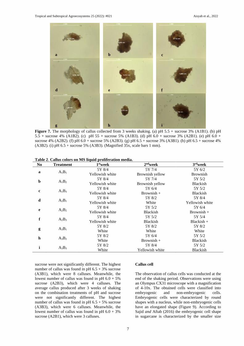
3 weeks of shaking started turning into a brownish
color. The average number of callus was 3 to 6.
Callus collected from 3 weeks shaking mostly non-
embryogenic which color turned into brownish and
blackish (Figure 7).
The color of callus
Callus color observations were performed using
munsell color chart. The 3% sucrose treatment
resulted in a slow-browning callus, while the 4%
and 5% sucrose treatments resulted in a fast-
browning callus. Treatment of pH 6.5 + 3% sucrose
(A3B1) produced the best callus which color did
not easily turn brown that shown in Table 2.
On the treatment A1B1 changes the color of the
callus became increasingly dark, on the 1st week of
color 5Y 8/4 that is yellowish white to change in
the 2nd week of being 5Y 7/4 that is brownish-
yellow, and in the 3rd week turned into 5Y 6/2 into
brown color. While on treatment A3B1 every week
does not change color, the color of the callus
remains on the 5Y 8/2 namely the white though in
the third week there were only little brown spots on
the callus.
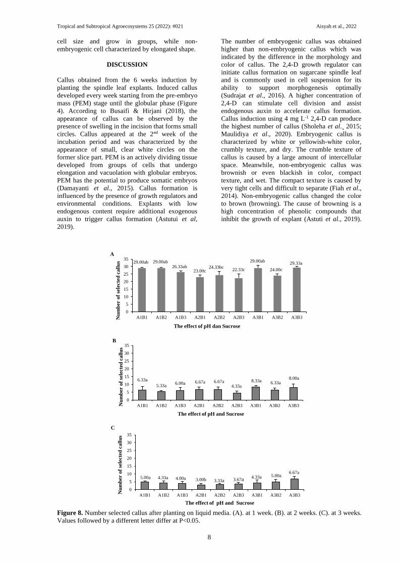
Number of selected callus
The average callus produced after 1 week of
shaking on the combination treatments of pH and
sucrose were not significantly different (Figure 8).
The highest number of callus was found in pH 6.5 +
5% sucrose (A3B3), which were 29 calluses.
Meanwhile, the lowest number of callus was found
in pH 6 + 5% sucrose (A2B3), which were 22
calluses. The average callus produced after 2 weeks
of shaking on the combination treatments of pH and
Figure 1. Development of spindle leaf explant of sugarcane on callus induction media. 2 weeks old spindle leaf
(a). 4 weeks old spindle leaf (b), 6 weeks old spindle leaf (magnified 8x, scale bars 1 mm) (c). (PEM: pre-
embryo mass, GE: Globular embryo).
Figure 2. The different colors of embryogenic and non embryogenic callus at the 6th week after planting.
Embryogenic callus with white or yellowish white color (a). Non-embryogenic callus with brownish or blackish
color (b). (Magnified 8-16x, scale bars 1 mm).
Figure 3. The difference of embryogenic and non-embryogenic callus at 9th week after planting. Embryogenic
callus (a). Non-embryogenic callus (b). (Magnified 8-16x, scale bars 1 mm). (GE: Globular embryo, SC:
Scutellar, NE: Non-embryogenic).
a b c
GE PEM
NE
SC
GE
a b
a b
Tropical and Subtropical Agroecosystems 25 (2022): #021 Aisyah et al., 2022
5
1a 1b 1c 1d
2a 2b 2c 2d
3a 3b 3c 3d
4a 4b 4c 4d
5a 5b 5c 5d
6a 6b 6c 6d
7a 7b 7c 7d
8a 8b 8c 8d
9a 9b 9c 9d
Figure 4. Callus proliferation on MS liquid media. 1) Media A1B1. 2) Media A1B2. 3) Media A1B3. 4) Media
A2B1. 5) Media A2B2. 6) Media A2B3. 7) Media A3B1. 8) Media A3B2. 9) Media A3B3. The initiation of
culture (a). 1 week shaking (b). 2 weeks shaking (c). 3 weeks shaking (d).
Tropical and Subtropical Agroecosystems 25 (2022): #021 Aisyah et al., 2022
6
Figure 5. Morphology of callus collected from 1 week shaking. (a) pH 5.5 + sucrose 3% (A1B1). (b) pH 5.5 +
sucrose 4% (A1B2). (c) pH 55 + sucrose 5% (A1B3). (d) pH 6.0 + sucrose 3% (A2B1). (e) pH 6.0 + sucrose 4%
(A2B2). (f) pH 6.0 + sucrose 5% (A2B3). (g) pH 6.5 + sucrose 3% (A3B1). (h) pH 6.5 + sucrose 4% (A3B2). (i)
pH 6.5 + sucrose 5% (A3B3). (Magnified 16-20x, scale bars 1 mm) (GE: Globular embryo, SC: Scutellar).
Figure 6. The morphology of callus collected from 2 weeks of shaking. (a) pH 5.5 + sucrose 3% (A1B1). (b) pH
5.5 + sucrose 4% (A1B2). (c) pH 55 + sucrose 5% (A1B3). (d) pH 6.0 + sucrose 3% (A2B1). (e) pH 6.0 +
sucrose 4% (A2B2). (f) pH 6.0 + sucrose 5% (A2B3). (g) pH 6.5 + sucrose 3% (A3B1). (h) pH 6.5 + sucrose 4%
(A3B2). (i) pH 6.5 + sucrose 5% (A3B3). (Magnified 35x, scale bars 1 mm) (GE: Globular embryo).
SC
GE
SC
GE
GE
SC
GE
GE GE
GE GE
GE
a b c
d e f
g i h
GE
a b c
d f e
g i h
GE
Tropical and Subtropical Agroecosystems 25 (2022): #021 Aisyah et al., 2022
7
Figure 7. The morphology of callus collected from 3 weeks shaking. (a) pH 5.5 + sucrose 3% (A1B1). (b) pH
5.5 + sucrose 4% (A1B2). (c) pH 55 + sucrose 5% (A1B3). (d) pH 6.0 + sucrose 3% (A2B1). (e) pH 6.0 +
sucrose 4% (A2B2). (f) pH 6.0 + sucrose 5% (A2B3). (g) pH 6.5 + sucrose 3% (A3B1). (h) pH 6.5 + sucrose 4%
(A3B2). (i) pH 6.5 + sucrose 5% (A3B3). (Magnified 35x, scale bars 1 mm).
Table 2. Callus colors on MS liquid proliferation media.
No Treatment 1stweek 2ndweek 3rdweek
a A1B1 5Y 8/4
Yellowish white
5Y 7/4
Brownish yellow
5Y 6/2
Brownish
b A1B2 5Y 8/4
Yellowish white
5Y 7/4
Brownish yellow
5Y 5/2
Blackish
c A1B3 5Y 8/4
Yellowish white
5Y 6/4
Brownish +
5Y 5/2
Blackish
d A2B1 5Y 8/4
Yellowish white
5Y 8/2
White
5Y 8/4
Yellowish white
e A2B2 5Y 8/4
Yellowish white
5Y 5/2
Blackish
5Y 6/4
Brownish +
f A2B3 5Y 8/4
Yellowish white
5Y 5/2
Blackish
5Y 5/4
Blackish +
g A3B1 5Y 8/2
White
5Y 8/2
White
5Y 8/2
White
h A3B2 5Y 8/2
White
5Y 6/4
Brownish +
5Y 5/2
Blackish
i A3B3 5Y 8/2
White
5Y 8/4
Yellowish white
5Y 5/2
Blackish
sucrose were not significantly different. The highest
number of callus was found in pH 6.5 + 3% sucrose
(A3B1), which were 8 calluses. Meanwhile, the
lowest number of callus was found in pH 6.0 + 5%
sucrose (A2B3), which were 4 calluses. The
average callus produced after 3 weeks of shaking
on the combination treatments of pH and sucrose
were not significantly different. The highest
number of callus was found in pH 6.5 + 5% sucrose
(A3B3), which were 6 calluses. Meanwhile, the
lowest number of callus was found in pH 6.0 + 3%
sucrose (A2B1), which were 3 calluses.
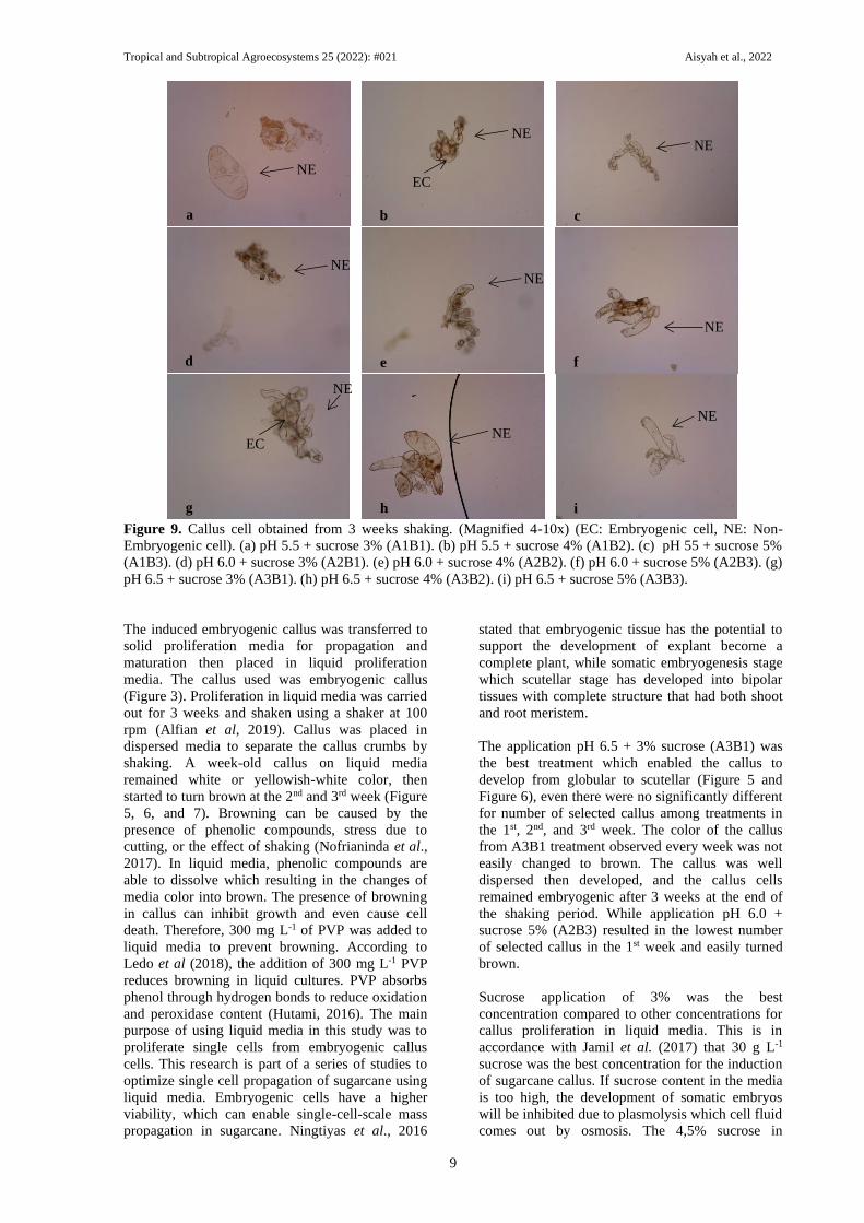
Callus cell
The observation of callus cells was conducted at the
end of the shaking period. Observations were using
an Olympus CX31 microscope with a magnification
of 4-10x. The obtained cells were classified into
embryogenic and non-embryogenic cells.
Embryogenic cells were characterized by round
shapes with a nucleus, while non-embryogenic cells
have an elongated shape (Figure 9). According to
Sajid and Aftab (2016) the embryogenic cell shape
in sugarcane is characterized by the smaller size
a b
d
c
e f
g i h
Tropical and Subtropical Agroecosystems 25 (2022): #021 Aisyah et al., 2022
8
cell size and grow in groups, while non-
embryogenic cell characterized by elongated shape.
DISCUSSION
Callus obtained from the 6 weeks induction by
planting the spindle leaf explants. Induced callus
developed every week starting from the pre-embryo
mass (PEM) stage until the globular phase (Figure
4). According to Busaifi & Hirjani (2018), the
appearance of callus can be observed by the
presence of swelling in the incision that forms small
circles. Callus appeared at the 2nd week of the
incubation period and was characterized by the
appearance of small, clear white circles on the
former slice part. PEM is an actively dividing tissue
developed from groups of cells that undergo
elongation and vacuolation with globular embryos.
PEM has the potential to produce somatic embryos
(Damayanti et al., 2015). Callus formation is
influenced by the presence of growth regulators and
environmental conditions. Explants with low
endogenous content require additional exogenous
auxin to trigger callus formation (Astutui et al,
2019).
The number of embryogenic callus was obtained
higher than non-embryogenic callus which was
indicated by the difference in the morphology and
color of callus. The 2,4-D growth regulator can
initiate callus formation on sugarcane spindle leaf
and is commonly used in cell suspension for its
ability to support morphogenesis optimally
(Sudrajat et al., 2016). A higher concentration of
2,4-D can stimulate cell division and assist
endogenous auxin to accelerate callus formation.
Callus induction using 4 mg L-1 2,4-D can produce
the highest number of callus (Sholeha et al. ̧2015;
Maulidiya et al., 2020). Embryogenic callus is
characterized by white or yellowish-white color,
crumbly texture, and dry. The crumble texture of
callus is caused by a large amount of intercellular
space. Meanwhile, non-embryogenic callus was
brownish or even blackish in color, compact
texture, and wet. The compact texture is caused by
very tight cells and difficult to separate (Fiah et al.,
2014). Non-embryogenic callus changed the color
to brown (browning). The cause of browning is a
high concentration of phenolic compounds that
inhibit the growth of explant (Astuti et al., 2019).
Figure 8. Number selected callus after planting on liquid media. (A). at 1 week. (B). at 2 weeks. (C). at 3 weeks.
Values followed by a different letter differ at P<0.05.
0
5
10
15
20
25
30
35
A1B1 A1B2 A1B3 A2B1 A2B2 A2B3 A3B1 A3B2 A3B3Nu
mb
er o
f se
lecte
d c
all
us
The effect of pH dan Sucrose
29.00ab 29.00ab26.33ab
23.00c24.33bc
22.33c
29.00ab
24.00c
29.33a
A
0
5
10
15
20
25
30
35
A1B1 A1B2 A1B3 A2B1 A2B2 A2B3 A3B1 A3B2 A3B3Nu
mb
er o
f se
lecte
d c
all
us
The effect of pH and Sucrose
6.33a
5.33a6.00a 6.67a6.67a
4.33a
8.33a 6.33a8.00a
B
0
5
10
15
20
25
30
35
A1B1 A1B2 A1B3 A2B1 A2B2 A2B3 A3B1 A3B2 A3B3
Nu
mb
er o
f se
lecte
d c
all
us
The effect of pH and Sucrose
5.00a 4.33a3.33a3.00b4.00a 3.67a
4.33a 5.00a 6.67a
C
Tropical and Subtropical Agroecosystems 25 (2022): #021 Aisyah et al., 2022
9
Figure 9. Callus cell obtained from 3 weeks shaking. (Magnified 4-10x) (EC: Embryogenic cell, NE: Non-
Embryogenic cell). (a) pH 5.5 + sucrose 3% (A1B1). (b) pH 5.5 + sucrose 4% (A1B2). (c) pH 55 + sucrose 5%
(A1B3). (d) pH 6.0 + sucrose 3% (A2B1). (e) pH 6.0 + sucrose 4% (A2B2). (f) pH 6.0 + sucrose 5% (A2B3). (g)
pH 6.5 + sucrose 3% (A3B1). (h) pH 6.5 + sucrose 4% (A3B2). (i) pH 6.5 + sucrose 5% (A3B3).
The induced embryogenic callus was transferred to
solid proliferation media for propagation and
maturation then placed in liquid proliferation
media. The callus used was embryogenic callus
(Figure 3). Proliferation in liquid media was carried
out for 3 weeks and shaken using a shaker at 100
rpm (Alfian et al, 2019). Callus was placed in
dispersed media to separate the callus crumbs by
shaking. A week-old callus on liquid media
remained white or yellowish-white color, then
started to turn brown at the 2nd and 3rd week (Figure
5, 6, and 7). Browning can be caused by the
presence of phenolic compounds, stress due to
cutting, or the effect of shaking (Nofrianinda et al.,
2017). In liquid media, phenolic compounds are
able to dissolve which resulting in the changes of
media color into brown. The presence of browning
in callus can inhibit growth and even cause cell
death. Therefore, 300 mg L-1 of PVP was added to
liquid media to prevent browning. According to
Ledo et al (2018), the addition of 300 mg L-1 PVP
reduces browning in liquid cultures. PVP absorbs
phenol through hydrogen bonds to reduce oxidation
and peroxidase content (Hutami, 2016). The main
purpose of using liquid media in this study was to
proliferate single cells from embryogenic callus
cells. This research is part of a series of studies to
optimize single cell propagation of sugarcane using
liquid media. Embryogenic cells have a higher
viability, which can enable single-cell-scale mass
propagation in sugarcane. Ningtiyas et al., 2016
stated that embryogenic tissue has the potential to
support the development of explant become a
complete plant, while somatic embryogenesis stage
which scutellar stage has developed into bipolar
tissues with complete structure that had both shoot
and root meristem.
The application pH 6.5 + 3% sucrose (A3B1) was
the best treatment which enabled the callus to
develop from globular to scutellar (Figure 5 and
Figure 6), even there were no significantly different
for number of selected callus among treatments in
the 1st, 2nd, and 3rd week. The color of the callus
from A3B1 treatment observed every week was not
easily changed to brown. The callus was well
dispersed then developed, and the callus cells
remained embryogenic after 3 weeks at the end of
the shaking period. While application pH 6.0 +
sucrose 5% (A2B3) resulted in the lowest number
of selected callus in the 1st week and easily turned
brown.
Sucrose application of 3% was the best
concentration compared to other concentrations for
callus proliferation in liquid media. This is in
accordance with Jamil et al. (2017) that 30 g L-1
sucrose was the best concentration for the induction
of sugarcane callus. If sucrose content in the media
is too high, the development of somatic embryos
will be inhibited due to plasmolysis which cell fluid
comes out by osmosis. The 4,5% sucrose in
NE NE
NE
NE
EC NE
NE
d e f
g
a
h
a
i
a
a c b
NE
NE
EC
NE
Tropical and Subtropical Agroecosystems 25 (2022): #021 Aisyah et al., 2022
10
sorghum caused explant darkened and secreted
phenolic compound in shorgum (Dreger et al.,
2019). The sucrose can increase the rate of
photosynthesis and embryo formation leads to the
optimum absorption of water and nutrients in
media. Sucrose in the media will be hydrolyzed for
callus respiration and stimulate the growth of some
tissues. During respiration, sucrose is converted
into energy in the form of ATP (Samudera et al.,
2019).
The pH of media also affected nutrient absorption
during proliferation in liquid media. The pH of
sterilized media decreased by 0.58 to 1 of acidity
compared to the initial pH. A very low pH is not
optimal for callus growth because nutrients in the
growing media cannot be absorbed properly and
inhibit cell growth. While a very high pH of media
can increase polyphenol enzyme that causes the
color of callus easily change to brown (Tarampak et
al., 2019). According Xi and Yang (2017), during
proliferation, there was no significant difference in
growth of roots and shoots of apple during
induction at pH 6.0-8.0, there was only a slight
decrease in the number of shoots proliferation at pH
>7.5. There was no significant different of number
of selected callus among all of the pH treatments.
The hydrolysis of sucrose in cells affected by the
temperature and pH of media, a lower pH at a
certain temperature can enhance the sucrose
hydrolysis. The commonly used pH of media is
slightly acidic ranges from 5.5 to 5.8 then the media
sterilized using an autoclave to provide the
appropriate temperature for sucrose catalysis. The
pH adjustment affecting the biological activity of
cells. In pine trees, the optimal pH for sucrose
synthesis is 7.7 and 6.7 depending on the plant
variety (Chen et al., 2015). According to
Widiastoety et al (2005), the commonly used pH
for tissue culture media is 5.0-6.5. The pH value
affects the absorption of components or nutrients in
the media for cell biochemical reactions which
affecting plant differentiation and morphogenesis.
Callus dispersed in a liquid medium took more than
three weeks to form larger clumps. These larger
clumps play an important role in reducing the
occurrence of browning in the callus to be
regenerated. At the clumps callus size that is too
small, the phenolic stress that may occur will be
higher because the cell clusters are too small to
survive. Liang et al., 2018 stated that there are two
main mechanisms for browning in plant tissue
culture, which is enzymatic browning and stress
browning caused by explant wound or culture
conditions. Modification of sucrose and pH in this
research are purposed to reduce browning in culture
conditions, focused on callus proliferation stage.
CONCLUSIONS
Based on the obtained data, it can be concluded that
the best combination treatment of pH and sucrose
for sugarcane callus proliferation in liquid culture
media was pH 6.5 + sucrose 3% (A3B1) with the
highest number of calluses and the color did not
easily turn to brown. Higher concentration of
sucrose caused callus darkened and lose the ability
to proliferate. More studies are needed to regenerate
the dispersed callus that has been proliferated in
liquid culture media.
Acknowledgements
The author thanks the Center for Development of
Advanced Science and Technology, for the support
of research place and facilities to carry out this
research.
Funding. This work was developed with a research
budget by supporting grant of Islamic Development
Bank project in collaboration with University of
Jember, with number of contract
2598/UN25.3.1/LT2020 for Dr. Ir. Parawita
Dewanti, MP.
Conflict of interests. All authors declare that there
is no conflict of interest related to this publication.
Compliance with ethical standards. The research
was carried out and presented by the authors under
ethical principles and scientific responsibility in the
handling of the data.
Data availability. The data is available with the
corresponding author
([email protected]), upon reasonable
request.
Author contribution statement (CRediT). Novi
Nur Aisyah: investigation, data curation, formal
analysis, writing-original draft. Laily Ilman
Widuri: validation, writing-review & editing.
Firdha Narulita Alfian: validation, writing-review
& editing. Parawita Dewanti: conceptualization,
funding acquisition, project administration,
supervisión.
REFERENCES
Alfian, F.N., Afdoria, N.N., Dewanti, P., Restanto,
D.P. and Sugiharto, B., 2019. Liquid
culture of somatic embryogenesis cell
proliferation of sugarcane (Saccharum
officinarum). International Journal of
Agriculture and Biology, 21(4), pp. 905-
910. DOI:
https://doi.org/10.17957/IJAB/15.0974.
Astuti, A.T., Nli, A.Z. and Suwirmen., 2019.
Induksi embriogenesis somatik pada
anggrek Vanda sumatra Schltr. dengan
penambahan beberapa konsentrasi asam
2,4-diklorofenoksiasetat (2,4-D). Jurnal
Tropical and Subtropical Agroecosystems 25 (2022): #021 Aisyah et al., 2022
11
Biologi Universitas Andalas, 7(1), pp. 6-
13. DOI:
https://doi.org/10.25077/jbioua.7.1.6-
13.2019.
Azmi, Y., Purwoko, B.S., Dewi, I.S., Syukur, M.
and Suhartini, T., 2017. Kultur antera hasil
persilangan padi lokal beras hitam dengan
varietas budidaya (Fatmawati dan Inpari
13). Jurnal Agronomi Indonesia, 45(3), pp.
228-234. DOI:
https://doi.org/10.24831/jai.v45i3.12544.
Busaifi, Riski. and Hirjani., 2018. Induksi kalus
embriogenik tanaman tebu (Saccharum
officinarum L.) pada berbagai kombinasi
2,4-D dan BAP secara in vitro. Agrosains,
5(2), pp. 91-102.
Chen, C.C., Bates, R. and Carlson, J., 2015. Effect
of environmental and cultural conditions
on medium ph and explant growth
performance of douglas-fir (Pseudotsuga
menziesii) shoot cultures. F1000Research,
3(298), pp. 1-16. DOI:
https://doi.org/10.12688/f1000research.5
919.2.
Damayanti, F., Mariska, I., Suharsono and
Widyastuti, U., 2015. Pendewasaan kalus
embriogenik somatik tanaman tebu
(Saccharum officinarum L.) dengan
kombinasi BAP dan kinetin. Prosiding
Semirata 2015 Bidang MIPA BKS-PTN
Barat, Pontianak, 29-35.
Directorate General of Plantation., 2019. Indonesian plantation statistics for
sugarcane commodities 2017-2019.
Jakarta: Directorate General of Plantation.
pp 3-21.
Dreger, M., Mol, R., Deja, A., Raj, E., Mańkowska,
G. and Wielgus, K., 2019. Improved plant
regeneration in callus cultures of Sorghum
bicolor (L.) Moench. In Vitro Cellular and
Developmental Biology – Plant 55, pp.
190-198. DOI:
https://doi.org/10.1007/s11627-019-09963-
9.
Fiah, R.L., Taryono and Toekidjo., 2014.
Kemampuan regenerasi kalus empat klon
tebu (Saccharum officinarum L.).
Vegetalika, 3(1), pp. 91-101. DOI:
https://doi.org/10.22146/veg.4018.
Gunawan, L. W., 1992. Teknik kultur jaringan
tumbuhan. Bogor: IPB Press. pp. 90.
Hanifa, A.P., 2013. Pengaruh sukrosa dan polietilen
glikol (PEG) terhadap kadar lipid dan
komposisi asam lemak pada kultur
suspensi Croton tiglium L. AgroSainT
UKI Toraja, 4(2), pp. 561-569. DOI:
https://doi.org/10.47178/agro.v4i2.650.
Hinchee, M.A.W., Connor-Ward, D.V., Newell,
C.A., Mc-Donell, R.E., Sato, S.J., Gasser,
C.S., Fischoff, D.A., Fraley, R.T. and
Horsch, R.B., 1988. Production of
transgenic soybean plants using
agrobacterium mediated DNA transfer.
BioTechnology, 6, pp. 915-922. DOI:
https://doi.org/10.1038/nbt0888-915.
Hutami, S., 2016. Masalah pencoklatan pada kultur
jaringan. AgroBiogen, 4(2), pp. 83-88. DOI: http://dx.doi.org/10.21082/jbio.v4n2.2008.
p83-88.
Jamil, S., Shahzad, R., Talha, G.M., Sakhawat, G.,
Rahman, S.U., Sultana, R. and Iqbal, M.Z.,
2017. Optimization of protocols for in
vitro regeneration of sugarcane
(Saccharum officinarum). Hindawi,
2017(8), pp. 1-8. DOI:
https://doi.org/10.1155/2017/2089381.
Ledo, A.D.S., Jenderek, M.M., Ledo, C.A.D.S. and
Silva, T.A., 2018. Antioxidants and
phenolic secretion in sugarcane genotypes
shoots culture. Agricultural Science, 10(5),
pp. 79-91. DOI:
https://doi.org/10.5539/jas.v10n5p79.
Lestari, E.G., 2011. Peranan zat pengatur tumbuh
dalam perbanyakan tanaman melalui kultur
jaringan. AgroBiogen, 7(1), pp. 63-68.
DOI:
http://dx.doi.org/10.21082/jbio.v7n1.2011.
p63-68.
Liang, S., He, Y., Zheng, H., Yuan, Q., Zhang, F.
and Sun, B., 2018. Effects of sucrose and
browning inhibitors on callus proliferation
and anti-browning of chinese kale. IOP
Conference Series: Earth and
Environmental Science 252 022018. DOI:
https://10.1088/1755-1315/252/2/022018.
Mastuti, R., 2017. Dasar-dasar kultur jaringan
tumbuhan. Malang: UB Press. pp. 26-30.
Maulidiya, A.U.K., Sugiharto, B., Dewanti, P. and
Handoyo, T., 2020. Expression of somatic
embryogenesis-related genes in sugarcane
(Saccharum officinarum L.). Crop Science
and Biotechnology, 2020(23), pp. 207-214.
DOI: https://doi.org/10.1007/s12892-020-
00024-x.
Minarsih, H., Riyadi, I., Sumaryono and Budiani,
A., 2013. Mikropogasi tebu (Saccharum
officinarum L.) menggunakan sistem
perendaman sesaat. Menara Perkebunan,
81(1), pp. 1-8.
Mishra, M.R.,Srivastava, R.K. and Akhtar, N.,
2019. Effect of nitrogen, phosporus, and
medium ph to enhance alkaloid production
from Catharanthus roseus cell suspension
culture. International Journal of Secondary
Tropical and Subtropical Agroecosystems 25 (2022): #021 Aisyah et al., 2022
12
Metabolite, 6(2), pp. 137-153. DOI:
https://dx.doi.org/10.21448/ijsm.559679.
Ningtiyas, W.N., Dewanti, P. and Sugiharto, B.,
2016. Preservation effect of PEG
(polyethylene glycol) on synthetic seed of
sugarcane (Saccharum officinarum) var.
NXI 1,3. Annales Bogorienses 20(2), pp.
63-68.
Nofrianinda, V., Yulianti, F. and Agustina, E.,
2017. Pertumbuhan planlet stroberi
(Fragaria ananassa D) var. dorit pada
beberapa variasi media modifikasi in vitro
di balai penelitian jeruk dan buah
subtropika (BALITJESTRO). Biotropic,
1(1), pp. 41-50. DOI:
https://doi.org/10.29080/biotropic.2017.1.1
.32-41.
Nurchayati, Y., Santoso, Nugroho, L.H. and
Indrianto, A., 2018. Penggunaan kinetin,
asam naftalen asetat, dan benzil adenin
dalam induksi kalus kecubung (Dantura
metel L.) secara in vitro. Buletin Anatomi
dan Fisiologi, 3(1), pp. 105-109. DOI:
https://doi.org/10.14710/baf.3.1.2018.105-
109.
Pardal, S.J., 2003. Perkembangan penelitian
regenerasi dan transformasi pada tanaman
kedelai. Buletin Agrobio, 5(2), pp. 37-44.
Sajid, Z.A. and Aftab, F., 2016. An efficient
method for the establishment of cell
suspension cultures in potato (Solanum
Tuberosum L.). Pakistan Journal of
Botany, 48(5), pp. 1993-1997.
Samudera, A.A., Rianto, H. and Historiawati.,
2019. Pengakaran in vitro eksplan tebu
(Saccharum officinarum, L.) varitas
bululawang pada berbagai konsentrasi
NAA dan sukrosa terhadap pertumbuhan
planlet tebu. Vigor, 4(1), pp. 5-13. DOI:
https://doi.org/10.31002/vigor.v4i1.1306.
Sapsuha, Y., Soetrisno, D. and Kusrantinah., 2011.
Induksi kalus dan embriogenesis somatik
in vitro pada lamtoro (Leucaena
leucocephala). Berita Biologi ̧ 10(5), pp.
627-633. DOI:
https://doi.org/10.14203/beritabiologi.v10i
5.1921.
Sharma, M.K., Chaudhary, R., Kureel, R.D. and
Sengar, R.S., 2018. Effect of culture media
ph on in vitro shoot multiplication in
sugarcane. International Journal of
Chemical Studies, 6(2), pp. 1308-1310.
Sholeha, W., Sugiharto, B., Setyati, D. and
Dewanti, P., 2015. Induksi embriogenesis
somatik menggunakan 2,4-
dichlorophenovyacetic acid (2,4-D) dan
kinetin pada eksplan gulungan daun muda
tanaman tebu. Ilmu Dasar, 16(1), pp. 17-
22.
Siregar, L.A.M., 2010. Pengaruh sitokinin eksogen
dan sukrosa terhadap produksi biomassa
dan alkaloid Canthinone dalam kultur
suspensi sel pasak bumi (Eurycoma
longifolia Jack.). Natur Indonesia, 12(2),
pp. 142-150. DOI:
https://doi.org/10.31258/jnat.12.2.143-151.
Sudrajat, H., Suharto, D. and Wijaya, N.R., 2016.
Pengaruh 2,4-D dan glukosa pada kalus
sarang semut (Myrmecodia pendans).
Agrovigor, 9(2), pp. 118-124. DOI:
https://doi.org/10.21107/agrovigor.v9i2.23
16.
Sulaiman, A.A., Sulaeman, Y., Mustikasari, N.,
Nursyamsi, D. and Syakir, A.M. (2019).
Increasing sugar production in indonesia
through land suitability analysis and sugar
mill restructuring. Land, 8(61), pp. 1-17.
DOI: https://doi.org/10.3390/land8040061.
Tarampak, T.C., Sulistiawati, and Nirmala, R.,
2019. Metode mengatasi browning pada
eksplan ulin (Eusideroxylon zwageri)
untuk inisiasi regenerasi secara in vitro.
Agroteknologi Tropika Lembab, 1(2), pp.
106-117.
Taryono., 2016. Pengantar bioteknologi untuk
pemuliaan tanaman. Yogyakarta: Gadjah
Mada University Press. pp. 28-32.
Widiastoety., Kartikaningrum, D.D. and Purbadi.,
2005. Pengaruh ph media terhadap
pertumbuhan plantlet anggrek dendrobium.
Jurnal Hortikultura, 15(1), pp. 18-21.
DOI:
http://dx.doi.org/10.21082/jhort.v15n1.200
5.p%25p.
Xi, Xiolin and Yang, L., 2017. Medium pH
between 5,5 and 7,5 has minimal effect on
tissue culture of apple. American Society
for Horticultura Science, 52(3), pp. 475-
478. DOI:
https://doi.org/10.21273/HORTSCI11443-
16.
Yaacob, J.S., Mahmad, N., Taha, R.M., Mohamed,
N., Yussof, A.I.M. and Saleh, A., 2014.
Optimization of culture conditions
(sucrose, ph, and photoperiod) for in vitro
regeneration and early detection of
somaclonal variation in ginger lime (Citrus
assamensis). Hindawi, 2014(1), pp. 1-9.
DOI: https://doi.org/10.1155/2014/262710.