Increased cytoplasmic level of migfilin is associated with higher grades of human leiomyosarcoma
15
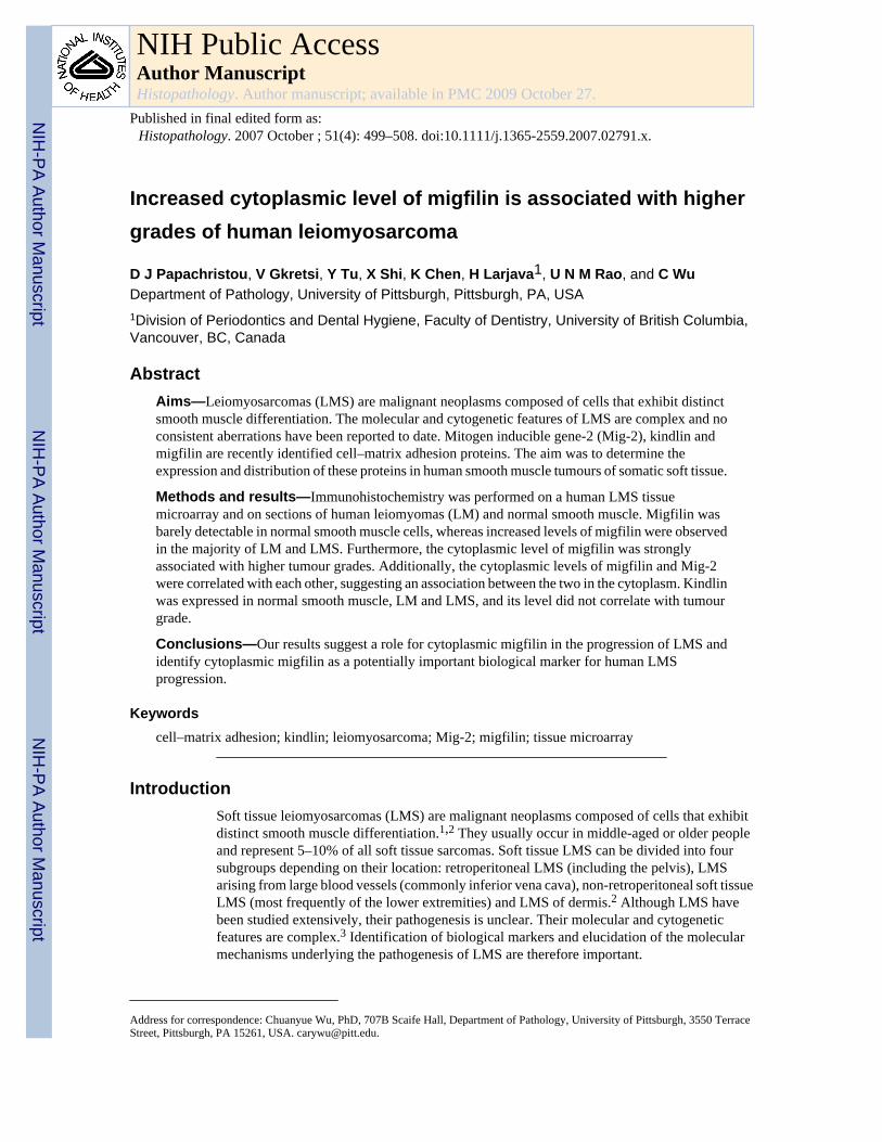
Increased cytoplasmic level of migfilin is associated with higher grades of human leiomyosarcoma D J Papachristou, V Gkretsi, Y Tu, X Shi, K Chen, H Larjava 1 , U N M Rao, and C Wu Department of Pathology, University of Pittsburgh, Pittsburgh, PA, USA 1 Division of Periodontics and Dental Hygiene, Faculty of Dentistry, University of British Columbia, Vancouver, BC, Canada Abstract Aims—Leiomyosarcomas (LMS) are malignant neoplasms composed of cells that exhibit distinct smooth muscle differentiation. The molecular and cytogenetic features of LMS are complex and no consistent aberrations have been reported to date. Mitogen inducible gene-2 (Mig-2), kindlin and migfilin are recently identified cell–matrix adhesion proteins. The aim was to determine the expression and distribution of these proteins in human smooth muscle tumours of somatic soft tissue. Methods and results—Immunohistochemistry was performed on a human LMS tissue microarray and on sections of human leiomyomas (LM) and normal smooth muscle. Migfilin was barely detectable in normal smooth muscle cells, whereas increased levels of migfilin were observed in the majority of LM and LMS. Furthermore, the cytoplasmic level of migfilin was strongly associated with higher tumour grades. Additionally, the cytoplasmic levels of migfilin and Mig-2 were correlated with each other, suggesting an association between the two in the cytoplasm. Kindlin was expressed in normal smooth muscle, LM and LMS, and its level did not correlate with tumour grade. Conclusions—Our results suggest a role for cytoplasmic migfilin in the progression of LMS and identify cytoplasmic migfilin as a potentially important biological marker for human LMS progression. Keywords cell–matrix adhesion; kindlin; leiomyosarcoma; Mig-2; migfilin; tissue microarray Introduction Soft tissue leiomyosarcomas (LMS) are malignant neoplasms composed of cells that exhibit distinct smooth muscle differentiation. 1,2 They usually occur in middle-aged or older people and represent 5–10% of all soft tissue sarcomas. Soft tissue LMS can be divided into four subgroups depending on their location: retroperitoneal LMS (including the pelvis), LMS arising from large blood vessels (commonly inferior vena cava), non-retroperitoneal soft tissue LMS (most frequently of the lower extremities) and LMS of dermis. 2 Although LMS have been studied extensively, their pathogenesis is unclear. Their molecular and cytogenetic features are complex. 3 Identification of biological markers and elucidation of the molecular mechanisms underlying the pathogenesis of LMS are therefore important. Address for correspondence: Chuanyue Wu, PhD, 707B Scaife Hall, Department of Pathology, University of Pittsburgh, 3550 Terrace Street, Pittsburgh, PA 15261, USA. [email protected]. NIH Public Access Author Manuscript Histopathology. Author manuscript; available in PMC 2009 October 27. Published in final edited form as: Histopathology. 2007 October ; 51(4): 499–508. doi:10.1111/j.1365-2559.2007.02791.x. NIH-PA Author Manuscript NIH-PA Author Manuscript NIH-PA Author Manuscript
Transcript of Increased cytoplasmic level of migfilin is associated with higher grades of human leiomyosarcoma
Increased cytoplasmic level of migfilin is associated with highergrades of human leiomyosarcoma
D J Papachristou, V Gkretsi, Y Tu, X Shi, K Chen, H Larjava1, U N M Rao, and C WuDepartment of Pathology, University of Pittsburgh, Pittsburgh, PA, USA1Division of Periodontics and Dental Hygiene, Faculty of Dentistry, University of British Columbia,Vancouver, BC, Canada
AbstractAims—Leiomyosarcomas (LMS) are malignant neoplasms composed of cells that exhibit distinctsmooth muscle differentiation. The molecular and cytogenetic features of LMS are complex and noconsistent aberrations have been reported to date. Mitogen inducible gene-2 (Mig-2), kindlin andmigfilin are recently identified cell–matrix adhesion proteins. The aim was to determine theexpression and distribution of these proteins in human smooth muscle tumours of somatic soft tissue.
Methods and results—Immunohistochemistry was performed on a human LMS tissuemicroarray and on sections of human leiomyomas (LM) and normal smooth muscle. Migfilin wasbarely detectable in normal smooth muscle cells, whereas increased levels of migfilin were observedin the majority of LM and LMS. Furthermore, the cytoplasmic level of migfilin was stronglyassociated with higher tumour grades. Additionally, the cytoplasmic levels of migfilin and Mig-2were correlated with each other, suggesting an association between the two in the cytoplasm. Kindlinwas expressed in normal smooth muscle, LM and LMS, and its level did not correlate with tumourgrade.
Conclusions—Our results suggest a role for cytoplasmic migfilin in the progression of LMS andidentify cytoplasmic migfilin as a potentially important biological marker for human LMSprogression.
Keywordscell–matrix adhesion; kindlin; leiomyosarcoma; Mig-2; migfilin; tissue microarray
IntroductionSoft tissue leiomyosarcomas (LMS) are malignant neoplasms composed of cells that exhibitdistinct smooth muscle differentiation.1,2 They usually occur in middle-aged or older peopleand represent 5–10% of all soft tissue sarcomas. Soft tissue LMS can be divided into foursubgroups depending on their location: retroperitoneal LMS (including the pelvis), LMSarising from large blood vessels (commonly inferior vena cava), non-retroperitoneal soft tissueLMS (most frequently of the lower extremities) and LMS of dermis.2 Although LMS havebeen studied extensively, their pathogenesis is unclear. Their molecular and cytogeneticfeatures are complex.3 Identification of biological markers and elucidation of the molecularmechanisms underlying the pathogenesis of LMS are therefore important.
Address for correspondence: Chuanyue Wu, PhD, 707B Scaife Hall, Department of Pathology, University of Pittsburgh, 3550 TerraceStreet, Pittsburgh, PA 15261, USA. [email protected].
NIH Public AccessAuthor ManuscriptHistopathology. Author manuscript; available in PMC 2009 October 27.
Published in final edited form as:Histopathology. 2007 October ; 51(4): 499–508. doi:10.1111/j.1365-2559.2007.02791.x.
NIH
-PA Author Manuscript
NIH
-PA Author Manuscript
NIH
-PA Author Manuscript
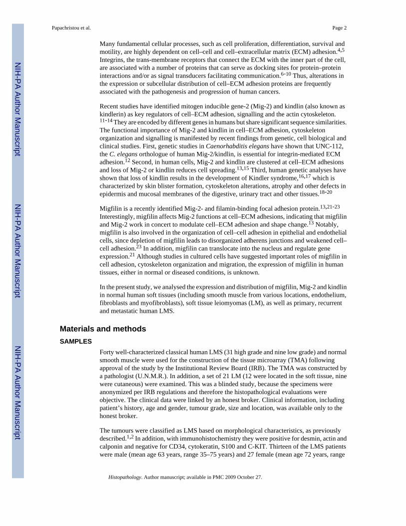
Many fundamental cellular processes, such as cell proliferation, differentiation, survival andmotility, are highly dependent on cell–cell and cell–extracellular matrix (ECM) adhesion.4,5Integrins, the trans-membrane receptors that connect the ECM with the inner part of the cell,are associated with a number of proteins that can serve as docking sites for protein–proteininteractions and/or as signal transducers facilitating communication.6-10 Thus, alterations inthe expression or subcellular distribution of cell–ECM adhesion proteins are frequentlyassociated with the pathogenesis and progression of human cancers.
Recent studies have identified mitogen inducible gene-2 (Mig-2) and kindlin (also known askindlerin) as key regulators of cell–ECM adhesion, signalling and the actin cytoskeleton.11-14 They are encoded by different genes in humans but share significant sequence similarities.The functional importance of Mig-2 and kindlin in cell–ECM adhesion, cytoskeletonorganization and signalling is manifested by recent findings from genetic, cell biological andclinical studies. First, genetic studies in Caenorhabditis elegans have shown that UNC-112,the C. elegans orthologue of human Mig-2/kindlin, is essential for integrin-mediated ECMadhesion.12 Second, in human cells, Mig-2 and kindlin are clustered at cell–ECM adhesionsand loss of Mig-2 or kindlin reduces cell spreading.13,15 Third, human genetic analyses haveshown that loss of kindlin results in the development of Kindler syndrome,16,17 which ischaracterized by skin blister formation, cytoskeleton alterations, atrophy and other defects inepidermis and mucosal membranes of the digestive, urinary tract and other tissues.18-20
Migfilin is a recently identified Mig-2- and filamin-binding focal adhesion protein.13,21-23
Interestingly, migfilin affects Mig-2 functions at cell–ECM adhesions, indicating that migfilinand Mig-2 work in concert to modulate cell–ECM adhesion and shape change.13 Notably,migfilin is also involved in the organization of cell–cell adhesion in epithelial and endothelialcells, since depletion of migfilin leads to disorganized adherens junctions and weakened cell–cell adhesion.23 In addition, migfilin can translocate into the nucleus and regulate geneexpression.21 Although studies in cultured cells have suggested important roles of migfilin incell adhesion, cytoskeleton organization and migration, the expression of migfilin in humantissues, either in normal or diseased conditions, is unknown.
In the present study, we analysed the expression and distribution of migfilin, Mig-2 and kindlinin normal human soft tissues (including smooth muscle from various locations, endothelium,fibroblasts and myofibroblasts), soft tissue leiomyomas (LM), as well as primary, recurrentand metastatic human LMS.
Materials and methodsSAMPLES
Forty well-characterized classical human LMS (31 high grade and nine low grade) and normalsmooth muscle were used for the construction of the tissue microarray (TMA) followingapproval of the study by the Institutional Review Board (IRB). The TMA was constructed bya pathologist (U.N.M.R.). In addition, a set of 21 LM (12 were located in the soft tissue, ninewere cutaneous) were examined. This was a blinded study, because the specimens wereanonymized per IRB regulations and therefore the histopathological evaluations wereobjective. The clinical data were linked by an honest broker. Clinical information, includingpatient’s history, age and gender, tumour grade, size and location, was available only to thehonest broker.
The tumours were classified as LMS based on morphological characteristics, as previouslydescribed.1,2 In addition, with immunohistochemistry they were positive for desmin, actin andcalponin and negative for CD34, cytokeratin, S100 and C-KIT. Thirteen of the LMS patientswere male (mean age 63 years, range 35–75 years) and 27 female (mean age 72 years, range
Papachristou et al. Page 2
Histopathology. Author manuscript; available in PMC 2009 October 27.
NIH
-PA Author Manuscript
NIH
-PA Author Manuscript
NIH
-PA Author Manuscript

35–95 years). Twenty-five (62.5%) of the tumours were primary, whereas six (15.0%) werelocally recurrent and nine (22.5%) metastatic. Fifteen (60%) of the primary LMS were locatedin the deep soft tissues, whereas 10 (40%) had a retroperitoneal location. Twenty-five LMSwere ≥ 50 mm and 15 were < 50 mm in greater diameter. They were classified as grade 1, 2and 3 according to their histological type, cellularity, pleomorphism and mitotic index.24,25
Grade 1 LMS were considered as low grade, grades 2 and 3 as high-grade neoplasms.24
GENERATION OF MONOCLONAL ANTI-KINDLIN ANTIBODYMouse monoclonal anti-migfilin and anti-Mig-2 antibodies have been previously described.13 To generate monoclonal anti-kindlin antibodies, a cDNA fragment was cloned encodinghuman kindlin residues 216–677 into the pGEX-5x-1 vector (Pharmacia, Sandwich, UK). Therecombinant vector was used to transform Escherichia coli cells. The expression of theglutathione-S-transferase (GST)–kindlin fusion protein was induced with isopropyl β-D-1-thiogalactopyranoside. GST–kindlin fusion protein was purified by affinity chromatographyusing glutathione-Sepharose 4B. Purified GST–kindlin fusion protein was used as an antigento immunize mice, as we have previously described.13,24 Hybridoma supernatants wereinitially screened for anti-kindlin activity by enzyme-linked immunosorbent assay (ELISA)using maltose-binding protein (MBP) fusion proteins containing kindlin residues 216–677.Antibodies that recognize MBP–kindlin in ELISA were selected and further tested by Westernblotting using green fluorescent protein (GFP)-tagged kindlin expressed by mammalian cells.The specificity of the monoclonal antibodies was confirmed by Western blot of kindlin-expressing and knocking-down cells as described in Results.
IMMUNOHISTOCHEMISTRYThe study was conducted on formalin-fixed paraffin-embedded tissue samples from primary,locally recurrent and metastatic LMS that were retrieved from the Department of Pathology,University of Pittsburgh and the University of Pittsburgh Medical Center Presbyterian-Shadyside Hospitals. The most representative areas of each tumour were selected for theconstruction of the TMA cores. Following depa-raffinization, standard immunohistochemistrywas performed using purified mouse monoclonal anti-migfilin (1 μg IgG/ml), anti-Mig2 (3μg IgG/ml) and anti-kindlin (16 μg IgG/ml) antibodies. Formalin-fixed paraffin-embeddedtissue samples from LM were analysed by immunohistochemistry following the same protocol.
The intensity of the immunopositivity was evaluated independently by two pathologists (D.J.P.and U.N.M.R.) and was scored on a scale of 0–3+ according to the following assessment: 0,no immunoreactivity; 1+, weak; 2+, moderate; 3+, strong immunopositivity. There was a highlevel of consistency of staining between the duplicate TMA cores.
SMALL INTERFERENCE RNA AGAINST KINDLINHuman immortalized keratinocytes (HaCaT) were transfected with a kindlin-specific smallinterfering (si) RNA with target sequence 5′-AAGACACAUCCAUAG CAUACU-3′ or a non-specific control RNA, using the Oligofectamine transfection system (Invitrogen, Carlsbad, CA,USA). Kindlin siRNA transfectants and control transfectants were analysed by Westernblotting with monoclonal anti-kindlin antibody 4A5. Equal loading was confirmed by re-probing the same membrane with an anti-actin antibody.
IMMUNOFLUORESCENT CELL STAININGImmunofluorescent staining was performed as described previously.25 Briefly, HaCaT cellswere plated on fibronectin-coated cover slips, fixed with 4% paraformaldehyde and stainedwith either the anti-kindlin mouse monoclonal antibody and rhodamine-red-conjugated goatantimouse secondary antibody, or just the secondary antibody (as a negative control).
Papachristou et al. Page 3
Histopathology. Author manuscript; available in PMC 2009 October 27.
NIH
-PA Author Manuscript
NIH
-PA Author Manuscript
NIH
-PA Author Manuscript
STATISTICAL ANALYSISStatistical analysis (χ2 test, Mann–Whitney U-test, Kendall’s τ-test, binary logistic regression)was performed using SPSS 12.0 for Windows (SPSS Inc., Chicago, IL, USA). A P-value <0.05 was considered to be statistically significant. The Mann–Whitney U-test was employedin order to test whether there were significant differences in means of the examined variablesbetween high- and low-grade tumours. Kendall’s τ-test was used in order to determine thestrength of association between the immunohistochemical profiles of the different proteins.
ResultsGENERATION AND CHARACTERIZATION OF MONOCLONAL ANTI-KINDLIN ANTIBODIES
Kindlin is a recently identified focal adhesion protein,15,16 loss of which has been associatedwith the Kindler syndrome. The level of kindlin protein in human tumours, however, has notpreviously been analysed. To facilitate studies of kindlin protein levels in human tumours, wegenerated a monoclonal antibody against human kindlin protein as described in Materials andmethods. Hybridoma supernatants were initially screened for anti-kindlin activity by ELISAusing MBP fusion proteins containing kindlin residues 216–677. Antibodies that recognizeMBP–kindlin in ELISA were selected and further tested by Western blotting using GFP-taggedkindlin expressed by mammalian cells. A positive clone (clone 4A5.14) was selected andsubsequently used to detect kindlin in normal human immortalized keratinocytes (HaCaT cells)transfected with either a control RNA or a siRNA against kindlin. As shown in Figure 1, clone4A5.14 recognizes a protein band with predicted human kindlin molecular mass (77.4 kDa).The level of this protein was dramatically reduced in the kindlin–siRNA-transfected cells(Figure 1, compare lane 1 with lane 2), confirming that clone 4A5.14 specifically recognizeskindlin.
To characterize further the 4A5.14 antikindlin antibody, immunofluorescence studies wereperformed using HaCaT cells. Clusters of kindlin were detected in focal adhesions consistentwith previous findings18,19 (Figure 2B, arrows). No specific staining was observed when thecells were stained with just the secondary antibody (Figure 2A), confirming the specificity ofthe immunofluorescent staining.
EXPRESSION OF KINDLIN IN NORMAL HUMAN SMOOTH MUSCLE, LM AND LMSTo examine the level of kindlin in normal smooth muscle tissue, sections of normal smoothmuscle were first stained using monoclonal anti-kindlin antibody 4A5.14. As shown in Figure3A, kindlin was diffusely expressed in normal smooth muscle cells, its localization beingexclusively cytoplasmic. Staining of LM with the monoclonal anti-kindlin antibody showedthat kindlin was also diffusely expressed in the cytoplasm in the majority of the examinedtumours (89%) (Figure 4A). Interestingly, strong kindlin immunoreactivity was detected inendothelial cells in LM tissues (Figure 4A).
Thirty-nine out of 40 (97.5%) LMS of all grades displayed diffuse, cytoplasmicimmunoreactivity for kindlin (Figure 3C–F). There was no variation in the pattern ofimmunoreactivity in different grades of LMS. Kindlin immunoreactivity did not displaysignificant differences between high- and low-grade neoplasms (Mann–Whitney U-test, P =0.5). In addition, no significant differences between low-grade LMS and LM were observed(Mann–Whitney U-test, P = 0.45). Strong kindlin immunoreactivity was expressed inendothelial cells within the tumour parenchyma (Figure 3B).
EXPRESSION OF MIG-2 IN NORMAL HUMAN SMOOTH MUSCLE, LM AND LMSDiffuse cytoplasmic and nuclear Mig-2 immunoreactivity was detected in normal smoothmuscle (Figure 5A). Diffuse Mig-2 immunoreactivity was also detected in all of the LM cases
Papachristou et al. Page 4
Histopathology. Author manuscript; available in PMC 2009 October 27.
NIH
-PA Author Manuscript
NIH
-PA Author Manuscript
NIH
-PA Author Manuscript
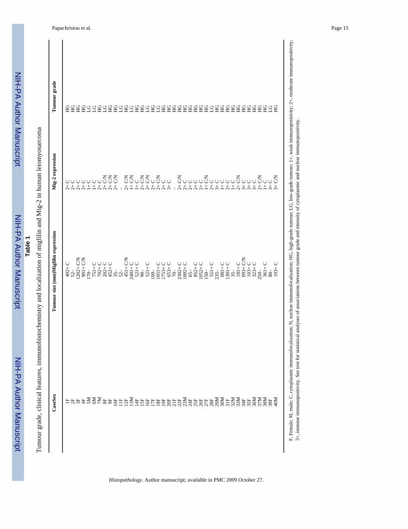
that were analysed (Figure 4B). In LMS, 38/40 (95%) of LMS were Mig-2+ (Table 1) (Figure5D,F). The staining pattern appeared similar in tumours of all grades. There was no significantcorrelation between the expression levels of Mig-2 among high- and low-grade LMS orbetween low-grade LMS and LM (Mann–Whitney U-test, P = 0.365 and 0.4, respectively).
EXPRESSION OF MIGFILIN IN NORMAL HUMAN SMOOTH MUSCLE, LM AND LMSMigfilin was barely detectable in normal smooth muscle cells (Figure 5B). Notably, however,intense migfilin immunopositivity was detected in endothelial cells (Figure 5C). Migfilin wasdiffusely expressed in 14 (10 conventional soft tissue LM and four cutaneous LM) out of 21(66.7%) LM cases (Figure 4C,D). Migfilin immunoreactivity in LM appeared to be principallycytoplasmic. Strong migfilin reactivity was detected in endothelial cells in all samples analysed(see, for example, Figure 4D). Diffuse migfilin expression was observed in 67.5% (27/40) ofthe examined LMS. Twenty-one out of 31 (68%) high-grade and six of nine (67%) low-gradetumours were migfilin immunopositive (Table 1). Both cytoplasmic and nuclearimmunoreactivity was observed, with more intense reactivity in the cytoplasm (Figure 5E,G).Migfilin was strongly expressed (2+/3+) in the cytoplasm of 14 out of 31 high-grade tumours,whereas only one out of nine low-grade tumours had strong cytoplasmic migfilin staining (2+) (Table 1).
CORRELATION OF THE EXPRESSION LEVEL OF MIGFILIN WITH TUMOUR GRADE INHUMAN LMS
When both nuclear and cytoplasmic levels were taken into account, there was no apparentcorrelation between the level of migfilin and LMS grade. Nuclear levels of migfilin were notsignificantly associated with tumour grade (P = 0.602). However, when only cytoplasmic levelswere considered for statistical analysis, a significantly stronger expression of migfilin in thehigh-grade compared with the low-grade LMS was demonstrated (Mann–Whitney U-test, P =0.032). Statistical analysis did not reveal any difference between the protein levels of migfilinin LM and low-grade LMS (Mann–Whitney U-test, P = 0.37).
CORRELATION BETWEEN THE LEVELS OF MIG-2, MIGFILIN AND KINDLIN IN LMSThe nuclear level of migfilin was not significantly related to that of Mig-2 (Kendall’s τ = 0.274,P = 0.06). Interestingly, however, when the statistical analysis included only their cytoplasmiclevels (Table 1), a significant correlation between migfilin and Mig-2 cytoplasmic levels wasrevealed (Kendall’s τ = 0.674, P = 0.014), indicating that the cytoplasmic levels of migfilinand Mig-2 are strongly associated in LMS. No significant association between kindlin andMig-2 or between kindlin and migfilin in LMS was revealed (P > 0.05 for both associations).
CLINICOPATHOLOGICAL FINDINGSSeveral clinicopathological parameters were analysed to determine the correlation of eachadhesion molecule with the tumour grade, gender and age, histopathological grade and size,location and biological behaviour (recurrent or metastatic) of the tumour. The intensity anddegree of immunoreactivity of Mig-2, migfilin and kindlin were not different within primaryand recurrent/metastatic LMS, or between small (< 50 mm) and large (≥ 50 mm) neoplasms.
ROLE OF MIGFILIN AS A POTENT BIOLOGICAL MARKERIn order to investigate the importance of migfilin, Mig-2 and kindlin as potent biologicalpredictors of tumour grade, their cytoplasmic levels were tested as categorical variables usingbinary logistic regression analysis. The most significant association among protein level andLMS grade was found for migfilin (P = 0.03). Evaluation of the levels of cytoplasmicexpression of migfilin could predict a high-grade LMS tumour with a sensitivity of 70%,specificity of 84% and overall accuracy of 74%.
Papachristou et al. Page 5
Histopathology. Author manuscript; available in PMC 2009 October 27.
NIH
-PA Author Manuscript
NIH
-PA Author Manuscript
NIH
-PA Author Manuscript
DiscussionCell–cell and cell–ECM adhesions regulate cell morphology and participate in manyfundamental cellular processes.7,8 Consequently, aberrations in the expression of cell adhesionmolecules are often associated with tumour development.26,27 In the present study, weinvestigated the level and distribution of three cell–ECM adhesion proteins, namely kindlin,Mig-2 and migfilin, in normal human smooth muscle, LM and primary, recurrent and metastaticLMS.
Kindlin was expressed in normal smooth muscle, LM and LMS. Its level of expression did notdisplay significant differences between high- and low-grade LMS, or between LM and low-grade LMS. No association was found between the immunohistochemical profiles of kindlinand Mig-2 or migfilin. This suggests that although kindlin and Mig-2 are structurally related,their levels of expression are not linked and up-regulation of one does not necessarily predictthe expression level of the other.
With regards to migfilin, our results show that it is readily detectable in the majority (67.5%)of LMS and LM (66.7%), but not in normal smooth muscle cells. This suggests that migfilinis involved in the pathogenesis of smooth muscle neoplasms. Importantly, statistical analysisrevealed that the cytoplasmic level of migfilin is significantly higher in high-grade comparedwith low-grade LMS. These findings, together with our recent findings that migfilin functionsas an important component of cell–ECM and cell–cell adhesion networks and regulates cellmigration,13,22,23,28 suggest a role for cytoplasmic migfilin in LMS progression.
Kato et al.29 have recently analysed Mig-2 expression in 13 cases of uterine LM and one caseof LMS. The results showed that Mig-2 expression was elevated in 12 out of 13 cases of humanuterine LM, but was reduced in the single case of uterine LMS that was analysed. The presentstudy analysed the subcellular distribution and expression levels of Mig-2 in 40 cases of LMS(31 high grade and nine low grade), as well as in soft tissue LM and normal smooth muscle.Consistent with the study by Kato et al.,29 abundant Mig-2 was detected in all LM that wereanalysed. However, Mig-2 immunoreactivity was also detected in the majority (38 out of 40LMS cases) of soft tissue LMS. This may reflect a different role for Mig-2 in the pathobiologyof deep soft LMS analysed in the current study in comparison with uterine LMS analysed byKato et al.29 Alternatively, this may stem from the difference in the numbers of LMS casesanalysed in these studies (one single uterine LMS in the study by Kato et al.29 versus 40 softtissue LMS cases in the current study).
Consistent with our recent results showing that migfilin and Mig-2 are co-localized at cell–ECM adhesions,13,22,23 statistical analysis of our immunohistochemical data reveal that thecytoplasmic levels of these proteins are positively and significantly correlated with each other.The parallel up-regulation of these proteins implies that the cytoplasmic migfilin–Mig-2complex may be involved in the pathogenesis and progression of human LMS. Migfilin hasbeen implicated in transcriptional regulation in mouse cardiomyocytes.21 Nuclear migfilin wasalso detected in the current study. It will be interesting to determine the role of nuclear migfilinin normal and transformed smooth muscle cells.
In conclusion, in the present study we have investigated the potential of migfilin and Mig-2 asmolecular markers that might contribute to the distinction between high- and low-grade LMS.The results have revealed a significant association between cytoplasmic migfilin levels andLMS grade, suggesting that the migfilin cytoplasmic level may be used to aid the determinationof different histological grades of LMS, especially in small biopsy specimens of deep soft tissueand retroperitoneal tumours.
Papachristou et al. Page 6
Histopathology. Author manuscript; available in PMC 2009 October 27.
NIH
-PA Author Manuscript
NIH
-PA Author Manuscript
NIH
-PA Author Manuscript
AcknowledgmentsThis work was supported by NIH grants GM65188 and DK54639 to C.W. and DE016099 to H.L.
References1. Enzinger, F.; Weis, SW. Leiomyosarcoma. In: Weis, SW.; Goldblum, JR., editors. Soft tissue tumors.
Vol. 4. Mosby; St Louis: 2001. p. 724-748.2. Evans, H.; Shipley, J. Leiomyosarcoma. In: Fletcher, CDM.; Unni, KK.; Mertens, F., editors. World
Health Organization classification of tumors. Pathology and genetics of tumors of soft tissue and bone.Lyon: IARC Press; 2002. p. 131-134.
3. Ren B, Yu YP, Jing L, et al. Gene expression analysis of human soft tissue leiomyosarcomas. HumPathol 2003;34:549–558. [PubMed: 12827608]
4. Sepulveda J, Gkretsi V, Wu C. Assembly and signaling of the cell–extracellular matrix adhesioncomplexes. Curr Top Dev Biol 2005;68:183–225. [PubMed: 16125000]
5. Jamora C, Fuchs E. Intercellular adhesion, signalling and the cytoskeleton. Nat Cell Biol 2002;4:E101–108. [PubMed: 11944044]
6. Burridge K, Chrzanowska-Wodnicka M. Focal adhesions, contractility, and signaling. Annu Rev CellDev Biol 1996;12:463–518. [PubMed: 8970735]
7. Calderwood DA, Shattil SJ, Ginsberg MH. Integrins and actin filaments: reciprocal regulation of celladhesion and signaling. J Biol Chem 2000;275:22607–22610. [PubMed: 10801899]
8. Howe A, Aplin AE, Alahari SK, Juliano RL. Integrin signaling and cell growth control. Curr OpinCell Biol 1998;10:220–231. [PubMed: 9561846]
9. Playford MP, Schaller MD. The interplay between Src and integrins in normal and tumor biology.Oncogene 2004;23:7928–7946. [PubMed: 15489911]
10. Hynes RO. Integrins: bidirectional, allosteric signaling machines. Cell 2002;110:673–687. [PubMed:12297042]
11. Schaller MD. UNC112. A new regulator of cell–extracellular matrix adhesions? J Cell Biol2000;150:F9–F11. [PubMed: 10893277]
12. Rogalski TM, Mullen GP, Gilbert MM, Williams BD, Moerman DG. The UNC-112 gene inCaenorhabditis elegans encodes a novel component of cell–matrix adhesion structures required forintegrin localization in the muscle cell membrane. J Cell Biol 2000;150:253–264. [PubMed:10893272]
13. Tu Y, Wu S, Shi X, Chen K, Wu C. Migfilin and Mig-2 link focal adhesions to filamin and the actincytoskeleton and function in cell shape modulation. Cell 2003;113:37–47. [PubMed: 12679033]
14. Weinstein EJ, Bourner M, Head R, Zakeri H, Bauer C, Mazzarella R. URP1: a member of a novelfamily of PH and FERM domain-containing membrane-associated proteins is significantly over-expressed in lung and colon carcinomas. Biochim Biophys Acta 2003;1637:207–216. [PubMed:12697302]
15. Kloeker S, Major MB, Calderwood DA, Ginsberg MH, Jones DA, Beckerle MC. The Kindlersyndrome protein is regulated by transforming growth factor-beta and involved in integrin-mediatedadhesion. J Biol Chem 2004;279:6824–6833. [PubMed: 14634021]
16. Siegel DH, Ashton GH, Penagos HG, et al. Loss of kindlin-1, a human homolog of the Caenorhabditiselegans actin–extracellular-matrix linker protein UNC-112, causes Kindler syndrome. Am J HumGenet 2003;73:174–187. [PubMed: 12789646]
17. Jobard F, Bouadjar B, Caux F, et al. Identification of mutations in a new gene encoding a FERMfamily protein with a pleckstrin homology domain in Kindler syndrome. Hum Mol Genet2003;12:925–935. [PubMed: 12668616]
18. Hovnanian A, Blanchet-Bardon C, de Prost Y. Poikiloderma of Theresa Kindler: report of a case withultrastructural study, and review of the literature. Pediatr Dermatol 1989;6:82–90. [PubMed:2664739]
19. Senturk N, Usubutun A, Sahin S, Bukulmez G, Erkek E, Topaloglu R. Kindler syndrome: absenceof definite ultrastructural feature. J Am Acad Dermatol 1999;40:335–337. [PubMed: 10025863]
Papachristou et al. Page 7
Histopathology. Author manuscript; available in PMC 2009 October 27.
NIH
-PA Author Manuscript
NIH
-PA Author Manuscript
NIH
-PA Author Manuscript
20. Haber RM, Hanna WM. Kindler syndrome. Clinical and ultrastructural findings. Arch Dermatol1996;132:1487–1490. [PubMed: 8961879]
21. Akazawa H, Kudoh S, Mochizuki N, et al. A novel LIM protein Cal promotes cardiac differentiationby association with CSX/NKX2-5. J Cell Biol 2004;164:195–405. [PubMed: 14734531]
22. Wu C. Migfilin and its binding partners: from cell biology to human diseases. J Cell Sci 2005;118:659–664. [PubMed: 15701922]
23. Gkretsi V, Zhang Y, Tu Y, et al. Physical and functional association of migfilin with cell–celladhesions. J Cell Sci 2005;118:697–710. [PubMed: 15671069]
24. Tu Y, Huang Y, Zhang Y, Hua Y, Wu C. A new focal adhesion protein that interacts with integrin-linked kinase and regulates cell adhesion and spreading. J Cell Biol 2001;153:585–598. [PubMed:11331308]
25. Li F, Zhang Y, Wu C. Integrin-linked kinase is localized to cell–matrix focal adhesions but not cell–cell adhesion sites and the focal adhesion localization of integrin-linked kinase is regulated by thePINCH-binding ANK repeats. J Cell Sci 1999;112:4589–4599. [PubMed: 10574708]
26. Christofori G. Changing neighbours, changing behaviour: cell adhesion molecule-mediated signallingduring tumour progression. EMBO J 2003;22:2318–2323. [PubMed: 12743026]
27. Popov Z, Gil-Diez de Medina S, Lefrere-Belda M-A, et al. Low E-cadherin expression in bladdercancer at the transcriptional and protein level provides prognostic information. Br J Cancer2000;83:209–214. [PubMed: 10901372]
28. Zhang Y, Tu Y, Gkretsi V, Wu C. Migfilin interacts with vasodilator-stimulated phosphoprotein(VASP) and regulates VASP localization to cell–matrix adhesions and migration. J Biol Chem2006;281:12397–12407. [PubMed: 16531412]
29. Kato K, Shiozawa T, Mitsushita J, et al. Expression of the mitogen-inducible gene-2 (mig-2) iselevated in human uterine leiomyomas but not in leiomyosarcomas. Hum Pathol 2004;35:55–60.[PubMed: 14745725]
AbbreviationsECM
extracellular matrix
ELISA enzyme-linked immunosorbent assay
GFP green fluorescent protein
GST glutathione-S-transferase
IRB Institutional Review Board
LM leiomyoma
LMS leiomyosarcoma
MBP maltose-binding protein
Mig-2 mitogen inducible gene-2
siRNA
Papachristou et al. Page 8
Histopathology. Author manuscript; available in PMC 2009 October 27.
NIH
-PA Author Manuscript
NIH
-PA Author Manuscript
NIH
-PA Author Manuscript
small interfering RNA
TMA tissue microarray
Papachristou et al. Page 9
Histopathology. Author manuscript; available in PMC 2009 October 27.
NIH
-PA Author Manuscript
NIH
-PA Author Manuscript
NIH
-PA Author Manuscript
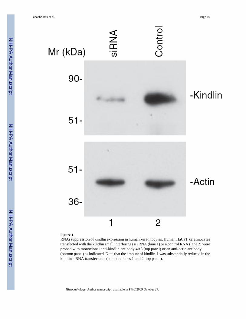
Figure 1.RNAi suppression of kindlin expression in human keratinocytes. Human HaCaT keratinocytestransfected with the kindlin small interfering (si) RNA (lane 1) or a control RNA (lane 2) wereprobed with monoclonal anti-kindlin antibody 4A5 (top panel) or an anti-actin antibody(bottom panel) as indicated. Note that the amount of kindlin-1 was substantially reduced in thekindlin siRNA transfectants (compare lanes 1 and 2, top panel).
Papachristou et al. Page 10
Histopathology. Author manuscript; available in PMC 2009 October 27.
NIH
-PA Author Manuscript
NIH
-PA Author Manuscript
NIH
-PA Author Manuscript
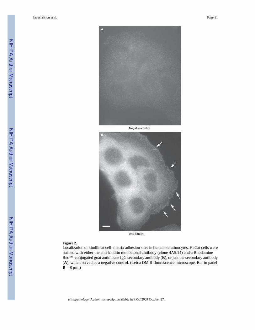
Figure 2.Localization of kindlin at cell–matrix adhesion sites in human keratinocytes. HaCat cells werestained with either the anti-kindlin monoclonal antibody (clone 4A5.14) and a RhodamineRed™-conjugated goat antimouse IgG secondary antibody (B), or just the secondary antibody(A), which served as a negative control. (Leica DM R fluorescence microscope. Bar in panelB = 8 μm.)
Papachristou et al. Page 11
Histopathology. Author manuscript; available in PMC 2009 October 27.
NIH
-PA Author Manuscript
NIH
-PA Author Manuscript
NIH
-PA Author Manuscript

Figure 3.Expression of kindlin in normal smooth muscle, low- and high-grade leiomyosarcoma (LMS).A, Normal smooth muscle cells display immunopositivity for kindlin. B, Moderateimmunopositivity for kindlin (2+) in a high-grade LMS. Endothelial cells of tumour vesselsare strongly positive for kindlin. C,E, Strong kindlin immunopositivity (3+) in a low-gradeLMS. D,F, High-grade LMS exhibiting strong immunoreactivity (3+) for kindlin.
Papachristou et al. Page 12
Histopathology. Author manuscript; available in PMC 2009 October 27.
NIH
-PA Author Manuscript
NIH
-PA Author Manuscript
NIH
-PA Author Manuscript

Figure 4.Expression of kindlin, Mig-2 and migfilin in leiomyoma (LM). A, Kindlin expression. Kindlinwas detected in the majority (89%) of the LM cases. A representative diffuse, cytoplasmickindlin staining of LM is shown. B, Mig-2 expression. Diffuse, cytoplasmic Mig-2 wasdetected in all of the LM cases. Representative Mig-2 reactivity is shown. C,D, Migfilinexpression. Migfilin was detected in 14 out of 21 (66.7%) LM cases. C, Strongly positive,primarily cytoplasmic migfilin reactivity in a case of soft tissue LM. D, Negative migfilinreactivity in a case of soft tissue LM. Note that strong migfilin immunoreactivity was detectedin endothelial cells (D).
Papachristou et al. Page 13
Histopathology. Author manuscript; available in PMC 2009 October 27.
NIH
-PA Author Manuscript
NIH
-PA Author Manuscript
NIH
-PA Author Manuscript

Figure 5.Expression of Mig-2 and migfilin in normal smooth muscle, low- and high-gradeleiomyosarcoma (LMS). A, Normal smooth muscle cells exhibit positive nuclear andcytoplasmic immunoreactivity for Mig-2. B, Normal smooth muscle cells exhibit noimmunoreactivity for migfilin. C, In normal soft tissue sections containing blood vessels, ahigh level of migfilin was detected in endothelial cells. D, Mig-2 exhibits moderate cytoplasmicimmunoreactivity (2+) in a low-grade LMS. E, Low-grade LMS exhibiting weakimmunopositivity for migfilin (1+). Endothelial cells show strong expression of migfilin. F,Strong nuclear and cytoplasmic immunoreactivity of Mig-2 (3+) in a high-grade LMS. G,High-grade LMS showing strong (3+), nuclear and cytoplasmic immunopositivity for migfilin.See Table 1 for a summary of tumour grade, clinical features and immunopositivity/localizationof migfilin and Mig-2 in all 40 human LMS that were analysed.
Papachristou et al. Page 14
Histopathology. Author manuscript; available in PMC 2009 October 27.
NIH
-PA Author Manuscript
NIH
-PA Author Manuscript
NIH
-PA Author Manuscript
NIH
-PA Author Manuscript
NIH
-PA Author Manuscript
NIH
-PA Author Manuscript
Papachristou et al. Page 15Ta
ble
1
Tum
our g
rade
, clin
ical
feat
ures
, im
mun
ohis
toch
emis
try a
nd lo
caliz
atio
n of
mig
filin
and
Mig
-2 in
hum
an le
iom
yosa
rcom
a
Cas
eSex
Tum
our
size
(mm
)Mig
filin
exp
ress
ion
Mig
-2 e
xpre
ssio
nT
umou
r gr
ade
1F40
2+ C
2+ C
HG
2F52
–2+
CH
G3F
1202
+ C
/N2+
CH
G4F
901+
C/N
2+ C
HG
5M17
0–1+
CLG
6M75
1+ C
1+ C
LG7M
702+
C2+
CH
G8F
202+
C2+
C/N
LG9F
452+
C2+
C/N
HG
10F
35–
2+ C
/NH
G11
F52
––
LG12
F45
1+ C
/N2+
C/N
HG
13M
2401
+ C
1+ C
/NLG
14F
521+
C1+
CH
G15
F90
–2+
C/N
HG
16F
521+
C2+
C/N
LG17
F10
0–2+
CH
G18
F10
51+
C2+
C/N
LG19
F17
53+
C2+
CH
G20
F65
3+ C
3+ C
HG
21F
70–
–H
G22
F23
02+
C2+
C/N
HG
23M
1002
+ C
2+ C
HG
24F
65–
2+ C
HG
25F
852+
C2+
CH
G26
F10
52+
C2+
CH
G27
F15
0–1+
C/N
HG
28F
551+
C2+
CLG
29M
235–
3+ C
HG
30M
081+
C1+
CH
G31
F13
01+
C2+
CH
G32
M35
–1+
CH
G33
M18
1+ C
2+ C
/NH
G34
F09
3+ C
/N3+
CH
G35
F10
3+ C
3+ C
HG
36M
323+
C3+
CH
G37
M25
0–2+
C/N
HG
38M
361+
C1+
CH
G39
F80
–3+
CLG
40M
103+
C3+
C/N
HG
F, F
emal
e; M
, mal
e; C
, cyt
opla
smic
imm
unol
ocal
izat
ion;
N, n
ucle
ar im
mun
oloc
aliz
atio
n; H
G, h
igh-
grad
e tu
mou
r; LG
, low
-gra
de tu
mou
r; 1+
, wea
k im
mun
opos
itivi
ty; 2
+, m
oder
ate
imm
unop
ositi
vity
;3+
, ins
tens
e im
mun
opos
itivi
ty. S
ee te
xt fo
r sta
tistic
al a
naly
ses o
f ass
ocia
tions
bet
wee
n tu
mou
r gra
de a
nd in
tens
ity o
f cyt
opla
smic
and
nuc
lear
imm
unop
ositi
vity
.
Histopathology. Author manuscript; available in PMC 2009 October 27.