
Incorporating Predictive Maintenance Practices into Marine ...
Upload
khangminh22Category
view
0download
0
Incorporating MRD Testing into Standard Practice: Are We There Yet?
Faith Davies, MD, MBBCh, MRCP, FRCPath Professor of Medicine
Medical Director, Myeloma Institute University of Arkansas for Medical Sciences
Little Rock, Arkansas
Welcome to Managing Myeloma, I am Dr. Faith Davies. Today, I wish to review incorporating minimal residual disease (MRD) testing into standard practice. In this presentation, I will describe the importance of MRD in patients with multiple myeloma, I will compare and contrast the current methodologies used to measures MRD, and I will outline the clinical applicability and the goal of MRD testing in routine clinical practice.
1
Incorporating MRD Testing into Standard Practice: Are We There Yet?
©2018 MediCom Worldwide, Inc.

Disclosures
• Consultant relationship with the following companies:
– Abbvie
– Amgen
– Bristol‐Myers Squibb
– Celgene Corporation
– Janssen
– Takeda Oncology
These are my disclosures.
2
Incorporating MRD Testing into Standard Practice: Are We There Yet?
©2018 MediCom Worldwide, Inc.

The Impact of Myeloma Treatment
Adapted from: "Iceberg" created by Uwe Kils (iceberg) and user Wiska Bodo (sky). (Work by Uwe Kils). http://www.ecoscope.com/iceberg/
Treatment
Disease burden
With modern multiple myeloma therapy, many more patients now are achieving a complete response (CR). Indeed, with some of the therapies this can vary between 30% and 90% of patients who are in a CR, which is measured by morphology of the bone marrow, a negative M‐component, and normalization of the serum‐freelight chains. However, we know that many patients do unfortunately relapse, and it is postulated that this occurs because of the presence of minimal residual disease.
3
Incorporating MRD Testing into Standard Practice: Are We There Yet?
©2018 MediCom Worldwide, Inc.

Composition of Residual Clonal Populations
Adapted from: "Iceberg" created by Uwe Kils (iceberg) and user Wiska Bodo (sky). (Work by Uwe Kils). http://www.ecoscope.com/iceberg/
Certainly, when we think about the biology of some of these residual cells, we can assume that they are the most resistant clones in the fact that they have survived the induction therapy that patients have had. Potentially, they have mutations that make them resistant to the therapy. They could be out to cycle and therefore do not undergo apoptosis, or they could actually be protected by the microenvironment of the bone marrow.
4
Incorporating MRD Testing into Standard Practice: Are We There Yet?
©2018 MediCom Worldwide, Inc.

Composition of Residual Clonal Populations
Leads to relapse
Adapted from: "Iceberg" created by Uwe Kils (iceberg) and user Wiska Bodo (sky). (Work by Uwe Kils). http://www.ecoscope.com/iceberg/
Whatever the mechanism for their ongoing presence, certainly these cells lead to relapse. We know that in many cases relapse is difficult to treat and, therefore, we should at least try and eradicate MRD so that we can get the best outcomes for our patients.
5
Incorporating MRD Testing into Standard Practice: Are We There Yet?
©2018 MediCom Worldwide, Inc.

out of 104 ‐ 105 cells
out of 105 ‐ 106 cells
out of 105 ‐ 106 cells
Next‐generation flow
Next‐generation sequencing
Multiparametric flow
Functional imaging
Lesion size 0.5 ‐ 1 ccm; raised or abnormal background signal
sFLC and immunofixation
out of 102 ‐ 103 cells
MRD Assessment Techniques
Kindly drawn by Dr. Leo RascheDavies F, et al. ASH Education Book 2017.
There are a lot of different methods used for testing MRD. We have the blood‐based methods, such as serum‐free light chains and immunofixation. We then have slightly more sensitive methods which are based on bone marrow assessment, so this can include flow cytometry, or what is now called next‐generation flow cytometry, which is more sensitive than flow cytometry. Then there are molecular techniques such as next‐generation sequencing. These technologies have different sensitivities, being able to detect between one in 104 and one in 106 myeloma cells compared to normal cells. In addition to this, though, we also need to think about some of the imaging techniques which may be taking account of the whole patient, such as PET CT scanning or MRI scanning. Unfortunately, at the moment, the peripheral blood technologies to assess MRD are maybe not quite sensitive enough, but certainly there is ongoing work looking at this area.
6
Incorporating MRD Testing into Standard Practice: Are We There Yet?
©2018 MediCom Worldwide, Inc.

Comparison of Different Bone Marrow MRD Techniques
Characteristic Flow Cytometry Immunoglobulin NGS
Applicability Nearly 100% ≥90%
Diagnostic Sample Not required, abnormal plasma cells can be identified in any sample by their distinct immunophenotypic pattern vs normal plasma cells
Sample with myeloma present (eg, presentation) required for identification of the dominant clonotype
Sample Requirements 2‐5 million cells 1 million cells
Sample Processing Requires a fresh sample; needs assessment within 24‐48 hours Can use both fresh and stored samples
Standardization Methodology varies between laboratories. Some attempts at standardization (eg, EuroFlow)
Standardized methodology available from commercial companies. Academic methodologies also available
Sample Quality Control Possible to check by global bone marrow cell analysis Not possible to check. Additional studies would be required
Quantitative Yes Yes
Sensitivity 1 in 104 1 in 106 1 in 105 1 in 106
Turnaround and Complexity
Hours. Requires skill flow cytometrist. Automated software available
1 week. Academic methodologies require bioinformatics support
Clonal Evolution Considers all clones with similar phenotype but evolving clone with change in phenotype may not be evaluable
Can take into account all minor clones with infrequent occurrence
Availability Many laboratories with four color; eight or more colors restricted to more specialized centers
Once company commercially and a number of academic platforms
NGS=next‐generation sequencingDavies F, et al. ASH Education Book 2017.
There are two commonly used techniques for MRD detection: flow cytometry and next‐generation sequencing molecular technology. Both of them have a number of positive and negative aspects. For next‐generation sequencing, there are now a number of commercial entities which are offering this, and it is very much a technology which would be available in this setting due to the complexity of the technique. The current turnaround time for this technology is about a week to two weeks. A diagnostic sample, ie, a sample with the disease present in it, as well as a follow‐up sample where querying MRD are required for the sequencing technology. The flow cytometry technology has a quicker turnaround and can be performed whether there is a sample with tumor available or not. One of the issues around this concerns standardization, as a number of different laboratories have a different sensitivity for this test. With both tests it is important to know whether the test is negative or positive, and what the sensitivity level of the test is; whether it is able to measure tumor cells to one in 104 or to one in 106. At the moment, many insurance companies will approve the flow cytometry test. There is some discussion and debate with many of the payors regarding the sequencing technology, but I think as more data becomes available and these technologies get approved by the FDA and incorporated into guidelines, then this area will be changing quite rapidly.
7
Incorporating MRD Testing into Standard Practice: Are We There Yet?
©2018 MediCom Worldwide, Inc.

Clinical Applicability of MRD Testing
Monitor response
Prognostic value
Monitor maintenance therapyMonitor maintenance therapy
Detect early relapseDetect early relapse
Marker of biology Marker of biology
The use for MRD testing in clinical practice is now becoming quite widespread. The technologies are able to assess and monitor response, but there is also data now showing the prognostic value of these technologies. There is a lot of research work going on about how these technologies can be used to monitor and potentially adapt maintenance therapy to be able to detect early relapse; and then how we can learn a little more about the biology of multiple myeloma. Over the next few moments, I would like to explore some of these in a little more detail.
8
Incorporating MRD Testing into Standard Practice: Are We There Yet?
©2018 MediCom Worldwide, Inc.

Predicting Outcome
Meta‐analysis ‐ effect of MRD on PFS and OS
Flow cytometry studies
Munshi NC, et al. JAMA Oncol. 2017;3(1):28‐35.; Landgren O, et al. Bone Marrow Transplant. 2016; 51(12):1565‐1568.
PFS
OS
9
There is now an awful lot of data showing the prognostic importance of MRD. This is some very early data where the studies mainly involved flow cytometry. The studies are maybe not quite as sensitive as some of the more recent technologies; these studies of flow cytometry had a sensitivity of one in 104. However you can see from the two meta‐analyses that both of the studies showed that MRD detection was prognostic for progression‐free survival (PFS) and for overall survival.
Incorporating MRD Testing into Standard Practice: Are We There Yet?
©2018 MediCom Worldwide, Inc.

MRD pos= MRD neg
PFS according to 10‐6 OS according to 10‐6
Avet‐Loiseau H, et al. ASH 2016. Abstract 246. Slides kindly provided by Prof. H. Avet Loiseau and IFM investigators.
IFM 2009 Superior PFS and OS According to MRD StatusNGS
MRD in the Era of Newer Therapies
10
In more recent data incorporating treatment with some of the newer therapies, this is an example from the IFM French study where patients were treated with bortezomib‐lenalidomide‐dexamethasone (VRd or RVD) and transplantation. You can clearly see that using the next‐generation sequencing test, which has a sensitivity in one in 106, that yet again, those patients that are MRD negative have a much better progression‐free and overall survival.
Incorporating MRD Testing into Standard Practice: Are We There Yet?
©2018 MediCom Worldwide, Inc.

P<0.001
0
25
50
75
100
Pa
tie
nts
(%
)
40 39 34 31 17 1negative MRD_RVD50 47 43 38 23 4negative MRD_Transp66 51 38 21 11 2positive MRD-RVD68 62 49 35 15 1positive MRD-Transplant
N at risk
0 12 24 36 48 60
Time since MRD assessment
positive MRD-Transplant
positive MRD-RVD
negative MRD_Transp
negative MRD_RVD
Prognostic Irrespective of TherapyIFM 2009 – RVD vs transplant
NGS MRD Negative Patients Have Improved Outcome Irrespective of Therapy Used
Avet‐Loiseau H, et al. ASH 2016. Abstract 246. Slides kindly provided by Prof. H. Avet Loiseau and IFM investigators.
11
A number of studies have now shown that regardless of the therapy used, if you achieve a complete response, then patients tend to do better. Again, using some data from the French study, this is comparing patients who received RVD alone or received RVD and a transplant. The two upper curves are those patients who achieved MRD negativity. Although more patients were MRD negative with the transplant, still it did not seem to matter with respect to progression‐free survival as long as they were in a complete response.
Incorporating MRD Testing into Standard Practice: Are We There Yet?
©2018 MediCom Worldwide, Inc.

Prognostic Irrespective of Age
Elderly patients – Myeloma XI study
de Tute RM, et al. ASH 2016. Abstract 245.
12
Newer data has also shown that MRD is prognostic irrespective of the patient’s age. This is an example from a UK study looking at elderly patients. Again, in elderly patients, those patients that achieved MRD negativity have a longer PFS and OS.
Incorporating MRD Testing into Standard Practice: Are We There Yet?
©2018 MediCom Worldwide, Inc.

CASTORPOLLUX
Patients en viesans progression (%
)
0
20
40
60
80
100
0 3 6 9 12 15 18 24
Vd MRD negativeDVd MRD negativeVd MRD positive
DVd MRD positive
Patients at risk Months21
626241225
626176189
626123172
52668134
3152076
27726
0104
0001
0000
Patients en vie san
s progression (%
)
0
20
40
60
80
100
0 3 6 9 12 15 21 27
Rd MRD negativeDRd MRD negativeRd MRD positive
DRd MRD positive
Patients at risk Months24
1671267215
1671233195
1671190178
1570166167
1566144161
1257120137
0659
0001
0000
18
10283854
Rd MRD–
DRd MRD–
DRd MRD+
Rd MRD+
Vd MRD–
DVd MRD–
DVd MRD+
Vd MRD+
• Lower risk of progression in MRD‐negative patients• More patients achieve MRD negativity when adding daratumumab• PFS benefit in MRD‐positive patients who received daratumumab‐containing regimens versus standard of care
PFS according to MRD at 10–5
Prognostic After Relapse Therapy
Avet‐Loiseau H, et al. ASH 2016. Abstract 246. Slides kindly provided by Prof. H. Avet Loiseau and IFM investigators.
13
Up until recently, many of the therapies that were able to achieve a complete response were mainly in the newly diagnosed setting. More recently, however, with the introduction of monoclonal antibodies and next‐generation proteasome inhibitors, we have discovered that patients in the relapsed setting are able to get better responses. Again, similar data can now be seen in the relapsed setting where patients who achieve MRD negativity have a longer progression‐free survival. In this instance, we are using the sequencing test but at the level of 10‐5.
Incorporating MRD Testing into Standard Practice: Are We There Yet?
©2018 MediCom Worldwide, Inc.

Outcome according to MRD status after autologous stem‐cell transplantation and cytogenetic risk profile.
Myeloma IX – Flow cytometryHR defined by FISH ‐ t(4;14), t(14;16), 1q+ and 17p‐
(A) Progression‐free survival (B) overall survival
Rawstron AC, et al. J Clin Oncol. 2013;31(20):2540‐2547.Other examples include Paiva B, et al. Blood. 2016;127(25):3165‐3174.; Schinke C, et al. Haematologica. 201;102(8):e313‐e316.
Transplant eligible
MRD Remains Important in High‐risk Patients
14
It is important to say that MRD does play a role in those patients who are thought to be high risk. A number of different studies define high risk in a different way; however, all of these studies show that those patients who are high risk with high‐risk cytogenetics, who are also MRD positive, have a poorer survival. In these two curves from the UK study, the yellow curve are the patients who have high‐risk cytogenetics but are also MRD positive. As you can see, those patients have a particularly poor outcome, suggesting that MRD and cytogenetics are both important when assessing a patient’s risk.
Incorporating MRD Testing into Standard Practice: Are We There Yet?
©2018 MediCom Worldwide, Inc.

Myeloma IX study
Rawstron AC, et al. Blood. 2015;125(12):1932‐1935.Other examples include de Tute RM, et al. ASH 2016. Abstract 245.; Avet‐Loiseau H, et al. ASH 2016. Abstract 246.; Paiva B, et al. ASH 2017. Abstract 905.
MRD is a Continuous VariableMinimal residual disease in myeloma by flow cytometry: independent prediction of survival benefit per log reduction
Sequential improvements in PFS and OS for each log depletion in MRD level, as assessed by multiparameter flow cytometry.This effect is demonstrable in all patients ([A] PFS; [B] OS) as well as those achieving conventional CR ([C] PFS; [D] OS).
15
As I mentioned earlier, though, it is important not to just talk about MRD as a positive or negative; it is actually a continuous variable. In this study, from the curves A and B, you can see this is progression‐free survival and overall survival, but each line is represented by the level of the depth of response. Those patients who have achieved a lower rate of disease burden have a much better overall survival. This seems to be according to logs, so if there is detectable disease at 10‐4, those patients have a better outcome than those patients that have detectable disease at 10‐3.
Incorporating MRD Testing into Standard Practice: Are We There Yet?
©2018 MediCom Worldwide, Inc.

Time of Measurement is Important
Time to achieve MRD negativity
CASTORPOLLUX
Rd
DRd
00 3 6 9 12 15 21 272418
Patients M
RD negative, %
5
10
20
25
30
15
RdDRd
Patients at risk
283286
272271
252247
243229
225202
202169
1815
01
00
8371
0
5
10
20
25
30
0 3 6 9 12 15 18 2421
Patients M
RD negative, %
Vd
DVd15
VdDVd
Patients at risk
247251
217229
202212
187190
130137
5558
01
00
1312
Avet‐Loiseau H, et al. ASH 2016. Abstract 246. Slides kindly provided by Prof. H. Avet Loiseau and the Pollux and Castor investigatorsOther examples include Barlogie B, et al. Blood. 2014;124(20):3043‐3051.; Weinhold N, et al. Leukemia. 2016;30(2):423‐430.
NGS ‐ (10–5)
16
Another nuance in the measuring of MRD is what is the best time to measure MRD. Traditionally in the transplant patient setting, this is being at about three months post transplant, but in the era of ongoing therapy, there is some discussion about the best time to measure MRD. This is some data from one of the antibody studies. You can see from these studies that, as time goes on, more and more patients are able to achieve MRD negativity. The maximum time that this may take is at about 15 months. Depending whether you measure MRD negativity at an early timepoint, say 3 months, or at a later timepoint, say at 15 months’ time, then you will have a different idea about whether the patient has achieved the level of disease burden reduction you wish to achieve. There is no consensus on the best time to measure at the moment; however, there is a lot of work going on in this area.
Incorporating MRD Testing into Standard Practice: Are We There Yet?
©2018 MediCom Worldwide, Inc.

de Tute RM, et al. Blood. 2017;130:904.Other examples include Rawstron AC, et al. J Clin Oncol. 2013;31(20):2540‐2547.
PFS Impact of MRD status 6 months post maintenance randomization
PFS: >50 mo MRD‐
PFS: 20 mo MRD+
PFS by MRD status post‐ASCT and 6 months post‐maintenance randomization
N=389 patients
Myeloma XI ‐ maintenance
pos pos
neg pospos neg
neg
Multiple Timepoints Can Improve Prediction of Prognosis
17
In addition, there is some discussion that those patients who remain MRD negative (who have two consecutive MRD negative tests), probably have an improved survival than those patients who just have one MRD negative test. On the left‐hand side of this slide, you can see the impact of progression‐free survival at just one timepoint, which is a six month timepoint. Again, those patients who achieve MRD negativity have a better PFS. However, in the right‐hand curve, there has been some further data added for patients at two timepoints; immediately post‐transplant and then six months into their maintenance treatment. You can see that the yellow curve is those patients who are MRD negative at both timepoints and those patients have the best outcome; whereas those patients who are MRD positive at both timepoints, which is the red curve, have the poorest outcome. There is some degree of sophistication as we move forward with these technologies to really ensure that we are able to give patients accurate information about their prognosis, but potentially also accurately and informatively alter patient’s therapy.
Incorporating MRD Testing into Standard Practice: Are We There Yet?
©2018 MediCom Worldwide, Inc.
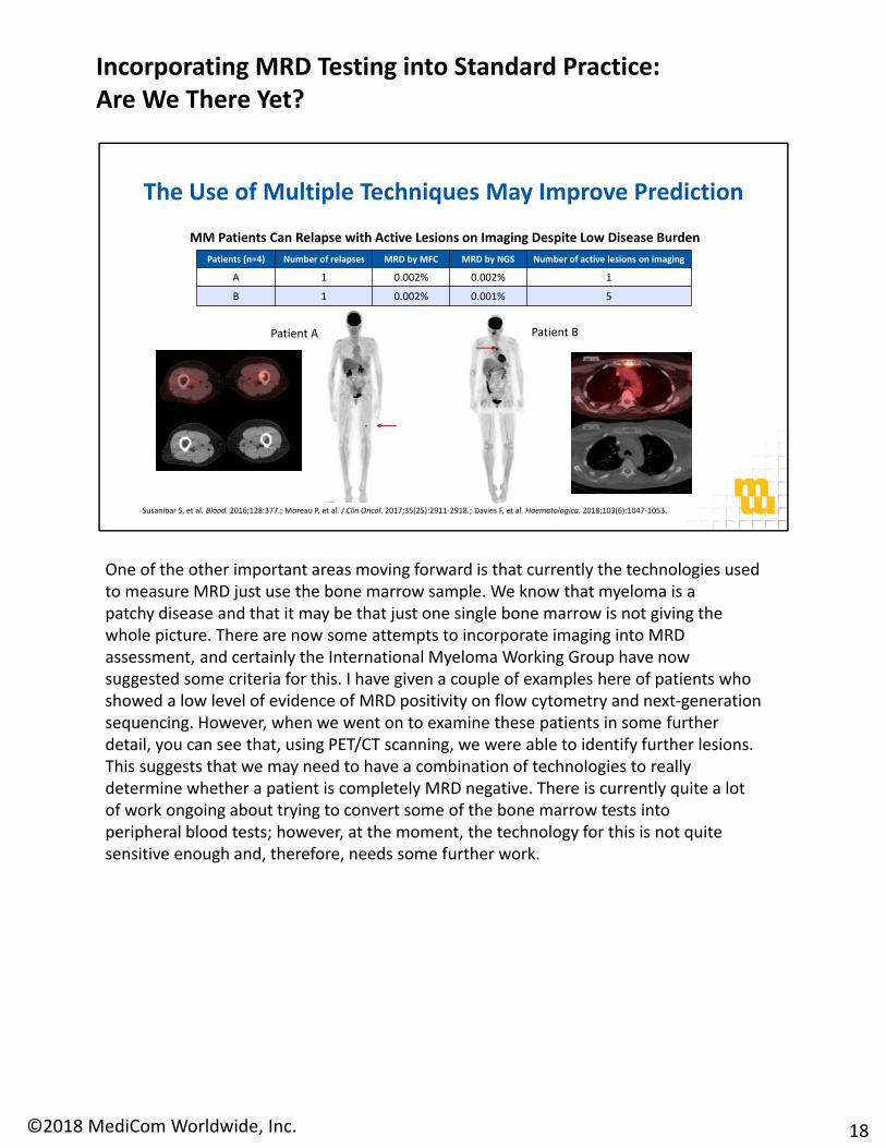
The Use of Multiple Techniques May Improve Prediction
Patient A Patient B
Patients (n=4) Number of relapses MRD by MFC MRD by NGS Number of active lesions on imaging
A 1 0.002% 0.002% 1
B 1 0.002% 0.001% 5
MM Patients Can Relapse with Active Lesions on Imaging Despite Low Disease Burden
Susanibar S, et al. Blood. 2016;128:377.; Moreau P, et al. J Clin Oncol. 2017;35(25):2911‐2918.; Davies F, et al. Haematologica. 2018;103(6):1047‐1053.
One of the other important areas moving forward is that currently the technologies used to measure MRD just use the bone marrow sample. We know that myeloma is a patchy disease and that it may be that just one single bone marrow is not giving the whole picture. There are now some attempts to incorporate imaging into MRD assessment, and certainly the International Myeloma Working Group have now suggested some criteria for this. I have given a couple of examples here of patients who showed a low level of evidence of MRD positivity on flow cytometry and next‐generation sequencing. However, when we went on to examine these patients in some further detail, you can see that, using PET/CT scanning, we were able to identify further lesions. This suggests that we may need to have a combination of technologies to really determine whether a patient is completely MRD negative. There is currently quite a lot of work ongoing about trying to convert some of the bone marrow tests into peripheral blood tests; however, at the moment, the technology for this is not quite sensitive enough and, therefore, needs some further work.
18
Incorporating MRD Testing into Standard Practice: Are We There Yet?
©2018 MediCom Worldwide, Inc.

Clinical Applicability of MRD Testing
Monitor response
Prognostic value
Monitor maintenance therapyMonitor maintenance therapy
Detect early relapseDetect early relapse
Marker of biology Marker of biology
What I hope I have done is to discuss a little bit about monitoring of response and how the new therapies induce a complete response, but importantly are able to increase the number of patients that achieve MRD negativity. There is certainly now a lot of evidence to show that being MRD negative has important prognostic value in many of the disease settings that we are treating patients in, and appears to be independent of age and of cytogenetic status. There is now a lot of work going on looking at patients who are having maintenance therapy to determine whether MRD can be used to direct this therapy, as well as whether MRD could be used as a way of detecting early relapse; especially as we now have a number of treatments which are less toxic and therefore may be able to be used or introduced early. As I said, there are a number of important studies going on looking at whether MRD is a marker of the biology of the disease and what we can learn further in this area.
19
Incorporating MRD Testing into Standard Practice: Are We There Yet?
©2018 MediCom Worldwide, Inc.

Key Points
• Molecular remission is a goal of therapy as it is a pre‐requisite for cure
• Need to understand the MRD clone (level and biology) so that it can be manipulated to achieve a cure
• Need to balance the goal of achieving a cure against short and long‐term side effects
• There are a number of open questions:
– The level of MRD and the detection method depends on the questions being asked
– MRD is a continuous variable
– Biology of the disease is important
– Levels required may/will be different for prognosis determination, clinical trial PFS and OS surrogacy, change in treatment decisions
Anderson KC, et al. Clin Cancer Res. 2017;23(15):3980‐3993.
To conclude, I would like to leave you with these key takeaway points: molecular remission in MRD is definitely a goal of therapy and indeed it is probably a prerequisite for a cure. However, in order to achieve this cure, we do need to learn a little bit more about the MRD clone so that we can manipulate it therapeutically. I think we need to be very conscious of balancing our need to achieve MRD negativity and a cure against some of the short‐ and longer‐term side effects of therapy. There are open questions, and as physicians, we need to be aware of these. The level of MRD that we are trying to achieve and the best method to detect this is very much in debate. I think this is particularly because of trying to standardize the technology and trying to ensure that the reports and the data that are returned to the treating physician are understandable and helpful. There are many working groups ongoing looking at this area, particularly between the commercial side of things and with the FDA. As I mentioned before, it is a continuous variable and so it is, therefore, important to be able to know, if you are using a local test, what the sensitivity of that test is. From a very practical point of view, I am not sure we are using the MRD test to change therapy at the moment. We certainly can utilize it to assess prognosis, but there are a number of clinical trials that are ongoing addressing whether we could utilize MRD testing for changing therapy. The two areas that are particularly important are, if a patient is MRD negative on multiple occasions, whether this could mean we could stop maintenance therapy; or if a patient is MRD positive on a number of occasions, whether this means we should intensify therapy. As I said, there are several cooperative groups looking at this and so hopefully there should be some answers in this area shortly. There is also a lot of work ongoing with the FDA as to whether MRD may be useful as a clinical trial endpoint rather than waiting for progression‐free and overall survival.
I hope you found this activity useful and I would like to thank you for listening.
20
Incorporating MRD Testing into Standard Practice: Are We There Yet?
©2018 MediCom Worldwide, Inc.
Copyright © 2022 FDOKUMEN