In Vivo Structure−Activity Relationship Study of Dorsomorphin Analogues Identifies Selective VEGF...
9
In Vivo Structure Activity Relationship Study of Dorsomorphin Analogues Identifies Selective VEGF and BMP Inhibitors Jijun Hao † , Joshua N. Ho † , Jana A. Lewis ‡ , Kaleh A. Karim † , R. Nathan Daniels § , Patrick R. Gentry ‡, , Corey R. Hopkins ‡, , Craig W. Lindsley ‡,§,, , and Charles C. Hong †,‡,,#, * † Division of Cardiovascular Medicine, Department of Medicine, ‡ Department of Pharmacology, § Department of Chemistry, Vanderbilt Program in Drug Discovery, Vanderbilt Institute of Chemical Biology, Vanderbilt University School of Medicine, Nashville, Tennessee 37232, and # Research Medicine, Veterans Administration TVHS, Nashville, Tennessee 37212 W ith advances in high-throughput screening capabilities, it is not difficult to identify com- pounds that target a particular protein or pathway. Rather, a greater challenge lies in identifying selective modulators and improving pharmaceutical, or ADMET (absorption, distribution, metabolism, excretion, and toxicity), properties of lead compounds ( 1). In the traditional approach to pharmaceutical development, the initial efforts at lead optimization are focused on identifying structural analogues with the highest po- tency against a therapeutic target in in vitro assays. However, when the subsequent in vivo results clash with the predictions based on in vitro tests, it is diffi- cult to determine whether such “failures” result from flawed biological underpinnings or compounds’ intrin- sic deficiencies, such as poor target selectivity or subop- timal in vivo bioavailability. In principal, these pitfalls can be circumvented with the use of the in vivo zebrafish model early in the lead optimization phase. Rapid external development, trans- parency, and high fecundity make zebrafish ideal for large-scale in vivo characterization of bioactive small molecules (2−5). Since embryonic cells are capable of integrating multiple signaling pathways to trigger pre- cise developmental outputs, a small molecule that se- lectively targets a signaling pathway involved in embry- onic patterning will phenocopy genetic mutations in that pathway, whereas nonspecific compounds will cause early embryonic lethality or nonspecific developmental delay. In addition, since drug exposure in embryos oc- curs by passive diffusion, the in vivo assessment takes into account compounds’ intrinsic physiochemical prop- *Corresponding author, [email protected]. Received for review November 12, 2009 and accepted December 19, 2009. Published online December 20, 2009 10.1021/cb9002865 © 2010 American Chemical Society ABSTRACT The therapeutic potential of small molecule signaling inhibitors is of- ten limited by off-target effects. Recently, in a screen for compounds that perturb the zebrafish embryonic dorsoventral axis, we identified dorsomorphin, the first selective inhibitor of bone morphogenetic protein (BMP) signaling. Here we show that dorsomorphin has significant “off-target” effects against the VEGF (vascular endothelial growth factor) type-2 receptor (Flk1/KDR) and disrupts zebrafish angio- genesis. Since both BMP and VEGF signals are known to be involved in vascular de- velopment, we sought to determine whether dorsomorphin’s antiangiogenic ef- fects are due to its impact on the BMP or VEGF signals through the development of analogues that target BMP but not VEGF signaling and vice versa. In a structureactivity relationship (SAR) study of dorsomorphin analogues based pri- marily on their effects on live zebrafish embryos, we identified highly selective and potent BMP inhibitors as well as selective VEGF inhibitors. One of the BMP inhibi- tors, DMH1, which exclusively targets the BMP but not the VEGF pathway, dorsal- ized the embryonic axis without disrupting the angiogenic process, demonstrating that BMP signaling was not involved in the angiogenic process. This is one of the first full-scale SAR studies performed in vertebrates and demonstrates the poten- tial of zebrafish as an attractive complementary platform for drug development that incorporates an assessment of in vivo bioactivity and selectivity in the context of a living organism. A RTICLE www.acschemicalbiology.org VOL.5 NO.2 • ACS CHEMICAL BIOLOGY 245
-
Upload
vanderbilt -
Category
Documents
-
view
3 -
download
0
Transcript of In Vivo Structure−Activity Relationship Study of Dorsomorphin Analogues Identifies Selective VEGF...

In Vivo Structure�Activity Relationship Studyof Dorsomorphin Analogues IdentifiesSelective VEGF and BMP InhibitorsJijun Hao†, Joshua N. Ho†, Jana A. Lewis‡, Kaleh A. Karim†, R. Nathan Daniels§, Patrick R. Gentry‡,�,Corey R. Hopkins‡,�, Craig W. Lindsley‡,§,�,�, and Charles C. Hong†,‡,�,#,*†Division of Cardiovascular Medicine, Department of Medicine, ‡Department of Pharmacology, §Department of Chemistry,�Vanderbilt Program in Drug Discovery, �Vanderbilt Institute of Chemical Biology, Vanderbilt University School of Medicine,Nashville, Tennessee 37232, and #Research Medicine, Veterans Administration TVHS, Nashville, Tennessee 37212
W ith advances in high-throughput screeningcapabilities, it is not difficult to identify com-pounds that target a particular protein or
pathway. Rather, a greater challenge lies in identifyingselective modulators and improving pharmaceutical, orADMET (absorption, distribution, metabolism, excretion,and toxicity), properties of lead compounds (1). In thetraditional approach to pharmaceutical development,the initial efforts at lead optimization are focused onidentifying structural analogues with the highest po-tency against a therapeutic target in in vitro assays.However, when the subsequent in vivo results clashwith the predictions based on in vitro tests, it is diffi-cult to determine whether such “failures” result fromflawed biological underpinnings or compounds’ intrin-sic deficiencies, such as poor target selectivity or subop-timal in vivo bioavailability.
In principal, these pitfalls can be circumvented withthe use of the in vivo zebrafish model early in the leadoptimization phase. Rapid external development, trans-parency, and high fecundity make zebrafish ideal forlarge-scale in vivo characterization of bioactive smallmolecules (2−5). Since embryonic cells are capable ofintegrating multiple signaling pathways to trigger pre-cise developmental outputs, a small molecule that se-lectively targets a signaling pathway involved in embry-onic patterning will phenocopy genetic mutations in thatpathway, whereas nonspecific compounds will causeearly embryonic lethality or nonspecific developmentaldelay. In addition, since drug exposure in embryos oc-curs by passive diffusion, the in vivo assessment takesinto account compounds’ intrinsic physiochemical prop-
*Corresponding author,[email protected].
Received for review November 12, 2009and accepted December 19, 2009.
Published online December 20, 2009
10.1021/cb9002865
© 2010 American Chemical Society
ABSTRACT The therapeutic potential of small molecule signaling inhibitors is of-ten limited by off-target effects. Recently, in a screen for compounds that perturbthe zebrafish embryonic dorsoventral axis, we identified dorsomorphin, the firstselective inhibitor of bone morphogenetic protein (BMP) signaling. Here we showthat dorsomorphin has significant “off-target” effects against the VEGF (vascularendothelial growth factor) type-2 receptor (Flk1/KDR) and disrupts zebrafish angio-genesis. Since both BMP and VEGF signals are known to be involved in vascular de-velopment, we sought to determine whether dorsomorphin’s antiangiogenic ef-fects are due to its impact on the BMP or VEGF signals through the developmentof analogues that target BMP but not VEGF signaling and vice versa. In astructure�activity relationship (SAR) study of dorsomorphin analogues based pri-marily on their effects on live zebrafish embryos, we identified highly selective andpotent BMP inhibitors as well as selective VEGF inhibitors. One of the BMP inhibi-tors, DMH1, which exclusively targets the BMP but not the VEGF pathway, dorsal-ized the embryonic axis without disrupting the angiogenic process, demonstratingthat BMP signaling was not involved in the angiogenic process. This is one of thefirst full-scale SAR studies performed in vertebrates and demonstrates the poten-tial of zebrafish as an attractive complementary platform for drug development thatincorporates an assessment of in vivo bioactivity and selectivity in the context ofa living organism.
ARTICLE
www.acschemicalbiology.org VOL.5 NO.2 • ACS CHEMICAL BIOLOGY 245

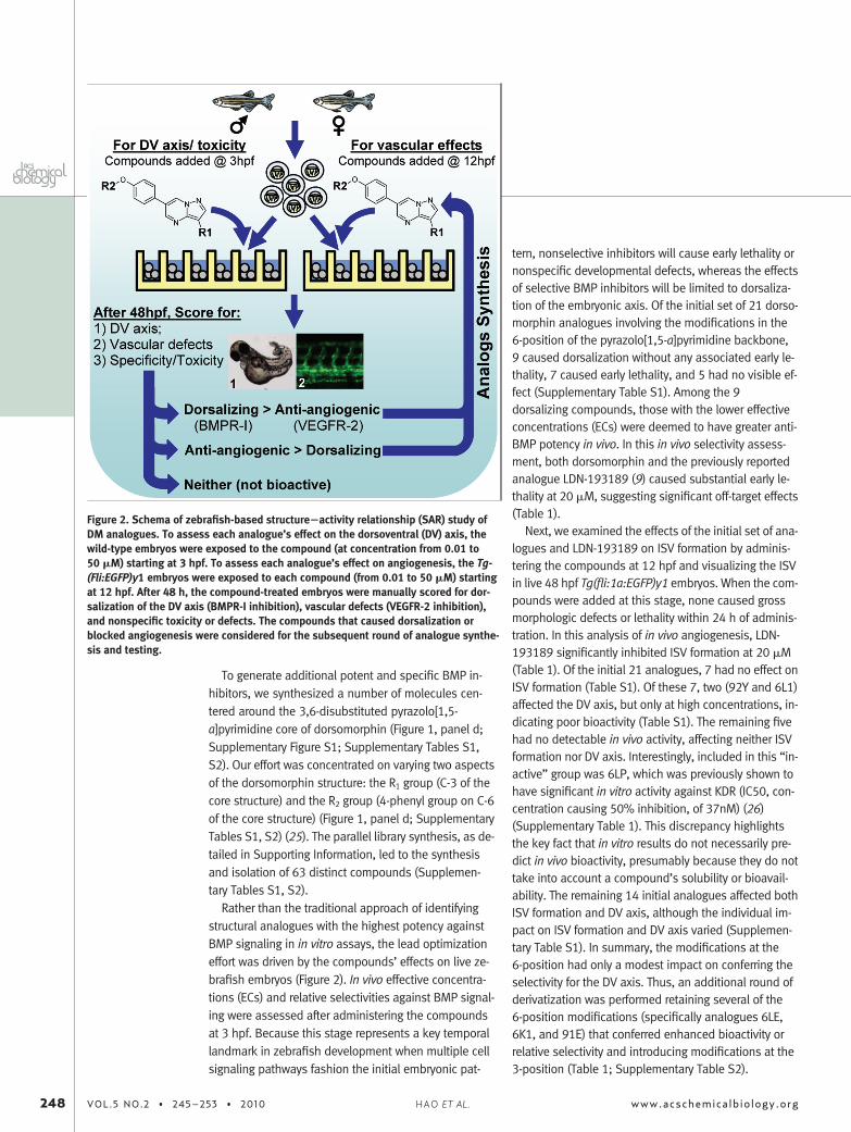
erties, such as the octanol�water partition coefficient(commonly referred to as log P), a major determinant ofdrug-likeness and bioactivity of a small molecule (6). Asa proof-of-principle, we identified dorsomorphin(Figure 1, panel a), the first selective small molecule in-hibitor of BMP signaling, on the basis of its ability tophenocopy the dorsoventral (DV) pattern defects seenin the BMP pathway mutants (Figure 1, panel b) (7).
Dorsomorphin and its analogue LDN-193189 havebeen used to direct differentiation of stem cells and todemonstrate the therapeutic potential of targeting BMPsignals for anemia and hyperossification syndromes(7−9). In particular, the discovery of dorsomorphin hasimmediate therapeutic implications for a debilitatingheretofore incurable condition known as fibrodysplasiaossificans progressiva, which has recently been shownto be caused by activating mutations in ALK2 (ACVR1), aBMP type-I receptor (10−12).
However, enthusiasm regarding their utility and thera-peutic potential is tempered by the fact that dorsomor-phin had significant “off-target” effects, including AMPKinhibition (13). Since AMPK plays a central role in en-ergy metabolism and is known to have beneficial cardio-vascular and antitumor effects, inhibiting AMPKhas potential to cause cardiotoxicity and promote tu-morigenesis (14−16). Here we show that dorsomorphinalso had significant inhibitory activity against the VEGFsignaling and caused a significant defect in the inter-somitic vessel (ISV) formation, an angiogenic processknown to require signaling by the VEGF type-II receptor(also known as Vegfr2/Kdr/Flk1) (17−20). However, be-cause BMP signaling is also known to be involved in an-giogenesis (21, 22), it was possible that dorsomorphintreatment revealed a novel role of the BMP signal in ISVformation.
To distinguish between these possibilities and to gen-erate additional potent and specific BMP and VEGF in-hibitors, we undertook a large-scale in vivo SAR study ofdorsomorphin analogues based on their effects on ze-brafish embryos. We synthesized 63 distinct com-pounds using the parallel library synthesis approachand tested them in zebrafish embryos to identify highlyselective and potent inhibitors of BMP as well as VEGFsignaling. One of the analogues, DMH1, which exclu-sively targets BMP but not VEGF signaling, dorsalizedthe embryonic axis without disrupting ISV formation,demonstrating that BMP signaling is not required for ze-brafish ISV formation.
RESULTS AND DISCUSSIONDuring the course of characterizing the effects of dorso-morphin (Figure 1, panel a) in zebrafish embryos, wefound that it consistently caused significant defects inISV formation (Figure 1, panel b), an angiogenic processknown to require signaling by the VEGF type-II recep-tors (Kdr/Flk1) (23). To examine in detail dorsomor-phin’s effects on ISV formation, the Tg(fli:1a:EGFP)y1transgenic embryos expressing GFP under the controlof an endothelial-specific promoter (24) were treatedwith various concentrations (0.1�100 �M) of dorsomor-phin starting at 12 h post fertilization (hpf). Becausethis stage follows the establishment of the dorsoven-tral (DV) axis, this analysis focused only on dorsomor-phin’s effects on angiogenesis. After dorsomorphintreatment, ISV was visualized in live 48 hpf embryos. Inthis in vivo angiogenesis model, dorsomorphin com-pletely inhibited ISV formation at 10 �M (Figure 1,panel b). At 5 �M, roughly 50% of the ISV were se-verely shortened or eliminated (dorsomorphin’s EC50,effective concentration affecting 50% of ISVs, was there-fore 5 �M; Table 1).
In cultured bovine aortic endothelial cells, dorsomor-phin inhibited the VEGF-stimulated Flk1 phosphoryla-tion in a dose-dependent manner (Figure 1, panel c),demonstrating that dorsomorphin was a potent VEGFsignal inhibitor. Nevertheless, since BMP signaling isalso known to be involved in angiogenesis (21, 22), itwas formally possible that dorsomorphin’s effects onthe zebrafish vasculature revealed a novel role of theBMP signal in ISV formation. To test this, we sought todevelop small molecules that specifically inhibited BMPbut not VEGF signaling.
Figure 1. Inhibition of both BMP and VEGF signaling by dorsomorphin, and thepyrazolo[1,5-a]pyrimidine backbone of DM for derivatization. a) Structure of dorso-morphin (DM). b) DM at a concentration of 2 �M dorsalized zebrafish embryos whenadministered at 3 h post fertilization (hpf), and 10 �M DM blocked intersomitic ves-sel (ISV) formation when administered at 12 hpf. Above, bright-field images oftreated embryos. Below, green fluorescence images of the Tg(Fli:EGFP)y1 transgenicembryos, which express GFP in the vasculature. Control embryos treated with DMSOare on the left, and DM-treated embryos are on the right. c) DM suppressed VEGF-dependent Flk1 phosphorylation in a dose-dependent manner in bovine arterial endo-thelial cells (BAECs). d) The pyrazolo[1,5-a]pyrimidine backbone of DM for derivat-ization involving modifications at the 3- and 6-positions (red circles).
246 VOL.5 NO.2 • 245–253 • 2010 www.acschemicalbiology.orgHAO ET AL.

TABLE 1. In vivo assessments and in vitro kinase assays of DM and selected analoguesa
aDorsomorphin (DM) and the selected analogues, along with the R1 and R2 structural modifications and the effects on ze-brafish embryos with respect to the dorsoventral (DV) axis, the intersomitic vessel (ISV) disruption, and nonspecific toxic-ity. For dorsalization, the EC100 (effective concentration 100%) represents the concentration when 100% of the treated em-bryos are severely dorsalized. As a result of significant day-to-day variability in severity of dorsalization at “sub-threshold”concentrations, the EC50 for severe dorsalization could not be reliably determined. For ISV disruption, the EC50 representsthe concentration when the formation of about 50% of the ISVs is inhibited. For nonspecific toxicity, the EC100 representsthe concentration when 100% of the treated embryos exhibit either early lethality within hours of compound addition, vari-able embryonic defects, or developmental delay. For comparison, the effects of the known KDR inhibitor SU5416 are shownat the bottom. Results from at least 20 embryos per condition.
ARTICLE
www.acschemicalbiology.org VOL.5 NO.2 • 245–253 • 2010 247
To generate additional potent and specific BMP in-hibitors, we synthesized a number of molecules cen-tered around the 3,6-disubstituted pyrazolo[1,5-a]pyrimidine core of dorsomorphin (Figure 1, panel d;Supplementary Figure S1; Supplementary Tables S1,S2). Our effort was concentrated on varying two aspectsof the dorsomorphin structure: the R1 group (C-3 of thecore structure) and the R2 group (4-phenyl group on C-6of the core structure) (Figure 1, panel d; SupplementaryTables S1, S2) (25). The parallel library synthesis, as de-tailed in Supporting Information, led to the synthesisand isolation of 63 distinct compounds (Supplemen-tary Tables S1, S2).
Rather than the traditional approach of identifyingstructural analogues with the highest potency againstBMP signaling in in vitro assays, the lead optimizationeffort was driven by the compounds’ effects on live ze-brafish embryos (Figure 2). In vivo effective concentra-tions (ECs) and relative selectivities against BMP signal-ing were assessed after administering the compoundsat 3 hpf. Because this stage represents a key temporallandmark in zebrafish development when multiple cellsignaling pathways fashion the initial embryonic pat-
tern, nonselective inhibitors will cause early lethality ornonspecific developmental defects, whereas the effectsof selective BMP inhibitors will be limited to dorsaliza-tion of the embryonic axis. Of the initial set of 21 dorso-morphin analogues involving the modifications in the6-position of the pyrazolo[1,5-a]pyrimidine backbone,9 caused dorsalization without any associated early le-thality, 7 caused early lethality, and 5 had no visible ef-fect (Supplementary Table S1). Among the 9dorsalizing compounds, those with the lower effectiveconcentrations (ECs) were deemed to have greater anti-BMP potency in vivo. In this in vivo selectivity assess-ment, both dorsomorphin and the previously reportedanalogue LDN-193189 (9) caused substantial early le-thality at 20 �M, suggesting significant off-target effects(Table 1).
Next, we examined the effects of the initial set of ana-logues and LDN-193189 on ISV formation by adminis-tering the compounds at 12 hpf and visualizing the ISVin live 48 hpf Tg(fli:1a:EGFP)y1 embryos. When the com-pounds were added at this stage, none caused grossmorphologic defects or lethality within 24 h of adminis-tration. In this analysis of in vivo angiogenesis, LDN-193189 significantly inhibited ISV formation at 20 �M(Table 1). Of the initial 21 analogues, 7 had no effect onISV formation (Table S1). Of these 7, two (92Y and 6L1)affected the DV axis, but only at high concentrations, in-dicating poor bioactivity (Table S1). The remaining fivehad no detectable in vivo activity, affecting neither ISVformation nor DV axis. Interestingly, included in this “in-active” group was 6LP, which was previously shown tohave significant in vitro activity against KDR (IC50, con-centration causing 50% inhibition, of 37nM) (26)(Supplementary Table 1). This discrepancy highlightsthe key fact that in vitro results do not necessarily pre-dict in vivo bioactivity, presumably because they do nottake into account a compound’s solubility or bioavail-ability. The remaining 14 initial analogues affected bothISV formation and DV axis, although the individual im-pact on ISV formation and DV axis varied (Supplemen-tary Table S1). In summary, the modifications at the6-position had only a modest impact on conferring theselectivity for the DV axis. Thus, an additional round ofderivatization was performed retaining several of the6-position modifications (specifically analogues 6LE,6K1, and 91E) that conferred enhanced bioactivity orrelative selectivity and introducing modifications at the3-position (Table 1; Supplementary Table S2).
Figure 2. Schema of zebrafish-based structure�activity relationship (SAR) study ofDM analogues. To assess each analogue’s effect on the dorsoventral (DV) axis, thewild-type embryos were exposed to the compound (at concentration from 0.01 to50 �M) starting at 3 hpf. To assess each analogue’s effect on angiogenesis, the Tg-(Fli:EGFP)y1 embryos were exposed to each compound (from 0.01 to 50 �M) startingat 12 hpf. After 48 h, the compound-treated embryos were manually scored for dor-salization of the DV axis (BMPR-I inhibition), vascular defects (VEGFR-2 inhibition),and nonspecific toxicity or defects. The compounds that caused dorsalization orblocked angiogenesis were considered for the subsequent round of analogue synthe-sis and testing.
248 VOL.5 NO.2 • 245–253 • 2010 www.acschemicalbiology.orgHAO ET AL.
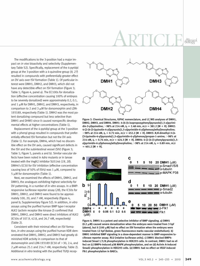
The modifications to the 3-position had a major im-pact on in vivo bioactivity and selectivity (Supplemen-tary Table S2). Specifically, replacement of the 4-pyridylgroup at the 3-position with a 4-quinoline group (9, 27)resulted in compounds with preferentially greater effecton DV axis over ISV formation (Table 1). Of particular in-terest were DMH1, DMH2, and DMH3, which did nothave any detectible effect on ISV formation (Figure 3;Table 1; Figure 4, panel a). The EC100s for dorsaliza-tion (effective concentration causing 100% of embryosto be severely dorsalized) were approximately 0.2, 0.1,and 1 �M for DMH1, DMH2, and DMH3, respectively, incomparison to 2 and 3 �M for dorsomorphin and LDN-193189, respectively (Table 1). DMH2 was the most po-tent dorsalizing compound but less selective thanDMH1 and DHM3 since it caused nonspecific develop-mental effects at higher concentrations (Table 1).
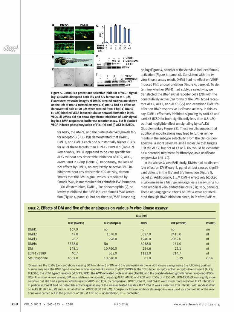
Replacement of the 4-pyridiyl group at the 3-positionwith a phenyl group resulted in compounds that prefer-entially effected ISV formation but not the DV axis(Table 1). For example, DMH4, which had no discern-ible effect on the DV axis, caused significant defects inthe ISV and the subintestinal vessel (SIV) (Figure 3;Table 1; Figure 5, panels a and b). Similar vascular de-fects have been noted in kdra mutants or in larvaetreated with the Vegfr2 inhibitor SU5146 (19, 20).DMH4’s EC50 for ISV inhibition (effective concentrationcausing loss of 50% of ISVs) was 1 �M, compared to5 �M for dorsomorphin (Table 1).
Next, we examined the effects of DMH1, DMH2, andDMH3, the analogues exhibiting highest selectivity forDV patterning, in a number of in vitro assays. In a BMP-responsive luciferase reporter assay (28), the IC50s forDMH1, DMH2, and DMH3 were found to be approxi-mately 100, 20, and 7 nM, respectively (Figure 4,panel b; Supplementary Figure S2). In addition, in vitroassays using the purified human BMP type-I receptorALK2 (activin receptor like kinase-2) confirmed thatDMH1, DMH2, and DMH3 were direct inhibitors of ALK2(IC50s of 107.9, 42.8, and 26.7 nM, respectively)(Table 2).
Consistent with their minimal effect on ISV forma-tion, in vitro assays using the purified human KDR dem-onstrated that DMH1, DMH2, and DMH3 had greatly di-minished KDR activity in comparison to that ofdorsomorphin and LDN-193189 (IC50 of �30, 2.4, and2 �M versus 25.1 and 214.7 nM, respectively; Table 2).Additional in vitro testing with the purified TGF� recep-
Figure 3. Chemical Structures, IUPAC nomenclature, and LC/MS analyses of DMH1,DMH2, DMH3, and DMH4. DMH1: 4-(6-(4-isopropoxyphenyl)pyrazolo[1,5-a]pyrimi-din-3-yl)quinoline; �98% at 214 nM, tR � 2.48 min, m/z � 381.2 [M � H]. DMH2:4-(2-(4-(3-(quinolin-4-yl)pyrazolo[1,5-a]pyrimidin-6-yl)phenoxy)ethyl)morpholine;�98% at 214 nM, tR � 0.72 min, m/z � 452.2 [M � H]. DMH3: N,N-dimethyl-3-(4-(3-(quinolin-4-yl)pyrazolo[1,5-a]pyrimidin-6-yl)phenoxy)propan-1-amine; �98% at214 nM, tR � 0.76 min, m/z � 424.3 [M � H]. DMH4: 4-(2-(4-(3-phenylpyrazolo[1,5-a]pyrimidin-6-yl)phenoxy)ethyl)morpholine; �98% at 214 nM, tR � 0.89 min, m/z� 401.2 [M � H].
Figure 4. DMH1 is a potent and selective inhibitor of BMP signaling. a) DMH1(0.2 �M) caused severe dorsalization when the embryos were treated from 3 hpf(above), but it (100 �M) had no effect on ISV formation when the embryos weretreated from 12 hpf (below, green fluorescence marks vascular endothelium). b)DMH1 inhibited BMP signaling in a dose-dependent manner in BMP-responsive lu-ciferase reporter assay. RLU (relative luciferase units). c) DMH1 blocked BMP4-induced Smad 1/5/8 phosphorylation in HEK293 cells. In contrast, DMH1 had no ef-fect on (c) BMP4-induced p38 MAPK phosphorylation, and on (d) Activin A-inducedSmad2 phosphorylation in HEK293 cells. (e) DMH1 had no effect on VEGF-inducedFlk1 phosphorylation in BAECs.
ARTICLE
www.acschemicalbiology.org VOL.5 NO.2 • 245–253 • 2010 249
tor ALK5, the AMPK, and the platelet-derived growth fac-tor receptor-� (PDGFR�) demonstrated that DMH1,DMH2, and DMH3 each had substantially higher IC50sfor all of these targets than LDN-193189 did (Table 2).Remarkably, DMH1 appeared to be very specific forALK2 without any detectable inhibition of KDR, ALK5,AMPK, and PDGFR� (Table 2). Importantly, the lack ofISV effects by DMH1, an exquisitely selective BMP in-hibitor without any detectable KDR activity, demon-strates that the BMP signal, which is mediated bySmad1/5/8, is not required for zebrafish ISV formation.
On Western blots, DMH1, like dorsomorphin (7), se-lectively inhibited the BMP-induced Smad1/5/8 activa-tion (Figure 4, panel c), but not the p38/MAP kinase sig-
naling (Figure 4, panel c) or the Activin A-induced Smad2activation (Figure 4, panel d). Consistent with the invitro kinase assay result, DHM1 had no effect on VEGF-induced Flk1 phosphorylation (Figure 4, panel e). To de-termine whether DMH1 had subtype selectivity, wetransfected the BMP signal reporter cells (28) with theconstitutively active (ca) forms of the BMP type-I recep-tors ALK2, ALK3, and ALK6 (29) and examined DMH1’seffect on BMP-responsive luciferase activity. In this as-say, DMH1 effectively inhibited signaling by caALK2 andcaALK3 (IC50 for both significantly less than 0.5 �M)but had negligible effect on signaling by caALK6(Supplementary Figure S3). These results suggest thatadditional modifications may lead to further refine-ments in the subtype selectivity. From the clinical per-spective, a more selective small molecule that targetsjust the ALK2, but not ALK3 or ALK6, would be desirableas a potential treatment for fibrodysplasia ossificansprogressiva (10, 12).
In the above in vivo SAR study, DMH4 had no discern-ible effect on DV (Figure 5, panel b), but caused signifi-cant defects in the ISV and SIV formation (Figure 5,panel a). Additionally, 1 �M DMH4 effectively blockedangiogenesis in a Matrigel angiogenesis assay using hu-man umbilical vein endothelial cells (Figure 5, panel c).These antiangiogenic effects of DMH4 were not medi-ated through BMP inhibition since, in in vitro BMP re-
Figure 5. DMH4 is a potent and selective inhibitor of VEGF signal-ing. a) DMH4 disrupted both ISV and SIV formation at 1 �M.Fluorescent vascular images of DMSO-treated embryo are shownon the left of DMH4-treated embryos. b) DMH4 had no effect ondorsoventral axis at 50 �M when treated from 3 hpf. c) DMH4(1 �M) blocked VEGF-induced tubular network formation in HU-VECs. d) DMH4 did not show significant inhibition of BMP signal-ing in a BMP-responsive luciferase reporter assay, but it blockedVEGF-induced phosphorylation of Flk1 (e) and (f) AKT in BAECs.
TABLE 2. Effects of DM and five of the analogues on various in vitro kinase assaysa
IC50 (nM)
ALK2 (BMPR-I) ALK5 (TGF�R-I) AMPK KDR (VEGFR2) PDGFR�
DMH1 107.9 no no no noDMH2 42.8 1578.0 3527.0 2418.0 ntDMH3 26.7 998.0 1940.0 2062.0 ntDMH4 3558.0 No 8038.0 161.0 ntDM 148.1 10,760.0 234.6 25.1 ntLDN-193189 40.7 565.0 1122.0 214.7 ntStaurosporine 4531.0 10,640.0 �1.0 3.29 6.14
aShown are the IC50s (concentrations causing 50% inhibition) of DM and the analogues for the in vitro kinase assays using the following purifiedhuman enzymes: the BMP type-I receptor activin receptor-like kinase 2 (ALK2/BMPR-I), the TGF� type-I receptor activin receptor-like kinase 5 (ALK5/TGF�R-I), the VEGF type-2 receptor (VEGFR2/KDR), the AMP-activated protein kinase (AMPK), and the platelet-derived growth factor receptor-� (PDG-FR�). In in vitro kinase assays, DM was relatively nonspecific, targeting ALK2, AMPK, and KDR with IC50s of �250 nM. LDN-193189 was slightly moreselective but still had significant effects against ALK5 and KDR. By comparison, DMH1, DMH2, and DMH3 were much more selective ALK2 inhibitors.In particular, DMH1 had no detectible activity against any of the kinases tested besides ALK2. DMH4 was a selective KDR inhibitor with modest effecton ALK2 (IC50 3.6 �M) and minimal effect on AMPK (IC50 8.0 �M). Nonspecific kinase inhibitor staurosporine was used as a control. All of the reac-tions were carried out in the presence of 10 �M ATP. no � no inhibition; nt � not tested.
250 VOL.5 NO.2 • 245–253 • 2010 www.acschemicalbiology.orgHAO ET AL.
porter and purified ALK2 kinase assays, DMH4 did notinhibit BMP signaling at such low concentrations(Figure 5, panel d; Table 2). Rather, DMH4 effectivelyblocked VEGF-stimulated phosphorylation of both Flk1and the downstream mediator Akt in a dose-dependentmanner (Figure 5, panels e and f). Furthermore, in an invitro assay using purified human Vegfr2 (KDR), DMH4was found to be a direct inhibitor with an IC50 of 161nM (Table 2). Interestingly, in our in vivo angiogenesisanalysis, the commonly utilized KDR inhibitor SU5146(Semaxanib) (30), which was originally developed as anantiangiogenesis therapy, was less potent than DMH4and not selective (Table 1).
For several compounds, the IC50s based on the invitro KDR kinase assays differed dramatically from theEC50s based on their effects on ISV formation (Tables 1,2). For example, dorsomorphin, LDN-193189, and 6LPwere potent KDR inhibitors in vitro (IC50s of 25.1, 214.7,and 37nM, respectively), yet all were relatively poor an-giogenesis inhibitors in embryos (EC50s of 5, 20, and�50 �M, respectively; Tables 1, 2). Such IC50�EC50disparities may reflect suboptimal bioavailabilities ofthese compounds in later stage (�24 hpf) embryos. Insummary, the zebrafish proved to be an effective plat-form for the rapid identification of highly selective VEGFinhibitors with excellent in vivo potencies and favorablephysiochemical properties.
Here, we utilized zebrafish to conduct the first full-scale in vivo SAR study in a vertebrate model to simulta-neously identify selective small molecule inhibitors ofBMP and VEGF pathways, both of which are recognizedas important therapeutic targets. Our analyses indicatethat modification of the 3-position of the pyrazolo[1,5-a]pyrimidine backbone of dorsomorphin plays a criticalrole in determining KDR versus BMP receptor selectivity.Using this approach, we identified a potent and selec-
tive KDR inhibitor (DMH4), as well as several highly se-lective BMP type-I receptor inhibitors, including DMH1,which does not target any of the related signaling path-ways tested. Exquisitely selective BMP inhibitors, suchas DMH1, that do not affect AMPK are preferred for fur-ther development as potential therapeutic leads sincethey should have reduced potential for cardiotoxicityand other undesired effects compared with the com-pounds that inhibit AMPK. Lastly, using DMH1, we dem-onstrated that BMP signaling is not required for angio-genesis in early zebrafish embryos.
The in vivo SAR’s success in identifying potent and se-lective inhibitors of not only BMP but also VEGF signal-ing demonstrates that the zebrafish provides an effec-tive multidimensional approach to simultaneouslyinterrogate multiple pathways on a whole organismscale. Moreover, since the in vivo SAR study assessesthe compounds’ bioactivities in live embryos, this ap-proach is inherently likely to identify compounds hav-ing favorable physiochemical properties and excellent“drug-likeness”. Thus, this approach is useful for avoid-ing the pitfall of pursuing “dead end” leads, such as6LP, which have excellent in vitro activity yet have poorin vivo bioactivity. The in vivo SAR study contrasts withthe traditional in vitro assay-based SAR studies that fo-cus on identifying analogues with the highest potency.Such a “linear approach”, which does not take into ac-count selectivity and physicochemical properties untillater in drug development, can lead to compounds thatultimately fail in preclinical animal models despite hav-ing a compelling biological rationale. In conclusion, wedemonstrate that the zebrafish is an attractive comple-mentary platform for pharmaceutical development thatincorporates the assessment of a lead compound’s invivo bioactivity and selectivity earlier in the developmentprocess.
METHODSZebrafish Experiments. The wild-type embryos were treated
with various concentrations (0.1�100 �M) of the compoundsstarting at 3 hpf to evaluate compounds’ effects on the dorso-ventral axis, and Tg(Fli:EGFP)y1 (18, 24) transgenic embryoswere treated from 12 hpf to evaluate the effects on ISV forma-tion. Twenty embryos were treated per well condition. Treatedembryos were manually dechorionated and observed at 24, 48,and 72 hpf.
Cells and Cell Culture. C2C12, C2C12BRA, and bovine aorticendothelial cells (BAEC) were cultured in DMEM supplementedwith 10% FBS (Gibco) and 1% penicillin/ streptomycin (Cellgro).
Human umbilical vein endothelial cells (HUVEC) were grown inEGM-2 Bulletkit medium (Lonza). Both BAEC and HUVEC celllines were cultured on 0.2% gelatin-coated dishes.
Endothelial Tubule Formation. HUVEC cells (1.5 � 104) wereplated on a 96-well microtiter plate precoated with 50 �L of EC-Matrix (Millipore) and were treated with DMSO or DMH4. After15 h of incubation, tubular network formation was examined un-der an inverted light microscope.
Western Blotting. For VEGF signaling, confluent BAEC cellswere serum-starved for 18�24 h and then treated with the com-pounds or DMSO for 30 min followed by 8 min of incubationwith 50 ng mL�1 VEGF165 (Alpha Diagnostics International, Inc.).For Smad and P38 MAPK activation, confluent HEK293 cells were
ARTICLE
www.acschemicalbiology.org VOL.5 NO.2 • 245–253 • 2010 251

serum-starved and then treated with the compounds or DMSOfor 30 min followed by 30 min incubation with BMP4 (50 ngmL�1) or Activin-A (40 ng mL�1). After stimulation, cells werethen lysed in CelLytic-M cell lysis buffer (Sigma) supplementedwith protease inhibitor cocktail (Sigma) and phosphatase inhibi-tor cocktail 2 (Sigma). Lysates were separated by SDS�PAGEand transferred onto PVDF membrane. The p-Flk1, p-Smad1/5/8, p-Smad2, p-AKT, p-P38MAPK, Flk1, AKT, and -tubulin weredetected by Odyssey system (Li-Cor bioscience) after incuba-tion with the appropriate primary and secondary antibodies. Pri-mary antibodies include antirabbit p-Flk1(Santa Cruz, 1:200 di-lution), antirabbit p-Smad1/5/8 (Cell Signaling Tech, 1:1000),antirabbit p-Smad2 (Cell Signaling Tech, 1:1000), antimousep-AKT (Cell Signaling Tech, 1:1000 dilution), antimousep-P38MAPK (Cell Signaling Tech, 1:1000), antimouse Flk1(Santa Cruz, 1:200 dilution), and antirabbit Akt (Cell signaling,1:1000 dilution). The secondary antibodies include IRDye 680-conjugated goat antirabbit IgG (Li-Cor Bioscience, 1:5000 dilu-tion) and IRDye 800CW-conjugated goat antimouse IgG (Li-CorBioscience, 1:5000 dilution).
BMP-Responsive Luciferase Reporter Assays. Stably trans-fected BMP-responsive C2C12BRA cells (containing the Id1promoter-firefly luciferase reporter; kind gift of D. Rifkin, NYUMedical Center (28)) were seeded in 96-well plates and incu-bated overnight with the compounds and BMP4 (50 ng mL�1).The cells were then lysed, and cell extracts were then subjectedto the firefly luciferase assay using Steady-Glo luciferase assaykit (Promega). The results were normalized to cell titers, as mea-sured using Cell Titer-Glo luminescence assay (Promega). Forsubtype analysis, C2C12BRA cells were transiently transfectedwith plasmids (0.1 �g) expressing constitutively active forms ofthe BMP type I receptors (caALK2, caALK3 or caALK6) using Lipo-fectamine kit (Invitrogen) in 96 well plates; 0.1 �g of pRL-TK Re-nilla luciferase (Promega) was used to control for transfection ef-ficiency. Relative activity was quantified by the ratio of firefly toRenilla luciferase activities using the dual luciferase assay kit(Promega).
Kinase Assay. All kinase assays were conducted by ReactionBiology Corp (Malvern, PA). In brief, compounds were tested at10 concentrations by 3-fold serial dilutions starting at 30 �M,using nonspecific kinase inhibitor staurosporine as control. Invitro kinase reactions were carried out in the presence of 10 �M(33P)ATP. Five kinases tested were the human BMP type-I recep-tor activin receptor-like kinase 2 (ALK-2/ACVR1), the humanTGF� type-I receptor activin receptor-like kinase 5 (Alk5/TGF�R1), the human VEGF type-II receptor (KDR/Flk-1/VEGFR2),the human AMP-activated protein kinase (AMPK/A1/B1/G1),and the human platelet-derived growth factor receptor-� (PDG-FR�).
Chemical Synthesis. The synthetic chemistry effort was con-centrated on varying two aspects of the 3,6-disubstitutedpyrazolo[1,5-a]pyrimidine core of dorsomorphin (Supplemen-tary Figure S3): the R1 group (C-3 of the core structure) and theR2 group (4-phenyl group on the C-6 of the core structure). Basedon the known synthetic methods available, we varied the R1
(phenyl, 2-, 3-, and 4-pyridyl, 3-quinoline, 4-quinoline,2-thiophene, and 2-thiazole) and R2 (alkyl ethers). Our librarysynthesis was focused on common intermediates 3 and 7,which were synthesized via different pathways depending onthe 3-aryl substitution required (Supplementary Schemes S1,S2). For the phenyl and pyridyl derivatives, the synthesis startedwith the known 4-aryl-1H-pyrazol-5-amine 1 (31−33), whichwas reacted with malondialdehyde 2 to afford the pyrazolo[1,5-a]pyrimidine core 3 (Supplementary Scheme S1). Next, the me-thoxy group was deprotected, and then the phenol was alky-lated under basic conditions with a number of groups affordingthe final products 4 (Supplementary Tables S1, S2) (31). For the
other 3-heteroaryl-substituted pyrazolo[1,5-a]pyrimidine com-pounds 7, the commercially available 4-bromo-1H-pyrazol-5-amine 5 was reacted as above with malondialdehyde 2, whichafforded the 3-bromo pyrazolo[1,5-a]pyrimidine core 6 (Supple-mentary Scheme S2). Next, 6 underwent transition-metal-catalyzed cross-coupling with either an appropriate boronicacid (34) or arylzinc bromide (35) to afford the desired 7. Com-pound 7 was then reacted as above to yield the final targets 4(Supplementary Tables S1, S2). This reaction sequence wasamenable to parallel library synthesis, and utilizing this ap-proach led to the synthesis and isolation of 63 distinct com-pounds. Detailed synthesis schemes are discussed in Support-ing Information.
Acknowledgment: The authors thank D. Rifkin (New York Uni-versity Medical Center) for the BMP-responsive reporter celllines and T. Imamura (The JFCR Cancer Institute, Tokyo) for theconstitutively active ALK constructs. The authors thank E. Ca-tania for her critical reading of the manuscript. This work wassupported by the Veterans Administration Career DevelopmentTransition Award (C.C.H.), the Developmental Grants from theCenter for Research in Fibrodysplasia Ossificans Progressiva andRelated Disorders (C.C.H.), the NIH K08HL081535 (C.C.H.), andthe GSK Cardiovascular Research and Education Foundation(C.C.H.). J.H., C.R.H., C.W.R., and C.C.H designed the experimentsand wrote the paper. J.H., J.N.H., and K.A.K. conducted zebrafishand in vitro analyses. J.A.L., R.N.D., and P.R.G. conducted chemi-cal synthesis.
Supporting Information Available: This material is availablefree of charge via the Internet at http://pubs.acs.org.
Competing Interests Statement: The authors have no compet-ing financial or personal interests to declare.
REFERENCES1. MacCoss, M., and Baillie, T. A. (2004) Organic chemistry in drug dis-
covery, Science 303, 1810–1813.2. Zon, L. I., and Peterson, R. T. (2005) In vivo drug discovery in the ze-
brafish, Nat. Rev. Drug Discovery 4, 35–44.3. Molina, G., Vogt, A., Bakan, A., Dai, W., Queiroz de Oliveira, P.,
Znosko, W., Smithgall, T. E., Bahar, I., Lazo, J. S., Day, B. W., andTsang, M. (2009) Zebrafish chemical screening reveals an inhibitorof Dusp6 that expands cardiac cell lineages, Nat. Chem. Biol. 5,680–687.
4. MacRae, C. A., and Peterson, R. T. (2003) Zebrafish-based smallmolecule discovery, Chem. Biol. 10, 901–908.
5. Hong, C. C. (2009) Large-scale small-molecule screen using ze-brafish embryos, Methods Mol. Biol. (Clifton, N.J.) 486, 43–55.
6. Lipinski, C. A., Lombardo, F., Dominy, B. W., and Feeney, P. J. (2001)Experimental and computational approaches to estimate solubilityand permeability in drug discovery and development settings,Adv. Drug Delivery Rev. 46, 3–26.
7. Yu, P. B., Hong, C. C., Sachidanandan, C., Babitt, J. L., Deng, D. Y.,Hoyng, S. A., Lin, H. Y., Bloch, K. D., and Peterson, R. T. (2008) Dor-somorphin inhibits BMP signals required for embryogenesis andiron metabolism, Nat. Chem. Biol. 4, 33–41.
8. Hao, J., Daleo, M. A., Murphy, C. K., Yu, P. B., Ho, J. N., Hu, J., Peter-son, R. T., Hatzopoulos, A. K., and Hong, C. C. (2008) Dorsomor-phin, a selective small molecule inhibitor of BMP signaling, pro-motes cardiomyogenesis in embryonic stem cells, PLoS ONE 3,e2904.
9. Yu, P. B., Deng, D. Y., Lai, C. S., Hong, C. C., Cuny, G. D., Bouxsein,M. L., Hong, D. W., McManus, P. M., Katagiri, T., Sachidanandan, C.,Kamiya, N., Fukuda, T., Mishina, Y., Peterson, R. T., and Bloch, K. D.(2008) BMP type I receptor inhibition reduces heterotopic [cor-rected] ossification, Nat. Med. 14, 1363–1369.
252 VOL.5 NO.2 • 245–253 • 2010 www.acschemicalbiology.orgHAO ET AL.

10. Shen, Q., Little, S. C., Xu, M., Haupt, J., Ast, C., Katagiri, T., Mund-los, S., Seemann, P., Kaplan, F. S., Mullins, M. C., and Shore, E. M.(2009) The fibrodysplasia ossificans progressiva R206H ACVR1 mu-tation activates BMP-independent chondrogenesis and zebrafishembryo ventralization, J. Clin. Invest. 119, 3462–3472.
11. Kaplan, F. S., Xu, M., Seemann, P., Connor, J. M., Glaser, D. L., Car-roll, L., Delai, P., Fastnacht-Urban, E., Forman, S. J., Gillessen-Kaesbach, G., Hoover-Fong, J., Koster, B., Pauli, R. M., Reardon, W.,Zaidi, S. A., Zasloff, M., Morhart, R., Mundlos, S., Groppe, J., andShore, E. M. (2009) Classic and atypical fibrodysplasia ossificansprogressiva (FOP) phenotypes are caused by mutations in the bonemorphogenetic protein (BMP) type I receptor ACVR1, Hum. Mutat.30, 379–390.
12. Shore, E. M., Xu, M., Feldman, G. J., Fenstermacher, D. A., Cho, T. J.,Choi, I. H., Connor, J. M., Delai, P., Glaser, D. L., LeMerrer, M.,Morhart, R., Rogers, J. G., Smith, R., Triffitt, J. T., Urtizberea, J. A.,Zasloff, M., Brown, M. A., and Kaplan, F. S. (2006) A recurrent muta-tion in the BMP type I receptor ACVR1 causes inherited and spo-radic fibrodysplasia ossificans progressiva, Nat. Genet. 38, 525–527.
13. Zhou, G., Myers, R., Li, Y., Chen, Y., Shen, X., Fenyk-Melody, J., Wu,M., Ventre, J., Doebber, T., Fujii, N., Musi, N., Hirshman, M. F., Good-year, L. J., and Moller, D. E. (2001) Role of AMP-activated protein ki-nase in mechanism of metformin action, J. Clin. Invest. 108,1167–1174.
14. Russell, R. R., 3rd, Li, J., Coven, D. L., Pypaert, M., Zechner, C., Palm-eri, M., Giordano, F. J., Mu, J., Birnbaum, M. J., and Young, L. H.(2004) AMP-activated protein kinase mediates ischemic glucose up-take and prevents postischemic cardiac dysfunction, apoptosis,and injury, J. Clin. Invest. 114, 495–503.
15. Sasaki, H., Asanuma, H., Fujita, M., Takahama, H., Wakeno, M., Ito,S., Ogai, A., Asakura, M., Kim, J., Minamino, T., Takashima, S.,Sanada, S., Sugimachi, M., Komamura, K., Mochizuki, N., andKitakaze, M. (2009) Metformin prevents progression of heart failurein dogs: role of AMP-activated protein kinase, Circulation 119,2568–2577.
16. Vazquez-Martin, A., Oliveras-Ferraros, C., Lopez-Bonet, E., and Me-nendez, J. A. (2009) AMPK: Evidence for an energy-sensing cytoki-netic tumor suppressor, Cell Cycle 8, 3679–3683.
17. Bussmann, J., Lawson, N., Zon, L., and Schulte-Merker, S. (2008) Ze-brafish VEGF receptors: a guideline to nomenclature, PLoS Genet.4, e1000064.
18. Isogai, S., Lawson, N. D., Torrealday, S., Horiguchi, M., and Wein-stein, B. M. (2003) Angiogenic network formation in the developingvertebrate trunk, Development (Cambridge, U.K.) 130, 5281–5290.
19. Covassin, L. D., Villefranc, J. A., Kacergis, M. C., Weinstein, B. M., andLawson, N. D. (2006) Distinct genetic interactions between multipleVegf receptors are required for development of different blood ves-sel types in zebrafish, Proc. Natl. Acad. Sci. U.S.A. 103, 6554–6559.
20. Habeck, H., Odenthal, J., Walderich, B., Maischein, H., and Schulte-Merker, S. (2002) Analysis of a zebrafish VEGF receptor mutant re-veals specific disruption of angiogenesis, Curr. Biol. 12, 1405–1412.
21. Heinke, J., Wehofsits, L., Zhou, Q., Zoeller, C., Baar, K. M., Helbing,T., Laib, A., Augustin, H., Bode, C., Patterson, C., and Moser, M.(2008) BMPER is an endothelial cell regulator and controls bonemorphogenetic protein-4-dependent angiogenesis, Circ. Res. 103,804–812.
22. Scharpfenecker, M., van Dinther, M., Liu, Z., van Bezooijen, R. L.,Zhao, Q., Pukac, L., Lowik, C. W., and ten Dijke, P. (2007) BMP-9 sig-nals via ALK1 and inhibits bFGF-induced endothelial cell prolifera-tion and VEGF-stimulated angiogenesis, J. Cell Sci. 120, 964–972.
23. Fraley, M. E., Rubino, R. S., Hoffman, W. F., Hambaugh, S. R., Ar-rington, K. L., Hungate, R. W., Bilodeau, M. T., Tebben, A. J., Rut-ledge, R. Z., Kendall, R. L., McFall, R. C., Huckle, W. R., Coll, K. E., andThomas, K. A. (2002) Optimization of a pyrazolo[1,5-a]pyrimidineclass of KDR kinase inhibitors: improvements in physical propertiesenhance cellular activity and pharmacokinetics, Bioorg. Med.Chem. Lett. 12, 3537–3541.
24. Lawson, N. D., and Weinstein, B. M. (2002) In vivo imaging of embry-onic vascular development using transgenic zebrafish, Dev. Biol.248, 307–318.
25. Daniels, R. N., Kim, K., Lebois, E. P., Muchalski, H., Hughes, M., andLindsley, C. W. (2008) Microwave-assisted protocols for the expe-dited synthesis of pyrazolo[1,5-a] and [3,4-d]pyrimidines, Tetrahe-dron Lett. 49, 305–310.
26. Fraley, M. E., Hoffman, W. F., Rubino, R. S., Hungate, R. W., Tebben,A. J., Rutledge, R. Z., McFall, R. C., Huckle, W. R., Kendall, R. L., Coll,K. E., and Thomas, K. A. (2002) Synthesis and initial SAR studiesof 3,6-disubstituted pyrazolo[1,5-a]pyrimidines: a new class of KDRkinase inhibitors, Bioorg. Med. Chem. Lett. 12, 2767–2770.
27. Cuny, G. D., Yu, P. B., Laha, J. K., Xing, X., Liu, J. F., Lai, C. S., Deng,D. Y., Sachidanandan, C., Bloch, K. D., and Peterson, R. T. (2008)Structure-activity relationship study of bone morphogenetic protein(BMP) signaling inhibitors, Bioorg. Med. Chem. Lett. 18, 4388–4392.
28. Zilberberg, L., ten Dijke, P., Sakai, L. Y., and Rifkin, D. B. (2007) Arapid and sensitive bioassay to measure bone morphogenetic pro-tein activity, BMC Cell Biol. 8, 41.
29. Fujii, M., Takeda, K., Imamura, T., Aoki, H., Sampath, T. K., Eno-moto, S., Kawabata, M., Kato, M., Ichijo, H., and Miyazono, K. (1999)Roles of bone morphogenetic protein type I receptors and Smadproteins in osteoblast and chondroblast differentiation, Mol. Biol.Cell 10, 3801–3813.
30. Fong, T. A., Shawver, L. K., Sun, L., Tang, C., App, H., Powell, T. J.,Kim, Y. H., Schreck, R., Wang, X., Risau, W., Ullrich, A., Hirth, K. P.,and McMahon, G. (1999) SU5416 is a potent and selective inhibi-tor of the vascular endothelial growth factor receptor (Flk-1/KDR)that inhibits tyrosine kinase catalysis, tumor vascularization, andgrowth of multiple tumor types, Cancer Res. 59, 99–106.
31. Daniels, R. N., Kim, K., Lebois, E. P., Muchalski, H., Hughes, M., andLindsley, C. W. (2008) Microwave-assisted protocols for the expe-dited synthesis of pyrazolo[1,5-a] and [3,4-d]pyrimidines, Tetrahe-dron Lett. 49, 305–310.
32. Fraley, M. E., Hoffman, W. F., Rubino, R. S., Hungate, R. W., Tebben,A. J., Rutledge, R. Z., McFall, R. C., Huckle, W. R., Kendall, R. L., Coll,K. E., and Thomas, K. A. (2002) Synthesis and initial SAR studiesof 3,6-disubstituted pyrazolo[1,5-a]pyrimidines: a new class of KDRkinase inhibitors, Bioorg. Med. Chem. Lett. 12, 2767–2770.
33. Fraley, M. E., Rubino, R. S., Hoffman, W. F., Hambaugh, S. R., Ar-rington, K. L., Hungate, R. W., Bilodeau, M. T., Tebben, A. J., Rut-ledge, R. Z., Kendall, R. L., McFall, R. C., Huckle, W. R., Coll, K. E., andThomas, K. A. (2002) Optimization of a pyrazolo[1,5-a]pyrimidineclass of KDR kinase inhibitors: improvements in physical propertiesenhance cellular activity and pharmacokinetics, Bioorg. Med.Chem. Lett. 12, 3537–3541.
34. Miyaura, N., and Suzuki, A. (1995) Palladium-catalyzed cross-coupling reactions of organoboron compounds, Chem. Rev. 95,2457–2483.
35. King, A. O., Okakadao, N., and E.-i, N. (1977) Highly general stereo-,regio-, and chemo-selective synthesis of terminal and internal con-jugated enynes by the Pd-catalyzed reaction of alkynylzinc re-agents with alkenyl halides, J. Chem. Soc., Chem. Comm. 683–684.
ARTICLE
www.acschemicalbiology.org VOL.5 NO.2 • 245–253 • 2010 253