Immunomodulatory Activities of Oat β-Glucan In Vitro and In Vivo
Upload
khangminh22Category
view
1download
0
ORIGINAL CONTRIBUTION Open Access
In-vivo and in-vitro evaluation ofpharmacological activities of Ardisiasolanacea leaf extractMohammad Rashedul Islam1, Jannatul Naima1, Nawreen Monir Proma1, Md. Saddam Hussain2,S. M. Naim Uddin1 and Mohammed Kamrul Hossain1*
Abstract
Background: The Bangladeshi rural and hilly areas people have long tradition to use various medicinal plants fortreating different diseases. That’s why, the crude methanolic leaf extract of Ardisia solanacea with its different fractions(petroleum ether, carbon tetrachloride, n-hexane and chloroform fractions) were subjected to investigate bioactivitiesin swiss albino mice; namely analgesic, CNS, and Oral hypoglycemic activities, while in-vitro evaluation of cytotoxicity.
Methods: Central nervous system activity was investigated by various method such as Elevated plus maze, Hole board,Hole cross and Open field test apparatus. Analgesic activity was evaluated by both acetic acid induced and tailimmersion method. Hypoglycemic activity was evaluated by oral glucose tolerance test and cytotoxicity was evaluatedby Brine shrimp lethality bioassay.
Results: In CNS activity, among others fractions, ASCF fraction produced a significant anxiolytic activity in bothelevated plus maze and Hole board test. During open-field test almost all the fractions of A. solanacea leaves extractdisplay decreased locomotor activities that indicates significant sedative activity. The ASME and ASCF showedsignificant peripheral analgesic activity at a dose of 200mg/kg and 400mg/kg body weight (p < 0.05). In tail immersionmethod, among others extracts chloroform fractions exhibited significant (p < 0.05) elongation of reaction time 30minafter oral dose of 200 and 400mg/kg body weight respectively. The methanolic and n-hexane extracts reduced bloodglucose level significantly after 90min with value of 53.94% and 48.15% respectively (p < 0.05). In case of cytotoxicityactivity, among other fractions carbon tetrachloride fraction showed lowest LC50 values.
Conclusions: From the above results, it is clear that different fractions of A. solanacea showed significantpharmacological potentiality in different in-vitro and in-vivo study model. So, it will be very much possible source foran isolating lead compound for curing the numerous disorders.
Keywords: Phytochemicals, Central nervous system, Analgesic, Hypoglycemic, Cytotoxicity
BackgroundUsing of complementary medicine to alleviate and im-prove health conditions in developed countries are in-creasing [1]. Keeping this purpose in mind, newmedicinal plants from different areas of the universeis being investigated [2]. Now-a-days most of thepeople prefer the alternative medicine on account ofits fewer toxic effects and health hazards [3].
Traditional medicine of Bangladesh is a unique con-glomerate of different ethnomedical consequences. Be-cause of geographic location and socioculturalscharacteristics of the country, it comprises traditionalyrooted elements influenced by local instinctive people,and also cover-by Indian Unani and Ayurveda medi-cine [4, 5]. The plants having phytochemicals possessmedicinal value that can have physiological andpharmacological actions on human body [6, 7].Genus, Ardisia is the leading genus of Myrsinaceaefamily. About 500 species of evergreen trees andshrubs are commenced throughout the tropical and
© The Author(s). 2019 Open Access This article is distributed under the terms of the Creative Commons Attribution 4.0International License (http://creativecommons.org/licenses/by/4.0/), which permits unrestricted use, distribution, andreproduction in any medium, provided you give appropriate credit to the original author(s) and the source, provide a link tothe Creative Commons license, and indicate if changes were made.
* Correspondence: [email protected] of Pharmacy, University of Chittagong, Chittagong 4331,BangladeshFull list of author information is available at the end of the article
Islam et al. Clinical Phytoscience (2019) 5:32 https://doi.org/10.1186/s40816-019-0128-9

sub-tropical regions of the universe [8]. Besides, A.compressa possesses significant hypoglycemic effectsin Type 2 diabetic rats, and antioxidant activity andA. crispa showed in vitro cytotoxic activity and anti-oxidative actions against breast cancer cell lines [9].Barks and leaves of A. colorata were extracted withethanol having antimicrobial activities [10]. Thepresent study protocol is designed to investigatephytochemical and pharmacological properties of A.solanacea leaves. A. solanacea Roxb. (Family: Primu-laceae; sub-family Myrsinoideae) is a glabrous shrubor small tree that reaches a maximum height of 20 ftunder habitat conditions. The plant has potentialpharmaceutical applications (such as antibacterial andantiviral properties) from extracts of plants from thegenus Ardisia were noted by Kobayashi and de Mejia[11]. More specifically, antibacterial activity againstfour serovars of salmonella were observed with plantextracts from A. solanacea [12] and there were posi-tive effects on the reduction of breast cancer cells[13]. The variety of ethnopharmacological uses andphytoconsituents had led our attraction to investigatephytochemicals and pharmacological properties of thisplant.
Materials and methodsPlant materialPlant sample of A. solanacea was collected from Hillyarea of Chittagong in July 2017. Then taxonomist Dr.Shaikh Bokhtiar Uddin, Professor, Department of Bot-any, University of Chittagong, identified it.
Preparation of the plant materialThe whole plant part of A. solanacea leaves was washedproperly, cut into small pieces, and then air dried forseveral days. After proper air-drying, the small pieceswere then grinded into coarse powder with the help ofgrinding machine in the Phytochemical Research La-boratory, Pharmacy department, University of Chitta-gong. The materials then were stored into air resistantcontainer.
Extraction of the plant materialAbout 700 g of the powdered material was taken in aclean, round bottomed flask (5 l) and soaked in 3.5 lof pure methanol (Merck KGaA, Germany). The res-ervoir with its content was sealed by foil and kept for15–20 days by occasional stirring and shaking. Thewhole mixture was then filtered through a fresh cot-ton plug and finally with a Whitman No.1 filterspaper. The volume of the filtrate was then reducedusing a Buchii Rota (Cole-Parmer, UK) evaporator atlow temperature and pressure. The weight of extract
yielded was 42.42 g. The percentage yield of the ex-tract was calculated using the following equation [14]:
%of yield of extracts ¼ Weight of extracted materialWeight of original plant material used
� 100
Phytochemical screening of the compoundPhytochemical analysis of the methanolic extracts of A.solanacea and its different soluble fractions were con-ducted using the standard procedures to identify variousconstituents described by Sofowara, Trease and Evans,and Harborne [15–17].
Experimental animalsYoung male Swiss-albino mice, aged 3–4 weeks with anaverage weight 20–25 g, were collected from theBangladesh Council of Scientific and Industrial ResearchLaboratories (BCSIR) and animal house of Pharmacy De-partment, Jahangirnagar University. The animals werehoused comfortably in a group of six in a single clean plas-tic cage with a metal net structured frame lid on its top.They were kept in standard environmental condition(temperature: (24 ± 1 °C), relative humidity: 55%–65% and12 h light dark/ 12 h dark circle) for one week in the ani-mal house of the Pharmacy Department, University ofChittagong, Bangladesh for adaptation. The animals wereprovided with standard laboratory food and water.
Study designFor both anxiolytic and sedative activities, fifty experi-mental animals were selected, numbered, weighed anddivided into ten groups. Group (І) was treated as control(saline), group (ІІ) for standard drug (Diazepam; 1.0 mg/kg) treatment and others group for different doses (200and 400 mg/kg) of methanolic extract of A. solanacealeaves and n-hexane, carbon tetrachloride and chloro-form carbon tetrachloride fractions respectively and forthe both central and peripheral analgesic activities, fortymices were selected randomly and grouped into eight.They were fasted for 12 h and later treated as follows:group (І) was treated as control (1% Tween 80 in normalsaline; 0.1 ml/10 g), group (ІІ) for standard drug (Diclofe-nac sodium; 50 mg/kg) treatment and others group fordifferent doses (200 and 400 mg/kg) of methanolic ex-tract of A. solanacea leaves and of its n-hexane andchloroform fraction respectively. In oral hypoglycemicactivity, 25 experimental animals were randomly selectedand divided into four groups. Here, group (I) served ascontrol and received vehicle (1% Tween 80 in saline, 10ml/kg body weight), group (II) received standard drug(Glibenclamide, 2 mg/ kg) and the four other groups re-ceived the sample extract at four different doses. Eachexperimental group comprising five mice’s. All the testsamples, control, and standard were administered orally.
Islam et al. Clinical Phytoscience (2019) 5:32 Page 2 of 11
Evaluation of anxiolytic activityElevated plus maze methodElevated plus maze is very important apparatus to investi-gate anxiolytic response [18] as well as neuroprotective ac-tions created by the test drugs. Animals that are exposedto novel maze avenue invites a way- repeal combat that isstronger in open arm as compared to closed arm. Rodents,which have an aversion for high and open space and putforward to enclosed arm, consequently pass a greateramount of time in enclosed arm. Animals, when enter intoopen arm, they become freeze, immobile, defecate and ex-hibit fear-like behaviors [19]. An actual reflection of anx-iety is reported due to increase level of plasma cortisol.Further, merits of this test procedure: (a) it is fast, simpleand less time-consuming, (b) noxious stimuli or trainingis not required and (c) this is reliable and predictable forperusing anxiety properties as well as anxiolytic activity ofdrug [20, 21]. Elevated plus maze apparatus is designed asa + shape, consisting of two open arms (25 × 5 cm, with avery slight, 0.5-cm, wall) across from each other and per-pendicular to two closed arms (25 × 5× 16 cm), and israised 40 cm above the floor [22]. Experimental animalswere installed individually in the center of the maze, headthat is facing towards open arm and stopwatch wasstarted. The subsequent parameters were observed for 5min. (1) At first we see, mouse is preferred to either openarm or close arm, (2) Counting of entries into open armand closed arm; when four paws of mouse is entered intoan arm is called arm entry. Control (saline), Standard (Di-azepam), and other test samples of A. solanacea leaveswere administered to the animals. After 30min, animalswere installed individually in the middle of the maze. Inthe end, the animals which were preferred to either openor closed arm, the average time spent in the open arm en-tries number in each testing group was compared.
Hole-board test methodHole-board apparatus consists of an enclosed arena withholes in the floor into which an animal can poke its head,referred to as head-dipping [23]. Measurement of neophiliaor directed exploration is obtained from the duration andfrequency of head dipping. This indicates the general loco-motor function of the animal is independent [24]. The in-creased level head dipping generally indicate neophilia,when low level are tend to result from absence of neophiliaor tend to indicate an increased anxiety-like state in thetesting animals [25]. Therefore, the anxiety state is in-creased as the number and incidence of head dipping is re-duced and vice-versa [26]. 30mins prior to treatment withcontrol, standards and test samples, the mice were installedsingly on the hole board apparatus, and we counted thehead dipping of mice into the holes at the altitude of theireyes during 5mins trial period using a tally counter.
Evaluation of sedative activityOpen-field test methodThis test was carried out in apparatus having a floor ofabout half-square meter in area and surrounded by a wallof 50 cm in height [27]. The floor is made of small squaresalternately which is colored in white and black. The num-ber of squares visited by the mice was counted and notedfor an interval of 3min (counted using a tally counter); at0, 30, 60, 90 and 120min; of the oral administration of thecontrol (saline), Standard (Diazepam) and the test samplesof different doses (200 and 400mg/kg).
Hole-crossed test apparatusThis test was carried out in a closed chamber having a sizeof (30 cm× 20 cm× 14 cm) surrounded by wooden walls, norooftop [28]. The chamber is partitioned in the central por-tion by placing a fixed wooden structure. The fixed woodenpartitioned had a round hole curve in it; diameter of 3.5 cmat a height of the hole is 7.5 cm. The mice were crossedthrough the hole from one chamber to another and by usinga tally counter, the number of crossing was counted for 3mins at 0, 30, 90, 120mins intervals respectively.
Evaluation of peripheral analgesic activityAcetic acid writhing methodAnalgesic activity of this plant extract is assessed by aceticacid writhing method [29, 30]. In this test, acetic acid wasused to trigger pain sensation in animals through intraper-itoneal administration and as a result, body of animalssquirms at regular interval to get away of pain. This con-tinuous contraction or squirms of the animal body istermed as “Writhing”. They continue to generate writhingas long as they feel pain. Any extract with analgesic prop-erty will give lesser number of writhes in animals with re-spect to control group. After 40mins, acetic acid (0.7%) atthe dose of 0.1ml/10 g body weight was injected by intra-peritoneally to all testing animals of different group. Thenumber of writhing or squirms responses generated byeach animal was recorded for fifteen minutes just after 5min of acetic acid injection. The following equation wasused for the measurement of percent inhibition of abdom-inal writhing: % inhibition of abdominal writhing = [(Wc −Wt)/Wc × 100],Where Wc stands for number of writhing’s of control
group, and Wt stands for number of writhing’s of testgroup.
Evaluation of central analgesic activityTail immersion testTail immersion test is a thermal method to evaluatecentral analgesic activity of methanolic extract, n-hexane and chloroform soluble fractions of A. sola-nacea leaves. The test is designed based on the ef-fect of centrally working analgesic drugs, which are
Islam et al. Clinical Phytoscience (2019) 5:32 Page 3 of 11

increasing the time of reaction in animal (mice) inresponse to hot water on water bath. The presenttest is done by the method described by Uma deviand Di stasi [31, 32]. The mice in each group washeld in a suitable restrainer with the whole tail ex-tending out immersed in a water bath thermostatic-ally maintained at 51 ± 1 °C. 2–3 cm of tail of themices pre-treated with control, Standard and testsamples were immersed in hot water of water bath.The time between immersion and deflection of tailwas recorded as reaction time or tail-flick latency.Then, latency period was observed at 30, 60, 90 and120 mins of oral administration. A cut of period of15 s were observed to avert tail tissue destruction ofmices. Baseline response at 30 mins, mices weretested for twice prior to drug administration. Threemeasurements were obtained from the baseline reac-tion time (Latency). After each measurement, ani-mals were gone back to the observation chambersfor 2 mins. The mean of these measurements is usedto calculate pre-drug latency period. After baseline,the treated mices were tested at 30, 60, 90, 120 mins.Finally, the percentage of the Maximal Possible Ef-fect (%MPE) was calculated using the followingequation [33]. % Of MPE = {(Post drug latency-Predrug latency)/ (Cut off time-Pre drug latency)} × 100.The percentage of time elongation of tail immersion
was calculated in respect to the control in respectivetime. If the percentage of elongation period is higher,the central analgesic action will be greater. The per-centage of time elongation was calculated from thefollowing equation [34]. % of Elongation = {(Latency oftest sample- Latency of control)/ Latency of test sam-ple} × 100.
Evaluation of hypoglycemic activityA glucose tolerance test is one of the most acceptablemethods to evaluate the hypoglycemic activity. This testwas performed following the procedure described by Joyand Kuttan [35] with slight modification. After 30 minsof extract administration, all groups were treated with10% glucose solution (2 g/kg body weight). After 30, 90and 120 min of glucose loading, blood samples were col-lected from tail vein. By using glucometer blood glucoselevel is measured. Test samples, control, and glibencla-mide were given orally. Before any test, every mouse wasweighed appropriately and the doses of the test samplesand control materials were fitted properly.
In-vitro evaluation of cytotoxic activityCytotoxic activity of this plant was predicted by usingthe brine shrimp lethality bioassay [36, 37].
Preparation of seawater38 g of pure NaCl (Salt) weighed properly, dissolved into1 l of seawater and then filtered the solution to get cleanand transparent solution.
Hatching of brine shrimpsThe test organism named as A. salina leach (brineshrimp eggs) was collect from pet shop. Prepared sea-water was taken into a small tank and brine shrimp eggswere employed to one edge of the tank that then wascovered with a lid. Shrimps in this tank were hatched fortwo days to give mature shrimp named as nauplii. Dur-ing this hatching, constant oxygen was supplied and thehatched shrimps were enticed to the lamp through theperforated dam. Ten living matured nauplii were trans-ferred to individual test tube by the help of Pasteurpipette.
Procedure5 mg of all individual samples were dissolved in 1 ml ofpure DMSO (dimethylsulfoxide) solution and then addenough seawater to make the volume 5ml. Varying con-centrations (800, 500, 300, 100 μg/ml) of samples wereobtained by using serial dilution method by adding sim-ulated seawater. Then these solutions were taken in indi-vidual test tube where each tube containing 5 mlseawater and 10 brine shrimp nauplii. The test tubeswere then observed for 24 h & survivors are counted byusing a magnifying glass [37, 38]. The data are processedin a simple program (Microsoft Excel 2007) to estimateLC50 values. Ampicillin trihydrate solution was used aspositive control.
Statistical analysisAll results are expressed as mean ± standard error ofmean (SEM). All statistical analyses were performed byone-way ANOVA Dunnett’s t-test where *P < 0.05 wasregarded as statistically significant. In addition, all datawere analyzed using SPSS software (Version: 20, IBMCorporation, New York, USA). LC50 values were esti-mated by linear regression equations through the usageof Microsoft Excel 2007 (Microsoft, Redmond, Washing-ton, USA).
ResultsPlant extractionThe % yield of the leave extract was 6.06 w/w dry matterand dark in color.
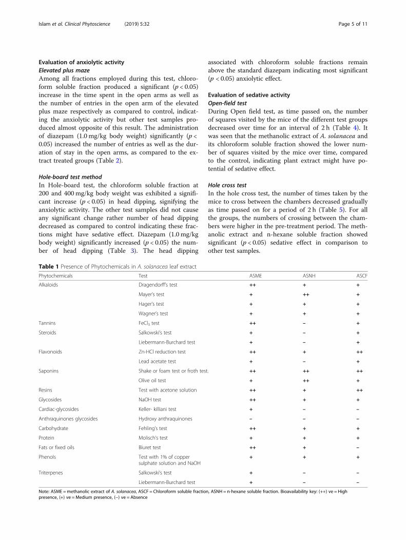
Phytochemical studyThe preliminary phytochemical studies carried out ondifferent extracts of A. solanacea leaf revealed the pres-ence of different phyto-constituents which are presentedin Table 1.
Islam et al. Clinical Phytoscience (2019) 5:32 Page 4 of 11
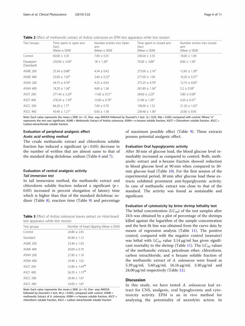
Evaluation of anxiolytic activityElevated plus mazeAmong all fractions employed during this test, chloro-form soluble fraction produced a significant (p < 0.05)increase in the time spent in the open arms as well asthe number of entries in the open arm of the elevatedplus maze respectively as compared to control, indicat-ing the anxiolytic activity but other test samples pro-duced almost opposite of this result. The administrationof diazepam (1.0 mg/kg body weight) significantly (p <0.05) increased the number of entries as well as the dur-ation of stay in the open arms, as compared to the ex-tract treated groups (Table 2).
Hole-board test methodIn Hole-board test, the chloroform soluble fraction at200 and 400 mg/kg body weight was exhibited a signifi-cant increase (p < 0.05) in head dipping, signifying theanxiolytic activity. The other test samples did not causeany significant change rather number of head dippingdecreased as compared to control indicating these frac-tions might have sedative effect. Diazepam (1.0 mg/kgbody weight) significantly increased (p < 0.05) the num-ber of head dipping (Table 3). The head dipping
associated with chloroform soluble fractions remainabove the standard diazepam indicating most significant(p < 0.05) anxiolytic effect.
Evaluation of sedative activityOpen-field testDuring Open field test, as time passed on, the numberof squares visited by the mice of the different test groupsdecreased over time for an interval of 2 h (Table 4). Itwas seen that the methanolic extract of A. solanacea andits chloroform soluble fraction showed the lower num-ber of squares visited by the mice over time, comparedto the control, indicating plant extract might have po-tential of sedative effect.
Hole cross testIn the hole cross test, the number of times taken by themice to cross between the chambers decreased graduallyas time passed on for a period of 2 h (Table 5). For allthe groups, the numbers of crossing between the cham-bers were higher in the pre-treatment period. The meth-anolic extract and n-hexane soluble fraction showedsignificant (p < 0.05) sedative effect in comparison toother test samples.
Table 1 Presence of Phytochemicals in A. solanacea leaf extract
Phytochemicals Test ASME ASNH ASCF
Alkaloids Dragendorff’s test ++ + +
Mayer’s test + ++ +
Hager’s test + + +
Wagner’s test + + +
Tannins FeCl3 test ++ – +
Steroids Salkowski’s test + – +
Liebermann-Burchard test + – +
Flavonoids Zn-HCl reduction test ++ + ++
Lead acetate test + – +
Saponins Shake or foam test or froth test. ++ ++ ++
Olive oil test + ++ +
Resins Test with acetone solution ++ + ++
Glycosides NaOH test ++ + +
Cardiac-glycosides Keller- killiani test + – –
Anthraquinones glycosides Hydroxy anthraquinones – – –
Carbohydrate Fehling’s test ++ + +
Protein Molisch’s test + + +
Fats or fixed oils Biuret test ++ + –
Phenols Test with 1% of coppersulphate solution and NaOH
+ + +
Triterpenes Salkowski’s test + – –
Liebermann-Burchard test + – –
Note: ASME =methanolic extract of A. solanacea, ASCF = Chloroform soluble fraction, ASNH = n-hexane soluble fraction. Bioavailability key: (++) ve = Highpresence, (+) ve = Medium presence, (−) ve = Absence
Islam et al. Clinical Phytoscience (2019) 5:32 Page 5 of 11
Evaluation of peripheral analgesic effectAcetic acid writhing methodThe crude methanolic extract and chloroform solublefraction has induced a significant (p < 0.05) decrease inthe number of writhes that are almost same to that ofthe standard drug diclofenac sodium (Table 6 and 7).
Evaluation of central analgesic activityTail immersion testIn tail immersion method, the methanolic extract andchloroform soluble fraction induced a significant (p <0.05) increased in percent elongation of latency timewhich is higher than that of the standard diclofenac so-dium (Table 8), reaction time (Table 9) and percentage
of maximum possible effect (Table 9). These extractspossess potential analgesic effect.
Evaluation Oral hypoglycemic activityAfter 30 min of glucose load, the blood glucose level re-markably increased as compared to control. Both, meth-anolic extract and n-hexane fraction showed reductionin blood glucose level at 90 min when compared to 30-min glucose load (Table 10). For the first session of theexperimental period, 30 min after glucose load these ex-tracts exhibited prominent anti-hyperglycemic activity.In case of methanolic extract was close to that of thestandard. The activity was found as sustainable andsignificant.
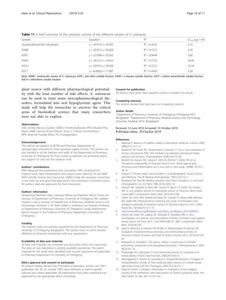
Evaluation of cytotoxicity by brine shrimp lethality testThe lethal concentration (LC50) of the test samples after24 h was obtained by a plot of percentage of the shrimpskilled against the logarithm of the sample concentrationand the best-fit line was obtained from the curve data bymeans of regression analysis (Table 11). The positivecontrol, compared with the negative control (seawater)was lethal with LC50 value 3.14 μg/ml has given signifi-cant mortality to the shrimp (Table 11). The LC50 valuesof the methanolic extract, petroleum ether, chloroform,carbon tetrachloride, and n hexane soluble fraction ofthe methanolic extract of A. solanacea were found as5.39 μg/ml, 5.60 μg/ml, 16.16 μg/ml, 3.30 μg/ml and24.00 μg/ml respectively (Table 11).
DiscussionIn this study, we have tested A. solanacea leaf ex-tract for CNS, analgesic, oral hypoglycemic and cyto-toxicity activity. EPM is an in vivo method foranalyzing the potentiality of anxiolytic action. In
Table 2 Effect of methanolic extract of Ardisia solanacea on EPM test apparatus while test session
Test Groups Time spent in open arm(Sec)(Mean ± SEM)
Number entries into Openarm(Mean ± SEM)
Time spent in closed arm(Sec)(Mean ± SEM)
Number entries into closedarm(Mean ± SEM)
Control 60.00 ± 3.35 7.00 ± 0.55 240.00 ± 3.35 18.00 ± 1.09
Diazepam(Standard)
230.00 ± 5.04a 18 ± 1.30a 70.00 ± 3.89a 8.00 ± 1.30a
ASME 200 25.34 ± 0.88a 4.34 ± 0.42 273.00 ± 2.16a 12.83 ± 1.30a
ASME 400 23.00 ± 1.05a 3.40 ± 0.25a 277.00 ± 1.04 16.20 ± 0.37a
ASNH 200 24.75 ± 4.76a 4.25 ± 0.63 275.25 ± 4.76a 12.75 ± 0.85a
ASNH 400 18.20 ± 1.36a 4.60 ± 1.36 281.80 ± 1.36a 5.2 ± 0.58a
ASCF 200 271.40 ± 2.29a 11.60 ± 0.51a 28.60 ± 2.29a 5.80 ± 0.58a
ASCF 400 278.20 ± 1.39a 15.00 ± 0.70a 21.80 ± 1.39a 4.20 ± 0.37a
ASCC 200 66.20 ± 1.77 7.00 ± 0.70 190.00 ± 1.52 21.20 ± 1.02a
ASCC 400 43.40 ± 1.21a 6.00 ± 1.58 236.40 ± 1.36a 23.60 ± 0.93
Note: Each value represents the mean ± SEM. (n = 5). One- way ANOVA followed by Dunnett’s t test. *p < 0.05, ##p < 0.005 compared with control. Where “a”represents the test was significant. ASME =Methanolic Extract of Ardisia solanacea, ASNH = n-hexane soluble fraction, ASCF = Chloroform soluble fraction, ASCC =Carbon-tetrachloride soluble fraction
Table 3 Effect of Ardisia solanacea leaves extract on Hole-boardtest apparatus while test session
Test groups Number of head dipping (Mean ± SEM)
Control 24.80 ± 2.05
Standard 45.80 ± 1.72
ASME 200 23.40 ± 1.03
ASME 400 20.00 ± 0.70
ASNH 200 27.80 ± 1.16
ASNH 400 24.40 ± 1.02
ASCF 200 52.80 ± 1.43##
ASCF 400 56.20 ± 1.15##
ASCC 200 26.40 ± 1.07
ASCC 400 24.00 ± 1.87
Note: Each value represents the mean ± SEM. (n = 5). One- way ANOVAfollowed by Dunnett’s t test. ## p < 0.005, compared with control. ASME =methanolic Extract of A. solanacea, ASNH = n-hexane soluble fraction, ASCF =chloroform soluble fraction, ASCC = carbon tetrachloride soluble fraction
Islam et al. Clinical Phytoscience (2019) 5:32 Page 6 of 11

which, experimental animals usually prefer coveredareas of the maze and might have a desire for seg-ment enclosed by protecting wall assumed to beaversive [39]. If animals which are treated with ex-perimental plant extracts exhibited any significantchanges in open arms statistically considered as theevidence of anxiolytic effectiveness of these givenplant extracts. Among other fractions, chloroformsoluble fraction at both doses level of 200 mg/kg and400 mg/kg which was indicated with increased timespent and entries in open arms and decreased timespent and entries in closed arms. The anxiolytic ef-fect of this fraction is as like as diazepam moleculewhich is considered as popular drug of benzodiazep-ine. In central system, GABA is known as a majorneurochemical. They could act by potentiating theGABAergic inhibition in the CNS system throughhyperpolarization, which can result in a firing rate of
vital neurons within the brain, or it may also be actby directly activating GABA receptors [40]. Severalstudies have implied that plants containing tannins,saponins and flavonoids may possess an effect onseveral multiple CNS disorders [41]. Preliminaryphytochemical investigations and plant extract sug-gest that several neuroactive steroids and flavonoidsare ligands for GABA receptors in the CNS; whichsuggest that they can act like benzodiazepine-likemolecule [40]. Another way to view anxiolytic activ-ity is hole board test. Head-dipping behavior of ex-perimental animals is the main basis of hole boardapparatus. If the test animals are sensitive to changesin the emotional state and or in anxiolytic state,head-dipping might have increased [42, 43]. In-creased head-dipping was observed with chloroformsoluble fraction (at both doses level of 200 mg/kgand 400 mg/kg) in respect to other fractions. Another
Table 4 Effect of methanolic extract of A. solanacea on Open-field test apparatus while test session
Testgroups
Number of squares visited (Mean ± SEM)
0 min 30 min 60min 90 min 120min
Control 85.2 ± 0.58 75.0 ± 0.70 71.2 ± 1.16 68 ± 0.701 62 ± 0.70
Standard 77.0 ± 0.70## 39.6 ± 0.51## 23.8 ± 0.86## 16 ± 0.70## 10 ± 0.701##
ASME 200 84.6 ± 0.51 51.0 ± 0.70## 32.2 ± 1.07## 26 ± 0.70## 22 ± 0.701##
ASME 400 83.2 ± 0.86 45.6 ± 1.03## 29.0 ± 0.70## 21.2 ± 0.80## 18.8 ± 0.66##
ASNH 200 87.6 ± 1.03 59.2 ± 1.07*** 39.6 ± 1.28## 32 ± 1.23## 27.6 ± 1.07##
ASNH 400 84.6 ± 0.68 54.8 ± 0.86## 34.8 ± 0.74## 26.8 ± 0.86## 25.8 ± 0.86##
ASCF 200 80.6 ± 0.50* 45.4 ± 0.93## 30.6 ± 0.93## 25 ± 0.70## 22.6 ± 0.51##
ASCF 400 83 ± 1.00 44 ± 1.14## 26.6 ± 0.51## 21 ± 0.70## 19.4 ± 0.50##
ASCC 200 83.8 ± 1.07 50 ± 0.70## 38.2 ± 1.16## 32.4 ± 0.75## 28.4 ± 0.75##
ASCC 400 81.8 ± 1.90 44 ± 1.73## 34.2 ± 0.58## 28 ± 1## 26 ± 0.84##
Note: Each value represents the mean ± SEM. (n = 5). One- way ANOVA followed by Dunnett’s t test. ##p < 0.005, *p < 0.05 compared with control. ASME =methanolic Extract of A. solanacea, ASNH = n-hexane soluble fraction, ASCF = chloroform soluble fraction, ASCC = carbon tetrachloride soluble fraction
Table 5 Effect of methanolic extract of Ardisia solanacea on Hole-crossed test apparatus while test session
Testgroups
Number of hole crossed (Mean ± SEM)
0 min 30 min 60 min 90 min 120min
Control 20 ± 0.70 19.2 ± 1.74 18.4 ± 1.54 18.2 ± 1.39 15.2 ± 0.58
Standard 19 ± 0.71 8.4 ± 1.37## 5.8 ± 0.74## 4.4 ± 0.93## 3.2 ± 0.58##
ASME 200 20.8 ± 0.58 10.4 ± 0.68## 7.8 ± 0.37## 5.2 ± 0.58## 3.4 ± 0.68##
ASME 400 21.8 ± 0.86 9.8 ± 0.74## 7.6 ± 0.40## 4.6 ± 0.40## 3.2 ± 0.49##
ASNH 200 21.2 ± 1.02 13.4 ± 0.51## 9.80 ± 0.74## 7.2 ± 0.37## 4.6 ± 0.51##
ASNH 400 23.4 ± 0.93 11.4 ± 0.51## 7.6 ± 0.51## 5.8 ± 0.37## 4.2 ± 0.58##
ASCF 200 20.6 ± 0.93 10.2 ± 0.80## 7.2 ± 0.37## 5 ± 0.55## 3.2 ± 0.66##
ASCF 400 22.6 ± 1.75 10.2 ± 1.16## 7.6 ± 1.55## 4.8 ± 1.12## 3.2 ± 1.02##
ASCC 200 24.2 ± 1.77 21.8 ± 1.59 21.4 ± 1.47 22.2 ± 1.36 20.6 ± 1.83*
ASCC 400 22.8 ± 1.96 20.6 ± 2.12 18.4 ± 1.64 17.2 ± 1.63 16.4 ± 1.20
Note: Each value represents the mean ± SEM. (n = 5). One- way ANOVA followed by Dunnett’s t test. ##p < 0.005, *p < 0.05 compared with control. ASME =Methanolic Extract of A. solanacea, ASNH = n-hexane soluble fraction, ASCF = Chloroform soluble fraction, ASCC = Carbon-tetrachloride soluble fraction
Islam et al. Clinical Phytoscience (2019) 5:32 Page 7 of 11

vital step in evaluating the action of drug on CNS is to in-spect its effect on locomotor activity of testing animal.Locomotor activity is a motive to act on CNS by upliftingalertness and decreasing the motion activity could inducea sedative action [44]. Level of CNS excitement can beevaluated from locomotor activity and reduction of thisactivity is closely related to sedation turn out from theCNS depression [45]. Here, hole cross and open filed testswere used to observe this activity. In respect to other frac-tions, chloroform soluble fraction and methanolic extractof A. solanacea leaves decrease the number of holecrossed and the number of square blocks from 1st (30min) observation to final observation (120min) and after-ward influenced the locomotor activity in mices which ex-hibits the sedative activity. Acetic acid induce test is well-used for assessing the peripheral analgesic activity [46].Acetic acid induces pain by enhancing the PG2 and PG2αat the receptor sites of the organ cavity which means thatcarboxylic acid acts indirectly by increasing the dischargeof endogenous mediators [47, 48]. Acetic acid produceswrithing reflex in experimental animal by chemo-sensitivenociceptor [49]. Non-steroidal anti-inflammatory drugs
act by blocking this stimulation of sensory neurons inresponse to inflammatory mediators [50]. Moreover,the level of analgesia can also be noted by the per-cent reduction in the number of abdominal squirmsor cramps [51]. The standard drug, methanolic extractand its chloroform soluble fraction (at both doseslevel of 200 mg/kg and 400 mg/kg) significantly de-creased the mean number of writhes. Subsequently,increase in the percent inhibition of abdominalsquirms. The effect was closely related to that ofstandard diclofenac sodium. Tail immersion test isbased on observation that morphine-like drugs select-ively enhances reaction time of typical tail withdrawalreflex in mices. The level of analgesia of extract is in-dicated by the increased in basal latency period [52,53]. Both the methanolic extract and its chloroformsoluble fraction (at 200 mg/kg and 400 mg/kg) in-crease the reaction time, basal latency and % MPEthan other fraction. The effect was found nearlystable from 30 min to 120 min. The basal latency wasincreased by the extracts indicates that they may actvia centrally mediated analgesic mechanism [53]. Inthis model, sensory nerves sensitize the nociceptorsand the involvement of endogenous substances such
Table 6 Evaluation of analgesic activity of Ardisia solanacea bycounting the number of writhing after the intraperitonealadministration of 0.7% acetic acid
Animal Group Mean % of Writhing % of inhibition
Control 56.6 – –
Standard 14.8 26.15 73.85
ASME 200 21.6 38.16 61.84
ASME 400 19.2 33.93 66.07
ASNH 200 42.4 74.92 25.08
ASNH 400 38.4 67.85 32.15
ASCF 200 23.0 40.64 59.36
ASCF 400 19.0 33.57 66.43
Note: ASME =Methanolic extract of Ardisia solanacea, ASNH = n-hexane solublefraction, ASCF = chloroform soluble fraction. ASME =methanolic extract ofArdisia A. solanacea, ASCF = Chloroform soluble fraction, ASNH = n-hexanesoluble fraction
Table 7 Effect of Ardisia solanacea leaves extract during acetic acid writhing test session
Animal Group Number of writhing (Mean ± SEM) % of inhibition of writhing
Control 56.6 ± 2.67
Standard 14.8 ± 1.16## 73.85%
ASME 200 21.6 ± 3.53## 61.84%
ASME 400 19.2 ± 1.83## 66.07%
ASNH 200 42.4 ± 3.50# 25.08%
ASNH 400 38.4 ± 1.03## 32.15%
ASCF 200 23.0 ± 1.48## 59.36%
ASCF 400 19.0 ± 1.42## 66.43%
Note: Each value represents the mean ± SEM. (n = 5). One- way ANOVA followed by Dunnett’s t test. ##p < 0.005, # p < 0.01 compared with control. ASME =methanolic extract of A. solanacea, ASCF = Chloroform soluble fraction, ASNH = n-hexane soluble fraction
Table 8 Percent elongation of latency time after administrationof all test samples
Testsamples
Dose(mg/kg)
% elongation of latency time
30 min 60 min 90 min 120min
Standard 50 53.57% 72.97% 70.45% 65.12%
ASME 200 200 38.09% 67.74% 65.79% 61.54%
ASME 400 400 50.00% 72.23% 69.77% 63.42%
ASNH 200 200 27.78% 54.55% 55.17% 55.88%
ASNH 400 400 22.23% 62.96% 59.37% 59.46%
ASCF 200 200 50.00% 72.23% 68.29% 65.90%
ASCF 400 400 50.00% 76.19% 72.34% 67.39%
ASME =methanolic extract of A. solanacea, ASCF = Chloroform soluble fraction,ASNH = n-hexane soluble fraction
Islam et al. Clinical Phytoscience (2019) 5:32 Page 8 of 11

as prostaglandins are minimized [54]. The methanolicextract of A. solanacea revealed the presence of phy-tochemicals such as alkaloids, tannins and flavonoid,which may be major contributors to this activity [55].The hypoglycemic property of plant sample was con-firmed by enhanced glucose tolerance in diabetic ornormoglycaemic treated mices indicating that anyplant samples have the capacity to correct impairedglucose tolerance in diabetes, hence, exhibit antidia-betic activity [56]. In this experiment, methanolic ex-tract and n-hexane soluble fraction (at the dose levelof 200 mg/kg) exhibited significant hypoglycemia byreducing orally administered glucose level. A similarreport has confirmed from the event of Syzygium aro-maticum extract in streptozotocin-induced diabeticmices following an oral glucose tolerance test [57].Differing degree of lethality to Artemia salina was
observed with exposure to different dose levels of thetest samples. Among the test samples, reasonablecytotoxicity was observed with methanolic extract andits pet-ether and carbon tetrachloride fractions atleast LC50 values. Flavonoids show anti-allergic, anti-cancer, anti-inflammatory and antimicrobial activities.Tannins, which are present in this plant may containsignificant cytotoxic and antitumor potency [57]. Inthe future, we will find out which components areexactly responsible for those activities by performingcompound identification technique such as chromato-graphic analysis.
ConclusionThese studies discover some pharmacological poten-tial that can be beneficial for modern medical scienceto develop medical science to discover drugs from
Table 9 Effects of methanolic extracts of different plant parts of A. solanacea on Tail Immersion Test in mice
Testsamples
Dose (mg/kg) Reaction times in seconds (Mean ± SEM) and % MPE
Pre-treatment 30 min 60 min 90 min 120min
Control 0.25 ml/ 25 g b.w 2.8 ± 0.37 2.6 ± 0.24(1.64%)
2 ± 0.32(6.56%)
2.6 ± 0.24(1.64%)
3 ± 0.32(1.63%)
Standard 50 2.6 ± 0.40 5.6 ± 0.25##
(24.41%)7.4 ± 0.24##
(38.70%)8.8 ± 0.37***
(50.00%)8.6 ± 0.40##
(48.38%)
ASME 200 200 2.4 ± 0.25 4.2 ± 0.37*
(14.28%)6.2 ± 0.37##
(30.16%)7.6 ± 0.40***
(41.27%)7.8 ± 0.37##
(42.85%)
ASME 400 400 2.6 ± 0.40 5.2 ± 0.37##
(20.97%)7.2 ± 0.37##
(37.09%)8.6 ± 0.24***
(48.38%)8.2 ± 0.20##
(45.16%)
ASNH 200 200 2.8 ± 0.37 3.6 ± 0.25(6.55%)
4.4 ± 0.24##
(13.12%)5.8 ± 0.37**
(24.59%)6.8 ± 0.37##
(32.78%)
ASNH 400 400 2.2 ± 0.20 3.4 ± 0.24(9.37%)
5.4 ± 0.25##
(25.00%)6.4 ± 0.24***
(32.81%)7.4 ± 0.25##
(40.63%)
ASCF 200 200 2 ± 0.00 5.2 ± 0.20##
(24.62%7.2 ± 0.20##
(40.00%)8.2 ± 0.20***
(47.69%)8.8 ± 0.37##
(52.30%)
ASCF 400 400 2.2 ± 0.20 5.2 ± 0.20***
(23.44%)8.4 ± 0.25***
(48.44%)9.4 ± 0.24***
(56.25%)9.2 ± 0.20***
(54.68%)
Note: Each value represents the mean ± SEM. (n = 5). One- way ANOVA followed by Dunnett’s t test. ##p < 0.005, *p < 0.05 compared with control. ASME =methanolic extract of A. solanacea, ASCF = Chloroform soluble fraction, ASNH = n-hexane soluble fraction
Table 10 Oral Hypoglycaemic activity of methanolic extract of A. solanacea and its n-hexane soluble fraction
Animal group DoseMg/kg
Mean ± SEM
0Minute 30 Minute 90 Minute 120 Minute
Control(Normal Saline)
6.64 ± 0.44 14.08 ± 0.12(+ 112.05)a
8.98 ± 0.89(− 36.22)b
6.76 ± 0.33(− 52)b
Standard (Glibenclamide) 10 5.82 ± 0.32 10.06 ± 0.73*
(+ 72.85)a4.6 ± 0.20##
(− 54.27)b4.18 ± 0.24##
(− 58.45)b
ASME 200 6.42 ± 0.31 10.16 ± 0.37*
(+ 58.25)a4.68 ± 0.14##
(− 53.94)b4.4 ± 0.10##
(− 56.69)b
ASNH 200 6.96 ± 0.24 10.80 ± 0.33(+ 55.17)a
5.6 ± 0.36#
(− 48.15)b4.74 ± 0.24##
(− 58.62)b
Figures in parentheses are the % increase (+) or decrease (−) glucose level; a = compared to the glucose level at zero time, b = compared to the glucose level at30 min after the glucose load, ASME =Methanolic extract of A. solanacea, ASNH = n-hexane soluble fraction. Each value represents the mean ± SEM. (n = 5). One-way ANOVA followed by Dunnett’s t test. ##p < 0.005, #p < 0.01, *p < 0.05 compared with control
Islam et al. Clinical Phytoscience (2019) 5:32 Page 9 of 11
plant source with different pharmacological potential-ity with the least number of side effects. A. solanaceacan be used to treat some neuropharamcological dis-orders, formulated into oral hypoglycemic agent. Thisstudy will help the researcher to uncover the criticalareas of biomedical science that many researcherswere not able to explore.
AbbreviationsCNS: Central Nervous System; DMSO: Dimethylsulfoxide; EPM: Elevated PlusMaze; GABA: Gamma Amino Butyric Acid; LC: Lethal Concentrations;MPE: Maximal Possible Effect; PG: Prostaglandins
AcknowledgementsThe authors are grateful to BCSIR and Pharmacy Department ofJahangirnagar University for their generous supply of mice. The authors arealso thankful to all the teachers and staffs of the Department of Pharmacy,University of Chittagong for their cordial co-operation by providing labora-tory support to carry out the research work.
Authors’ contributionsMKH designed the experiments and conception. MRI conducted theresearch work. Data interpretation and analysis were aided by JN and NMP.MSH critically reviews the manuscript. SMNU made the necessary correctionsin the write up and gave final approval for the submission of revised version.All authors read and approved the final manuscript.
Authors’ informationMohammad Rashedul Islam, Jannatul Naima and Nawreen Monir Proma areLecturer of Department of Pharmacy, University of Chittagong. Md. SaddamHussain is also a Lecturer of Department of Pharmacy, Noakhali Science andTechnology University. S. M. Naim Uddin is working as an Assistant Professorat Department of Pharmacy, University of Chittagong. Finally MohammedKamrul Hossain is the Professor of Pharmacy Department, University ofChittagong.
FundingThe research work was partially supported by the Department of Pharmacy,University of Chittagong, Bangladesh. The authors have no other relevantaffiliations or financial involvement with any organization.
Availability of data and materialsAll data and materials are contained and described within the manuscript.The data set was deposited in publicly available repositories. The plant’smaterials for the study were identified and voucher specimens are depositedat Pharmacy Department of University of Chittagong.
Ethics approval and consent to participateAll authors hereby declare that “Principles of laboratory animal care” (NIHpublication No. 85–23, revised 1985) were followed, as well as specificnational laws where applicable. All experiments have been examined andapproved by the appropriate ethics committee.
Consent for publicationAll authors have given their valuable consent to publish this article.
Competing interestsThe authors declare that they have no competing interests.
Author details1Department of Pharmacy, University of Chittagong, Chittagong 4331,Bangladesh. 2Department of Pharmacy, Noakhali Science and TechnologyUniversity, Noakhali 3814, Bangladesh.
Received: 14 June 2019 Accepted: 10 October 2019
References1. Marshall E. Bastions of tradition adapt to alternative medicine. Science. 2000;
288(5471):1571–2.2. Budzinski JW, Foster BC, Vandenhoek S, Arnason JT. An in vitro evaluation of
human cytochrome P450 3A4 inhibition by selected commercial herbalextracts and tinctures. Phytomedicine. 2000;7(4):273–82.
3. Ibrahim M, Hussain MS, Sultana F, Abhi N, Ahmed T, Uddin SN, et al.Therapeutic potentiality of Diospyros blancoi Linn. Seeds against pain,Thrombus and inflammation: an in vivo and in vitro study. AJMBR. 2019;12:36–41.
4. Claquin P. Private health care providers in rural Bangladesh. Social Scienceand Medicine. Part B: Medical Anthropology. 1981;15(2):153–7.
5. Bhardwaj SM, Paul BK. Medical pluralism and infant mortality in a rural areaof Bangladesh. Soc Sci Med. 1986;23(10):1003–10.
6. Hossain MF, Talukder B, Rana MN, Tasnim R, Nipun TS, Uddin SN, HossenSM. In vivo sedative activity of methanolic extract of Stericulia villosa Roxb.Leaves BMC Complement Altern Med. 2016;16(1):398.
7. Uddin SN, Amin MN, Shahid-Ud-Daula AF, Hossain H, Haque MM, RahmanMS, Kader MA. Phytochemical screening and study of antioxidant andanalgesic potentials of ethanolic extract of Stephania japonica Linn. J MedPlants Res. 2014;8(37):1127–33.
8. http://www.efloras.org/florataxon.aspx?flora_id=2&taxon_id=2100000729. Nordin ML, Kadir AA, Zakaria ZA, Abdullah R, Abdullah MN. In vitro
investigation of cytotoxic and antioxidative activities of Ardisia crispa againstbreast cancer cell lines, MCF-7 and MDA-MB-231. BMC Complement AlternMed. 2018;18(1):87.
10. Syed E, Mashroor B, Hossain FM, Bi-Illah N, Bhattacharjee R, Hannan JM.Evaluation of phytochemical screening and antimicrobial activities ofEthanolic extracts of leaves and bark of Ardisia colorata. IJPLS. 2013;2(4):158–64.
11. Kobayashi H, de Mejía E. The genus Ardisia: a novel source of health-promoting compounds and phytopharmaceuticals. J Ethnopharmacol. 2005;96(3):347–54.
12. Phadungkit M, Luanratana O. Anti-Salmonella activity of constituents ofArdisia elliptica Thunb. Nat Prod Res. 2006;20(7):693–6.
13. Moongkarndi P, Kosem N, Luanratana O, Jongsomboonkusol S, Pongpan N.Antiproliferative activity of Thai medicinal plant extracts on human breastadenocarcinoma cell line. Fitoterapia. 2004;75(3–4):375–7.
14. Ezeja M, Omeh Y, Ezeigbo I, Ekechukwu A. Evaluation of the analgesicactivity of the methanolic stem bark extract of Dialium guineense (wild). AnnMed Health Sci Res. 2011;1(1):55–62.
Table 11 A brief overview of the cytotoxic activity of the different extracts of A. solanacea
Sample Equation R2 LC50 (μg / ml)
Standard(Ampicillin trihydrate) y = 29.975x + 35.092 R2 = 0.3532 3.14
ASME y = 20.971x + 34.658 R2 = 0.7212 5.39
ASPE y = 23.998x + 32.032 R2 = 0.9444 5.60
ASNH y = 28.521x + 10.633 R2 = 0.7723 24.00
ASCH y = 20.971x + 24.658 R2 = 0.7212 16.16
ASCT y = 8.4982x + 71.087 R2 = 0.4343 3.30
Note: ASME =methanolic extract of A. solanacea, ASPE = pet ether soluble fraction, ASNH = n-hexane soluble fraction, ASCT = carbon tetrachloride soluble fraction,ASCH = chloroform soluble fraction
Islam et al. Clinical Phytoscience (2019) 5:32 Page 10 of 11
15. Bolanle AO, Funmilola AS, Adedayo A. Proximate analysis, mineral contents,amino acid composition, anti-nutrients and phytochemical screening ofBrachystegia eurycoma harms and Pipper Guineense Schum and Thonn. AmJ Food Nutr. 2014;2:11–7.
16. Ugbaja CC, Fawibe OO, Oyelakin AS, Fadimu IO, Ajiboye AA, Agboola DA.Comparative phytochemical and nutritional composition of Trichosanthescucumerina (L.) and some Solanum lycopersicum (L.) cultivars in nigeria. AmJ Plant Sci. 2017;8(02):297.
17. Harborne JB. Phytochemical methods chapman and hall. Ltd London. 1973;4:49–188.
18. Sunanda BP, Latha K, Rammohan B, Uma Maheswari MS, Surapaneni KM.Evaluation of the neuroprotective effects of curcumin (turmeric) againstscopolamine induced cognitive impairment in mice. Int J PharmPhytochem Res. 2014;6:133–6.
19. Pellow S, Chopin P, File SE, Briley M. Validation of open: closed arm entriesin an elevated plus-maze as a measure of anxiety in the rat. J NeurosciMethods. 1985;14(3):149–67.
20. Vogel HG, editor. Drug discovery and evaluation: pharmacological assays.Springer Science & Business Media. 2002.
21. Emamghoreishi M, Khasaki M, Aazam MF. Coriandrum sativum: evaluation ofits anxiolytic effect in the elevated plus-maze. J Ethnopharmacol. 2005;96(3):365–70.
22. Miyakawa T, Yagi T, Kagiyama A, Niki H. Radial maze performance, open-field and elevated plus-maze behaviors in Fyn-kinase deficient mice: furtherevidence for increased fearfulness. Mol Brain Res. 1996;37(1–2):145–50.
23. File SE, Wardill AG. Validity of head-dipping as a measure of exploration in amodified hole-board. Psychopharmacol. 1975;44(1):53–9.
24. Ljungberg T, Ungerstedt U. Automatic registration of behaviour related todopamine and noradrenaline transmission. Eur J Pharmacol. 1976;36(1):181–8.
25. Crawley JN. Exploratory behavior models of anxiety in mice. NeurosciBiobehav Rev. 1985;9(1):37–44.
26. Kliethermes CL, Crabbe JC. Pharmacological and genetic influences on hole-board behaviors in mice. Pharmacol Biochem Be. 2006;85(1):57–65.
27. Gupta BD, Dandiya PC, Gupta ML. A psycho-pharmacological analysis ofbehaviour in rats. Jpn J Pharmacol. 1971;21(3):293–8.
28. Takagi K, Watanabe M, Saito H. Studies of the spontaneous movement ofanimals by the hole cross test; effect of 2-dimethyl-aminoethanol and itsacyl esters on the central nervous system. Jpn J Pharmacol. 1971;21(6):797–810.
29. Koster R. Acetic acid for analgesic screening. InFed proc. 1959;18:412.30. Vogel GH, Vogel WH. Drug discovery and evaluation: pharmacological
assays springer-Verlag. Berlin: Germany; 1997.31. Devi PU, Ganasoundari A, Rao BS, Srinivasan KK. In vivo radioprotection by
ocimum flavonoids: survival of mice. Radiat Res. 1999;151(1):74–8.32. Di Stasi LC, Costa M, Mendaçolli SL, Kirizawa M, Gomes C, Trolin G.
Screening in mice of some medicinal plants used for analgesic purposes inthe state of Sao Paulo. J Ethnopharmacol. 1988;24(2–3):205–11.
33. Fan SH, Ali NA, Basri DF. Evaluation of analgesic activity of the methanolextract from the galls of Quercus infectoria (Olivier) in rats. Evid BasedComplement Alternat Med. 2014;2014.
34. Kumawat RK, Kumar S, Sharma S. Evaluation of analgesic activity of variousextracts of Sida tiagii Bhandari. Acta Pol Pharm. 2012;69:1103–9.
35. Joy KL, Kuttan R. Anti-diabetic activity of Picrorrhiza kurroa extract. JEthnopharmacol. 1999;67(2):143–8.
36. Meyer BN, Ferrigni NR, Putnam JE, Jacobsen LB, Nichols DJ, McLaughlin JL.Brine shrimp: a convenient general bioassay for active plant constituents.Planta Med. 1982;45(5):31–4.
37. Mclaughlin JL, Rogers LL, Anderson JE. The use of biological assays toevaluate botanicals. Drug Inf J. 1998;32(2):513–24.
38. Amin MN, Majumder MS, Moghal MM, Banik S, Kar A, Hossain MM.Anthelmintic and cytotoxic activities of two medicinal plants: Polygonumviscosum and Aphanamixis polystachya growing in Bangladesh. J Sci Res.2014;6(2):339–45.
39. Collimore KC, Rector NA. Treatment of anxiety disorders with comorbiddepression: a survey of expert CBT clinicians. Cogn Behav Pract. 2014;21(4):485–93.
40. Verma A, Jana GK, Sen S, Chakraborty R, Sachan S, Mishra A.Pharmacological evaluation of Saraca indica leaves for central nervoussystem depressant activity in mice. J Pharm Sci Res. 2010;2(6):338–43.
41. Yadav G, Garg VK, Thakur N, Khare P. Locomotor activity of methanolicextract of Saraca indica bark. Adv Biol Res. 2013;7:1–3.
42. Wei XY, Yang JY, Wang JH, Wu CF. Anxiolytic effect of saponins from Panaxquinquefolium in mice. J Ethnopharmacol. 2007;111(3):613–8.
43. Foyet HS, Tsala DE, Bouba AA, Hritcu L. Anxiolytic and antidepressant likeeffects of the aqueous extract of Alafia multiflora stem barks in rodents. AdvPharmacol Sci. 2012;2012:912041.
44. Bhattacharya SK, Satyan KS. Experimental methods for evaluation ofpsychotropic agents in rodents: I--anti-anxiety agents. Indian J Exp Biol.1997;35(6):565–75.
45. Islam NU, Khan I, Rauf A, Muhammad N, Shahid M, Shah MR.Antinociceptive, muscle relaxant and sedative activities of goldnanoparticles generated by methanolic extract of Euphorbia milii. BMCComplement Altern Med. 2015;15:160.
46. Trongsakul S, Panthong A, Kanjanapothi D, Taesotikul T. The analgesic,antipyretic and anti-inflammatory activity of Diospyros variegata Kruz. JEthnopharmacol. 2003;85(2–3):221–5.
47. Ezeja MI, Omeh YS, Ezeigbo II, Ekechukwu A. Evaluation of the analgesicactivity of the methanolic stem bark extract of Dialium guineense (wild). AnnMed Health Sci Res. 2011;1(1):55–62.
48. Lee KH, Choi EM. Analgesic and anti-inflammatory effects of Ligularia fischerileaves in experimental animals. J Ethnopharmacol. 2008;120(1):103–7.
49. Onasanwo SA, Elegbe RA. Antinociceptive and anti-inflammatory propertiesof the leaf extract of Hedranthera barteri in rats and mice. Afr J Biomed Res.2006;2:109–18.
50. Akindele AJ, Ibe IF, Adeyemi OO. Analgesic and antipyretic activities ofDrymaria cordata (Linn.) Willd (Caryophyllaceae) extract. Afr J TraditComplement Altern Med. 2012;9(1):25–35.
51. Machioro M, Blank MFA, Moura RHV, Antioniolli AR. Antinociceptive activityof the aqueous extract of Erythrina velutina leaves. Fitoterapia. 2005;6:637–42.
52. Elisabetsky E, Amador TA, Albuquerque RR, Nunes DS. Do CT Carvalho a.analgesic activity of Psychotria colorata (Willd. Ex R. & S.) Muell. Arg.Alkaloids. J Ethnopharmacol. 1995;48(2):77–83.
53. Pal S, Sen T, Chaudhuri AK. Neuropsychopharmacological profile of themethanolic fraction of Bryophyllum pinnatum leaf extract. J PharmPharmacol. 1999;51(3):313–8.
54. Bachhav RS, Gulecha VS, Upasani CD. Analgesic and anti-inflammatoryactivity of Argyreia speciosa root. Indian J Pharmacol. 2009;41(4):158.
55. Uche FI, Aprioku JS. The Phytochemical Constituents, Analgesic and Anti-inflammatory effects of methanol extract of Jatropha curcas leaves in Miceand Wister albino rats. JASEM. 2008;12(4).
56. Khathi A, Masola B, Musabayane CT. Effects of Syzygium aromaticum-derivedoleanolic acid on glucose transport and glycogen synthesis in the rat smallintestine. J diabetes. 2013;5(1):80–7.
57. Raju GS, Moghal MR, Dewan SM, Amin MN, Billah M. Characterization ofphytoconstituents and evaluation of total phenolic content, anthelmintic,and antimicrobial activities of Solanum violaceum Ortega. Avicenna JPhytomed. 2013;3(4):313.
Publisher’s NoteSpringer Nature remains neutral with regard to jurisdictional claims inpublished maps and institutional affiliations.
Islam et al. Clinical Phytoscience (2019) 5:32 Page 11 of 11
Copyright © 2022 FDOKUMEN


![Condensed bridgehead nitrogen heterocyclic system: Synthesis and pharmacological activities of 1,2,4-triazolo-[3,4- b]-1,3,4-thiadiazole derivatives of ibuprofen and biphenyl-4-yloxy](https://static.fdokumen.com/doc/165x107/632834412089eb31f609dd2b/condensed-bridgehead-nitrogen-heterocyclic-system-synthesis-and-pharmacological.jpg)