In vitro Cultured Primary Roots Derived from Stem Segments of Cassava (Manihot esculenta) Can Behave...
15
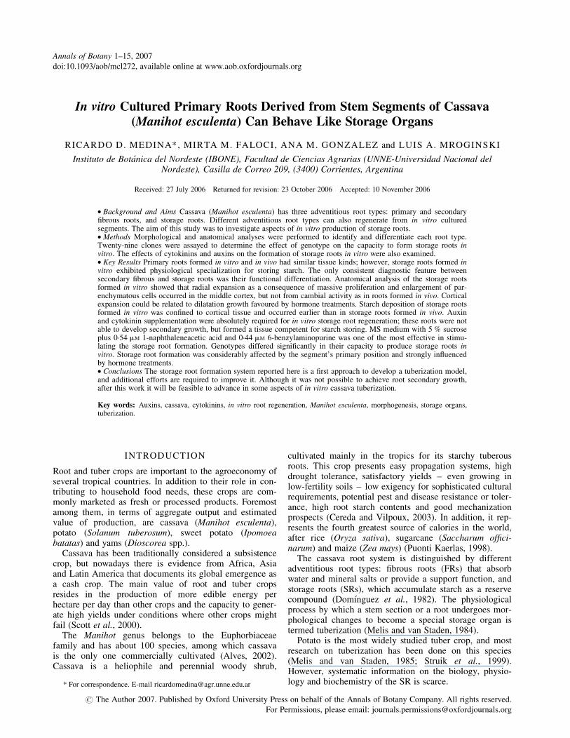
In vitro Cultured Primary Roots Derived from Stem Segments of Cassava (Manihot esculenta) Can Behave Like Storage Organs RICARDO D. MEDINA*, MIRTA M. FALOCI, ANA M. GONZALEZ and LUIS A. MROGINSKI Instituto de Bota ´nica del Nordeste (IBONE), Facultad de Ciencias Agrarias (UNNE-Universidad Nacional del Nordeste), Casilla de Correo 209, (3400) Corrientes, Argentina Received: 27 July 2006 Returned for revision: 23 October 2006 Accepted: 10 November 2006 † Background and Aims Cassava (Manihot esculenta) has three adventitious root types: primary and secondary fibrous roots, and storage roots. Different adventitious root types can also regenerate from in vitro cultured segments. The aim of this study was to investigate aspects of in vitro production of storage roots. † Methods Morphological and anatomical analyses were performed to identify and differentiate each root type. Twenty-nine clones were assayed to determine the effect of genotype on the capacity to form storage roots in vitro. The effects of cytokinins and auxins on the formation of storage roots in vitro were also examined. † Key Results Primary roots formed in vitro and in vivo had similar tissue kinds; however, storage roots formed in vitro exhibited physiological specialization for storing starch. The only consistent diagnostic feature between secondary fibrous and storage roots was their functional differentiation. Anatomical analysis of the storage roots formed in vitro showed that radial expansion as a consequence of massive proliferation and enlargement of par- enchymatous cells occurred in the middle cortex, but not from cambial activity as in roots formed in vivo. Cortical expansion could be related to dilatation growth favoured by hormone treatments. Starch deposition of storage roots formed in vitro was confined to cortical tissue and occurred earlier than in storage roots formed in vivo. Auxin and cytokinin supplementation were absolutely required for in vitro storage root regeneration; these roots were not able to develop secondary growth, but formed a tissue competent for starch storing. MS medium with 5 % sucrose plus 0 . 54 mM 1-naphthaleneacetic acid and 0 . 44 mM 6-benzylaminopurine was one of the most effective in stimu- lating the storage root formation. Genotypes differed significantly in their capacity to produce storage roots in vitro. Storage root formation was considerably affected by the segment’s primary position and strongly influenced by hormone treatments. † Conclusions The storage root formation system reported here is a first approach to develop a tuberization model, and additional efforts are required to improve it. Although it was not possible to achieve root secondary growth, after this work it will be feasible to advance in some aspects of in vitro cassava tuberization. Key words: Auxins, cassava, cytokinins, in vitro root regeneration, Manihot esculenta, morphogenesis, storage organs, tuberization. INTRODUCTION Root and tuber crops are important to the agroeconomy of several tropical countries. In addition to their role in con- tributing to household food needs, these crops are com- monly marketed as fresh or processed products. Foremost among them, in terms of aggregate output and estimated value of production, are cassava (Manihot esculenta), potato (Solanum tuberosum), sweet potato (Ipomoea batatas) and yams (Dioscorea spp.). Cassava has been traditionally considered a subsistence crop, but nowadays there is evidence from Africa, Asia and Latin America that documents its global emergence as a cash crop. The main value of root and tuber crops resides in the production of more edible energy per hectare per day than other crops and the capacity to gener- ate high yields under conditions where other crops might fail (Scott et al., 2000). The Manihot genus belongs to the Euphorbiaceae family and has about 100 species, among which cassava is the only one commercially cultivated (Alves, 2002). Cassava is a heliophile and perennial woody shrub, cultivated mainly in the tropics for its starchy tuberous roots. This crop presents easy propagation systems, high drought tolerance, satisfactory yields – even growing in low-fertility soils – low exigency for sophisticated cultural requirements, potential pest and disease resistance or toler- ance, high root starch contents and good mechanization prospects (Cereda and Vilpoux, 2003). In addition, it rep- resents the fourth greatest source of calories in the world, after rice (Oryza sativa), sugarcane (Saccharum offici- narum) and maize (Zea mays) (Puonti Kaerlas, 1998). The cassava root system is distinguished by different adventitious root types: fibrous roots (FRs) that absorb water and mineral salts or provide a support function, and storage roots (SRs), which accumulate starch as a reserve compound (Domı ´nguez et al., 1982). The physiological process by which a stem section or a root undergoes mor- phological changes to become a special storage organ is termed tuberization (Melis and van Staden, 1984). Potato is the most widely studied tuber crop, and most research on tuberization has been done on this species (Melis and van Staden, 1985; Struik et al., 1999). However, systematic information on the biology, physio- logy and biochemistry of the SR is scarce. * For correspondence. E-mail [email protected] # The Author 2007. Published by Oxford University Press on behalf of the Annals of Botany Company. All rights reserved. For Permissions, please email: [email protected] Annals of Botany 1–15, 2007 doi:10.1093/aob/mcl272, available online at www.aob.oxfordjournals.org
Transcript of In vitro Cultured Primary Roots Derived from Stem Segments of Cassava (Manihot esculenta) Can Behave...
In vitro Cultured Primary Roots Derived from Stem Segments of Cassava(Manihot esculenta) Can Behave Like Storage Organs
RICARDO D. MEDINA*, MIRTA M. FALOCI, ANA M. GONZALEZ and LUIS A. MROGINSKI
Instituto de Botanica del Nordeste (IBONE), Facultad de Ciencias Agrarias (UNNE-Universidad Nacional delNordeste), Casilla de Correo 209, (3400) Corrientes, Argentina
Received: 27 July 2006 Returned for revision: 23 October 2006 Accepted: 10 November 2006
†Background and Aims Cassava (Manihot esculenta) has three adventitious root types: primary and secondaryfibrous roots, and storage roots. Different adventitious root types can also regenerate from in vitro culturedsegments. The aim of this study was to investigate aspects of in vitro production of storage roots.†Methods Morphological and anatomical analyses were performed to identify and differentiate each root type.Twenty-nine clones were assayed to determine the effect of genotype on the capacity to form storage roots invitro. The effects of cytokinins and auxins on the formation of storage roots in vitro were also examined.†Key Results Primary roots formed in vitro and in vivo had similar tissue kinds; however, storage roots formed invitro exhibited physiological specialization for storing starch. The only consistent diagnostic feature betweensecondary fibrous and storage roots was their functional differentiation. Anatomical analysis of the storage rootsformed in vitro showed that radial expansion as a consequence of massive proliferation and enlargement of par-enchymatous cells occurred in the middle cortex, but not from cambial activity as in roots formed in vivo. Corticalexpansion could be related to dilatation growth favoured by hormone treatments. Starch deposition of storage rootsformed in vitro was confined to cortical tissue and occurred earlier than in storage roots formed in vivo. Auxinand cytokinin supplementation were absolutely required for in vitro storage root regeneration; these roots were notable to develop secondary growth, but formed a tissue competent for starch storing. MS medium with 5 % sucroseplus 0.54 mM 1-naphthaleneacetic acid and 0.44 mM 6-benzylaminopurine was one of the most effective in stimu-lating the storage root formation. Genotypes differed significantly in their capacity to produce storage roots invitro. Storage root formation was considerably affected by the segment’s primary position and strongly influencedby hormone treatments.†Conclusions The storage root formation system reported here is a first approach to develop a tuberization model,and additional efforts are required to improve it. Although it was not possible to achieve root secondary growth,after this work it will be feasible to advance in some aspects of in vitro cassava tuberization.
Key words: Auxins, cassava, cytokinins, in vitro root regeneration, Manihot esculenta, morphogenesis, storage organs,tuberization.
INTRODUCTION
Root and tuber crops are important to the agroeconomy ofseveral tropical countries. In addition to their role in con-tributing to household food needs, these crops are com-monly marketed as fresh or processed products. Foremostamong them, in terms of aggregate output and estimatedvalue of production, are cassava (Manihot esculenta),potato (Solanum tuberosum), sweet potato (Ipomoeabatatas) and yams (Dioscorea spp.).Cassava has been traditionally considered a subsistence
crop, but nowadays there is evidence from Africa, Asiaand Latin America that documents its global emergence asa cash crop. The main value of root and tuber cropsresides in the production of more edible energy perhectare per day than other crops and the capacity to gener-ate high yields under conditions where other crops mightfail (Scott et al., 2000).The Manihot genus belongs to the Euphorbiaceae
family and has about 100 species, among which cassavais the only one commercially cultivated (Alves, 2002).Cassava is a heliophile and perennial woody shrub,
cultivated mainly in the tropics for its starchy tuberousroots. This crop presents easy propagation systems, highdrought tolerance, satisfactory yields – even growing inlow-fertility soils – low exigency for sophisticated culturalrequirements, potential pest and disease resistance or toler-ance, high root starch contents and good mechanizationprospects (Cereda and Vilpoux, 2003). In addition, it rep-resents the fourth greatest source of calories in the world,after rice (Oryza sativa), sugarcane (Saccharum offici-narum) and maize (Zea mays) (Puonti Kaerlas, 1998).The cassava root system is distinguished by different
adventitious root types: fibrous roots (FRs) that absorbwater and mineral salts or provide a support function, andstorage roots (SRs), which accumulate starch as a reservecompound (Domınguez et al., 1982). The physiologicalprocess by which a stem section or a root undergoes mor-phological changes to become a special storage organ istermed tuberization (Melis and van Staden, 1984).Potato is the most widely studied tuber crop, and most
research on tuberization has been done on this species(Melis and van Staden, 1985; Struik et al., 1999).However, systematic information on the biology, physio-logy and biochemistry of the SR is scarce.* For correspondence. E-mail [email protected]
# The Author 2007. Published by Oxford University Press on behalf of the Annals of Botany Company. All rights reserved.
For Permissions, please email: [email protected]
Annals of Botany 1–15, 2007
doi:10.1093/aob/mcl272, available online at www.aob.oxfordjournals.org

Numerous papers on cultivation and utilization ofManihot have been published, but they have little bearingon the in vivo tuberization process. The first completestudy of the anatomy of cassava stem and root was con-ducted by Rateaver (1951), who also included experimen-tal trials on organ regeneration. Indira and Kurian (1977)classified the roots into normal and tuber forming, basedon an analysis of the anatomical changes undergoneduring cassava root tuberization. Mogilner et al. (1967a)reported the effect of photoperiod and segment primaryposition on yield capacity of the SRs. On the other hand,Mogilner et al. (1967b) assayed reciprocal grafts of hardytapioca (Manihot flabellifolia) and cassava and their influ-ence on SR formation and growth of aerial parts. Melisand van Staden (1985) studied the nature and distributionof cytokinins and abscisic acid in cassava tuberous roots.Carvalho et al. (1993) analysed, in cassava roots of twoclones, the protein content and profile of the roots atdifferent tuberization stages, and the activities of theenzymes related to starch biosynthesis in different frac-tions during amyloplast isolation. More recently it hasbeen shown, using SDS–PAGE (Cabral and Carvalho,2000) and two-dimensional gel electrophoresis (Cabraland Carvalho, 2001), that variation in the protein patterncomplexity was correlated with root type. Batista de Souzaet al. (2004) revealed five genes with a higher expressionlevel in SRs than in FRs.However, at present, only two studies have mentioned
sporadic in vitro SR formation. One of them reported aprotocol to regenerate a virus-free plant, in whichoccasional SR differentiation was observed from apicalmeristem tips incubated in a medium supplemented with1-naphthaleneacetic acid (NAA) and 6-benzylaminopurine(BAP) (Kartha et al., 1974). Similar observations weremade by Cabral et al. (1993), who developed aregeneration system via organogenesis and somaticembryogenesis.These findings led to the need to develop an in vitro SR
production model which would serve as a tool for geneticand physiological approaches.The aims of the present research were: (a) to develop an
in vitro SR formation system for cassava; (b) to establish atuberization criterion to compare roots differentiated in thesoil vs. roots obtained in vitro; (c) to distinguish FRs fromSRs regenerated in vitro; (d) to test a range of 29 cassavagenotypes for their capacity to form SRs in vitro; and(e) to determine the influence of some intrinsic (segmentprimary position) and extrinsic (plant growth regulatorsapplied of the culture media) factors on the formation ofSRs in vitro.
MATERIALS AND METHODS
Plant material and culture conditions for SR formation
Cassava (M. esculenta Crantz) stem cuttings with approxi-mately six buds were planted in pots with a 1 : 1 mixtureof black soil and fine sand to improve drainage, and main-tained under greenhouse conditions.
Uninodal segments (approx. 5–8 mm long) dissectedfrom cassava plants obtained in vitro were used as thesource of explants for all the experiments. Donor plantswere cultured on Murashige and Skoog basal medium(MS) (Murashige and Skoog, 1962), containing 3 %sucrose, supplemented with 0.054 mM NAA, 0.044 mM
BAP and 0.289 mM gibberellic acid, and solidified with0.75 % agar (Sigma; A-1296). The SR formation mediumwas comprised of MS, with 5 % sucrose, 0.75 % agar(Sigma; A-1296), with the addition of 0.54 mM NAA plus0.44 mM BAP. The pH of the culture media was adjustedto 5.8, and tubes (43 mL capacity) were autoclaved at1.46 kg cm22 for 20 min. The cultures were covered withResinite AF-50w film (Casco S.A.C. Company, BuenosAires) and incubated in a growth room at 27+ 28C undera 14 h photoperiod regime with an irradiance of116 mmol m22 s21 provided by cool white fluorescentlamps.
Morphological and anatomical analysis
In order to identify and differentiate SRs and FRs, mor-phological and anatomical analyses were performed.Histological examination was performed on the ‘EC 27’
cassava clone with a high capacity to regenerate SRsin vitro and the ‘1468’ cassava clone with low productionof SRs in vitro.The plants that had been grown under glasshouse con-
ditions were carefully uprooted and gently cleaned withtap water 7 months after planting. Roots produced in vitrowere sampled and analysed after cultivation (3, 6, 9, 12,15, 18 and 21 d). All samples were fixed in FAA (forma-lin, acetic acid, 70 % ethanol, 5 : 5 : 90) and dehydratedwith ‘Deshidratante histologico BIOPURw’ S.R.L.(Gonzalez and Cristobal, 1997). They were embedded inparaffin as described by Johansen (1940), and sectionedinto 10 mm thick serial sections with a rotatory microtome.Sections were mounted on glass slides, stained with safra-nin (C.I. 50240)–astra blue (Luque et al., 1996) andobserved under a light microscope. Starch was visualizedusing Lugol’s reagent.For scanning electron microscopy, proximal segments
of the regenerated SRs and FRs were fixed in FAA, andthen dehydrated through a graded acetone series.Subsequently, the samples were critical-point dried, usingliquid CO2 as the transitional fluid, coated with gold–palladium in a sputter coater and finally viewed with aJEOL 5800 LV scanning electron microscope (SEM).
Effect of genotype on in vitro SR formation capacity
Twenty-nine clones were assayed. The plants weregrown in vitro in the cassava germplasm bank at theTissue Culture Laboratory of the IBONE. The clones eval-uated were the following: ‘EC 55’, ‘6’, ‘Cg ¼ Catigua’,‘EC 24’, ‘EC 27’, ‘EC 35’, ‘EC 1’, ‘MCol 1505’,‘Amarilla’, ‘EC 11’, ‘EC 90’, ‘EC 78’, ‘Palomita’,‘Carape’, ‘EC 3’, ‘70’, ‘140’, ‘EC 5’, ‘EC 7’, ‘76’, ‘CM3306-4’, ‘60’, ‘EC 64’, ‘9’, ‘MPar 75’, ‘CM’, ‘CM3372-4’, ‘EM 2600-2’ and ‘1468’.
Medina et al. — In Vitro Cultured Storage Roots in CassavaPage 2 of 15

Effect of segment primary position on in vitro SR formation
To determine the effect of the segment primary positionon SR production, plants with at least ten nodes were usedas the explant source. Uninodal segments of the ‘EC 27’clone were excised from the apex (position 10) to the base(position 1) of the donor plants. Clone ‘EC 27’ wasselected due to its exceptional capacity for production ofSRs in vitro.
Effect of cytokinins and auxins on in vitro SR formation
In the first experiment, the effect of the cytokinins BAP(0.44, 4.44 and 13.32 mM), zeatin (ZEA; 0.46, 4.56 and13.69 mM), thidiazuron (TDZ; 0.045, 0.45 and 1.36 mM),2-isopentenyladenine (2iP; 0.49, 4.92 and 14.76 mM) andkinetin (KIN; 0.46, 4.65 and 13.94 mM) or the auxinsNAA (0.54, 5.38 and 16.13 mM), 3-indoleacetic acid (IAA;0.57, 5.71 and 17.14 mM), indolebutyric acid (IBA; 0.49,4.93 and 14.78 mM) and 2,4-dichlorophenoxyacetic acid(2,4-D; 0.45, 4.52 and 13.57 mM), applied alone to theculture induction media was evaluated. The controlmedium was composed of MS containing 5 % sucrose,without plant growth regulators.In the second experiment, based on previous reports
(Kartha et al., 1974; Cabral et al., 1993), the influence ofthe combined supply of NAA and BAP at different con-centrations (BAP at 0.004, 0.022, 0.044, 0.22, 0.44, 2.22and 4.44 mM; NAA at 0.005, 0.027, 0.054, 0.27, 0.54, 2.69and 5.38 mM) on the in vitro SR formation was evaluated.For these experiments, uninodal segments of the clone
‘EC 27’ were also used as the source of explants.
Experimental design
The experiments were conducted in a randomizedcomplete block design (i.e. 29 clones � 1 culture con-dition; 10 explant primary positions � 1 culture condition;1 clone � n different media) with three replications. Eachreplication consisted of ten test tubes with one nodalsegment per tube, making 30 explants per treatment.
Data recording and statistical analysis
Explants were incubated on the SR formation mediumand data recorded after 15 and 30 d of culture for genotypeand segment primary position studies. Data on the evalu-ation of the effects of the culture media on SR formationwere scored only after 15 d of culture.The non-destructive measurements taken were: percen-
tage of explants with SRs, number of SRs per explant, SRlength and basal diameter, percentage of explants thatformed shoots and the length of the regenerated shoots;and the destructive measurement was SR dry weight perexplant. To compare more integrally the results of thedifferent treatments, an adimensional relative storage rootproduction index (RSRPI) was calculated.RSRPI ¼ [(% explants with SRs) � (no. of SRs �
explant) � (SR dry weight � explant)]/100
This index was used for all experiments, except to studythe effect of the segment primary position on the in vitroSR formation, for which only the percentage of explantswith SRs and the number of SRs per explant wereconsidered.Means are given with standard errors indicated in the
bar chart graphics. Data were subjected to one-way analy-sis of variance (ANOVA) and comparisons of means weremade by Duncan’s multiple comparison test (P � 0.05).Based on the percentage of explants with SRs, the
number of SRs per explant and the SR dry weight perexplant, distance phenograms were constructed usingInfoStat software professional version 1.1 (InfoStat, 2002).The data consisted of the three quantitative charactersscored for each of the 29 clones and 36 media analysed,respectively [each clone or medium represented one oper-ational taxonomic unit (OTU)]. Cluster analysis was basedon the average Euclidean distance coefficient. The methodconsisted of calculating a distance coefficient betweeneach pair of the OTUs and linking the OTUs in a 2-Dgraph (phenogram). The resulting OTU � OTU distancematrix served as input in the calculation of a phenogramby the unweighted pair-group method, using arithmeticaverages, UPGMA (Sneath and Sokal, 1973). The datathat arise from the distance coefficient calculation usingthe three variables mentioned above were subjected tomultivariate analysis of variance (MANOVA) according toLawley–Hotelling (P , 0.0001) and comparisons ofmeans performed by Hotelling’s test (P � 0.05).
RESULTS
Morphological analysis
The roots derived from stems growing in pots appeared ingroups emerging from a callus proliferating at the distalend of the cutting, and originated an adventitious rootsystem. Seven months later, three types of roots werefound. In Fig. 1A, a light yellow root is observed, withuniform section and not more than 1 mm thick (fromhereon called an in vivo primary FR). In Fig. 1B and C,two root types are shown that evidence a woody aspectand hard consistency. The root in Fig. 1B has a tortuousshape with a furrowed peridermis in some portions and abrown, brassy colour. This root type (in vivo secondaryFR) is long and with a resistant cord aspect, as noticedwhen taken out of the substrate. In Fig. 1C a portion of anin vivo sessile SR is shown, that has a maximum thicknessof around 6 cm, the diameter diminishing towards thedistal end (radical meristem tips), also protected by a darkbrown, scaly peridermis. Many in vivo SRs were insertedinto the stem through a peduncle.Uninodal segments of cassava clones ‘EC 27’ and
‘1468’ cultured in vitro on the SR formation medium wereanalysed. After 3 d of culture, the explants sprouted andformed adventitious roots which originated from a director indirect process. After 15 d of culture, the roots wereclearly distinguished as two types, fine FRs (Fig. 2A) andthick SRs, which could be sessile (Fig. 2B) or peduncu-lated (Fig. 2C, arrow: peduncle) according to the insertion.
Medina et al. — In Vitro Cultured Storage Roots in Cassava Page 3 of 15

This terminology was adopted from Alves (2002) andEl-Sharkawy (2003). Explants with both root types wereoften observed (Fig. 2D).
In vitro FRs were fine and had a zone with abundantroot hairs (Fig. 2E). The root surface analysed by SEMpresented a homogeneous and smooth epidermis com-posed of epidermal cells elongated longitudinally. Thesecells were closely packed, forming a compact layerwithout intercellular spaces (Fig. 2F).The basal diameter of in vitro SRs was notably larger
than that of FRs, in most cases it ranged between 2 and3 mm (Fig. 2G). This type of root presented a furrowedsurface with longitudinal fissures (Fig. 2G, H; arrow) causedby root thickening. In the zone of fissures, the epidermiswas loose and the subepidermic layers belonging to thecortical parenchyma were seen. Where the epidermis waspreserved, the epidermal cells were rounded and compactlyjoined together without intercellular spaces (Fig. 2H).
Anatomical analysis
The root transections showed peculiarities according tothe root type, but not according to the genotypes analysed.Transverse sections of the in vivo primary FRs (Fig. 1D)
showed a continuous layer of small cells that form the epi-dermis, in which root hairs can be differentiated; the cortex
consisted of 6–11 layers of small parenchymatous cellsclosely arranged with small intercellular spaces. No starchgranules were observed in this tissue. Two layers of corti-cal cells stand out, an external layer beneath the epidermis(hypodermis) with cells lengthened in the radial directionand an internal layer (endodermis) with small cells thatsurround the central cylinder. Adjoining the endodermiswas the pericycle, composed of 1–3 layers of parenchyma-tous cells, from which lateral roots arose. The vascularcylinder consisted of a centrally located exarch stele com-prising xylem and phloem tissues. The xylem was eithertetrarch or pentarch within the same clone ‘EC 27’ or‘1468’. Xylem and phloem alternated with each other.Inside the vascular cylinder was the pith region formed bythin-walled parenchymatous cells.The transections of the in vitro primary FRs
(Fig. 2I–K) demonstrated that this root type was highlysimilar to those primary FRs grown in pots. In vitro(Fig. 2I–K) and in vivo (Fig. 1D) primary FRs lackedstarch granules and had an identical tissue compositionand organization.The other two root types from plants growing in pots
undergo secondary growth. In the anatomical analyses oneshowed a limited cambial activity forming an in vivosecondary FR with absorption and support functions, andthe other showed a massive proliferation of the xylem
FIG. 1. Morphological and anatomical characters of in vivo fibrous (FR) and storage roots (SR) of cassava stem cuttings cultured in pots. (A) PrimaryFR. (B) Secondary FR. (C) Sessile SR. (D) Transverse section of a primary FR. (E) Transverse section of a secondary FR. E, x, secondary xylem;p, peridermis. (F) Transverse section of an SR (arrow ¼ xylem parenchyma). (G–I) Transverse section of an SR stained with safranin and Lugol’sreagent. (H) Detail of the secondary xylem. (I) Detail of the secondary phloem. Scale bars: A–C ¼ 1 cm; D–G ¼ 50 mm; H ¼ 100 mm. I ¼ 20 mm.
Medina et al. — In Vitro Cultured Storage Roots in CassavaPage 4 of 15
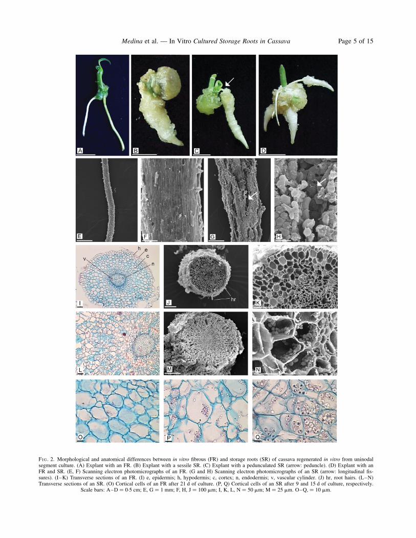
FIG. 2. Morphological and anatomical differences between in vitro fibrous (FR) and storage roots (SR) of cassava regenerated in vitro from uninodalsegment culture. (A) Explant with an FR. (B) Explant with a sessile SR. (C) Explant with a pedunculated SR (arrow: peduncle). (D) Explant with anFR and SR. (E, F) Scanning electron photomicrographs of an FR. (G and H) Scanning electron photomicrographs of an SR (arrow: longitudinal fis-sures). (I–K) Transverse sections of an FR. (I) e, epidermis; h, hypodermis; c, cortex; n, endodermis; v, vascular cylinder. (J) hr, root hairs. (L–N)Transverse sections of an SR. (O) Cortical cells of an FR after 21 d of culture. (P, Q) Cortical cells of an SR after 9 and 15 d of culture, respectively.
Scale bars: A–D ¼ 0.5 cm; E, G ¼ 1 mm; F, H, J ¼ 100 mm; I, K, L, N ¼ 50 mm; M ¼ 25 mm. O–Q, ¼ 10 mm.
Medina et al. — In Vitro Cultured Storage Roots in Cassava Page 5 of 15
parenchyma, termed the in vivo SR, whose function isdedicated to reserve storage compounds.In transverse section, in vivo secondary FRs presented,
from the centre to the edge (Fig. 1E): secondary xylemconstituted by an axial system with solitary or multiplevessel members, sometimes filled with tylose; an axial par-enchyma with thick-walled cells and fibres; and a radialsystem composed of rays (uniseriate, biseriate or multiseri-ate of up to three bands) of thin-walled parenchyma cells.It is necessary to highlight that, in some cases, plants thatwere cultivated for more than a cycle presented starchgranules in the rays of the secondary xylem. Subsequently,the vascular cambium was observed, composed of smallcells and, adjacent to this, secondary phloem and layers ofthin-walled parenchyma cells were found with no evidenceof starch-granule accumulation. Externally was the perider-mis, formed by the phellogen-producing phelloderm (layerof cells without suberized walls) centripetally, and phellem(layer of cork cells that constitutes the external bark) cen-trifugally. The cell layers inside the functional phellogenup to the secondary phloem, comprise the internal bark.
In vivo SRs (Fig. 1F) and secondary FRs had, in generalterms, a similar structure to that seen in the transverse sec-tions; however, they clearly differ in the xylem parench-yma size which constitutes the principal reserve tissue(Fig. 1F, arrow; Fig. 1G, blue staining indicates thepresence of starch). Up to a certain point (portion) of thesecondary xylem, axial and radial systems can be differen-tiated, with an evident reserve accumulation with a radialpattern (Fig. 1H). Following this portion, such organiz-ation becomes less conspicuous because all parenchyma-tous cells become competent to store starch granules(lower part of Fig. 1G) forming a spongier tissue. In con-trast to the secondary FR, the parenchyma layers externalto the vascular cambium also accumulate starch granules.In the parenchymatous cells of secondary phloem (Fig. 1I)and adjacent cortical parenchymatous cells, the accumu-lation pattern spreads in bands or layers parallel to thesurface (upper part of Fig. 1G).The epidermis of in vitro SRs was composed of a uni-
seriate stratum of cells, which was fragmented mainly nearthe root base, due to growth and dilatation of the corticaltissue in the radial direction (Fig. 2L, M). The cortex wasformed of .11 layers of cells which were relatively orga-nized in radial files. The middle cortex was divided peri-clinally, increasing the layer number and generating aswelling in the radial direction. As a consequence of thisradial thickening, fissures originated and reached up to theupper third of the cortical tissue. Moreover, this tissuepresented large intercellular spaces (Fig. 2L). The mostremarkable finding was the abundant deposition of starchgranules (Fig. 2 N) either singly or as 2–4 elements.Single granules had a cylindrical shape, and the compoundgranules were oval and truncated. This morphology iscoincident with that of starch granules synthesized in vivo.Delimitating the cortex was the endodermis composed of asingle layer of cells. Around the vascular cylinder was thepericycle which consisted of one or three cell layers. Likethe FR structure, the xylem alternated with the phloemand it was either tetrarch or pentarch within the same
clone ‘EC 27’ or ‘1468’. The central region of the vascu-lar cylinder was occupied by parenchymatous cells,forming the pith. In some cases, the pith was hollow.After 21 d of culture, the in vitro FRs did not show
starch granules (Fig. 2O) in any of the evaluated clones.However, after 6 d of culture, small starch granules wereobserved in the SRs, sometimes grouped together, similarto those found at 9 d (Fig. 2P). After 15 d of culture, someparenchymatous cells were almost filled with starchgranules undergoing an extraordinary increase in size(Fig. 2Q), and compound granules were easily distinguish-able. After 21 d of culture, the SR cortical tissue presenteda large deposition of starch granules (data not shown).When serial slides of the transverse sections wereanalysed, centrifugal starch deposition was determined. Onthe other hand, longitudinal sections showed that the storedecreased towards the root distal end.
Effect of genotype on in vitro SR formation
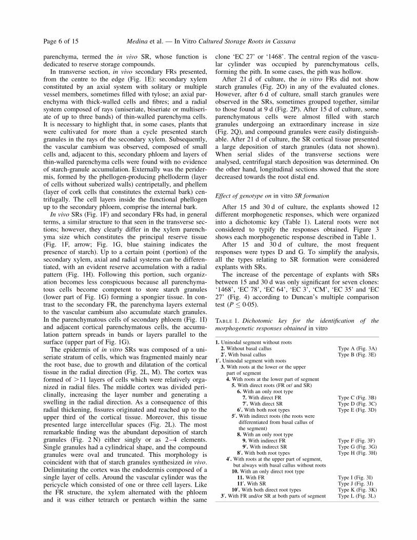
After 15 and 30 d of culture, the explants showed 12different morphogenetic responses, which were organizedinto a dichotomic key (Table 1). Lateral roots were notconsidered to typify the responses obtained. Figure 3shows each morphogenetic response described in Table 1.After 15 and 30 d of culture, the most frequent
responses were types D and G. To simplify the analysis,all the types relating to SR formation were consideredexplants with SRs.The increase of the percentage of explants with SRs
between 15 and 30 d was only significant for seven clones:‘1468’, ‘EC 78’, ‘EC 64’, ‘EC 3’, ‘CM’, ‘EC 35’ and ‘EC27’ (Fig. 4) according to Duncan’s multiple comparisontest (P � 0.05).
TABLE 1. Dichotomic key for the identification of themorphogenetic responses obtained in vitro
1. Uninodal segment without roots2. Without basal callus Type A (Fig. 3A)20. With basal callus Type B (Fig. 3E)
10. Uninodal segment with roots3. With roots at the lower or the upper
part of segment4. With roots at the lower part of segment5. With direct roots (FR or/ and SR)6. With an only root type7. With direct FR Type C (Fig. 3B)70. With direct SR Type D (Fig. 3C)
60. With both root types Type E (Fig. 3D)50. With indirect roots (the roots were
differentiated from basal callus ofthe segment)8. With an only root type9. With indirect FR Type F (Fig. 3F)90. With indirect SR Type G (Fig. 3G)
80. With both root types Type H (Fig. 3H)40. With roots at the upper part of segment,
but always with basal callus without roots10. With an only direct root type11. With FR Type I (Fig. 3I)110. With SR Type J (Fig. 3J)
100. With both direct root types Type K (Fig. 3K)30. With FR and/or SR at both parts of segment Type L (Fig. 3L)
Medina et al. — In Vitro Cultured Storage Roots in CassavaPage 6 of 15

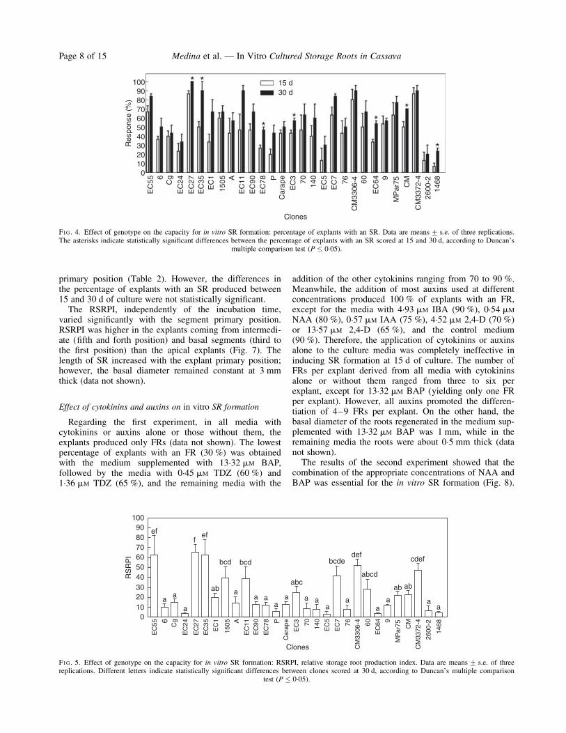
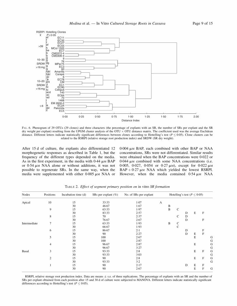
Although all evaluated genotypes produced SRs in vitro(Fig. 5), their capacity to do so varied significantly.According to the average linkage using the Euclideandistance of the 29 clones assayed, they were grouped intofour clusters with a cut-off limit of 1. The cluster was cor-related with the RSRPI values (Fig. 6). The groups withan RSRPI of 10–30 and 10–20 were distinguished bydifferences in the average dry weight of SRs (.16 mg and,16 mg, respectively). MANOVA indicated significantdifferences among genotypes according to Hotteling’s test(P � 0.05). Among the high yield clones (RSRPI . 39),clone ‘EC 27’ demonstrated high response uniformity.With regard to SR length, the longest ones derived from
the ‘EM 2600-2’ clone (10 cm long) and the shortest fromthe ‘EC 5’ clone (approx. 4 cm long). The basal diametervaried between 2 and 3 mm. The average percentage ofexplants that formed shoots ranged from 60 % (clone ‘60’)to 100 % (clones ‘EC 90’ and ‘EC 3’), showing a high
variation of shoot length from 5 mm (clones ‘EC 5’,‘Palomita’, ‘EC 24’ and ‘EC 55’) to 60 mm long (clone‘70’) (data not shown).
Effect of segment primary position on in vitro SR formation
After 15 d of culture, the explants also demonstrated the12 morphogenetic responses shown in the dichotomic key.The most frequent response was type G. The types relatingto SR formation represented a greater percentage ofresponse than those that only included FRs. The type Lresponse was only observed from intermediate or basalsegments.From the first to sixth positions, the percentage of
explants with SR reached 90–100 %. At 15 and 30 d ofculture, this variable and the number of SRs per explantwere significantly different depending on the segment
FIG. 3. In vitro morphogenetic responses differentiated from uninodal segment culture of cassava in SR formation medium (MS 5 %sucroseþ 0.54 mM NAAþ 0.44 mM BAP). Bc, basal callus; fr, fibrous roots; lr, lateral roots; sr, storage roots.
Medina et al. — In Vitro Cultured Storage Roots in Cassava Page 7 of 15
primary position (Table 2). However, the differences inthe percentage of explants with an SR produced between15 and 30 d of culture were not statistically significant.The RSRPI, independently of the incubation time,
varied significantly with the segment primary position.RSRPI was higher in the explants coming from intermedi-ate (fifth and forth position) and basal segments (third tothe first position) than the apical explants (Fig. 7). Thelength of SR increased with the explant primary position;however, the basal diameter remained constant at 3 mmthick (data not shown).
Effect of cytokinins and auxins on in vitro SR formation
Regarding the first experiment, in all media withcytokinins or auxins alone or those without them, theexplants produced only FRs (data not shown). The lowestpercentage of explants with an FR (30 %) was obtainedwith the medium supplemented with 13.32 mM BAP,followed by the media with 0.45 mM TDZ (60 %) and1.36 mM TDZ (65 %), and the remaining media with the
addition of the other cytokinins ranging from 70 to 90 %.Meanwhile, the addition of most auxins used at differentconcentrations produced 100 % of explants with an FR,except for the media with 4.93 mM IBA (90 %), 0.54 mM
NAA (80 %), 0.57 mM IAA (75 %), 4.52 mM 2,4-D (70 %)or 13.57 mM 2,4-D (65 %), and the control medium(90 %). Therefore, the application of cytokinins or auxinsalone to the culture media was completely ineffective ininducing SR formation at 15 d of culture. The number ofFRs per explant derived from all media with cytokininsalone or without them ranged from three to six perexplant, except for 13.32 mM BAP (yielding only one FRper explant). However, all auxins promoted the differen-tiation of 4–9 FRs per explant. On the other hand, thebasal diameter of the roots regenerated in the medium sup-plemented with 13.32 mM BAP was 1 mm, while in theremaining media the roots were about 0.5 mm thick (datanot shown).The results of the second experiment showed that the
combination of the appropriate concentrations of NAA andBAP was essential for the in vitro SR formation (Fig. 8).
0
15 d30 d
Clones
EC
55
EC
24
EC
27E
C35
EC
1
EC
3
EC
64
MP
ar75
EC
5E
C7
76 60 9
CM
3306
-4
CM
3372
-4
CM
2600
-214
6870 140
1505 A
EC
11E
C90
EC
78 PC
arap
e6C
g
Res
pons
e (%
)
102030405060708090
100
FIG. 4. Effect of genotype on the capacity for in vitro SR formation: percentage of explants with an SR. Data are means+ s.e. of three replications.The asterisks indicate statistically significant differences between the percentage of explants with an SR scored at 15 and 30 d, according to Duncan’s
multiple comparison test (P � 0.05).
1009080
ef eff
aa a
a aa
a a aa
aa
a aa
cdef
ab ab
bcd bcd
a
ababc
abcd
defbcde
706050403020100
RS
RP
I
Clones
EC
55
EC
24
EC
27
EC
35
EC
1
EC
3
EC
64
MP
ar75
EC
5
EC
7
76 60 9
CM
3306
-4
CM
3372
-4
CM
2600
-2
146870 14
0
1505 A
EC
11E
C90
EC
78 PC
arap
e6C
g
FIG. 5. Effect of genotype on the capacity for in vitro SR formation: RSRPI, relative storage root production index. Data are means+ s.e. of threereplications. Different letters indicate statistically significant differences between clones scored at 30 d, according to Duncan’s multiple comparison
test (P � 0.05).
Medina et al. — In Vitro Cultured Storage Roots in CassavaPage 8 of 15
After 15 d of culture, the explants also differentiated 12morphogenetic responses as described in Table 1, but thefrequency of the different types depended on the media.As in the first experiment, in the media with 0.44 mM BAPor 0.54 mM NAA alone or without additions, it was notpossible to regenerate SRs. In the same way, when themedia were supplemented with either 0.005 mM NAA or
0.004 mM BAP, each combined with other BAP or NAAconcentrations, SRs were not differentiated. Similar resultswere obtained when the BAP concentrations were 0.022 or0.044 mM combined with some NAA concentrations (i.e.0.005, 0.027, 0.054 or 0.27 mM), except for 0.022 mM
BAPþ 0.27 mM NAA which yielded the lowest RSRPI.However, when the media contained 0.54 mM NAA
RSRPIx
ClonesHotellingP ≤ 0.05
≤ 6
>39
ef
ef
bebbggdggghjklhjkljkljkehljlhlhiacdacadiiikijklc
h
ff
10–30
SRDW>16 mg
10–20
SRDW<16 mg
EC11EC35EC27EC55
EC7
EC360
AmarillaCarapé
6EC78
CMEC1
EC90Catiguá
140EC64
701468
EM 2600-2EC24
PalomitaEC5
MCol 1505
CM3372-4CM3306-4
MPar75
976
0.00 0.25 0.50 0.75 1.00Distance Index
1.25 1.50 1.75 2.00
FIG. 6. Phenogram of 29 OTUs (29 clones) and three characters (the percentage of explants with an SR, the number of SRs per explant and the SRdry weight per explant) resulting from the UPGM cluster analysis of the OTU � OTU distance matrix. The coefficient used was the average Euclideandistance. Different letters indicate statistically significant differences between clones according to Hottelling’s test (P � 0.05). Clone clusters can be
related to the RSRPI (relative storage root production index) and SRDW (SR dry weight).
TABLE 2. Effect of segment primary position on in vitro SR formation
Nodes Positions Incubation time (d) SRs per explant (%) No. of SRs per explant Hotelling’s test (P � 0.05)
Apical 10 15 33.33 1.07 A30 46.67 1.67 B
9 15 63.33 1.97 B C30 83.33 2.57 D E F
8 15 70 2.37 C D30 76.67 2.67 D F
Intermediate 7 15 63.33 1.93 B C30 66.67 1.93 C
6 15 86.67 2.1 D F30 90 2.27 D E F
5 15 100 2.67 E G30 100 2.87 G
4 15 96.67 2.87 E G30 96.67 3.27 G
Basal 3 15 93.33 2.8 E F G30 93.33 3.03 G
2 15 90 2.8 E F G30 93.33 3.2 G
1 15 90 2.17 D E F30 90 2.67 E F G
RSRPI, relative storage root production index. Data are means+ s.e. of three replications. The percentage of explants with an SR and the number ofSRs per explant obtained from each position after 15 and 30 d of culture were subjected to MANOVA. Different letters indicate statistically significantdifferences according to Hottelling’s test (P � 0.05).
Medina et al. — In Vitro Cultured Storage Roots in Cassava Page 9 of 15

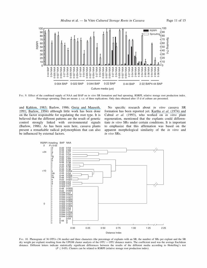
combined with 0.022 or 0.044 mM BAP, the percentage ofexplants with SRs and the number of SRs per explantincreased significantly although the SR dry weight perexplant (data nor shown) and the RSRPI remained low(Fig. 9).The best responses were achieved when 0.54 mM NAA
was combined with either 0.22 or 0.44 mM BAP (Fig. 9).It is important to recognize that the MS medium sup-plemented with 0.54 mM NAAþ 0.44 mM BAP promotedSR formation in 100 % of the explants (Fig. 8) althoughthe number of SRs per explant, the SR dry weight perexplant (data not shown) and the RSRPI were lower thanthe other combinations. A considerable performance wasalso demonstrated by media supplied with 0.27 mM
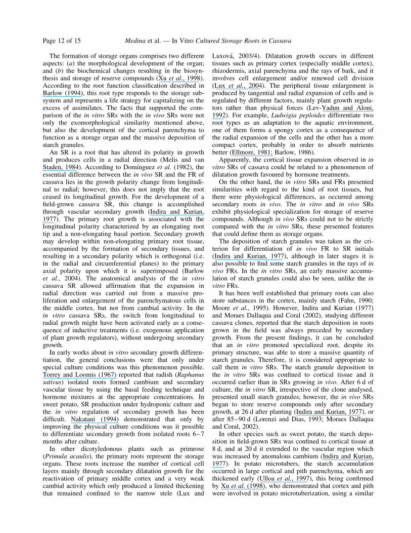
NAAþ 0.22 or 0.44 mM BAP.The distance phenogram constructed considering the
three variables scored for the media evaluation at 15 d ofculture discriminated three clusters with a cut-off limit of1. These groups could be associated with the media’s
capacity to promote high, intermediate or low (or null) invitro SR production (Fig. 10). MANOVA showed statisti-cal differences between the results of the different mediaaccording to Hotteling’s test (P � 0.05).Higher concentrations of NAA and BAP combinations
decreased the SR differentiation, even damaging the shootregeneration (Fig. 9) and the rooting process (Fig. 8). Theresultant RSRPI were similar to those scored for the lowerconcentrations of plant growth regulators. A remarkablefact was that the percentage of explants with shoots wassignificantly higher in the media that did not favour SRformation (Fig. 9).
DISCUSSION
Adventitious roots are present in many terrestrial plantsand may help the species adapt to each life mode throughthe development of alternative forms. Radical dimorphismand trimorphism has been extensively documented (Samb
4
3
2
1
0
RS
RP
I
Positions
1 2 3 4 5 6 7 8 9 10(apicalnode)
(basalnode)
15 d30 d
FIG. 7. Effect of segment primary position on in vitro SR formation. RSRPI, relative storage root production index. Data are means+ s.e. of threereplications. Data were scored at 15 and 30 d of culture.
1009080706050403020100
0.44
BA
P
0.004 BAP 0.022 BAP 0.044 BAP 0.44 BAP 4.44 BAP0.22 BAP 2.22 BAP
Culture media (mM)
0.54
NA
A0.
005
NA
A
0.00
5 N
AA
0.02
7 N
AA
0.02
7 N
AA
0.05
4 N
AA
0.05
4 N
AA
0.27
NA
A0.
54 N
AA
0.27
NA
A0.
54 N
AA
0.00
5 N
AA
0.02
7 N
AA
0.05
4 N
AA
0.27
NA
A0.
54 N
AA
0.00
5 N
AA
0.02
7 N
AA
0.05
4 N
AA
0.27
NA
A0.
54 N
AA
0.00
5 N
AA
0.02
7 N
AA
0.05
4 N
AA
0.27
NA
A0.
54 N
AA
2.69
NA
A5.
38 N
AA
2.69
NA
A
2.69
NA
A
5.38
NA
A
5.38
NA
A
0.54
NA
A
0.54
NA
A
Res
pons
e (%
)
% with storage roots
% with fibrous roots
FIG. 8. Effect of the combined supply of NAA and BAP on in vitro root formation. The percentage of explants with an SR and the percentage ofexplants with an FR. Data are means+ s.e. of three replications. Only data obtained after 15 d of culture are presented.
Medina et al. — In Vitro Cultured Storage Roots in CassavaPage 10 of 15
and Kahlem, 1983; Barlow, 1986; Greig and Mauseth,1991; Barlow, 1994) although little work has been doneon the factor responsible for regulating the root type. It isbelieved that the different patterns are the result of geneticcontrol strongly linked with environmental signals(Barlow, 1986). As has been seen here, cassava plantspresent a remarkable radical polymorphism that can alsobe influenced by external factors.
No specific research about in vitro cassava SRformation has been reported yet. Kartha et al. (1974) andCabral et al. (1993), who worked on in vitro plantregeneration, mentioned that the explants could differen-tiate in vitro SRs under certain conditions. It is importantto emphasize that this affirmation was based on theapparent morphological similarity of the in vitro andin vivo SRs.
1009080706050403020100
0.44
BA
P
0.004 BAP 0.022 BAP 0.044 BAP 0.44 BAP 4.44 BAP0.22 BAP 2.22 BAP
Culture media (mM)
0.54
NA
A0.
005
NA
A
0.00
5 N
AA
0.02
7 N
AA
0.02
7 N
AA
0.05
4 N
AA
0.05
4 N
AA
0.27
NA
A0.
54 N
AA
0.27
NA
A0.
54 N
AA
0.00
5 N
AA
0.02
7 N
AA
0.05
4 N
AA
0.27
NA
A0.
54 N
AA
0.00
5 N
AA
0.02
7 N
AA
0.05
4 N
AA
0.27
NA
A0.
54 N
AA
0.00
5 N
AA
0.02
7 N
AA
0.05
4 N
AA
0.27
NA
A0.
54 N
AA
2.69
NA
A5.
38 N
AA
2.69
NA
A
2.69
NA
A
5.38
NA
A
5.38
NA
A
0.54
NA
A
0.54
NA
A
RS
RP
I
1009080706050403020100
Sprouting (%
)
RSRPI
Sprouting
FIG. 9. Effect of the combined supply of NAA and BAP on in vitro SR formation and bud sprouting. RSRPI, relative storage root production index.Percentage sprouting. Data are means+ s.e. of three replications. Only data obtained after 15 d of culture are presented.
RSRPIx
HotellingP ≤ 0.05
BAP NAA
>24
0.220.440.440.440.222.222.22
2.22
0.22
0.220.0440.0220.022
0.0440.0440.0440.0440.0220.0220.0220.0440.0440.0440.0440.044
0.44
0.54
0.00 0.25 0.50 0.75
Distance Index
1.00 1.25 2.25
0.005
0.005
0.0270.0540.27
0.27
0.27
0.0270.054
0.540.54
0.54
0.54
0.540.27
0.27
5.385.38
5.382.69
2.69
2.69
0.005
0.0050.005
0.027
0.0270.0270.054
0.054
0.54
0.054
0.54
0.440
0 00
0.22
0.44
0.440.44
4.44
4.444.44
dcgffgabjjjikhikhihkkeejlllllllllllllllll
<10
0
FIG. 10. Phenogram of 36 OTUs (36 media) and three characters (the percentage of explants with an SR, the number of SRs per explant and the SRdry weight per explant) resulting from the UPGM cluster analysis of the OTU � OTU distance matrix. The coefficient used was the average Euclideandistance. Different letters indicate statistically significant differences between the results of the different media according to Hottelling’s test
(P � 0.05). Clusters can be related to RSRPI (relative storage root production index).
Medina et al. — In Vitro Cultured Storage Roots in Cassava Page 11 of 15
The formation of storage organs comprises two differentaspects: (a) the morphological development of the organ;and (b) the biochemical changes resulting in the biosyn-thesis and storage of reserve compounds (Xu et al., 1998).According to the root function classification described inBarlow (1994), this root type responds to the storage sub-system and represents a life strategy for capitalizing on theexcess of assimilates. The facts that supported the com-parison of the in vitro SRs with the in vivo SRs were notonly the exomorphological similarity mentioned above,but also the development of the cortical parenchyma tofunction as a storage organ and the massive deposition ofstarch granules.An SR is a root that has altered its polarity in growth
and produces cells in a radial direction (Melis and vanStaden, 1984). According to Domınguez et al. (1982), theessential difference between the in vivo SR and the FR ofcassava lies in the growth polarity change from longitudi-nal to radial; however, this does not imply that the rootceased its longitudinal growth. For the development of afield-grown cassava SR, this change is accomplishedthrough vascular secondary growth (Indira and Kurian,1977). The primary root growth is associated with thelongitudinal polarity characterized by an elongating roottip and a non-elongating basal portion. Secondary growthmay develop within non-elongating primary root tissue,accompanied by the formation of secondary tissues, andresulting in a secondary polarity which is orthogonal (i.e.in the radial and circumferential planes) to the primaryaxial polarity upon which it is superimposed (Barlowet al., 2004). The anatomical analysis of the in vitrocassava SR allowed affirmation that the expansion inradial direction was carried out from a massive pro-liferation and enlargement of the parenchymatous cells inthe middle cortex, but not from cambial activity. In thein vitro cassava SRs, the switch from longitudinal toradial growth might have been activated early as a conse-quence of inductive treatments (i.e. exogenous applicationof plant growth regulators), without undergoing secondarygrowth.In early works about in vitro secondary growth differen-
tiation, the general conclusions were that only underspecial culture conditions was this phenomenon possible.Torrey and Loomis (1967) reported that radish (Raphanussativus) isolated roots formed cambium and secondaryvascular tissue by using the basal feeding technique andhormone mixtures at the appropriate concentrations. Insweet potato, SR production under hydroponic culture andthe in vitro regulation of secondary growth has beendifficult. Nakatani (1994) demonstrated that only byimproving the physical culture conditions was it possibleto differentiate secondary growth from isolated roots 6–7months after culture.In other dicotyledonous plants such as primrose
(Primula acaulis), the primary roots represent the storageorgans. These roots increase the number of cortical celllayers mainly through secondary dilatation growth for thereactivation of primary middle cortex and a very weakcambial activity which only produced a limited thickeningthat remained confined to the narrow stele (Lux and
Luxova, 2003/4). Dilatation growth occurs in differenttissues such as primary cortex (especially middle cortex),rhizodermis, axial parenchyma and the rays of bark, and itinvolves cell enlargement and/or renewed cell division(Lux et al., 2004). The peripheral tissue enlargement isproduced by tangential and radial expansion of cells and isregulated by different factors, mainly plant growth regula-tors rather than physical forces (Lev-Yadun and Aloni,1992). For example, Ludwigia peploides differentiate tworoot types as an adaptation to the aquatic environment,one of them forms a spongy cortex as a consequence ofthe radial expansion of the cells and the other has a morecompact cortex, probably in order to absorb nutrientsbetter (Ellmore, 1981; Barlow, 1986).Apparently, the cortical tissue expansion observed in in
vitro SRs of cassava could be related to a phenomenon ofdilatation growth favoured by hormone treatments.On the other hand, the in vitro SRs and FRs presented
similarities with regard to the kind of root tissues, butthere were physiological differences, as occurred amongsecondary roots in vivo. The in vitro and in vivo SRsexhibit physiological specialization for storage of reservecompounds. Although in vivo SRs could not to be strictlycompared with the in vitro SRs, these presented featuresthat could define them as storage organs.The deposition of starch granules was taken as the cri-
terion for differentiation of in vivo FR to SR initials(Indira and Kurian, 1977), although in later stages it isalso possible to find some starch granules in the rays of invivo FRs. In the in vitro SRs, an early massive accumu-lation of starch granules could also be seen, unlike the invitro FRs.It has been well established that primary roots can also
store substances in the cortex, mainly starch (Fahn, 1990;Moore et al., 1995). However, Indira and Kurian (1977)and Moraes Dallaqua and Coral (2002), studying differentcassava clones, reported that the starch deposition in rootsgrown in the field was always preceded by secondarygrowth. From the present findings, it can be concludedthat an in vitro promoted specialized root, despite itsprimary structure, was able to store a massive quantity ofstarch granules. Therefore, it is considered appropriate tocall them in vitro SRs. The starch granule deposition inthe in vitro SRs was confined to cortical tissue and itoccurred earlier than in SRs growing in vivo. After 6 d ofculture, the in vitro SR, irrespective of the clone analysed,presented small starch granules; however, the in vivo SRsbegan to store reserve compounds only after secondarygrowth, at 26 d after planting (Indira and Kurian, 1977), orafter 85–90 d (Lorenzi and Dias, 1993; Moraes Dallaquaand Coral, 2002).In other species such as sweet potato, the starch depo-
sition in field-grown SRs was confined to cortical tissue at8 d, and at 20 d it extended to the vascular region whichwas increased by anomalous cambium (Indira and Kurian,1977). In potato microtubers, the starch accumulationoccurred in large cortical and pith parenchyma, which arethickened early (Ulloa et al., 1997), this being confirmedby Xu et al. (1998), who demonstrated that cortex and pithwere involved in potato microtuberization, using a similar
Medina et al. — In Vitro Cultured Storage Roots in CassavaPage 12 of 15

composition of media to that used in the present work. Xuet al. (1998) observed the same growth pattern in tubersgrowing in vivo, as did Vreugdenhil et al. (1999), whoalso reported that the starch accumulation was visible after5 d of culture, in tubers of plants growing in pots andtreated with short day conditions.It was observed that the in vitro SR formation was
strongly affected by the genotype in cassava. Likewise, theproduction of the plants grown in the field was signifi-cantly influenced by this factor (Montaldo et al., 1979),but it also varied with the year of evaluation (Henain andCenoz, 1970) or planting site (Iglesias et al., 1998).Significant differences in the in vitro potato tuberizationdue to genotype have been extensively reported(Belletti et al., 1994; Leclerc et al., 1994; Gopal et al.,1998).Information on how the stem position in the cassava
plant affects the formation of the SR is scarce. Cabral andCarvalho (2000) observed that apical stem cuttings did notproduce shoots or roots, while basal cuttings regeneratedvigorous shoots and showed earlier SR formation. Thepresent experiments were conducted under in vitro con-ditions and the results were similar to those obtained byCabral and Carvalho (2000), since the explants comingfrom apical parts were less responsive than those frommiddle and basal parts. However, Mogilner et al. (1967a)reported that apical stem cuttings produced the highestSR fresh weight although the number of SRs per stemcuttings was lower. The effect of segment primary positionmay be related to several factors, such as stem age, budmaturity in different portions of the stem, and stemreserves (Cabral and Carvalho, 2000), which have beenobserved to increase from the top to the bottom ofthe plant stem (Mogilner et al., 1967a; Ramanujan andIndira, 1984).The in vitro tuberization of radish was absolutely depen-
dent on the requirements for an auxin and a cytokinin; inthe absence of any one critical component, the responsewas markedly reduced or absent (Loomis and Torrey,1964; Torrey and Loomis, 1967; Webster and Radin,1972). Radish roots cultured under favourable conditionswere able to undergo secondary growth although thecambial activity ceased after 28 d (Loomis and Torrey,1964). Optimal concentrations of IAA (10 mM) and BAP(5 mM) stimulated an extraordinary increase in root diam-eter during the first 2–3 weeks of culture. IAA could beeffectively substituted with NAA or 2,4-D in the presenceof BAP, although the medium with NAA at 1 mM allowedthe development of a better radial symmetrical structure.Peterson (1973) also found that an auxin and a cytokininwere necessary for maximum radial growth of the excisedroots of turnip (Brassica rapa) behaving like radish.Neither Torrey and Loomis (1967) nor Peterson (1973)mentioned anything about starch deposition in differen-tiated SRs. In other species with a dimorphic root system,such as Jussiaea repens, the root type emerging at a nodeis controlled by the ratio of auxins and cytokinins (Samband Kahlem, 1983).Cassava in vitro SRs also required an auxin (NAA) and
a cytokinin (BAP) to regenerate. They were not able to
develop secondary growth, but formed a tissue competentto store reserve compounds, unlike in vitro FRs.It has been postulated that hormones cause nutrient
mobilization by creating new source–sink relationships,especially cytokinins. The metabolism of the treated areamay be stimulated by the hormones so the nutrients movetowards it (Salisbury and Ross, 1994; Taiz and Zeiger,1998). Previous reports (Palmer and Smith, 1969; vanStaden and Dimalla, 1976) suggested that cytokinins inthe tubers act by establishing a metabolic storage sinkwhich attracts metabolites to the organ in which they arepresent. This phenomenon could be related to the increaseof the tuber growth when a cytokinin was exogenouslyapplied (Gopal et al., 1998). According to Melis andvan Staden (1984), cytokinins could attract metabolitestogether with auxins. Later Ehneß and Roitsh (1997)demonstrated that the sugar uptake was increased inresponse to cytokinin treatment in cells of Chenopodiumrubrum. In a further review, the regulation of source–sinkrelationships by cytokinins and their interactions withother signals were dealt with extensively (Roitsh andEhneß, 2000). These relationships (i.e. between auxins andcytokinins) may cause additive, synergic or inhibitoryeffects (Gifford and Evans, 1981). The NAA and BAPcombinations possibly promoted, in some in vitro cassavaroots, the establishment of a metabolic sink and inducedthem to become storage organs.In tuber crops, there is an inverse relationship between
vegetative and tuber growth. Factors that promote vegeta-tive growth inhibit tuber growth. This was clearly observedin potato (Trippi, 1982) and dahlia plants (Halevy andBiran, 1975). In cassava plants, the shoot and SRs growsimultaneously, creating competition for the assimilates.This fact suggested that factors that favoured aerial growthmay depress root growth (Ternes, 2002). In the last exper-iment, in which the influence of a combined supply ofNAA and BAP was analysed, the combinations thatpromoted the better development of SRs did not favourshoot growth.The SR formation system reported here is a first
approach to development of a tuberization model althoughadditional efforts are required to improve it. It was notpossible to achieve root secondary growth, but after thiswork it will be feasible to advance in some aspects of invitro cassava tuberization.Two root types were differentiated in vitro and they
could distinguished morphological and anatomically.Based on these criteria, 29 clones of cassava were able toform SRs in vitro although their capacity was significantlydifferent. On the other hand, the SR formation was con-siderably affected by the segment primary position and itwas strongly influenced by hormone treatment.
ACKNOWLEDGEMENTS
The authors wish to express their gratitude to ErnestinaGaldeano, PhD, for her valuable comments on themanuscript, and Aldo Goytia for the photographic plates.This paper is a part of the thesis of the senior author.
Medina et al. — In Vitro Cultured Storage Roots in Cassava Page 13 of 15

The authors are grateful to CONICET and SGCyT(UNNE) for their financial support.
LITERATURE CITED
Alves AAC. 2002. Cassava botany and physiology. In: Hillocks RJ,Thresh JM, Bellotti AC, eds. Cassava: biology, production and util-ization. Wallingford, UK: CAB International, 67–89.
Barlow PW. 1986. Adventitious roots of whole plants: their forms, func-tions, and evolution. In: Jackson MB, ed. New root formation inplants and cuttings. Dordrecht, The Netherlands: Martinus NijhoffPublishers, 67–110.
Barlow PW. 1994. The origin, diversity and biology of shoot-borneroots. In: Davis TD, Haissig BE, eds. Biology of adventitious rootformation. New York: Plenum Press, 1–23.
Barlow PW, Volkmann D, Baluska F. 2004. Polarity in roots. In:Lindsey K, ed. Polarity in plants. Oxford: Blackwell Publishing,192–241.
Batista de Souza C, Carvalho L, Mattos Cascardo J. 2004.Comparative gene expression study to identify genes possibly relatedto storage root formation in cassava (Manihot esculenta Crantz). In:Zuniga C, ed. Proceedings of the 6th International ScientificMeeting of the Cassava Biotechnology Network. Cali, Colombia:CIAT, 153.
Belletti P, Lanteri S, Lotito S, Saracco F. 1994. Production of potatomicro-tubers through in vitro culture. Acta Horticulturae 362:141–148.
Cabral GB, Carvalho LJCB. 2000. The formation of storage root incassava. In: Carvalho L, Thro A, Duarte Vilarinhos A, eds.Proceedings of the 4th International Scientific Meeting of theCassava Biotechnology Network. Brasilia, Brazil: EMBRAPARecursos Geneticos e Biotecnologia, 345–356.
Cabral GB, Carvalho LJCB. 2001. Analysis of proteins associated withstorage root formation in cassava using two-dimensional gel electro-phoresis. Revista Brasileira de Fisiologia Vegetal 13: 41–48.
Cabral GB, Aragao FJL, Matsumoto K, Monte-Neshich DC, Rech L.1993. Cassava tissue culture: multiple shoots and somatic embryo-genesis. In: Roca W, Thro A, eds. Proceedings of the 1stInternational Scientific Meeting of the Cassava BiotechnologyNetwork. Cali, Colombia: CIAT, 180–184.
Carvalho LJCB, Mattos Cascardo J, Ferreira M, Loureiro M. 1993.Studies on proteins and enzymes related to tuberization and starchbiosynthesis in cassava roots. In: Roca W, Thro A, eds. Proceedingsof the 1st International Scientific Meeting of the CassavaBiotechnology Network. Cali, Colombia: CIAT, 234–238.
Cereda MP, Vilpoux O. 2003. Conservacao de raızes. In: Cereda MP,Vilpoux O, eds. Tecnologıa, usos e potencialidades de tuberosasamilaceas Latinoamericanas. Sao Paulo, Brazil: Fundacao Cargill,13–29.
Domınguez CE, Ceballos LF, Fuentes C. 1982. Morfologıa de la plantade yuca. In: Domınguez CE, ed. Yuca: Investigacion, Produccion yUtilizacion. Cali, Colombia: CIAT/PNUD, 28–49.
Ehneß R, Roitsh T. 1997. Co-ordinated induction of mRNAs for extra-cellular invertase and a glucose transporter in Chenopodium rubrumby cytokinins. The Plant Journal 11: 539–548.
El-Sharkawy MA. 2003. Cassava biology and physiology. PlantMolecular Biology 53: 621–641.
Ellmore GS. 1981. Root dimorphism in Ludwigia peploides(Onagraceae): structure and gas content of mature roots. AmericanJournal of Botany 68: 557–568.
Fahn A. 1990. Plant anatomy, 4th edn. Oxford, UK: Pergamon Press.Gifford RM, Evans LT. 1981. Photosynthesis carbon portioning, and
yield. Annual Review of Plant Physiology 32: 485–509.Gonzalez AM, Cristobal CL. 1997. Anatomıa y ontogenia de semillas
de Helicteres Lhotzkyana (Sterculiaceae). Bonplandia 9: 287–294.Gopal J, Minocha JL, Dhaliwal HS. 1998. Microtuberization in potato
(Solanum tuberosum L.). Plant Cell Reports 17: 794–798.Greig N, Mauseth JD. 1991. Structure and function of dimorphic prop
roots in Piper auritum L. Bulletin of the Torrey Botanical Club 118:176–183.
Halevy AH, Biran I. 1975. Hormonal regulation of tuberization inDahlia. Acta Horticulturae 47: 319–330.
Henain AE, Cenoz HM. 1970. Comportamiento de clones de mandioca(Manihot esculenta Crantz), cultivados en el Nordeste Argentino.Corrientes, Argentina: Facultad de Agronomıa y Veterinaria,Universidad Nacional del Nordeste.
Iglesias C, Lenis J, Calle F. 1998. Estructuracion de un programa demejoramiento y multiplicacion de semilla de yuca en la CostaAtlantica Colombiana. In: Meek Munoz E, Aldana Navarrete H, eds.Primer encuentro tecnico nacional de produccion y transformacionde yuca. Tolu, Colombia: Le Print Club Express, 59–73.
Indira P, Kurian T. 1977. A study on the comparative anatomicalchanges undergoing tuberization in the roots of cassava and sweetpotato. Journal of Root Crops 3: 29–32.
InfoStat. 2002. InfoStat version 1.1. Manual del Usuario. GrupoInfoStat. Cordoba, Argentina: Universidad Nacional de Cordoba.
Johansen DA. 1940. Plant microtechnique. New York: McGraw-HillBook Co.
Kartha KK, Gamborg OL, Constabel F, Shyluk JP. 1974.Regeneration of cassava plants from apical meristems. Plant ScienceLetters 2: 107–113.
Leclerc Y, Donnelly DJ, Seabrook JEA. 1994. Microtuberization oflayered shoots and nodal cuttings of potato: the influence of growthregulators and incubation periods. Plant Cell, Tissue and OrganCulture 37: 113–120.
Lev-Yadun S, Aloni R. 1992. Experimental induction of dilatation mer-istems in Melia azedarach L. Annals of Botany 70: 379–386.
Loomis RS, Torrey JG. 1964. Chemical control of vascular cambiuminitiation in isolated radish roots. Proceedings of the NationalAcademy of Sciences of the USA 52: 3–11.
Lorenzi JO, Dias CAC. 1993. Cultura da mandioca. Campinas, Brazil:Coordenadoria de Assistencia Tecnica Integral.
Luque R, Sousa HC, Kraus JE. 1996. Metodos de coloracao de Roeser(1972)-modificado- e Kropp (1972) visando a subtituicao do azul deastra por azul de alciao 8 GS ou 8 GX. Acta Botanica Brasilica 10:199–212.
Lux A, Luxova M. 2003/4. Growth and differentiation of root endoder-mis in Primula acaulis Jacq. Biologia Plantarum 47: 91–97.
Lux A, Luxova M, Abe J, Morita S. 2004. Root cortex: structural andfunctional variability and responses to environmental stress. RootResearch 13: 117–131.
Melis RJM, van Staden J. 1984. Tuberization and hormones. Zeitschriftfur Pflanzenphysiologie 113: 271–283.
Melis RJM, van Staden J. 1985. Tuberization in cassava (Manihot escu-lenta Crantz): cytokinin and abscisic acid activity in tuberous roots.Journal of Plant Physiology 118: 357–366.
Mogilner I, Orioli GA, Bletter CM. 1967a. Ensayo de topofisis y foto-periodismo en mandioca. Bonplandia 2: 266–272.
Mogilner I, Portuguez Arias JD, Gotuzzo AD, Acosta JA. 1967b.Influencia de la parte aerea de Manihot flabellifolia en la formacionde raıces reservantes de Manihot esculenta utilizado como pie.Bonplandia 2: 137–142.
Montaldo A, Gunz T, Montilla JJ, Perez Aleman S, Reveron AE.1979. La yuca o mandioca (Manihot esculenta). San Jose, CostaRica: Instituto Interamericano de Ciencias Agrıcolas.
Moore R, Clark WD, Stern KR. 1995. Botany. Dubuque, IA:Wm. C. Brown Publishers.
Moraes Dallaqua MA, Coral DJ. 2002. Morfo-anatomia. In: CeredaMP, ed. Agricultura: tuberosas amilaceas Latinoamericanas. SaoPaulo, Brazil: Fundacao Cargill, 48–65.
Murashige T, Skoog F. 1962. A revised medium for rapid growth andbioassay with tobacco tissue cultures. Physiologia Plantarum 15:473–497.
Nakatani M. 1994. In vitro formation of tuberous roots in sweet potato.Japanese Journal of Crop Science 63: 158–159.
Palmer CE, Smith OE. 1969. Effect of abscicic-acid on elongation andkinetin induced tuberization of isolated stolons of Solanum tubero-sum L. Plant and Cell Physiology 10: 657–664.
Peterson RL. 1973. Control of cambial activity in roots of turnips(Brassica rapa). Canadian Journal of Botany 51: 475–480.
Puonti-Kaerlas J. 1998. Cassava biotechnology. In: Tombs MP, ed.Biotechnology and genetic engineering reviews. Andover, UK:Intercept Ltd, 329–364.
Medina et al. — In Vitro Cultured Storage Roots in CassavaPage 14 of 15

Ramanujan T, Indira P. 1984. Effect of girdling on the distribution oftotal carbohydrates and hydrocyanic acid in cassava. Indian Journalof Plant Physiology 27: 355–360.
Rateaver B. 1951. Anatomy and regeneration in the stem and root ofManihot utilissima Pohl. PhD thesis, University of Michigan, AnnArbor, MI.
Roitsh T, Ehneß R. 2000. Regulation of source/sink relations by cytoki-nins. Plant Growth Regulation 32: 359–367.
Samb PI, Kahlem G. 1983. Determinisme de l’organogenese racinairede Jussiaea repens L. Zeitschrift fur Pflanzenphysiologie 109:279–284.
Salisbury FB, Ross CW. 1994. Fisiologıa vegetal. Mexico D.F, Mexico:Grupo Editorial Iberoamerica S.A. de C.V.
Scott GJ, Best R, Rosegrant M, Bokanga M. 2000. Roots and tubers inthe global food system: a vision statement to the year 2020. Lima,Peru: Training and Communications Department of the InternationalPotato Center.
Sneath PH, Sokal RR. 1973. Numerical taxonomy – the principles andpractice of numerical classification. San Francisco, CA: W.H.Freeman & Co.
van Staden J, Dimalla GG. 1976. Endogenous cytokinins and tuberizationin potato Solanum tuberosum. Annals of Botany 40: 1117–1119.
Struik PC, Vreugdenhil D, van Eck HJ, Bachem CW, Visser RGF.1999. Physiological and genetic control of tuber formation. PotatoResearch 42: 313–331.
Taiz L, Zeiger E. 1998. Plant physiology, 2nd edn. Sunderland, MA:Sinauer Associates, Inc.
Ternes M. 2002. Fisiologia da planta. In: Cereda MP, ed. Agricultura:tuberosas amilaceas Latinoamericanas. Sao Paulo, Brazil: FundacaoCargill, 66–82.
Torrey JG, Loomis RS. 1967. Auxin–cytokinin control of secondaryvascular tissue formation in isolated roots of Raphanus. AmericanJournal of Botany 54: 1098–1106.
Trippi VS. 1982. Ontogenia y senilidad en plantas. Cordoba, Argentina:Direccion General de Publicaciones, Universidad Nacional deCordoba.
Ulloa RM, Mac Intosh GC, Melchiorre M, Mentaberry AN, DallariP, Moriconi DN, Tellez-Inon MT. 1997. Protein kinase activity indifferent stages of potato (Solanum tuberosum L.) microtuberization.Plant Cell Reports 16: 426–429.
Vreugdenhil D, Xu X, Jung ChJ, Lammeren AAM, Ewing EE. 1999.Initial anatomical changes associated with tuber formation on single-node potato (Solanum tuberosum L.) cuttings: a re-evaluation.Annals of Botany 84: 675–680.
Webster BD, Radin JW. 1972. Growth and development of culturedradish roots. American Journal of Botany 59: 744–751.
Xu X, Vreugdenhil D, van Lammeren AAM. 1998. Cell division andcell enlargement during potato tuber formation. Journal ofExperimental Botany 49: 573–582.
Medina et al. — In Vitro Cultured Storage Roots in Cassava Page 15 of 15