In vitro biological evaluation of electrospun cellulose acetate fiber mats containing asiaticoside...
10
In vitro biological evaluation of electrospun cellulose acetate fiber mats containing asiaticoside or curcumin Orawan Suwantong, 1,2 Uracha Ruktanonchai, 3 Pitt Supaphol 1,2 1 The Petroleum and Petrochemical College, Chulalongkorn University, Pathumwan, Bangkok, Thailand 2 The Center for Petroleum, Petrochemicals and Advanced Materials, Chulalongkorn University, Pathumwan, Bangkok, Thailand 3 National Nanotechnology Center, Thailand Science Park, Klong Luang, Phathumthani, Thailand Received 24 September 2009; revised 23 October 2009; accepted 6 January 2010 Published online 22 April 2010 in Wiley InterScience (www.interscience.wiley.com). DOI: 10.1002/jbm.a.32797 Abstract: Ultra-fine cellulose acetate (CA; M w 30,000 Da; degree of acetyl substitution 2.4) fiber mats containing ei- ther asiaticoside [from the plant Centella asiatica (L.); either in the form of a crude extract (CACE) or pure substance (PAC)] or curcumin (CM; from the plant Curcuma longa L.) were successfully prepared. The proposed use of these materials is as topical/transdermal patches or wound dress- ings. Here, the potential for use of these herb-loaded CA fiber mats as wound dressings was evaluated in terms of the stability and the antioxidant activity of the as-loaded herbal substances, the ability to support both the attach- ment and the proliferation of fibroblasts and the ability of the cultured fibroblasts to synthesize collagen. Normal human dermal fibroblasts (NHDF) were used as the refer- ence fibroblastic cells. The results showed that the as- loaded herbal substances were stable even after the herb- loaded CA fiber mats had been aged either at room temper- ature or at 40 C for a period of up to 4 months. The inclu- sion of asiaticoside [either 2% (w/w) CACE or 40% (w/w) PAC] rendered the resulting CA fiber mats their superiority in supporting the attachment, promoting the proliferation, and upregulating the production of collagen of the seeded and/or the cultured NHDF to the corresponding solvent-cast films and the neat CA fiber mats. On the other hand, the presence of CM imparted the antioxidant activity to the resulting CA fiber mats. V C 2010 Wiley Periodicals, Inc. J Biomed Mater Res Part A: 94A: 1216–1225, 2010. Key Words: electrospinning, cellulose acetate, asiaticoside, curcumin, wound dressings INTRODUCTION Over the past decade, electrospinning has been heavily explored, mainly due to its simplicity and capability of pro- ducing ultra-fine fibers, with sizes ranging from <10 lm down to tens of nanometers. 1 However, industrial utilization of the technique is often hampered by the extremely low productivity. There are numerous variants of the technique that have been proposed in the open literature, with the major aim of increasing the productivity. Some of these are multi-jet approaches from free liquid surface, 2 rising bub- bles, 3,4 multiple nozzles, 5 and hollow tubes. 6 The highly po- rous nature of the electrospun fiber matrices provides a greater surface area to volume or mass ratio, when com- pared with the corresponding solvent-cast films. This char- acteristic is particularly inductive for applications in life sci- ence, such as cell/tissue culture and drug delivery. 7 The electrospun fiber matrices would allow nutrients to diffuse into and, at the same time, wastes to diffuse out from the cellular construct and allow drug molecules to diffuse out from the matrix much more conveniently. In addition, they allow for high oxygen permeation, facilitate profusion of exudates, and provide a good protection of wounds from infection and dehydration, which are important characteris- tics of functional wound dressings. 7 Because of the afore- mentioned characters of the electrospun fiber matrices, the proposed use for these materials in life science are, in par- ticular, as scaffolds for cell/tissue culture, 8–11 carriers for delivery of drugs, 12–16 and wound dressings. 17–19 Among various pharmacological agents, extracts from certain plants that are known to promote the healing, and, at the same time, to suppress the inflammation, of wounds have been heavily explored. 20 Centella asiatica (L.) Urban or Asiatic Pennywort has long been used as the traditional medicine in many Asian countries to heal wounds, burns, and ulcerous abnormalities of the skin, cure stomach and duodenal ulcers and are effective in the treatment of lep- rosy, lupus, sceleroderma, and diseases of the veins. 21 It has been shown to increase collagen synthesis in vitro and Additional Supporting Information may be found in the online version of this article. Correspondence to: P. Supaphol; e-mail: [email protected] Contract grant sponsor: National Nanotechnology Center; contract grant number: BR0108 Contract grant sponsor: TGIST; contract grant number: TG-55-09-49-069D Contract grant sponsors: The Center for Petroleum, Petrochemicals and Advanced Materials (CPPAM), The Petroleum and Petrochemical College (PPC), Chulalongkorn University 1216 V C 2010 WILEY PERIODICALS, INC.
-
Upload
independent -
Category
Documents
-
view
2 -
download
0
Transcript of In vitro biological evaluation of electrospun cellulose acetate fiber mats containing asiaticoside...

In vitro biological evaluation of electrospun cellulose acetate fiber matscontaining asiaticoside or curcumin
Orawan Suwantong,1,2 Uracha Ruktanonchai,3 Pitt Supaphol1,2
1The Petroleum and Petrochemical College, Chulalongkorn University, Pathumwan, Bangkok, Thailand2The Center for Petroleum, Petrochemicals and Advanced Materials, Chulalongkorn University,
Pathumwan, Bangkok, Thailand3National Nanotechnology Center, Thailand Science Park, Klong Luang, Phathumthani, Thailand
Received 24 September 2009; revised 23 October 2009; accepted 6 January 2010
Published online 22 April 2010 in Wiley InterScience (www.interscience.wiley.com). DOI: 10.1002/jbm.a.32797
Abstract: Ultra-fine cellulose acetate (CA; Mw � 30,000 Da;
degree of acetyl substitution � 2.4) fiber mats containing ei-
ther asiaticoside [from the plant Centella asiatica (L.); either
in the form of a crude extract (CACE) or pure substance
(PAC)] or curcumin (CM; from the plant Curcuma longa L.)
were successfully prepared. The proposed use of these
materials is as topical/transdermal patches or wound dress-
ings. Here, the potential for use of these herb-loaded CA
fiber mats as wound dressings was evaluated in terms of
the stability and the antioxidant activity of the as-loaded
herbal substances, the ability to support both the attach-
ment and the proliferation of fibroblasts and the ability of
the cultured fibroblasts to synthesize collagen. Normal
human dermal fibroblasts (NHDF) were used as the refer-
ence fibroblastic cells. The results showed that the as-
loaded herbal substances were stable even after the herb-
loaded CA fiber mats had been aged either at room temper-
ature or at 40�C for a period of up to 4 months. The inclu-
sion of asiaticoside [either 2% (w/w) CACE or 40% (w/w)
PAC] rendered the resulting CA fiber mats their superiority
in supporting the attachment, promoting the proliferation,
and upregulating the production of collagen of the seeded
and/or the cultured NHDF to the corresponding solvent-cast
films and the neat CA fiber mats. On the other hand, the
presence of CM imparted the antioxidant activity to the
resulting CA fiber mats. VC 2010 Wiley Periodicals, Inc. J Biomed
Mater Res Part A: 94A: 1216–1225, 2010.
Key Words: electrospinning, cellulose acetate, asiaticoside,
curcumin, wound dressings
INTRODUCTION
Over the past decade, electrospinning has been heavilyexplored, mainly due to its simplicity and capability of pro-ducing ultra-fine fibers, with sizes ranging from <10 lmdown to tens of nanometers.1 However, industrial utilizationof the technique is often hampered by the extremely lowproductivity. There are numerous variants of the techniquethat have been proposed in the open literature, with themajor aim of increasing the productivity. Some of these aremulti-jet approaches from free liquid surface,2 rising bub-bles,3,4 multiple nozzles,5 and hollow tubes.6 The highly po-rous nature of the electrospun fiber matrices provides agreater surface area to volume or mass ratio, when com-pared with the corresponding solvent-cast films. This char-acteristic is particularly inductive for applications in life sci-ence, such as cell/tissue culture and drug delivery.7 Theelectrospun fiber matrices would allow nutrients to diffuseinto and, at the same time, wastes to diffuse out from thecellular construct and allow drug molecules to diffuse out
from the matrix much more conveniently. In addition, theyallow for high oxygen permeation, facilitate profusion ofexudates, and provide a good protection of wounds frominfection and dehydration, which are important characteris-tics of functional wound dressings.7 Because of the afore-mentioned characters of the electrospun fiber matrices, theproposed use for these materials in life science are, in par-ticular, as scaffolds for cell/tissue culture,8–11 carriers fordelivery of drugs,12–16 and wound dressings.17–19
Among various pharmacological agents, extracts fromcertain plants that are known to promote the healing, and,at the same time, to suppress the inflammation, of woundshave been heavily explored.20 Centella asiatica (L.) Urban orAsiatic Pennywort has long been used as the traditionalmedicine in many Asian countries to heal wounds, burns,and ulcerous abnormalities of the skin, cure stomach andduodenal ulcers and are effective in the treatment of lep-rosy, lupus, sceleroderma, and diseases of the veins.21 It hasbeen shown to increase collagen synthesis in vitro and
Additional Supporting Information may be found in the online version of this article.
Correspondence to: P. Supaphol; e-mail: [email protected]
Contract grant sponsor: National Nanotechnology Center; contract grant number: BR0108
Contract grant sponsor: TGIST; contract grant number: TG-55-09-49-069D
Contract grant sponsors: The Center for Petroleum, Petrochemicals and Advanced Materials (CPPAM), The Petroleum and Petrochemical College
(PPC), Chulalongkorn University
1216 VC 2010 WILEY PERIODICALS, INC.

extracellular matrix (ECM) accumulation in vivo as well asto enhance tensile strength of wound tissues.22–24 The fourmajor triterpenoid components of the plant extracts are asi-atic acid, asiaticoside, madecassic acid, and madecassoside.Among these, asiaticoside [see chemical structure in Fig.1(a)], a trisaccharide triterpene, is supposed to be the mostactive compound associated with the healing of wounds.This is evidenced by the observed increase in antioxidantlevels at an initial stage of the healing of excision-type cuta-neous wounds in rats,20 the observed increase in the prolif-eration and the production of types I and III pro-collagenmRNAs and the levels of the corresponding proteins ofhuman dermal fibroblasts,23–26 and the stimulation of ECMaccumulation in murine experimental wounds.22,24 Curcu-min (CM) [see chemical structure in Fig. 1(b)] from theplant Curcuma longa L. has been widely known for its anti-tumor, antioxidant, and anti-inflammatory properties.27–30 Itwas reported to enhance cutaneous wound healing in ratsand guinea pigs.31 Wounds of the animals treated with CMshowed early re-epithelialization, improved neovasculariza-tion, increased migratory activity of various cells includingdermal myofibroblasts, fibroblasts, and macrophages intothe wound bed and a higher collagen content.31 Moreover,incorporation of CM in collagen films was shown to helpincrease wound reduction, enhance cell proliferation, andprovide efficient free radical scavenging activity.32
Various electrospun polymeric fiber matrices have beendeveloped as carriers for delivery of drugs.12–16 Recently,we reported the successful preparation of ultra-fine cellu-lose acetate (CA) fiber mats containing various amounts ofCM [5–20% (w/w of CA)] by electrospinning.15 The averagediameters of the CM-loaded CA fibers ranged between 314and 340 nm. The release characteristic of CM from the CM-loaded CA fiber mats and the corresponding solvent-castfilms was carried out by the total immersion and the trans-
dermal diffusion through a pig skin methods in the B/T/Mmedium at 37�C. In the total immersion method, about 90–95% of CM was released from the CM-loaded CA fiber matspecimens, whereas only about 3–9% of the substance didso from the CM-loaded CA film counterparts.15 We alsoreported the preparation of the ultra-fine CA fiber mats con-taining either a Centella asiatica crude extract (CACE) orpure asiaticoside (PAC) by electrospinning.16 Incorporationof either CACE or PAC (40% w/w of CA) in the neat CA so-lution did not affect the morphology of the obtained fibers,as the neat and the herb-loaded CA fibers were bothsmooth. The average diameters of these fibers rangedbetween 301 and 545 nm. The release characteristic of asi-atisocide from the CACE- and the PAC-loaded CA fiber matsand the corresponding solvent-cast films was tested by thetotal immersion and the transdermal diffusion through a pigskin methods in the A/B/M or the P/B/M medium at theskin or the physiological temperature of 32 or 37�C, respec-tively. In the total immersion method, the maximumamounts of asiaticoside released from the CACE- and thePAC-loaded CA fiber mats into the A/B/M medium wereabout 77 and 98%, whereas those released into the P/B/Mmedium were about 92 and 99%, respectively.16
The aim of the present contribution is to further investi-gate the potential for use of the electrospun CA fiber matri-ces that contained either CM or asiaticoside (in the form ofeither CACE or PAC) as topical/transdermal patches orwound dressings. These electrospun fiber mats were eval-uated in vitro with normal human fibroblast cells (NHDF),in terms of the indirect cytotoxicity, antioxidant activity ofthe as-loaded herbal substances, the attachment and theproliferation of the seeded/cultured cells, and the produc-tion of collagen of the cultured cells. Morphological observa-tion of the cultured cells was also investigated by scanningelectron microscopy (SEM). Comparisons were made againstthe corresponding solvent-cast films. Moreover, the stabilityof the as-loaded substances in the electrospun CA fibermats was also investigated at room temperature and at40�C as a function of the aging period (ranging from 1 to 4months).
EXPERIMENTAL DETAILS
MaterialsCA [white powder; Mw � 30,000 Da; acetyl content ¼39.7% (w/w); degree of acetyl substitution � 2.4] was pur-chased from Sigma-Aldrich (Switzerland). CM (�95.0% pu-rity) was purchased from Fluka BioChemika. CACE [triter-pene content ¼ 95%; asiaticoside ¼ 37.5% (HPLC); andmadecassic and asiatic acids ¼ 56.2% (HPLC)] and PAC(90% purity) were purchased from Shanghai Angoal Chemi-cal (China). Acetone (Carlo Erba, Italy), N,N-dimethylaceta-mide [DMAc, Labscan (Asia), Thailand], sodium chloride, an-hydrous disodium hydrogen orthophosphate, and sodiumdihydrogen orthophosphate (Ajax Chemicals, Australia) wereof analytical reagent grade and used without furtherpurification.
FIGURE 1. Chemical structures of (a) asiaticoside and (b) CM (enol
form).
ORIGINAL ARTICLE
JOURNAL OF BIOMEDICAL MATERIALS RESEARCH A | 15 SEP 2010 VOL 94A, ISSUE 4 1217

Preparation of neat and herb-loaded CAfiber mats and filmsAn amount of CA powder was dissolved in 2:1 v/v acetone/DMAc to prepare the base CA solution at 17% w/v. CM-con-taining CA solutions were prepared by dissolving the sameamount of CA powder and varying amounts of CM powderat 5, 10, 15, or 20% (w/w of CA) in the acetone/DMAc mix-ture. Similarly, CACE- and PAC-containing CA solutions wereprepared by dissolving the same amount of CA powder anda fixed amount of either CACE or PAC powder at 40% (w/wof CA) in the acetone/DMAc mixture. However, according toa previous study,16 the 40% (w/w) CACE-loaded CA fibermats were toxic towards normal human dermal fibroblasts(NHDF) at the extraction ratios of �5 mg/mL. Consequently,the CACE-loaded CA fiber mats were also prepared from theCA solution that contained 2% (w/w of CA) of CACE. Eachof the as-prepared solutions was then subjected to the elec-trospinning (ES-30P 5W; Gamma High Voltage Research).The electric field applied to the solution was fixed at 17.5kV/15 cm and the feeding rate of the solution was fixed at1 mL/h (by means of a Kd Scientific syringe pump). The so-lution was electrospun continuously for 18 h, resulting inthe fiber mats of 90 6 10 lm in thickness. For comparisonpurposes, the neat and the herb-loaded CA films were alsoprepared by solvent-casting from 4% w/v CA solution in2:1 v/v acetone/DMAc and the CA solutions that containedthe same amounts of the herbal substances used to preparethe herb-loaded electrospun CA fiber mats, respectively. Thethicknesses of the solvent-cast films were 80 6 10 lm.
Stability of herbal substances in herb-loadedCA fiber matsThe stability of CM, CACE, and PAC in the respective herb-loaded electrospun CA fiber mats was evaluated after thematerials had been aged for different time intervals (i.e., 1–4 months) at room temperature and at 40�C. The herb-loaded materials, sealed in plastic bags, were either storedin a desiccator (for the samples that had been aged at roomtemperature) or an oven (for the ones that had been agedat 40�C). At a given time point, each specimen (circular discof 2.8 cm in diameter) was dissolved in 4 mL of 2:1 v/v ac-etone/DMAc. Then, 0.5 mL of the solution was mixed with8 mL of phosphate buffer saline (PBS, pH 7.4; Sigma-Aldrich)and the amount of the respective herbal substance in eachspecimen was determined either by UV-vis spectrophotometry(to quantify the amount of CM in the CM-loaded CA fibermats; see detail of the analytical procedure in Ref. 15) orHPLC (to quantify the amount of asiaticoside in either theCACE- or the PAC-loaded CA fiber mats; see detail of the ana-lytical procedure in Ref. 16) against the predetermined cali-bration curve for each respective herbal substance.
Cell cultureNHDF (sixth passage) were cultured in Dulbecco’s modifiedEagle’s medium (DMEM; Invitrogen), supplemented by 10%fetal bovine serum (FBS; Invitrogen), 1% L-glutamine (Invi-trogen) and 1% antibiotic and antimycotic formulation [con-taining penicillin G sodium, streptomycin sulfate, and
amphotericin B (Invitrogen)]. The medium was replacedonce in every 2 days and the cultures were maintained at37�C in a humidified atmosphere containing 5% CO2.
Indirect cytotoxicity evaluationThe indirect cytotoxicity evaluation of the 2% (w/w) CACE-loaded electrospun CA fiber mats and the correspondingsolvent-cast CA films was conducted in adaptation from theISO 10993-5 standard test method in a 96-well tissue-cul-ture polystyrene plate (TCPS; NunclonTM, Denmark) usingNHDF (seventh passage). The specimens, cut from the herb-loaded fiber mat and film samples, were sterilized by UVradiation for 1 h and then were immersed in serum-freemedium (SFM; containing DMEM, 1% L-glutamine, 1% lac-talbumin, and 1% antibiotic and antimycotic formulation)for 24 h in incubation to produce the extraction media ofvarying concentrations (i.e., 10, 5, and 0.5 mg/mL). NHDFwere separately cultured in wells of TCPS at 8000 cells/wellin serum-containing DMEM for 24 h to allow cell attach-ment. The cells were then starved with SFM for 24 h. Afterthat, the medium was replaced with an extraction mediumand the cells were re-incubated for 24 h. Finally, the viabil-ity of the cells that had been cultured with each of theextraction media was determined with 3-(4,5-dimethylthia-zol-2-yl)-2,5-diphenyltetrazolium bromide (MTT) assay (seelater), with the viability of the cells that have been culturedby the fresh SFM being used as control.
Antioxidant activityNHDF were plated in 90 lL of DMEM at a density of 8000cells/well in 96-well TCPS. After the cultures reached con-fluence (typically, 48 h after plating), the test solutions con-taining a certain amount of the herbal substance that hadbeen released from the herb-loaded fiber mat and filmspecimens (circular discs of 2.8 cm in diameter) at 6, 12,24, and 48 h after submersion in PBS were added at 10 lL/well. At 16 h after further incubation, a culture mediumsupplemented with 12 lM H2O2 solution was added intoeach well at 10 lL/well. After 3 h of further incubation, theviability of the cells following the treatment with H2O2 wasquantified by the MTT assay (see later). The viability of thecells that had been cultured with the fresh and the H2O2-supplemented DMEM was used as controls.
Viability of attached cells and cell proliferationThe specimens, cut from the neat and the herb-loaded elec-trospun CA fiber mats and the corresponding solvent-castfilms into circular discs of 15 mm in diameter, had beensterilized by UV radiation for 1 h before being immersed inDMEM overnight in wells of a 24-well TCPS. A metal ringwas used to make a good contact between each specimenand the well. NHDF from the cultures were trypsinized[0.25% trypsin containing 1 mM EDTA (Invitrogen)],counted by a hemacytometer (NEUBAUER improved bright-line, HBG, Germany) and seeded at 30,000 cells/well on thespecimens and empty wells of TCPS (i.e., control). The cul-tures were maintained in an incubator. In the attachmentassay, NHDF were allowed to attach on the specimens andTCPS for 2, 6, and 24 h, respectively. At each cell seeding
1218 SUWANTONG, RUKTANONCHAI, AND SUPAPHOL BIOLOGICAL EVALUATION OF ELECTROSPUN CELLULOSE ACETATE FIBER MATS

time point, the viability of the attached cells was quantifiedby the MTT assay (see later). Each specimen was rinsedwith PBS to remove unattached cells before the quantifica-tion. As no studies related to the expression of the attach-ment proteins or the strength of the attached cells were car-ried out, this evaluation only served as the qualitativemeasure of the cell attachment study. In the proliferationassay, the cells at 30,000 cells/well were cultured on thespecimens and empty wells of TCPS (i.e., control) and incu-bated for 1, 3, 5, and 7 days. At each cell culturing timepoint, the viability of the proliferated cells was quantifiedby the MTT assay (see later).
Quantification of viable cells (MTT assay)The MTT assay is based on the fact that metabolically activecells interact with a tetrasolium salt in an MTT reagent toproduce formazan dye, which absorbs light at the wave-length of 550 nm. The intensity of the absorbance is propor-tional to the number of viable cells. First, each sample wasincubated for 3 (or 1) h at 37�C with 300 (or 100) lL/wellof MTT solution at 0.5 (or 5) mg/mL without phenol red fora 24-well (or 96-well) TCPS. After further incubation forabout 4 h, MTT solution was removed. A buffer solutioncontaining DMSO at 500 (or 100) lL/well was then addedinto each well to dissolve the dye. The solution was thentransferred to a cuvette and placed in a microplate reader(SpectraMax M2; Molecular Devices), from which the ab-sorbance at 550 nm was measured.
Morphological observation of cultured cellsAfter the culture medium had been removed, the cell-cul-tured specimens were rinsed with PBS twice and the cellswere then fixed with 3% glutaraldehyde solution [dilutedfrom 50% glutaraldehyde solution (Electron Microscopy Sci-ence) with PBS] at 500 lL/well. After 30 min, they wererinsed with PBS twice. After cell fixation, the specimenswere dehydrated in an ethanol solution of varying concen-trations (i.e., 30, 50, 70, and 90%, respectively) and pureethanol, for about 2 min each. The specimens were thendried in 100% hexamethyldisilazane (HMDS; Sigma) for 5min and then in air after the removal of HMDS. After com-pletely dried, the specimens were mounted on SEM stubs,coated with gold, and observed by SEM.
Quantification of synthesized collagenThe amount of collagen synthesized by the cultured cellswas quantified on day 7 after cell culturing using a SircolTM
collagen assay (Biocolor, UK).* Briefly, NHDF were first cul-tured on each specimen, cut from the neat and the herb-loaded electrospun CA fiber mat, and the corresponding sol-vent-cast film samples into circular discs of 15 mm in diam-eter, at 30,000 cells/well. On day 7, after cell culturing, thesupernatant from each cell-cultured specimen was pipettedout at 50 lL. It was later mixed with 50 lL of 0.5M acetic
acid and then shaken for 2 h at room temperature. Afterthat, 1 mL of the dye reagent was added and gently mixedat room temperature for 30 min. Dyed collagen was precipi-tated out by centrifugation for 10 min and the bound dyewas then recovered with 1 mL of the alkali dye-releasing re-agent. Optical density of the recovered dye was then meas-ured at 540 nm using the microplate reader. The actualamount of the synthesized collagen was finally quantifiedagainst a standard curve of the manufacturer-provided acid-soluble collagen standard to cover the amount of collagen inthe range of 0–100 lg.
The amount of DNA signifying the number of prolifer-ated cells that had been cultured on each of the fiber mator the film specimens for 7 days was quantified by a DNAQuantification Kit (Sigma-Aldrich). Briefly, after the superna-tant from each cell-cultured specimen was taken out for col-lagen quantification, the cultured cells were thoroughlywashed twice with 400 lL of PBS. The cells were then lysedwith 300 lL of a cell lysis buffer. The obtained suspensionwas centrifuged for 10 min to precipitate the cell debris.The supernatant was pipetted out at 20 lL, which was latermixed with 2 mL of 0.1 lg/mL Bisbenzimide H 33258 solu-tion in 10X Fluorescent Assay Buffer. The fluorescent emis-sion intensity of the obtained solution was then measuredat 460 nm, after it had been excited at 360 nm, using themicroplate reader. The actual amount of DNA was finallyquantified against a standard curve of the manufacturer-provided DNA standard to cover the amount of DNA in therange of 20–1000 ng.
Statistical analysisData were presented as means 6 standard errors of means.Statistical analysis was carried out by the one-way analysisof variance (one-way ANOVA) and Scheffe’s post hoc test inSPSS (SPSS). The statistical significance was accepted at p <
0.05.
RESULTS AND DISCUSSION
Stability of herbal substances in herb-loadedCA fiber matsThe stability of CM or asiaticoside (AC; either in the form ofCACE or PAC) in the CA fiber mats that had been aged atroom temperature or at 40�C for various time intervals,ranging from 0 to 4 months, was investigated. The assess-ment was based on the detectable amounts of the substan-ces in comparison with those of the substances initially con-tained in the spinning solutions. The initial amounts of theherbal substances in the CM- and the PAC-loaded CA fibermats were calculated based on the weight of the herb pow-der that had been dissolved in the spinning solutions. Onthe other hand, that of AC in the CACE-loaded CA fiber matswas calculated based on the weight of the incorporatedCACE powder multiplied by 0.507 (i.e., based on the actualcontent of AC in CACE as determined by HPLC which was50.7%). Figure 2 shows the results of such analyses. Imme-diately after fabrication (i.e., at 0 month of aging), the rela-tive amounts of the herbal substances in the herb-loadedCA fiber mats that had been aged at both conditions ranged
*A quantitative dye-binding method designed for the analysisof acid-soluble collagens extracted from mammalian tissuesand collagens released into culture medium by mammaliancells during in vitro culture.
ORIGINAL ARTICLE
JOURNAL OF BIOMEDICAL MATERIALS RESEARCH A | 15 SEP 2010 VOL 94A, ISSUE 4 1219
between 87.4 and 97.6%. An increase in both the aging pe-riod and the aging temperature generally caused the rela-tive, detectable amounts of the herbal substances todecrease. Among the various herbal substances, the relative,detectable amounts of AC in the CACE-loaded CA fiber matswere the lowest, with the values ranging between 81.3 and91.6% and 74.6 and 90.0% for the specimens that had beenaged at room temperature and at 40�C, respectively.
Indirect cytotoxicity evaluation of 2% (w/w)CACE-loaded CA fiber mats and filmsThe cytotoxicity of the CM-, CACE-, and PAC-loaded CA fibermats and the corresponding films had already been eval-uated with NHDF in previous reports.15,16 Although most ofthe herb-loaded materials released no substances in the lev-els that were harmful to the cells, the 40% (w/w) CACE-loaded ones, at the extraction ratios �5 mg/mL, releasedcertain substances in the levels that were toxic to thecells.16 As a result, the content of CACE initially loaded inthe CA solutions used to prepare the electrospun fiber matsand the solvent-cast films was decreased to 2% (w/w of
CA). For the potential for use of the 2% (w/w) CACE-loadedCA fiber mats and the corresponding films as topical/trans-dermal patches or wound dressings, it is necessary to evalu-ate these materials for their toxicity against NHDF as well.Figure 3 shows the viability of NHDF that had been culturedwith the extraction media from these materials in compari-son with that of the cells that had been cultured with thefresh culture medium (i.e., control). Evidently, the cells thathad been cultured with the extraction media from bothtypes of the 2% (w/w) CACE-loaded materials, at any givenextraction ratio, exhibited the viability that was greater thanthose of the cells that had been cultured with the fresh cul-ture medium. The obtained results indicated that thesematerials released no substances in the levels that wereharmful to the cells.
Antioxidant activity of herbal substances inherb-loaded fiber mats and filmsHydrogen peroxide, H2O2, is produced naturally in a verysmall quantity as a by-product of oxygen metabolism, whichcan be catalytically decomposed into water and oxygen byenzymes known as peroxidases. It is one of the most power-ful oxidizers, which, under certain circumstances, can beconverted to hydroxyl radicals (�OH), of which reactivitysecond only to fluorine. When being present in a largeenough quantity, H2O2 is toxic to cells and living tissues, asit involves in the oxidation of various cellular components,such as DNA as well as a number of proteins and lipids,leading to mutagenesis and apoptosis.33–35 The cytotoxicityof H2O2 has also been reported to be a result of the diffu-sion of it into the mitochondrial matrix, causing a loss tothe integrity of certain mitochondrial components thatfinally leads to cell death.36 On the basis of this, MTT shouldbe an ideal method for the assessment of the antioxidant ac-tivity of the studied herbal substances against H2O2 upon
FIGURE 2. Stability of herbal substances in the herb-loaded CA fiber
mats that had been aged for various time intervals ranging from 0 to
4 months at (a) room temperature and (b) 40�C (n ¼ 3). *p < 0.05
compared with the stability of the herbal substances in the herb-
loaded CA fiber mats that had been investigated at 0 month.
FIGURE 3. Indirect cytotoxicity evaluation of 2% (w/w) CACE-loaded
CA fiber mats and corresponding films, expressed in terms of the via-
bility of normal human dermal fibroblast (NHDF) that had been cul-
tured with the extraction media from these materials in comparison
with that of the cells that had been cultured with fresh culture me-
dium (i.e., control) (n ¼ 3). *p < 0.05 compared with control at a
given extraction ratio.
1220 SUWANTONG, RUKTANONCHAI, AND SUPAPHOL BIOLOGICAL EVALUATION OF ELECTROSPUN CELLULOSE ACETATE FIBER MATS

its exposure to NHDF (viz. since the reduction of MTT toformazan in viable cells takes place via reactions catalyzedby mitochondrial dehydrogenases, coupled with oxidativephosphorylation37).
Figure 4 shows the viability of NHDF that were treatedwith H2O2-supplemented culture medium after they hadbeen pretreated with PBS solutions containing certainamounts of the herbal substance that was released from theCM-, CACE-, and PAC-loaded CA fiber mats and the corre-sponding films. The viability of the cells that had been cul-tured with the fresh and the H2O2-supplemented culturemedia was used as controls. The viability of the cells thathad been cultured with the fresh culture medium was usedas the base value to obtain the relative viability valuesreported in the figure. Evidently, for the cells that had beencultured with the H2O2-supplemented culture medium, theirviability was only about 63–65% with respect to that of thecells that had been cultured with the fresh medium. Thisconfirms the toxicity of H2O2 towards the cells. When thecells were pre-incubated with the PBS solutions containing
CM that had been released from the majority of the CM-loaded CA fiber mats and films, their viability was signifi-cantly increased upon their exposure to H2O2. Specifically,the viability of NHDF pretreated with the PBS solutions con-taining CM that had been released from the CM-loaded CAfilms ranged between about 81 and 131%, while that of thecells pretreated with the solutions from the CM-loaded CAfiber mat counterparts ranged between 79 and 110%.
On the other hand, the viability of NHDF that had beenpre-incubated with the PBS solutions containing CACE orPAC that had been released from the 40% (w/w) CACE- orthe 40% (w/w) PAC-loaded CA fiber mats and films was rel-atively low, with the values ranging from 56 to 85%. Specifi-cally, only the cells pretreated with the PBS solutions con-taining PAC that had been released from the 40% (w/w)PAC-loaded CA fiber mats for 12 and 24 h showed thegreatest increase in the viability from that of the cells thathad been cultured with the H2O2-supplemented culture me-dium (82 and 85%, respectively). A significant increase inthe viability of the cells was observed with the cells thathad been pretreated with the PBS solutions containingCACE that had been released from the 2% (w/w) CACE-loaded CA fiber mats (i.e., 112–114%). Such an improve-ment was not observed in the cells that had been pretreatedwith the PBS solutions containing CACE that had beenreleased from the film counterparts. The much greateramount of CACE that was able to diffuse out from the fibermats than from the film counterparts is the probable causefor such an observation.
The obtained results indicated clearly that CM was abetter antioxidant than either CACE or PAC. Though notshown, the release characteristics of these herbal substan-ces from the corresponding fiber mat and film specimensin PBS at the specified time points were also investigated.The results showed that the herbal substances were ableto diffuse out from the fiber mats more readily than fromthe films. In addition, the cumulative amounts of CACE orPAC released from both types of specimens was far greaterthan those of CM. On the basis of this, it is certain that theobserved superiority in the antioxidant activity of CM incomparison with that of CACE and PAC was obviously nota result of the content, but should be a result of its activity.Considering the chemical structures of CM (in its enolform) and asiaticoside shown in Figure 1, it is quickly rec-ognized that, due to the presence of the phenyl rings andthe double bonds in the vicinity of the hydroxyl groups ofeach CM molecule, its electronic property is more stabi-lized. Upon its donation of a radical, the ability of theremaining electron to delocalize around the p-orbitals ofthe phenyl rings and the double bonds renders CM a betterantioxidant than asiaticoside. Notwithstanding, the greatestantioxidant efficacy of CACE that had been released fromthe 2% (w/w) CACE-loaded CA fiber mats against H2O2
could be due to the synergistic effect from the various tri-terpenoid components in CACE, in addition to asiaticoside(see Supporting Information for the chemical structures ofthe other triterpenoid components in CACE, in addition tothat of asiaticoside).
FIGURE 4. Viability of NHDF that were treated with H2O2-supple-
mented DMEM after they had been pretreated with PBS solution con-
taining a certain amount of the herbal substance that was released
from the (a) CM-loaded, (b) CACE-loaded and PAC-loaded CA fiber
mats and corresponding films at different times after submersion in
PBS (n ¼ 3). The viability of the cells that had been cultured with the
fresh and the H2O2-supplemented culture media was used as controls.
*p < 0.05 compared with control and #p < 0.05 compared with H2O2.
ORIGINAL ARTICLE
JOURNAL OF BIOMEDICAL MATERIALS RESEARCH A | 15 SEP 2010 VOL 94A, ISSUE 4 1221
Attachment and proliferation of fibroblasts seeded/cultured on CACE- and PAC-loaded fiber mats and filmsTo investigate the potential for use of the herb-loaded CAfiber mats and the corresponding films as wound dressings,NHDF were seeded or cultured on these matrices for vari-ous time intervals. The viability of the cells at various cellseeding/culturing time points was evaluated by MTT assayin comparison of that of the cells that had been seeded orcultured on TCPS (positive control) as well as the neat CAfiber mat and film specimens (internal controls). It shouldbe noted that, due to the interference from the yellow colorof CM, MTT assay could not be used to evaluate the viabilityof the cells seeded or cultured on the CM-loaded materials.
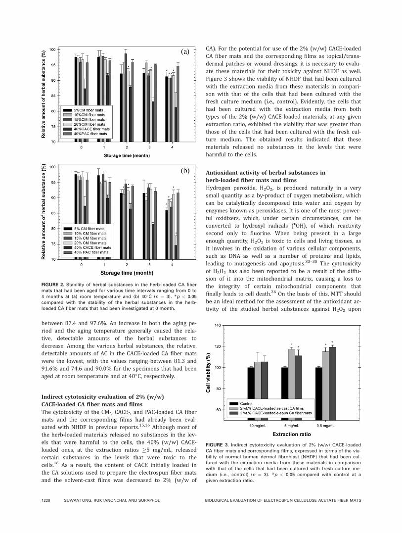
Figure 5 shows the viability of the attached cells on vari-ous substrates, reported as the relative values to the viabil-ity of the cells that had been seeded on TCPS for 2 h. Forany given type of the substrates, the viability of the attachedcells increased with an increase in the cell seeding time,except for that of the cells seeded on the 40% (w/w) CACE-loaded CA fiber mats that showed a decrease in the value at24 h after cell seeding, indicating the toxicity of the loadedsubstances towards the cells.16 Generally, the fibrous sub-strates showed better support for the attachment of thecells than the film counterparts did. Interestingly, while theinclusion of either CACE or PAC in the CA films did notcause a significant increase in the viability of the seededcells from that observed on the neat materials, a significantincrease in such values was observed for the cells that hadbeen seeded on the fiber mats that contained either 2%(w/w) CACE or 40% (w/w) PAC for 24 h, with the lattershowing the greatest value. Quite a similar result wasobtained in the proliferation studies, shown in Figure 6. Thetoxicity of both the fiber mats and the films that contained40% (w/w) CACE became more obvious,16 as the viability
of the cultured cells being found to decrease with increasingthe cell culturing time. Although the CA films that contained2% (w/w) CACE showed a marginal increase in the viabilityof the cultured cells, a significant increase was observed forthe fiber mats that contained either 2% (w/w) CACE or40% (w/w) PAC, with the latter showing the greatest valuesat all cell culturing time points.
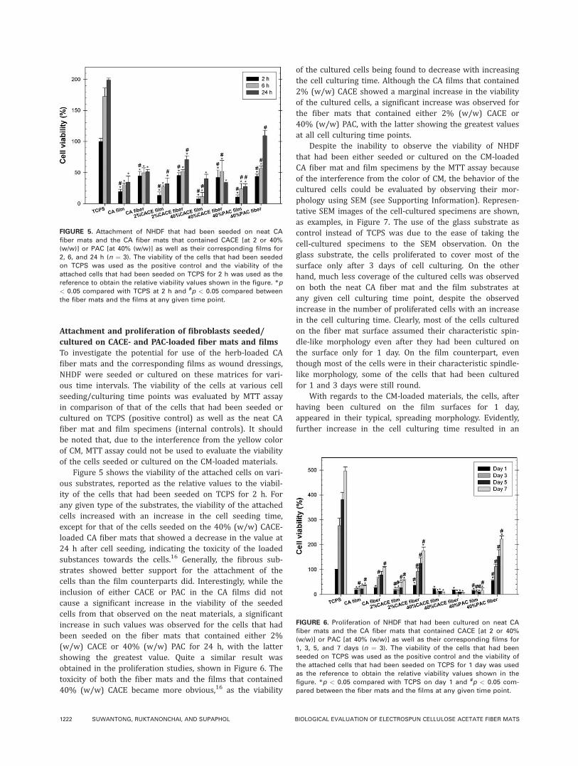
Despite the inability to observe the viability of NHDFthat had been either seeded or cultured on the CM-loadedCA fiber mat and film specimens by the MTT assay becauseof the interference from the color of CM, the behavior of thecultured cells could be evaluated by observing their mor-phology using SEM (see Supporting Information). Represen-tative SEM images of the cell-cultured specimens are shown,as examples, in Figure 7. The use of the glass substrate ascontrol instead of TCPS was due to the ease of taking thecell-cultured specimens to the SEM observation. On theglass substrate, the cells proliferated to cover most of thesurface only after 3 days of cell culturing. On the otherhand, much less coverage of the cultured cells was observedon both the neat CA fiber mat and the film substrates atany given cell culturing time point, despite the observedincrease in the number of proliferated cells with an increasein the cell culturing time. Clearly, most of the cells culturedon the fiber mat surface assumed their characteristic spin-dle-like morphology even after they had been cultured onthe surface only for 1 day. On the film counterpart, eventhough most of the cells were in their characteristic spindle-like morphology, some of the cells that had been culturedfor 1 and 3 days were still round.
With regards to the CM-loaded materials, the cells, afterhaving been cultured on the film surfaces for 1 day,appeared in their typical, spreading morphology. Evidently,further increase in the cell culturing time resulted in an
FIGURE 5. Attachment of NHDF that had been seeded on neat CA
fiber mats and the CA fiber mats that contained CACE [at 2 or 40%
(w/w)] or PAC [at 40% (w/w)] as well as their corresponding films for
2, 6, and 24 h (n ¼ 3). The viability of the cells that had been seeded
on TCPS was used as the positive control and the viability of the
attached cells that had been seeded on TCPS for 2 h was used as the
reference to obtain the relative viability values shown in the figure. *p
< 0.05 compared with TCPS at 2 h and #p < 0.05 compared between
the fiber mats and the films at any given time point.
FIGURE 6. Proliferation of NHDF that had been cultured on neat CA
fiber mats and the CA fiber mats that contained CACE [at 2 or 40%
(w/w)] or PAC [at 40% (w/w)] as well as their corresponding films for
1, 3, 5, and 7 days (n ¼ 3). The viability of the cells that had been
seeded on TCPS was used as the positive control and the viability of
the attached cells that had been seeded on TCPS for 1 day was used
as the reference to obtain the relative viability values shown in the
figure. *p < 0.05 compared with TCPS on day 1 and #p < 0.05 com-
pared between the fiber mats and the films at any given time point.
1222 SUWANTONG, RUKTANONCHAI, AND SUPAPHOL BIOLOGICAL EVALUATION OF ELECTROSPUN CELLULOSE ACETATE FIBER MATS
observed decrease in the number of the cells that had beencultured on these surfaces. In addition, increasing theloaded CM content in the films also resulted in an observeddecrease in the number of the cells. These results suggestthat CM, in the levels that were loaded and released intothe culture medium, pose an adverse effect to the behaviorof the cells. On the contrary, even though the number of thecells that had been cultured on the fiber mat surfaces, forany given CM loading, did not appear to decrease appreci-ably with an increase in the cell culturing time, as in thecase of the film counterparts, the majority of the cells werestill round, even after they had been cultured on the surfa-ces for more than 1 day. This is in contrast to the cells thathad been cultured on the neat CA fiber mat surface, whichappeared to spread rather well over the surface. The resultsclearly demonstrate the adverse effect of CM on the cellbehavior.
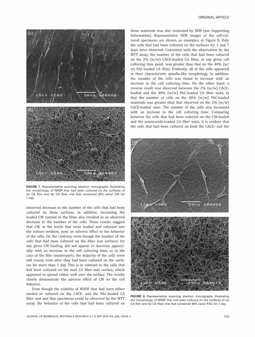
Even though the viability of NHDF that had been eitherseeded or cultured on the CACE- and the PAC-loaded CAfiber mat and film specimens could be observed by the MTTassay, the behavior of the cells that had been cultured on
these materials was also evaluated by SEM (see SupportingInformation). Representative SEM images of the cell-cul-tured specimens are shown, as examples, in Figure 8. Onlythe cells that had been cultured on the surfaces for 1 and 7days were observed. Consistent with the observation by theMTT assay, the number of the cells that had been culturedon the 2% (w/w) CACE-loaded CA films, at any given cellculturing time point, was greater than that on the 40% (w/w) PAC-loaded CA films. Evidently, all of the cells appearedin their characteristic spindle-like morphology. In addition,the number of the cells was found to increase with anincrease in the cell culturing time. On the other hand, areverse result was observed between the 2% (w/w) CACE-loaded and the 40% (w/w) PAC-loaded CA fiber mats, inthat the number of cells on the 40% (w/w) PAC-loadedmaterials was greater than that observed on the 2% (w/w)CACE-loaded ones. The number of the cells also increasedwith an increase in the cell culturing time. Comparingbetween the cells that had been cultured on the CM-loadedand the asiaticoside-loaded CA fiber mats, it is evident thatthe cells that had been cultured on both the CACE- and the
FIGURE 7. Representative scanning electron micrographs illustrating
the morphology of NHDF that had been cultured on the surfaces of
(a) CA film and (b) CA fiber mat that contained 20% (w/w) CM for
1 day.
FIGURE 8. Representative scanning electron micrographs illustrating
the morphology of NHDF that had been cultured on the surfaces of (a)
CA film and (b) CA fiber mat that contained 40% (w/w) PAC for 1 day.
ORIGINAL ARTICLE
JOURNAL OF BIOMEDICAL MATERIALS RESEARCH A | 15 SEP 2010 VOL 94A, ISSUE 4 1223

PAC-loaded CA fiber mats expanded more readily on thesurfaces than they did on the CM-loaded ones, the resultsthat suggested the better biocompatibility of the asiatico-side-loaded materials than the CM-loaded counterparts.
Quantification of synthesized collagenTo investigate whether the loaded herbal substances influ-ence the differentiation of the cultured fibroblasts, theamount of collagen synthesized by NHDF that had been cul-tured on the CM-, CACE-, and PAC-loaded CA fiber mat andthe corresponding film specimens, in comparison with thosethat had been cultured on TCPS, for 7 days was then quanti-fied by the SircolTM collagen assay.* To suitably compare theamount of collagen synthesized by the cells that had beencultured on these different substrates, the amount of thesynthesized collagen, for a given cell-cultured specimen,needed to be normalized by the number of the cells. As pre-viously mentioned, quantification for the number of the cellsthat had been cultured on the CM-loaded materials couldnot be completed with the MTT assay, due to the interfer-ence from the color of the loaded CM. Consequently, thenumber of the cells that had been cultured on the differentsubstrates was then quantified based on the amount of DNAmaterials, using the DNA quantification assay.
Figure 9 shows the normalized amount of collagen syn-thesized by NHDF that had been cultured on the varioussubstrates for 7 days. Evidently, the normalized amount ofthe collagen synthesized by the cells that had been culturedon the neat CA film substrates was significantly lower thanthat by the cells that had been cultured on both the neat CAfiber mat specimens and TCPS. The results are in agreementwith the fact that the number of the cells that had been cul-tured on the fiber mat specimens were generally greaterthan that of the cells that had been cultured on the filmcounterparts, even though the cells on the fiber mat surfa-ces appeared to be more round. The significantly greaternumber of cells on the fiber mat specimens than on the filmcounterparts could result in the cells on the fiber mat speci-mens exhibiting a cell-to-cell mediating interactions that up-regulated the production of collagen and other ECM materi-als faster than those on the film counterparts, hence theobserved greater normalized amount of the synthesized col-lagen on day 7 after cell culturing.
Obviously, the cells that had been cultured on the CM-loaded CA fiber mat specimens synthesized collageneousmatters in the levels that were either equivalent or lowerthan the cells that had been cultured on TCPS. A significantincrease in the normalized amount of the synthesized colla-gen was observed for the cells that had been cultured onthe CA fiber mats that contained either 2% (w/w) CACE or40% (w/w) PAC, with the cells grown on the latter exhibit-ing the greatest value. On the basis of the results shownhere, asiaticoside, either in the form of CACE or PAC, was
more effective than CM in upregulating the production ofcollageneous matters in vitro. However, whether or not asi-aticoside was solely responsible for such an observationrequires further systematic investigation.
CONCLUSION
Because of the implication of asiaticoside and CM aswound-healing mediators, asiaticoside [in the form of eithercrude extract (CACE) or pure substance (PAC)] and CMwere individually incorporated with CA in the form of elec-trospun fiber mats. These herb-loaded fiber mats were eval-uated for the stability of the as-loaded herbal substances,the indirect cytotoxicity, antioxidant activity of the as-loadedherbal substances, the ability to support the attachment andthe proliferation of the seeded/cultured fibroblasts, and theability of the cultured fibroblasts to synthesize collagen. Inmany cases, comparisons were made against the corre-sponding solvent-cast films. The results showed that the as-loaded herbal substances in the fiber mats, measuring eitherat room temperature or 40�C, were stable up to 4 monthsof storage (i.e., �74.6% with respect to the initial contentsloaded in the spinning solutions). The neat CA fiber matsprovided the better support for both the attachment andthe proliferation of the fibroblasts. Similar results wereobtained for all of the herb-loaded CA fiber mats. However,while the cells that had been cultured on the 2% (w/w)CACE-loaded and the 40% (w/w) PAC-loaded CA fiber matsappeared in their phenotypic spindle-like shape, those thathad been cultured on all of the CM-loaded CA fiber matswere round. Among these various herb-loaded CA fiber matsubstrates, the 40% (w/w) PAC-loaded ones exhibited thegreatest ability to support the attachment and the prolifera-tion of the fibroblasts, followed by the 2% (w/w) CACE-loaded ones. The large numbers of the cells that had beenproliferated on these substrates on day 7 agreed particu-larly well with the great amounts of collagen synthesized bythese cells. Despite its ability to mediate the collagen
FIGURE 9. Normalized amount of collagen synthesized by NHDF that
had been cultured on TCPS (i.e., control), neat CA fiber mat and film
specimens, CM-loaded CA fiber mat and film specimens, CACE-
loaded CA fiber mat and film specimens, and PAC-loaded CA fiber
mat and film specimens for 7 days (n ¼ 3). *p < 0.05 compared with
TCPS and #p < 0.05 compared with the neat CA fiber mat or the film
specimens.
*A quantitative dye-binding method designed for the analysisof acid-soluble collagens extracted from mammalian tissuesand collagens released into culture medium by mammaliancells during in vitro culture.
1224 SUWANTONG, RUKTANONCHAI, AND SUPAPHOL BIOLOGICAL EVALUATION OF ELECTROSPUN CELLULOSE ACETATE FIBER MATS
production in the cultured fibroblasts, asiaticoside was infe-rior to CM in terms of the antioxidant activity.
ACKNOWLEDGMENTS
Orawan Suwantong acknowledged a doctoral scholarshipreceived from the Thailand Graduate Institute of Science andTechnology.
REFERENCES1. Reneker DH, Yarin AL. Electrospinning jets and polymer nanofib-
ers. Polymer 2008;49:2387–2425.
2. Lukas D, Sarkar A, Pokorny P. Self-organization of jets in electro-
spinning from free liquid surface: A generalized approach. J Appl
Phys. 2008;103:084309.
3. Liu Y, He J-H. Bubble electrospinning for mass production of
nanofibers. Int J Nonlinear Sci Numer Simul 2007;8:393–396.
4. Liu Y, He J-H, Xu L, Yu J-Y. The principle of bubble electrospin-
ning and its experimental verification. J Polym Eng 2008;28:
55–65.
5. Kim G, Cho Y-S, Kim WD. Stability analysis for multi-jets electro-
spinning process modified with a cylindrical electrode. Eur Polym
J 2006;42:2031–2038.
6. Varabhas JS, Chase GG, Reneker DH. Electrospun nanofibers
from a porous hollow tube. Polymer 2008;49:4226–4229.
7. Khil MS, Cha DI, Kim HY, Kim IS, Bhattarai N. Electrospun nanofi-
brous polyurethane membrane as wound dressing. J Biomed
Mater Res B Appl Biomater 2003;67:675–679.
8. Li WJ, Laurencin CT, Caterson EJ, Tuan RS, Ko FK. Electrospun
nanofibrous structure: A novel scaffold for tissue engineering.
J Biomed Mater Res 2002;60:613–621.
9. Yoshimoto H, Shin YM, Terai H, Vacanti JP. A biodegradable
nanofiber scaffold by electrospinning and its potential for bone
tissue engineering. Biomaterials 2003;24:2077–2082.
10. Suwantong O, Waleetorncheepsawat S, Sanchavanakit N, Pava-
sant P, Cheepsunthorn P, Bunaprasert T, Supaphol P. In vitro bio-
compatibility of electrospun poly(3-hydroxybutyrate) and poly(3-
hydroxybutyrate-co3-hydroxyvalerate) fiber mats. Int J Biol Mac-
romol 2007;40:217–223.
11. Wutticharoenmongkol P, Pavasant P, Supaphol P. Osteoblastic
phenotype expression of MC3T3-E1 cultured on electrospun poly-
mcaprolactone fiber mats filled with hydroxyapatite nanopar-
ticles. Biomacromolecules 2007;8:2602–2610.
12. Kenawy ER, Bowlin GL, Mansfield K, Layman J, Simpson DG,
Sanders EH, Wnek GE. Release of tetracycline hydrochloride from
electrospun poly(ethylene-co-vinylacetate), poly(lactic acid), and a
blend. J Control Release 2002;81:57–64.
13. Taepaiboon P, Rungsardthong U, Supaphol P. Vitamin-loaded
electrospun cellulose acetate nanofiber mats as transdermal and
dermal therapeutic agents of vitamin A acid and vitamin E. Eur J
Pharm Biopharm 2007;67:387–397.
14. Tungprapa S, Jangchud I, Supaphol P. Release characteristics of
four model drugs from drug-loaded electrospun cellulose acetate
fiber mats. Polymer 2007;48:5030–5041.
15. Suwantong O, Opanasopit P, Ruktanonchai U, Supaphol P. Elec-
trospun cellulose acetate fiber mats containing curcumin and
release characteristic of the herbal substance. Polymer 2007;48:
7546–7557.
16. Suwantong O, Ruktanonchai U, Supaphol P. Electrospun cellulose
acetate fiber mats containing asiaticoside or centella asiatica
crude extract and the release characteristics of asiaticoside. Poly-
mer 2008;49:4239–4247.
17. Noh HK, Lee SW, Kim JM, Oh JE, Kim KH, Chung CP, Choi SC,Park WH, Min BM. Electrospinning of chitin nanofibers: Degrada-
tion behavior and cellular response to normal human keratino-cytes and fibroblasts. Biomaterials 2006;27:3934–3944.
18. Han I, Shim KJ, Kim YJ, Im SU, Sung YK, Kim M, Kang IK, Kim
JC. Effect of poly(3-hydroxybutyrate-co-3-hydroxyvalerate) nano-
fiber matrices cocultured with hair follicular epithelial and der-
mal cells for biological wound dressing. Artif Organs 2007;31:
801–808.
19. Zhou Y, Yang D, Chen X, Xu Q, Lu F, Nie J. Electrospun water-
soluble carboxyethyl chitosan/poly(vinyl alcohol) nanofibrous
membrane as potential wound dressing for skin regeneration.
Biomacromolecules 2008;9:349–354.
20. Pierce GF, Mustoe TA. Pharmacologic enhancement of wound
healing. Annu Rev Med 1995;46:467–481.
21. Kartnig T. Herbs, spices and medicinal plants. In: Craker LE,
Simon JE, editors. Herbs, Spices and Medicinal Plants. Phoenix:
Oxyx Press; 1988. p 145–173.
22. Suguna L, Sivakumar P, Chandrakasan G. Effects of centella asiat-
ica extract on dermal wound healing in rats. Ind J Exp Biol 1996;
34:1208–1211.
23. Maquart FX, Bellon G, Gillery P, Wegrowski Y, Borel JP. Stimula-
tion of collagen synthesis in fibroblast cultures by a triterpene
extracted from Centella asiatica. Connect Tissue Res 1990;24:
107–120.
24. Maquart FX, Chastang F, Simeon A, Birembaut P, Gillery P,
Wegrowski Y. Triterpenes from Centella asiatica stimulate extrac-
ellular matrix accumulation in rat experimental wounds. Eur J
Dermatol 1999;9:289–296.
25. Shukla A, Rasik AM, Dhawan BN. Asiaticoside-induced elevation
of antioxidant levels in healing wounds. Phytother Res 1999;13:50–54.
26. Shim PJ, Park JH, Chang MS, Lim MJ, Kim DH, Jung YH, Jew SS,
Park EH, Kim HD. Asiaticoside-mimetics as wound healing agent.
Bioorg Med Chem Lett 1996;6:2937–2940.
27. Sharma RA, Gescher AJ, Steward WP. Curcumin: The story so
far. Eur J Cancer 2005;41:1955–1968.
28. Jayaprakasha GK, Jagan L, Rao M, Sakariah KK. Chemistry and
biological activities of C. longa. Trends Food Sci Technol 2005;15:
533–548.
29. Jayaprakasha GK, Rao J, Sakariah KK. Antioxidant activities of
curcumin, demethoxycurcumin and bisdemethoxycurcumin. Food
Chem 2006;98:720–724.
30. Maheshwari RK, Singh AK, Gaddipati J, Srimal RC. Multiple bio-
logical activities of curcumin: A short review. Life Sci 2006;78:
2081–2087.
31. Sidhu GS, Mani H, Gaddipati JP, Singh AK, Seth P, Banaudha KK,
Patnaik GK, Maheshwari RK. Curcumin enhances wound healing
in streptozotocin induced diabetic rats and genetically diabetic
mice. Wound Repair Regeneration 1999;7:362–374.
32. Gopinath D, Ahmed MR, Gomathi K, Chitra K, Sehgal PK, Jayaku-
mar R. Dermal wound healing processes with curcumin incorpo-
rated collagen films. Biomaterials 2004;25:1911–1917.
33. Hampton MB, Orrenius S. Dual regulation of caspase activity by
hydrogen peroxide: Implications for apoptosis. FEBS Lett 1997;
414:552–556.
34. Tada-Oikawa S, Oikawa S, Kawanishi M, Yamada M, Kawanishi S.
Generation of hydrogen peroxide precedes loss of mitochondrial
membrane potential during DNA alkylation-induced apoptosis.
FEBS Lett 1999;442:65–69.
35. Kowaltowski AJ, Vercesi AE, Rhee SG, Netto LE. Catalases and
thioredoxin peroxidase protect Saccharomyces cerevisiae against
Ca2þ-induces mitochondrial membrane permeabbilization and cell
death. FEBS Lett 2000;473:177–182.
36. Mronga T, Stahnke T, Goldbaum O, Richter-Landsberg C. Mito-
chondrial pathway is involved in hydrogen-peroxide-induced apo-
ptotic cell death of oligodendrocytes. Glia 2004;46:446–455.
37. Ekmekcioglu C, Strauss-Blasche G, Leibetseder VJ, Marktl W. Tox-
icological and biochemical effects of different beverages on
human intestinal cells. Food Res Int 1999;32:421–427.
ORIGINAL ARTICLE
JOURNAL OF BIOMEDICAL MATERIALS RESEARCH A | 15 SEP 2010 VOL 94A, ISSUE 4 1225