In vitro antagonism of Thielaviopsis paradoxa by Trichoderma longibrachiatum
10
In vitro antagonism of Thielaviopsis paradoxa by Trichoderma longibrachiatum Vladimir Sa´nchez 1 , Oscar Rebolledo 2 , Rosa M. Picaso 3 , Elizabeth Ca´rdenas 3 , Jesu´s Co´rdova 1 , Orfil Gonza´lez 1 & Gary J. Samuels 4 1 Departamento de Ingenierı´a Quı´mica, Centro Universitario de Ciencias Exactas e Ingenierı´as, Universidad de Guadalajara, Blvd., Marcelino Garcı´a Barraga ´n 1451, C.P. 44430, Guadalajara, Jal, Mexico; 2 Facultad de Ciencias Biolo ´gicas y Agropecuarias, Universidad de Colima, A.P. 36, C.P. 28100, Tecoma ´n, Col, Mexico; 3 Colegio de Postgraduados, Montecillo, Texcoco, C.P. 56230, Estado de Mexico, Mexico; 4 United States Department of Agriculture, Agricultural Research Service, Systematic Botany and Mycology Lab, Rm. 304, B-011A, BARC-W, Beltsville , MD, 20705, USA Received 17 May 2006; accepted in revised form 28 November 2006 Abstract Seventy-nine Trichoderma strains were isolated from soil taken from 28 commercial plantations of Agave te- quilana cv. ‘Azul’ in the State of Jalisco, Mexico. Nine of these isolates produced nonvolatile metabolites that completely inhibited the growth of Thielaviopsis paradoxa on potato dextrose agar plates. These isolates were identified as Trichoderma longibrachiatum on the basis of their morphology and DNA sequence analysis of two genes (ITS rDNA and translation elongation factor EF-1a). Mycoparasitism of Th. paradoxa by T. longibr- achiatum strains in dual cultures was examined by scanning electron microscopy. The Trichoderma hyphae grew alongside the Th. paradoxa hyphae, but penetration of Thielaviopsis hyphae by Trichoderma was no apparent. Aleurioconidia of Th. paradoxa were parasitized by Trichoderma. Both hyphae and aleurioconidia of Th. pa- radoxa lost turgor pressure, wrinkled, collapsed and finally disintegrated. In liquid cultures, all nine Trichoderma isolates produced proteases, b-1,3-glucanases and chitinases that would be responsible for the degradation of Thielaviopsis hyphae. These results demonstrate that the modes of action of T. longibrachiatum involved against Th. paradoxa in vitro experiments are mycoparasitism and the production of nonvolatile toxic metabolites. Key words: Agave tequilana, Antagonism, Mycoparasitism, Thielaviopsis paradoxa, Trichoderma longibr- achiatum Introduction Thielaviopsis paradoxa is a soilborne plant patho- gen that causes diseases in diverse economically important crop plants. This pathogen has been reported to attack date palm (Phoenix dactylifera) in the United States [1] and Kuwait [2]; pineapple (Ananas comosus) in Brazil [3]; and coconut (Cocos nucifera) in Venezuela [4]. Recently, Th. paradoxa has been found affecting Agave tequilana Weber cv. ‘Azul’ (Agave ‘Azul’ ) in Jalisco, Mexico, causing the wilt and death of >23% of these plants, where roots and stems are mainly attacked [5]. Agave ‘Azul’ is a very important Mexican crop used to produce the traditional beverage of Tequila by an alcoholic fermentation. Traditionally, control of Th. paradoxa is achieved by cultural practices and fungicides such as benomyl and triadimefon [3]. However, it is desirable to integrate biological control agents into disease management plans to minimize the envi- ronmental impact of agrochemicals. Mycopathologia (2007) 163: 49–58 Ó Springer 2007 DOI 10.1007/s11046-006-0085-y
-
Upload
independent -
Category
Documents
-
view
0 -
download
0
Transcript of In vitro antagonism of Thielaviopsis paradoxa by Trichoderma longibrachiatum
In vitro antagonism of Thielaviopsis paradoxa by Trichodermalongibrachiatum
Vladimir Sanchez1, Oscar Rebolledo2, Rosa M. Picaso3, Elizabeth Cardenas3, JesusCordova1, Orfil Gonzalez1 & Gary J. Samuels41Departamento de Ingenierıa Quımica, Centro Universitario de Ciencias Exactas e Ingenierıas, Universidad deGuadalajara, Blvd., Marcelino Garcıa Barragan 1451, C.P. 44430, Guadalajara, Jal, Mexico; 2Facultad deCiencias Biologicas y Agropecuarias, Universidad de Colima, A.P. 36, C.P. 28100, Tecoman, Col, Mexico;3Colegio de Postgraduados, Montecillo, Texcoco, C.P. 56230, Estado de Mexico, Mexico; 4United StatesDepartment of Agriculture, Agricultural Research Service, Systematic Botany and Mycology Lab, Rm. 304,B-011A, BARC-W, Beltsville , MD, 20705, USA
Received 17 May 2006; accepted in revised form 28 November 2006
Abstract
Seventy-nine Trichoderma strains were isolated from soil taken from 28 commercial plantations of Agave te-quilana cv. ‘Azul’ in the State of Jalisco, Mexico. Nine of these isolates produced nonvolatile metabolites thatcompletely inhibited the growth of Thielaviopsis paradoxa on potato dextrose agar plates. These isolates wereidentified as Trichoderma longibrachiatum on the basis of their morphology and DNA sequence analysis of twogenes (ITS rDNA and translation elongation factor EF-1a). Mycoparasitism of Th. paradoxa by T. longibr-achiatum strains in dual cultures was examined by scanning electronmicroscopy. The Trichoderma hyphae grewalongside the Th. paradoxa hyphae, but penetration of Thielaviopsis hyphae by Trichoderma was no apparent.Aleurioconidia of Th. paradoxa were parasitized by Trichoderma. Both hyphae and aleurioconidia of Th. pa-radoxa lost turgor pressure, wrinkled, collapsed and finally disintegrated. In liquid cultures, all nineTrichodermaisolates produced proteases, b-1,3-glucanases and chitinases that would be responsible for the degradation ofThielaviopsis hyphae. These results demonstrate that the modes of action of T. longibrachiatum involved againstTh. paradoxa in vitro experiments are mycoparasitism and the production of nonvolatile toxic metabolites.
Key words: Agave tequilana, Antagonism, Mycoparasitism, Thielaviopsis paradoxa, Trichoderma longibr-achiatum
Introduction
Thielaviopsis paradoxa is a soilborne plant patho-gen that causes diseases in diverse economicallyimportant crop plants. This pathogen has beenreported to attack date palm (Phoenix dactylifera)in the United States [1] and Kuwait [2]; pineapple(Ananas comosus) in Brazil [3]; and coconut (Cocosnucifera) in Venezuela [4]. Recently, Th. paradoxahas been found affecting Agave tequilana Webercv. ‘Azul’ (Agave ‘Azul’ ) in Jalisco, Mexico,
causing the wilt and death of >23% of theseplants, where roots and stems are mainly attacked[5]. Agave ‘Azul’ is a very important Mexican cropused to produce the traditional beverage ofTequila by an alcoholic fermentation.
Traditionally, control of Th. paradoxa isachieved by cultural practices and fungicides suchas benomyl and triadimefon [3]. However, it isdesirable to integrate biological control agents intodisease management plans to minimize the envi-ronmental impact of agrochemicals.
Mycopathologia (2007) 163: 49–58 � Springer 2007DOI 10.1007/s11046-006-0085-y
Trichoderma species are of agricultural impor-tance because they have been used as agents forthe biological control of phytopathogenic fungi,including soilborne, foliar and postharvest patho-gens. Trichoderma species have been utilized tocontrol root diseases caused by nematodes [6]; theymay be also used to promote plant growth [7, 8]and to induce disease resistance in plants [8, 9].The mechanisms by which Trichoderma speciescontrol plant pathogens include mycoparasitism[10, 11], competition for nutrients and growthspace [12], and production of toxic metabolites,including the antibiotics [13, 14].
Effective biological control agents are oftenisolated from the root or rhizosphere of a specificcrop where biocontrol activity is suspected tooccur. Such ‘rhizosphere competent’ strains arebetter adapted to crop field conditions and mayprovide better control of diseases than thosestrains isolates from other sources [15]. Theobjectives of the present research were (i) to isolateTrichoderma strains from soil samples collected onAgave ‘Azul’ fields; and (ii) to investigate themechanisms of biocontrol involved in any antag-onistic activity against Th. paradoxa.
Materials and methods
Fungal strains
In February 2002, soil samples from twenty-eightcommercial plantations of Agave ‘Azul’, wereobtained from four localities of Jalisco State,Mexico (Acatic, Tepatitlan, Las Motas and
Arandas). Five soil samples of approximately 1 kgeach were collected from the rhizosphere (in adepth of 0–15 cm) of each plantation, placed intopolyethylene bags and stored at 4�C. The isolationof Trichoderma strains was carried out by thedilution plating method proposed by Heller andTheiler-Hedtrich [16]. Five Petri dishes containingacidified V8 juice agar were inoculated with 0.1 mlof each diluted soil sample (1/1000, w/v) and thenincubated for 3 days at 25�C. Spores of Tricho-derma strains were finally transferred to Petridishes, containing potato dextrose agar (PDA,Difco), to obtain pure cultures. The nine mostantagonistic of these strains (Table 1) weredeposited in the American Type Culture Collec-tion (ATCC; Manassas, Virginia, USA) and theCentraalbureau voor Schimmelcultures (CBS;Utrecht, The Netherlands). The reference numbersfor each strain are given in Table 1. The phyto-pathogen utilized in this study was Th. paradoxa(ATCC MYA-1387). All the fungal strains weremaintained on PDA slants at 5�C.
Morphological observations
Isolates of Trichoderma were cultured on cornmealdextrose agar (CMD; Difco cornmeal agar, 2%dextrose, 1% antibiotic solution [0.2% of Sigmastreptomycin sulfate and 0.2% of Sigma neomycinsulfate]). Petri dishes (9 cm-diam) containing theabove medium were incubated at 20�C with 12 hcool white fluorescent light and 12 h darkness for1 week. Characteristic of colony appearance as thepresence of diffusing pigment in the medium, the
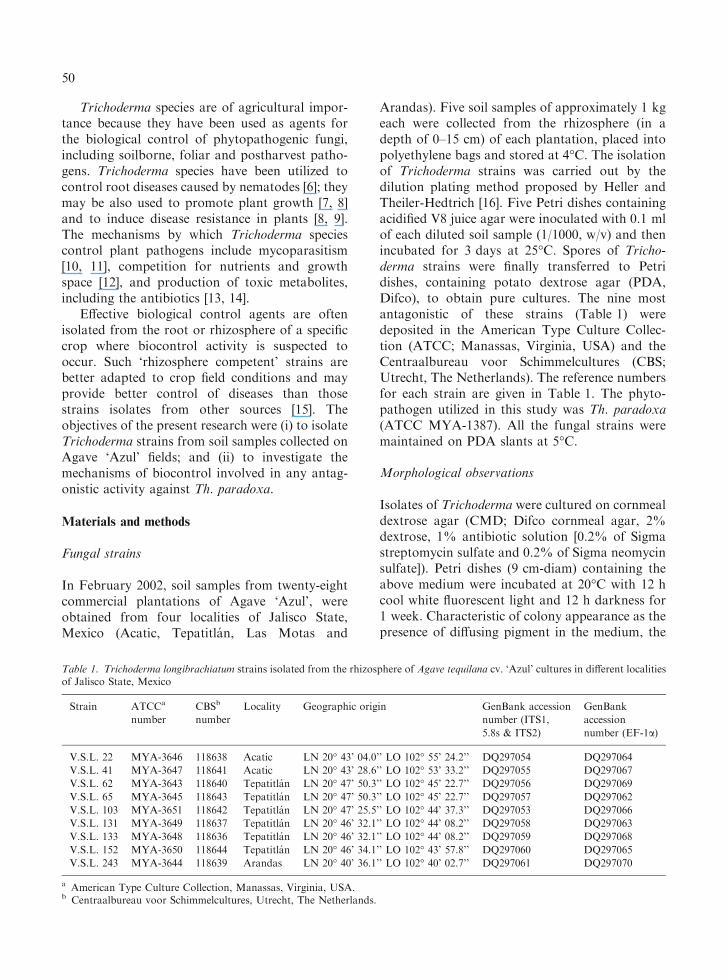
Table 1. Trichoderma longibrachiatum strains isolated from the rhizosphere of Agave tequilana cv. ‘Azul’ cultures in different localitiesof Jalisco State, Mexico
Strain ATCCa
number
CBSb
number
Locality Geographic origin GenBank accession
number (ITS1,
5.8s & ITS2)
GenBank
accession
number (EF-1a)
V.S.L. 22 MYA-3646 118638 Acatic LN 20� 43’ 04.0’’ LO 102� 55’ 24.2’’ DQ297054 DQ297064
V.S.L. 41 MYA-3647 118641 Acatic LN 20� 43’ 28.6’’ LO 102� 53’ 33.2’’ DQ297055 DQ297067
V.S.L. 62 MYA-3643 118640 Tepatitlan LN 20� 47’ 50.3’’ LO 102� 45’ 22.7’’ DQ297056 DQ297069
V.S.L. 65 MYA-3645 118643 Tepatitlan LN 20� 47’ 50.3’’ LO 102� 45’ 22.7’’ DQ297057 DQ297062
V.S.L. 103 MYA-3651 118642 Tepatitlan LN 20� 47’ 25.5’’ LO 102� 44’ 37.3’’ DQ297053 DQ297066
V.S.L. 131 MYA-3649 118637 Tepatitlan LN 20� 46’ 32.1’’ LO 102� 44’ 08.2’’ DQ297058 DQ297063
V.S.L. 133 MYA-3648 118636 Tepatitlan LN 20� 46’ 32.1’’ LO 102� 44’ 08.2’’ DQ297059 DQ297068
V.S.L. 152 MYA-3650 118644 Tepatitlan LN 20� 46’ 34.1’’ LO 102� 43’ 57.8’’ DQ297060 DQ297065
V.S.L. 243 MYA-3644 118639 Arandas LN 20� 40’ 36.1’’ LO 102� 40’ 02.7’’ DQ297061 DQ297070
a American Type Culture Collection, Manassas, Virginia, USA.b Centraalbureau voor Schimmelcultures, Utrecht, The Netherlands.
50
conidial colour and the odour were described andrecorded. Microscopic observations were carriedout from preparations initially mounted in 3%KOH followed by distilled water. Thirty measure-ments for each of the following structures weremade for each isolate: conidium length and width,phialide length and width, base of phialide, cellssupporting the phialides and chlamydospore lengthand width. All measurements were made fromdigital images (magnification of 1600) taken usingthe beta 4.0.2 version of Scion Image (ScionCorp., Frederick, MD, USA). Trichodermastrains were identified using the interactivekey available at http://nt.ars-grin.gov/taxadescrip-tions/keys/TrichodermaIndex.cfm.
DNA extraction, amplification and sequencing
DNA from Trichoderma strains was extracted,according to previously described methods [17].Two gene regions: internal transcribed spacers(ITS1, 5.8S, ITS2 rDNA), and translation elon-gation factor (EF-1a) were amplified by PCRusing the primers ITS1 and ITS4 for ITS [18], andthe primers EF1-728F [19] and TEF1 rev [20] forEF-1a. PCR was carried out using a PTC-200thermocycler (MJ Research, Waltham, MA,USA), in a total volume of 25 ll containing 2.5 llof 10�Buffer (New England Biolabs, Ipswich,MA, USA), 0.5 ll of 10 mM dNTPs, 0.5 ll of10 lM forward primer, 0.5 ll of 10 lM reverseprimer, 0.25 ll of Taq Polymerase (New EnglandBiolabs, Ipswich, MA, USA), 0.5 ll of 10–100 ng/ll genomic DNA and 20.25 ll of distilled water.The amplification program included an initialdenaturalization at 95�C for 5 min, followed by 30cycles of 30 s at 94�C, 30 s at 45�C, 1 min at 72�C,and a final extension of 10 min at 72�C. PCRproducts were purified with the QIAquick PCRpurification kit (Qiagen Inc., Valencia, CA, USA).10 ll of DNA sequencing reactions were per-formed using the Big Dye Terminator CycleSequencing kit (Perkin Elmer Applied Biosystems,Foster City, CA, USA), according to the manu-facturer’s specifications, and the products wereanalyzed directly on ABI prism 3100 DNAsequencer (Applied Biosystems, Foster City, CA,USA).
Molecular analyses
DNA sequences were edited and assembled usingthe software Sequencher 4.2.2 (Gene Codes,Madison, WI, USA), aligned with Clustal X 1.81and then visually adjusted [21]. The sequenceswere BLAST-searched against sequences availablein GenBank. The sequences obtained during thisstudy were deposited in the GenBank, and theaccession numbers are given in Table 1.
Effect of diffusible, nonvolatile metabolites producedby Trichoderma strains on mycelial growthof Thielaviopsis paradoxa
The effect of diffusible, nonvolatile metabolitesproduced by Trichoderma isolates on the growth ofTh. paradoxa was assessed using the cellophanemethod proposed by Dennis and Webster [22]. Asterile cellophane membrane (9 cm-diam) wasplaced on the surface of PDA in plates contained25 ml of medium. A 5 mm mycelial plug from anactively growing edge of a 3 day-old colony ofeach isolate of Trichoderma was placed in thecenter of each cellophane membrane. The Petridishes were incubated for 2 days at 25�C in dark-ness, after which the cellophane membrane withthe fungal mycelia was removed. A 5 mm mycelialplug, taken from an actively growing edge of3 days-old colony of Th. paradoxa, was placed onthe center of the plate in the same position where aTrichoderma isolate was grown. The plates wereincubated at 25�C in darkness for 4 days, and thediameters of each fungal colony were measured.Controls were prepared by placing a 5 mm myce-lial plug of Th. paradoxa instead of the antagoniston the pre-inoculated Thielaviopsis plate. Eachtreatment was replicated four times.
Mycoparasitism
Mycoparasitism of Th. paradoxa by T. longibr-achiatum strains was studied in dual cultures, fol-lowing the procedure described by Dennis andWebster [23]. Petri dishes (9 cm-diam) containing25 ml of PDA were inoculated with mycelial plugs(5 mm-diam) taken from an actively growing edgeof 3 day-old colonies of both fungi. These mycelialdisks were simultaneously placed 5 cm apart from
51
each other on the surface of the medium. Theplates were incubated at 25�C under continuouslight for 7 days. The time for the first contactbetween the antagonist and the pathogen, and theadvance of antagonism on the pathogen colonywere measured. Control plates were prepared byinoculating only Th. paradoxa. Each treatmentwas replicated four times.
Scanning electron microscopy
Mycelial samples (0.5 cm2) were cut from theinteraction zone between the antagonist and phy-topathogen 2 days after the first contact wasestablished in a dual culture. These samples werevapor-fixed with 2% (w/v) osmium tetroxide for20 h at room temperature; air dried and kept in adesiccator [11]. The pieces were mounted on cop-per stubs, coated with gold-palladium using asputter coater (JFC-1100), and examined with ascanning electron microscope (JEOL, JSM-35C) at15 kV. One piece of each T. longibrachiatum strainpaired with the pathogen was examined.
Characterization of extracellular enzymes
Production of extracellular enzymes by T. lon-gibrachiatum strains was studied using the mediumdescribed by Anjani Kumari and Panda [24]. Forchitinase production, 12.5 g of chitin and 2.8 g(NH4)2SO4 were added as the sole carbon andnitrogen sources respectively, and glucose, citricacid, peptone and urea were eliminated from themedium [25]. The initial pH of the media wasadjusted to 5.0 for the production of proteases [26]and b-1,3-glucanases [27], and to 5.6 for chitinaseproduction [25]. Finally, these media were steril-ized (121�C for 20 min). Erlenmeyer flasks(250 ml) containing 100 ml of medium were inoc-ulated with 0.2 ml of a spore suspension (107
spores per ml) of each T. longibrachiatum strainfrom a 5 days-old culture on PDA plates. Theflasks were incubated on a rotary shaker at180 rpm and 30�C for 36 h for b-1,3-glucanases[27], at 28�C for 36 h for proteases and at 28�C for43 h for chitinases [25]. After incubation, 5% (v/v)of mycelial suspension was used to inoculate theproduction media. Five ml of fermented liquidwere removed daily from each flask for 4 days.Samples were then centrifuged at 3500 rpm for10 min at 4�C and the supernatants were filtered
with Whatman No. 1 filter paper and used to assayenzyme activities for proteases and chitinases [25],and b-1,3-glucanases [27]. Each assay consisted offour replicates. Single units of protease, b-1,3-glucanase and chitinase activity were defined as theamount of enzyme that catalyzed the released of1 lmol of tyrosine, glucose and N-acetylglucos-amine per minute, respectively. All cultures andenzyme assays were repeated independently withsimilar results.
Results
Effect of nonvolatile metabolites producedby Trichoderma strains on the Thielaviopsisparadoxa growth
A total of 79 Trichoderma strains were isolatedfrom 28 soil samples collected from the rhizo-sphere of cultivated areas of Agave ‘Azul’ in Jali-sco state, Mexico. The isolated strains werecultured on PDA covered by cellophane mem-branes. After 2 days of incubation, the membranescontaining the biomass of each of the 79 Tricho-derma strains were removed, and Th. paradoxa wasthen inoculated onto the semi-solid media in orderto determinate the effect of nonvolatile metabolitesproduced by Trichoderma on its growth.
Only nine of those strains completely inhibitedthe mycelial growth of Th. paradoxa during the12 days of the experiment. It was hypothesizedthat a diffusible non-volatile substance wasresponsible for the inhibiting of Thielaviopsisgrowth. These nine isolates were selected for fur-ther studies and were identified as T. longibrachi-atum based on their morphological characteristicsand DNA sequence analysis of two genes (ITSrDNA and translation elongation factor EF-1a)(Table 1).
Mycoparasitism
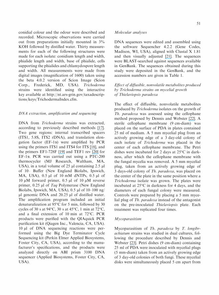
In order to study parasitism of the nine T. lon-gibrachiatum strains on Th. paradoxa, dual cultureswere established. Because the radial growth rates ofthe individual strains of T. longibrachiatum weredifferent, the time of contact with the Thielaviopsisculture differed among the isolates (Table 2).However, the growth of Th. paradoxa stoppedas soon as contact with the Trichoderma was
52
established. Generally, T. longibrachiatum rapidlyovergrew the phytopathogen colony and acomplete invasion and sporulation occurred after5 days of culture (Table 2). One Trichodermastrain, V.S.L. 41, grew slower than the others andonly after 7 days of incubation the invasion andsporulation on the Thielaviopsis colony was fullyconsummated. On the other hand, cultures of Th.paradoxa minus antagonist completely coveredPetri dishes after 6 days.
Mycoparasitism observed by scanning electronmicroscopy
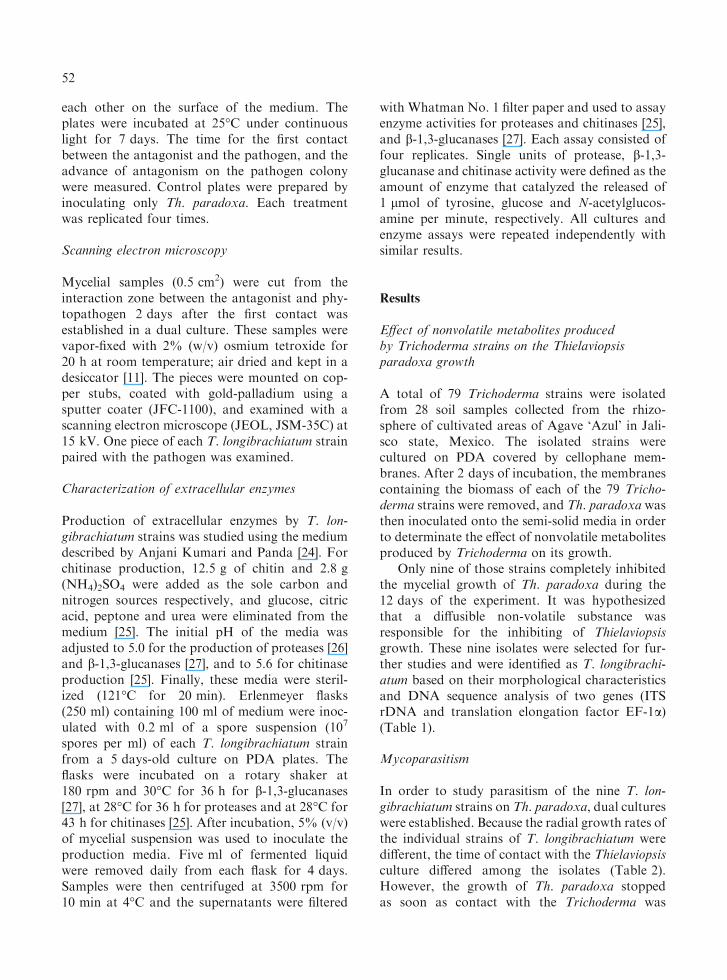
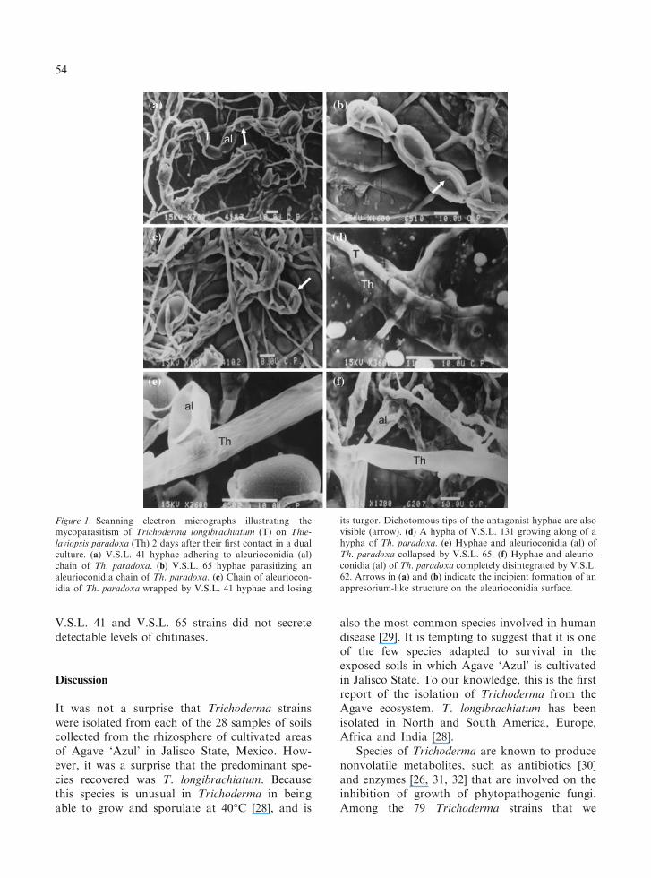
Two days after the first mycelial contact wasestablished in dual cultures, observations of my-coparasitism were carried out by scanning electronmicroscopy (Figure 1). Hyphae of the T. longibr-achiatum strains formed an appressorium-likestructure (Figure 1a, b) in close contact with thealeurioconidial chains of Th. paradoxa. IsolatesV.S.L. 62 and V.S.L. 133 produced penetrationholes (not shown) in Th. paradoxa aleurioconidia.The penetrated aleurioconidia rapidly lost turgorand collapsed (Figure 1c, e). Interestingly, duringthe contact with the host aleurioconidia, hyphae ofV.S.L. 41 strain branched dichotomously at the tip(Figure 1c).
The hyphae of T. longibrachiatum were notobserved to coil around the hyphae of Th. parad-oxa. Instead, the hyphae of T. longibrachiatumgrew alongside the hyphae of Th. paradoxa; how-ever, penetration was not evident (Figure 1d).Despite the absence of visible penetration, Th.paradoxa hyphae lost turgor pressure, wrinkled
and collapsed (Figure 1e). Finally, both hyphaeand aleurioconidia of Th. paradoxa completelydisintegrated (Figure 1f).
Characterization of extracellular enzymes
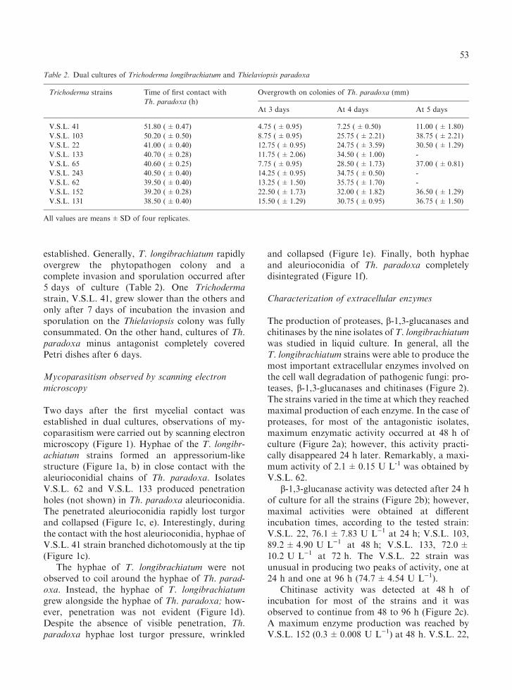
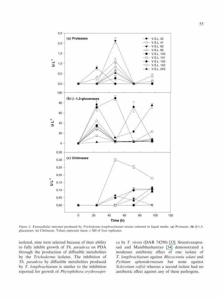
The production of proteases, b-1,3-glucanases andchitinases by the nine isolates of T. longibrachiatumwas studied in liquid culture. In general, all theT. longibrachiatum strains were able to produce themost important extracellular enzymes involved onthe cell wall degradation of pathogenic fungi: pro-teases, b-1,3-glucanases and chitinases (Figure 2).The strains varied in the time at which they reachedmaximal production of each enzyme. In the case ofproteases, for most of the antagonistic isolates,maximum enzymatic activity occurred at 48 h ofculture (Figure 2a); however, this activity practi-cally disappeared 24 h later. Remarkably, a maxi-mum activity of 2.1±0.15 U L-1 was obtained byV.S.L. 62.
b-1,3-glucanase activity was detected after 24 hof culture for all the strains (Figure 2b); however,maximal activities were obtained at differentincubation times, according to the tested strain:V.S.L. 22, 76.1±7.83 U L)1 at 24 h; V.S.L. 103,89.2±4.90 U L)1 at 48 h; V.S.L. 133, 72.0±10.2 U L)1 at 72 h. The V.S.L. 22 strain wasunusual in producing two peaks of activity, one at24 h and one at 96 h (74.7±4.54 U L)1).
Chitinase activity was detected at 48 h ofincubation for most of the strains and it wasobserved to continue from 48 to 96 h (Figure 2c).A maximum enzyme production was reached byV.S.L. 152 (0.3±0.008 U L)1) at 48 h. V.S.L. 22,
Table 2. Dual cultures of Trichoderma longibrachiatum and Thielaviopsis paradoxa
Trichoderma strains Time of first contact with
Th. paradoxa (h)
Overgrowth on colonies of Th. paradoxa (mm)
At 3 days At 4 days At 5 days
V.S.L. 41 51.80 (±0.47) 4.75 (±0.95) 7.25 (±0.50) 11.00 (±1.80)
V.S.L. 103 50.20 (±0.50) 8.75 (±0.95) 25.75 (±2.21) 38.75 (±2.21)
V.S.L. 22 41.00 (±0.40) 12.75 (±0.95) 24.75 (±3.59) 30.50 (±1.29)
V.S.L. 133 40.70 (±0.28) 11.75 (±2.06) 34.50 (±1.00) -
V.S.L. 65 40.60 (±0.25) 7.75 (±0.95) 28.50 (±1.73) 37.00 (±0.81)
V.S.L. 243 40.50 (±0.40) 14.25 (±0.95) 34.75 (±0.50) -
V.S.L. 62 39.50 (±0.40) 13.25 (±1.50) 35.75 (±1.70) -
V.S.L. 152 39.20 (±0.28) 22.50 (±1.73) 32.00 (±1.82) 36.50 (±1.29)
V.S.L. 131 38.50 (±0.40) 15.50 (±1.29) 30.75 (±0.95) 36.75 (±1.50)
All values are means±SD of four replicates.
53
V.S.L. 41 and V.S.L. 65 strains did not secretedetectable levels of chitinases.
Discussion
It was not a surprise that Trichoderma strainswere isolated from each of the 28 samples of soilscollected from the rhizosphere of cultivated areasof Agave ‘Azul’ in Jalisco State, Mexico. How-ever, it was a surprise that the predominant spe-cies recovered was T. longibrachiatum. Becausethis species is unusual in Trichoderma in beingable to grow and sporulate at 40�C [28], and is
also the most common species involved in humandisease [29]. It is tempting to suggest that it is oneof the few species adapted to survival in theexposed soils in which Agave ‘Azul’ is cultivatedin Jalisco State. To our knowledge, this is the firstreport of the isolation of Trichoderma from theAgave ecosystem. T. longibrachiatum has beenisolated in North and South America, Europe,Africa and India [28].
Species of Trichoderma are known to producenonvolatile metabolites, such as antibiotics [30]and enzymes [26, 31, 32] that are involved on theinhibition of growth of phytopathogenic fungi.Among the 79 Trichoderma strains that we
Figure 1. Scanning electron micrographs illustrating themycoparasitism of Trichoderma longibrachiatum (T) on Thie-laviopsis paradoxa (Th) 2 days after their first contact in a dualculture. (a) V.S.L. 41 hyphae adhering to aleurioconidia (al)chain of Th. paradoxa. (b) V.S.L. 65 hyphae parasitizing analeurioconidia chain of Th. paradoxa. (c) Chain of aleuriocon-idia of Th. paradoxa wrapped by V.S.L. 41 hyphae and losing
its turgor. Dichotomous tips of the antagonist hyphae are alsovisible (arrow). (d) A hypha of V.S.L. 131 growing along of ahypha of Th. paradoxa. (e) Hyphae and aleurioconidia (al) ofTh. paradoxa collapsed by V.S.L. 65. (f) Hyphae and aleurio-conidia (al) of Th. paradoxa completely disintegrated by V.S.L.62. Arrows in (a) and (b) indicate the incipient formation of anappresorium-like structure on the aleurioconidia surface.
54
isolated, nine were selected because of their abilityto fully inhibit growth of Th. paradoxa on PDAthrough the production of diffusible metabolitesby the Trichoderma isolates. The inhibition ofTh. paradoxa by diffusible metabolites producedby T. longibrachiatum is similar to the inhibitionreported for growth of Phytophthora erythrosepti-
ca by T. virens (DAR 74290) [33]. Sreenivasapra-sad and Manibhushanrao [34] demonstrated amoderate antibiotic effect of one isolate ofT. longibrachiatum against Rhizoctonia solani andPythium aphanidermatum but none againstSclerotium rolfsii whereas a second isolate had noantibiotic effect against any of these pathogens.
U L
-1
0,0
0,5
1,0
1,5
2,0
2,5
V.S.L. 22 V.S.L. 41 V.S.L. 62 V.S.L. 65 V.S.L. 103 V.S.L. 131 V.S.L. 133 V.S.L. 152 V.S.L. 243
(a) Proteases
U L
-1
0
20
40
60
80
100
(b) β -1,3-glucanases
Time (h)
0 20 40 60 80 100 120
U L
-1
0,00
0,05
0,10
0,15
0,20
0,25
0,30
0,35
(c) Chitinases
Figure 2. Extracellular enzymes produced by Trichoderma longibrachiatum strains cultured in liquid media. (a) Proteases. (b) b-1,3-glucanases. (c) Chitinases. Values represent mean±SD of four replicates.
55
Mycoparasitism of pathogenic fungi byTrichoderma species is proposed as a mechanismof biocontrol and involves enzymes that degradecell wall constituents (CWDEs) [35]. In order totest this premise, T. longibrachiatum strains weredual cultured with Th. paradoxa. In these cultures,contact between the hyphae of T. longibrachiatumand the plant pathogen was achieved at differentculture times (between 38 and 52 h) according tothe tested antagonistic strain (Table 2). After theestablishment of physical contact, all the studiedstrains of T. longibrachiatum were able to growover, to sporulate on, and completely to inhibit themycelial growth of Th. paradoxa. Similar resultshave been found for other Trichoderma/pathogencombinations, e.g., T. harzianum against Rhizoc-tonia solani [10], Crinipellis perniciosa [36], andSclerotium rolfsii [32], T. harzianum, T. atrovirideand T. longibrachiatum against R. cerealis [37].In their study of T. harzianum against R. solani,Benhamou and Chet [10] observed the first contactbetween the two fungi within 2 days of dual cul-ture and a complete inhibition of R. solani imme-diately after this contact. These workers suggestedthat the antagonistic activity of T. harzianum wasnot due to the diffusion of toxic metabolites.
Scanning electron microscope studies showedthat all the isolates of T. longibrachiatum parasit-ized aleurioconidial chains of Th. paradoxa, caus-ing their loss of turgor, collapse and totaldisintegration. To our knowledge this is the firstreport of the direct parasitism of fungal spores ofphytopathogens by species of Trichoderma. In thiscontext, Davanlou et al. [38] demonstrated theparasitism of macroconidia and endoconidial chl-amydospores of Fusarium culmorum by Pythiumoligandrum.
It has been shown that Trichoderma speciesattack the host hyphae, forming coils around them[11, 34, 39]. We did not observe coiling of hyphaeof T. longibrachiatum around hyphae of Th.paradoxa. Sreenivasaprasad and Manibhushanrao[34] observed hyphae of T. longibrachiatum to coilaround hyphae of Rhizoctonia solani but nothyphae of Pythium aphanidermatum. We observedthe antagonistic fungi growing closely along thehyphae of Th. paradoxa (Figure 1d). Althoughpenetration and holes were not observed on thehyphal cells, the loss of turgor pressure, collapseand total desintegration of the phytopathogen
hyphae were noticed routinely (Figure 1 e, f).These observations are consistent with observa-tions by Benhamou and Chet [10, 11]. Sreeni-vasaprasad and Manibhushanrao [34] reported theformation of ‘haustoria’ like short branches inhyphae of T. longibrachiatum that were adjacent tohyphae of P. aphanidermatum, which becamevacuolated and collapsed. We observed similarstructures in our study (Figure 1a, b).
Extracellular enzymes produced by our T. lon-gibrachiatum isolates were examined in liquid cul-tures. It was established that most of these strainsproduced the three main hydrolytic enzymes thatare involved in fungal cell wall degradation(CWDEs). The highest activities of protease,b)1,3-glucanase and chitinase were detectedafter 48 h of culture, respectively for V.S.L.62 (2.1±0.15 U L)1), V.S.L. 103 (89.2±4.90U L)1) and V.S.L. 152 (0.3±0.008 U L)1)(Figure 2). This protease activity was substantiallylower than that produced by T. harzianum T39(58 U L)1) and T. harzianum NCIM1185(54 U L)1) after 5 days of culture [26]. Equally,the chitinase activity was lower than that producedby T. harzianum NCIM1185 (197 U L)1) after6 days of incubation [25]. On the the other hand,b)1,3-glucanase activity was higher than thatproduced by T. harzianum T39 (1.1 U L)1) after5 days and T. harzianum NCIM1185 (1.48 U L)1)after 4 days of culture [26].
Production of CWDEs by Trichoderma speciesdepends on various cultural and physicochemicalconditions such as the carbon and nitrogen sour-ces, inductors, pH, temperature, time of culture,age of the inoculum and agitation [25, 40, 41].Furthermore, high concentrations of glucose andammonium are known to repress the biosynthesisof these extracellular enzymes [42, 43]. Additionalexperiments will be necesary to determine theadequate cultural and physicochemical conditionsto optimize the production of CWDEs for eachstrain of T. longibrachiatum.
The results presented here show that themycoparasitism of T. longibrachiatum on Th.paradoxa is made through the production ofextracellular enzymes that degrade cell wallconstituents of the pathogen, and that diffusible,nonvolatile metabolites are also involved in theantagonism of Th. paradoxa by T. longibrachia-tum.
56
Acknowledgements
This research was supported by grant 42303-Zfrom Consejo Nacional de Ciencia y Tecnologıa(CONACYT). The authors thank Dr. AdnanIsmaiel (BPI) for his support with DNA analysis.The first author thanks the Systematic Botany andMycology Laboratory, United States Departmentof Agriculture (BPI) for granting him permissionto work in that laboratory and Ms. Lutorri Ashley(BPI) for her technical assistance in the laboratory.
References
1. Klotz LJ, Fawcett HS. Black scorch of the date palm
caused by Thielaviopsis paradoxa. J Agric Res 1932; 44:
155–166.
2. Suleman P, Al-Musallam A, Menezes CA. The effect of
solute potential and water stress on black scorch caused
by Chalara paradoxa and Chalara radicicola on date
palms. Plant Dis 2001; 85: 80–83.
3. de Matos AP. Pathological aspects of the pineapple crop
with emphasis on the Fusariosis. Rev Fac Agron (Mar-
acay) 1995; 21: 179–197.
4. Parra D, Morillo F, Sanchez P, Pineda J, Guerra J.
Presencia de Thielaviopsis paradoxa De Seynes Hohn en el
tubo digestivo de Rhynchophorus palmarum Linneo (Cole-
optera: Curculionidae). Entomotropica 2003; 18: 49–55.
5. Fucikovsky L. ‘‘Tristeza’’ and death of Agave tequilana
Weber var. Blue. In: De Boer SH, ed. Plant Pathogenic
Bacteria. Proceedings of the 10th International Confer-
ence on Plant Pathogenic Bacteria, Kluwer Academic
Publishing, Dordrecht, Netherlands, 2001; 359–361.
6. Sharon E, Bar-Eyal M, Chet I, Herrera-Estrella A,
Kleifeld O, Spiegel Y. Biological control of the root-knot
nematode Meloidogyne javanica by Trichoderma harzia-
num. Phytopathology 2001; 91: 687–693.
7. Harman GE. Myths and dogmas of biocontrol. Changes
in perceptions derived from research on Trichoderma
harzianum T22. Plant Dis 2000; 84: 377–393.
8. Harman GE, Petzoldt R, Comis A, Chen J. Interactions
between Trichoderma harzianum strain T22 and maize
inbred line Mo17 and effects of these interactions on
diseases caused by Pythium ultimum and Colletotrichum
graminicola. Phytopathology 2004; 94: 147–153.
9. Yedidia I., Benhamou N., Chet I. Induction of defense
responses in cucumber plants (Cucumis sativus L.) by the
biocontrol agent Trichoderma harzianum. Appl Environ
Microbiol 1999; 65: 1061–1070.
10. Benhamou N, Chet I. Hyphal interactions between
Trichoderma harzianum and Rhizoctonia solani: ultrastruc-
ture and gold cytochemistry of the mycoparasitic process.
Phytopathology 1993; 83: 1062–1071.
11. Benhamou N, Chet I. Cellular and molecular mechanisms
involved in the interaction between Trichoderma harzia-
num and Pythium ultimum. Appl Environ Microbiol 1997;
63: 2095–2099.
12. Sivan A, Chet I. The possible role of competition between
Trichoderma harzianum and Fusarium oxysporum on
rhizosphere colonization. Phytopathology 1989; 79: 198–
203.
13. Howell CR, Stipanovic RD. Gliovirin, a new antibiotic
from Gliocladium virens, and its role in the biological
control of Pythium ultimum. Can J Microbiol 1983; 29:
321–324.
14. Lumsden RD, Locke JC, Adkins ST, Walter JF, Ridout
CJ. Isolation and localization of the antibiotic gliotoxin
produced by Gliocladium virens from alginate prill in soil
and soilless media. Phytopathology 1992; 82: 230–235.
15. Cook RJ. Making greater use of introduced microorgan-
isms for biological control of plant pathogens. Annu Rev
Phytopathol 1993; 31: 53–80.
16. Heller WE, Theiler-Hedtrich R. Antagonism of Chaeto-
mium globosum, Gliocladium virens and Trichoderma viride
to four soil-borne Phytophthora species. J Phytopathol
1994; 141: 390–394.
17. Dodd SL, Lieckfeldt E, Chaverri P, Overton BE, Samuels
GJ. Taxonomy and phylogenetic relationships of two
species of Hypocrea with Trichoderma anamorphs. Mycol
Prog 2002; 1: 409–428.
18. WhiteTJ,BrunsT,LeeS,Taylor J.Amplificationanddirect
sequencing of fungal ribosomal RNA genes for phyloge-
netics. In: Innis MA, Gelfand DH, Sninsky JJ, White TJ,
eds. PCR Protocols: A Guide to Methods, Applications,
Academic Press, San Diego, CA, 1990: 315–322.
19. Carbone I, Kohn LM. A method for designing primer sets
for speciation studies in filamentous ascomycetes. Myco-
logia 1999; 91: 553–556.
20. Samuels GJ, Dodd SL, Gams W, Castlebury LA, Petrini
O. Trichoderma species associated with the green mold
epidemic of commercially grown Agaricus bisporus. Myc-
ologia 2002; 94: 146–170.
21. Thompson JD, Gibson TJ, Plewniak F, Jeanmougin F,
Higgins DG. The Clustal X windows interface: flexible
strategies for multiple sequence alignment aided by quality
analysis tools. Nucleic Acids Res 1997; 24: 4876–4882.
22. Dennis C, Webster J. Antagonistic properties of species
groups of Trichoderma.I Production of non-volatile anti-
biotics. Trans Br Mycol Soc 1971; 57: 25–39.
23. Dennis C, Webster J. Antagonistic properties of species
groups of Trichoderma. III. Hyphal interactions. Trans Br
Mycol Soc 1971; 57 :363–369.
24. Anjani kumari J, Panda T. Studies on critical analysis of
factors influencing improved production of protoplasts
from Trichoderma reesei mycelium. Enzyme Microb
Technol 1992; 14: 241–248.
25. Kapat A, Rakshit SK, Panda T. Optimization of carbon
and nitrogen sources in the medium and environmental
factors for enhanced production of chitinase by Tricho-
derma harzianum. Bioprocess Eng 1996; 15: 13–20.
26. Elad Y, Kapat A. The role of Trichoderma harzianum
protease in the biocontrol of Botrytis cinerea. Eur J Plant
Pathol 1999; 105: 177–189.
27. Theodore K, Panda T. Production of b)1,3-glucanasefrom Trichoderma harzianum in surface and submerged
culture processes and its role on protoplast generation
from Trichoderma reesei mycelium. Bioprocess Eng 1994;
10: 161–166.
57
28. Samuels GJ, Petrini O, Kuhls K, Lieckfeldt E, Kubicek
CP. The Hypocrea schweinitzii complex and Trichoderma
sect. Longibrachiatum. Stud Mycol 1998; 41: 1–54.
29. Chouaki T, Lavarde V, Lachaud L, Raccurt CP, Henne-
quin C. Invasive infections due to Trichoderma species:
report of 2 cases, findings of in vitro susceptibility testing,
and review of the literature. Clin Infect Dis 2002; 35:1360–
1367.
30. Sivasithamparam K, Ghisalberti EL. Secondary metabo-
lism in Trichoderma and Gliocladium. In: Kubicek CP,
Harman GE, eds. Trichoderma and Gliocladium. Vol.
1,Taylor and Francis, London, 1998; 139–191.
31. Lorito M, Harman GE, Hayes CK, Broadway RM,
Tronsmo A, Woo SL, Di Pietro A. Chitinolytic enzymes
produced by Trichoderma harzianum: Antifungal activity
of purified endochitinase and chitobiosidase. Phytopa-
thology 1993; 83: 302–307.
32. El-Katatny MH, Gudelj M, Robra K-H, Elnaghy MA,
Gubitz GM. Characterization of a chitinase and an endo-
b-1,3-glucanase from Trichoderma harzianum Rifai T24
involved in control of the phytopathogen Sclerotium
rolfsii. Appl Microbiol Biotechnol 2001; 56: 137–143.
33. Etebarian HR, Scott ES, Wicks TJ. Trichoderma harzia-
num T39 and T. virens DAR 74290 as potential biological
control agents for Phytophthora erythroseptica. Eur J
Plant Pathol 2000; 106: 329–337.
34. Sreenivasaprasad S, Manibhushanrao K. Antagonistic
potential of Gliocladium virens and Trichoderma longibr-
achiatum to phytopathogenic fungi. Mycopathologia
1990; 109: 19–26.
35. Chet I, Invar J, Hadar Y. Fungal antagonists and
mycoparasites. In: Wicklow DT, Soderstrom B, eds. The
Mycota IV: Environmental and Microbial Relationships,
Springer-Verlag, Berlin, 1997: 165–184.
36. De Marco JL, Lima LHC, Valle de Sousa M, Felix CR. A
Trichoderma harzianum chitinase destroys the cell wall of
the phytopathogen Crinipellis perniciosa, the causal agent
of witches’ broom disease of cocoa. World J Microbiol
Biotechnol 2000; 16: 383–386.
37. Innocenti G, Roberti R, Montanari M, Zakrisson E.
Efficacy of microorganisms antagonistic to Rhizoctonia
cerealis and their cell wall degrading enzymatic activities.
Mycol Res 2003; 107: 421–427.
38. Davanlou M, Madsen AM, Madsen CH, Hockenhull J.
Parasitism of macroconidia, chlamydospores and hyphae
of Fusarium culmorum by mycoparasitic Pythium species.
Plant Pathol 1999; 48: 352–359.
39. Lu Z, Tombolini R, Woo S, Zeilinger S, Lorito M,
Jansson JK. In vivo study of Trichoderma-pathogen-plant
interactions, using constitutive and inducible green fluo-
rescent protein reporter systems. Appl Environ Microbiol
2004; 70: 3073–3081.
40. El-Katatny MH, Somitsch W, Robra K-H, El-Katatny
MS, Gubitz. Production of chitinase and b-1,3-glucanaseby Trichoderma harzianum for control of the phytopath-
ogenic fungus Sclerotium rolfsii. Food Technol Biotechnol
2000; 38: 173–180.
41. Viterbo A, Ramot O, Chernin L, Chet I. Significance of
lytic enzymes from Trichoderma spp. in the biocontrol of
fungal plant pathogens. Ant van Leeuw 2002; 81: 549–556.
42. de la Cruz J, Pintor-Toro JA, Benitez T, Llobell A,
Romero LC. A novel endo-b-1,3-glucanase, BGN13.1,
involved in the mycoparasitism of Trichoderma harzianum.
J Bacteriol 1995; 177: 6937–6945.
43. Donzelli BGG, Harman GE. Interaction of ammonium,
glucose, and chitin regulates the expression of cell wall-
degrading enzymes in Trichoderma atroviride strain P1.
Appl Environ Microbiol 2001; 67: 5643–5647.
Address for correspondence: Vladimir Sanchez, Tecnologico deEstudios Superiores de Villa Guerrero, Carretera FederalToluca-Ixtapan de la Sal, Km 64.5, C.P. 51760, La Finca VillaGuerrero, Estado de Mexico, MexicoFax: +714-146-14-65E-mail: [email protected]
58