Chromian spinel in the Ivrea-Verbano layered igneous complex, western Alps, Italy
Upload
independentCategory
view
0download
0
ORIGINAL PAPER
In situ high-temperature X-ray and neutron powder diffractionstudy of cation partitioning in synthetic Mg(Fe0.5Al0.5)2O4 spinel
Nicoletta Marinoni • Davide Levy • Monica Dapiaggi •
Alessandro Pavese • Ronald I. Smith
Received: 4 February 2009 / Accepted: 9 May 2010 / Published online: 26 May 2010
� Springer-Verlag 2010
Abstract The intra-crystalline cation partitioning over
T- and M-sites in a synthetic Mg(Fe,Al)2O4 spinel sample has
been determined as a function of temperature by Rietveld
structure refinements from powder diffraction data, com-
bining in situ high-temperature neutron powder diffraction
(NPD; POLARIS diffractometer, at ISIS, Rutherford
Appleton Laboratory, UK), to determine the Mg and Al
occupancy factors, with in situ high-temperature X-ray
powder diffraction, to fix the Fe3? distribution. The results
obtained agree with a two-stage reaction, in which an ini-
tial exchange between Fe3? and Mg, the former leaving
and the latter entering tetrahedral sites, is successively
followed by a rearrangement involving also Al. The mea-
sured cation distribution has then been compared and dis-
cussed with that calculated by the Maximum Configuration
Entropy principle, for which only NPD patterns have been
used. The cation partitioning has finally been interpreted in
the light of the configuration model of O’Neill and
Navrotsky.
Keywords Synthetic spinel � X-ray powder diffraction �Neutron powder diffraction � Cation ordering
versus temperature
Introduction
Spinels are ternary oxides (SG: Fd�3m) with an elementary
cell containing 32 oxygen atoms in cubic close packing
arrangement, 16 octahedrally (M) and 8 tetrahedrally (T)
coordinated sites which can host several bi-, tri- and tetra-
valent cations of the first group (typically in natural phases:
Fe3?, Fe2?, Al, Mg, Cr, Zn and Ti). AB2O4 spinel phases
can exhibit two possible extreme cation distributions,
usually addressed to as ‘‘normal’’ or ‘‘inverse’’ configura-
tions. In the former, A2?- and B3?-cations are tetrahedrally
and octahedrally coordinated, respectively, whereas in the
latter the tetrahedral sites are occupied by half of the B3?
cations with the displaced A2? cations octahedrally coor-
dinated in the vacated M-sites. In addition, intermediate
cation arrangements are often observed in nature. Several
studies have shown that spinel cation partitioning changes
as a function of pressure and temperature, indicating that
both energy and configuration entropy play a key role in
determining the atomic distribution over the two different
crystallographic sites (Antao et al. 2005; Lavina et al.
2002; Pavese et al. 1999).
Many investigations, using a variety of complementary
techniques, have looked at intra-crystalline cation order–
disorder in AB2O4 spinels formed in binary systems (see
Lavina et al. 2004 for a review). However, just a modest
number of studies have hitherto been carried out on atomic
partitioning in spinels involving three cation chemical
species. In particular, Mg–Al–Fe3? bearing spinels are
found in a wide range of geological environments, from
N. Marinoni (&) � M. Dapiaggi � A. Pavese
Dipartimento di Scienze della Terra ‘‘Ardito Desio’’, Universita
degli Studi di Milano, Via Botticelli 23, 20133 Milan, Italy
e-mail: [email protected]
D. Levy
Dipartimento di Scienze Mineralogiche e Petrologiche,
Universita degli Studi di Torino, Via Valperga Caluso 35,
10125 Turin, Italy
A. Pavese
Section of Milan, National Research Council,
IDPA, Via Botticelli 23, 20133 Milan, Italy
R. I. Smith
ISIS Facility, Science and Technology Facilities Council,
Rutherford Appleton Laboratory, Harwell Science
and Innovation Campus, Didcot OX11 0QX, UK
123
Phys Chem Minerals (2011) 38:11–19
DOI 10.1007/s00269-010-0377-0

upper mantle to crust, and crystallize under diverse phys-
ical–chemical conditions. Their importance as petrogenetic
indicators has motivated a relevant interest (Sack and
Ghiorso 1991; Gasparik and Newton 1984).
Relatively few studies can be found in literature on Mg–
Al–Fe3? spinels by in situ measurements (Harrison et al.
1999; Pavese et al. 1999), although such experiments are a
key to get an insight into the ‘‘real time’’ behaviour of
phases under non-ambient conditions of temperature and
pressure (Harrison and Putnis 1996; O’Neill et al. 1992;
Wood et al. 1986). The comparatively recent investigation
by Martignago et al. (2003, 2006) reports a thorough in situ
high-temperature X-ray diffraction study on synthetic and
natural Mg–Al–Fe3? spinel specimens. But, foremost, the
atomic partitioning in three cation spinels has usually been
determined exploiting crystal–chemical models that relate
site occupancy factors to bond lengths, rather than provide
the cation distribution by a direct measurement of the site
contents.
In such a light, the present investigation aims at mea-
suring the cation partitioning as a function of T in a synthetic
Mg–Al–Fe3? spinel sample by combining in situ high-
temperature (HT) X-ray powder diffraction (XRPD) and
neutron powder diffraction (NPD). HT-XRPD is effective to
determine the Fe3? distribution with respect to Mg and Al
(whose cations are isoelectronic), while HT-NPD allows
one to reliably discriminate between Mg and Al, exploiting
their neutron scattering length difference (0.5375 and
0.345 9 10-12 cm, respectively). In this view, the present
study represents a novelty, fixing the cation partitioning as a
function of T in Mg–Al–Fe3? spinel through a direct in situ
determination of the site contents, without any crystal–
chemical modelling or a priori assumption. The cation
distribution so determined is also compared with that
derived from the Maximum Configuration Entropy princi-
ple, and then used to calculate the thermodynamic param-
eters of the configuration model (O’Neill and Navrotsky
1984). Moreover, the composition we explore in the present
work, i.e. Fe3?:Al *1, differs from those of Mg–Al–Fe3?
spinels investigated in earlier studies.
Experimental
Spinel synthesis
Mg(Fe,Al)2O4 spinel was synthesised by a high-tempera-
ture solid-state reaction of MgO, Al2O3 and Fe2O3 reagent
grades (Aldrich chemicals). The oxide mixture (MgO,
Al2O3 and Fe2O3 in terms of a mole ratio of 1:0.5:0.5) was
first ground at room temperature by a planetary ball mill for
11 h; then pressed into pellets and treated in a muffle
furnace at 1,100�C, without atmosphere control, for 72 h
with a cooling rate about 20�C/h. The full treatment cycle
was repeated twice. The sample crystallinity was checked
by means of a Bragg–Brentano Panalytical X’pert Pro
MPD diffractometer. The lattice parameters of the syn-
thetic spinel were determined at room temperature by using
Si (NBS SRM 640b) as an internal standard. No trace of the
reagent oxides was found. The spinel refinement yields a
cell edge of 8.2395(7) A, which corresponds to an average
value of the cell parameter between the two end-members,
spinel in sensu stricto [MgAl2O4 with a = 8.0859 A
(Andreozzi et al. 2000, 2001)] and hercinite [MgFe2O4
with a = 8.3600 A (Nakatsuka et al. 2004)].
Mossbauer spectroscopy measurements, here not reported
for the sake of brevity, confirmed iron to occur in the sample
as Fe3?. The Mg(Fe,Al)2O4 composition was determined by
the ICP-AES technique, using a model JV24 of JOBIN–
YVON, and results in Mg0.99(2)(Al0.48(2)Fe0.49(2))2O4-x,
where x is such as to provide electro-neutrality. Taking into
account the uncertainties reported above, we assume the
nominal composition of Mg(Fe0.5Al0.5)2O4, in all the struc-
ture refinements discussed henceforth.
Data collection
High-temperature X-ray diffraction data ([298 K) were
collected with a Philips X’Pert h-h diffractometer (CuKaradiation monochromatised by a flat graphite crystal),
equipped with a furnace (AHT PAP1600), calibrated for
sample position and temperature, from 5� to 100� 2h, with
a 2h-step of 0.02� and counting time of 3 s/step. 20%wt Si
powder (NBS SRM 640b) was added to spinel for goni-
ometer zero calibration at room temperature. High-tem-
perature measurements were recorded at room temperature
and every 100th degree from 373 to 1,273 K, in air.
High-temperature time-of-flight powder neutron dif-
fraction data were collected using the Polaris medium
resolution diffractometer at the ISIS pulsed spallation
source (RAL-Rutherford Appleton Laboratory, UK).
Approximately 3 g of sample was loaded into a thin-walled
cylindrical vanadium sample can and placed in a RAL
furnace, which operates under high vacuum. Diffraction
patterns were collected at the same temperatures as the
XRPD measurements, with the furnace temperature being
monitored and controlled using a type K thermocouple
placed in contact with the outside of the sample can. Data
collected in the backscattering (average 2h = 145�) and 2h*90� detector banks over the d-spacing range 0.4–4.2 A
were used in Rietveld refinements.
Data treatment
All the diffraction patterns were analysed by the full profile
Rietveld method implemented in the GSAS software (Larson
12 Phys Chem Minerals (2011) 38:11–19
123
and Von Dreele 2004) using the EXPGUI graphical interface
(Toby 2001). In the case of the XRPD patterns, the back-
ground was modelled by Chebyshev polynomials with ten
terms; the Bragg diffraction peaks were described by
pseudo-Voigt-like profiles, whose Gaussian and Lorentzian
components of the FWHM were modelled as a function of 2hby FWHMGAUSS
2 = g1tan2h ? g2tanh ? g3 (Caglioti et al.
1958) and FWHMLOR = l1 tanh ? l2/cosh (McCusker et al.
1999), respectively. The coefficients g1, g3, l1 and l2 of the
FWHM were refined. The peak asymmetry was treated
according to Finger et al. (1994).
The lattice parameters, isotropic atomic displacement
parameters and oxygen coordinate (u,u,u) were refined,
along with the site occupancy factors of Fe. Only the Fe
occupancy can be reliably determined by XRPD, since iron
scatters X-rays much more strongly than magnesium and
aluminium which, in turn, are quasi-isoelectronic species. In
this analysis, we have treated Al and Mg like one chemical
species, adopting the X-ray scattering curve of the former.
Such a simplification does not skew our results, as proven by
repeating the calculation using the scattering curve of Mg in
place of Al’s. Note that tests, performed to assess the effect of
the starting cation distribution on the final results, showed
that essentially identical structure parameters were produced
by the least squares regardless of the starting values.
With the NPD patterns, the background was modelled
using a Chebyschev polynomial of the first kind with 14
terms. For structure refinement with Polaris data GSAS peak
function 3 (two back-to-back exponential functions convo-
luted with a pseudo-Voigt function) is used. The exponential
functions describe the moderator effects on the diffraction
peak shape, while the pseudo-Voigt function (which is a sum
of Gaussian and Lorentzian functions) describes the instru-
mental and sample contributions to the peak shape (Larson
and Von Dreele 2004). The starting peak shape and width
parameters, which are contained in the instrument parameter
file, were obtained from analysis of a silicon powder sample
(NBS SRM 640b). The oxygen coordinate (u), the isotropic
atomic displacement parameters, the tetrahedral site neutron
scattering length (btet), constraining that of the M-site (boct)
to the chemical composition, were also refined. The site
occupancy factors of Mg and Al were then determined by
means of btet and boct, keeping the site contents of Fe fixed at
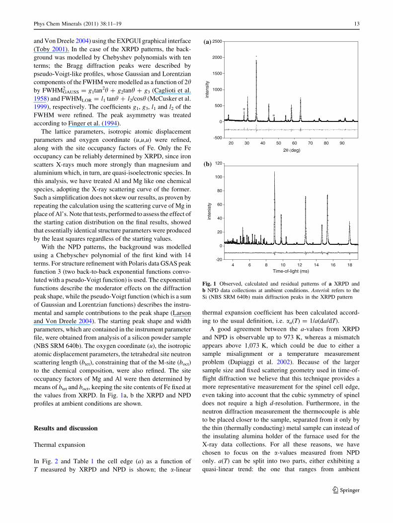
the values from XRPD. In Fig. 1a, b the XRPD and NPD
profiles at ambient conditions are shown.
Results and discussion
Thermal expansion
In Fig. 2 and Table 1 the cell edge (a) as a function of
T measured by XRPD and NPD is shown; the a-linear
thermal expansion coefficient has been calculated accord-
ing to the usual definition, i.e. aa(T) = 1/a(da/dT).
A good agreement between the a-values from XRPD
and NPD is observable up to 973 K, whereas a mismatch
appears above 1,073 K, which could be due to either a
sample misalignment or a temperature measurement
problem (Dapiaggi et al. 2002). Because of the larger
sample size and fixed scattering geometry used in time-of-
flight diffraction we believe that this technique provides a
more representative measurement for the spinel cell edge,
even taking into account that the cubic symmetry of spinel
does not require a high d-resolution. Furthermore, in the
neutron diffraction measurement the thermocouple is able
to be placed closer to the sample, separated from it only by
the thin (thermally conducting) metal sample can instead of
the insulating alumina holder of the furnace used for the
X-ray data collections. For all these reasons, we have
chosen to focus on the a-values measured from NPD
only. a(T) can be split into two parts, either exhibiting a
quasi-linear trend: the one that ranges from ambient
2θ (deg)
inte
nsity
-500
0
500
1000
1500
2000
2500
Time-of-light (ms)
20 30 40 50 60 70 80 90
4 6 8 10 12 14 16 18
inte
nist
y
-20
0
20
40
60
80
100
120
(a)
(b)
* *
Fig. 1 Observed, calculated and residual patterns of a XRPD and
b NPD data collections at ambient conditions. Asterisk refers to the
Si (NBS SRM 640b) main diffraction peaks in the XRPD pattern
Phys Chem Minerals (2011) 38:11–19 13
123

conditions to 773 K (haai & 9.03(2) 10-6 K-1), and that
from 773 K to the highest temperature achieved
(haai & 14.5(1) 10-6 K-1). These two curves intersect
one another at *760 K, which provides an estimation of
the temperature whereat occurs the slope discontinuity.
Note that the slope discontinuity is likely related to the
onset of a steady octahedral/tetrahedral cation diffusion
underlying an atomic rearrangement that will be discussed
in the following sections.
Cation partitioning
In Fig. 3a the Fe3? content in the tetrahedral site versus
temperature determined by XRPD is plotted. Below 773 K
no well-defined partitioning rearrangement is observed,
save a weak indication of a slight Fe-adjustment at 573 K,
a possible token of thermal re-equilibration. At higher
temperature, Fe3? starts apparently moving from tetrahe-
dral to octahedral sites, in keeping with the slope change of
a as a function of T, discussed above. The variation with T
of the tetrahedral site neutron scattering length (Fig. 4)
follows a trend qualitatively similar to that of Fe3? shown
in Fig. 3a, and ‘‘not incompatible’’ with a possible though
very modest and non-quantifiable re-equilibration cation
diffusion onset at about 573 K. Combining the Fe3? par-
titioning from XRPD with the neutron scattering lengths of
the T- and M-sites after NPD refinements, the Mg and Al
distribution has been inferred and plotted in Fig. 3a–c.
Three temperature ranges can be observed and discussed,
using the results and T-points of Table 2:
1. room temperature \ T \ 773 K: no solid evidence of
steady and relevant cation diffusion is observed, within
the limits of the present experimental resolution and of
the order–disorder reaction kinetics;
2. 773 B T \ 973 K: mainly Fe3? and Mg exchanges are
involved, as proven by the regular increase of Fe and
Mg in M-and T-sites, respectively;
3. T C 973 K: Al, too, participates positively in the diffu-
sion process. In particular, Mg-atoms tend to prefer the
T-sites, whereas [IV]Al decreases in favour of a sixfold
coordination. Fe3? keeps diffusing from T- to M-sites.
Note that earlier works, by Martignago et al. (2006) and
Antao et al. (2005), claim to observe signals of cation
rearrangement at higher Ts than ours, i.e. over the 820–
850 K range. Such discrepancies have to be considered
taking into account that: (1) different chemical composi-
tions are involved; (2) synthetic samples often bear such a
variety of deviations from ideal as makes it difficult to
compare one work with another, in particular as far as
reactivity and the related phenomenology are concerned.
A further possible cation partitioning scheme based on
the notion of ‘‘Maximum Configuration Entropy’’ (MCE)
has been used to derive an Mg–Al–Fe3? distribution
accordingly. The present case can be envisaged as an
extension of the MEM technique, developed for electron
Fig. 2 Lattice parameter a as a function of temperature. Filled and
open symbols refer to XRPD and NPD refinements, respectively
Table 1 Structural parameters as a function of temperature: lattice
parameter (A), oxygen positional parameter (u), isotropic thermal
parameters (9100; A2) of oxygen (UO), tetrahedral (UT) and octa-
hedral (UM) cation, determined by XRPD and NPD structural
refinements
T (K) a u UO UT UM Rwp%
XRPD
298 8.2395(2) 0.2581(5) 3.31(9) 2.60(9) 2.46(9) 1.87
373 8.2439(2) 0.2578(4) 3.38(9) 2.48(8) 2.63(9) 1.85
473 8.2509(2) 0.2571(5) 3.70(8) 2.68(9) 2.19(9) 1.79
573 8.2580(2) 0.2575(5) 3.86(7) 2.63(8) 2.51(8) 1.77
673 8.2660(2) 0.2576(5) 4.25(9) 3.23(9) 2.68(8) 1.78
773 8.2750(2) 0.2573(5) 4.51(9) 3.12(9) 2.92(8) 1.83
873 8.2841(1) 0.2569(5) 4.42(7) 3.24(9) 2.99(9) 1.81
973 8.2940(1) 0.2573(5) 4.73(8) 3.60(8) 2.93(9) 1.79
1,073 8.3045(2) 0.2562(5) 4.85(9) 3.48(9) 3.12(9) 1.74
1,173 8.3152(2) 0.2566(5) 4.99(9) 3.55(8) 3.25(8) 1.75
1,273 8.3258(2) 0.2573(5) 5.02(9) 3.48(9) 3.66(8) 1.74
NPD
298 8.2395(1) 0.25954(6) 1.04(1) 0.46(1) 0.54(1) 1.77
373 8.2439(1) 0.25951(8) 1.06(1) 0.51(1) 0.59(1) 1.73
473 8.2507(2) 0.25953(8) 1.19(1) 0.63(1) 0.68(1) 1.66
573 8.2579(2) 0.25944(8) 1.31(1) 0.73(1) 0.82(1) 1.63
673 8.2653(2) 0.25946(9) 1.46(1) 0.87(1) 0.96(1) 1.61
773 8.2734(2) 0.25946(9) 1.58(1) 0.98(1) 1.07(1) 1.59
873 8.2825(1) 0.25942(9) 1.71(1) 1.15(1) 1.18(1) 1.52
973 8.2935(1) 0.25936(9) 1.85(1) 1.29(1) 1.29(1) 1.49
1,073 8.3078(2) 0.25945(9) 2.03(1) 1.48(1) 1.33(2) 1.46
1,173 8.3205(2) 0.25949(9) 2.20(1) 1.67(2) 1.45(2) 1.41
1,273 8.3297(2) 0.25935(9) 2.39(1) 1.79(2) 1.69(2) 1.52
Rwp% = 100H(P
((Iobs - Ical)2w/Iobs
2 w))
14 Phys Chem Minerals (2011) 38:11–19
123

density (see Merli et al. 2009 for applications of mineral-
ogical interest) and here adapted to the determination of
cation partitioning.
This approach allows one to use the results from NPD
data collections only, though at the cost of introducing the
MCE principle, which relies on seeking such a cation
distribution as:
a. maximises the configuration entropy (Sconfig), namely
Sconfig ¼X
a¼1;3
X
j¼1;n
�/jxaj ln xa
j
� �ð1Þ
where /j is the jth-site multiplicity to Z (formula units
per cell) ratio, and xja corresponds to the occupancy
factor of the a-species in the jth-site;
b. fulfils the crystal–chemical constraints below, i.e.
xatet þ 2xa
oct ¼ Ca ð2aÞ
boct ¼ xFeoct � bFe þ xMg
oct � bMg þ xAloct � bAl ð2bÞ
btet ¼ xFetet � bFe þ xMg
tet � bMg þ xA1tet � bAl ð2cÞ
where Ca is the content of the a-species per formula
unit, boct and btet the measured site neutron scattering
lengths and ba the neutron scattering length of the a-
species.
On an energetic viewpoint, such a principle attributes a
driving role to the -T 9 Sconfig contribution in stabilizing a
given atom distribution through a solid at ambient pressure
and temperature T. By means of Eqs. 2a, 2b, 2c, it is
straightforward to demonstrate that this problem reduces to
searching for a maximum in a one-dimension space. The
cation partitioning obtained, set out in Table 2, shows
some differences with respect to that from combining
XRPD with NPD, discussed above. This fact can be
interpreted as a hint that energy plays so vital a part in
stabilizing an atomic rearrangement as to compensate the
mere configuration entropy contribution, which grows in
importance upon T. For instance, above 1,200 K, the cation
distribution measured by HT-XRPD and HT-NPD is
affected by a ‘‘configuration entropy penalty’’ of &2.4 kJ/
mol with respect to that based on MCE.
Cation–oxygen bond lengths
In Fig. 5 the tetrahedral and octahedral cation–oxygen
bond lengths (dtet and doct) as a function of temperature are
Fig. 3 a Fe3?, b Mg and c Al occupancy factors in T-site versus T,
by Rietveld refinements. Fe3? from XRPD; Mg and Al from NPD,
constraining the Fe3? occupancy factors to the values from XRPD.
Data from Table 2
Fig. 4 btet T-site neutron scattering length (10-12 cm) as a function
of T, determined from NPD structural refinements
Phys Chem Minerals (2011) 38:11–19 15
123

reported. A discrepancy is observed between the dtet- and
doct-values from XRPD and NPD, and reflects X-rays being
sensitive to electron density, whereas neutrons interact with
the atomic nuclei directly (Finney 1995). This might be
consistent with the hypothesis of an oxygen atom polari-
zation, characterized by an electron cloud having its
barycentre shifted towards the octahedral site. In addition,
the scattering power of the O atoms relative to Fe, Mg and
Al is higher with neutrons than with X-rays, meaning that
the oxygen atom positions will be determined more accu-
rately in the NPD results (Finney 1995).
At T \700 K the bond length changes are due to mere
thermal expansion, whereas in the higher temperature
regime, there occurs an additional contribution due to
cation diffusion over the T- and M-sites, affecting dtet and
doct through both the average sizes of the cations involved
and the different response to T of their bonds. The XRPD
points in Fig. 5 seem to be more scattered than the NPD
measurements: for instance, the T–O bond lengths from
XRPD range from 1.887(7) to 1.909(7) A´
during cation
diffusion, whereas the ones from NPD look like less
responsive to heating. In addition, the larger momentum
transfer range (smaller d-spacing) measured on Polaris,
which allows better refinement of site occupancy and
thermal vibration parameters, and the better oxygen scat-
tering power (as mentioned in the previous comment) mean
the NPD bond lengths are probably a more reliable indi-
cation of what is happening as the temperature is increased.
Theoretical cation–oxygen distances have been calcu-
lated following two methods:
1. using the tabulated cation–oxygen bond lengths of
Shannon 1976, that are in good agreement with the
ones reported by Lavina et al. 2002 and O’Neill and
Navrotsky 1983 for Al, Mg and Fe3? at room
temperature, and the thermal expansion coefficients
determined by the Megaw’s formula (Megaw 1938);
2. resorting to the model of Hazen and Yang (1999) and
the parameters therein reported.
Both methods use linear models to describe the effect of
a solid solution on the response of a site to heating. The
theoretical curves exhibit a degree of agreement with the
observed ones in terms of general trend and, in particular,
the former model nears our XRPD data up to about 800 K.
Isotropic thermal parameter (Uiso) and oxygen
coordinate (u)
The isotropic thermal parameters as a function of T are
plotted in Fig. 6 and, as expected, those measured by NPD
Table 2 Cation distribution of Fe3?, Mg and Al in T-site, as a function of T
T (K) Fe3? Mg Al Fe3? (MCE) Mg (MCE) Al (MCE) DSMCE-O [J/(mol K)] TDSMCE-O (kJ/mol)
298 0.517 0.365 0.116 0.5471 0.2740 0.1789 0.302 0.090
373 0.518 0.363 0.115 0.5425 0.2740 0.1835 0.202 0.076
473 0.519 0.365 0.116 0.5480 0.2740 0.1780 0.301 0.142
573 0.509 0.384 0.107 0.5441 0.2751 0.1808 0.428 0.245
673 0.511 0.369 0.119 0.5409 0.2762 0.1829 0.312 0.210
773 0.502 0.386 0.111 0.5372 0.2773 0.1855 0.421 0.325
873 0.491 0.403 0.105 0.5312 0.2773 0.1915 0.543 0.474
973 0.480 0.394 0.125 0.5154 0.2845 0.2001 0.411 0.400
1,073 0.463 0.423 0.113 0.5076 0.2845 0.2079 0.626 0.672
1,173 0.435 0.474 0.090 0.4944 0.2913 0.2143 1.139 1.336
1,273 0.402 0.532 0.065 0.4780 0.2962 0.2258 1.896 2.413
The estimated uncertainties for Fe3?, Mg and Al are *0.01, *0.004 and *0.006, respectively. The uncertainties on the Al/Mg occupancy
factors here reported are obtained setting those of Fe at the values from XRPD and do not take into account the uncertainty on the Fe3?
occupancy factors
X (MCE): tetrahedral occupancy factors of the X-species, determined according to the principle of maximum configuration entropy (ME);
DSMCE-O: configuration entropy difference between MCE-configuration and observed partitioning
Fig. 5 Experimental and calculated (T–O) and (M–O) bond lengths
versus T. The size of the symbols is comparable to their estimated
standard deviations
16 Phys Chem Minerals (2011) 38:11–19
123
are smaller than their counterparts determined by XRPD.
These differences are well known to reflect that neutrons
and X-rays have different interactions with atoms. One has
also to take into account that the thermal parameters are
often affected by spurious effects due to the experimental
setup (absorption phenomena, for instance).
As a whole the Uisos, which increase upon heating
because of the increase of the atomic thermal vibration
amplitude, show behaviours consistent with the masses of
the related atoms: T- and M-sites, characterized by com-
parable masses, have similar thermal parameters, while O,
lighter than the others, exhibits a larger one.
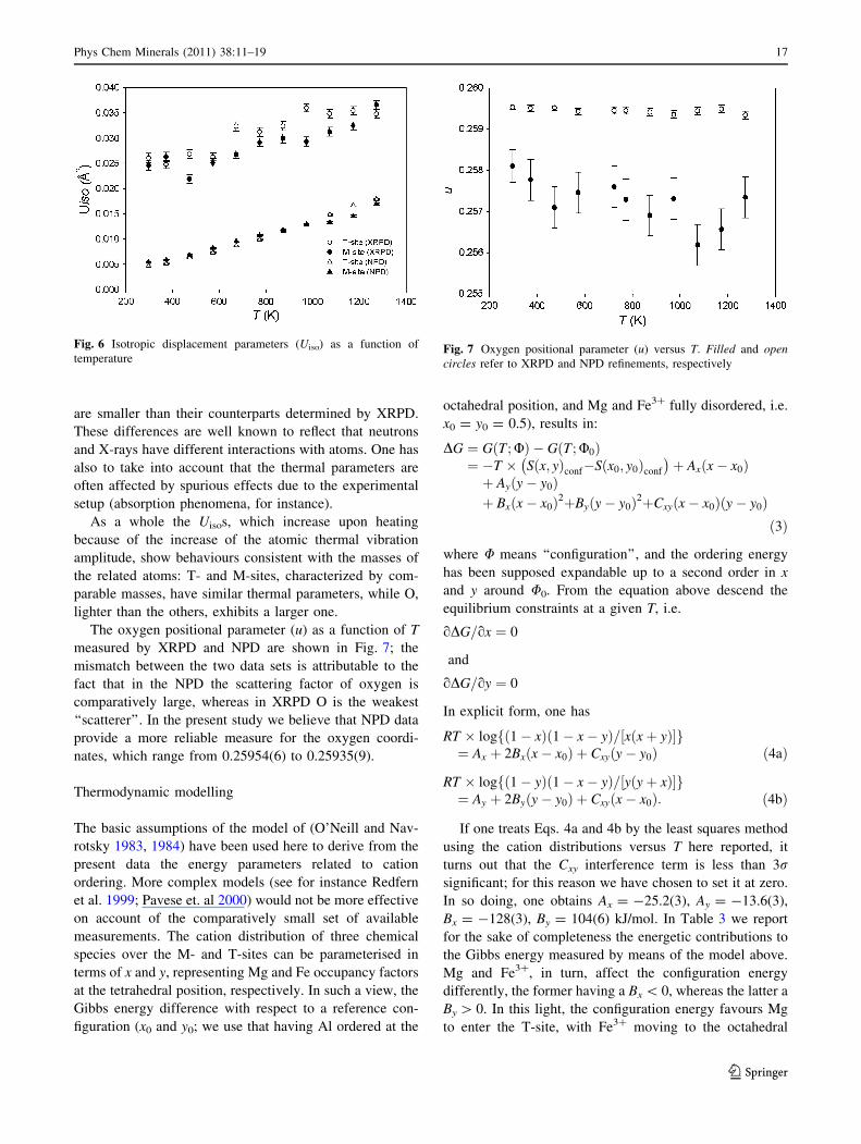
The oxygen positional parameter (u) as a function of T
measured by XRPD and NPD are shown in Fig. 7; the
mismatch between the two data sets is attributable to the
fact that in the NPD the scattering factor of oxygen is
comparatively large, whereas in XRPD O is the weakest
‘‘scatterer’’. In the present study we believe that NPD data
provide a more reliable measure for the oxygen coordi-
nates, which range from 0.25954(6) to 0.25935(9).
Thermodynamic modelling
The basic assumptions of the model of (O’Neill and Nav-
rotsky 1983, 1984) have been used here to derive from the
present data the energy parameters related to cation
ordering. More complex models (see for instance Redfern
et al. 1999; Pavese et. al 2000) would not be more effective
on account of the comparatively small set of available
measurements. The cation distribution of three chemical
species over the M- and T-sites can be parameterised in
terms of x and y, representing Mg and Fe occupancy factors
at the tetrahedral position, respectively. In such a view, the
Gibbs energy difference with respect to a reference con-
figuration (x0 and y0; we use that having Al ordered at the
octahedral position, and Mg and Fe3? fully disordered, i.e.
x0 = y0 = 0.5), results in:
DG ¼ G T; Uð Þ � G T ; U0ð Þ¼ �T � S x; yð Þconf�S x0; y0ð Þconf
� �þ Ax x� x0ð Þ
þ Ay y� y0ð Þþ Bx x� x0ð Þ2þBy y� y0ð Þ2þCxy x� x0ð Þ y� y0ð Þ
ð3Þ
where U means ‘‘configuration’’, and the ordering energy
has been supposed expandable up to a second order in x
and y around U0. From the equation above descend the
equilibrium constraints at a given T, i.e.
oDG=ox ¼ 0
and
oDG=oy ¼ 0
In explicit form, one has
RT � log 1� xð Þ 1� x� yð Þ= x xþ yð Þ½ �f g¼ Ax þ 2Bx x� x0ð Þ þ Cxy y� y0ð Þ ð4aÞ
RT � log 1� yð Þ 1� x� yð Þ= y yþ xð Þ½ �f g¼ Ay þ 2By y� y0ð Þ þ Cxy x� x0ð Þ: ð4bÞ
If one treats Eqs. 4a and 4b by the least squares method
using the cation distributions versus T here reported, it
turns out that the Cxy interference term is less than 3rsignificant; for this reason we have chosen to set it at zero.
In so doing, one obtains Ax = -25.2(3), Ay = -13.6(3),
Bx = -128(3), By = 104(6) kJ/mol. In Table 3 we report
for the sake of completeness the energetic contributions to
the Gibbs energy measured by means of the model above.
Mg and Fe3?, in turn, affect the configuration energy
differently, the former having a Bx \ 0, whereas the latter a
By [ 0. In this light, the configuration energy favours Mg
to enter the T-site, with Fe3? moving to the octahedral
Fig. 6 Isotropic displacement parameters (Uiso) as a function of
temperatureFig. 7 Oxygen positional parameter (u) versus T. Filled and opencircles refer to XRPD and NPD refinements, respectively
Phys Chem Minerals (2011) 38:11–19 17
123

position, in keeping with an experimental cation
partitioning trend (Fig. 3) featured by a degree of
depletion of Fe3? from the T-site, and an enrichment of
Mg therein.
If one searches for that U that minimises DG at a given
temperature, one finds out a cation distribution having a
trend versus T consistent with the observed one, though
bearing different occupancy factor values. Such an aspect
can be interpreted as a token of a partial inadequacy to
model DG, and of the need of a larger data set to better
constraint the thermodynamic parameters.
Conclusions
Structure refinements of X-ray and neutron powder dif-
fraction data on a synthetic Mg(Fe,Al)2O4 spinel have
allowed one to propose an intra-crystalline cation parti-
tioning scheme as a function of temperature. The Fe
occupancy has been fixed by HT-XRPD, whereas HT-NPD
has enabled the Mg and Al partitioning to be determined.
In this light, the present work represents an instance of
synergic use of two ‘‘parallel’’ diffraction experiments
(HT-XRPD and HT-NPD) to fix univocally and directly the
cation partitioning in an Mg–Al–Fe3? spinel.
Cation diffusion onset was qualitatively ‘‘monitored’’ by
observing a discontinuity (*760 K) on the lattice param-
eter thermal expansion coefficient, and some discrepancies
with earlier studies on comparatively similar samples are
revealed.
The sample here studied exhibits a re-adjustment of
Fe3? and Mg, only, entering M- and T-sites, respectively,
up to 973 K, where a rearrangement of the Al partitioning
starts, too. In the high-temperature regime, Fe3? and Al
tend to migrate towards the sixfold coordination site,
whereas Mg shows a preference for the tetrahedral site,
achieving an occupancy factor of 0.532 at 1,273 K. In
particular, the [IV]Fe/[IV]Al ratio, that maintains much lar-
ger than 1 at any T, passes from 4.4, at room temperature,
to 6.2, at 1,273 K, betokening a remarkable propensity of
Al for the octahedral coordination, specially at HT.
Therefore, one in general gathers that: (1) the T-site is
mainly occupied by Fe3? and Mg, in terms of [IV]Fe3?
[[IV]Mg up to 973 K, whereas at higher Ts [IV]Fe3?
\[IV]Mg; (2) Al principally dwells at the octahedral site and
its occupancy factor increases upon heating. This produces
a configuration less and less disordered, as one can realize
by inspecting the trend of the configuration entropy of
Table 3. In this view, this study leads to a cation distri-
bution that differs from those of Martignago et al. (2006),
who observe a tendency towards an increase of disorder as
a function of temperature, proven by an increase of the
configuration entropy in the Fe-richest sample they studied.
However, one has to take into account that the quoted
authors have investigated Mg–Al–Fe spinels with different
compositions with respect to ours, i.e. poorer of Fe. In this
view, the Fe3?:Al ratio seems to be a key to govern the
cation ordering which stabilizes at a given temperature in
Mg–Al–Fe3? bearing spinels. We wish, however, to
underscore once more that synthetic spinels bear deviations
from ideal that might be material to the intra-crystalline
atomic diffusion and related phenomena.
The temperature-dependent cation distribution has also
been inferred by the MCE principle, using NPD data only;
the results suggest that the configuration energy plays a
relevant part in affecting the partitioning, at the expense of
the configuration entropy contribution of the Gibbs energy.
The presented cation distribution as a function of T
has been interpreted using the configuration model of
O’Neill and Navrotsky (1984) [Eq. 3; Ax = -25.2(3), Ay =
-13.6(3), Bx = -128(3), By = 104(6) kJ/mol], which
leads to that [IV]Mg[0.5, [IV]Fe\0.5 and [IV]Fe/[IV]Al �1
promote stability of the Mg–Al–Fe3? solid solution upon
increasing temperature, if Fe3?:Al *1.
Acknowledgments Neutron scattering beam time at ISIS was pro-
vided by the CCLRC (now STFC). The research was financially
supported by Italian MURST and CNR. The authors acknowledge
Prof. Umberto Russo (University of Padova, Italy) for Mossbauer
measurements. The authors are indebted to the reviewers for their
suggestions that have really enhanced the original manuscript.
References
Andreozzi GB, Princivalle F, Skogby H, Della Giusta A (2000)
Cation ordering and structural variations with temperature in
MgAl2O4 spinel: an X-ray single-crystal study. Am Mineral
85:1164–1171
Andreozzi GB, Princivalle F, Skogby H, Della Giusta A (2001)
Cation ordering and structural variations with temperature in
MgAl2O4 spinel: an X-ray single-crystal study. Am Mineral
86:204
Table 3 Gibbs energy contributions, with respect to a reference
configuration (x0, y0)
T (K) -TDS(J/mol/K)
DE(J/mol)
DG(J/mol)
Sobserved
(J/mol/K)
773 -2,066 2,007 -59 25.74
873 -2,296 1,961 -335 25.70
973 -2,853 2,237 -616 26.00
1,073 -3,012 2,126 -886 25.87
1,173 -2,835 1,685 -1,151 25.48
1,273 -2,304 925 -1,379 24.88
DS = S(Uobserved; T)config-S(Ureference)config; DE = Ax(x - x0) ?
Ay(y - y0) ? Bx(x - x0)2 ? By(y - y0)2; DG = G(Uobserved; T) -
G(Ureference; T). x and y correspond to the Mg and Fe3? occupancy
factors at the tetrahedral site, respectively. x0 = 0.5; y0 = 0.5.
U means ‘‘configuration’’
18 Phys Chem Minerals (2011) 38:11–19
123

Antao SM, Hassan I, Parise JB (2005) Cation ordering in magnesio-
ferrite, MgFe2O4, to 982�C using in situ synchrotron X-ray
powder diffraction. Am Mineral 90:219–228
Caglioti G, Paoletti A, Ricci FP (1958) Choice of collimators for a
crystal spectrometer for neutron diffraction. Nucl Instrum 3:223–
228
Dapiaggi M, Artioli G, Petras L (2002) A newly developed high-
temperature chamber for in situ X-ray diffraction: setup and
calibration procedures. Rigaku J 19:35–41
Finger LW, Cox DE, Jephcoat AP (1994) A correction for powder
diffraction peak asymmetry due to axial divergence. J Appl
Crystallogr 27:892–900
Finney JL (1995) The complementary use of X-ray and neutron
diffraction in the study of crystals. Acta Crystallogr B51:447–
467
Gasparik T, Newton RC (1984) The reversed alumina contents of
orthopyroxene in equilibrium with spinel and forsterite in the
system MgO–Al2O3–SiO2. Contrib Mineral Petrol 85:186–196
Harrison RJ, Putnis A (1996) Magnetic properties of the magnetite–
spinel solid solution: Curie temperatures, magnetic susceptibil-
ities and cation ordering. Am Mineral 81:375–384
Harrison RJ, Dove MT, Knight KS, Putnis A (1999) In situ neutron
diffraction study of non-convergent cation ordering in the
(Fe3O4)1-x(MgAl2O4)x spinel solid solution. Am Mineral
84:555–563
Hazen RM, Yang H (1999) Effects of cation substitution and order–
disorder on P–V–T equations of state of cubic spinels. Am
Mineral 84:1956–1960
Larson AC, Von Dreele RB (2004) General structure analysis system
(GSAS). Los Alamos National Laboratory Report, LAUR 86–748
Lavina B, Salviulo G, Della Giusta A (2002) Cation distribution and
structure modelling of spinel solid solutions. Phys Chem
Minerals 29:10–18
Lavina B, Salviulo G, Della Giusta A (2004) Structure modelling and
cation partitioning of spinel solid solutions at high T, Pconditions. Phys Chem Minerals 31:45–51
Martignago F, Dal Negro A, Carbonin (2003) How Cr3? and Fe3?
affect Mg–Al order disorder transformation at high temperature
in natural spinels. Phys Chem Minerals 30:401–408
Martignago F, Andreozzi GB, Dal Negro A (2006) Thermodynamics and
kinetics of cation ordering in natural and synthetic Mg(Al, Fe3?)2O4
spinels from in situ high-temperature X-ray diffraction. Am
Mineral 91:306–312
McCusker LB, Von Dreele RB, Cox DE et al (1999) Rietveld
refinement guidelines. J Appl Crystallogr 32:36–50
Megaw HD (1938) Thermal expansion of crystals in relation to their
structure. Z Kristallogr A100:58–76
Merli M, Pavese A, Curetti N (2009) Maximum entropy method: an
unconventional approach to explore observables related to the
electron density in phengites. Phys Chem Minerals 36:19–28
Nakatsuka A, Ueno H, Nakayama N, Mizota T, Maekawa H (2004)
Single-crystal X-ray diffraction study of cation distribution in
MgAl2O4–MgFe2O4 spinel solid solution. Phys Chem Minerals
31:278–287
O’Neill HSt, Navrotsky A (1983) Simple spinels: crystallographic
parameters, cation radii, lattice energies and cation distribution.
Am Mineral 68:181–194
O’Neill HSt, Navrotsky A (1984) Cation distribution and thermody-
namic properties of binary spinel solid solutions. Am Mineral
69:733–753
O’Neill HSt, Annersten H, Virgo D (1992) The temperature
dependence of the cation distribution in magnesioferrite
(MgFe2O4) from powder XRD structural refinements and
Moessbauer spectroscopy. Am Mineral 77:725–740
Pavese A, Artioli G, Russo U, Hoser A (1999) Cation partitioning
versus temperature in (Mg0.70Fe0.23)Al1.97O4 synthetic spinel by
in situ neutron powder diffraction. Phys Chem Minerals 26:242–
250
Pavese A, Levy D, Hoser A (2000) Cation distribution in synthetic
zinc ferrite (Zn0.97Fe2.02O4) from in situ high temperature
neutron powder diffraction. Am Mineral 85:1497–1502
Redfern SAT, Harrison RJ et al (1999) Thermodynamics and kinetics
of cation ordering in MgAl2O4 spinel up to 1600�C from in situ
neutron diffraction. Am Mineral 84:299–310
Sack RO, Ghiorso MS (1991) Chromian spinel as petrogenetic
indicators: thermodynamics and petrological applications. Am
Mineral 76:827–847
Shannon RD (1976) Revised effective ionic radii and systematic
studies of interatomic distances in halides and chalcogenides.
Acta Crystallogr A32:751–767
Toby BH (2001) EXPGUI, a graphical user interface for GSAS.
J Appl Crystallogr 34:210–213
Wood BJ, Kirkpatrick RJ, Montez B (1986) Order–disorder phenom-
ena in MgAl2O4 spinel. Am Mineral 71:999–1006
Phys Chem Minerals (2011) 38:11–19 19
123
Copyright © 2022 FDOKUMEN