Monitoring cerebral oxygenation during balloon occlusion with multichannel NIRS
Upload
independentCategory
view
6download
0
JOURNAL OF VIROLOGY, Oct. 2011, p. 10710–10718 Vol. 85, No. 200022-538X/11/$12.00 doi:10.1128/JVI.05110-11Copyright © 2011, American Society for Microbiology. All Rights Reserved.
In Situ Cleavage of Baculovirus Occlusion-Derived Virus ReceptorBinding Protein P74 in the Peroral Infectivity Complex�
Ke Peng,1,2 Jan W. M. van Lent,1 Just M. Vlak,1 Zhihong Hu,2 and Monique M. van Oers1*Laboratory of Virology, Wageningen University, Droevendaalsesteeg 1, 6708 PB, Wageningen, Netherlands,1 and
State Key Laboratory of Virology, Wuhan Institute of Virology, Chinese Academy of Sciences,Wuhan 430071, Hubei Province, People’s Republic of China2
Received 13 May 2011/Accepted 8 August 2011
Proteolytic processing of viral membrane proteins is common among enveloped viruses and facilitates virusentry. The Autographa californica multicapsid nucleopolyhedrovirus (AcMNPV) occlusion-derived virus (ODV)protein P74 is part of a complex of essential peroral infectivity factors (PIFs). Here we report that P74 isefficiently cleaved into two fragments of about equal size by an occlusion body (OB) endogenous alkalineprotease during ODV release when AcMNPV OBs are derived from larvae. The cleavage is specific for P74,since the other known peroral infectivity factors in the same complex (PIF1, PIF2, and PIF3) were not cleavedunder the same conditions. P74 cleavage was not observed in OBs produced in three different insect cell lines,suggesting a larval host origin of the responsible protease. P74 in OBs produced in larvae of two different hostspecies was cleaved into fragments with the same apparent molecular mass, indicating that the virus incor-porates a similar alkaline protease from different hosts. Coimmunoprecipitation analysis revealed that the twoP74 subunit fragments remain associated with the recently discovered PIF complex. We propose that under invivo ODV infection conditions, P74 undergoes two sequential cleavage events, the first one being performed byan ODV-associated host alkaline protease and the second carried out by trypsin in the host midgut.
For many enveloped viruses, proteolytic processing of virusmembrane proteins is required to facilitate virus entry. In prin-ciple, the proteolytic cleavage converts a proprotein into anactive conformation and/or exposes the functional domain,e.g., the fusion domain, to mediate virus binding and/or fusion.Cleavage of these proproteins may occur posttranslationallyduring transportation of the protein through the trans-Golginetwork. The F protein of many paramyxoviruses, for instance,is cleaved by the host protein furin (35). Alternatively, theprotein is cleaved during virus entry either at the surface ofrecipient cells, such as influenza virus hemagglutinin (HA),which is cleaved by human airway trypsin-like protease (HAT)(6), or in the endosome, for example, the cleavage of thesevere acute respiratory syndrome coronavirus (SARS-CoV) Sprotein by cathepsin L (31).
Baculoviruses produce two viral phenotypes that follow dif-ferent routes of infection (reviewed by Slack and Arif [32]).The occlusion-derived virus (ODV) derived from occlusionbodies (OBs) initiates infection in midgut epithelial cells ofhost larvae, and the budded virus (BV) is involved in systemicspread of the viral infection within the host. Like the F proteinof paramyxoviruses, the BV fusion (F) protein of Spodopteraexigua multicapsid nucleopolyhedrovirus (SeMNPV) is post-translationally cleaved by furin, and this cleavage is essentialfor the function of F (43).
With baculovirus ODVs, the situation is more complex.ODVs are embedded in a large proteinaceous crystal to formOBs. After being ingested by the insect host per os, the crystal
structure of the OBs dissociates under the alkaline conditionsin the midgut (pH 9 to 11), thereby releasing the ODVs (re-viewed in reference 30). These ODVs then bind and fuse withthe microvilli of midgut epithelium cells to start the first roundof infection (32), and this process is known as oral infection. Sofar, six ODV membrane proteins have been found to be es-sential for oral infectivity, and these were named per os infec-tivity factors (PIFs; described by Kikhno et al. [19]). They areP74 (PIF0), PIF1, PIF2, PIF3, PIF4, and PIF5 (ODV-E56) (9,11, 17, 19, 23, 28). Three PIFs, P74, PIF1, and PIF2, have beenshown to function in ODV binding (16, 23), while the func-tion(s) of the other three PIFs is still unknown. Recently, Penget al. (26) reported that at least four of these PIFs, PIF1, -2,and -3 and P74, are present in the ODV membrane in the formof a complex. The P74 protein, representative of a class ofhighly conserved proteins among baculoviruses, was reportedto undergo a proteolytic cleavage mediated by insect midguttrypsins, releasing a �20-kDa fragment from the N terminus ofP74. This cleavage was shown to be essential for P74-mediatedper os infectivity (34).
For a number of baculoviruses, including AcMNPV, an al-kaline protease was found to be associated with larvae-derivedOBs (L-OBs) (7, 8, 20, 21, 25, 38, 44). This protease wassuggested to function in the degradation of the major matrixprotein of OBs (polyhedrin) and/or to assist in the release ofODVs (25, 38). However, the functional significance of thepresence of this alkaline protease in OBs is not yet fully un-derstood. Recently, Slack and Arif (32) envisioned that thisOB-associated alkaline protease could play a synergistic role ininfection by proteolytic activation of released ODVs, but ex-perimental data to support this supposition are lacking.
In this study we provide evidence that a potential host-derived alkaline protease associated with AcMNPV L-OBs
* Corresponding author. Mailing address: Laboratory of Virology,Wageningen University, Droevendaalsesteeg 1, 6708 PB Wageningen,Netherlands. Phone: 31-317-485082. Fax: 31-317-484820. E-mail:[email protected].
� Published ahead of print on 17 August 2011.
10710

cleaves P74 efficiently and specifically during ODV releaseunder alkaline conditions. We propose a sequential proteolyticcleavage model of P74 accommodating both the OB endoge-nous alkaline protease and midgut host trypsin.
MATERIALS AND METHODS
Viruses, cells, and insects. The AcMNPV E2 strain was used as wild-type (wt)virus in this study. The AcMNPV bacmid is derived from the Bac-to-Bac system(Invitrogen). Spodoptera frugiperda Sf9 cells (Invitrogen) were propagated inSf-900II medium (Invitrogen) with 5% fetal bovine serum (FBS). BTI-Tn-5B1-4cells (Tn-High Five; Invitrogen) were grown in Express Five SFM medium(Invitrogen) and Se-UCR (15) in Grace’s insect medium (Sigma) supplementedwith 5% FBS. All three cell lines were propagated as monolayers at 27°C.Spodoptera exigua and Trichoplusia ni larvae were reared on an artificial diet at27°C, 40% humidity, with a 16/8 h (light/dark) photoperiod. In this study, OBsproduced from cell culture or larvae have been named C-OB or L-OB, respec-tively. Correspondingly, ODVs purified from C-OB or L-OB are named C-ODVor L-ODV. The origin of OBs and ODVs is also indicated, e.g., Sf9-C-ODVindicates that the ODVs are purified from C-OBs produced in Sf9 cells, whileSe-L-OB indicates that the L-OBs are produced in S. exigua larvae.
P74 deletion and repair viruses. An AcMNPV bacmid with a deletion of thep74 open reading frame (ORF) was constructed as previously described (26). Forthis, a PCR product with 50-bp overhangs homologous to flanking regions of thep74 gene was generated with primer pair p74-del-F (GCGGCGTCGTGTCCAACACGACGCCGTTCATGTACATGCAGACCTCCGAGACTGCTCGGATCCACTAGTAACG) and p74-del-R (GCGTATAGCGAGCTAGTGGCTAACGCTTGCCCCACCAAAGTAGATTCGTCAAACCTCTAGATGCATGCTCG) with Phusion polymerase (Finnzymes). Primers were designed to replacea fragment from nucleotide (nt) 301 to 1638 of the p74 ORF with the chloram-phenicol resistance gene (cat). Primer sequences for generating the cat gene areunderlined. To repair the p74 deletion bacmid, the coding sequence of the p74gene plus its putative promoter region (from position �150 relative to the ATGstart codon) were amplified by PCR using the primer pair p74-rep-F (GCGCCATGGGCACAACGAAATGATTATATATTA) and p74-rep-R (GCGGCATGCTTATTTGTCATCGTCATCCTTATAGTCAAATAACAAATCAATTGTTTTATAAT). NcoI and SphI restriction sites were introduced by the underlinedsequences of the primers and used to clone the sequenced PCR product into amodified pFastBacDual vector that lacked the p10 promoter and contains theentire polyhedrin gene (pFBD�P10-polh) as described before (26). A Flag tagsequence (shown in italics in the sequence) was introduced by the reverse repairprimer to generate a P74 protein with a C-terminal Flag tag. The resultingplasmid was used to construct a “p74 repair” bacmid using the Bac-to-Bactransposition protocol (Invitrogen). The bacmids were used to transfect Sf9 cellsto generate mutant and repair viruses.
Virus production and purification. A wt AcMNPV BV stock was generated aspreviously described (26). To produce and purify C-OBs, Sf9, Tni-High Five, orSe-UCR cells were infected with BVs at a multiplicity of infection of 5 50% tissueculture infectious dose units per cell, and infected cells were collected at 5 dayspostinfection by low-speed centrifugation. The cell pellet was washed 3 timeswith deionized water (Millipore) and then resuspended in 0.2% SDS and incu-bated at 37°C for 2 h or 4°C overnight with gentle rocking. The cell suspensionwas sonicated and centrifuged at 4,000 � g for 20 min. The pellet was washed andresuspended in phosphate-buffered saline (PBS) and centrifuged over a 35-to-65% (wt/wt) sucrose gradient at 98,000 � g for 60 min at 4°C. The banded OBswere diluted in PBS and collected by centrifugation at 55,000 � g for 60 min. TheOB pellet was washed 3 times with PBS and stored at 4°C for later use.
L-OBs were produced in S. exigua or T. ni larvae (third instar) after oralinfection with C-OBs produced in Sf9 cells. To purify L-OBs from infectedlarvae, the liquefied or liquefying larvae were collected and ground in deionizedwater containing 0.5% SDS and 1% Triton X-100. The suspension was homog-enized, sonicated, and passed through two layers of cheesecloth. The L-OB-containing material was then centrifuged at 4,000 � g for 20 min. The pellet waswashed twice with 0.5% SDS–1% Triton X-100 in deionized water, resuspendedin PBS, and purified through a sucrose gradient in the same way as C-OBs. Theresulting L-OB pellet was washed twice with 1 M NaCl and once with PBS.Finally, the L-OBs were stored in PBS.
ODV purification under various conditions. Prior to ODV purification, thePBS in the OB suspensions was replaced with deionized water. For heat inacti-vation of the endogenous protease, the L-OB suspension was heated at 80°C for40 min (heated OB) prior to ODV purification (25). ODVs were either purifiedat room temperature or at 4°C (low-temperature purification). For low-temper-
ature purification, OBs were incubated on ice water (0°C) for 40 min. OBs werethen treated with cold (4°C) alkaline DAS solution (0.1 M Na2CO3, 166 mMNaCl, and 10 mM EDTA; pH 10.5) for 10 min at 4°C, and subsequent steps ofpurification were also performed at 4°C. In experiments in which the endogenousprotease needed to be inhibited, NaOH was added to the ODV-releasing solu-tion at a final concentration of 0.1 M at various time points after the addition ofDAS. For time point 0 min, NaOH was mixed with DAS solution, and thismixture was used to dissolve OBs. Nondissolved debris was removed by centrif-ugation at 1,500 � g for 3 min. The supernatant was collected, and ODVs werepelleted by centrifugation at 20,600 � g for 25 min at 4°C.
In experiments where ODVs needed to be purified over a sucrose gradient,ODVs were released from OBs at 4°C, and the ODV-containing supernatant waslayered onto a 25-to-65% (wt/wt) continuous sucrose gradient in 10 mM Tris-HCl, pH 7.5, and centrifuged at 98,000 � g for 90 min at 4°C. The multiple virusbands were collected, diluted in 0.1� TE (1 mM Tris-HCl [pH 7.5], 0.1 mMEDTA), and collected by centrifugation at 55,000 � g for 60 min at 4°C.
Coimmunoprecipitation. For coimmunoprecipitation (co-IP) analysis, Se-L-OB of the Ac-Rep-P74-Flag virus was used in order to detect both P74-N andP74-C-Flag subunits. For each experiment L-ODVs were liberated from 2 � 109
L-OBs, and the L-ODVs were purified through a sucrose gradient as describedabove. The ODV pellet was resuspended in 600 �l IP buffer (25 mM Tris-HCl,150 mM NaCl; pH 7.2) containing 0.5% Triton X-100, sonicated briefly, andincubated at 4°C for 3 h with gentle rotation. Meanwhile, 15 �l of PIF1 antiserumor preimmune serum (26) was incubated with a 20-�l bed volume of proteinG-agarose (Pierce) in 500 �l of IP buffer at 4°C for 3 h. Subsequent steps of co-IPwere performed as previously described (26). A portion of the supernatant wasreserved as input sample. The captured proteins and input sample were treatedwith Laemmli buffer (125 mM Tris-HCl, 2% sodium dodecyl sulfate, 5% 2-mer-captoethanol, 10% glycerol, 0.001% bromophenol blue; pH 6.8) at 95°C for 10min. Proteins were then separated by 10% SDS-PAGE.
Polyclonal antibody generation for P74. A 5� segment of 1,299 bp of the p74ORF was selected for making an expression construct for P74 polyclonal anti-body (P74-PAb) generation. The DNA fragment was PCR amplified with theprimers P74-Ab-F (GCGGGATCCATGGCGGTTTTAACAGCC) and P74-Ab-R (GCGAAGCTTTTAAGCGATTCGAGTTAACGCT). BamHI andHindIII restriction sites (underscored) were introduced by the forward andreverse primers, respectively. The amplified DNA was ligated into the pJet1.2vector (Fermentas) for sequencing analysis and then cloned into the pET28avector (Novagen) between the BamHI and HindIII sites for protein expression.The pET28a-P74 construct codes for an N-terminal fusion of a 6�His tag withthe AcMNPV P74 fragment. Escherichia coli BL21 cells were transformed withpET28a-P74, and expression was induced with isopropyl-�-D-thiogalactopyrano-side (IPTG; 1 mM) for 4 h at 37°C. After washing with PBS three times, thebacteria pellet was resuspended in lysis buffer (0.1% SDS, 1% Triton X-100, PBS[pH 7.4]), incubated at 37°C with gentle rotation for 30 min, and then sonicated.Protein purification from the bacterial inclusion bodies was performed as previ-ously described (26). The purified protein was analyzed with SDS-PAGE, Coo-massie brilliant blue staining, and Western blotting with anti-His and P74monoclonal antibodies (P74 MAb) to determine the specificity, purity, and con-centration of the protein. Purified protein was sent to Eurogentec (Seraing,Belgium) to generate polyclonal antibodies in rabbits.
SDS-PAGE, antibodies, and Western blot analysis. ODV samples were heatedin Laemmli buffer at 95°C for 10 min and separated by 10% SDS-PAGE. Fornonreducing SDS-PAGE, samples were treated with Laemmli buffer lacking2-mercaptoethanol. The nonreduced samples were also heated at 95°C for 10min. Anti-His monoclonal antibody from mouse and anti-Flag polyclonal anti-body from rabbit were purchased from Sigma. The P74 MAb (11), anti-PIF1,anti-PIF2, and anti-PIF3 antibodies have previously been described (26). West-ern blot analysis was performed as previously described (26, 27). Dilutions of theprimary antibodies were as follows: His MAb (1:2,000 dilution), Flag PAb (2�g/ml), PIF1 PAb (1:2,000 dilution), PIF2 PAb (1:2,000 dilution), PIF3 PAb(1:1,000 dilution), P74 MAb (1:50 dilution), and P74 PAb (1:2,000 dilution).Membranes were then probed with appropriate alkaline phosphatase-conjugatedsecondary antibodies (Sigma). The signal was developed with nitroblue tetrazo-lium–5-bromo-4-chloro-3-indolylphosphate (Roche).
Computational analysis. Conserved domain analysis of the P74 amino acidsequence was performed with the NCBI conserved domain server (http://www.ncbi.nlm.nih.gov/Structure/cdd/wrpsb.cgi). Transmembrane domain predictionof P74 was performed with the TMHMM 2.0 server as described before (27).Disulfide bonds were predicted by using the DiANNA server (http://clavius.bc.edu/�clotelab/DiANNA/) (12).
VOL. 85, 2011 CLEAVAGE OF BACULOVIRUS P74 BY AN OB PROTEASE 10711
RESULTS
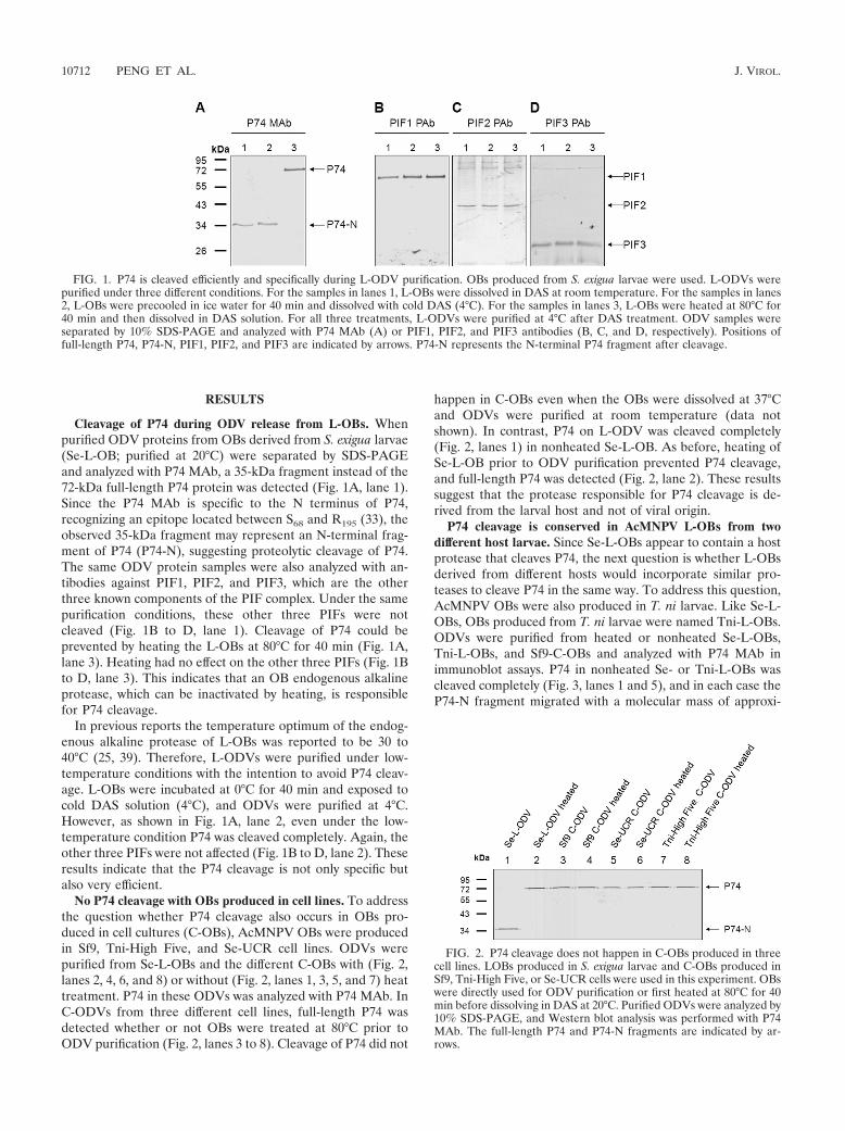
Cleavage of P74 during ODV release from L-OBs. Whenpurified ODV proteins from OBs derived from S. exigua larvae(Se-L-OB; purified at 20°C) were separated by SDS-PAGEand analyzed with P74 MAb, a 35-kDa fragment instead of the72-kDa full-length P74 protein was detected (Fig. 1A, lane 1).Since the P74 MAb is specific to the N terminus of P74,recognizing an epitope located between S68 and R195 (33), theobserved 35-kDa fragment may represent an N-terminal frag-ment of P74 (P74-N), suggesting proteolytic cleavage of P74.The same ODV protein samples were also analyzed with an-tibodies against PIF1, PIF2, and PIF3, which are the otherthree known components of the PIF complex. Under the samepurification conditions, these other three PIFs were notcleaved (Fig. 1B to D, lane 1). Cleavage of P74 could beprevented by heating the L-OBs at 80°C for 40 min (Fig. 1A,lane 3). Heating had no effect on the other three PIFs (Fig. 1Bto D, lane 3). This indicates that an OB endogenous alkalineprotease, which can be inactivated by heating, is responsiblefor P74 cleavage.
In previous reports the temperature optimum of the endog-enous alkaline protease of L-OBs was reported to be 30 to40°C (25, 39). Therefore, L-ODVs were purified under low-temperature conditions with the intention to avoid P74 cleav-age. L-OBs were incubated at 0°C for 40 min and exposed tocold DAS solution (4°C), and ODVs were purified at 4°C.However, as shown in Fig. 1A, lane 2, even under the low-temperature condition P74 was cleaved completely. Again, theother three PIFs were not affected (Fig. 1B to D, lane 2). Theseresults indicate that the P74 cleavage is not only specific butalso very efficient.
No P74 cleavage with OBs produced in cell lines. To addressthe question whether P74 cleavage also occurs in OBs pro-duced in cell cultures (C-OBs), AcMNPV OBs were producedin Sf9, Tni-High Five, and Se-UCR cell lines. ODVs werepurified from Se-L-OBs and the different C-OBs with (Fig. 2,lanes 2, 4, 6, and 8) or without (Fig. 2, lanes 1, 3, 5, and 7) heattreatment. P74 in these ODVs was analyzed with P74 MAb. InC-ODVs from three different cell lines, full-length P74 wasdetected whether or not OBs were treated at 80°C prior toODV purification (Fig. 2, lanes 3 to 8). Cleavage of P74 did not
happen in C-OBs even when the OBs were dissolved at 37°Cand ODVs were purified at room temperature (data notshown). In contrast, P74 on L-ODV was cleaved completely(Fig. 2, lanes 1) in nonheated Se-L-OB. As before, heating ofSe-L-OB prior to ODV purification prevented P74 cleavage,and full-length P74 was detected (Fig. 2, lane 2). These resultssuggest that the protease responsible for P74 cleavage is de-rived from the larval host and not of viral origin.
P74 cleavage is conserved in AcMNPV L-OBs from twodifferent host larvae. Since Se-L-OBs appear to contain a hostprotease that cleaves P74, the next question is whether L-OBsderived from different hosts would incorporate similar pro-teases to cleave P74 in the same way. To address this question,AcMNPV OBs were also produced in T. ni larvae. Like Se-L-OBs, OBs produced from T. ni larvae were named Tni-L-OBs.ODVs were purified from heated or nonheated Se-L-OBs,Tni-L-OBs, and Sf9-C-OBs and analyzed with P74 MAb inimmunoblot assays. P74 in nonheated Se- or Tni-L-OBs wascleaved completely (Fig. 3, lanes 1 and 5), and in each case theP74-N fragment migrated with a molecular mass of approxi-
FIG. 1. P74 is cleaved efficiently and specifically during L-ODV purification. OBs produced from S. exigua larvae were used. L-ODVs werepurified under three different conditions. For the samples in lanes 1, L-OBs were dissolved in DAS at room temperature. For the samples in lanes2, L-OBs were precooled in ice water for 40 min and dissolved with cold DAS (4°C). For the samples in lanes 3, L-OBs were heated at 80°C for40 min and then dissolved in DAS solution. For all three treatments, L-ODVs were purified at 4°C after DAS treatment. ODV samples wereseparated by 10% SDS-PAGE and analyzed with P74 MAb (A) or PIF1, PIF2, and PIF3 antibodies (B, C, and D, respectively). Positions offull-length P74, P74-N, PIF1, PIF2, and PIF3 are indicated by arrows. P74-N represents the N-terminal P74 fragment after cleavage.
FIG. 2. P74 cleavage does not happen in C-OBs produced in threecell lines. LOBs produced in S. exigua larvae and C-OBs produced inSf9, Tni-High Five, or Se-UCR cells were used in this experiment. OBswere directly used for ODV purification or first heated at 80°C for 40min before dissolving in DAS at 20°C. Purified ODVs were analyzed by10% SDS-PAGE, and Western blot analysis was performed with P74MAb. The full-length P74 and P74-N fragments are indicated by ar-rows.
10712 PENG ET AL. J. VIROL.

mately 35 kDa. Heat treatment blocked P74 cleavage in bothSe- and Tni-L-OBs, and full-length P74 was detected (Fig. 3,lanes 2 and 6). In contrast, P74 on C-ODVs was not cleavedand was present as a full-length protein no matter whether theSf9-C-OBs were heated or not (Fig. 3, lanes 3 and 4). The factthat P74 in L-OBs derived from different hosts was cleavedinto similar, if not identical, protein fragments suggests thatsimilar proteases from different hosts are incorporated intoAcMNPV OBs. Alternatively, the cleaved region of P74 issensitive to and can be cleaved by different proteases.
Cleavage of P74 is highly efficient. To further estimate theefficiency of P74 cleavage, the endogenous alkaline proteasewas inactivated with NaOH during ODV release from OBs aspreviously reported (25). To this aim, L-OBs were exposed toDAS solution supplemented with NaOH to a final concentra-tion of 0.1 M at different times after exposure to DAS. Thepurified ODVs were then analyzed with P74 MAb. As shown inFig. 4, if NaOH was added to the DAS solution at 0 or 1 min,only full-length P74 was detected (Fig. 4, lanes 2 and 3), indi-cating inactivation of the protease. However, if NaOH wasadded after L-OBs were exposed to DAS solution for 3 or 10min, only the P74-N cleaved fragment was detected (Fig. 4,lanes 4 and 5). When L-ODVs were purified without prior heattreatment and without NaOH treatment, P74 was also cleavedcompletely (Fig. 4, lane 6). Like before, when L-OBs wereheated prior to ODV purification, full-length P74 was detected(Fig. 4, lane 1). These results showed that the alkaline proteasecan be inactivated by NaOH, and P74 cleavage was accom-plished within 3 min after L-OBs were exposed to alkalineconditions (DAS). It should be noted that in NaOH-treatedsamples (Fig. 4, lanes 4 and 5), the signal intensity of the P74-Nfragment was lower than that of the untreated sample (Fig. 4,lane 6) despite the same amount of OBs being used. The pH ofthe DAS solution mixed with 0.1 M NaOH was around 12.8,while the pH of DAS solution alone is around 10.5. It ispossible that the higher pH in the NaOH-treated solutionsweakened the association between the P74-N fragment and theODV and, therefore, less P74-N fragment was copurified withODVs. Protein-protein interactions are often sensitive to pH,as has been previously reported (18, 22, 29, 36).
P74 is cleaved into two fragments. Without detecting theP74-C fragment, it is difficult to judge whether P74 is cleavedinto two subunits or whether the C-terminal fragment under-goes further processing. A polyclonal antibody of P74 wasgenerated in this study with the intention to cover moreepitopes of P74. However, in practice the PAb only efficientlyrecognized the P74-N fragment, like the P74 MAb, and apotential P74-C fragment(s) was not identified (data notshown). To address this issue, a bacmid with a deletion of thep74 ORF was constructed, and from this a P74 repair bacmidwas made that encoded P74 with a C-terminal Flag tag (Fig.5A). The recombinant virus Ac-Rep-P74-Flag was as infectiousas wt AcMNPV, while the p74-deleted virus was not orallyinfectious (data not shown). The Ac-Rep-P74-Flag virus wasthen used for analysis of the cleaved P74-C fragment. L-OBs ofAc-Rep-P74-Flag virus were produced in S. exigua larvae andpurified. L-ODVs were then prepared from heated or non-heated L-OBs, and ODV samples were separated by SDS-PAGE and analyzed with P74 MAb and Flag PAb to detectP74-N and P74-C subunits, respectively. With the MAb, a35-kDa P74-N fragment was detected for the nonheated L-OBs (Fig. 5B, lane 1), while a 75-kDa P74-Flag protein wasdetected with the heated L-OBs (Fig. 5B, lane 2). When thesame samples were probed with Flag PAb, a 40-kDa P74-Cfragment (P74-C-Flag) and a 75-kDa P74-Flag protein weredetected in the nonheated and heated samples, respectively(Fig. 5B, lanes 3 and 4). The molecular masses of P74-N andP74-C-Flag added up approximately to the molecular mass offull-length P74-Flag, about 75 kDa (the predicted size is 74.8kDa). These results strongly suggest that P74-Flag was cleavedinto two fragments: P74-N and P74-C-Flag.
Both P74-N and P74-C-Flag fragments are associated withthe PIF complex. It was noticed that the P74-N subunit couldbe copurified with ODVs, indicating that the P74-N subunitremains associated with the ODVs after P74 cleavage. Ourprevious study showed that P74 may associate with a stablecomplex of PIF1, PIF2, and PIF3 present on the ODV surface(26). To investigate whether in L-ODVs the cleaved P74 sub-units still associate with this complex, a co-IP analysis withPIF1 antibody and preimmune serum as a control was per-formed with L-ODV membrane proteins. Ac-Rep-P74-Flag
FIG. 3. P74 cleavage is conserved in L-OBs produced in two differenthosts. L-OBs were purified from infected S. exigua and T. ni larvae.Sf9-C-OBs were included as a control. OBs were either treated with DASto purify ODVs directly or were heated at 80°C for 40 min before dis-solving in DAS. Purified ODVs were separated by 10% SDS-PAGE.Western analysis was performed with P74 MAb. Arrows indicate thepositions of full-length P74 and the cleaved P74-N fragment.
FIG. 4. P74 cleavage is highly efficient. Se-L-OB was used in thisexperiment. Heated OBs were incubated at 80°C for 40 min beforeODV purification. L-OBs were dissolved in DAS, and NaOH wasadded to the OB dissolving solution to a final concentration of 0.1 Mat the indicated time after adding DAS. For the sample “NaOH 0min,” NaOH was mixed with DAS and the mixed solution was used todissolve L-OBs. Purified ODVs were separated by 10% SDS-PAGE,and Western analysis was performed with P74 MAb. Full-length P74and the cleaved P74-N fragment are indicated by arrows.
VOL. 85, 2011 CLEAVAGE OF BACULOVIRUS P74 BY AN OB PROTEASE 10713

L-ODVs were used to allow analysis of both P74-N and P74-C-Flag fragments. P74 PAb and Flag PAb were used to detectP74-N and P74-C-Flag fragments, respectively. As shown inFig. 6A, P74-Flag was cleaved into P74-N and P74-C-Flagsubunits (lanes 1 and 3), and both fragments were precipitatedby the PIF1 antibody (lanes 2 and 4). In contrast, the two P74fragments were not precipitated by preimmune serum (Fig. 6B,lanes 2 and 4), although they were present in the input sample(Fig. 6B, lanes 1 and 3). Heated Ac-Rep-P74-Flag L-ODVswere included to show the position of full-length P74-Flag (Fig.6A and B, lane 5). Similarly, PIF1 and PIF2 were both precip-itated by the PIF1 antibody (Fig. 6A, lanes 6 to 9), but not bythe preimmune antiserum (Fig. 6B, lanes 6 to 9). These resultsshowed that both the P74-N and P74-C-Flag fragments remainassociated with the PIF complex after the cleavage.
The P74-N and P74-C-Flag fragments do not interact in acovalent way. For many viral membrane proteins, the cleavedsubunits interact with each other either in a covalent (35, 37,43) or a noncovalent way (13). The fact that both P74-N andP74-C-Flag subunits were found in association with the PIFcomplex suggests that these two subunits may also interact witheach other. To investigate whether the P74-N and P74-C-Flagsubunits interact in a covalent way, nonreducing SDS-PAGEanalysis was performed. L-ODVs of Ac-Rep-P74-Flag viruswere purified from heated and nonheated L-OBs and treated
with protein sample buffers with or without the reducing agent2-mercaptoethanol. Samples were separated by SDS-PAGEfollowed by Western analysis with P74 MAb and Flag PAb. Inthe nonreduced sample the P74-N fragment migrated as a32-kDa protein (Fig. 7A, lane 1), while in the reduced samplethe fragment had a molecular mass of 35 kDa (Fig. 7A, lane 2).The P74-C-Flag subunit migrated with a molecular mass of 40kDa under both reducing and nonreducing conditions (Fig. 7B,lanes 1 and 2). The position of full-length P74-Flag was shownin heated ODV sample (Fig. 7A and B, lanes 3). These resultssuggested that the two P74 fragments are not associated cova-lently through disulfide bonds.
DISCUSSION
Host-derived alkaline proteases have been found in a num-ber of baculovirus L-OBs, but their functional significance re-mained enigmatic, except for some suggested roles in solubi-lization of the OB matrix or the release of ODV frompolyhedra (25, 38). Recently, Slack and Arif (32) speculatedthat such proteases could play a synergistic role by ensuringactivation of released ODVs for virus infection. Here we showthat P74, a conserved ODV protein with midgut epitheliumbinding properties (16) and a component of the PIF complex,is cleaved by the OB alkaline protease during release of ODVs
FIG. 5. P74 is cleaved into two subunits. (A) Schematic of the p74 recombinant bacmids constructed in this study. The orientations of p74 andthe adjacent p10 and me53 genes in the bacmids are indicated. All bacmids contain the polh ORF under its own promoter. Part of the p74 ORF,ranging from nt 301 to 1638, was replaced with the chloramphenicol acetyltransferase (cat) gene by homologous recombination to generate theAc-del-p74 bacmid. In the repaired bacmid, the p74 ORF with its putative promoter region and a Flag tag sequence fused to the 3� end wereincluded. Del, deletion; Rep, repair. (B) Detection of the P74-N and P74-C-Flag subunits. Ac-Rep-P74-Flag L-OBs were produced and purifiedfrom S. exigua larvae. Heating was at 80°C for 40 min for heated OBs. ODVs were purified from nonheated or heated OBs and were separatedby 10% SDS-PAGE. Western analysis was performed with P74 MAb or Flag PAb. Positions of full-length P74-Flag and the P74-N and P74-C-Flagfragments are indicated.
10714 PENG ET AL. J. VIROL.

from OBs and probably prior to infection. This proteolyticcleavage could be part of a mechanism for activation of P74 tofunction in ODV binding to midgut microvilli and/or furtherdownstream steps in the entry process.
P74 cleavage occurred in a highly efficient way, as evidencedby the fact that cleavage was accomplished within 3 min afterL-OBs were exposed to the alkaline solution. Furthermore,P74 was cleaved completely even if L-ODVs were purified at
FIG. 6. The cleaved P74-N and P74-C-Flag subunits associate with the PIF complex after cleavage. Ac-Rep-P74-Flag L-ODVs were releasedfrom Se-L-OBs and purified through a sucrose gradient. ODV membrane proteins were extracted for co-IP analysis. Co-IP was performed withPIF1 antiserum and with preimmune serum as a negative control. Co-IP input and eluate were separated by 10% SDS-PAGE followed by Westernanalysis with P74 PAb, Flag PAb, or PIF1 or PIF2 antibodies. To show the position of full-length P74-Flag, L-ODVs were purified from heatedL-OBs and detected with Flag PAb. Positions of the full-length P74-Flag, P74-N, P74-C-Flag, PIF1, PIF2, and IgG heavy chain are indicated byarrows. (A) Co-IP results with PIF1 antibody. (B) Co-IP results with PIF1 preimmune serum.
FIG. 7. P74-N and P74-C-Flag subunits are not associated through disulfide bonding. L-ODVs of Ac-Rep-P74-Flag virus were purified fromheated or nonheated L-OBs. The nonheated L-ODV sample was treated with reducing or nonreducing Laemmli buffer, and the heated L-ODVswere treated with reduced Laemmli buffer. Proteins were separated by 10% SDS-PAGE. Western analysis was performed with P74 MAb and FlagPAb. Arrows point to full-length P74-Flag, P74-N, P74-N nonreduced, and P74-C-Flag.
VOL. 85, 2011 CLEAVAGE OF BACULOVIRUS P74 BY AN OB PROTEASE 10715

4°C. It was reported before that ODVs are released from OBswithin 12 min after entry into the insect midgut (1, 32). There-fore, it is possible that P74 is already cleaved before ODV isfully released into the midgut lumen and hence before it con-tacts its host receptor. If this is true the cleaved P74 shouldrepresent a functional form. To test this possibility L-ODVsfrom Tni-L-OBs were purified in the same way as described inthe previously reported functional analysis of AcMNPV P74(16), and we found that P74 was present only in the cleavedform in the ODV (data not shown). Whether it is the cleavedform of P74 that functions in ODV binding needs furtherinvestigation.
P74 is encoded by a core gene of baculoviruses, and it alsohas homologues in several other large invertebrate, nuclear-replicating DNA viruses, such as nudiviruses (42), salivarygland hypertrophy viruses (14), and whispoviruses (41), as wellas polydnavirus particles (3). Therefore, the function and/ormode of action of P74 in virus entry might be conserved in allthese viruses. Analysis of sequence data showed that the P74protein contains two conserved domains, each belonging to aspecific superfamily: the Baculo_p74_N superfamily and theBaculo_p74 superfamily (Fig. 8A). Judging from the size ofthe P74-N subunit (35 kDa), the cleavage is likely to occur inthe region between these two conserved domains. It was pre-viously shown that in ODVs purified from C-OBs, full-length
P74 associated with a complex of PIF1, PIF2, and PIF3 (26).Co-IP analysis showed that after cleavage both P74-N andP74-C subunits remained associated with the PIF complex.These results suggest that cleavage of P74 is not a prerequisitefor its association with the other three PIFs in the complex.Nonreducing SDS-PAGE showed that the two subunits are notassociated covalently, but they may interact with each other ina noncovalent way, like the S1 and S2 fragments of the murinecoronavirus S protein or HIV gp120 and gp41 (13, 24).Whether the two subunits interact with the PIF complex sep-arately or whether they interact with each other in a noncova-lent way and then associate with the PIF complex is yet un-known. It is tempting to speculate that these two domains forma conserved functional conformation after proteolytic cleavageto facilitate oral infection.
Cleavage of P74 was not observed in OBs produced in celllines derived from three different insects. Similar results werereported in previous studies (25, 44). This suggests that theprotease in the L-OBs is not encoded by the virus but is derivedfrom the host. The fact that P74 was cleaved in the same wayin AcMNPV L-OBs produced in different hosts indicates thatthe virus is able to select similar proteases from different hoststo cleave P74. This conservation further suggests a functionalsignificance for the association of the protease with the OBsand the subsequent P74 cleavage event. If the protease is
FIG. 8. Proposed model for sequential cleavage of P74 in L-ODV based on conserved domain predictions and experimental data. (A) Con-served domains in P74. The AcMNPV P74 amino acid sequence was analyzed using the NCBI Conserved Domains server. Baculo_p74_N (greenblock) and Baculo_p74 superfamilies (orange block) were identified, as indicated. The homology with the Baculo_p74_N superfamily starts withamino acid (aa) 5 and lasts to aa 309 of AcMNPV P74. The Baculo_p74 superfamily domain starts at aa 333 and lasts until aa 583. The only disulfidebond (between cysteine 72 and cysteine 109) predicted with high probability by the DiANNA server is indicated. The score for this prediction was0.99261, within a range of 0 to 1. Two conserved C-terminal transmembrane domains predicted by the TMHMM 2.0 server are also indicated (TM;blue blocks). (B) Model of P74 sequential cleavage. The two conserved P74 domains and the potential intramolecular disulfide bond in the P74-Nsubunit are indicated. The first cleavage of P74 is proposed to happen between the two conserved domains and during OB disintegration underalkaline conditions. A potential mode of interaction between the two fragments is indicated by dashed lines. After the first cleavage by theendogenous protease, the P74-N subunit is predicted to undergo a second proteolytic cleavage by a host trypsin present in the insect midgut.
10716 PENG ET AL. J. VIROL.

indeed derived from the host, how the protease is recognizedand accommodated into the OB structure and whether certainviral proteins are involved in these processes are highly inter-esting questions for further research.
Where this alkaline protease is located in the AcMNPV OBstructure is also unclear. The protease is unlikely to be presenton the surface of L-OBs, as in this study the OBs were rou-tinely washed with 0.5% SDS and 1 M NaCl while P74 cleavagewas preserved. Spiking C-OBs with larval homogenate beforeOB/ODV purification did not lead to P74 cleavage (data notshown), thereby excluding the possibility that cleavage by theprotease is due to surface contamination during OB purifica-tion. It is possible that the alkaline protease is associated withthe polyhedrin matrix. However, this localization may not givethe enzyme quick and proper access to P74 upon ODV release.For Spodoptera littoralis NPV, an alkaline protease was foundto localize on the ODV membrane (25). Considering the highefficiency of P74 cleavage, it is reasonable to assume that theprotease, or at least some of the protease, is also located on theODV membrane in AcMNPV.
The biological significance of the alkaline protease is notclear, since in a previous study AcMNPV OBs (C-OBs) pro-duced in vitro were quantitatively as infectious to cabbagelooper larvae as those produced in vivo (L-OBs) (40). How-ever, a later study showed that mortality occurred significantlymore rapidly following infection with L-OBs than with C-OBs(5). The endogenous P74 cleavage, which probably happensbefore ODVs are fully released and contacts its putative hostreceptor, could be one of the reasons for this faster mortality.Cleavage of P74 into two associating fragments, as demon-strated by co-IP, may facilitate the protein to switch to anactive conformation and/or expose certain functional domains.The P74 on C-ODVs may be proteolytically cleaved by the hostalkaline protease(s) present in the midgut after the ODVs arefully released. The delay of C-ODV P74 activation may delaythe entry process and hence the time of death. The incorpo-ration of alkaline proteases into the ODVs may therefore havean evolutionary advantage by speeding up the infection pro-cess.
Proteolytic cleavage is a strategy employed by many virusesto activate their membrane proteins. Examples include theSARS-CoV S protein (2), paramyxovirus F protein (35), andinfluenza virus HA (37). In these viruses cleavage occurs dur-ing virus entry on the cell surface or in endosomes inside thecell, or posttranslationally during protein translocation. Thesituation is different in the case of baculovirus P74 cleavage. Itseems that the virus is able to specifically capture a host-encoded alkaline protease and incorporate it into the virusstructure, which will become activated only under alkaline con-ditions, e.g., in the midgut of the infected host insect. The P74cleavage event reported in this study is likely to happen duringthe release of ODVs from OBs and results in two associatingfragments: P74-N and P74-C. After this cleavage, the P74-Nfragment probably undergoes a second proteolytic cleavage bya host midgut trypsin (34), which has been shown to be essen-tial for infectivity (at least for C-OBs). The proposed two-stepsequential proteolytic cleavage may represent a novel virusmembrane protein activation mechanism (Fig. 8B), which incontrast to vertebrate virus membrane protein activationmechanisms occurs under alkaline conditions. Incorporation of
an endogenous alkaline protease into the virus has also beenreported for other insect viruses, such as Choristoneura biennisentomopoxvirus (4) and invertebrate iridescent virus 6 (10).This common feature suggests that incorporation of a proteaseinto the virus structure has an evolutionary advantage for someinvertebrate insect viruses.
The present study reports the efficient and specific cleavageof P74 by an endogenous alkaline protease during the releaseof ODVs from L-OBs and sheds light on the potential signif-icance of this protease in the process of ODV entry. To furtherinvestigate the biological significance of this processing event,the cleavage site in P74 for the alkaline protease will be iden-tified in order to generate mutants defective in this P74 cleav-age. Computational analysis of P74 proteins of closely relatedbaculoviruses have not (yet) revealed a common motif forpotential alkaline protease cleavage, which complicates thegenetic approach somewhat. It would also be interesting topurify and identify the endogenous alkaline protease to ad-dress the question of how it is selected and incorporated by thevirus.
ACKNOWLEDGMENTS
This work was supported by a grant (07PhD05) in the Joint PhDTraining Program provided by the Chinese Academy of Sciences andthe Royal Dutch Academy of Sciences.
We are grateful to Gary Blissard from Boyce Thompson Institute forPlant Research, Cornell University, for providing the P74 MAb. Weare in debt to Berend Jan Bosch and Peter J. Rottier from UtrechtUniversity, Faculty of Veterinary Medicine, Division of Virology,Utrecht, Netherlands, for insightful discussions.
REFERENCES
1. Adams, J. R., and J. T. McClintock. 1991. Baculoviridae, nuclear polyhe-drosis viruses, part 1. Nuclear polyhedrosis viruses of insects, p. 87–204. InJ. R. Adams and J. R. Bonami (ed.), Atlas of invertebrate viruses. CRCPress, Boca Raton, FL.
2. Belouzard, S., V. C. Chu, and G. R. Whittaker. 2009. Activation of the SARScoronavirus spike protein via sequential proteolytic cleavage at two distinctsites. Proc. Natl. Acad. Sci. U. S. A. 106:5871–5876.
3. Bezier, A., et al. 2009. Polydnaviruses of braconid wasps derive from anancestral nudivirus. Science 323:926–930.
4. Bilimoria, S. L., and B. M. Arif. 1979. Subunit protein and alkaline proteaseof entomopoxvirus spheroids. Virology 96:596–603.
5. Bonning, B. C., K. Hoover, S. Duffey, and B. D. Hammock. 1995. Productionof polyhedra of the Autographa californica nuclear polyhedrosis virus usingthe Sf21 and Tn5B1-4 cell lines and comparison with host-derived polyhedraby bioassay. J. Invertebr. Pathol. 66:224–230.
6. Bottcher-Friebertshauser, E., et al. 2010. Cleavage of influenza virus hem-agglutinin by airway proteases TMPRSS2 and HAT differs in subcellularlocalization and susceptibility to protease inhibitors. J. Virol. 84:5605–5614.
7. Crawford, A. M., and J. Kalmakoff. 1977. Effect of alkaline protease on theantigenic nature of wiseana nuclear polyhedrosis virus polyhedron protein.J. Virol. 24:412–415.
8. Eppstein, D. A., J. A. Thoma, H. A. Scott, and S. Y. Young III. 1975.Degradation of matrix protein from a nuclear-polyhedrosis virus of Trichop-lusia ni by an endogenous protease. Virology 67:591–594.
9. Fang, M., Y. Nie, S. Harris, M. A. Erlandson, and D. A. Theilmann. 2009.Autographa californica multiple nucleopolyhedrovirus core gene ac96 en-codes a per os infectivity factor (PIF-4). J. Virol. 83:12569–12578.
10. Farara, T., and J. Attias. 1986. Further characterization of an alkalineprotease activity associated with iridescent virus type 6. Brief report. Arch.Virol. 87:307–314.
11. Faulkner, P., J. Kuzio, G. V. Williams, and J. A. Wilson. 1997. Analysis ofP74, a PDV envelope protein of Autographa californica nucleopolyhedrovi-rus required for occlusion body infectivity in vivo. J. Gen. Virol. 78:3091–3100.
12. Ferre, F., and P. Clote. 2005. DiANNA: a web server for disulfide connec-tivity prediction. Nucleic Acids Res. 33:W230–W232.
13. Gallagher, T. M., and M. J. Buchmeier. 2001. Coronavirus spike proteins inviral entry and pathogenesis. Virology 279:371–374.
14. Garcia-Maruniak, A., et al. 2009. Two viruses that cause salivary glandhypertrophy in Glossina pallidipes and Musca domestica are related and forma distinct phylogenetic clade. J. Gen. Virol. 90:334–346.
VOL. 85, 2011 CLEAVAGE OF BACULOVIRUS P74 BY AN OB PROTEASE 10717

15. Gelernter, W. D., and B. A. Federici. 1986. Continuous cell line fromSpodoptera exigua (Lepidoptera: Noctuidae) that supports replication ofnuclear polyhedrosis viruses from Spodoptera exigua and Autographa califor-nica. J. Invertebr. Pathol. 48:199–207.
16. Haas-Stapleton, E. J., J. O. Washburn, and L. E. Volkman. 2004. P74mediates specific binding of Autographa californica M nucleopolyhedrovirusocclusion-derived virus to primary cellular targets in the midgut epithelia ofHeliothis virescens larvae. J. Virol. 78:6786–6791.
17. Harrison, R. L., W. O. Sparks, and B. C. Bonning. 2010. Autographa cali-fornica multiple nucleopolyhedrovirus ODV-E56 envelope protein is re-quired for oral infectivity and can be substituted functionally by Rachiplusiaou multiple nucleopolyhedrovirus ODV-E56. J. Gen. Virol. 91:1173–1182.
18. Janknecht, R., C. Sander, and O. Pongs. 1991. (HX)n repeats: a pH-con-trolled protein-protein interaction motif of eukaryotic transcription factors?FEBS Lett. 295:1–2.
19. Kikhno, I., S. Gutierrez, L. Croizier, G. Croizier, and M. L. Ferber. 2002.Characterization of pif, a gene required for the per os infectivity ofSpodoptera littoralis nucleopolyhedrovirus. J. Gen. Virol. 83:3013–3022.
20. Kozlov, E. A., N. M. Sidorova, and S. B. Serebryani. 1975. Proteolyticcleavage of polyhedral protein during dissolution of inclusion bodies of thenuclear polyhedrosis viruses of Bombyx mori and Galleria mellonella underalkaline conditions. J. Invertebr. Pathol. 25:97–101.
21. Langridge, W. H., and K. Balter. 1981. Protease activity associated with thecapsule protein of Estigmene acres granulosis virus. Virology 114:595–600.
22. Leach, J. L., et al. 1996. Isolation from human placenta of the IgG trans-porter, FcRn, and localization to the syncytiotrophoblast: implications formaternal-fetal antibody transport. J. Immunol. 157:3317–3322.
23. Ohkawa, T., J. O. Washburn, R. Sitapara, E. Sid, and L. E. Volkman. 2005.Specific binding of Autographa californica M nucleopolyhedrovirus occlu-sion-derived virus to midgut cells of Heliothis virescens larvae is mediated byproducts of pif genes Ac119 and Ac022 but not by Ac115. J. Virol. 79:15258–15264.
24. Pancera, M., et al. 2010. Structure of HIV-1 gp120 with gp41-interactiveregion reveals layered envelope architecture and basis of conformationalmobility. Proc. Natl. Acad. Sci. U. S. A. 107:1166–1171.
25. Payne, C. C., and J. Kalmakoff. 1978. Alkaline protease associated with virusparticles of a nuclear polyhedrosis virus: assay, purification, and properties.J. Virol. 26:84–92.
26. Peng, K., M. M. van Oers, Z. Hu, J. W. van Lent, and J. M. Vlak. 2010.Baculovirus per os infectivity factors form a complex on the surface ofocclusion-derived virus. J. Virol. 84:9497–9504.
27. Peng, K., et al. 2010. Identification of protein-protein interactions of theocclusion-derived virus-associated proteins of Helicoverpa armigera nucleo-polyhedrovirus. J. Gen. Virol. 91:659–670.
28. Pijlman, G. P., A. J. Pruijssers, and J. M. Vlak. 2003. Identification of pif-2,
a third conserved baculovirus gene required for per os infection of insects.J. Gen. Virol. 84:2041–2049.
29. Qiao, S. W., et al. 2008. Dependence of antibody-mediated presentation ofantigen on FcRn. Proc. Natl. Acad. Sci. U. S. A. 105:9337–9342.
30. Rohrmann, G. F. 2010. Baculovirus molecular biology. National Library ofMedicine, National Center for Biotechnology Information, Bethesda, MD.
31. Simmons, G., et al. 2005. Inhibitors of cathepsin L prevent severe acuterespiratory syndrome coronavirus entry. Proc. Natl. Acad. Sci. U. S. A.102:11876–11881.
32. Slack, J., and B. M. Arif. 2007. The baculoviruses occlusion-derived virus:virion structure and function. Adv. Virus Res. 69:99–165.
33. Slack, J. M., and S. D. Lawrence. 2005. Evidence for proteolytic cleavage ofthe baculovirus occlusion-derived virion envelope protein P74. J. Gen. Virol.86:1637–1643.
34. Slack, J. M., S. D. Lawrence, P. J. Krell, and B. M. Arif. 2008. Trypsincleavage of the baculovirus occlusion-derived virus attachment protein P74 isprerequisite in per os infection. J. Gen. Virol. 89:2388–2397.
35. Smith, E. C., A. Popa, A. Chang, C. Masante, and R. E. Dutch. 2009. Viralentry mechanisms: the increasing diversity of paramyxovirus entry. FEBS J.276:7217–7227.
36. Sprague, E. R., W. L. Martin, and P. J. Bjorkman. 2004. pH dependence andstoichiometry of binding to the Fc region of IgG by the herpes simplex virusFc receptor gE-gI. J. Biol. Chem. 279:14184–14193.
37. Steinhauer, D. A. 1999. Role of hemagglutinin cleavage for the pathogenicityof influenza virus. Virology 258:1–20.
38. Summers, M. D., and G. E. Smith. 1975. Trichoplusia ni granulosis virusgranulin: a phenol-soluble, phosphorylated protein. J. Virol. 16:1108–1116.
39. Tweeten, K. A., L. A. Bulla, Jr., and R. A. Consigli. 1978. Characterization ofan alkaline protease associated with a granulosis virus of Plodia interpunc-tella. J. Virol. 26:703–711.
40. Vail, P. V., D. L. Jay, and W. F. Hink. 1973. Replication and infectivity of thenuclear polyhedrosis virus of the alfalfa looper, Autographa californica, pro-duced in cells grown in vitro. J. Invertebr. Pathol. 22:231–237.
41. Wang, Y., O. R. Bininda-Emonds, M. M. van Oers, J. M. Vlak, and J. A.Jehle. 2011. The genome of Oryctes rhinoceros nudivirus provides novelinsight into the evolution of nuclear arthropod-specific large circular double-stranded DNA viruses. Virus Genes 42:444–456.
42. Wang, Y., and J. A. Jehle. 2009. Nudiviruses and other large, double-stranded circular DNA viruses of invertebrates: new insights on an old topic.J. Invertebr. Pathol. 101:187–193.
43. Westenberg, M., et al. 2002. Furin is involved in baculovirus envelope fusionprotein activation. J. Virol. 76:178–184.
44. Wood, H. A. 1980. Protease degradation of Autographa californica nuclearpolyhedrosis virus proteins. Virology 103:392–399.
10718 PENG ET AL. J. VIROL.
Copyright © 2022 FDOKUMEN